Note: Descriptions are shown in the official language in which they were submitted.
Method for manufacturing a patient-specific prosthesis for a fractured long
bone
Field
The invention relates to a method for manufacturing a patient-specific
prosthesis for a
fractured long bone of a patient and to a patient-specific prosthesis for a
fractured long bone
of a particular patient.
Background
US 2004/0230311 Al discloses a shoulder prosthesis comprising a stem to be
inserted
.. into the canal of the diaphyseal fragment of the humerus of a patient, an
intermediary part
reduced to a medial pillar and a head which is a generally spherical hollow
cap. This type of
shoulder prosthesis allows reattaching tuberosity fragments and humeral head
fragments of
the original humerus to the prosthesis. For reducing the risk of osteonecrosis
of the
reattached bone fragments and diaphyseal fragment, it is preferable that these
fragments are
mechanically loaded in the patient's body. The known shoulder prosthesis do
not always
ensure that every fragment is appropriately mechanically loaded, since the
shape and the
size of the fragments differ from one patient to another and do not always
correspond to the
bone fragments.
The invention aims to solve the deficiencies of the abovementioned prior art.
Summary
An aim of the invention is to provide a new method for manufacturing a
prosthesis for a
fractured long bone of a patient, implying fewer chances of osteonecrosis of
the reattached
bone fragments after implantation into the patient's body.
According to a broad aspect, the invention is defined below:
A method for manufacturing a prosthesis for a fractured long bone of a
patient, the
method comprising the steps of:
A) providing data representative of the fractured long bone of the patient,
the fractured long
bone comprising a diaphyseal fragment comprising a medullary cavity;
B) based on the data provided at step A), designing the prosthesis
specifically to the patient,
the prosthesis comprising a stem part configured to be inserted into the
medullary cavity
for securing the stem part to the diaphyseal fragment, step B) comprising:
1
CA 3032732 2019-02-05
- a sub-step of choosing, specifically to the patient, at least one contact
zone of
the medullary cavity onto which a respective chosen mechanical stress is
planned to be applied by the stem part, when the stem part is inserted into
the
medullary cavity and
- a sub-step of
designing the stem part so that the stem part may be inserted into
the medullary cavity and thus apply the chosen mechanical stress to said at
least one contact zone; and
C) manufacturing the prosthesis including the stem part designed at step B).
In the invention, the data representative of the fractured long bone of the
patient (step
A) is used for designing (step B) and manufacturing (step C) a patient-
specific prosthesis
including a stem part with patient-specific features that allow, specifically
to the patient,
distributing mechanical loads onto the bone fragment, when the prosthesis is
inserted into the
patient's body. In other words, the prosthesis is manufactured with the most
appropriate
shape, tailored to the bone fragment. Thus, a patient-specific mechanical
stress is applied to
the bone fragment when the prosthesis is introduced into the patient's body
and during
healing of the fracture. The mechanical stress effectively applied to the bone
fragment
corresponds to conditions that may be planned in advance, prior to the step C
of
manufacturing the prosthesis. In particular, before the step of manufacturing,
the surgeon
may choose or plan, specifically to a particular patient, how the bone
fragment will be
submitted to mechanical stress when the prosthesis is implemented in the
patient's body, and
the prosthesis is manufactured in a way that this mechanical stress is
actually achieved. The
risk of post-surgery osteonecrosis is highly reduced, since the bone fragment
is mechanically
loaded exactly according to the needs of the patient's body.
The stem part may have the most appropriate shape, tailored to be nested into
the
medullary cavity of the diaphyseal fragment of the patient to whom the
prosthesis is intended,
although the shape, the bone density and/or other parameters of the diaphyseal
fragment
may differ from one patient to another. Thanks to its patient-specific nesting
shape, the stem
part is sure to apply appropriate mechanical stress onto one or more contact
zones of the
medullary cavity. The mechanical stress to be applied can be chosen in
advance, for example
by the surgeon, as the patient's data is provided at step A), prior to
designing the prosthesis
(step B). Thus, the risk of osteonecrosis is reduced after the prosthesis is
implemented into
the patient.
Further optional and advantageous features of the invention are defined below:
2
CA 3032732 2019-02-05
¨ said at least one contact zone of the medullary cavity continuously
covers a
circumference of the medullary cavity.
¨ during sub-step B1), the chosen mechanical stress is chosen to be evenly
distributed
onto each of said at least one contact zone of the medullary cavity.
¨ step B) comprises the further sub-step of, based on the data provided at
step A),
designing the stem part so that the stem part comprises, specifically to the
patient:
o a coverable diaphyseal portion, configured to be covered by the
diaphyseal
fragment when the stem part is secured to the diaphyseal fragment, whereas the
rest of the stem part is left uncovered by the diaphyseal fragment; and
o a first visible mark, visually delimiting the coverable diaphyseal portion
from the
rest of the stem part.
¨ step A) comprises providing data representative of epiphyseal fragments
of the fractured
long bone; and step B) comprises, based on the data provided at step A),
designing the
prosthesis so that the stem part is configured for securing at least one of
the epiphyseal
fragments to the stem part.
¨ step B) comprises:
o a sub-step of choosing, specifically to the patient, respective securing
positions of
the epiphyseal fragments relative to each other, representative of how the
epiphyseal fragments are planned to be positioned relative to each other when
secured to the stem part, the securing positions being chosen so that a
respective
chosen mechanical stress is applied onto each epiphyseal fragment by at least
one of the other epiphyseal fragments, when the epiphyseal fragments are
secured at the respective chosen securing positions of the stem part, and
o a sub-step of designing the stem part so that the stem part is configured
for
securing the epiphyseal fragments at the respective chosen securing positions.
¨ step 6) comprises the further sub-step of designing the stem part so that
the stem part
comprises a second visible mark indicating the respective securing positions
of the
epiphyseal fragments on the stem part.
¨ step B) comprises the further sub-step of designing the stem part based
on the data
provided at step A), so that the stem part comprises, specifically to the
patient, coverable
epiphyseal portions, being configured for being covered respectively by the
epiphyseal
fragments when the epiphyseal fragments are secured to the stem part at the
respective
3
CA 3032732 2019-02-05
securing positions; wherein the second visible mark is designed so as to
visually delimit
the coverable epiphyseal portions from each other.
¨ the stem part comprises, for at least one of the epiphyseal fragments, a
respective plug,
for securing the concerned epiphyseal fragment to the stem part, said at least
one plug
applying mechanical stress onto the epiphyseal fragment secured thereto.
¨ the epiphyseal fragments comprise:
o at least one viable tuberosity fragment, secured to a muscle of the
patient, the
muscle being attached to said at least one viable tuberosity fragment by means
of
a tendon of the muscle and
o a damaged articular fragment, initially being part of an damaged joint of
the
patient, for articulating the long bone with an auxiliary bone of the patient;
step B) comprises:
o based on the data provided at step A), designing the prosthesis so that:
= the stem part is configured for securing said at least one viable
tuberosity
fragment to the stem part, and
= the prosthesis further comprises a head part, being configured to be
secured to
the stem part in replacement for the damaged articular fragment of the
fractured long bone of the patient,
o a sub-step of choosing, specifically to the patient, respective securing
positions of
said at least one viable tuberosity fragment and of the head part relative to
each
other, representative of how said at least one viable tuberosity fragment and
head
part are planned to be positioned relative to each other when secured to the
stem
part, the securing positions being chosen so that a respective chosen
mechanical
stress is applied to each of said at least one viable tuberosity fragment by
the
head part, when said at least one viable tuberosity fragment and the head part
are
secured at the respective chosen securing positions of the stem part, and
o a sub-step of designing the stem part so that said at least one viable
tuberosity
fragment and the head part may be secured to the stem part at the respective
chosen securing positions;
step C) comprises providing or manufacturing the head part of the prosthesis.
¨ the head part comprises:
o a standard cap, comprising an articular surface of concave or convex
shape for
forming a prosthetic joint for replacement of the damaged joint of the
patient, and
4
CA 3032732 2019-02-05
comprising a securing surface opposed to the articular surface, the standard
cap
being secured to the stem part by means of the securing surface;
o a patient-specific insert, designed during step B) patient specifically, and
manufactured during step C), configured to:
= be interposed between the standard cap and the stem part, and
^ apply the respective mechanical stress onto each of said at least one
viable
tuberosity fragment.
- step A) includes using CT scans of the patient and step C) includes
additive
manufacturing of at least a part of the prosthesis.
- the fractured long bone is a fractured humerus, the prosthesis being a
shoulder
prosthesis.
Another object of the invention is defined below:
A patient-specific prosthesis for a fractured long bone of a particular
patient, the
prosthesis comprising a stem part that is designed based on data
representative of the
fractured long bone of this particular patient, so as to:
- be inserted into the medullary cavity of a diaphyseal fragment of the
fractured long bone
of the patient for securing the stem part to the diaphyseal fragment, and
- apply a chosen mechanical stress to at least one contact zone of the
medullary cavity,
chosen based on said data.
Further optional and advantageous features of the invention are defined below:
the stem part comprises, specifically to the patient:
- a coverable diaphyseal portion, including at least a part of the stem
part and being
configured to be covered by the diaphyseal fragment when the stem part is
inserted into
the medullary cavity, whereas the rest of the stem part is left uncovered by
the
diaphyseal fragment; and
- a first visible mark, visually delimiting the coverable diaphyseal
portion from the rest of
the stem part.
An aim of the invention is to provide a new method for manufacturing a
prosthesis for a
fractured long bone of a patient, implying fewer chances of osteonecrosis of
the reattached
bone fragments after implantation into the patient's body.
The invention is defined as follows:
A method for manufacturing a prosthesis for a fractured long bone of a
patient, the
method comprising the steps of:
5
CA 3032732 2019-02-05
A) providing data representative of the fractured long bone of the patient,
the fractured
long bone comprising epiphyseal fragments, each epiphyseal fragment preferably
being:
¨ either a tuberosity fragment, secured to a muscle of the patient, the muscle
being
attached to the tuberosity fragment by means of a tendon of the muscle,
¨ or an
articular fragment, being part of a joint of the patient, for articulating the
long
bone with an auxiliary bone of the patient;
B) based on the data provided at step A), designing the prosthesis
specifically to the
patient, the prosthesis comprising a stem part configured for securing the
epiphyseal
fragments to the stem part, step B) comprising:
¨ a sub-step of choosing, specifically to the patient, respective securing
positions of the
epiphyseal fragments relative to each other, representative of how the
epiphyseal
fragments are planned to be positioned relative to each other when secured to
the
stem part, the securing positions being chosen so that a respective chosen
mechanical stress is applied onto each epiphyseal fragment by at least one of
the
other epiphyseal fragments, when the epiphyseal fragments are secured at the
respective chosen securing positions of the stem part, and
¨ a sub-step of designing the stem part so that the stem part is configured
for securing
the epiphyseal fragments at the respective chosen securing positions; and
C) manufacturing the prosthesis including the stem part designed at step
6).
In the invention, the data representative of the fractured long bone of the
patient (step
A) is used for designing (step B) and manufacturing (step C) a patient-
specific prosthesis
including a stem part with patient-specific features that allow, specifically
to the patient,
distributing mechanical loads onto the bone fragments, when the prosthesis is
inserted into
the patient's body. In other words, the prosthesis is manufactured with the
most appropriate
shape, tailored to the bone fragments. Thus, a patient-specific mechanical
stress is applied to
the bone fragments when the prosthesis is introduced into the patient's body
and during
healing of the fracture. The mechanical stress effectively applied to the bone
fragments
corresponds to conditions that may be planned in advance, prior to the step C
of
manufacturing the prosthesis. In particular, before the step of manufacturing,
the surgeon
may choose or plan, specifically to a particular patient, how the bone
fragments will be
submitted to mechanical stress when the prosthesis is implemented in the
patient's body, and
the prosthesis is manufactured in a way that this mechanical stress is
actually achieved. The
6
CA 3032732 2019-02-05
risk of post-surgery osteonecrosis is highly reduced, since the bone fragments
are
mechanically loaded exactly according to the needs of the patient's body.
The stem part is built so that the epiphyseal fragments may be reattached at
predetermined respective securing positions by the surgeon. Preferably, the
stem part is
configured with a patient-specific shape and features, for obtaining that the
epiphyseal
fragments can only be reattached at these respective securing positions. When
positioned
with this scheme, a patient-specific mechanical stress is applied by the bone
fragments to
each other. In this method, the stress applied on the bone fragments is chosen
prior to
designing the prosthesis, thanks to the data representative of the patient.
This method
ensures that every manufactured prosthesis, intended for a respective patient,
will apply the
intended mechanical stress to the fragments, even if the fragments differ from
one patient to
the other, in terms of shape, bone density or other parameters. This allows
reducing the risk
of osteonecrosis when the prosthesis is implemented in the patient.
Further optional and advantageous features of the invention are defined below:
¨ Step B) comprises the further sub-step of designing the stem part so that
the stem part
comprises a visible mark indicating the respective securing positions of the
epiphyseal
fragments on the stem part.
¨ Step B) comprises the further sub-step of designing the stem part based
on the data
provided at step A), so that the stem part comprises, specifically to the
patient, coverable
epiphyseal portions, being configured for being covered respectively by the
epiphyseal
fragments when the epiphyseal fragments are secured to the stem part at the
respective
chosen positions; wherein the visible mark is designed so as to visually
delimit the
coverable epiphyseal portions from each other.
¨ The stem part comprises, for at least one of the epiphyseal fragments, a
respective plug,
for securing the concerned epiphyseal fragment to the stem part, said at least
one plug
applying mechanical stress onto the epiphyseal fragment secured thereto.
Another object of the invention is defined as follows:
A patient-specific prosthesis for a fractured long bone of a particular
patient, the
fractured long bone comprising epiphyseal fragments, the prosthesis comprising
a stem part
that is designed based on data representative of the fractured long bone of
this particular
patient, the stem part being configured so that each epiphyseal fragment may
be secured to
the stem part at a chosen securing position relative to the other epiphyseal
fragments, the
securing positions being chosen based on said data so that, when the
epiphyseal fragments
7
CA 3032732 2019-02-05
are secured at the respective securing positions, a respective chosen
mechanical stress is
applied onto each epiphyseal fragment by at least one of the other epiphyseal
fragments.
Further optional and advantageous features of the invention are defined below:
¨ The stem part comprises, specifically to the patient:
o a visible
mark indicating the respective securing positions of the epiphyseal fragments
on the stem part; and
o coverable epiphyseal portions, being configured for being covered
respectively by the
epiphyseal fragments when the epiphyseal fragments are secured to the stem
part at
the respective securing positions; wherein the visible mark is designed so as
to
visually delimit the coverable epiphyseal portions from each other.
¨ The stem part comprises, for at least one of the epiphyseal fragments, a
respective plug,
for securing the concerned epiphyseal fragment to the stem part, said at least
one plug
applying mechanical stress onto the epiphyseal fragment secured thereto.
An aim of the invention is to provide a new method for manufacturing a
prosthesis for a
fractured long bone of a patient, implying fewer chances of osteonecrosis of
the reattached
bone fragments after implantation into the patient's body.
The invention is defined as follows:
A method for manufacturing a prosthesis for a fractured long bone of a
patient, the
method comprising the steps of:
A) providing data representative of the fractured long bone of the patient,
the fractured
long bone comprising:
¨ at least one viable tuberosity fragment, secured to a muscle of the
patient, the muscle
being attached to said at least one viable tuberosity fragment by means of a
tendon of
the muscle and
¨ a damaged articular fragment, initially being part of a damaged joint of the
patient, for
articulating the long bone with an auxiliary bone of the patient;
B)
based on the data provided at step A), designing the prosthesis specifically
to the
patient, the prosthesis comprising:
¨ a stem part, configured for securing said at least one viable tuberosity
fragment to the
stem part, and
¨ a head part, being configured to be secured to the stem part in
replacement for the
damaged articular fragment,
step B) comprising:
8
CA 3032732 2019-02-05
¨ a sub-step of choosing, specifically to the patient, respective securing
positions of said
at least one viable tuberosity fragment and of the head part relative to each
other,
representative of how said at least one viable tuberosity fragment and head
part are
planned to be positioned relative to each other when secured to the stem part,
the
securing positions being chosen so that a respective chosen mechanical stress
is
applied to each of said at least one viable tuberosity fragment by the head
part, when
said at least one viable tuberosity fragment and the head part are secured at
the
respective chosen securing positions of the stem part, and
¨ a sub-step of designing the stem part so that said at least one viable
tuberosity
fragment and the head part may be secured to the stem part at the respective
chosen
securing positions; and
C)
manufacturing the prosthesis including the stem part designed at step B) and
providing or manufacturing the head part of the prosthesis.
In the invention, the data representative of the fractured long bone of the
patient (step
A) is used for designing (step B) and manufacturing (step C) a patient-
specific prosthesis
including a stem part and a head part with patient-specific features that
allow, specifically to
the patient, distributing mechanical loads onto the bone fragments, when the
prosthesis is
inserted into the patient's body. In other words, the prosthesis is
manufactured with the most
appropriate shape, tailored to the bone fragments. Thus, a patient-specific
mechanical stress
is applied to the bone fragments when the prosthesis is introduced into the
patient's body and
during healing of the fracture. The mechanical stress effectively applied to
the bone
fragments corresponds to conditions that may be planned in advance, prior to
the step C of
manufacturing the prosthesis. In particular, before the step of manufacturing,
the surgeon
may choose or plan, specifically to a particular patient, how the bone
fragments will be
submitted to mechanical stress when the prosthesis is implemented in the
patient's body, and
the prosthesis is manufactured in a way that this mechanical stress is
actually achieved. The
risk of post-surgery osteonecrosis is highly reduced, since the bone fragments
are
mechanically loaded exactly according to the needs of the patient's body.
In the invention, a head part of standard design, or at least partially of
patient-specific
design, is used for replacing the damaged fragment and for applying a chosen
mechanical
stress onto the viable tuberosity fragments. Also, the stem part, including
the securing
positions, is designed so that the viable fragments and the head part may be
positioned, or
must be positioned, so that the chosen mechanical stress is actually applied
when the
9
CA 3032732 2019-02-05
prosthesis is implanted within the patient. The head part and the stem part,
including the
securing positions, are designed specifically to one particular patient, based
on the data
provided at step A), so that mechanical stress tailored to the patient is
applied when the
prosthesis manufactured at step C) is actually positioned within the patient.
This method
ensures that every manufactured prosthesis, intended for a respective patient,
will apply the
intended mechanical stress to the fragments, even if the fragments differ from
one patient to
the other, in terms of shape, bone density or other parameters. This allows
reducing the risk
of osteonecrosis when the prosthesis is implemented in the patient.
Further optional and advantageous features of the invention are defined below:
The head part comprises:
¨ a standard cap, comprising an articular surface of concave or convex
shape for forming a
prosthetic joint for replacement of the damaged joint of the patient, and
comprising a
securing surface opposed to the articular surface, the standard cap being
secured to the
stem part by means of the securing surface; and
¨ a patient-specific insert, designed during step B) patient specifically, and
manufactured
during step C), configured to :
o be interposed between the standard cap and the stem part, and
o apply the respective mechanical stress onto each of said at least one
viable
tuberosity fragment.
Another object of the invention is defined as follows:
A patient-specific prosthesis for a fractured long bone of a particular
patient, the
fractured long bone comprising:
¨ at least one viable tuberosity fragment, secured to a muscle of the
patient by means of a
tendon of the muscle,
¨ a damaged articular fragment, initially being part of a damaged joint of the
patient, for
articulating the long bone with an auxiliary bone of the patient;
wherein the prosthesis comprises:
¨ a stem part, configured for securing said at least one viable tuberosity
fragment to the
stem part, and
¨ a head part, being configured to be secured to the stem part in replacement
for the
damaged articular fragment,
wherein the stem part is designed based on data representative of the
fractured long bone of
this particular patient, the stem part comprising securing positions, chosen
specifically to this
CA 3032732 2019-02-05
patient based on said data, so that the viable tuberosity fragment and the
head part may each
be secured to the stem part at a respective securing position, the securing
positions being
chosen so that, when said at least one viable tuberosity fragment and the head
part are
secured at the securing positions, a respective chosen mechanical stress is
applied onto
each viable tuberosity fragment by the head part.
Further optional and advantageous features of the invention are defined below:
The head part comprises:
¨ a standard cap, comprising an articular surface of concave or convex
shape for forming a
prosthetic joint for replacement of the damaged joint of the patient, and
comprising a
securing surface opposed to the articular surface, the standard cap being
secured to the
stem part by means of the securing surface;
¨ a patient-specific insert, designed patient specifically and configured
to:
o be interposed between the standard cap and the stem part, and
o apply the respective mechanical stress onto each of said at least one
viable
tuberosity fragment.
Brief description of the drawing
Further advantages and advantageous features of the invention are disclosed in
the
following description, provided in reference to the appended drawings, solely
for exemplary
non-limitative purpose. In the drawing, figure 1 is an exploded view of
several embodiments.
Detailed description of embodiments
Variants, examples and preferred embodiments of the invention are described
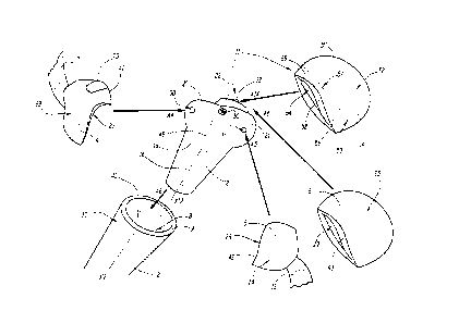
hereinbelow. A fractured long bone of the patient is depicted on figure 1. In
the present case,
the fractured long bone is a humerus, broken in four fragments at its shoulder
end. However,
the invention also applies to other long bones of the body, such as the hip
end of a femoral
bone.
The invention is preferably applied to a fractured long bone of a human
patient.
However, it may be applied to a fractured long bone of an animal patient.
The depicted fractured long bone comprises a diaphyseal fragment 2, or shaft
fragment.
The diaphyseal fragment 2 includes essentially the shaft part of the original
bone.
The fragment 2 defines a diaphyseal axis X2, which is extending along the
fragment 2.
This fragment 2 is of generally tubular shape around axis X2. The fragment 2
comprises a
11
CA 3032732 2019-02-05
medullary cavity 8, extending along axis X2, in particular coaxially. The
cavity 8 is opened at
a proximal end 10 of the fragment 2, where the bone is fractured. In the
present case of a
humeral diaphyseal fragment 2, the proximal end 10 is defined along axis X2
towards the
shoulder joint of the patient, opposite to a distal end of the fragment 2
directed towards the
elbow joint of the patient (not shown).
At the proximal end 10, the fragment 2 forms a fracture line 9, delineating
the open
cavity 8. The fracture line 9 surrounds the axis X2.
The depicted fractured long bone further comprises three epiphyseal fragments
4, 5 and
6. At least some of these fragments 4, 5 and 6 are to be reattached at the end
10 of the
fragment 2 for reconstructing the original bone.
Each fragment 4, 5 and 6 respectively has an internal surface 27, 28 and 29,
as well as
an external surface 23, 24 and 25. The surface 27 of the fragment 4 is opposed
to the surface
23. The surface 27 is delimited from the surface 23 by a fracture line 41 of
the fragment 4,
surrounding surface 23 and surface 27. The surface 28 of the fragment 5 is
opposed to the
surface 24. The surface 28 is delimited from the surface 24 by a fracture line
42 of the
fragment 5, surrounding surface 23 and surface 27. The surface 29 of the
fragment 6 is
opposed to the surface 25. The surface 29 is delimited from the surface 25 by
a fracture line
43 of the fragment 6, surrounding surface 25 and surface 29.
Depending on the fracture type and on the long bone considered, more or less
than
three epiphyseal fragments may be formed from the initial end of the patient's
long bone.
Only one epiphyseal fragment may be formed. However, the present invention
preferably
applies to cases where more than one epiphyseal fragments are formed.
Some of the epiphyseal fragments, like fragments 4 and 5 of the present
example, may
be designated as "tuberosity fragments". These particular fragments are each
secured to a
muscle of the patient by means of a tendon of the concerned muscle. In other
words, the
implantation sites of the concerned muscles are located on these tuberosity
fragments.
A muscle 7 is implanted to the external surface 23 of the fragment 4. In the
present
case, the fragment 4 includes the greater tuberosity of the original bone,
originally fixed with a
partially-illustrated supraspinatus muscle 7 of the patient.
A muscle 13 is implanted to the external surface 24 of the fragment 5. In the
present
case, the fragment 5 includes the lesser tuberosity of the original bone,
originally fixed with a
partially-illustrated subscapularis muscle 13 of the patient.
12
CA 3032732 2019-02-05
Some other epiphyseal fragments, like fragment 6 of the present example, may
be
designated as "articular fragment". Each articular fragment is initially part
of a joint of the
patient, for articulating the long bone with an auxiliary bone of the patient.
In the present case, the articular fragment 6 is a humeral head of the
humerus, which is
originally part of the shoulder joint, for articulating the humerus with a
scapula of the patient
(not shown on the drawings). In this case, the scapula forms the "auxiliary
bone" of the
shoulder joint. More precisely, the external surface 25 of the humeral head 6
has a generally
spherical and convex shape and is initially articulated with a corresponding
concave surface
of a glenoid part of the scapula.
Should some fragments have cracks or have geometrical defects may be spatially
reconstructed with bone graft, cement or any other suitable replacement
material. In the
present example, fragment 4 comprises a portion 70 of replacement material for
filling a notch
of the fragment 4. In this case, the portion 70 forms a part of the edge of
the fragment 4,
considered as a part of the fracture line 41, for the sake of simplicity.
As visible on figure 1, a prosthesis 11 comprises a stem part 12 and
advantageously a
head part 14. In the case the fractured long bone is a humerus, the prosthesis
11 constitutes
a shoulder prosthesis, or at least a humeral component of a shoulder
prosthesis. This
prosthesis 11, at least the stem part 12, is patient-specific. In other words,
the prosthesis 11
is designed depending on the geometry of the fractured long bone of one
specific patient, the
prosthesis 11 being intended to be implanted into this particular patient.
The stem part 12 comprises:
- a coverable diaphyseal portion 16, forming a distal end of the stem part
12 along axis
X12; and
- coverable epiphyseal portions 20, 21 and 22, located at a proximal end of
the stem part
12 along axis X12.
In some embodiments, the portions 16, 20, 21 and 22 form a single integral
piece. In
some other embodiments, one or more portions may be a separate piece assembled
with the
others.
The stem part 12 is configured to be inserted into the medullary cavity 8 of
the
diaphyseal fragment 2, through the open proximal end 10, as depicted with the
arrow A8,
parallel to axis X2. The stem part 12 preferably defines an axis X12, that is
parallel or coaxial
with axis X2 when the stem part 12 is secured to the fragment 2. This
insertion of the stem
part 12 preferably ensures securing the stem part 12 to the fragment 2. In
some
13
CA 3032732 2019-02-05
embodiments, supplementary means may be used for reinforcing the securing,
such as
cement, fasteners, or other conventional means.
The coverable diaphyseal portion 16 is configured for securing the stem part
12 to the
fragment 2, preferably by fitting or anchoring of the portion 16 into the
medullary cavity 8. At
least, the portion 16 has a shape that corresponds to the shape of the
medullary cavity 8,
specifically to this patient. The portion 16 is configured so that its
exterior surface is covered
entirely by the diaphyseal fragment 2 when the stem part 12 is secured to the
diaphyseal
fragment 2, whereas the rest of the stem part 12, located at a proximal end of
the stem part
12, is left uncovered by the diaphyseal fragment 2. In this case, "the rest of
the stem part 12"
includes the coverable epiphyseal portions 20, 22 and 24.
When inserted into the medullary cavity 8, the stem part 12 applies mechanical
stress to
the fragment 2, to an extent than may be chosen in advance prior or during
designing of the
prosthesis 11, thanks to a method explained below. In the present example, the
stem part 12
is configured so that the predetermined mechanical stress is applied to a
continuous contact
zone 40 of the fragment 2, also chosen in advance. This zone 40 is formed at
the surface
thereof, inside the cavity 8, as depicted on figure 1. This zone 40 is
entirely in contact with the
portion 16 of the stem part 12.
The zone 40 preferably extends all around axis X2, drawing a ring-like shape.
In other
words, the zone extends continuously along a circumference of the medullary
cavity around
axis X2.
In other embodiment, the mechanical stress may be applied to several distinct
zones of
the cavity 8.
In any case, the amount of mechanical stress to be applied is chosen in
advance, the
contact zone is chosen in advance, and the prosthesis is manufactured so that
said chosen
mechanical stress is actually applied onto said chosen contact zone 40 when
the prosthesis
11 is secured to the fragment 2. Thus, the prosthesis is made patient-
specifically. An
appropriate mechanical stress may be applied onto the diaphyseal fragment 2 so
as to avoid
osteonecrosis, or any other consequence relative to a lack of osseous
mechanical loading.
In the present invention, by "mechanical stress" applied to a bone fragment,
it is meant
a mechanical stress sufficient to avoid osseous necrosis. The mechanical
stress applied to
the bone fragments is also chosen not to exceed the mechanical resistance of
the bone
fragments. In other words, the mechanical stress is chosen not to break or
fracture the bone
fragments. The appropriate stress to be applied may be calculated depending on
the
14
CA 3032732 2019-02-05
geometry of the considered bone fragments and on the osseous density of these
fragments.
For example, the mechanical stress may be a pressure exerted onto the
considered bone
fragment, traction, flexion or the like.
Preferably, the mechanical stress is distributed evenly on the contact zone
40, or on the
contact zones if several contact zones are foreseen. In other words, a same
amount of stress
is applied to any part of the contact zone 40 or zones. Preferably, the
mechanical stress is
applied in radial outward directions around axis X12 by the coverable portion
16 onto the
zone 40.
In order to be able to apply the appropriate amount of stress onto the contact
zone and
to choose what zone of the medullar cavity should serve as the contact zone, a
specific
method of manufacturing is performed for obtaining the patient-specific
prosthesis 11.
For each prosthesis to be obtained and each patient to be treated, the method
comprises a preliminary step A of providing data representative of the
fractured long bone of
this patient, including essentially information relative to geometry of the
diaphyseal fragment
2. The information may also be relative to the osseous density of the fragment
2.
Preferably, this step of providing data is at least partially achieved by
scanning the
relevant part of the patient including the fractured long bone, for example
with a method of
CT-scan ("computerized tomography").
Successively to step A, based on the data gathered at step A, a step B of the
method is
performed. This step B includes a sub-step of choosing:
- which zone or zones of the medullary cavity 8 need to be stressed, and
- the magnitude of the stress to be applied onto said zone.
Once the zone and the magnitude of the stress are chosen, the stem part 12 of
the
prosthesis is designed accordingly, during a subsequent sub-step of step B.
The designed
stem part 12 is designed so as to be in contact with the chosen zone, becoming
the
aforementioned contact zone 40, so as to apply the chosen pressure, when
effectively
mounted to the epiphyseal fragment.
Thus, this step B allows designing the prosthesis 11, including in particular
the stem
part 12, specifically to the patient intended to receive this prosthesis 11.
During a step C of the method, successive to the step B, the prosthesis 11 is
manufactured, including the stem part 12 as it was designed during the second
step. Thus,
the patient-specific prosthesis 11 is obtained, for a specific patient.
CA 3032732 2019-02-05
The step C of manufacturing preferably includes additive manufacturing of the
entire
stem part 12. In some embodiment, only portions of the stem part 12 that are
required to be
patient-specific, such as the exterior surface in contact with the contact
zone 40, are
manufactured by additive manufacturing, these patient specific portion being
combined with
standard portions for forming the stem part 12.
For the present invention, any part obtained by additive manufacturing may be
metallic.
Instead, appropriate plastic material may be used.
Preferably, the stem part 12 comprises a visible mark 18 as visible on figure
1. The
visible mark 18 is formed at the exterior surface of the stem part 12. The
mark 18 may be a
shallow carving, an embossed marking, a colored marking, or the combination
thereof, on the
surface of the stem part 12. In any case, the mark 18 is configured to be
visible to the eye of
the surgeon during surgery. The mark 18 preferably forms a line, continuous or
dashed.
Instead, the mark 18 may form a dot, or several dots.
The mark 18 visually delimits the coverable diaphyseal portion 16 from the
rest of the
stem part 12. The prosthesis 11, in particular the coverable portion 16 and
the mark 18, is
designed specifically to the patient so that, when the stem part 12 is
inserted into the cavity 8
properly, in particular at the right position relative to the fragment 2 along
the diaphyseal axis
X2, the mark 18 and the fracture line 9 are superposed. Thus, during surgery,
the surgeon is
informed by the mark 18 whether the stem part 12 is properly positioned into
the cavity 8, at
least concerning the position of the stem part 12 relative to the fragment 2
along the axis X2.
Also, the shape of the mark 18 may indicate to the surgeon whether the stem
part 12 is
properly positioned relative to fragment 2, around axis X2. For example, the
mark 18 may
indicate with a dot, or have a portion reproducing the shape of, a
differential pattern around
axis X2, such as a dent or a notch, of the fracture line 9. The surgeon has to
ensure that the
dot or the portion is aligned with the dent.
Preferably, a correct position of the stem part 12 is obtained when the mark
18 and the
fracture line 9 are aligned and/or superposed. In case of misalignment, the
surgeon may
adjust the position of the stem part 12 relative to the fragment 2 during
surgery. Thus, the
mark 18 promotes adequate positioning of the stem part 12, and makes surgery
easier. In this
adequate positioning, the chosen stress is sure to be applied to the chosen
contact zone 40
by the stem part 12 onto the fragment 2.
For obtaining the mark 18 disclosed above, the data of the patient provided
during the
preliminary step A) of the method is used. During the step B) of designing the
prosthesis 11,
16
CA 3032732 2019-02-05
the stem part 12 is designed including the coverable diaphyseal portion 16 and
the mark 18,
so that they may achieve the above-disclosed functions. In particular, the
stem part 12 is
designed so that the mark 18 delimits the coverable diaphyseal portion 16
and/or indicates
where the fracture line 9 is foreseen to be located when the stem part 12 is
inserted into the
.. medullary cavity 8, covers the portion 16 and the stem part 12 applies the
chosen stress to
the zone 40.
In an embodiment, the portion 16 of the stem part 12 is radially outwardly
expansible
around axis X12. The expansion may be activated by the surgeon. For example,
the stem
part 12 is designed and manufactured including an expansion screw 60, coaxial
with axis
X12. The screw may be actuated from outside of the portion 16 by the surgeon:
for example
the screw head is accessible from the opposite end of the stem part 12. The
stem part 12
may be introduced into the cavity 8 to the desired position, and then, the
surgeon may
actuate the screw 60 for expanding the stem part radially outwardly, so that
the stem part
applies the chosen mechanical stress onto the zone 40. Any other suitable stem
part
.. expansion actuator than the screw 60 may be provided instead.
The stem part 12 is configured for receiving the epiphyseal fragments 4, 5 and
6. In
other words, these fragments 4, 5 and 6 may be secured to the stem part 12.
Specifically to the patient, the stem part 12 is designed so as to ensure a
patient-
specific positioning of the fragments 4, 5 and 6 relative to the stem part 12
and to each other.
The positioning of the fragment 2 relative to the stem part 12 may also be
planned patient-
specifically, as explained above, so that eventually, the fragments 2, 4, 5
and 6 are positioned
relative to each other in a chosen manner when secured to the stem part 12. As
concerns the
fragments 4, 5 and 6, the stem part 12 is designed so that the fragments are
positioned at
chosen securing positions, defined in advance, specifically to the patient. In
particular, this
positioning is chosen specifically to the shape of the fragments 4, 5 and 6,
and preferably also
to the shape of the fragment 2.
In the method of manufacturing the prosthesis 11, step A) may include
providing data
relative to the fragments 4, 5 and 6 of the long bone of the patient. This may
be performed
alternatively or additionally to providing data relative to the fragment 2.
The data provided
may be relative to the shape of the fragments 4, 5 and 6. The data may also be
relative to the
osseous density of the fragments 4, 5 and 6. The data for fragments 4, 5 and 6
may be
obtained by CT scanning, as explained above for fragment 2.
17
CA 3032732 2019-02-05
Step B) may include a sub-step of choosing, specifically to the patient,
respective
securing positions of the fragments 4, 5 and 6. The chosen securing positions
illustrate how
the fragments 4, 5 and 6 will be positioned relative to each other when
secured to the stem
part 12. The chosen securing positions may depend from the actual size and
shapes of the
fragments 4, 5 and 6 of the considered patient.
This sub-step of choosing the securing positions may be performed
alternatively or
additionally to the sub-step of choosing the contact zone 40 for fragment 2.
In this case, once the securing positions are chosen, the sub-step of
designing the stem
part 12 is performed, so that the stem part 12 is configured for securing the
epiphyseal
fragments at the respective chosen securing positions. In other words, the
designed stem part
enables or even imposes that, when the fragments 4, 5 and 6 are secured
thereto, they are in
the planned positions.
Then, during step C), the prosthesis 11, including in particular the stem part
12 with the
features designed in step B) above, is manufactured.
The fragments 4, 5 and 6 are configured to be positioned onto the stem part 12
as
illustrated in figure 1 with the arrows A4, A5 and A6, respectively.
When the fragments 4, 5 and 6 are secured to the stem part 12, each coverable
epiphyseal portion 20, 21 and 22 is configured for being essentially covered,
preferably
completely covered, by one of the epiphyseal fragments 4, 5 and 6,
respectively. Preferably,
for receiving the fragments 4, 5 or 6, each respective coverable portion 20,
21 and 22 has an
external surface, shaped in correspondence to an internal surface 27, 28 or 29
of the
respective concerned fragment 4, 5 or 6. When received properly, namely
according to the
chosen securing position, each fragment 4, 5 and 6 preferably entirely covers
the exterior
surface of the concerned portion 20, 21 and 22. For this purpose, the portions
20, 21 and 22
of the stem part 12 are designed patient specifically during step B).
Preferably, the stem part 12 is designed so that the reattached fragments 4, 5
and 6 are
positioned in their original position relative to each other and to the
diaphyseal fragment 2, as
at the time when the bone was not yet fractured.
In an alternative embodiment, the stem part 12 may be configured so that one
or more
of the fragment 2, 4, 5 and 6 is planned to be in a different position than
its original position
relative to the other fragments.
Preferably, the stem part 12 is designed so that the fragments 2, 4, 5 and 6
bear
against each other by means of their respective fracture lines 9, 41, 42 or 43
when they are
18
CA 3032732 2019-02-05
secured to the stem part 12. Thus, the chosen securing positions are
preferably positions
where the fragments 4, 5 and 6 bear against each other, and optionally at
least one of said
fragments 4, 5 and 6 bear against fragment 2. At least two of the fragments 2,
4, 5 and 6 bear
against each other in this manner. For example, when secured to the stem part
12, fragment
4 is in abutting contact with fragment 5, the fracture line 41 being in
abutting contact with the
fracture line 42.
In an embodiment, the stem part 12 is designed with a chosen securing position
for the
fragments 4, 5 and 6, enabling that the fragments 4, 5 and 6, and optionally
fragment 2, apply
mechanical stress to each other when the fragments 2, 4, 5 and 6 are actually
positioned at
these chosen securing positions.
For example, the stem part 12 is designed so that positioning the fragment 4
at its
securing position on the stem part 12 implies that this fragment 4 is
compressed between
fragments 2, 5 and 6, if these fragments 2, 5 and 6 are also positioned at
their respective
securing positions. In this case, the fragment 2, 5 and 6 apply mechanical
stress to the
fragment 4 by means of their respective fracture lines 9, 42 and 43, onto the
fracture line 41
of the fragment 4. In this case, resulting mechanical stress is also applied
onto the fragments
2, 5 and 6. This mechanical stress is achieved by planning an adequate
positioning of the
fragments 2, 4, 5 and 6 relative to each other. Specifically, the stem part 12
may be designed
so that the fragments 2, 4, 5 and 6 must be tightly fitted against each other
by the surgeon
when secured to the stem part 12 at their respective chosen securing
positions. The amount
of mechanical stress to be applied to each fragment 2, 4, 5 and 6 by one or
more other of
these fragments may also be chosen by appropriate designing of the stem part
12 and choice
of positioning of the fragments.
Thus, choosing the securing positions during step B) is made so that the
abovementioned mechanical stress, with a chosen magnitude, is applied on the
fragments 4,
5 and 6 when actually positioned this way. Thus, the obtained prosthesis 11
avoids
osteonecrosis by submitting the reattached fragment to an appropriate
mechanical stress.
For ensuring that the planned positioning of the fragments 4, 5 and 6 is
achieved, the
exterior surface of the stem part 12, in particular for the portions 20, 21
and 22, preferably has
a shape corresponding to the shape of fragments 4, 5 and 6, in particular
corresponding to
the surfaces 27, 28 and 29 of said fragments. The surgeon is informed that one
epiphyseal
fragment is correctly positioned, according to the chosen securing position,
if the epiphyseal
19
CA 3032732 2019-02-05
fragment fits onto the stem part 12. In case of incorrect positioning of the
epiphyseal
fragment, said fragment does not fit with the stem part 12.
Additionally or alternatively, for ensuring that the chosen securing position
of the
fragments 4, 5 and 6 is achieved, in the example illustrated on figure 1, the
stem part 12 may
comprise three plugs 30, 31 and 32, each constituting a distinct securing
element of one of
the fragments 4, 5 and 6, respectively. Each plug 30, 31 and 32 protrudes from
the exterior
surface of one of the portions 20, 21 or 22, respectively. Each plug is
preferably frustoconical,
pyramidal, or shaped as a dome. Conversely, each fragment 4, 5 and 6 may have
a blind
bore, provided on its internal surface 27, 28 and 29 respectively. These blind
bores are
preferably drilled or carved in the fragments 4, 5 and 6 by the surgeon with a
specific tool.
The shape of each bore corresponds to the shape of the plugs 30, 31 and 32, so
that each
plug may be inserted into the bore of one fragment 4, 5 or 6 when the
concerned fragment 4,
5 or 6 is positioned on the stem part 12. Thus, when the plug is inserted into
the appropriate
bore of a given fragment 4, 5 or 6, the surgeon is sure that this fragment is
correctly
positioned onto the stem part 12.
Preferably, the method of manufacturing includes designing and manufacturing
one or
more patient-specific jigs, depending on the chosen securing position and on
the data
representative of the patient. Each jig, for example embodied as a drill
guide, may be used by
the surgeon for drilling the blind bore on one of the epiphyseal fragments.
Each jig is adapted
to the shape of the bone fragments to be reworked by the surgeon. Thus, the
surgeon is sure
to drill the blind bores at the appropriate position on the fragments 4, 5 and
6, thus enabling a
positioning of the fragments 4, 5 and 6 at the chosen securing positions.
In a preferable embodiment, the plugs are configured so as to apply mechanical
stress
onto the fragments 4, 5 and 6. For example one of the epiphyseal fragments may
be
compressed between the plug, on which it is mounted, and another epiphyseal
fragment. In
this example, the plug radially applies mechanical stress to the blind bore of
the epiphyseal
fragment and said fragment receives mechanical stress onto the fracture line
in an opposed
direction. Thus, the risk of osseous necrosis is reduced.
Furthermore, the plugs may be designed so as to apply mechanical stress to the
blind
hole of the fragment by tight fitting into the blind hole.
More than one plug may be provided for each epiphyseal portion, and a
plurality of
plugs may be provided for positioning each epiphyseal fragment.
CA 3032732 2019-02-05
In some embodiments, the plugs are formed integral with the concerned
epiphyseal
portion. Alternatively, one or more of the securing element may be a separate
part assembled
with the concerned epiphyseal portion.
In some embodiments, instead of the aforementioned plugs, any other type of
positioning element may be provided.
Additionally or alternatively, the stem part 12 comprises a visible mark 45
indicating the
respective chosen securing positions of the epiphyseal fragments 4, 5 and 6 on
the stem part.
The visible mark 45 is formed at the exterior surface of the stem part 12. The
mark 45 may be
a shallow carving, an embossed marking, a colored marking, or the combination
thereof, on
the surface of the stem part 12. In any case, the mark 45 is configured to be
visible to the eye
of the surgeon during surgery. The mark 45 preferably forms one or more lines,
continuous or
dashed. Instead, the mark 45 may form one or more dots.
In a preferred embodiment, as illustrated on figure 1, the mark 45 visually
delimits the
coverable epiphyseal portions 20, 21 and 22 from each other and optionally
from the rest of
the stem part 12. The coverable portions 20, 21 and 22 and the mark 45 are
designed
specifically to the patient so that, when the fragments 4, 5 and 6 of this
patient are positioned
at the planned position on the stem part 12, the fracture lines 41, 42 and 43
are superposed
with the mark 45. In this aspect, the chosen securing positions are indicated
by the mark 45.
During surgery, the surgeon is informed by the mark 45 whether the fragments
4, 5 and 6 are
properly positioned on the stem part 12.
Thus, the mark 45 promotes adequate positioning of the fragments 4, 5 and 6,
and
makes surgery easier.
For obtaining the mark 45 disclosed above, the data of the patient provided
during the
step A) of the method is used. During the step B), the stem part 12 is
designed including the
coverable portions 20, 21, 22 and the mark 45, so that they may achieve the
above-disclosed
functions. In particular, the stem part 12 is designed so that the mark 45
delimits the
coverable epiphyseal portions 20, 21 and 22 from each other and/or indicates
where the
fracture lines 41, 42 and 43 are foreseen to be located when the fragments 4,
5 and 6 are
positioned according to the chosen securing positions.
For securing the fragments 4, 5 and 6 to the stem part 12, further
conventional means
suitable to the situation may be used, such as fasteners, cement and/or
sutures.
21
CA 3032732 2019-02-05
The surgeon may decide that the initial articular fragment 6 is reattached to
the stem
part 12, as disclosed above. In this situation, the head part 14 is optional.
For this situation,
the prosthesis 11 may be provided without such head part 14.
In some specific cases, the surgeon may decide that some of the epiphyseal
fragments
are viable and may be reattached to the stem part 12 and some other fragments
are
damaged and may not be reattached. In this case, the damaged epiphyseal
fragments may
require to be replaced with prosthetic means.
In the case the articular fragment 6 is damaged, while the fragments 4 and 5
are viable,
the fragment 6 may be replaced by the prosthetic head part 14 shown on figure
1. Thus, the
head part 14 is configured to be secured to the prosthetic stem part 12 so as
to replace the
damaged articular fragment 6 when the prosthesis 11 is introduced in the
patient's body. The
head part 14 is preferably secured at portion 22 of the stem part 12. The
securing may be
achieved with fasteners or any other suitable securing means. In the case the
head part 14 is
provided, the plug 32 is optional. Appropriate securing means may be provided
on the portion
22 additionally or alternatively to plug 32.
In this case, the method comprises, during step A), providing data relative to
the viable
fragments 4 and 5 and to the damaged fragment 6. Then, during step B), the
prosthesis 11 is
designed specifically to the patient, including the stem part 12 and the head
part 14 being
patient-specific.
In the case a head part 14 is used instead of reattaching the fragment 6, the
method is
similar than the previously explained method. Step B) includes a sub-step of
choosing,
specifically to the patient, respective securing positions of the viable
fragments 4 and 5 and of
the head part 14 relative to each other, and advantageously, relative to
fragment 2. In other
words, in this sub-step, the position of the head part 14 is chosen, instead
of the position of
the reattached fragment 6 for the case explained above. The chosen "securing
positions" are
representative of how the viable fragments 4 and 5 and the head part 14 are
planned to be
effectively positioned relative to each other when secured to the stem part 12
by the surgeon.
The fragments 4 and 5 and the head part 14 are configured to be positioned
onto the stem
part 12 as illustrated in figure 1 with the arrows A4, A5 and A14,
respectively. The fragments
4 and 5 are positioned as disclosed above, although fragment 6 is replaced by
the head part
14. Thus, the head part is positioned in a similar way than the fragment 6 of
the previous
case. When secured to the stem part 12, the head part 14 preferably covers the
portion 22
essentially or totally.
22
CA 3032732 2019-02-05
In this case, once the securing positions are chosen, the sub-step of
designing the stem
part 12 is performed. The head part 14 may also be designed patient-
specifically. In this
designing sub-step, the stem part 12, and optionally at least a part of the
head part 14, is
designed so that the stem part 12 allows securing the fragments 4 and 5 and
the head part 14
at the chosen securing positions. In other words, the designed stem part 12
enables or even
imposes that, when the fragments 4 and 5 the head part 14 are secured thereto,
they are in
the planned positions.
Then, during step C), the prosthesis 11, including in particular the stem part
12 with the
features designed in step B) above, is manufactured. If all or part of the
head part 14 is
designed patient-specifically during step B), the patient specific features of
the head part 14,
or all the head part 14 is also manufactured during step C).
The head part 14 may comprise a standard cap 50. "Standard" means that the cap
50 is
not patient-specific, although a cap of appropriate size and shape may be
chosen among a
definite collection of standard caps. Thus, the standard cap 50 is provided or
manufactured
separately from the patient-specific features manufactured at step C).
The standard cap 50 comprises an articular surface 52 of convex shape,
preferably
spherical, for forming a prosthetic joint of the patient, combined with the
glenoid cavity of the
scapula of the patient, or any other auxiliary bone considered, in replacement
for the surface
of fragment 6. In other words, the cap 50 may be shaped as a dome as depicted
on figure
20 1. By "prosthetic joint", it is meant that at least a part of the joint
is prosthetic. In this case, the
standard cap 50 is a prosthetic part of the joint.
Alternatively to a convex shape, depending on the situation, a concave shape
may be
used.
The standard cap 50 also comprises a trunnion 55, shown in dashed lines in
figure 1,
25 protruding in an opposed to the surface 52.
The standard cap 50 also comprises a securing surface 54, opposed to the
surface 52
and formed at the end of the trunnion 55. The standard cap 50 is configured to
be secured to
the stem part 12 by means of the securing surface 54.
The head part 14 also comprises a patient-specific insert 56. This insert 56
is patient
specifically designed during step B) according to the data provided at step
A), and
manufactured during step C) according to this design. The insert 56 is a
separate piece from
the cap 50 and is assembled with it before or during surgery.
23
CA 3032732 2019-02-05
The insert 56 is interposed between the cap 50 and the stem part 12. More
precisely,
the insert 56 has a surface 57 bearing against a border surface 53 of the cap
50. The border
surface 53 surrounds the trunnion 55, and is opposed to the surface 52. The
insert 56 also
has an opposed surface 58, that is configured to bear against the fracture
lines 41 and 42 of
the fragments 4 and 5. The insert 56 is preferably shaped as a ring or a
washer, as depicted
on figure 1, so that the cap 50 may be secured to the stem part 12 through the
insert 56. In
this case, the trunnion 55 passes through a central hole of the insert 56. The
insert 56 is
preferably fitted, for example conically fitted, onto the trunnion 55.
Alternatively, the cap of the head part 14 is made patient-specifically,
designed during
step B) and manufactured during step C). In this case, the cap and the insert
may form a
single piece instead of two distinct assembled pieces.
During step B), the respective securing positions of the fragments 4 and 5 and
of the
head part 14 are chosen so that a respective chosen mechanical stress is
applied to each
fragment 4 and 5 by the head part 14. In a preferable embodiment, the
fragments 4 and 5 are
compressed between the head part 14 and the fragment 2. Alternatively, the
head part 14
may apply mechanical stress on the fragments 4 and 5 without relying on the
fragment 2.
More specifically, step C) may include patient-specifically designing the
insert 56 so that
the insert 56 applies the respective stress onto the fragments 4 and 5 with a
chosen
magnitude. In particular, the shape of the surface 58 may be designed in
correspondence
.. with the shape of the fracture lines 41 and 42, so that the surface 58 may
distribute the
applied stress along the fracture lines 41 and 42.
Thus, when the prosthesis 11 is manufactured and implanted in the patient, a
mechanical stress defined in advance patient specifically is applied to the
reattached
fragments 4 and 5, and optionally to the fragment 2, by the head part 14, and
optionally by
the stem part 12.
The patient specific parts of the head part 14 may be obtained, during step
C), by
additive manufacturing. Preferably, the insert 56 is manufactured by additive
manufacturing. If
the cap 50 is patient-specific, the insert 56 and the cap 50 may both be
obtained by additive
manufacturing.
Instead of having a patient specific surface 58, the head part 14 may be
chosen
standard. Thus, only the relative securing position of the fragments 4 and 5
and of the head
part 14 may allow defining the mechanical stress to be applied.
24
CA 3032732 2019-02-05
After the prosthesis ills manufactured at step C) for the specific patient to
be treated,
the surgeon may proceed as follows for installing the prosthesis 11 in the
patient's body.
The surgeon firstly secures the stem part 12 onto the fragment 2, by
introduction of the
coverable portion 16 into the cavity 8. The surgeon checks that the mark 18 is
superposed
with the fracture line 9 and may adjust the position of the stem part if
necessary. In the
adequate position, the stem part 12 applies the chosen mechanical stress to
the zone 40 of
the cavity 8.
Secondly, the surgeon secures the fragments 4 and 5 onto the stem part 12. For
this
purpose, the surgeon may have prepared the fragments 4 and 5 in advance,
including drilling
the blind holes. The blind holes may be drilled with help of a tool and/or a
jig made patient-
specifically during step C), designed at step B) based on the data of step A).
In this case, the
fragments 4 and 5 are positioned onto the stem part 12 with the plugs 30 and
31 inserted into
the blind holes. The surgeon checks whether the positioning of the fragments 4
and 5 is
correct by checking if the fragments 4 and 5 match with the mark 45. If
necessary, the
position of the fragments 4 and 5 is adjusted by the surgeon with help of the
mark 45.
Thirdly, the fragment 6 is secured to the stem part 12. For this purpose, the
surgeon
may have prepared the fragment 6 in advance, including drilling the blind
hole. The blind hole
may be drilled with help of a tool and/or a jig made patient-specifically
during step C),
designed at step B) based on the data of step A). The surgeon checks whether
the
positioning of the fragment 6 is correct by checking if the fragment 6 matches
with the mark
45. If necessary, the position of the fragment 6 is adjusted by the surgeon
with help of the
mark 45. Securing the fragment 6 applies mechanical stress onto the fragments
4 and 5.
It can be provided that the fragments 4 and 5 are interposed between the
fracture line 9
of fragment 2 and the fragment 6. In this case, mechanical stress is also
applied to fragment
2 by fragments 4 and 5. In another embodiments, the fragments 4 and 5 are
supported by the
plugs 30 and 31, onto which the mechanical stress is transmitted, without
transmitting the
mechanical stress to the fragment 2.
Fasteners, sutures, adhesive means or any suitable means may be used by the
surgeon for maintaining the fragments 2, 4, 5 and 6 onto the stem part 12.
If the fragment 6 is to be replaced, said fragment 6 is not secured to the
stem part 12.
Instead, the head part 14 is secured to the stem part 12. Securing the head
part 14 applies
mechanical stress onto the fragments 4 and 5, and optionally to the fragment
2, in a similar
manner.
CA 3032732 2019-02-05
Fasteners, sutures, adhesive means or any suitable means may be used by the
surgeon for maintaining the fragments 2, 4 and 5 and the head part onto the
stem part 12.
The external surface of at least one of the coverable portions 16, 20, 21 and
22 is
preferably rough, or even comprises barbs, for helping securing the bone
fragments 2, 4, 5
and 6 secured thereto.
26
CA 3032732 2019-02-05