Note: Descriptions are shown in the official language in which they were submitted.
CA 03033505 2019-02-08
WO 2018/031920 PCT/US2017/046566
- 1 -
METHODS AND COMPOSITIONS RELATING TO IMPROVED HUMAN
RED BLOOD CELL SURVIVAL IN GENETICALLY MODIFIED
IMMUNODEFICIENT NON-HUMAN ANIMALS
GOVERNMENT SPONSORSHIP
[0001] This invention was made with government support under Grant No.
R24
OD018259 awarded by the National Institutes of Health. The government has
certain
rights in the invention.
REFERENCE TO RELATED APPLICATION
[0002] This application claims priority from U.S. Provisional Patent
Application
Serial No. 62/373,671, filed August 11,2016, the entire content of which is
incorporated
herein by reference.
FIELD OF THE INVENTION
[0003] General aspects of this disclosure relate to methods and
compositions for
assessment of human red blood cells in an immunodeficient genetically modified
animal.
In specific aspects, methods and compositions for assessment of human red
blood cells
in an immunodeficient genetically modified mouse are provided.
BACKGROUND OF THE INVENTION
[0004] Blood disorders such as malaria, sickle-cell anemia and aplastic
anemia
affect much of the world's population, especially those of African descent.
Currently, 3.2
billion people live in areas that are at risk of malaria transmission while 3
million
individuals have the sickle cell trait (CDC 2016). Advanced treatment of these
and other
blood disorders has been limited and there is a continuing need for non-human
animal
models, for assays of putative pharmaceutical therapeutics and other
treatments.
SUMMARY OF THE INVENTION
[0005] A genetically modified immunodeficient non-human animal whose
genome
includes a genetic modification that renders the non-human animal deficient in
macrophages and/or macrophage anti-human red blood cell activity so as to
prolong the
CA 03033505 2019-02-08
WO 2018/031920 PCT/US2017/046566
- 2 -
survival of human red blood cells when administered into said non-human animal
is
provided according to aspects of the present invention.
[0006] A genetically modified immunodeficient mouse whose genome
includes a
genetic modification that renders the mouse deficient in macrophages and/or
macrophage
anti-human red blood cell activity so as to prolong the survival of human red
blood cells
when administered into said mouse is provided according to aspects of the
present
invention.
[0007] A genetically modified NSG, NRG, or NOG mouse whose genome
includes
a genetic modification that renders the mouse deficient in macrophages and/or
macrophage anti-human red blood cell activity so as to prolong the survival of
human
red blood cells when administered into said mouse is provided according to
aspects of
the present invention.
[0008] A genetically modified immunodeficient non-human animal whose
genome
includes a genetic modification that renders the non-human animal deficient in
macrophages and/or macrophage anti-human red blood cell activity so as to
prolong the
survival of human red blood cells when administered into said non-human animal
is
provided according to aspects of the present invention, wherein the genetic
modification
is a mutation of a lysosomal trafficking regulator (Lyst) gene such that the
non-human
animal does not express functional lysosomal trafficking regulator protein,
rendering the
non-human animal deficient in macrophages and/or macrophage anti-human red
blood
cell activity.
[0009] A genetically modified immunodeficient mouse whose genome
includes a
genetic modification that renders the mouse deficient in macrophages and/or
macrophage
anti-human red blood cell activity so as to prolong the survival of human red
blood cells
when administered into said mouse is provided according to aspects of the
present
invention, wherein the genetic modification is a mutation of a mouse lysosomal
trafficking regulator gene such that the mouse does not express functional
lysosomal
trafficking regulator protein, rendering the mouse deficient in macrophages
and/or
macrophage anti-human red blood cell activity.
[0010] A genetically modified immunodeficient mouse whose genome includes a
genetic modification that renders the mouse deficient in macrophages and/or
macrophage
anti-human red blood cell activity so as to prolong the survival of human red
blood cells
when administered into said mouse is provided according to aspects of the
present
CA 03033505 2019-02-08
WO 2018/031920 PCT/US2017/046566
- 3 -
invention, wherein the genetic modification is a mutation of a mouse lysosomal
trafficking regulator gene such that the mouse does not express functional
lysosomal
trafficking regulator protein, rendering the mouse deficient in macrophages
and/or
macrophage anti-human red blood cell activity and wherein the mutation
comprises
deletion of a 25 bp sequence: GAGCCGGTAGCTTTGGTTCAACGGA (SEQ ID NO:
1) from exon 5 of the Lyst gene in the genome of the genetically modified
immunodeficient mouse.
[0011] A
genetically modified NSG, NRG, or NOG mouse whose genome includes
a genetic modification that renders the genetically modified NSG, NRG, or NOG
mouse
deficient in macrophages and/or macrophage anti-human red blood cell activity
so as to
prolong the survival of human red blood cells when administered into said
genetically
modified NSG, NRG, or NOG is provided according to aspects of the present
invention,
wherein the genetic modification is a mutation of a mouse lysosomal
trafficking
regulator (Lyst) gene such that the genetically modified NSG, NRG, or NOG does
not
.. express functional lysosomal trafficking regulator protein, rendering the
genetically
modified NSG, NRG, or NOG deficient in macrophages and/or macrophage anti-
human
red blood cell activity.
[0012]
According to aspects of the present invention, the genetically modified
immunodeficient mouse is a Lyst"ilimmunodeficient mouse.
[0013] According to aspects of the present invention, the genetically
modified
NSG, NRG, or NOG mouse is a Lyst"liNSG, NRG, or NOG mouse.
[0014]
According to aspects of the present invention, the genetically modified NSG,
NRG, or NOG mouse is an NSG, NRG, or NOG mouse that is homozygous for beige
mutation Lystbg.
[0015] According to aspects of the present invention, the genetically
modified
immunodeficient mouse is a NOD .0 g-Prkdc'd 112rg"lwfi IS zJ mouse homozygous
for
beige mutation Lystbg.
[0016]
According to aspects of the present invention, the genetically modified
immunodeficient mouse is a NO .0 g -Rag] "lm' 112 r g"lw-11 1 SzJ mouse
homozygous
for beige mutation Lystbg.
[0017]
According to aspects of the present invention, the genetically modified
immunodeficient mouse is a NOD.Cg-Prkdc'd 112rg"lsug 1JicTac mouse homozygous
for
beige mutation Lystbg.
CA 03033505 2019-02-08
WO 2018/031920 PCT/US2017/046566
- 4 -
[0018] According to aspects of the present invention, the genetically
modified
immunodeficient mouse is a NOD.Cg-Prkdcs" 112rg"lwfilLyst <em1Mvw>/Sz (NSG
Lyst knock out) mouse.
[0019] A genetically modified immunodeficient non-human animal whose
genome
includes a genetic modification that renders the non-human animal deficient in
macrophages and/or macrophage anti-human red blood cell activity so as to
prolong the
survival of human red blood cells when administered into said non-human animal
is
provided according to aspects of the present invention wherein the genetic
modification
includes a transgene encoding human CD47 such that the non-human animal
expresses
human CD47 protein and further includes a mutation of its endogenous CD47 gene
such
that the endogenous CD47 is not expressed, rendering the genetically modified
immunodeficient non-human animal deficient in macrophages and/or macrophage
anti-
human red blood cell activity.
[0020] A genetically modified immunodeficient mouse whose genome
includes a
.. genetic modification that renders the mouse deficient in macrophages and/or
macrophage
anti-human red blood cell activity so as to prolong the survival of human red
blood cells
when administered into said genetically modified immunodeficient mouse is
provided
according to aspects of the present invention wherein the genetic modification
includes a
transgene encoding human CD47 such that the mouse expresses human CD47 protein
.. and further includes a mutation of mouse CD47 gene such that the mouse CD47
is not
expressed, rendering the genetically modified immunodeficient mouse deficient
in
macrophages and/or macrophage anti-human red blood cell activity.
[0021] A genetically modified NSG, NRG, or NOG mouse whose genome
includes
a genetic modification that renders the NSG, NRG, or NOG deficient in
macrophages
.. and/or macrophage anti-human red blood cell activity so as to prolong the
survival of
human red blood cells when administered into said genetically modified NSG,
NRG, or
NOG mouse is provided according to aspects of the present invention wherein
the
genetic modification includes a transgene encoding human CD47 such that the
genetically modified NSG, NRG, or NOG mouse expresses human CD47 protein and
further includes a mutation of mouse CD47 gene such that the mouse CD47 is not
expressed, rendering the genetically modified NSG, NRG, or NOG mouse deficient
in
macrophages and/or macrophage anti-human red blood cell activity.
CA 03033505 2019-02-08
WO 2018/031920 PCT/US2017/046566
- 5 -
[0022]
According to aspects of the present invention, the genetically modified
immunodeficient mouse is a NOD.Cg-Prkdc<scid> Cd47<tmlFpl> Il2rg<tmlWjl>
Tg(CD47)2Sz/Sz (NSG Cd47 KO human CD47 Tg) mouse.
[0023] A
genetically modified immunodeficient non-human animal whose
genome includes a genetic modification that renders the non-human animal
deficient in
macrophages and/or macrophage anti-human red blood cell activity so as to
prolong the
survival of human red blood cells when administered into said non-human animal
is
provided according to aspects of the present invention wherein the genetic
modification
includes a transgene encoding herpes simplex virus 1 thymidine kinase such
that the
mouse expresses herpes simplex virus 1 thymidine kinase protein which, in
combination
with a nucleoside analog, renders the non-human animal deficient in
macrophages.
[0024] A
genetically modified immunodeficient mouse whose genome includes a
genetic modification that renders the genetically modified immunodeficient
mouse
deficient in macrophages and/or macrophage anti-human red blood cell activity
so as to
.. prolong the survival of human red blood cells when administered into said
genetically
modified immunodeficient mouse is provided according to aspects of the present
invention wherein the genetic modification includes a transgene encoding
herpes simplex
virus 1 thymidine kinase such that the genetically modified immunodeficient
mouse
expresses herpes simplex virus 1 thymidine kinase protein which, in
combination with a
nucleoside analog, renders the genetically modified immunodeficient mouse
deficient in
macrophages. A nucleoside analog such as ganciclovir, acyclovir or a
combination
thereof can be used.
[0025] A
genetically modified immunodeficient non-human animal of the present
invention further includes human red blood cells administered into the blood
system of
the genetically modified immunodeficient non-human animal, such as by
intraperitoneal
(IP) or intravenous (IV) administration.
[0026] A
genetically modified immunodeficient mouse of the present invention
further includes human red blood cells administered into the blood system of
the
genetically modified immunodeficient mouse, such as by intraperitoneal (IP) or
intravenous (IV) administration.
[0027]
Optionally, human hematopoietic cells (HSC) are administered to a
genetically modified immunodeficient mouse of the present invention, such that
human
red blood cells are generated in the genetically modified immunodeficient
mouse.
CA 03033505 2019-02-08
WO 2018/031920 PCT/US2017/046566
- 6 -
[0028]
Human red blood cells survive longer in a genetically modified
immunodeficient non-human animal, such as a genetically modified
immunodeficient
mouse, of the present invention than in an immunodeficient non-human animal,
such as
an immunodeficient mouse, of the same type whose genome does not include the
genetic
modification. For example, human red blood cells survive longer in a
genetically
modified NSG, NRG, or NOG mouse of the present invention than in an NSG, NRG,
or
NOG mouse whose genome does not include the genetic modification.
[0029]
Optionally, the human red blood cells present in the genetically modified
immunodeficient non-human animal of the present invention, such as a
genetically
.. modified immunodeficient mouse of the present invention, are infected by an
infectious
agent.
[0030]
Optionally, an infectious agent capable of infecting human red blood cells is
administered to a genetically modified immunodeficient non-human animal, such
as a
genetically modified immunodeficient mouse of the present invention. According
to
particular aspect, the infectious agent is a Plasmodium parasite, such as
Plasmodium
falciparum (P. falciparum), Plasmodium ovale (P. ovale), Plasmodium vivax (P.
vivax),
or Plasmodium malariae (P. malariae).
[0031]
Optionally, the human red blood cells administered to a genetically modified
immunodeficient non-human animal of the present invention, such as a
genetically
modified immunodeficient mouse of the present invention, are derived from an
individual human or population of human individuals, wherein the individual
human or
population of human individuals have sickle cell anemia.
[0032] A method of assaying an effect of a putative therapeutic agent is
provided
according to aspects of the present invention which includes administering an
amount of
the putative therapeutic agent to a genetically modified immunodeficient non-
human
animal of the present invention, wherein the genetically modified
immunodeficient non-
human animal of the present invention includes human red blood cells; and
measuring
the effect of the putative therapeutic agent.
[0033] A method of assaying an effect of a putative therapeutic agent is
provided
according to aspects of the present invention which includes administering an
amount of
the putative therapeutic agent to a genetically modified immunodeficient mouse
of the
present invention, wherein the genetically modified immunodeficient mouse of
the
CA 03033505 2019-02-08
WO 2018/031920 PCT/US2017/046566
- 7 -
present invention includes human red blood cells; and measuring the effect of
the
putative therapeutic agent.
[0034] Abbreviations used for certain genetically modified immunodeficient
mouse
strains:
MD1: NOD.Cg-Prkdc'd 112rg"lwfilLyst <em1Mvw>/Sz (NSG mouse strain with Lyst
knock out by deletion of SEQ ID NO:1 from mouse Lyst gene).
MD2: NSG CD47 KO Tg(hCD47) (NSG mouse strain with mouse CD47 knocked out
and including a transgene encoding human CD47).
MD3: NSG CSF1r-HTK (NSG mouse strain including transgene in which the CSF1r
promoter drives expression of herpes thymidine kinase).
MD4: B6.1295-Rag 1 <tmlMom> CD47 KO Il2rg<tmlWjl>/Sz ((BL/6 mouse strain with
mouse IL2rg knock out and mouse CD47 knock out, also called BL/6
Rag1"11CD47KOIL2rg"11).
BRIEF DESCRIPTION OF THE DRAWINGS
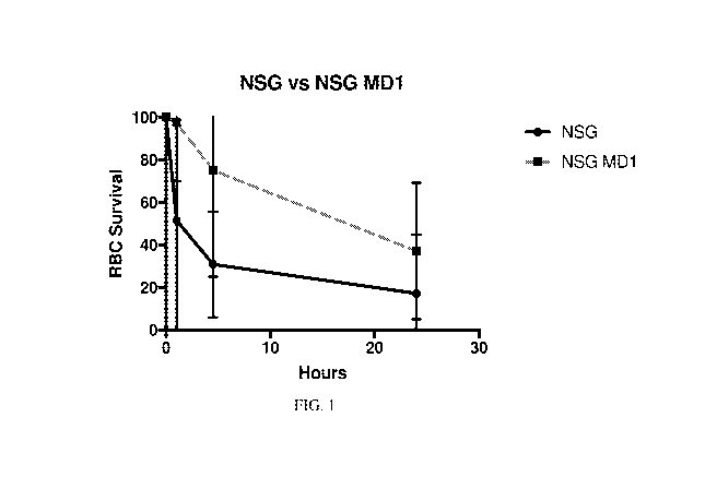
[0035] Figure 1 is a graph showing the survival of human red blood
cells (RBC) in
NSG Lyst (MD1) knock-out (KO) mice compared to NSG control mice and
demonstrating a two-fold increase in the number of human red blood cells that
survived
in the immunodeficient genetically modified mouse at 24 hours after
administration of
human red blood cells.
[0036] Figure 2 is a graph showing the results from NSG HSV-1-tk
transgenic (Tg)
mice compared to NSG control mice and demonstrating a significantly higher
number of
human red blood cells that survived in the immunodeficient genetically
modified mouse
at up to 24 hours after administration of human red blood cells.
[0037] Figure 3 is a graph showing results from BL/6 Rag gamma MD4 mice
compared to NSG control mice.
[0038] Figure 4 is a graph showing results from NSG MD2 mice compared
to NSG
control mice and demonstrating a greater than two-fold increase in the number
of human
red blood cells that survived in the immunodeficient genetically modified
mouse at 24
hours after administration of human red blood cells and survival of the human
red blood
cells in significantly greater numbers in NSG MD2 mice compared to NSG control
mice
at 48, 72 and 96 hours after administration of the human red blood cells.
CA 03033505 2019-02-08
WO 2018/031920 PCT/US2017/046566
- 8 -
[0039] Figure 5 is a graph showing results comparing human RBC survival
in
several different genetically modified immunodeficient strains of the present
invention
compared to NSG control mice and demonstrating an increase in the number of
human
red blood cells that survived in the immunodeficient genetically modified MD1
and
MD2 mice compared to NSG control mice.
DETAILED DESCRIPTION OF THE INVENTION
[0040] Scientific and technical terms used herein are intended to have
the meanings
commonly understood by those of ordinary skill in the art. Such terms are
found defined
and used in context in various standard references illustratively including J.
Sambrook
and D.W. Russell, Molecular Cloning: A Laboratory Manual, Cold Spring Harbor
Laboratory Press; 3rd Ed., 2001; F.M. Ausubel, Ed., Short Protocols in
Molecular
Biology, Current Protocols; 5th Ed., 2002; B. Alberts et al., Molecular
Biology of the
Cell, 4th Ed., Garland, 2002; D.L. Nelson and M.M. Cox, Lehninger Principles
of
Biochemistry, 4th Ed., W.H. Freeman & Company, 2004; A. Nagy, M. Gertsenstein,
K.
Vintersten, R. Behringer, Manipulating the Mouse Embryo: A Laboratory Manual,
3rd
edition, Cold Spring Harbor Laboratory Press; December 15, 2002, ISBN-10:
0879695919; Kursad Turksen (Ed.), Embryonic stem cells: methods and protocols
in
Methods Mol Biol. 2002;185, Humana Press; Current Protocols in Stem Cell
Biology,
ISBN: 9780470151808; Chu, E. and Devita, V.T., Eds., Physicians' Cancer
Chemotherapy Drug Manual, Jones & Bartlett Publishers, 2005; J.M. Kirkwood et
al.,
Eds., Current Cancer Therapeutics, 4th Ed., Current Medicine Group, 2001;
Remington:
The Science and Practice of Pharmacy, Lippincott Williams & Wilkins, 21st Ed.,
2005;
L.V. Allen, Jr. et al., Ansel's Pharmaceutical Dosage Forms and Drug Delivery
Systems,
8th Ed., Philadelphia, PA: Lippincott, Williams & Wilkins, 2004; and L.
Brunton et al.,
Goodman & Gilman's The Pharmacological Basis of Therapeutics, McGraw-Hill
Professional, 12th Ed., 2011.
[0041] The singular terms "a," "an," and "the" are not intended to be
limiting and
include plural referents unless explicitly stated otherwise or the context
clearly indicates
.. otherwise.
[0042] A genetically modified immunodeficient non-human animal whose
genome
includes a genetic modification, wherein the genetic modification renders the
non-human
animal deficient in macrophages and/or macrophage anti-human red blood cell
activity,
CA 03033505 2019-02-08
WO 2018/031920 PCT/US2017/046566
- 9 -
is provided according to aspects of the present invention. The genetically
modified
immunodeficient non-human animal further includes human red blood cells
according to
aspects of the present invention. Human red blood cells administered into the
blood
system of the genetically modified immunodeficient non-human animal survive
longer in
the genetically modified immunodeficient non-human animal than in an
immunodeficient non-human animal of the same type whose genome does not
include
the genetic modification.
[0043] The term "immunodeficient non-human animal" refers to a non-human
animal characterized by one or more of: a lack of functional immune cells,
such as T
cells and B cells; a DNA repair defect; a defect in the rearrangement of genes
encoding
antigen-specific receptors on lymphocytes; and a lack of immune functional
molecules
such as IgM, IgG 1 , IgG2a, IgG2b, IgG3 and IgA.
[0044] According to aspects of the present invention, a genetically
modified
immunodeficient non-human animal whose genome includes a genetic modification,
wherein the genetic modification renders the non-human animal deficient in
macrophages and/or macrophage anti-human red blood cell activity, provided
according
to aspects of the present invention is a mouse. While description herein
refers primarily
to aspects of the present invention in which the genetically modified
immunodeficient
non-human animal is a mouse, the genetically modified immunodeficient non-
human
animal can also be a mammal such as a rat, gerbil, guinea pig, hamster,
rabbit, pig,
sheep, or non-human primate.
[0045] The phrase "genetically modified immunodeficient mouse" as used
herein
refers to an immunodeficient mouse whose genome includes a genetic
modification,
wherein the genetic modification renders the immunodeficient mouse deficient
in
macrophages and/or macrophage anti-human red blood cell activity.
[0046] The phrase "deficient in macrophages" refers to a reduction in
the number of
macrophages compared to the number of macrophages present in a comparable
immunodeficient mouse which does not have the genetic mutation that renders
the
immunodeficient mouse deficient in macrophages.
[0047] The term "immunodeficient mouse" refers to a mouse characterized by
one or
more of: a lack of functional immune cells, such as T cells and B cells; a DNA
repair
defect; a defect in the rearrangement of genes encoding antigen-specific
receptors on
lymphocytes; and a lack of immune functional molecules such as IgM, IgG 1 ,
IgG2a,
CA 03033505 2019-02-08
WO 2018/031920 PCT/US2017/046566
- 10 -
IgG2b, IgG3 and IgA. Immunodeficient mice can be characterized by one or more
deficiencies in a gene involved in immune function, such as Rag] and Rag2
(Oettinger,
M.A et al., Science, 248:1517-1523, 1990; and Schatz, D. G. et al., Cell,
59:1035-1048,
1989) Immunodeficient mice may have any of these or other defects which result
in
abnormal immune function in the mice.
[0048]
Particularly useful immunodeficient mouse strains are NOD.Cg-Prkdcsctd
iargtmlWil/SZJ, commonly referred to as NOD scid gamma (NSG) mice, described
in
detail in Shultz LD et al, 2005, J. Immunol, 174:6477-89; NOD.Cg-RagltmlMom
112relw-11/SzJ, Shultz LD et al, 2008 Clin Exp Immunol 154(2):270-84 commonly
referred to as NRG mice; and NOD.Cg-Prkdcscid 112rg"-isuglEcTac, described in
detail in
Ito, M. et al., Blood 100, 3175-3182 (2002) commonly referred to as NOG mice.
[0049]
The term "severe combined immune deficiency (SCID)" refers to a condition
characterized by absence of T cells and lack of B cell function.
[0050]
Common forms of SCID include: X-linked SCID which is characterized by
gamma chain gene mutations in the IL2RG gene and the lymphocyte phenotype T(-)
B(+) NK(-); and autosomal recessive SCID characterized by Jak3 gene mutations
and the
lymphocyte phenotype T(-) B(+) NK(-), ADA gene mutations and the lymphocyte
phenotype T(-) B(-) NK(-), IL-7R alpha-chain mutations and the lymphocyte
phenotype
T(-) B(+) NK(+), CD3 delta or epsilon mutations and the lymphocyte phenotype
T(-)
B(+) NK(+), RAG1/RAG2 mutations and the lymphocyte phenotype T(-) B(-) NK(+),
Artemis gene mutations and the lymphocyte phenotype T(-) B(-) NK(+), CD45 gene
mutations and the lymphocyte phenotype T(-) B(+) NK(+).
[0051] In
further aspects, a genetically modified immunodeficient mouse has a
defect in its endogenous gene encoding DNA-dependent protein kinase, catalytic
subunit
(Prkdc) which causes the mouse to express a defective endogenous DNA-dependent
protein kinase, catalytic subunit and/or a reduced amount of endogenous DNA-
dependent protein kinase, catalytic subunit, or the mouse may not express
endogenous
DNA-dependent protein kinase, catalytic subunit at all. The immunodeficient
mouse can
optionally be Prkdc null such that it lacks a functional endogenous Prkdc
gene).
[0052] A genetically modified mouse according to aspects of the present
invention
has the severe combined immunodeficiency mutation (Prkdc), commonly referred
to
as the scid mutation. The scid mutation is well-known and located on mouse
chromosome 16 as described in Bosma, et al., Immunogenetics 29:54-56, 1989.
Mice
CA 03033505 2019-02-08
WO 2018/031920 PCT/US2017/046566
- 11 -
homozygous for the scid mutation are characterized by an absence of functional
T cells
and B cells, lymphopenia, hypoglobulinemia and a normal hematopoetic
microenvironment. The scid mutation can be detected, for example, by detection
of
markers for the scid mutation using well-known methods, such as PCR or flow
cyotometry.
[0053] A
genetically modified mouse according to aspects of the present invention
has an IL2 receptor gamma chain deficiency. The term "IL2 receptor gamma chain
deficiency" refers to decreased IL2 receptor gamma chain. Decreased IL2
receptor
gamma chain can be due to gene deletion or mutation. Decreased IL2 receptor
gamma
chain can be detected, for example, by detection of IL2 receptor gamma chain
gene
deletion or mutation and/or detection of decreased IL2 receptor gamma chain
expression
using well-known methods.
[0054]
According to aspects of the present invention, a genetically modified
immunodeficient NSG mouse is provided whose genome includes a genetic
modification, wherein the genetic modification renders the mice deficient in
macrophages and/or macrophage anti-human red blood cell activity.
[0055]
According to aspects of the present invention, a genetically modified
immunodeficient NRG mouse is provided whose genome includes a genetic
modification, wherein the genetic modification renders the mice deficient in
macrophages and/or macrophage anti-human red blood cell activity.
[0056]
According to aspects of the present invention, a genetically modified
immunodeficient NOG mouse is provided whose genome includes a genetic
modification, wherein the genetic modification renders the mice deficient in
macrophages and/or macrophage anti-human red blood cell activity.
[0057] Genetic modification that produces an immunodeficient mouse
deficient in
macrophages and/or macrophage anti-human red blood cell activity
[0058]
According to aspects of the present invention, a genetically modified
immunodeficient mouse is provided wherein the genetic modification is a
mutation of a
lysosomal trafficking regulator (Lyst) gene such that the mouse does not
express
functional lysosomal trafficking regulator protein rendering the non-human
animal
deficient in macrophages and/or macrophage anti-human red blood cell activity.
The
genetically modified immunodeficient non-human animals further include human
red
blood cells according to aspects of the present invention. Human red blood
cells
CA 03033505 2019-02-08
WO 2018/031920 PCT/US2017/046566
- 12 -
administered into the blood system of the genetically modified immunodeficient
non-
human animals survive longer in the genetically modified immunodeficient non-
human
animals than in immunodeficient non-human animals of the same type whose
genome
does not include the genetic modification. The Lyst gene is located at
Chr13:13590409-
13777440 bp, + strand in mouse and is conserved in numerous species including
human,
chimpanzee, Rhesus monkey, dog, cow, rat, chicken, zebrafish, and frog.
[0059] According to aspects of the present invention, a genetically
modified
immunodeficient mouse having one or more spontaneous or induced mutations at
the
Lyst locus are provided wherein the mouse does not express functional
lysosomal
trafficking regulator protein rendering the mouse deficient in macrophages
and/or
macrophage anti-human red blood cell activity. The genetically modified
immunodeficient mouse further includes human red blood cells according to
aspects of
the present invention. Human red blood cells administered into the blood
system of the
genetically modified immunodeficient mouse survive longer in the genetically
modified
immunodeficient mouse than in an immunodeficient mouse of the same type whose
genome does not include the genetic modification.
[0060] According to aspects of the present invention, a genetically
modified
immunodeficient mouse having a deletion in exon 5 at the Lyst locus is
provided
wherein the mouse does not express functional lysosomal trafficking regulator
protein
rendering the mouse deficient in macrophages and/or macrophage anti-human red
blood
cell activity. The genetically modified immunodeficient mouse further includes
human
red blood cells according to aspects of the present invention. Human red blood
cells
administered into the blood system of the genetically modified immunodeficient
mouse
survive longer in the genetically modified immunodeficient mouse than in an
immunodeficient mouse of the same type whose genome does not include the
genetic
modification.
[0061] According to aspects of the present invention, a genetically
modified
immunodeficient mouse having a 25 bp deletion in exon 5 at the Lyst locus is
provided
wherein the mouse does not express functional lysosomal trafficking regulator
protein
rendering the mouse deficient in macrophages and/or macrophage anti-human red
blood
cell activity. According to particular aspects of the present invention, the
25 bp deletion
is deletion of the a 25 bp segment of genomic DNA in exon 5 at the Lyst locus
having
the nucleotide sequence GAGCCGGTAGCTTTGGTTCAACGGA (SEQ ID NO: 1). The
CA 03033505 2019-02-08
WO 2018/031920 PCT/US2017/046566
- 13 -
genetically modified immunodeficient mouse further includes human red blood
cells
according to aspects of the present invention. Human red blood cells
administered into
the blood system of the genetically modified immunodeficient mouse survive
longer in
the genetically modified immunodeficient mouse than in an immunodeficient
mouse of
the same type whose genome does not include the genetic modification.
[0062] According to aspects of the present invention, a genetically
modified
immunodeficient mouse having one or more spontaneous or induced mutations at
the
Lyst locus such that the mice are Lyst'll is provided wherein the mouse does
not express
functional lysosomal trafficking regulator protein rendering the mouse
deficient in
macrophages and/or macrophage anti-human red blood cell activity. The
genetically
modified immunodeficient mouse further includes human red blood cells
according to
aspects of the present invention. Human red blood cells administered into the
blood
system of the genetically modified immunodeficient mouse survive longer in the
genetically modified immunodeficient mouse than in an immunodeficient mouse of
the
same type whose genome does not include the genetic modification.
[0063] According to aspects of the present invention, a genetically
modified
immunodeficient mouse homozygous for a beige mutation Lystbg is provided
wherein the
mouse does not express functional lysosomal trafficking regulator protein
rendering the
mouse deficient in macrophages and/or macrophage anti-human red blood cell
activity.
A Lystbg remutation (Lystbg-J) occurred spontaneously in the C57BL/6J strain
at The
Jackson Laboratory (J:5311). This allele is defined by a noncomplementation
test with
Lystbg This is the result of a 3 bp deletion in exon 54 of Lyst causing an
isoleucine
deletion at codon 3741 near the carboxy terminus of the protein. This deletion
affects the
WD40 domain. The genetically modified immunodeficient mouse further includes
human red blood cells according to aspects of the present invention. Human red
blood
cells administered into the blood system of the genetically modified
immunodeficient
mouse survive longer in the genetically modified immunodeficient mouse than in
an
immunodeficient mouse of the same type whose genome does not include the
genetic
modification.
[0064] According to aspects of the present invention, a genetically
modified
immunodeficient NOD.Cg-Prkdcscid 112rg"lwfilSzJ mouse homozygous for a beige
mutation Lystbg is provided wherein the mouse does not express functional
lysosomal
trafficking regulator protein rendering the mouse deficient in macrophages
and/or
CA 03033505 2019-02-08
WO 2018/031920 PCT/US2017/046566
- 14 -
macrophage anti-human red blood cell activity. The genetically modified
immunodeficient mouse further includes human red blood cells according to
aspects of
the present invention. Human red blood cells administered into the blood
system of the
genetically modified immunodeficient mouse survive longer in the genetically
modified
immunodeficient mouse than in an immunodeficient mouse of the same type whose
genome does not include the genetic modification.
[0065] According to aspects of the present invention, a genetically
modified
immunodeficient NOD.Cg-Prkdc'd 112r g"lwfilLyst <em1Mvw>/Sz (NSG Lyst knock
out - MD1) mouse is provided wherein the mouse does not express functional
lysosomal
trafficking regulator protein rendering the mouse deficient in macrophages
and/or
macrophage anti-human red blood cell activity. The genetically modified
immunodeficient mouse further includes human red blood cells according to
aspects of
the present invention. Human red blood cells administered into the blood
system of the
genetically modified immunodeficient mouse survive longer in the genetically
modified
immunodeficient mouse than in an immunodeficient mouse of the same type whose
genome does not include the genetic modification.
[0066] According to aspects of the present invention, a genetically
modified
immunodeficient NOD.Cg-Rag/"-im' 112rg"lwfilSzJ mouse homozygous for a beige
mutation Lystbg is provided wherein the mouse does not express functional
lysosomal
trafficking regulator protein rendering the mouse deficient in macrophages
and/or
macrophage anti-human red blood cell activity. The genetically modified
immunodeficient mouse further includes human red blood cells according to
aspects of
the present invention. Human red blood cells administered into the blood
system of the
genetically modified immunodeficient mouse survive longer in the genetically
modified
immunodeficient mouse than in an immunodeficient mouse of the same type whose
genome does not include the genetic modification.
[0067] According to aspects of the present invention, a genetically
modified
immunodeficient mouse is provided wherein the genetic modification includes a
transgene encoding human CD47 such that the mouse expresses human CD47 protein
and further includes a mutation of the mouse CD47 gene in the genome of the
mouse
such that the mouse does not express functional mouse CD47 protein, rendering
the
mouse deficient in macrophages and/or macrophage anti-human red blood cell
activity.
The genetically modified immunodeficient mouse further includes human red
blood cells
CA 03033505 2019-02-08
WO 2018/031920 PCT/US2017/046566
- 15 -
according to aspects of the present invention. Human red blood cells
administered into
the blood system of the genetically modified immunodeficient mouse survive
longer in
the genetically modified immunodeficient mouse than in an immunodeficient
mouse of
the same type whose genome does not include the genetic modification.
[0068] According to aspects of the present invention, a genetically
modified
immunodeficient NSG mouse is provided, wherein the genetic modification
includes a
transgene encoding human CD47 such that the mouse expresses human CD47 protein
and further includes a mutation of the mouse CD47 gene in the genome of the
mouse
such that the mouse does not express functional mouse CD47 protein, rendering
the
mouse deficient in macrophages and/or macrophage anti-human red blood cell
activity.
The genetically modified immunodeficient mouse further includes human red
blood cells
according to aspects of the present invention. Human red blood cells
administered into
the blood system of the genetically modified immunodeficient mouse survive
longer in
the genetically modified immunodeficient mouse than in an immunodeficient
mouse of
the same type whose genome does not include the genetic modification.
[0069] According to aspects of the present invention, a genetically
modified
immunodeficient NRG mouse is provided, wherein the genetic modification
includes a
transgene encoding human CD47 such that the mouse expresses human CD47 protein
and further includes a mutation of the mouse CD47 gene in the genome of the
mouse
such that the mouse does not express functional mouse CD47 protein, rendering
the
mouse deficient in macrophages and/or macrophage anti-human red blood cell
activity.
The genetically modified immunodeficient mouse further includes human red
blood cells
according to aspects of the present invention. Human red blood cells
administered into
the blood system of the genetically modified immunodeficient mouse survive
longer in
the genetically modified immunodeficient mouse than in an immunodeficient
mouse of
the same type whose genome does not include the genetic modification.
[0070] According to aspects of the present invention, a genetically
modified
immunodeficient NOG mouse is provided, wherein the genetic modification
includes a
transgene encoding human CD47 such that the mouse expresses human CD47 protein
and further includes a mutation of the mouse CD47 gene in the genome of the
mouse
such that the mouse does not express functional mouse CD47 protein, rendering
the
mouse deficient in macrophages and/or macrophage anti-human red blood cell
activity.
The genetically modified immunodeficient mouse further include human red blood
cells
CA 03033505 2019-02-08
WO 2018/031920 PCT/US2017/046566
- 16 -
according to aspects of the present invention. Human red blood cells
administered into
the blood system of the genetically modified immunodeficient mouse survive
longer in
the genetically modified immunodeficient mouse than in an immunodeficient
mouse of
the same type whose genome does not include the genetic modification.
[0071] According to aspects of the present invention, a genetically
modified
immunodeficient NOD .Cg -Prkdc < scid > Cd47 <tmlFpl>
Il2rg <tmlWjl>
Tg(CD47)2Sz/Sz (NSG Cd47 KO human CD47 Tg) mouse is provided. The genetically
modified immunodeficient mouse further includes human red blood cells
according to
aspects of the present invention. Human red blood cells administered into the
blood
system of the genetically modified immunodeficient mouse survive longer in the
genetically modified immunodeficient mouse than in an immunodeficient mouse of
the
same type whose genome does not include the genetic modification.
[0072]
According to aspects of the present invention, a genetically modified
immunodeficient mouse is provided, wherein the genetic modification includes a
transgene encoding herpes simplex virus 1 thymidine kinase such that the mouse
expresses herpes simplex virus 1 thymidine kinase protein which, in
combination with a
nucleoside analog, renders the mouse deficient in macrophages. Any nucleoside
analog
which is toxic to macrophages in combination with herpes simplex virus 1
thymidine
kinase protein can be used, exemplified by, but not limited to, ganciclovir,
acyclovir or a
combination thereof. The genetically modified immunodeficient mouse further
includes
human red blood cells according to aspects of the present invention. Human red
blood
cells administered into the blood system of the genetically modified
immunodeficient
mouse survive longer in the genetically modified immunodeficient mouse than in
an
immunodeficient mouse of the same type whose genome does not include the
genetic
modification.
[0073]
Any of various methods can be used to produce a genetically modified
immunodeficient non-human animal, such as a mouse, whose genome includes a
genetic
modification. Genetic modifications are produced using standard methods of
genetic
engineering such as, but not limited to, chemical mutagenesis, irradiation,
homologous
recombination and transgenic expression of antisense RNA. Such techniques are
well-
known in the art and further include, but are not limited to, pronuclear
microinjection
and transformation of embryonic stem cells. Methods for generating genetically
modified animals whose genome includes a disrupted gene that can be used
include, but
CA 03033505 2019-02-08
WO 2018/031920 PCT/US2017/046566
- 17 -
are not limited to, those described in J. P. Sundberg and T. Ichiki, Eds.,
Genetically
Engineered Mice Handbook, CRC Press; 2006; M. H. Hofker and J. van Deursen,
Eds.,
Transgenic Mouse Methods and Protocols, Humana Press, 2002; A. L. Joyner, Gene
Targeting: A Practical Approach, Oxford University Press, 2000; Manipulating
the
Mouse Embryo: A Laboratory Manual, 3rd edition, Cold Spring Harbor Laboratory
Press; December 15, 2002, ISBN-10: 0879695919; Kursad Turksen (Ed.), Embryonic
stem cells: methods and protocols in Methods Mol Biol. 2002;185, Humana Press;
Current Protocols in Stem Cell Biology, ISBN: 978047015180; Meyer et al. PNAS
USA,
vol. 107 (34), 15022-15026.
[0074] Homology-based recombination gene modification strategies can be
used to
genetically modify an immunodeficient mouse by "knock-in" to introduce a
nucleic acid
encoding an exogenous protein or proteins e.g., a nucleotide sequence encoding
herpes
simplex virus 1 thymidine kinase or a nucleotide sequence encoding human CD47
into
the genome of the immunodeficient mouse. Similarly, a homology-based
recombination
gene modification strategy can be used to genetically modify an
immunodeficient mouse
by "knock-out" or mutate a gene encoding an enogenous protein or proteins
e.g., mouse
CD47 or mouse Lyst.
[0075] Homology-based recombination gene modification strategies
include gene
editing approaches such as those using homing endonucleases, integrases,
meganucleases, transposons, nuclease-mediated processes using a zinc finger
nuclease
(ZFN), a Transcription Activator-Like (TAL), a Clustered Regularly Interspaced
Short
Palindromic Repeats (CRISPR)-Cas, or a Drosophila Recombination-Associated
Protein
(DRAP) approach. See, for example, Cerbini et al., PLoS One. 2015; 10(1):
e0116032;
Shen et al., PLoS ONE 8(10): e77696; and Wang et al., Protein & Cell, February
2016,
Volume 7, Issue 2, pp 152-156.
[0076] Genomic editing is performed, for example, by methods described
herein,
and as detailed in J. P. Sundberg and T. Ichiki, Eds., Genetically Engineered
Mice
Handbook, CRC Press; 2006; M. H. Hofker and J. van Deursen, Eds., Transgenic
Mouse
Methods and Protocols, Humana Press, 2002; A. L. Joyner, Gene Targeting: A
Practical
Approach, Oxford University Press, 2000; Manipulating the Mouse Embryo: A
Laboratory Manual, 3rd edition, Cold Spring Harbor Laboratory Press; December
15,
2002, ISBN-10: 0879695919; Kursad Turksen (Ed.), Embryonic stem cells: methods
and
protocols in Methods Mol Biol. 2002;185, Humana Press; Current Protocols in
Stem Cell
CA 03033505 2019-02-08
WO 2018/031920 PCT/US2017/046566
- 18 -
Biology, ISBN: 978047015180; Meyer et al., PNAS USA, 2010, vol. 107 (34),
15022-
15026; and Doudna, J. et al. (eds.) CRISPR-Cas: A Laboratory Manual, 2016,
CSHP. A
brief description of several genomic editing techniques is described herein.
[0077] Nuclease Techniques for Genetic Modification of Mice
[0078] A genetic modification method, such as but not limited to, a
nuclease genetic
editing technique, can be used to introduce a desired DNA sequence into the
genome at a
predetermined target site, such as methods using a homing endonuclease,
integrase,
meganuclease, transposon, nuclease-mediated process using a zinc finger
nuclease
(ZFN), a Transcription Activator-Like (TAL), a Clustered Regularly Interspaced
Short
Palindromic Repeats (CRISPR)-Cas, or Drosophila Recombination-Associated
Protein
(DRAP). Briefly, a genetic modification method that can be used includes
introducing
into an ES cell, iPS cell, somatic cell, fertilized oocyte or embryo, RNA
molecules
encoding a targeted TALEN, ZFN, CRISPR or DRAP and at least one
oligonucleotide,
then selecting for an ES cell, iPS cell, somatic cell, fertilized oocyte or
embryo with the
desired genetic modification.
[0079] For example, a desired nucleic acid sequence can be introduced
into the
genome of a mouse at a predetermined target site by a nuclease technique, such
as, but
not limited to, CRISPR methodology, TAL (transcription activator-like Effector
methodology, Zinc Finger-Mediated Genome Editing or DRAP to produce a
genetically
modified mouse provided according to embodiments of the present invention
whose
genome includes a nucleic acid encoding human CD47 or herpes simplex virus 1
thymidine kinase protein operably linked to a promoter, wherein the animal
expresses the
encoded human CD47 or herpes simplex virus 1 thymidine kinase protein.
[0080] As used herein, the terms "target site" and "target sequence" in
the context of
a nuclease genetic editing technique refer to a nucleic acid sequence that
defines a
portion of a chromosomal sequence to be edited and to which a nuclease is
engineered to
recognize and bind, provided sufficient conditions for binding exist.
[0081] CRISPR-Cas System
[0082] CRISPRs (Clustered Regularly Interspaced Short Palindromic
Repeats) are
loci containing multiple short direct repeats that are found in the genomes of
approximately 40% of sequenced bacteria and 90% of sequenced archaea and
confer
resistance to foreign DNA elements, see Horvath, 2010, Science, 327: 167-170;
CA 03033505 2019-02-08
WO 2018/031920 PCT/US2017/046566
- 19 -
Barrangou et al, 2007, Science, 315: 1709-1712; and Makarova et al, 2011,
Nature
Reviews Microbiology. 9: 467-477.
[0083] CRISPR repeats range in size from 24 to 48 base pairs. They
usually show
some dyad symmetry, implying the formation of a secondary structure such as a
hairpin,
.. but are not truly palindromic. CRISPR repeats are separated by spacers of
similar length.
[0084] The CRISPR-associated (cas) genes are often associated with
CRISPR
repeat-spacer arrays. More than forty different Cas protein families have been
described
(Haft et al. 2005, PLoS Comput Biol. 1 (6): e60). Particular combinations of
cas genes
and repeat structures have been used to define 8 CRISPR subtypes, some of
which are
associated with an additional gene module encoding repeat-associated
mysterious
proteins (RAMPs).
[0085] There are diverse CRISPR systems in different organisms, and one
of the
simplest is the type II CRISPR system from Streptococcus pyogenes: only a
single gene
encoding the Cas9 protein and two RNAs, a mature CRISPR RNA (crRNA) and a
partially complementary trans-acting RNA (tracrRNA), are necessary and
sufficient for
RNA-guided silencing of foreign DNAs (Gasiunas et al, 2012, PNAS 109: E2579-
E2586; Jinek et al, 2012, Science 337: 816-821). Maturation of crRNA requires
tracrRNA and RNase III (Deltcheva et al, 2011, Nature 471: 602-607). However,
this
requirement can be bypassed by using an engineered small guide RNA (sgRNA)
.. containing a designed hairpin that mimics the tracrRNA-crRNA complex (Jinek
et al.,
2012, Science 337: 816-821). Base pairing between the sgRNA and target DNA
causes
double-strand breaks (DSBs) due to the endonuclease activity of Cas9. Binding
specificity is determined by both sgRNA-DNA base pairing and a short DNA motif
(protospacer adjacent motif [PAM] sequence: NGG) juxtaposed to the DNA
complementary region (Marraffini & Sontheimer, 2010, Nature Reviews Genetics,
11:
181-190). For example, the CRISPR system requires a minimal set of two
molecules, the
Cas9 protein and the sgRNA, and therefore can be used as a host-independent
gene-
targeting platform. The Cas9/CRISPR can be harnessed for site-selective RNA-
guided
genome editing, such as targeting insertion see for example, Carroll, 2012,
Molecular
Therapy 20: 1658-1660; Chang et al, 2013, Cell Research 23: 465-472; Cho et
al, 2013,
Nature Biotechnol 31: 230-232; Cong et al, 2013, Science 339: 819-823; Hwang
et al,
2013, Nature Biotechnol 31: 227-229; Jiang et al, 2013, Nature Biotechnol 31:
233-239;
Mali et al, 2013, Science 339: 823-826; Qi et al, 2013, Cell 152: 1173-1183;
Shen et al,
CA 03033505 2019-02-08
WO 2018/031920 PCT/US2017/046566
- 20 -
2013, Cell Research 23: 720-723; and Wang et al, 2013, Cell 153: 910-918). In
particular, Wang et al. 2013, Cell 153: 910-918 describe targeted insertion
using the
CRISPR/Cas9 system combined with oligonucleotides.
[0086] Generation of a genetically modified mouse according to aspects
of the
present invention may include injection or transfection of appropriate nucleic
acids, such
as an expression construct encoding cas9 and an expression construct encoding
a guide
RNA specific for the gene to be targeted, for use in CRISPR, into a
preimplantation
embryo or stem cells, such as embryonic stem (ES) cells or induced pluripotent
stem
(iPS) cells. Optionally, cas9 and the guide RNA are encoding in a single
expression
construct.
[0087] TAL (transcription activator-like) Effectors
[0088] Transcription activator-like (TAL) effectors or TALE
(transcription
activator-like effector) are derived from a plant pathogenic bacteria genus,
Xanthomonas,
and these proteins mimic plant transcriptional activators and manipulate the
plant
transcript, see Kay et al., 2007, Science, 318:648-651.
[0089] TAL effectors contain a centralized domain of tandem repeats,
each repeat
containing approximately 34 amino acids, which are key to the DNA binding
specificity
of these proteins. In addition, they contain a nuclear localization sequence
and an acidic
transcriptional activation domain, for a review see Schornack et al 2006, J.
Plant
Physiol., 163(3): 256-272; Scholze and Boch, 2011, Curr Opin Microbiol, 14:47-
53.
[0090] Specificity of TAL effectors depends on the sequences found in
the tandem
repeats. The repeated sequence includes approximately 102 bp and the repeats
are
typically 91-100% homologous with each other (Bonas et al, 1989, Mol Gen Genet
218:
127-136). Polymorphism of the repeats is usually located at positions 12 and
13 and
there appears to be a one-to-one correspondence between the identity of the
hypervariable diresidues at positions 12 and 13 with the identity of the
contiguous
nucleotides in the TAL-effector's target sequence, see Moscou and Bogdanove
2009,
Science 326: 1501; and Boch et al 2009, Science 326:1509-1512. The two
hypervariable
residues are known as repeat variable diresidues (RVDs), whereby one RVD
recognizes
one nucleotide of DNA sequence and ensures that the DNA binding domain of each
TAL-effector can target large recognition sites with high precision (15 -
30nt).
Experimentally, the code for DNA recognition of these TAL-effectors has been
determined such that an HD sequence at positions 12 and 13 leads to a binding
to
CA 03033505 2019-02-08
WO 2018/031920 PCT/US2017/046566
- 21 -
cytosine (C), NG binds to T, NI to A, C, G or T, NN binds to A or G, and IG
binds to T.
These DNA binding repeats have been assembled into proteins with new
combinations
and numbers of repeats, to make artificial transcription factors that are able
to interact
with new sequences and activate the expression of a reporter gene in plant
cells (Boch et
al 2009, Science 326:1509-1512). These DNA binding domains have been shown to
have general applicability in the field of targeted genomic editing or
targeted gene
regulation in all cell types, see Gaj et al., Trends in Biotechnol, 2013,
31(7):397-405.
Moreover, engineered TAL effectors have been shown to function in association
with
exogenous functional protein effector domains such as a nuclease, not
naturally found in
natural Xanthomonas TAL-effect or proteins in mammalian cells. TAL nucleases
(TALNs or TALENs) can be constructed by combining TALs with a nuclease, e.g.
FokI
nuclease domain at the N-terminus or C-terminus, Kim et al. 1996, PNAS 93:1156-
1160;
Christian et al 2010, Genetics 186:757-761; Li et al, 2011, Nucleic Acids Res
39: 6315-
6325; and Miller et al, 2011, Nat Biotechnol 29: 143-148. The functionality of
TALENs
to cause deletions by NHEJ has been shown in rat, mouse, zebrafish, Xenopus,
medaka,
rat and human cells, Ansai et al, 2013, Genetics, 193: 739-749; Carlson et al,
2012,
PNAS, 109: 17382-17387; Hockemeyer et al, 2011, Nature Biotechnol., 29: 731-
734;
Lei et al, 2012, PNAS, 109: 17484-17489; Moore et al, 2012, PLoS ONE, 7:
e37877;
Stroud et al, 2013, J. Biol. Chem., 288: 1685-1690; Sung et al, 2013, Nature
Biotechnol
31: 23-24; Wefers et al, 2013, PNAS 110: 3782-3787.
[0091] For TALEN, methods of making such are further described in the
US patents
U58420782, U58450471, U58450107, U58440432, U58440431 and US patent
applications US20130137161, US20130137174.
[0092] Other useful endonucleases may include, for example, HhaI,
HindIII, NotI,
BbvCI, EcoRI, Bg/I, and AlwI. The fact that some endonucleases (e.g., FokI)
only
function as dimers can be capitalized upon to enhance the target specificity
of the TAL
effector. For example, in some cases each FokI monomer can be fused to a TAL
effector
sequence that recognizes a different DNA target sequence, and only when the
two
recognition sites are in close proximity do the inactive monomers come
together to create
a functional enzyme. By requiring DNA binding to activate the nuclease, a
highly site-
specific restriction enzyme can be created.
[0093] In some embodiments, the TALEN may further include a nuclear
localization
signal or sequence (NLS). A NLS is an amino acid sequence that facilitates
targeting the
CA 03033505 2019-02-08
WO 2018/031920 PCT/US2017/046566
- 22 -
TALEN nuclease protein into the nucleus to introduce a double stranded break
at the
target sequence in the chromosome.
[0094] Nuclear localization signals are known in the art, see, for
example, Makkerh
et al. 1996, Curr Biol. 6:1025-1027. NLS include the sequence PKKKRKV from
SV40
Large T-antigen, Kalderon 1984, Cell, 39: 499-509; RPAATKKAGQAKKK (SEQ ID
NO: 9) from nucleoplasmin, Dingwallet al., 1988, J Cell Biol., 107, 841-9.
Further
examples are described in McLane and Corbett 2009, IUBMB Life, 61, 697-70;
Dopie et
al. 2012, PNAS, 109, E544¨E552.
[0095] The cleavage domain may be obtained from any endonuclease or
exonuclease. Non-limiting examples of endonucleases from which a cleavage
domain
may be derived include, but are not limited to, restriction endonucleases and
homing
endonucleases. See, for example, 2002-2003 Catalog, New England Biolabs,
Beverly,
Mass.; and Belfort et al. (1997) Nucleic Acids Res. 25:3379-3388. Additional
enzymes
that cleave DNA are known, e.g., SI Nuclease; mung bean nuclease; pancreatic
DNase I;
micrococcal nuclease; yeast HO endonuclease. See also Linn et al. (eds.)
Nucleases,
Cold Spring Harbor Laboratory Press, 1993. One or more of these enzymes, or
functional fragments thereof, may be used as a source of cleavage domains.
[0096] Zinc Finger-Mediated Genome Editing
[0097] The use of zinc finger nucleases (ZFN) for gene editing, such as
for targeted
insertion via a homology-directed repair process, has been well established.
For example
see Carbery et al, 2010, Genetics, 186: 451-459; Cui et al, 2011, Nature
Biotechnol., 29:
64-68; Hauschild et al, 2011, PNAS, 108: 12013-12017; Orlando et al, 2010,
Nucleic
Acids Res., 38: e152-e152; and Porteus & Carroll, 2005, Nature Biotechnology,
23: 967-
973.
[0098] Components of the ZFN-mediated process include a zinc finger
nuclease
with a DNA binding domain and a cleavage domain. Such are described for
example in
Beerli et al. (2002) Nature Biotechnol., 20:135-141; Pabo et al. (2001) Ann.
Rev.
Biochem., 70:313-340; Isalan et al. (2001) Nature Biotechnol. 19:656-660;
Segal et al.
(2001) Curr Opin. Biotechnol., 12:632-637; and Choo et al. (2000) Curr Opin.
Struct.
Biol., 10:411-416; and U.S. Pat. Nos. 6,453,242 and 6,534,261. Methods to
design and
select a zinc finger binding domain to a target sequence are known in the art,
see for
example Sera, et al., Biochemistry 2002,41,7074-7081; U.S. Pat. Nos.
6,607,882;
6,534,261 and 6,453,242.
CA 03033505 2019-02-08
WO 2018/031920 PCT/US2017/046566
-23 -
[0099] In some embodiments, the zinc finger nuclease may further
include a nuclear
localization signal or sequence (NLS). A NLS is an amino acid sequence that
facilitates
targeting the zinc finger nuclease protein into the nucleus to introduce a
double stranded
break at the target sequence in the chromosome. Nuclear localization signals
are known
in the art. See, for example, Makkerh et al. (1996) Current Biology 6:1025-
1027.
[00100] The cleavage domain may be obtained from any endonuclease or
exonuclease. Non-limiting examples of endonucleases from which a cleavage
domain
may be derived include, but are not limited to, restriction endonucleases and
homing
endonucleases. See, for example, 2002-2003 Catalog, New England Biolabs,
Beverly,
Mass.; and Belfort et al. (1997) Nucleic Acids Res. 25:3379-3388. Additional
enzymes
that cleave DNA are known (e.g., SI Nuclease; mung bean nuclease; pancreatic
DNase I;
micrococcal nuclease; yeast HO endonuclease). See also Linn et al. (eds.)
Nucleases,
Cold Spring Harbor Laboratory Press, 1993. One or more of these enzymes (or
functional fragments thereof) may be used as a source of cleavage domains. A
cleavage
domain also may be derived from an enzyme or portion thereof, as described
above, that
requires dimerization for cleavage activity.
[00101] Two zinc finger nucleases may be required for cleavage, as each
nuclease
includes a monomer of the active enzyme dimer. Alternatively, a single zinc
finger
nuclease may include both monomers to create an active enzyme dimer.
Restriction
endonucleases (restriction enzymes) are present in many species and are
capable of
sequence-specific binding to DNA (at a recognition site), and cleaving DNA at
or near
the site of binding. Certain restriction enzymes (e.g., Type ITS) cleave DNA
at sites
removed from the recognition site and have separable binding and cleavage
domains. For
example, the Type ITS enzyme FokI catalyzes double stranded cleavage of DNA,
at 9
nucleotides from its recognition site on one strand and 13 nucleotides from
its
recognition site on the other. See, for example, U.S. Pat. Nos. 5,356,802;
5,436,150 and
5,487,994; as well as Li et al. (1992) PNAS 89:4275-4279; Li et al. (1993)
PNAS
90:2764-2768; Kim et al. (1994) PNAS 91:883-887; Kim et al. (1994) J. Biol.
Chem.
269:31, 978-31, 982. Thus, a zinc finger nuclease may include the cleavage
domain from
at least one Type ITS restriction enzyme and one or more zinc finger binding
domains,
which may or may not be engineered. Exemplary Type ITS restriction enzymes are
described for example in International Publication WO 07/014275, the
disclosure of
which is incorporated by reference herein in its entirety. Additional
restriction enzymes
CA 03033505 2019-02-08
WO 2018/031920 PCT/US2017/046566
- 24 -
also contain separable binding and cleavage domains, and these also are
contemplated by
the present disclosure. See, for example, Roberts et al. (2003) Nucleic Acids
Res. 31:
418-420. An exemplary Type ITS restriction enzyme, whose cleavage domain is
separable from the binding domain, is FokI. This particular enzyme is active
as a dimer
(Bitinaite et al. 1998, PNAS 95: 10,570-10,575). Accordingly, for the purposes
of the
present disclosure, the portion of the FokI enzyme used in a zinc finger
nuclease is
considered a cleavage monomer. Thus, for targeted double stranded cleavage
using a
FokI cleavage domain, two zinc finger nucleases, each including a FokI
cleavage
monomer, may be used to reconstitute an active enzyme dimer. Alternatively, a
single
polypeptide molecule containing a zinc finger binding domain and two FokI
cleavage
monomers may also be used. In certain embodiments, the cleavage domain may
include
one or more engineered cleavage monomers that minimize or prevent
homodimerization,
as described, for example, in U.S. Patent Publication Nos. 20050064474,
20060188987,
and 20080131962, each of which is incorporated by reference herein in its
entirety. By
way of non-limiting example, amino acid residues at positions 446, 447, 479,
483, 484,
486, 487, 490, 491, 496, 498, 499, 500, 531, 534, 537 and 538 of FokI are all
targets for
influencing dimerization of the FokI cleavage half-domains. Exemplary
engineered
cleavage monomers of FokI that form obligate heterodimers include a pair in
which a
first cleavage monomer includes mutations at amino acid residue positions 490
and 538
of FokI and a second cleavage monomer that includes mutations at amino-acid
residue
positions 486 and 499. Thus, in one embodiment, a mutation at amino acid
position 490
replaces Glu (E) with Lys (K); a mutation at amino acid residue 538 replaces
Ile (I) with
Lys (K); a mutation at amino acid residue 486 replaces Gln (Q) with Glu (E);
and a
mutation at position 499 replaces Ile (I) with Lys (K). Specifically, the
engineered
cleavage monomers may be prepared by mutating positions 490 from E to K and
538
from I to K in one cleavage monomer to produce an engineered cleavage monomer
designated "E490K:I538K" and by mutating positions 486 from Q to E and 499
from Ito
L in another cleavage monomer to produce an engineered cleavage monomer
designated
"Q486E:I499L." The above described engineered cleavage monomers are obligate
heterodimer mutants in which aberrant cleavage is minimized or abolished.
Engineered
cleavage monomers may be prepared using a suitable method, for example, by
site-
directed mutagenesis of wild-type cleavage monomers (FokI) as described in
U.S. Patent
Publication No. 20050064474.
CA 03033505 2019-02-08
WO 2018/031920 PCT/US2017/046566
- 25 -
[00102] The zinc finger nuclease described above may be engineered to
introduce a
double stranded break at the targeted site of integration. The double stranded
break may
be at the targeted site of integration, or it may be up to 1, 2, 3, 4, 5, 10,
15, 20, 25, 30, 35,
40, 45, 50, 100 or 1000 nucleotides away from the site of integration. In some
embodiments, the double stranded break may be up to 1, 2, 3, 4, 5, 10, 15, or
20
nucleotides away from the site of integration. In other embodiments, the
double stranded
break may be up to 10, 15, 20, 25, 30, 35, 40, 45 or 50 nucleotides away from
the site of
integration. In yet other embodiments, the double stranded break may be up to
50, 100 or
1000 nucleotides away from the site of integration.
[00103] The DRAP technology has been described in US6534643, US6858716 and
US6830910 and Watt et al, 2006.
[00104] Generation of a genetically modified immunodeficient mouse whose
genome
includes a genetic modification, wherein the genetic modification renders the
mouse
deficient in macrophages and/or macrophage anti-human red blood cell activity
can be
achieved by introduction of a gene targeting vector into a preimplantation
embryo or
stem cells, such as embryonic stem (ES) cells or induced pluripotent stem
(iPS) cells.
[00105] The term "gene targeting vector" refers to a double-stranded
recombinant
DNA molecule effective to recombine with and mutate a specific chromosomal
locus,
such as by insertion into or replacement of the targeted gene.
[00106] For targeted gene disruption or introduction of a desired nucleic acid
sequence, a gene targeting vector is made using recombinant DNA techniques and
includes 5' and 3' sequences which are homologous to the stem cell endogenous
target
gene. The gene targeting vector optionally and preferably further includes a
selectable
marker such as neomycin phosphotransferase, hygromycin or puromycin. Those of
ordinary skill in the art are capable of selecting sequences for inclusion in
a gene
targeting vector and using these with no more than routine experimentation.
Gene
targeting vectors can be generated recombinantly or synthetically using well-
known
methodology.
[00107] For methods of DNA injection of a gene targeting vector into a
preimplantation embryo, the gene targeting vector is linearized before
injection into non-
human preimplantation embryos. Preferably, the gene targeting vector is
injected into
fertilized oocytes. Fertilized oocytes are collected from superovulated
females the day
after mating (0.5 dpc) and injected with the expression construct. The
injected oocytes
CA 03033505 2019-02-08
WO 2018/031920 PCT/US2017/046566
- 26 -
are either cultured overnight or transferred directly into oviducts of 0.5-day
p.c.
pseudopregnant females. Methods for superovulation, harvesting of oocytes,
gene
targeting vector injection and embryo transfer are known in the art and
described in
Manipulating the Mouse Embryo: A Laboratory Manual, 3rd edition, Cold Spring
Harbor Laboratory Press; December 15, 2002, ISBN-10: 0879695919. Offspring can
be
tested for the presence of target gene disruption by DNA analysis, such as
PCR,
Southern blot or sequencing. Mice having disrupted target gene can be tested
for target
protein expression such as by using ELISA or Western blot analysis and/or mRNA
expression such as by RT-PCR.
[00108] Alternatively the gene targeting vector may be transfected into stem
cells (ES
cells or iPS cells) using well-known methods, such as electroporation, calcium-
phosphate precipitation and lipofection.
[00109] Mouse ES cells are grown in media optimized for the particular line.
Typically ES media contains 15% fetal bovine serum (FBS) or synthetic or semi-
synthetic equivalents, 2 mM glutamine, 1 mM Na Pyruvate, 0.1 mM non-essential
amino
acids, 50 U/ml penicillin and streptomycin, 0.1 mM 2-mercaptoethanol and 1000
U/ml
LIF (plus, for some cell lines chemical inhibitors of differentiation) in
Dulbecco's
Modified Eagle Media (DMEM). A detailed description is known in the art
(Tremml et
al., 2008, Current Protocols in Stem Cell Biology, Chapter 1:Unit 1C.4. For
review of
inhibitors of ES cell differentiation, see Buehr, M.,et al. (2003). Genesis of
embryonic
stem cells. Philosophical Transactions of the Royal Society B: Biological
Sciences 358,
1397-1402.
[00110] The cells are screened for target gene disruption or introduction of a
desired
nucleic acid sequence by DNA analysis, such as PCR, Southern blot or
sequencing. Cells
with the correct homologous recombination event disrupting the target gene can
be tested
for target protein expression such as by using ELISA or Western blot analysis
and/or
mRNA expression such as by RT-PCR. If desired, the selectable marker can be
removed
by treating the stem cells with Cre recombinase. After Cre recombinase
treatment the
cells are analyzed for the presence of the nucleic acid encoding the target
protein.
[00111] Selected stem cells with the correct genomic event disrupting the
target gene
or introducing the desired nucleic acid sequence can be injected into
preimplantation
embryos. For microinjection, ES or iPS cell are rendered to single cells using
a mixture
of trypsin and EDTA, followed by resuspension in ES media. Groups of single
cells are
CA 03033505 2019-02-08
WO 2018/031920 PCT/US2017/046566
- 27 -
selected using a finely drawn-out glass needle (20-25 micrometer inside
diameter) and
introduced through the embryo's zona pellucida and into the blastocysts cavity
(blastocoel) using an inverted microscope fitted with micromanipulators.
Alternatively to
blastocyst injection, stem cells can be injected into early stage embryos
(e.g. 2-cell, 4-
cell, 8-cell, premorula or morula). Injection may be assisted with a laser or
piezo pulses
drilled opening the zona pellucida. Approximately 9-10 selected stem cells (ES
or iPS
cells) are injected per blastocysts, or 8-cell stage embryo, 6-9 stem cells
per 4-cell stage
embryo, and about 6 stem cells per 2-cell stage embryo. Following stem cell
introduction, embryos are allowed to recover for a few hours at 37 C in 5%
CO2, 5% 02
in nitrogen or cultured overnight before transfer into pseudopregnant
recipient females.
In a further alternative to stem cell injection, stem cells can be aggregated
with morula
stage embryos. All these methods are well established and can be used to
produce stem
cell chimeras. For a more detailed description see Manipulating the Mouse
Embryo: A
Laboratory Manual, 3rd edition (A. Nagy, M. Gertsenstein, K. Vintersten, R.
Behringer,
Cold Spring Harbor Laboratory Press; December 15, 2002, ISBN-10: 0879695919,
Nagy
et al., 1990, Development 110, 815-821; U57576259: Method for making genetic
modifications, U57659442, US 7,294,754, Kraus et al. 2010, Genesis 48, 394-
399).
[00112] Pseudopregnant embryo recipients are prepared using methods known in
the
art. Briefly, fertile female mice between 6-8 weeks of age are mated with
vasectomized
or sterile males to induce a hormonal state conductive to supporting
surgically
introduced embryos. At 2.5 days post coitum (dpc) up to 15 of the stem cell
containing
blastocysts are introduced into the uterine horn very near to the uterus-
oviduct junction.
For early stage embryos and morula, such embryos are either cultured in vitro
into
blastocysts or implanted into 0.5 dpc or 1.5 dpc pseudopregnant females
according to the
embryo stage into the oviduct. Chimeric pups from the implanted embryos are
born 16-
20 days after the transfer depending on the embryo age at implantation.
Chimeric males
are selected for breeding. Offspring can be analyzed for transmission of the
ES cell
genome by coat color and nucleic acid analysis, such as PCR, Southern blot or
sequencing. Further, the expression of the target gene can be analyzed for
target mRNA
or protein expression such as by protein analysis, e.g. immunoassay, or
functional assays,
to confirm target gene disruption. Offspring having the target gene disruption
or
introduction of a desired nucleic acid sequence are intercrossed to create non-
human
animals homozygous for the target gene disruption or presence of the desired
nucleic
CA 03033505 2019-02-08
WO 2018/031920 PCT/US2017/046566
- 28 -
acid sequence. The transgenic mice are crossed to the immunodeficient mice to
create a
congenic immunodeficient strain with the target gene disruption or presence of
the
desired nucleic acid sequence.
[00113] Methods of assessing a genetically modified mouse to determine whether
the
target gene is disrupted such that the mouse lacks the capacity to express the
target gene
or whether the desired nucleic acid sequence has been introduced such that the
mouse
expresses the desired encoded protein are well-known and include standard
techniques
such as nucleic acid assays, spectrometric assays, immunoassays and functional
assays.
[00114] One or more standards can be used to allow quantitative determination
of
target protein in a sample.
[00115] Assays for assessment of functional target protein in an animal having
a
putative disruption of the target gene can be performed. Assays for assessment
of
function of the target protein in an animal having a putative disruption of
the target gene
are known in the art as exemplified in Deering et al., Clin Vaccine Immunol
January
2006, vol. 13, No. 1, 68-76.
[00116] The term "wild-type" refers to a naturally occurring or unmutated
organism,
protein or nucleic acid.
[00117] Optionally, a genetically modified immunodeficient mouse according to
aspects of the present invention is produced by selective breeding. A first
parental strain
of non-human animal which has a first desired genotype may be bred with a
second
parental strain of non-human animal which has a second desired genotype to
produce
offspring which are genetically modified non-human animals having the first
and second
desired genotypes. For example, a first mouse which is immunodeficient may be
bred
with a second mouse which has a Lyst gene disruption such that expression of
Lyst is
.. absent or reduced to produce offspring which are immunodeficient and have a
Lyst gene
disruption such that expression of Lyst is absent or reduced. In further
examples, a
NOD.Cg-Prkdcs" 112re-1W/1 /SZJ mouse, a NOD.Cg-Rag/tmim'n //2rgtm/SzJ mouse or
a NOD.Cg-Prkdcs" 112rgtmisuglEcTac mouse may be bred with a mouse which has a
target gene disruption such that expression of the target gene is absent or
reduced to
produce offspring which are immunodeficient and have a target gene disruption
such that
expression of the target protein is absent or reduced. In still further
examples, a
NOD.Cg-Prkdcs" InrgtmlWil/SZJ mouse, a NOD.Cg-Rag/tmim'n //2rgtm/SzJ mouse or
a NOD.Cg-Prkdcs" 112rgtmisuglEcTac mouse may be bred with a mouse which has an
CA 03033505 2019-02-08
WO 2018/031920 PCT/US2017/046566
- 29 -
introduced nucleic acid in the genome encoding a protein to be expressed
producing
offspring which are immunodeficient and express the desired protein encoded by
the
introduced nucleic acid in the genome.
[00118] Aspects of the invention provide a genetically modified mouse that
includes a
target gene disruption in substantially all of their cells, as well as a
genetically modified
mouse that include a target gene disruption in some, but not all their cells.
[00119] Embodiments of the invention provide a genetically modified
immunodeficient mouse that includes a nucleotide sequence encoding a desired
protein,
such as human CD47 or herpes simplex virus 1 thymidine kinase in substantially
all of
their cells, as well as a genetically modified immunodeficient mouse that
includes a
nucleotide sequence encoding a desired protein, such as human CD47 or herpes
simplex
virus 1 thymidine kinase in some, but not all of their cells. One or multiple
copies (such
as concatamers) of the nucleotide sequence encoding a desired protein, such as
human
CD47 or herpes simplex virus 1 thymidine kinase can be integrated into the
genomes of
the cells of the immunodeficient mouse according to aspects of the present
invention.
[00120] Mouse Model Including Human Red Blood Cells
[00121] A genetically modified immunodeficient mouse further includes
human red
blood cells according to aspects of the present invention.
[00122] Human red blood cells can be administered into non-human animals via
various routes, such as, but not limited to, intravenous or intraperitoneal
administration.
[00123] The human red blood cells can be administered one or more times to the
genetically modified immunodeficient mouse. Increased survival of human red
blood
cells in a genetically modified immunodeficient mouse of the present invention
allows
for reduced number of injections of human red bloods cells to allow for
assessment of
human red blood cells, such as by assays described herein.
[00124] Human red blood cells are optionally washed to remove other blood
components prior to administration to a genetically modified immunodeficient
mouse of
the present invention. Washing of human red blood cells is accomplished
according to
various well-known methods, such as by gentle centrifugation of a human whole
blood
sample to pellet the human red blood cells, removal of the buffy coat and
plasma, and
resuspension of the human red blood cells in a suitable liquid, such as an iso-
osmolar
buffer. Optionally, the volume of liquid used to resuspend the washed human
red blood
cells is substantially equivalent ( 5%) to the original volume of the human
whole blood
CA 03033505 2019-02-08
WO 2018/031920 PCT/US2017/046566
- 30 -
sample so that the washed human red blood cells are termed "undiluted washed
human
red blood cells."
[00125] According to aspects of the present invention, human red blood cells
are
introduced to an immunodeficient genetically modified mouse by administration
of
human hematopoietic stem cells (HSC) which engraft in the immunodeficient
mouse and
produce human red blood cells by differentiation of the HSC.
[00126] The terms "human stem cells" and "human HSC" are used herein refers to
multipotent stem cells expressing c-Kit receptor. Examples of multipotent stem
cells
expressing c-Kit receptor include, but are not limited to, haematopoietic stem
cells, also
known as hemocytoblasts. C-Kit receptor is well-known in the art, for example
as
described in Vandenbark GR et al., 1992, Cloning and structural analysis of
the human c-
kit gene, Oncogene 7(7): 1259-66; and Edling CE, Hallberg B, 2007, c-Kit--a
hematopoietic cell essential receptor tyrosine kinase, Int. J. Biochem. Cell
Biol.
39(11):1995-8.
[00127] Isolation of human HSC, administration of the human HSC to a host
mouse
and methods for assessing engraftment in the host mouse thereof are well-known
in the
alt
[00128] Human HSC for administration to an immunodeficient mouse can be
obtained
from any tissue containing human HSC such as, but not limited to, umbilical
cord blood,
bone marrow, GM-CSF-mobilized peripheral blood and fetal liver.
[00129] Human HSC can be administered into newborn mice by administration via
various routes, such as, but not limited to, into the heart, liver and/or
facial vein. Human
HSC can be administered into adult mice by various routes, such as, but not
limited to,
administration into the tail vein, into the femur bone marrow cavity or into
the spleen. In
a further example, fetal liver containing the human HSC can be engrafted under
the renal
capsule.
[00130] Administering human HSC to a mouse can include administering a
composition comprising human HSC to the mouse. The composition can further
include,
for example, water, a tonicity-adjusting agent (e.g., a salt such as sodium
chloride), a pH
buffer (e.g., citrate), and/or a sugar (e.g., glucose).
[00131] Engraftment of human HSC in an immunodeficient mouse is characterized
by
the presence of differentiated human hematopoietic cells in the
immunodeficient mice.
Engraftment of human HSC can be assessed by any of various methods, such as,
but not
CA 03033505 2019-02-08
WO 2018/031920 PCT/US2017/046566
-31 -
limited to, flow cytometric analysis of cells in the animals to which the
human HSC are
administered at one or more time points following the administration of human
HSC.
[00132] Exemplary methods for isolation of human HSC, administration of the
human
HSC to a host mouse and methods for assessing engraftment thereof are
described herein
and in T. Pearson et al., Curr. Protoc. Immunol. 81:15.21.1-15.21.21, 2008;
Ito, M. et al,
Blood 100: 3175-3182; Traggiai, E. et al, Science 304: 104-107; Ishikawa, F.
et al,
Blood 106: 1565-1573; Shultz, L. D. et al, J. Immunol. 174: 6477-6489;
Holyoake TL et
al, Exp Hematol., 1999, 27(9):1418-27.
[00133] According to aspects of the present invention, the human HSC
administered
to an immunodeficient mouse are isolated from an original source material to
obtain a
population of cells enriched in human HSC. The isolated human HSC may or may
not
be pure. According to aspects, human HSC are purified by selection for a cell
marker,
such as CD34. According to aspects, administered human HSC are a population of
cells
in which CD34+ cells constitute about 1- 100% of total cells, although a
population of
cells in which CD34+ cells constitute fewer than 1% of total cells can also be
used.
According to embodiments, administered human HSC are T cell depleted cord
blood
cells in which CD34+ cells make up about 1-3% of total cells, lineage depleted
cord
blood cells in which CD34+ cells make up about 50% of total cells, or CD34+
positively
selected cells in which CD34+ cells make up about 90% of total cells.
[00134] The number of HSCs administered is not considered limiting with regard
to
generation of human red blood cells in an immunodeficient genetically modified
mouse
of the present invention. A single HSC can generate red blood cells in a host
immunodeficient genetically modified mouse. Thus, the number of administered
HSCs
is generally in the range of 1x10³ to 1x10⁶ (1,000 to 1,000,000) CD34+ cells
where the
recipient is a mouse, although more or fewer can be used.
[00135] Thus, a method according to aspects of the present invention can
include
administering about 103 (1000) to about 106 (1,000,000), about 103 (1000) to
about 105
(100,000), about 104 (10,000) to about 106 (1,000,000), about 105 (100,000) to
about 107
(10,000,000), about 1 x 10³ (1,000) to about 1 x 10⁴ (10,000), about 5 x 10³
(5,000) to
about 5 x 10⁴ (50,000), about 1 x 10⁴ (10,000) to about 1 x 10⁵ (100,000),
about 5 x 10⁴
(50,000), to about 5 x 10⁵ (500,000), about 1 x 10⁵ (100,000) to about 1 x 10⁶
(1,000,000), about 5 x 10⁵ (500,000) to about 5 x 10⁶ (5,000,000), about 1 x
106
(1,000,000), to about 1 x 10⁷ (10,000,000), about 2 x 10⁴ (20,000) to about 5
x 105
CA 03033505 2019-02-08
WO 2018/031920 PCT/US2017/046566
- 32 -
(500,000), or about 5 x 10⁴ (50,000) to about 2 x 10⁵ (200,000), human HSC to
the
immunodeficient genetically modified mouse. The method can include
administering at
least about 1 x 10², about 2 x 10², about 3 x 10², about 4 x 10², about 5 x
102, about 6 x
102, about 7 x 10², about 8 x 10², about 9 x 10², about 1 x 10³, about 2 x
103, about 3 x
103, about 4 x 10³, about 5 x 10³, about 6 x 10³, about 7 x 10³, about 8 x
103, about 9 x
103, about 1 x 10⁴, about 2 x 10⁴, about 3 x 10⁴, about 4 x 10⁴, about 5 x
104, about 6 x
104, about 7 x 10⁴, about 8 x 10⁴, about 9 x 10⁴, about 1 x 10⁵, about 2 x
105, about 3 x
105, about 4 x 10⁵, about 5 x 10⁵, about 6 x 10⁵, about 7 x 10⁵, about 8 x
105, about 9 x
105, about 1 x 10⁶, about 2 x 10⁶, about 3 x 10⁶, about 4 x 10⁶, about 5 x
106, about 6 x
106, about 7 x 10⁶, about 8 x 10⁶, about 9 x 10⁶, or about 1 x 10⁷ human HSC,
to the
immunodeficient genetically modified mouse. Those of ordinary skill will be
able to
determine a number of human HSC to be administered to a specific mouse using
no more
than routine experimentation.
[00136] Engraftment is successful where human HSCs and/or cells differentiated
from
the human HSCs in the recipient immunodeficient genetically modified mouse are
detected at a time when the majority of any administered non-HSC have
degenerated.
Detection of differentiated HSC can be achieved by detection of human red
blood cells
in a sample obtained from the mouse following administration of the human HSC.
[00137] Engraftment of human HSC in an immunodeficient genetically modified
mouse according to aspects of the present invention includes "conditioning" of
the
immunodeficient genetically modified mouse prior to administration of the
human HSC,
for example by sub-lethal irradiation of the recipient animal with high
frequency
electromagnetic radiation, generally using gamma radiation, or treatment with
a
radiomimetic drug such as busulfan or nitrogen mustard. Conditioning is
believed to
reduce numbers of host hematopoietic cells, create appropriate
microenvironmental
factors for engraftment of human HSC, and/or create microenvironmental niches
for
engraftment of human HSC. Standard methods for conditioning are known in the
art,
such as described herein and in J. Hayakawa et al, 2009, Stem Cells, 27(1):175-
182.
[00138] Methods are provided according to aspects of the present invention
which
include administration of human HSC to an immunodeficient genetically modified
mouse without "conditioning" the immunodeficient genetically modified mouse
prior to
administration of the human HSC. Methods are provided according to aspects of
the
present invention which include administration of human HSC to an
immunodeficient
CA 03033505 2019-02-08
WO 2018/031920 PCT/US2017/046566
- 33 -
genetically modified mouse without "conditioning" by radiation or radiomimetic
drugs
of the immunodeficient genetically modified mouse prior to administration of
the human
HSC.
[00139] Infection
[00140] According to particular aspects, human red blood cells administered
to a
genetically modified immunodeficient mouse of the present invention are
infected with
an infectious agent.
[00141] According to particular aspects, human red blood cells
administered to a
genetically modified immunodeficient mouse of the present invention are
infected with a
Plasmodium parasite. The Plasmodium parasite is Plasmodium falciparum (P.
falciparum) according to aspects of the present invention. The Plasmodium
parasite is
Plasmodium ovale (P. ovale,), Plasmodium vivax (P. vivax,), or Plasmodium
malariae
(P. malariae,) according to aspects of the present invention.
[00142] According to particular aspects, an infectious agent is
administered to a
genetically modified immunodeficient mouse of the present invention, wherein
the
genetically modified immunodeficient mouse includes human RBC and the
infectious
agent is capable of infecting the human RBC. According to particular aspects,
the
infectious agent administered to the genetically modified immunodeficient
mouse is a
Plasmodium parasite. The Plasmodium parasite is Plasmodium falciparum (P.
falciparum) according to aspects of the present invention. The Plasmodium
parasite is
Plasmodium ovale (P. ovale), Plasmodium vivax (P. vivax), or Plasmodium
malariae (P.
malariae) according to aspects of the present invention.
[00143] Disease
[00144] According to particular aspects, human red blood cells
administered to a
genetically modified immunodeficient mouse of the present invention are
affected by a
disorder or disease.
[00145] According to particular aspects, human red blood cells
administered to a
genetically modified immunodeficient mouse of the present invention are
derived from
an individual human or population of human individuals (e.g. pooled samples)
wherein
the individual human or population of human individuals have sickle cell
anemia.
[00146] Assays
[00147] Methods of assaying an effect of a putative therapeutic agent
are provided
according to aspects of the present invention which include administering an
amount of
CA 03033505 2019-02-08
WO 2018/031920 PCT/US2017/046566
- 34 -
the putative therapeutic agent to a genetically modified immunodeficient mouse
including human red blood cells; and measuring the effect of the putative
therapeutic
agent.
[00148] A putative therapeutic agent used in a method of the present
invention can be
any chemical entity, illustratively including a synthetic or naturally
occurring compound
or a combination of a synthetic or naturally occurring compound, a small
organic or
inorganic molecule, a protein, a peptide, a nucleic acid, a carbohydrate, an
oligosaccharide, a lipid or a combination of any of these.
[00149] Methods of assaying an effect of a putative therapeutic agent
are provided
according to aspects of the present invention which include: treating a mouse
with a
macrophage toxin producing a treated mouse with fewer than normal macrophages;
administering human red blood cells to the treated mouse; administering an
amount of
the putative therapeutic agent to the treated mouse; and measuring the effect
of the
putative therapeutic agent. Non-limiting examples of macrophage toxins include
.. bisphosphonates, such as but not limited to zoledronate, clodronate,
pamidronate and
ibandronate.
[00150] Methods of assaying an effect of a putative therapeutic agent
are provided
according to aspects of the present invention which include: treating a
genetically
modified immunodeficient mouse, wherein the genetically modified
immunodeficient
mouse is a genetically modified immunodeficient mouse according to aspects of
the
present invention, with a macrophage toxin producing a treated genetically
modified
immunodeficient mouse with fewer than normal macrophages; administering human
red
blood cells to the treated genetically modified immunodeficient mouse;
administering an
amount of the putative therapeutic agent to the treated genetically modified
immunodeficient mouse; and measuring the effect of the putative therapeutic
agent.
Non-limiting examples of macrophage toxins include bisphosphonates, such as
but not
limited to zoledronate, clodronate, pamidronate and ibandronate.
[00151] Methods of assaying an effect of a putative therapeutic
treatment are
provided according to aspects of the present invention which include
administering a
putative therapeutic treatment to a genetically modified immunodeficient mouse
including human red blood cells; and measuring the effect of the putative
therapeutic
treatment. As a non-limiting example, the genetically modified immunodeficient
mouse
can be further genetically modified, such as by gene therapy to treat a
disorder or disease
CA 03033505 2019-02-08
WO 2018/031920 PCT/US2017/046566
- 35 -
of red blood cells. Disorders and diseases of red blood cells include, but are
not limited
to, malaria and other infectious diseases of red blood cells, sickle cell
anemia, and the
like.
[00152] Therapeutic agents for treatment of malaria can be administered
to a
genetically modified immunodeficient mouse including human red blood cells,
wherein
the human red blood cells may be infected with a Plasmodium parasite such as
P.
falciparum, P. ovale, P. vivax, or P. malariae according to aspects of the
present
invention. Examples of therapeutic agents for treatment of malaria include
amodiaquine;
artemisinin; artemisinin derivatives, such as artesunate, artemether,
dihydroartemisinin,
artelinic acid, and artemotil; atovaquone; chloroguanide (proguanil);
chloroquine;
cinchoine; cinchonidine; clindamycin; doxycycline; halofantrine,
hydroxychloroquine;
lumefantrine; mefloquine; piperaquine; primaquine, pyrimethamine;
pyronaridine;
quinine; quinidine; sulfadoxine; tafenoquine; and tetracycline.
[00153] According to aspects of the present invention, one or more
therapeutic agents
in incorporated into human red blood cells and administered to an
immunodeficient
genetically modified mouse to determine the effect of the therapeutic agent. A
therapeutic agent can be incorporated into human RBC, for example, by
incubation of
the therapeutic agent and the RBC together to allow diffusion or active uptake
of the
therapeutic agent into the RBC or by administration, such as by
microinjection. The
therapeutic agent may be associated with a carrier which stimulates uptake by
RBC, such
as, but not limited to, liposomes.
[00154] Standards suitable for assays are well-known in the art and the
standard used
can be any appropriate standard.
[00155] Assay results can be analyzed using statistical analysis by any
of various
methods, exemplified by parametric or non-parametric tests, analysis of
variance,
analysis of covariance, logistic regression for multivariate analysis,
Fisher's exact test,
the chi-square test, Student's T-test, the Mann-Whitney test, Wilcoxon signed
ranks test,
McNemar test, Friedman test and Page's L trend test. These and other
statistical tests are
well-known in the art as detailed in Hicks, CM, Research Methods for Clinical
Therapists: Applied Project Design and Analysis, Churchill Livingstone
(publisher); 5th
Ed., 2009; and Freund, RJ et al., Statistical Methods, Academic Press; 3rd
Ed., 2010.
CA 03033505 2019-02-08
WO 2018/031920 PCT/US2017/046566
- 36 -
[00156] Embodiments of inventive compositions and methods are
illustrated in the
following examples. These examples are provided for illustrative purposes and
are not
considered limitations on the scope of inventive compositions and methods.
[00157] Examples
[00158] Methods and Materials
[00159] Animals
[00160] Mice used in this study were on the NSG or C57BL/6 strain
background and
raised at The Jackson Laboratory (Bar Harbor, ME). Male and female mice aged
from
two to four months were used. All animals were housed in a BSL2 certified room
as
injection of human blood was a main component of the project. Standard
bedding, food
and water were available ad libitum. Cages were maintained at 23 C on a 12h-
light/dark
cycle (lights on at 6:00). NSG mice represent a "standard" strain and were
therefore used
as the control against other newly developed strains.
[00161] Human blood samples
[00162] Human blood samples in heparin were received weekly. Once obtained,
blood was tested for Lymphocytic Choriomeningitis Virus (LCMV) and bacteria
before
use.
[00163] Testing Procedures
[00164] Each separate experiment consisted of 5 NSG mice and 5 next
generation
NSG mice or BL/6 Rag gamma CD47 (MD4) KO mice. All experiments were conducted
in a designated BSL2 certified room. 200u1 of washed undiluted human red blood
cells
was injected into the mouse by either intravenous or intraperitoneal
injections. The type
of injection was kept consistent throughout each respective experiment. After
each initial
injection, mice were bled from the tail at predetermined time points.
[00165] Administration of Ganciclovir
[00166] Ganciclovir (GVC) was administered to the NSG MD3 mouse strain
in order
to induce ablation of macrophages. GVC was administered to five mice for four
consecutive days prior to the beginning of the experiment.
[00167] Antibody cocktails
[00168] Two different antibody mixtures were used: 1) FITC Glycophorin A
(GPA)
mixed with APC Ter-119 and FACS buffer and 2) FITC CD4 lb mixed with APC Ter-
119, PE Glycophorin A and FACS buffer. To make the former, the FITC GPA was
used
at a 1:500 dilution while the APC Ter-119 was used at a 1:50 dilution. The
latter was
CA 03033505 2019-02-08
WO 2018/031920 PCT/US2017/046566
- 37 -
made using FITC CD41B at a 1:20 dilution, APC Ter-119 at a 1:50 dilution and
PE GPA
at a 1:500 dilution. All mixtures were vortexed to ensure thorough mixing.
[00169] Flow Cytometry
[00170] In order to quantitate and differentiate cell populations from
peripheral blood
using Fluorescein isothiocyanate (FITC) For Human Glycophorin A (GPA) at a
1:500
dilution (E-Biosciences) and Allophycocyanin (APC) at a 1:50 dilution for each
sample,
with 50u1 of antibody cocktail per 2u1 of blood. The antibody was mixed with
the blood
and refrigerated for 30-60 minutes at 4 C to ensure sufficient staining. The
blood was
then resuspended in lmL of PBS Azide. Flow cytometry was performed using an
Attune
flow cytometer (Thermo Fisher Scientific, Waltham, MA) as per the
manufacturer's
protocol. Samples were then quantified using the cell analyzing software
FlowJo
(FlowJo LLC, Ashland, OR).
[00171] Results
[00172] Genetically Engineering a Mouse for improved human RBC survival
[00173] NSG Lyst (MD1) KO mouse strain (NSG MD1 mice)
[00174] This mouse is unique in that it has had the Lyst gene knocked
out, a gene
that when mutated/absent can result in a human disease syndrome. Without this
gene, the
mouse's innate immune system is compromised resulting in lysosomal dysfunction
and
immune cell dysfunction.
[00175] Generation of an Immunodeficient Genetically Modified Mouse ¨ NSG
Lyst knock out MD1
[00176] The NSG Lyst knock out MD1 mouse was generated at The Jackson
Laboratory by pronuclear injection of Cas9 RNA (100 ng) and a single guide
sequence
(50 ng), sgRNA-1537 (ATCCGTTGAACCAAAGCTAC, SEQ ID NO:2) into NSG
fertilized oocytes.
[00177] For sgRNA-1537: Transferred 55 embryos (3 pseudos), 14
liveborn, 2/14
(14%) NHEJ
[00178] The CRISPR strategy resulted in the specific deletion of a 25
base pair
deletion (GAGCCGGTAGCTTTGGTTCAACGGA , SEQ ID NO:1) in exon 5 of the
mouse Lyst gene.
[00179] Four primers were designed for genotyping:
Primer #1578
GGGTGAATATTGAAGTTCTGAGAC (SEQ ID NO: 3)
CA 03033505 2019-02-08
WO 2018/031920
PCT/US2017/046566
- 38 -
Primer #1579
CATTTGAATCCTGTCTCAGAATGA (SEQ ID NO: 4)
Primer #1580
GCCACCAAAGAACAGGTCCTTT (SEQ ID NO: 5)
Primer #1581
GAAGTGGGAATACTCACAACGC (SEQ ID NO: 6)
[00180] To genotype sg1537-targeted mutants, use primers 1580/1581 for
genotyping PCR and sequencing. The product should be -903bp, and the optimal
PCR
program using NEB Standard Taq (M0273) follows:
95C-30sec
95C-15sec }
60C-30sec } 30X cycles
68C-lmin }
68C-5min
4C-hold
[00181] Founder mutants identified from the 1537-sgRNA targeted set
are: #7 and
#12.
[00182] >1580/1581 Amplicon (903bp) (SEQ ID NO: 7)
GCCACCAAAGAACAGGTCCTTTCTGACACCATGTCTGTGGAAAACTCCAGAGAAGTCAT
TCTGAGACAGGATTCAAATGGTGACATATTAAGTGAGCCAGCTGCTTTGTCTATTCTCA
GTAACATGAATAATTCTCCTTTTGACTTATGTCATGTTTTGTTATCTCTATTGGAAAAA
GTTTGTAAGTTTGACATTGCTTTGAATCATAATTCTTCCCTAGCACTCAGTGTAGTACC
CACACTGACTGAGTTCCTAGCAGGCTTTGGGGACTGCTGTAACCAGAGTGACACTTTGG
AGGGACAACTGGTTTCTGCAGGTTGGACAGAAGAGCCGGTAGCTTTGGTTCAACGGATG
CTCTTTCGAACCGTGCTGCACCTTATGTCAGTAGACGTTAGCACTGCAGAGGCAATGCC
AGAAAGTCTTAGGAAAAATTTGACTGAATTGCTTAGGGCAGCTTTAAAAATTAGAGCTT
GCTTGGAAAAGCAGCCTGAGCCTTTCTCCCCGAGACAAAAGAAAACACTACAGGAGGTC
CAGGAGGGCTTTGTATTTTCCAAGTATCGTCACCGAGCCCTTCTACTACCTGAGCTTCT
GGAAGGAGTTCTACAGCTCCTCATCTCTTGTCTTCAGAGTGCAGCTTCAAATCCCTTTT
ACTTCAGTCAAGCCATGGATTTAGTTCAAGAATTTATCCAGCACCAAGGATTTAATCTC
TTTGAAACAGCAGTTCTTCAGATGGAATGGCTGCTTTCAAGGGACGGTGTTCCTTCAGA
AGCTGCAGAACATTTGAAAGCTCTGATAAACAGTGTAATAAAAATAATGAGTACTGTGA
AAAAGGTGAAATCAGAGCAACTTCATCATTCCATGTGCACAAGGAAAAGACACCGGCGT
TGTGAGTATTCCCACTTC
[00183] >1578/1581 Amplicon (1625bp) (SEQ ID NO: 8)
GGGTGAATATTGAAGTTCTGAGACACTATAAACTACTCTATGTCTTAATTTTAACATTA
CTAAAGATTTCTAAATGGTGAGCACAGCAACTGGATAACCCAGAGTCTCATATTTTGAA
ATCACAATGCAATATATAGGTTCAACTTAGGTCTACTTTCCTAACTCTTCCTTGCTATT
CA 03033505 2019-02-08
WO 2018/031920 PCT/US2017/046566
- 39 -
TTCAAATCAGTTTGTATCCCCTGAGCTAATTCCACTTGGCATTGAGAATTAAAAAGATA
AGTGTGGGGAGAAGTGACCCAGAGATGCAACTAGGGAAAGCAGCCTTGAAGGGAAATTA
TGCCAGGCCAGACTCATGCCGGTTGTGAGTCATTGCCTGTGTGTTTAACAGTTACTAAC
CTAAGACTTCTTTCTTGATTTCATTAGATTTTAACCTGCCACTGTCATCTGATATAATC
CTGACCAAAGAAAAGAACTCAAGTTTGCAAAAATCAACTCAGGGAAAATTATATTTAGA
AGGAAGTGCTCCATCTGGTCAGGTTTCTGCAAAAGTAAACCTTTTTCGAAAAATCAGGC
GACAGCGTAAAAGTACCCATCGTTATTCTGTAAGAGATGCAAGAAAGACACAGCTCTCC
ACCTCTGACTCCGAAGGCAACTCAGATGAAAAGAGTACGGTTGTGAGTAAACACAGGAG
GCTCCACGCGCTGCCACGGTTCCTGACGCAGTCTCCTAAGGAAGGCCACCTCGTAGCCA
AACCTGACCCCTCTGCCACCAAAGAACAGGTCCTTTCTGACACCATGTCTGTGGAAAAC
TCCAGAGAAGTCATTCTGAGACAGGATTCAAATGGTGACATATTAAGTGAGCCAGCTGC
TTTGTCTATTCTCAGTAACATGAATAATTCTCCTTTTGACTTATGTCATGTTTTGTTAT
CTCTATTGGAAAAAGTTTGTAAGTTTGACATTGCTTTGAATCATAATTCTTCCCTAGCA
CTCAGTGTAGTACCCACACTGACTGAGTTCCTAGCAGGCTTTGGGGACTGCTGTAACCA
GAGTGACACTTTGGAGGGACAACTGGTTTCTGCAGGTTGGACAGAAGAGCCGGTAGCTT
TGGTTCAACGGATGCTCTTTCGAACCGTGCTGCACCTTATGTCAGTAGACGTTAGCACT
GCAGAGGCAATGCCAGAAAGTCTTAGGAAAAATTTGACTGAATTGCTTAGGGCAGCTTT
AAAAATTAGAGCTTGCTTGGAAAAGCAGCCTGAGCCTTTCTCCCCGAGACAAAAGAAAA
CACTACAGGAGGTCCAGGAGGGCTTTGTATTTTCCAAGTATCGTCACCGAGCCCTTCTA
CTACCTGAGCTTCTGGAAGGAGTTCTACAGCTCCTCATCTCTTGTCTTCAGAGTGCAGC
TTCAAATCCCTTTTACTTCAGTCAAGCCATGGATTTAGTTCAAGAATTTATCCAGCACC
AAGGATTTAATCTCTTTGAAACAGCAGTTCTTCAGATGGAATGGCTGCTTTCAAGGGAC
GGTGTTCCTTCAGAAGCTGCAGAACATTTGAAAGCTCTGATAAACAGTGTAATAAAAAT
AATGAGTACTGTGAAAAAGGTGAAATCAGAGCAACTTCATCATTCCATGTGCACAAGGA
AAAGACACCGGCGTTGTGAGTATTCCCACTTC
[00184] Offspring carrying the modified allele in the germ-line were
interbred to
generate the homozygous genetically modified genome. All Fl matings produced
normal
litter sizes with a Mendelian distribution of the locus. The resulting inbred
strain of
mouse is designated NSG Lyst (MD1) KO mouse strain (NSG MD1 mice) which does
not express functional Lyst protein.
[00185] Figure 1 shows the retention of human RBC from these NSG Lyst
(MD1)
knockout (KO) mice compared to NSG control mice. Although the NSG Lyst (MD1)
KO
appears to retain human RBC more efficiently than the NSG mice, there was not
statistical significance beyond 24 hours in this experiment.
[00186] Figure 1: NSG vs NSG MD 1. The graph shows RBC survival within
NSG
(solid line) when compared to NSG MD1 KO (broken line). NSG MD1 mice did
retain
human at a higher rate than NSG. Data was not significant up to 24 hours in
this
experiment (P value > 0.1). Intraperitoneal injection.
[00187] NSG mouse strain including transgene in which the CSF1r promoter
drives
expression of herpes thymidine kinase (NSG-MD3 mice)
CA 03033505 2019-02-08
WO 2018/031920 PCT/US2017/046566
- 40 -
[00188] Mouse cells
do not typically express herpes simplex virus 1 thymidine kinase
(HSV-1-tk), a derivative of the herpes virus. However, by attaining a mouse
model that
expresses cell-specific HSV-1-tk on its macrophages, conditional ablation of
macrophages was attained. HSV-1-tk alone is not lethal to mammalian cells.
[00189] The gene
encoding the receptor for macrophage colony-stimulating factor
(CSF-1R) is expressed exclusively in cells of the myeloid lineages as well as
trophoblasts. A conserved element in the second intron, Fms-Intronic
Regulatory
Element (FIRE), is essential for macrophage-specific transcription of the
gene, see for
example, Sasmono R T et al. Blood 2003;101:1155-1163
[00190] A BAC clone
carrying the mouse Csflr Gene: RP23-30G17 was used to
clone the mouse Csflr promoter into a vector, then three-way ligation was used
to
introduce an IRES and HSV TK Gene/pA provided as PCR Amplicons to produce an
expression construct in which HSV TK is driven by Csfrlr in pUC57 simple. The
total
size of this transgene is 9,850 bp including the 7,510 bp of the mouse Csflr
5' flanking
region and sequences extending into 5' end of exon 3. The HSV TK gene is
expressed 3'
to the IRES element and HSV TK gene transcription is terminated by the TK pA
sequence
[00191] For
generation of these mice, the expression construct encoding HSV-1-tk
under control of the CSF1r (colony stimulating factor 1 receptor) promoter is
injected
into NOD x NOD scid fertilized eggs. A transgenic founder is then crossed with
NSG to
establish the NSG HSV-1-tk Tg strain. To make the NSG strain homozygous for
the
HSV-1-tk transgene, the two strains are intercrossed.
[00192] The cell-
specific expression of herpes simplex virusl thymidine kinase
(HSV-1-tk) provides a simple and highly efficient technique to achieve
conditional
ablation of targeted cell types in transgenic mice. The ablation is induced by
treating
transgenic animals expressing HSV-1-tk with the antiherpetic drug ganciclovir.
In tissues
of mice expressing HSV-1-tk driven by the Csflr promoter, administration of
ganciclovir
is expected to lead to destruction of cells within the macrophage lineage.
Tissues not
expressing HSV-1-tk are insensitive to drug treatment.
[00193] Ganciclovir
is a synthetic analogue of 2'-deoxy-guanosine. It is first
phosphorylated to ganciclovir monophosphate by viral kinases. Subsequently,
cellular
kinases catalyze the formation of ganciclovir diphosphate and ganciclovir
triphosphate,
which is present in 10-fold greater concentrations in CMV or herpes simplex
virus
CA 03033505 2019-02-08
WO 2018/031920 PCT/US2017/046566
- 41 -
(HSV)-infected cells than uninfected cells. Ganciclovir triphosphate is a
competitive
inhibitor of deoxyguanosine triphosphate (dGTP) incorporation into DNA and
preferentially inhibits viral DNA polymerases more than cellular DNA
polymerases. In
addition, ganciclovir triphosphate serves as a poor substrate for chain
elongation, thereby
disrupting viral DNA synthesis by a second route. The macrophages within the
transgenic mice (NSG-Tg(Csflr-HSV-1-tk) will be killed by the gancyclovir
similarly to
a Herpes virus infected cell.
[00194] HSV-1-k is however able to phosphorylate specific nucleoside
analogs.
These nucleoside monophosphates phosphorylated by cellular kinases to the
nucleoside
triphosphate and incorporated into DNA which in turn leads to cells death. In
order to
induce macrophage death, NSG-MD3 mice were treated with the antiherpetic
gancyclovir for four consecutive days prior to experimentation. Figure 2
depicts the
results from these HSV-1-tk Tg mice when compared to NSG control mice. Again,
the
number of human RBCs that survived in the NSG MD3 mice was higher than in NSG
controls at the time points measured. When the raw data was put through a
paired t-test,
there was a slight significance difference (P< 0.05) of up to 24 hours.
[00195] Figure 2: NSG vs NSG MD3. The graph shows RBC survival within NSG
(solid line) when compared to NSG MD3 (broken line). NSG MD3 mice did retain
human at a higher rate than NSG. Data was significant up to 24 hours (P value
< 0.05).
Intraperitoneal injection.
[00196] B6.129S-Ragl<tmlMom> CD47 KO Il2rg<tmlWjl>/Sz (BL/6 Rag gamma
MD4).
[00197] Among all the mice generated, this was the only mouse strain of
the BL/6
background. In addition, this mouse has CD47 knocked out of its genome. Mouse
RBCs
express CD47 that is ubiquitously expressed throughout the body. CD47
interacts with
an inhibitory immunoreceptor, an engagement that administers a regulatory
signal which
in turn inhibits cell phagocytosis. Essentially, this protein functions as a
"don't-eat-me"
signal. Although human RBC express CD47, it is not homologous with murine
CD47.
This lack of homology is what predominantly causes recognition of the human
RBC as
foreign and subsequent clearance. Figure 3 depicts these BL/6 Rag gamma MD4
mice
when compared to NSG control mice.
[00198] Interestingly, human RBC were cleared more rapidly within these
BL/6 Rag
gamma MD4 mice than in NSG controls.
CA 03033505 2019-02-08
WO 2018/031920 PCT/US2017/046566
- 42 -
[00199] Figure 3: NSG vs BL/6 Rag gamma MD4. The graph shows RBC survival
within NSG (solid line) when compared to BL/6 rag gamma MD4 (broken line).
BL/6
Rag Gamma MD4 KO mice did not retain human at a higher rate than NSG. Data was
significant (P>0.01). Intraperitoneal injection.
[00200] NOD.Cg-Prkdc<scid> CD47 KO Il2rg<tmlWjl> Tg(hMD2)Sz/Sz (NSG
MD2).
[00201] Similar to the BL/6 Rag gamma MD4 mice, NSG MD2 mice have had the
gene that encodes for the murine version of CD47 knocked out. In addition to
this
genetic modification, the human version of CD47 has been knocked into their
respective
genome.
[00202] For generation of these mice, a CD47 knockout allele (see, e.g.
Oldenborg et
al., Lethal autoimmune hemolytic anemia in CD47-deficient nonobese diabetic
(NOD)
mice, Blood, 2002 May 15;99(10):3500-4) was backcrossed onto the NSG
background to
generate NSG CD47 Knockout mice. A bacterial artificial chromosome, BAC RP11-
121A9, encoding human CD47 was injected into NOD x NOD scid fertilized eggs.
One
transgenic out of 32 potential founders was present. This founder was crossed
with NSG
to establish the NSG human CD47 Tg strain. To make the NSG strain homozygous
for
the mouse CD47 KO and the human CD47 transgene, the two strains were
intercrossed
and fixed all of the CD47 alleles to homozygosity.
[00203] Figure 4 depicts these NSG MD2 mice when compared to NSG control
mice.
[00204] In multiple experiments with the NSG MD2 strain of mouse,
significant data
was obtained, transgenically introducing human CD47 into the NSG mouse
effectively
improved RBC survival. As can be in Figure 4, while human RBC survival in NSG
mice
only lasted just over 40 hours, human RBC survival in the NSG MD2 mice lasted
nearly
100 hours (P value: 0.009).
[00205] Figure 4: NSG vs NSG MD2. The graph shows RBC survival within NSG
(solid line) when compared to NSG MD2 mice (broken line). NSG MD2. mice did
retain
human at a higher rate than NSG. Data was significant (P value < 0.001).
Intraperitoneal
injection.
[00206] Figure 5 shows results comparing human RBC survival in several
different
genetically modified immunodeficient strains. As can been seen, the data
demonstrate
human RBCs circulating within NSG MD1 and MD2 mice up to 96 hours.
CA 03033505 2019-02-08
WO 2018/031920 PCT/US2017/046566
-43 -
[00207] Figure 5: NSG vs NSG MD1 vs NSG MD2 vs BL/6 Rag Gamma MD4. The
comparison shows multiple strains tested at once. Intraperitoneal injection.
[00208]
Any patents or publications mentioned in this specification are incorporated
herein by reference to the same extent as if each individual publication is
specifically and
individually indicated to be incorporated by reference.
[00209]
The compositions and methods described herein are presently representative
of preferred embodiments, exemplary, and not intended as limitations on the
scope of the
invention. Changes therein and other uses will occur to those skilled in the
art. Such
changes and other uses can be made without departing from the scope of the
invention as
set forth in the
claims.