Note: Descriptions are shown in the official language in which they were submitted.
CA 03033680 2019-02-11
WO 2018/031784
PCT/US2017/046312
SYSTEM AND METHOD FOR DETECTION OF A STIMULATED MOTION REACTION
FIELD
[0001]
The present disclosure relates to activity monitoring, and particularly to
motion monitoring devices and methods.
BACKGROUND
[0002]
This section provides background information related to the present
disclosure which is not necessarily prior art.
[0003]
During various procedures, such as various throat procedures or other
procedures occurring near and/or adjacent to nerve fiber, a determination of
nerve
integrity or stimulation may be selected. Determining nerve integrity may
include
ensuring or monitoring stimulation activity along a nerve. This may include
transmission
of or receiving an induced signal on a nerve. In performing such integrity
monitoring,
an electrode or electrode containing element is connected to a nerve or nerve
fiber to
monitor or stimulate the nerve fiber. Monitoring of an evoked signal at a
single time or
over a period of time can assist in determining integrity and continuity of a
nerve.
Various monitoring systems include the NIM-Response 3.0 sold by Medtronic,
Inc.
having a place of business in Minneapolis, Minnesota. The monitor systems can
include or be operated with an electrode including an APS electrode that
allows for
automatic and periodic stimulation of a nerve that may be monitored by the
system.
SUMMARY
[0004]
This section provides a general summary of the disclosure, and is not a
comprehensive disclosure of its full scope or all of its features.
[0005]
A system to provide stimulation to selected nerve bundles or paths is
disclosed that includes a selected cuff or other connector of electrodes to
connect to
nerves or nerve bundles. The connectors may be wired or wireless. A wireless
stimulator assembly can be positioned adjacent to or near a nerve for
stimulating the
nerve and/or detecting a stimulation of the nerve. The connector may include
electrodes that may contact the nerve when connected. The connector may
include an
active fixation that positively connects or surrounds at least a portion of
the nerve
bundle. In the alternative, electrodes may be placed to contact the nerve, but
not
surround the nerve.
1
CA 03033680 2019-02-11
WO 2018/031784
PCT/US2017/046312
[0006]
A monitoring system may include a processor that may be an application
specific processor or can be a general purpose processor that is able to
execute
instructions stored in a memory. The memory may be a physical memory that is
incorporated into the monitoring system or accessed via a network. The
instructions
are executed by the processor to analyze a received signal, such as an
electromyography (EMG), in a muscle to assist in determining integrity of a
nerve over
time. The monitoring system may further include a display device, audio
output, or
other output for a user to view the results of the monitoring.
[0007]
Signals received may be with a monitoring electrode that is positioned at
a location away from the stimulating or transmission electrode. The monitoring
electrode may, for example, be on an endotracheal tube. The tube may be
positioned
in a tracheal passage of a subject, such as a human patient during a selected
procedure. The monitoring electrode positioned on the tube may sense a
response,
such as an EMG response in a muscle, due to stimulation of a selected nerve by
a
transmitting electrode and a signal may be displayed on the monitoring system.
The
signal at the monitoring electrode may be sensitive to motion and/or position
change. A
position monitoring or movement monitoring sensor may be provided with the
tube.
[0008]
Further areas of applicability will become apparent from the description
provided herein. The description and specific examples in this summary are
intended
for purposes of illustration only and are not intended to limit the scope of
the present
disclosure.
DRAWINGS
[0009]
The drawings described herein are for illustrative purposes only of
selected embodiments and not all possible implementations, and are not
intended to
limit the scope of the present disclosure.
[0010]
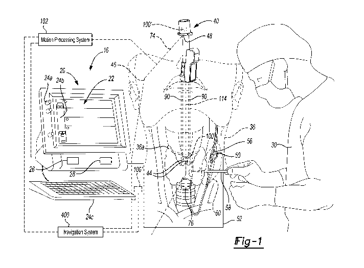
Fig. 1 is an environmental view of a monitoring system and an electrode
assembly;
[0011]
Fig. 2 is a perspective view of an electrode assembly, according to
various embodiments, with an endotracheal tube;
[0012] Fig. 3 is a detail view of a portion of the endotracheal tube of
Fig. 2; and
[0013] Fig. 4 is a flowchart of operation of a monitoring system,
according to
various embodiments.
[0014] Corresponding reference numerals indicate corresponding parts
throughout the several views of the drawings.
2
CA 03033680 2019-02-11
WO 2018/031784
PCT/US2017/046312
DETAILED DESCRIPTION
[0015]
Example embodiments will now be described more fully with reference to
the accompanying drawings.
[0016]
With initial reference to FIG. 1 a monitoring system 16, such as a NIM
nerve integrity monitoring system may include a monitor assembly 20 that has a
display
screen or device 22 and one or more input devices. The input device may
include one
or more systems or structures to input commands of information such as knobs
24a, a
touch screen 24b, a keyboard 24c, or other appropriate input devices. Input
devices
may also include audio or other tactile input devices, including electronic or
physical
input devices.
[0017]
The monitor assembly 20 may further include a processor 26 and a
memory 28. It is understood that the processor 26 may access the memory 28 to
execute instructions stored on or access other data on the memory 28. The
memory 28
may include a physical memory, such as a spinning hard disk drive, solid state
memory,
or other appropriate types of memory. Further, the memory 28 may not be
incorporated
into the monitor assembly 20, but may be accessed by processor 26, such as via
communications network. The processor 26 may be a general purpose processor
that
is operable to execute instructions for generating a selected output, as
discussed
further herein. The processor 26 may further include onboard memory. Moreover,
the
processor 26 may include a specific purpose processor such as an application
specific
integrated circuit (ASIC). Accordingly, the processor 26 may execute
instructions stored
on memory 28, which may be a non-transitory memory, to provide an output for
display
on the display device 22. A user 30 may then view the display device 22 for
selected
purposes, as discussed further herein.
[0018]
Connected with the monitor assembly 20, may be one or more stimulation
or monitoring assemblies. For example, in various procedures such as a
thyroidectomy
or other thyroid surgeries, monitoring of a recurrent laryngeal nerve (RLN), a
vagus
nerve, or other appropriate nerve, in a patient 36 may be selected. Monitoring
of the
RLN may include a nerve monitoring endotracheal tube assembly 40 that may have
one or more monitoring portions, including one or more conductive electrode
contacts
44. The electrode contacts 44 may be in contact with selected portions of the
patient
36, such as a human patient. The electrode contacts 44 may be connected to the
monitor 20 via a connection, such as an optional wired connection (also
referred to as a
line or hardline) 46 or wireless connection including a wireless transmitter
48. It is
understood, however, regardless of the connection to the monitor 20, a
transmitted
3
CA 03033680 2019-02-11
WO 2018/031784
PCT/US2017/046312
signal from the electrode contacts 44 may be made to the monitor assembly 20.
An
exemplary endotracheal tube may include a NIM Trivantage monitoring tube sold
by
Medtronic, Inc. Exemplary endotracheal tubes may further include those
disclosed in
U.S. Pat. App. No. 14/455,258, now U.S. Pat. App. Pub. No. 2016/0038072,
incorporated herein by reference. It is understood, however, that the tube 40
may
include portions in addition to those currently available or different from
those currently
available on the NIM Trivantage monitoring tube and those discussed above.
[0019]
In addition, other instruments may be connected to the monitor 20, such
as electrode assemblies, including an electrode that may send or receive
periodic
stimulation pulses, including, according to various embodiments, a connected
electrode
assembly 50, as illustrated in FIG. 1. The connected electrode assembly 50 may
be
connected with a physical connection, such as a wire 52 to the monitor 20. The
connected electrode assembly 50 may also, or alternatively, be connected via a
wireless transmission or otherwise provide a stimulation to a nerve 56. Other
instruments may also be connected with the monitor 20 that may be used to send
or
receive stimulation signals to the patient to assist in determining whether
nerve damage
or other tissue damage has occurred or could occur. An instrument 58 (e.g. a
scalpel,
forceps, etc.) may be manipulated by the user 30, such as a human surgeon, and
need
not be directly connected to the monitor 20. The instrument 58 may have a
stimulation
signal that is transmitted to evoke a nerve action potential that may be
received through
the contacts 44 of the tube 40. The instrument 58 may be used to dissect
and/or resect
tissue within the patient and through an incision 60.
[0020]
The operation of the monitoring system and the use of the monitoring
system 16 may be similar to the NIM monitoring system sold by Medtronic,
Inc.,
including the NIM-Response 3.0 nerve monitoring system. In operation, the
electrode
assembly 50 may be connected with the nerve 56 and a signal may be transmitted
along the connection 52 from the monitor system 20 through the electrode 50 to
the
nerve 56. The electrode contacts 44 may then receive an EMG signal, such as in
a
muscle, that is evoked by the stimulation of the electrode 50.
[0021] The
tube 40, as discussed briefly above, is illustrated in detail in Figs. 2
and 3. The tube 40 may be an EMG endotracheal tube assembly 40 and a
corresponding housing 70. The EMG endotracheal tube assembly 40 may include
the
housing 70 and an electronic assembly including a wireless transmission
assembly 48
and/or the wired connection 46. The tube assembly 40 includes a tube distal
(first) end
74 and a proximal (second) end 76. The distal end 74 is connected to a
connector 80,
4
CA 03033680 2019-02-11
WO 2018/031784
PCT/US2017/046312
which may be connected to a pump for supplying a selected material such as a
gas
and/or a fluid to a patient via the tube 40. The tube 40 may be inserted in a
throat of
the patient 36 and the gas and/or fluid may be supplied to, for example, lungs
of the
patient 36. The proximal end 76 includes an inflatable portion 86 (shown in an
inflated
state), which may be used to seal off, for example, a trachea, to prevent any
other fluid
or substance from passing around the inflated portion 86 and entering the
lungs.
[0022]
The tube assembly 40 includes the contacts 44. The contacts 44 may be
in electrical connection with electrodes 90 formed on or in the housing 70.
The
contacts 44 and/or the electrodes 90 may be painted or printed on the EMG tube
housing 70. For example, a conductive paint or ink may be applied to the tube
housing
70, or a portion thereof to form the conductive portions. Further, the
electrodes and
transmission portions may include traces that are formed on flexible printed
circuit
boards and applied to the housing 70. A coating or protective layer may also
be
provided over the painted portions, while allowing the contacts 44 to be
exposed to an
external environment. In another embodiment, the electrodes 90 and/or the
contacts
44 are printed on the EMG tube housing 70 and/or are implemented as a portion
of a
flexible printed circuit board (PCB).
[0023]
The electrodes 90 may extend from the contacts 44 to the connection
portion, including the wireless transmitter 48 and/or the wired connection 46.
The
electrodes 90 may extend in parallel along the tube housing 70 and are
separated as to
not be in contact with each other. One or more insulation layers 94 may be
applied
over the electrodes 90 to prevent external electrical contact with the
electrodes 90.
Each of the insulation layers 94 may cover one or more of the electrodes 90
and may
not wrap fully around the tube housing 70. The insulation layers 94 may be
nonconductive stamps formed of nonconductive material (e.g., rubber).
[0024]
As discussed above, the tube assembly 40 may be used with the
monitoring assembly 20 to monitor an electrical activity signal in the patient
36. The
stimulation being sensed through the contacts 44 may be provided through the
electrode assembly 50 in connection with the monitor assembly 20. As discussed
above, the NIM stimulation and monitoring system sold by Medtronic, Inc. may
be
provided to sense at or stimulate the nerve 56, or any selected nerve, at a
selected rate
or interval and sense or detect the stimulation of the nerve 56. Upon sensing
the
stimulation from the nerve (including, for example, an EMG response of a
muscle
and/or electrical activity related thereto) the monitoring system 20 may
monitor the
EMG signal after determining a baseline and to determine the baseline.
Further, the
5
CA 03033680 2019-02-11
WO 2018/031784
PCT/US2017/046312
user or other appropriate individual may observe the monitoring system 20,
such as the
display 22, to ensure integrity of nerves during a surgical procedure.
Procedures may
include a throat or thyroid removal procedure, as discussed above.
[0025] The multiple contacts 44 can be provided around the tube
assembly 40
for various purposes. In various embodiments, the multiple electrodes and/or
contacts
therefore may be used for differentiating between left and right nerves,
differentiating
between different nerves and nerve branches, compensating for users placing
the tube
contacts in a variety of depths and/or axial positions relative to the
anatomy. The
multiple electrodes may be placed axially along the tube to allow for
measurements at a
distance from a selected location as well. Referential recording electrodes
may also be
placed a distance from other recording electrodes to minimize noise and
interference.
Upon movement of the contacts 44 relative to the patient 36 (including
internal tissue,
such as muscle 36a that may include vocal fold in a human larynx) the signal
to the
monitoring system 20 may change. Change of the signal to the monitoring system
20
may be interpreted or possibly interpreted as an injury to the nerve 56. The
system 20,
upon determining a change or sensing a change in the received stimulation, may
provide an indication to the user 30 that an injury has occurred and that the
procedure
should be stopped. , if the signal to the monitoring system 20, however,
changes only
due to movement, whether intentional or unintentional, of the tube assembly 40
then no
injury has occurred.
[0026] Accordingly, a movement or motion detection system including
one or
more move or motion sensors 100 may be provided on the tube assembly 40. The
motion sensor 100 may be interconnected with the wireless module 48 or the
conductive connection (e.g. a wired connection) 46 to the monitoring system
20.
Motion detected by the one or more motion sensor 100 can assist in determining
whether the tube assembly 40 has moved relative to the patient 36 and assist
in
determining whether a change in signal sensed by the contacts 44 is due to
movement
of the tube assembly 40 or due to a cut or injury to the nerve 56. A signal
from the
motion sensor 100 may be transmitted to a motion processing system 102. The
signal
may be processed with the motion processing system 102 to determine type,
speed,
amount, etc. of motion. The processed motion signal may then be transmitted to
the
processor 26. It is understood, however, that the motion processing system may
be
incorporated into the monitoring system 20. For example, the processor 26 may
execute specific instructions stored on the memory system 28 to operate at the
motion
processing system 102 to determine motion of the motion system 100.
6
CA 03033680 2019-02-11
WO 2018/031784
PCT/US2017/046312
[0027]
With additional reference to Fig. 3 the motion sensor 100 can be provided
near or at the proximal end 76 of the tube assembly 40. In various
embodiments, the
motion sensor 100 is at or near the location of the contacts 44. For example,
the
motion sensor 100 may be provided substantially adjacent to the contacts 44.
In
providing the motion sensor 100 near or adjacent to the contacts 44, movement
of the
contacts 44 can be substantially directly determined due to sensed or measured
motion
of the motion sensor 100. One skilled in the art, however, will understand,
that the
motion sensor 100 may be provided at the distal end or near the distal end 74.
Optionally, the assembly 40 may include the motion sensor 100 and a second
motion
sensor 100'. The second motion sensor 100' may be spaced apart from the first
motion
sensor 100, such as being placed at the distal end 76. The second motion
sensor 100'
may also be provided with communication to one or more of the wireless
transmission
assembly 48 or through the line 46 to transmit to the motion processing system
102
and/or the monitor 20. Thus, it is further understood, that the assembly 40
may include
one or more of the motion sensors 100, and the discussion herein of the motion
sensor
100 is merely exemplary.
[0028] The motion sensor 100 may be used to measure an amount or type of
motion and determine motion of the contacts 44 and/or the tube assembly 40.
The
motion of the tube assembly 40 and/or the contacts 44 may be determined in
absolute
measurement and/or relative to the patient 36. For example, the motion sensor
100
can be used to determine rotational or angular movement, such as in the
direction of
arrow 104 around a central or longitudinal axis 106 of the tube assembly 40.
Further,
the motion sensor 100 can be used to determine axial motion such as in the
direction of
double headed arrow 108 along the axis 106 of the tube assembly 40. Therefore,
the
motion sensor 100 can be provided to determine one or more dimensional
movement of
the tube assembly 40 including the contacts 44. As discussed herein, the
motion
sensor 100 may also be operated to sense or determine an evoked response to
the
stimulation alone or in combination with the contacts 44. An evoked response
may
generally include a movement of the muscle, thus motion on the tube assembly
due to
a contracting muscle may be sensed and/or measured with the motion sensor 100.
[0029]
The motion sensor 100 can include selected motion sensors such as an
accelerometer or accelerometer portion 110. The motion sensor 100 can include
one
or more types of motion sensors, as discussed further herein. In various
embodiments,
the motion sensor 100 may include only the accelerometer 110.
7
CA 03033680 2019-02-11
WO 2018/031784
PCT/US2017/046312
[0030]
The accelerometer portion 110 can include an appropriately sized
accelerometer that may be positioned on the housing 70 of the tube assembly
40.
Exemplary accelerometers include microelectrode-mechanical systems (MEMS). It
is
understood that other appropriate types of accelerometers may also be used
such as
thermal MEMS or other selected accelerometer devices. Generally, the
accelerometer
portion 110 may be provided at a selected dimension such as several microns in
thickness to be provided in or on top of the housing 70. The accelerometer
portion 110
may include, for example, the XTRINSIC MA84910 accelerometer sold by NXP
Semiconductors Netherlands B.V., having a place of business in the
Netherlands.
[0031] The
accelerometer portion 110 may be fixed to the housing at a selected
position relative to the contacts 44. For example, the accelerometer portion
110 may
be adhered or molded to the housing 70. As discussed above, the electrodes 90
may
be covered with the coating 94, and the coating 94 may fix or assist in fixing
the
position of the accelerometer portion 110 relative to the contacts 44.
[0032] The
accelerometer portion 110, and any other appropriate portions
including with the motion sensor 100, may be connected to the transmission
device 48
and/or the line 46 through appropriate communication lines such as a
communication
conduit 114. The communication line 114 can include conductive printed ink or
paint or
other appropriate electrode forming mechanisms, similar to the connectors or
electrodes 90. The accelerometer portion 110, however, is generally provided
to
communicate with the monitoring assembly 20.
[0033]
As is understood by one skilled in the art, the accelerometer 110 may
measure acceleration in one or more axis, including two axes accelerometer or
a three
axes accelerometer. The accelerometer, when at rest, such as in the patient 36
during
an operative procedure, will generally measure acceleration down towards the
center of
Earth due to gravity. Upon movement of the accelerometer 110, the
accelerometer 110
may measure the change in acceleration in one or more axes due to movement of
the
accelerometer 110. For example, during the operative procedure the tube
assembly 40
may be twisted around the axis 106 in the direction of arrow 104 and the
accelerometer
110 may measure the acceleration due to movement of the tube assembly 40
around
the axis 106.
[0034] As discussed above, movement of the tube assembly 40 may cause
movement of the contacts 44 and therefore a change in the EMG signal at the
monitoring system 20. As discussed further herein, a determined correlation
between
(such as occurring at the same time or close in time) a sensed or measured
movement
8
CA 03033680 2019-02-11
WO 2018/031784
PCT/US2017/046312
by the accelerometer 110 and a change in the EMG signal sensed by the contacts
44
may allow for a determination that injury has not occurred to the patient 36
and the
procedure may continue. Thus, the monitor system 20 may determine that warning
state (i.e. a selected change in the EMG signal) is a false warning.
[0035] The motion sensor 100 may include portions in addition to the
accelerometer 110, as noted above, including a one or more axes gyroscope,
such as a
three axis gyroscope 120. The three axis gyroscope 120 may measure a
rotational
movement or angular movement directly due to the included gyroscope. As noted
above the accelerometer 110 may need to infer or determine motion relative to
gravity.
Therefore, the gyroscope 120 may be able to act as a back-up or check for the
accelerometer 110 by determining angular motion directly. It is understood,
however,
that the gyroscope 120 may be provided alone as the motion sensor 100.
[0036]
The gyroscope 120 as a part of the motion sensor 100 may be positioned
near or adjacent near the contacts 44. Therefore, as discussed herein,
movement of
the electrodes 44 may be determined by sensing motion via the gyroscope 120
and
may be transmitted as a signal along the communication line 114, as discussed
above.
The monitoring system 20 can then determine that motion of the gyroscope 120
has
occurred and may infer that motion of the electrode contacts 44 has also
occurred.
[0037]
Exemplary gyroscopes can include MEMS gyroscopes that can be formed
on selected substrates, including silicon substrates similar to integrated
circuits or
printed circuit boards. Exemplary MEMS gyroscopes can include those sold by
NXP
Semiconductors Netherlands B.V., to measure an angular rate. As the gyroscope
may
measure one or more axes of motion such as include yaw, pitch and roll.
Therefore,
the gyroscope 120 may be used to determine direct angular motion or
acceleration of
the motion sensor 100 and, therefore determine angular motion of the contacts
44.
Again, as discussed herein, motion of the contacts 44 may be used to deduce a
reason
for a change in the signal.
[0038]
The motion sensor 100 may further include magnetometers 130. A
magnetometer may be used to determine magnet poles that may affect a
determined
movement or orientation of the motion sensor 100. Magnetometer 130 may also or
alternatively be operated to determine a position of the magnetometer 130
relative to
magnetic poles, such as those generated by the Earth. The magnetometer 130 may
operate to determine an absolute position relative to a magnetic pole, such as
of the
Earth, or motion relative to a magnetic pole.
9
CA 03033680 2019-02-11
WO 2018/031784
PCT/US2017/046312
[0039]
The magnetometer 130 may include small or micro-magnetometers, such
as MEMS magnetometers including those manufactured and sold by NXP
Semiconductors Netherlands B.V., having a place of business in the
Netherlands. The
magnetometer may send a signal with the communication line 114 to the monitor
assembly 20 to assist in determining motion or movement of the motion sensor
100
and, therefore, the contacts 44.
[0040]
The motion sensor 100 can include various components and portions,
including those discussed above. In exemplary embodiments, the motion sensor
100
may include only one of the accelerometers 110, gyroscope 120, or magnetometer
130.
Further, it is understood that the motion sensor 100 may include any two of
the three or
all three of the components discussed above. According to various embodiments,
the
motion sensor 100 may include a six axis motion detector including the
accelerometer
110 and the magnetometer 130.
It may be selected, however, that additional
information may be necessary to assist in ensuring accuracy for determining
motion or
movement of the contacts 44, therefore all three of the components may be
provided.
Further, in various embodiments, only the accelerometer 110 may be provided to
assist
in determining motion of the contacts 44. Including the gyroscope 120 may
assist in
reducing the effects of magnetic interference and/or various types of
acceleration, such
as linear acceleration of the motion sensor 100.
[0041]
Returning reference to Fig. 1 and 2, one skilled in the art will further
understood, a tracking or navigation system 400 may be used to determine
position
change and/or motion of the tube assembly 40 including the contacts 44. For
example,
the tracking system 400 may include an electromagnetic tracking system that
may
include a localizer that is configured to emit or receive a magnetic field. A
tracking
sensor may be provided as the motion sensor 100, 100', 100" that is configured
to emit
or receive a magnetic field. A tracking system may then determine a location
(including
x-y-z and orientation) of the tracking sensor based on the sensed field
(either at the
localizer or at the tracking device). A tracked change in location may be used
to
determine motion. Exemplary tracking systems include those disclosed in U.S.
Pat.
App. Pub. 2014/014869, incorporated herein by reference. It is further
understood that
various tracking systems may include optical tracking systems, sonic tracking
systems,
electro-potential tracking systems, etc.
A tracked change in location may be
transmitted to the motion processing system 102 and/or the monitoring 20.
[0042]
As discussed above, the tube assembly 40 may further include the motion
sensor 100' at or near a distal end of the tube assembly 40. It is also
understood that
CA 03033680 2019-02-11
WO 2018/031784
PCT/US2017/046312
the motion sensor 100' may be the only motion sensor on the tube assembly 40
and
that motion of the contacts 44 may be inferred from the motion sensor 100'.
The
motion sensor 100' may be identical to the motion sensor 100 or different,
such as
including more or less components.
[0043] In
further embodiments a third motion sensor 100" may be provided on
the patient 36. Including motion sensor 100" fixed directly on the patient 36
can allow
for determination of motion or movement of the patient 36 independent of the
tube
assembly 40 including motion sensors 100 and 100'. The third motion sensor
100"
may also transmit a signal to the motion processing system 102 and/or the
monitor 20,
as discussed above. The determination of movement of the patient 36 can be
used to
assist in determining whether the tube 40 has simply moved in space or moved
relative
to the patient 36, such as relative to the nerve 56 and/or muscle tissue
innervated by
the nerve 56. This can be used to assist to determine whether an anomalous or
changed received signal is due to motion of the tube 40 relative to the
patient 36
causing the contacts 44 to move relative to the patient 36.
[0044]
If the motion sensor 100, 100' move in a manner similar or identical to the
motion sensor 100" then a change in signal may signify a possible injury.
If the
motion sensor 100, 100' move in a manner different (such as at a different
rate or time
or amount) to the motion sensor 100" then a change in signal may signify only
that the
contacts 44 have moved relative to the patient 36. As discussed herein, a
threshold
amount of difference may be used in the determination. Further, monitoring
system 20
may be provided to determine the type of change and possible injury.
[0045]
Accordingly, it is understood that an appropriate number of motion
sensors can be provided on the tube assembly 40, such as at or adjacent to the
contacts 44, and on the patient 36, that operate independent of the tube
assembly 40,
to assist in determining possible relative motion of the contacts 44 to the
patient 36. As
discussed above, movement of the contacts 44 relative to the patient 36, such
that their
position moves over time relative to tissue of the patient 36, may cause a
change in the
signal received or monitored by the monitoring assembly 20.
[0046] With
additional reference to Fig. 4, a flowchart 200 illustrates an
exemplary operation of the monitoring system 20 which may include monitoring
of an
induced signal, such as with the electrode assembly 50, that is sensed at the
contacts
44, of a sensing electrode, that is able to contact tissue, such as a muscle,
that is
stimulated due to stimulating the nerve 56. It is understood, as discussed
above, that
the tube 40 may be provided with the contacts 44 that are configured to sense
11
CA 03033680 2019-02-11
WO 2018/031784
PCT/US2017/046312
stimulation of tissue. It is understood, however, that electrode contacts can
be provided
in any appropriate instrumentation or portion that may contact the patient 36
or other
appropriate subject to sense a stimulation. The tube 40 is merely exemplary
and
provided for illustration of the current disclosure.
[0047] With
specific reference to Fig. 4, therefore, the flowchart 200 may begin at
START block 210. Stimulation may start in block 214, such as by operating the
monitoring system 16 that include stimulating the nerve 56. As discussed above
stimulation may include a periodic stimulation that may be sensed by the
monitoring
system 20. The periodic stimulation may have a period that accounts for
various
factors, such as refractory response time of the nerve 56. Nevertheless,
stimulation
may start in block 214.
[0048]
The monitoring system 20 may then sense the EMG signal from the
stimulation in block 216. As discussed above the sensed stimulation may be
sensed
through the contacts 44 in contact with the tissue of the patient 36 and due
to the
stimulation provided with the stimulation electrode 50. The sensed stimulation
may
cause a signal to be transmitted wirelessly via the wireless transmission
system 48 or
through the wired transmission system 46. The sensed signal may then be
monitored
and/or analyzed by the monitoring system 20.
[0049]
After receiving an initial signal, such as for a selected start-up time
after
stimulation of the nerve 56 with the electrode 50, the monitoring system 20
may
determine a baseline signal based on the sensed signal in block 220. The
baseline
signal determined at block 220 may be determined by the processor system 26
that is
executing instructions recalled from the memory system 28. For example, the
processor system 26 may determine a baseline signal including a frequency or
period
of stimulation, amplitude of stimulation including power or voltage, or other
appropriate
parameter. Regardless, the determined baseline stimulation signal may be used
by the
monitor system 20 to determine a baseline for later comparison.
[0050]
As discussed above, the baseline signal may be determined when the
nerve 56 is substantially uncompromised, such as prior to a procedure on a
patient that
may interfere with the nerve integrity or continuity. Accordingly, the
baseline signal may
be used to determine if any change from the baseline occurs, such as due to
injury or
the like relative to the nerve 56. Moreover, as discussed above, movement of
the
contacts 44 relative to tissue may also cause a change in the signal from an
initial
baseline. Therefore, the baseline signal may be used to assist in determining
any
change after an initial period in time.
12
CA 03033680 2019-02-11
WO 2018/031784
PCT/US2017/046312
[0051]
The baseline signal, further, is determined at a first selected location of
the contact 44. As discussed above, the sensed EMG signal may be different at
different locations within the subject 36. Therefore, the contacts 44 on the
tube 40 may
be placed at the first location, which may be predetermined location, such as
adjacent
to or in contact with a specific muscle group. Movement from the first
location,
therefore, may cause a change in the sensed signal, including the EMG signal.
If the
contacts 44 move, therefore, the contacts 44 may be at a second location that
is
different than the first location. The second location may be rotationally,
angularly, or
axially displaced from the first location.
[0052]
Following determination of the baseline in block 220, the sensed signal
can be monitored in block 224. Further, the position or position change of the
contacts
44, including the tube 40, can be monitored in block 226. The monitoring of
the signal
in block 224 and the position change in block 226 can both be done with the
monitoring
system 20. Signals may be sent to the monitoring system 20 to allow for
determination
of the signal due to nerve 56 stimulation after determining the baseline
signal and for
sensing a change in position.
[0053]
At a selected period or frequency, such as every 100 milliseconds, 200
milliseconds, one second, or other appropriate frequency, a determination or
measuring
of whether the received signal has changed from the baseline may occur in
decision
block 230. A change in the signal from the baseline may include any change or
a
change beyond a threshold. For example, a change in the signal may include an
amplitude change, a frequency change, a period change, or the like. A
threshold
change may include the change in the sensed signal such as a change in period
of
about 1% to about 10%, for example including a change of about 50
milliseconds.
Therefore, if the sensed signal change includes a change in period of greater
than 50
milliseconds a, determination in block 230 that a signal change has occurred
will follow
a YES path in block 232. It is understood that any appropriate changes in
signal may
also be measured and used to determine whether a change in signal has occurred
such
as a change in power, voltage, or the like and thresholds may include
percentage
changes or absolute value changes.
[0054]
If the YES path 232 is followed, an optional initial warning message may
be displayed or otherwise presented to the user 30 in block 234. The warning
message
may include any appropriate warning message such as a flashing screen, a blank
screen, a verbal message, an auditory message, a visual message, or the like.
13
CA 03033680 2019-02-11
WO 2018/031784
PCT/US2017/046312
Regardless the warning message at block 234 may indicate to the user 30 that
the
signal has changed and that a possible injury to the patient 36 has occurred.
[0055]
The warning message in block 234 immediately following the YES path
232 may, however, be optional. Rather, the YES path 232 may follow to a second
determination block of whether a position change was determined when the
signal
changed in block 240. As discussed above, the motion sensor 100 may be
provided on
the tube assembly 40. A position change of the tube assembly 40 may then be
determined with the motion sensor 100 according to an appropriate mechanism,
including the selected position sensing portions as discussed above.
Therefore,
determination of whether a position change occurred in block 240 may be made
by the
monitor system 20. A signal from the motion sensor 100 may be analyzed by the
processor system 26 to determine a position change. The processor 26 may
execute
instructions recalled from the memory 28 to assist in determining whether the
motion
sensor 100 senses a change in position. The processor system 26, therefore,
can also
determine whether the position of the contacts 44 and/or the tube assembly 40
has
changed.
[0056]
The determination block 240 can follow a NO path in block 254 if no
position change is determined. The NO path 254 may then follow to the send
warning
message in block 234. As discussed above, if a change in the sensed signal
occurs
and no determined position change in the motion sensor 100 is determined, then
a
change of the signal sensed by the contacts 44 may have changed for a reason
other
than movement of the contacts 44. Further possible reasons for change may be
due to
a change in integrity of a nerve 56 and the user 30 may be provided with a
message to
determine continuity of the nerve 56 and to stop the procedure, at least
momentarily.
[0057] If
a changed position is determined in block 240, however, a YES path
258 may be followed to a further decision block 262 including determining
whether the
position change was greater than a threshold. The motion sensor 100 can be
used to
measure a change in position of the contacts 44 or other appropriate portion
of the tube
assembly 40. However, a selected change, such as a small change or a slow
change
in position may not lead to a determination that the change in the signal
determined in
block 230 is due to movement of the motion sensor 100. Therefore, a selected
threshold may be used to assist in eliminating or minimizing possible errors
of position
measurements.
[0058]
Various thresholds may include a speed of change, an absolute value of
change, a percent amount of change in position, or other appropriate
thresholds may
14
CA 03033680 2019-02-11
WO 2018/031784
PCT/US2017/046312
be used in determination block 262. In one example, if the measured change in
the
EMG signal is greater than plus or minus 50% from a previous measurement (e.g.
when measuring once per second), but the latency of the measured signal has
not
changed, it may then be determined that movement of the contacts 44 due to
movement of the tube 40 has caused a change in the signal. Exemplary amplitude
changes may be, however, about 20% to about 80%, including about 40% to about
60%. As discussed above, this may be an automatic determination and, if
selected no
warning may be given in block 234, as discussed herein. In a further example,
a multi-
axis sensor that detects force exerted by gravity in one axis of the sensor
decreases
while the force exerted by gravity in another axis increases, for example by a
difference
of more than 30%, the processor may execute instructions to determine that the
tube
40 has rotated in a known direction.
[0059]
If the change is determined to be greater than a threshold in block 262, a
NO path 266 may be followed to send a warning message in block 234. Again, if
the
change in position may not be correlated to the change in the signal then a
warning
message may be provided to the user 30 to determine a continuity and integrity
of the
nerve 56. Thus, if the amount of change is not at or greater than the
threshold, the
determination may be similar to no change in position being determined.
[0060]
If the change is determined to be greater than the threshold in block 262,
then the YES path in block 270 may be followed. Initially, in an optional
embodiment, a
second determination block 274 may determine whether a patient position change
occurred in a same manner similar to or same as the position change of the
contacts in
block 240 may be made. As discussed above, an optional motion sensor 100" can
be
positioned on the patient 36. The motion sensor 100" allows a position of the
patient
36 to also be monitored during a procedure. If a position change of the
patient 36
matches the determined position change in block 240 of the motion sensor 100,
then it
may be inferred or determined that the patient 36 is moved during the
procedure, such
as turned on a side from a supine position. If the position of the patient 36
changes in a
manner that is substantially the same or identical to that of the positon
sensor 100 of
the tube assembly 40, then a change in the signal sensed at the contacts 44
may again
be determined or inferred to be a change due to something other than a
movement of
the contacts 44 relative to the tissue of the patient 36. This is due to the
position of the
contacts 44 in absolute terms relative to space outside of the patient 36 does
not cause
a change in the signal sensed at the contacts 44. Rather, a change in position
of the
contacts 44 relative to the tissue that the contacts 44 are contacting can
cause a
CA 03033680 2019-02-11
WO 2018/031784
PCT/US2017/046312
change in the sensed signal. Therefore, if it is determined that the patient
had a
position change in a manner that is the same as the change determined of the
tube 40
in block 240, a YES path 276 may be followed to provide the warning signal in
block
234. Again, the change in the sensed signal due to stimulation will have
changed;
however, both the position of the tube and the patient will be determined to
have
changed in a substantially similar manner such that the contacts 44 have not
moved
relative to the patient 36.
[0061]
Again, optionally, if a position change of the patient determination in
block
274 is made and a position of the patient changed in a manner different than
the
position of the tube 40 then a NO path 290 may be followed to a determination
block
whether a procedure done signal has been received in block 292. In this
instance, the
position of the patient, even if it has changed, has changed in a manner
different than
the position of the tube assembly 40. Therefore, it may be determined that the
positon
of the tube 40 relative to the patient 36 has changed and not only an absolute
positon
change of the tube 40.
[0062]
If the position change of the tube is greater than a threshold such that
the
YES path 270 is followed and no optional patient position sensor is provided
the YES
path 270 may follow to the decision block 292 if a procedure done signal has
been
received. Again, if the position change of the tube assembly 40, including the
contacts
44, has changed greater than a threshold a change in the sensed stimulation
signal
may be determined to be caused due to the movement of the tube assembly 40,
including at contacts 44.
[0063]
If a procedure done signal has been received then a YES path 296 may
be followed to end at block 300. Ending at block 300 means ending stimulation
or
monitoring of the nerve 56 or other appropriate ending portions of a surgical
procedure.
[0064]
If a procedure done signal has not been received in block 292, a NO path
310 may be followed to determine a new baseline with a changed sense signal in
block
320. As discussed above, a change sense signal may initiate a determination or
check
of whether the tube 40 has changed position. If it is determined that the
position of the
tube assembly 40 has changed it may be determined, such as with the processor
system 26 executing instructions recalled from the memory 28, that the positon
change
has caused the sensed signal change. If the position change has caused a
sensed
signal change then an injury to the nerve 56 may be determined to have not
occurred.
Therefore, if no signal to end the procedure has been received in block 292 a
new
baseline with the new sensed signal, different from the determined baseline
signal in
16
CA 03033680 2019-02-11
WO 2018/031784
PCT/US2017/046312
block 220, may be determined in block 320. After determining the new (i.e.
second)
baseline in block 320 monitoring of the signal in block 224 and monitoring in
the
position change of block 226 may continue. In this instance the procedure may
then
continue and monitoring of the sensing stimulation signal and the sensed
position may
continue and be periodically checked, as discussed above.
[0065]
Further, it is understood that if no sensed changed in the stimulation is
determined then a NO path 330 may be followed to continue monitoring the
sensed
stimulation signal in block 224 and the monitored position change signal in
block 226
and these may be checked at a selected period, as discussed above.
Accordingly, the
monitoring system 16 can monitor a stimulation signal of the patient 36 and
assist in
making a determination whether the position change of the tube 40 has caused a
sensed change in the stimulation signal. If a sensed position change of the
tube 40
with the contacts 44 is made, then a determination may be made of whether the
sensed
position change has caused the change in sensed electrical stimulation.
[0066] With
continuing reference to Fig. 4, and also to Fig. 1, the motion sensor
100 may be used as a monitoring portion with the monitoring system 16 to
determine if
a selected muscle group is being stimulated. For example, as discussed above,
the
muscles 36a may include or be associated with vocal folds of the subject 36.
Stimulation of the nerve 56 may cause movement of the vocal folds 36a.
Movement of
the vocal folds 36a as an evoked response due to the stimulation of the nerve
56 may
be sensed and/or measured with the motion sensor 100 (or other motion sensor
associated with the tube assembly 40). Thus, the motion sensor 100 may be used
to
determine whether the nerve 56 is damaged or not by sensing motion of the tube
assembly 40 due to movement of muscles relative to the vocal folds 36a.
[0067] In
various embodiments, the motion sensor 100 (e.g. the accelerometer)
can operate as the monitoring portion to detect the stimulated signal, which
is the
stimulation of the nerve 56, and may replace and/or augment the detection of
the
stimulated signal of the contacts 44, which may operate as the monitoring
portion.
When the nerve 56 (e.g. the recurrent laryngeal nerve and/or vagus nerve) is
stimulated
to evoke a response, the corresponding musculature causes vocal fold adduction
and
the vocal fold collides or constricts around the endotracheal tube assembly
40. These
vocal fold collisions on the tube 40 induce a corresponding motion (e.g.
vibration) that
may be sensed by the motion detector 100 mounted on the tube assembly 40.
Thus,
the motion sensor 100 offers a redundant and/or alternative sensing of nerve
stimulation and detecting vocal fold movement.
17
CA 03033680 2019-02-11
WO 2018/031784
PCT/US2017/046312
[0068]
In various embodiments, with reference to Fig. 4, once stimulation has
started in block 214 motion may be sensed due to a stimulation response in
block 500.
The motion sensed in block 500 may be with motion sensor 100, or any
appropriate
motion sensor, associated with the tube 40. The tube assembly 40 may move due
to
contact or collisions of selected anatomical structures, including vocal folds
of the
subject 36. If muscles are moved due to stimulation of the nerve 56 that
caused the
vocal folds to move and contact the tube 40 the motion may be sensed with the
motion
sensor 100. Such motion may then be inferred to relate to an evoked response
of the
muscles intended to be stimulated with stimulation of the nerve 56. Therefore,
rather
than sensing an EMG signal induced by stimulation in block 216, a motion may
be
sensed in block 500 to infer the same continuity of the nerve 56 to the
muscles 36a.
[0069]
It is understood that the motion processing system 102 may receive a
motion signal from the motion sensor 100 and that the motion signal may be
forwarded
to the monitoring system 16. The monitoring system 16 may then execute
selected
instructions to determine that the evoked response at the selected muscle is
occurring
based on the motion signal. For example, a timing of the stimulation may be
known
and the timing and/or amplitude of the sensed motion may be related to the
stimulation.
A correlation of the time of the stimulation and the time and/or amplitude of
the sensed
motion may be used, therefore, to determine that the nerve 56 is uninjured. If
the
timing and/or amplitude (e.g. lower or no sensed motion) of the sensed motion
changes
a determination by the monitoring system 16 may be made that damage has
occurred.
As discussed herein, a warning message may then be provided to the user 30.
[0070]
The monitoring system 16, therefore, upon receiving the signal from the
motion processing system 102 regarding the sensed motion of the motion sensor
due
to movement of the vocal folds may display or present the message to the user
30,
such as with the display device 22. The display device 22 may display a graph
including a representation, such as a wave, representing motion. The user 30
may
view the display device 22 to determine that motion is occurring, such as a
selected
rate, to ensure that the evoked response is still occurring. It is understood
that the
sensing of motion in block 500 may occur in addition to or as an alternative
to the
sensed EMG signal in block 216. Accordingly, it is understood that sensing
motion with
the motion sensor 100 to determine that the evoked response is occurring may
be an
alternative and/or in place of sensing EMG signal.
[0071]
A determination of a baseline motion to do the stimulated response may
optionally occur in block 520. The determination of the baseline motion
response due
18
CA 03033680 2019-02-11
WO 2018/031784
PCT/US2017/046312
to sensed motion due to the simulated response is optional, and may not be
necessary.
Further, it is also optional to then enter the process 200 further, as
discussed above, to
monitor position change in block 226. Monitoring position change in block 226
may be
optional, and particularly optional when only sensing motion due to a
stimulated
response to determine that the evoked response is occurring. As discussed
above,
monitoring a position change in block 226 may occur or be selected when
ensuring that
movement of the contacts 44 move causes an EMG signal change. If an EMG signal
is
not being used to determine that the evoked response is occurring, monitoring
position
change in block 226 may not be selected. Therefore, determining a baseline for
motion
in block 520 and/or monitoring position change in block 226 when sensing and
monitoring motion due to a stimulated response in block 500 may both be
optional.
[0072]
Regardless, a message may be provided regarding the sensed motion in
block 530 with the display device 22. It is understood that other appropriate
messages
may also be provided to the user 30, such as an auditory or tactile message.
For
example, a periodic tone may be provided to the user 30 which relates to
movement or
sensed vibration of the tube assembly 40. The process may then end at 300 when
the
user 30 determines or selects to end the procedure on the subject 36.
[0073]
Accordingly, the evoked response at the muscle 36a due to stimulation of
the nerve 56 may be measured or determined with the motion sensor 100 on the
tube
assembly 40. As discussed above, vibrations of tube assembly 40 may be
analyzed
and used to determine that an evoked response is occurring at the selected
muscle
tissue due to stimulation of the nerve 56. The processor system 26 may
evaluate the
motion signal based upon the sensed motion by the motion sensor 100 processed
by
the motion processing system 102 to determine motion of the tube assembly 40.
Selected signals may be provided to the user 30 such as based upon a change in
a
motion signal (such as an amplitude change of motion over a selected period of
time) or
other appropriate change in the motion signal. The sensing of the motion with
the
motion sensor 100, therefore, may be used in place of, or as an alternative
to, sensing
the EMG signal with the contacts 44 as discussed above.
[0074] This
offers the advantage of a sensor (i.e. the motion sensor 100, 100',
100") that is not electrically coupled to the patient 36; thus, providing a
means of
sensing vocal fold movement, muscle movement, and nerve stimulation in the
presence
of possible electrical interference, such electrical interference may be
caused by high
frequency electrosurgery. Signals caused by various electrosurgery systems may
cause interference with monitoring systems that attempt to sense nerve
stimulation with
19
CA 03033680 2019-02-11
WO 2018/031784
PCT/US2017/046312
EMG signals during the use of high frequency electrosurgical units because the
voltage
signals generally used to cut and cauterize tissue are much larger than the
biopotentials of interest to the user 36 and/or monitored with the monitoring
system 16.
Another advantage of the motion sensor detecting the stimulated nerve and
vocal fold
movement is that the motion sensor 100 is not dependent upon placement
relative to
the muscle 36a (e.g. muscles relative to the vocal folds) intended to have an
evoked
response. The tube 40 conducts the vibrations caused by the vocal fold
collisions to
the motion sensor 100 regardless of the position or orientation of the motion
sensor
100.
It is understood, therefore, that the motion sensor 100 may include any
appropriate motion sensor, including all of those discussed herein, that may
be used to
sense and/or measure motion of the tube 40.
[0075]
It is understood that a correlation between the sensed stimulation change
and the sensed position change may be made based upon various other factors
such
as the changes occurring at a same time or substantially similar times, such
as within
10-500 microseconds of each other, including about 100 microseconds of each
other.
Accordingly, it will be understood that a sensed change in stimulation that
occurs at a
time that is outside of a selected threshold of time, such as about 100
milliseconds,
may be eliminated or disregarded as a possible change due to movement of the
tube
assembly 40, including the contacts 44. A selected threshold of time may be
selected
based upon selected parameters.
[0076]
An inconsequential change in position of the contacts 44 may, according
to various embodiments, be disregarded when a change in a sensed stimulation
signal
is also measured to allow a procedure to continue without halting or
interfering with the
procedure to check for an injury to the nerve 56. A sensed change in the
stimulation
signal may provide or be used to cause a warning signal for viewing by the
user 30.
However, the user 30 may also be provided with a signal that a sensed change
in
position is determined.
Therefore the user 30 may perform a less invasive
determination of integrity of the nerve 56, such as monitoring the sensed
stimulation
signal for a selected period of time. In a selected alternative embodiment,
however, the
processor system 26 may execute instructions to make a determination of
whether the
sensed stimulation change is caused by movement of the tube assembly 40
determined
by the position sensors 100, 100' and either provide a warning signal to the
user 30 or
only recalibrate the monitoring system 20 and determine the new baseline
signal in
block 320 and continue the procedure, as discussed above. Therefore, the
monitoring,
determining a change in the received signal based on a position change, and
CA 03033680 2019-02-11
WO 2018/031784
PCT/US2017/046312
determining a new baseline may all be automatic without further intervention
or input
form the user 30.
[0077]
Example embodiments are provided so that this disclosure will be
thorough, and will fully convey the scope to those who are skilled in the art.
Numerous
specific details are set forth such as examples of specific components,
devices, and
methods, to provide a thorough understanding of embodiments of the present
disclosure. It will be apparent to those skilled in the art that specific
details need not be
employed, that example embodiments may be embodied in many different forms and
that neither should be construed to limit the scope of the disclosure. In some
example
embodiments, well-known processes, well-known device structures, and well-
known
technologies are not described in detail.
[0078]
The foregoing description of the embodiments has been provided for
purposes of illustration and description. It is not intended to be exhaustive
or to limit the
disclosure. Individual elements or features of a particular embodiment are
generally not
limited to that particular embodiment, but, where applicable, are
interchangeable and
can be used in a selected embodiment, even if not specifically shown or
described. The
same may also be varied in many ways. Such variations are not to be regarded
as a
departure from the disclosure, and all such modifications are intended to be
included
within the scope of the disclosure.
21