Note: Descriptions are shown in the official language in which they were submitted.
1
ASPIRATORS
Technical Field
This invention relates to in-line testers, and in particular, but without
limitation, to in-line testers
that can be used in conjunction with a nasogastric (hereinafter "NG")
aspirator to test for the correct
placement of an NG tube.
Background
Aspirators are used in a wide range of medical procedures where fluids need to
be drawn from
within a body cavity, for example, for diagnostic, sampling and/or therapeutic
purposes.
An aspirator generally comprises a vacuum source connected to a tube that can
be inserted, or
fed, into a body cavity. The vacuum source can be of any suitable type, such
as a syringe; a syringe-based
manual pump (such as that disclosed in published UK patent No: GB25235918); or
an electric vacuum
pump (such as that described in published UK patent application No GB2547012).
When the vacuum is applied to the free end of the tube, the vacuum draws fluid
from the body
cavity through the tube, provided, of course, that the tip of the tube is
located within the fluid to be
aspirated. A liquid trap is usually interposed between the tube and the vacuum
source to prevent
aspirated liquids from being drawn into the vacuum source, thereby reducing
the likelihood of damaging
and/or contaminating the vacuum source.
One example of a liquid trap is disclosed in published UK patent No:
GB2523591B, in which a
porous or perforated membrane is used to allow gasses to pass through it, but
when wetted by aspirated
liquids, blocks the aspirate from passing through it.
When using an aspirator, care needs to be taken to ensure that the vacuum is
not too high and
that the quantity and rate of aspiration is monitored. Monitoring and control
circuitry can often be used
to facilitate this, as well as the manual interventions of an experienced
operator.
It is commonplace to use aspirators to assist in determining whether the tip
of a NG feeding tube
is correctly positioned. When feeding a patient using a NG feeding tube, care
must be taken to ensure
that the tip of the NG tube is positioned within the patient's stomach, rather
than in his/her lungs. The
reason that this is important is that a NG tube is fed into the patient's
throat via his/her nose, and due to
the bifurcation of the patient's throat into the oesophagus and trachea, it is
possible for the tip of the NG
tube to be fed into the trachea, rather than the oesophagus (i.e. the wrong
way) resulting in the tip of the
Date Regue/Date Received 2022-08-12
2
NG tube being positioned within the patient's lung. If feeding commences with
the NG tube positioned
in the patient's lung, the results can be very serious.
As such, medical protocols require that before NG feeding commences, correct
positioning of the
NG feeding tube tip is determined. The only definitive way to determine the
correct placement of a NG
feeding tube is via a chest X-ray or other imaging procedure. However, chest x-
rays have been found, in
certain cases, to be less than definitive because the angle of exposure, level
of exposure, patient position
and the skill of the radiographer are all important in achieving a diagnostic
x-ray image that is fit for
purpose. Further, the time taken for an x-ray to be booked, carried out,
processed and reported can vary
considerably - during which time a subject is denied feeding, the subject most
likely being in a critical
condition. Additionally, for neonates particularly, the subject will be
exposed to radiation with the
associated potentially negative consequences.
Another way to check for the correct positioning of an NG tube is to aspirate
and test a sample of
fluid via the NG tube prior to feeding. This is indeed the current clinical
standard in the UK, whereby fluids
are aspirated via the NG tube and are checked for acidity using pH paper. A
colour change to indicate
acidic pH is considered to be an indicator that the NG tube is in the correct
location (it being assumed that
the stomach contents are acidic, whereas lung fluids are not) and so feeding
may commence.
If, however, a basic (alkali or pH > 5.5) pH reading is obtained, then the
assumption is that the
location of the tube is not in the stomach and the NG tube will be withdrawn
and reinserted before a
further pH test is carried out.
However, pH testing has been found to be unreliable insofar as it can yield
false negative results,
for example, if the patient is taking antacids to prevent reflux. In this
case, irrespective of whether the
NG tube is in the stomach or not, a basic or neutral pH will be indicated from
any sample obtained using
the current clinical standard.
Therefore, the current guidelines stipulate that if an acidic pH reading is
obtained, then it is safe
to commence feeding, whereas if a basic (alkali) pH reading is obtained,
further investigation is required.
The basic "rule of thumb" is therefore: acid = feed; alkali/neutral = do not
feed.
However, current pH testing protocols fail to take into account the
possibility of false positive
results (i.e. acid, but not safe to feed), which can prove problematic to
patient health, or fatal in extreme
cases. False positives can occur when the NG tube tip is located in the lung
of a patient, but where the
patient is suffering from reflux resulting in some gastric content being
inhaled/present in the lung. Clearly,
in this case, it is possible for the pH of an aspirated sample to be acidic -
indicating, according to current
clinical protocols, that the NG tube tip is in the correct location, i.e. in
the stomach, and so feeding will
Date Regue/Date Received 2022-08-12
3
commence. However, in this example, the NG tube would not be correctly
located, and the consequences
of commencing feeding could be very serious.
It will be appreciated that the aforesaid protocols, devices and methods are
not ideal, and a need
therefore exists for an improved and/or an alternative protocol, device and/or
method.
Summary
According to a first aspect of the invention, there is provided an in-line
tester comprising: an inlet,
connectable, in use, to an aspirator tube; an outlet connectable, in use, to a
vacuum source; and a
chamber interposed between the inlet and the outlet, the chamber comprising: a
first tester arranged
such that fluids drawn into the chamber by the vacuum source come into contact
therewith; a second
tester arranged such that fluids drawn into the chamber by the vacuum source
pass through it; and a
reservoir interposed between the first tester and the second tester; wherein
the first tester comprises a
first calorimetric substance adapted to exhibit a colour change in the
presence of a target substance, and
wherein the second tester comprises colorimetric capnometer, which exhibits a
colour change in the
presence of carbon dioxide, the in-line tester further comprising a liquid-
stop device which, when dry,
permits the passage of gas from the inlet to the outlet, but when wetted by a
liquid, inhibits or prevents
the passage of fluids from the inlet to the outlet.
According to a second aspect of the invention there is provided an in-line
tester comprising: a
chamber comprising an inlet, an outlet and a porous or perforated element
separating the inlet from the
outlet, the porous or perforated element comprising a liquid-absorbent layer
comprising a first
colorimetric substance adapted to exhibit a colour change in the presence of a
target substance and a
porous or perforated hydrophobic layer, the porous or perforated element, when
dry, permitting the
passage of gas from the inlet to the outlet, but when wetted by a liquid,
inhibiting or preventing the
passage of fluids from the inlet to the outlet, the in-line tester further
comprising, a calorimetric
capnometer interposed between the porous or perforated element and the outlet.
The in-line tester is suitably used in conjunction with an NG aspirator, that
is so say, having its
inlet connectable, in use, to the outlet of an NG tube, and its outlet
connectable, in use, to a vacuum
source. Suitably, therefore, the inlet or outlet may comprise connectors for
releasably connecting items
thereto, such a "Luer lock" connectors, bayonet-type fittings, screw threads,
push-fit connectors, being
either male or female. Such a configuration suitably facilitates attaching and
detaching items to the in-
line tester.
Date Regue/Date Received 2022-08-12
4
Suitably, the inlet comprises a connector for connecting the inlet to the
connector of enteral or
NG tube, such as a female Luer or female Luer-lock connector. Suitably, the
outlet comprises a connector
suitable for connecting it to an enteral syringe, such as a male Luer, a male
Luer-lock or an "ENFit"
connector.
The liquid-stop device suitably comprises a porous of perforated disc, which
is at least partially
manufactured from, or coated with, a hydrophobic material; or which has a
hydrophobic layer on it. The
function of such a liquid-stop device is essentially that; when it is dry,
gasses (including air) are able to
pass through the pores or perforations therein, thus permitting the passage or
transduction of gases/air
through the liquid-stop device. However, when the liquid-stop device is wetted
by a liquid, in this case an
aspirated liquid, the hydrophobicity of the liquid-stop device repels the
liquid from its surface ¨ towards
the pores/perforations, which are not hydrophobic. This results in liquid
preferentially covering the liquid-
stop device's pores/perforations, thereby preventing gas/air from passing
through it. As such, when the
liquid-stop device is dry, for example, during the initial stage of an
aspiration, where air is primarily drawn
up through the NG tube, the aspirated air is able to pass through the liquid-
stop device. However,
subsequently, when liquid is aspirated, when that liquid eventually reaches
the liquid-stop device, it will
wet it, thereby preventing any further passage of fluid (that is to say
liquids or gases) through the liquid-
stop device.
In one possible embodiment of the invention, the liquid-stop device comprises
a porous or
perforated element, which separates the inlet from the outlet at some point
along the fluid flow path.
Therefore, fluids (liquid or gases) drawn into the in-line tester must come
into contact with the porous or
perforated element at some point. In one possible embodiment, the liquid-stop
device comprises a small
piece of pH indicator paper with a hydrophobic plastics backing layer. Thus,
liquids drawn into the in-line
tester can be tested for pH by the pH paper, but when the pH paper is wetted,
the liquid also wets the
hydrophobic layer behind it, thereby closing-off the fluid pathway. In other
embodiments, the pH paper
can be substituted for capnometry indicator paper, which changes colour in the
presence, or otherwise,
of carbon dioxide.
The test trip (pH or CO2) and hydrophobic layer can be integrally formed, for
example by
laminating or bonding the test strip to the hydrophobic layer; in other cases,
they can be separate
components, which are simply placed together; or in yet further embodiments,
they can be entirely
separate, that is to say, with a finite gap, of any size, between the test
strip or tester and the liquid-stop
device.
Date Regue/Date Received 2022-08-12
5
The liquid-stop device inhibits or prevents the passage of fluids from the
inlet to the outlet,
thereby effectively acting as a valve, which automatically closes the fluid
passageway between the inlet
and the outlet upon coming into contact with a liquid.
This is particularly beneficial in NG aspirator applications, where gasses
(air/gas from the
stomach/lung) are usually aspirated before liquids. Thus, by placing the
calorimetric capnometer
downstream of the liquid-stop device and before the outlet, sequential testing
may be possible.
Specifically, when the in-line tester of the invention is used in conjunction
with an NG aspirator, typically
gasses will be aspirated before liquids are drawn-up through the NG tube.
Initially, the liquid-stop device
will be dry, which enables the aspirated gasses to pass through it to then
come into contact with the
calorimetric capnometer, and so test for carbon dioxide in the aspirate.
Later, liquids may then be
aspirated, and when this happens, the liquids come into contact with the first
element, and are tested for
the target substance thereby. When, eventually, the liquid-stop device is
wetted by an aspirated liquid, it
inhibits or prevents the passage of liquids through it, thereby keeping the
calorimetric capnometer dry.
Suitably, a one-way valve is provided at, or downstream of the outlet of the
in-line tester, and
where such is provided, a sample of aspirated gas can be trapped/retained
within the chamber between
the (now wetted) liquid-stop device and the one-way valve. This is
particularly beneficial when using
calorimetric capnometry because calorimetric capnameters tend to revert to
their initial colour after a
relatively short period of time. However, by effectively trapping a sample of
aspirated gas in the chamber
between the liquid-stop device and a one-way check valve, reversion of the
colour of the calorimetric
capnometer back to its initial state is slowed or inhibited.
The first tester is adapted to exhibit a colour change in the presence of a
target substance. The
target substance is suitably a substance found in the stomach of a patient.
The target substance can be stomach acid (e.g. NCI), in which case, the first
tester may comprise,
e.g. on a liquid-absorbent layer thereof, a calorimetric substance that is an
acid-base indicator, such as
litmus paper or paper comprising Bromothymol sulfonephthalein.
Additionally or alternatively, the target substance can be a stomach-related
marker, which may
comprise any compound or biological structure, such as a cell or a cell
fragment, an enzyme, a chemical
etc., which is found within the stomach of a patient, but preferably not the
lung of a patient.
In certain embodiments of the invention, the stomach-related marker may
comprise any one or
more of the group comprising: gastric enzyme (or substrate thereof); gastric
hormone; pepsin;
pepsinogen; intrinsic factor (IF); vitamin B12-IF complex; mucin; gastrin;
gastric lipase; and trypsin.
Date Regue/Date Received 2022-08-12
6
In an embodiment, the in-line tester comprises detection means for gastric
lipase. The detection
means for gastric lipase may comprise tributyrin.
The device may contain means for detecting the presence of two or more stomach-
related
compounds. Advantageously, the device comprises means for detecting two
stomach-related markers.
Suitably, therefore, the in-line tester may comprise a further tester having a
further calorimetric
substance adapted to exhibit a colour change in the presence of a second
target substance. In certain
embodiments, the first and further testers may be incorporated into a single
device, that is to say being
is divided into discrete regions, each discrete region comprising a different
calorimetric substance
adapted to exhibit a colour change in the presence of different target
substances.
Suitably, the outlet of the porous or perforated element sealingly separates
the inlet from the
outlet.
Suitably, the liquid-stop device comprises a hydrophobic layer that comprises
pores or
perforations that enable, when dry, air to pass through them, but when wetted
by liquid, the
hydrophobicity of the hydrophobic layer repels the liquid from its surface and
is forced towards the
openings of the pores or perforations.
The calorimetric capnometer is adapted to detect carbon dioxide, and this may
be by using a
specially adapted form of indicator paper, impregnated with a dye that changes
colour from, say, purple
to yellow in the presence of carbon dioxide. Carbon dioxide monitoring to
check NG tube position has
been suggested (Thomas and Falcone, J Am Coll Nutr 1998, 17(2):195-7). Various
trials have used either
capnography (direct carbon dioxide measurement) or calorimetric capnometry
(based on colour change
of adapted pH paper with sulfonephthalein). It has already been shown that
that there is a higher
concentration of carbon dioxide in exhaled air from the lungs compared to any
air obtained from a gastric
aspirate. However, the use of measuring carbon dioxide provides no information
about tube placement
within the gastrointestinal tract and administration of enteral nutrition may
be delivered into the
oesophagus which would increase the risk of aspiration to the lung.
The Applicants have identified the problems associated with the prior art and
surprisingly
discovered that a combination of being able to detect at least one stomach-
related marker and carbon
dioxide provides a much more reliable means for determining the location of,
for example, a NG feeding
tube in a subject.
The detection means for carbon dioxide and at least one detection means for a
stomach-related
marker may be disposed on at least one substrate.
Date Regue/Date Received 2022-08-12
7
The substrate may comprise a matrix. Advantageously, the substrate comprises a
cellulose-based
matrix. The substrate may be porous and/or perforated to permit the flow of
fluid therethrough.
In an embodiment, the device comprises two or more detection means for a
stomach-related
marker. The two or more detection means may be disposed on the same substrate
or different substrates.
The substrate may be an adapted form of pH filter paper, impregnated with a
dye which changes
colour from purple to yellow in the presence of carbon dioxide. Alternatively,
the substrate may comprise
adapted pH paper carrying sulfonephthalein or Bromothymol sulfonephthalein,
which is an acid-base
indicator.
The detection means for carbon dioxide may be capable of distinguishing the
level of carbon
dioxide present. A known carbon dioxide detector (available form Mercury
Medical
http://www.mercurymed.com/catalogs/ADR_CarbonDioxideDetector.pdf) changes
colour depending
upon the level of carbon dioxide present. 5% carbon dioxide detected in a
sample is indicative of normal
exhalation value and would indicate that the NG tube is located in the lung of
a subject. Levels below that
would indicate that the NG tube is either not in the lung or that the subject
may be experiencing other
medical problems, particularly where a stomach-related marker is not detected,
indicating that the tube
may possibly be in the lung.
The subject may be a mammal. Advantageously, the subject is a human.
Brief Description of the Drawings
Preferred embodiments of the invention shall now be described, by way of
example only, with
reference to the accompanying examples and drawings in which:
Figure 1 is a perspective view of first embodiment of an in-line tester in
accordance with the
invention;
Figure 2 is an exploded view of the in-line tester of Figure 1;
Figure 3 is a sectioned view of the in-line tester of Figure 1;
Figures 4 and 5 are plan views of decals suitable for use with the in-line
tester of Figure 1;
Figure 6 is a schematic plan view of a second embodiment of an in-line tester
in accordance with
the invention;
Figure 7 is a schematic sectional view of Figure 6 on VII-VII;
Figure 8 is a schematic sectional view of Figure 6 on VIII-VIII;
Figure 9 is a schematic sectional view of Figure 6 on IX-IX;
Figure 10 is an exploded view of Figure 9;
Date Regue/Date Received 2022-08-12
8
Figure 11 is a schematic circuit diagram of the embodiment of the in-line
tester of Figures 1 to 3;
Figure 12 is a schematic circuit diagram of the embodiment of the in-line
tester of Figures 6 to 9;
Figure 13 is a schematic exterior view of the in-line tester shown in Figure
6; and
Figure 14 is a schematic circuit diagram showing a variation of the circuit of
Figure 12,
incorporating a coarse liquid-stop device.
Detailed Description
Referring to Figures 1 to 5 of the drawings, the in-line tester 10 comprises a
main body 12
manufactured via a plastics injection moulding process from a transparent
material, such as ABS. The
main body 12 defines a hollow interior chamber 14 and has an inlet 16
connectable, in use, to an NG tube
18, and an outlet 20, connectable in use to a vacuum source (e.g. a vacuum
pump; or a syringe 22 - as
shown in Figure 1). A vacuum is applied to the outlet 20 of the in-line tester
10 to draw a sample of
aspirate (gas and/or liquid) from within a patient via the NG tube 18, and the
aspirate enters the hollow
interior chamber 14 of the in-line tester 10 via the inlet 16.
Referring to Figures 2 and 3 in particular, the main body 12 is formed by
three main parts, namely:
a generally dish-shaped first part 30; an annular back plate disc 32; and an
insert 34.
The inlet 16 is formed as a tubular spigot extending concentrically from the
outer face 36 of the
generally dish-shaped first part 30. The inlet 16 has a through hole 38 that
provides a fluid communication
pathway into the interior of the main body 12, and also has external screw
thread formations 40 for
engaging complementary internal thread formations of a Luer-type connector 42
at the end of the NG
tube 18.
The insert 34 is mostly located within the generally dish-shaped first part
30, but has an outlet
spigot 20 formed integrally therewith, which sealingly extends through a
tapered central through hole 42
in the annular back plate disc 32. The outlet spigot has a blind hole 33 in it
(explained in greater detail
below) and a plain outer surface, which can connect to the inlet of a syringe
22, or to a vacuum pump (not
shown).
As can be seen by comparing Figures 2 and 3, the generally dish-shaped first
part 30 has a planar
peripheral edge 44, which is sealingly connected (for example by gluing or
welding) to the outer periphery
of the annular back plate disc 32. The insert 34 is thus retained in-situ.
The insert 34 has a generally circular dish-shaped profile, with an internally-
rebated lip 46, which
retains, by frictional engagement, a circular porous or perforated element 48
(first tester incorporating a
liquid-stop device). The dimensions of the lip 46 are sized so as to form a
valve seat against which the
Date Regue/Date Received 2022-08-12
9
porous or perforated element sea lingly seats. A seal can be perfected, if
necessary, using a bead of sealant
or adhesive (not shown).
As can be seen from the drawings, the porous or perforated element 48
comprises an air-
permeable membrane, which permits air to pass through it, but not fluids. In
the illustrated embodiment,
the porous or perforated element 48 is both a pH tester and a liquid-stop
device, and thus comprises two
components, namely a liquid-absorbent layer 50, such as paper (located closest
to the inlet 16), and a
porous or perforated hydrophobic layer 52 (downstream of the liquid-absorbent
layer 50). The two layers
50, 52 are conjoined to form a laminated structure, although they may equally
be just clamped or
otherwise held together. The main advantage of putting the two layers in close
proximity, or touching
one another, is that the wetting of the liquid-absorbent layer 50 very
quickly, if not immediately, also wets
the liquid-stop device, i.e. porous or perforated hydrophobic layer 52. Thus,
the wetting of the first layer
50 causes the liquid-stop layer 52 to automatically close off almost
immediately. However, in other
embodiments, there may be a separation between these two layers 50, 52, or
indeed, they may be located
in entirely different regions of the in-line tester 10.
In this embodiment, the liquid-stop device, i.e. the hydrophobic layer 52,
comprises pores or
perforations that enable, when dry, air to pass through them (i.e. through the
hydrophobic layer 52).
However, when wetted by liquid, e.g. liquid absorbed by the liquid-absorbent
layer 50, the liquid is
repelled from the surface of the hydrophobic layer 52 and forced to overlie
the less hydrophobic regions,
that is to say, the openings of the pores or perforations. Provided the pores
or perforations of the
hydrophobic layer 52 are small enough (i.e. significantly smaller than the
size of a liquid droplet), the liquid
that overlies the pores or perforations effectively blocks the pores or
perforations, thus inhibiting or
preventing the passage of air or liquid through them.
Most preferably, the size of the pores and/or perforations is selected to
permit the passage of
vapour through the liquid-stop device, but not larger liquid drops. The reason
for this is that certain CO2
.. test papers require the CO2 to be a "wet sample" in order create the
reaction that causes a colour change.
Thus, many CO2 test papers are configured to detect CO2 in a "breath" sample,
that is to say, a gas sample
comprising CO2, plus a small amount of water vapour. This, the liquid-stop
device of the invention is
suitably configured such that water vapour (such as that found in a patient's
breath sample) can pass
therethrough, but not liquid droplets above a certain size, as would be the
case where an actual liquid
sample (e.g. saline, liquid water, liquid feed, stomach acid, bile etc.) has
been aspirated.
The aforesaid configuration conveniently converts the in-line tester 10 into a
self-closing valve
that permits air or gasses (including, in certain cases, water vapour) to pass
through it when the porous
Date Regue/Date Received 2022-08-12
10
or perforated element 48 is dry, but which when the porous or perforated
element 48 is wetted, self-seals
to prevent fluids from passing through it and further downstream.
It will be appreciated that once the liquid-stop device has been wetted-out by
an aspirated liquid
sample, the device hydraulically locks, thereby preventing further aspiration
of liquid or gas. Therefore,
it is preferable that the liquid-stop device comprises a "coarse filter" and a
"fine filter", the latter being,
in many cases, the porous or perforated, hydrophobic membrane/element/disc
described herein. The
coarse liquid stop device could be a baffle arrangement, or a tortuous fluid
pathway, which prevents or
inhibits the fine filter from being inadvertently splashed with aspirated
liquid. A detailed description of
this is provided hereinbelow, with reference to Figure 14 of the accompanying
drawings.
The liquid-absorbent layer 50 is coated or impregnated, in the illustrated
embodiment, in two
discrete areas 54, 56, by different calorimetric substances adapted to exhibit
a colour change in the
presence of different target substances.
In one embodiment, the first area 54 is adapted to change colour according to
the pH of an
aspirated liquid, and the second area 56 is adapted to change colour in the
presence of a target stomach-
related marker, as described herein. The colour of the two regions 54, 56 can
be viewed from without
the tester 10 via the transparent generally dish-shaped first part 30. A
generally C-shaped decal 56,
comprising a colour chart corresponding to the or each calorimetric substance
is affixed to the outer
surface 36 of the generally dish-shaped first part 30. In the illustrated
embodiment, the generally C-
shaped decal 56 surrounds, and slightly overlaps, the porous or perforated
element 48 so that a visual
comparison of colour of the porous or perforated element 48 to the decal 56
can be made.
In one embodiment, the liquid-absorbent layer is manufactured of litmus paper,
and this forms
the first area 54, such that the acidity/alkalinity of the aspirate can be
tested. However, the second area
56 is a stomach-related marker detector, which is coated, or impregnated, with
tributyrin, which tests for
gastric lipase. Tributyrin will produce alcohol and butyric acid on contact
with gastric lipase, and the
butyric acid will lower the pH on the litmus paper, giving an acidic pH
reading. This method can effectively
correct the "false negatives" of relatively high pH in gastric aspirates from
patients on antacids.
Below (downstream of) the porous or perforated element 48, the insert 36
comprises an internal
chamber 58 into which aspirated gasses are vented after having passed through
the porous or perforated
element 48. The side wall of the insert 34 comprises one or more through
apertures 60, through which
the aspirated gasses are vented, in use. The apertures are located slightly
above a shelf part 62 of the
insert 34, upon which is located a calorimetric capnometer 64 in the form of a
paper test strip
impregnated with a substance that undergoes a colour change in the presence of
greater than 5% CO2.
Date Regue/Date Received 2022-08-12
11
The shelf part 62 has a base 63 and two side walls 65, which serve to
frictionally retain the calorimetric
capnometer test strip 64 when the device is assembled. This particular
configuration usefully causes the
aspirated gasses to be concentrated, and to vent over the surface of, the
calorimetric capnometer test
strip 64, thus enabling the calorimetric capnometer 64 to test more
effectively for the presence of
relatively low concentrations of CO2 in the aspirated gas.
The calorimetric capnometer test strip 64 comprises a dye carrying substrate
(such as used in
Mercury Medical carbon dioxide
detector
http://www.mercurymed.comicatalogs/ADR_CarbonDioxideDetector.pdf), which
functions by detecting
acid formed in exhalation of carbon dioxide form a subject. Indicator colour
is indicative of the following
conditions: Blue - No CO2 is present; Green - Between 1% - 2% CO2 is present;
Yellow 5% or more CO2 is
present.
The aspirated gas then flows out of the in-line tester 10, via the outlet 20,
and this is accomplished
by the provision of through holes 66 extending through the side wall of the
outlet spigot 20 upstream of
the annular back plate disc 32.
The flow path of aspirate is indicated schematically, in Figure 3, by the
chain-dash arrow 70, that
is to say, in via the inlet 16, through the porous or perforated element, out
through the apertures 60 and
over the calorimetric capnometer 64, under the insert 34, through holes 66 and
out through the outlet
33, 20.
In use, the inlet 16 is connected to an NG tube 18 and an aspirate is drawn up
the NG tube 18 and
into the device 10. Initially, gas containing carbon dioxide will flow form
the patient into the device,
passing through the porous pH indicator 260 substrate, through the bores 223
and contact the carbon
dioxide detecting substrate 280. If the carbon dioxide level is above a
predetermined threshold to indicate
exhaled air, then a colour change occurs in the carbon dioxide detecting
substrate which if positive is
indicative of the NG tube being located in the lung of a patient.
Subsequent aspiration results in liquid entering the device which is absorbed
by liquid absorbent
layer. If stomach acid is present, the pH detector will change colour to
indicate the presence of acid, and
if stomach enzyme or another target stomach-related substance is present, then
an acid will be catalysed
causing a pH indicator to show the presence of an acid.
The decal 56 referred to previously, has different colour comparison areas, as
shown in Figure 4
of the drawings, against which the respective calorimetric test strips can be
compared, in use, by a medical
practitioner. In the example shown in Figure 4, there are four areas. A first
area 80 comprises a colour
chart corresponding to the first area 54 of the porous or perforated element
48; a second area 82
Date Regue/Date Received 2022-08-12
12
comprises a colour chart corresponding to the second area 56 of the porous or
perforated element 48;
and a third area 84 (located either side of a cut-out 86) corresponding to the
colorimetric capnometer.
Thus, a practitioner can compare each of the three colorimetric test
strips/areas against their
corresponding colour charts. An outer peripheral region 88 is also provided,
for displaying text, logos,
instructions, CE markings etc.
In an alternative embodiment, as shown in Figure 5, the decal 56 is adapted to
cover most/all of
the upper surface of the main body. In this embodiment, its colour corresponds
to a "fail" colour for each
of the tests (which may be a different colour in different areas of the decal
56). The decal 56 has tick-
shaped through apertures cut into it, such that if a "positive" test result is
confirmed using each of the
.. thee colorimetric tests, the respective test trip/area will become visible,
due to a difference in colour,
compared with the regions of the decal 56 surrounding each cut out 90.
The aforedescribed embodiment of the invention has been found, in clinical
trials, and tests, to
provide a solution to one or more of the problems outlined in the introductory
part of this disclosure,
namely providing a double - or triple check for the correct positioning, or
otherwise, of the tip of an
aspirator tube in a patient. However, in certain situations,
practice/protocols dictate other NG tube use
methodologies. For example, in certain hospitals/environments, patients are
intubated each time a feed
is to be given. In this case, a clean, empty NG tube is inserted into the
patient immediately prior to each
feed, and after each feed, the NG tube is withdrawn and then discarded. In
these situations, the NG tube
is empty prior to aspiration of gases/liquids, in which case the
aforedescribed embodiment of the
invention has been shown to work satisfactorily.
In other hospitals/environments, however, the feed protocols can be somewhat
different. By way
of an example, in certain hospitals, where a patient is receiving ongoing
nasogastric feeding, the NG tube
is kept in place and is only withdrawn/replaced at certain intervals. As such,
the same NG tube may be
used to feed a patient several times before it is eventually discarded and
replaced. In these situations,
except for the first time the NG tube is used, the NG tube will inevitably
contain some liquid ¨ be that
stomach liquid, residual feed from the previous feed, or a saline flush
solution. As such, when the NG tube
is used subsequently and an aspirate taken, the first few drops of aspirate
may not be representative of
the liquid surrounding the tip of the NG tube, but rather may be
representative of the residual contents
of the NG tube. When the previously-described embodiment of the invention is
used, in these situations,
it can result in the aspiration test procedure being stopped prematurely, for
example when a drop of
saline flush liquid, or residual feed from within the tube, is aspirated onto
the pH test strip, which
according to the afore described embodiment, would result in the in-line
tester being "hydraulically
Date Regue/Date Received 2022-08-12
13
locked" by the liquid-stop device becoming wet. As such, in these situations,
the aforedescribed in-line
tester may be somewhat ineffective because, by virtue of its self-closing
feature, it is unable to test a
sample of aspirate from the region surrounding the NG tube's tip, but rather
simply tests an aspirate that
was already in the NG tube prior to the test commencing. A need therefore
exists for a further
embodiment of the invention, which addresses this particular issue.
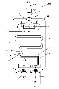
Referring now to Figures 6 to 10 of the drawings, another embodiment of an in-
line tester 100 in
accordance with the invention is shown. In this embodiment, the in-line tester
100 comprises a main body
12, again manufactured via a plastics injection moulding process, from a
transparent material, such as
ABS. The main body 12 defines a hollow interior chamber 14 as shall be
described herein below.
The in-line tester has an inlet 16, which is connectable, in use, to an NG
tube 18 via a luer
connector 102. The inlet 16 connects the NG tube 18, to a first portion 104 of
the chamber 14. The first
portion 104 of the chamber 14, which is shown in cross section in Figure 7,
comprises a base wall 106,
which supports a small strip of pH indicator paper 108. A cavity 110 is
provided on one side of the pH test
strip 108, such that fluid entering the in-line tester 100, via the inlet 16
is able to come into contact with
.. at least one surface or part thereof.
The first chamber part 104 has an outlet 112, which leads to a reservoir part
114 of the chamber
14.
The inlet 16 comprises a restriction, in the form of a Venturi 116 in a
preferred embodiment,
which causes liquid drawn up through the NG tube 18 to be "sprayed" over the
exposed surface of the pH
test strip 108. The provision of a constriction or Venturi 116 serves to cause
the incoming liquid to "fan-
out" as it enters the first chamber part 104, thereby ensuring that an
adequate area of the pH test strip
108 is wetted by the incoming liquid.
The outlet 112 of the first chamber part 104 leads to a reservoir portion 114
of the chamber 14.
In the illustrated embodiment, the reservoir 114 comprises a serpentine
pathway within the main body
12, which can retain approximately 4m1 of liquid, when full. The volume of the
reservoir 114 is ideally
selected to be slightly larger than the internal volume of the NG tube 18 and
so the exact volume of the
reservoir is not fixed. The reason for providing a reservoir 114 is to enable
a quantity of liquid within the
NG tube 18 to be accumulated within the main body 12 of the in-line tester
110, for reasons that shall
become apparent later.
The reservoir part 114 of the chamber 14 comprises an outlet 118, which feeds
into a further test
chamber part 120 of the chamber 14. The further test chamber part 120 is shown
in cross section in Figure
8 of the drawings, from which it can be seen that the outlet 118 of the
reservoir is in fluid communication
Date Regue/Date Received 2022-08-12
14
with a small disc tester 122 trapped, by its peripheries, between an upper
part and a lower part of the
main body 12. The test disc 122 is a CO2 tester, which exhibits a colour
change in the presence of carbon
dioxide.
The test disc 122 can be manufactured from a perforated, hydrophobic material,
which enables
gases exiting the reservoir 114 via its outlet 118 to pass through the test
disc 122 and into a lower part
124 of the second test chamber 120, before exiting via an outlet 126 into a
further pathway 128.
As gases are aspirated through the NG tube 18, they pass through the first
chamber part 104,
through the reservoir part 114 and eventually into the second test chamber
120, where they interact with
the test disc 122. If the gas contains carbon dioxide, it will cause the test
disc 122 to change colour, which
will be visible to a user (not shown) observing the in-line tester 110,
through its transparent main body
portion. The aspirated gas can be drawn into a connected vacuum source, in the
illustrated example, a
syringe 22, and the aspirated gas can, therefore, be tested for the presence
or otherwise of carbon
dioxide.
In other embodiments of the invention, the test disc 122 is formed from two
parts, namely a
downstream calorimetric test disc 122, and an upstream porous or perforated
element comprising a
hydrophobic material 52. In this embodiment of the invention, the two parts
work together as previously
described, namely with the calorimetric test disc 122 being able to test for
the presence of CO2, with the
porous or perforated, hydrophobic layer 52, acting as a liquid-stop device 52
upstream of the calorimetric
capnometer. Again, the two layers 122, 52 need not necessarily be in intimate
contact with each other,
although this may be beneficial in certain circumstances. Nevertheless, there
is a liquid-stop device 52
located upstream of the CO2 tester, which means that when the test is
complete, that is to say when a
sample of aspirated liquid comes into contact with the porous or perforated,
hydrophobic layer 52, the
in-line tester 100 is essentially hydraulically locked, thereby preventing
further aspiration. Meanwhile, a
sample of aspirated gas will be trapped downstream of the liquid-stop device
52, thereby inhibiting or
slowing the reversion of any colour change in the calorimetric capnometer for
a certain period of time.
It is possible to connect the outlet of the in-line tester to a vacuum pump,
or, in a preferred
embodiment to a syringe 22.
Where a small syringe 22 is used, it may be necessary to repeatedly withdraw
the syringe plunger
(not shown) to obtain a sufficient test volume via the NG tube 18. To
accomplish this, the in-line tester
110 is provided with a secondary outlet 128, to which is connected a one-way
valve 130. The syringe 22
and one-way valve 130 are operatively interconnected via a chamber 132 formed
in the main body 12 of
the in-line tester 100. The one-way valve 130 has a particular "cracking
pressure", above which, the valve
Date Regue/Date Received 2022-08-12
15
will open to allow gas to be expelled 134 from the in-line tester. The
cracking pressure of the one-way
valve 130 is designed to be lower than the permeability of the liquid-stop
device such that upon
depression of the syringe plunger (not shown) fluid is forced from the syringe
22, via the chamber 132
and out via the one-way check valve 130, as indicated by arrow 134 in Figure
6. However, upon
withdrawing the plunger, the one-way check valve 130 closes, thus enabling
fluid to be drawn up through
the NG tube, through the in-line tester 100, and, ultimately, into the
syringe, as indicated by arrow 136.
The benefits of this particular configuration of the invention are manifold.
In particular, because the main body 12 of the in-line tester 100 is
manufactured from a
transparent plastics material, it is possible for a user (now shown) to
observe the progression of aspirated
fluids through the tester 100. This usefully enables a user of the device to
"see" when e.g. saline flush has
been aspirated, followed by stomach content, for example, by a colour change
in the reservoir.
In a first example, where the NG tube 18 is initially empty, upon repeated
withdrawal and
compression of the syringe 22 plunger, fluid will be "pumped" up through the
NG tube in the manner
previously described. Because the NG tube 18 is initially empty, the first
fluid that will be drawn into the
in-line tester 100 will be gas/air from within the NG tube itself or the
patient's stomach/lung. This gas/air
will simply flow through the first chamber part and over the pH test strip
108, through the reservoir 114,
where it will eventually come into contact, and pass through the CO2 test disc
122.
The user, by observing the colour of the CO2 test disc 122 will be able to
ascertain whether it is
an air/stomach gas sample, or whether it is a "breath" sample of air aspirated
from the patient's lung, for
example.
Eventually, liquid (hopefully gastric juice) will be aspirated up the NG tube
18, where it will enter
the in-line tester 100 via the inlet 16. The aspirated liquid will be
"sprayed" by the Venturi 116 at the
tester inlet 116, and the design of the Venturi 116 is such that aspirated
liquid is sprayed/dispensed over
the exposed surface of the pH test strip 108. By observing a colour change in
the pH test strip 108, a user
(not shown) will be able to determine whether an acidic, (e.g. a gastric
juice) sample has been aspirated,
or whether something else has been aspirated.
Liquid will then be drawn into the reservoir 114, where it will gradually fill
the reservoir and the
user will be able to observe the progress of the liquid as it is drawn into
the reservoir.
In a second situation, for example where the NG tube 18 has been flushed with
water or saline
prior to use, it will initially contain a quantity, typically 4m1, of saline
solution or water. In this situation,
the "first liquid" to come into contact with the pH test strip 108 ought to be
neutral, which might,
ordinarily, indicate incorrect placement of the NG tubes tip in the patient's
stomach. However, this could
Date Regue/Date Received 2022-08-12
16
be a simple "false negative" because the first liquid aspirated is, in fact,
saline solution rather than gastric
juice. The test therefore needs to continue until such time as the flush
liquid within the NG tube 18 has
been recovered, which will, hopefully, be followed by a sample of gastric
juice.
Therefore, the invention comprises a reservoir 114, into which this initial
liquid may be
accumulated. Here, the volume of the reservoir 114 is slightly greater than
the internal volume of the NG
tube 18 such that when the reservoir is full, the user knows that what is
being tested ought to be gastric
juice, rather than flush liquid. The user (not shown) is therefore able to
observe the progression of the
flush liquid through the system, by observing the reservoir, which is visible
from outside the in-line tester
100 by virtue of it being manufactured from a transparent plastic, and,
ultimately, to test the pH of a
gastric juice sample thereafter.
Although not shown in Figure 6 for clarity, the in-line tester 100 suitably
comprises one or more
decals, each comprising a colour chart corresponding to the or each
calorimetric substance. The decal or
decals are suitably affixed to an outer surface of the in-line tester 100,
adjacent to, but preferably slightly
overlapping the testers 108, 122 - so that a visual comparison of colour of
the testers 108, 122 to the
colours or other indications on the decal can be made.
The in-line tester 100, shown in Figures 6, 7 and 8 of the drawings, is shown,
schematically, in
cross section in Figure 9, which is a cross section of Figure 6 on IX-IX.
In a preferred embodiment of the invention, the main body 12 of the in-line
tester 100 is
manufactured from two plastics injection moulded components, which fit
together to form the device
shown, schematically, in Figure 6. The various chambers 104, 120, 132 can be
formed simply by providing
recesses or grooves in the mating surfaces of the two components.
Referring to Figure 9 and 10 of the drawings ¨ Figure 10 being an exploded
view of Figure 9 the
inlet 16 is formed in two halves from each of the main body pieces 160, 162.
The two parts 160, 162 have
a flat mating surface 164, which when pushed together, form a fluid-tight seal
between the two pieces
160, 162. Channels or cavities within the in-line tester 100 can be formed by
providing recesses or cavities
in each of those mating surfaces 164.
Referring to Figures 9 and 10, it can be seen that the Venturi 116 is formed
by a pair of opposing
inclined surfaces 166 formed in each of the main body pieces 160, 162. The
first cavity 104, which houses
the pH test strip 108 is likewise formed with an additional recess part 168
formed in one of the pieces
162, for locating and retaining the pH test strip 108.
The outlet 112 of the first chamber part 104 is formed by complimentary
recesses formed in each
of the pieces 160, 162.
Date Regue/Date Received 2022-08-12
17
The serpentine reservoir 114 is formed by a correspondingly shaped serpentine
groove formed in
one of the main body pieces 162.
Likewise, the second chamber 120 is formed by a relatively deeper depression
in the second main
body part 162 and that enables the c02 test disc 122 to be housed therein.
Further recesses formed in the
main body pieces 160, 162 form the various other channels/cavities as
indicated, schematically in Figures
9 and 10.
The two main body pieces 160, 162 can either be glued together, for example,
by using an
adhesive or welding along the shut line to form fluid-tight cavities/channels
within the main body 12, or,
where the mating surfaces 164 are sufficiently flat and/or intimate, such
sealing may be accomplished by
simply clamping, clipping, or otherwise holding together, the two main body
pieces 160, 162.
By way of example, an in-line tester 100 similar to that described above with
reference to Figures
6 to 10 of the drawings, is shown in Figure 13, in which a partially
transparent decal 56 covers a front face
of the in-line tester 100. The decal 56 has a first part 562, which surrounds
the pH test strip 108 viewing
window. The first part 562 is divided into two differently-coloured regions
564, 566, which are colored to
match the colour of the pH paper 108 when a "fail" / "do not feed"; or a
"pass" condition is detected,
respectively. The two regions are indicated, for the avoidance of doubt by "X"
and "stomach" indicia,
respectively.
The reservoir 114 part of the in-line tester 100 is visible through a
transparent 568 part of the
decal 56. Graduations 570, indicating the volume of aspirated liquid, are
optionally provided.
A third part 572 of the decal 56, which surrounds the CO2 test strip 122
viewing window. The
third part 572 is divided into two differently-coloured regions 574, 576,
which are colored to match the
colour of the CO2 paper 122 when a "fail" / "do not feed"; or a "pass"
condition is detected, respectively.
The two regions 574, 576 are indicated, for the avoidance of doubt by "tick"
and "lung" indicia,
respectively.
Referring now to Figures 11 and 12 of the drawings, which are schematic
circuit diagrams for the
in-line testers 10, 100 shown in Figures 1 to 3 and 6 to 9 above,
respectively.
In Figure 11, the in-line tester 10 is fitted to an NG tube 18 at its inlet
16, and to a syringe 22 at its
outlet 20. The tip of the NG tube is placed in the stomach 13 of a patient
(not shown).
The in-line tester 10 comprises a chamber 14, which houses a first tester,
namely a disc of pH test
paper (e.g. litmus paper) 50, which is backed by a liquid-stop device, namely
a perforated plastics,
hydrophobic disc 52, which when wetted by aspirated liquids, closes-off and
stops/inhibits further
aspiration. A fluid passageway 15 connects the chamber 14 to a further chamber
that houses a
Date Regue/Date Received 2022-08-12
18
calorimetric capnometer 64, in this case, a strip of CO2-sensitive indicator
paper that changes colour in
the presence of CO2.
The syringe's plunger can be withdrawn to aspirate a sample of fluid from the
stomach, via the
NG tube 18 and into the in-line tester 10.
Aspirated gas passes through the pH test paper SO and the liquid-stop device
52, where it then
comes into contact with the calorimetric capnometer 64 to indicate the
presence or otherwise of CO2 in
the aspirated gas sample.
Thereafter, liquids may be aspirated from the stomach 13, via the NG tube 18,
where they come
into contact with the first tester 50 and indicate the presence, or not, of a
target substance, e.g. stomach
acid and/or a substance (e.g. a protein) only found in the stomach 13. The
aspirated liquid contacts the
liquid-stop device 52, causing the in-line tester 10 to hydraulically lock,
thereby signifying the end of the
procedure.
A one-way valve 130 may optionally be provided in a branch spurred-off between
the syringe 22
and the outlet 20. This enables the syringe plunger to be depressed, and fluid
within the syringe 22 to be
vented via the one-way valve 132. This configuration permits a relatively
small syringe 22 to be used as
part of a pump, rather than having to use a relatively large syringe to obtain
an adequate quantity of
aspirate.
Referring now to Figure 12, the in-line tester 100 is fitted to an NG tube 18
at its inlet 16, and to
a syringe 22 at its outlet 20. The tip of the NG tube is placed in the stomach
13 of a patient (not shown).
The in-line tester 100 comprises a chamber 14, which has several parts.
A first chamber part 104 houses a first tester, for example a strip of pH test
paper (e.g. litmus
paper) 108 and/or a strip of other indicator paper, which changes colour in
the presence of a target
substance.
The first chamber part 104 is connected to a reservoir 114, which can
accumulate a quantity of
aspirated liquid.
The reservoir 114 connects to a further chamber 120, which houses a liquid-
stop device 52,
namely a perforated plastics, hydrophobic disc, which when wetted by aspirated
liquids, closes-off and
stops/inhibits further aspiration.
Downstream of the liquid-stop device 52, there is provided a calorimetric
capnometer 122, in this
case, a disc of CO2-sensitive indicator paper that changes colour in the
presence of CO2.
A one-way valve 130 is provided in a branch spurred-off, e.g. via a chamber
132, between the
syringe 22 and the outlet 20.
Date Regue/Date Received 2022-08-12
19
The syringe's plunger can be withdrawn to aspirate a sample of fluid from the
stomach, via the
NG tube 18 and into the in-line tester 10. Aspirated gas passes through the
first tester 108 and the liquid-
stop device 52, where it then comes into contact with the colorimetric
capnometer 122 to indicate the
presence or otherwise of CO2 in the aspirated gas sample.
Thereafter, liquids may be aspirated from the stomach 13, via the NG tube 18,
where they come
into contact with the first tester 104 and indicate the presence, or not, of a
target substance, e.g. stomach
acid and/or a substance (e.g. a protein) only found in the stomach 13. The
aspirated liquid then fills the
reservoir 114 until it eventually contacts the liquid-stop device 52, causing
the in-line tester 10 to
hydraulically lock, thereby signifying the end of the procedure.
By virtue of the one-way valve 130, the syringe plunger to be depressed, and
fluid within the
syringe 22 can be vented via the chamber 132 and the one-way valve 132. This
configuration permits a
relatively small syringe 22 to be used as part of a pump, rather than having
to use a relatively large syringe
to obtain an adequate quantity of aspirate.
Referring now to Figure 14 of the drawings, a slight variation of the circuit
diagram shown in Figure
12 is described. Identical reference signs have been used to identify
identical features, for the avoidance
of repetition, and for clarity. In Figure 14, it can be seen that the in-line
tester 100 has been modified by
the addition of a coarse liquid-stop device 520 locate upstream of the
previously-described porous or
perforated, hydrophobic membrane/disc 52. The coarse liquid-stop device 520
comprises a chamber 522,
having an inlet 524 connected to the outlet of the reservoir 114, and an
outlet 526 connected to the inlet
of the chamber 120. Baffling 526 is provided within the chamber 522, to
prevent liquid drops from being
inadvertently splashed onto, or reaching the outlet 526. Here, liquids and
gasses can be drawn into the
coarse liquid-stop device 520, as may happen when the reservoir 114 is full,
and the coarse liquid-stop
device 520 provides a further means for preventing the porous or perforated,
hydrophobic
membrane/disc 52 from wetting out, as liquid droplets will be collected in the
chamber 522, rather than
passing through the coarse liquid-stop device 520 to the porous or perforated,
hydrophobic
membrane/disc 52 downstream of it.
The main purpose of the coarse liquid-stop device 520 is that it enables,
where the reservoir 114
is full of aspirated liquid, say saline flush, to nevertheless permit the
passage of air/gas bubbles to the CO2
test strip 122, via the porous or perforated, hydrophobic membrane/disc 52.
This may occur where an
NG tube has been used previously and thus contains a saline flush liquid.
However, if, somehow, the NG
tube has become misplaced, as may happen where the patient "wretches" the NG
tube back up their
oesophagus, then the next time the NG tube needs to be used, it will be
checked for correct placement.
Date Regue/Date Received 2022-08-12
20
Now, the first few ml of aspirate will be saline flush, or residual feed
within the NG tube, and this liquid
will fill, or partially fill the reservoir 114. Subsequent aspiration
eventually empties the NG tube 18, such
that gas is now, finally, aspirated. This aspirated gas will bubble through
the liquid already in the reservoir,
and without a coarse liquid-stop device 520 present, the liquid would tend to
be splashed onto the porous
or perforated, hydrophobic membrane/disc 52, thereby wetting it, and causing
the in-line tester 100 to
hydraulically lock. However, by placing a coarse liquid-stop device 520
upstream of the porous or
perforated, hydrophobic membrane/disc 52, the aspirated gas is able to pass
through the reservoir, and
the porous or perforated, hydrophobic membrane/disc 52 before it reaches the
CO2 paper 122, with the
splashed liquid effectively being filtered-out by the coarse liquid-stop
device 520. This enables a user to
reliably test an aspirated gas sample ¨ even after already having aspirated a
liquid sample.
The coarse liquid-stop device 520, where provided, can be incorporated into
the reservoir 114, or
into the chamber 120, as desired.
EXAMPLE 1:
Samples were used to generate the following truth table comparing a device in
accordance with
the present invention having means for pH detection, enzyme detection and
carbon dioxide detection
with the current clinical standard (pH paper).
There were three scenarios: 1) Normal pH content in stomach; 2) Patient on
antacid medication;
and 3) Stomach content in the lung. Each method and device was used and the
results set out below.
TABLE 1:
SCENARIO DEVICE IN ACCORDANCE WITH CURRENT CLINICAL STANDARD (PH
PAPER)
PRESENT INVENTION
Normal pH content Confirmed Confirmed
in stomach
Patient on antacid Confirmed False negative confirmation
tube not in
medication stomach, patient has delayed
feed
Stomach content in Confirmed False negative confirmation
tube not in
the lung stomach, patient has delayed
feed
EXAMPLE 2:
Samples were used to generate the following truth table comparing a device in
accordance with
the present invention having means for pH detection, enzyme detection and
carbon dioxide detection
with the current clinical standard (pH paper). In this example, the three
markers tested for in the device
Date Regue/Date Received 2022-08-12
21
according to the present invention are separated out to give a clearer
demonstration of the advantages
of a device in accordance with the present invention.
There were three scenarios: 1) Normal pH content in stomach; 2) Patient on
antacid medication;
and 3) Stomach content in the lung. The presence or absence of three markers
was known and each
method and device was used and the results set out below.
TABLE 2:
SCENARIO 1s-r 2ND 3R0 DEVICE CURRENT
MARKER MARKER MARKER ACCORDING TO CLINICAL
PH ENZYME CO2 PRESENT STANDARD
(PH)
INVENTION
Normal pH content in Positive Positive Negative 1st, 2"I
and 3 1st marker
stomach markers confirmed
confirmed only
Patient on antacid Negative Positive Negative 1st, 2nd
and 3rd None or
medication markers confirmed
confirmed
Stomach content in Positive Positive Positive 1st, 2nd
and 3rd 1st confirmed
the lung (or M. markers confirmed only ¨
Cattarhalis infection dangerous
in lung)
In complicated situations where for example, a patient is taking antacids or
there is stomach
content in the lung, only devices in accordance with the present invention
will confirm the actual location
of the NG tube for enteral feeding. Known devices and methods will only give
an accurate indication of
location when a patient has a normal stomach pH. In all other scenarios, the
result from using known
devices can be dangerous and has potentially fatal consequences should feeding
via the tube be initiated
where an incorrect placement of the tube is mistakenly indicated as being in
the stomach.
The invention is not restricted to the details of the foregoing embodiments,
which are merely
exemplary of the invention. For example, any shapes, sizes, relative
dimensions etc. are illustrative, and
not limiting, as are any material selections and/or design choices (e.g. type
of check valve).
Date Regue/Date Received 2022-08-12