Note: Descriptions are shown in the official language in which they were submitted.
CA 03034356 2019-02-19
WO 2018/051187 PCT/IB2017/001317
ANEURYSM CLOSURE DEVICE
CROSS-REFERENCE TO RELATED APPLICATIONS
[0001] This application claims priority to the United States provisional
patent application serial
no. 62/394,564, filed September 14, 2016. Priority to the provisional patent
application is
expressly claimed, and the disclosure of the provisional application is hereby
incorporated herein
by reference in their entireties and for all purposes.
FIELD OF THE INVENTION
[0002] The invention relates to devices, a systems, and associated methods for
use, delivery, and
manufacture for changing the blood flow into an aneurysm designed to induce
aneurysm
thrombosis and/or the exclusion from blood flow and pressure of the aneurysm
in order to
prevent further growth and eventual rupture.
BACKGROUND OF THE INVENTION
[0003] A brain (cerebral) aneurysm is a protrusion of different shapes from
the otherwise smooth
cylindrical wall of the vessel, usually caused by a weak area in the vessel
wall that gives in under
blood pressure. In most cases, a brain aneurysm causes no symptoms and goes
unnoticed. In
some cases, the brain aneurysm ruptures, causing a hemorrhagic stroke. When a
brain aneurysm
ruptures in the most common area, the result is a hemorrhage (most commonly
subarachnoid).
Depending on the severity of the hemorrhage, permanent neurological deficiency
or death may
result. The most common location for brain aneurysms is in and around the
network of blood
vessels at the base of the brain called the circle of Willis.
[0004] Saccular aneurysm is the most common type of aneurysm. It account for
80% to 90% of
all intracranial aneurysms and is the most common cause of non-traumatic
subarachnoid
hemorrhage (SAH). It is also known as a "berry" aneurysm because of its shape.
The berry
aneurysm looks like a sac or berry having a neck, or a stem and a sac (body),
formed at a
bifurcation or on a straight segment of an artery.
[0005] Currently, there are three primary treatments for a cerebral aneurysm:
(a) craniotomy and
surgical clipping, (b) endovascular coiling, and (c) flow diverters. Surgical
clipping requires a
-1-
CA 03034356 2019-02-19
WO 2018/051187 PCT/IB2017/001317
craniotomy to expose the aneurysm which is then closed by attaching a clip to
the neck (base) of
the aneurysm, thereby providing a physical barrier to isolate the aneurismal
sac. Although
effective, this procedure is highly invasive and may require long recovery
times. Also, it is
available only for aneurisms that are close to the brain surface at an
accessible position.
[0006] Endovascular coiling is a minimally-invasive procedure in which a pre-
shaped coil
(typically of shape-memory metal) is released into the aneurismal sac from a
catheter. The coil
fills the aneurismal sac causing the blood flow within the aneurismal sac to
become slow and
non-laminar. The blood flow disruption within the aneurismal sac results in
the formation of a
clot and exclusion of further blood flow into the structure, thereby
preventing further expansion
of the aneurysm. When successful, the thrombus eventually may be covered by a
layer of
endothelial cells, reforming the inner vessel wall. However, not all coiling
procedures are
successful. Coiling may result in aneurysm recanalization in which new routes
of blood flow in
the aneurism are formed, reapplying blood pressure on the aneurismal wall and
further
expanding it. Coiling also may require the implantation of additional devices
such as stents (in
order to retain the coils in the aneurism to prevent their sagging into the
parent vessel) and/or the
use of multiple coils (released in order to affect clotting in the aneurismal
sac). The use of
multiple devices increases the procedure time, treatment cost, and probability
of an adverse
event.
[0007] Flow diverters are stent like devices to be deployed in the parent
vessel across the neck of
the aneurism to alter or restrict blood flow into the aneurysm. The goal of
the diverters is to
cause thrombosis within the aneurismal sac. Flow diverters have limitations.
For example,
diverters generally should be used in relatively straight vessels and often do
not perform well
when the aneurysm is located at or near vessel junctions and bends.
Additionally, the gaps
between the struts of the diverter in many cases are too large to induce
thrombosis in the
aneurismal sac or may cause occlusion of the parent vessel due to clotting
and/or inflammatory
reactions. Finally, the diverter may cause small perforations near the
aneurismal neck, causing
bleeding, or may occlude nearby small diameter arteries (perforators), each of
which may have
neurological sequelae.
-2-
CA 03034356 2019-02-19
WO 2018/051187 PCT/IB2017/001317
[0008] Accordingly, there is a need for an improved, efficient, and cost
effective device and
associated methods, for treating cerebral aneurysm, that is independent of the
vessel and
aneurism anatomy and that will lower the cost and duration of the procedure,
as well as reduce
the probability of adverse events like perforation, occlusion of nearby
perforators and will have
higher rate of success in excluding the aneurism.
SUMMARY OF THE INVENTION
[0009] The present invention relates to clot-forming devices ("CFDs"),
systems, and associated
methods for use, delivery, and manufacture for changing the blood flow into an
aneurysm
designed to induce aneurysm thrombosis and/or the exclusion from blood flow
and pressure of
the aneurysm in order to prevent further growth and eventual rupture.
[0010] In one aspect, the invention provides a device having: (a) a central
attachment member;
(b) a plurality of self-expanding arms attached to the central attachment
member and extending
radially therefrom and (c) one or more porous panels attached to the arms and
extending radially
from the central attachment member; wherein the device is configured to adopt
a crimped
conformation having a first cross-sectional diameter and a deployed
conformation having a
second cross-sectional diameter that is larger than the first cross-sectional
diameter, and a three-
dimensional shape that is approximately spherical, semi-spherical, ovoid, or
semi-ovoid.
Optionally, the device may be used for aneurysm closure according to the
methods described
herein such that the central attachment member, arms, and mesh panels are
sized such that they
form a barrier or screen between a vessel and an aneurysm when the device is
in the deployed
conformation and positioned within the aneurysm.
[0011] In some embodiments, the device, including the central attachment
member, arms, and
mesh panels, are sized to fit within the lumen of a catheter when the device
is in the crimped
conformation. Optionally, the first cross-sectional diameter fits into a
delivery system with a
crossing profile less than about 10, 8, or 6 French (i.e., between about 4-10
French, 6-10 French,
4-8 French, or 6-8 French).
-3-
CA 03034356 2019-02-19
WO 2018/051187 PCT/IB2017/001317
[0012] In some embodiments, the self-expanding arms comprise a shape memory
material that
has a memorized shape that defines the three-dimensional shape that is
approximately spherical,
semi-spherical, ovoid, or semi-ovoid.
[0013] In some embodiments, the mesh covering, formed by the one or more mesh
panels,
extends radially from the central attachment member to a distance of 10% or
more of the length
of the arms including, for example, at least 10%, 20%, 30%, 40%, 50%, 60%,
70%, 80%, 90% or
more of the length of the arms, and including, for example, at least about
10%, 20%, 30%, or
40% and not more than 60%, 70%, 80%, or 90% of the length of the arms. As
described herein,
the mesh panel(s) may porous and may be formed from a polymer or wire mesh, a
perforated
polymer membrane, or a mesh of filaments.
[0014] In some embodiments, the mesh panels may contain a thrombogenic agent.
[0015] In some embodiments, the arms may be substantially linear and may
further comprise
straight, wavy, or spiral wires. In other embodiments, the arms define a
closed shape such as an
ellipse, petal shape, or reuleaux triangle. Optionally, the arms are joined by
connecting struts
that do not contact the attachment member. Optionally, at least one arm also
contains a radio-
opaque marker at or near a distal end.
[0016] In some embodiments, each arm further defines an eyelet at or near a
distal end. The
eyelets may be integral to the arms or attached to the arms via struts, as
described herein.
[0017] The attachment member may be annular, toroidal, or any suitable shape
in accordance
with the principles described herein. Optionally, the attachment member
contains one or more
holes. Further, the device also main have a thread loop disposed through the
one or more holes
or eyelets and extending in the proximal direction.
[0018] In some embodiments, the device also has a guidewire disposed along a
longitudinal axis
of the device and through the attachment member annulus or equivalent
structure. Optionally,
for embodiments in which the arms further defines an eyelet at or near a
distal end, the guidewire
is further disposed through the eyelets when the device is in the crimped
conformation. Such a
configuration may be used to maintain the device in the crimped conformation
either with or
without the use of an external sheath.
-4-
CA 03034356 2019-02-19
WO 2018/051187 PCT/IB2017/001317
[0019] In other embodiments, the device also may contain one or more wires
attached at a first
end and extending into an interior three-dimensional space defined by the arms
in the deployed
conformation. The wires may be attached at the first end to the attachment
member or the arms.
[0020] In another aspect, the invention provides methods for closing an
aneurysm and/or
reducing blood flow through the neck of an aneurysm by deploying within the
aneurysm any of
the devices described herein. Preferably, the devices are delivered by, and
deployed from a
catheter.
[0021] In one embodiment, the method:
(a) provides a catheter housing a device in a crimped conformation, the device
having:
(i) a central attachment member;
(ii) a plurality of self-expanding arms attached to the central attachment
member and
extending radially therefrom in a distal direction, wherein the arms define a
deployed conformation having a three-dimensional shape that is approximately
spherical, semi-spherical, ovoid, or semi-ovoid; and
(iii) one or more porous panels attached to the arms and extending radially
from the
central attachment member;
(b) passing the catheter to a target aneurysm;
(c) inserting the device into the aneurysm;
(d) deploying the device into the deployed conformation; and
(e) withdrawing the catheter.
[0022] Optionally and as necessary, the method also includes the step of
repositioning the device
within the aneurysm which is performed after step (d) and repeated as desired.
Repositioning
may be effected through the use of a device having one or more holes in the
central attachment
member through which a thread loop is placed to facilitate partial or total
device retrieval by the
operator, as described in more detail herein.
[0023] In some embodiments, the foregoing method causes thrombosis within the
aneurysm.
-5-
CA 03034356 2019-02-19
WO 2018/051187 PCT/IB2017/001317
[0024] In another aspect, the invention provides systems having a catheter, an
aneurysm closure
device contained therein in a crimped conformation, and accessory structures
to facilitate the
delivery, positioning, and retrieval of the device. In one embodiment, the
system contains:
(a) a catheter;
(b) a device in a crimped conformation within the catheter housing, the device
having:
(i) an annular central attachment member;
(ii) a plurality of self-expanding arms attached to the central attachment
member and
extending radially therefrom, wherein the arms define a deployed conformation
having a three-dimensional shape that is approximately spherical, semi-
spherical,
ovoid, or semi-ovoid; and
(iii) one or more porous panels attached to the arms and extending radially
from the
central attachment member;
(c) an annular pushrod contacting a proximal side of the central attachment
member; and
(d) a guidewire extending along a longitudinal axis of the catheter and
disposed through a
pushrod annulus, a central attachment member annulus, and a distal catheter
lumen opening.
[0025] "Proximal" is a relative term that refers to the direction or side
towards the entry point of
the catheter into the vessel. For example, an operator withdrawing a catheter
from a patient is
translating the catheter in the proximal direction.
[0026] "Distal" is a relative term that refers to the direction or side away
from the entry point.
For example, an operator inserting a catheter into a patient is translating
the catheter in a distal
direction.
[0027] "Top," when referring to a CFD, is a relative term referring to the
portion of the device
that is toward the aneurysmal sac or peripheral ends of the device arms.
[0028] "Bottom" when referring to a CFD, is a relative term referring to the
portion of the device
that is toward the aneurysmal neck or the blood vessel.
DESCRIPTION OF DRAWINGS
-6-
CA 03034356 2019-02-19
WO 2018/051187 PCT/IB2017/001317
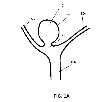
[0029] FIG. 1A is a schematic diagram showing the general structure of a
cerebral berry
aneurism at a Y-shaped bifurcation.
[0030] FIG. 1B is a schematic diagram showing the general shape and
positioning of one type of
CFD deployed in the aneurism shown in FIG. 1A.
[0031] FIG. 2 illustrates exemplary but non-exhaustive arm shapes.
[0032] FIG. 3A is a plan (flat) view of a first CFD embodiment of the
invention.
[0033] FIGS. 3B-C are perspective views of the first CFD embodiment shown in
FIG. 3A, in
possible deployed conformations.
[0034] FIG. 3D shows the first CFD embodiment shown in FIG. 3A in a crimped
conformation
within the lumen of a delivery sheath (e.g., a catheter).
[0035] FIG. 4A is a plan (flat) view of a second CFD embodiment of the
invention.
[0036] FIGS. 4B-C are perspective views of the second CFD embodiment shown in
FIG. 4A, in
possible deployed conformations.
[0037] FIG. 5A is a plan (flat) view of a third CFD embodiment of the
invention.
[0038] FIGS. 5B-C are perspective views of the third CFD embodiment shown in
FIG. 5A, in
possible deployed conformations.
[0039] FIG. 5D shows the third CFD embodiment, as shown in FIGS. 5A-C, in a
crimped
conformation.
[0040] FIG. 5E shows the distal end of the crimped conformation shown in FIG.
5D.
[0041] FIG. 6 shows a CFD (e.g., a CFD of the first embodiment) in a partially
crimped
conformation during the repositioning process.
[0042] FIGS. 7A-B are perspective views of possible deployed conformations of
a fourth CFD
embodiment.
-7-
CA 03034356 2019-02-19
WO 2018/051187 PCT/IB2017/001317
[0043] FIG. 7C shows the fourth CFD embodiment, as shown in FIGS. 7A-B, in one
particular
crimped conformation.
[0044] FIG. 7D shows a CFD embodiment, as shown in FIGS. 7A-C, in a possible
crimped
conformation within the lumen of a delivery sheath (e.g., a catheter).
[0045] FIG. 8 is a perspective view of a possible deployed conformation of CFD
having inner
wires.
[0046] FIG. 9A is a schematic view of a CFD delivery system.
[0047] FIG. 9B is a schematic view of another CFD delivery system.
DETAILED DESCRIPTION
[0048] The present invention provides a self-expanding Clot Forming Device
(CFD) designed to
be deployed within an aneurysmal sac from a catheter, and its associated
delivery devices,
methods for use, and methods for manufacture. The CFD may be deployed within
an aneurysm
located along a substantially straight portion or tortuous portion of a blood
vessel wall, or an
aneurysm at or near a junction or bifurcation point of a blood vessel(s).
Generally, the CFD is
formed from a centrally-disposed attachment member (e.g., a ring) having a
plurality of arms
extending therefrom in a radial pattern. The arms support a mesh covering at
least the lower
portion of the CFD. The CFD, when deployed, forms a three-dimensional shape
that is
approximately spherical, semi-spherical, ovoid, or semi-ovoid and is open at
the top. The
material properties and parameters allow the CFD to self-fit to the aneurismal
shape.
[0049] FIG. 1A schematically illustrates an aneurysm 10 having an aneurysmal
sac 12 and an
aneurysmal neck 14 at the junction of main blood vessel 15a and tributary
branches 15b,c. FIG.
1B schematically illustrates one embodiment of a CFD 100, described in more
detail below,
deployed within the aneurysmal sac 12, wherein the centrally-disposed
attachment member 110
is disposed within or toward the aneurysmal neck 14 with the arms 120
extending radially into
the body of the aneurysmal sac 12 automatically fitting to its shape. The mesh
130 is supported
by arms 120 and covers substantially all of the aneurysmal neck 14 opening.
The features and
-8-
CA 03034356 2019-02-19
WO 2018/051187 PCT/IB2017/001317
specific embodiments of the CFD 100, delivery devices, methods for use, and
methods for
manufacture are now described in more detail.
[0050] Attachment Member
[0051] The attachment member is centrally-disposed and configured to provide
an attachment
point for a plurality of arms. It is sized to fit within the lumen of a
catheter or inner
member/jacket from which the CFD is to be deployed. There is no limitation on
the shape of the
attachment member, however, a generally circular shape is preferred such that
the shape matches
that of the deployment member lumen. In some embodiments, the attachment
member is a ring
(e.g., a circular ribbon), a toroid, or a disc. (See, for example, FIGS. 1B,
3B-3D, 4A-4C, 5A-5C,
7A-7B, and 8). Optionally, the attachment member is generally annular (e.g., a
ring or toroid)
and adapted to accommodate a centrally-disposed guide wire. (See, for example,
FIGS. 5D, 7D,
and 9A-B). Optionally, the attachment member has one or more holes (e.g., two,
three, four, or
more) adapted to accept a retrieval thread, described in more detail below.
(See, for example,
FIG. 6). Optionally, a radio-opaque marker is incorporated or affixed to the
attachment member.
[0052] Arms
[0053] A plurality of arms (e.g., two, three, four, five, six, seven, eight,
or more) are attached to
the attachment member on one centrally-disposed end and extend from the
attachment member
in a radial pattern. The radial pattern may be symmetrical or asymmetrical,
but a symmetrical
pattern is preferred. The arms may be manufactured as separate elements and
subsequently
attached to the attachment member, or the arms and attachment member may be
manufactured as
a single contiguous piece. Optionally, a radio-opaque marker is incorporated
or affixed to one or
more of the arms. (See, for example, FIGS. 3A, 4A-4C, 5A, and 7A). Preferably,
the radio-
opaque marker is disposed at or near the distal end of the arm(s).
[0054] The arms are constructed to be self-expanding such that the CFD is
capable of adopting a
crimped conformation and a deployed conformation, the latter being its
memorized shape
adopted when the CFD is released from the sheath/catheter. In the crimped
conformation, the
distal ends of the arms are closely disposed to the longitudinal axis of the
CFD such that the CFD
has a first, smaller diameter adapted to be housed within the catheter or
delivery device. When
-9-
CA 03034356 2019-02-19
WO 2018/051187 PCT/IB2017/001317
deployed, the arms self-expand to be disposed farther from the longitudinal
axis, resulting in the
CFD adopting an approximately spherical, semi-spherical, ovoid, or semi-ovoid
shape, wherein
the CFD has a second, larger diameter defined by the expanded arms. (compare,
for example,
FIGS. 3B-3C and 3D).
[0055] The arms, and optionally the attachment member, may be constructed of
shape memory
materials and using manufacturing methods that are well-known in the art.
Specifically, the
arms, and optionally the attachment member, may be constructed of known shape
memory alloys
including, for example, NiTi. These components can be formed by etching or
laser cutting a
tubing or flat sheet of material into the patterns shown. The components then
may be heat
treated after formation, as known by those skilled in the art, to take
advantage of the shape
memory characteristics and/or super elasticity. Metal surfaces may be
processed chemically
and/or electrochemically in order to achieve the required surface smoothness.
[0056] The arms may have any convenient shape suitable for supporting the
mesh. For example,
arms may be substantially linear elements or may define and enclose a
geometric space. In the
latter configuration, the geometric space is defined on its perimeter by
struts and void in the
interior. FIG. 2 illustrates some useful arm 20 shapes including, for example,
straight 20a, wavy
20b, spiral 20c (e.g., spring or coiled), elliptical 20d, petal-shaped 20e
(e.g., folium/leaf-shaped),
and a reuleaux triangle (i.e., the triangular shape formed by three
overlapping circles). It is
understood that arms 20 in a crimped conformation are substantially linear and
parallel to the
longitudinal axis of the CFD. But, in the deployed conformation, arms adopt a
curvilinear shape
from the central to distal ends, thereby defining the spherical, semi-
spherical, ovoid, or semi-
ovoid shape of the CFD designed to conform to the aneurysmal sac 12. (See, for
example, the
deployed conformations illustrated in FIGs. 1B, 3B, 3C, 4B, 4C, 5B, 5C, 7A,
7B, and 8).
[0057] Optionally, arms further comprise eyelets at or near the distal end.
The eyelets may be
integral to the arms or may be attached to the arms by struts. (See, for
example, FIGs 2, 4A-4C,
5A-5C, and 7A-7B). Eyelets are configured to circumnavigate the longitudinal
axis of the CFD
in the crimped configuration and adapted to accept a guide wire. (See, for
example, FIGs. 5D-
5E).
-10-
CA 03034356 2019-02-19
WO 2018/051187 PCT/IB2017/001317
[0058] Optionally, some or all of the arms may be attached on one or both
sides to adjacent arms
through connectors. Connectors are struts that attach one arm to an adjacent
arm but do not
attach directly to attachment member. (See, for example, FIGs. 3A-3C).
Connectors may be
formed from a shape memory material and, preferably, are formed from the same
shape memory
material as the arms. Connectors may be fabricated as separate elements and
later attached to
arms or may be integral and contiguous with the arms, being fabricated as a
single piece.
Connectors may be used to enhance CFD rigidity and/or provide additional
surface area and/or
support for the mesh covering.
[0059] Mesh Covering
[0060] The CFD further comprises a mesh covering supported by the arms. The
mesh covering
is a porous, semi-porous, or non-porous net made from a mesh of fibers or
wires (e.g., metal or
thermoplastic polymer such as EPTFE, polyurethane, etc.), or a perforated
polymer membrane
(e.g., Dacron) having holes. It is configured to limit, change, and/or reduce
blood flow into the
aneurysmal sac 12 when disposed across the aneurysmal neck 14. The slow and
non-linear
blood flow occurring through the mesh 130 is intended to cause clotting in the
aneurysmal sac 12
such that the clot eventually excludes further blood flow and pressure within
the sac 12, thereby
preventing expansion and rupture of the aneurysm 10. In some embodiments, the
mesh covering
is porous to blood cells, platelets, and/or clotting factors.
[0061] The mesh is configured to restrict blood flow through the aneurysmal
neck 14.
Accordingly, the mesh covers at least the bottom 10%, 20%, 30%, 40%, 50%, 75%
of the height
of the CFD (i.e., the distance H, as illustrated in FIG. 3B, from the bottom
of the attachment
member to the distal end of arms 120 when CFD is in a deployed conformation).
Alternatively,
the mesh covers the substantially entirety of CFD. The mesh covering may be
attached to the
inside or the outside of the arms and/or connectors, if present. Optionally,
the mesh covers an
annular opening in the attachment member.
[0062] The mesh covering may be continuous (i.e., a single piece of mesh to
form the covering)
or discontinuous (i.e., multiple pieces of mesh that together form the
covering). Continuous
mesh coverings are illustrated, for example, in FIGS. 4A and 7A-7B (linear
arms) and FIG. 5A
(arms defining geometric shapes). In these embodiments, the mesh covering is
formed by a
-11-
CA 03034356 2019-02-19
WO 2018/051187 PCT/IB2017/001317
single mesh panel. Discontinuous mesh coverings may be formed from a plurality
of mesh
panels. For example, the void formed by arms defining a geometric space (see,
for example,
arms 20d and 20e in FIG. 2, and FIG. 3A, ) may be partially or totally covered
by one set of
mesh panels, and the void space between the arms covered by a second set of
mesh panels.
Mesh panels in the void space between the arms optionally may be attached to
connectors, when
present, for support (see, for example, connectors 122 in FIG. 3A). For
embodiments in which
the arms are linear, mesh panels may be attached to adjacent arms in order to
form a contiguous
barrier of multiple (discontinuous) mesh panels. Optionally, whether
continuous or
discontinuous mesh coverings are used, the mesh panel(s) may comprise one or
more creases to
facilitate smooth and reproducible folding when the CFD is in the crimped
(folded)
configuration.
[0063] For embodiments in which discontinuous mesh coverings are used, the
plurality of mesh
panels may be on the same side of the wire frame (i.e., arms and optional
connectors) or on
opposite sides of the wire frame. For example, all mesh panels may be affixed
either to the outer
surface or to the inner surface of the arms for support. Alternatively, some
mesh panels may be
affixed to the outer surface and other mesh panels may be affixed to the inner
surface of the
arms. In one example, the mesh panels covering or partially covering the voids
formed by arms
defining a geometric space may be affixed to the inside surface of the arms.
Optionally, these
mesh panels are creased to fold inward when crimped. And, the mesh panels
covering the void
space between the arms are affixed to the outside surface of the arms.
Optionally, these mesh
panels are creased to fold either inward or outward when crimped. In another
embodiment in
which the CFD is formed from linear arms, the mesh panels covering the void
space between the
arms may be alternated between the inside and the outside of the wire frame.
For example, in a
CFD having six arms and therefore defining six separate void spaces between
the arms, the first,
third, and fifth void space may be covered by mesh panels affixed to the
inside of the wire frame,
and the second, fourth, and sixth void space may be covered by mesh panels
affixed to the
outside of the wire frame.
[0064] The mesh covering may be configured to limit the outward deflection of
the arms in the
deployed conformation. For example, the self-expanding arms may be fabricated
to have a
resting state in which the arms define a CFD structure that is larger than
desired for deployment
-12-
CA 03034356 2019-02-19
WO 2018/051187 PCT/IB2017/001317
within an aneurysm in order to ensure that the arms have a sufficient opening
force to fully
deploy the CFD. The circumference/diameter of the deployed conformation may be
limited to a
size less than the resting state of arms by appropriately limiting the size
and shape of the mesh
covering. The external or internal surface of CFD (i.e., the arms) may be
coated with a polymer
mesh by means of, for example, an electrospinning procedure, application of
perforated
membrane, or metal wire net.
[0065] Optionally, one or more of the mesh panels may be coated with a
thrombogenic factor.
Suitable thrombogenic factors include, for example, Factors VII, VIII, IX, X,
XI, and XII.
Thrombogenic factors may be encapsulated or incorporated into a polymer
coating that is applied
to the mesh panels. Alternatively, the thrombogenic factors may be affixed or
adhered to the
mesh panels (e.g., by dipping and drying).
[0066] CFD Construction And Design
[0067] The following implementations and embodiments are intended to
illustrate additional
structural and functional elements of the CFD and the principles of CFD
function and design.
These embodiments are not intended to be limiting. All components of the
delivery catheter
shall be fabricated from suitable biocompatible material for interventional
invasive use.
[0068] FIG. 3A is a plan view of one embodiment of CFD 100. In this
embodiment, arms 120
are formed from struts 123 defining a petal shape and are attached to a
centrally-disposed
attachment member 110 (shown in FIGs. 3B-D). Struts 123 are illustrated as
wire which may be
round. However, arms 120 may be wavy or spiral/spring-like, as described in
FIG. 2. The four
arms 120 are disposed in a symmetrical radial pattern resulting in a
quatrefoil or flower-shaped
configuration. Adjacent arms 120a,b are connected by connector 122. Like the
arms 120,
connectors 122 may have any configuration described in FIG. 2. Mesh 130 covers
the void
spaces defined by the arms 120 and connectors 122. Radio-opaque markers 121
are affixed near
the distal end of arms 120a,c. This plan view may be used to represent the CFD
100 as it would
be fabricated from a shape memory material prior to three-dimensional shaping.
[0069] FIG. 3B is a perspective view of CFD 100 illustrated in FIG. 3A in a
deployed
conformation. Attachment member 110 is illustrated as a ring having holes 111.
Arms 120
-13-
CA 03034356 2019-02-19
WO 2018/051187 PCT/IB2017/001317
extend radially from attachment member 110 and adopt a curvilinear shape over
the height H.
Arms 120 are interconnected by connectors 122. Mesh 130 covers the void spaces
defined by
the arms 120 and connectors 122 and extends more than 75% of the height. In
this
configuration, arms 120 define a semi-spherical or bowl-shaped form of the CFD
100.
[0070] FIG. 3C is a perspective view of CFD 100 in a different deployed
conformation in which
arms 120 define a substantially spherical shape open at the top.
[0071] FIG. 3D is a plan view of CFD 100 in its crimped conformation held in
place within a
delivery device such as a catheter 190. Attachment member 110 slideably
engaged with and the
same shape as the catheter lumen 191. Arms 120 are substantially straight and
parallel with the
central, longitudinal axis of the lumen 191. The distal and proximal ends of
the catheter 190 are
indicated.
[0072] FIG. 4A is a plan view of a second embodiment of CFD 200. In this
embodiment, arms
220 are formed from struts extending substantially linearly from a centrally-
disposed attachment
member 210. Arms 220 terminate on their distal ends with integral eyelets 240.
It is understood
that the integral eyelets 240 illustrated in this embodiment may be
substituted for the eyelet/strut
configuration illustrated in the following embodiment (see, FIG. 5). Arms 220
are illustrated as
straight wire which may be round. However, arms 220 may be wavy or
spiral/spring-like, as
described in FIG. 2. The six arms 220 are disposed in a symmetrical radial
pattern, although
symmetry is not required and is not a limitation of this invention. This
embodiment is illustrated
without connectors join adjacent arms 220, but connectors may be added, if
desired. Mesh 230
is illustrated as circular and is attached to arms 220 but can be of a
different shape provided that
in the deployed conformation it would close the entry to the aneurism through
the neck. Radio-
opaque markers 221 are affixed near the distal end of at least one arm 220.
This plan view may
be used to represent the CFD 200 as it would be fabricated from a shape memory
material prior
to three-dimensional shaping.
[0073] FIG. 4B is a perspective view of CFD 200 illustrated in FIG. 4A in a
deployed
conformation. Attachment member 210 is illustrated as a ring having holes 211.
Arms 120
extend radially from attachment member 110 and adopt a curvilinear shape over
the height.
Mesh 230 covers the void spaces between the arms 220 and connectors 122 and
covers the lower
-14-
CA 03034356 2019-02-19
WO 2018/051187 PCT/IB2017/001317
half of the CFD 200. In this configuration, arms 120 define a substantially
spherical shape open
at the top.
[0074] FIG. 4C is a perspective view of CFD 200 except that mesh 230 extends
more than 75%
of the height of CFD 200.
[0075] FIG. 5A is a plan view of a third embodiment of CFD 300. In this
embodiment, arms
320 are formed from struts 323 defining a substantially elliptical shape and
are attached to a
centrally-disposed attachment member 310. Struts 323 are illustrated as
straight wire which may
be round but, alternatively, struts 323 may have any conformation described in
FIG. 2. Arms
320 have, on their distal ends, struts 341 terminating in islets 340. It is
understood that this
eyelet/strut configuration may be substituted for integral eyelets as
described above. The six
arms 320 are disposed in a symmetrical radial pattern resulting in a hexafoil
or flower-shaped
configuration. This embodiment is illustrated without connectors joining
adjacent arms 320, but
connectors may be added, if desired. Mesh 330 covers the void spaces defined
by the arms 120.
Radio-opaque markers 321 are affixed near the distal end of at least one arm
320. This plan
view may be used to represent the CFD 300 as it would be fabricated from a
shape memory
material prior to three-dimensional shaping.
[0076] FIGS. 5B and 5C illustrate perspective views of alternate deployed
conformations of
CFD 300. FIG. 5B illustrates a CFD 300 having a semi-spherical shape and FIG.
5C illustrates a
CFD 300 that is substantially spherical.
[0077] FIGS. 5D and 5E illustrate the CFD 300 in its crimped conformation.
Although this
illustration is presented in the context of CFD 300, it is understood that the
same principles can
be applied to any CFD of the invention in which eyelets are present on the
distal termini of the
arms. In this embodiment, CDF 300 is closed into its crimped conformation such
that the
plurality of eyelets 340 are aligned about the central longitudinal axis. A
guide wire 350 is
passed through the annulus in attachment member 310 at the proximal end of CFD
300, along
the length of the longitudinal axis, and through the plurality of eyelets 340
at the distal end. The
crimping pressure on the device is released and the guide wire 350 holds CFD
300 in its crimped
conformation. Mesh 330 is omitted from this illustration for clarity.
-15-
CA 03034356 2019-02-19
WO 2018/051187 PCT/IB2017/001317
[0078] In use, CFD 300 may be moved freely along guidewire 350 in its crimped
conformation
to facilitate accurate positioning of the device in the aneurism. CFD 300 may
be ejected from a
catheter and yet maintain the crimped conformation by guidewire 350. CFD 300
then may be
deployed by withdrawal of guidewire 350 in the proximal direction, freeing
eyelets 340, and
resulting in expansion of the CFD 300 body under the self-expanding force of
arms 320.
[0079] FIG. 6 illustrates an optional configuration that may be applied to any
CFD embodiment
described herein and which facilitates retrieval and/or repositioning of the
CFD. In this
embodiment, attachment member 110 further comprises one or more (e.g., two,
three, four, or
more) holes 111 to which a retrieval thread 160 is secured. Thread 160 may be
a metal wire or
polymer fiber or thread and is under the control of the operator. Preferably,
thread 160 is a
continuous loop that passes through hole(s) 160. After deployment from
catheter 190, the
operator may, on occasion, desire to retrieve or reposition CFD 100. To do so,
the operator
applies a pulling force (F) in the proximal direction (indicated by arrows) to
partially or fully
retract CFD 100 into the catheter 190 or other deployment device. The pulling
force (F) causes
CFD 100 to re-crimp as it comes in contact with the distal edge of the
catheter lumen 191. If
device retrieval is desired, the pulling force (F) is maintained until CFD 100
is retrieved entirely
within the catheter lumen 191 and the catheter 190 then may be withdrawn. If
repositioning is
desired, it may be sufficient to only partially retrieve CFD 100 into the
catheter lumen. Once
repositioning is complete, pulling force (F) is released in order that CFD 100
is fully redeployed.
Optionally, the repositioning process may be repeated as many times as is
necessary to achieve
proper CFD 100 placement within the aneurysm. Upon final CFD 100 placement,
thread loop
160 may be cut at cut-point 161 by the operator and the thread 160 then may be
withdrawn from
the catheter, thereby freeing CFD 100 within the target aneurysm.
[0080] FIGS. 7A-7B illustrate a fourth embodiment of the invention. In this
embodiment CFD
400 comprises spiral or spring-like arms 420 attached to a centrally-disposed
attachment member
410, wherein each arm 420 terminates in an eyelet 440. Optionally, radio-
opaque markers
421a,b are place on the attachment member 410 and one or more arms 420,
respectively.
Optionally, holes 411 are provided in the body of the attachment member 410.
FIG. 7A is a
perspective view of a substantially spherical CFD 400 in which mesh 430 covers
about half of
-16-
CA 03034356 2019-02-19
WO 2018/051187 PCT/IB2017/001317
the sphere. FIG. 7B is a perspective view of a substantially spherical CFD 400
in which mesh
430 covers substantially the entire sphere.
[0081] FIG. 7C illustrates CFD 400 in one possible crimped conformation in
which arms
420a,b,c are pushed into each other and a guidewire 450 is placed through the
lumen of the spiral
along the centrally-disposed longitudinal axis. In use, CFD 400 may slide
freely over guidewire
450 to facilitate positioning while being maintained in its crimped
conformation outside of the
catheter lumen. For deployment, guidewire 450 is retracted in the proximal
direction relative to
CFD 400, thereby freeing arms 420 to adopt the deployed conformation.
Optionally, CFD 400
may be held in place by push rod 492 during guide wire 450 retraction. In
another embodiment
of the crimped conformation (not illustrated), arms 420a,b,c are reversibly
interlocked as
illustrated in FIG. 7C but are not held in place by guidewire 450. Instead,
arms 420a,b,c are held
in the crimped conformation by virtue of their containment within a catheter
lumen. Guidewire
450 terminates on the attachment member 410 and acts as a push rod to eject
CFD 400 from the
catheter. Upon ejection, arms 420a,b,c, automatically expand to the deployed
conformation as
illustrated in FIG. 7B.
[0082] FIG. 7D illustrates a related embodiment in which CFD 400 is held in
its crimped
conformation by guidewire 450 as above, but then loaded into an outer sheath
490 which may be
a catheter or an inner tube designed to fit within a catheter.
[0083] FIG. 8 illustrates yet another optional feature that may be applied to
any of the CFDs
described herein. In this embodiment, the inner space 101 of the CFD 100
contains one or more
metal or polymeric threads or wires 170. The wires 170 may be attached on one
end to the
attachment member 110 and/or arms 120. The other end of wires 170 remain free
within the
inner space 101. The purpose of wires 170 is to further disturb blood flow
within the aneurysmal
sac and accelerate thrombosis. In some embodiments, wires 170 are kinked,
crimped, bent,
coiled, spiraled, or spring-like.
[0084] Deployment Systems And Methods
[0085] FIG. 9A illustrates one embodiment of a deployment system for a CFD
including any of
those described herein (e.g., CFD 100, CFD 200, CFD, 300, and CFD 400, and
further including
-17-
CA 03034356 2019-02-19
WO 2018/051187 PCT/IB2017/001317
any of the optional features described herein). The deployment system
comprises an outer
sheath such as a catheter or other suitable external jacket 1190 having a
lumen 1191, an inner
member 1192 and a pushrod 1194. Lumen 1191 is adapted to house a CFD 1100 in a
crimped
conformation. The outer sheath 1190 may have an outer diameter in the range
between 0.5 to 1.0
or between 1.0 to 3 mm. An inner member 1192 may be a flexible or a semi-
flexible tube. The
tube inner diameter shall be glidingly compatible with guide wire 1150. Guide
wire 1150 is
typically about .009-.014" or .018 -.035". Distal tip 1196 is attached to the
distal end of the outer
sheath 1190 as described in more detail below. Distal tip 1196 provides better
"deliverability" of
outer sheath 1190 through the vascular system. The rounded profile (e.g., cone-
or dome-shape)
of distal tip 1196 facilitates smooth passage of the outer sheath 1190 through
the vasculature,
thereby reducing mechanical damage to the inner vessel walls (e.g.,
endothelial cells).
Optionally, distal tip 1196 is malleable and may be formed from any suitable
material including,
for example, silicone-based polymers.
[0086] The delivery catheter has a pushrod 1194 inserted into the interior
lumen 1191 of the
outer sheath 1190 and directly abuts the CFD 1100. The pushrod 1194 has an
outer diameter
glidingly compatible with the inner diameter of the outer sheath 1190. The
pushrod 1194 is
adapted to move lengthwise inside the interior lumen 1191 of the outer sheath
1190 from the
proximal end of the outer sheath 1190 to push and deploy the CFD 1100 in the
target
implantation site. The pushrod 1194 may be hollow in order to provide passage
for an inner
lumen, guide wire 1150 and threads 1160 (attached to attachment member 1110)
for CFD
retraction and repositioning, as described above. The outer sheath 1190 and
inner tube 1192 may
be equipped with radiopaque markers to be visible in X-Ray and allow a
controlled positioning.
In this embodiment, the CFD 1100 is held in its crimped conformation by virtue
of its placement
with the catheter lumen 1191. CFD 1100 deploys immediately upon ejection from
the catheter
lumen 1191. In one embodiment, distal tip 1196 is attached to the distal end
of inner lumen
1192. After CFD 1100 deployment by outer sheath 1190 retraction, distal tip
1196 is withdrawn
through the annulus of central attachment member 1110 by withdrawing inner
tube 1192. In
another embodiment, distal tip 1196 is attached to the distal end of outer
sheath 1190 and is
sufficiently malleable to allow passage of CFD 1110 through a central annulus
upon deployment
from outer sheath 1190.
-18-
CA 03034356 2019-02-19
WO 2018/051187 PCT/IB2017/001317
[0087] FIG. 9B illustrates an alternate embodiment of the deployment system
1000 for use with
CFDs having similar configurations to CFD 200, CFD 300, and CFD 400. The outer
sheath
1190 is omitted for clarity. In this embodiment, CFD 1300 is maintained in a
crimped
conformation using guidewire 1350 disposed along the central longitudinal axis
and through the
eyelets 1340. As above, pushrod 1194 ejects CFD 1300 from the catheter lumen
1191 (not
shown). Once ejected, guidewire 1350 is withdrawn in the proximal direction,
freeing eyelets
1340 and causing CFD 1300 to be deployed under the opening force of the arms.
[0088] In a related embodiment, the CFD 1300 need not be housed within a
catheter or other
outer sheath for positioning and deployment because the guidewire 1350 without
the outer sheath
maintains the CFD 1300 in its crimped conformation, the pushrod 1194 and
threads 1160 may be
used to translocate the CFD 1300 in both the proximal and distal directions,
and the pushrod
1194 may be used to hold the CFD 1300 in place while the guidewire 1350 is
withdrawn for
deployment. This configuration is less advantageous than catheter delivery
because CFD 1300
cannot be easily retracted or partially retracted to facilitate repositioning.
[0089] Relatedly, the invention also provides methods for treating and
aneurysm and/or
implanting a CFD described herein. In one specific embodiment, the method
comprises: (i)
providing a catheter loaded with a CFD in its crimped conformation, (ii)
advancing the catheter
of a guidewire to a target aneurysm, (iii) placing the crimped CFD within the
aneurysm,
optionally based on X-ray image control, (iv) deploying the CFD, optionally
based on X-ray
image control, (v) repositioning the CFD, if required, and (vi) removing the
catheter and the
guide wire.
[0090] It will be appreciated by persons having ordinary skill in the art that
many variations,
additions, modifications, and other applications may be made to what has been
particularly
shown and described herein by way of embodiments, without departing from the
spirit or scope
of the invention. Therefore it is intended that scope of the invention, as
defined by the claims
below, includes all foreseeable variations, additions, modifications or
applications.
-19-