Note: Descriptions are shown in the official language in which they were submitted.
PC72423A
Antibody Purification
This application claims the benefit of U. S. Provisional Application No.
62/635,943 filed on February 27, 2018, the contents of which is hereby
incorporated by
reference in its entirety.
Field
The present invention relates to methods for purifying antibodies from
impurities,
such as antibody degradation products. Purification methods disclosed herein
involve
the use of hydroxyapatite resins.
Background
Antibodies are important biologic molecules for medical, diagnostic,
industrial,
and other uses. While many methods and reagents are available for the
recombinant
production of antibodies, due to, for example, the size and molecular
complexity of
antibodies, it frequently remains difficult to efficiently produce and purify
a recombinant
antibody of interest, particularly at large / industrial scale production
levels.
For example, during the production of a recombinant antibody of interest, at
times, a degradation product related to the antibody of interest may arise;
the
degradation product is an unwanted impurity. This degradation product may have
some molecular properties that are very similar to the antibody of interest
(e.g. identical
or almost identical amino acid sequences or mass). Because of the molecular
similarities between the intact antibody of interest and the degraded version
of the
antibody, it may be very difficult to effectively separate the intact antibody
from the
degraded antibody.
Accordingly, there is a need for new and improved methods for the purification
of
antibodies from impurities.
Summary
Provided herein are methods for purifying an antibody of interest from one or
more impurities.
In some embodiments, provided herein is a method of purifying an antibody
comprising: A) loading an antibody preparation in a load buffer onto a
hydroxyapatite
(HA) resin, wherein: the antibody preparation comprises: I) an intact antibody
of interest
1
CA 3034795 2019-02-25
and II) a clipped version of the antibody of interest, wherein the clipped
version of the
antibody of interest is a degradation production from the intact antibody of
interest, and
has a mass that is less than 10% different than the mass of the intact
antibody of
interest; and B) eluting the intact antibody of interest from the HA resin
with an elution
buffer.
In some embodiments, provided herein is a method of purifying a bispecific
antibody comprising: A) loading an antibody preparation in a load buffer onto
a
hydroxyapatite (HA) resin, wherein: the antibody preparation comprises: I) an
intact
bispecific antibody of interest; and II) at least one impurity species,
wherein the impurity
species are selected from the group consisting of: a) a clipped version of the
bispecific
antibody of interest, wherein the clipped version of the bispecific antibody
of interest is a
degradation production from the intact bispecific antibody of interest, and
has a mass
that is less than 10% different than the mass of the intact bispecific
antibody of interest;
b) a first parent antibody, wherein the first parent antibody is a
monospecific antibody
having the same antigen specificity as a first arm of the intact bispecific
antibody; c) a
second parent antibody, wherein the second parent antibody is a monospecific
antibody
having the same antigen specificity as a second arm of the intact bispecific
antibody;
and d) high molecular mass species (HMMS); and B) eluting the intact
bispecific
antibody of interest from the HA resin with an elution buffer.
In some embodiments, provided herein is a method of purifying a bispecific
antibody comprising: A) loading an antibody preparation in a load buffer onto
a
hydroxyapatite (HA) resin, wherein: I) the antibody preparation comprises: a)
an intact
bispecific antibody of interest and b) a clipped version of the bispecific
antibody of
interest, wherein the clipped version of the antibody of interest is a
degradation
production from the intact bispecific antibody of interest, and has a mass
that is less
than 10% different than the mass of the intact bispecific antibody of
interest; and II) the
ratio of molecules of the clipped bispecific antibody to molecules of the
intact bispecific
antibody in the antibody preparation is between at least 1:50 and no greater
than 1:5;
B) eluting the intact bispecific antibody from the HA resin with an elution
buffer. In
some embodiments, the method further comprises the step of C) collecting a
purified
fraction eluted from the HA resin, wherein the purified fraction comprises the
intact
bispecific antibody.
2
CA 3034795 2019-02-25
In some embodiments, provided herein is a method of purifying an antibody
comprising: A) loading an antibody preparation in a load buffer onto a
hydroxyapatite
(HA) resin, wherein: I) the antibody preparation comprises: a) an intact
antibody of
interest and b) a clipped version of the antibody of interest, wherein the
clipped version
of the antibody of interest is a degradation production from the intact
antibody of
interest, and has a mass that is less than 10% different than the mass of the
intact
antibody of interest; and II) the clipped version of the antibody comprises at
least 1%,
2%, 3%, 4%, 5%, 6%, 7%, 8%, 9%, 10%, 15%, or 20% of the antibody preparation
by
mass; B) eluting the intact antibody from the HA resin with an elution buffer,
and C)
collecting a purified fraction eluted from the HA resin, wherein the purified
fraction
comprises the intact antibody, and contains less than 1%, 2%, 3%, 4%, 5%, 6%,
7%,
8%, 9%, 10%, 15%, or 20% by mass clipped antibody, wherein the purified
fraction
contains a lower % by mass clipped antibody than the antibody preparation.
In some embodiments, in a method provided herein involving eluting an antibody
of interest from an HA resin with an elution buffer, the elution buffer
comprises an ion.
Optionally, the concentration of the ion in the buffer is increased during the
elution.
Optionally, the concentration of the ion in the buffer around the HA resin is
increased
during the elution.
In some embodiments, in a method provided herein involving an antibody, the
antibody is a heterodimeric bispecific antibody.
In some embodiments, in a method provided herein involving collecting a
purified
fraction eluted from the HA resin, wherein the purified fraction comprises the
intact
antibody of interest, the purified fraction comprises at least 80%, 90%, 95%,
96%, 97%,
98%, or 99% by mass intact antibody of interest.
In some embodiments, in a method provided herein involving a purified fraction
comprising an intact bispecific antibody and a clipped bispecific antibody,
the ratio of
clipped bispecific antibody molecules to intact bispecific antibody molecules
in the
purified fraction is no greater than 1:400, 1:200, 1:100, or 1:50.
In some embodiments, in a method provided herein involving an antibody of
interest that is an anti-CD3 antibody or that is a bispecific antibody that
contains an anti-
CD3 arm, the antibody comprises at least one of the following: i) a VH region
comprising an amino acid sequence as shown in SEQ ID NO: 1; ii) a heavy chain
comprising an amino acid sequence as shown in SEQ ID NO: 2; iii) a VH region
3
CA 3034795 2019-02-25
comprising an amino acid sequence as shown in SEQ ID NO: 1 and a VL region
comprising an amino acid sequence as shown in SEQ ID NO: 3; or iv) a heavy
chain
comprising an amino acid sequence as shown in SEQ ID NO: 2 and a light chain
comprising an amino acid sequence as shown in SEQ ID NO: 4.
In some embodiments, in a method provided herein involving a bispecific
antibody, the bispecific antibody is: i) an anti-BCMA / anti-CD3 bispecific
antibody
comprising an anti-BCMA arm and an anti-CD3 arm, or ii) an anti-FLT3 / anti-
CD3
bispecific antibody comprising an anti-FLT3 arm and an anti-CD3 arm.
In some embodiments, in a method provided herein involving a bispecific
antibody comprising an anti-BCMA arm, the anti-BCMA arm comprises at least one
of
the following: i) a VH region comprising an amino acid sequence as shown in
SEQ ID
NO: 5; ii) a heavy chain comprising an amino acid sequence as shown in SEQ ID
NO:
6; iii) a VH region comprising an amino acid sequence as shown in SEQ ID NO: 5
and a
VL region comprising an amino acid sequence as shown in SEQ ID NO: 7; or iv) a
heavy chain comprising an amino acid sequence as shown in SEQ ID NO: 6 and a
light
chain comprising an amino acid sequence as shown in SEQ ID NO: 8.
In some embodiments, in a method provided herein involving a bispecific
antibody comprising an anti-FLT3 arm, the anti-FLT3 arm comprises at least one
of the
following: i) a VH region comprising an amino acid sequence as shown in SEQ ID
NO:
9; ii) a heavy chain comprising an amino acid sequence as shown in SEQ ID NO:
10; iii)
a VH region comprising an amino acid sequence as shown in SEQ ID NO: 9 and a
VL
region comprising an amino acid sequence as shown in SEQ ID NO: 11; or iv) a
heavy
chain comprising an amino acid sequence as shown in SEQ ID NO: 10 and a light
chain
comprising an amino acid sequence as shown in SEQ ID NO: 12.
In some embodiments, in a method provided herein involving loading an
antibody preparation onto an HA resin, the antibody preparation is loaded onto
the HA
resin to a density on the resin of between 2, 3, 4, or 5 g/L and 8, 9, 10, 12,
15, or 20
g/L.
In some embodiments, in a method provided herein involving loading an
antibody preparation onto an HA resin, at least 1, 5, 10, 50, 100, 500, 1000,
or 5000
grams of antibody preparation is loaded onto the HA resin.
4
CA 3034795 2019-02-25
In some embodiments, in a method provided herein involving an antibody
preparation, the antibody preparation comprises at least 50%, 60%, 70%, or 80%
but
less than 90% 95%, 97%, 98%, or 99% by mass intact antibody of interest.
In some embodiments, in a method provided herein involving a clipped antibody,
the clipped antibody has a mass that is less than 0.1%, 0.5%, 1%, or 2%
different than
the mass of the intact antibody. In some embodiments, in a method provided
herein
involving a clipped antibody, the clipped antibody has a mass that is between
about 5
and 100 Daltons greater than the mass of the intact antibody. In some
embodiments, in
a method provided herein involving a clipped antibody, the clipped antibody
has a mass
that is about 18 Daltons greater than the mass of the intact antibody.
In some embodiments, in a method provided herein involving a clipped antibody,
the clipped antibody has a cleaved peptide bond in a polypeptide chain of the
antibody,
and wherein the cleaved peptide bond is in a heavy chain of the antibody. In
some
embodiments, in a method provided herein involving a clipped antibody, the
clipped
antibody has a cleaved peptide bond in a polypeptide chain of the antibody,
and
wherein the cleaved peptide bond is in a light chain of the antibody.
In some embodiments, in a method provided herein involving a clipped antibody,
the clipped antibody contains the same number of amino acids and the same
amino
acid sequences as the intact antibody. Alternatively, in some embodiments, the
clipped
antibody contains a different number of amino acids as the intact antibody.
In some embodiments, in a method provided herein involving an antibody of
interest that comprises a VH and VL domain which specifically bind to CD3, a
corresponding clipped antibody comprises a cleaved peptide bond in the VH
domain
that specifically binds CD3.
In some embodiments, in a method provided herein involving an HA resin, the
HA resin is ceramic hydroxyapatite (cHA) resin.
In some embodiments, in a method provided herein, an HA resin is washed with
a wash buffer comprising phosphate ions after loading the antibody preparation
onto
the HA resin but prior to eluting the intact bispecific antibody from the
resin. Optionally,
the wash buffer comprises phosphate ions at concentration between about 5, 10,
15,
20, and 30, 40, or 50 mM.
In some embodiments, in a method provided herein involving an elution buffer
containing an ion, the ion is phosphate. In some embodiments, the
concentration of
5
CA 3034795 2019-02-25
phosphate ion during elution may increase from about 30, 40, or 50 mM to about
60,
70, 80, 100, 150, or 200 mM.
In some embodiments, in a method provided herein the pH of at least one of the
load buffer, wash buffer, and elution buffer is at or between about pH 7.0 and
8Ø
In some embodiments, in a method provided herein involving an antibody
preparation, the antibody preparation contains proteins that were previously
loaded
onto and eluted from at least one of: i) a protein A resin and ii) an ion
exchange resin.
Optionally, the antibody preparation contains proteins that were previously
loaded onto
and eluted from both of: i) a protein A resin and ii) an ion exchange resin.
In some embodiments, the antibody prepared using the method as described
herein is isolated and/or purified for use as or in the preparation of
pharmaceuticals.
In some embodiments, provided is an antibody purified using the methods as
described herein.
Brief Description of the Figures / Drawings
FIG. 1 depicts a schematic representation of a method of preparing a
bispecific
antibody that may be purified according to methods provided herein.
FIG. 2 depicts a schematic representation of an exemplary i) intact bispecific
antibody (left side panel) and ii) clipped version of the bispecific antibody
(right side
panel), in which the clipped bispecific antibody is an impurity that may be
present in an
antibody preparation with the intact bispecific antibody.
FIG. 3 depicts a chromatogram showing the separation of an anti-BCMA / anti-
CD3 bispecific antibody of interest ("POI") from multiple different impurities
via elution
from an HA resin.
FIG. 4 depicts a graph showing the relative amounts of different protein
species
(including the antibody of interest and various impurities) in different
fractions eluted
from an HA resin according to an HA chromatography run as depicted in the
chromatogram of FIG. 3.
FIG. 5 depicts a chromatogram showing the separation of an anti-BCMA / anti-
CD3 bispecific antibody of interest ("POI") from multiple different impurities
via elution
from an HA resin.
FIG. 6 depicts a graph showing the relative amounts of different protein
species
(including the antibody of interest and various impurities) in different
fractions eluted
6
CA 3034795 2019-02-25
from an HA resin according to an HA chromatography run as depicted in the
chromatogram of FIG. 5, in which an anti-BCMA / anti-CD3 bispecific antibody
of
interest is separated from multiple different impurities via elution from an
HA resin
FIG. 7 depicts a graph showing the relative amounts of different protein
species
(including the antibody of interest and various impurities) in different
fractions eluted
from an HA resin according to an HA chromatography run, in which an anti-FLT3
/ anti-
CD3 bispecific antibody of interest is separated from multiple different
impurities via
elution from an HA resin.
Detailed Description
Provided herein are methods for purifying an antibody of interest from one or
more impurities. Methods provided herein involve the use of a hydroxyapatite
resin to
separate the antibody of interest from impurities. In some embodiments, the
antibody
of interest is a bispecific antibody. In some embodiments, an impurity is an
antibody
that is related to the antibody of interest (i.e. it has a similar or the same
amino acid
sequence(s) as the antibody of interest), but it is modified in one or more
ways as
compared to the antibody of interest, and it has a different mass than the
antibody of
interest. Optionally, the mass of an antibody impurity species is very similar
to the
mass of the antibody of interest. For example, in some embodiments, the mass
of an
antibody impurity species is less than 5%, 2%, 1%, 0.5%, 0.2%, 0.1%, 0.05%,
0.02%,
or 0.01% different from the mass of the antibody of interest. Optionally,
methods
provided herein may be used for the large scale purification of an antibody of
interest
from one or more impurities.
Definitions
Unless otherwise defined, all terms of art, notations and other scientific
terms or
terminology used herein are intended to have the meanings commonly understood
by
those of skill in the art to which this invention pertains. In some cases,
terms with
commonly understood meanings are defined herein for clarity and/or for ready
reference, and the inclusion of such definitions herein should not necessarily
be
construed to represent a substantial difference over what is generally
understood in the
art.
7
CA 3034795 2019-02-25
The following terms, unless otherwise indicated, shall be understood to have
the
following meanings:
An "antibody" is an immunoglobulin molecule capable of specific binding to a
target, such as a carbohydrate, polynucleotide, lipid, polypeptide, etc.,
through at least
one antigen recognition site, located in the variable region of the
immunoglobulin
molecule. As used herein, the term encompasses not only intact polyclonal or
monoclonal antibodies, but also fragments thereof (such as Fab, Fab',
F(a131)2, Fv),
single chain (ScFv) and domain antibodies (including, for example, shark and
camelid
antibodies), diabodies, and fusion proteins comprising an antibody, and any
other
modified configuration of the immunoglobulin molecule that comprises an
antigen
recognition site. The term "antibody" includes monospecific, bispecific, and
multispecific antibodies. An antibody includes an antibody of any class, such
as IgG,
IgA, or IgM (or subclass thereof), and the antibody need not be of any
particular class.
Depending on the antibody amino acid sequence of the constant region of its
heavy
chains, immunoglobulins can be assigned to different classes. There are five
major
classes of immunoglobulins: IgA, IgD, IgE, IgG, and IgM, and several of these
may be
further divided into subclasses (isotypes), e.g., IgG1, IgG2, IgG3, IgG4, IgA1
and IgA2.
The heavy-chain constant regions that correspond to the different classes of
immunoglobulins are called alpha, delta, epsilon, gamma, and mu, respectively.
The
subunit structures and three-dimensional configurations of different classes
of
immunoglobulins are well known.
As used herein, the terms "heavy chain", "light chain", "variable region" or
"variable domain", "framework region", "constant domain", and the like, have
their
ordinary meaning in the immunology art and refer to domains in naturally
occurring
immunoglobulins and the corresponding domains of recombinant binding proteins
(e.g.
humanized antibodies, bispecific antibodies, single chain antibodies, chimeric
antibodies, etc.). The basic structural unit of naturally occurring
immunoglobulins is a
tetramer having two light chains and two heavy chains, usually expressed as a
glycoprotein of about 150,000 Da. The amino-terminal (N-terminal) portion of
each
chain includes a variable region of about 100 to 110 or more amino acids
primarily
responsible for antigen recognition. The carboxy-terminal (C-terminal) portion
of each
chain defines a constant region. Each light chain is comprised a light chain
variable
domain (VL) and a light chain constant domain (CL). Each heavy chain is
comprised of
8
CA 3034795 2019-02-25
a heavy chain variable region (VH) and a heavy chain constant region, having
CH1,
hinge, CH2 and CH3 domains. The variable regions of an IgG molecule comprise
regions of hypervariability, termed the complementarity determining regions
(CDRs),
which contain the residues in contact with antigen, and non-CDR segments,
termed
.. framework regains (FR), which generally maintain the structure and
determine the
positioning of the CDR loops (although certain framework residues may also
contact
antigen). Each VH and VL comprises three CDRs and four FRs, arranged from
amino-
terminus to carboxy-terminus in the following structure: n-FR1, CDR1, FR2,
CDR2,
FR3, CDR3, FR4-c. lmmunoglobulin molecules can be of any type (e.g., IgG, IgE,
IgM,
IgD, IgA and IgY) and class (e.g., IgGI, IgG2, IgG 3, IgG4, IgAl and IgA2) or
subclass.
A "bispecific" or "dual-specific" is a hybrid antibody having two different
antigen
binding sites. The two antigen binding sites of a bispecific antibody bind to
two different
epitopes, which may reside on the same or different protein targets.
An "intact" antibody refers to a recombinant antibody that contains all of the
expected peptide bonds and amino acids of the recombinant antibody (i.e. that
would
be expected based on the nucleic acid sequence(s) encoding the polypeptide(s)
of the
antibody). In contrast, a "clipped" antibody refers to a version of the
corresponding
"intact" antibody that is missing at least one peptide bond, as compared to
the
corresponding "intact" antibody. References herein to an "antibody of
interest"
generally refer to an intact antibody of interest, unless the context clearly
dictates
otherwise.
Reference to "about" a value or parameter herein includes embodiments that are
directed to that value or parameter per se, as well as to values or parameters
that may
be as much as 10% below or above the stated numerical value for that
parameter. For
example, a reference to "about 5 mg" includes 5 mg and also any value between
4.5
mg and 5.5 mg.
Methods
Methods provided herein may be used to purify an antibody of interest away
from
.. one or more impurities. In methods provided herein, an antibody preparation
(also
referred to herein as a "starting sample") containing the antibody of interest
and one or
more impurity molecules is loaded onto a hydroxyapatite (HA) resin, which
binds to the
antibody of interest and optionally one or more impurity molecules. The HA
resin is
9
CA 3034795 2019-02-25
then washed to remove any loosely bound impurities. (In some embodiments, all
impurity molecules may flow through and not bind to the HA resin.) Next, the
antibody
of interest is eluted from the HA resin using a phosphate elution buffer,
which is
typically introduced onto the resin via a gradient of increasing phosphate ion
concentration. Elution of the antibody of interest from the HA resin yields a
purified
sample containing the antibody of interest, and fewer (or no) impurities than
were
present with the antibody of interest in the starting sample. During the
elution of the
antibody of interest from the HA resin, any impurity molecules bound to the HA
resin
may also elute at some point during the gradient of increasing phosphate ion
concentration. However, the impurity molecules elute from the HA resin under
sufficiently different conditions from the conditions of elution of the
antibody of interest,
such that the antibody of interest may be effectively separated from the
impurity
molecules during the elution process. Additional details about the above
method steps
and related materials and steps are provided below.
Hydroxyapatite resin
Various hydroxyapatite resins are available commercially, and any available
form
of the material can be used with methods provided herein. Optionally, a
hydroxyapatite
is in a crystalline form. Optionally, a hydroxyapatite is agglomerated to form
particles
and sintered at high temperatures into a stable porous ceramic mass.
In some embodiments, an HA resin provided herein is a ceramic hydroxyapatite
(cHA) resin. "ceramic hydroxyapatite" / "cHA" refers to an insoluble
hydroxylated
calcium phosphate of the formula Ca1o(PO4)6(OH)2, which has been sintered at
high
temperatures into a spherical, macroporous ceramic form. As used herein
"ceramic
hydroxyapatite" / "cHA" encompasses, but is not limited to, Type I and Type II
ceramic
hydroxyapatite, and also encompasses any suitable particle size, unless
otherwise
specified. Typical cHA particle sizes that may be used with methods provided
herein,
include, for example, a particle size between 1-100 vim or 1-1000 vim in
diameter, such
as 20 vim, 40 m or 80 vim. Exemplary cHA resins that may be used with methods
provided herein include CHTTm Type I and Type II resins (Bio-Rad). Any
reference
herein to an "HA resin" or the like encompasses cHA resin.
Typically, in a method provided herein, the HA resin is provided in one or
more
chromatography columns. The column properties, such as the column's diameter,
CA 3034795 2019-02-25
length, and packing density can be selected based on various factors,
including the
needs of a particular purification project (i.e. the amount of protein to be
purified), and
factors relating to the HA resin to be used in the column, such as the its
pore size,
particle size, compressibility, load capacity, and dynamic binding capacity.
In addition,
methods provided herein are frequently described in relation to HA resin in a
chromatography column; however, other suitable related configurations for the
resin are
not excluded. Also, reference herein to a "HA column", or the like refers to a
chromatography column that is packed with a HA resin.
Equilibrating a HA column prior to protein loading
In some embodiments, prior to loading a sample containing an antibody of
interest onto an HA column, methods provided herein may comprise a step of pre-
equilibrating the column with one or more equilibration buffer(s). The
equilibration
buffers may be introduced onto the column, for example, to ensure that the HA
resin is
clean at the start of the method (i.e. to ensure that the resin does not have
impurities
already bound to the resin) and/or to ensure that the solution surrounding the
HA resin
is compatible with the sample to be loaded onto the resin.
In some embodiments, an equilibration buffer is a phosphate buffer comprising,
for example, sodium phosphate, wherein the concentration of the phosphate ions
in the
buffer is from about 100 to 500 mM. Such equilibration buffers may also be
referred to
herein as "high phosphate equilibration buffers" or the like. For example, in
an
embodiment, a high phosphate equilibration buffer may contain about 250 to 450
mM
phosphate ions; in other embodiments, it may contain about 200, 250, 300, 350,
400, or
450 mM phosphate ions. This equilibration buffer contains a relatively high
concentration of phosphate ions in the buffer to elute any contaminants /
impurities that
are already present on the HA resin (i.e. that are there before the sample
containing the
protein of interest is loaded onto the resin; such impurities might be
present, for
example, if the HA resin had been used previously for a purification method,
and the
resin was not fully cleaned after the previous use). A high phosphate
equilibration
buffer may have a pH of about 6.0 to 9Ø For example, in an embodiment, a
high
phosphate equilibration buffer may have a pH of about 7.0 to 8.0; in other
embodiments, it may have a pH of about 7.0, 7.5, or 8Ø In one embodiment, a
high
phosphate equilibration buffer contains about 400 mM phosphate ions, and has a
pH of
11
CA 3034795 2019-02-25
about 7.5. In some embodiments, a high phosphate equilibration buffer may also
be
referred to herein as "Equilibration Buffer 1".
In some embodiments, an equilibration buffer is a phosphate buffer comprising,
for example, sodium phosphate, wherein the concentration of the phosphate ions
in the
buffer is from about 1 to 20 mM. Such equilibration buffers may also be
referred to
herein as "low phosphate equilibration buffers" or the like. For example, in
an
embodiment, a low phosphate equilibration buffer may contain about 1 to 10 mM
phosphate ions; in other embodiments, it may contain about 1, 2, 3, 4, 5, or
10 mM
phosphate ions. This equilibration buffer contains a relatively low
concentration of
phosphate ions in the buffer in order to generate conditions around the HA
resin
conducive for the protein of interest to bind to the resin. Optionally, a low
phosphate
equilibration buffer may additionally contain HEPES in a concentration from
about 1 to
50 mM. For example, in an embodiment, a low phosphate equilibration buffer may
contain about 2 to 30 mM HEPES; in other embodiments, it may contain about 5,
10,
.. 15, 20, or 25 mM HEPES. A low phosphate equilibration buffer may have a pH
of about
6.0 to 9Ø For example, in an embodiment, a low phosphate equilibration
buffer may
have a pH of about 7.0 to 8.0; in other embodiments, it may have a pH of about
7.0,
7.5, or 8Ø In one embodiment, a low phosphate equilibration buffer contains
about 2
mM phosphate ions, 20 mM HEPES, and has a pH of about 7.5. In some
embodiments, a low phosphate equilibration buffer may also be referred to
herein as
"Equilibration Buffer 2". Importantly, however, with methods provided herein,
a low
phosphate equilibration buffer may be used to pre-equilibrate the resin
without the prior
use of a high phosphate equilibration buffer during the method.
Loading a HA column with the sample containing the antibody of interest
Once a HA column is ready for protein loading, the sample containing the
antibody of interest and impurities is loaded onto the HA column. The buffer
in which
the sample loaded onto the HA column may be referred to herein as the "load
buffer".
In some embodiments, when a sample containing an antibody of interest is
initially
obtained for use with a method as provided herein, the sample is already in a
suitable
load buffer for loading the sample onto the HA column. In other embodiments,
however, prior to loading a sample onto the HA column, the sample may be
treated
(e.g. diluted, concentrated, or buffer exchanged) in order to modify the
buffer conditions
12
CA 3034795 2019-02-25
of the sample, such that the sample will be in a suitable buffer for loading
onto the
column. For example, with methods provided herein, a load buffer cannot have a
high
concentration of phosphate ions that would impede the binding of the antibody
of
interest to the HA resin. Accordingly, if an initial sample containing the
antibody of
interest contains a high concentration of phosphate ions, that sample would
need to be,
for example diluted or buffer exchanged, until the concentration of phosphate
ions in the
sample is reduced to a suitably low concentration that permits the binding of
the
antibody of interest to the HA resin.
In some embodiments, a load buffer contains no more than about 10 mM
phosphate ions. For example, in some embodiments, a load buffer contains less
than
about 10 mM, 5 mM, 4 mM, 3 mM, 2 mM, or 1 mM phosphate ions. In some
embodiments a load buffer contains 0 mM phosphate ions. Optionally, a load
buffer
may contain various other salts or buffer components (e.g. Tris, glycine). A
load buffer
may have a pH of about 6.0 to 9Ø For example, in an embodiment, a load
buffer may
have a pH of about 7.0 to 8.0; in other embodiments, it may have a pH of about
7.0,
7.5, or 8Ø
In some embodiments, a sample containing the antibody of interest may be
loaded onto an HA resin to a density on the resin of at least 1 g/L, 2 g/L, 3
g/L, 4 g/L, 5
g/L, 6 g/L, 7 g/L, 8 g/L, 9 g/L, 10 g/L, 12 g/L, 15 g/L, 20 g/L, 25 g/L, or 30
g/L. In some
embodiments, a sample containing the antibody of interest may be loaded onto
an HA
resin to a density on the resin of between at least 1 g/L, 2 g/L, 3 g/L, 4
g/L, 5 g/L, 6 g/L,
7 g/L, 8 g/L, 9 g/L, 10 g/L, 12 g/L, 15 g/L, 20 g/L, or 25 g/L and no more
than 2 g/L, 3
g/L, 4 g/L, 5 g/L, 6 g/L, 7 g/L, 8 g/L, 9 g/L, 10 g/L, 12 g/L, 15 g/L, 20 g/L,
25 g/L, or 30
g/L, wherein the second value is larger than the first value.
Washing the HA column
After loading the sample containing the antibody of interest and impurities on
to
the HA column, but prior to elution of the antibody of interest, methods
provided herein
may optionally comprise an additional step of washing the loaded column with
one or
more wash buffer(s) to, for example, remove non-specifically immobilized
impurities or
otherwise prepare or equilibrate the column for the elution step. The
properties of any
wash buffer can be determined by one of ordinary skill in the art. In one
embodiment
the wash buffer is a phosphate buffer comprising, for example, sodium
phosphate, and
13
CA 3034795 2019-02-25
the concentration of the phosphate ions in the buffer is from about 5 to 50
mM. For
example, in an embodiment, a wash buffer may contain about 10 to 40 mM
phosphate
ions; in other embodiments, it may contain about 10, 20, 30, 40, or 50 mM
phosphate
ions. Optionally, a wash buffer may additionally contain HEPES in a
concentration from
about 1 to 50 mM. For example, in an embodiment, a wash buffer may contain
about 2
to 30 mM HEPES; in other embodiments, it may contain about 5, 10, 15, 20, or
25 mM
HEPES. A wash buffer may have a pH of about 6.0 to 9Ø For example, in an
embodiment, a wash buffer may have a pH of about 7.0 to 8.0; in other
embodiments, it
may have a pH of about 7.0, 7.5, or 8Ø In one embodiment, a wash buffer
contains
about 40 mM phosphate ions, 20 mM HEPES, and has a pH of about 7.5.
Eluting the antibody of interest from the HA column
The methods provided herein comprise the step of eluting the bound antibody of
interest from the HA resin. The bound antibody of interest is eluted by one or
more
elution buffers. Typically, the elution buffer contains one or more salts or
ions, and the
concentration of the salts or ions is increased during the elution.
In some embodiments, an elution buffer provided herein comprises phosphate
ions. Optionally, the concentration of phosphate ions in the elution buffer is
increased
from an initial concentration of about 20 mM to about 200 mM during the
elution. For
example, in an embodiment, the concentration of phosphate ions in the elution
buffer is
.. increased from an initial concentration of about 40 mM to about 80 mM or
about 40 mM
to about 100 mM during the elution. The specific manner and rate of increasing
the
concentration of phosphate ions in the elution buffer may be determined as is
suitable
for the antibody of interest, and that takes into account the types of
impurity molecules
that are also bound to the HA resin. For example, the concentration of
phosphate ions
in the elution buffer may be raised in a gradual / shallow linear gradient.
Use of a
shallow gradient may permit the effective separation of one or more molecules
that
elute from the HA resin under similar, but different conditions.
Alternatively, in some
embodiments, the concentration of phosphate ions in the elution buffer may be
raised in
a steep gradient, or it may be raised stepwise. Optionally, an elution buffer
may
additionally contain HEPES in a concentration from about 1 to 50 mM. For
example, in
an embodiment, an elution buffer may contain about 2 to 30 mM HEPES; in other
embodiments, it may contain about 5, 10, 15, 20, or 25 mM HEPES. An elution
buffer
14
CA 3034795 2019-02-25
may have a pH of about 6.0 to 9Ø For example, in an embodiment, an elution
buffer
may have a pH of about 7.0 to 8.0; in other embodiments, it may have a pH of
about
7.0, 7.5, or 8Ø In one embodiment, an elution buffer contains about 40-80 mM
phosphate ions (increasing over a gradient), 20 mM HEPES, and has a pH of
about 7.5.
The elution conditions, including, but not limited to the properties of the
elution
buffer suitable for use with a HA resin (such as the buffer composition, pH,
concentration, ionic strength, and the like); any necessary step or gradient
change in
the properties of the elution buffer; number of column volumes of elution
buffer to be
used; flow rate and the like can be determined to optimize the elution of the
antibody of
interest from the HA column, as well as the separation of the antibody of
interest from
impurity molecules.
Following elution, the one or more peak fractions containing the antibody of
interest are optionally collected individually or separately and optionally
pooled, the pH
optionally adjusted, optionally filtered and then optionally stored prior to
additional
processing as desired. The peak fractions for collection can be identified by
any
suitable means, such as identification using ultraviolet at A280 and starting
collection
when the ultraviolet signal rises above a desired amount and/or at a desired
point in the
elution conditions.
Material eluted from the HA resin and containing the antibody of interest may
be
optionally be referred to herein as a "purified fraction" or the like. The
purified fraction
may contain material from a single fraction eluted from the HA resin, or it
may be the
combination of multiple fractions eluted from the HA resin that have been
pooled
together. Typically, the purified fraction is prepared such that it is
balanced between
collecting a high amount of the antibody of interest, but a low amount of
impurity
molecules. These competing goals must often be balanced, for example, because
there may be an at least partial overlap between the conditions when the
antibody of
interest elutes from the HA resin, and when a species of impurity molecule
elutes from
the HA resin.
Any of the buffers for methods provided herein (e.g. an equilibration buffer,
load
buffer, wash buffer, or elution buffer) may also comprise additional or
alternative
suitable components such as acetate, succinate, MES, ACES, MOPSO, PIPES, BES,
TAPSO, AMPSO, TRICINE, EPPS, Bicine, DIPSO, HEPPSO, imidazole, Tris, Bis-tris,
CA 3034795 2019-02-25
=
TAPS, arginine, glycine, acetonitrile, ethanol, methanol, 1% sodium dodecyl
sulfate
(SDS) or other surfactants, and the like.
In some embodiments, any of the buffers provided herein may have a pH of
about 6.0 to 9Ø In other embodiments, any of the buffers provided herein may
have a
pH of about 5.0 to 9.0, 5.5 to 9.0, 6.5 to 9.0, 7.0 to 9.0, 7.5 to 9.0, 7.0 to
8.0, or 6.5 to
8.5.
In buffers provided herein described as containing "phosphate ions", the
phosphate ions may be generated in the buffer from any suitable phosphate
salt, such
as sodium phosphate or potassium phosphate. In addition, solutions provided
herein
that are described as being prepared with "sodium phosphate" may be prepared
with
any suitable sodium phosphate salt (e.g. monobasic or dibasic).
After the antibody of interest has been eluted from the HA column, the HA
column is optionally cleaned to remove impurities and other components which
degrade
the column resin and prepare it for storage subsequent use. In one embodiment,
the
column is first regenerated using a buffer such as one containing sodium
phosphate at
a concentration of about 0.4 M and at a pH of about 7.5; followed by an
optional
sanitization step using a cleaning solution such as about 1 M NaOH and about
0.5 M
potassium phosphate, and then prepared for storage using a storage solution
such as
about 0.1 M NaOH.
Antibodies
Methods provided herein may be used to purify an antibody of interest from one
or more impurities. For example, the purified antibody can be used as or in
the
preparation of pharmaceuticals.
In some embodiments, an antibody purified according to a method provided
herein is any type of antibody provided herein. For example, an antibody
purified
according to a method provided herein may be a full-length antibody or an
antibody
fragment (e.g. an scFv or Fab), and it may be monospecific or bispecific.
Typically, an
antibody of interest purified according to a method provided herein is a
recombinant
antibody.
16
CA 3034795 2019-02-25
=
IgG Antibodies
In some embodiments, an antibody that may be purified according to a method
provided herein is an immunoglobulin G (IgG) antibody. As is known in the art,
an IgG
antibody contains two heavy chains and two light chains, and has a general "Y"
shape.
In standard IgG molecules, the two heavy chains have the same amino acid
sequence,
and the two light chains have the same amino acid sequence. An IgG antibody
may be
described as having two "arms" (i.e. a "first arm" and a "second arm"), in
which each
arm contains one heavy chain and one light chain, linked together by a
disulfide bond.
In standard IgG molecules, the first arm of the antibody is identical to the
second arm of
the antibody (due to each arm containing a heavy chain and light chain that
have the
same amino acid sequence as the heavy chain and light chain in the other arm,
respectively). The N-terminal region of the heavy chain contains the heavy
chain
variable region (VH), and the N-terminal region of the light chain contains
the light chain
variable region (VL). The VH and VL regions contain the portion of the
antibody that
.. specifically binds to an antigen. Thus, each arm of an IgG antibody can
specifically
bind to an antigen. In standard IgG molecules, both the first arm and the
second arm of
the IgG antibody bind to the same antigen (due to the fact that both arms
contain heavy
chains and light chains having the same respective amino acid sequence). A
standard
IgG antibody may be referred to as being "homodimeric", based on having 2 arms
that
are the same. An IgG antibody purified according to a method provided herein
may be
of the subclass IgG1, IgG2, IgG3, or IgG4.
Bispecific IgG antibodies
In some embodiments, an antibody that may be purified according to a method
provided herein is a bispecific IgG antibody. In a bispecific IgG antibody,
each of the
two arms of the antibody specifically binds to a different antigen. In
addition, the amino
acid sequence of the heavy chain in the first arm of the bispecific IgG
antibody is
different from the amino acid sequence of the heavy chain in the second arm of
the
same bispecific IgG antibody, and similarly, the amino acid sequence of the
light chain
in the first arm of the bispecific IgG antibody is typically different from
the amino acid
sequence of the light chain in the second arm of the same bispecific IgG
antibody. A
bispecific IgG antibody may therefore be referred to as being "heterodimeric",
based on
having 2 arms that are different. The first arm of a bispecific IgG antibody
may be
17
CA 3034795 2019-02-25
described as being specific for a "first antigen", and the second arm of a
bispecific IgG
antibody may be described as being specific for a "second antigen". In some
embodiments, the bispecific antibody has an IgG1, IgG2, IgG3, or IgG4 isotype.
In
some embodiments, the bispecific antibody comprises an immunologically inert
Fc
region.
Bispecific IgG antibodies ¨ methods of making
Methods for making bispecific antibodies are known in the art (see, e.g.,
Suresh
et al., Methods in Enzymology 121:210, 1986). Traditionally, the recombinant
production of bispecific antibodies was based on the coexpression of two
immunoglobulin heavy chain-light chain pairs, with the two heavy chains having
different specificities (Millstein and Cuello, Nature 305, 537-539, 1983).
More recently, methods have been developed in which the following general
steps are taken to prepare bispecific heterodimeric antibodies:
1) A first homodimeric antibody (also referred to herein as a "first parent
antibody") and a second homodimeric antibody (also referred to herein as a
"second
parent antibody") are individually expressed and purified. The first
homodimeric
antibody is specific for a first target antigen of the bispecific antibody
being prepared,
and the second homodimeric antibody is specific for a second target antigen of
the
bispecific antibody being prepared. Thus, for example, if the objective is to
prepare a
bispecific antibody having specificity for BCMA and CD3, a monoclonal anti-
BCMA
antibody (the "first parent antibody") and a monoclonal anti-CD3 antibody (the
"second
parent antibody") are separately expressed and purified.
2) Next, the purified first homodimeric / parent antibody and the purified
second
homodimeric / parent antibody are mixed and incubated together under
conditions that
promote antibody arm exchange, such that heterodimeric bispecific antibodies
are
formed that contain a first arm from the first parent antibody and a second
arm from the
second parent antibody. These conditions typically involve a sequence of
reducing
conditions followed by oxidizing conditions. The reducing conditions promote
cleavage
of the disulfide bonds holding the two heavy chains of the homodimeric
antibodies
together, and thereby permit switching of antibody arms between the first
parent
antibody and second parent antibody. The subsequent oxidizing conditions then
form
new disulfide bridges which stabilize the newly-formed bispecific antibodies.
This
18
CA 3034795 2019-02-25
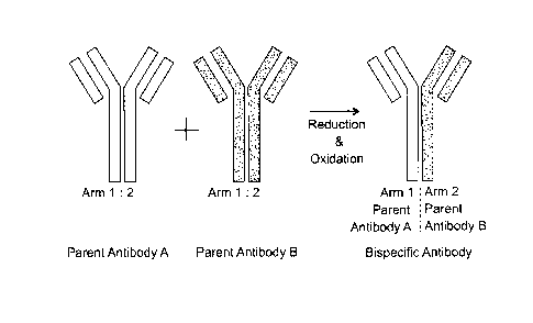
general approach for generating bispecific antibodies is outlined in FIG. 1.
In FIG. 1, a
first parent antibody ("Parent Antibody A"; grey color) and a second parent
antibody
("Parent Antibody B"; black color) are depicted, each of which is a
monospecific
homodimer, and contains a first arm and a second arm. Typically, the first
parent
antibody and the second parent antibody are specific for different antigens.
Then, the
first parent antibody and second parent antibody are mixed together and
exposed to
reduction and oxidation steps that result in the formation of the bispecific
antibody of
interest, which contains a first arm from the Parent Antibody A, and a second
arm from
the Parent Antibody B, and the respective specificities of both arms.
Optionally, the amino acid sequence of an antibody heavy chain may be
modified in one or more ways to promote the formation of bispecific
antibodies. For
example, the heavy chain of one arm of a bispecific antibody may contain an
amino
acid modification in the first hinge region, such that the
substituted/replaced amino acid
in the first hinge region has an opposite charge to the corresponding amino
acid in the
hinge region of the other arm of the formed bispecific antibody. This is
described, for
example, in International Patent Application No. PCT/US2011/036419
(W02011/143545). In another approach, the formation of a desired
heteromultimeric or
heterodimeric protein (e.g., bispecific antibody) is enhanced by altering or
engineering
an interface between a first and a second immunoglobulin-like Fc region (e.g.,
a hinge
region and/or a CH3 region). In this approach, the bispecific antibody may
contain a
CH3 region, wherein the CH3 region comprises a first CH3 polypeptide and a
second
CH3 polypeptide which interact together to form a CH3 interface, wherein one
or more
amino acids within the CH3 interface destabilize homodimer formation and are
not
electrostatically unfavorable to homodimer formation. This approach is also
described in
International Patent Application No. PCT/US2011/036419 (W02011/143545).
The above method and other methods for preparing bispecific antibodies are
further described, for example, in: International Patent Application No.
PCT/1132011/054899 (W02012/059882), PCT/US2011/036419 (W02011/143545), and
Giese et al, Biotechnology Progress, "Bispecific Antibody Process Development:
Assembly and Purification of Knob and Hole Bispecific Antibodies", 17 Jan
2018, and
references cited therein, each of which are incorporated by reference herein
for all
purposes. Methods provided herein for the purification of antibodies may be
used to
purify bispecific antibodies that were prepared by any suitable method.
19
CA 3034795 2019-02-25
Bispecific IgG antibodies - specificity
In some embodiments, an antibody that may be purified according to a method
provided herein is a full-length human bispecific IgG antibody, wherein a
first antibody
variable domain of the first arm of the bispecific antibody is capable of
binding to a first
antigen, and a second antibody variable domain of the second arm of the
bispecific
antibody is capable of binding to a second antigen. The first antigen and
second
antigen may have any characteristics of an antigen as described herein. In
some
embodiments, the first antigen occurs on a first cell type, and the second
antigen on a
second cell type.
In some embodiments, an antibody that may be purified according to a method
provided herein is a full-length human bispecific IgG antibody, wherein a
first antibody
variable domain of the antibody is capable of recruiting the activity of a
human immune
effector cell by specifically binding to an effector antigen located on the
human immune
effector cell, and wherein a second antibody variable domain of the antibody
is capable
of specifically binding to a target antigen.
A human immune effector cell that can be bound by an antibody provided herein
can be any of a variety of immune effector cells known in the art. For
example, the
immune effector cell can be a member of the human lymphoid cell lineage,
including,
but not limited to, a T cell (e.g., a cytotoxic T cell), a B cell, and a
natural killer (NK) cell.
The immune effector cell can also be, for example, a member of the human
myeloid
lineage, including, but not limited to, a monocyte, a neutrophilic
granulocyte, and a
dendritic cell. Such immune effector cells may have either a cytotoxic or an
apoptotic
effect on a target cell or other desired effect upon activation by binding of
an effector
antigen. The effector antigen is an antigen (e.g., a protein or a polypeptide)
that is
expressed on the human immune effector cell. Examples of effector antigens
that can
be bound by an antibody provided herein include, but are not limited to, human
CD3 (or
CD3 (Cluster of Differentiation) complex), CD16, NKG2D, NKp46, CD2, CD28,
CD25,
CD64, and CD89.
The target antigen is expressed on a target cell in a diseased condition
(e.g., an
inflammatory disease, a proliferative disease (e.g., cancer), an immunological
disorder,
a neurological disease, a neurodegenerative disease, an autoimmune disease, an
infectious disease (e.g., a viral infection or a parasitic infection), an
allergic reaction, a
graft-versus-host disease or a host-versus-graft disease). A target antigen is
not
CA 3034795 2019-02-25
=
effector antigen. Examples of the target antigens include, but are not limited
to, BCMA,
EpCAM (Epithelial Cell Adhesion Molecule), CCR5 (Chemokine Receptor type 5),
CD19, HER (Human Epidermal Growth Factor Receptor)-2/neu, HER-3, HER-4, EGFR
(Epidermal Growth Factor Receptor), FLT3 (Fms-Like Tyrosine kinase 3), PSMA,
CEA,
MUC-1 (Mucin), MUC2, MUC3, MUC4, MUC5AC, MUC5B, MUC7, ClhCG, Lewis-Y,
CD20, CD33, CD30, ganglioside GD3, 9-0-Acetyl-GD3, GM2, Globo H, fucosyl GM1,
Poly SA, GD2, Carboanhydrase IX (MN/CA IX), CD44v6, Shh (Sonic Hedgehog), Wue-
1, Plasma Cell Antigen, (membrane-bound) IgE, MCSP (Melanoma Chondroitin
Sulfate
Proteoglycan), CCR8, TNF-alpha precursor, STEAP, mesothelin, A33 Antigen, PSCA
(Prostate Stem Cell Antigen), Ly-6; desmoglein 4, E-cadherin neoepitope, Fetal
Acetylcholine Receptor, CD25, CA19-9 marker, CA-125 marker and MIS (Muellerian
Inhibitory Substance) Receptor type II, sTn (sialylated Tn antigen; TAG-72),
FAP
(fibroblast activation antigen), endosialin, EGFRvIll, LG, SAS and CD63.
In some embodiments, an antibody purified according to a method provided
herein may be any antibody as described in U.S. Application No. 15/085,644,
filed
March 30, 2016 (Publication No. US20160297885), or U.S. Application No.
15/993,874,
filed May 31, 2018 (Publication No. U520180346601), which are hereby
incorporated
by reference in their entirety for all purposes.
In some embodiments, an antibody purified according to a method provided
herein may be a bispecific IgG antibody, in which one arm of the antibody
specifically
binds to Cluster of Differentiation 3 (CD3). Information about CD3 is
provided, for
example, via UniProtKB ID # P07766.
In some embodiments, in a bispecific IgG antibody in which one arm of the
antibody specifically binds to CD3, the VH region of the heavy chain of the
CD3-binding
arm has an amino acid sequence comprising the amino acid sequence:
EVQLVESGGGLVQPGGSLRLSCAASGFTFSDYYMTVVVRQAPGKGLEVVVAFIRNRAR
GYTSDHNPSVKGRFTISRDNAKNSLYLQMNSLRAEDTAVYYCARDRPSYYVLDYWGQ
GTTVTVSS (SEQ ID NO: 1). In some embodiments, in a bispecific IgG antibody in
which one arm of the antibody specifically binds to CD3, the heavy chain of
the CD3-
binding arm has an amino acid sequence comprising the amino acid sequence:
EVQLVESGGGLVQPGGSLRLSCAASGFTFSDYYMTVVVRQAPGKGLEVVVAFIRNRAR
GYTSDHNPSVKGRFTISRDNAKNSLYLQMNSLRAEDTAVYYCARDRPSYYVLDYINGQ
GTTVTVSSASTKGPSVFPLAPCSRSTSESTAALGCLVKDYFPEPVTVSWNSGALTSGV
21
CA 3034795 2019-02-25
HTFPAVLQSSGLYSLSSVVTVPSSNFGTQTYTCNVDHKPSNTKVDKTVERKCRVRCP
RCPAPPVAGPSVFLFPPKPKDTLMISRTPEVTCVVVAVSHEDPEVQFNVVYVDGVEVH
NAKTKPREEQFNSTFRWSVLTVVHQDWLNGKEYKCKVSNKGLPSSIEKTISKTKGQP
REPQVYTLPPSREEMTKNQVSLTCLVKGFYPSDIAVEWESNGQPENNYKTTPPMLDS
DGSFFLYSRLTVDKSRWQQGNVFSCSVMHEALHNHYTQKSLSLSPGK (SEQ ID NO:
2). In some embodiments, in a bispecific IgG antibody in which one arm of the
antibody
specifically binds to CD3, the VH region of the heavy chain of the CD3-binding
arm has
an amino acid sequence comprising a CDR1, a CDR2, and a CDR3 of the VH
sequence shown in SEQ ID NO: 1.
In some embodiments, in a bispecific IgG antibody in which one arm of the
antibody specifically binds to CD3, the VL region of the light chain of the
CD3-binding
arm has an amino acid sequence comprising the amino acid sequence:
DIVMTQSPDSLAVSLGERATINCKSSQSLFNVRSRKNYLAVVYQQKPGQPPKLLISWAS
TRESGVPDRFSGSGSGTDFTLTISSLQAEDVAVYYCKQSYDLFTFGSGTKLEIK (SEQ
ID NO: 3). In some embodiments, in a bispecific IgG antibody in which one arm
of the
antibody specifically binds to CD3, the light chain of the CD3-binding arm has
an amino
acid sequence comprising the amino acid sequence:
DIVMTQSPDSLAVSLGERATINCKSSQSLFNVRSRKNYLAVVYQQKPGQPPKLLISWAS
TRESGVPDRFSGSGSGTDFTLTISSLQAEDVAVYYCKQSYDLFTFGSGTKLEIKRTVAA
PSVFIFPPSDEQLKSGTASVVCLLNNFYPREAKVQWKVDNALQSGNSQESVTEQDSK
DSTYSLSSTLTLSKADYEKHKVYACEVTHQGLSSPVTKSFNRGEC (SEQ ID NO: 4).
In some embodiments, in a bispecific IgG antibody in which one arm of the
antibody
specifically binds to CD3, the VL region of the light chain of the CD3-binding
arm has an
amino acid sequence comprising a CDR1, a CDR2, and a CDR3 of the VL sequence
shown in SEQ ID NO: 3.
In some embodiments, in a bispecific IgG antibody in which one arm of the
antibody specifically binds to CD3, the VH region of the heavy chain of the
CD3-binding
arm has an amino acid sequence comprising the amino acid sequence shown in SEQ
ID NO: 1, and the VL region of the light chain of the CD3-binding arm has an
amino
.. acid sequence comprising the amino acid sequence shown in SEQ ID NO: 3. In
some
embodiments, in a bispecific IgG antibody in which one arm of the antibody
specifically
binds to CD3, the heavy chain of the CD3-binding arm has an amino acid
sequence
comprising the amino acid sequence shown in SEQ ID NO: 2, and the light chain
of the
22
CA 3034795 2019-02-25
=
CD3-binding arm has an amino acid sequence comprising the amino acid sequence
shown in SEQ ID NO: 4. In some embodiments, in a bispecific IgG antibody in
which
one arm of the antibody specifically binds to CD3, the VH region of the heavy
chain of
the CD3-binding arm has an amino acid sequence comprising a CDR1, a CDR2, and
a
CDR3 of the VH sequence shown in SEQ ID NO: 1, and the VL region of the light
chain
of the CD3-binding arm has an amino acid sequence comprising a CDR1, a CDR2,
and
a CDR3 of the VL sequence shown in SEQ ID NO: 3.
In some embodiments, an antibody purified according to a method provided
herein may be a bispecific IgG antibody, in which one arm of the antibody
specifically
binds to B-cell maturation antigen (BCMA). Information about BCMA is provided,
for
example, via UniProtKB ID # Q02223.
In some embodiments, in a bispecific IgG antibody in which one arm of the
antibody specifically binds to BCMA, the VH region of the heavy chain of the
BCMA-
binding arm has an amino acid sequence comprising the amino acid sequence:
EVQLLESGGGLVQPGGSLRLSCAASGFTFSSYPMSVVVRQAPGKGLEVVVSAIGGSGG
SLPYADIVKGRFTISRDNSKNTLYLQMNSLRAEDTAVYYCARYWPMDIWGQGTLVTVS
S (SEQ ID NO: 5). In some embodiments, in a bispecific IgG antibody in which
one
arm of the antibody specifically binds to BCMA, the heavy chain of the BCMA-
binding
arm has an amino acid sequence comprising the amino acid sequence:
EVQLLESGGGLVQPGGSLRLSCAASGFTFSSYPMSVVVRQAPGKGLEVVVSAIGGSGG
SLPYADIVKGRFTISRDNSKNTLYLQMNSLRAEDTAVYYCARYWPMDIWGQGTLVTVS
SASTKGPSVFPLAPCSRSTSESTAALGCLVKDYFPEPVTVSWNSGALTSGVHTFPAVL
QSSGLYSLSSVVTVPSSNFGTQTYTCNVDHKPSNTKVDKTVERKCEVECPECPAPPV
AGPSVFLFPPKPKDILMISRTPEVTCVVVAVSHEDPEVQFNVVYVDGVEVHNAKTKPR
EEQFNSTFRVVSVLIVVHQDWLNGKEYKCKVSNKGLPSSIEKTISKTKGQPREPQVYT
LPPSREEMTKNQVSLTCEVKGFYPSDIAVEWESNGQPENNYKTTPPMLDSDGSFFLY
SKLTVDKSRWQQGNVFSCSVMHEALHNHYTQKSLSLSPGK (SEQ ID NO: 6). In
some embodiments, in a bispecific IgG antibody in which one arm of the
antibody
specifically binds to BCMA, the VH region of the heavy chain of the BCMA-
binding arm
has an amino acid sequence comprising a CDR1, a CDR2, and a CDR3 of the VH
sequence shown in SEQ ID NO: 5.
In some embodiments, in a bispecific IgG antibody in which one arm of the
antibody specifically binds to BCMA, the VL region of the light chain of the
BCMA-
23
CA 3034795 2019-02-25
binding arm has an amino acid sequence comprising the amino acid sequence:
EIVLTQSPGTLSLSPGERATLSCRASQSVSSSYLAVVYQQKPGQAPRLLMYDASIRATG
IPDRFSGSGSGTDFTLTISRLEPEDFAVYYCQQYQSWPLTFGQGTKVEIK (SEQ ID
NO: 7). In some embodiments, in a bispecific IgG antibody in which one arm of
the
antibody specifically binds to BCMA, the light chain of the BCMA-binding arm
has an
amino acid sequence comprising the amino acid sequence:
EIVLTQSPGTLSLSPGERATLSCRASQSVSSSYLAVVYQQKPGQAPRLLMYDASIRATG
IPDRFSGSGSGTDFTLTISRLEPEDFAVYYCQQYQSWPLTFGQGTKVEIKRTVAAPSV
FIFPPSDEQLKSGTASVVCLLNNFYPREAKVQWKVDNALQSGNSQESVTEQDSKDST
YSLSSTLTLSKADYEKHKVYACEVTHQGLSSPVTKSFNRGEC (SEQ ID NO: 8). In
some embodiments, in a bispecific IgG antibody in which one arm of the
antibody
specifically binds to BCMA, the VL region of the light chain of the BCMA-
binding arm
has an amino acid sequence comprising a CDR1, a CDR2, and a CDR3 of the VL
sequence shown in SEQ ID NO: 7.
In some embodiments, in a bispecific IgG antibody in which one arm of the
antibody specifically binds to BCMA, the VH region of the heavy chain of the
BCMA-
binding arm has an amino acid sequence comprising the amino acid sequence
shown
in SEQ ID NO: 5, and the VL region of the light chain of the BCMA-binding arm
has an
amino acid sequence comprising the amino acid sequence shown in SEQ ID NO: 7.
In
some embodiments, in a bispecific IgG antibody in which one arm of the
antibody
specifically binds to BCMA, the heavy chain of the BCMA-binding arm has an
amino
acid sequence comprising the amino acid sequence shown in SEQ ID NO: 6, and
the
light chain of the BCMA-binding arm has an amino acid sequence comprising the
amino
acid sequence shown in SEQ ID NO: 8. In some embodiments, in a bispecific IgG
antibody in which one arm of the antibody specifically binds to BCMA, the VH
region of
the heavy chain of the BCMA-binding arm has an amino acid sequence comprising
a
CDR1, a CDR2, and a CDR3 of the VH sequence shown in SEQ ID NO: 5, and the VL
region of the light chain of the BCMA-binding arm has an amino acid sequence
comprising a CDR1, a CDR2, and a CDR3 of the VL sequence shown in SEQ ID NO:
7.
In some embodiments, in a bispecific IgG antibody in which one arm of the
antibody specifically binds to BCMA, the VH region of the heavy chain of the
BCMA-
binding arm has an amino acid sequence comprising the amino acid sequence:
EVQLLESGGGLVQPGGSLRLSCAASGFTFSSYPMSWVRQAPGKGLEVVVSAIGGSGG
24
CA 3034795 2019-02-25
SLPYADSVKGRFTISRDNSKNTLYLQMNSLRAEDTAVYYCARYWPMDIWGQGTLVTV
SS (SEQ ID NO: 13). In some embodiments, in a bispecific IgG antibody in which
one
arm of the antibody specifically binds to BCMA, the VL region of the light
chain of the
BCMA-binding arm has an amino acid sequence comprising the amino acid
sequence:
EIVLIQSPGTLSLSPGERATLSCRASQSVSSTYLAVVYQQKPGQAPRLLMYDASIRATG
IPDRFSGSGSGTDFTLTISRLEPEDFAVYYCQQYQEWPLTFGQGTKVEIK (SEQ ID
NO: 14). In some embodiments, in any reference herein to an antibody
comprising the
a VH region that has an amino acid sequence comprising the amino acid sequence
shown in SEQ ID NO: 5, the antibody may alternatively comprise a VH region
comprising the amino acid sequence shown in SEQ ID NO: 13. In some
embodiments,
in any reference herein to an antibody comprising the a VL region that has an
amino
acid sequence comprising the amino acid sequence shown in SEQ ID NO: 7, the
antibody may alternatively comprise a VL region comprising the amino acid
sequence
shown in SEQ ID NO: 14. Similarly, also included herein are anti-BCMA heavy
and
light chains containing the VH and VL sequence of SEQ ID NO: 13 and SEQ ID NO:
14,
respectively.
In some embodiments, an antibody purified according to a method provided
herein may be a bispecific IgG antibody, in which one arm of the antibody
specifically
binds to fms-like tyrosine kinase 3 (FLT3). Information about FLT3 is
provided, for
example, via UniProtKB ID # P36888.
In some embodiments, in a bispecific IgG antibody in which one arm of the
antibody specifically binds to FLT3, the VH region of the heavy chain of the
FLT3-
binding arm has an amino acid sequence comprising the amino acid sequence:
EVQLLESGGGLVQPGGSLRLSCAASGFTFSSYAMNVVVRQAPGKGLEVVVSAISGGGR
STYYADSVKGRFTISRDNSKNTLYLQMNSLRAEDTAVYYCARDLSPSDVGWGYGFDI
WGQGTLVTVSS (SEQ ID NO: 9). In some embodiments, in a bispecific IgG antibody
in which one arm of the antibody specifically binds to FLT3, the heavy chain
of the
FLT3-binding arm has an amino acid sequence comprising the amino acid
sequence:
EVQLLESGGGLVQPGGSLRLSCAASGFTFSSYAMNVVVRQAPGKGLEVVVSAISGGGR
STYYADSVKGRFTISRDNSKNTLYLQMNSLRAEDTAVYYCARDLSPSDVGWGYGFDI
WGQGTLVTVSSASTKGPSVFPLAPCSRSTSESTAALGCLVKDYFPEPVTVSWNSGAL
TSGVHTFPAVLQSSGLYSLSSVVTVPSSNFGTQTYTCNVDHKPSNTKVDKTVERKCE
VECPECPAPPVAGPSVFLFPPKPKDTLMISRTPEVTCVVVAVSHEDPEVQFNVVYVDG
CA 3034795 2019-02-25
VEVHNAKTKPREEQFNSTFRWSVLTVVHQDWLNGKEYKCKVSNKGLPSSIEKTISKT
KGQPREPQVYTLPPSREEMTKNQVSLTCEVKGFYPSDIAVEWESNGQPENNYKTTPP
MLDSDGSFFLYSKLTVDKSRWQQGNVFSCSVMHEALHNHYTQKSLSLSPG (SEQ ID
NO: 10). In some embodiments, in a bispecific IgG antibody in which one arm of
the
antibody specifically binds to FLT3, the VH region of the heavy chain of the
FLT3-
binding arm has an amino acid sequence comprising a CDR1, a CDR2, and a CDR3
of
the VH sequence shown in SEQ ID NO: 9.
In some embodiments, in a bispecific IgG antibody in which one arm of the
antibody specifically binds to FLT3, the VL region of the light chain of the
FLT3-binding
arm has an amino acid sequence comprising the amino acid sequence:
EIVLIQSPATLSLSPGERATLSCRASQSVSSNLAVVYQQKPGQAPRLLIYDTFTRATGIP
ARFSGSGSGTDFTLTISSLEPEDFAVYYCQQYGSSPPTFGQGTRLEIK (SEQ ID NO:
11). In some embodiments, in a bispecific IgG antibody in which one arm of the
antibody specifically binds to FLT3, the light chain of the FLT3-binding arm
has an
amino acid sequence comprising the amino acid sequence:
EIVLTQSPATLSLSPGERATLSCRASQSVSSNLAVVYQQKPGQAPRLLIYDTFTRATGIP
ARFSGSGSGTDFTLTISSLEPEDFAVYYCQQYGSSPPTFGQGTRLEIKRTVAAPSVFIF
PPSDEQLKSGTASVVCLLNNFYPREAKVQWKVDNALQSGNSQESVTEQDSKDSTYSL
SSTLTLSKADYEKHKVYACEVTHQGLSSPVTKSFNRGEC (SEQ ID NO: 12). In some
embodiments, in a bispecific IgG antibody in which one arm of the antibody
specifically
binds to FLT3, the VL region of the light chain of the FLT3-binding arm has an
amino
acid sequence comprising a CDR1, a CDR2, and a CDR3 of the VL sequence shown
in
SEQ ID NO: 11.
In some embodiments, in a bispecific IgG antibody in which one arm of the
antibody specifically binds to FLT3, the VH region of the heavy chain of the
FLT3-
binding arm has an amino acid sequence comprising the amino acid sequence
shown
in SEQ ID NO: 9, and the VL region of the light chain of the FLT3-binding arm
has an
amino acid sequence comprising the amino acid sequence shown in SEQ ID NO: 11.
In some embodiments, in a bispecific IgG antibody in which one arm of the
antibody
specifically binds to FLT3, the heavy chain of the FLT3-binding arm has an
amino acid
sequence comprising the amino acid sequence shown in SEQ ID NO: 10, and the
light
chain of the FLT3-binding arm has an amino acid sequence comprising the amino
acid
sequence shown in SEQ ID NO: 12. In some embodiments, in a bispecific IgG
antibody
26
CA 3034795 2019-02-25
in which one arm of the antibody specifically binds to FLT3, the VH region of
the heavy
chain of the FLT3-binding arm has an amino acid sequence comprising a CDR1, a
CDR2, and a CDR3 of the VH sequence shown in SEQ ID NO: 9, and the VL region
of
the light chain of the FLT3-binding arm has an amino acid sequence comprising
a
CDR1, a CDR2, and a CDR3 of the VL sequence shown in SEQ ID NO: 11.
In some embodiments, provided herein is a bispecific anti-BCMA / anti-CD3
antibody, in which the anti-BCMA arm of the antibody has any of the
characteristics
described above for an anti-BCMA arm, and the anti-CD3 arm of the antibody has
any
of the characteristics described above for an anti-CD3 arm. In some
embodiments,
.. provided herein is a bispecific anti-FLT3 / anti-CD3 antibody, in which the
anti-FLT3
arm of the antibody has any of the characteristics described above for an anti-
FLT3
arm, and the anti-CD3 arm of the antibody has any of the characteristics
described
above for an anti-CD3 arm.
Also provided herein are methods of purifying a monospecific antibody having
affinity for any of the above antigens, and/or which contain any of the amino
acid
sequences described above. For example, also provided herein is purification
of a
monospecific, homodimeric anti-CD3 antibody comprising the VH amino acid
sequence
as shown in SEQ ID NO: 1.
Impurities
Methods provided herein may be used to purify an antibody of interest from one
or more impurities.
Impurities include, for example, clipped versions of the antibody of interest,
protein aggregates, and in the case of a bispecific antibody of interest,
parental
monospecific antibodies related to the formation of the bispecific antibody of
interest.
These different impurities may also be referred to herein as different
"species of
impurity", "impurity molecules", or the like.
Clipped Versions of an Antibody of Interest
"Clipped versions of an antibody of interest", "clipped antibodies", or the
like refer
to a recombinant antibody in which one or more polypeptide bonds in the
antibody has
been cleaved, as compared to a corresponding intact antibody of interest. In
contrast,
an "intact" antibody refers to a recombinant antibody that contains all of the
expected
27
CA 3034795 2019-02-25
peptide bonds and amino acids of the recombinant antibody (i.e. that would be
expected based on the nucleic acid sequence(s) encoding the polypeptide(s) of
the
antibody)
As such, clipped antibodies may be considered to be degradation products
related to the antibody of interest. Cleavage of a peptide bond in an antibody
may
occur, for example, via enzymatic (e.g. protease-mediated) or non-enzymatic
activities.
In some embodiments, when a peptide bond in a polypeptide of an antibody is
cleaved, after the cleavage, a cleaved portion of the polypeptide chain might
no longer
be covalently linked to the rest of the antibody; in that case, the cleaved
portion of the
polypeptide chain may dissociate from the rest of the antibody. This most
commonly
occurs when the cleavage is in a peptide bond near an N or C terminus of the a
polypeptide chain, and it results in a clipped antibody which has lost one or
more amino
acids as compared to the corresponding intact antibody. These clipped
antibodies
have less mass than the corresponding intact antibody, due to the loss of one
or more
amino acids from the antibody.
Alternatively, in some other embodiments, when a peptide bond in a polypeptide
of an antibody is cleaved, after the cleavage, a cleaved portion of the
polypeptide chain
_
might still remain covalently linked to the rest of the antibody (for example,
by an intra-
chain or inter-chain disulfide bond). In this case, even though there is a
cleavage a
peptide bond of the antibody, the cleaved portion of the polypeptide chain
will remain
tethered to the rest of the antibody, via the remaining intact covalent
bond(s) that link
the cleaved portion of the polypeptide chain to the rest of the antibody. In
this
circumstance, the clipped antibody will still have the same number of amino
acids and
amino acid sequences as compared to the intact antibody. In addition, in at
least some
embodiments, this type of clipped antibody may have a slightly greater mass
than the
corresponding intact antibody. This gain of mass may be the result, for
example, of one
or more chemical reactions that occur upon the cleavage of the peptide bond.
During
such reactions, one or more atoms (e.g. H, 0) may react with atoms of the
antibody
polypeptide chain and become covalently linked to the antibody chain, which
results in
a gain of mass by the clipped antibody as compared to the corresponding intact
antibody.
Since a "clipped" antibody is generated from a corresponding "intact"
antibody,
the "clipped" version of the antibody has the same amino acid sequence (in the
case of
28
CA 3034795 2019-02-25
no loss of amino acids from the antibody as the result of the peptide bond
cleavage) or
nearly the same amino acid sequence (in the case of the loss of one or more
amino
acid sequences from the antibody as the result of the peptide bond cleavage)
as the
corresponding "intact" version of the antibody.
Typically, a clipped version of an antibody has a mass that is similar to the
mass
of the corresponding intact antibody. As described above, in some embodiments,
a
clipped antibody may have a mass that is less than the corresponding intact
antibody
(for example, in the event that the clipping results in the loss of one or
more amino
acids from the antibody). Alternatively, in some embodiments, a clipped
antibody may
have a mass that is greater than the corresponding intact antibody (in the
event that the
clipping does not result in the loss of any amino acids from the antibody, and
instead,
results in the gain of at least an atom by the antibody via one or more
reactions that
occur as a result of the cleavage of the peptide bond).
In some embodiments, a clipped version of an antibody has a mass that is no
more than 50%, 40%, 30%, 25%, 20%, 15%, 10%, 5%, 4%, 3%, 2%, 1%, 0.5%, 0.4%,
0.3%, 0.2%, 0.1%, 0.05%, 0.04%, 0.03%, 0.02%, 0.01% different than the mass of
the
corresponding intact antibody of interest. Put another way, in some
embodiments, a
clipped version of an antibody of interest has a mass that differs from the
mass of the
corresponding intact antibody of interest by no more than 50%, 40%, 30%, 25%,
20%,
15%, 10%, 5%, 4%, 3%, 2%, 1%, 0.5%, 0.4%, 0.3%, 0.2%, 0.1%, 0.05%, 0.04%,
0.03%, 0.02%, or 0.01%.
As described above, in some embodiments, a clipped version of an antibody of
interest has a mass that is less than the corresponding intact antibody of
interest. For
example, in some embodiments, a clipped version of an antibody has a mass that
is no
more than 50%, 40%, 30%, 25%, 20%, 15%, 10%, 5%, 4%, 3%, 2%, 1%, 0.5%, 0.4%,
0.3%, 0.2%, 0.1%, 0.05%, 0.04%, 0.03%, 0.02%, 0.01% less than the mass of the
corresponding intact antibody of interest. In other words, if a clipped
version of an
antibody has a mass that is no more than 10% less than the mass of the
corresponding
intact antibody of interest, then, for example, if the intact antibody of
interest has a
mass of 100,000 Da, then the clipped version of the antibody has a mass that
is no
more than 10,000 Da less than that (10% of 100,000 is 10,000) - i.e. it has a
mass
between 90,000 and 100,000 Da.
29
CA 3034795 2019-02-25
As also described above, in some embodiments, a clipped version of an antibody
of interest has a mass that is greater than the corresponding intact antibody
of interest.
For example, in some embodiments, a clipped version of an antibody has a mass
that is
no more than 5%, 4%, 3%, 2%, 1%, 0.5%, 0.4%, 0.3%, 0.2%, 0.1%, 0.05%, 0.04%,
0.03%, 0.02%, or 0.01% greater than the mass of the corresponding intact
antibody of
interest. In other words, if a clipped version of an antibody has a mass that
is no more
than 1% greater than the mass of the corresponding intact antibody of
interest, then, for
example, if the intact antibody of interest has a mass of 100,000 Da, then the
clipped
version of the antibody has a mass that is no more than 1,000 Da more than
that (1% of
100,000 is 1,000) ¨ i.e. it has a mass between 100,000 and 101,000 Da.
High Molecular Mass Species (HMMS) / Protein aggregates
In some embodiments, an impurity relevant to a method provided herein is
referred to as "high molecular mass species" (HMMS). HMMS refers to any high
.. molecular mass contaminant or impurity, but typically is an association of
at least two
proteins forming an aggregate. By way of example, HMMS may include multiple
molecules of an antibody of interest that have aggregated together and/or
aggregates
of proteins from host cells that were used to produce an antibody of interest.
Aggregates may arise by any process including, for example, covalent or non-
covalent
linking of molecules.
Parental Antibodies
In some embodiments, an impurity relevant to a method provided herein is a
"parent antibody", or "parental antibody" or the like. This type of impurity
molecule is
relevant to methods provided herein in which the antibody of interest is
bispecific
antibody that is generated from two different parent antibodies, for example,
as outlined
in FIG. 1. These parent antibodies are monospecific, homodimers. Parent
antibodies
may be present in an antibody preparation with a bispecific antibody of
interest due to
multiple possible mechanisms, such as: i) in some situations, some parent
antibody
molecules do not separate into a first arm and a second arm during the
reduction step
to separate parent antibodies into a separate first arm and second arm (and
thus, the
antibodies remain as monospecific homodimers); or ii) in some situations, a
separated
first arm and a second arm from the same type of parent antibody join
together, such
CA 3034795 2019-02-25
=
they form a monospecific homodimer (rather than being involved in forming a
heterodimer bispecific antibody). As used herein "parent antibody" refers to
homodimeric molecules which occur by any of the above mechanisms. In addition,
an
antibody preparation as provided herein may contain as an impurity a parent
antibody
from one or both parent species.
Purification of an antibody of interest from impurities
Provided herein are methods for purifying an antibody of interest from one or
more impurities.
In some embodiments, in a method provided herein, an antibody of interest is
in
an antibody preparation (also referred to herein as a "starting sample") that
contains the
antibody of interest, as well as one or more species of impurity molecule. In
this
starting material for a method as provided herein, the antibody of interest
may
comprise, for example, at least about 10%, 20%, 30%, 40%, 50%, 60%, 70%, 75%,
80%, 85%, 90%, or 95% by mass of the protein in the antibody preparation.
Then, in
some embodiments of a method provided herein, a purified fraction (also
referred to
herein as a "purified sample") is collected as eluate from the HA resin. This
purified
fraction contains the antibody of interest, and in some embodiments, still
contains one
or more impurity molecules. In some embodiments, in a purified fraction
provided
herein, the antibody of interest may comprise, for example, at least about
20%, 30%,
40%, 50%, 60%, 70%, 75%, 80%, 85%, 90%, 95%, 96%, 97%, 98%, or 99% by mass
of the protein in the purified fraction.
In some embodiments, in a method provided herein, a starting sample is at
least
about 10%, 20%, 30%, 40%, 50%, 60%, 70%, 75%, 80%, 85%, 90%, or 95% by mass
antibody of interest, and a subsequent purified sample in the same method is
at least
about 20%, 30%, 40%, 50%, 60%, 70%, 75%, 80%, 85%, 90%, 95%, 96%, 97%, 98%,
or 99% by mass antibody of interest, wherein the second value is larger than
the first
value.
In some embodiments, in a method provided herein, a starting sample contains
at least about 0.1%, 0.5%, 1%, 2%, 3%, 4%, 5%, 6%, 7%, 8%, 9%, 10%, 15%, 20%,
or
25% by mass clipped version of an antibody of interest. In some embodiments,
in a
method provided herein, a purified sample contains no more than about 0.01%,
0.05%,
0.1%, 0.5%, 1%, 2%, 3%, 4%, 5%, 6%, 7%, 8%, 9%, 10%, 15%, or 20% by mass
31
CA 3034795 2019-02-25
clipped version of an antibody of interest. In some embodiments, in a method
provided
herein, a starting sample contains at least about 0.1%, 0.5%, 1%, 2%, 3%, 4%,
5%,
6%, 7%, 8%, 9%, 10%, 15%, 20%, or 25% by mass clipped version of an antibody
of
interest, and a subsequent purified sample in the same method contains no more
than
about 0.01%, 0.05%, 0.1%, 0.5%, 1%, 2%, 3%, 4%, 5%, 6%, 7%, 8%, 9%, 10%, 15%,
or 20% by mass clipped version of an antibody of interest, wherein the second
value is
smaller than the first value.
In some embodiments, in a method provided herein, a starting sample contains
an intact antibody of interest and a clipped version of the antibody of
interest, wherein
the ratio of clipped antibody to intact antibody is at least about 1:100,
1:50, 1:25, 1:20,
1:10, 1:5, 1:4, or 1:3. In some embodiments, in a method provided herein, a
purified
sample contains an intact antibody of interest and a clipped version of the
antibody of
interest, wherein the ratio of clipped antibody to intact antibody is no more
than about
1:100, 1:50, 1:25, 1:20, or 1:10. In some embodiments, in a method provided
herein, a
starting sample contains an intact antibody of interest and a clipped version
of the
antibody of interest, wherein the ratio of clipped antibody to intact antibody
in the
starting sample is at least about 1:100, 1:50, 1:25, 1:20, 1:10, 1:5, 1:4, or
1:3 and
wherein the ratio of clipped antibody to intact antibody in a subsequent
purified sample
in the same method is no more than about 1:200, 1:100, 1:50, 1:25, 1:20, or
1:10,
.. wherein the second ratio is smaller than the first ratio. In some
embodiments, in a
method provided herein, a starting sample contains an intact antibody of
interest and a
clipped version of the antibody of interest, wherein the ratio of clipped
antibody to intact
antibody in the starting sample is between about one of the ratios in group A
(group A
ratios: 1:100, 1:50, 1:25, 1:20, 1:10, 1:5, or 1:4) and one of the ratios in
group B (group
.. B ratios: 1:50, 1:25, 1:20, 1:10, 1:5, 1:4 or 1:3) and wherein the ratio of
clipped
antibody to intact antibody in a subsequent purified sample in the same method
is no
more than about 1:200, 1:100, 1:50, 1:25, 1:20, or 1:10, wherein the ratio in
the purified
sample is smaller than in the starting sample.
In some embodiments, a starting sample provided according to a method
provided herein contains at least 1, 5, 10, 15, 20, 25, 50, 100, 200, 500,
1000, 2000,
5000, or 10,000 grams of the intact antibody of interest. In some embodiments,
a
purified sample provided according to a method provided herein contains at
least 1, 5,
10, 15, 20, 25, 50, 100, 200, 500, 1000, 2000, 5000, or 10,000 grams of the
intact
32
CA 3034795 2019-02-25
antibody of interest. In some embodiments, a starting sample provided
according to a
method provided herein contains at least 5, 10, 15, 20, 25, 50, 100, 200, 500,
1000,
2000, 5000, or 10,000 grams of the intact antibody of interest, and a
subsequent
purified sample in the same method contains at least 1, 5, 10, 15, 20, 25, 50,
100, 200,
500, 1000, 2000, or 5000 grams of the intact antibody of interest, wherein the
first value
is larger than the second value.
In addition, any above descriptions relating to the a) the amount or b) purity
of an
antibody of interest in a starting sample or purified sample may be taken
together in
reference to the same sample. For example, as stated above, a starting sample
may
contain at least about 80% by mass antibody of interest; in addition, as also
stated
above, a starting sample may contain at least about 10 grams antibody of
interest.
Accordingly, also provided herein is a starting sample that contains at least
about 80%
by mass antibody of interest, and at least 10 grams of antibody of interest,
etc.
General Techniques
The practice of the present invention will employ, unless otherwise indicated,
conventional techniques of molecular biology (including recombinant
techniques),
microbiology, cell biology, biochemistry and immunology, which are within the
skill of
the art. Such techniques are explained fully in the literature, such as,
Molecular Cloning:
A Laboratory Manual, second edition (Sambrook et al., 1989) Cold Spring Harbor
Press; Oligonucleotide Synthesis (M.J. Gait, ed., 1984); Methods in Molecular
Biology,
Humana Press; Cell Biology: A Laboratory Notebook (J.E. Cellis, ed., 1998)
Academic
Press; Animal Cell Culture (R.I. Freshney, ed., 1987); Introduction to Cell
and Tissue
Culture (J.P. Mather and P.E. Roberts, 1998) Plenum Press; Cell and Tissue
Culture:
Laboratory Procedures (A. Doyle, J.B. Griffiths, and D.G. Newell, eds., 1993-
1998) J.
Wiley and Sons; Methods in Enzymology (Academic Press, Inc.); Handbook of
Experimental Immunology (D.M. Weir and C.C. Blackwell, eds.); Gene Transfer
Vectors
for Mammalian Cells (J.M. Miller and M.P. Cabs, eds., 1987); Current Protocols
in
Molecular Biology (F.M. Ausubel et al., eds., 1987); PCR: The Polymerase Chain
Reaction, (Mullis et al., eds., 1994); Current Protocols in Immunology (J.E.
Coligan et
al., eds., 1991); Short Protocols in Molecular Biology (Wiley and Sons, 1999);
lmmunobiology (C.A. Janeway and P. Travers, 1997); Antibodies (P. Finch,
1997);
Antibodies: a practical approach (D. Catty., ed., IRL Press, 1988-1989);
Monoclonal
33
CA 3034795 2019-02-25
antibodies: a practical approach (P. Shepherd and C. Dean, eds., Oxford
University
Press, 2000); Using antibodies: a laboratory manual (E. Harlow and D. Lane
(Cold
Spring Harbor Laboratory Press, 1999); The Antibodies (M. Zanetti and J.D.
Capra,
eds., Harwood Academic Publishers, 1995), as well as in subsequent editions
and
corresponding websites of the above references, as applicable.
The following examples are offered for illustrative purposes only, and are not
intended to limit the scope of the present invention in any way. Indeed,
various
modifications of the invention in addition to those shown and described herein
will
become apparent to those skilled in the art from the foregoing description and
fall within
the scope of the appended claims.
Examples
Example 1: Purification of an anti-BCMA / anti-CD3 bispecific antibody by cHA
resin
chromatography
Objective:
In this example, methods for separating a full-length bispecific human IgG of
interest from various impurities were examined. The antibody of interest was a
heterodimeric bispecific anti-BCMA / anti-CD3 antibody (i.e. one arm of the
bispecific
antibody was specific for BCMA, and the other arm was specific for CD3). The
impurities present with the bispecific antibody of interest at the start of
the purification
included: i) a clipped version of the intact anti-BCMA / anti-CD3 bispecific
antibody of
interest; ii) homodimeric, monospecific anti-BCMA parent antibodies; iii)
homodimeric,
monospecific anti-CD3 antibodies; and iv) protein aggregates / high molecular
mass
species (HMMS).
Purification of the intact bispecific antibody of interest from the clipped
version of
the bispecific antibody presented a particular challenge, as the clipped
version of the
bispecific antibody contained the same number of amino acids and same amino
acid
sequences as the intact bispecific antibody of interest, and differed from the
mass of the
intact bispecific antibody by only 18 Daltons (Da). Specifically, the mass of
the intact
anti-BCMA / CD3 bispecific antibody is 148095.5 Da, while the mass of the
corresponding clipped version of the bispecific antibody is 148113.5
(determined by
34
CA 3034795 2019-02-25
=
mass spectrometry). Thus, the clipped bispecific antibody has a difference in
mass of
less than 0.1% (even less than 0.02%) as compared to the mass of the intact
bispecific
antibody (i.e. 0.1% of 148095 Da is 148 Da; 0.02% of 148095 Da is 29.6 Da).
Put
another way, the mass of the clipped bispecific antibody is about 100.01% of
the mass
of the intact bispecific antibody of interest. Accordingly, the mass of the
clipped
bispecific antibody is very similar to that of the intact bispecific antibody
of interest.
The clipped version of the bispecific antibody contains a cleavage in a
peptide
bond of the anti-CD3 heavy chain, between the 56th and 57th amino acid in the
heavy
chain. The anti-CD3 heavy chain has an amino acid sequence as shown in SEQ ID
NO:
2, and accordingly, the cleavage occurs between the amino acids R and G in the
"RG"
sequence of SEQ ID NO: 2 ("RG" only occurs once in SEQ ID NO: 2). It is
believed that
this cleavage results in the gain of 1 oxygen atom and 2 hydrogen atoms in the
clipped
bispecific antibody (which would correspond to the gain of mass of 18 Da), as
compared to the intact bispecific antibody. Also, although there is a cleaved
peptide
bond in the anti-CD3 heavy chain, the cleaved 56-amino acid portion of the
heavy chain
(i.e. the first 56 amino acids of the chain) remains bound to the rest of the
antibody via
an intact, remaining intra-chain disulfide bond. The intra-chain disulfide
bond is
between C22 and C98 of the anti-CD3 heavy chain (i.e. C22 and C98 of the
sequence
shown in SEQ ID NO: 2). The continued tethering of the cleaved 56-amino acid
portion
to the rest of the antibody by the intra-chain peptide bond is shown
schematically in
FIG. 2.
In FIG. 2, the schematic on the left depicts an intact bispecific antibody of
interest, which contains intact heavy and light chains for both the anti-CD3
and anti-
BCMA arms of the bispecific antibody (in the figure, the longer chains
represent the
heavy chains of the antibody, and the shorter chains represent the light
chains). In
addition, FIG. 2 also shows various intra-antibody disulfide bonds, including
intra-chain
disulfide bonds (e.g. linking different amino acids in the same light chain or
heavy
chain), and well as inter-chain disulfide bonds (e.g. linking the heavy chain
of the anti-
BCMA arm to the heavy chain of the anti-CD3 arm, or linking the light chain of
the anti-
CD3 arm to the heavy chain of the anti-CD3 arm).
Another challenge presented in developing a method of purifying the intact
bispecific antibody of interest from the various impurities was the objective
of
developing a method that would effectively separate the bispecific antibody of
interest
CA 3034795 2019-02-25
from impurities when relatively large amounts of bispecific antibody of
interest were to
be purified. For example, one objective of the work related to this Example
was to
develop a method for purification of a bispecific antibody that would be
effective for
methods in which at least 1 gram of bispecific antibody was to be purified. As
is known
in the art, purification of proteins on a large scale frequently presents
numerous
difficulties that are not present (or are significantly less of a problem)
during purification
of the same proteins on a small scale, due to, for example, difficulties in
obtaining sharp
chromatographic resolution between different proteins when performing
chromatography on a large scale.
Materials and Methods:
The starting material for this work was an antibody preparation containing the
bispecific IgG anti-BCMA/CD3 antibody of interest, as well as various
impurities, such
as those noted above. The amino acid sequences of the polypeptides of the
bispecific
antibody are shown in the following SEQ ID NOs: BCMA heavy chain: SEQ ID NO:
6;
BCMA light chain: SEQ ID NO: 8; CD3 heavy chain: SEQ ID NO: 2; CD3 light
chain:
SEQ ID NO: 4. The bispecific antibody of interest had been prepared from two
separate parent antibodies, as described previously herein. More specifically,
the
parent monospecific anti-CD3 and anti-BCMA antibodies had been purified via
Protein-
A chromatography, and these purified parent monospecific anti-CD3 and anti-
BCMA
antibodies had been used to generate the bispecific anti-BCMA/CD3 antibody of
interest. The generated bispecific anti-BCMA/CD3 antibody had then been
purified via
ion exchange chromatography. The antibody preparation used as the starting
material
for the purification work in this Example was eluate from the ion exchange
column,
which contained the bispecific antibody of interest and various remaining
impurities, and
it had buffers / salts at an approximate concentration of: 50 mM Tris and 60
mM glycine,
pH 7.5. The antibody preparation contained over 85% by mass intact
bispecific
antibody of interest; it also contained about 8% clipped bispecific antibody,
about 1%
monospecific anti-BCMA parent antibodies, about 1% monospecific anti-CD3
parent
antibodies, and about 2% protein aggregates / high molecular mass species
(HMMS).
Thus, while the antibody preparation used as the starting material in this
method
contained intact bispecific antibody of interest that was already relatively
pure, an
36
CA 3034795 2019-02-25
= =
objective of this method was to develop a method to increase the purity of the
intact
bispecific antibody.
Results:
Multiple different chromatography resins and conditions were tested, in order
to
try to identify a suitable resin and buffer conditions that would permit the
effective
purification of the intact anti-BCMA/CD3 bispecific antibody of interest from
the clipped
bispecific antibody, the monospecific parent antibodies, and protein
aggregates. During
this process, for example, multiple different ion-exchange and hydrophobic
interaction
resins were tested, as well as various buffer and pH conditions. After
extensive testing,
hydroxyapatite resin was the only identified resin that could permit effective
purification
of the intact anti-BCMA/CD3 bispecific antibody from various impurities,
including the
clipped bispecific antibody.
FIG. 3 shows a chromatographic profile of the elution of the bispecific
antibody of
interest from a ceramic hydroxyapatite ("cHA") resin column, and the
concurrent
separation of the anti-BCMA/CD3 bispecific antibody of interest from multiple
different
impurities, including the clipped version of the bispecific antibody, both
parent antibody
species, and high molecular mass protein species. For the chromatography run
depicted in FIG. 3, the antibody preparation that was loaded onto the cHA
resin was
spiked with extra parent anti-BCMA and parent anti-CD3 antibodies, in order to
more
clearly identify the position of the elution of these molecules from the cHA
column
(however, no extra intact or clipped bispecific antibodies were added). In
FIG. 3, the X-
axis depicts, from left to right, the sequence of eluate from the cHA column
(i.e. the
material to the left is eluted from the cHA column earlier / at lower salt
than the material
to the right). Typically, the eluate is collected in sequential fractions from
the column;
thus, the X-axis may also be considered to depict the sequence of eluate
fractions from
the cHA resin column. The Y-axis depicts both UV absorbance (at 280 nM) and
conductivity, as separately noted in the graph. The UV absorbance corresponds
to the
presence of eluted protein, and the conductivity corresponds to the salt
concentration in
the eluted material. Thus, FIG. 3 depicts the profile of the elution of
different proteins
from the cHA resin column, as the salt concentration in the elution buffer
flowing
through the cHA resin increases. Following the UV graph in FIG. 3 from left to
right, the
graph shows various peaks and shoulders, which correspond to either the intact
37
CA 3034795 2019-02-25
bispecific anti-BCMA / CD3 antibody of interest, or various impurities.
Specifically, from
left to right, the first UV peak corresponds to the elution of the first
parent antibody
("parent 1"; the monospecific anti-BCMA homodimer). The early portion of the
next
major peak (and the largest peak in the graph) corresponds to the intact
bispecific
antibody protein of interest ("POI"). The later / tail end of that same major
peak
corresponds to the clipped version of the bispecific antibody ("Clip"). While
there is
some overlap between the elution profile of the intact bispecific antibody as
compared
to the clipped bispecific antibody, these two antibody types elute from the
cHA column
under sufficiently different salt conditions and fractions in order to
significantly separate
the intact bispecific antibody from the clipped version of the bispecific
antibody. Finally,
after the clipped version of the bispecific antibody elutes, the last major
peak / shoulder
from the cHA column corresponds to the elution of the second parent antibody
(monospecific anti-CD3 homodimer), as well as high molecular mass species
("HMMS")(also referred to as protein aggregates) from the cHA column. Thus,
the
graph of FIG. 3 shows that the intact anti-BCMA / CD3 antibody can be
effectively
purified from multiple impurities including related clipped and parental
antibody species
by cHA resin chromatography.
FIG. 4 provides a graph showing more detailed information about the different
molecular species present in the sequentially eluted fractions from the cHA
resin
column, according to the cHA elution profile depicted in FIG. 3. Specifically,
FIG. 4
provides detailed information about the relative amounts of: i) the intact
anti-BCMA /
CD3 bispecific antibody protein of interest ("POI"); ii) the clipped version
of the
bispecific antibody; iii) the first parent antibody / the monospecific anti-
BCMA antibody;
iv) the second parent antibody / the monospecific anti-CD3 antibody; and v)
high
molecular mass species ("HMMS") in the different fractions eluted from the cHA
column. The X-axis of FIG. 4 depicts, from left to right, the sequence of
eluate from the
cHA column (i.e. from low salt to high salt; each data point indicates a
fraction eluted
from the column). The Y-axis of FIG. 4 depicts, on the left side, the
percentage of each
of i) the intact anti-BCMA / CD3 bispecific antibody protein of interest
("POI"); ii) the
clipped version of the bispecific antibody; iii) the first parent antibody /
the monospecific
anti-BCMA antibody; and iv) the second parent antibody / the monospecific anti-
CD3
antibody in each fraction. The Y-axis of FIG. 4 depicts, on the right side,
the % HMMS
in each fraction.
38
CA 3034795 2019-02-25
The method as used to generate the data in FIG. 3 was also used with an
antibody preparation that had not been spiked with any additional antibodies;
data from
this chromatography run is presented in FIG. 5. Thus, the data in FIG. 5
reflects the
purification via cHA resin of a typical antibody preparation eluted from an
ion exchange
column during the preparation of anti-BCMA / CD3 bispecific antibodies, which
contains
primarily intact anti-BCMA / CD3 bispecific antibody of interest, as well as
various
impurities, including the clipped version of the bispecific antibody. In FIG.
5, the X-axis
depicts, from left to right, the sequence of eluted material from the cHA
column. The Y-
axis depicts both UV absorbance (at 280 nM) and conductivity, as separately
noted in
the graph. The Parent 1 and Parent 2 peaks are smaller in FIG. 5 than in FIG.
3,
because the sample loaded on the cHA column for FIG. 5 was not spiked with
additional Parent 1 and Parent 2 antibody (whereas for FIG. 3, the sample was
spiked
with additional Parent 1 and Parent 2 antibody).
FIG. 6 provides a graph showing additional information about recovery of the
intact anti-BCMA / CD3 bispecific antibody of interest from the cHA resin, as
well as the
relative amount of clipped bispecific antibody in various eluted fractions
from the cHA
resin. The X-axis depicts, from left to right, the sequence of eluate from the
cHA
column (i.e. from low salt to high salt; each data point indicates a fraction
eluted from
the column). Along the vertical / Y-axis, three different variables are
plotted for each
fraction: Variable 1)(diamonds): the cumulative intact bispecific antibody
recovery ("%
P01"), which is the total amount of intact anti-BCMA / CD3 bispecific antibody
(i.e. the
protein of interest) recovered from the cHA column through that fraction in
the
chromatography run (put another way, it is as if all of material eluted from
the cHA
column during the run up to and including that fraction are collected and
pooled, and
then the total amount of intact anti-BCMA / CD3 bispecific antibody in that
pooled
material is measured); also, the "% POI" value is presented as the percentage
of the
total amount of the intact anti-BCMA / CD3 bispecific antibody / protein of
interest that
was loaded onto the cHA column which has been recovered (i.e. rather than
being
presented as a value in grams). Variable 2)(squares): the % of the protein in
each
respective fraction which is the clipped bispecific antibody ("% Clip").
Variable
3)(triangles): the cumulative clipped bispecific antibody recovery
("Cumulative Clip"),
which is the total amount of clipped bispecific antibody recovered from the
cHA column
39
CA 3034795 2019-02-25
through that fraction in the chromatography run. In addition, in the FIG. 6
graph, the left
side Y-axis lists values for the % POI, and the right side Y-axis lists values
for % Clip.
FIG. 6 also contains information showing how certain different fractions in
FIG. 6
correspond to different points in the UV absorbance in the chromatography
profile as
shown in FIG. 5. So, for example, FIG. 5 shows that the largest peak of UV
absorbance / protein elution from the cHA resin corresponds to the protein of
interest /
intact anti-BCMA / CD3 bispecific antibody. The top of this large peak of UV
absorbance / protein elution from the cHA column is also referred to as the
"Apex" of
the run; the chromatography fraction peak corresponding to this Apex point is
noted on
FIG. 6. Then, various points after the Apex peak are also notated in FIG. 6.
These
points are "90%", "80%", "70%", "60%", and "50%", and they are calculated as
follows.
The UV absorbance value at the Apex is set as the starting point for
additional
calculations. Then, the UV value which is 90% of the Apex UV value is
determined.
The 90% Apex UV value which occurs after the Apex UV value is achieved during
the
chromatography run (i.e. towards the tail end of the peak) is noted as the
"90%"
fraction; it may also be referred to herein as the "90% post Post Apex"
fraction, or the
like. This process is repeated for the 80%, 70%, 60%, and 50% values (i.e.
each of
these values is further down the tail end from the peak, and thus represents
an
increasingly large amount of collected material). As FIG. 6 shows, as the % of
the Apex
value decreases, the % Clip in the fractions increases. This is consistent
with the
process in which the clipped bispecific antibody elutes from the cHA column at
a later
time / under higher salt conditions than the intact anti-BCMA / CD3 bispecific
antibody.
Accordingly, if a larger fraction of the Apex peak is collected, (i.e. to a
lower % post
Peak Apex fraction), then more clipped bispecific antibody will also be
collected, due to
the partial overlap between the elution profiles of the intact bispecific
antibody and the
clipped bispecific antibody.
Various values from FIG. 6 are also provided below in Table 1. As shown in
Table 1, according to the cHA purification method provided herein, for
example, if the
90% post Peak Apex eluate from the cHA column is collected (i.e. the POI %
recovery
through the "90% post Peak Apex" fraction), then 54% of the intact bispecific
antibody /
protein of interest that was loaded onto the cHA column is recovered, and this
recovered protein pool contains 0 % clipped bispecific antibody. In another
example, if
the 50% post Peak Apex eluate from the cHA column is collected (i.e. the POI %
CA 3034795 2019-02-25
recovery through the "50% post Peak Apex" fraction), then 74% of the intact
bispecific
antibody / protein of interest that was loaded onto the cHA column is
recovered, and
this recovered protein pool contains 0.2 % clipped bispecific antibody.
Accordingly, as
shown in FIG. 6 and Table 1, robust purification of the intact bispecific
antibody of
interest from the clipped bispecific antibody can be effectively achieved via
a cHA resin.
Table 1
Peak Pool % POI % Cumulative Clip
Peak Apex 34 0
90% post Peak Apex 54 0
80% post Peak Apex 61 0.1
70% post Peak Apex 67 0.1
60% post Peak Apex 70 0.2
50% post Peak Apex 74 0.2
For the cHA chromatography results as shown in FIGs 3-6, the cHA
chromatography was performed as follows. The antibody preparation containing
the
bispecific anti-BCMA / CD3 antibody of interest and various impurities was
loaded onto
a chromatography column containing a cHA resin. The cHA resin was cHA Type 1,
40
M bead size (Bio-Rad). For the chromatography runs depicted in FIGs. 3 and 4,
the
cHA resin was loaded with sample to a protein density on the cHA resin of 30
g/L. For
the chromatography runs depicted in FIGs. 5 and 6, the cHA resin was loaded
with
sample to a protein density on the cHA resin of 10 g/L. Before loading sample
onto the
cHA resin, the resin was pre-equilibrated with 5 column volumes of
Equilibration Buffer
1, followed by 5 column volumes of Equilibration Buffer 2. The composition of
the
various buffers described in this method are listed below in Table 2. The
antibody
preparation containing the partially purified bispecific anti-BCMA / CD3
antibody of
interest was loaded onto the cHA resin column in a Load Buffer containing
approximately 50 mM Tris and 60 mM glycine, pH 7.5. After the antibody
preparation
was loaded onto the cHA resin column, the column was then washed with 3 column
volumes of Wash Buffer. Then, the bispecific antibody of interest (i.e. the
intact
bispecific antibody) was eluted from the cHA resin using a 20 column volumes
of
Elution Buffer, in which the sodium phosphate concentration in the Elution
Buffer was
increased from 40 mM to 80 mM over the course of the elution. As shown in FIGs
3-6,
the various impurities that were present in the antibody preparation with the
bispecific
41
CA 3034795 2019-02-25
antibody of interest also elute from the cHA column in response to the Elution
Buffer,
but they do so under sufficiently different salt concentrations from the
intact anti-BCMA /
CD3 bispecific antibody of interest, such that the intact bispecific antibody
can be
effectively separated from the various impurities, including the clipped
version of the
intact bispecific antibody.
After the protein of interest is eluted from the cHA resin column by the
Elution
Buffer according to the protocol described above, the cHA resin may then be
stripped
with 5 column volumes Strip Buffer, followed by sanitization with 5 column
volumes
Sanitization Buffer, followed by 5 column volumes of Storage Buffer.
For each of the above analyses of different material eluted from the cHA
column,
the type and amount of the different molecular species in various fractions /
pools was
determined by analytical cation exchange (CEX) analysis.
Table 2
Buffer Name Composition Column Volume(s)
Equilibration Buffer 1 400 mM sodium phosphate,
5
pH 7.5
Equilibration Buffer 2 20 mM HEPES, 2 mM 5
sodium phosphate, pH 7.5
Load Buffer Protein pool in N/A
approximately 50 mM Tris,
60 mM glycine, adjusted to
pH 7.5;
Wash Buffer 20 mM HEPES, 40 mM 3
sodium phosphate
Elution Buffer 20 column volume gradient
20
from 40-100 mM sodium
phosphate
Strip Buffer 400 mM sodium phosphate,
5
pH 7.5
Sanitization Buffer 500 mM potassium 5
phosphate, 1 M sodium
hydroxide
Storage Buffer 100 mM sodium hydroxide
5
42
CA 3034795 2019-02-25
Example 2: Purification of an anti-BCMA / anti-CD3 bispecific antibody by cHA
resin
chromatography ¨ mass loading challenges
Objective:
The objective in this example was to determine whether the bispecific antibody
purification method described in Example 1 could be effectively used with
various cHA
resin column mass loading challenges. For example, a specific objective was to
determine whether, for various mass loading challenges on the cHA resin
column, it
would be possible to consistently recover more than 50% of the input
bispecific
antibody, while at the same time having no more than 1% clipped bispecific
antibody as
impurity in the recovered, purified bispecific antibody product.
Materials and Methods:
The materials and methods for this Example were the same as in Example 1,
except that the cHA resin was loaded with antibody preparation sample to a
protein
density on the cHA resin (in different chromatography runs) of 8 g/L, 10 g/L,
or 12 g/L.
Results:
The results from these chromatography runs are summarized below in Table 3.
For all 3 of the chromatography runs, the 90% post Peak Apex material (as
described in
Example 1) was collected and analyzed. As shown in Table 3, for each of the 8
g/L, 10
g/L, and 12 g/L mass challenges, over 50% of the loaded intact bispecific
antibody was
recovered as purified bispecific antibody, and the recovered purified
bispecific antibody
product contained less than 1% clipped bispecific antibody as an impurity.
Thus, these experiments show that the intact bispecific antibody of interest
can
be consistently effectively separated from the clipped bispecific antibody by
cHA resin
chromatography, at various cHA resin mass loading challenges.
Table 3
Mass Challenge Cumulative POI Recovery Cumulative Clip
8 grams / liter 56% 0.4
10 grams / liter 58% 0.5
12 grams/liter 58% 0.8
43
CA 3034795 2019-02-25
Example 3: Purification of an anti-BCMA / anti-CD3 bispecific antibody by cHA
resin
chromatography ¨ different pH challenges
Objective:
The objective in this example was to determine whether the bispecific antibody
purification method described in Example 1 could be could be effectively
performed
under different pH conditions.
Materials and Methods:
The materials and methods for this Example were the same as in Example 1,
except that the pH of the buffers used throughout the method was pH 7.0, pH
7.5, or pH
8.0 (in different chromatography runs). For these chromatography runs, the cHA
resin
was loaded with antibody preparation sample to a protein density of 30 g/L on
the cHA
resin.
Results:
The results from these chromatography runs are summarized below in Table 4.
For all 3 of the chromatography runs, the Peak Apex material (as described in
Example
1) was collected and analyzed. As shown in Table 4, the intact bispecific
antibody
protein of interest was successfully recovered for each of the different
tested pH
conditions, and pH 7.5 yielded the highest recovery of the protein of
interest. (The
amount of clipped bispecific antibody in the purified material was not
separately
determined for these chromatography runs.)
Thus, these experiments show that the intact bispecific antibody of interest
can
be effectively purified by cHA resin chromatography at different pH
conditions.
Table 4
Buffer pH Cumulative POI Recovery
7.0 30%
7.5 40%
8.0 20%
44
CA 3034795 2019-02-25
Example 4: Purification of an anti-FLT3 / anti-CD3 bispecific antibody by cHA
resin
chromatography
Objective:
The objective of this example was to determine whether the bispecific antibody
purification method described in Example 1 could be effectively performed with
a
different bispecific antibody than used in Example 1, in which there also was
a need to
separate an intact bispecific antibody of interest from various impurities,
including a
related clipped bispecific antibody of similar mass. In this example, the
antibody of
.. interest was a heterodimeric bispecific anti-FLT3 / anti-CD3 antibody.
Materials and Methods:
The heterodimeric bispecific anti-FLT3 / anti-CD3 antibody used in this
example
contained the same anti-CD3 heavy chain and light chain as in Example 1. The
amino
acid sequences of the polypeptides in the FLT3 arm of the bispecific antibody
are
shown in the following SEQ ID NOs: FLT3 heavy chain: SEQ ID NO: 10; FLT3 light
chain: SEQ ID NO: 12.
The clipped version of the bispecific antibody in the example had a clip in
the
same position of the anti-CD3 heavy chain as described in Example 1.
The materials and methods for this Example were the same as in Example 1,
except that that, as described above, the antibody preparation sample
contained
bispecific anti-FLT3 / anti-CD3 antibody. The cHA resin was loaded with
antibody
preparation sample to protein density on the cHA resin of 10 g/L.
Results:
FIG. 7 shows a graph that provides information about the recovery of the
intact
bispecific anti-FLT3 / CD3 antibody of interest from the cHA resin, as well as
the
relative amount of clipped bispecific antibody in various eluted fractions
from the cHA
resin. The X-axis depicts, from left to right, the sequence of eluate from the
cHA
column (i.e. from low salt to high salt; each data point indicates a fraction
eluted from
the column). Along the vertical / Y-axis, three different variables are
plotted for each
fraction: Variable 1)(diamonds): % P01; Variable 2)(squares): % Clip; and
Variable
3)(triangles), each of which was determined as described in Example 1 for FIG.
6. In
CA 3034795 2019-02-25
addition, in the FIG. 7 graph, the left side Y-axis lists values for the %
POI, and the right
side Y-axis lists values for % Clip. FIG. 7 also contains information showing
how certain
different fractions in FIG. 7 correspond to different points in the UV
absorbance in the
corresponding chromatography profile (not shown). The points notated as
"Apex",
"85%", and "60%" were determined in the same way as described for FIG. 6.
Various values from FIG. 7 are also provided below in Table 5. As shown in
Table 5, the cHA resin purification method provided herein permits, for
example, the
recovery of over 80% intact bispecific anti-FLT3 / CD3 antibody of interest,
while having
less than 1% clipped bispecific antibody in the purified antibody product.
Accordingly, as shown in the FIG. 7 and Table 5, purification of intact
bispecific
anti-FLT3 / CD3 from the clipped bispecific antibody can be effectively
achieved via a
cHA resin.
Table 5
Peak Pool % POI % Cumulative Clip
Peak Apex 55 0.2
85% post Peak Apex 72 0.4
60% post Peak Apex 82 0.7
Although the disclosed teachings have been described with reference to various
applications, methods, kits, and compositions, it will be appreciated that
various
changes and modifications can be made without departing from the teachings
herein
and the claimed invention below. The foregoing examples are provided to better
illustrate the disclosed teachings and are not intended to limit the scope of
the
teachings presented herein. While the present teachings have been described in
terms
of these exemplary embodiments, the skilled artisan will readily understand
that
numerous variations and modifications of these exemplary embodiments are
possible
without undue experimentation. All such variations and modifications are
within the
scope of the current teachings.
All references cited herein, including patents, patent applications, papers,
text
books, and the like, and the references cited therein, to the extent that they
are not
already, are hereby incorporated by reference in their entirety. In the event
that one or
more of the incorporated literature and similar materials differs from or
contradicts this
application, including but not limited to defined terms, term usage, described
46
CA 3034795 2019-02-25
techniques, or the like, this application controls.
The foregoing description and Examples detail certain specific embodiments of
the invention and describes the best mode contemplated by the inventors. It
will be
appreciated, however, that no matter how detailed the foregoing may appear in
text, the
invention may be practiced in many ways and the invention should be construed
in
accordance with the appended claims and any equivalents thereof.
It is understood that wherever embodiments are described herein with the
language "comprising," otherwise analogous embodiments described in terms of
"consisting of" and/or "consisting essentially of" are also provided.
Where aspects or embodiments of the invention are described in terms of a
Markush group or other grouping of alternatives, the present invention
encompasses
not only the entire group listed as a whole, but each member of the group
individually
and all possible subgroups of the main group, but also the main group absent
one or
more of the group members. The present invention also envisages the explicit
exclusion
of one or more of any of the group members in the claimed invention.
Unless otherwise defined, all technical and scientific terms used herein have
the
same meaning as commonly understood by one of ordinary skill in the art to
which this
invention belongs. In case of conflict, the present specification, including
definitions, will
control. Throughout this specification and claims, the word "comprise," or
variations
such as "comprises" or "comprising" will be understood to imply the inclusion
of a stated
integer or group of integers but not the exclusion of any other integer or
group of
integers. Unless otherwise required by context, singular terms shall include
pluralities
and plural terms shall include the singular. Any example(s) following the term
"e.g." or
"for example" is not meant to be exhaustive or limiting. The term "or" when
used in the
context of a listing of multiple options (e.g. "A, B, or C") shall be
interpreted to include
any one or more of the options, unless the context clearly dictates otherwise.
Exemplary methods and materials are described herein, although methods and
materials similar or equivalent to those described herein can also be used in
the
practice or testing of the present invention. The materials, methods, and
examples are
illustrative only and not intended to be limiting.
47
CA 3034795 2019-02-25
SEQUENCE LISTING IN ELECTRONIC FORM
In accordance with Section 111(1) of the Patent Rules, this description
contains a sequence listing in electronic form in ASCII text format
(file: 85055572 Seq 25-FEB-19 v1.txt).
A copy of the sequence listing in electronic form is available from the
Canadian Intellectual Property Office.
The sequences in the sequence listing in electronic form are reproduced
in the following table.
SEQUENCE TABLE
<110> Pfizer Inc.
Iskra, Timothy
Sacramo, Ashley Margaret
<120> Antibody Purification
<130> 85055572
<150> US 62/635,943
<151> 2018-02-27
<160> 12
<170> PatentIn version 3.5
<210> 1
<211> 121
<212> PRT
<213> Artificial Sequence
<220>
<223> Synthetic Construct
<400> 1
Glu Val Gin Leu Val Glu Ser Gly Gly Gly Leu Val Gin Pro Gly Gly
1 5 10 15
Ser Leu Arg Leu Ser Cys Ala Ala Ser Gly Phe Thr Phe Ser Asp Tyr
20 25 30
Tyr Met Thr Trp Val Arg Gin Ala Pro Gly Lys Gly Leu Glu Trp Val
35 40 45
Ala Phe Ile Arg Asn Arg Ala Arg Gly Tyr Thr Ser Asp His Asn Pro
50 55 60
Ser Val Lys Gly Arg Phe Thr Ile Ser Arg Asp Asn Ala Lys Asn Ser
65 70 75 80
48
CA 3034795 2019-02-25
=
Leu Tyr Leu Gln Met Asn Ser Leu Arg Ala Glu Asp Thr Ala Val Tyr
85 90 95
Tyr Cys Ala Arg Asp Arg Pro Ser Tyr Tyr Val Leu Asp Tyr Trp Gly
100 105 110
Gln Gly Thr Thr Val Thr Val Ser Ser
115 120
<210> 2
<211> 447
<212> PRT
<213> Artificial Sequence
<220>
<223> Synthetic Construct
<400> 2
Glu Val Gln Leu Val Glu Ser Gly Gly Gly Leu Val Gln Pro Gly Gly
1 5 10 15
Ser Leu Arg Leu Ser Cys Ala Ala Ser Gly Phe Thr Phe Ser Asp Tyr
20 25 30
Tyr Met Thr Trp Val Arg Gln Ala Pro Gly Lys Gly Leu Glu Trp Val
35 40 45
Ala Phe Ile Arg Asn Arg Ala Arg Gly Tyr Thr Ser Asp His Asn Pro
50 55 60
Ser Val Lys Gly Arg Phe Thr Ile Ser Arg Asp Asn Ala Lys Asn Ser
65 70 75 80
Leu Tyr Leu Gln Met Asn Ser Leu Arg Ala Glu Asp Thr Ala Val Tyr
85 90 95
Tyr Cys Ala Arg Asp Arg Pro Ser Tyr Tyr Val Leu Asp Tyr Trp Gly
100 105 110
Gln Gly Thr Thr Val Thr Val Ser Ser Ala Ser Thr Lys Gly Pro Ser
115 120 125
Val Phe Pro Leu Ala Pro Cys Ser Arg Ser Thr Ser Glu Ser Thr Ala
130 135 140
Ala Leu Gly Cys Leu Val Lys Asp Tyr Phe Pro Glu Pro Val Thr Val
145 150 155 160
Ser Trp Asn Ser Gly Ala Leu Thr Ser Gly Val His Thr Phe Pro Ala
165 170 175
Val Leu Gln Ser Ser Gly Leu Tyr Ser Leu Ser Ser Val Val Thr Val
180 185 190
Pro Ser Ser Asn Phe Gly Thr Gln Thr Tyr Thr Cys Asn Val Asp His
195 200 205
Lys Pro Ser Asn Thr Lys Val Asp Lys Thr Val Glu Arg Lys Cys Arg
210 215 220
Val Arg Cys Pro Arg Cys Pro Ala Pro Pro Val Ala Gly Pro Ser Val
225 230 235 240
Phe Leu Phe Pro Pro Lys Pro Lys Asp Thr Leu Met Ile Ser Arg Thr
245 250 255
Pro Glu Val Thr Cys Val Val Val Ala Val Ser His Glu Asp Pro Glu
260 265 270
Val Gln Phe Asn Trp Tyr Val Asp Gly Val Glu Val His Asn Ala Lys
275 280 285
Thr Lys Pro Arg Glu Glu Gln Phe Asn Ser Thr Phe Arg Val Val Ser
290 295 300
Val Leu Thr Val Val His Gln Asp Trp Leu Asn Gly Lys Glu Tyr Lys
305 310 315 320
49
CA 3034795 2019-02-25
. ,
Cys Lys Val Ser Asn Lys Gly Leu Pro Ser Ser Ile Glu Lys Thr Ile
325 330 335
Ser Lys Thr Lys Gly Gin Pro Arg Glu Pro Gin Val Tyr Thr Leu Pro
340 345 350
Pro Ser Arg Glu Glu Met Thr Lys Asn Gin Val Ser Leu Thr Cys Leu
355 360 365
Val Lys Gly Phe Tyr Pro Ser Asp Ile Ala Val Glu Trp Glu Ser Asn
370 375 380
Gly Gin Pro Glu Asn Asn Tyr Lys Thr Thr Pro Pro Met Leu Asp Ser
385 390 395 400
Asp Gly Ser Phe Phe Leu Tyr Ser Arg Leu Thr Val Asp Lys Ser Arg
405 410 415
Trp Gin Gin Gly Asn Val Phe Ser Cys Ser Val Met His Glu Ala Leu
420 425 430
His Asn His Tyr Thr Gin Lys Ser Leu Ser Leu Ser Pro Gly Lys
435 440 445
<210> 3
<211> 112
<212> PRT
<213> Artificial Sequence
<220>
<223> Synthetic Contruct
<400> 3
Asp Ile Val Met Thr Gin Ser Pro Asp Ser Leu Ala Val Ser Leu Gly
1 5 10 15
Glu Arg Ala Thr Ile Asn Cys Lys Ser Ser Gin Ser Leu Phe Asn Val
20 25 30
Arg Ser Arg Lys Asn Tyr Leu Ala Trp Tyr Gin Gin Lys Pro Gly Gin
35 40 45
Pro Pro Lys Leu Leu Ile Ser Trp Ala Ser Thr Arg Glu Ser Gly Val
50 55 60
Pro Asp Arg Phe Ser Gly Ser Gly Ser Gly Thr Asp Phe Thr Leu Thr
65 70 75 80
Ile Ser Ser Leu Gin Ala Glu Asp Val Ala Val Tyr Tyr Cys Lys Gin
85 90 95
Ser Tyr Asp Leu Phe Thr Phe Gly Ser Gly Thr Lys Leu Glu Ile Lys
100 105 110
<210> 4
<211> 219
<212> PRT
<213> Artificial Sequence
<220>
<223> Synthetic Construct
<400> 4
Asp Ile Val Met Thr Gin Ser Pro Asp Ser Leu Ala Val Ser Leu Gly
1 5 10 15
Glu Arg Ala Thr Ile Asn Cys Lys Ser Ser Gin Ser Leu Phe Asn Val
20 25 30
CA 3034795 2019-02-25
Arg Ser Arg Lys Asn Tyr Leu Ala Trp Tyr Gin Gin Lys Pro Gly Gin
35 40 45
Pro Pro Lys Leu Leu Ile Ser Trp Ala Ser Thr Arg Glu Ser Gly Val
50 55 60
Pro Asp Arg Phe Ser Gly Ser Gly Ser Gly Thr Asp Phe Thr Leu Thr
65 70 75 80
Ile Ser Ser Leu Gin Ala Glu Asp Val Ala Val Tyr Tyr Cys Lys Gin
85 90 95
Ser Tyr Asp Leu Phe Thr Phe Gly Ser Gly Thr Lys Leu Glu Ile Lys
100 105 110
Arg Thr Val Ala Ala Pro Ser Val Phe Ile Phe Pro Pro Ser Asp Glu
115 120 125
Gin Leu Lys Ser Gly Thr Ala Ser Val Val Cys Leu Leu Asn Asn Phe
130 135 140
Tyr Pro Arg Glu Ala Lys Val Gin Trp Lys Val Asp Asn Ala Leu Gin
145 150 155 160
Ser Gly Asn Ser Gin Glu Ser Val Thr Glu Gin Asp Ser Lys Asp Ser
165 170 175
Thr Tyr Ser Leu Ser Ser Thr Leu Thr Leu Ser Lys Ala Asp Tyr Glu
180 185 190
Lys His Lys Val Tyr Ala Cys Glu Val Thr His Gin Gly Leu Ser Ser
195 200 205
Pro Val Thr Lys Ser Phe Asn Arg Gly Glu Cys
210 215
<210> 5
<211> 115
<212> PRT
<213> Artificial Sequence
<220>
<223> Synthetic Construct
<400> 5
Glu Val Gin Leu Leu Glu Ser Gly Gly Gly Leu Val Gin Pro Gly Gly
1 5 10 15
Ser Leu Arg Leu Ser Cys Ala Ala Ser Gly Phe Thr Phe Ser Ser Tyr
20 25 30
Pro Met Ser Trp Val Arg Gin Ala Pro Gly Lys Gly Leu Glu Trp Val
35 40 45
Ser Ala Ile Gly Gly Ser Gly Gly Ser Leu Pro Tyr Ala Asp Ile Val
50 55 60
Lys Gly Arg Phe Thr Ile Ser Arg Asp Asn Ser Lys Asn Thr Leu Tyr
65 70 75 80
Leu Gin Met Asn Ser Leu Arg Ala Glu Asp Thr Ala Val Tyr Tyr Cys
85 90 95
Ala Arg Tyr Trp Pro Met Asp Ile Trp Gly Gin Gly Thr Leu Val Thr
100 105 110
Val Ser Ser
115
<210> 6
<211> 441
<212> PRT
<213> Artificial Sequence
51
CA 3034795 2019-02-25
<220>
<223> Synthetic Construct
<400> 6
Glu Val Gln Leu Leu Glu Ser Gly Gly Gly Leu Val Gln Pro Gly Gly
1 5 10 15
Ser Leu Arg Leu Ser Cys Ala Ala Ser Gly Phe Thr Phe Ser Ser Tyr
20 25 30
Pro Met Ser Trp Val Arg Gln Ala Pro Gly Lys Gly Leu Glu Trp Val
35 40 45
Ser Ala Ile Gly Gly Ser Gly Gly Ser Leu Pro Tyr Ala Asp Ile Val
50 55 60
Lys Gly Arg Phe Thr Ile Ser Arg Asp Asn Ser Lys Asn Thr Leu Tyr
65 70 75 80
Leu Gln Met Asn Ser Leu Arg Ala Glu Asp Thr Ala Val Tyr Tyr Cys
85 90 95
Ala Arg Tyr Trp Pro Met Asp Ile Trp Gly Gln Gly Thr Leu Val Thr
100 105 110
Val Ser Ser Ala Ser Thr Lys Gly Pro Ser Val Phe Pro Leu Ala Pro
115 120 125
Cys Ser Arg Ser Thr Ser Glu Ser Thr Ala Ala Leu Gly Cys Leu Val
130 135 140
Lys Asp Tyr Phe Pro Glu Pro Val Thr Val Ser Trp Asn Ser Gly Ala
145 150 155 160
Leu Thr Ser Gly Val His Thr Phe Pro Ala Val Leu Gln Ser Ser Gly
165 170 175
Leu Tyr Ser Leu Ser Ser Val Val Thr Val Pro Ser Ser Asn Phe Gly
180 185 190
Thr Gln Thr Tyr Thr Cys Asn Val Asp His Lys Pro Ser Asn Thr Lys
195 200 205
Val Asp Lys Thr Val Glu Arg Lys Cys Glu Val Glu Cys Pro Glu Cys
210 215 220
Pro Ala Pro Pro Val Ala Gly Pro Ser Val Phe Leu Phe Pro Pro Lys
225 230 235 240
Pro Lys Asp Thr Leu Met Ile Ser Arg Thr Pro Glu Val Thr Cys Val
245 250 255
Val Val Ala Val Ser His Glu Asp Pro Glu Val Gln Phe Asn Trp Tyr
260 265 270
Val Asp Gly Val Glu Val His Asn Ala Lys Thr Lys Pro Arg Glu Glu
275 280 285
Gln Phe Asn Ser Thr Phe Arg Val Val Ser Val Leu Thr Val Val His
290 295 300
Gln Asp Trp Leu Asn Gly Lys Glu Tyr Lys Cys Lys Val Ser Asn Lys
305 310 315 320
Gly Leu Pro Ser Ser Ile Glu Lys Thr Ile Ser Lys Thr Lys Gly Gln
325 330 335
Pro Arg Glu Pro Gln Val Tyr Thr Leu Pro Pro Ser Arg Glu Glu Met
340 345 350
Thr Lys Asn Gln Val Ser Leu Thr Cys Glu Val Lys Gly Phe Tyr Pro
355 360 365
Ser Asp Ile Ala Val Glu Trp Glu Ser Asn Gly Gln Pro Glu Asn Asn
370 375 380
Tyr Lys Thr Thr Pro Pro Met Leu Asp Ser Asp Gly Ser Phe Phe Leu
385 390 395 400
Tyr Ser Lys Leu Thr Val Asp Lys Ser Arg Trp Gln Gln Gly Asn Val
405 410 415
52
CA 3034795 2019-02-25
Phe Ser Cys Ser Val Met His Glu Ala Leu His Asn His Tyr Thr Gin
420 425 430
Lys Ser Leu Ser Leu Ser Pro Gly Lys
435 440
<210> 7
<211> 108
<212> PRT
<213> Artificial Sequence
<220>
<223> Synthetic Construct
<400> 7
Glu Ile Val Leu Thr Gin Ser Pro Gly Thr Leu Ser Leu Ser Pro Gly
1 5 10 15
Glu Arg Ala Thr Leu Ser Cys Arg Ala Ser Gin Ser Val Ser Ser Ser
20 25 30
Tyr Leu Ala Trp Tyr Gin Gin Lys Pro Gly Gin Ala Pro Arg Leu Leu
35 40 45
Met Tyr Asp Ala Ser Ile Arg Ala Thr Gly Ile Pro Asp Arg Phe Ser
50 55 60
Gly Ser Gly Ser Gly Thr Asp Phe Thr Leu Thr Ile Ser Arg Leu Glu
65 70 75 80
Pro Glu Asp Phe Ala Val Tyr Tyr Cys Gin Gin Tyr Gin Ser Trp Pro
85 90 95
Leu Thr Phe Gly Gin Gly Thr Lys Val Glu Ile Lys
100 105
<210> 8
<211> 215
<212> PRT
<213> Artificial Sequence
<220>
<223> Synthetic Construct
<400> 8
Glu Ile Val Leu Thr Gin Ser Pro Gly Thr Leu Ser Leu Ser Pro Gly
1 5 10 15
Glu Arg Ala Thr Leu Ser Cys Arg Ala Ser Gin Ser Val Ser Ser Ser
20 25 30
Tyr Leu Ala Trp Tyr Gin Gin Lys Pro Gly Gin Ala Pro Arg Leu Leu
35 40 45
Met Tyr Asp Ala Ser Ile Arg Ala Thr Gly Ile Pro Asp Arg Phe Ser
50 55 60
Gly Ser Gly Ser Gly Thr Asp Phe Thr Leu Thr Ile Ser Arg Leu Glu
65 70 75 80
Pro Glu Asp Phe Ala Val Tyr Tyr Cys Gin Gin Tyr Gin Ser Trp Pro
85 90 95
Leu Thr Phe Gly Gin Gly Thr Lys Val Glu Ile Lys Arg Thr Val Ala
100 105 110
Ala Pro Ser Val Phe Ile Phe Pro Pro Ser Asp Glu Gin Leu Lys Ser
115 120 125
53
CA 3034795 2019-02-25
=
Gly Thr Ala Ser Val Val Cys Leu Leu Asn Asn Phe Tyr Pro Arg Glu
130 135 140
Ala Lys Val Gin Trp Lys Val Asp Asn Ala Leu Gin Ser Gly Asn Ser
145 150 155 160
Gin Glu Ser Val Thr Glu Gin Asp Ser Lys Asp Ser Thr Tyr Ser Leu
165 170 175
Ser Ser Thr Leu Thr Leu Ser Lys Ala Asp Tyr Glu Lys His Lys Val
180 185 190
Tyr Ala Cys Glu Val Thr His Gin Gly Leu Ser Ser Pro Val Thr Lys
195 200 205
Ser Phe Asn Arg Gly Glu Cys
210 215
<210> 9
<211> 124
<212> PRT
<213> Artificial Sequence
<220>
<223> Synthetic Construct
<400> 9
Glu Val Gin Leu Leu Glu Ser Gly Gly Gly Leu Val Gin Pro Gly Gly
1 5 10 15
Ser Leu Arg Leu Ser Cys Ala Ala Ser Gly Phe Thr Phe Ser Ser Tyr
20 25 30
Ala Met Asn Trp Val Arg Gin Ala Pro Gly Lys Gly Leu Glu Trp Val
35 40 45
Ser Ala Ile Ser Gly Gly Gly Arg Ser Thr Tyr Tyr Ala Asp Ser Val
50 55 60
Lys Gly Arg Phe Thr Ile Ser Arg Asp Asn Ser Lys Asn Thr Leu Tyr
65 70 75 80
Leu Gin Met Asn Ser Leu Arg Ala Glu Asp Thr Ala Val Tyr Tyr Cys
85 90 95
Ala Arg Asp Leu Ser Pro Ser Asp Val Gly Trp Gly Tyr Gly Phe Asp
100 105 110
Ile Trp Gly Gin Gly Thr Leu Val Thr Val Ser Ser
115 120
<210> 10
<211> 449
<212> PRT
<213> Artificial Sequence
<220>
<223> Synthetic Construct
<400> 10
Glu Val Gin Leu Leu Glu Ser Gly Gly Gly Leu Val Gin Pro Gly Gly
1 5 10 15
Ser Leu Arg Leu Ser Cys Ala Ala Ser Gly Phe Thr Phe Ser Ser Tyr
20 25 30
Ala Met Asn Trp Val Arg Gin Ala Pro Gly Lys Gly Leu Glu Trp Val
35 40 45
54
CA 3034795 2019-02-25
. ,
. ,
Ser Ala Ile Ser Gly Gly Gly Arg Ser Thr Tyr Tyr Ala Asp Ser Val
50 55 60
Lys Gly Arg Phe Thr Ile Ser Arg Asp Asn Ser Lys Asn Thr Leu Tyr
65 70 75 80
Leu Gln Met Asn Ser Leu Arg Ala Glu Asp Thr Ala Val Tyr Tyr Cys
85 90 95
Ala Arg Asp Leu Ser Pro Ser Asp Val Gly Trp Gly Tyr Gly Phe Asp
100 105 110
Ile Trp Gly Gln Gly Thr Leu Val Thr Val Ser Ser Ala Ser Thr Lys
115 120 125
Gly Pro Ser Val Phe Pro Leu Ala Pro Cys Ser Arg Ser Thr Ser Glu
130 135 140
Ser Thr Ala Ala Leu Gly Cys Leu Val Lys Asp Tyr Phe Pro Glu Pro
145 150 155 160
Val Thr Val Ser Trp Asn Ser Gly Ala Leu Thr Ser Gly Val His Thr
165 170 175
Phe Pro Ala Val Leu Gln Ser Ser Gly Leu Tyr Ser Leu Ser Ser Val
180 185 190
Val Thr Val Pro Ser Ser Asn Phe Gly Thr Gln Thr Tyr Thr Cys Asn
195 200 205
Val Asp His Lys Pro Ser Asn Thr Lys Val Asp Lys Thr Val Glu Arg
210 215 220
Lys Cys Glu Val Glu Cys Pro Glu Cys Pro Ala Pro Pro Val Ala Gly
225 230 235 240
Pro Ser Val Phe Leu Phe Pro Pro Lys Pro Lys Asp Thr Leu Met Ile
245 250 255
Ser Arg Thr Pro Glu Val Thr Cys Val Val Val Ala Val Ser His Glu
260 265 270
Asp Pro Glu Val Gln Phe Asn Trp Tyr Val Asp Gly Val Glu Val His
275 280 285
Asn Ala Lys Thr Lys Pro Arg Glu Glu Gln Phe Asn Ser Thr Phe Arg
290 295 300
Val Val Ser Val Leu Thr Val Val His Gln Asp Trp Leu Asn Gly Lys
305 310 315 320
Glu Tyr Lys Cys Lys Val Ser Asn Lys Gly Leu Pro Ser Ser Ile Glu
325 330 335
Lys Thr Ile Ser Lys Thr Lys Gly Gln Pro Arg Glu Pro Gln Val Tyr
340 345 350
Thr Leu Pro Pro Ser Arg Glu Glu Met Thr Lys Asn Gln Val Ser Leu
355 360 365
Thr Cys Glu Val Lys Gly Phe Tyr Pro Ser Asp Ile Ala Val Glu Trp
370 375 380
Glu Ser Asn Gly Gln Pro Glu Asn Asn Tyr Lys Thr Thr Pro Pro Met
385 390 395 400
Leu Asp Ser Asp Gly Ser Phe Phe Leu Tyr Ser Lys Leu Thr Val Asp
405 410 415
Lys Ser Arg Trp Gln Gln Gly Asn Val Phe Ser Cys Ser Val Met His
420 425 430
Glu Ala Leu His Asn His Tyr Thr Gln Lys Ser Leu Ser Leu Ser Pro
435 440 445
Gly
<210> 11
<211> 107
<212> PRT
<213> Artificial Sequence
CA 3034795 2019-02-25
. .
. .
<220>
<223> Synthetic Construct
<400> 11
Glu Ile Val Leu Thr Gin Ser Pro Ala Thr Leu Ser Leu Ser Pro Gly
1 5 10 15
Glu Arg Ala Thr Leu Ser Cys Arg Ala Ser Gin Ser Val Ser Ser Asn
20 25 30
Leu Ala Trp Tyr Gin Gin Lys Pro Gly Gin Ala Pro Arg Leu Leu Ile
35 40 45
Tyr Asp Thr Phe Thr Arg Ala Thr Gly Ile Pro Ala Arg Phe Ser Gly
50 55 60
Ser Gly Ser Gly Thr Asp Phe Thr Leu Thr Ile Ser Ser Leu Glu Pro
65 70 75 80
Glu Asp Phe Ala Val Tyr Tyr Cys Gin Gin Tyr Gly Ser Ser Pro Pro
85 90 95
Thr Phe Gly Gin Gly Thr Arg Leu Glu Ile Lys
100 105
<210> 12
<211> 214
<212> PRT
<213> Artificial Sequence
<220>
<223> Synthetic Construct
<400> 12
Glu Ile Val Leu Thr Gin Ser Pro Ala Thr Leu Ser Leu Ser Pro Gly
1 5 10 15
Glu Arg Ala Thr Leu Ser Cys Arg Ala Ser Gin Ser Val Ser Ser Asn
20 25 30
Leu Ala Trp Tyr Gin Gin Lys Pro Gly Gin Ala Pro Arg Leu Leu Ile
35 40 45
Tyr Asp Thr Phe Thr Arg Ala Thr Gly Ile Pro Ala Arg Phe Ser Gly
50 55 60
Ser Gly Ser Gly Thr Asp Phe Thr Leu Thr Ile Ser Ser Leu Glu Pro
65 70 75 80
Glu Asp Phe Ala Val Tyr Tyr Cys Gin Gin Tyr Gly Ser Ser Pro Pro
85 90 95
Thr Phe Gly Gin Gly Thr Arg Leu Glu Ile Lys Arg Thr Val Ala Ala
100 105 110
Pro Ser Val Phe Ile Phe Pro Pro Ser Asp Glu Gin Leu Lys Ser Gly
115 120 125
Thr Ala Ser Val Val Cys Leu Leu Asn Asn Phe Tyr Pro Arg Glu Ala
130 135 140
Lys Val Gin Trp Lys Val Asp Asn Ala Leu Gin Ser Gly Asn Ser Gin
145 150 155 160
Glu Ser Val Thr Glu Gin Asp Ser Lys Asp Ser Thr Tyr Ser Leu Ser
165 170 175
Ser Thr Leu Thr Leu Ser Lys Ala Asp Tyr Glu Lys His Lys Val Tyr
180 185 190
Ala Cys Glu Val Thr His Gin Gly Leu Ser Ser Pro Val Thr Lys Ser
195 200 205
Phe Asn Arg Gly Glu Cys
210
56
CA 3034795 2019-02-25