Note: Descriptions are shown in the official language in which they were submitted.
CA 03035378 2019-02-27
WO 2018/045239 PCT/US2017/049746
CARRIER-PD-L1 BINDING AGENT COMPOSITIONS FOR TREATING CANCERS
FIELD OF THE INVENTION
[0001] This application relates to novel compositions of binding agents and
carrier proteins
and methods of making and using the same, in particular, as a cancer
therapeutic.
BACKGROUND
[0002] Chemotherapy remains a mainstay for systemic therapy for many types of
cancer,
including melanoma. Most chemotherapeutic agents are only slightly selective
to tumor
cells, and toxicity to healthy proliferating cells can be high (Allen TM.
(2002) Cancer 2:750-
763), often requiring dose reduction and even discontinuation of treatment. In
theory, one
way to overcome chemotherapy toxicity issues as well as improve drug efficacy
is to target
the chemotherapy drug to the tumor using antibodies that are specific for
proteins selectively
expressed (or overexpressed) by tumors cells to attract targeted drugs to the
tumor, thereby
altering the biodistribution of the chemotherapy and resulting in more drug
going to the
tumor and less affecting healthy tissue. Despite 30 years of research,
however, specific
targeting rarely succeeds in the therapeutic context.
[0003] Conventional antibody dependent chemotherapy (ADC) is designed with a
toxic agent
linked to a targeting antibody via a synthetic protease-cleavable linker. The
efficacy of such
ADC therapy is dependent on the ability of the target cell to bind to the
antibody, the linker to
be cleaved, and the uptake of the toxic agent into the target cell. Schrama,
D. et at. (2006)
Nature reviews. Drug discovery 5:147-159.
[0004] Antibody-targeted chemotherapy promised advantages over conventional
therapy
because it provides combinations of targeting ability, multiple cytotoxic
agents, and
improved therapeutic capacity with potentially less toxicity. Despite
extensive research,
clinically effective antibody-targeted chemotherapy remains elusive: major
hurdles include
the instability of the linkers between the antibody and chemotherapy drug,
reduced tumor
toxicity of the chemotherapeutic agent when bound to the antibody, and the
inability of the
conjugate to bind and enter tumor cells. In addition, these therapies did not
allow for control
over the size of the antibody-drug conjugates.
CA 03035378 2019-02-27
WO 2018/045239 PCT/US2017/049746
[0005] There remains a need in the art for antibody-based cancer therapeutics
that retain
cytotoxic effect for targeted drug delivery to provide reliable and improved
anti-tumor
efficacy over prior therapeutics.
[0006] The programmed cell death protein-1 (PD-1, also known as CD279,
hereinafter "PD-
1") receptor is expressed on the surface of activated T cells, B cells, as
well as myeloid cells.
PD-1 ligands include programmed death ligand-1 (PD-L1, also known as B7-H1,
CD274,
hereinafter "PD-L1") and programmed death ligand-2 (PD-L2, also known as B7-DC
and
CD273, hereinafter "PD-L2"), and are commonly expressed on the surface of
dendritic cells
or macrophages. PD-Li is expressed on many tumors including cancers developing
in
various organs such as head and neck, lung, stomach, colon, pancreas, breast,
kidney,
bladder, ovary, cervix, as well as melanoma, glioblastoma, multiple myeloma,
lymphoma,
and various leukemias. PD-Li is commonly over-expressed on the surface of
tumor cells, for
example, metastatic non-small cell lung carcinomas (NSLC).
[0007] When binding to the PD-1 receptors of activated T cells, the PD-Li
expressing tumor
cells can exploit the inhibitory signaling of the PD-1 pathway, thereby
limiting or even
halting a host's own anti-tumor immune responses from T cells. On the flip
side of this
inhibitory signaling, the blocking or interference of the interaction of PD-1
to PD-Li/PD-L2
would disrupt the inhibition signaled by the pathway. As such, immunotherapies
based on
antibodies against PD-1, PD-Li or PD-L2 aim to overcome such immune response
resisting
ability of tumors and to restore or re-stimulate a host's own immune mechanism
against
tumors.
[0008] Accordingly, there is a need for increasing the therapeutic
effectiveness of an
immunotherapy treatment of a patient suffering from a cancer which expresses
PD-Li or PD-
L2.
SUMMARY
[0009] It has been unexpectedly and surprisingly found that the therapeutic
effectiveness of
an immunotherapy treatment of a patient suffering from a cancer which
expresses PD-Li or
PD-L2 can be increased by the administration of (a) a therapeutically
effective amount of
nanoparticles or a nanoparticle composition as described herein, wherein the
nanoparticles
are capable of binding to PD-Li or PD-L2, and (b) a PD-1 immunotherapy.
2
CA 03035378 2019-02-27
WO 2018/045239 PCT/US2017/049746
[0010] Both anti-PD-1 and anti-PD-Li antibodies have been developed and
approved for
treating various cancers. Anti-PD-1 antibodies include, but are not intended
to be limited to,
Nivolumab (OPDIVO ), developed by Bristol-Myers Squibb U.S. and approved in
the U.S.
for treatment of metastatic melanoma and squamous NSCL cancer; Pembrolizumab
(KEYTRUDA ), developed by Merck U.S. and approved for treatment of metastatic
melanoma. Anti-PD-Li antibodies include, but are not intended to be limited
to,
atezolizumab (TECENTRIQ ), developed by Roche, Switzerland (Genentech U.S.)
and
approved for treatment of the most common type of bladder cancer, i.e.,
urothelial carcinoma;
. BMS-936559/MDX-1105 (Bristol Myers Squibb), MeDI4736
(MedImmune/AstraZeneca),
and MSB00100718C (EMD Serono). See, e.g., Philips and Atkins "Therapeutic uses
of anti-
PD-1 and anti-PD-Li antibodies" International Immunology Vol. 27(1) pp. 39-46.
[0011] Atezolizumab is a humanized monoclonal antibody targeting the PD-1
pathway so as
to block the immune checkpoint inhibition signaled thereby. The PD-1 pathway
refers herein
to the signaling of the inhibition of T cell immune responses upon the
interaction of the PD-1
and PD-Li/PD-L2. Therapies using other anti-PD-Li antibodies (e.g., avelumab,
durvalumab, BMS 936559,) for treating various other types of cancers
including, for
example, non-squamous NSCLC, renal cell carcinoma and bladder cancer, are
under
investigation and development as well.
[0012] Like PD-L1, PD-L2 binds to PD-1. Human PD-Li and PD-L2 are reported to
share
about 41 per cent of amino acide sequence identity with each other and have
similar
functionality. The binding of PD-L2 with PD-1 also inhibits T cell
proliferation as well as
cytokine production, demonstrating a similar inhibitory regulation of T cell
immune
responses. Therapies using anti-PD-L2 antibodies for treating various other
types of cancers
including, for example, non-squamous NSCLC, renal cell carcinoma and bladder
cancer, are
also under investigation and development.
[0013] According to the present invention, the nanoparticles comprise (a)
carrier protein (b) a
first binding agent, and (c) optionally a therapeutic agent, wherein the
nanoparticles are
capable of binding to PD-Li or PD-L2. In a preferred embodiment, the
nanoparticles are
held together by non-covalent bonds between one or more of the components of
the
nanoparticles (carrier protein, binding agents, and/or therapeutic agent).
3
CA 03035378 2019-02-27
WO 2018/045239 PCT/US2017/049746
[0014] In one aspect, a method for treating a patient suffering from a cancer
which expresses
PD-Li or PD-L2 is provided, where the method comprises administering to the
patient (a)
nanoparticles (or a nanoparticle composition comprising nanoparticles),
wherein each of the
nanoparticles comprise a carrier protein, first binding agents having an
antigen binding
portion, wherein said antigen is PD-Li or PD-L2, and optionally at least one
therapeutic
agent, wherein the nanoparticles are capable of binding to PD-Li or PD-L2, and
(b) a PD-1
immunotherapy. In one embodiment, the PD-1 immunotherapy comprises a second
binding
agent capable of binding to PD-1.
[0015] In another aspect, the present invention relates to a method for
increasing the
therapeutic effectiveness of an immunotherapy treatment of a patient suffering
from a cancer
which expresses PD-Li or PD-L2, the method comprising administering to the
patient (a) a
therapeutically effective amount of a nanoparticle composition as described
herein, and (b) a
PD-1 immunotherapy. In one embodiment, the PD-1 immunotherapy comprises
administering a second binding agent capable of binding to PD-1.
[0016] In one aspect, the present invention relates to a a method for treating
a patient
suffering from a cancer which expresses PD-Li or PD-L2, where the method
comprises
administering to the patient (a) nanoparticles (or a nanoparticle composition
comprising
nanoparticles), wherein each of the nanoparticles comprise albumin, antibodies
having an
antigen-binding portion, wherein said antigen is PD-Li or PD-L2, and
paclitaxel; such that
the nanoparticles are capable of binding to PD-Li or PD-L2, and (b) a PD-1
immunotherapy.
In one embodiment, the PD-1 immunotherapy comprises a second antibody capable
of
binding to PD-1 (an anti-PD-1 antibody). In one embodiment, the antibody is an
anti-PD-Li
antibody. In one embodiment, the antibody is an anti-PD-L2 antibody.
[0017] In some embodiments, a CTLA-4 immunotherapy is administered to the
patient in
combination with the nanoparticles that are capable of binding PD-Li or PD-L2.
In one
embodiment, the CTLA-4 immunotherapy is administered in addition to the PD-1
immunotherapy. In one embodiment, the CTLA-4 immunotherapy is administered
instead of
the PD-1 immunotherapy. In one embodiment, the CTLA-4 immunotherapy is an anti-
CTLA-
4 antibody.
[0018] In one aspect, each of the nanoparticles of the nanoparticle
composition comprises
between about 400 to about 1000 said first binding agents.
4
CA 03035378 2019-02-27
WO 2018/045239 PCT/US2017/049746
[0019] In some aspects, the first binding agents are aptamers. In some
aspects, the second
binding agent of the PD-1 immunotherapy is an aptamer.
[0020] In some aspects, the first binding agents are antibodies (e.g., anti-PD-
Li antibodies or
anti-PD-L2 antibodies). In some aspects, the second binding agent of the PD-1
immunotherapy is an antibody (e.g., an anti-PD-1 antibody). In some aspects,
the anti-PD-1
antibody comprises nivolumab, pembrolizumab, pidilizumab, PDR001, or
biosimilars
thereof. In some aspects, the anti-PD-Li antibody is atezolizumab, avelumab,
durvalumab, or
BMS 936559 (MDX1105). In some aspects, the binding agent of the CTLA-4
immunotherapy is an anti-CTLA-4 antibody. In one embodiment, the anti-CTLA-4
antibody
is ipilimumab.
[0021] In some aspects, the first binding agent and/or the second binding
agent is a fusion
protein. In one embodiment, the fusion protein is AMP-224 (PD-L2 IgG2a fusion
protein;
Amplimmune/GlaxoSmith Klein); AMP-514 (MEDI0680) (PD-L2 fusion protein;
Amplimmune/GlaxoSmith Klein), or a biosimilar thereof.
[0022] In some aspects, the nanoparticles or nanoparticle composition is
lyophilized.
[0023] In some aspects, the second binding agent of the PD-1 immunotherapy is
a free
binding agent, wherein the free binding agent is not complexed with or
otherwise integrated
onto and/or into a nanoparticle composition.
[0024] In some aspects, PD-1 immunotherapy is an immunotherapy nanoparticle
composition
comprising the second binding agent complexed with or integrated onto and/or
into a
nanoparticle composition, wherein the immunotherapy nanoparticle composition
comprises a
carrier protein and said second binding agent.
[0025] In some aspects, the second binding agent of the immunotherapy
nanoparticle
composition is an antibody. In some aspects, the second binding agent of the
immunotherapy
nanoparticle composition is an anti-PD-1 antibody. In some aspects, the
antibody of the
immunotherapy nanoparticle composition comprises atezolizumab, nivolumab,
pembrolizumab, avelumab or durvalumab, pidilizumab, BMS 936559, PDR001, or a
biosimilar thereof.
CA 03035378 2019-02-27
WO 2018/045239 PCT/US2017/049746
[0026] In some aspects, the the second binding agent of the immunotherapy
nanoparticle
composition is a fusion protein. In one embodiment, the fusion protein is AMP-
224 (PD-L2
IgG2a fusion protein; Amplimmune/GlaxoSmith Klein); AMP-514 (MEDI0680) (PD-L2
fusion protein; Amplimmune/GlaxoSmith Klein), or a biosimilar thereof.
[0027] In some aspects, the second binding agent of the immunotherapy
nanoparticle
composition is an aptamer. In some aspects, the second binding agent of the
immunotherapy
nanoparticle composition is a PD-1 aptamer.
[0028] In some aspects, the immunotherapy nanoparticle and/or nanoparticle
composition is
lyophilized.
[0029] In some aspects, the nanoparticle composition and the PD-1
immunotherapy are
administered sequentially.In some aspects, the nanoparticle composition is
administered prior
to administration of the PD-1 immunotherapy. In some aspects, the PD-1
immunotherapy is
administered prior to administration of the nanoparticle composition. In some
aspects, the
nanoparticle composition and the PD-1 immunotherapy are administered
concurrently.
[0030] In some embodiments, the present invention relates to a method for
increasing the
therapeutic effectiveness of an immunotherapy treatment of a patient suffering
from a cancer
which expresses PD-Li or PD-L2. In one embodiment, the method comprises
administering
to the patient a therapeutically effective amount of the nanoparticles or
nanoparticle
composition as described herein, and a PD-1 or CTLA-4 immunotherapy comprising
a
second binding agent. In one embodiment, the second binding agent is capable
of binding to
PD-1 or CTLA-4. In one embodiment, PD-1 or CTLA-4 immunotherapy comprises
nanoparticles comprising a carrier protein (e.g., albumin) and the second
binding agent, and
optionally a therapeutic agent (e.g., paclitaxel).
[0031] In some embodiments, the present invention relates to a method for
treating a patient
suffering from a cancer which expresses PD-Li or PD-L2. In some embodiments,
the method
comprises administering to the patient a therapeutically effective amount of a
nanoparticle
composition as described herein, and administering top the patient an
immunotherapy
comprising a second binding agent, wherein the binding agents of the
nanoparticle
composition are capable of binding to PD-L1, PD-L2, or PD-1, and the second
binding agent
of the immunotherapy is capable of binding to PD-L1, PD-L2, or PD-1 .
6
CA 03035378 2019-02-27
WO 2018/045239 PCT/US2017/049746
[0032] Without being bound by theory, the binding agent is believed to be
bound by the
carrier protein through hydrophobic interactions, which, by their nature, are
weak. Yet the
activity of the individual components, as well as their relative relationship
in the nanoparticle
are preserved despite lyophilization and reconstitution of the composition. It
is still further
contemplated that binding to the carrier protein, e.g., complexation of the
binding agent to the
carrier protein, occurs through an albumin binding motif on the binding agent,
and/or an
antibody-binding motif on the carrier protein. Albumin-binding motifs and
antibody-binding
motifs are described in PCT Application No. PCT/US17/45643 , filed August 4,
2017, which
is incorporated herein by reference in its entirety. In some embodiments, the
binding agent is
a non-therapeutic and non-endogenous human antibody, a fusion protein, or an
aptamer.
[0033] Further challenges are imposed because the nanoparticles are used in
therapy.
[0034] While rearrangement of the hydrophobic components in the nanoparticle
may be
mitigated through covalent bonds between the components, such covalent bonds
pose
challenges for the therapeutic use of nanoparticles in cancer treatment. The
binding agent,
carrier protein, and additional therapeutic agent typically act at different
locations in a tumor
and through different mechanisms. Non-covalent bonds permit the components of
the
nanoparticle to dissociate at the tumor. Thus, while a covalent bond may be
advantageous for
lyophilization, it may be disadvantageous for therapeutic use.
[0035] The size of nanoparticles, and the distribution of the size, is also
important.
Nanoparticles may behave differently according to their size. At large sizes,
nanoparticles or
the agglomeration of the particles may block blood vessels either of which can
affect the
performance and safety of the composition.
[0036] When administered intravenously, large particles (e.g. greater than 1
[tm) are typically
disfavored because they can become lodged in the microvasculature of the
lungs. At the
same time, larger particles can accumulate in the tumor or specific organs.
For example,
TheraSphereg 20-60 micron glass particles that are injected into the hepatic
artery feeding a
tumor of the liver for the delivery of a radioactive element, also known as
radioembolization,
are in clinical use for liver cancer.
[0037] Therefore, for intravenous administration, particles under 1 [tm are
used. Particles
over 1 [tm are, more typically, administered directly into a tumor ("direct
injection") or into
an artery feeding into the site of the tumor.
7
CA 03035378 2019-02-27
WO 2018/045239 PCT/US2017/049746
[0038] Finally, cryoprotectants and agents that assist in the lyophilization
process must be
safe and tolerated for therapeutic use.
[0039] Without wishing to be bound by theory, the binding agent is believed to
be bound to
the carrier protein through hydrophobic interactions which, by their nature,
are weak. Yet,
the activity of the individual components, and their relative relationship in
the nanoparticle
are still achieved despite lyophilization and reconstitution of the
composition.
[0040] In one aspect, provided herein are nanoparticle compositions comprising
nanoparticles wherein each of the nanoparticles comprises a carrier protein,
binding agents,
and optionally at least one therapeutic agent, wherein the binding agents are
arranged
outward from the surface of the nanoparticles and wherein the nanoparticles
are capable of
binding to PD-L1, PD-L2, or PD-1 in vivo.
[0041] In another aspect, provided herein are nanoparticle compositions
comprising
nanoparticles wherein each of the nanoparticles comprises a carrier protein
that is not
albumin, binding agents, and optionally at least one therapeutic agent,
wherein the binding
agents are arranged on an outside surface of the nanoparticles and wherein the
nanoparticles
are capable of binding to PD-L1, PD-L2, or PD-1 in vivo. In one embodiment,
the
nanoparticles comprise between about 100 to about 1000 binding agents,
preferably about
400 to about 800 binding agents. When nanoparticles multimerize, the number of
binding
agents is increased proportionally. For example, if a 160 nm nanoparticle
contains 400
binding agents, a 320 nm dimer contains about 800 binding agents.
[0042] In another aspect, provided herein are nanoparticle compositions
comprising
nanoparticles, wherein each of the nanoparticles comprises carrier protein
binding agents, and
optionally at least one therapeutic agent that is not paclitaxel, wherein the
nanoparticles are
capable of binding to PD-L1, PD-L2, or PD-1 in vivo. In one embodiment, the
nanoparticles
further comprise paclitaxel. In one embodiment, the binding agents are
arranged on a surface
of the nanoparticles such that a binding portion of the binding agent (e.g.,
variable region of
an antibody) is directed outward from that surface.
[0043] In other embodiments, the nanoparticles multimerize, e.g. dimerize.
Multimerization
may be observed as multiples of the weight or size of the unit molecule, e.g.
160 nm particles
multimerize to about 320 nm, 480 nm, 640 nm, etc. In some embodiments, less
than 20% of
8
CA 03035378 2019-02-27
WO 2018/045239 PCT/US2017/049746
the nanoparticles in a population are multimers. In some embodiments, more
than 80% of the
nanoparticles in a population are multimers.
[0044] In one embodiment, the weight ratio of carrier-bound drug to binding
agent (e.g.,
albumin- bound paclitaxel and anti-PD-Li or anti-PD-L2 antibody) is between
about 5: 1 to
about 1:1. In one embodiment, the weight ratio of carrier-bound drug to
binding agent is
about 10:4. In one embodiment, the binding agents are a substantially single
layer on all or
part of the surface of the nanoparticle. In one embodiment, less than 0.01% of
nanoparticles
in the composition have a size selected from greater than 200 nm, greater than
300 nm,
greater than 400 nm, greater than 500 nm, greater than 600 nm, greater than
700 nm and
greater than 800 nm. Larger sizes are believed to be the result of
multimerization of several
nanoparticles, each comprising a core and binding agent coating on all or part
of the surface
of each nanoparticle.
[0045] The invention further includes lyophilized compositions, and
lyophilized
compositions that do not materially differ from, or are the same as, the
properties of freshly-
prepared nanoparticles. In particular, the lypholized composition, upon
resuspending in
aqueous solution, is similar or identical to the fresh composition in terms of
particle size,
particle size distribution, toxicity for cancer cells, binding agent affinity,
and binding agent
specificity. Surprisingly, lyophilized nanoparticles after resuspension retain
the properties of
freshly-made nanoparticles, notwithstanding the presence of two different
protein
components in these particles.
[0046] In one aspect, this invention relates to lyophilized nanoparticles or a
lyophilized
nanoparticle composition comprising nanoparticles, wherein each of the
nanoparticles
comprises a carrier-bound drug core and an amount of binding agent that binds
PD-L1, PD-
L2 or PD-1. In one embodiment, the binding agent is arranged on a surface of
the core such
that a binding portion of the binding agent is directed outward from that
surface, wherein the
binding agents retain their association with the outside surface of the
nanoparticle upon
reconstitution with an aqueous solution. In one embodiment, the lyophilized
composition is
stable at room temperature for at least about 3 months, 4 months, 5 months, 6
months, 7
months, 8 months, 9 months, 10 months, 11 months, 12 months, or longer. In one
embodiment, the lyophilized composition is stable at room temperature for at
least 3 months.
In one embodiment, the reconstituted nanoparticles retain the activity of the
therapeutic agent
9
CA 03035378 2019-02-27
WO 2018/045239 PCT/US2017/049746
and are capable of binding to the target in vivo. In another embodiment, the
composition is
stable at about 20 C to about 25 C for up to about 12 months or longer.
[0047] In one embodiment, the average reconstituted nanoparticle size is from
about 90 nm
to about 1 p.m. In a preferred embodiment, the average reconstituted
nanoparticle size is from
about 100 nm to about 200 nm, and more preferably about 100 nm to about 160
nm. In one
embodiment, in the average reconstituted nanoparticle size is from greater
than 800 nm to
about 3.5 p.m, comprising multimers of smaller nanoparticles, e.g. multimers
of 90-200 nm
nanoparticles. In one embodiment, the weight ratio of core to binding agent is
from greater
than 1:1 to about 1:3. In one embodiment, in the average reconstituted
nanoparticle size is
about 90 nm to about 225 nm.
[0048] In one aspect, this disclosure relates to lyophilized nanoparticles or
a lyophilized
nanoparticle composition comprising nanoparticles, wherein each of the
nanoparticles
comprises: (a) an albumin-bound paclitaxel core and (b) a binding agent that
binds PD-L1,
PD-L2 or PD-larranged on a surface of the albumin-bound paclitaxel core such
that the
binding portion of the binding agent is directed outward from that surface,
wherein the
binding agents retain their association with the surface of the nanoparticle
upon reconstitution
with an aqueous solution, and said lyophilized composition is stable at about
20 C to about
25 C for at least 3 months and the reconstituted nanoparticles are capable of
binding to PD-
L1, PD-L2 or PD- lin vivo.
[0049] In one embodiment, the average reconstituted nanoparticle size is not
substantially
different from the particle size of the freshly prepared nanoparticles. In
some embodiments,
the average particle sizes are between 90 nm and 800 nm, including 90, 100,
110, 130, 150,
160, 200, 300, 400, 500, 600, 700 or 800nm. In other embodiments, the average
particles are
larger, e.g. from greater than 800 nm to about 3.5 p.m. In some embodiments,
the particles
are multimers of nanoparticles. In some embodiments the nanoparticles have
average particle
sizes of about 90 nm to about 225 nm either freshly made or after
lyophilization and
resuspension in an aqueous solution suitable for injection.
[0050] In some embodiments, the weight ratio of albumin-bound paclitaxel to
binding agents
is between about 5:1 to about 1:1. In other embodiments, the weight ratio of
albumin-bound
paclitaxel to binding agent is about 10:4. In further embodiments, the weight
ratio of
albumin- bound paclitaxel to binding agent is from greater than 1:1 to about
1:3.
CA 03035378 2019-02-27
WO 2018/045239 PCT/US2017/049746
[0051] In some embodiments, the core is albumin-bound paclitaxel (e.g.,
ABRAXANEg),
and the binding agents are selected from binding agents that selectively
recognize PD-Li or
PD-L2. In some embodiments, the core is albumin-bound paclitaxel (e.g.,
ABRAXANEg),
and the binding agents selectively recognize PD-1. In some embodiments, the
core is
albumin-bound paclitaxel (e.g., ABRAXANEg), and the binding agents selectively
recognize
CTLA-4.
[0052] In some embodiments, the at least one therapeutic agent is located
inside the
nanoparticle. In other embodiments, the at least one therapeutic agent is
located on the
outside surface of the nanoparticle. In yet other embodiments, the at least
one therapeutic
agent is located inside the nanoparticle and on the outside surface of the
nanoparticle.
[0053] In some embodiments, the nanoparticle contains more than one type of
therapeutic
agent. For example, a taxane and a platinum drug, e.g. paclitaxel and
cisplatin.
[0054] In some embodiments, the binding agents comprise atezolizumab,
nivolumab,
pembrolizumab, avelumab or durvalumab, pidilizumab, BMS 936559, or biosimilars
thereof.
In some embodiments, the binding agents are a substantially single layer of
binding agents on
all or part of the surface of the nanoparticle.
[0055] In further embodiments, the antibodies are less glycosylated than
normally found in
natural human antibodies. Such glycosylation can be influenced by e.g. the
expression
system, or the presence of glycosylation inhibitors during expression. In some
embodiments,
the glycosylation status of an antibody or other binding agent is altered
through enzymatic or
chemical action.
[0056] In some embodiments, the at least one therapeutic agent is selected
from abiraterone,
bendamustine, bortezomib, carboplatin, cabazitaxel, cisplatin, chlorambucil,
dasatinib,
docetaxel, doxorubicin, epirubicin, erlotinib, etoposide, everolimus,
gefitinib, idarubicin,
imatinib, hydroxyurea, imatinib, lapatinib, leuprorelin, melphalan,
methotrexate,
mitoxantrone, nedaplatin, nilotinib, oxaliplatin, paclitaxel, pazopanib,
pemetrexed, picoplatin,
romidepsin, satraplatin, sorafenib, vemurafenib, sunitinib, teniposide,
triplatin, vinblastine,
vinorelbine, vincristine, and cyclophosphamide.
[0057] In some embodiments, the binding agents, carrier protein and, when
present,
therapeutic agent, are bound through non-covalent bonds.
11
CA 03035378 2019-02-27
WO 2018/045239 PCT/US2017/049746
[0058] In some embodiments, the carrier protein is selected from gelatin,
elastin, gliadin,
legumin, zein, a soy protein, a milk protein, and a whey protein. In other
embodiments, the
carrier protein is albumin, for example, human serum albumin. In some
embodiments, the
carrier protein is a recombinant protein, e.g., recombinant human serum
albumin.
[0059] In some embodiments, the nanoparticle composition is formulated for
intravenous
delivery. In other embodiments, the nanoparticle composition is formulated for
direct
injection or perfusion into a tumor.
[0060] In some embodiments, the second binding agent of the immunotherapy is
formulated
for intravenous delivery. In other embodiments, the second binding agent of
the
immunotherapy is formulated for direct injection or perfusion into a tumor.
[0061] In some embodiments, the average nanoparticle size in the nanoparticle
composition
is from greater than 800 nm to about 3.5 p.m.
[0062] In some embodiments, the nanoparticles have a dissociation constant
between about 1
x 10-11M and about lx 10-9M.
[0063] In another aspect, provided herein are methods of making nanoparticle
compositions,
wherein said methods comprise contacting the carrier protein and the
optionally at least one
therapeutic agent with the antibodies in a solution having a pH of between 5.0
and 7.5 and a
temperature between about 5 C and about 60 C, between about 23 C and about 60
C, or
between about 55 C and about 60 C under conditions and ratios of components
that will
allow for formation of the desired nanoparticles. In one embodiment, the
nanoparticle is
made at 55- 60 C and pH 7Ø In another aspect, provided herein are methods of
making the
nanoparticle compositions, wherein said method comprises (a) contacting the
carrier protein
and optionally the at least one therapeutic agent to form a core and (b)
contacting the core
with the antibodies in a solution having a pH of about 5.0 to about 7.5 at a
temperature
between about 5 C and about 60 C, between about 23 C and about 60 C, or
between about
55 C and about 60 C under conditions and ratios of components that will allow
for formation
of the desired nanoparticles.
[0064] The amount of components (e.g., carrier protein, antibodies,
therapeutic agents,
combinations thereof) is controlled in order to provide for formation of the
desired
nanoparticles. A composition wherein the amount of components is too dilute
will not form
12
CA 03035378 2019-02-27
WO 2018/045239 PCT/US2017/049746
the nanoparticles as described herein. In a preferred embodiment, weight ratio
of carrier
protein to binding agent is 10:4. In some embodiments, the amount of carrier
protein is
between about 1 mg/mL and about 100 mg/mL. In some embodiments, the amount of
binding agent is between about 1 mg/mL and about 30 mg/mL. For example, in
some
embodiments, the ratio of carrier protein: binding agent: solution is
approximately 9 mg of
carrier protein (e.g., albumin) to 4 mg of binding agent in 1 mL of solution
(e.g., saline). An
amount of therapeutic agent (e.g., paclitaxel) can also be added to the
carrier protein.
[0065] The nanoparticles as described herein are pre-formed, meaning that the
carrier protein
(e.g., albumin), therapeutic agent (e.g., paclitaxel) and binding agents
(e.g., antibodies) are
mixed in vitro under conditions that allow formation of the nanoparticles,
prior to
administration to the patient (and/or prior to lyophilization of the
nanoparticles). In some
embodiments, the pre-formed nanoparticles are diluted in an aqueous solution
prior to
administration to the patient. By way of non-limiting example, the pre-formed
nanoparticles
may be diluted for administration no more than 5, 10, 20, 30, 45 mutes, or 60
minutes, or 1,
2, 3, 4, 5, 6, 7, 8, 9, 10, 12, or 24 hours prior to administration to the
patient.
[0066] In further embodiments, the nanoparticles are made as above, and then
lyophilized.
[0067] In another aspect, provided herein are methods for treating a cancer
cell, the method
comprising contacting the cell with an effective amount of a nanoparticle
composition and an
immunotherapy disclosed herein to treat the cancer cell.
[0068] In another aspect, provided herein are methods for treating a tumor in
a patient in
need thereof, the method comprising contacting the tumor with an effective
amount of a
nanoparticle composition and an immunotherapy disclosed herein to treat the
tumor. In
some embodiments, the size of the tumor is reduced.
[0069] Generally, the immunotherapy (PD-1 immunotherapy and/or CTLA-4
immunotherapy) is administered in a manner consistent with standard clinical
protocols, e.g.,
consistent with an FDA- (or other regulatory body) approved label.
[0070] In some embodiments, the methods provided herein include the steps of:
a)
administering the nanoparticle composition and immunotherapy once a week for
three weeks;
b) ceasing administration of the nanoparticle composition and immunotherapy
for one week;
and c) repeating steps a) and b) as necessary to treat the cancer or tumor.
13
CA 03035378 2019-02-27
WO 2018/045239 PCT/US2017/049746
[0071] In related embodiments, the treatment comprises administration of the
nanoparticle
composition prior to administration of the immunotherapy. In one embodiment,
the
nanoparticle composition is administered between about 6 and 48, or 12 and 48
hours prior to
administration of the immunotherapy. In another embodiment, the nanoparticle
composition
is administered between 6 and 12 hours prior to administration of the
immunotherapy. In yet
another embodiment, the nanoparticle composition is administered between 2 and
8 hours
prior to administration of the immunotherapy. In still other embodiments, the
nanoparticle
composition is administered a week prior to administration of the
immunotherapy.
[0072] In related embodiments, the treatment comprises administration of the
immunotherapy prior to administration of the nanoparticle composition. In one
embodiment,
the immunotherapy is administered between about 6 and 48, or 12 and 48 hours
prior to
administration of the nanoparticle composition. In another embodiment, the
immunotherapy
is administered between 6 and 12 hours prior to administration of the
nanoparticle
composition. In yet another embodiment, the immunotherapy is administered
between 2 and
8 hours prior to administration of the nanoparticle composition. In still
other embodiments,
the immunotherapy is administered a week prior to administration of the
nanoparticle
composition.
[0073] In some embodiments, the therapeutically effective amount of the
nanoparticle
composition comprises about 75 mg/m2 to about 175 mg/m2 of the carrier protein
(i.e.,
milligrams carrier protein per m2 of the patient). In other embodiments, the
therapeutically
effective amount comprises about 75 mg/m2 to about 175 mg/m2 of therapeutic
agent (e.g.,
paclitaxel). In other embodiments, the therapeutically effective amount
comprises about 30
mg/m2 to about 70 mg/m2 of the binding agent. In yet other embodiments, the
therapeutically
2 2
effective amount comprises about 30 mg/m to about 70 mg/m bevacizumab.
[0074] In one embodiment, the lyophilized composition comprises from about 75
mg/m2 to
about 175 mg/m2 of the carrier protein which is preferably albumin; from about
30 mg/m2 to
about 70 mg/m2 of the binding agent; and from about 75 mg/m2 to about 175
mg/m2 of
paclitaxel.
14
CA 03035378 2019-02-27
WO 2018/045239 PCT/US2017/049746
[0075] In some embodiments, the present invention relates to a kit comprising:
(a) an amount
of the nanoparticle composition as described herein, (b) an amount of an
immunotherapy
agent capable of binding to PD-1, and optionally (c) instructions for use.
BRIEF DESCRIPTION OF THE DRAWINGS
[0076] The following figures are representative only of the invention and are
not intended as
a limitation. For the sake of consistency, nanoparticles using ABRAXANE and
rituximab
employ the acronym "AR" and the number after AR such as AR160 is meant to
confer the
average particle size of these nanoparticles (in nanometers, based on
Mastersizer 2000
analysis). Likewise, when the binding agent is atezolizumab, the acronym is
"AA" and the
number thereafter is the average particle size of the nanoparticles (in
nanometers, based on
Malvern Nanosight analysis).
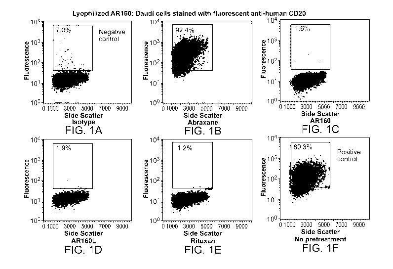
[0077] FIG. 1 depicts the results of an experiment in which CD20 positive
Daudi
lymphoma cells were labeled with fluorescent tagged anti-human CD20 or isotype
matched control in panels F and A, respectively, and analyzed by flow
cytometry. In the
other panels, the Daudi cells were pretreated with ABRAXANE (ABX),
ABX/rituximab nanoparticles (AR160), lyophilized and resuspended AR160
(AR160L),
or Rituxan prior to CD20 labeling. CD20 binding was specifically blocked by
the
AR160 nanoparticles and Rituxan, but not ABX alone, indicating that AR160 and
AR160L binds CD20 on these cells and block binding of the fluorescent anti-
CD20
antibody.
[0078] FIG. 2 is a histogram overlay of the scatterplots of FIG. 1.
[0079] FIGs. 3A-B depict particle size comparisons of ABX alone relative to
ABX/rituximab nanoparticles (AR; FIG. 3A) and ABX/trastuzumab nanoparticles
(AT;
FIG. 3B), both freshly made and lyophilized/resuspended.
[0080] FIG. 4 compares the toxicity of ABX and AR particles in a Daudi cell
proliferation assay.
[0081] FIGs. 5A-5C depict the results obtained in mice treated with either
labeled
ABRAXANE , labeled ABRAXANE coated with non-specific (bevacizumab) antibodies
(AB IgG), or labeled ABRAXANE coated with Rituximab (AR160). FIG. 5A depicts
the
fluorescence accumulation in regions of interest (ROI) in each tumor (ROI 2,
3, and 4) and in
CA 03035378 2019-02-27
WO 2018/045239 PCT/US2017/049746
background areas (ROT 1, 5, and 6). ROT 1, 5 and 6 serve as background
references. FIG 5B
is a bar graph of the average fluorescence per unit of tumor area of mice in
all three treatment
groups and shows gross tumor delivery. FIG. 5C is a bar graph of the average
fluorescence
per unit of tumor area, normalized by background ROT, to give proportion of
drug delivered
to tumor versus body. The data demonstrate that administration of AR160
nanoparticles
results in an increased fluorescence as compared to ABRAXANE alone or
ABRAXANE
coated with non-specific antibodies.
[0082] FIG. 6 depicts the survival of the mice treated with a single dose of
saline, BEV24
(bevacizumab at 24 mg/kg), ABX30 (ABX at 30 mg/kg), AB160 (12mg/kg BEV and 30
mg/kg ABX) and AB225 (24 mg/kg BEV and 30 mg/kg ABX). At 30 days post-
administration, the survival of mice treated with AB225 and with AB160 far
exceeds the
survival of mice treated with BEV alone or ABRAXANE alone.
[0083] FIG. 7 shows the binding affinity between atezolizumab and ABX. The Kd
was
determined to be 1.462x10⁻⁹. Biolayer interferometry (BLItz) (Forte
Bioscience) was
performed using streptavidin probes.
[0084] FIG. 8A shows the particle size distribution for ABX alone (average
size of 90 nm)
and ABX-atezolizumab nanoparticles (AA; average size of 129 nm), as determined
by
Mastersizer NS300. FIG. 8B is a photograph of the ABX-atezolizumab
nanoparticles from
FIG. 8A.
[0085] FIGs. 9A-9E show flow cytometry of ABX-atezolizumab nanoparticles
(AA130)
competing with labeled anti-PD-Li antibody for binding to a PD-Li positive
human
melanoma cell line, C8161. C8161 cells were pre-treated with isotype control
antibody (FIG.
9A), no treatment (FIG. 9B), ABRAXANE (FIG. 9C), atezolizumab (FIG. 9D), or
AA130
(FIG. 9E), then labeled with fluorescently-labeled anti-PD-Li antibody.
[0086] FIG. 10 shows the dose-dependent toxicity of ABX (solid line) and AA130
(broken
line) on C8161 cells.
[0087] FIGs. 11A-11D show the change in tumor volume over time in mice that
were
injected with 2x10⁶ PD-Li positive C8161 melanoma tumor cells, then treated by
100u1 IV
tail vein injection with saline (FIG. 11A), atezolizumab alone (18 mg/kg; FIG.
11B), ABX
alone (45 mg/kg; FIG. 11C) and AA130 (18 mg/kg atezolizumab and 45 mg/kg ABX;
FIG.
16
CA 03035378 2019-02-27
WO 2018/045239 PCT/US2017/049746
11D) one time. Tumor growth was monitored 3 times per week. Tumor size was
calculated
with the equation: (length x width2)/2.
[0088] FIG. 12 depicts the survival of the mice from the experiment shown in
FIGs. 11A-
11D. Kaplan Meier curves were generated using Graph Pad software. The median
survival
for each group was 14, 13, 16, and 21.5 days for saline, atezolizumab,
Abraxane and AA130,
repectively. Survival differences between AA130 and all other groups were
significant, with
p-values of 0.0008 for saline, 0.0015 for atezolizumab, and 0.0113 for ABX.
DETAILED DESCRIPTION
[0089] After reading this description it will become apparent to one skilled
in the art how to
implement the invention in various alternative embodiments and alternative
applications.
However, all the various embodiments of the present invention will not be
described herein.
It will be understood that the embodiments presented here are presented by way
of an
example only, and not limitation. As such, this detailed description of
various alternative
embodiments should not be construed to limit the scope or breadth of the
present invention as
set forth below.
[0090] Before the present invention is disclosed and described, it is to be
understood that the
aspects described below are not limited to specific compositions, methods of
preparing such
compositions, or uses thereof as such may, of course, vary. It is also to be
understood that the
terminology used herein is for the purpose of describing particular aspects
only and is not
intended to be limiting.
[0091] The detailed description of the invention is divided into various
sections only for the
reader's convenience and disclosure found in any section may be combined with
that in
another section. Titles or subtitles may be used in the specification for the
convenience of a
reader, which are not intended to influence the scope of the present
invention.
Definitions
[0092] Unless defined otherwise, all technical and scientific terms used
herein have the same
meaning as commonly understood by one of ordinary skill in the art to which
this invention
belongs. In this specification and in the claims that follow, reference will
be made to a
number of terms that shall be defined to have the following meanings:
17
CA 03035378 2019-02-27
WO 2018/045239 PCT/US2017/049746
[0093] The terminology used herein is for the purpose of describing particular
embodiments
only and is not intended to be limiting of the invention. As used herein, the
singular forms
"a", "an" and "the" are intended to include the plural forms as well, unless
the context clearly
indicates otherwise.
[0094] "Optional" or "optionally" means that the subsequently described event
or
circumstance can or cannot occur, and that the description includes instances
where the event
or circumstance occurs and instances where it does not.
[0095] The term "about" when used before a numerical designation, e.g.,
temperature, time,
amount, concentration, and such other, including a range, indicates
approximations which
may vary by ( + ) or ( -) 10%, 5%, 1%, or any subrange or subvalue there
between.
Preferably, the term "about" when used with regard to a dose amount means that
the dose
may vary by +/- 10%. For example, "about 400 to about 800 binding agents"
indicates that an
outside surface of a nanoparticles contain an amount of binding agent between
360 and 880
particles.
[0096] "Comprising" or "comprises" is intended to mean that the compositions
and
methods include the recited elements, but not excluding others. "Consisting
essentially
of' when used to define compositions and methods, shall mean excluding other
elements
of any essential significance to the combination for the stated purpose. Thus,
a composition
consisting essentially of the elements as defined herein would not exclude
other materials or
steps that do not materially affect the basic and novel characteristic(s) of
the claimed
invention. "Consisting of' shall mean excluding more than trace elements of
other
ingredients and substantial method steps. Embodiments defined by each of these
transition
terms are within the scope of this invention.
[0097] The term "nanoparticle" or "nanoparticle complex" as used herein refers
to particles
having at least one dimension which is less than 5 microns. In preferred
embodiments, such
as for intravenous administration, the nanoparticle has at least one dimension
which is less
than 1 micron. For direct administration, the nanoparticle is larger. Even
larger particles are
expressly contemplated by the invention.
[0098] In a population of particles, the sizes of individual particles are
distributed about a
mean. Particle sizes for the population can therefore be represented by an
average, and also
by percentiles. D50 is the particle size below which 50% of the particles
fall. 10% of
18
CA 03035378 2019-02-27
WO 2018/045239 PCT/US2017/049746
particles are smaller than the D TO value and 90% of particles are smaller
than D90. Where
unclear, the "average" size is equivalent to D50. So, for example, AB160 and
AR160 refer to
nanoparticles having an average size of 160 nanometers.
[0099] The term "nanoparticle" may also encompass discrete multimers of
smaller unit
nanoparticles. For example, a 320 nm particle comprises a dimer of a unit 160
nm
nanoparticle. For 160 nm nanoparticles, multimers would therefore be
approximately 320 nm,
480 nm, 640 nm, 800 nm, 960 nm, 1120 nm, and so on.
[0100] The term "carrier protein" as used herein refers to proteins that
function to transport
binding agents and/or therapeutic agents. The binding agents of the present
disclosure can
reversibly bind to the carrier proteins. Examples of carrier proteins are
discussed in more
detail below.
[0101] The term "core" as used herein refers to central or inner portion of
the nanoparticle
which may be comprised of a carrier protein, a carrier protein and a
therapeutic agent, or
other agents or combination of agents. In some embodiments, a portion of the
binding agent
may be associated with (e.g., non-covalently bound to) the core.
[0102] The term "therapeutic agent" as used herein means an agent which is
therapeutically
useful, e.g., an agent for the treatment, remission or attenuation of a
disease state,
physiological condition, symptoms, or etiological factors, or for the
evaluation or diagnosis
thereof. A therapeutic agent may be a chemotherapeutic agent, for example,
mitotic
inhibitors, topoisomerase inhibitors, steroids, anti-tumor antibiotics,
antimetabolites,
alkylating agents, enzymes, proteasome inhibitors, or any combination thereof
[0103] As used herein, the term, "binding agent", "binding agent specific
for," or "binding
agent that specifically binds" refers to an agent that binds to a target
antigen and does not
significantly bind to unrelated compounds. Examples of binding agents that can
be
effectively employed in the disclosed methods include, but are not limited to,
lectins,
proteins, and antibodies, such as monoclonal antibodies, e.g. humanized
monoclonal
antibodies, chimeric antibodies, or polyclonal antibodies, or antigen-binding
fragments
thereof, as well as aptamers, fusion proteins, and aptamers. In an embodiment
the binding
agent is an exogenous antibody. An exogenous antibody is an antibody not
naturally
produced in a particular mammal, e.g. in a human, by the mammalian immune
system.
19
CA 03035378 2019-02-27
WO 2018/045239 PCT/US2017/049746
[0104] The term "antibody" or "antibodies" as used herein refers to
immunoglobulin
molecules and immunologically active portions of immunoglobulin molecules
(i.e.,
molecules that contain an antigen binding site that immuno-specifically bind
an antigen).
The term also refers to antibodies comprised of two immunoglobulin heavy
chains and two
immunoglobulin light chains as well as a variety of forms including full
length antibodies and
portions thereof; including, for example, an immunoglobulin molecule, a
monoclonal
antibody, a chimeric antibody, a CDR- grafted antibody, a humanized antibody,
a Fab, a
Fab', a F(ab')2, a Fv, a disulfide linked Fv, a scFv, a single domain antibody
(dAb), a
diabody, a multispecific antibody, a dual specific antibody, an anti-idiotypic
antibody, a
bispecific antibody, a functionally active epitope-binding fragment thereof,
bifunctional
hybrid antibodies (e.g., Lanzavecchia et at., Eur. Jlmmunot. 17, 105 (1987))
and single
chains (e.g., Huston et at., Proc. Natl. Acad. Sci. US.A., 85, 5879-5883
(1988) and Bird et at.,
Science 242, 423-426 (1988), which are incorporated herein by reference).
(See, generally,
Hood et at., Immunology, Benjamin, N.Y., 2ND ed. (1984); Harlow and Lane,
Antibodies. A
Laboratory Manual, Cold Spring Harbor Laboratory (1988); Hunkapiller and Hood,
Nature,
323, 15-16 (1986), which are incorporated herein by reference). The antibody
may be of any
type (e.g., IgG, IgA, IgM, IgE or IgD). Preferably, the antibody is IgG. An
antibody may be
non-human (e.g., from mouse, goat, or any other animal), fully human,
humanized, or
chimeric. Antibody or antibodies include any biosimilar(s) of the antibodies
disclosed herein.
Biosimilars, as used herein, refers to a biopharmaceutical which is deemed to
be comparable
in quality, safety, and efficacy to a reference product marketed by an
innovator company
(Section 351(i) of the Public Health Service Act (42 U.S.C. 262(i)).
[0105] The term "dissociation constant," also referred to as "Kd," refers to a
quantity
expressing the extent to which a particular substance separates into
individual
components (e.g., the protein carrier, antibody, and optional therapeutic
agent).
[0106] The terms "lyophilized," "lyophilization" and the like as used herein
refer to a process
by which the material (e.g., nanoparticles) to be dried is first frozen and
then the ice or frozen
solvent is removed by sublimation in a vacuum environment. An excipient is
optionally
included in pre-lyophilized formulations to enhance stability of the
lyophilized product upon
storage. In some embodiments, the nanoparticles can be formed from lyophilized
components
(carrier protein, antibody and optional therapeutic) prior to use as a
therapeutic. In other
embodiments, the carrier protein, binding agent, e.g., antibody, and optional
therapeutic agent
CA 03035378 2019-02-27
WO 2018/045239 PCT/US2017/049746
are first combined into nanoparticles and then lyophilized. The lyophilized
sample may
further contain additional excipients.
[0107] The term "bulking agents" comprise agents that provide the structure of
the freeze-
dried product. Common examples used for bulking agents include mannitol,
glycine, lactose
and sucrose. In addition to providing a pharmaceutically elegant cake, bulking
agents may
also impart useful qualities in regard to modifying the collapse temperature,
providing freeze-
thaw protection, and enhancing the protein stability over long-term storage.
These agents can
also serve as tonicity modifiers. In some embodiments, the lyophilized
compositions
described herein comprise bulking agents. In some embodiments, the lyophilized
compositions described herein do not comprise bulking agents.
[0108] The term "buffer" encompasses those agents which maintain the solution
pH in an
acceptable range prior to lyophilization and may include succinate (sodium or
potassium),
histidine, phosphate (sodium or potassium),
Tris(tris(hydroxymethyl)aminomethane),
diethanolamine, citrate (sodium) and the like. The buffer of this invention
has a pH in the
range from about 5.5 to about 6.5; and preferably has a pH of about 6Ø
Examples of buffers
that will control the pH in this range include succinate (such as sodium
succinate), gluconate,
histidine, citrate and other organic acid buffers.
[0109] The term "cryoprotectants" generally includes agents which provide
stability to the
protein against freezing-induced stresses, presumably by being preferentially
excluded from
the protein surface. They may also offer protection during primary and
secondary drying,
and long- term product storage. Examples are polymers such as dextran and
polyethylene
glycol; sugars such as sucrose, glucose, trehalose, and lactose; surfactants
such as
polysorbates; and amino acids such as glycine, arginine, and serine.
[0110] The term "lyoprotectant" includes agents that provide stability to the
protein during
the drying or 'dehydration' process (primary and secondary drying cycles),
presumably by
providing an amorphous glassy matrix and by binding with the protein through
hydrogen
bonding, replacing the water molecules that are removed during the drying
process. This
helps to maintain the protein conformation, minimize protein degradation
during the
lyophilization cycle and improve the long-term products. Examples include
polyols or sugars
such as sucrose and trehalose.
21
CA 03035378 2019-02-27
WO 2018/045239 PCT/US2017/049746
1 1 1] The term "pharmaceutical formulation" refers to preparations which are
in such form
as to permit the active ingredients to be effective, and which contains no
additional
components that are toxic to the subjects to which the formulation would be
administered.
[0112] "Pharmaceutically acceptable" excipients (vehicles, additives) are
those which can
reasonably be administered to a subject mammal to provide an effective dose of
the active
ingredient employed.
[0113] "Reconstitution time" is the time that is required to rehydrate a
lyophilized
formulation into a solution.
[0114] A "stable" formulation is one in which the protein therein essentially
retains its
physical stability and/or chemical stability and/or biological activity upon
storage. For
example, various analytical techniques for measuring protein stability are
available in the art
and are reviewed in Peptide and Protein Drug Delivery, 247-301, Vincent Lee
Ed., Marcel
Dekker, Inc., New York, N.Y., Pubs. (1991) and Jones, A. Adv. Drug Delivery
Rev. 10:29-90
(1993). Stability can be measured at a selected temperature for a selected
time period.
[0115] The term "epitope" as used herein refers to the portion of an antigen
which is
recognized by a binding agent, e.g., an antibody. Epitopes include, but are
not limited to, a
short amino acid sequence or peptide (optionally glycosylated or otherwise
modified)
enabling a specific interaction with a protein (e.g., an antibody) or ligand.
For example, an
epitope may be a part of a molecule to which the antigen-binding site of a
binding agent
attaches.
[0116] The term "treating" or "treatment" covers the treatment of a disease or
disorder (e.g.,
cancer), in a subject, such as a human, and includes: (i) inhibiting a disease
or disorder, i.e.,
arresting its development; (ii) relieving a disease or disorder, i.e., causing
regression of the
disease or disorder; (iii) slowing progression of the disease or disorder;
and/or (iv) inhibiting,
relieving, or slowing progression of one or more symptoms of the disease or
disorder. In
some embodiments "treating" or "treatment" refers to the killing of cancer
cells.
[0117] The term "kill" with respect to a cancer treatment is directed to
include any type of
manipulation that will lead to the death of that cancer cell or at least of
portion of a
population of cancer cells.
22
CA 03035378 2019-02-27
WO 2018/045239
PCT/US2017/049746
[0118] The term "aptamer" refers to a nucleic acid molecule that is capable of
binding to a
target molecule, such as a polypeptide. For example, an aptamer of the
invention can
specifically bind to PD-L1, PD-L2, PD-1, or CTLA-4. The generation of
antibodies with a
particular binding specificity and the therapeutic use of aptamers are well
established in the
art. See, e.g., U.S. Pat. No. 5,475,096, U.S. Pat. Nos. 5,270,163, 5,582,981,
5,840,867,
6,011,020, 6,051,698, 6,147,204, 6,180,348 and 6,699,843, and the therapeutic
efficacy of
Macugeng (Eyetech, New York) for treating age-related macular degeneration,
each of
which is incorporated herein by reference in its entirety.
[0119] The term "oligomer" or "oligomeric" or "oligomerized" as used herein
refers to
oligomers composed of two or more monomers.
[0120] Fusion proteins are bioengineered polypeptides that join one peptide
(e.g., the
crystallizable fragment (Fc) domain of an antibody) with another biologically
active agent,
e.g., a protein domain, peptide, or nucleic acid or peptide aptamer, to
generate a molecule
with desired structure¨function properties and significant therapeutic
potential. The gamma
immunoglobulin (IgG) isotype is often used as the basis for generating Fc-
fusion proteins
because of favorable characteristics such as recruitment of effector function
and increased
plasma half-life. Given the range of aptamers, both peptide and nucleic acids,
that can be
used as fusion partners, fusion proteins have numerous biological and
pharmaceutical
applications.
[0121] The term "sequentially," as used herein, refers to the administration
of two or more
treatments one after another in any order. In some embodiments, the treatments
are
administered within more than 48 hours of each other. In some embodiments, the
treatments
are administered within about 48 hours of each other, within about 36 hours of
each other,
within about 24 hours of each other, within about 12 hours of each other,
within about 10
hours of each other, within about 8 hours of each other, within about 6 hours
of each other,
within about 4 hours of each other, within about two hours of each other, or
within about 1
hour of each other.
[0122] The term "concurrently," as used herein, refers two or more treatments
administered
at substantially about the same time in any order.
23
CA 03035378 2019-02-27
WO 2018/045239 PCT/US2017/049746
[0123] The term "PD-1," as used herein, refers to programmed cell death
protein-1, also
known as CD279, which is expressed on the surface of activated T cells, B
cells, as well as
myeloid cells.
[0124] The term "PD-Li", as used herein, refers to programmed death-ligand 1,
also known
as B7-H1 or CD274, is a PD-1 ligand which is commonly expressed on the surface
of
dendritic cells or macrophages.
[0125] The term "PD-L2," as used herein, refers to programmed death ligand-2,
also known
as B7-DC or CD273, is a PD-1 ligand which is commonly expressed on the surface
of
dendritic cells or macrophages.
[0126] The terms "biosimilar" or "biosimilar," also known as "follow-on
biologic"
or "subsequent entry biologic-, as used herein, refers to a biologic product
which is
substantially an identical copy of a product approved by a regulatory agency.
[0127] The terms "synergistic" or "synergistic effect" or "synergistically
effective amount"
"or "synergistic efficacy", as used herein, refer to a greater-than-additive
therapeutic effect
which is produced by the administration of at least two agents, and which
exceeds that which
would otherwise result from administration of one of the agents without the
administration of
the other agent. For example, the therapeutic effect of the nanoparticle
composition is
increased when administered sequentially or concurrently with a binding agent
to provide a
synergistic effect, provided that the increase is greater than the additive
effectiveness of the
binding agent and the nanoparticle composition when administered alone. The
term
"synergistically therapeutic amount" typically refers to a less than standard
therapeutic
amount of one or both therapeutic agents, meaning that the amount required for
the desired
therapeutic effectiveness is lower than when the therapeutic agent is used
alone. A
synergistically therapeutic amount also includes when one therapeutic agent is
given at a
standard therapeutic dose and another therapeutic agent is administered in a
less than
standard therapeutic dose
[0128] The term "therapeutically effective amount" or "therapeutic
effectiveness," as used
herein, of a nanoparticle composition or binding agent refers to nanoparticle
composition or
binding agent levels in which the physiological effects of a disease or
disorder are, at a
minimum, ameliorated. A therapeutically effective amount can be given in one
or more
administrations using one or more tablets, capsules or other pharmaceutical
units. The
24
CA 03035378 2019-02-27
WO 2018/045239 PCT/US2017/049746
amount of a nanoparticle composition or binding agent which constitutes a
therapeutically
effective amount will vary depending on the nanoparticle composition or
binding agent, the
disorder and its severity, and the general health, age, sex, body weight and
tolerance to drugs
of the subject to be treated, but can be determined routinely by one of
ordinary skill in the art.
In some embodiments, the term "therapeutically effective amount" refers to a
synergistically
effective amount or synergistically therapeutic amount.
[0129] Additionally, some terms used in this specification are more
specifically defined
below.
Overview
[0130] The current invention is predicated, in part, on the surprising
discovery that optionally
lyophilized nanoparticles comprising a carrier protein, a binding agent, e.g.,
an antibody, an
aptamer, or a fusion protein, having a PD-Li or PD-L2 binding domainõ and a
therapeutic
agent provide targeted therapy to a tumor while minimizing toxicity to the
patient. The
nanoparticles as described herein are thus a significant improvement versus
conventional
ADCs.
[0131] The invention is furether predicated, in part, on the syngergy of
immune checkpoint
inhibitor immunotherapy (e.g., PD-1 immunotherapy and/or CTLA-4 immunotherapy)
with
the nanoparticles. Without being bound by theory, it is contemplated that
binding of PD-Li
or PD-L2 by the binding agents (e.g., antibodies) as described herein will
deplete or diminish
the amount of PD-Li or PD-L2 available to bind PD-1 on T cells, thereby
increasing the
therapeutic effectiveness of PD-1-based immunotherapy. Administration of the
nanoparticles,
alone or in combination with PD-1 immunotherapy, may increase the number of T
cells that
are free from PD-1 pathway-mediated inhibition, and restore the patient's
immune response
against a PD-L1- or PD-L2-expressing cancer.
[0132] For conventional ADCs to be effective, it is critical that the linker
be stable enough
not to dissociate in the systemic circulation but allow for sufficient drug
release at the tumor
site. Alley, S.C., et at. (2008) Bioconjug Chem 19:759-765. This has proven to
be a major
hurdle in developing effective drug conjugate (Julien, D.C., et al. (2011)
MAbs 3:467-478;
Alley, S.C., et at. (2008) Bioconjug Chem 19:759-765); therefore, an
attractive feature of the
nanoparticles described herein is that a biochemical linker is not required.
CA 03035378 2019-02-27
WO 2018/045239 PCT/US2017/049746
[0133] Another shortcoming of current ADCs is that higher drug penetration
into the tumor
has not been substantively proven in human tumors. Early testing of ADCs in
mouse models
suggested that tumor targeting with antibodies would result in a higher
concentration of the
active agent in the tumor (Deguchi, T. et at. (1986) Cancer Res 46: 3751-
3755); however,
this has not correlated in the treatment of human disease, likely because
human tumors are
much more heterogeneous in permeability than mouse tumors. Jain, R.K. et at.
(2010) Nat
Rev Clin Oncol 7:653-664. Also, the size of the nanoparticle is critical for
extravasation from
the vasculature into the tumor. In a mouse study using a human colon
adenocarcinoma
xenotransplant model, the vascular pores were permeable to liposomes up to 400
nm. Yuan,
F., et al. (1995) Cancer Res 55: 3752-3756. Another study of tumor pore size
and
permeability demonstrated that both characteristics were dependent on tumor
location and
growth status, with regressing tumors and cranial tumors permeable to
particles less than 200
nm. Hobbs, S.K., et al. (1998) Proc Natl Acad Sci USA 95:4607-4612. The nano-
immune
conjugate (nanoparticles) described herein overcomes this issue by the fact
that the large
complex, which is less than 200 nm intact, is partially dissociated in
systemic circulation into
smaller functional units that are easily able to permeate tumor tissue.
Furthermore, once the
conjugate arrives to the tumor site, the smaller toxic payload can be released
and only the
toxic portion needs to be taken up by tumor cells, not the entire conjugate.
[0134] The advent of antibody- (i.e. AVASTINg) coated albumin nanoparticles
containing a
therapeutic agent (i.e., ABRAXANDID) has led to a new paradigm of directional
delivery of
two or more therapeutic agents to a predetermined site in vivo. See PCT Patent
Publication
Nos. WO 2012/154861 and WO 2014/055415, each of which is incorporated herein
by
reference in its entirety.
[0135] When compositions of albumin and a binding agent, e.g., antibody, are
admixed
together in an aqueous solution at specific concentrations and ratios, the
binding agents useful
in this invention spontaneously self-assemble into and onto the albumin to
form nanoparticles
having multiple copies of the binding agent (up to 500 or more). Without being
limited to
any theory, it is contemplated that binding agents (e.g., antibodies) non-
covalently bind to the
carrier protein (e.g., albumin) via one or more albumin-binding motifs of the
binding agent,
and one or more antibody-binding motifs on the carrier protein. Examples of
such motifs can
be found in PCT Application No. PCT/U517/45643, which is incorporated herein
by
reference in its entirety.
26
CA 03035378 2019-02-27
WO 2018/045239 PCT/US2017/049746
[0136] While protein compositions comprising a single source protein are
commonly stored
in lyophilized form where they exhibit significant shelf-life, such
lyophilized compositions
generally do not contain a self-assembled nanoparticle of two different
proteins integrated
together by hydrophobic-hydrophobic interactions. Moreover, the nanoparticle
configuration
wherein a majority of the binding portions of the binding agent are exposed on
the surface of
the nanoparticles lends itself to being susceptible to dislodgement or
reconfiguration by
conditions which otherwise would be considered benign. For example, during
lyophilization,
ionic charges on the proteins are dehydrated thereby exposing the underlying
charges.
Exposed charges allow for charge-charge interactions between the two proteins
which can
alter the binding affinity of each protein to the other. In addition, the
concentration of the
nanoparticles increases significantly as the solvent (e.g., water) is removed.
Such increased
concentrations of nanoparticles could lead to irreversible oligomerization.
Oligomerization is
a known property of proteins that reduces the biological properties of the
oligomer as
compared to the monomeric form and increases the size of the particle,
sometimes beyond 1
micron.
[0137] On the other hand, a stable form of a nanoparticle composition is
required for clinical
and/or commercial use, where a shelf-life of at least 3 months is required and
shelf-lives of
greater than 6 months or 9 months are preferred. Such a stable composition
must be readily
available for intravenous injection, must retain its self-assembled form upon
intravenous
injection so as to direct the nanoparticle to the predetermined site in vivo,
must have a
maximum size of less than 1 micron so as to avoid any ischemic event when
delivered into
the blood stream, and finally must be compatible with the aqueous composition
used for
injection.
Compounds
[0138] As will be apparent to the skilled artisan upon reading this
disclosure, the present
disclosure relates to compositions of nanoparticles containing a carrier
protein, binding
agents, and optionally at least one therapeutic agent, wherein said
compositions are optionally
lyophilized.
[0139] In some embodiments, the carrier protein can be albumin, gelatin,
elastin (including
topoelastin) or elastin-derived polypeptides (e.g., a-elastin and elastin-like
polypeptides
(ELPs)), gliadin, legumin, zein, soy protein (e.g., soy protein isolate
(SPI)), milk protein (e.g.,
P-lactoglobulin (BLG) and casein), or whey protein (e.g., whey protein
concentrates (WPC)
27
CA 03035378 2019-02-27
WO 2018/045239 PCT/US2017/049746
and whey protein isolates (WPI)). In preferred embodiments, the carrier
protein is albumin.
In preferred embodiments, the albumin is egg white (ovalbumin), bovine serum
albumin
(BSA), or the like. In even more preferred embodiments, the carrier protein is
human serum
albumin (HSA). In some embodiments, the carrier protein is a recombinant
protein, e.g.
recombinant human serum albumin. In some embodiments, the carrier protein is a
generally
regarded as safe (GRAS) excipient approved by the United States Food and Drug
Administration (FDA).
[0140] In some embodiments, the binding agents are antibodies.
[0141] In some embodiments, the anti-PD-1 antibody comprises nivolumab,
pembrolizumab,
pidilizumab, PDR001, or biosimilars thereof. In some aspects, the anti-PD-Li
antibody is
atezolizumab, avelumab, durvalumab, or BMS 936559 (M1DX1105). In some aspects,
the
binding agent of the CTLA-4 immunotherapy is an anti-CTLA-4 antibody. In one
embodiment, the anti-CTLA-4 antibody is ipilimumab.
[0142] In some aspects, the first binding agent and/or the second binding
agent is a fusion
protein. In one embodiment, the fusion protein is AMP-224 (PD-L2 IgG2a fusion
protein;
Amplimmune/GlaxoSmith Klein); AMP-514 (MEDI0680) (PD-L2 fusion protein;
Amplimmune/GlaxoSmith Klein), or a biosimilar thereof AMP-224 and AMP-514
target
PD-1.
[0143] . In some embodiments, the antibodies are a substantially single layer
of antibodies
on all or part of the surface of the nanoparticle.
Table 1 depicts a list of non-limiting list of antibodies.
Table 1: Example Antibodies
Generic Name Brand Name Type Example of Possible
Indication
Solid tumor, gastric cancer,
Avelumab anti-PD-Li; human IgG1 Merkel cell carcinoma,
non-
(MSB0010718C) BAVENCIO mAb small cell lung cancer
NSCLC, head and neck,
Durvalumab anti-PD-Li; human IgG1K bladder, gastric,
pancreatic,
(MEDI4736) IMFINZITm mAb HCC and blood cancers
28
CA 03035378 2019-02-27
WO 2018/045239 PCT/US2017/049746
Generic Name Brand Name Type Example of Possible
Indication
anti-Delta-like 1 (secondary
binding to some forms of Lymphoma, myeloma,
Pidilizumab (CT- PD-1); humanized IgG1 diffuse intrinsic pontine
011) mAb glioma
BMS 936559/ melanoma, non-small cell
MDX-1105 anti-PD-Li mAb lung cancer
metastatic melanoma;
squamous non-small cell
Nivolumab (BMS- Anti-PD-1; human IgG4 lung cancer; renal cell
936558) OPDIVO mAb carcinoma
atezolizumab bladder cancer, NSCLC,
(RG7446; melanoma, breast, renal
cell
MPDL3280A) TECENTRIQ anti-PD-Li mAb carcinoma, lymphoma
Anti-CTLA-4; Human IgG1 Melanoma
Ipilimumab YERVOY mAb
Anti-PD-1; Human IgG4 Melanoma
Pembrolizumab KEYTRUDA mAb
[0144] In some embodiments, the at least one therapeutic agent is selected
from
abiraterone, bendamustine, bortezomib, carboplatin, cabazitaxel, cisplatin,
chlorambucil,
dasatinib, docetaxel, doxorubicin, epirubicin, erlotinib, etoposide,
everolimus, gefitinib,
idarubicin, imatinib, hydroxyurea, imatinib, lapatinib, leuprorelin,
melphalan, methotrexate,
mitoxantrone, nedaplatin, nilotinib, oxaliplatin, paclitaxel, pazopanib,
pemetrexed, picoplatin,
romidepsin, satraplatin, sorafenib, vemurafenib, sunitinib, teniposide,
triplatin, vinblastine,
vinorelbine, vincristine, and cyclophosphamide. Preferably, the therapeutic
agent is
paclitaxel. Additional therapeutic agents are known, for example those listed
in PCT
Publication No. W02017/031368, which is incorporated herein by reference in
its entirety.
29
CA 03035378 2019-02-27
WO 2018/045239 PCT/US2017/049746
[0145] It is to be understood that the therapeutic agent may be located inside
the
nanoparticle, on the outside surface of the nanoparticle, or both. The
nanoparticle may
contain more than one therapeutic agent, for example, two therapeutic agents,
three
therapeutic agents, four therapeutic agents, five therapeutic agents, or more.
Furthermore, a
nanoparticle may contain the same or different therapeutic agents inside and
outside the
nanoparticle.
[0146] In one aspect, the nanoparticle comprises at least 100 binding agents
non-covalently
bound to the surface of the nanoparticle. In one aspect, the nanoparticle
comprises at least
200 binding agents non-covalently bound to the surface of the nanoparticle. In
one aspect, the
nanoparticle comprises at least 300 binding agents non-covalently bound to the
surface of the
nanoparticle. In one aspect, the nanoparticle comprises at least 400 binding
agents non-
covalently bound to the surface of the nanoparticle. In one aspect, the
nanoparticle comprises
at least 500 binding agents non-covalently bound to the surface of the
nanoparticle. In one
aspect, the nanoparticle comprises at least 600 binding agents non-covalently
bound to the
surface of the nanoparticle.
[0147] In one aspect, the nanoparticle comprises between about 100 and about
1000 binding
agents non-covalently bound to the surface of the nanoparticle. In one aspect,
the
nanoparticle comprises between about 200 and about 1000 binding agents non-
covalently
bound to the surface of the nanoparticle. In one aspect, the nanoparticle
comprises between
about 300 and about 1000 binding agents non-covalently bound to the surface of
the
nanoparticle. In one aspect, the nanoparticle comprises between about 400 and
about 1000
binding agents non-covalently bound to the surface of the nanoparticle. In one
aspect, the
nanoparticle comprises between about 500 and about 1000 binding agents non-
covalently
bound to the surface of the nanoparticle. In one aspect, the nanoparticle
comprises between
about 600 and about 1000 binding agents non-covalently bound to the surface of
the
nanoparticle. In one aspect, the nanoparticle comprises between about 200 and
about 800
binding agents non-covalently bound to the surface of the nanoparticle. In one
aspect, the
nanoparticle comprises between about 300 and about 800 binding agents non-
covalently
bound to the surface of the nanoparticle. In preferred embodiments, the
nanoparticle
comprises between about 400 and about 800 binding agents non-covalently bound
to the
surface of the nanoparticle. Contemplated values include any value or subrange
within any
of the recited ranges, including endpoints.
CA 03035378 2019-02-27
WO 2018/045239 PCT/US2017/049746
[0148] In one aspect, the average particle size in the nanoparticle
composition is less than
about 1 p.m. In one aspect, the average particle size in the nanoparticle
composition is
between about 90 nm and about 1 p.m. In one aspect, the average particle size
in the
nanoparticle composition is between about 90 nm and about 900 nm. In one
aspect, the
average particle size in the nanoparticle composition is between about 90 nm
and about 800
nm. In one aspect, the average particle size in the nanoparticle composition
is between about
90 nm and about 700 nm. In one aspect, the average particle size in the
nanoparticle
composition is between about 90 nm and about 600 nm. In one aspect, the
average particle
size in the nanoparticle composition is between about 90 nm and about 500 nm.
In one
aspect, the average particle size in the nanoparticle composition is between
about 90 nm and
about 400 nm. In one aspect, the average particle size in the nanoparticle
composition is
between about 90 nm and about 300 nm. In one aspect, the average particle size
in the
nanoparticle composition is between about 90 nm and about 200 nm. In a
preferred
embodiment, the average particle size in the nanoparticle composition is
between about 100
nm and about 180 nm. In an especially preferred embodiment, the mean particle
size in the
nanoparticle composition is about 130 nm or about 160 nm. Contemplated values
include
any value, subrange, or range within any of the recited ranges, including
endpoints. In one
embodiment, the nanoparticle size is determined using a Mastersizer 2000. In
one
embodiment, the nanoparticle size is determined using a Malvern Nanosight.
[0149] In one aspect, the nanoparticle composition is formulated for
intravenous injection. In
order to avoid an ischemic event, the nanoparticle composition formulated for
intravenous
injection should comprise nanoparticles with an average particle size of less
than about 1 p.m.
[0150] In one aspect, the average particle size in the nanoparticle
composition is greater than
about 1 p.m. In one aspect, the average particle size in the nanoparticle
composition is
between about 1 p.m and about 5 p.m. In one aspect, the average particle size
in the
nanoparticle composition is between about 1 p.m and about 4 p.m. In one
aspect, the average
particle size in the nanoparticle composition is between about 1 p.m and about
3 p.m. In one
aspect, the average particle size in the nanoparticle composition is between
about 1 p.m and
about 2 p.m. In one aspect, the average particle size in the nanoparticle
composition is
between about 1 p.m and about 1.5 pm. Contemplated values include any value,
subrange, or
range within any of the recited ranges, including endpoints.
31
CA 03035378 2019-02-27
WO 2018/045239 PCT/US2017/049746
[0151] In one aspect, the nanoparticle composition is formulated for direct
injection into a
tumor. Direct injection includes injection into or proximal to a tumor site,
perfusion into a
tumor, and the like. When formulated for direct injection into a tumor, the
nanoparticle may
comprise any average particle size. Without being bound by theory, it is
believed that larger
particles (e.g., greater than 500 nm, greater than 1 [tm, and the like) are
more likely to be
immobilized within the tumor, thereby providing a beneficial effect. Larger
particles can
accumulate in the tumor or specific organs. See, e.g., 20-60 micron glass
particle that is used
to inject into the hepatic artery feeding a tumor of the liver, called
"TheraSphereg" (in
clinical use for liver cancer). Therefore, for intravenous administration,
particles under 1 [tm
are typically used. Particles over 1 [tm are, more typically, administered
directly into a tumor
("direct injection") or into an artery feeding into the site of the tumor.
[0152] In one aspect, less than about 0.01% of the nanoparticles within the
composition have
a particle size greater than 200 nm, greater than 300 nm, greater than 400 nm,
greater than
500 nm, greater than 600 nm, greater than 700 nm, or greater than 800 nm. In
one aspect,
less than about 0.001% of the nanoparticles within the composition have a
particle size
greater than 200 nm, greater than 300 nm, greater than 400 nm, greater than
500 nm, greater
than 600 nm, greater than 700 nm, or greater than 800 nm. In a preferred
embodiment, less
than about 0.01% of the nanoparticles within the composition have a particle
size greater than
800 nm. In a more preferred embodiment, less than about 0.001% of the
nanoparticles within
the composition have a particle size greater than 800 nm.
[0153] In a preferred aspect, the sizes and size ranges recited herein relate
to particle sizes of
the reconstituted lyophilized nanoparticle composition. That is, after the
lyophilized
nanoparticles are resuspended in an aqueous solution (e.g., water, other
pharmaceutically
acceptable excipient, buffer, etc.), the particle size or average particle
size is within the range
recited herein.
[0154] In one aspect, at least about 50%, 60%, 70%, 80%, 90%, 95%, 96%, 97%,
98%,
99%, 99.5%, or 99.9% of the nanoparticles are present in the reconstituted
composition
as single nanoparticles. That is, fewer than about 50%, 40%, 30%, etc. of the
nanoparticles
are dimerized or oligomerized.
32
CA 03035378 2019-02-27
WO 2018/045239 PCT/US2017/049746
[0155] In some embodiments, the nanoparticles in the composition have less
than 20% by
number dimerization, less than 10% by number dimerization and preferably less
than 5% by
number dimerization.
[0156] In some embodiments, the size of the nanoparticle can be controlled by
the adjusting
the amount (e.g., ratio) of carrier protein to binding agent. The size of the
nanoparticles, and
the size distribution, is also important. The nanoparticles of the invention
may behave
differently according to their size. At large sizes, an agglomeration may
block blood vessels.
Therefore, agglomeration of nanoparticles can affect the performance and
safety of the
composition. On the other hand, larger particles may be more therapeutic under
certain
conditions (e.g., when not administered intravenously).
[0157] In one aspect, the nanoparticle composition comprises at least one
additional
therapeutic agent. In one embodiment, the at least one additional therapeutic
agent is non-
covalently bound to the outside surface of the nanoparticle. In one
embodiment, the at least
one additional therapeutic agent is arranged on the outside surface of the
nanoparticle. In one
embodiment, the at least one additional therapeutic agent is selected from
abiraterone,
bendamustine, bortezomib, carboplatin, cabazitaxel, cisplatin, chlorambucil,
dasatinib,
docetaxel, doxorubicin, epirubicin, erlotinib, etoposide, everolimus,
gemcitabine, gefitinib,
idarubicin, imatinib, hydroxyurea, imatinib, lapatinib, leuprorelin,
melphalan, methotrexate,
mitoxantrone, nedaplatin, nilotinib, oxaliplatin, pazopanib, pemetrexed,
picoplatin,
romidepsin, satraplatin, sorafenib, vemurafenib, sunitinib, teniposide,
triplatin, vinblastine,
vinorelbine, vincristine, and cyclophosphamide. In one embodiment, the at
least one
additional therapeutic agent is an anti-cancer binding agent, e.g., an anti-
cancer antibody.
Additional anti-cancer antibodies are known, for example those listed in PCT
Publication No.
W02017/031368, which is incorporated herein by reference in its entirety.
Methods of Making Nanoparticles
[0158] In some aspects, the current invention relates to methods of making
nanoparticle
compositions as described herein.
[0159] In one aspect, the nanoparticles of the nanoparticle composition are
formed by
contacting the carrier protein or carrier protein-therapeutic agent particle
with the binding
agent at a ratio of about 10:1 to about 10:30 carrier protein particle or
carrier protein-
therapeutic agent particle to binding agent. In one embodiment, the ratio is
about 10:2 to
33
CA 03035378 2019-02-27
WO 2018/045239 PCT/US2017/049746
about 10:25. In one embodiment, the ratio is about 10:2 to about 1:1. In a
preferred
embodiment, the ratio is about 10:2 to about 10:6. In an especially preferred
embodiment,
the ratio is about 10:4. Contemplated ratios include any value, subrange, or
range within any
of the recited ranges, including endpoints.
[0160] In one embodiment, the amount of solution or other liquid medium
employed to form
the nanoparticles is particularly important. No nanoparticles are formed in an
overly dilute
solution of the carrier protein (or carrier protein-therapeutic agent) and the
antibodies. An
overly concentrated solution will result in unstructured aggregates. In some
embodiments,
the amount of solution (e.g., sterile water, saline, phosphate buffered
saline) employed is
between about 0.5 mL of solution to about 20 mL of solution. In some
embodiments, the
amount of carrier protein is between about 1 mg/mL and about 100 mg/mL. In
some
embodiments, the amount of binding agent is between about 1 mg/mL and about 30
mg/mL.
For example, in some embodiments, the ratio of carrier protein:binding
agent:solution is
approximately 9 mg of carrier protein (e.g., albumin) to 4 mg of binding
agent, e.g., antibody
(e.g., BEV) in 1 mL of solution (e.g., saline). An amount of therapeutic agent
(e.g., taxol)
can also be added to the carrier protein. For example, 1 mg of taxol can be
added 9 mg of
carrier protein (10 mg carrier protein-therapeutic) and 4 mg of binding agent,
e.g., antibody,
Fc fusion molecule, or aptamer, in 1 mL of solution. When using a typical i.v.
bag, for
example, with the solution of approximately 1 liter one would need to use
1000x the amount
of carrier protein/carrier protein- therapeutic agent and antibodies compared
to that used in 1
mL. Thus, one cannot form the present nanoparticles in a standard i.v. bag.
Furthermore,
when the components are added to a standard i.v. bag in the therapeutic
amounts of the
present invention, the components do not self-assemble to form nanoparticles.
[0161] In one embodiment, the carrier protein or carrier protein-therapeutic
agent particle is
contacted with the binding agent in a solution having a pH between about 4 and
about 8. In
one embodiment, the carrier protein or carrier protein-therapeutic agent
particle is contacted
with the binding agent in a solution having a pH of about 4. In one
embodiment, the carrier
protein or carrier protein-therapeutic agent particle is contacted with the
binding agent in a
solution having a pH of about 5. In one embodiment, the carrier protein or
carrier protein-
therapeutic agent particle is contacted with the binding agent in a solution
having a pH of
about 6. In one embodiment, the carrier protein or carrier protein-therapeutic
agent particle is
contacted with the binding agent in a solution having a pH of about 7. In one
embodiment,
34
CA 03035378 2019-02-27
WO 2018/045239 PCT/US2017/049746
the carrier protein or carrier protein-therapeutic agent particle is contacted
with the binding
agent in a solution having a pH of about 8. In a preferred embodiment, the
carrier protein or
carrier protein-therapeutic agent particle is contacted with the binding agent
in a solution
having a pH between about 5 and about 7.
[0162] In one embodiment, the carrier protein particle or carrier protein-
therapeutic agent
particle is incubated with the binding agent at a temperature of about 5 C to
about 60 C, or
any range, subrange, or value within that range including endpoints. In a
preferred
embodiment, the carrier protein particle or carrier protein-therapeutic agent
particle is
incubated with the binding agent at a temperature of about 23 C to about 60
C.
[0163] Without being bound by theory, it is believed that the stability of the
nanoparticles
within the nanoparticle composition is, at least in part, dependent upon the
temperature
and/or pH at which the nanoparticles are formed, as well as the concentration
of the
components (i.e., carrier protein, binding agent, and optionally therapeutic
agent) in the
solution. In one embodiment, the Kd of the nanoparticles is between about 1 x
10-11M and
about 2 x 10⁻⁵M. In one embodiment, the Kd of the nanoparticles is between
about 1 x 10-11
M and about 2 x 10⁴ M. In one embodiment, the Kd of the nanoparticles is
between about 1 x
10-11M and about 7 x 10⁻⁹M. In a preferred embodiment, the Kd of the
nanoparticles is
between about 1 x 10-11M and about 3 x 10⁻⁸M. Contemplated values include any
value,
subrange, or range within any of the recited ranges, including endpoints.
Lyophilization
[0164] Lyophilization, or freeze-drying, removes water from a composition. In
the process,
the material to be dried is first frozen and then the ice or frozen solvent is
removed by
sublimation in a vacuum environment. An excipient may be included in pre-
lyophilized
formulations to enhance stability during the freeze-drying process and/or to
improve stability
of the lyophilized product upon storage. Pikal, M. Biopharm. 3(9)26-30 (1990)
and Arakawa
et al., Pharm. Res. 8(3):285- 291 (1991).
[0165] While proteins may be lyophilized, the process of lyophilization and
reconstitution
may affect the properties of the protein. Because proteins are larger and more
complex than
traditional organic and inorganic drugs (i.e. possessing multiple functional
groups in addition
to complex three-dimensional structures), the formulation of such proteins
poses special
problems. For a protein to remain biologically active, a formulation must
preserve intact the
CA 03035378 2019-02-27
WO 2018/045239 PCT/US2017/049746
conformational integrity of at least a core sequence of the protein's amino
acids while at the
same time protecting the protein's multiple functional groups from
degradation. Degradation
pathways for proteins can involve chemical instability (i.e. any process which
involves
modification of the protein by bond formation or cleavage resulting in a new
chemical entity)
or physical instability (i.e. changes in the higher order structure of the
protein). Chemical
instability can result from deamidation, racemization, hydrolysis, oxidation,
beta elimination
or disulfide exchange. Physical instability can result from denaturation,
aggregation,
precipitation or adsorption, for example. The three most common protein
degradation
pathways are protein aggregation, deamidation and oxidation. Cleland, et al.,
Critical
Reviews in Therapeutic Drug Carrier Systems 10(4): 307-377 (1993).
[0166] The lyophilized compositions of this invention are prepared by standard
lyophilization
techniques with or without the presence of stabilizers, buffers, etc.
Surprisingly, these
conditions do not alter the relatively fragile structure of the nanoparticles.
Moreover, at best,
these nanoparticles retain their size distribution upon lyophilization and,
more importantly,
can be reconstituted for in vivo administration (e.g., intravenous delivery)
in substantially the
same form and ratios as if freshly made.
Formulations
[0167] In one aspect, the nanoparticle composition is formulated for systemic
delivery, e.g.,
intravenous administration.
[0168] In one aspect, the nanoparticle composition is formulated for direct
injection into a
tumor. Direct injection includes injection into or proximal to a tumor site,
perfusion into a
tumor, and the like. Because the nanoparticle composition is not administered
systemically, a
nanoparticle composition is formulated for direct injection into a tumor may
comprise any
average particle size. Without being bound by theory, it is believed that
larger particles (e.g.,
greater than 500 nm, greater than 1 p.m, and the like) are more likely to be
immobilized
within the tumor, thereby providing what is believed to be a better beneficial
effect.
[0169] In another aspect, provided herein is a composition comprising
nanoparticles as
provided herein, and at least one pharmaceutically acceptable excipient.
[0170] In general, the compositions provided herein can be formulated for
administration to a
patient by any of the accepted modes of administration. Various formulations
and drug
36
CA 03035378 2019-02-27
WO 2018/045239 PCT/US2017/049746
delivery systems are available in the art. See, e.g., Gennaro, A.R., ed.
(1995) Remington's
Pharmaceutical Sciences, 18th ed., Mack Publishing Co.
[0171] In general, nanoparticles as provided herein will be administered as
pharmaceutical
compositions by any one of the following routes: oral, systemic (e.g.,
transdermal, intranasal
or by suppository), or parenteral (e.g., intramuscular, intravenous or
subcutaneous)
administration.
[0172] The compositions are comprised of, in general, a nanoparticle of the
present invention
in combination with at least one pharmaceutically acceptable excipient.
Acceptable
excipients are non-toxic, aid administration, and do not adversely affect the
therapeutic
benefit of the claimed compounds. Such excipient may be any solid, liquid,
semi-solid or, in
the case of an aerosol composition, gaseous excipient that is generally
available to one of
skill in the art.
[0173] Solid pharmaceutical excipients include starch, cellulose, talc,
glucose, lactose,
sucrose, gelatin, malt, rice, flour, chalk, silica gel, magnesium stearate,
sodium stearate,
glycerol monostearate, sodium chloride, dried skim milk and the like. Liquid
and semisolid
excipients may be selected from glycerol, propylene glycol, water, ethanol and
various oils,
including those of petroleum, animal, vegetable or synthetic origin, e.g.,
peanut oil, soybean
oil, mineral oil, sesame oil, etc. Preferred liquid carriers, particularly for
injectable solutions,
include water, saline, aqueous dextrose, and glycols. Other suitable
pharmaceutical excipients
and their formulations are described in Remington's Pharmaceutical Sciences,
edited by E.
W. Martin (Mack Publishing Company, 18th ed., 1990).
[0174] The present compositions may, if desired, be presented in a pack or
dispenser device
containing one or more unit dosage forms containing the active ingredient.
Such a pack or
device may, for example, comprise metal or plastic foil, such as a blister
pack, or glass, and
rubber stoppers such as in vials. The pack or dispenser device may be
accompanied by
instructions for administration. Compositions comprising a nanoparticle of the
invention
formulated in a compatible pharmaceutical carrier may also be prepared, placed
in an
appropriate container, and labeled for treatment of an indicated condition.
37
CA 03035378 2019-02-27
WO 2018/045239 PCT/US2017/049746
Treatment Methods
[0175] The nanoparticle compositions as described herein are useful in
treating cancer cells
and/or tumors in a mammal having a cancer or tumor that expresses PD-Li and/or
PD-L2. In
a preferred embodiment, the mammal is a human (i.e., a human patient).
Preferably, the
lyophilized nanoparticle composition is reconstituted (suspended in an aqueous
excipient)
prior to administration.
[0176] In one aspect is provided a method for treating a cancer cell, the
method comprising
contacting the cell with an effective amount of nanoparticles and an
immunotherapy (e.g.,
PD-1 or CTLA-4) as described herein to treat the cancer cell. Treatment of a
cancer cell
includes, without limitation, reduction in proliferation, killing the cell,
preventing metastasis
of the cell, and the like.
[0177] "Immune therapy", "immune therapies", "immunotherapy" or
"immunotherapies," as
used herein, generally refer to treatments of a disease by inducing,
enhancing, or suppressing
an immune response. In some cases, immune therapies or immunotherapies can
either elicit
or activate or amplify immune responses (also known as "activation
immunotherapies"), or
reduce or suppress immune responses (also known as "suppression
immunotherapies"). For
example, cancer immune therapy or cancer immunotherapy attempts to stimulate
or activate
the immune responses against tumors or cancer cells. As would be understood by
one skilled
in the art, immune therapy or immunotherapy can utilize a variety of
approaches or
mechanisms including, but not limited to, antibodies, antigens, use and/or
activation of
immune responsive cells such as lymphocytes, macrophages, dendritic cells,
other antigen
presenting cells, natural killer cells (NK Cells; e.g., NK-92), T-cells (e.g.,
helper T-cells,
cytotoxic T lymphocytes (CTL), etc.), therapies involving immune modulators
(including, but
not limited to: interleukins (e.g., IL-2, IL-7, IL-12, etc.), cytokines (e.g.,
interferons, G-CSF,
imiquimod, etc.), chemokines (e.g., CCL3, CCL26, CXCL7, etc.),
immunomodulatory imide
drugs, etc.) and the like. Immune therapy or immunotherapy can be administered
by the use
of one type of antibody or multiple types of antibodies. Immune therapy or
immunotherapy
approaches can also be administered alone or in combination with other
therapeutic agents or
mechanisms, such as, for example, chemotherapy agents, and the like, in order
to enhance
immune responses against, for example, tumors.
[0178] In one aspect is provided a method for treating a tumor in a patient in
need thereof,
the method comprising administering to the patient a therapeutically effective
amount of a
38
CA 03035378 2019-02-27
WO 2018/045239 PCT/US2017/049746
nanoparticle composition and an immunotherapy as described herein to treat the
tumor. In
one embodiment, the size of the tumor is reduced. In one embodiment, the tumor
size does
not increase (i.e. progress) for at least a period of time during and/or after
treatment.
[0179] In one embodiment, the nanoparticle composition is administered
intravenously. In
one embodiment, the nanoparticle composition is administered directly to the
tumor. In one
embodiment, the nanoparticle composition is administered by direct injection
or perfusion
into the tumor.
[0180] In one embodiment, the immunotherapy is administered intravenously. In
one
embodiment, the immunotherapy is administered directly to the tumor. In one
embodiment,
the immunotherapy is administered by direct injection or perfusion into the
tumor.
[0181] In one aspect, a method for treating a patient suffering from a cancer
which expresses
PD-Li or PD-L2 is provided, where the method comprises administering to the
patient a
nanoparticle composition comprising nanoparticles, wherein each of the
nanoparticles
comprise a carrier protein, binding agents having a PD-Li or PD-L2 binding
portion, and
optionally at least one therapeutic agent, wherein the nanoparticles are
capable of binding to
PD-Li or PD-L2. In some embodiments, the method further comprises
administering a PD-1
immunotherapy to the patient. In one embodiment, the PD-1 immunotherapy
comprises
administering a second binding agent capable of binding to PD-1.
[0182] In another aspect, the present invention relates to a method for
increasing the
therapeutic effectiveness of an immunotherapy treatment of a patient suffering
from a cancer
which expresses PD-Li or PD-L2, the method comprising administering to the
patient a
therapeutically effective amount of the nanoparticle composition described
herein. In some
embodiments, the method further comprises administering a PD-1 immunotherapy
to the
patient. In one embodiment, the PD-1 immunotherapy comprises administering a
second
binding agent capable of binding to PD-1.
[0183] In one embodiment, the method comprises:
a) administering the nanoparticle composition once a week for three weeks;
b) ceasing administration of the nanoparticle composition for one week; and
c) optionally repeating steps a) and b) as necessary to treat the tumor.
39
CA 03035378 2019-02-27
WO 2018/045239 PCT/US2017/049746
[0184] In one aspect, the PD-1 immunotherapy is administered concurrently with
the
nanoparticle composition. In one aspect, the PD-1 immunotherapy is
administered before the
nanoparticle composition. In one aspect, the PD-1 immunotherapy is
administered subsequent
to the nanoparticle composition. In one aspect, the PD-1 immunotherapy is
administered
according to the regulatory entity (e.g., FDA)-approved label.
[0185] In some aspects, each of the nanoparticles of the nanoparticle
composition comprises
between about 400 to about 800 said binding agents.
[0186] In some aspects, the first binding agents (binding agents in the
nanoparticles) are
aptamers. In some aspects, the second binding agent of the PD-1 immunotherapy
is an
aptamer.
[0187] In some aspects, the first binding agents(binding agents in the
nanoparticles) are
antibodies.In some aspects, the second binding agent of the PD-1 immunotherapy
is an
antibody.
[0188] In some aspects, the anti-PD-1 antibody comprises nivolumab,
pembrolizumab,
pidilizumab, PDR001, or biosimilars thereof. In some aspects, the anti-PD-Li
antibody is
atezolizumab, avelumab, durvalumab, or BMS 936559 (MDX1105), or biosimilar
thereof In
some aspects, the binding agent of the CTLA-4 immunotherapy is an anti-CTLA-4
antibody.
In one embodiment, the anti-CTLA-4 antibody is ipilimumab, or biosimilar
thereof.
[0189] In some aspects, the first binding agent and/or the second binding
agent is a fusion
protein. In one embodiment, the fusion protein is AMP-224 (PD-L2 IgG2a fusion
protein;
Amplimmune/GlaxoSmith Klein); AMP-514 (MEDI0680) (PD-L2 fusion protein;
Amplimmune/GlaxoSmith Klein), or a biosimilar thereof In some aspects, the
nanoparticle
composition is lyophilized.
[0190] In some aspects, the second binding agent of the PD-1 immunotherapy is
a free
binding agent, wherein the free binding agent is not complexed with or
otherwise integrated
onto and/or into a nanoparticle composition.
[0191] In some aspects, PD-1 immunotherapy is an immunotherapy nanoparticle
composition
comprising the second binding agent complexed with or integrated onto and/or
into a
nanoparticle composition, wherein the immunotherapy nanoparticle composition
comprises a
CA 03035378 2019-02-27
WO 2018/045239 PCT/US2017/049746
carrier protein and said second binding agent. In some aspects, the
immunotherapy
nanoparticle composition is lyophilized.
[0192]
[0193] In some aspects, the second binding agent of the immunotherapy
nanoparticle
composition is an antibody.In some aspects, the second binding agent of the
immunotherapy
nanoparticle composition is an anti-PD-1 antibody. In some aspects, the anti-
PD-1 antibody
comprises nivolumab, pembrolizumab, pidilizumab, PDR001, or biosimilars
thereof
[0194] In some aspects, the second binding agent of the immunotherapy
nanoparticle
composition is an aptamer.In some aspects, the second binding agent of the
immunotherapy
nanoparticle composition is a PD-1 aptamer.
[0195] In some aspects, the second binding agent of the immunotherapy
nanoparticle
composition is a fusion protein, some aspects, the second binding agent of the
immunotherapy nanoparticle composition is a PD-1-targeting fusion protein. In
one
embodiment, the fusion protein is AMP-224 (PD-L2 IgG2a fusion protein;
Amplimmune/GlaxoSmith Klein); AMP-514 (MEDI0680) (PD-L2 fusion protein;
Amplimmune/GlaxoSmith Klein), or a biosimilar thereof.
[0196] In some aspects, the nanoparticle composition and the PD-1
immunotherapy are
administered sequentially.In some aspects, the nanoparticle composition is
administered prior
to administration of the PD-1 immunotherapy. In some aspects, the PD-1
immunotherapy is
administered prior to administration of the nanoparticle composition. In some
aspects, the
nanoparticle composition and the PD-1 immunotherapy are administered
concurrently.
[0197] In some embodiments, the present invention relates to a method for
increasing the
therapeutic effectiveness of an immunotherapy treatment of a patient suffering
from a cancer
which expresses PD-Li or PD-L2. The method comprises administering to the
patient a
therapeutically effective amount of the nanoparticle composition as described
herein, and a
PD-1 immunotherapy comprising a second binding agent, wherein when the binding
agents
of the nanoparticle composition are capable of binding to PD-Li and/or PD-L2,
the second
binding agent of the immunotherapy is capable of binding to PD-1, and wherein
when the
binding agents of the nanoparticle composition are capable of binding to PD-1,
the second
binding agent of the immunotherapy is capable of binding to PD-Li and/or PD-
L2.
41
CA 03035378 2019-02-27
WO 2018/045239 PCT/US2017/049746
[0198] In some embodiments, the present invention relates to a method for
treating a patient
suffering from a cancer which expresses PD-Li or PD-L2. The method comprises
administering to the patient a therapeutically effective amount of the
nanoparticle
composition as described herein, and an immunotherapy comprising a second
binding agent,
wherein the binding agents of the nanoparticle composition are capable of
binding to PD-L1,
PD-L2, or PD-1, and the second binding agent of the immunotherapy is capable
of binding to
PD-L1, PD-L2, or PD-1, respectively.
[0199] In one embodiment, a method for treating a patient suffering from a
cancer which
expresses PD-Li or PD-L2 comprises administering to the patient a nanoparticle
composition
comprising nanoparticles and a PD-1 immunotherapy. Each of the nanoparticles
of the
nanoparticle composition comprises: (a) a carrier protein, (b) binding agents
having a PD-Li
or PD-L2 binding portion and (c) optionally at least one therapeutic agent.
Upon
reconstitution with an aqueous solution, the binding agents of the
nanoparticles are capable of
binding to PD-Li or PD-L2.
[0200] In one embodiment, a method for increasing the therapeutic
effectiveness of an
immunotherapy treatment of a patient suffering from a cancer which expresses
PD-Li or PD-
L2 comprises administering to the patient (a) a therapeutically effective
amount of the
nanoparticle composition described herein and (b) a PD-1 immunotherapy. In
some
embodiments, the nanoparticle composition is lyophilized, and upon
reconstitution with an
aqueous solution, the binding agents of the nanoparticles are capable of
binding to PD-Li or
PD-L2.
[0201] In some aspects, the amount of the nanoparticles and the amount of the
second
binding agents are determined in a relative ratio with each other.
[0202] In some aspects, a ratio of synergistically effective amounts of the
nanoparticle
composition and the second binding agent of the immunotherapy increases the
therapeutic
effectiveness of the immunotherapy such that the effectiveness of the
immunotherapy is
substantially greater than an administration thereof alone. In one aspect, the
ratio of the
amount of the nanoparticle composition to the second binding agent can range
from about
1:1, 1:1.5, 1:2, 1:2.5, 1:3, 1:4, 1:5, 1:6, 1:7, 1:8, 1:9 or 1:10 to about
1:4, 1:5, 1:6, 1:7, 1:8,
1:9, 1:10, 1:11, 1:12, 1:15 or about 1:20.
42
CA 03035378 2019-02-27
WO 2018/045239 PCT/US2017/049746
[0203] In another aspect, the method for increasing the therapeutic efficacy
of an
immunotherapy reduces the therapeutically effective dose of the second binding
agent
required or preferred in the immunotherapy by administering the nanoparticles
as described
herein above to the patient. The ratio of the amount of the nanoparticle
composition and the
amount of the second binding agent is in a range of from about 1:1 to about
1:10 and/or
wherein the synergistic therapeutic effectiveness of administration of such
combination can
achieve a synergistic therapeutic effectiveness that is at least about 5%, or
about 10%, or
about 15%, or about 20%, or about 25%, or about 30%, or about 35%, or about
40%, or about
45%, or about 50%, or about 55%, or about 60%, or about or about 65%, or about
70%, or
about 80%, or about 90% or about 100% greater than the therapeutic
effectiveness of mono-
administration of the second binding agent. In another aspect, the synergistic
therapeutic
effectiveness of administration of such combination is at least about 25%, or
about 30%, or
about 35%, or about 40%, or about 45%, or about 50%, greater than the
therapeutic
effectiveness of mono-administrations of either the nanoparticle composition
or the second
binding agent.
[0204] In one embodiment, the second binding agent of the immunotherapy
comprises about
60 mg/mL for intravenous delivery over a period of from about 30 minutes to
about 60
minutes (e.g., atezolizumab).
[0205] In one embodiment, the second binding agent of the immunotherapy
comprises about
1.0 mg/kg to about 3.0 mg/kg for intravenous delivery for a period of about 60
minutes (e.g.,
nivolumab).
[0206] In one embodiment, the second binding agent of the immunotherapy
comprises about,
2 mg/kg for intravenous delivery for a period of about 30 minutes (e.g.,
pembrolizumab).
[0207] In some embodiments, the present invention relates to a method for
increasing the
therapeutic effectiveness of an immunotherapy treatment of a patient suffering
from a cancer
which expresses PD-Li or PD-L2. The method comprises administering to the
patient a
therapeutically effective amount of the nanoparticle composition as described
herein above,
and an immunotherapy comprising a second binding agent, wherein when the
binding agents
of the nanoparticles are capable of binding to PD-Li and/or PD-L2, the second
binding agent
of the immunotherapy is capable of binding to PD-1, and wherein when the
binding agents of
43
CA 03035378 2019-02-27
WO 2018/045239 PCT/US2017/049746
the nanoparticles are capable of binding to PD-1, the second binding agent of
the
immunotherapy is capable of binding to PD-Li and/or PD-L2.
[0208] In some embodiments, the present invention relates to a method for
treating a patient
suffering from a cancer which expresses PD-Li or PD-L2. The method comprises
administering to the patient a therapeutically effective amount of the
nanoparticle
composition as described herein above, and an immunotherapy comprising a
second binding
agent, wherein the binding agents of the nanoparticles are capable of binding
to PD-L1, PD-
L2, PD-1, wherein the second binding agent of the immunotherapy is capable of
binding to
the same one of PD-L1, PD-L2, PD-1 as the binding agents of the nanoparticles
[0209] In some aspects, the amount of the nanoparticles is of an effective
amount of the
nanoparticle composition. In some aspect, the amount of the nanoparticles is
of an amount
less than the effective amount of the nanoparticle composition when
administered to the
patient alone.
[0210] In some aspects, the second binding agents are of an effective amount.
In some
aspects, the second binding agents are of an amount less than the effective
amount when
administered to the patient alone.
[0211] In one embodiment, the therapeutically effective amount of the
nanoparticles
described herein comprises about 1 mg/m2 to about 200 mg/m2 antibody, about 2
mg/m2 to
about 150 mg/m2, about 5 mg/m2 to about 100 mg/m2, about 10 mg/m2 to about 85
mg/m2,
about 15 mg/m2 to about 75 mg/m2, about 20 mg/m2 to about 65 mg/m2, about 25
mg/m2 to
about 55 mg/m2, about 30 mg/m2 to about 45 mg/m2, or about 35 mg/m2 to about
40 mg/m2
antibody. In other embodiments, the therapeutically effective amount comprises
about 20
mg/m2 to about 90 mg/m2 antibody. In one embodiment, the therapeutically
effective amount
comprises 30 mg/m2 to about 70 mg/m2 antibody. In one embodiment, the
therapeutically
effective amount of the nanoparticles described herein comprises about 50
mg/m2 to about
200 mg/m2 carrier protein or carrier protein and therapeutic agent. In a
preferred
embodiment, the therapeutically effective amount comprises about 75 mg/m2 to
about 175
mg/m2 carrier protein or carrier protein and therapeutic agent. Contemplated
values include
any value, subrange, or range within any of the recited ranges, including
endpoints.
[0212] In one embodiment, the therapeutically effective amount of the
nanoparticle
composition comprises about 20 mg/m2 to about 90 mg/m2 binding agent, e.g.,
antibody,
44
CA 03035378 2019-02-27
WO 2018/045239 PCT/US2017/049746
aptamer or Fe fusion. In a preferred embodiment, the therapeutically effective
amount
comprises 30 mg/m2 to about 70 mg/m2 binding agent, e.g., antibody, aptamer or
Fe fusion.
Contemplated values include any value, subrange, or range within any of the
recited ranges,
including endpoints.
[0213] Cancers or tumors that can be treated by the compositions and methods
described
herein include, but are not limited to: biliary tract cancer; brain cancer,
including
glioblastomas and medulloblastomas; breast cancer; cervical cancer;
choriocarcinoma; colon
cancer; endometrial cancer; esophageal cancer, gastric cancer; hematological
neoplasms,
including acute lymphocytic and myelogenous leukemia; multiple myeloma; AIDS
associated leukemias and adult T-cell leukemia lymphoma; intraepithelial
neoplasms,
including Bowen's disease and Paget's disease; liver cancer (hepatocarcinoma);
lung cancer;
lymphomas, including Hodgkin's disease and lymphocytic lymphomas;
neuroblastomas; oral
cancer, including squamous cell carcinoma; ovarian cancer, including those
arising from
epithelial cells, stromal cells, germ cells and mesenchymal cells; pancreas
cancer; prostate
cancer; rectal cancer; sarcomas, including leiomyosarcoma, rhabdomyosarcoma,
liposarcoma, fibrosarcoma and osteosarcoma; skin cancer, including melanoma,
Kaposi's
sarcoma, basocellular cancer and squamous cell cancer; testicular cancer,
including germinal
tumors (seminoma, non-seminoma[teratomas, choriocarcinomas]), stromal tumors
and germ
cell tumors; thyroid cancer, including thyroid adenocarcinoma and medullar
carcinoma; and
renal cancer including adenocarcinoma and Wilms tumor. In important
embodiments, cancers
or tumors include breast cancer, lymphoma, multiple myeloma, and melanoma.
[0214] In general, the compounds of this invention will be administered in a
therapeutically
effective amount by any of the accepted modes of administration for agents
that serve similar
utilities. The actual amount of the compound of this invention, i.e., the
nanoparticles, will
depend upon numerous factors such as the severity of the disease to be
treated, the age and
relative health of the subject, the potency of the compound used, the route
and form of
administration, and other factors well known to the skilled artisan.
[0215] An effective amount of such agents can readily be determined by routine
experimentation, as can the most effective and convenient route of
administration, and the
most appropriate formulation. Various formulations and drug delivery systems
are available
in the art. See, e.g., Gennaro, A.R., ed. (1995) Remington's Pharmaceutical
Sciences, 18th
ed., Mack Publishing Co.
CA 03035378 2019-02-27
WO 2018/045239 PCT/US2017/049746
[0216] An effective amount or a therapeutically effective amount or dose of an
agent, e.g., a
compound of the invention, refers to that amount of the agent or compound that
results in
amelioration of symptoms or a prolongation of survival in a subject. Toxicity
and therapeutic
efficacy of such molecules can be determined by standard pharmaceutical
procedures in cell
cultures or experimental animals, e.g., by determining the LD50 (the dose
lethal to 50% of
the population) and the ED50 (the dose therapeutically effective in 50% of the
population).
The dose ratio of toxic to therapeutic effects is the therapeutic index, which
can be expressed
as the ratio LD50/ED50. Agents that exhibit high therapeutic indices are
preferred.
[0217] The effective amount or therapeutically effective amount is the amount
of the
compound or pharmaceutical composition that will elicit the biological or
medical response
of a tissue, system, animal or human that is being sought by the researcher,
veterinarian,
medical doctor or other clinician. Dosages may vary within this range
depending upon the
dosage form employed and/or the route of administration utilized. The exact
formulation,
route of administration, dosage, and dosage interval should be chosen
according to methods
known in the art, in view of the specifics of a subject's condition.
[0218] Dosage amount and interval may be adjusted individually to provide
plasma levels of
the active moiety that are sufficient to achieve the desired effects; i.e.,
the minimal effective
concentration (MEC). The MEC will vary for each compound but can be estimated
from, for
example, in vitro data and animal experiments. Dosages necessary to achieve
the MEC will
depend on individual characteristics and route of administration. In cases of
local
administration or selective uptake, the effective local concentration of the
drug may not be
related to plasma concentration.
EXAMPLES
[0219] The present disclosure is illustrated using nanoparticles composed of
albumin-bound
paclitaxel (i.e., ABRAXANDID) or cisplatin as core, and antibodies that
recognize PD-Li
(e.g., atezolizumab). One skilled in the art would understand that making and
using the
nanoparticles of the Examples are for the sole purpose of illustration, and
that the present
disclosure is not limited by this illustration.
[0220] Any abbreviation used herein, has normal scientific meaning. All
temperatures are C
unless otherwise stated. Herein, the following terms have the following
meanings unless
otherwise defined:
46
CA 03035378 2019-02-27
WO 2018/045239 PCT/US2017/049746
ABX = ABRAXANE (albumin-bound
paclitaxel)
ADC = antibody dependent chemotherapy
BEV = bevacizumab
BSA = bovine serum albumin
dH20 = distilled water
nM = nanomolar
EdU = 5-ethyny1-2'-deoxyuridine
FITC = Fluorescein isothiocyanate
kD = kilo-dalton
Kd = dissociation constant
kg = kilogram
molar
mg = milligram
ml or mL = milliliter
m2
square meters
MM3
cubic millimeter
microgram
[t1 = microliter
[tm = micrometer/micron
PBS = Phosphate buffered saline
pK = pharmacokinetics
RT = room temperate
rpm = rotations per minute
Example 1: Antigen Binding of Lyophilized AR160
[0221] CD20 positive Daudi lymphoma cells were labeled with fluorescent tagged
anti-
human CD20 or isotype matched control in panel F and A, respectively, and
analyzed by
flow cytometry. In the other panels, the Daudi cells were pretreated with ABX,
AR160,
AR160L (AR160 lyophilized and resuspended into a solution suitable for
injection), or
Rituxan prior to CD20 labeling. Figure 1 demonstrates that CD20 binding was
specifically blocked by the AR particles and Rituxan, but not ABX alone. These
results
suggest that the AR binds to its CD20 ligand on these cells blocking binding
of the
fluorescent anti-CD20.
[0222] Figure 2 is a histogram overlay of the data presented in Figure 1.
47
CA 03035378 2019-02-27
WO 2018/045239 PCT/US2017/049746
[0223] Figures 3A and 3B depict the particle size comparisons of ABX alone
relative to
AR (FIG. 3A) and AT (FIG. 3B) freshly made and lyophilized.
[0224] FIG 4 presents the results of a Daudi proliferation assay comparing the
toxicity of
ABX and the AR particles. The data demonstrates the lyophilized and non-
lyophilized
nanoparticles have essentially the same toxicity in the Daudi assay.
Example 2: Fluorescent analysis of tumor accumulation of AlexaFluor 750
labeled
nanoparticles.
[0225] Mice received intravenous (IV) injections of equal amounts of either
labeled
ABRAXANE , labeled ABRAXANE coated with non-specific antibodies (AB IgG), or
labeled ABRAXANE coated with Rituximab (AR160). Regions of interest (ROT) 2,
3, and
4 (FIG. 5A) track tumor accumulation based on a fluorescence threshold; ROT 1,
5, and 6
(FIG. 5A) serve as background references. Fluorescence was determined in the
ROIs 24
hours post injection. FIG 5B is a bar graph of the average fluorescence per
unit of tumor area
of mice in all three treatment groups were determined to provide the gross
tumor delivery.
FIG. 5C is a bar graph of the average fluorescence per unit of tumor area
normalized by
background ROT to give proportion of drug delivered to tumor versus body. The
data
demonstrate that administration of AR160 nanoparticles results in an increased
fluorescence
as compared to ABRAXANE alone or ABRAXANE coated with non-specific
antibodies.
Example 3: In vivo efficacy of ABX-Rituximab nanoparticles having a size of
225 nm
[0226] To make a nanoparticle having a size of 225 nm, the particles were
prepared as
described in PCT Pub. No. W02017/031368 (incorporated herein by reference in
its
entirety), but the ratio of BEV to ABRAXANE was 4:5, i.e., 4 parts BEV and 5
parts
ABRAXANE. This ratio produced nanoparticles having a size of 225 nm (AB225).
The
effect of AB225 was assayed in animals as described in PCT Pub. No.
W02017/031368.
FIG. 6 depicts the survival of the mice treated with a single dose of saline,
BEV, ABX,
AB160 and AB225 and with AB160 with a BEV pretreatment. At 30 days post-
administration the survival of mice treated with AB225, and with AB160 with or
without
pretreatment with BEV far exceeds the survival of mice treated with BEV alone
of
ABRAXANE alone.
48
CA 03035378 2019-02-27
WO 2018/045239 PCT/US2017/049746
Example 4: Making Atezolizumab-ABRAXANEO Nanoparticles
[0227] Atezolizumab and ABRAXANE (ABX) were co-incubated at room temperature
for
30 minutes at a concentration of 4 mg/mL and 10mg/mL, respectively to form the
nanoparticle, AA130.
[0228] To determine whether atezolizumab and ABX are capable of interacting to
form
nanoparticle complexes, Biolayer interferometry (BLItz) (Forte Bioscience) was
performed
using streptavidin probes. 10Oug/m1 of biotinylated atezolizumab in lx PBS was
bound to
the streptavidin probe. After washing unbound atezolizumab from the probe, the
antibody-
bound probe was exposed to ABX at concentrations of 100, 500, 1000 pg/mL in 1X
PBS.
An antibody probe exposed to PBS was used as background and background was
subtracted.
BLItz software was used to calculate dissociation constants (FIG. 7). The Kd
was determined
to be 1.462x10⁻⁹.
Example 5: Size Determination of Atezolizumab-ABRAXANEO Nanoparticles
[0229] Mastersizer N5300 was employed to determine the particle size of
atezolizumab
bound ABX relative to ABX alone. Nanosight uses dynamic light scattering and
Brownian
motion to calculate particle size.
[0230] Atezolizumab and ABX were co-incubated to form the nanoparticle, AA130,
as
described above. ABX was diluted 1:200 and atezolizumab-bound ABX was diluted
1:800;
three 30-second video clips were captured and analyzed to determine particle
size (FIG. 8A).
FIG. 8B is a still image from one of the video clips of AA130. The aveage
particle size of the
atezolizumab-ABX nanoparticles was determined to be about 129 nm; average size
of ABX
alone is about 90 nm.
Example 6: AA130 Binds PD-Li
[0231] Flow cytomety was performed to access binding of atezolizumab and
atezolizumab
bound Abraxane to the ligand, PD-Li. The PD-Li positive melanoma cell line,
C8161 was
used for this experiment. AA130 was made as described above and an aliquot of
the
nanoparticles was spun at 6000 rpm for 10 minutes to remove any unbound
atezolizumab.
C8161 cells were stained with FITC labeled isotype control and anti-human PD-
Li as
negative and positive controls, respectively. The C8161 cells were incubated
for 30 minutes
49
CA 03035378 2019-02-27
WO 2018/045239 PCT/US2017/049746
with ABX and atezolizumab alone and the AA130 nanoparticle. After the
incubation the
cells were labeled with FITC labeled anti-human PD-Li for 30 minutes and
washed with
FACS buffer (lx PBS + 0.5% BSA and 0.05% Na azide). After washing, the cells
were
analyzed by flow cytometer on the Guava 8HT and data analysis performed with
Gauvasoft
software (Millipore).
[0232] C8161 cells were pre-treated with isotype control antibody (FIG. 9A),
no treatment
(FIG. 9B), ABRAXANE (FIG. 9C), atezolizumab (FIG. 9D), or AA130 (FIG. 9E),
then
labeled with fluorescently-labeled anti-PD-Li antibody. The atezolizumab in
the context of
the 130 nm particle retains its ability to bind its ligand, PD-Li.
Example 7: AA130 Cellular Toxicity
[0233] C8161 melanoma cells were exposed to ABX and AA130 at paclitaxel
concentrations
from 0 to 200 pg/mL overnight to determine cell toxicity. The cells were also
incubated with
EdU, a thymidine analog. The next day the cells were harvested, fixed with 2%
paraformaldehyde and permeabolized with 1% saponin. After permeabolization the
cells
were incubated for 30 minutes with a FITC labeled anti-EdU antibody to
determine the
percentage of cells proliferating. After washing, the cells were analyzed by
flow cytometer
on the Guava 8HT and data analysis performed with Gauvasoft software
(Millipore). The
proliferation index was calculated by normalization to an untreated positive
control.
[0234] FIG. 10 shows the dose-dependent toxicity of ABX (solid line) and AA130
(broken
line) on C8161 cells. The AA130 has cellular toxicity similar to ABX alone.
Example 8: In vivo efficacy of AA130 nanoparticles
[0235] Athymic nude mice (Harlan Sprague Dawley) were injected with 2x10⁶ PD-
Li
positive C8161 melanoma tumor cells. The tumors were allowed to grow until
about 600
mm3 and were treated by 10011.1 IV tail vein injection with saline,
atezolizumab alone (18
mg/kg), ABX alone (45 mg/kg) and AA130 (18 mg/kg atezolizumab and 45 mg/kg
ABX)
one time (FIGs. 11A-11D). Tumor growth was monitored 3 times/week. Tumor size
was
calculated with the equation: (length x width2)/2.
[0236] Tumor growth curves (FIG. 12) show slowed tumor growth in the mice
treated with
AA130 relative to saline and the individual drugs alone. Kaplan Meier curves
were
CA 03035378 2019-02-27
WO 2018/045239 PCT/US2017/049746
generated using Graph Pad software. The median survival for each group was 14,
13, 16, and
21.5 days for saline, atezolizumab, ABX and AA130, repectively. Survival
differences
between AA130 and all other groups were significant with p-values of 0.0008
for saline,
0.0015 for atezolizumab, and 0.0113 for Abraxane.
51