Note: Descriptions are shown in the official language in which they were submitted.
CA 03036592 2019-03-11
WO 2018/111670 PCT/US2017/065035
Antibodies to Human Alpha-Synuclein
FIELD
[001] This disclosure concerns monoclonal antibodies, such as single-domain
monoclonal
antibodies, specific for a-synuclein. This disclosure further concerns the use
of such antibodies,
such as for the detection and treatment of Parkinson's Disease.
BACKGROUND
[002] Alpha-synuclein is a protein that is abundant in the human brain.
Smaller amounts are
found in the heart, muscles, and other tissues. In the brain, alpha-synuclein
is found mainly at
the tips of nerve cells (neurons) in specialized structures called presynaptic
terminals. Within
these structures, alpha-synuclein interacts with phospholipids and proteins.
Presynaptic
terminals release chemical messengers, called neurotransmitters, from
compartments known
as synaptic vesicles. The release of neurotransmitters relays signals between
neurons and is
critical for normal brain function.
[003] Although the function of alpha-synuclein is not well understood, studies
suggest that it
plays a role in maintaining a supply of synaptic vesicles in presynaptic
terminals by clustering
synaptic vesicles. It may also help regulate the release of dopamine, a type
of neurotransmitter
that is critical for controlling the start and stop of voluntary and
involuntary movements.
[004] The human alpha-synuclein protein is made of 140 amino acids and is
encoded by the
SNCA gene. An alpha-synuclein fragment, known as the non-Abeta component (NAC)
of
Alzheimer's disease amyloid, originally found in an amyloid-enriched fraction,
was shown to be
a fragment of its precursor protein, NACP. It was later determined that NACP
was the human
homologue of Torpedo synuclein. Therefore, NACP is now referred to as human
alpha-
synuclein.
Tissue expression
[005] Alpha-synuclein makes up as much as 1% of all proteins in the cytosol of
brain cells. lis
predominantly expressed in the neocortex, hippocampus, substantia nigra,
thalamus, and
cerebellum. It is predominantly a neuronal protein, but can also be found in
the neuroglial cells.
In melanocytic cells, SNCA protein expression may be regulated by MITF.
1
CA 03036592 2019-03-11
WO 2018/111670 PCT/US2017/065035
[006] It has been established that alpha-synuclein is extensively localized in
the nucleus of
mammalian brain neurons, suggesting a role of alpha-synuclein in the nucleus.
Synuclein is
however found predominantly in the presynaptic termini, in both free or
membrane-bound
forms, with roughly 15% of synuclein being membrane-bound in any moment in
neurons.
[007] Recently, it has been shown that alpha-synuclein is localized in
neuronal mitochondria.
Alpha-synuclein is highly expressed in the mitochondria in olfactory bulb,
hippocampus,
striatum and thalamus, where the cytosolic alpha-synuclein is also rich.
However, the cerebral
cortex and cerebellum are two exceptions, which contain rich cytosolic alpha-
synuclein but very
low levels of mitochondria! alpha-synuclein. It has been shown that alpha-
synuclein is localized
in the inner membrane of mitochondria, and that the inhibitory effect of alpha-
synuclein on
complex I activity of mitochondrial respiratory chain is dose-dependent. Thus,
it is suggested
that alpha-synuclein in mitochondria is differentially expressed in different
brain regions and
the background levels of mitochondrial alpha-synuclein may be a potential
factor affecting
mitochondrial function and predisposing some neurons to degeneration.
[008] At least three isoforms of synuclein are produced through alternative
splicing. The
majority form of the protein, and the one most investigated, is the full-
length protein of 140
amino acids. Other isoforms are alpha-synuclein-126, which lacks residues 41-
54 due to loss of
exon 3; and alpha-synuclein-112, which lacks residue 103-130 due to loss of
exon 5.
Clinical significance
[009] Alpha-synuclein aggregates to form insoluble fibrils in pathological
conditions
characterized by Lewy bodies, such as Parkinson's disease, dementia with Lewy
bodies and
multiple system atrophy. These disorders are known as synucleinopathies. Alpha-
synuclein is
the primary structural component of Lewy body fibrils. Occasionally, Lewy
bodies contain tau
protein; however, alpha-synuclein and tau constitute two distinctive subsets
of filaments in the
same inclusion bodies. Alpha-synuclein pathology is also found in both
sporadic and familial
cases with Alzheimer's disease.
[010] The aggregation mechanism of alpha-synuclein is uncertain. There is
evidence of a
structured intermediate rich in beta structure that can be the precursor of
aggregation and,
2
CA 03036592 2019-03-11
WO 2018/111670 PCT/US2017/065035
ultimately, Lewy bodies. A single molecule study in 2008 suggests alpha-
synuclein exists as a
mix of unstructured, alpha-helix, and beta-sheet-rich conformers in
equilibrium. Mutations or
buffer conditions known to improve aggregation strongly increase the
population of the beta
conformer, thus suggesting this could be a conformation related to pathogenic
aggregation.
Among the strategies for treating synucleinopathies are compounds that inhibit
aggregation of
alpha-synuclein. It has been shown that the small molecule cuminaldehyde
inhibits fibrillation
of alpha-synuclein. The Epstein-Barr virus has been implicated in these
disorders.
[011] In rare cases of familial forms of Parkinson's disease, there is a
mutation in the gene
coding for alpha-synuclein. Five point mutations have been identified thus
far: A53T, A30P,
E46K, H50Q, and G51D. Genomic duplication and triplication of the gene appear
to be a rare
cause of Parkinson's disease in other lineages, although more common than
point mutations.
Hence certain mutations of alpha-synuclein may cause it to form amyloid-like
fibrils that
contribute to Parkinson's disease.
[012] Certain sections of the alpha-synuclein protein may play a role in the
tauopathies.
SUMMARY
[013] Disclosed herein are a-synuclein-specific antibodies. The antibodies
bind specifically to
human a-synuclein. The antibodies provided herein include immunoglobulin
molecules, such as
IgG antibodies, as well as antibody fragments and single-domain (VH)
antibodies. Further
provided are compositions including the antibodies that bind, for example
specifically bind, to
a-synuclein, nucleic acid molecules encoding these antibodies, expression
vectors comprising
the nucleic acid molecules, and isolated host cells that express the nucleic
acid molecules. Also
provided are immunoconjugates comprising the antibodies disclosed herein and
an effector
molecule. Fusion proteins comprising the antibodies are also provided, such as
fusion proteins
comprising human Fc.
[014] The antibodies and compositions provided herein can be used for a
variety of purposes,
such as for confirming the diagnosis of a pathological condition characterized
by Lewy bodies,
termed a synucleinopathy. Common synucleinopathies include Parkinson's
disease, dementia
with Lewy bodies, and multiple system atrophy. Thus, provided herein is a
method of
3
CA 03036592 2019-03-11
WO 2018/111670 PCT/US2017/065035
confirming the diagnosis of a synucleinopathy in a subject by contacting a
sample from the
subject diagnosed with Parkinson's disease with a monoclonal antibody that
binds a-synuclein,
and detecting binding of the antibody to the sample. An increase in binding of
the antibody to
the sample relative to binding of the antibody to a control sample confirms
the diagnosis. In
some embodiments, the method further includes contacting a second antibody
that specifically
recognizes the a-synuclein-specific antibody with the sample, and detecting
binding of the
second antibody.
[015] Similarly, provided herein is a method of detecting a disorder
characterized by
aggregation of a-synuclein in a subject. The method includes contacting a
sample from the
subject with a monoclonal antibody described herein, and detecting binding of
the antibody to
the sample. An increase in binding of the antibody to the sample relative to a
control sample
detects the aggregation of a-synuclein in the subject. In some embodiments,
the methods
further comprise contacting a second antibody that specifically recognizes the
a-synuclein -
specific antibody with the sample, and detecting binding of the second
antibody.
[016] Further provided is a method of treating a subject having a pathological
condition
characterized by Lewy bodies, termed a synucleinopathy. The method includes
selecting a
subject having a synucleinopathy, and administering to the subject a
therapeutically effective
amount of a monoclonal antibody specific for a-synuclein, or an
immunoconjugate, fusion
protein or composition comprising the antibody.
[017] The foregoing and other objects, features, and advantages of the
invention will become
more apparent from the following detailed description, which proceeds with
reference to the
accompanying figures.
BRIEF DESCRIPTION OF THE DRAWINGS
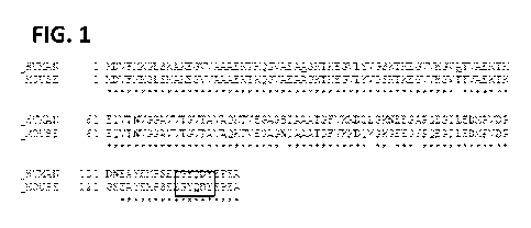
[018] Figure 1 shows the a-synuclein amino acid sequence (human and mouse),
and shows
the hexapeptide YQDYEP corresponding to amino acid positions 133-138 in the C-
terminal.
DETAILED DESCRIPTION
I. Abbreviations
4
CA 03036592 2019-03-11
WO 2018/111670
PCT/US2017/065035
[019] CAR: chimeric antigen receptor
CDC: complement-dependent cytotoxicity
cDNA: complementary DNA
CDR: complementarity determining region
CTL: cytotoxic T lymphocyte
ELISA: enzyme-linked immunosorbent assay
EM: effector molecule
FACS: fluorescence activated cell sorting
GPI: glycosylphosphatidylinositol
hFc: human Fc
HRP: horseradish peroxidase
Ig: immunoglobulin
i.v.: intravenous
KD dissociation constant
LDH: lactate dehydrogenase
mAb: monoclonal antibody
MAC: membrane attack complex
NHS: normal human serum
PBMC: peripheral blood mononuclear cells
PCR: polymerase chain reaction
PE: Pseudomonas exotoxin
PE: phycoerythrin
Pfu: plaque forming units
RIPA: radioimmunoprecipitation assay
VH: variable heavy
VL: variable light
II. Terms and Methods
CA 03036592 2019-03-11
WO 2018/111670 PCT/US2017/065035
[020] Unless otherwise explained, all technical and scientific terms used
herein have the same
meaning as commonly understood by one of ordinary skill in the art to which
this disclosure
belongs. The singular terms "a," "an," and "the" include plural referents
unless context clearly
indicates otherwise. "Comprising A or B" means including A, or B, or A and B.
It is further to be
understood that all base sizes or amino acid sizes, and all molecular weight
or molecular mass
values, given for nucleic acids or polypeptides are approximate, and are
provided for
description. Although methods and materials similar or equivalent to those
described herein
can be used in the practice or testing of the present disclosure, suitable
methods and materials
are described below. All publications, patent applications, patents, and other
references
mentioned herein are incorporated by reference in their entirety. In case of
conflict, the
present specification, including explanations of terms, will control. In
addition, the materials,
methods, and examples are illustrative only and not intended to be limiting.
[021] Definitions of common terms in molecular biology may be found in
Benjamin Lewin,
Genes V, published by Oxford University Press, 1994 (ISBN 0-19-854287-9);
Kendrew et al.
(eds.), The Encyclopedia of Molecular Biology, published by Blackwell Science
Ltd., 1994 (ISBN
0-632-02182-9); and Robert A. Meyers (ed.), Molecular Biology and
Biotechnology: a
Comprehensive Desk Reference, published by VCH Publishers, Inc., 1995 (ISBN 1-
56081-569-8).
[022] In order to facilitate review of the various embodiments of the
disclosure, the following
explanations of specific terms are provided:
[023] Antibody: A polypeptide ligand comprising at least a light chain or
heavy chain
immunoglobulin variable region which recognizes and binds (such as
specifically recognizes and
specifically binds) an epitope of an antigen, such as a-synuclein, or a
fragment thereof.
lmmunoglobulin molecules are composed of a heavy and a light chain, each of
which has a
variable region, termed the variable heavy (VH) region and the variable light
(VL) region.
Together, the VH region and the VL region are responsible for binding the
antigen recognized by
the antibody.
[024] Antibodies include intact immunoglobulins and the variants and portions
of antibodies
well known in the art, such as single-domain antibodies (e.g. VH domain
antibodies), Fab
fragments, Fab' fragments, F(ab)T2 fragments, single chain Fv proteins
("scFv"), and disulfide
6
CA 03036592 2019-03-11
WO 2018/111670 PCT/US2017/065035
stabilized Fv proteins ("dsFv"). A scFy protein is a fusion protein in which a
light chain variable
region of an immunoglobulin and a heavy chain variable region of an
immunoglobulin are
bound by a linker, while in dsFys, the chains have been mutated to introduce a
disulfide bond
to stabilize the association of the chains. The term "antibody" also includes
genetically
engineered forms such as chimeric antibodies (for example, humanized murine
antibodies) and
heteroconjugate antibodies (such as bispecific antibodies). See also, Pierce
Catalog and
Handbook, 1994-1995 (Pierce Chemical Co., Rockford, Ill.); Kuby, J.,
Immunology, 3<sup>rd</sup> Ed.,
W. H. Freeman & Co., New York, 1997.
[025] Typically, a naturally occurring immunoglobulin has heavy (H) chains and
light (L) chains
interconnected by disulfide bonds. There are two types of light chain, lambda
(X) and kappa (k).
There are five main heavy chain classes (or isotypes) which determine the
functional activity of
an antibody molecule: IgM, IgD, IgG, IgA and IgE.
[026] Each heavy and light chain contains a constant region and a variable
region, (the regions
are also known as "domains"). In combination, the heavy and the light chain
variable regions
specifically bind the antigen. Light and heavy chain variable regions contain
a "framework"
region interrupted by three hypervariable regions, also called
"complementarity-determining
regions" or "CDRs." The extent of the framework region and CDRs has been
defined according
to Kabat et al. (see, Kabat et al., Sequences of Proteins of Immunological
Interest, U.S.
Department of Health and Human Services, 1991) and the ImMunoGeneTics database
(IMGT)
(see, Lefranc, Nucleic Acids Res 29:207-9, 2001). The IMGT and Kabat databases
are available
online. The sequences of the framework regions of different light or heavy
chains are relatively
conserved within a species, such as humans. The framework region of an
antibody, that is the
combined framework regions of the constituent light and heavy chains, serves
to position and
align the CDRs in three-dimensional space.
[027] The CDRs are primarily responsible for binding to an epitope of an
antigen. The CDRs of
each chain are typically referred to as CDR1, CDR2, and CDR3, numbered
sequentially starting
from the N-terminus, and are often identified by the chain in which the
particular CDR is
located. Thus, a VH CDR3 (or H-CDR3) is located in the variable domain of the
heavy chain of the
antibody in which it is found, whereas a VL CDR1 (or L-CDR1) is the CDR1 from
the variable
7
CA 03036592 2019-03-11
WO 2018/111670 PCT/US2017/065035
domain of the light chain of the antibody in which it is found. An antibody
that binds a-
synuclein, for example, will have a specific VH region and the VL region
sequence, and thus
specific CDR sequences. Antibodies with different specificities (i.e.
different combining sites for
different antigens) have different CDRs. Although it is the CDRs that vary
from antibody to
antibody, only a limited number of amino acid positions within the CDRs are
directly involved in
antigen binding. These positions within the CDRs are called specificity
determining residues
(SDRs).
[028] References to "VH" or "VH" refer to the variable region of an
immunoglobulin heavy
chain, including that of an Fv, scFv, dsFy or Fab. References to "VL" or "VL"
refer to the variable
region of an immunoglobulin light chain, including that of an Fv, scFv, dsFy
or Fab.
[029] A "monoclonal antibody" is an antibody produced by a single clone of B-
lymphocytes or
by a cell into which the light and/or heavy chain genes of a single antibody
have been
transfected. Monoclonal antibodies are produced by methods known to those of
skill in the art,
for instance by making hybrid antibody-forming cells from a fusion of myeloma
cells with
immune spleen cells. Monoclonal antibodies include humanized monoclonal
antibodies.
[030] A "chimeric antibody" contains structural elements from two or more
different antibody
molecules, often from different animal species. For example, a chimeric
antibody can have
framework residues from one species, such as human, and CDRs (which generally
confer
antigen binding) from another species, such as a murine antibody that
specifically binds a-
synuclein.
[031] A "human" antibody (also called a "fully human" antibody) is an antibody
that includes
human framework regions and all of the CDRs from a human immunoglobulin. In
one example,
the framework and the CDRs are from the same originating human heavy and/or
light chain
amino acid sequence. However, frameworks from one human antibody can be
engineered to
include CDRs from a different human antibody. A "humanized" immunoglobulin is
an
immunoglobulin including a human framework region and one or more CDRs from a
non-
human (for example a mouse, rabbit, rat, or synthetic) immunoglobulin. The non-
human
immunoglobulin providing the CDRs is termed a "donor," and the human
immunoglobulin
providing the framework is termed an "acceptor." In one embodiment, all the
CDRs are from
8
CA 03036592 2019-03-11
WO 2018/111670 PCT/US2017/065035
the donor immunoglobulin in a humanized immunoglobulin. Constant regions need
not be
present, but if they are, they must be substantially identical to human
immunoglobulin
constant regions, i.e., at least about 85-90%, such as about 95% or more
identical. Hence, all
parts of a humanized immunoglobulin, except possibly the CDRs, are
substantially identical to
corresponding parts of natural human immunoglobulin sequences. A "humanized
antibody" is
an antibody comprising a humanized light chain and a humanized heavy chain
immunoglobulin.
A humanized antibody binds to the same antigen as the donor antibody that
provides the CDRs.
The acceptor framework of a humanized immunoglobulin or antibody may have a
limited
number of substitutions by amino acids taken from the donor framework.
Humanized or other
monoclonal antibodies can have additional conservative amino acid
substitutions which have
substantially no effect on antigen binding or other immunoglobulin functions.
Humanized
immunoglobulins can be constructed by means of genetic engineering (see, e.g.,
U.S. Pat. No.
5,585,089).
[032] A "single-domain antibody" (sdAb) or "nanobody" is an antibody fragment
consisting of
a single monomeric variable antibody domain. Like a whole antibody, it can
bind selectively to a
specific antigen. With a molecular weight of only 12-15 kDa, nanobodies are
much smaller than
common antibodies (150-160 kDa) which are composed of two heavy protein chains
and two
light chains, and even smaller than Fab fragments (-50 kDa, one light chain
and half a heavy
chain) and single-chain variable fragments (-25 kDa, two variable domains, one
from a light and
one from a heavy chain). The smaller size and single domain make nanobodies
easier to
transform into bacterial cells for bulk production, making them ideal for
research purposes. A
nanobody can be obtained by immunization of dromedaries, camels, llamas,
alpacas or sharks
with the desired antigen and subsequent isolation of the mRNA coding for heavy-
chain
antibodies. By reverse transcription and polymerase chain reaction, a gene
library of
nanobodies containing several million clones is produced. Screening techniques
like phage
display and ribosome display help to identify the clones binding the antigen.
[033] A different method uses gene libraries from animals that have not been
immunized
beforehand. Such naïve libraries usually contain only antibodies with low
affinity to the desired
9
CA 03036592 2019-03-11
WO 2018/111670 PCT/US2017/065035
antigen, making it necessary to apply affinity maturation by random
mutagenesis as an
additional step.
[034] When the most potent clones have been identified, their DNA sequence is
optimized,
for example to improve their stability towards enzymes. Another goal is
humanization to
prevent immunological reactions of the human organism against the antibody.
Humanization is
unproblematic because of the homology between camelid VHH and human VH
fragments. The
final step is the translation of the optimized nanobody in E. coli, S.
cereyisiae or other suitable
organisms.
[035] Alternatively, nanobodies can be made from common murine or human IgG
with four
chains. The process is similar, comprising gene libraries from immunized or
naïve donors and
display techniques for identification of the most specific antigens.
Monomerization is usually
accomplished by replacing lipophilic by hydrophilic amino acids. The
nanobodies can likewise
be produced in E. coli, S. cereyisiae or other organisms.
[036] An "intrabody" is an antibody that works within the cell to bind to an
intracellular
protein. Due to the lack of a reliable mechanism for bringing antibodies into
a living cell from
the extracellular environment, this typically requires the expression of the
antibody within the
target cell, which can be accomplished by gene therapy. As a result,
intrabodies are defined as
antibodies that have been modified for intracellular localization. For
example, the antibody
may remain in the cytoplasm, or it may have a nuclear localization signal, or
it may undergo
cotranslational translocation across the membrane into the lumen of the
endoplasmic
reticulum, provided that it is retained in that compartment through a KDEL
sequence.
[037] Because antibodies ordinarily are designed to be secreted from the cell,
intrabodies
often require special alterations, including the use of single-chain
antibodies (scFvs),
modification of immunoglobulin VL domains for hyperstability, selection of
antibodies resistant
to the more reducing intracellular environment, or expression as a fusion
protein with maltose
binding protein or other stable intracellular proteins. Such optimizations may
improve the
stability and structure of intrabodies.
[038] Binding affinity: Affinity of an antibody for an antigen. In one
embodiment, affinity is
calculated by a modification of the Scatchard method described by Frankel et
al. (Mol.
CA 03036592 2019-03-11
WO 2018/111670 PCT/US2017/065035
Immunol., 16:101-106, 1979). In another embodiment, binding affinity is
measured by an
antigen/antibody dissociation rate. In another embodiment, a high binding
affinity is measured
by a competition radioimmunoassay. In another embodiment, binding affinity is
measured by
ELISA. An antibody that "specifically binds" an antigen (such as a-synuclein)
is an antibody that
binds the antigen with high affinity and does not significantly bind other
unrelated antigens.
[039] Conservative variant: "Conservative" amino acid substitutions are those
substitutions
that do not substantially affect or decrease the affinity of a protein, such
as an antibody to a-
synuclein. For example, a monoclonal antibody that specifically binds a-
synuclein can include at
most about 1, at most about 2, at most about 5, at most about 10, or at most
about 15
conservative substitutions and specifically bind a a-synuclein polypeptide.
The term
"conservative variant" also includes the use of a substituted amino acid in
place of an
unsubstituted parent amino acid, provided that antibody specifically binds a-
synuclein. Non-
conservative substitutions are those that reduce an activity or binding to a-
synuclein.
[040] Conservative amino acid substitution tables providing functionally
similar amino acids
are well known to one of ordinary skill in the art. The following six groups
are examples of
amino acids that are considered to be conservative substitutions for one
another:
1) Alanine (A), Serine (S), Threonine (T);
2) Aspartic acid (D), Glutamic acid (E);
3) Asparagine (N), Glutamine (Q);
4) Arginine (R), Lysine (K);
5) lsoleucine (I), Leucine (L), Methionine (M), Valine (V); and
6) Phenylalanine (F), Tyrosine (Y), Tryptophan (W).
[041] Complementarity determining region (CDR): Amino acid sequences which
together
define the binding affinity and specificity of the natural Fv region of a
native Ig binding site. The
light and heavy chains of an Ig each have three CDRs, designated L-CDR1, L-
CDR2, L-CDR3 and
H-CDR1, H-CDR2, H-CDR3, respectively.
[042] Degenerate variant: In the context of the present disclosure, a
"degenerate variant"
refers to a polynucleotide encoding a a-synuclein polypeptide or an antibody
that binds a-
synuclein that includes a sequence that is degenerate as a result of the
genetic code. There are
11
CA 03036592 2019-03-11
WO 2018/111670 PCT/US2017/065035
20 natural amino acids, most of which are specified by more than one codon.
Therefore, all
degenerate nucleotide sequences are included as long as the amino acid
sequence of the a-
synuclein polypeptide or antibody that binds a-synuclein encoded by the
nucleotide sequence
is unchanged.
[043] Dementia with Lewy Bodies: also known as Lewy body dementia (LBD),
diffuse Lewy
body disease, cortical Lewy body disease, and senile dementia of Lewy type. A
type of
progressive neurodegenerative dementia closely associated with Parkinson's
disease primarily
affecting older adults. Its primary feature is a more rapid cognitive decline
than with
Parkinson's, which may lead to hallucinations, as well as varied attention and
alertness when
compared to a person's baseline function.
[044] People with LBD display an inability to plan or a loss of analytical or
abstract thinking
and show markedly fluctuating cognition. Wakefulness varies from day to day,
and alertness
and short-term memory rise and fall. Persistent or recurring visual
hallucinations with vivid and
detailed imagery often are an early diagnostic symptom. The disorder is
characterized
anatomically by the presence of Lewy bodies, clumps of alpha-synuclein and
ubiquitin protein
in neurons, detectable in post mortem brain histology.
[045] Diagnostic: Identifying the presence or nature of a pathologic
condition, such as, but not
limited to, Parkinson's disease. Diagnostic methods differ in their
sensitivity and specificity. The
"sensitivity" of a diagnostic assay is the percentage of diseased individuals
who test positive
(percent of true positives). The "specificity" of a diagnostic assay is one
minus the false positive
rate, where the false positive rate is defined as the proportion of those
without the disease
who test positive. While a particular diagnostic method may not provide a
definitive diagnosis
of a condition, it suffices if the method provides a positive indication that
aids in diagnosis.
"Prognostic" is the probability of development (e.g., severity) of a
pathologic condition, such as
cancer or metastasis.
[046] Effector molecule: The portion of a chimeric molecule that is intended
to have a desired
effect on a cell to which the chimeric molecule is targeted. Effector molecule
is also known as
an effector moiety (EM), therapeutic agent, or diagnostic agent, or similar
terms.
12
CA 03036592 2019-03-11
WO 2018/111670 PCT/US2017/065035
[047] Therapeutic agents include such compounds as nucleic acids, proteins,
peptides, amino
acids or derivatives, glycoproteins, radioisotopes, lipids, carbohydrates, or
recombinant viruses.
Nucleic acid therapeutic and diagnostic moieties include antisense nucleic
acids, derivatized
oligonucleotides for covalent cross-linking with single or duplex DNA, and
triplex forming
oligonucleotides. Alternatively, the molecule linked to a targeting moiety,
such as an anti- a-
synuclein antibody, may be an encapsulation system, such as a liposome or
micelle that
contains a therapeutic composition such as a drug, a nucleic acid (such as an
antisense nucleic
acid), or another therapeutic moiety that can be shielded from direct exposure
to the
circulatory system. Means of preparing liposomes attached to antibodies are
well known to
those of skill in the art (see, e.g., U.S. Pat. No. 4,957,735; and Connor et
al., Pharm Ther 28:341-
365, 1985). Diagnostic agents or moieties include radioisotopes and other
detectable labels.
Detectable labels useful for such purposes are also well known in the art, and
include
radioactive isotopes such as 35S, 11C, 13N, 150, 18F, 19F, 99m-rc, 1311, 3H,
14C, 15N, 90y, 99TC, "In and
1251, fluorophores, chemiluminescent agents, and enzymes.
[048] Epitope: An antigenic determinant. These are particular chemical groups
or peptide
sequences on a molecule that are antigenic, i.e. that elicit a specific immune
response. An
antibody specifically binds a particular antigenic epitope on a polypeptide,
such as a-synuclein.
[049] Framework region: Amino acid sequences interposed between CDRs.
Framework
regions include variable light and variable heavy framework regions. The
framework regions
serve to hold the CDRs in an appropriate orientation for antigen binding.
[050] Host cells: Cells in which a vector can be propagated and its DNA
expressed. The cell
may be prokaryotic or eukaryotic. The term also includes any progeny of the
subject host cell. It
is understood that all progeny may not be identical to the parental cell since
there may be
mutations that occur during replication. However, such progeny are included
when the term
"host cell" is used.
[051] Hybridoma: A hybrid cell for the production of monoclonal antibodies. A
hybridoma is
produced by fusion of an antibody-producing cell (such as a B cell obtained
from an immunized
animal, for example a mouse, rat or rabbit) and a myeloma cell.
13
CA 03036592 2019-03-11
WO 2018/111670 PCT/US2017/065035
[052] Immune response: A response of a cell of the immune system, such as a B
cell, T cell, or
monocyte, to a stimulus. In one embodiment, the response is specific for a
particular antigen
(an "antigen-specific response"). In one embodiment, an immune response is a T
cell response,
such as a CD4+ response or a CD8+ response. In another embodiment, the
response is a B cell
response, and results in the production of specific antibodies.
[053] Immunoconjugate: A covalent linkage of an effector molecule to an
antibody or
functional fragment thereof. The effector molecule can be, e.g., a detectable
label. A "chimeric
molecule" is a targeting moiety, such as a ligand or an antibody, conjugated
(coupled) to an
effector molecule. The term "conjugated" or "linked" refers to making two
polypeptides into
one contiguous polypeptide molecule. In one embodiment, an antibody is joined
to an effector
molecule. In another embodiment, an antibody joined to an effector molecule is
further joined
to a lipid or other molecule to a protein or peptide to increase its half-life
in the body. The
linkage can be either by chemical or recombinant means. In one embodiment, the
linkage is
chemical, wherein a reaction between the antibody moiety and the effector
molecule has
produced a covalent bond formed between the two molecules to form one
molecule. A peptide
linker (short peptide sequence) can optionally be included between the
antibody and the
effector molecule. Because immunoconjugates were originally prepared from two
molecules
with separate functionalities, such as an antibody and an effector molecule,
they are also
sometimes referred to as "chimeric molecules." The term "chimeric molecule,"
as used herein,
therefore refers to a targeting moiety, such as a ligand or an antibody,
conjugated (coupled) to
an effector molecule.
[054] Isolated: An "isolated" biological component, such as a nucleic acid,
protein (including
antibodies) or organelle, has been substantially separated or purified away
from other
biological components in the environment (such as a cell) in which the
component naturally
occurs, i.e., other chromosomal and extra-chromosomal DNA and RNA, proteins
and organelles.
Nucleic acids and proteins that have been "isolated" include nucleic acids and
proteins purified
by standard purification methods. The term also embraces nucleic acids and
proteins prepared
by recombinant expression in a host cell as well as chemically synthesized
nucleic acids.
14
CA 03036592 2019-03-11
WO 2018/111670 PCT/US2017/065035
[055] Label: A detectable compound or composition that is conjugated directly
or indirectly to
another molecule, such as an antibody or a protein, to facilitate detection of
that molecule.
Specific, non-limiting examples of labels include fluorescent tags, enzymatic
linkages, and
radioactive isotopes. In one example, a "labeled antibody" refers to
incorporation of another
molecule in the antibody. For example, the label is a detectable marker, such
as the
incorporation of a radiolabeled amino acid or attachment to a polypeptide of
biotinyl moieties
that can be detected by marked avidin (for example, streptavidin containing a
fluorescent
marker or enzymatic activity that can be detected by optical or colorimetric
methods). Various
methods of labeling polypeptides and glycoproteins are known in the art and
may be used.
Examples of labels for polypeptides include, but are not limited to, the
following: radioisotopes
or radionucleotides (as 35S, 11C, 13N, 150, 18F, 19F, 99m-rc, 1311, 3H, 14C,
15N, 90y, 99TC, "In and 1251),
fluorescent labels (such as fluorescein isothiocyanate (FITC), rhodamine,
lanthanide phosphors),
enzymatic labels (such as horseradish peroxidase, beta-galactosidase,
luciferase, alkaline
phosphatase), chemiluminescent markers, biotinyl groups, predetermined
polypeptide
epitopes recognized by a secondary reporter (such as a leucine zipper pair
sequences, binding
sites for secondary antibodies, metal binding domains, epitope tags), or
magnetic agents, such
as gadolinium chelates. In some embodiments, labels are attached by spacer
arms of various
lengths to reduce potential steric hindrance.
[056] Linker: In some cases, a linker is a peptide within an antibody binding
fragment (such as
an Fv fragment) which serves to indirectly bond the variable heavy chain to
the variable light
chain. "Linker" can also refer to a peptide serving to link a targeting
moiety, such as an
antibody, to an effector molecule, such as a cytotoxin or a detectable label.
[057] The terms "conjugating," "joining," "bonding" or "linking" refer to
making two
polypeptides into one contiguous polypeptide molecule, or to covalently
attaching a
radionuclide or other molecule to a polypeptide, such as an scFv. In the
specific context, the
terms include reference to joining a ligand, such as an antibody moiety, to an
effector molecule.
The linkage can be either by chemical or recombinant means. "Chemical means"
refers to a
reaction between the antibody moiety and the effector molecule such that there
is a covalent
bond formed between the two molecules to form one molecule.
CA 03036592 2019-03-11
WO 2018/111670 PCT/US2017/065035
[058] Mammal: This term includes both human and non-human mammals. Similarly,
the term
"subject" includes both human and veterinary subjects.
[059] Multiple system atrophy (MSA): a degenerative neurological disorder that
depicts a
group of disorders characterized by the neuronal degeneration mainly in the
substantia nigra,
striatum, autonomic nervous system and cerebellum. Many patients have symptoms
and signs
of cerebellar ataxia and parkinsonian manifestations. More than half of the
patients with
striatonigral degeneration have orthostatic hypotension, which proves at
autopsy to be
associated with loss of intermediolateral horn cells (origin of the
presynaptic cholinergic
sympathetic neurons) and of pigmented nuclei of the brainstem.
[060] This combined parkinsonian and autonomic disorder is referred to as the
Shy¨Drager
syndrome. In addition to orthostatic hypotension, other features of autonomic
failure include
impotence, loss of sweating, dry mouth and urinary retention and incontinence.
Vocal cord
palsy is an important and sometimes initial clinical manifestation of the
disorder.
[061] Both MRI and CT scanning frequently show atrophy of the cerebellum and
pons in those
with cerebellar features. The putamen is hypodense on T2-weighted MRI and may
show an
increased deposition of iron in Parkinsonian form. In cerebellar form, a "hot
cross" sign has
been emphasized; it reflects atrophy of the pontocereballar fibers that
manifest in T2 signal
intensity in atrophic pons.
[062] MSA often presents with some of the same symptoms as Parkinson's
disease. However,
those with MSA generally show minimal if any response to the dopamine
medications used for
Parkinson's disease.
[063] Multiple system atrophy can be explained as cell loss and gliosis or a
proliferation of
astrocytes in damaged areas of the central nervous system. This damage forms a
scar which is
then termed a glial scar. The presence of these inclusions (also known as Papp-
Lantos bodies) in
the movement, balance, and autonomic-control centers of the brain are the
defining
histopathologic hallmark of MSA. Recent studies have shown that the major
filamentous
component of glial and neuronal cytoplasmic inclusions is a-synuclein.
Mutations in this
substance may play a role in the disease. Tau proteins have been found in some
GC1s.
16
CA 03036592 2019-03-11
WO 2018/111670 PCT/US2017/065035
[064] Operably linked: A first nucleic acid sequence is operably linked with a
second nucleic
acid sequence when the first nucleic acid sequence is placed in a functional
relationship with
the second nucleic acid sequence. For instance, a promoter, such as the CMV
promoter, is
operably linked to a coding sequence if the promoter affects the transcription
or expression of
the coding sequence. Generally, operably linked DNA sequences are contiguous
and, where
necessary to join two protein-coding regions, in the same reading frame.
[065] Parkinson's disease: is a long term disorder of the central nervous
system that mainly
affects the motor system. The symptoms generally come on slowly over time.
Early in the
disease, the most obvious are shaking, rigidity, slowness of movement, and
difficulty with
walking. Thinking and behavioral problems may also occur. Dementia becomes
common in the
advanced stages of the disease. Depression and anxiety are also common
occurring in more
than a third of people with PD. Other symptoms include sensory, sleep, and
emotional
problems. The main motor symptoms are collectively called "parkinsonism", or a
"parkinsonian
syndrome.TI
[066] The cause of Parkinson's disease is believed to involve both genetic and
environmental
factors. Those with a family member affected are more likely to get the
disease themselves.
There is also an increased risk in people exposed to certain pesticides and
among those who
have had prior head injuries. The motor symptoms of the disease result from
the death of cells
in the substantia nigra, a region of the midbrain. This results in not enough
dopamine in these
areas. The reason for this cell death is involves the build-up of proteins
into Lewy bodies in the
neurons. Diagnosis of typical cases is mainly based on symptoms, with tests
such as
neuroimaging being used to rule out other diseases.
[067] There is no cure for Parkinson's disease. Initial treatments is
typically with the
antiparkinson medication levodopa, with dopamine agonists being used once
levodopa
becomes less effective. As the disease progresses and neurons continue to be
lost, these
medications become less effective while at the same time they produce a
complication marked
by involuntary writhing movements.] Surgery to place the microelectrodes for
deep brain
stimulation has been used to reduce motor symptoms in severe cases where drugs
are
ineffective.
17
CA 03036592 2019-03-11
WO 2018/111670 PCT/US2017/065035
[068] Pharmaceutical agent: A chemical compound or composition capable of
inducing a
desired therapeutic or prophylactic effect when properly administered to a
subject or a cell.
[069] Pharmaceutically acceptable carriers: The pharmaceutically acceptable
carriers of use
are conventional. Remington's Pharmaceutical Sciences, by E. W. Martin, Mack
Publishing Co.,
Easton, Pa., 15th Edition, 1975, describes compositions and formulations
suitable for
pharmaceutical delivery of the antibodies disclosed herein.
[070] In general, the nature of the carrier will depend on the particular mode
of
administration being employed. For instance, parenteral formulations usually
comprise
injectable fluids that include pharmaceutically and physiologically acceptable
fluids such as
water, physiological saline, balanced salt solutions, aqueous dextrose,
glycerol or the like as a
vehicle. For solid compositions (such as powder, pill, tablet, or capsule
forms), conventional
non-toxic solid carriers can include, for example, pharmaceutical grades of
mannitol, lactose,
starch, or magnesium stearate. In addition to biologically neutral carriers,
pharmaceutical
compositions to be administered can contain minor amounts of non-toxic
auxiliary substances,
such as wetting or emulsifying agents, preservatives, and pH buffering agents
and the like, for
example sodium acetate or sorbitan monolaurate.
[071] Preventing, treating or ameliorating a disease: "Preventing" a disease
refers to inhibiting
the full development of a disease. "Treating" refers to a therapeutic
intervention that
ameliorates a sign or symptom of a disease or pathological condition after it
has begun to
develop. "Ameliorating" refers to the reduction in the number or severity of
signs or symptoms
of a disease.
[072] Purified: The term purified does not require absolute purity; rather, it
is intended as a
relative term. Thus, for example, a purified peptide preparation is one in
which the peptide or
protein is more enriched than the peptide or protein is in its natural
environment within a cell.
In one embodiment, a preparation is purified such that the protein or peptide
represents at
least 50% of the total peptide or protein content of the preparation.
Substantial purification
denotes purification from other proteins or cellular components. A
substantially purified
protein is at least 60%, 70%, 80%, 90%, 95% or 98% pure. Thus, in one
specific, non-limiting
example, a substantially purified protein is 90% free of other proteins or
cellular components.
18
CA 03036592 2019-03-11
WO 2018/111670 PCT/US2017/065035
[073] Recombinant: A recombinant nucleic acid is one that has a sequence that
is not naturally
occurring or has a sequence that is made by an artificial combination of two
otherwise
separated segments of sequence. This artificial combination is often
accomplished by chemical
synthesis or by the artificial manipulation of isolated segments of nucleic
acids, for example, by
genetic engineering techniques.
[074] Sample (or biological sample): A biological specimen containing genomic
DNA, RNA
(including mRNA), protein, or combinations thereof, obtained from a subject.
Examples include,
but are not limited to, peripheral blood, tissue, cells, urine, saliva, tissue
biopsy, fine needle
aspirate, surgical specimen, and autopsy material.
[075] Sequence identity: The similarity between amino acid or nucleic acid
sequences is
expressed in terms of the similarity between the sequences, otherwise referred
to as sequence
identity. Sequence identity is frequently measured in terms of percentage
identity (or similarity
or homology); the higher the percentage, the more similar the two sequences
are. Homologs or
variants of a polypeptide or nucleic acid molecule will possess a relatively
high degree of
sequence identity when aligned using standard methods.
[076] Methods of alignment of sequences for comparison are well known in the
art. Various
programs and alignment algorithms are described in: Smith and Waterman (1981)
Adv. Appl.
Math. 2:482; Needleman and Wunsch (1970)J. Mol. Biol. 48:443; Pearson and
Lipman (1988)
Proc. Natl. Acad. Sci. U.S.A. 85:2444; Higgins and Sharp (1988) Gene 73:237;
Higgins and Sharp
(1989) CAB/OS 5:151; Corpet et al. (1988) Nucleic Acids Research 16:10881; and
Pearson and
Lipman (1988) Proc. Natl. Acad. Sci. U.S.A. 85:2444. Altschul et al. (1994)
Nature Genet. 6:119,
presents a detailed consideration of sequence alignment methods and homology
calculations.
[077] The NCBI Basic Local Alignment Search Tool (BLAST) (Altschul et al.
(1990)J. Mol. Biol.
215:403) is available from several sources, including the National Center for
Biotechnology
Information (NCBI, Bethesda, Md.) and on the internet, for use in connection
with the sequence
analysis programs blastp, blastn, blastx, tblastn and tblastx. A description
of how to determine
sequence identity using this program is available on the NCBI website on the
internet.
[078] Homologs and variants of a VL or a VH of an antibody that specifically
binds a-synuclein
or a fragment thereof are typically characterized by possession of at least
about 75%, for
19
CA 03036592 2019-03-11
WO 2018/111670 PCT/US2017/065035
example at least about 80%, 90%, 95%, 96%, 97%, 98% or 99% sequence identity
counted over
the full length alignment with the amino acid sequence of the antibody using
the NCBI Blast 2.0,
gapped blastp set to default parameters. For comparisons of amino acid
sequences of greater
than about 30 amino acids, the Blast 2 sequences function is employed using
the default
BLOSUM62 matrix set to default parameters, (gap existence cost of 11, and a
per residue gap
cost of 1). When aligning short peptides (fewer than around 30 amino acids),
the alignment
should be performed using the Blast 2 sequences function, employing the PAM30
matrix set to
default parameters (open gap 9, extension gap 1 penalties). Proteins with even
greater
similarity to the reference sequences will show increasing percentage
identities when assessed
by this method, such as at least 80%, at least 85%, at least 90%, at least
95%, at least 98%, or at
least 99% sequence identity. When less than the entire sequence is being
compared for
sequence identity, homologs and variants will typically possess at least 80%
sequence identity
over short windows of 10-20 amino acids, and may possess sequence identities
of at least 85%
or at least 90% or 95% depending on their similarity to the reference
sequence. Methods for
determining sequence identity over such short windows are available at the
NCBI website on
the internet. One of skill in the art will appreciate that these sequence
identity ranges are
provided for guidance only; it is entirely possible that strongly significant
homologs could be
obtained that fall outside of the ranges provided.
[079] Subject: Living multi-cellular vertebrate organisms, a category that
includes both human
and veterinary subjects, including human and non-human mammals.
[080] Synthetic: Produced by artificial means in a laboratory, for example a
monoclonal
antibody produced by hybridoma technology or expressed from a cDNA construct.
[081] Synucleinopathy: A neurodegenerative disease characterized by the
abnormal
accumulation of aggregates of a-synuclein proteins in neurons, nerve fibers,
or glial cells. There
are three main types of synucleinopathy: Parkinson's disease, dementia with
Lewy bodies, and
multiple system atrophy. Other rare disorders, such as various neuroaxonal
dystrophies, also
have a-synuclein pathologies.
[082] Therapeutically effective amount: A quantity of a specific substance
sufficient to achieve
a desired effect in a subject being treated. For instance, this can be the
amount necessary to
CA 03036592 2019-03-11
WO 2018/111670 PCT/US2017/065035
inhibit or suppress growth of a tumor. In one embodiment, a therapeutically
effective amount
is the amount necessary to eliminate, reduce the size, or prevent metastasis
of a tumor. When
administered to a subject, a dosage will generally be used that will achieve
target tissue
concentrations (for example, in tumors) that has been shown to achieve a
desired in vitro
effect.
[083] Vector: A nucleic acid molecule as introduced into a host cell, thereby
producing a
transformed host cell. A vector may include nucleic acid sequences that permit
it to replicate in
a host cell, such as an origin of replication. A vector may also include one
or more selectable
marker genes and other genetic elements known in the art.
III. a-Synuclein-Specific Monoclonal Antibodies
[084] Disclosed herein is the MJFR-14-6-4-2 antibody, a rabbit anti-a-
synuclein filment
antibody. This antibody is specific to a six amino acid C-terminal consensus
motif on the a-
synuclein amino acid sequence.
[085] Complementary determining region (CDR) sequencing for the antibody
identifies the
specific amino acid residues of the antibody within the variable domain that
directly/physically
interact with the antigen. An antibody variable region has a heavy and light
chain (designated
V(H)/V(L)) containing CDRs and interface framework (FRM) amino acid residues
which confer
the strength and antigen binding affinity. An IgG serotype antibody has 2
variable regions, each
with 3 potential CDRs for a potential total of 6 CDRs that collectively confer
the specificity of
the antibody's recognition of its antigen. Out of this, the following
nomenclature is defined:
[086] CDR1, CDR2, CDR3 = complementarity determining region 1, 2, 3 etc. These
are not
necessarily sequentially designated in a linear sequence representation of the
antibody protein
(more on this below)
[087]
[088] FRM1, 2, 3, = framework 1, 2, 3 regions (FRM1 associates with CDR1, FRM2
with CDR2,
etc).
[089]
21
CA 03036592 2019-03-11
WO 2018/111670 PCT/US2017/065035
[090] As antibodies mature to iteratively recognize their antigens with
increasing affinity,
CDRs are highly variable and their changes are what increase the specific,
physical interaction
with the antigen (the typified lock and key mechanism). FRMs are also located
in the variable
region but they are less malleable compared to CDRs. The FRMs don't change
iteratively per se,
but they impact the antibody:antigen interface by hinging/structurally
shifting (determined by
individual amino acid biochemical characteristics) when they encounter antigen
to allow CDRs
to maximally come into spatial proximity and thus physical contact with
specific targets on the
antigen.
[091]
[092] Importantly, the CDR/FRM "pair" may not be proximal in linear amino acid
sequence (so
they don't align in the linear sequence data), but they are spatially proximal
when the antibody
protein is folded into its tertiary structure. This also why a given CDR/FRM
pair don't necessarily
"match" in terms of number of amino acid residues either. CDRs can vary from
each other in
their number of amino acids, as can FRMs, and the number of residues in a
CDR/FRM pair often
don't have the same number of amino acid residues.
[093]
[094] ANTIBODY SEQUENCES
[095] Amino acid sequence of heavy chain (SEQ ID NO:1)
METGLRWLLLVAVLKGVQCQEQLVESGGDLVKPGASLTLTCTASGFSFSSNYWMCWFRQAPGKG
PEWIACIYAGNSGSTYYATWAKGRFT ISKTSSTTVTLQMTSLTAADTATYFCWRRGAYGYYGDL
NLWGPGTLVTVSS
[096] DNA sequence encoding heavy chain (SEQ ID NO:2)
ATGGAGACTGGGCTGCGCTGGCTICTCCIGGICGCTGTGCTCAAAGGIGTCCAGTGICAGGAGC
AGCTGGTGGAGTCCGGGGGAGACCTGGTCAAGCCTGGGGCGTCCCTGACACTCACCTGCACAGC
CTCTGGATTCTCCTTCAGTAGCAACTACTGGATGTGCTGGTTCCGCCAGGCTCCAGGGAAGGGG
CCGGAGTGGATCGCATGCATTTATGCTGGTAATAGTGGTAGCACTTACTACGCGACCTGGGCGA
AAGGCCGATTCACCATCTCCAAAACCTCGTCGACCACGGTGACTCTGCAAATGACCAGICTGAC
AGCCGCGGACACGGCCACCTATTTCTGTTGGAGAAGGGGTGCTTATGGATATTATGGTGATCTT
AATTIGTGGGGCCCAGGCACCCIGGICACCGICTCCTCAGGGCAACCTAAGGCTCCATCAGICT
TCCCACTGGCCCCCTGCTGCGGGGACACACCCAGCTCCACGGTGACCCTGGGCTGCCTGGTCAA
22
CA 03036592 2019-03-11
WO 2018/111670 PCT/US2017/065035
AGGGTACCTCCCGGAGCCAGTGACCGTGACCTGGAACTCGGGCACCCTCACCAATGGGGTACGC
ACCT T CCCGT CCGT CCGGCAGT CC T CAGGCC T C TAC T CGC T GAGCAGCGT GGT GAGCGT
GACC T
CAAGCAGCCAGCCCGTCACCTGCAACGTGGCCCACCCAGCCACCAACACCAAAGTGGACAAGAC
CGT T GCGCCC T CGACAT GCAGCAAGCCCACGT GCCCACCCCC T GAAC T CC T GGGGGGACCGT C T
GTC T T CAT C T T CCCCCCAAAACCCAAGGACACCC T CAT GAT C T CACGCACCCCCGAGGT CACAT
GCGTGGIGGIGGACGTGAGCCAGGATGACCCCGAGGIGCAGT TCACATGGTACATAAACAACGA
GCAGNTGCGCACCGCCCGGGCCGCCGCTACGGGNGCAGCAGT TCAACAGCACGATCCGCGNNNG
NCAGCNCCCTCCCCATCGCGCACNGNACTGGCTGAGGGCAAGNAGTICAAGTGCAAAGTCCANA
NNAGGCACTCCCGGCCCCATCNANAAANNNICINCAAANNNNANGGNNANNCCNNNNCNNNNCT
ANNNNGNNNT C C GGNN GNNCNNANNNN CAN GNN GNNANCNNNNNNCNNNNNNAT NAN GNNNNNN
NNCNNNNAANNNNNNNNNGNNNNNNN
[097] Amino acid sequence of heavy chain FRM1 (SEQ ID NO:3):
QEQLVESGGDLVKPGASLTLTCTASGFS FS
[098] Amino acid sequence of heavy chain CDR1 (SEQ ID NO:4):
SNYWMC
[099] Amino acid sequence of heavy chain FRM2 (SEQ ID NO:5):
W FRQAPGKGPEW IA
[100] Amino acid sequence of heavy chain CDR2 (SEQ ID NO:6):
CI YAGNS GS TYYATWAKG
[101] Amino acid sequence of heavy chain FRM3 (SEQ ID NO:7):
RFT I SKTSS T TVTLQMTSLTAADTATYFCWR
[102] Amino acid sequence of heavy chain CDR3 (SEQ ID NO:8):
RGAYGYYGDLNL
[103] Amino acid sequence of heavy chain FRM4 (SEQ ID NO:9)
WGPGTLVTVSS
[104] Amino acid sequence of light chain (SEQ ID NO:10):
MDTRAPTQLLGLLLLWLPGAT FAQVLTQTASSVSAAVGGTVT I SCQSSQSVYKNNYLAWYQQKP
GQPPNLL I YDAS T LAS GVS SRFRGS GS GT QFT LT IS GVQCDDAATYYCQGGFPCRTADCNVFGG
GTE VVVK
[105] DNA sequence encoding light chain (SEQ ID NO: 11)
23
CA 03036592 2019-03-11
WO 2018/111670 PCT/US2017/065035
ATGGACACGAGGGCCCCCACTCAGCTGCTGGGGCTCCTGCTGCTCTGGCTCCCAGGTGCCACAT
T T GCCCAAGT GCT GACCCAGAC T GCAT CGT CCGT GTCT GCAGCT GT GGGAGGCACAGT CACCAT
CAGT TGC CAGTCCAGTCAGAGTGT T TATAAGAACAAC TACT TAGCCTGG TAT CAGCAGAAAC CA
GGGCAGCCTCCCAACCTCCTGATCTATGATGCATCCACTCTGGCATCTGGGGTCTCATCGCGGT
T CAGAGGCAGT GGAT CT GGGACACAGT T CAC TCT CACCAT CAGCGGCGT GCAGT GT GACGAT GC
TGCCACTTACTACTGTCAAGGCGGATTTCCTTGTCGTACTGCTGATTGTAATGTTTTCGGCGGA
GGGACCGAGGT GGT GGT CAAAGGT GAT CCAGT T GCACC TAC T GT CCT CATCT T CCCACCAGCTG
CTGATCAGGIGGCAACTGGAACAGICACCATCGTGIGTGIGGCGAATAAATACTITCCCGATGT
CACCGTCACCIGGGAGGIGGATGGCAC CACCCAAACAACTGGCATCGAGAACAG TAAAACACCG
CAGAATTCTGCAGATTGTACCTACAACCTCAGCAGCACTCTGACACTGACCAGCACACAGTACA
ACAGCCACAAAGAG TACACCTGCAAGGTGACCCAGGGCAC GACCTCAGTCGTCCAGAGCT TCAA
TAGGGGTGACTGTTAGAGCGAGAGCGGCCGCTCGAGGCCGGCAAGGCCGGATCCCCCGACCTCG
ACCTCTGGCTAATAAAGGAAAT T TAT T T TCAT TGCAATAGTGIGT TGGAAT TITT TGIGICTCT
CAC T CGGAANGGACATAT GGGANGGCAAAT CAT T T GGT CGAGAT CCC T CGGANAT C T C TAGC
TA
GAGGATCGATCCCCGCCCCGGANGAACTAANNNTGACTACGACATCTCTGCCCCTNCNTCNCGG
GGCANNGCATGTAATCCCT
[106] Amino acid sequence of light chain FRM1 (SEQ ID NO:12)
AQVLTQTASSVSAAVGGTVT I SC
[107] Amino acid sequence of light chain CDR1 (SEQ ID NO:13):
QS S QSVYKNNYLA
[108] Amino acid sequence of light chain FRM2 (SEQ ID NO:14):
WYQQKPGQPPNLL I Y
[109] Amino acid sequence of light chain CDR2 (SEQ ID NO:15):
DAS T LAS
[110] Amino acid sequence of light chain FRM3 (SEQ ID NO:16):
GVS SRFRGS GS GTQFTL T I SGVQCDDAATYYC
[111] Amino acid sequence of light chain CDR3 (SEQ ID NO:17):
QGGFPCRTADCNV
[112] Amino acid sequence of light chain FRM4 (SEQ ID NO:18):
FGGGTEVVVK
24
CA 03036592 2019-03-11
WO 2018/111670 PCT/US2017/065035
[113] In some embodiments, the monoclonal antibody that binds, such as
specifically binds, a-
synuclein is a single domain antibody.
[114] In some embodiments, the monoclonal antibody that binds, such as
specifically binds, a-
synuclein is a Fab fragment, a Fab' fragment, a F(ab)T2 fragment, a single
chain variable
fragment (scFv), or a disulfide stabilized variable fragment (dsFv). In other
embodiments, the
antibody is an immunoglobulin molecule. In particular examples, the antibody
is an IgG.
[115] In some embodiments, the monoclonal antibody is chimeric or synthetic.
[116] In some embodiments, the disclosed antibodies bind a-synuclein (soluble
or cell-surface
a-synuclein) with a dissociation constant (Kd) in the high pm (-50-100) to low
nm range. In one
embodiment, the monoclonal antibodies bind a-synuclein with a binding affinity
of about 30
pM.
[117] The monoclonal antibodies disclosed herein can be labeled, such as with
a fluorescent,
enzymatic, or radioactive label.
[118] Also provided are fusion proteins comprising an antibody disclosed
herein and a
heterologous protein. In some examples, the heterologous protein is an Fc
protein. In one non-
limiting example, the Fc protein is a human Fc protein, such as human IgGyl
Fc.
[119] Further provided herein are compositions comprising a therapeutically
effective amount
of a disclosed antibody, immunoconjugate or fusion protein and a
pharmaceutically acceptable
carrier.
[120] Also provided herein are isolated nucleic acid molecules encoding the
disclosed
monoclonal antibodies, immunoconjugates and fusion proteins. In some examples,
the isolated
nucleic acid molecule is operably linked to a promoter.
[121] Also provided are expression vectors comprising the isolated nucleic
acid molecules
disclosed herein. Isolated host cells comprising the nucleic acid molecules or
vectors are also
provided herein.
V. Antibodies and Antibody Fragments
[122] The monoclonal antibodies disclosed herein can be of any isotype. The
monoclonal
antibody can be, for example, an IgM or an IgG antibody, such as IgGi or an
IgG2. The class of an
CA 03036592 2019-03-11
WO 2018/111670 PCT/US2017/065035
antibody that specifically binds a-synuclein can be switched with another (for
example, IgG can
be switched to IgM), according to well-known procedures. Class switching can
also be used to
convert one IgG subclass to another, such as from IgGi to IgG2.
[123] Antibody fragments are also encompassed by the present disclosure, such
as single-
domain antibodies (e.g., VH domain antibodies), Fab, F(a13')2, and Fv. These
antibody fragments
retain the ability to selectively bind with the antigen. These fragments
include:
(1) Fab, the fragment which contains a monovalent antigen-binding fragment of
an antibody
molecule, can be produced by digestion of whole antibody with the enzyme
papain to yield an
intact light chain and a portion of one heavy chain;
(2) Fab', the fragment of an antibody molecule can be obtained by treating
whole antibody with
pepsin, followed by reduction, to yield an intact light chain and a portion of
the heavy chain;
two Fab' fragments are obtained per antibody molecule;
(3) (FabT)2, the fragment of the antibody that can be obtained by treating
whole antibody with
the enzyme pepsin without subsequent reduction; F(a13')2 is a dimer of two
Fab' fragments held
together by two disulfide bonds;
(4) Fv, a genetically engineered fragment containing the variable region of
the light chain and
the variable region of the heavy chain expressed as two chains;
(5) Single chain antibody (such as scFv), a genetically engineered molecule
containing the
variable region of the light chain, the variable region of the heavy chain,
linked by a suitable
polypeptide linker as a genetically fused single chain molecule;
(6) A dimer of a single chain antibody (scFV2), defined as a dimer of a scFV
(also known as a
"miniantibody"); and
(7) VH single-domain antibody, an antibody fragment consisting of a heavy
chain variable
domain.
Methods of making these fragments are known in the art (see for example,
Harlow and Lane,
Antibodies: A Laboratory Manual, Cold Spring Harbor Laboratory, New York,
1988).
26
CA 03036592 2019-03-11
WO 2018/111670 PCT/US2017/065035
[124] In some cases, antibody fragments can be prepared by proteolytic
hydrolysis of the
antibody or by expression in a host cell (such as E. coli) of DNA encoding the
fragment. Antibody
fragments can be obtained by pepsin or papain digestion of whole antibodies by
conventional
methods. For example, antibody fragments can be produced by enzymatic cleavage
of
antibodies with pepsin to provide a 5S fragment denoted F(a13')2. This
fragment can be further
cleaved using a thiol reducing agent, and optionally a blocking group for the
sulfhydryl groups
resulting from cleavage of disulfide linkages, to produce 3.5S Fab' monovalent
fragments.
Alternatively, an enzymatic cleavage using pepsin produces two monovalent Fab'
fragments
and an Fc fragment directly (see U.S. Pat. No. 4,036,945 and U.S. Pat. No.
4,331,647).
[125] Other methods of cleaving antibodies, such as separation of heavy chains
to form
monovalent light-heavy chain fragments, further cleavage of fragments, or
other enzymatic,
chemical, or genetic techniques may also be used, so long as the fragments
bind to the antigen
that is recognized by the intact antibody.
[126] One of skill will realize that conservative variants of the antibodies
can be produced.
Such conservative variants employed in antibody fragments, such as dsFy
fragments or in scFy
fragments, will retain critical amino acid residues necessary for correct
folding and stabilizing
between the VH and the VL regions, and will retain the charge characteristics
of the residues in
order to preserve the low pl and low toxicity of the molecules Amino acid
substitutions (such as
at most one, at most two, at most three, at most four, or at most five amino
acid substitutions)
can be made in the VH and/or the VL regions to increase yield. Conservative
amino acid
substitution tables providing functionally similar amino acids are well known
to one of ordinary
skill in the art. The following six groups are examples of amino acids that
are considered to be
conservative substitutions for one another: 1) Alanine (A), Serine (S),
Threonine (T); 2) Aspartic
acid (D), Glutamic acid (E); 3) Asparagine (N), Glutamine (Q); 4) Arginine
(R), Lysine (K); 5)
lsoleucine (I), Leucine (L), Methionine (M), Valine (V); and 6) Phenylalanine
(F), Tyrosine (Y),
Tryptophan (W).
VI. Immunoconju gates and Fusion Proteins
[127] The disclosed monoclonal antibodies specific for a-synuclein can be
conjugated to a
therapeutic agent or effector molecule lmmunoconjugates include, but are not
limited to,
27
CA 03036592 2019-03-11
WO 2018/111670 PCT/US2017/065035
molecules in which there is a covalent linkage of a therapeutic agent to an
antibody. A
therapeutic agent is an agent with a particular biological activity directed
against a particular
target molecule or a cell bearing a target molecule. One of skill in the art
will appreciate that
therapeutic agents can include various drugs, encapsulating agents (such as
liposomes) which
themselves contain pharmacological compositions, radioactive agents such as
1251, 32p, 14C, 3H
and 35S and other labels, target moieties and ligands. The choice of a
particular therapeutic
agent depends on the particular target molecule or cell, and the desired
biological effect.
[128] With the therapeutic agents and antibodies described herein, one of
skill can readily
construct a variety of clones containing functionally equivalent nucleic
acids, such as nucleic
acids which differ in sequence but which encode the same effector moiety or
antibody
sequence. Thus, the present disclosure provides nucleic acids encoding
antibodies and
conjugates and fusion proteins thereof.
[129] Effector molecules can be linked to an antibody of interest using any
number of means
known to those of skill in the art. Both covalent and noncovalent attachment
means may be
used. The procedure for attaching an effector molecule to an antibody varies
according to the
chemical structure of the effector. Polypeptides typically contain a variety
of functional groups;
such as carboxylic acid (-COOH), free amine (-N H2) or sulfhydryl (-SH)
groups, which are
available for reaction with a suitable functional group on an antibody to
result in the binding of
the effector molecule. Alternatively, the antibody is derivatized to expose or
attach additional
reactive functional groups. The derivatization may involve attachment of any
of a number of
known linker molecules. The linker can be any molecule used to join the
antibody to the
effector molecule. The linker is capable of forming covalent bonds to both the
antibody and to
the effector molecule. Suitable linkers are well known to those of skill in
the art and include,
but are not limited to, straight or branched-chain carbon linkers,
heterocyclic carbon linkers, or
peptide linkers. Where the antibody and the effector molecule are
polypeptides, the linkers
may be joined to the constituent amino acids through their side groups (such
as through a
disulfide linkage to cysteine) or to the alpha carbon amino and carboxyl
groups of the terminal
amino acids.
28
CA 03036592 2019-03-11
WO 2018/111670 PCT/US2017/065035
[130] In some circumstances, it is desirable to free the effector molecule
from the antibody
when the immunoconjugate has reached its target site. Therefore, in these
circumstances,
immunoconjugates will comprise linkages that are cleavable in the vicinity of
the target site.
Cleavage of the linker to release the effector molecule from the antibody may
be prompted by
enzymatic activity or conditions to which the immunoconjugate is subjected
either inside the
target cell or in the vicinity of the target site.
[131] In view of the large number of methods that have been reported for
attaching a variety
of radiodiagnostic compounds, radiotherapeutic compounds, label (such as
enzymes or
fluorescent molecules) drugs, toxins, and other agents to antibodies one
skilled in the art will
be able to determine a suitable method for attaching a given agent to an
antibody or other
polypeptide.
[132] The antibodies or antibody fragments disclosed herein can be derivatized
or linked to
another molecule (such as another peptide or protein). In some cases, the
antibody or antibody
fragment (such as a VH domain) is fused to a heterologous protein, for example
an Fc protein.
[133] In general, the antibodies or portion thereof is derivatized such that
the binding to the
target antigen is not affected adversely by the derivatization or labeling.
For example, the
antibody can be functionally linked (by chemical coupling, genetic fusion,
noncovalent
association or otherwise) to one or more other molecular entities, such as
another antibody
(for example, a bispecific antibody or a diabody), a detection agent, a
pharmaceutical agent,
and/or a protein or peptide that can mediate association of the antibody or
antibody portion
with another molecule (such as a streptavidin core region or a polyhistidine
tag).
[134] One type of derivatized antibody is produced by cross-linking two or
more antibodies (of
the same type or of different types, such as to create bispecific antibodies).
Suitable
crosslinkers include those that are heterobifunctional, having two distinctly
reactive groups
separated by an appropriate spacer (such as m-maleimidobenzoyl-N-
hydroxysuccinimide ester)
or homobifunctional (such as disuccinimidyl suberate). Such linkers are
commercially available.
[135] An antibody that binds (for example specifically binds) a-synuclein or a
fragment thereof
can be labeled with a detectable moiety. Useful detection agents include
fluorescent
compounds, including fluorescein, fluorescein isothiocyanate, rhodamine, 5-
dimethylamine-1-
29
CA 03036592 2019-03-11
WO 2018/111670 PCT/US2017/065035
napthalenesulfonyl chloride, phycoerythrin, lanthanide phosphors and the like.
Bioluminescent
markers are also of use, such as luciferase, Green fluorescent protein (GFP),
Yellow fluorescent
protein (YFP). An antibody can also be labeled with enzymes that are useful
for detection, such
as horseradish peroxidase, B-galactosidase, luciferase, alkaline phosphatase,
glucose oxidase
and the like. When an antibody is labeled with a detectable enzyme, it can be
detected by
adding additional reagents that the enzyme uses to produce a reaction product
that can be
discerned. For example, when the agent horseradish peroxidase is present the
addition of
hydrogen peroxide and diaminobenzidine leads to a colored reaction product,
which is visually
detectable. An antibody may also be labeled with biotin, and detected through
indirect
measurement of avidin or streptavidin binding. It should be noted that the
avidin itself can be
labeled with an enzyme or a fluorescent label.
[136] An antibody may be labeled with a magnetic agent, such as gadolinium.
Antibodies can
also be labeled with lanthanides (such as europium and dysprosium), and
manganese.
Paramagnetic particles such as superparamagnetic iron oxide are also of use as
labels. An
antibody may also be labeled with a predetermined polypeptide epitopes
recognized by a
secondary reporter (such as leucine zipper pair sequences, binding sites for
secondary
antibodies, metal binding domains, epitope tags). In some embodiments, labels
are attached by
spacer arms of various lengths to reduce potential steric hindrance.
[137] An antibody can also be labeled with a radiolabeled amino acid. The
radiolabel may be
used for both diagnostic and therapeutic purposes. For instance, the
radiolabel may be used to
detect a-synuclein by x-ray, emission spectra, or other diagnostic techniques.
Examples of
labels for polypeptides include, but are not limited to, the following
radioisotopes or
radionucleotides: 3H, 14C, 15N, 35s, 90y, 99-rc, "In, 1251, 1311.
[138] An antibody can also be derivatized with a chemical group such as
polyethylene glycol
(PEG), a methyl or ethyl group, or a carbohydrate group. These groups may be
useful to
improve the biological characteristics of the antibody, such as to increase
serum half-life or to
increase tissue binding.
[139] The antibodies described herein can also be used to target any number of
different
diagnostic or therapeutic compounds to cells expressing a-synuclein on their
surface. Thus, an
CA 03036592 2019-03-11
WO 2018/111670 PCT/US2017/065035
antibody of the present disclosure can be attached directly or via a linker to
a drug that is to be
delivered directly to cells expressing cell-surface a-synuclein. This can be
done for therapeutic,
diagnostic or research purposes. Therapeutic agents include such compounds as
nucleic acids,
proteins, peptides, amino acids or derivatives, glycoproteins, radioisotopes,
lipids,
carbohydrates, or recombinant viruses. Nucleic acid therapeutic and diagnostic
moieties
include antisense nucleic acids, derivatized oligonucleotides for covalent
cross-linking with
single or duplex DNA, and triplex forming oligonucleotides.
[140] Alternatively, the molecule linked to an anti- a-synuclein antibody can
be an
encapsulation system, such as a liposome or micelle that contains a
therapeutic composition
such as a drug, a nucleic acid (for example, an antisense nucleic acid), or
another therapeutic
moiety that is preferably shielded from direct exposure to the circulatory
system. Means of
preparing liposomes attached to antibodies are well known to those of skill in
the art (see, for
example, U.S. Pat. No. 4,957,735; Connor et al., Pharm. Ther. 28:341-365,
1985).
[141] Antibodies described herein can also be covalently or non-covalently
linked to a
detectable label. Detectable labels suitable for such use include any
composition detectable by
spectroscopic, photochemical, biochemical, immunochemical, electrical, optical
or chemical
means. Useful labels include magnetic beads, fluorescent dyes (for example,
fluorescein
isothiocyanate, Texas red, rhodamine, green fluorescent protein, and the
like), radiolabels (for
example, 3H, 1251, 35s, 14C, or 32.-,r),
enzymes (such as horseradish peroxidase, alkaline
phosphatase and others commonly used in an ELISA), and colorimetric labels
such as colloidal
gold or colored glass or plastic (such as polystyrene, polypropylene, latex,
and the like) beads.
[142] Means of detecting such labels are well known to those of skill in the
art. Thus, for
example, radiolabels may be detected using photographic film or scintillation
counters,
fluorescent markers may be detected using a photodetector to detect emitted
illumination.
Enzymatic labels are typically detected by providing the enzyme with a
substrate and detecting
the reaction product produced by the action of the enzyme on the substrate,
and colorimetric
labels are detected by simply visualizing the colored label.
VII. Compositions and Methods of Use
31
CA 03036592 2019-03-11
WO 2018/111670 PCT/US2017/065035
[143] Compositions are provided that include one or more of the disclosed
antibodies that
bind (for example specifically bind) a-synuclein in a carrier. Compositions
comprising fusion
proteins, immunoconjugates or immunotoxins are also provided. The compositions
can be
prepared in unit dosage forms for administration to a subject. The amount and
timing of
administration are at the discretion of the treating clinician to achieve the
desired outcome.
The antibody can be formulated for systemic or local (such as intracerebral)
administration. In
one example, the antibody is formulated for parenteral administration, such as
intravenous
administration.
[144] The compositions for administration can include a solution of the
antibody dissolved in a
pharmaceutically acceptable carrier, such as an aqueous carrier. A variety of
aqueous carriers
can be used, for example, buffered saline and the like. These solutions are
sterile and generally
free of undesirable matter. These compositions may be sterilized by
conventional, well known
sterilization techniques. The compositions may contain pharmaceutically
acceptable auxiliary
substances as required to approximate physiological conditions such as pH
adjusting and
buffering agents, toxicity adjusting agents and the like, for example, sodium
acetate, sodium
chloride, potassium chloride, calcium chloride, sodium lactate and the like.
The concentration
of antibody in these formulations can vary widely, and will be selected
primarily based on fluid
volumes, viscosities, body weight and the like in accordance with the
particular mode of
administration selected and the subject's needs.
[145] A typical pharmaceutical composition for intravenous administration
includes about 0.1
to 10 mg of antibody per subject per day. Dosages from 0.1 up to about 100 mg
per subject per
day may be used, particularly if the agent is administered to a secluded site
and not into the
circulatory or lymph system, such as into a body cavity or into a lumen of an
organ. Actual
methods for preparing administrable compositions will be known or apparent to
those skilled in
the art and are described in more detail in such publications as Remington's
Pharmaceutical
Science, 19th ed., Mack Publishing Company, Easton, Pa. (1995).
[146] Antibodies may be provided in lyophilized form and rehydrated with
sterile water
before administration, although they are also provided in sterile solutions of
known
concentration. The antibody solution is then added to an infusion bag
containing 0.9% sodium
32
CA 03036592 2019-03-11
WO 2018/111670 PCT/US2017/065035
chloride, USP, and in some cases administered at a dosage of from 0.5 to 15
mg/kg of body
weight. Considerable experience is available in the art in the administration
of antibody drugs,
which have been marketed in the U.S. since the approval of RITUXAN in 1997.
Antibodies can
be administered by slow infusion, rather than in an intravenous push or bolus.
In one example,
a higher loading dose is administered, with subsequent, maintenance doses
being administered
at a lower level. For example, an initial loading dose of 4 mg/kg may be
infused over a period of
some 90 minutes, followed by weekly maintenance doses for 4-8 weeks of 2 mg/kg
infused
over a 30-minute period if the previous dose was well tolerated.
[147] Controlled release parenteral formulations can be made as implants, oily
injections, or
as particulate systems. For a broad overview of protein delivery systems see,
Banga, A. J.,
Therapeutic Peptides and Proteins: Formulation, Processing, and Delivery
Systems, Technomic
Publishing Company, Inc., Lancaster, Pa., (1995). Particulate systems include
microspheres,
microparticles, microcapsules, nanocapsules, nanospheres, and nanoparticles.
Microcapsules
contain the therapeutic protein, such as a cytotoxin or a drug, as a central
core. In microspheres
the therapeutic is dispersed throughout the particle. Particles, microspheres,
and microcapsules
smaller than about 1 p.m are generally referred to as nanoparticles,
nanospheres, and
nanocapsules, respectively. Capillaries have a diameter of approximately 5 p.m
so that only
nanoparticles are administered intravenously. Microparticles are typically
around 100 p.m in
diameter and are administered subcutaneously or intramuscularly. See, for
example, Kreuter, J.,
Colloidal Drug Delivery Systems, J. Kreuter, ed., Marcel Dekker, Inc., New
York, N.Y., pp. 219-
342 (1994); and Tice & Tabibi, Treatise on Controlled Drug Delivery, A.
Kydonieus, ed., Marcel
Dekker, Inc. New York, N.Y., pp. 315-339, (1992).
[148] Polymers can be used for ion-controlled release of the antibody
compositions disclosed
herein. Various degradable and nondegradable polymeric matrices for use in
controlled drug
delivery are known in the art (Langer, Accounts Chem. Res. 26:537-542, 1993).
For example, the
block copolymer, polaxamer 407, exists as a viscous yet mobile liquid at low
temperatures but
forms a semisolid gel at body temperature. It has been shown to be an
effective vehicle for
formulation and sustained delivery of recombinant interleukin-2 and urease
(Johnston et al.,
Pharm. Res. 9:425-434, 1992; and Pec et al., J. Parent. Sci. Tech. 44(2):58-
65, 1990).
33
CA 03036592 2019-03-11
WO 2018/111670 PCT/US2017/065035
Alternatively, hydroxyapatite has been used as a microcarrier for controlled
release of proteins
(ljntema et al., Int. J. Pharm. 112:215-224, 1994). In yet another aspect,
liposomes are used for
controlled release as well as drug targeting of the lipid-capsulated drug
(Betageri et al.,
Liposome Drug Delivery Systems, Technomic Publishing Co., Inc., Lancaster, Pa.
(1993)).
Numerous additional systems for controlled delivery of therapeutic proteins
are known (see
U.S. Pat. No. 5,055,303; U.S. Pat. No. 5,188,837; U.S. Pat. No. 4,235,871;
U.S. Pat. No.
4,501,728; U.S. Pat. No. 4,837,028; U.S. Pat. No. 4,957,735; U.S. Pat. No.
5,019,369; U.S. Pat.
No. 5,055,303; U.S. Pat. No. 5,514,670; U.S. Pat. No. 5,413,797; U.S. Pat. No.
5,268,164; U.S.
Pat. No. 5,004,697; U.S. Pat. No. 4,902,505; U.S. Pat. No. 5,506,206; U.S.
Pat. No. 5,271,961;
U.S. Pat. No. 5,254,342 and U.S. Pat. No. 5,534,496).
A. Therapeutic Methods
[149] The antibodies, compositions, fusion proteins and immunoconjugates
disclosed herein
can be administered to slow or inhibit the growth of Lewy bodies or inhibit
the aggregation of
a-synuclein. In these applications, a therapeutically effective amount of an
antibody is
administered to a subject in an amount sufficient to inhibit aggregation of a-
synuclein or
growth of Lewy bodies. Suitable subjects may include those diagnosed with a
synucleinopathy
such as Parkinson's disease, dementia with Lewy bodies, or multiple system
atrophy.
[150] In one non-limiting embodiment, provided herein is a method of treating
a subject with
a synucleinopathy by selecting a subject having a synucleinopathy and
administering to the
subject a therapeutically effective amount of an antibody, composition, fusion
protein or
immunoconjugate disclosed herein.
[151] Also provided herein is a method of inhibiting the aggregation of a-
synuclein by
selecting a subject having a synucleinopathy and administering to the subject
a therapeutically
effective amount of an antibody, composition, fusion protein or
immunoconjugate disclosed
herein.
[152] A therapeutically effective amount of a a-synuclein-specific antibody,
fusion protein,
composition or immunoconjugate will depend upon the severity of the disease
and the general
state of the patient's health. A therapeutically effective amount of the
antibody is that which
34
CA 03036592 2019-03-11
WO 2018/111670 PCT/US2017/065035
provides either subjective relief of a symptom(s) or an objectively
identifiable improvement as
noted by the clinician or other qualified observer.
B. Methods for Diagnosis and Detection
[153] Methods are provided herein for detecting expression of a-synuclein in
vitro or in vivo.
In some cases, a-synuclein expression is detected in a biological sample. The
sample can be any
sample, including, but not limited to, tissue from biopsies, autopsies and
pathology specimens.
Biological samples also include sections of tissues, for example, frozen
sections taken for
histological purposes. Biological samples further include body fluids, such as
blood, serum,
plasma, sputum, spinal fluid or urine. A biological sample is typically
obtained from a mammal,
such as a human or non-human primate.
[154] In one embodiment, provided is a method of determining if a subject has
a
synucleinopathy by contacting a sample from the subject with a monoclonal
antibody disclosed
herein; and detecting binding of the antibody to the sample. An increase in
binding of the
antibody to the sample as compared to binding of the antibody to a control
sample identifies
the subject as having cancer.
[155] In another embodiment, provided is a method of confirming a diagnosis of
a
synucleinopathy in a subject by contacting a sample from a subject diagnosed
with a
synucleinopathy with a monoclonal antibody disclosed herein; and detecting
binding of the
antibody to the sample. An increase in binding of the antibody to the sample
as compared to
binding of the antibody to a control sample confirms the diagnosis of a
synucleinopathy in the
subject.
[156] In some examples of the disclosed methods, the monoclonal antibody is
directly labeled.
In some examples, the methods further include contacting a second antibody
that specifically
binds the monoclonal antibody with the sample; and detecting the binding of
the second
antibody. An increase in binding of the second antibody to the sample as
compared to binding
of the second antibody to a control sample detects cancer in the subject or
confirms the
diagnosis of a synucleinopathy in the subject.
[157] In some cases, the synucleinopathy is Parkinson's disease, dementia with
Lewy bodies,
multiple system atrophy, or any other type of synucleinopathy that expresses a-
synuclein.
CA 03036592 2019-03-11
WO 2018/111670 PCT/US2017/065035
[158] In some examples, the control sample is a sample from a subject without
a
synucleinopathy. In particular examples, the sample is a blood or tissue
sample.
[159] In some cases, the antibody that binds (for example specifically binds)
a-synuclein is
directly labeled with a detectable label. In another embodiment, the antibody
that binds (for
example, specifically binds) a-synuclein (the first antibody) is unlabeled and
a second antibody
or other molecule that can bind the antibody that specifically binds a-
synuclein is labeled. As is
well known to one of skill in the art, a second antibody is chosen that is
able to specifically bind
the specific species and class of the first antibody. For example, if the
first antibody is a human
IgG, then the secondary antibody may be an anti-human-IgG. Other molecules
that can bind to
antibodies include, without limitation, Protein A and Protein G, both of which
are available
commercially.
[160] Suitable labels for the antibody or secondary antibody are described
above, and include
various enzymes, prosthetic groups, fluorescent materials, luminescent
materials, magnetic
agents and radioactive materials. Non-limiting examples of suitable enzymes
include
horseradish peroxidase, alkaline phosphatase, beta-galactosidase, or
acetylcholinesterase. Non-
limiting examples of suitable prosthetic group complexes include
streptavidin/biotin and
avidin/biotin. Non-limiting examples of suitable fluorescent materials include
umbelliferone,
fluorescein, fluorescein isothiocyanate, rhodamine, dichlorotriazinylamine
fluorescein, dansyl
chloride or phycoerythrin. A non-limiting exemplary luminescent material is
luminol; a non-
limiting exemplary a magnetic agent is gadolinium, and non-limiting exemplary
radioactive
labels include 1251, 131.,
I 35S or 3H.
[161] In an alternative embodiment, a-synuclein can be assayed in a biological
sample by a
competition immunoassay utilizing a-synuclein standards labeled with a
detectable substance
and an unlabeled antibody that specifically binds a-synuclein. In this assay,
the biological
sample, the labeled a-synuclein standards and the antibody that specifically
bind a-synuclein
are combined and the amount of labeled a-synuclein standard bound to the
unlabeled antibody
is determined. The amount of a-synuclein in the biological sample is inversely
proportional to
the amount of labeled a-synuclein standard bound to the antibody that
specifically binds a-
synuclein.
36
CA 03036592 2019-03-11
WO 2018/111670 PCT/US2017/065035
[162] The immunoassays and method disclosed herein can be used for a number of
purposes.
In one embodiment, the antibody that specifically binds a-synuclein may be
used to detect the
production of a-synuclein in cells in cell culture. In another embodiment, the
antibody can be
used to detect the amount of a-synuclein in a biological sample, such as a
tissue sample, or a
blood or serum sample. In some examples, the a-synuclein is soluble a-
synuclein (e.g. a-
synuclein in a cell culture supernatant or soluble a-synuclein in a body fluid
sample, such as a
blood or serum sample).
[163] In one embodiment, a kit is provided for detecting a-synuclein in a
biological sample,
such as a blood sample or tissue sample. For example, to confirm a diagnosis
in a subject, a
biopsy can be performed to obtain a tissue sample for histological
examination. Alternatively, a
blood sample can be obtained to detect the presence of soluble a-synuclein
protein or
fragment. Kits for detecting a polypeptide will typically comprise a
monoclonal antibody that
specifically binds a-synuclein, such as any of the antibodies disclosed
herein. In some
embodiments, an antibody fragment, such as an scFy fragment, a VH domain, or a
Fab is
included in the kit. In a further embodiment, the antibody is labeled (for
example, with a
fluorescent, radioactive, or an enzymatic label).
[164] In one embodiment, a kit includes instructional materials disclosing
means of use of an
antibody that binds a-synuclein. The instructional materials may be written,
in an electronic
form (such as a computer diskette or compact disk) or may be visual (such as
video files). The
kits may also include additional components to facilitate the particular
application for which the
kit is designed. Thus, for example, the kit may additionally contain means of
detecting a label
(such as enzyme substrates for enzymatic labels, filter sets to detect
fluorescent labels,
appropriate secondary labels such as a secondary antibody, or the like). The
kits may
additionally include buffers and other reagents routinely used for the
practice of a particular
method. Such kits and appropriate contents are well known to those of skill in
the art.
[165] In one embodiment, the diagnostic kit comprises an immunoassay. Although
the details
of the immunoassays may vary with the particular format employed, the method
of detecting
a-synuclein in a biological sample generally includes the steps of contacting
the biological
sample with an antibody which specifically reacts, under immunologically
reactive conditions,
37
CA 03036592 2019-03-11
WO 2018/111670 PCT/US2017/065035
to a a-synuclein polypeptide. The antibody is allowed to specifically bind
under immunologically
reactive conditions to form an immune complex, and the presence of the immune
complex
(bound antibody) is detected directly or indirectly.
[166] Methods of determining the presence or absence of an antigen are well
known in the
art. For example, the antibodies can be conjugated to other compounds
including, but not
limited to, enzymes, magnetic beads, colloidal magnetic beads, haptens,
fluorochromes, metal
compounds, radioactive compounds or drugs. The antibodies can also be utilized
in
immunoassays such as but not limited to radioimmunoassays (RIAs), ELISA, or
immunohistochemical assays. The antibodies can also be used for fluorescence
activated cell
sorting (FACS). FACS employs a plurality of color channels, low angle and
obtuse light-scattering
detection channels, and impedance channels, among other more sophisticated
levels of
detection, to separate or sort cells (see U.S. Pat. No. 5,061,620). Any of the
monoclonal
antibodies that bind a-synuclein, as disclosed herein, can be used in these
assays. Thus, the
antibodies can be used in a conventional immunoassay, including, without
limitation, an ELISA,
an RIA, FACS, tissue immunohistochemistry, Western blot or
immunoprecipitation.
[167] The following examples are provided to illustrate certain particular
features and/or
embodiments. These examples should not be construed to limit the disclosure to
the particular
features or embodiments described.
EXAMPLES
Example 1: PEPperMAP Linear and Conformational Epitope Mappings of Rabbit
Monoclonal
Antibody MJFR 14-6-4-2 against alpha-Synuclein
[168] Microarray Content: The protein sequence of a-synuclein was elongated by
neutral
GSGSGSG linkers at the C- and N-terminus to avoid truncated peptides. The
elongated sequence
was translated into 15 amino acid linear peptides with a peptide-peptide
overlap of 14 amino
acids. The resulting linear peptide microarrays contained 140 different
peptides printed in
duplicate (280 peptides spots) and were framed by additional HA (YPYDVPDYAG)
control
peptides (74 spots).
38
CA 03036592 2019-03-11
WO 2018/111670 PCT/US2017/065035
[169] For the conformational epitope mappings, the elongated sequence was
translated into
7, 10, and 13 amino acid peptides with peptide-peptide overlaps of 6, 9 and 12
amino acids.
After peptide synthesis, all peptides were cyclized via a thioether linkage
between a C-terminal
cysteine side chain thiol group and an appropriately modified N-terminus. The
resulting
conformational peptide microarrays contained 435 different peptides printed in
duplicate (870
peptides spots) and were framed by additional HA (YPYDVPDYAG) control peptides
(86 spots).
[170] Samples: Rabbit anti-a-synuclein filament antibody MJFR-14-6-4-2.
Washing Buffer: PBS, pH 7.4 with 0.05% Tween 20 (3 x 1 min after each assay)
for the linear
epitope mapping, PBS, pH 7.4 with 0.005% Tween 20 (2 x 10 sec after each
assay) for the
conformational epitope mapping.
Blocking Buffer: Rockland blocking buffer MB-070 in washing buffer (30 min
before the first
assay).
Incubation Buffer: Washing buffer with 10% blocking buffer.
Assay Conditions: Antibody concentrations of 1 p.g/ml, 10 p.g/mland 100
p.g/m1(MJF-R13 (8-8))
or dilutions of 1:1000 and 1:100 (MJFR-14-6-4-2) in incubation buffer;
incubation for 16 h at 4 C
and shaking at 140 rpm.
Secondary Antibody: Sheep anti-rabbit IgG (H+L) DyLight680 (1:5000); 45 min
staining in
incubation buffer at RT
Control Antibody: Mouse monoclonal anti-HA (12CA5) DyLight800 (1:2000); 45 min
staining in
incubation buffer at RT.
Scanner: LI-COR Odyssey Imaging System; scanning offset 0.65 mm, resolution 21
p.m, scanning
intensities of 7/7 (red = 700 nm/green = 800 nm).
Results:
[171] Incubation with rabbit monoclonal antibody MJFR-14-6-4-2 at dilutions of
1:1000 and
1:100 was followed by staining with secondary and control antibodies and read
out at scanning
intensities of 7/7 (red/green). We observed a clear monoclonal response of
rabbit monoclonal
antibody MJFR-14-6-4-2 with the linear a-synuclein peptides with the C-
terminal consensus
motif YQDYEP with high signal-to-noise ratios.
39
CA 03036592 2019-03-11
WO 2018/111670 PCT/US2017/065035
[172] The epitope of the MJF-R14 aSyn filament conformation-specific antibody
mapped to a
hexapeptide ¨ YQDYEP ¨ corresponding to amino acid positions 133-138 in the C-
terminal (Fig.
2, 3).
[173] We observed a clear monoclonal response of rabbit monoclonal antibody
MJFR-14-6-4-2
with the cyclic constrained a-synuclein peptides with the C-terminal consensus
motif DYEP,
YQDYEP and EEGYQDYEP (Fig. 4) with high signal-to-noise ratios; the signal
against peptide
MPVDPDNEAYE also exhibited a heterogeneous spot morphology and was based on a
microarray artifact
Discussion and Conclusion
[174] The PEPperMAP Linear Epitope Mappings of rabbit anti-a-synuclein
filament antibody
MJFR-14-6-4-2 were performed with 15 aa peptides of a-synuclein with a peptide-
peptide
overlap of 14 aa; the corresponding PEPperMAP Conformational Epitope Mappings
were
performed with 7, 10 and 13 amino acid cyclic constrained a-synuclein peptides
with peptide-
peptide overlaps of 6, 9 and 12 amino acids. The a-synuclein peptide
microarray variants were
incubated with the antibody samples at dilutions of 1:1000 and 1:100 (MJFR-14-
6-4-2) in
incubation buffer followed by staining with the secondary sheep anti-rabbit
IgG (H+L)
DyLight680 antibody and read-out with a LI-COR Odyssey Imaging System.
Quantification of
spot intensities and peptide annotation were done with PepSlide Analyzer.
[175] Pre-staining of both a-synuclein peptide microarray variants with
secondary and control
antibodies did not reveal any background interaction of the antigen-derived
peptides that could
interfere with the main assays. In contrast incubation with the antibody
samples resulted in the
following observations:
[176] Rabbit monoclonal antibody MJFR-14-6-4-2 showed clear and strong
monoclonal
response against peptides with the C-terminal consensus motif YQDYEP with both
the linear
and the cyclic constrained a-synuclein; a clear conformational contribution
was not observed;
however, a strong decrease of spot intensities from linear peptide
AYEMPSEEGYQDYEP to
YEMPSEEGYQDYEPE may hint at an induced conformation by the C-terminal proline
that was
CA 03036592 2019-03-11
WO 2018/111670 PCT/US2017/065035
significantly disturbed by a shift of the proline to the N-terminus of the
peptide, and hence
possibly explain the observed dot blot activity.
Example 2: a-Synuclein -Specific Monoclonal Antibodies for Detecting
synucleinopathy in a
Subject or Confirming the Diagnosis of Synucleinopathy in a Subject
[177] This example describes the use of a-synuclein -specific monoclonal
antibodies, such as
the monoclonal antibodies disclosed herein for the detection of a
synucleinopathy in a subject.
This example further describes the use of these antibodies to confirm the
diagnosis of a
synucleinopathy in a subject.
[178] A blood sample is obtained from the patient diagnosed with, or suspected
of having a
synucleinopathy (such as Parkinson's disease, dementia with Lewy bodies, or
multiple system
atrophy). A blood sample taken from a patient that does not have a
synucleinopathy can be
used as a control. An ELISA is performed to detect the presence of soluble a-
synuclein in the
blood sample. Proteins present in the blood samples (the patient sample and
control sample)
are immobilized on a solid support, such as a 96-well plate, according to
methods well known in
the art (see, for example, Robinson et al., Lancet 362:1612-1616, 2003).
Following
immobilization, a-synuclein-specific monoclonal antibody directly labeled with
a fluorescent
marker is applied to the protein-immobilized plate. The plate is washed in an
appropriate
buffer, such as PBS, to remove any unbound antibody and to minimize non-
specific binding of
antibody. Fluorescence can be detected using a fluorometric plate reader
according to standard
methods. An increase in fluorescence intensity of the patient sample, relative
to the control
sample, indicates the anti- a-synuclein antibody specifically bound proteins
from the blood
sample, thus detecting the presence of a-synuclein protein in the sample.
Detection of a-
synuclein protein in the patient sample indicates the patient has a
synucleinopathy, or confirms
diagnosis of a synucleinopathy in the subject.
[179] In view of the many possible embodiments to which the principles of the
disclosed
invention may be applied, it should be recognized that the illustrated
embodiments are only
preferred examples of the invention and should not be taken as limiting the
scope of the
41
CA 03036592 2019-03-11
WO 2018/111670
PCT/US2017/065035
invention. Rather, the scope of the invention is defined by the following
claims. We therefore
claim as our invention all that comes within the scope and spirit of these
claims.
42