Note: Descriptions are shown in the official language in which they were submitted.
CA 03037527 2019-03-19
WO 2018/057900 PCT/US2017/052970
METHOD AND DEVICE FOR MINIMALLY INVASIVE IN VIVO TRANSFECTION OF
ADIPOSE TISSUE USING ELECTROPORATION
CROSS REFERENCE TO RELATED APPLICATIONS
[0001] This application claims priority to U.S. Provisional Patent Application
Serial
Nos. 62/398,932, filed September 23, 2016, entitled "Method and Device for
Minimally Invasive
In Vivo Transfection of Adipose Tissue Using Electroporation", and 62/480,180,
filed March 31,
2017, entitled "Method and Device for Minimally Invasive In Vivo Transfection
of Adipose
Tissue Using Electroporation", each of which is incorporated in its entirety
herein
TECHNICAL FIELD
[0002] This invention relates to a method and device for minimally invasive in
vivo
transfection of adipose tissue using electroporation.
BACKGROUND
[0003] In the 1970's, it was discovered that electrical fields could be used
to create
pores in cells without causing permanent damage to the cell. This discovery
made it possible for
large molecules, ions, and water to be introduced into a cell's cytoplasm
through the cell wall. In
some instances, electroporation can be used in topical treatments, such as
head and neck cancer,
to introduce chemicals and other compounds into the tumor. During these
procedures, the patient
may or may not be under general anesthesia so pain and involuntary muscle
movement must be
minimized.
[0004] Skeletal muscle is a well-characterized target for electroporation-
mediated (EP)
delivery of DNA in vivo. Myocytes are capable of producing and secreting
proteins for long
periods of time, and it has been repeatedly demonstrated that EP enhanced DNA
vaccinations
into muscle are able to generate an immune response. Skin is another popular
target for EP; it is
easily accessed and contains a rich variety of immune cells. The natural
immune function of skin
and its high rate of cellular turnover typically leads to rapid, strong
humoral responses to EP-
enhanced DNA delivery. However, applications of muscle EP DNA delivery are
complicated by
CA 03037527 2019-03-19
WO 2018/057900 PCT/US2017/052970
the variable thickness of subcutaneous fat, preventing a "one size fits all"
approach since
different fat thicknesses result in different needle penetration depths into
the muscle tissue.
[0005] Historically, adipose tissue has been viewed as an inert tissue
primarily used to
store energy in the form of lipid droplets. As such, EP-enhanced DNA
procedures have not been
directed to that specific layer of tissue. However, recent studies have shown
that subcutaneous
fat actually serves many dynamic roles. Adipose tissue contains many stem
cells and immune
cells, and acts as an endocrine organ by secreting numerous hormones, secretes
many local
signals, and contains an elaborate network of capillaries. Any attempts to
achieve in vivo
transfection of adipose tissue have been limited to surgical techniques that
require the
administrator to cut away and physically remove samples of the patient's skin
to allow contact
with the adipose tissue directly. These treatments are extremely invasive and
are not suitable for
clinical devices.
SUMMARY
[0006] A method of electroporating adipocytes in the adipose layer of tissue
includes
providing a first electrode having a first contact surface which has a first
perimeter, providing a
second electrode having a second contact surface which has a second perimeter,
obtaining a fold
of tissue and positioning the fold of tissue between the first electrode and
the second electrode
such that the first contact surface of the first electrode is facing toward
the second contact surface
of the second electrode, producing a treatment zone therebetween. The tissue
positioned within
the treatment zone includes an adipose layer of tissue. The method includes
applying an
electrical signal to the first electrode and second electrode.
[0007] An electroporation device for use with a fold of tissue (which includes
a skin
layer, an adipose layer, and a smooth muscle layer) includes a frame, a first
electrode coupled to
the frame, and a second electrode coupled to the frame opposite the first
electrode. The first
electrode has a first contact surface defining a first perimeter and the
second electrode has a
second contact surface defining a second perimeter. The first contact surface
and the second
contact surface define a treatment zone therebetween. The first and second
electrodes are
- 2 -
CA 03037527 2019-03-19
WO 2018/057900 PCT/US2017/052970
configured such that the tissue positioned within the treatment zone includes
a skin layer, an
adipose layer, and a surface muscle layer.
BRIEF DESCRIPTION OF THE DRAWINGS
[0008] Fig. 1 is a schematic view of an electroporation device of the present
invention.
[0009] Fig. 2 is an alternative embodiment of an electroporation device.
[0010] Fig. 3 is a section view taken along line 3-3 of Fig. 2.
[0011] Fig. 4 is a perspective view of a plate electrode.
[0012] Fig. 5 is a perspective view of an alternative embodiment of a plate
electrode.
[0013] Fig. 6 is an electrical field distribution map illustrating the plate
electrode of
Fig. 5 applied to a fold of tissue.
[0014] Fig. 7 is an electrical field distribution map illustrating the plate
electrode of
Fig. 4 applied to a fold of tissue.
[0015] Fig. 8 is a perspective view of a two plate electrode setup applied to
a fold of
tissue.
[0016] Fig. 9 is an E-field simulation of the setup illustrated in Fig. 8.
[0017] Fig. 10 is an electric current density map of the setup illustrated in
Fig. 8.
[0018] Fig. 11 is a perspective view of a needle-in three electrode setup
applied to a
fold of tissue.
[0019] Fig. 12 is an E-field simulation of the setup illustrated in Fig. 11.
[0020] Fig. 13 is an electric current density map of the setup illustrated in
Fig. 11.
[0021] Fig. 14 is a perspective view of a three electrode plate setup applied
to a fold of
tissue.
[0022] Fig. 15 is an E-field simulation of the setup illustrated in Fig. 14.
[0023] Fig. 16 is an electric current density map of the setup illustrated in
Fig. 14.
[0024] Fig. 17. Guinea pig fat pad data. Plasmid: GFP at 0.5 mg/mL, 250111.
Electrical
parameters: 200V, 3 pulses, 100 msec duration, 200 msec delay. Green areas
indicate cells
expressing GFP. The tissue section is 100 microns thick.
[0025] Fig. 18. Higher magnification of Fig. 17.
- 3 -
CA 03037527 2019-03-19
WO 2018/057900 PCT/US2017/052970
[0026] Fig. 19. Guinea pig adipose tissue data. Individual adipocytes
expressing GFP
around the border of the cells, surrounding the non-expressing interior where
the lipid droplet
resides. Numerous individual cells are transfected. Plasmid: GFP at 0.5 mg/mL,
250111.
Electrical parameters: 200V, 3 pulses, 100 msec duration, 200 msec delay.
[0027] Fig. 20. Rabbit adipose tissue data. Green= GFP expression. Red= lipid
(Oil
Red 0 stain). Blue= cell nuclei (DAPI stain). Plasmid: GFP at 0.5 mg/mL,
250111. Electrical
parameters: 200V, 3 pulses, 100 msec duration, 200 msec delay.
[0028] Fig. 21. Confocal images of guinea pig adipose tissue. Numerous cell
nuclei are
not associated with any transfected areas. The GFP is expressed all around the
edge of the cell.
Plasmid: GFP at 0.5 mg/mL, 250111. Electrical parameters: 200V, 3 pulses, 100
msec duration,
200 msec delay.
[0029] Fig. 22. Transfected area does not change appreciable with injection
volume
between about 50 pi and 200111. Plasmid: Red Fluorescent Protein (RFP) at 0.5
mg/mL, 250111.
Electrical parameters: 200V, 3 pulses, 100 msec duration, 200 msec delay.
[0030] Fig. 23. Multiple injections followed by a single electroporation
event. Each
injection site is distinctly visible. Numbers indication injection order.
Plasmid: GFP at 0.5
mg/mL, 250111. Electrical parameters: 200V, 3 pulses, 100 msec duration, 200
msec delay.
[0031] Fig. 24. Varying pulse intensity and number can produce brighter GFP
signal
(more transfected cells). Plasmid: GFP at 0.5 mg/mL, 250111. Electrical
parameters: 200V, 3
pulses, 100 msec duration, 200 msec delay.
[0032] Fig. 25. dMAb delivery into adipose using EP. Hyaluronidase pre-treated
2
hours before DNA EP. Plasmid= pGX9249. Arrows indicate first and second
treatments,
respectively. X-axis is days since last treatment. Treatment 1: 1 mg total
DNA, 200V, 3 pulses,
100 msec duration. Treatment 2: 2 mg total DNA, 75V, 8 pulses, 100 msec
duration.
[0033] Fig. 26. Same study as Fig. 25, showing individual guinea pig dMAb
concentration. The animal highlighted in red received rapid pulses 9100 msec
delay) instead of 1
sec delay pulses.
[0034] Fig. 27. Insulin needle versus jet injector - fluid distribution in
adipose tissue.
Dye=methylene blue. No EP.
- 4 -
CA 03037527 2019-03-19
WO 2018/057900 PCT/US2017/052970
[0035] Fig. 28. Enzymatic tissue breakdown of adipose tissue (pretreated with
enzyme)
improves fluid distribution. Dye = methylene blue.
[0036] Figs. 29 and 30. EP Optimization. Note trends in resistance (Fig. 29)
and current
(Fig. 30) with increasing pulse number. Also note variability in 100 V
treatment due to muscle
twitch. Pulse duration = 100 msec. Pulse delay= 100 msec.
[0037] Fig. 31. Immunogenicity Comparison. Electroporation of DNA into adipose
and
flank skin. Parameters: Voltage and Treatment Volume.
[0038] Fig. 32. 3D computer model of a tissue-electrode assembly. Noninvasive
EP,
with tissue folded between two plate electrodes.
[0039] Fig. 33. 3D computer model of another tissue-electrode assembly.
Invasive EP
using parallel needle electrodes inserted directly into tissue.
[0040] Fig. 34. Simulated electric field distribution within different tissue
types for
needle (top) and plate (bottom) electrode configurations using a 200V
excitation voltage. The
histograms (from left to right: adipose, muscle, skin) quantify the electric
field distribution
within each tissue type for electric fields higher than 50 V/cm. The images on
the right show the
electric field distributions used in the quantitative analysis, with outlines
and labels for skin (S),
adipose (A), and muscle (M). Each scale bar segment (white or black) is 10 mm
in length, and
the total scale bar length is 20 mm.
[0041] Fig. 35. Dye injection into guinea pig subcutaneous fat pad. A Intact
fat pad
after a single, 100 [iL injection. B. Single site injection sectioned along
sagittal plane to show
fluid distribution within tissue. C. Intact fat pad after five 50 [iL
injections. Arrows indicate
injection sites.
[0042] Fig. 36. Adipose-EP procedure, showing A shaved interscapular region
prior to
application of electrodes. B. the treatment site gripped between two
noninvasive plate electrodes.
C. a back view of the gripped treatment site.
[0043] Fig. 37. Top: GFP reporter construct expression (green) distribution
throughout
intact guinea pig fat pads following noninvasive adipose-EP at ranging from
50V to 200V.
Bottom: Comparison of fluorescent signal at treatment site for guinea pigs
receiving plasmid
DNA injection without EP (left) or with 200V adipose-EP (right). Markers
indicate collagen
- 5 -
CA 03037527 2019-03-19
WO 2018/057900 PCT/US2017/052970
septa(*), GFP-expressing adipocytes (arrowheads), and regions of high
autofluorescence (arrow).
Scale bars are lOmm (top) or 1001.tm (bottom).
[0044] Fig. 38. An intact guinea fat pad, following adipose-EP at 200V (left)
was used
for further histological analysis, with the dotted line indicating the
sectioning plane. Right) The
section on the left was cut along the dotted line into histological sections
1001.tm thick. GFP
(green) is overlaid with brightfield color image of unstained tissue. Scale
bars are lOmm (left)
and lmm (right).
[0045] Fig. 39. Confocal image showing GFP expression (green) and nuclei
(blue) in a
single focal plane (middle column) and two different 3-d perspectives (right
two columns).
[0046] Fig. 40. Gene expression kinetics and histological changes following
adipose-
EP at 200V. Scale bars for GFP expression (top) are lOmm, and scale bars for
H&E stained
sections (bottom) are 20011m.
[0047] Figs. 41 and 42. Guinea pig antibody response to adipose-EP and ID-EP
vaccination with plasmid DNA encoding flu antigen. Guinea pigs were vaccinated
at week 0,
week 3, week 6, and week 21 with 25 jig plasmid. Fig. 41: Humoral
immunogenicity kinetics of
different adipose-EP treatment methods in guinea pigs, with ID-EP (skin) for
comparison (n=4).
Fig. 42: The same immunogenicity data, grouped by EP voltage (n=8 for adipose
HV and
adipose L V, n = 4 for skin). Data are geometric mean titer standard error.
Adipose-EP
treatment parameters are abbreviated as HV = high voltage (200V), LV = low
voltage (50V), and
for the graph of Fig. 41, the number of DNA injection sites is indicated by a
number (1 or 5).
[0048] Fig. 43. Guinea pig T-cell immune response to adipose-EP and ID-EP
vaccination with plasmid DNA encoding flu antigen. Guinea pigs were vaccinated
at week 0,
week 3, week 6, and week 21 with 25 jig plasmid, and ELISPOT was performed 18
days
following the final vaccination. Results are shown for peptide pool 1.
Treatments groups are
divided by EP site (skin or adipose), and adipose-EP treatments are further
divided by voltage
(HV = 200V, LV = 50V) and number of plasmid injection sites (1 or 5). Data are
geometric
mean standard error (n=4).
[0049] Fig. 44 is a perspective view of an alternative embodiment of plate
electrodes
coupled to an electroporation device.
- 6 -
CA 03037527 2019-03-19
WO 2018/057900 PCT/US2017/052970
[0050] Fig. 45 is a photograph of the electroporation device of Fig. 44 being
used on a
test subject.
[0051] Figs. 46-48. Electrical data, in particular, current data (Fig. 46),
voltage data
(Fig. 47), and resistance data (Fig. 48), each averaged from application of
both insulated and
non-insulated calipers to four guinea pigs.
DETAILED DESCRIPTION OF ILLUSTRATIVE EMBODIMENTS
[0052] The inventors have developed an electroporation device and method that
target
and transfect the adipose layer of tissue in vivo in a minimally invasive way.
More specifically,
the treatment utilizes a plurality of plate electrodes, in conjunction with an
injection mechanism,
to expose a region of tissue to a volume of an agent that may be pre-measured,
then produce an
electrical field within the same region of tissue configured to target the
adipose layer causing
electroporation in the corresponding adipocytes.
I) DEFINITIONS
[0053] Unless otherwise defined, all technical and scientific terms used
herein have the
same meaning as commonly understood by one of ordinary skill in the art. In
case of conflict, the
present document, including definitions, will control. Preferred methods and
materials are
described below, although methods and materials similar or equivalent to those
described herein
can be used in practice or testing of the present invention. All publications,
patent applications,
patents and other references mentioned herein are incorporated by reference in
their entirety. The
materials, methods, and examples disclosed herein are illustrative only and
not intended to be
limiting.
[0054] The terms "comprise(s)," "include(s)," "having," "has," "can,"
"contain(s)," and
variants thereof, as used herein, are intended to be open-ended transitional
phrases, terms, or
words that do not preclude the possibility of additional acts or structures.
The singular forms "a,"
"and," and "the" include plural references unless the context clearly dictates
otherwise. The
present disclosure also contemplates other embodiments "comprising,"
"consisting of," and
"consisting essentially of," the embodiments or elements presented herein,
whether explicitly set
forth or not.
- 7 -
CA 03037527 2019-03-19
WO 2018/057900 PCT/US2017/052970
[0055] The term "about" as used herein as applied to one or more values of
interest,
refers to a value that is similar to a stated reference value. In certain
aspects, the term "about"
refers to a range of values that fall within 20%, 19%, 18%, 17%, 16%, 15%,
14%, 13%, 12%,
11%, 10%, 9%, 8%, 7%, 6%, 5%, 4%, 3%, 2%, 1 %, or less in either direction
(greater than or
less than) of the stated reference value unless otherwise stated or otherwise
evident from the
context (except where such number would exceed 100% of a possible value)
[0056] "Agent" may mean a polypeptide, a polynucleotide, a small molecule, or
any
combination thereof. The agent may be a recombinant nucleic acid sequence
encoding an
antibody, a fragment thereof, a variant thereof, or a combination thereof, as
detailed in
PCT/US2014/070188, which is incorporated herein by reference. "Agent" may mean
a
composition comprising a polypeptide, a polynucleotide, a small molecule, or
any combination
thereof. The composition may comprise a recombinant nucleic acid sequence
encoding an
antibody, a fragment thereof, a variant thereof, or a combination thereof, as
detailed in
PCT/US2014/070188, which is incorporated herein by reference. The agent may be
formulated
in water or a buffer, for example. The buffer may be saline-sodium citrate
(SSC) or phosphate-
buffered saline (PBS), for example. The ionic content of the buffers may
increase conductivity,
resulting in increased current flow in the targeted tissue. The concentration
of the formulated
polynucleotide may be between l[ig and 20 mg/ml. The concentration of the
formulated
polynucleotide may be l[tg/ml, 10[tg/ml, 25[tg/ml, 50[tg/ml, 100[tg/ml,
250[tg/ml, 500[tg/ml,
750[tg/ml, lmg/ml, 10mg/ml, 15mg/ml, or 20mg/ml, for example.
[0057] "Antibody" may mean an antibody of classes IgG, IgM, IgA, IgD, or IgE,
or
fragments, fragments or derivatives thereof, including Fab, F(ab')2, Fd, and
single chain
antibodies, and derivatives thereof. The antibody may be an antibody isolated
from the serum
sample of mammal, a polyclonal antibody, a monoclonal antibody, affinity
purified antibody, or
mixtures thereof which exhibits sufficient binding specificity to a desired
epitope or a sequence
derived therefrom. The antibody may be a synthetic antibody as described
herein.
[0058] "Antibody fragment" or "fragment of an antibody" as used
interchangeably
herein refers to a portion of an intact antibody comprising the antigen-
binding site or variable
region. The portion does not include the constant heavy chain domains (i.e.,
CH2, CH3, or CH4,
depending on the antibody isotype) of the Fe region of the intact antibody.
Examples of antibody
- 8 -
CA 03037527 2019-03-19
WO 2018/057900 PCT/US2017/052970
fragments include, but are not limited to, Fab fragments, Fab' fragments, Fab'-
SH fragments,
F(ab')2 fragments, Fd fragments, Fv fragments, diabodies, single-chain Fv
(scFv) molecules,
single-chain polypeptides containing only one light chain variable domain,
single-chain
polypeptides containing the three CDRs of the light-chain variable domain,
single-chain
polypeptides containing only one heavy chain variable region, and single-chain
polypeptides
containing the three CDRs of the heavy chain variable region.
[0059] "Fragment" as used herein means a nucleic acid sequence or a portion
thereof
that encodes a polypeptide capable of eliciting an immune response in a
mammal. The fragments
can be DNA fragments selected from at least one of the various nucleotide
sequences that encode
protein fragments set forth below. "Fragment" may also refer to a polypeptide
sequence or a
portion thereof that is capable of eliciting an immune response in a mammal.
[0060] A "peptide," "protein," or "polypeptide" as used herein can mean a
linked
sequence of amino acids and can be natural, synthetic, or a modification or
combination of
natural and synthetic.
[0061] "Polynucleotide" or "oligonucleotide" or "nucleic acid" as used herein
means at
least two nucleotides covalently linked together. A polynucleotide can be
single stranded or
double stranded, or can contain portions of both double stranded and single
stranded sequence.
The polynucleotide can be DNA, both genomic and cDNA, RNA, or a hybrid. The
polynucleotide can contain combinations of deoxyribo- and ribo-nucleotides,
and combinations
of bases including uracil, adenine, thymine, cytosine, guanine, inosine,
xanthine hypoxanthine,
isocytosine, isoguanine, and synthetic or non-naturally occurring nucleotides
and nucleosides.
Polynucleotides can be obtained by chemical synthesis methods or by
recombinant methods.
[0062] " Subj ect" as used herein can mean a mammal. The mammal can be a
human,
chimpanzee, guinea pig, pig, macaque, dog, cat, horse, cow, mouse, rat, or
other non-human
primate.
[0063] "Variant" as used herein with respect to a nucleic acid means (i) a
portion or
fragment of a referenced nucleotide sequence; (ii) the complement of a
referenced nucleotide
sequence or portion thereof; (iii) a nucleic acid that is substantially
identical to a referenced
nucleic acid or the complement thereof; or (iv) a nucleic acid that hybridizes
under stringent
- 9 -
CA 03037527 2019-03-19
WO 2018/057900 PCT/US2017/052970
conditions to the referenced nucleic acid, complement thereof, or a sequences
substantially
identical thereto.
[0064] "Variant" can further be defined as a peptide or polypeptide that
differs in amino
acid sequence by the insertion, deletion, or conservative substitution of
amino acids, but retains
at least one biological activity. Representative examples of "biological
activity" include the
ability to be bound by a specific antibody or to promote an immune response.
Variant can also
mean a protein with an amino acid sequence that is substantially identical to
a referenced protein
with an amino acid sequence that retains at least one biological activity. A
conservative
substitution of an amino acid, i.e., replacing an amino acid with a different
amino acid of similar
properties (e.g., hydrophilicity, degree and distribution of charged regions)
is recognized in the
art as typically involving a minor change. These minor changes can be
identified, in part, by
considering the hydropathic index of amino acids, as understood in the art
(Kyte et al., J. Mol.
Biol. 1982, 157, 105-132). The hydropathic index of an amino acid is based on
a consideration of
its hydrophobicity and charge. It is known in the art that amino acids of
similar hydropathic
indexes can be substituted and still retain protein function. In one aspect,
amino acids having
hydropathic indexes of 2 are substituted. The hydrophilicity of amino acids
can also be used to
reveal substitutions that would result in proteins retaining biological
function. A consideration of
the hydrophilicity of amino acids in the context of a peptide permits
calculation of the greatest
local average hydrophilicity of that peptide, a useful measure that has been
reported to correlate
well with antigenicity and immunogenicity. Substitution of amino acids having
similar
hydrophilicity values can result in peptides retaining biological activity,
for example
immunogenicity, as is understood in the art. Substitutions can be performed
with amino acids
having hydrophilicity values within 2 of each other. Both the hydrophobicity
index and the
hydrophilicity value of amino acids are influenced by the particular side
chain of that amino acid.
Consistent with that observation, amino acid substitutions that are compatible
with biological
function are understood to depend on the relative similarity of the amino
acids, and particularly,
the side chains of those amino acids, as revealed by the hydrophobicity,
hydrophilicity, charge,
size, and other properties.
[0065] A variant may be a nucleic acid sequence that is substantially
identical over the
full length of the full gene sequence or a fragment thereof The nucleic acid
sequence may be
- 10 -
CA 03037527 2019-03-19
WO 2018/057900 PCT/US2017/052970
80%, 81%, 82%, 83%, 84%, 85%, 86%, 87%, 88%, 89%, 90%, 91%, 92%, 93%, 94%,
95%,
96%, 97%, 98%, 99%, or 100% identical over the full length of the gene
sequence or a fragment
thereof. A variant may be an amino acid sequence that is substantially
identical over the full
length of the amino acid sequence or fragment thereof. The amino acid sequence
may be 80%,
81%, 82%, 83%, 84%, 85%, 86%, 87%, 88%, 89%, 90%, 91%, 92%, 93%, 94%, 95%,
96%,
97%, 98%, 99%, or 100% identical over the full length of the amino acid
sequence or a fragment
thereof.
[0066] "Vector" as used herein means a nucleic acid sequence containing an
origin of
replication. A vector can be a viral vector, bacteriophage, bacterial
artificial chromosome, or
yeast artificial chromosome. A vector can be a DNA or RNA vector. A vector can
be a self-
replicating extrachromosomal vector, and preferably, is a DNA plasmid.
[0067] For the recitation of numeric ranges herein, each intervening number
there
between with the same degree of precision is explicitly contemplated. For
example, for the range
of 6-9, the numbers 7 and 8 are contemplated in addition to 6 and 9, and for
the range 6.0-7.0,
the number 6.0, 6.1, 6.2, 6.3, 6.4, 6.5, 6.6, 6.7, 6.8, 6.9, and 7.0 are
explicitly contemplated.
II) ELECTROPORATION DEVICE
[0068] The invention is directed to an electroporation device including an
application
device having a plurality of non-invasive plate electrodes. The
electroporation device may also
include a power supply providing an electroporation signal to the plate
electrodes, where, when
the electrodes are in electrical contact with a biological sample, the
electroporation signal
supplied to the electrodes is primarily absorbed by the adipose layer of
tissue such that an
electrical field is created in the targeted adipose layer. This electrical
field causes electroporation
to occur within the cell wall of the corresponding adipocytes, thereby
increasing the permeability
of the cell membranes, and allowing an agent, for example, to be introduced
into the cells. As
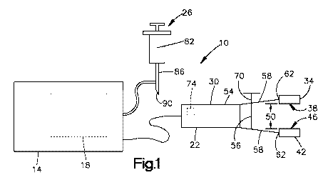
illustrated in Fig. 1, the electroporation device 10 of the present invention
includes a housing 14
containing an electroporation (EP) signal generator 18, an applicator 22
removably coupled to
the housing 14, and an injection device 26 to inject a pre-determined volume
of an agent, such as
lipids, into the adipose layer of the sample before electroporation occurs.
- 11 -
CA 03037527 2019-03-19
WO 2018/057900 PCT/US2017/052970
[0069] As shown in Fig. 1, the hand-held applicator 22 of the present
invention
includes a frame 30, a first electrode 34 coupled to the frame 30, with the
first electrode 34
having a first contact surface 38, and a second electrode 42 coupled to the
frame 30, with the
second electrode 42 having a second contact surface 46 such that the first
contact surface 38 and
the second contact surface 46 face one another and are substantially aligned.
During use, the
applicator 22 is configured to allow the user to manipulate the frame 30
causing the distance
between the first contact surface 38 and the second contact surface 46 to
change. For the
purposes of this application, the distance between the first contact surface
38 and the second
contact surface 46 is herein referred to as the "electrode distance 50."
[0070] The frame 30 of the applicator 22 includes a base 54, and a plurality
of resilient
arms 58 each extending from the base 54 to produce a corresponding distal end
62. When
assembled, each distal end 62 is configured to have a respective one of the
first and second
electrodes 34, 42 coupled thereto. In the illustrated embodiment, the arms 58
are configured so
that the user may resiliently deform the arms 58, allowing the distal ends 62
and their
corresponding electrodes 34, 42, to move. The corresponding electrodes 34, 42,
may move
independently or in tandem with respect to one another, for example. In the
illustrated
embodiment, the frame 30 includes two arms 58 (Fig. 1); however in an
alternative
embodiments, the frame 30 may include more or fewer arms 58 to support the
number of
electrodes necessary for treatment of the desired target tissue.
[0071] As illustrated in Fig. 2, an alternative embodiment of the applicator
22' includes
a frame 30' having three arms 58a', 58b', 58c' extending therefrom. More
specifically, the frame
30' includes two opposing arms 58a', 58b' configured to support the first and
second electrodes
34, 42 such that the first contact surface 38 and the second contact surface
46 face and are
substantially aligned with one another. The frame 30' also includes a third
arm 58c' configured to
support a third electrode 66' such that the third contact surface 68' of the
third electrode 66' is
positioned perpendicular to the first and second contact surfaces 38', 46'. In
the alternative
applicator 22', the electrode distance 50' is defined as the distance between
the first contact
surface 38' and the second contact surface 46' (i.e., the distance between the
two contact surfaces
facing one another).
- 12 -
CA 03037527 2019-03-19
WO 2018/057900 PCT/US2017/052970
[0072] The applicator 22 also includes an adjustment mechanism 56 to fix or
otherwise
manipulate the electrode distance 50 in preparation for and during treatment.
The adjustment
mechanism 56 includes a rod 70 extending between and threadably engaging both
arms 58 of the
frame 30 such that rotation of the rod 70 with respect to the arms 58 in a
first direction causes the
electrode distance 50 to shrink. In contrast, rotation of the rod 70 with
respect to the arms 58 in a
second direction, opposite the first direction, causes the electrode distance
50 to increase. In the
illustrated embodiment, the rod 70 is configured such that the electrode
distance 50 will remain
fixed unless the rod 70 is rotated by the user (i.e., the threads are not back
drivable). In
alternative embodiments, the adjustment mechanism 56 may include a latch (not
shown)
adjustable between a disengaged configuration, where the electrode distance 50
is adjustable by
the user, and an engaged configuration, where the electrode distance 50 is
fixed. In still other
embodiments, the adjustment mechanism 56 may include any form of adjustment
mechanism
well known in the art and not described herein.
[0073] The applicator 22 may also include a sensor 74 in operable
communication with
the signal generator 18 and configured to determine the electrode distance 50
during operation of
the device 10. During use, the sensor 74 sends signals to the signal generator
18 indicating the
current electrode distance 50. In some embodiments, the sensor 74 may include
a resistance
sensor, optical sensor, and the like coupled to the frame 30 of the applicator
22. In the illustrated
embodiment, the sensor 74 provides signals to the signal generator 18 allowing
the generator 18
to record the electrode distance 50 data and automatically compensate for
different electrode
distances 50 during treatment. In alternative embodiments the sensor 74 may
indicate the
distance on a display (not shown) allowing the user to compensate for the
electrode distance 50
manually. In still other embodiments, the applicator 22 may be configured such
that the user may
enter a pre-determined electrode distance 50 into the signal generator 18
whereby the applicator
22 will automatically adjust electrodes 34, 42 to produce the proper electrode
distance 50. In still
other embodiments, the sensor may be mechanical in nature, displaying the
electrode distance 50
on a dial and the like.
[0074] Illustrated in Fig. 4, the first plate electrode 34 of the applicator
22 has a first
contact surface 38 configured to directly contact the target tissue. The first
plate electrode 34
may have any shape. The shape may be rectangular, for example. The first plate
electrode 34
- 13 -
CA 03037527 2019-03-19
WO 2018/057900 PCT/US2017/052970
also includes a first perimeter 78 extending around and defining the extent of
the contact surface
38. During use, the first plate electrode 34 is in operable communication with
the signal
generator 18 and is configured to engage and form electrical conductivity with
the sample tissue
during treatment. As such, the plate electrode 34 is able to apply the
electroporation signals
produced by the signal generator 18 to the target tissue. The plate electrode
34 is also able to
detect parameters in the target tissue, such as impedance, voltage, current,
and the like, and relay
that information back to the signal generator 18 for diagnostics and feedback.
In the illustrated
embodiment, the first contact surface 38 of the first plate electrode 34 is
substantially planar;
however, in alternative embodiments, the first contact surface 38 may be
curvilinear in contour.
In still other embodiments, the first contact surface 38 may include any shape
or size intended to
maximize the amount of surface area in contact between the electrode 34 and
the target tissue. In
still other embodiments, the applicator may include a plurality of electrodes
34, each of which
includes a first contact surface 38 specifically sized and shaped to
correspond with a particular
area of a patient or test subject's body. In still other embodiments, the size
and shape of the
electrodes may be used to focus the distribution of the electric field within
the target tissue. In
still other embodiments, the first contact surface 38 may include a pattern or
knurl formed
therein. In still other embodiments, the first contact surface 38 may include
a coating or adhesive
applied thereto to improve the conductivity or grip between the electrode 34
and the target tissue.
[0075] Figs. 5 and 6 illustrate an alternative embodiment of a plate electrode
34". The
plate electrode 34" is substantially similar to and operates in the same
manner as the plate
electrode 34, described above. As such, only the differences between the two
electrodes 34", 34
will be described in detail herein. Best illustrated in Fig. 5, the contact
surface 38" of the plate
electrode 34" includes a base 54", and a plurality of protrusions 58" each
extending substantially
normal to the base 54" a protrusion depth 64" to form a distal end 62". During
use, the
protrusions 58" of the plate electrode 34" press into the target tissue
without piercing
therethrough allowing the protrusions 58" to disrupt and alter the top layer
of skin, improving the
electric field distribution within the target tissue (compare Fig. 6 to Fig.
7) and also improving
grip. More specifically, the protrusions 58" increase the magnitude of the
electric field formed
within the target tissue for a given input voltage. While the protrusions 58"
of the illustrated
embodiment all produce a similar protrusion depth 64", it is understood that
each protrusion 58"
- 14 -
CA 03037527 2019-03-19
WO 2018/057900 PCT/US2017/052970
may be sized differently as necessary to produce the desired conductivity and
grip with the target
tissue. Still further, the illustrated embodiment includes a protrusion depth
64" of approximately
500 microns, 600 microns, 700 microns, 800 microns, 900 microns, 1 mm, 1.5 mm,
2 mm, 2.5
mm, and 3 mm.
[0076] In the illustrated embodiment, each protrusion 58" of the electrode 34"
is
substantially pyramidal in shape. Each pyramidal protrusion may also has a
base width of
approximately 500 microns by 500 microns, 600 microns by 600 microns, 700
microns by 700
microns, 800 microns by 800 microns, 900 microns by 900 microns, 1 mm by 1 mm,
1.5 mm by
1.5 mm, 2 mm by 2 mm, 2.5 mm by 2.5 mm, 3 mm by 3 mm. In alternative
embodiments, each
pyramidal protrusion may also be non-square in base dimensions. In alternative
embodiments,
each protrusion 58" may form any other shapes configured to press into and
deform the target
tissue without piercing the tissue during operation. For example, the
protrusions 58" may be
cylindrical, rectangular, conical, frusto-conical, or frusto-pyramidal in
shape. Furthermore, each
protrusion 58" may include a width of approximately 500 microns, 600 microns,
700 microns,
800 microns, 900 microns, I mm, 1.5 mm, 2 mm, 2.5 mm, or 3 mm, for example.
[0077] In still further embodiments, each protrusion 58" may be configured to
pierce
the target tissue. For example, such protrusions 58" may include a needle (not
shown) extending
from the base 54". In still other embodiments, such protrusions 58" may be
shaped like a
hypodermic needle, trochar needle, and the like. In still other embodiments,
such protrusions 58"
may have a blunt tip or a flat tip. In still other embodiments, each
protrusion 58" may be shaped
differently than other protrusions 58" on the same electrode.
[0078] Illustrated in Fig. 5, the protrusions 58" of the electrode 34" are
evenly
positioned on the base 58" of the plate electrode 34" in the form of a
rectangular array. In
alternative embodiments, the protrusions 58" may be positioned in any pattern
necessary to
provide the necessary conductivity and grip with the target tissue. For
example, the protrusions
58" may be positioned in concentric rings (not shown) or other patterns.
[0079] Figs. 44 and 45 illustrate another alternative embodiment of a plate
electrode
34" '. The plate electrode 34' "is substantially similar to and operates in
the same manner as the
plate electrode 34, described above. As such, only the differences between the
two electrodes 34"
',34 will be described in detail herein. Best illustrated in Fig. 44, the
contact surface 38" of the
- 15 -
CA 03037527 2019-03-19
WO 2018/057900 PCT/US2017/052970
plate electrode 34" includes one or more non-insulated portions 500, where the
contact surface
38" creates a first resistance with the target tissue when in contact
therewith, and one or more
insulated portions 504, where the contact surface 38" creates a second
resistance with the target
tissue when in contact therewith that is larger than the first resistance.
During use, the interaction
of the non-insulated and insulated portions 500", 504" of the contact surface
38" with the target
tissue effects the resulting electrical field applied to the target tissue. In
particular, by insulating
at least a portion of the contact surface 38" of the electrode 34", the
electrode 34" better focuses
the electric field within the adipose layer of the target tissue, thereby
decreasing the amount of
muscle twitch and pain experienced by the patient. Stated differently, the
alternative plate
electrode 34" reduces the amount of current traveling through the muscle when
compared to a
similarly shaped and sized plate electrode that does not have an insulated
portion (see Figs. 46
through 48; showing difference between an "insulated calipers" where at least
one insulated
portion 504" is present, and a non-insulated calipers where no insulated
portion 504" is present).
[0080] Illustrated in Fig. 44 and 45, the insulated portion 504" of the plate
electrode
34" includes a layer of insulating material 508" positioned between the
contact surface 38" and
the target tissue to increase the resistance therebetween (see Fig. 45). In
the illustrated
embodiment, the electrode 34" includes a sheath 512" formed from insulating
material 508"
that is removably positionable over at least a portion of the electrode 34" '.
Dependent upon the
size and shape of the sheath 512" ', different sizes and shapes of the contact
surface 3 8" may be
covered by the insulating material 508' ". In still other embodiments, the
insulating material 508"
may be applied to the contact surface 38" of the electrode 34" (e.g., like a
coating). In still other
embodiments, the insulating material 508" may be applied to the contact
surface 38" with a
removable adhesive (not shown).
[0081] The second plate electrode 42 is substantially similar to and operates
in the same
manner as the first plate electrode 34. The second plate electrode 42 includes
a second contact
surface 46 having a second perimeter 60 defining the extent of the second
contact surface 46. As
such, the second plate electrode 42 will not be described in detail herein.
While the illustrated
embodiment shows the second plate electrode 42 being the same size and shape
as the first
electrode 34, it is understood that the second electrode 42 may be sized and
shaped differently
than the first electrode 34. Furthermore, the second contact surface 46 of the
second electrode 42
- 16 -
CA 03037527 2019-03-19
WO 2018/057900 PCT/US2017/052970
may be sized and shaped differently than the first contact surface 38 of the
first electrode 34. In
still other embodiments, one plate electrode may include protrusions 58" while
another electrode
may not. Still further, one plate electrode may include an insulated portion
504" while the other
electrode may not.
[0082] As illustrated in Fig. 1, the device 10 can further includes an
injection device 26
to inject agent into the target tissue at a desired location. More
specifically, the injection device
26 includes a reservoir 82 configured to hold a predetermined volume of agent
therein, and an
injection needle 86 extending from and in fluid communication with the
reservoir 82 to produce
a distal end 90. During use, the user inserts the injection needle 86 into the
target tissue so that
the distal end 90 is positioned at the desired depth (i.e., within the adipose
layer 104; see Fig.
11). The user may then inject the fluid contained within the reservoir 82
through the needle 86,
out the distal end 90, and into the desired tissue. While the illustrated
injection device 26
includes a hypodermic needle (i.e., an insulin needle), in alternative
embodiments, it is
understood that a jet injector or other forms of injection may be utilized.
[0083] Furthermore, in some embodiments, the needle 86 of the injection device
26
may be in operable communication with the signal generator 18 and able to
perform as an
electrode similar to the first and second plate electrodes 34, 42 (see Fig.
1). In such
embodiments, the signal generator 18 may be able to both send the treating
signal to the needle
86 as well as receive information, such as impedance, current flow, and the
like back to the
signal generator 18 for diagnostics and feedback purposes.
[0084] The injection device 26 may also include a depth limiter (not shown) to
control
the position of the distal end 90 within the sample tissue. During use, the
depth limiter may be
set to a predetermined depth so that the depth limiter will stop the distal
end 90 of the needle 86
from penetrating beyond the desired depth into the tissue. In some
embodiments, the depth
limiter may include a hard stop defining a guide hole. In such embodiments,
the length of the
guide hole dictates the depth the needle penetrates the target tissue. In
still other embodiments,
the depth limiter may also include an electric coupler to electrically couple
the needle 86 with the
pulse generator 18.
- 17 -
CA 03037527 2019-03-19
WO 2018/057900 PCT/US2017/052970
III) TREATMENT OF ADIPOSE LAYER
[0085] The above described device may be used in various therapeutic methods
intended to transfect adipose tissue with an agent using electroporation. Each
treatment or
"setup" provides flexibility regarding the size, shape, and characteristics of
the resulting
electrical field created within the sample tissue. Each setup also provides
various levels of
invasiveness.
a) Two Electrode Setup
[0086] To administer the treatment via the two-electrode treatment setup, the
user first
obtains a patient, taking note of the area or region they wish to treat
(hereinafter the "tissue
region 100"). For the purposes of this application, the tissue region 100 may
include skin tissue
having, for example, one or more of a skin layer 104, an adipose layer 108,
and a smooth muscle
layer 112.
[0087] Skin Layer. The skin layer may have two parts: an outer epidermis
portion and a
dermis portion, to which the epidermis may be connected. Beneath the dermis, a
subcutaneous
layer may exist and may contain areolar and adipose tissues. Fibers from the
dermis may extend
down into the subcutaneous layer and connect the subcutaneous layer to the
skin layer. The
subcutaneous layer may be attached to underlying tissues and organs.
[0088] Epidermis. The epidermis may be composed of stratified squamous
epithelium
and contain keratinocytes, melanocytes, and nonpigmented granular dendrocytes
(for example,
Langerhans' cells and Granstein cells). The keratinocytes may be organized
into several layers.
The number of layers may depend on location in the body. For example, where
exposure to
friction is great, the epidermis may have many layers, for example five
layers. Where exposure
to friction is not great, the epidermis may have less than five layers, for
example. The epidermis
may have one or more of the following layers: stratum basale, stratum
spinosum, stratum
granulosum, stratum lucidum, and/or the stratum corneum.
[0089] Dermis. The dermis may be composed of connective tissue that contains
collagenous and elastic fibers. The dermis may be thick or think depending on
the location in the
body. For example, the dermis may be thicker in the palms and soles, yet thin
in the eyelids. The
- 18 -
CA 03037527 2019-03-19
WO 2018/057900 PCT/US2017/052970
dermis may contain blood vessels, nerves, glands, and hair follicles. The
dermis may have a
papillary region or layer, which may consist of loose connective tissue that
contains fine elastic
fibers. The papillary region may also have dermal papillae that project into
the epidermis. These
papillae may contain capillaries, corpuscles of touch (or Meissner's
corpuscles), which are nerve
endings that are sensitive to touch. Dermal papillae may cause ridges in the
overlying epidermis.
[0090] The remaining portion of the dermis may be the reticular region or
layer. This
region may contain densely packed connective tissue and bundles of collagenous
and coarse
elastic fibers. Varying thicknesses of the reticular region may be
responsible, at least in part, for
differences in the thickness of the skin.
[0091] Adipose Layer. The adipose layer or tissue may be a form of loose
connective
tissue in which adipocytes store fat. An adipocyte may have its cytoplasm and
nuclei pushed to
the edge of the cell by the droplet of fat within the cell. Each adipocyte may
be surrounded by a
collagenous basement membrane for structural support, and may be in contact
with a capillary.
Clusters of adipocytes may be contained within "lobes," which may be held
together by
collagenous septa. The adipose tissue may be found wherever loose connective
tissue is located.
The adipose tissue may be in the subcutaneous layer below the skin.
[0092] Smooth Muscle Layer. The smooth muscle layer may be located in the
walls of
hollow internal structures such as, for example, blood vessels. Smooth muscle
may also be
attached to hair follicles. The smooth muscle layer is nonstriated,
involuntary muscle tissue and
may be influenced by involuntary nerves and some hormones. The smooth muscle
layer is a type
of muscle layer that is distinct from cardiac muscle tissue and skeletal
muscle tissue. Skeletal
muscle is attached primarily to bones and may move parts of the skeleton.
Skeletal muscle is also
striated, because striations, or alternating light and dark bandlike
structures are visible when the
tissue is examined under a microscope, and voluntary, whereby it can be made
to contract and
relax by conscious control. Cardiac muscle tissue is striated and involuntary
and forms most of
the wall of the heart.
[0093] With the area of treatment selected, the user obtains the injection
device 26 and
inserts the needle 86 into the tissue region 100 such that the distal end 90
is positioned within the
adipose layer 108. The user then injects a volume of an agent, optionally a
pre-measured volume
- 19 -
CA 03037527 2019-03-19
WO 2018/057900 PCT/US2017/052970
of an agent, into the adipose layer 108 of the tissue region 108, creating an
injection site 116.
Once the injection is complete, the user removes the needle 86 from the tissue
region 100.
[0094] With the needle 86 removed, the user manipulates a portion of the
tissue region
100 containing the injection site 116 and creates a fold 120 therein. The
tissue is manipulated so
that the tissue contained within the fold 120 is limited to a skin layer 104,
an adipose layer 108,
and a smooth muscle layer 112. No skeletal muscle (not shown) is included in
the fold 120. The
resulting fold 120 includes a first side 124, a second side 128 opposite the
first side 124, and a
top 132 extending between the first side 124 and the second side 128. (Fig.
8). The fold 120 also
defines a fold thickness 134 defined as the distance between the first side
124 and the second
side 128.
[0095] After preparing the fold 120, the user manipulates the frame 30 or
adjustment
mechanism 67 of the applicator 22 until the electrode distance 50 is slightly
larger than the fold
thickness 134. The user then positions the applicator 22 so that each
electrode 34, 42 is
positioned on opposing sides of the fold 120 with the contact surfaces 38, 46
facing inward (see
Fig. 9). More specifically, the user positions the applicator 22 so that the
first contact surface 38
of the first plate electrode 34 is in contact with the first side 124 of the
fold 120, and the second
contact surface 46 of the second plate electrodes 42 is in contact with the
second side 128 of the
fold 120, creating a treatment zone 136 therebetween. Once in position, the
user may increase or
decrease the electrode distance 50 to effectively clamp the fold 120 between
the two electrodes
34, 42.
[0096] For the purposes of this application, the treatment zone 136 is defined
as the
volume of space positioned between the first and second electrodes 34, 42 and
defined on two
sides by the first and second contact surfaces 38, 46, and defined on the
remaining sides by an
imaginary barrier extending between the first perimeter 78 of the first
contact surface 38 and the
second perimeter 60 of the second contact surface 46 (see Figs. 8-10). As
such, after the user has
positioned the first and second electrodes 34, 42 on opposite sides of the
fold 120, the treatment
zone 136 of the presently described treatment will contain at least a portion
of the fold 120 and at
least a portion of the injection site 116 therein. In the illustrated
embodiment, the tissue
positioned within the treatment zone 136 during treatment is limited to a skin
layer 104, an
- 20 -
CA 03037527 2019-03-19
WO 2018/057900 PCT/US2017/052970
adipose layer 108, and a smooth muscle layer 112. The treatment zone 136 does
not include any
skeletal muscle therein.
[0097] While the illustrated embodiment illustrates the contact surfaces 38,
42 of the
electrodes 34, 42 being placed in direct contact with the fold 120, it is
understood that conductive
gel (not shown) or other substances may be utilized to improve the electrical
communication
between the electrodes 34, 42 and the fold 120.
[0098] Once the electrodes 34, 42 are in position, the sensor 74 of the
applicator 22
determines the electrode distance 50 and relays that information to the signal
generator 18
allowing it to set the parameters of the electroporation signal 150
accordingly. The signal
generator 18 may also produce a test signal (i.e., a low voltage pulse) where
the resulting current
and voltage may be detected by the electrodes 34, 42 and subsequently used by
the signal
generator 18 to calculate the impedance of the tissue being treated.
Furthermore, the data
detected by the electrodes 34, 42 during the test signal may also be used to
verify that the pulses
were fired successfully. To do so, the signal generator 18 compares whether
the current flow is
maintained for the duration of the pulses, if the timings do not match (i.e.,
more than one or two
data collection points are missing), then the pulses can be considered
incomplete. More
specifically, at least one of the pulse voltage 158, pulse length 162, number
of pulses and/or
pulse delay 166 of the electroporation signal may at least partially be
determined by the electrode
distance 50 (described below). In the illustrated embodiment, the electrode
distance 50 is
determined automatically by the sensor 74 of the applicator 22. However, in
alternative
embodiments, the user may manually measure the electrode distance 50 and input
the electrode
distance 50 into the device 10.
[0099] In the illustrated embodiment, the electroporation signal 150 consists
of a series
of electrical "pulses 154," where each pulse 154 is delivered at a
predetermined pulse voltage
158 and lasts a predetermined pulse length 162. Furthermore, each individual
pulse 154 is
separated in time from adjacent pulses 154 by a pulse delay 166. (Fig. 26). In
the illustrated
embodiment, the electroporation signal includes a pulse voltage 158 of between
approximately
50 V and approximately 200 V. In other embodiment, the signal may include a
pulse voltage 158
between approximately 5 V and approximately 10 V. In still other embodiments,
the signal may
include a pulse voltage 158 of approximately 1 kV. Furthermore, the
illustrated electroporation
-21 -
CA 03037527 2019-03-19
WO 2018/057900 PCT/US2017/052970
pulse length 162 is approximately 100 microseconds, 200 microseconds, 300
microseconds, 400
microseconds, 500 microseconds, 600 microseconds, 700 microseconds, 800
microseconds, 900
microseconds, 1 millisecond, 10 milliseconds, 50 milliseconds, 75
milliseconds, and 100
milliseconds. Still further, the electroporation pulse delay 166 is
approximately 1 millisecond, 50
milliseconds, 100 milliseconds, 500 milliseconds, and 1 second. Still further,
each
electroporation signal includes between approximately 1 pulse and
approximately 10 pulses.
Together, in some embodiments the electroporation signal 150 may include 3
pulses at
approximately 200 V of approximately 100 milliseconds in duration with 200
milliseconds of
delay between pulses. In other embodiments, the electroporation signal 150 may
include 3 pulses
at approximately 50 V of approximately 100 milliseconds in duration with 200
millisecond delay
between pulses. In still other embodiments, the electroporation signal 150 may
include 10 pulses
at approximately 50 V of 100 milliseconds in duration with 1 second delay
between pulses. In
still other embodiments, the electroporation signal 150 may include 8 pulses
of 75 V of
approximately 100 milliseconds of duration with approximately 100 milliseconds
of delay
between pulses. In still other embodiments, the electroporation signal 150 may
include 3 pulses
of between approximately 500 V and approximately 1000 V of approximately 10
microseconds
and approximately 100 microseconds duration with approximately 100
milliseconds to
approximately 1 second delay between pulses. In still other embodiments the
electroporation
signal may include a single pulse.
[00100] After setting the parameters of the electroporation signal, the signal
generator
18 of the device 10 sends the desired signal to the first and second
electrodes 34, 42 such that
one of the first electrode 34 acts as one of the positive electrode or the
negative electrode while
the second electrode 42 act as the other of the positive electrode or the
negative electrode. More
specifically, the signal generator 18 may adjust the parameters of the
electroporation signal 150
at least partially dependent upon the impedance value detected and the
electrode distance 50.
Upon receiving the electroporation signal, the electrodes 34, 42 conduct the
signal in series to the
fold 120 creating an electric field therein (Figs. 9 and 10). The resulting
electric field is
concentrated in the adipose layer 108 creating a transfection region within
the fold 120. More
specifically, the electric field may create a transfection region that is
substantially spherical or
ellipsoid in shape. However, in alternative embodiments, the size and shape of
the transfection
- 22 -
CA 03037527 2019-03-19
WO 2018/057900 PCT/US2017/052970
region may at least partially depend upon the electric field distribution
within the target tissue
and the location and quantity of agent that was injected into the target
tissue. Furthermore,
current freely flows through the underlying muscle layer 112 and is relatively
low near the
injection site 116 when compared to a similar treatment conducted by
penetrating needle
electrode configurations commonly used for subcutaneous or intramuscular
electroporation
delivery. Such characteristics of the electric field are potentially
beneficial for treatments where
immune response is not desired.
[00101] After electroporation is complete, the electrodes 34, 42 may be
removed from
the fold 120.
b) Needle-in Three Electrode Setup
[00102] To administer the treatment via the three electrode setup, the user
first obtains
a patient, taking note of the tissue region 100 they wish to treat. For the
purposes of this
application, the tissue region 100 may include skin tissue having, for
example, one or more of a
skin layer 104, an adipose layer 108, and a smooth muscle layer 112, as
described in detail
above. With the tissue region 100 selected, the user manipulates a portion of
the tissue region
100 and creates a fold 120 therein. More specifically, the user manipulates
the tissue region 110,
creating a fold 120 of tissue that includes a skin layer 104, an adipose layer
108, and a smooth
muscle layer 112. No skeletal muscle is included in the fold 120. Furthermore,
the resulting fold
120 of tissue includes a first side 124, a second side 128 opposite the first
side 124, and a top 132
extending between the first side 124 and the second side 128. The fold 120
also defines a fold
thickness 134 defined as the distance between the first side 124 and the
second side 128.
[00103] With the fold 120 prepared, the user obtains the injection device 26
and inserts
the needle 86 lengthwise through the fold 120 substantially parallel to the
first side 124 and the
second side 128. The user then injects a pre-measured volume of agent into the
adipose layer 108
of the tissue region 100, creating an injection site 116. Once the injection
is complete, the user
does not remove the needle 86 from the tissue 100.
[0104] With the injection site 116 created and with the needle 86 still
positioned in the
tissue 100, the user manipulates the frame 30 or adjustment mechanism 56 of
the applicator 22
until the electrode distance 50 is slightly larger than the fold thickness
134. The user then
- 23 -
CA 03037527 2019-03-19
WO 2018/057900 PCT/US2017/052970
positions the applicator 22 so that each electrode 34, 42 is positioned on
opposing sides of the
fold 120 (see Fig. 11-13). More specifically, the user positions the
applicator 22 so that the first
contact surface 38 of the first plate electrode 34 is in contact with the
first side 124 of the fold
120, and the second contact surface 46 of the second plate electrodes 42 is in
contact with the
second side 128 of the fold 120, creating a treatment zone 136 therebetween
(described above;
see Figs. 11-13).
[0105] While the illustrated embodiment illustrates the contact surfaces 38,
46 of the
electrodes 34, 42 being placed in direct contact with the fold 120, it is
understood that conductive
gel (not shown) or other substances may be utilized to improve the electrical
communication
between the electrodes 34, 42 and the fold 120.
[0106] Once the electrodes 34, 42 are in position, the sensor 74 of the
applicator 22
determines the electrode distance 50 and sets the parameters of the
electroporation signal
accordingly (described above). The signal generator 18 may also produce a test
signal (described
above) where the resulting current and voltage may be detected by the
electrodes 34, 42 or
needle 86 and subsequently used by the signal generator 18 to calculate the
impedance of the
tissue being treated. In the illustrated embodiment, the electroporation
signal 150 consists of a
series of electrical pulses 154, where each pulse 154 is given at
predetermined a pulse voltage
158 and lasts a predetermined pulse length 162. Furthermore, each individual
pulse 154 is
separated in time from adjacent pulses 154 by a pulse delay 166. (see Fig.
26). In the illustrated
embodiment, the electroporation signal includes a pulse voltage 158 of between
approximately 5
V and approximately 500 V. The pulse voltage may be, for example, 5V, by, 20V,
40V, 60V,
80V, 100V, 150V, 200V, 250V, 300V, 350V, 400V, 450V, or 500V. Furthermore, the
illustrated
electroporation pulse length 162 is between approximately 1 microsecond and
approximately
100 milliseconds. Still further, the electroporation pulse delay 166 is
between approximately 10
millisecond and approximately 1 second. Still further, each electroporation
signal includes
between approximately 1 and approximately 10 pulses. In alternative
embodiments, the
parameters of the electroporation signal 150 may be altered to allow optimal
performance for
different agents. Signal parameters may be adjusted depending on the agent
being used, the
degree of transfection, and tissue damage desired. For example, dMAb
constructs generally
- 24 -
CA 03037527 2019-03-19
WO 2018/057900 PCT/US2017/052970
require lower voltage, shorter pulse duration, and longer inter-pulse delays,
while DNA vaccines
generally require higher voltage, shorter delay, and longer pulses.
[0107] After setting the parameters of the electroporation signal, the signal
generator 18
of the device 10 sends the electroporation signal to the first electrode 34,
the second electrode 42,
and the needle 86 such that the needle 86 acts as one of the positive
electrode or the negative
electrode while the first and second electrodes 34, 42 together, act as the
other of the positive
electrode or the negative electrode. Upon receiving the electroporation
signal, the electrodes 34,
42 and needle 86 conduct the signal to the fold 120 creating an electrical
field therein (Figs. 12
and 13). The resulting electric field is concentrated in the adipose layer 108
around the needle
86, decreasing in strength radially therefrom. The electric field also creates
a transfection region
that tracks along the needle in a very elongated ellipsoid shape. Furthermore,
electrical current is
highest around the needle 86.
[0108] After electroporation is complete, the electrodes 34, 42 and needle 86
may be
removed from the fold 120.
c) Three Plate Setup
[0109] To administer the treatment with the three plate setup, the user first
obtains a
patient, taking note of the tissue region 100 they wish to treat. For the
purposes of this
application, the tissue region 100 may include skin tissue having, for
example, one or more of a
skin layer 104, an adipose layer 108, and a smooth muscle layer 112 as
described in detail above.
With the area of treatment selected, the user obtains the injection device 26
and inserts the needle
86 into the tissue region 100 such that the distal end 90 is positioned within
the adipose layer
108. The user then injects a pre-measured volume of agent into the adipose
layer 108 of the
tissue region 108, creating an injection site 116. Once the injection is
complete, the user removes
the needle 86 from the tissue region 100.
[0110] With the needle 86 removed, the user obtains a portion of the tissue
region 100
containing the injection site 116 and creates a fold 120 that includes the
injection site 116, a skin
layer 104, an adipose layer 108, and a smooth muscle layer 112. No skeletal
muscle is included
in the fold 120. Furthermore, the resulting fold 120 of tissue includes a
first side 124, a second
side 128 opposite the first side 124, and a top 132 extending between the
first side 124 and the
- 25 -
CA 03037527 2019-03-19
WO 2018/057900 PCT/US2017/052970
second side 128 of the fold 120. The fold 120 also defines a fold thickness
134 defined as the
distance between the first side 124 and the second side 128.
[0111] After preparing the fold 120, the user manipulates the frame 30' or
adjustment
mechanism 56' of the applicator 22' until the electrode distance 50' is
slightly larger than the fold
thickness 134. The user then positions the applicator 22' so that the first
contact surface 38' of the
first plate electrode 34' is in contact with the first side 124 of the fold
120, and the second contact
surface 46' of the second plate electrodes 42' is in contact with the second
side 128 of the fold
120, creating a treatment zone 136 therebetween (described above). The user
also positions the
third plate electrode 66' so that the third contact surface 68' is in contact
with the top 132 of the
fold 120 and generally positioned between the first and second electrodes 34',
42' (see Figs. 14-
16) such that the third plate electrode 66' does not directly contact either
the first or second
electrodes 34', 42'.
[0112] While the illustrated embodiment illustrates the contact surfaces 3 8',
46', 68' of
the electrodes 34', 42', 66' being placed in direct contact with the fold 120,
it is understood that
coupling or conductive gel (not shown) or other substances may be utilized to
improve the
electrical communication between the electrodes 34', 42', 66' and the fold
120.
[0113] Once the electrodes 34', 42', 66' are in position, the sensor 74' of
the applicator
22' determines the electrode distance 50 between the first and second
electrodes 34', 42' and sets
the parameters of the electroporation signal accordingly (described above).
The signal generator
18 may also produce a test signal (described above) where the resulting
current and voltage may
be detected by the electrodes 34', 42', 66' and subsequently used by the
signal generator 18 to
calculate the impedance of the tissue being treated. In the illustrated
embodiment, the
electroporation signal consists of a series of electrical "pulses 154," where
each pulse 154 is
given at predetermined a pulse voltage 158 and last a predetermined pulse
length 162.
Furthermore, each individual pulse 154 is separated by in time by adjacent
pulses 154 by a pulse
delay 166 (see Fig. 26). In the illustrated embodiment, the electroporation
signal includes a pulse
voltage 158 of between approximately 5 V and approximately 500 V. Furthermore,
the
illustrated electroporation pulse length 162 is between approximately 1
microsecond and
approximately 100 milliseconds. Still further, the electroporation pulse delay
166 is between
- 26 -
CA 03037527 2019-03-19
WO 2018/057900 PCT/US2017/052970
approximately 10 millisecond and approximately 1 second. Still further, each
electroporation
signal includes between approximately 1 and approximately 10 pulses.
[0114] After setting the parameters of the electroporation signal, the device
10 applies
the electroporation signal to the first, second, and third electrodes 34',
42', 66' such that the first
and second electrodes 34', 42' act as one of the positive electrode and the
negative electrode
while the third electrode 66' acts as the other of the positive electrode and
the negative electrode.
Upon receiving the electroporation signal, the electrodes 34', 42', 66'
conduct the signal in series
to the fold 120 creating an electrical field therein (Figs. 15 and 16). The
resulting electric field is
strongest just below the third plate electrode 66' and decreases in strength
with increasing tissue
depth. Furthermore, electric current is strong in the skin layer 104 and much
weaker in the
adipose layer 108 creating a desirable balance between a strong electric field
while maintaining a
lower current at the injection site. Such electrical fields are generally
optimal for DNA injections
in shallow subcutaneous fat.
[0115] After electroporation is complete, the electrodes 34', 42', 66' may be
removed
from the fold 120.
IV) EXAMPLES.
[0116] Example 1. Experimental Results. In vivo treatments were performed on
subcutaneous fat pads of female Hartley Guinea Pigs using a variation of the
above described
treatment. During the experiment, the user shaved the hair near the treatment
area proximate the
back of the guinea pig's neck. Afterwards, hair removal cream was used to
completely remove
any remaining stubble from the treatment area. Plasmids were then injected
into the adipose
layer of the treatment area using an insulin syringe, creating an injection
site. The injection site
and skin tissue of the treatment area were then manipulated, separating the
skin, adipose, and
smooth muscle layers from any skeletal muscle. The resulting fold of skin was
then positioned
between a pair of plate electrodes, each electrode having the corresponding
contact surface
covered in conductive gel. Finally, electrical pulses were sent to the plate
electrodes, where one
plate electrode acted as the positive electrode and the other plate electrode
acted as the negative
electrode. After the treatment was complete, samples of the tissue in the
treatment area were
taken for analysis (see Figs. 17-30).
- 27 -
CA 03037527 2019-03-19
WO 2018/057900 PCT/US2017/052970
[0117] Dye injection studies demonstrated that injectate preferentially
travels down
collegenous septa surrounding adipose lobes, and these observations were
consistent with GFP
transfection patterns. To demonstrate section of coded proteins, adipose-
targeted EP treatment
was performed using DNA encoding monoclonal antibodies (dMAbs), which led to
detectable
systemic levels of protein. Finally, adipose-targeted EP DNA vaccination of
plasmid encoding
H1N1 nucleoprotein was shown to be immunogenic. Compared to traditional
intramuscular
routes, adipose-targeted EP DNA vaccinations may offer tolerability advantages
due to the lower
voltages, shallower injections, and noninvasive electrodes being used.
[0118] At higher magnification, a "honeycomb" pattern characteristic of
adipocytes
could be seen in bright green due to the numerous transfected adipocytes. Also
noted were bright
green collagenous septa as solid green lines traveling through the adipocyte
network. See Fig.
18, wherein the green transfected region corresponds to the volume of tissue
clamped between
the plate electrodes. A closer look at a section of adipose tissue revealed
many individual
adipocytes. The interior of each cell is not brightly lit, because the lipid
droplet does not express
protein. Rather, the green fluorescent protein (GFP) is found around the edges
of each cell,
where protein synthesis, production, trafficking, and modification occur (see
Figs. 17, 18, and
19).
[0119] This technology was also demonstrated in rabbits (in the subcutaneous
fat at the
base of the neck). See Fig. 20, wherein the Oil Red 0 stain highlighting the
lipid droplets, as well
as the green ring of GFP surrounding transfected cells. Nuclei are shown in
blue.
[0120] Confocal images of transfected guinea pig adipose tissue shows that the
protein
is produced and expressed around the entire border of the cells. The numerous,
smaller cells
surrounding the adipocytes do not express GFP, which is consistent with a
degree of specificity
for adipocytes (see Fig. 21).
[0121] Injection volume may be reduced from about 200 pi to 50 pi without
impacting
the size of the transfected region (see Fig. 22). Multiple injections,
followed by a single EP, may
be one way to increase the number of transfected cells (see Fig. 23).
Decreasing voltage and
increasing the number of pulses may improve GFP signal, possibly indicating
more transfected
cells. 80V treatments caused noticeably weaker muscle twitches compared to
200V treatments
(see Fig. 24). dMAb was produced at peak levels of around 1000 ng/mL in guinea
pigs following
- 28 -
CA 03037527 2019-03-19
WO 2018/057900 PCT/US2017/052970
a second treatment into adipose. The first treatment was performed using un-
optimized
parameters (see Fig. 25).
[0122] The guinea pig receiving rapid pulses produced more dMAb than the
guinea
pigs receiving pulses with longer inter-pulse delays of 1 second (see Fig.
26). Jet injection
appeared to distribute the DNA more uniformly throughout the tissue and
transfect more cells.
See Fig. 27. Enzymatic breakdown of the tissue improved fluid distribution
throughout the tissue
(see Fig. 28). As the number of pulses increases, electrical resistance
decreases. At 25V, the
current was essentially undetectable (see Fig. 30). At 100V, the muscle twitch
was intense
enough to cause fluctuations in the electrical readings (see Fig. 29).
[0123] Example 2. Immunogenicity. In vivo treatments were provided to five
experimental groups, all of which received the same dose of DNA. For the
experiment, one
group was given intradermal EP to act as a positive control group. The
remaining four groups
were treated in the adipose layer of tissue using various combinations of high
or low voltage and
high or low injection volumes. Pulse voltages for the four adipose groups
varied between
approximately 50 V to approximately 200 V while injection parameters varied
between
approximately 100 microliters in a single site, to five injections of 50
microliters are each site.
For each injection, plasmid was diluted to increase the volume without
changing the overall
DNA dose.
[0124] As illustrated in Fig. 31, the adipose groups treated with high voltage
and high
injection volume had a stronger and more rapid humoral immune response (i.e.,
end-point titer)
than any other group, including the intradermal treated group. The strength of
this response
continued to increase through 6 weeks. Both lower voltage treatments (i.e.,
with high and low
volume) had little to no response after 3 weeks, and had weak or no response
after 6 weeks.
[0125] Both volume of the DNA injection and the voltage of the EP appear to
influence
the immune response in adipose tissue. The greater the volume of agent
injected into the tissue
corresponds with increased contact between cells and agent. Increased volume
may address
issues related to the administration of water-formulated DNA into adipose
tissue. In addition, the
higher the EP voltage, the more cells that may undergo EP. Furthermore, any
tissue damage as a
result of higher voltage, may aid in the induction of an immune response and
associated cells.
- 29 -
CA 03037527 2019-03-19
WO 2018/057900 PCT/US2017/052970
[0126] Example 3. Materials and methods for examples 4-9. To simulate
different EP
modalities, two tissue models were created as 3D CAD assemblies in SolidWorks
2013
(SolidWorks Corp., Concord, MA, USA). Both models consisted of three
electrically isotropic
layers: skin, adipose, and muscle. The stratum corneum was not included in
these models
because voltages as low as 50V will permeabilize the stratum corneum within
microseconds, and
its contribution to total tissue resistance then becomes negligible. The
thickness of the stratum
corneum is on the order of 20 um, and very thin layers can cause artifacts in
finite element
analysis. To model tissue clamped between plate electrodes, a folded tissue
geometry was
created and two square plate electrode geometries with contact area measuring
400 cm2 were
placed on opposite sides of the fold (Fig. 32). To model penetrating needle
electrodes within the
same tissue, a flat tissue geometry was created and two 19mm, 22-gauge needle
geometries were
placed into the tissue with an inter-electrode spacing of 10 mm and a
penetration depth of 18 mm
(Fig. 33).
[0127] The two tissue-electrode assemblies were exported to ANSYS Maxwell
2015.2
(ANSYS Software, Canonsburg, PA, USA) for finite element analysis. Electrical
conductivity
values for each tissue type were based on literature values, and were assumed
to be constant.
Conductivity values and general tissue dimensions used in the models are
listed in Table 1. An
excitation voltage was applied to one electrode while the opposing electrode
was assigned a
voltage of zero, and cross-sections bisecting the electrodes in the x-y
analysis plane were created
to visualize the electric field strength.
Table 1.
Material Cendudivityõ Sim Thickness (folded), mm* Thickness (flat), mm
Skin 0..25
Adipose 0,05 10 5
ivluscie 03 1 35
Stainless steer 1.1x10⁶ N/A N/A
*Thh:knes.s measured at the plane bisecting the Antrodes
- 30 -
CA 03037527 2019-03-19
WO 2018/057900 PCT/US2017/052970
[0128] Animals. All animal studies were performed under protocols approved by
an
institutional animal care and use committee. Female Hartley guinea pigs were
used for all in vivo
studies. Treatments and blood collections were performed while animals were
maintained under
general anesthesia by inhaled isoflurane. Subcutaneous injections were
performed into the
interscapular subcutaneous fat pads (located at the scruff of the neck) using
a 29-gauge insulin
needle oriented parallel to the spine. For terminal studies, guinea pigs were
first placed under
general anesthesia and then humanely euthanized by intracardiac injection of
pentobarbital.
[0129] Plasmids. Gene expression studies utilized plasmid DNA encoding green
fluorescent protein (GFP). Immune studies were carried out using plasmid DNA
encoding full
length nucleoprotein (NP) from Influenza A (H1N1, A/Puerto Rico/ 8). All
plasmid formulations
were prepared in saline sodium citrate buffer for a final buffer concentration
of lx.
[0130] Dye injection studies. Methylene blue (Sigma-Aldrich, St. Louis, MO,
USA)
was dissolved in deionized water at a concentration of 0.5 mg/mL. For single-
site injections,
guinea pigs were injected subcutaneously with 100 [IL of methylene blue
solution. For multi-site
injections, five separate 50 [IL subcutaneous injections were performed,
spaced approximately 5
mm apart. Following injection, the entire fat pad was gripped tightly between
two plate
electrodes to simulate the full treatment protocol. Animals were immediately
euthanized and the
fat pads were imaged intact, and then dissected along the sagittal plane and
imaged again to
visualize dye distribution within the tissue.
[0131] General adipose-EP treatment procedure. Treatment sites were shaved and
cleaned Adipose-EP treatments were performed on the subcutaneous fat pad in
the interscapular
region, while skin treatments were performed on the flank. For adipose
treatments, tissue was
pinched between two fingers to isolate the fat pad, and DNA was injected using
a 29-gauge
insulin needle oriented parallel to the spine. Immediately following DNA
injection, two plate
electrodes attached to opposing caliper jaws were coated with conductive gel
and then used to
pinch the tissue surrounding the injection site, and pulses were administered
using the Elgen
1000 control unit (Inovio Pharmaceuticals, San Diego, CA). For ID-EP
treatments, DNA was
injected intradermally followed immediately by electroporation using the
surface electroporation
(SEP) device consisting of a 4x4 array of needle electrodes.
- 31 -
CA 03037527 2019-03-19
WO 2018/057900 PCT/US2017/052970
[0132] Gross imaging and histological analysis. For green fluorescent protein
(GFP)
studies, adipose-EP was performed with GFP plasmid, and intact fat pads were
removed at
predetermined time points and imaged using a FluorChem R imaging system
(ProteinSimple,
San Jose, CA, USA). Fat pads were then frozen, and samples measuring
approximately lOmm x
lOmm were cut from the transfected region of the fat pad and cryosectioned at
a thickness of 30
microns either along the transverse plane to view the depth of transfection,
or along the coronal
plane to view the horizontal distribution of transfected cells. Some sections
were fixed in 4%
formalin, cleared in xylene, stained with either DAPI or Hoechst 3342 (Life
Technologies,
Carlsbad, CA), and coverslipped using Fluoromount (eBioscience, San Diego,
CA). Other
sections were fixed in formalin, cleared in xylene, stained with hematoxylin
and eosin (H&E),
and coverslipped using Permount (VWR, Radnor, PA, USA). Sections that were H&E
stained
were imaged in brightfield using an Olympus BX51 microscope (Olympus, Center
Valley, PA)
equipped with a MicroPublisher 3.3 camera (Qlmaging, Surrey, BC, Canada).
Fluorescence
images were captured with a Retiga 3000 camera (Qlmaging, Surrey, BC, Canada).
Confocal
images were acquired as high resolution multi-paneled and auto- stitched z-
stacks of the whole
tissue using a Zeiss LSM 780 laser scanning confocal microscope (Carl Zeiss,
Jena, Germany)
and the images were further processed using Zen 2012 (Carl Zeiss) and IN/TARTS
software
(Bitplane, Belfast, UK).
[0133] GFP expression and cellular kinetics. Adipose-EP was performed on 14
guinea
pigs, using 1001.tg of a plasmid coding for GFP and EP parameters of 200V, 3
pulses, 100ms
duration, and 100ms inter-pulse delay. As controls, two guinea pigs were
treated with EP but did
not receive plasmid injection, while two additional guinea pigs received the
plasmid injection but
were not treated with EP. Controls were sacrificed 3 days following
treatments, and treated
guinea pigs were sacrificed at intervals (n=2) following the treatment,
ranging from 3 hours to 14
days post-treatment, as well as a long-term follow-up at day 60. Fat pads were
imaged intact for
GFP expression and then sectioned and stained with H&E to visualize signs of
cellular
infiltration at the treatment site.
[0134] Immunogenicity study. Guinea pigs were treated with 251.tg of NP DNA
Four
groups of guinea pigs (n=4) received adipose EP treatments as described above,
and each group
received either a single 100 [IL DNA injection or five separate 50 [IL
injections, followed by a
- 32 -
CA 03037527 2019-03-19
WO 2018/057900 PCT/US2017/052970
single EP treatment consisting of three 100 msec square wave pulses with a 200
msec inter-pulse
delay. Guinea pigs vaccinated via ID-EP with the SEP device (n=3) served as a
comparator
group for this study since this method has been previously shown to transfect
epidermal cells, but
not subcutaneous adipocytes. The adipose-EP groups are as follows: high
voltage EP with 1
injection site (HV-1), high voltage EP with 5 injection sites (HV-5), low
voltage EP with 1
injection site (LV-1), and low voltage EP with 5 injection sites (LV-5). For
guinea pigs receiving
injections, a single EP procedure was performed immediately following the
final injection. The
total dose of plasmid DNA was identical for all groups. The study design is
illustrated in Table 2.
Every three weeks for the duration of the study, 300 [IL of blood was
collected and plasma was
stored at -20 C until analysis. Treatments were administered at week 0, week
3, and week 6 of
the study, immediately following blood collection. At week 21 of the study,
all animals were
boosted and 18 days later, 3 mL of blood was collected and peripheral blood
mononuclear cells
were separated for ELISpot analysis.
Table 2.
Treatment Total Total DNA
Group site injections Voltage, V voiume, tL
dose, tg
HV4 4 Adipose 1 200 100 25
1V4 4 Adipose 1 50 100 25
LV-5 4 Adipose 5 50 250 25
HV-5 4 Adipose S 200 250 25
1D-EP 3 Skin 1 25 50 25
[0135] ELISA Serum from vaccinated guinea pigs was analyzed using enzyme-
linked
immunosorbent assay (ELISA). ELISAs were performed using 96-well plates
(Thermo Fisher
Scientific, Waltham, MA, USA) coated overnight with 100 IlL/well of 0.3
1.tg/mL pNP antigen
(Sino Biological, Beijing, China) in Dulbecco's phosphate buffered saline
(PBS) (VWR). Plates
were washed, blocked with PBS containing 3% Bovine Serum Albumin (BSA) (Sigma-
Aldrich)
and 0.05% Tween-20 (Sigma-Aldrich) at 150 IlL/well for one hour at 37 C, and
then washed
again. Serum was serially diluted from 1 :50 to 1 :2952450 in PBS containing
1% BSA and
0.05% Tween-20 (sample dilution buffer) at 100 IlL/well and incubated for two
hours at 37 C.
Plates were then washed, and horseradish peroxidase-conjugated goat anti-
guinea pig IgG
- 33 -
CA 03037527 2019-03-19
WO 2018/057900 PCT/US2017/052970
(Sigma-Aldrich) was diluted 1:10 000 with sample dilution buffer and added to
each well at 100
[t1/well for one hour at 37 C. Plates were washed and Tetramethylbenzidine
(TMB) substrate
solution (VWR) was added to each well at 100 [iL/well and the color
development was stopped
with TMB stop reagent solution (VWR) after 6 minutes. Absorbance values at 450
nm in each
well were measured using a SpectraMax PLUS 384 plate reader (Molecular
Devices, Sunnyvale,
CA, USA), and the cutoff for a positive titer was calculated as described by
Frey, at al., in which
the mean absorbance and standard deviation of the negative controls (in this
case, the pre-bleed
samples) were used to calculate cutoff absorbance values. End-point titers
were used for all
ELISA results presented.
[0136] ELIS pot. Vaccinated guinea pigs were boosted at week 21 of the immune
study, and 18 days later, 3 mL peripheral blood was drawn and collected in
EDTA tubes to
perform interferon gamma (IFN-y) ELISpot, using methods previously developed
in-house. The
blood was diluted 1:1 with HBSS and centrifuged over Ficoll-Paque Plus (GE
Healthcare
Biosciences, Pittsburgh, PA, USA). The buffy coat was harvested and
resuspended at a
concentration of lx106 live cells/mL in R10 medium, and plated at a density of
lx105 cells /
well on 96-well Millipore IP plates coated overnight with 5 [tg/mL primary
anti-IFN-y antibody
(V-E4) and blocked with 1X PBS containing 10% (w/v) sucrose and 2% (w/v) BSA
In triplicate,
PBMCs were incubated for 18 hours with either Concanavalin A (ConA), or one of
three
different NP antigen peptide pools previously found to be immunostimulatory.
Following a wash
to remove cells, 0.2 [ig biotinylated secondary anti-IFN-y antibody (N-G3) was
added to each
well and allowed to incubate for 2 hours. Wells were then washed and 100 [EL
BCIP/NBT
detection reagent substrate was added to each well for 15 minutes. Plates were
imaged using a
CTL-Immunospot S6 ELISPOT plate reader, and CTL-Immunospot software was used
to
process and count the spots. For each animal, spot counts were normalized by
subtracting the
counts of unstimulated cells.
[0137] Statistical methods. To compare ELISA titer data of adipose-EP treated
groups,
repeated measures factorial ANOVA was performed on log-transformed titer data
over all
collected time points, using EP voltage, number of injection sites, and
treatment week as factors.
For comparisons of ELISA titer data between all treatments, data were
stratified by time point
and then one-way ANOVA was performed on log-transformed data using treatment
group as the
- 34 -
CA 03037527 2019-03-19
WO 2018/057900 PCT/US2017/052970
factor, and pairwise comparisons were made using Tukey post-hoc testing when
the F-test was
significant. The type II error was minimized and there was no correction for
multiple
comparisons in this case. ELISpot data was analyzed first within adipose-EP
treated groups using
factorial ANOVA, with EP voltage and number of treatment sites as factors. One-
way ANOVA
was performed to compare ELISA data for all treatment groups, including ID-EP.
The cutoff for
significance was defined as p< 0.05, and all observations of non-significant
trends and
differences were accompanied by p-values.
[0138] Example 4. Finite element analysis and parameter optimization. To
understand
the electrical properties of adipose tissue from an EP perspective, finite
element analysis was
carried out. This allowed the quantification of the predicted electric field
distribution within each
tissue type of interest (in this case, skin, muscle and adipose) which define
the voltage ranges
appropriate for EP in adipose tissue using the electrode design illustrated in
Fig. 32. Finite
element analysis in the x-y plane indicated that standard needle electrodes
distributed a strong
gradient of electric fields equally through skin, adipose, and muscle (Fig.
34, top), while plate
electrodes generate a more uniform electric field almost exclusively within
adipose tissue (Fig.
34, bottom). Needle electrodes were predicted to provide field strengths
higher than 350 V/cm to
12-14% of each tissue, while approximately half of the tissue receives a field
strength below 150
V/cm. Plate electrodes were predicted to produce electric fields between 150
V/cm and 350
V/cm in 95% of the treated adipose tissue, and muscle received no electric
field above 100 V/cm.
In these simulations, plate electrodes produced electric fields below 150 V/cm
in 87% of skin.
[0139] The finite element analysis suggested that non-invasive plate
electrodes actively
concentrate the electric field into adipose tissue, acting conversely to
needle electrodes, which
provide the same electric field indiscriminately to each tissue they
penetrate. Additionally, the
field generated by plate electrodes is more uniform compared to needle
electrodes.
[0140] The finite element model assumed constant electrical conductivity for
each
tissue type. Recent evidence suggests that conductivity is in fact a function
of electric field
strength, and therefore changes dynamically during EP. However, these dynamic
models have
only been validated in skin, muscle, and tumor tissue, so constant
conductivity was chosen to
avoid making any assumptions that might overestimate the electric field
distribution. Further, it
was assumed that the non-pinched fat layer located directly beneath the
electrodes and limiting
- 35 -
CA 03037527 2019-03-19
WO 2018/057900 PCT/US2017/052970
the current flow into underlying muscle was compressed to a thickness of lmm.
This assumption
was made in order to avoid overestimating the insulating capacity of adipose
tissue, though in
reality the underlying fat is likely much thicker even with the electrodes
firmly in place. The
results of the simulations can be considered a "worst case" scenario, and
validation of a dynamic
conductivity model in adipose will likely increase the predicted electric
field strength throughout
the fat.
[0141] Plate electrodes were chosen for subsequent experiments based on the
superior
calculated electric field distribution throughout adipose tissue, the
minimization of electric field
in other tissue types, and the noninvasive nature of the device. An optimized
prototype of the
plate electrode could easily be applied clinically and that the data generated
in this finite element
analysis could be extrapolated to the denser and thicker adipose regions in
humans.
[0142] Example 5. Dye injection studies. While the fluid dynamic properties of
a bolus
IM or ID injection are well characterized, the distribution of a fluid within
in vivo subcutaneous
fat was less clear. Additionally, the physical effect on that compressing the
injection site between
plate electrodes might have on fluid distribution was unknown. These dynamics
were
investigated by dye injection studies, which were carried out to allow
visualization of the
distribution of injected fluid within fat. After injection and squeezing
between calipers, dye was
visible within intact fat pads as an elongated bolus shape (Fig. 35, top
left). After dissecting the
fat, dye appeared to be retained primarily within the collagenous septa
dividing adipose lobes
(Fig. 35, top right). The blue stain was retained within the fat pad, with
little no stain present in
the overlying skin or the underlying muscle. Because the dye did not appear to
travel throughout
the tissue after being squeezed between electrodes, the same dye analysis was
performed with
multiple injection sites with the objective of increasing the distribution of
DNA throughout the
adipose tissue. When five separate 50 [IL dye injections were performed and
then clamped
between electrode plates, five individual dye sites remained visible, although
some were more
prominent than others (Fig. 35, bottom). When dissected, each individual
injection sites
possessed similar dye distribution within the adipose tissue, with dye
concentrated along the
collagenous septa.
[0143] Dye studies suggested that some injectate is in contact with the
dermis, but no
gene expression was observed outside of the fat pad. This observation is
consistent with the finite
- 36 -
CA 03037527 2019-03-19
WO 2018/057900 PCT/US2017/052970
element analysis, which suggests that electric fields of sufficient strength
to cause transfection
are almost exclusively generated in the adipose tissue. When multiple
injections were performed,
some sites were more prominent than others, which may be due to nearby
injection sites merging
when they are squeezed between the plate electrodes. The strongest dye
staining occurred along
the collagen septa dividing adipose lobes, and indeed, many transfected
adipocytes were
clustered around these septa. It is likely that the electric current and the
DNA solution primarily
travel via these collagenous septa, and adipose transfection occurs when DNA
solution escapes
these channels and comes into contact with nearby adipocytes prior to EP.
[0144] On a cellular level, there were ample numbers of transfected adipocytes
at the
injection site, easily distinguishable by their large size, characteristic
globular shape and unique
"ring-shaped" gene expression pattern caused by the inert lipid droplet
occupying the center of
each cell. The treatments appeared highly selective for adipocytes, because
despite the numerous
other cell types occupying space between adipocytes, GFP was only expressed
around
adipocytes. This is likely due to the propensity of EP to preferentially
transfect larger cells at
lower field strengths. Adipocytes are globular cells with very large diameter
(50-100 [tm), and it
is possible that their shape and diameter make them more susceptible to EP
than other, smaller
cell types. Adipose tissue also harbors numerous immune cells, endothelial
cells, stem cells, and
fibroblasts, so it was somewhat surprising that there was no obvious
widespread transfection of
these other cell types.
[0145] Example 6. In vivo transfection of adipocytes. To assess to in vivo
expression of
a reporter construct in adipose tissue, plasmid coding for GFP was injected
into guinea pig fat
pads as and electroporated with voltages ranging from 50V to 200V using
noninvasive plate
electrodes as described in the general adipose-EP treatment procedure section
in Example 3. The
treatment site and EP clamping procedure is shown in Fig. 36. Three days after
performing these
treatments, intact fat pads were removed and imaged at the gross tissue level.
GFP was expressed
exclusively at the injection site within the subcutaneous fat pads in a region
approximately 5-
10mm in length and 1-2mm across, and there was no visible difference in signal
area or intensity
between the EP voltages tested (Fig. 37, top). No GFP expression was detected
in animals
receiving plasmid injection without adipose-EP. At the microscopic cellular
level, adipocytes
were distinguished by their large diameter (50-100 [tm) and characteristic
globular shape due to
- 37 -
CA 03037527 2019-03-19
WO 2018/057900 PCT/US2017/052970
the lipid droplet occupying the center of the cell volume (Fig. 37, bottom).
The fat pads of guinea
pigs receiving adipose-EP possessed numerous GFP-expressing adipocytes that
were easily
distinguishable by their sharp fluorescent outline. There were regions of
strong, diffuse
autofluorescence located in the extracellular space between adipocytes, and
the collagen septa
were also prominently fluorescent. In guinea pigs receiving plasmid DNA
injection without
adipose-EP, there were no detectable GFP-expressing adipocytes or regions of
high
autofluorescence, and the collagen septa were visible, but less prominent.
Further histological
analysis was performed to visualize the distribution of reporter construct
through the depth of the
fat pad. The strongest and most abundant GFP signal was localized to
adipocytes adjacent to the
collagenous septa dividing the adipose lobes (Fig. 38). No GFP was detectable
in the overlying
skin layer. Gene expression was detectable several millimeters deep into the
fat, and was
generally consistent with fluid distribution observed in dye injection
studies. High resolution
confocal images revealed that GFP was expressed in a distinct punctate manner
surrounding each
transfected adipocyte (Fig. 39). GFP expression was not associated with the
numerous nuclei
surrounding and in between the adipocytes, which are indicative of a smaller,
secondary cell
population within adipose. This population includes preadipocytes,
fibroblasts, and endothelial
cells.
[0146] Example 7. Gene expression kinetics and histological analysis. To
investigate
the kinetics of reporter construct expression in an adipocyte population, a
time-course study was
undertaken where samples of treated fat pad were removed, sectioned and
analyzed at defined
time points following 200V adipose-EP with noninvasive plate electrodes. Gene
expression was
measurable as early as 24 hours following adipose-EP treatment, and expression
was sustained
throughout the 60 days monitored (Fig. 40, top). There was no clear
qualitative difference in the
intensity or distribution of the GFP fluorescence over the first 7 days. The
signal appeared more
diffuse beginning at day 14, and even weaker and more diffuse at day 60. Each
distinct site of
GFP expression was on the order of lOmm in diameter. Histological changes, as
observed
through H&E staining of adipose sections, following adipose-EP were noticeable
beginning a
day 3, continued through day 14, and appeared to mostly resolve by day 60
(Fig. 40, bottom). No
detectable difference in tissue physiology at 3 hours or 24 hours post-
treatment was observed. At
these early time points, adipocytes were well-defined, lipid storage droplets
were identifiable as
- 38 -
CA 03037527 2019-03-19
WO 2018/057900 PCT/US2017/052970
empty voids due to xylene clearance, and collagenous septa were visible due to
darker eosin
staining and numerous nuclei. Beginning at day 3 and persisting through the
length of the 60
days of observation, collagenous septa at the treatment site were noticeably
more prominent,
likely due to the visualization of large numbers of nuclei from infiltrating
cells. In regions where
the collagenous septa were more prominent, the extracellular space around
adipocytes became
populated with higher numbers of cells as well. These histological changes
were most prominent
between 3 and 7 days post-treatment, and at 60 days, the infiltration into the
extracellular space
was mild and the cell density within the collagenous septa was still elevated,
but less
pronounced.
[0147] Adipose tissue was shown to be capable of rapid and sustained gene
expression
after a single adipose-EP treatment. There were no histological signs of
cellular infiltration until
3 days post-treatment, even though gene expression was robust as early as 24
hours after
treatment. However, these kinetics may differ for a highly immunogenic
antigen, rather than
GFP. Adipose-EP appeared to primarily transfect adipocytes, suggesting that
immunogenicity is
predominantly due to the antigen produced by adipocytes. Small numbers of
adipocytes (20-60
cells) can be selectively transfected in vivo by directly applying EP to
surgically exposed adipose
tissue using forcep electrodes with approximately 0.065 cm2 contact surface
area. Here, a
noninvasive EP technique is used to transfect large numbers of adipocytes
using plate electrodes
with approximately 100 times the surface area.
[0148] Example 8. Humoral immunogenicity. To assess the applicability of
adipose
tissue as a target tissue for DNA vaccination and whether an immune response
can be elicited,
guinea pigs were immunized with a construct expressing the influenza
nucleoprotein (PR8)
antigen, using adipose-EP or ID-EP as a comparison, and binding titers were
measured using
ELISA The adipose-EP experimental groups included high voltage EP with 1
injection site (HV-
1), high voltage EP with 5 injection sites (HV-5), low voltage EP with 1
injection site (LV-1),
and low voltage EP with 5 injection sites (LV-5). All guinea pigs received the
same total DNA
dose. HV adipose-EP and ID-EP resulted in similar antibody response kinetics,
but LV adipose-
EP treatments resulted in highly variable and generally lower antibody
responses compared to
HV adipose-EP or ID-EP (Fig. 41). Titer differences between the four different
adipose-EP
treatments were assessed using repeated measures factorial ANOVA There was a
main effect of
- 39 -
CA 03037527 2019-03-19
WO 2018/057900 PCT/US2017/052970
voltage (p = 0.0062) and time point (p = 0.0065), but not of number of
injection sites (p = 0.16)
on titers. It appeared that multiple injection sites provides a faster onset
of humoral immunity,
but the interaction between number of injection sites and time was not
significant (p = 0.13).
Simple main effects analysis revealed that the titer difference between HV and
L V adipose-EP
treatments was significant from week 6 onward (0.0056 <p < 0.039). There was
no difference in
titers between HV adipose-EP and ID-EP at any time point (0.31<p<0.79), and ID-
EP provided
generally higher titers than LV adipose-EP at all time points (0.075 <p <
0.12). The lack of
significant difference between ID-EP and LV adipose-EP is potentially due to
the number of
replicates for ID-EP in this exploratory study (n=3). The delivery of a DNA
vaccine, delivered
into adipose tissue via EP, to induce robust humoral responses. The humoral
immune response
following adipose EP DNA vaccinations were shown to be both voltage- and
spatial distribution-
dependent, with higher voltage in particular being critical to achieve rapid,
high-magnitude
antibody responses. This is the first demonstration that transfected
adipocytes can elicit an
immune response. Strong voltage dependence was observed, despite there being
no voltage-
dependent differences in gene expression. The positive impact of multiple
treatment sites was
anticipated, since more cells are in contact with plasmid.
[0149] Example 9. Cellular Immunogenicity. To investigate the cellular arm of
the
immune response, ELISpot was performed on peripheral blood from immunized
guinea pigs.
HV-1 (n=3), HV-5 (n=2), and ID-EP (n=2) had fewer replicates as a result of
low viable cell
counts. L V-5 (n=3) had one guinea pig die due to unrelated reasons earlier in
the study. All
vaccinated guinea pigs produced IFN-y in response to all three peptide pools,
as well as ConA.
Peptide pool 1, which was the most immunogenic, was used for further analysis
(Fig. 43). Spot
counts appeared similar between ID-EP and HV adipose-EP groups, and appeared
to trend lower
for LV adipose-EP, with LV-1 in particular appearing to elicit the weakest
cellular immune
response. Within adipose-EP treatment groups, factorial ANOVA was revealed no
significant
difference in log-transformed spot counts for voltage (p = 0.15) or number of
injection sites (p =
0.26), and there was no interaction between these two factors (p = 0.39). One-
way ANOVA
comparison of all treatment groups, including ID-EP, showed no significant
difference in log-
transformed spot counts (p = 0.31).
- 40 -
CA 03037527 2019-03-19
WO 2018/057900 PCT/US2017/052970
[0150] Adipose-EP was capable of producing equivalent cellular immune
responses to
IDEP, and though the differences between groups were not significant due to
low replicates and
high variability, the spot counts trended lower for those guinea pigs
receiving the lowest voltage
and only one treatment site. These findings support the dependence of adipose-
EP
immunogenicity upon both EP voltage and DNA distribution, and suggest that
electroporation
parameters and DNA distribution within tissue are important factors that can
independently be
tuned to improve immune responses.
[0151] The voltage-dependence of the antibody response has two key factors.
First,
higher voltages produce a larger, stronger electric field, so more cells can
potentially be
transfected. Transfection efficiency and immune responses have been shown to
be voltage-
dependent in other tissues such as skin and muscle. However, the results of
the optimization
studies indicated that ample transfection was occurring even at low voltages,
so this is unlikely to
be the only explanation. The second explanation for the voltage-dependent
antibody response is
that higher voltages require higher electric current, which can cause tissue
damage or irritation
due to resistive heating. Although the treatment sites had no external signs
of damage,
histological analysis showed marked cellular infiltration within adipose
tissue beginning 3 days
post-treatment. It has previously been suggested that EP has an adjuvant
effect. Therefore, it is
possible that the cellular infiltration observed is linked to the mild thermal
damage caused by EP,
and plays a role in the increased immune response at high voltages. Although
200V is similar to
the voltages used in IM DNA EP vaccinations, the current never exceeded IA,
which is also
similar to IM-EP. Therefore, a similar amount of electrical energy is spread
over a larger surface
area (6.25 cm2) compared to a typical 19-inch, 22-gauge needle (0.88 cm2), so
the energy density
at the electrode surface should be approximately 7-fold lower in the adipose
DNA EP treatment
compared to IM-EP.
[0152] The mild positive effect of increasing number of DNA injection sites on
immunogenicity is likely due to the increased number of cells in contact with
DNA prior to EP.
It was demonstrated that there is no detectable benefit to gene expression by
increasing injection
volume at a single site beyond 50111,õ so the same dose of DNA was distributed
over 5 different
sites, each receiving 50 [EL injections. The fact that multi-site treatments
may provide an
immunogenicity benefit compared to single-site, same dose treatment,
particularly at early time
-41 -
CA 03037527 2019-03-19
WO 2018/057900 PCT/US2017/052970
points, provides evidence that adipose EP DNA vaccination can directly benefit
by exposing
more adipocytes to DNA
[0153] The results suggest that the immune response can be amplified by
involving
more adipocytes and providing optimal electroporation parameters. Other
factors such as pulse
duration, number of pulses, inter-pulse delay, and DNA concentration may all
contribute to the
immune response. ID-EP has been shown to be dose-sparing, but these immune
data show that
adipose-EP was capable of generating similarly robust immune responses at the
same dose as ID-
EP. Adipose tissue has the potential to accommodate much larger doses than ID-
EP, similar to
muscle, without the downsides of tolerability and invasiveness associated with
IM-EP. The
examples demonstrate that an adipose-targeted DNA vaccine is immunogenic
following
optimization of DNA delivery and electroporation parameters. This approach
provides rapid and
sustained immune responses, and does not require invasive needle electrodes.
At a fixed dose of
DNA, the magnitude and onset of the immune response both improve with
electroporation
voltage and increasing number of injection sites. Adipose-targeted EP DNA
vaccination offers
potential safety, tolerability, and ease-of-use advantages over IM
administration and does not
suffer from the dosage or cell turnover limitations of ID treatments.
- 42 -