Note: Descriptions are shown in the official language in which they were submitted.
INDIRECT ATTACHMENT OF A NEEDLE TO A MESH SUTURE
[0001] Priority is claimed to US Patent Application No. 15/825,960, filed
November
29, 2017.
FIELD OF THE DISCLOSURE
[0002] The present disclosure is directed to mesh sutures having structural
characteristics that strengthen closure, prevent suture pull-through, and/or
resist
infection.
BACKGROUND
[0003] One of the foundations of surgery is the use of sutures to re-appose
soft
tissue, i.e., to hold tissue in a desired configuration until it can heal. In
principle, suturing
constitutes introducing a high tensile foreign construct (looped suture) into
separate
pieces of tissue in order to hold those pieces in close proximity until scar
formation can
occur, establishing continuity and strength between tissues. Sutures initially
provide the
full strength of the repair, but then become secondarily reinforcing or
redundant as the
tissue heals. The time until tissue healing reaches its maximal strength and
is
dependent on suture for approximation, therefore, is a period of marked
susceptibility to
failure of the repair due to forces naturally acting to pull the tissues
apart.
[0004] Conventional sutures provide a circular or single-point cross-
sectional profile
extended over the length of the suture material. Such a suture has the great
benefit of
1
CA 3038198 2019-03-27
radial symmetry, which eliminates directional orientation, allowing the user
(e.g.,
physician, surgeon, medic, etc.) to not have to worry about orienting the
suture during
use. However, a considerable disadvantage of conventional sutures with a
single-point
cross-section is that this construct cannot effectively distribute force, and
instead,
actively concentrates force at a geometric point (e.g., the point at the
leading edge of
the circle) creating a sharp edge in the axial dimension. Under these
conditions, the
tissue is continuously exposed to tension, increasing the likelihood that
stress
concentration at a geometric point or sharp edge will cut through the tissue.
[0005] More recently, as described in U.S. Patent No. 9,237,889, Dr.
Gregory
Dumanian has invented a macroporous mesh suture that advantageously leverages
the
body's natural healing response to resist twice the magnitude of load as that
of
conventional sutures before pulling through. This macroporosity encourages
tissue
growth in, around, and through the entire suture.
[0006] For most applications, the size (e.g., diameter) of conventional
sutures are
less than 1 mm. It is common for needles to be directly attached to standard
sutures,
with a drilled hole creating an interval void at the end opposite the sharp
tip. This drilled
hole receives the first end of the suture to be directly attached.
Alternatively, the suture
is placed (i.e. swaged) onto a flat or v-shaped channel located at the end of
the needle
opposite the sharp tip, with the channel then being bent or crimped to achieve
a direct
attachment of the needle to the first end of the conventional suture.
[0007] Macroporous mesh sutures are much larger than conventional sutures.
This
creates a problem of needle attachment because the size of such macroporous
mesh
2
CA 3038198 2019-03-27
sutures range from lmm to 5mm or more. Standard direct attachments via drill
holes or
channels at the end of the needle away from its sharp tip would require an
introducing
element or trocar far larger than a standard needle. Examples of a large
introducing
elements or trocars connected to macroporous meshes is in the art of
gynecology slings
and tapes. Far better, however, is for the introducing agent (needle) to be
smaller than
the macroporous mesh suture to minimize tissue trauma. Macroporous mesh
sutures do
not require a large hole, as the suture collapses during passage through
tissue. A mesh
suture directly attached to a needle that large would not only be difficult
and
cumbersome for the surgeon to use, the larger needle diameters required would
unnecessarily create large holes in the tissue during use and therefore
unnecessarily
harm normal tissue during use. For this reason, a method of indirectly
attaching a mesh
suture to a standard sized needle is described herein. For example, to attach
a mesh
suture directly into a hole or channel in a conventional surgical needle, the
hole,
channel, and needle itself would need to be the same approximate size as the
mesh
suture. A mesh suture directly attached to a needle that large would not only
be difficult
and cumbersome for the surgeon to use, the larger needle diameters required
would
unnecessarily harm normal tissue during use. For this reason, a method of
indirectly
attaching a mesh suture to a standard sized needle is described herein.
GENERAL DESCRIPTION
[0008] The present disclosure is directed to a medical device including a
novel
structure for indirectly attaching a macroporous mesh suture to a standard-
sized
surgical needle, and a novel method of manufacturing such a medical device.
Such
3
CA 3038198 2019-03-27
macroporous mesh sutures have cross-sectional dimensions much larger than
conventional mono-filament and solid braid type sutures, and prior to the
present
disclosure, there has been no need (and no solution) to attach such large
macroporous
mesh sutures to standard -sized surgical needles. Those skilled in the art
realize that
standard-sized suture needles are commonly in the range from .2 to 1.0 mm in
cross-
sectional diameter. For a standard drilled end needle, the internal void
(e.g., blind bore)
created by the drill for insertion of the suture will be less than the cross-
sectional
diameter of the needle. The present disclosure therefore provides a unique
intervening
segment (or segments) for indirectly effecting attachment of a mesh suture to
a
standard sized needle. This intervening segment effectively tapers and/or
reduces the
cross-sectional dimension of the macroporous mesh suture down to a manageable
size
for insertion into a conventional drilled needle or channeled needle, for
example, or to a
needle adapted to receive or otherwise join with the intervening segment. No
such
innovation has previously been deployed because no comparable macroporous mesh
sutures existed.
BRIEF DESCRIPTION OF THE DRAWINGS
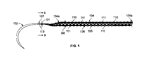
[0009] Figure 1 is a perspective view of a medical device constructed in
accordance
with the present disclosure showing a mesh suture attached to a surgical
needle via an
intervening segment.
[0010] Figure 2 is an exploded view of a portion of the surgical needle and
mesh
suture of Figure 1 shown in cross-section.
4
CA 3038198 2019-03-27
[0011] Figure 3 is a cross-sectional view of the medical device of Figure 1
taken
through line 3-3 of Figure 1.
[0012] Figures 4 and 5 are detailed views of the mesh suture of Figure 1.
[0013] Figure 6 is a cross-sectional view of the mesh wall of the suture of
Figure 1
taken through line 6-6 of Figure 5.
[0014] Figure 7 is a partial exploded view of an alternative medical device
constructed in accordance with the principles of the present disclosure.
[0015] Figure 8 is a partial cross-sectional detail view of
another alternative medical
device constructed in accordance with the principles of the present
disclosure.
DETAILED DESCRIPTION
[0016] Figure 1 depicts a medical device 100 that includes a
surgical needle 102 and
an elongated suture 104 attached to the surgical needle 102. The needle 102
can be
contoured or curved needle with a flattened cross-sectional profile, but
needles with
generally any geometry could be used. The suture 104 has a first end 104a
attached to
the needle 102 and a second end 104b located a distance away from the needle
102.
The length of the suture 104 in Fig. 1 is representative only, and in
practice, the length
could be any desirable length as discussed below. The suture 104 can include a
plurality of individual fibers 111, only a few of which are identified in
Figure 1 for
simplicity. The fibers 111 are braided, knitted, or otherwise woven, extruded,
or fused
together into a mesh construct defining a plurality of pores 110, which
advantageously
facilitate tissue incorporation, as will be discussed below.
I CA 3038198 2019-03-27
[0017] In the depicted embodiment, the needle 102 is indirectly attached to
the suture
104 by way of an intervening segment 107. The intervening segment 107 is
disposed
between the first end 104a of the elongated mesh suture 104 and the needle
102. In
this version, the intervening segment 107 includes at least some of the
plurality of fibers
111 converging from the first end 104a of the mesh suture 104 into a bundled
configuration 113 having a cross-sectional dimension D1 that is smaller than a
cross-
sectional dimension D2 of the mesh suture 104. In one alternative version, the
plurality
of fibers 111 comprising the mesh suture 104 can include a single alpha fiber
that is
thicker than or stronger than all of the remaining fibers. In this instance,
one version of
the medical device 100 can include an intervening segment 107 that includes
only the
alpha fiber extending from the first end 104a of the suture 104, such that as
the first end
104a of the mesh suture 104 transitions (e.g., tapers, converges, etc.) to the
intervening
segment 107, a length of the alpha fiber that then continues beyond to define
the
intervening segment 107 for attaching directly or indirectly to the needle 102
as
discussed in more detail below.
[0018] In some versions, the cross-sectional dimension of the mesh suture 104
can
be in a range of approximately 1 mm to approximately 10 mm, or even as large
as
approximately 25 mm. In some versions, the cross-sectional dimension of the
intervening segment 107 can be in a range of approximately 0.1 mm to
approximately
50 mm, and a length L (Figure 2) of the intervening segment 107 can be in a
range of
approximately 0.5 mm to approximately 200 mm. For most uses, the cross-
sectional
dimension of the intervening segment 107 will be in a range of approximately
0.2 mm to
6
CA 3038198 2019-03-27
approximately 20 mm, and a length L (Figure 2) of the intervening segment 107
can be
in a range of approximately 0.5 mm to approximately 50 mm.
[0019] In some versions, the cross-section of the intervening segment 107 can
be
generally circular such that the cross-sectional dimension D1 of the
intervening segment
will represent a diameter of the intervening segment 107. In some versions,
the cross-
section of the suture 104 will be either generally circular or generally flat
(e.g.,
rectangular) such that the cross-sectional dimension 02 of the suture 104 will
be either
a diameter or a width dimension of the suture 104, as will be discussed more
thoroughly
below. In some embodiments, there can be multiple intervening segments 107
(either
alone or in sequence) to indirectly attach either end of the suture 104 to the
needle 102.
In some versions, the intervening segment 107 includes only one of the
plurality of
fibers 111 converging from the first end 104a of the mesh suture 104 into
configuration
113 having a cross-sectional dimension D1 that is smaller than a cross-
sectional
dimension 02 of the mesh suture 104. In some versions, a single filament
indirectly
attaches the needle 102 to the mesh suture 104, and in some versions a portion
of the
mesh suture fibers 111 join with a cross-sectional dimension to fit into the
drill or
channel end opposite the sharp point of the needle 102. In other versions, the
single
fiber or the portion of mesh suture fibers 111 that are indirectly attached to
the needle
102 join with the longitudinal elements of the mesh suture 104 to limit
roping.
[0020] With continued reference to Figure 1, the plurality of fibers 111 taper
from the
larger cross-sectional dimension D2 at the first end 104a of the mesh suture
104 to the
smaller cross-sectional dimension D1 in the bundled configuration 113. So
configured,
7
1 CA 3038198 2019-03-27
the bundled configuration 113 of the plurality of fibers 111 in the
intervening segment
107 facilitate indirect attachment of the mesh suture to the surgical needle
102, which in
the depicted version includes a drilled needle having a blind bore 117, as
shown in
Figures 2 and 3.
[0021] In some
versions, the plurality of fibers 111 in the intervening segment 107 are
fixed together in the bundled configuration 113 by way of heat annealing,
welding,
wrapping, staking, bonding, and/or adhering. Fixing the fibers together can
help
facilitate handling and attachment to the needle 102 by disposing a terminal
end 109 of
the intervening segment 107 into the blind bore 117, as seen in Figures 1 and
3. In
other versions, the plurality of fibers 111 are not fixed together but join
solely at the
indirect attachment to the needle 102. In other versions, the fibers 111 in
the intervening
segment 107 can be held together by a sheath (not shown) made out of any type
of
material that is disposed or wrapped around the bundled configuration 113. For
example, one sheath may include a plastic sheet of material wrapped tightly
around the
bundled configuration 113, an individual fiber wrapped multiple times around
the
bundled configuration 113 and tied off, a heat shrinkable rubber tube disposed
about
the bundled configuration 113, or some other means. In some versions, after
the
intervening segment 107 is inserted into the blind bore 117, that portion of
the needle
102 may be worked with a tool, for example, to include a crimp 121 (shown in
Figure 3)
that assists with retaining the intervening segment 107 in the blind bore 117.
Alternatively, the indirect attachment can be achieved by having only a
portion or
minority of the filaments 111 reach the blind bore 107, with the other fibers
joining within
the intervening segment 107 to become the mesh suture 104a.
8
CA 3038198 2019-03-27
[0022] While the needle in Figures 1 and 2 has been described as including a
drilled
needle, in other versions, the needle can include a channeled needle or some
other
type of needle. With a channeled needle, the needle 102 would include an open
elongated channel instead of the blind bore 117. Similarly though, the
terminal end 109
of the intervening segment 107 would be inserted into the channel and the
channel
would be crimped to retain the intervening segment 107 in connection with the
needle.
With either drilled or channeled needles, it is also possible to incorporate
additional or
alternative retention means between the needle 102 and intervening segment 107
such
as adhesive, welding, staking, swaging, etc. In other versions, the needle 102
can be a
"French eye" needle where the mesh suture 104 or intervening segment 107
passes
through a continuous or discontinuous loop formed by the end of the needle 102
opposite the sharp point.
[0023] As mentioned, the intervening segment 107 comprises a bundled
configuration 113 of a plurality of fibers 111. In some versions, the
plurality of fibers 111
in the intervening segment 107 can be braided together into a configuration
with a
smaller cross-section dimension D1 than the suture 104. Thus, the intervening
segment
107 may include a tight braid to achieve this, or may include a loose braid
with the fibers
111 collapsed onto themselves, or may include a sheath or casing of some type
(not
shown) In other versions, the plurality of fibers 111 can simply be aligned
parallel
together and in close contact with each other. Other configurations are
possible. In
these configurations, the intervening segment 107 is generally non-porous. In
other
versions, however, the intervening segment 107 could be micro-porous or nano-
porous.
And in any configuration, the intervening segment 107 could include surface
texture
9
CA 3038198 2019-03-27
defined by the external geometry of the plurality of fibers 111 bundled
together, barbs,
or adhesive chemical elements to draw the filaments towards each other.
[0024] As mentioned above, the mesh suture 104 of the present disclosure can
include a tubular mesh suture, a flat mesh suture, or some other configuration
of mesh
suture. As shown in Figure 1, one version of the mesh suture 104 can include a
tubular
wall 105 extending the entire length of the suture 104 between the first and
second
ends 104a, 104b. The tubular wall 105 defines a hollow core 108. In other
versions,
less than the entire length of the suture 104 can be tubular. For example, it
is
foreseeable that either or both of the first and second ends 14a, 14b can have
a non-
tubular portion or portion of other geometry. Such non-tubular portions could
be for
serving as an intervening segment (as discussed herein throughout) for
attaching the
first end 14a of the suture 14 to the needle 12, for tying off the second end
14b, or
otherwise for example. In versions where the entire length of the suture 104
is tubular,
as shown, the entire length of the suture 104 including the ends and central
portion can
also have a generally constant or uniform cross-sectional dimension D2, i.e.,
diameter
or thickness, in the absence of stresses. That is, no portion of the suture
104 is
meaningfully larger in diameter than any other portion of the suture 104.
Moreover, no
aspect, end, or other portion of the suture 104 is intended to be or is
actually passed
through, disposed in, received in, or otherwise positioned inside of the
hollow core 108.
The hollow core 108 is adapted for receiving tissue in-growth only. In other
embodiments, substantially the entire suture 104 can be substantially flat or
planar
without a hollow core. In such versions, the suture 104 may include a single
flat suture
CA 3038198 2019-03-27
wall, and the cross-sectional dimension D2 can be a width of the flat suture
wall which is
greater than a thickness of the suture wall.
[0025] In some embodiments, the suture 104, whether tubular, flat, or
otherwise, can
have a length extending from the first end 104a to the second end 104b that is
greater
than or equal to approximately 20 cm, greater than or equal to approximately
30 cm,
greater than or equal to approximately 40 cm, greater than or equal to
approximately 50
cm, greater than or equal to approximately 60 cm, greater than or equal to
approximately 70 cm, greater than or equal to approximately 80 cm, greater
than or
equal to approximately 90 cm, and/or greater than or equal to approximately
100 cm, or
even bigger. In some embodiments of tubular sutures, the tubular wall 105 can
have a
diameter in a range of approximately 1 mm to approximately 10 mm, and even as
big as
25 mm (2.5 cm). Moreover, in some embodiment, a flat suture can have a width
in a
range of approximately 1 mm to approximately 10 mm, and even as big as
approximately 30 mm. Regardless of the shape, the suture 104 and also the
intervening segment 107 of the version described above can be constructed of a
material such as, for example, polyethylene terephthalate, nylon, polyolefin,
polypropylene, silk, polymers p-dioxanone, co-polymer of p-dioxanone, c-
caprolactone,
glycolide, L(-lactide, D(+)-lactide, meso-lactide, trimethylene carbonate,
polydioxanone
homopolymer, poly-4-hydroxybutyrate, fibers derived from spider silk,
grapheme,
stainless steel, surgical steel, titanium, aluminum, any other metals
including metal
alloys suitable for the intended purpose, and any combination(s) of the
aforementioned
materials.
11
CA 3038198 2019-03-27
[0026] So constructed, with tubular sutures 104, the tubular wall 105 of the
suture
104 can be radially deformable such that it adopts a first cross-sectional
profile in the
absence of lateral stresses and a second cross-sectional profile in the
presence of
lateral stresses. For example, in the absence of lateral stresses, the tubular
wall 105
and therefore the suture 104 depicted in Figure 1, for example, can have a
circular
cross-sectional profile, thereby exhibiting radial symmetry. In the presence
of a lateral
stress, such a suture 104 could then exhibit a partially or wholly collapsed
conformation.
The stiffness of the materials may vary from a suture that completely
collapses with
lateral stress, to a suture that retains its original profile with lateral
stress.
[0027] As mentioned above, the suture 104 of Figure 1 includes a mesh suture
104
defining a plurality of pores 110 for facilitating tissue incorporation
through the mesh
wall 105. As depicted in Figure 4, in at least one version of the medical
device 100, at
least some of the wall 105, whether tubular, flat, or otherwise, can be
macroporous
defining the plurality of pores 110 (e.g., openings, apertures, holes, etc.),
only a few of
which are expressly identified by reference number and lead line in Figures 1
and 4 for
clarity. The pores 110 extend completely through the mesh wall 105 and, in
tubular
versions, to the hollow core 108. In one version, the wall 105 can be
constructed of a
knitted, woven, or braided mesh material used in abdominal wall hernia repair.
[0028] As used herein, the term "macroporous" can include pore sizes that are
at
least greater than or equal to approximately 200 microns and, in some
versions, greater
than or equal to 500 microns. In some versions of the medical device 100, the
size of at
least some the pores 110 in the suture 104 can be in a range of approximately
500
12
CA 3038198 2019-03-27
1
microns to approximately 4 millimeters. In another version, at least some of
the pores
110 can have a pore size in the range of approximately 500 microns to
approximately
2.5 millimeters. In another version, at least some of the pores 110 can have a
pore size
in the range of approximately 1 millimeter to approximately 2.5 millimeters.
In another
version, the size of at least some of the pores 110 can be approximately 2
millimeters.
Moreover, in some versions, the pores 110 can vary in size. Some of the pores
110 can
be macroporous (e.g., greater than approximately 200 microns) and some of the
pores
110 can be microporous (e.g., less than approximately 200 microns). The
presence of
microporosity (i.e., pores less than approximately 200 microns) in such
versions of the
disclosed suture may only be incidental to the manufacturing process, which
can
including knitting, weaving, extruding, blow molding, or otherwise, but not
necessarily
intended for any other functional reason regarding biocompatibility or tissue
integration.
The presence of microporosity (i.e. some pores less than approximately 200
microns in
size) as a byproduct or incidental result of manufacturing does not change the
character
of the disclosed macroporous suture (e.g., with pores greater than
approximately 200
microns, and preferably greater than approximately 500 microns, for example),
which
facilitates tissue in-growth to aid biocompatibility, reduce tissue
inflammation, and
decrease suture pull-through.
[0029] In versions of the disclosed suture that has both macroporosity and
microporosity, the number of pores 110 that are macroporous can be in a range
from
approximately 1% of the pores to approximately 99% of the pores (when measured
by
pore cross-sectional area), in a range from approximately 5% of the pores to
approximately 99% of the pores (when measured by pore cross-sectional area),
in a
13
I CA 3038198 2019-03-27
range from approximately 10% of the pores to approximately 99% of the pores
(when
measured by pore cross-sectional area), in a range from approximately 20% of
the
pores to approximately 99% of the pores (when measured by pore cross-sectional
area), in a range from approximately 30% of the pores to approximately 99% of
the
pores (when measured by pore cross-sectional area), in a range from
approximately
50% of the pores to approximately 99% of the pores (when measured by pore
cross-
sectional area), in a range from approximately 60% of the pores to
approximately 99%
of the pores (when measured by pore cross-sectional area), in a range from
approximately 70% of the pores to approximately 99% of the pores (when
measured by
pore cross-sectional area), in a range from approximately 80% of the pores to
approximately 99% of the pores (when measured by pore cross-sectional area),
or in a
range from approximately 90% of the pores to approximately 99% of the pores
(when
measured by pore cross-sectional area).
[0030] So configured, the pores 110 in the suture 104 are arranged and
configured
such that the suture 104 is adapted to facilitate and allow tissue in-growth
and
integration through the pores 110 in the mesh wall 105 when introduced into a
body.
That is, the pores 110 are of sufficient size to achieve maximum
biocompatibility by
promoting local/normal tissue in-growth through the pores 110 of the suture
104 and,
with tubular sutures, into the hollow core 108. As such, tissue growth through
the pores
110 enables the suture 104 and resultant tissue to combine and cooperatively
increase
the strength and efficacy of the medical device 100, while also decreasing
irritation,
inflammation, local tissue necrosis, and likelihood of pull through. Instead,
the suture 14
14
CA 3038198 2019-03-27
1
promotes the production of healthy new tissue throughout the suture construct
including
inside the pores 110, and with tubular sutures 104, the hollow core 108.
[0031] While a tubular version of the suture 104 has been described as
including a
single elongated hollow core 108, in some embodiments, a suture according to
the
present disclosure can comprise a tubular wall defining a hollow core
including one or
more interior voids (e.g., extending the length of the suture). In some
versions, at least
some of the interior voids can have a size or diameter > approximately 200
microns, >
approximately 300 microns, > approximately 400 microns, > approximately 500
microns,
> approximately 600 microns, > approximately 700 microns, > approximately 800
microns, > approximately 900 microns, > approximately 1 millimeter, or >
approximately
2 millimeters. In some embodiments, a suture according to the present
disclosure can
comprise a tubular wall defining a hollow core including one or more (e.g., 1,
2, 3, 4, 5,
6, 7, 8, or more) lumens (e.g., running the length of the suture). In some
embodiments,
a suture according to the present disclosure can comprise a tubular wall
defining a
hollow core including a honeycomb structure, a 30 lattice structure, or other
suitable
interior matrix, which defines one or more interior voids. In some versions,
at least
some of the interior voids in the honeycomb structure, 3D lattice structure,
or other
suitable matrix can have a size or diameter > approximately 200 microns, >
approximately 300 microns, > approximately 400 microns, > approximately 500
microns,
> approximately 600 microns, > approximately 700 microns, > approximately 800
microns, > approximately 900 microns, > approximately 1 millimeter, or >
approximately
2 millimeters. In some embodiments, a void comprises a hollow core. In some
embodiments, a hollow core can include a hollow cylindrical space in the
tubular wall,
1 CA 3038198 2019-03-27
but as described, the term "hollow core" is not limited to defining a
cylindrical space, but
rather could include a labyrinth of interior voids defined by a honeycomb
structure, a 3D
lattice structure, or some other suitable matrix. In some embodiments, sutures
comprise a hollow, flexible structure that has a circular cross-sectional
profile in its non-
stressed state, but which collapses into a more flattened cross-sectional
shape when
pulled in an off-axis direction. In some embodiments, sutures are provided
that exhibit
radial symmetry in a non-stressed state. In some embodiments, radial symmetry
in a
non-stressed state eliminates the need for directional orientation while
suturing. In
some embodiments, sutures are provided that exhibit a flattened cross-
sectional profile
when off-axis (longitudinal axis) force is applied (e.g., tightening of the
suture against
tissue), thereby more evenly distributing the force applied by the suture on
the tissue.
In some embodiments, sutures are provided that exhibit a flattened cross-
sectional
profile when axial force is applied. In some embodiments, sutures comprise
flexible
structure that adopts a first cross-sectional profile in its non-stressed
state (e.g.,
suturing profile), but adopts a second cross-sectional shape when pulled in an
off-axis
direction (e.g., tightened profile). In some embodiments, a suture is hollow
and/or
comprises one or more internal voids (e.g., that run the length of the
suture). In some
embodiments, internal voids are configured to encourage the suture to adopt a
preferred conformation (e.g., broadened leading edge to displace pressures
across the
contacted tissue) when in a stressed states (e.g., tightened profile). In some
embodiments, internal voids are configured to allow a suture to adopt radial
exterior
symmetry (e.g., circular outer cross-sectional profile) when in a non-stressed
state. In
some embodiments, varying the size, shape, and/or placement of internal voids
alters
16
CA 3038198 2019-03-27
one or both of the first cross-sectional profile (e.g., non-stressed profile,
suturing profile)
and second cross-sectional profile (e.g., off-axis profile, stressed profile,
tightened
profile). In some embodiments, an internal element is absorbed over time,
rendering the
space confined by the outer mesh changing as to shape and size. In some
elements,
the space confined by the outer mesh is used to deliver cells or medicaments
for
delivery to the tissues.
[0032] Sutures,
which are substantially linear in geometry, have two distinct ends,
as described above with reference to Figure 1, for example. In some
embodiments,
both ends are identical. In some embodiments, each end is different. In some
embodiments, one or both ends are structurally unadorned. In some embodiments,
the
end away from the needle 102 is a free end, has a taper, is attached to a
barb, is a loop,
is attached to another needle directly or indirectly, or is attached
indirectly to a planar
mesh. In some embodiments, one or more ends is attached to or at least
configured for
attachment to a needle via swaging, sonic welding, adhesive, tying, or some
other
means (as shown Figure 1). In some embodiments, the second end 104b of the
suture
104 is configured to include an anchor for anchoring the suture 104 against
the tissue
through which the suture 104 is inserted. In some embodiments, the second end
104b
of the suture 104 is configured to anchor the suture at the beginning of the
closure. In
some embodiments, the second end 104b of the suture 104 includes an anchor
that is a
structure that prevents the suture 104 from being pulled completely through
the tissue.
In some embodiments, the anchor has a greater dimension than the rest of the
suture
104 (at least 10% greater, at least 25% greater, at least 50% greater, at
least 2-fold
greater, at least 3-fold greater, at least 4-fold greater, at least 5-fold
greater, at least 6-
17
CA 3038198 2019-03-27
fold greater, at least 10-fold greater, etc.). In some embodiments, the anchor
comprises
a structure with any suitable shape for preventing the suture 104 from being
pulled
through the hole (e.g., ball, disc, plate, cylinder), thereby preventing the
suture 14 from
being pulled through the insertion hole. In some embodiments, the anchor of
the suture
104 comprises a closed loop. In some embodiments, the closed loop is of any
suitable
structure including, but not limited to a crimpled loop, flattened loop, or a
formed loop.
In some embodiments, a loop can be integrated into the end of the suture 104.
In some
embodiments, a separate loop structure can be attached to the suture 104. In
some
embodiments, the needle 102 can be passed through the closed loop anchor to
create a
cinch for anchoring the suture 104 to that point. In some embodiments, the
anchor can
comprise one or more structures (e.g., barb, hook, etc.) to hold the end of
the suture
104 in place. In some embodiments, one or more anchor 22 structures (e.g.,
barb,
hook, etc.) are used in conjunction with a closed loop to ratchet down the
cinch and hold
its position. In some embodiments, a knotless anchoring system can be
provided. In
some embodiments, a needle can be attached to the second end 104b to create a
double armed suture. In some embodiments, a single mesh suture or multiple
mesh
sutures are attached through indirect attachments to a larger device such as a
reconstruction mesh or implant to aid in deployment of the larger device.
[0033] In some embodiments, and as briefly mentioned relative to Figure 1,
the
present disclosure provides suturing needles with cross-sectional profiles
indirectly
attached to a mesh suture via an intervening segment and configured to prevent
suture
pull-through and methods of use thereof. In some embodiments, suturing needles
are
provided comprising cross-section shapes (e.g. flat, elliptical, transitioning
over the
18
CA 3038198 2019-03-27
length of the needle, etc.) that reduce tension against the tissue at the
puncture site and
reduce the likelihood of tissue tear. In some embodiments, one cross-sectional
dimension of the needle is greater than the orthogonal cross-sectional
dimension (e.g.,
1.1x greater, 1.2x greater, 1.3x greater, 1.4x greater, 1.5x greater, 1.6x
greater, 1.7x
greater, 1.8x greater, 1.9x greater, >2x greater, 2.0x greater, 2.1x greater,
2.2x greater,
2.3x greater, 2.4x greater, 2.5x greater, 2.6x greater, 2.7x greater, 2.8x
greater, 2.9x
greater, 3.0x greater, >3.0x greater, 3.1x greater, 3.2x greater, 3.3x
greater, 3.4x
greater, 3.5x greater, 3.6x greater, 3.7x greater, 3.8x greater, 3.9x greater,
4.0x greater,
>4.0x greater... >5.0x greater... >6.0x greater... >7.0x greater... >8.0x
greater... >9.0x
greater... >10.0x greater). In some embodiments, suturing needles are provided
circular in shape at its point (e.g., distal end), but transition to a
flattened profile (e.g.,
ribbon-like) to the rear (e.g. proximal end). In some embodiments, the face of
the
flattened area is orthogonal to the radius of curvature of the needle. In some
embodiments, suturing needles create a slit (or flat puncture) in the tissue
as it is
passed through, rather than a circle or point puncture. In some embodiments,
suturing
needles are provided circular in shape at its point (e.g., distal end), but
transition to a 2D
cross-sectional profile (e.g., ellipse, crescent, half moon, gibbous, etc.) to
the rear (e.g.
proximal end). In some embodiments, suturing needles provided herein find use
with
the sutures described herein. In some embodiments, suturing needles find use
with
sutures of the same shape and/or size. In some embodiments, suturing needles
and
sutures are not of the same size and/or shape. In some embodiments, suturing
needles
provided herein find use with traditional sutures. Various types of suture
needles are
well known in the art. In some embodiments, suturing needles provided herein
19
CA 3038198 2019-03-27
comprise any suitable characteristics of suturing needles known to the field,
but
modified with dimensions described herein. Any introduction device of the mesh
suture
through tissue is defined as a needle, and therefore we do not limit our
embodiments to
those defined here, but rather any sharp instrument that can penetrate tissue
to pass
the suture.
[0034] In some embodiments, the present disclosure also provides
compositions,
methods, and devices for anchoring the suture at the end of the closure (e.g.,
without
tying the suture to itself). In some embodiments, one or more securing
elements (e.g.,
staples) are positioned over the terminal end of the suture to secure the end
of the
closure. In some embodiments, one or more securing elements (e.g., staples)
are
secured to the last "rung" of the suture closure (e.g., to hold the suture
tight across the
closure). In some embodiments, a securing element is a staple. In some
embodiments,
a staple comprises stainless steel or any other suitable material. In some
embodiments,
a staple comprises a plurality of pins that can pass full thickness through 2
layers of
suture. In some embodiments, staple pins are configured to secure the suture
end
without cutting and/or weakening the suture filament. In some embodiments, a
staple
forms a strong joint with the suture. In some embodiments, a staple is
delivered after
the needle is cut from the suture. In some embodiments, a staple is delivered
and the
needle removed simultaneously
[0035] In some embodiments, the present disclosure provides devices (e.g.,
staple guns) for delivery of a staple into tissue to secure the suture end. In
some
embodiments, a staple deployment device simultaneously or near-simultaneously
CA 3038198 2019-03-27
delivers a staple and removes the needle from the suture. In some embodiments,
a
staple deployment device comprises a bottom lip or shelf to pass under the
last rung of
suture (e.g., between the suture and tissue surface) against which the pins of
the staple
can be deformed into their locked position. In some embodiments, the bottom
lip of the
staple deployment device is placed under the last rung of suture, the free
tail of the
suture is placed within the stapling mechanism, and the suture is pulled
tight. In some
embodiments, while holding tension, the staple deployment device is activated,
thereby
joining the two layers of suture together. In some embodiments, the device
also cuts off
the excess length of the free suture tail. In some embodiments, the staple
deployment
device completes the running suture and trims the excess suture in one step.
In some
embodiments, a suture is secured without the need for knot tying. In some
embodiments, only 1 staple is needed per closure. In some embodiments, a
standard
stapler is used to apply staples and secure the suture end. In some
embodiments, a
staple is applied to the suture end manually. The staple may or may not have
tissue
integrative properties.
[0036] In some embodiments, sutures provided herein provide tissue
integrative
properties to increase the overall strength of the repair (e.g., at an earlier
time-point
than traditional sutures). In some embodiments, sutures are provided with
enhanced
tissue adhesion properties. In some embodiments sutures are provided that
integrate
with the surrounding tissue. In some embodiments, tissue integrative
properties find
use in conjunction with any other suture characteristics described herein. In
some
embodiments, sutures allow integration of healing tissue into the suture. In
some
embodiments, tissue growth into tubular sutures and/or through flat sutures is
promoted
21
CA 3038198 2019-03-27
(e.g., by the surface texture of the suture). In some embodiments, tissue
growth into
the suture prevents sliding of tissue around suture, and/or minimizes
micromotion
between suture and tissue. In some embodiments, tissue in-growth into tubular
sutures
and/or through flat sutures increases the overall strength of the repair by
multiplying the
surface area for scar in establishing continuity between tissues.
Conventionally, the
strength of a repair is dependent only on the interface between the two tissue
surfaces
being approximated. In some embodiments in-growth of tissue into the suture
adds to
the surface area of the repair, thereby enhancing its strength. In some
embodiments,
increasing the surface area for scar formation, the closure reaches
significant strength
more quickly, narrowing the window of significant risk of dehiscence.
[0037] In some embodiments, the surface and/or internal texture of a suture
promotes tissue adhesion and/or ingrowth. In some embodiments, as discussed
above
specifically with reference to Figure 1, a suture of the present disclosure
can comprise a
porous (e.g., macroporous) and/or textured material. In some embodiments, a
suture
comprises a porous (e.g., macroporous) and/or textured exterior. In some
embodiments, pores in the suture allow tissue in-growth and/or integration. In
some
embodiments, a suture comprises a porous ribbon-like structure, instead of a
tubular
like structure. In some embodiments, a porous suture comprises a 2D cross-
sectional
profile (e.g., elliptical, circular (e.g., collapsible circle), half moon,
crescent, concave
ribbon, etc.). In some embodiments, a porous suture comprises polypropylene or
any
other suitable suture material as discussed above. In some embodiments, pores
are
between 500 pm and 3.5 mm or greater in diameter (e.g., e.g., >500 pm in
diameter
(e.g., ,>500 pm, >600 pm , >700 pm , 800 pm, >900 pm, >1 mm, or more ). In
some
22
CA 3038198 2019-03-27
embodiments pores are of varying sizes. In some embodiments, a suture
comprises
any surface texture suitable to promote tissue in-growth and/or adhesion. In
some
embodiments, suitable surface textures include, but are not limited to
ribbing, webbing,
mesh, barbs, barbs with different directions or geometries, grooves, etc. In
some
embodiments, the suture may include filaments or other structures (e.g., to
provide
increased surface area and/or increased stability of suture within tissue). In
some
embodiments, interconnected porous architecture is provided, in which pore
size,
porosity, pore shape and/or pore alignment facilitates tissue in-growth.
[0038] In some embodiments, a suture comprises a mesh and/or mesh-like
exterior. In some embodiments, a mesh exterior provides a flexible suture that
spreads
pressure across the closure site, and allows for significant tissue in-growth.
In some
embodiments, the density of the mesh is tailored to obtain desired
flexibility, elasticity,
and in-growth characteristics.
[0039] In some embodiments, a suture is coated and/or embedded with
materials
to promote tissue ingrowth. Examples of biologically active compounds that may
be
used sutures to promote tissue ingrowth include, but are not limited to, cell
attachment
mediators, such as the peptide containing variations of the "RGD" integrin
binding
sequence known to affect cellular attachment, biologically active ligands, and
substances that enhance or exclude particular varieties of cellular or tissue
ingrowth.
Such substances include, for example, laminin and other extracellular
matrices, tissue
inductive scaffolds, osteoinductive substances, such as bone morphogenic
proteins
(BMP), epidermal growth factor (EGF), fibroblast growth factor (FGF), platelet-
derived
23
CA 3038198 2019-03-27
growth factor (PDGF), insulin-like growth factor (IGF-I and II), TGF-13, etc.
Examples of
pharmaceutically active compounds that may be used to promote tissue ingrowth
include, but are not limited to, acyclovir, cephradine, malfalen, procaine,
ephedrine,
adriomycin, daunomycin, plumbagin, atropine, guanine, digoxin, quinidine,
biologically
active peptides, chlorin e<sub>6</sub>, cephalothin, proline and proline analogues
such as cis-
hydroxy-L-proline, penicillin V, aspirin, ibuprofen, steroids,
antimetabolites,
immunomodulators, nicotinic acid, chemodeoxycholic acid, chlorambucil, and the
like.
Therapeutically effective dosages may be determined by either in vitro or in
vivo
methods.
[0040] Sutures
are well known medical devices in the art. In some embodiments,
sutures have braided or monofilament constructions. In some embodiments
sutures are
provided in single-armed or double-armed configurations with a surgical needle
mounted to one or both ends of the suture, or may be provided without surgical
needles
mounted. In some embodiments, the end of the suture distal to the needle
comprises
one or more structures to anchor the suture. In some embodiments, the distal
end of
the suture comprises one or more of a: closed loop, open loop, anchor point,
barb,
hook, etc. In some embodiments, sutures comprise one or more biocompatible
materials. In some embodiments, sutures comprise one or more of a variety of
known
bioabsorbable and nonabsorbable materials. For example, in some embodiments,
sutures comprise one or more aromatic polyesters such as polyethylene
terephthalate,
nylons such as nylon 6 and nylon 66, polyolefins such as polypropylene, silk,
and other
nonabsorbable polymers. In some embodiments, sutures comprise one or more
polymers and/or copolymers of p-dioxanone (also known as 1,4-dioxane-2-one), E-
24
CA 3038198 2019-03-27
caprolactone, glycolide, L(-)-lactide, D(+)-lactide, meso-lactide, poly-4-
hydroxybutyrate,
trimethylene carbonate, fibers derived from spider silk, graphene, and
combinations
thereof. In some embodiments, sutures comprise polydioxanone homopolymer. The
above listing of suture materials should not be viewed as limiting. In some
embodiments, the disclosed sutures can be constructed of metal filaments such
as
stainless steel filaments. Suture materials and characteristics are known in
the art. Any
suitable suture materials or combinations thereof are within the scope of the
present
disclosure. In some embodiments, sutures comprise sterile, medical grade,
surgical
grade, and or biodegradable materials. In some embodiments, a suture is coated
with,
contains, and/or elutes one or more bioactive substances (e.g., antiseptic,
antibiotic,
anesthetic, promoter of healing, etc.). In some embodiments, the suture
filaments
and/or the hollow core 108 of any of the disclosed sutures can contain a drug
product
for delivery to the patient, the medicament could take the form of a solid, a
gel, a liquid,
or otherwise. In some embodiments, the suture filaments and or the hollow core
108 of
any of the disclosed sutures can be seeded with cells or stem cells to promote
healing,
ingrowth or tissue apposition.
[0041] In some embodiments, the structure and material of the suture
provides
physiologically-tuned elasticity. In some embodiments, a suture of appropriate
elasticity
is selected for a tissue. In some embodiments, suture elasticity is matched to
a tissue.
For example, in some embodiments, sutures for use in abdominal wall closure
will have
similar elasticity to the abdominal wall, so as to reversibly deform along
with the
abdominal wall, rather than act as a relatively rigid structure that would
carry higher risk
of pull-through. In some embodiments, elasticity would not be so great
however, so as
CA 3038198 2019-03-27
to form a loose closure that could easily be pulled apart. In some
embodiments,
deformation of the suture would start occurring just before the elastic limit
of its
surrounding tissue, e.g., before the tissue starts tearing or irreversibly
deforming.
[0042] In some embodiments, sutures described herein provide a suitable
replacement or alternative for surgical repair meshes (e.g., those used in
hernia repair).
In some embodiments, the use of sutures in place of mesh reduces the amount of
foreign material placed into a subject. In some embodiments, the decreased
likelihood
of suture pull-through allows the use of sutures to close tissues not possible
with
traditional sutures (e.g., areas of poor tissue quality (e.g., muscle tissue
lacking fascia,
friable or weak tissue) due to conditions like inflammation, fibrosis,
atrophy, denervation,
congenital disorders, attenuation due to age, or other acute and chronic
diseases). Like
a surgical mesh, sutures described herein permit a distribution of forces
greater than
that achieved by standard sutures delocalizing forces felt by the tissue and
reducing the
chance of suture pull-though and failure of the closure.
[0043] In some embodiments, sutures are permanent, removable, or
absorbable.
In some embodiments, permanent sutures provide added strength to a closure or
other
region of the body, without the expectation that the sutures will be removed
upon the
tissue obtaining sufficient strength. In such embodiments, materials are
selected that
pose little risk of long-term residency in a tissue or body. In some
embodiments,
removable sutures are stable (e.g., do not readily degrade in a physiological
environment), and are intended for removal when the surrounding tissue reaches
full
closure strength. In some embodiments, absorbable sutures integrate with the
tissue in
26
CA 3038198 2019-03-27
the same manner as permanent or removable sutures, but eventually (e.g., >1
week, >
2 weeks, >3 weeks, >4 weeks, >10 weeks, >25 weeks, > 1 year) biodegrade and/or
are
absorbed into the tissue after having served the utility of holding the tissue
together
during the post-operative and/or healing period. In some embodiments
absorbable
sutures present a reduced foreign body risk.
[0044] Although prevention of dehiscence of abdominal closures (e.g.,
hernia
formation) is specifically described at an application of embodiments of the
present
disclosure, the sutures described herein are useful for joining any tissue
types
throughout the body. In some embodiments, sutures described herein are of
particular
utility to closures that are subject to tension and/or for which cheese-wiring
is a concern.
Exemplary tissues within which the present disclosure finds use include, but
are not
limited to: connective tissue, fascia, ligaments, muscle, dermal tissue,
cartilage, tendon,
or any other soft tissues. Exemplary tissues also include bone. Specific
applications of
sutures described herein include reattachments, plication, suspensions,
slings, etc.
Sutures described herein find use in surgical procedures, obstetrics and
cervical
cerclage, non-surgical medical procedures, veterinary procedures, in-field
medical
procedures, etc. The scope of the present disclosure is not limited by the
potential
applications of the sutures described herein.
[0045] One method of manufacturing a medical device in accordance with the
present disclosure can include forming a plurality of fibers 111 into a
tubular mesh
suture 104 with a tubular wall 105 having a plurality or pores 110 and
defining a hollow
core 108, each pore 110 having a pore size that is greater than 200 microns.
In some
27
CA 3038198 2019-03-27
version, this can include braiding or knitted the fibers 111 together around a
mandrel,
for example, and then subsequently removing the mandrel. In some versions, the
fibers
111 may be fixed together where they cross or intersect each other. This
fixation may
include applying an adhesive, staking, heating, compressing, welding the
fibers 111
together, or otherwise. This fixation may occur before or after the mandrel is
removed.
[0046] Additionally, the method of manufacturing can include directly
attaching either
the first end 104a or the second end 104b, or even both ends of the mesh
suture 104 to
the surgical needle 102. Attaching the suture 104 to the needle 102 may also
include
forming the intervening segment 107, and then attaching the intervening
segment 107
to the needle 102 such that the suture 104 is indirectly attached to the
needle 102. As
discussed above, in one version, forming the intervening segment 107 can
include
collecting at least some of the plurality of fibers 111 extending from the
first end 104a of
the mesh suture 104 and arranging them in a bundled configuration 113 that has
a
cross-sectional dimension D1 smaller than a cross-sectional dimension D2 of
the suture
104. In some versions, this includes braiding, bonding, compressing, adhering,
or
knitting the plurality of fibers 111 into the bundled configuration 113. In
some other
versions, this can include arranging the plurality of fibers 111 parallel to
each other and
in contact with each other with or without the use of a cap, cover, or sheath
to contain
and compress the fibers down to the size of a conventional surgical needle for
purposes
of attachment. In other versions, a minority of fibers or even a single fiber
are
manufactured to reach the needle indirectly.
28
CA 3038198 2019-03-27
[0047] Finally, forming the intervening segment 107 includes fixing the
plurality of
fibers 111 in the bundled configuration 113 together, as mentioned above. This
can be
achieved by applying heat to secure the fibers 111 together, applying adhesive
to
adhere the fibers 111 together, applying energy (e.g., sonic energy, laser
energy, etc.)
to weld the fibers 111 together, staking the fibers 111 together, compressing
the fibers
111 together with pressure, or some other process alone or in combination with
the
above. In still other methods, forming the intervening segment 107 can include
placing
a cap or cover, wrapping a plastic sheet, shrinking a rubber tube, or tying an
individual
filament around the fibers 111 to maintain the bundled configuration 113.
[0048] With the intervening segment 107 formed, the terminal end 109 can be
inserted into the blind bore 117 of the needle 102 and the needle 102 can
optionally be
crimped. In some versions, a further or alternative step of fixing the
intervening
segment 107 into the blind bore 109 with an adhesive, or some other process
such as
welding, bonding, staking, etc., can be performed. In other versions where the
needle
102 includes a channeled needle, the step of attaching the needle 102 to the
intervening segment 107 of course includes at least disposing the terminal end
109 in
the channel and crimping the channel.
[0049] As discussed, forming the tubular wall 105 can include forming a tube
from a
mesh material. The tubular mesh wall 105 may be formed by directly weaving,
braiding,
or knitting fibers into a tube shape. Alternatively, forming the tubular mesh
wall 16 can
include weaving, braiding, or knitting fibers into a planar sheet and
subsequently
forming the planar sheet into a tube or flat shape. Finally, as mentioned
throughout,
29
CA 3038198 2019-03-27
forming the mesh suture 104 can include forming a flat planar mesh wall,
instead of a
tubular mesh wall 105. In this configuration, the same steps as those stated
above
would similarly apply with the exception of using a mandrel to form the tube.
Instead,
the flat planar mesh wall would simply be braided, knitted, or otherwise
formed or even
cut from a larger sheet of pre-formed mesh. Of course, other manufacturing
possibilities including extrusion exist and manipulating a plurality of fibers
is not the only
possibility for creating a porous mesh wall within the scope of the present
disclosure,
but rather, are mere examples.
[0050] Still further, a method of manufacturing a medical device 100 in
accordance
with the present disclosure can include providing an anchor on the second end
104b of
the wall 105 opposite the needle 102. In some versions of the method, and as
one
example only, providing the anchor can be as simple as forming a loop.
[0051] In some embodiments, the mesh wall 105 can be divided into two or
more
mesh wall portions by one or more intervening features such as knots,
inflexible rod-like
members, monofilament or multi-filament suture segments, etc. Such a construct
can
be referred to as a segmented mesh suture constructed in accordance with the
present
disclosure.
[0052] One optional feature of the medical device 100 of Figures 1-6 is that
it can
include one or more anti-roping elements 106, which are best seen in Figure 4.
That is,
the medical device 100 can include one or more, or a plurality of, anti-roping
elements
106 in the form of elongated elements 106 extending substantially (or
entirely) the entire
length of the suture 104 between the first and second ends 104a, 104b. The
elongated
CA 3038198 2019-03-27
elements 106 are fixed (or are not fixed) to the mesh wall 105 of the suture
104 at a
plurality of points P and thereby serve to resist elongation of the suture 104
upon the
application of an axial tensile load to the medical device 100. In some
embodiments,
the elongated elements 106 can be fixed to the mesh wall 105 in any available
manner
including, without limitation, welding, gluing, tying, braiding, heating,
staking, dipping,
chemically bonding, etc. In some embodiments, the elongated elements 106 are
not
fixed to the individual fibers of a tubular mesh walled suture 104. In some
embodiments,
the various fibers that make up the mesh wall 105 of any of the sutures
described
herein can also be fixed together at the intersection between fibers/filaments
in any
available manner including, without limitation, welding, gluing, tying,
braiding, heating,
staking, dipping, chemically bonding, etc. As shown in Figure 5 illustrating a
tubular
suture 104, for example, the present version of the anti-roping elements 106
can be
arranged such that each anti-roping element 106 is interleaved between
adjacent
elements of the remainder of the mesh suture 104, which can add to the
integrity and
stability of the suture 104. In other embodiments, the anti-roping elements
106 can be
positioned entirely on an outer perimeter or on an inner perimeter of the
tubular suture
104. In other embodiments, some of the elements 106 can be positioned on an
inner
perimeter, some can be positioned on an outer perimeter, and/or some can be
interleaved such as depicted in Figure 5. In other embodiments, some or all of
the anti-
roping elements may reside in the central core of a tubular mesh suture 104.
In some
embodiments, the anti-roping elements themselves are not entirely linear
single
filaments, but rather are a braid of fine filaments that act to run the length
of the suture
either obliquely or in step-wise fashion to resist elongation.
31
CA 3038198 2019-03-27
[0053] As mentioned above, "roping" is a phenomenon in the weaving industry
whereby woven, braided, or knitted mesh materials tend to elongate under
tension.
This elongation can cause the various elements that make up the mesh material
to
collapse relative to each other and thereby reduce (e.g., close) the size of
the pores
disposed in the mesh. As such, the "anti-roping" elements 106 of the present
disclosure, which are embodied as longitudinal elements in Figures 1-6,
advantageously
resist this elongation of the mesh suture and collapsing of the pores when the
suture
experiences axial tensile loads. This resistance is achieved because the anti-
roping
elements adds structural integrity to the overall construct and prevents the
various mesh
elements from moving relative to each other and/or deforming under tension. By
maintaining the desired structural configuration of the mesh suture during and
after
threading into soft tissue, the pores remain appropriately sized to facilitate
tissue
integration and the overall width and/or dimension of the suture remains
appropriately
sized to limit and/or prevent suture pull through. These anti-roping elements
may or
may not continue and form the indirect attachment to the needle.
[0054] In Figures 1-6, the anti-roping elements 106 are each substantially
straight
(aka, substantially linear). In other embodiments, however, one or more the
anti-roping
elements 106 could foreseeably have different shapes, including for example, S-
shaped, U-shaped, Zig-zag shaped, etc. Additionally, in Figures 1-6, each of
the anti-
roping elements 106 is a separate element. But, in other embodiments, any two
or
more of the elements 106 can be connected such that a single element 106 may
extend
the length of the suture 104, then include a U-shaped turn, and extend back
along the
length of the suture 104 adjacent to (e.g., parallel to) the preceding length.
Also, in
32
CA 3038198 2019-03-27
Figures 1-6, the anti-roping elements 106 are disposed parallel to each other
and are
equally spaced apart from each other. In alternative versions, the anti-roping
elements
106 could have unequal spacing and/or could be disposed in a non-parallel
manner.
Further still, in Figures 1-6, the anti-roping elements 106 are depicted as
having a
thickness that is generally the same as the thickness of the other elements
forming the
mesh construct of the elongated suture 104. In other embodiments, any one or
more of
the anti-roping elements 106 could be thicker or thinner than the other
elements forming
the mesh construct of the elongated suture 104. Further yet, while Figures 1-6
show
four (4) anti-roping elements, alternative embodiments could include any
number so
long as the desired objective is achieved without compromising or detracting
from the
macroporous character of the suture 104. Finally, while Figures 1-6 illustrate
a hollow
tubular suture 104, other embodiments of the medical device 100 as mentioned
could
include other geometries including, for example, a planar (e.g., flat ribbon)
geometry.
Therefore, it can be understood based on the foregoing description that the
anti-roping
elements 106 on such planar sutures 104 could include a plurality of
substantially
straight elements extending the length of the suture 104, and being parallel
to each
other and equally spaced apart. Alternatively, the anti-roping elements 106 on
the
planar suture 104 could take on any of the alternative constructs discussed
with respect
to the tubular construct expressly depicted in Figures 1-6.
[0055] As mentioned throughout the foregoing, some embodiments of the mesh
wall
105 of the suture 104 of the present disclosure can be flat as opposed to
tubular in
construction. The foundational mesh of a flat suture 104 can be constructed in
a
manner similar to the foundational mesh of the tubular versions described
above. For
33
CA 3038198 2019-03-27
example, one method of manufacturing a flat suture 104 includes manufacturing
a flat
mesh wall 105 by weaving, braiding, or knitting fibers into a flat wall shape
having some
predefined width and length dimension. Alternatively, forming the flat mesh
wall 105
can include weaving, braiding, or knitting fibers into a planar mesh sheet and
subsequently cutting the planar sheet into strips.
[0056] Throughout the foregoing description, the medical device 100 of the
present
disclosure has been mostly described as including a mesh suture 104, a needle
102,
and a single intervening segment 107. In other versions, the medical device
100 can
include a plurality of intervening segments.
[0057] For example, Figure 7 depicts a detail view of one alternative medical
device
100 including a mesh suture 104, needle 102, and first and second intervening
segments 107a, 107b. The first intervening segment 107a is at least partially
formed as
part of the mesh suture 104 in a manner similar to the intervening segment 107
described throughout the present disclosure. The second intervening segment
107b is
formed as the distal part of the needle 102. So configured, the first and
second
intervening segments 107a, 107b are adapted to be connected together to attach
the
needle 102 to the suture 104. More specifically, in the version depicted in
Figure 7, the
first intervening segment 107a includes a male locking feature 131a disposed
on a
terminal end 119 of the intervening segment 107, and the second intervening
segment
107b includes a female locking feature 131b. The male and female locking
features
131a, 131b are each constructed of any relatively rigid biocompatible
material. That is,
in some versions, the male locking feature 131a has a locking protrusion 133a
and can
34
CA 3038198 2019-03-27
be constructed of a plastic component welded, swaged, or otherwise fixed to
the
terminal end 119 of the first intervening segment 107a, and the female locking
feature
131b has a locking aperture 133b and can be formed as part of the metal
material of the
needle 102. To attach the needle 102 to the suture 104, the locking protrusion
133a of
the male locking feature 131a is simply snap-fit into the locking aperture
133b of the
female locking feature 131b. The features 131a, 131b can be retained together
with
friction, mechanical interlock, adhesive, magnetic elements, ball and socket,
compression fit, or otherwise. Note that in some embodiments, the inter-
positioning of
the male and female intervening segments described above can also be reversed
such
that the male and female locking protrusion and aperture features are opposite
to that
described above.
[0058] Likewise, while the means for connecting the intervening segment 107 to
the
needle 102 has included either inserting a portion of the intervening segment
107 into
the blind bore 117 or into a channel (not shown) formed in the needle 102,
other
versions of intervening segment arrangements are also contemplated. For
example,
Figure 8 depicts an alternative version where the intervening segment 107
includes first
and second sequentially or serially arranged intervening segments 107a, 107b,
where
the second intervening segment 107b (shown in cross-section in Figure 8)
includes a
cylindrical collar defining a female bore or channel 119 and the needle 102
includes a
male protrusion 135 that is disposed in the female bore or channel 119. The
first
intervening segment 107b in Figure 8 is essentially the same as those
described above
in reference to Figures 1-6. In Figure 8, the first intervening segment 107a
and the
male protrusion 135 of the needle 102 can be secured into the blind bore or
channel
CA 3038198 2019-03-27
119 of the second intervening segment 107b in any manner mentioned hereinabove
relative to the intervening segment 107 and needle 102 in Figures 1-6. As
described
above, in some embodiments, the inter-positioning of these male and female
protrusions and channels or bores of the intervening segments described above
can be
reversed such that the male and female protrusion and channel or bore features
are
opposite to that described above. In some embodiments, both male to male and
female
to female intervening segment attachments are also contemplated, such as by
the use
of adhesive or other commonly known joining or bonding methods.
[0059] Although the disclosure has been described in connection with specific
preferred embodiments, it should be understood that the disclosure as claimed
should
not be unduly limited to such specific embodiments. Indeed, various
modifications of
the described modes for carrying out the disclosure would be apparent to those
skilled
in the relevant fields are intended to be within the scope of the present
disclosure. For
example, and importantly, although the application includes discrete
descriptions of
different embodiments of the invention, it can be understood that any features
from one
embodiment can be easily incorporated into any one or more of the other
embodiments.
36
CA 3038198 2019-03-27