Note: Descriptions are shown in the official language in which they were submitted.
CA 03039302 2019-04-03
WO 2018/096531 PCT/IL2017/051273
BLOOD PUMPS
CROSS-REFERENCES TO RELATED APPLICATIONS
The present application claims priority from US Provisional Patent Application
62/425,814 to Tuval, filed Nov. 23, 2016, entitled "Blood pumps," which is
incorporated
herein by reference.
FIELD OF EMBODIMENTS OF THE INVENTION
Some applications of the present invention generally relate to medical
apparatus.
Specifically, some applications of the present invention relate to apparatus
and methods
associated with placing a pump in one or more of a subject's renal veins,
and/or in the
subject's vena cava.
BACKGROUND
It is common for cardiac dysfunction or congestive heart failure to develop
into
kidney dysfunction, which, in turn, causes congestive heart failure symptoms
to develop
or worsen. Typically, systolic and/or diastolic cardiac dysfunction causes
systemic
venous congestion, which gives rise to an increase in renal venous and
interstitial
pressure. The increase in the pressure causes fluid retention by the body to
increase due
both to kidney dysfunction and renal neurohormonal activation, both of which
typically
develop as a result of the increase in renal venous and interstitial pressure.
The resulting
fluid retention causes congestive heart failure to develop or worsen, by
causing a blood
volume overload at the heart and/or by increasing systemic resistance.
Similarly, it is
common for kidney dysfunction and/or renal neurohormonal activation to develop
into
cardiac dysfunction and/or congestive heart failure. This pathophysiological
cycle, in
which cardiac dysfunction and/or congestive heart failure leads to kidney
dysfunction
and/or renal neurohormonal activation, or in which kidney dysfunction and/or
renal
neurohormonal activation leads to cardiac dysfunction and/or congestive heart
failure,
each dysfunction leading to deterioration in the other dysfunction, is called
the cardio-
renal syndrome.
Increased renal venous pressure has been experimentally shown to cause
azotemia,
and a reduction in glomerular filtration rate, renal blood flow, urine output,
and sodium
excretion. It has also been shown to increase plasma renin and aldosterone,
and protein
1
CA 03039302 2019-04-03
WO 2018/096531 PCT/IL2017/051273
excretion. Venous congestion may also contribute to anemia via three different
pathways:
A reduction in the kidney's erythropoietin production, hemodilution by fluid
retention,
and an inflammatory response leading to a reduced gastro-intestinal iron
uptake.
Mechanistically, increased renal venous pressure may cause intracapsular
pressure
and, subsequently, interstitial peritubular pressure, to rise. A rise in
peritubular pressure
may impact tubular function (reduce sodium excretion), as well as diminish
glomerular
filtration, by raising the pressure in the Bowman capsule.
In heart failure patients, increased renal venous pressure may not only result
from
increased central venous (right atrial) pressure, but also from
intraperitoneal fluid
accumulations (ascites) exerting direct pressure on the renal veins. Reduction
of
intraabdominal pressure in heart failure patients by removal of fluid (e.g.,
via
paracentesis, and/or ultrafiltration), has been shown to reduce plasma
creatinine levels.
Increased venous return resulting from activation of the "leg muscle pump"
during
physical activity such as walking may raise systemic venous pressure,
particularly in heart
failure patients, and may result in reflux into the renal veins.
Typically, in patients suffering from acute heart failure, elevated systemic
venous
pressures cause increased renal parenchymal pressure and increased
intraabdominal
pressure, factors that can contribute to deterioration of renal perfusion and
function. In
addition, high systemic venous pressures may impede lymphatic drainage of
pulmonary
interstitial fluid resulting in aggravation and prolongation of pulmonary
congestion in
patients with acute pulmonary edema.
SUMMARY OF EMBODIMENTS
In accordance with some applications of the present invention, a blood pump is
placed inside a blood vessel of a subject, the blood pump including (a) an
impeller
configured to pump blood by rotating, and (b) a support cage that is shaped to
define (i) a
narrow portion that is configured to be disposed around the impeller, and to
maintain a
separation between a wall of the blood vessel and the impeller, and (ii) a
radial extension
from the narrow portion of the support cage that extends radially outward with
respect to
the narrow portion of the support cage, the extension being configured to
substantially
maintain a longitudinal axis of the impeller in alignment with a local
longitudinal axis of
the blood vessel by contacting the wall of the blood vessel. For some
applications, the
2
CA 03039302 2019-04-03
WO 2018/096531 PCT/IL2017/051273
narrow portion and the radial extension of the support cage are two separately-
formed
components. Alternatively, the narrow portion and the radial extension of the
support
cage are separate portions of a single integrated component. In accordance
with
respective applications, the radial extension includes radially-protruding
support arms, a
bulbous extension that constitutes a radial extension from the narrow portion
of the cage,
and/or a frustoconical support cage that constitutes a radial extension from
the narrow
portion of the cage.
Typically, such applications are used with an impeller that is undersized with
respect to the vessel in which it is placed. Such an impeller may be used, for
example, in
cases in which a larger impeller would undergo a substantial amount of
vibration while
rotating. Alternatively or additionally, such an impeller may be used in cases
in which, if
the portion of the cage that is configured to separate between the impeller
and the vessel
wall was larger, there would be a risk that the portion of the cage would
become radially
compressed by the walls of the vessel, which may result in the impeller
becoming
deformed (e.g., by the upstream and downstream ends of the impeller axis
becoming
misaligned), and/or in the impeller becoming misaligned with the local
longitudinal axis
of the vessel. Typically, for such applications, a narrow portion of the cage
surrounds the
impeller and is configured to maintain a separation between a wall of the
blood vessel and
the impeller, for example, in case the vessel narrows, such that, in the
absence of the
narrow portion of the cage, the walls of the vessel would collapse onto the
impeller. The
radial extension is typically configured to anchor the blood pump within the
vessel by
exerting an outward radial force upon the vessel wall, and to substantially
maintain a
longitudinal axis of the impeller in alignment with a local longitudinal axis
of the blood
vessel by contacting the wall of the blood vessel. Typically, a stiffness of
the narrow
portion of the cage is greater than that of the radial extension, such that
the narrow portion
of the cage is configured to maintain the separation between the wall of the
blood vessel
and the impeller, even if the wall of the vessel exerts pressure upon the
support cage that
causes the radial extension to deform.
For some applications, material (e.g., blood-impermeable material) is disposed
on
the support cage. Typically, the material is coupled to the support cage such
as to contact
the vessel wall and to occlude the blood vessel in the region of the blood
vessel that
surrounds the impeller. The material typically defines a hole therethrough in
a central
3
CA 03039302 2019-04-03
WO 2018/096531 PCT/IL2017/051273
region of the vessel, in a vicinity of the impeller. The material is
configured to occlude
backflow of blood around the outside of the impeller, but such as to allow
antegrade blood
flow in the central region of the vessel in the vicinity of the impeller.
For some applications, such a blood pump is configured to be placed within a
subject's renal vein and to pump blood from the subject's renal vein into the
subject's vena
cava, e.g., as described herein with reference to Figs. 13A-B. For some
applications, such
a blood pump is configured to be placed within a subject's vena cava upstream
of the
junctions of the vena cava with all of the subject's renal veins, and to pump
blood in a
retrograde (i.e., upstream) direction, e.g., as described herein with
reference to Fig. 22B.
Alternatively or additionally, such a blood pump is configured to be placed
within a
subject's vena cava downstream of the junctions of the vena cava with all of
the subject's
renal veins, and to pump blood in an antegrade (i.e., downstream) direction,
e.g. as
described herein with reference to Fig. 22C. For some such applications, an
occlusion
element is configured to be placed within the subject's vena cava upstream of
the
junctions of the vena cava with all of the subject's renal veins, and to
partially occlude the
vena cava, e.g., as described herein with reference to Fig. 22C. For some
applications,
upstream and downstream blood pumps are disposed on a single catheter, e.g.,
as
described herein with reference to Figs. 1A-C. Alternatively, an upstream
occlusion
element, and a downstream blood pump are disposed on a single catheter, e.g.,
as
described herein with reference to Figs. 5A-B, 16, and 22C. In accordance with
some
applications, the catheter is introduced into the vena cava from a vein that
is above the
inferior vena cava (e.g., the jugular vein or the subclavian vein), in which
case the
upstream pump or occlusion element is disposed upon the catheter distally with
respect to
the downstream blood pump, as described herein with reference to Figs. 1A and
3.
Alternatively, the catheter is introduced into the vena cava from a vein that
is below the
junctions of the vena cava with the subject's renal veins (e.g., the femoral
vein), in which
case the upstream pump or occlusion element is disposed upon the catheter
proximally
with respect to the downstream blood pump, e.g., as described herein with
reference to
Fig. 4.
For some applications, an occlusion element and/or a blood pump is placed in a
subject's infra-renal vena cava (i.e., within the vena cava, upstream of
junctions of the
vena cava with all of a subject's renal veins). Typically, the occlusion
element and/or
4
CA 03039302 2019-04-03
WO 2018/096531 PCT/IL2017/051273
blood pump is inserted into the vena cava of a subject suffering from acute
heart failure.
Typically, in patients suffering from acute heart failure, elevated systemic
venous
pressures cause increased renal parenchymal pressure and increased
intraabdominal
pressure, factors that can contribute to deterioration of renal perfusion and
function. In
addition, high systemic venous pressures may impede lymphatic drainage of
pulmonary
interstitial fluid resulting in aggravation and prolongation of pulmonary
congestion in
patients with acute pulmonary edema. For some applications, the occlusion
element is
configured to cause partial occlusion of the infra-renal vena cava, and/or the
blood pump
is used to pump blood in a retrograde direction within the infra-renal vena
cava.
Typically, use of the occlusion element and/or the blood pump in this manner
reduces
cardiac preload, by causing lower body venous pooling. Typically, reducing
cardiac
preload ameliorates pulmonary congestion and/or improve cardiac loading
conditions and
function.
Typically, an indication of cardiac preload is measured, for example, by
measuring
central venous pressure, renal venous pressure, cardiac diameter and/or
cardiac volume.
Further typically, an indication of cardiac output and/or arterial pressure is
measured, for
example, by measuring arterial blood flow, minute flow, arterial flow
velocity, and/or
arterial blood pressure. For some applications, a control unit monitors the
indication of
cardiac preload, and modulates the extent to which the occlusion element
occludes the
infra-renal vena cava, and/or the rate at which the blood pump pumps blood, in
response
thereto. For some applications, the control unit sets the extent to which the
occlusion
element occludes the infra-renal vena cava, and/or the rate at which the blood
pump
pumps blood, by determining the highest degree of obstruction, or reverse
blood flow,
attainable without decreasing cardiac output and/or arterial pressure by more
than a given
threshold.
For some applications, a downstream pump is placed downstream of the junctions
of the vena cava with all of the subject's renal veins, and pumps blood
through the vena
cava, in the downstream direction, away from the junctions of the vena cava
with the
renal veins. Furthermore, an occlusion element is placed upstream of the
junctions of the
vena cava with all of the subject's renal veins and is configured to partially
occlude the
subject's vena cava upstream of the junctions of the vena cava with the
subject's renal
veins. The occlusion element is configured to partially occlude the subject's
vena cava
5
CA 03039302 2019-04-03
WO 2018/096531 PCT/IL2017/051273
such that, in response to the pumping of the downstream blood pump, there is
not a
substantial increase of blood flow from the subject's lower body toward the
subject heart,
but such that a region of low pressure within the vena cava is generated,
between the
occlusion element and the downstream blood pump, within which the blood
pressure is
lower than the subject's central venous pressure. Typically, by generating a
region of low
pressure, blood flow from the renal veins into the vena cava increases,
thereby lowering
renal blood pressure and enhancing renal perfusion. For some applications, the
combination of the downstream pump and the upstream occlusion element is
configured
such that the overall effect of the downstream pump and the upstream occlusion
element
is that (a) central venous pressure is lowered relative to lower body venous
pressure (e.g.,
by the pumping of the downstream pump not fully compensating for the reduction
in
pressure caused by the occlusion of the vena cava by the upstream occlusion
element),
and (b) renal venous pressure is lowered relative to lower body venous
pressure and
central venous pressure, due to the region of low pressure being generated
within the vena
cava, between the occlusion element and the downstream blood pump.
For some applications, a control unit controls the extent to which the
occlusion
element occludes the vena cava and the rate at which the pump pumps blood,
responsively
to one or more of the parameters detected by sensors. For example, based upon
the
parameters detected by the sensors, the control unit may control the extent to
which the
occlusion element occludes the vena cava and the rate at which the pump pumps
blood,
such that the ratio between renal venous pressure and lower body pressure is a
first ratio,
and such that the ratio between central venous pressure and lower body
pressure is a
second ratio, which is different from the first ratio. Typically, the first
ratio is designated
based upon the extent to which it is desirable to decrease the subject's renal
venous
pressure, such as to increase renal perfusion, in accordance with the
techniques described
herein. Further typically, the second ratio is designated based upon the
extent to which it
is desirable to decrease the subject's cardiac preload, in accordance with the
techniques
described herein.
In general, in the specification and in the claims of the present application,
the
term "proximal" and related terms, when used with reference to a device or a
portion
thereof, should be interpreted to mean an end of the device or the portion
thereof that,
when inserted into a subject's body, is typically closer to a location through
which the
6
CA 03039302 2019-04-03
WO 2018/096531 PCT/IL2017/051273
device is inserted into the subject's body. The term "distal" and related
terms, when used
with reference to a device or a portion thereof, should be interpreted to mean
an end of the
device or the portion thereof that, when inserted into a subject's body, is
typically further
from the location through which the device is inserted into the subject's
body.
In general, in the specification and in the claims of the present application,
the
term "downstream" and related terms, when used with reference to a blood
vessel, or with
reference to a portion of a device that is configured to be placed inside a
blood vessel,
should be interpreted to mean a location within the blood vessel, or a portion
of the device
that is intended for placement at a location within the blood vessel, that is
downstream,
with respect to the direction of antegrade blood flow through the blood
vessel, relative to
a different location within the blood vessel. The term "upstream" and related
terms, when
used with reference to a blood vessel, or with reference to a portion of a
device that is
configured to be placed inside a blood vessel, should be interpreted to mean a
location
within the blood vessel, or a portion of the device that is intended for
placement at a
location within the blood vessel, that is upstream with respect to the
direction of antegrade
blood flow through the blood vessel, relative to a different location within
the blood
vessel.
There is therefore provided, in accordance with some applications of the
present
invention, apparatus including:
a blood pump configured to be placed inside a blood vessel of a subject, the
blood
pump including:
an impeller configured to pump blood by rotating; and
a support cage that is shaped to define:
a narrow portion that is configured to be disposed around the
impeller, and to maintain a separation between a wall of the blood vessel
and the impeller, and
a radial extension from the narrow portion of the support cage that
extends radially outward with respect to the narrow portion of the support
cage, the radial extension being configured to substantially maintain a
longitudinal axis of the impeller in alignment with a local longitudinal axis
of the blood vessel by contacting the wall of the blood vessel.
7
CA 03039302 2019-04-03
WO 2018/096531 PCT/IL2017/051273
For some applications, the narrow portion of the support cage and the radial
extension include a single integrated component. For some applications, the
narrow
portion of the support cage and the radial extension include respective
components that
are formed separately from each other.
For some applications, the radial extension includes a plurality of radially-
protruding support arms that protrude from the narrow portion of the support
cage. For
some applications, the radial extension includes a frustoconical cage that is
disposed
around the narrow portion of the support cage.
For some applications, a stiffness of the narrow portion of the support cage
is
greater than a stiffness of the radial extension, such that the narrow portion
of the cage is
configured to maintain the separation between the wall of the blood vessel and
the
impeller, even if the wall of the vessel exerts pressure upon the support cage
that causes
the radial extension to deform.
For some applications, the apparatus further includes a material coupled to
the
support cage, the material defining a hole therethrough in a vicinity of the
impeller, the
material being configured to occlude backflow of blood around an outside of
the impeller,
and to allow antegrade blood flow in the vicinity of the impeller.
For some applications, the blood pump is configured to be placed within a
renal
vein of the subject and to pump blood from the subject's renal vein into a
vena cava of the
subject.
For some applications, the blood pump is configured to be placed within a vena
cava of the subject upstream of junctions of the vena cava with all renal
veins of the
subject, the pump being configured to pump blood through the vena cava in a
retrograde
direction.
For some applications, the blood pump is configured to be placed within a vena
cava of the subject downstream of junctions of the vena cava with all renal
veins of the
subject, the pump being configured to pump blood through the vena cava in an
antegrade
direction.
For some applications, the apparatus further includes an additional blood
pump,
the additional blood pump being configured to be placed within the subject's
vena cava
upstream of junctions of the vena cava with all renal veins of the subject,
the additional
8
CA 03039302 2019-04-03
WO 2018/096531 PCT/IL2017/051273
blood pump being configured to pump blood through the vena cava in a
retrograde
direction.
For some applications, the apparatus further includes an occlusion element
configured to be placed within the subject's vena cava upstream of junctions
of the vena
cava with all renal veins of the subject, the occlusion element being
configured to
partially occlude blood flow through the vena cava upstream of junctions of
the vena cava
with all renal veins of the subject.
For some applications, the radial extension includes a bulbous extension that
extends radially and distally from the narrow portion of the support cage. For
some
applications, a maximum diameter of the bulbous extension, when the bulbous
extension
is in a radially non-constrained configuration thereof, is at least 1.1 times
greater than a
maximum diameter of the narrow portion of the support cage, when the narrow
portion is
in a radially non-constrained configuration thereof.
There is further provided, in accordance with some applications of the present
invention method including:
inserting a blood pump into a blood vessel of a subject, the blood pump
including:
an impeller configured to pump blood by rotating; and
a support cage that is shaped to define:
a narrow portion that is configured to be disposed around the
impeller, and to maintain a separation between a wall of the blood vessel
and the impeller, and
a radial extension from the narrow portion of the support cage that
extends radially outward with respect to the narrow portion of the support
cage, the extension being configured to substantially maintain a
longitudinal axis of the impeller in alignment with a local longitudinal axis
of the blood vessel by contacting the wall of the blood vessel; and
pumping blood through the blood vessel, by rotating the impeller, by operating
the
blood pump.
There is further provided, in accordance with some applications of the present
invention, a method including:
identifying a subject as suffering from acute heart failure;
9
CA 03039302 2019-04-03
WO 2018/096531 PCT/IL2017/051273
in response thereto, reducing cardiac preload of the subject by partially
occluding
a vena cava of the subject at an infra-renal location;
monitoring one or more physiological parameters of the subject selected from
the
group consisting of: lower body venous pressure, central venous pressure,
central venous
blood flow, renal venous pressure, cardiac diameter, cardiac volume, arterial
pressure, and
arterial blood flow; and
modulating an extent to which the vena cava is occluded at the infra-renal
location, responsively to the one or more physiological parameters.
There is further provided, in accordance with some applications of the present
invention, a method including:
identifying a subject as suffering from acute heart failure;
in response thereto, reducing cardiac preload of the subject by pumping blood
in a
retrograde direction at an infra-renal location within a vena cava of the
subject;
monitoring one or more physiological parameters of the subject selected from
the
group consisting of: lower body venous pressure, central venous pressure,
central venous
blood flow, renal venous pressure, cardiac diameter, cardiac volume, arterial
pressure, and
arterial blood flow; and
modulating a rate at which the blood is pumped in the retrograde direction at
the
infra-renal location, responsively to the one or more physiological
parameters.
There is further provided, in accordance with some applications of the present
invention, apparatus including:
an occlusion element configured to reduce cardiac preload of a subject by
being
placed in a vena cava of the subject at an infra-renal location, and to
partially occlude the
subject's vena cava at the infra-renal location;
one or more sensors configured to monitor one or more physiological parameters
of the subject selected from the group consisting of: lower body venous
pressure, central
venous pressure, central venous blood flow, renal venous pressure, cardiac
diameter,
cardiac volume, arterial pressure, and arterial blood flow; and
a computer processor configured to modulate an extent to which the occlusion
element occludes the vena cava at the infra-renal location, responsively to
the one or more
physiological parameters.
CA 03039302 2019-04-03
WO 2018/096531 PCT/IL2017/051273
For some applications, the occlusion element includes a balloon configured to
be
inflated at the infra-renal location, and the computer processor is configured
to modulate
the extent to which the occlusion element occludes the vena cava at the infra-
renal
location by modulating an extent to which the balloon is inflated.
For some applications, the occlusion element includes an expandable frame
having
material covered thereto, and the computer processor is configured to modulate
the extent
to which the occlusion element occludes the vena cava at the infra-renal
location by
modulating an extent to which the frame is expanded.
For some applications, the occlusion element includes a nozzle, and the
computer
processor is configured to modulate the extent to which the occlusion element
occludes
the vena cava at the infra-renal location by modulating a diameter of an
opening of the
nozzle.
For some applications, the one or more sensors are configured to monitor a
parameter of the subject that is indicative of cardiac output of the subject,
and the
computer processor is configured to modulate the extent to which the occlusion
element
occludes the vena cava at the infra-renal location responsively to the
parameter that is
indicative of the cardiac output.
For some applications, the one or more sensors include a thermodilution
catheter
configured to monitor the parameter that is indicative of the cardiac output.
For some applications, the one or more sensors are further configured to
monitor a
parameter of the subject that is indicative of cardiac preload of the subject,
and the
computer processor is configured to modulate the extent to which the occlusion
element
occludes the vena cava at the infra-renal location responsively to the
parameter that is
indicative of the cardiac output in combination with the parameter that is
indicative of the
cardiac preload.
For some applications, the one or more sensors are configured to monitor a
parameter of the subject that is indicative of arterial blood pressure of the
subject, and the
computer processor is configured to modulate the extent to which the occlusion
element
occludes the vena cava at the infra-renal location responsively to the
parameter that is
indicative of the arterial blood pressure.
11
CA 03039302 2019-04-03
WO 2018/096531 PCT/IL2017/051273
For some applications, the one or more sensors are further configured to
monitor a
parameter of the subject that is indicative of cardiac preload of the subject,
and the
computer processor is configured to modulate the extent to which the occlusion
element
occludes the vena cava at the infra-renal location responsively to the
parameter that is
indicative of the arterial blood pressure in combination with the parameter
that is
indicative of the cardiac preload.
For some applications, the apparatus further includes a blood pump configured
to
be placed at a location within the vena cava that is downstream of junctions
of the vena
cava with all renal veins of the subject, the blood pump being configured to
reduce the
subject's renal venous pressure relative to the subject's central venous
pressure by
pumping blood in an antegrade direction through the vena cava from the
location.
For some applications, the computer processor is further configured to
modulate a
rate at which the blood pump pumps blood in the antegrade direction,
responsively to the
one or more physiological parameters.
For some applications, the computer processor is configured to modulate the
rate
at which the blood pump pumps blood in the antegrade direction responsively to
the one
or more physiological parameters in coordination with modulating the extent to
which the
vena cava is occluded at the infra-renal location responsively to the one or
more
physiological parameters, such as to:
maintain a first ratio between the subject's renal venous pressure and the
subject's
lower body venous pressure, and
maintain a second ratio between the subject's central venous pressure and the
subject's lower body venous pressure,
the second ratio being different from the first ratio.
There is further provided, in accordance with some applications of the present
invention, apparatus including:
a blood pump configured to reduce cardiac preload of a subject by being placed
at
an infra-renal location within a vena cava of the subject, and pumping blood
in a
retrograde direction from the location;
one or more sensors configured to monitor one or more physiological parameters
of the subject selected from the group consisting of: lower body venous
pressure, central
12
CA 03039302 2019-04-03
WO 2018/096531 PCT/IL2017/051273
venous pressure, central venous blood flow, renal venous pressure, cardiac
diameter,
cardiac volume, arterial pressure, and arterial blood flow; and
a computer processor configured to modulate a rate at which the blood pump
pumps blood in the retrograde direction, responsively to the one or more
physiological
parameters.
For some applications, the one or more sensors are configured to monitor a
parameter of the subject that is indicative of cardiac output of the subject,
and the
computer processor is configured to modulate the rate at which the blood pump
pumps
blood in the retrograde direction responsively to the parameter that is
indicative of the
cardiac output.
For some applications, the one or more sensors include a thermodilution
catheter
configured to monitor the parameter that is indicative of the cardiac output.
For some applications, the one or more sensors are further configured to
monitor a
parameter of the subject that is indicative of cardiac preload of the subject,
and the
computer processor is configured to modulate at which the blood is pumped
includes
modulating the rate at which the blood pump pumps blood in the retrograde
direction
responsively to the parameter that is indicative of the cardiac output in
combination with
the parameter that is indicative of the cardiac preload.
For some applications, the one or more sensors are configured to monitor a
parameter of the subject that is indicative of arterial blood pressure of the
subject, and the
computer processor is configured to modulate the rate at which the blood pump
pumps
blood in the retrograde direction responsively to the parameter that is
indicative of the
arterial blood pressure.
For some applications, the one or more sensors are further configured to
monitor a
parameter of the subject that is indicative of cardiac preload of the subject,
and the
computer processor is configured to modulate at which the blood is pumped
includes
modulating the rate at which the blood pump pumps blood in the retrograde
direction
responsively to the parameter that is indicative of the arterial blood
pressure in
combination with the parameter that is indicative of the cardiac preload.
For some applications, the apparatus further includes a second blood pump
configured to be placed at a downstream location within the vena cava that is
downstream
13
CA 03039302 2019-04-03
WO 2018/096531 PCT/IL2017/051273
of junctions of the vena cava with all renal veins of the subject, the second
blood pump
being configured to reduce the subject's renal venous pressure relative to the
subject's
central venous pressure by pumping blood in an antegrade direction through the
vena cava
from the location that is downstream of junctions of the vena cava with all
renal veins of
the subject.
For some applications, the computer processor is further configured to
modulate a
rate at which the second blood pump pumps blood in the antegrade direction
from the
downstream location, responsively to the one or more physiological parameters.
For some applications, the computer processor is configured to modulate the
rate
at which the second blood pump pumps blood in the antegrade direction from the
downstream location responsively to the one or more physiological parameters
by
modulating the rate at which the blood is pumped in the antegrade direction
through the
vena cava from the downstream location in coordination with modulating the
rate at
which blood is pumped in the retrograde direction at the infra-renal location
within the
subject's vena cava, such as to:
maintain a first ratio between the subject's renal venous pressure and the
subject's
lower body venous pressure, and
maintain a second ratio between the subject's central venous pressure and the
subject's lower body venous pressure,
the second ratio being different from the first ratio.
The present invention will be more fully understood from the following
detailed
description of embodiments thereof, taken together with the drawings, in
which:
BRIEF DESCRIPTION OF THE DRAWINGS
Figs. 1A, 1B, and 1C are schematic illustrations of a blood-pump catheter
placed
within a subject's vena cava, an upstream pump being disposed upon the
catheter, distally
to a downstream pump, in accordance with some applications of the present
invention;
Figs. 2A, 2B, 2C, 2D, and 2E are schematic illustrations of arrangements of
impellers that are configured to pump blood in opposite directions from one
another, in
accordance with some applications of the present invention;
14
CA 03039302 2019-04-03
WO 2018/096531 PCT/IL2017/051273
Fig. 3 is a schematic illustration of the catheter of Figs. 1A, 1B, and 1C
inserted
into the subject's vena cava via the subject's right jugular vein, in
accordance with some
applications of the present invention;
Fig. 4 is a schematic illustration of a blood-pump catheter inserted into a
subject's
vena cava via the subject's femoral vein, a downstream pump being disposed
upon the
catheter distally to an upstream pump, in accordance with some applications of
the present
invention;
Figs. 5A and 5B are schematic illustrations of a catheter that includes a
downstream pump and an occlusion element, such as a balloon (Fig. 5A), or a
covered
frame (Fig. 5B), in accordance with some applications of the present
invention;
Fig. 6 is a schematic illustration of a blood-pump catheter placed within a
subject's
vena cava, an upstream pump being disposed upon the catheter, distally to a
downstream
pump, and a support stent being disposed upon the catheter between the
upstream and
downstream pumps, in accordance with some applications of the present
invention;
Figs. 7A, 7B, 7C, 7D, and 7E are schematic illustrations of a blood-pump
catheter
for placing within a subject's vena cava, an upstream impeller being disposed
upon the
catheter, distally to a downstream impeller, the upstream and downstream
impellers being
disposed within a support cage that supports the walls of a portion of the
vena cava
between the upstream and downstream impellers, in accordance with some
applications of
the present invention;
Figs. 8A, 8B, and 8C are graphs showing the pressure drop recorded in models
of
a subject's left and right renal veins, during experiments that were conducted
using blood
pumps, in accordance with some applications of the present invention;
Figs. 9A and 9B are schematic illustrations of a blood-pump catheter for
placing
within a subject's vena cava, an upstream impeller being disposed upon the
catheter,
proximally to a downstream impeller, the upstream and downstream impellers
being
disposed within a support cage that supports the walls of a portion of the
vena cava
between the upstream and downstream impellers, in accordance with some
applications of
the present invention;
CA 03039302 2019-04-03
WO 2018/096531 PCT/IL2017/051273
Figs. 10A, 10B, 10C, and 10D are schematic illustrations of a support sleeve
having an open distal end, and/or impeller cages for use therewith, in
accordance with
some applications of the present invention;
Figs. 11A, 11B, and 11C are schematic illustrations of an impeller cage and a
support sleeve that are formed from a single tube of a shape-memory allow
(such as
nitinol), and a cage assembly element configured to hold closed one of the
ends of the
impeller cage, in accordance with some applications of the present invention;
Fig. 12 is a schematic illustration of impeller-based blood pumps inserted
into a
subject's left and right renal veins, in accordance with some applications of
the present
invention;
Figs. 13A and 13B are schematic illustrations of an impeller cage that
includes
radially-protruding support arms that are configured to substantially align
the longitudinal
axis of an impeller with a local longitudinal axis of a blood vessel, in
accordance with
some applications of the present invention;
Fig. 14 is a schematic illustration of a pressure sensor disposed on a shaft
of an
impeller-based blood pump, in accordance with some applications of the present
invention;
Fig. 15 is a schematic illustration of a blood pump configured to pump blood
from
a subject's right atrium into the subject's coronary sinus, in accordance with
some
applications of the present invention;
Fig. 16 is a schematic illustration of a catheter that includes a downstream
pump
and a balloon, in accordance with some applications of the present invention;
Fig. 17 is a schematic illustration of apparatus that was used in an
experiment
performed in accordance with some applications of the present invention;
Fig. 18 is a graph showing the results of the experiment that was performed in
accordance with some applications of the present invention;
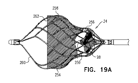
Figs. 19A and 19B are schematic illustrations of a blood pump, in accordance
with
some applications of the present invention;
16
CA 03039302 2019-04-03
WO 2018/096531 PCT/IL2017/051273
Fig. 19C is a schematic illustration showing relative dispositions into which
an
impeller and a support cage are placed prior to crimping the impeller and the
support
cage, in accordance with some applications of the present invention;
Fig. 19D is a schematic illustration of an impeller of a blood pump, in
accordance
with some applications of the present invention
Figs. 20A and 20B are schematic illustrations of an impeller of a blood pump,
in
accordance with some applications of the present invention
Figs. 21A and 21B are schematic illustrations of a blood pump that includes an
impeller, an impeller cage, and a frustoconical support cage, in accordance
with some
applications of the present invention;
Figs. 22A and 22B are schematic illustrations of an occlusion element and a
blood
pump, the occlusion element or the blood pump being placed in a subject's
infra-renal
vena cava (i.e., within the vena cava, upstream of junctions of the vena cava
with all of a
subject's renal veins), in accordance with respective applications of the
present invention;
Fig. 22C is a schematic illustration of a catheter that includes a downstream
pump
and an upstream balloon, in accordance with some applications of the present
invention;
and
Fig. 23 is a curve showing the relationship between (a) cardiac preload and
(b)
cardiac output and/or arterial pressure, when the occlusion element of Fig.
22A or the
blood pump of Fig. 22B is used, in accordance with some applications of the
present
invention.
DETAILED DESCRIPTION OF EMBODIMENTS
Reference is made to Figs. 1A-C, which are schematic illustrations of a blood-
pump catheter 20 placed within a subject's vena cava 22, via a guide catheter
23, an
upstream pump 24U being disposed upon the catheter, distally to a downstream
pump
24D, in accordance with some applications of the present invention. Typically,
the distal
portion of blood-pump catheter 20 is configured to be straight, when the
catheter is in a
non-constrained state, such that both the upstream and the downstream pumps
are
disposed along the axis of the catheter, within the vena cava.
17
CA 03039302 2019-04-03
WO 2018/096531 PCT/IL2017/051273
Each of the upstream and downstream pumps 24U and 24D typically includes a
radially-expandable impeller 28 disposed inside a radially-expandable impeller
cage 30.
Typically, impeller 28 and impeller cage 30 are shape-set such as to assume
radially
expanded configurations thereof in the absence of any radially constraining
force acting
upon the impeller and the cage. The blood pumps are inserted into the
subject's vena
cava, while the blood pumps are in radially constrained configurations inside
the guide
catheter, and are configured to assume substantially radially non-constrained
configurations by being released from the guide catheter inside the subject's
vena cava.
(It is noted that, for some applications, in the vena cava, the blood pumps
may not be fully
radially non-constrained, due to the walls of the vena cava applying a
radially
compressive force to the blood pumps.) For some applications, an engagement
mechanism engages the impeller and the cage with respect to one another, such
that in
response to the cage becoming radially constrained, the impeller becomes
radially
constrained, e.g., in accordance with apparatus and methods described in
described in US
2016/0022890 to Schwammenthal, which is incorporated herein by reference.
It is noted that the term "impeller" is generally used herein to denote a
bladed
rotor, as shown in Figs. 1A-C, for example. When the bladed rotor is placed
inside a
blood vessel (such as vena cava 22) and rotated, the bladed rotor functions as
an impeller,
by modifying the flow of blood through the blood vessel, and/or by generating
a pressure
difference between the upstream end and the downstream end of the impeller.
It is noted that reference numeral 24 is generally used to denote a blood pump
in
the present application. When a pump that is placed upstream is being referred
to,
reference numeral 24U is used, and when a pump that is placed downstream is
being
referred to, reference numeral 24D is used. Similarly, reference numeral 28 is
generally
used to denote an impeller in the present application. When an impeller that
is placed
upstream is being referred to, reference numeral 28U is used, and when an
impeller that is
placed downstream is being referred to, reference numeral 28D is used.
Blood-pump catheter 20 is typically placed inside the subject's vena cava 22,
and
operated therein, in order to provide acute treatment of a subject suffering
from cardiac
dysfunction, congestive heart failure, low renal blood flow, high renal
vascular resistance,
arterial hypertension, diabetes, and/or kidney dysfunction. For example, the
blood-pump
catheter may be placed inside the subject's vena cava, and operated therein,
for a period of
18
CA 03039302 2019-04-03
WO 2018/096531 PCT/IL2017/051273
more than one hour (e.g., more than one day), less than one week (e.g., less
than four
days), and/or between one hour and one week (e.g., between one day and four
days). For
some applications, the blood-pump catheter is chronically placed inside the
subject's vena
cava in order to provide chronic treatment of a subject suffering from cardiac
dysfunction,
congestive heart failure, low renal blood flow, high renal vascular
resistance, arterial
hypertension, diabetes, and/or kidney dysfunction. For some applications, a
course of
treatment is applied to a subject over several weeks, several months, or
several years,
during which the blood-pump catheter is intermittently placed inside the
subject's vena
cava, and the subject is intermittently treated in accordance with the
techniques described
herein. For example, the subject may be intermittently treated at intervals of
several days,
several weeks, or several months.
For some applications, blood-pump catheter 20 is inserted into vena cava 22,
via
the subject's subclavian vein 40, as shown in Fig. 1A. Typically, the blood-
pump catheter
is inserted under fluoroscopic imaging. Alternatively, the blood-pump catheter
is inserted
under ultrasound imaging, such as to reduce exposure of the subject to
radiation and/or
contrast agent. The catheter is placed into the vena cava such that upstream
pump 24U is
disposed upstream of the junctions of the vena cava and all of the subject's
renal veins 42,
and such that downstream pump 24D is disposed downstream of the junctions of
the vena
cava and all of the subject's renal veins. Typically, the upstream pump is
configured to
pump blood through the vena cava in the upstream direction, away from the
renal veins,
and the downstream pump is configured to pump blood through the vena cava in
the
downstream direction, away from the renal veins.
The effect of both of pumps 24U and 24D pumping blood in the above-described
manner is that, between the pumps, and adjacent to the junctions of the vena
cava with the
renal veins, there is a low-pressure region of the vena cava, within which
blood pressure
is lower than the subject's central venous pressure. Functionally, this region
may be
viewed as a compartment within the vena cava within which blood pressure is
controlled
(by controlling pumps 24U and 24D), regardless of the blood pressure elsewhere
within
the vena cava. This typically increases blood flow from the renal veins into
the vena
cava, lowers pressure within the subject's renal veins, and causes renal
perfusion to
increase. The effect of pumps 24U and 24D on blood flow through the renal
veins and the
vena cava is indicated by arrows 44 in Fig. 1B.
19
CA 03039302 2019-04-03
WO 2018/096531 PCT/IL2017/051273
As described hereinabove, the effect of operating blood pumps 24U and 24D is
that between the pumps there is a low-pressure region of the vena cava.
However,
typically, the pumps are operated simultaneously such that the pressure within
other
portions of the vena cava is substantially unchanged relative to when blood-
pump catheter
20 is not in operation. For example, the pumps are typically operated
simultaneously such
that the pressure within the vena cava downstream of downstream pump 24D is
not
substantially increased relative to when blood-pump catheter 20 is not in
operation.
Similarly, the pumps are typically operated simultaneously such that the
pressure within
the vena cava upstream of upstream pump 24U is not substantially increased
relative to
when blood-pump catheter 20 is not in operation. This is because the pumps are
typically
operated simultaneously such that outside of the region between the two pumps,
the
effects of the pumping by the upstream and downstream pumps cancel each other
with
respect to pressure. It is noted that there is likely to be some increase in
the pressure
within the vena cava downstream of downstream pump and upstream of upstream
pump
due to the increased blood flow from the renal veins into the vena cava.
Similarly, the pumps are typically operated simultaneously such that venous
return
to the vena cava from regions upstream of the upstream pump and downstream
from the
downstream pump is substantially unchanged relative to when blood-pump
catheter 20 is
not in operation. In this manner, the pumps are typically operated
simultaneously such as
to have a generally synergistic effect on pressure and flow in the region
between the
pumps, but to have an antagonistic effect on pressure and flow outside of the
region, such
that, outside of the region, the effects of the two pumps typically
substantially cancel each
other out.
Typically, blood-pump catheter 20 pumps blood in a manner that enhances the
rate
of blood flow through the renal veins and into the vena cava, but does not
cause a
substantial change in the direction of the blood flow relative to the natural
direction of
flow through the renal veins, or from the renal veins to the vena cava (i.e.,
relative to
blood flow in the absence of pumping by the blood-pump catheter). That is to
say, the
blood-pump catheter pumps blood in the downstream direction through the renal
veins
and then directly into the portion of the vena cava that is adjacent to the
renal veins, rather
than, for example, pumping the blood from the renal veins into a different
portion of the
subject's veins (such as, an upstream location within the vena cava). It is
noted that, due
CA 03039302 2019-04-03
WO 2018/096531 PCT/IL2017/051273
to the pumping of the downstream pump in the downstream direction, there is
likely to be
some blood flow from the renal veins to the portion of the vena cava that is
below the
renal veins. Further typically, blood-pump catheter 20 enhances blood flow
through the
renal veins without removing blood from the subject's venous system into a non-
venous
receptacle, such as an artificial lumen of a blood pump.
As described hereinabove, typically blood-pump catheter 20 is placed inside
the
vena cava of a subject suffering from cardiac dysfunction, congestive heart
failure, low
renal blood flow, high renal vascular resistance, arterial hypertension,
diabetes, and/or
kidney dysfunction. Typically, operating the blood-pump catheter in the vena
cava of
such a subject causes a lowering and flattening of the subject's renal vein
pressure profile,
even though the subject's central venous pressure is elevated and has
additional effects,
e.g., as described with reference to Fig. 4B of US 2016/0022890 to
Schwammenthal,
which is incorporated herein by reference.
Typically, each of upstream and downstream pumps 24U and 24D includes an
impeller 28, for example, any one of the impellers described in US
2016/0022890 to
Schwammenthal, which is incorporated herein by reference. In accordance with
respective applications, impeller 28 may have a single blade, two blades
(e.g., as
described in US 2016/0022890 to Schwammenthal, which is incorporated herein by
reference), three blades (e.g., as described in US 2016/0022890 to
Schwammenthal), or
more than three blades. For some applications, one or both of blood pumps 24U
and 24D
includes more than one impeller. Typically, ceteris paribus, by using more
than one
impeller in at least one of the pumps, in order to generate a given flow of
blood with the
pump, the force that impacts each of the impellers within the pump is smaller
than if a
single impeller were to be used in the pump.
For some applications, one or both of the pumps includes radially expandable
impeller cage 30. For some applications, impeller cage 30 is configured to
hold open the
inner wall of the vena cava and to separate the inner wall of the vena cava
from the
impeller, such that the vena cava does not become injured by the impeller.
Alternatively,
the impeller cage is sized such that the cage is not used to hold open the
inner wall of the
vena cava (the diameter of the cage being less than that of the vena cava, at
least in some
subjects). Even in such cases, the cage typically functions to separate the
inner wall of the
vena cava from the impeller, for example, in case the walls of the vena cava
at least
21
CA 03039302 2019-04-03
WO 2018/096531 PCT/IL2017/051273
partially collapse inwardly, such that the vena cava does not become injured
by the
impeller. Such applications are described with reference to Figs. 9A-B, for
example.
As described hereinabove, typically, impeller 28 and cage 30 are shape-set
such
as to assume radially expanded configurations thereof in the absence of any
radially
constraining force acting upon the impeller and/or the cage. For some
applications, an
engagement mechanism engages the impeller and the cage with respect to one
another,
such that in response to the cage becoming radially constrained the impeller
becomes
radially constrained, e.g., in accordance with apparatus and methods described
in
described in US 2016/0022890 to Schwammenthal, which is incorporated herein by
reference. For some applications, the stiffness of cage 30 is sufficiently
great that
pressure exerted upon the cage by the inner wall of the vena cava does not
deform the
cage. The cage thereby protects the impeller from being deformed by pressure
from the
inner wall of the vena cava. Such applications are described hereinbelow, with
reference
to Figs. 9A-B, for example.
Referring now to Fig. 1C, typically, when blood-pump catheter 20 is placed
inside
vena cava 22, impeller 28 and impeller cage 30 are substantially radially non-
constrained,
due to the relatively low radial force exerted by the vena cava wall on the
cage. (It is
noted that, for some applications, in the vena cava, the impeller and/or
impeller cage may
not be fully radially non-constrained, due to the walls of the vena cava
applying a radially
compressive force to the blood pumps.) For some applications, the impeller
cage is
configured to come into contact with the inner wall of the vena cava, when the
impeller
cage assumes its radially non-constrained configuration inside the vena cava,
e.g., as
shown in Fig. 1C. For such applications, a span SP of impeller 28, when the
impeller is in
a non-constrained configuration thereof inside the vena cava is more than 14
mm (e.g.,
more than 16 mm), and/or less than 28 mm (e.g., less than 22 mm), e.g., 14-28
mm, or 16-
22 mm. Typically, for such applications, a diameter D of cage 30, when the
cage is in a
non-constrained configuration thereof inside the vena cava is more than 14 mm
(e.g.,
more than 16 mm), and/or less than 40 mm (e.g., less than 35 mm), e.g., 14-40
mm, or 16-
mm. Further typically, when blood-pump catheter 20 is used to enhance blood
flow
30 from the renal veins into the subject's vena cava, as described herein,
a longitudinal
distance D1 between centers of the impellers of the upstream and downstream
pumps,
measured along the longitudinal axis of the catheter, is typically more than 3
cm (e.g.,
22
CA 03039302 2019-04-03
WO 2018/096531 PCT/IL2017/051273
more than 6 cm), and/or less than 18 cm (e.g., less than 14 cm), e.g., 3-18
cm, or 6-14 cm.
For some applications, distance D1 is adjustable and is set based upon
measurements that
are performed upon a subject.
For some applications, impeller cage 30 is configured such that in its
radially non-
constrained configuration, the cage has a diameter that is less than that of
the vena cava at
least in some subjects, for example, as described hereinbelow with reference
to Figs. 9A-
B.
For some applications, impellers 28 of upstream and downstream pumps 24U and
24D are rotated at respective rotation rates, in order to cause the pumping of
blood in the
upstream and downstream directions to be performed at respective rates.
Alternatively,
the impellers are rotated at the same rotation rate (and, typically, in the
same direction),
but the impellers are sized, shaped, and/or oriented such that the rate at
which blood is
pumped, respectively, in the upstream and downstream directions, by the
respective
impellers, is not equal.
Typically, a control unit 52 and a user interface 54 are disposed outside the
subject's body. Further typically, the control unit receives inputs from one
or more
pressure sensors 56, 58, and/or 60, e.g., as shown in Figs. 1A-C.
In accordance with some applications:
(a) a pressure sensor 56 is disposed on the upstream side of upstream blood
pump
24U and is configured to measure pressure within the vena cava upstream of the
low-
pressure region of the vena cava, which is typically indicative of venous
pressure within
the subject's lower body;
(b) a pressure sensor 58 disposed between the two blood pumps, and is
configured
to measure pressure within the low-pressure region of the vena cava between
the two
blood pumps, which is typically indicative of blood pressure within the
subject's renal
veins; and/or
(c) a pressure sensor 60 is disposed on the downstream side of downstream
blood
pump 24D and is configured to measure pressure within the vena cava downstream
of the
low-pressure region of the vena cava, which is typically indicative of the
subject's central
venous pressure close to the subject's right heart.
23
CA 03039302 2019-04-03
WO 2018/096531 PCT/IL2017/051273
For some applications, blood-pump catheter 20 includes pressure sensor 58
disposed between the two blood pumps, and is configured to measure pressure
within the
low-pressure region of the vena cava between the two blood pumps, which is
typically
indicative of blood pressure within the subject's renal veins, and the blood-
pump catheter
does not include pressure sensor 56, or pressure sensor 60.
For some applications, control unit 52 controls pumps 24U and 24D, e.g., by
controlling rotation of impellers 28, responsively to one or more of the above-
described
inputs. Typically, user interface 54 displays the subject's current lower-body
venous
pressure, renal venous pressure, and/or central venous pressure, based upon
the signals
generated by sensors 56, 58, and/or 60. Typically, based upon the current
values of the
subject's lower-body venous pressure, renal venous pressure, and/or central
venous
pressure, a user (such as a healthcare professional) inputs a target value for
the subject's
renal venous pressure, via the user interface. In response thereto, control
unit 52 controls
the speed of the rotation of the impellers, such that the impellers pump blood
away from
the renal veins at a flow rate that is such as to reduce the renal venous
pressure toward the
target level, as indicated by the user. For some applications, in response to
a signal
received from sensor 60 indicating that the central venous pressure is at the
target renal
venous pressure, the control unit stops the impellers rotating. For some
applications, the
control unit receives an input from an additional sensor (such as a flow
sensor and/or an
oxygen-saturation sensor, and/or a thermal flow sensor, e.g., as described
with reference
to Figs. 22Ai-22Cii of US 2016/0022890 to Schwammenthal, which is incorporated
herein by reference), and the control unit controls the speed of the rotation
of the
impellers responsively to an input from the additional sensor.
It is noted that control unit 52 typically includes a computer processor that
comprises circuitry and that is configured to execute the actions described
herein.
Typically, the operations described herein that are performed by the computer
processor
transform the physical state of a memory, which is a real physical article
that is in
communication with the computer processor, to have a different magnetic
polarity,
electrical charge, or the like, depending on the technology of the memory that
is used.
Control unit 52 is typically a hardware device programmed with computer
program
instructions to produce a special-purpose computer. For example, when
programmed to
24
CA 03039302 2019-04-03
WO 2018/096531 PCT/IL2017/051273
perform the techniques described herein, control unit 52 typically acts as a
special-
purpose, renal-venous-pressure-modulating computer processor.
It is further noted that user interface 54 typically includes any type of user
interface configured to receive inputs from a user and/or to provide outputs
to the user.
For example, the user interface may include one or more input devices (such as
a
keyboard, a mouse, a trackball, a joystick, a touchscreen monitor, a touchpad,
a voice-
command interface, a smartphone, a tablet computer, and/or other types of
input devices
that are known in the art), and/or one or more output devices (such as a
monitor, an audio
output device, a smartphone, a tablet computer, and/or other types of output
devices that
are known in the art).
Reference is now made to Figs. 2A, 2B, 2C, 2D, and 2E, which are schematic
illustrations of arrangements of impellers 28U and 28D that are configured to
pump blood
in opposite directions from one another, in accordance with some applications
of the
present invention. (For illustrative purposes, Figs. 2A-E show the impellers
in the
absence of impeller cages, although typically, the impellers are used together
with
impeller cages 30, as described hereinabove.)
Typically, impellers of pumps 24U and 24D are coupled to one or more motors 46
(Fig. 1A), which impart rotational motion to the impellers, via one or more
rotation shafts,
the shaft(s) being housed inside blood-pump catheter 20. In accordance with
respective
applications, the motors are disposed outside of the subject's body (as
shown), or are
placed inside the subject's body (not shown).
Referring now to Fig. 2A, for some applications, impellers 28 of upstream and
downstream pumps 24U and 24D are rotated in the same rotational direction as
one
another, as viewed from an external reference point (e.g., in the direction of
arrow 48 (i.e.,
clockwise), or counterclockwise), but the impellers are disposed on the
catheter such that
the rotation of the impellers in this direction of rotation causes the
impellers to pump
blood in respective, opposite directions. It is noted that the rotational
direction of the
impellers "as viewed from an external reference point" should be interpreted
to mean the
direction of rotational motion of the impellers as observed from any point
that is not
undergoing the same rotational motion as either of the impellers.
CA 03039302 2019-04-03
WO 2018/096531 PCT/IL2017/051273
Typically, for such applications, a single motor is used to rotate both of the
impellers. A shaft 50 is used to impart the rotational motion from the motor
to the
proximal impeller. An additional shaft 51, which is in series with shaft 50,
couples the
proximal impeller to the distal impeller and imparts the rotational motion
from the
proximal impeller to the distal impeller. For some applications, by using a
single series of
shafts to impart rotation to impellers 28 of both upstream and downstream
pumps 24U
and 24D, the diameter of blood-pump catheter 20 is reduced relative to if
parallel shafts
were used, in order to impart rotation to the upstream and downstream
impellers.
For some applications, the angles and/or orientations of the impeller blades
of
impellers 28 of upstream and downstream pumps 24U and 24D may be such as to
cause
the impellers to pump blood in respective, opposite directions. For some
applications, as
shown in Fig. 2A, each impeller is shaped and/or oriented in the mirror image
of the
other, the axis of reflection being orthogonal to the longitudinal axes of the
impellers. For
such applications, the upstream and downstream impellers are of opposing
handedness to
one another, a first one of the impellers being a left-handed impeller, and
the other one of
the impellers being a right-handed impeller. It is generally the case that
impellers of
opposing handedness that are positioned parallel to one another, facing the
same direction
as one another, and rotating in opposite rotational directions from one
another, generate
flow in the same direction as one another. In accordance with some
applications of the
present invention, the upstream and downstream impellers are disposed upon
shaft 51
such that the impellers are facing in opposite directions to one another. As
described
hereinabove, for such applications, the impellers are typically rotated in the
same
rotational direction as one another, as viewed from an external reference
point. The result
of the impellers (a) being of opposing handedness to one another, and (b)
facing in
opposite directions, is that, when the impellers are rotated in the same
direction as one
another about an axis defined by shaft 51, the impellers pump blood in
opposite directions
from one another.
Typically, the blades of the downstream impeller are oriented such that, as
the
downstream impeller rotates in the direction of arrow 48, the downstream
impeller pumps
in the downstream direction. The blades of the upstream impeller are oriented
such that,
as the upstream impeller rotates in the direction of arrow 48, the upstream
impeller pumps
in the upstream direction.
26
CA 03039302 2019-04-03
WO 2018/096531 PCT/IL2017/051273
Referring now to Fig. 2B, for some applications, the upstream impeller 28U and
the downstream impeller 28D are rotated in opposite directions from one
another, as
viewed from an external reference point, in order to generate blood flow in
opposite
directions from one another. For example, impellers that are of the same
handedness as
one another and that are facing the same direction as one another may be used.
For some
such applications, a single motor is used to rotate both of the impellers.
Shaft 50 is used
to impart the rotational motion from the motor to the proximal impeller.
Additional shaft
51, which is in series with shaft 50, couples the proximal impeller to the
distal impeller
and imparts the rotational motion from the proximal impeller to the distal
impeller. A
gear mechanism 70 is disposed between the proximal impeller and the distal
impeller
(e.g., along shaft 51, as shown), and is configured to reverse the direction
of rotational
motion that is imparted from the proximal impeller to the distal impeller,
such that the
distal impeller rotates in an opposite direction of rotation to the direction
of rotation of the
proximal impeller. For example, as shown in Fig. 2B, the downstream impeller
(which in
this case is the proximal impeller) rotates in the direction of arrow 48,
while the upstream
impeller rotates in the direction of arrow 72 (i.e., the opposite direction to
that of arrow
48).
For some applications, it is advantageous to rotate the downstream impeller in
the
opposite direction from the upstream impeller (e.g., as shown in Fig. 2B),
rather than
rotating the downstream impellers in the same direction as the upstream
impeller (e.g., as
shown in Fig. 2A). For some applications, if the downstream impeller rotates
in the same
direction as the upstream impeller, then blood flowing through the vena cava
that impacts
the downstream impeller is already at least partially undergoing rotational
motion in the
direction of rotation of the downstream impeller (by virtue of the rotational
motion
imparted to the blood flow by the upstream impeller). Due to the blood already
undergoing rotational motion in the same direction as the downstream impeller,
the effect
of the rotational motion of the downstream impeller upon the blood flow is
less than if the
blood flow had not already been undergoing the rotational motion in the same
direction as
the downstream impeller, or if the blood had been undergoing rotational motion
in the
opposite direction to that of the downstream impeller. Therefore, for some
applications,
the upstream and downstream impellers are configured to pump blood in opposite
directions from one another by rotating in opposite directions from one
another, e.g.,
using techniques described with reference to any one of Figs. 2B, 2D, or 2E.
27
CA 03039302 2019-04-03
WO 2018/096531 PCT/IL2017/051273
Referring now to Fig. 2C, for some applications, impellers 28 of upstream and
downstream pumps 24U and 24D are rotated in the same rotational direction as
one
another, as viewed from an external reference point (e.g., in the direction of
arrow 48 (i.e.,
clockwise), or counterclockwise), but the impellers are disposed on the
catheter such that
the rotation of the impellers in this direction of rotation causes the
impellers to pump
blood in respective, opposite directions. The configuration shown in Fig. 2C
is generally
similar to that of Fig. 2A, with the impellers being of opposing handedness to
one another,
and facing in opposite directions to one another. However, in the
configuration shown in
Fig. 2C, an additional impeller 74 is disposed between the upstream and the
downstream
.. impellers. Impeller 74 is configured not to be actively rotated. As
indicated by the two-
dimensional arrows indicating the direction of blood flow, blood that is
rotated by the
upstream impeller impacts impeller 74, causing the rotational motion of the
blood flow to
be at least partially reduced. Due to the reduction in the rotational motion
of the blood
flow, the effect of the rotation of the downstream impeller upon the blood
flow is greater
than it would be in the absence of impeller 74.
Referring now to Fig. 2D, for some applications, motor 46 is used to rotate a
first
one of the impellers in a first direction. For example, as shown in Fig. 2D,
motor 46 is
used to rotate downstream impeller 28D in the direction of arrow 48 (i.e.,
clockwise). A
second motor 75 is used to rotate the second one of the impellers in the
opposite direction
.. to the first direction. For example, as shown in Fig. 2D, motor 75 is used
to rotate
upstream impeller 28U in the direction of arrow 72 (i.e., counterclockwise). A
first
rotation shaft 76 extends from first motor 46 to the first impeller and
imparts the
rotational motion in the first direction to the first impeller. A second
rotation shaft 78
extends from second motor 75 to the second impeller and imparts the rotational
motion in
the opposite direction to the first direction to the second impeller.
Typically, within
blood-pump catheter 20, first rotation shaft 76 and second rotation shaft 78
are coaxial
with one another, as shown. For some such applications, impellers that are of
the same
handedness as one another are used as the upstream impeller 28U and the
downstream
impeller 28D.
Reference is now made to Fig. 2E, which is a schematic illustration of
upstream
and downstream pumps 24U and 24D being disposed on respective catheters 66 and
68, in
accordance with some applications of the present invention. For some
applications, a first
28
CA 03039302 2019-04-03
WO 2018/096531 PCT/IL2017/051273
catheter 66 is inserted into vena cava 22 through a guide catheter 67 that is
inserted via
the subject's femoral vein, or via another vein that is below the subject's
inferior vena
cava. Upstream blood pump 24U is disposed on the first catheter, and is
configured to be
placed within the vena cava upstream of the junctions of the vena cava with
all of the
subject's renal veins, and to pump blood through the vena cava in the manner
described
hereinabove. A second catheter 68 is inserted into the vena cava through a
guide catheter
69 that is inserted via the subject's jugular vein, subclavian vein, or via a
different vein
that is above the subject's inferior vena cava. Downstream blood pump 24D is
disposed
on the second catheter, and is configured to be placed within the vena cava
downstream of
the junctions of the vena cava with all of the subject's renal veins, and to
pump blood
through the vena cava in the manner described hereinabove.
For applications in which the upstream and downstream blood pumps include
impellers, typically, respective motors 46 and 75 (e.g., as shown Fig. 2D) are
used to
control rotation of the impellers. For some applications, as described
hereinabove with
reference to Fig. 2D, motor 46 rotates the downstream pump in a first
direction (e.g., the
direction of arrow 48), and motor 75 rotates the upstream pump in the opposite
direction
(e.g., the direction of arrow 72). Further typically, control unit 52 (Fig.
1A) controls both
pumps (e.g., by controlling the rates of rotation of the impellers). For some
applications,
pressure sensors 56, 58 and 60 are disposed upon the first and/or second
catheters, and are
configured to detect indications of, respectively, lower body venous pressure,
renal
venous pressure, and central venous pressure. The control unit is configured
to control
the operation of the upstream and downstream pumps responsively to the
detected
indications, in accordance with the techniques described hereinabove.
For some applications, the impellers of the upstream and downstream pumps are
configured to pump blood in the same direction as one another, e.g., in the
antegrade
direction to enhance blood flow through a vessel.
Reference is now made to Fig. 3, which is a schematic illustration of blood-
pump
catheter 20 being inserted into the subject's vena cava 22 via the subject's
right jugular
vein 62 (through guide catheter 23), in accordance with some applications of
the present
invention. For some applications, instead of being inserted via the subclavian
vein (as
shown in Fig. 1A, for example), blood-pump catheter 20 is inserted into the
vena cava via
the subject's right jugular vein, or via another vein that is above the
subject's inferior vena
29
CA 03039302 2019-04-03
WO 2018/096531 PCT/IL2017/051273
cava. In all other aspects, blood-pump catheter 20 and the functioning thereof
are
generally as described with reference to Figs. 1A-C.
Reference is now made to Fig. 4, which is a schematic illustration of blood-
pump
catheter 20 being inserted into the subject's vena cava 22 via the subject's
femoral vein 64
(through guide catheter 23), downstream pump 24D being disposed upon the
catheter
distally to upstream pump 24U, in accordance with some applications of the
present
invention. For some applications, instead of being inserted via the subclavian
vein (as
shown in Fig. 1A, for example), blood-pump catheter 20 is inserted into the
vena cava, via
the subject's femoral vein 64, or via another vein that is below the subject's
inferior vena
cava. Typically, downstream blood pump 24D is disposed on blood-pump catheter
20
distally to upstream blood pump 24U. Blood-pump catheter 20 is configured to
be placed
within the vena cava, such that the upstream pump is disposed upstream of the
junctions
of the vena cava with all of the subject's renal veins 42, and such that the
downstream
pump is disposed downstream of the junctions of the vena cava with all of the
subject's
renal veins. Other than the dispositions of the upstream and downstream blood
pumps
with respect to blood-pump catheter 20, blood-pump catheter 20, as shown in
Fig. 4, and
the functioning thereof, is generally similar to that described with reference
to blood-
pump catheter 20 as shown in Figs. 1A-C.
Reference is now made to Figs. 5A-B, which are schematic illustrations of
blood-
pump catheter 20, the catheter including downstream pump 24D and an occlusion
element, such as a balloon 80 (Fig. 5A), or a covered frame 82 (Fig. 5B), in
accordance
with some applications of the present invention. For some applications (not
shown), a
nozzle is used as the upstream occlusion element, e.g., as described in co-
pending PCT
Patent Application No. PCT/IL2017/051092 to Tuval, filed Sep. 28, 2017, which
is
incorporated herein by reference. For some applications, downstream pump is
placed
inside vena cava 22, downstream of the junctions of the vena cava with all of
the subject's
renal veins. The downstream pump pumps blood through the vena cava, in the
downstream direction, away from the junctions of the vena cava with the renal
veins, in
the manner described hereinabove. As an alternative to, or in addition to,
using an
upstream pump as described hereinabove, the occlusion element is placed inside
the vena
cava upstream of the junctions of the vena cava with the subject's renal
veins. Typically,
the occlusion element is configured to partially occlude the subject's vena
cava upstream
CA 03039302 2019-04-03
WO 2018/096531 PCT/IL2017/051273
of the junctions of the vena cava with the subject's renal veins. The
occlusion element is
configured to partially occlude the subject's vena cava such that, in response
to the
pumping of the downstream blood pump, there is not a substantial increase of
blood flow
from the subject's lower body toward the subject heart, but such that a region
of low
pressure within the vena cava is generated, between the occlusion element and
the
downstream blood pump, within which the blood pressure is lower than the
subject's
central venous pressure. Typically, by generating a region of low pressure,
blood flow
from the renal veins into the vena cava increases, thereby lowering renal
blood pressure
and enhancing renal perfusion. It is noted that the occlusion element is
configured to
partially occlude, but not to totally occlude, the vena cava, in such a manner
as to generate
a region of low pressure within the vena cava, but to allow a substantial flow
of blood
through the vena cava.
When blood-pump catheter 20 is used to enhance blood flow from the renal veins
into the subject's vena cava, as described herein, a longitudinal distance D2
between the
longitudinal center of the impeller of the downstream pump and the
longitudinal center of
the occlusion element, measured along the longitudinal axis of the catheter,
is typically
more than 3 cm (e.g., more than 6 cm), and/or less than 18 cm (e.g., less than
14 cm), e.g.,
3-18 cm, or 6-14 cm.
For some applications, the occlusion element is balloon 80, as shown in Fig.
5A.
Alternatively or additionally, the occlusion element is covered frame 82, as
shown in Fig.
5B. For example, the frame may be a frame (e.g., a rigid or semi-rigid frame)
made of a
shape-memory element (such as nitinol) that is covered with a material (e.g.,
a blood-
impermeable material) 83 (e.g., polyester, polyurethane, and/or a different
polymer).
As described hereinabove, typically, the occlusion element is configured to
partially occlude the vena cava upstream of the junctions of the vena cava
with the
subject's renal veins. For some applications, the diameter to which the
occlusion element
is expanded is controllable. For example, inflation of the balloon may be
controllable, or
the frame may be expandable (e.g., by heating the frame, or by applying an
electrical
current to the frame). For some applications, the extent to which the
occlusion element
occludes the vena cava is controlled by a control unit (e.g., control unit 52)
responsively
to the blood pressure detected by blood pressure sensor 56, 58, and/or 60, in
response to
an input from a different sensor (such as a flow sensor and/or an oxygen-
saturation
31
CA 03039302 2019-04-03
WO 2018/096531 PCT/IL2017/051273
sensor, and/or a thermal flow sensor, e.g., as described with reference to
Figs. 22Ai-Cii of
US 2016/0022890 to Schwammenthal, which is incorporated herein by reference),
and/or
in response to an input from a user. For some applications, the rate at which
pump 24D
pumps blood away from the renal veins (e.g., the rate at which impeller 28 of
the pump is
.. rotated), and/or the extent to which the occlusion element occludes the
vena cava, is
controlled by a control unit responsively to the blood pressure detected by
blood pressure
sensor 56, 58, and/or 60, in response to an input from a different sensor
(such as a flow
sensor and/or an oxygen-saturation sensor, and/or a thermal flow sensor, e.g.,
as described
with reference to Figs. 22Ai-Cii of US 2016/0022890 to Schwammenthal, which is
incorporated herein by reference), and/or in response to an input from a user.
Although Figs. 5A and 5B show the downstream blood pump and the occlusion
element disposed on a catheter that is inserted into the vena cava from above
the junctions
of the vena cava with the subject's renal veins (e.g., via the subject's
subclavian vein or
jugular vein), for some applications, the downstream blood pump and the
occlusion
element are disposed on a catheter that is inserted into the vena cava from
below the
junctions of the vena cava with the subject's renal veins (e.g., via the
subject's femoral
vein), mutatis mutandis. Alternatively or additionally, the occlusion element
is disposed
on a first catheter which is inserted into the vena cava from below the
junctions of the
vena cava with the subject's renal veins (e.g., via the subject's femoral
vein), and the
downstream blood pump is disposed on a second catheter, which inserted into
the vena
cava from above the junctions of the vena cava with the subject's renal veins
(e.g., via the
subject's subclavian vein, or jugular vein).
As described hereinabove, for some applications, using impellers that rotate
in the
same direction as one another for upstream and downstream pumps causes blood
flow that
impacts the downstream impeller to already be undergoing rotational motion in
the same
direction as the downstream impeller, which, in turn, may cause the effect of
the
rotational motion of the downstream impeller upon the blood to be less than if
the blood
flow had not been undergoing the rotational motion in the same direction as
the
downstream impeller. For some applications, an occlusion element, such as a
balloon 80
(Fig. 5A), covered frame 82 (Fig. 5B), or a nozzle (not shown, e.g., as
described in co-
pending PCT Patent Application No. PCT/IL2017/051092 to Tuval, filed Sep. 28,
2017,
which is incorporated herein by reference) is used instead of an upstream
impeller, such
32
CA 03039302 2019-04-03
WO 2018/096531 PCT/IL2017/051273
that the blood flow that impacts the downstream impeller is not undergoing
rotational
motion in the same direction as the downstream impeller.
For some applications, an occlusion element is placed within the vena cava
upstream of junctions of the vena cava with all of the renal veins even in the
absence of a
downstream blood pump, for example, as described in further detail hereinbelow
with
reference to Fig. 22A.
Reference is now made to Fig. 6, which is a schematic illustration of blood-
pump
catheter 20 placed within a subject's vena cava 22, upstream pump 24U being
disposed
upon the catheter, distally to downstream pump 24D, and a support stent 160
being
disposed upon the catheter between the upstream and downstream pumps, in
accordance
with some applications of the present invention. As described hereinabove,
typically
during operation of pumps 24U and 24D, a region of low pressure is generated
within the
vena cava between the two pumps. Typically, stent 160 is configured to support
the walls
of the vena cava at the low-pressure region, such that the vena cava does not
become
obstructed at the low-pressure region, due to the walls of the vena cava
collapsing. For
example, if, due to the pumping of the upstream and downstream pumps, the
pressure
within the vena cava in the region between the pumps falls below the subject's
intraabdominal pressure, then the walls of the vena cava may collapse in the
absence of a
support structure, such as support stent 160.
For some applications, stent 160 has a generally similar shape to cage 30.
Although Fig. 6 shows stent 160 disposed upon a blood-pump catheter, upon
which the
upstream pump is disposed distally to the downstream pump, for some
applications, stent
160 is disposed upon a blood-pump catheter, upon which the downstream pump is
disposed distally to the upstream pump, as described hereinabove. Similarly,
although
Fig. 6 shows stent 160 disposed upon a blood-pump catheter, upon which the
upstream
pump is disposed distally to the downstream pump, for some applications, stent
160 is
disposed upon a blood-pump catheter upon which an occlusion element and a
downstream
pump are disposed, e.g., as shown in Figs. 5A-B.
Reference is now made to Figs. 7A, 7B, 7C, 7D, and 7E, which are schematic
illustrations of blood-pump catheter 20 placed within a subject's vena cava
22, upstream
impeller 28U being disposed upon the catheter, distally to downstream impeller
28D, the
upstream and downstream impellers being disposed within a support cage 170
that
33
CA 03039302 2019-04-03
WO 2018/096531 PCT/IL2017/051273
supports the walls of a portion of the vena cava between the upstream and
downstream
impellers, in accordance with some applications of the present invention.
Although some
of Figs. 7A-E shows support cage 170 disposed upon a blood-pump catheter, upon
which
the upstream pump is disposed distally to the downstream pump, for some
applications,
support cage 170 is disposed upon a blood-pump catheter, upon which the
downstream
pump is disposed distally to the upstream pump, as described hereinabove.
Similarly,
although Figs. 7A-E shows support cage 170 disposed upon a blood-pump
catheter, upon
which the upstream pump is disposed distally to the downstream pump, for some
applications, support cage 170 is disposed upon a blood-pump catheter upon
which an
occlusion element and a downstream pump are disposed, e.g., as shown in Figs.
5A-B.
As described hereinabove, typically during operation of pumps 24U and 24D, a
region of low pressure is generated within the vena cava between the two
pumps.
Typically, support cage 170 is configured to support the walls of the vena
cava at the low-
pressure region, such that the vena cava does not become obstructed at the low-
pressure
region, due to the walls of the vena cava collapsing. For example, if, due to
the pumping
of the upstream and downstream pumps, the pressure within the vena cava in the
region
between the pumps falls below the subject's intraabdominal pressure, then the
walls of the
vena cava may collapse in the absence of a support structure, such as support
cage 170.
Typically, the support cage is radially expandable and is shape to assume a
radially expanded configuration thereof in the absence of any radially
constraining force
acting upon the support cage. For example, the support cage may be made of a
shape
memory material, e.g., a shape memory metal or alloy (such as, nitinol).
Typically, the
support cage is configured to extend longitudinally along more than 50 percent
of a region
between the first and second impellers, the support cage being configured to
thereby
support the inner wall of the vena cava in an open configuration in the region
between the
first and second impellers. For some applications, support cage is configured
to extend at
least from the longitudinal center of the downstream impeller to the
longitudinal center of
the upstream impeller. For some applications, a length Li (Fig. 7B) of the
support cage,
when the cage is in a substantially non-constrained configuration thereof
inside the vena
cava, is more than 3 cm (e.g., more than 6 cm), and/or less than 18 cm (e.g.,
less than 14
cm), e.g., 3-18 cm, or 6-14 cm. Further typically, a diameter D3 (Fig. 7B) of
support cage
170, when the cage is in a substantially non-constrained configuration thereof
inside the
34
CA 03039302 2019-04-03
WO 2018/096531 PCT/IL2017/051273
vena cava, is more than 14 mm (e.g., more than 16 mm), and/or less than 35 mm
(e.g.,
less than 25 mm), e.g., 14-35 mm, or 16-25 mm. It is noted that, for some
applications,
the support cage may be somewhat radially constrained when disposed within the
vena
cava due to radial compression of the walls of the vena cava upon the support
cage.
For some applications, as shown in Figs. 7A-C, the impellers are placed inside
support cage 170, in the absence of individual cages that are disposed around
the
respective impellers. For such applications, the support cage is typically
configured to (a)
support the walls of the vena cava at the low-pressure region, as described
hereinabove,
and (b) to maintain a separation between the impellers and the inner wall of
the vena cava,
in a generally similar manner to that described hereinabove with respect to
cage 30.
Fig. 7A shows a blood-pump catheter as described, placed inside the subject's
vena cava 22. Fig. 7B shows a three-dimensional view of impellers and support
cage 170
as described, in the absence of the subject's anatomy. Fig. 7C shows a three-
dimensional
view of impellers and support cage 170, in the absence of the subject's
anatomy, and with
the support cage including support elements 172, in accordance with some
applications of
the present invention. As described hereinabove, for some applications, an
axial shaft 51
is disposed between the proximal and distal impellers and is configured to
impart
rotational motion from the proximal impeller to the distal impeller. For some
applications, support elements 172 extend from the support cage, and are
coupled to axial
shaft 51, such as to maintain the disposition of shaft 51 along the
longitudinal axis of the
support cage. In this manner, the disposition of the axial shaft is typically
maintained
along the longitudinal axis of the vena cava. Further typically, the support
elements
maintain the longitudinal axes of the proximal and distal impellers in
alignment with one
another, and in alignment with the longitudinal axis of the vena cava.
For some applications, as shown in Figs. 7D-E, impellers 28D and 28U are
placed
inside support cage 170, in the presence of individual cages that are disposed
around the
respective impellers. Typically, the individual cages in which the impellers
are disposed
are generally similar to impeller cage 30, as described hereinabove.
For some applications, support cage 170 is shaped to define individual
impeller
cages 174, one or more of which are formed as a single integrated structure
together with
the support cage, as shown in Fig. 7D. For example, one or more of the
individual
impeller cages and the support cage may be cut from a single piece of a shape
memory
CA 03039302 2019-04-03
WO 2018/096531 PCT/IL2017/051273
metal or alloy (e.g., nitinol) using techniques as described hereinbelow with
reference to
Figs. 11A-C.
Alternatively, individual impeller cages 30 may be formed separately from
support
cage 170, as shown in Fig. 7E. For such applications, the impellers may be
placed inside
individual impeller cages 30, as described hereinabove, and individual
impeller cages 30
may then be placed inside support cage 170. For some such applications,
individual
impeller cages 30 are placed inside support cage 170 inside the subject's body
(e.g., inside
the subject's vena cava). Alternatively, individual impeller cages 30 are
placed inside
support cage 170 outside the subject's body (e.g., within guide catheter 23,
shown in Fig.
1A), and the individual impeller cages are deployed inside the subject's body
(e.g., inside
the subject's vena cava) together with the support cage.
In general, Figs. 6 and Figs. 7A-E show examples of blood-pump catheter 20 in
which the blood-pump catheter includes a support structure (e.g., stent 160,
or support
cage 170), a longitudinal center of the support structure being disposed
between the
upstream and downstream blood pumps (e.g., between the upstream and downstream
impellers). For some applications, the longitudinal center of the support
structure is
disposed equidistantly from the upstream and downstream blood pumps (e.g., the
upstream and downstream impellers). The support structure is configured to
support an
inner wall of the vena cava in an open configuration during the pumping of the
blood by
the first and second pumps. For some applications (not shown), a support
structure, such
as the structure shown in Figs. 6, 7A-E, and/or as described hereinbelow with
reference to
Figs. 9A-B, 10A-D, and/or 11A-C, is used in conjunction with a blood-pump
catheter that
includes a downstream pump and an upstream occlusion element (e.g., as shown
in Figs.
5A-B), mutatis mutandis. For such applications, the support structure is
configured to
support an inner wall of the vena cava in an open configuration during the
pumping of the
blood by the downstream pump.
Reference is now made to Figs. 8A, 8B, and 8C, which are graphs showing the
pressure drop recorded in models of a subject's left and right renal veins,
during
experiments that were conducted using pumps, in accordance with some
applications of
the present invention.
In the experiments, a model of the vena cava and renal veins was used. The
model
was made of flexible silicone filled with saline. Upstream and downstream
pumps as
36
CA 03039302 2019-04-03
WO 2018/096531 PCT/IL2017/051273
described herein were placed inside the vena cava, respectively below and
above the renal
veins. The pumps were activated to pump the saline through the vena cava in
the manner
described herein, and the drop in pressure in left and right renal veins was
measured
relative to the pressure in the left and right renal veins before the pumps
were activated.
Fig. 8A shows a plot of the measured pressure reduction (dP) in the left and
right
renal veins for respective rates of revolutions per minute (RPM) of the pumps
(which was
always the same for both pumps), for when the pumps were placed in the vena
cava in the
absence of either a support stent (as shown in Fig. 6) or a support cage or
sleeve (as
shown in Figs. 7A-E, 9A-B, 10A-D, and/or 11A-C) between the upstream and
downstream pumps.
Fig. 8B shows a plot of the measured pressure reduction (dP) in the left and
right
renal veins for respective rates of revolutions per minute (RPM) of the pumps
(which was
always the same for both pumps), for when the pumps were placed in the vena
cava in the
presence of a support stent (as shown in Fig. 6) between the upstream and
downstream
pumps.
Fig. 8C shows a plot of the measured pressure reduction (dP) in the left and
right
renal veins for respective rates of revolutions per minute (RPM) of the pumps
(which was
always the same for both pumps), for when the pumps were placed in the vena
cava in the
presence of a support cage between the upstream and downstream pumps, the
support
cage being configured as shown in Fig. 7E.
As may be observed in Figs. 8A-C, the greatest pressure reduction was achieved
when the pumps were used in conjunction with a support cage that extends at
least from
the longitudinal center of the downstream impeller to the longitudinal center
of the
upstream impeller (the results of which are shown in Fig. 8C). In addition,
the most even
pressure reduction of both the left and right renal veins was achieved when
the pumps
were used in conjunction with a support cage that extends at least from the
longitudinal
center of the downstream impeller to the longitudinal center of the upstream
impeller.
When a support stent as shown in Fig. 6 was disposed between the upstream and
downstream pumps (the result of which are shown in Fig. 8B), there was still a
greater
and more even pressure reduction than when the upstream and downstream pumps
were
used in the absence of any supporting structure between the upstream and
downstream
pumps (the results of which are shown in Fig. 8A).
37
CA 03039302 2019-04-03
WO 2018/096531 PCT/IL2017/051273
Therefore, the results shown in Figs. 8A-C indicate that the efficacy of the
reducing renal venous pressure by pumping blood through the vena cava using
upstream
and downstream pumps as described herein may be improved by placing a support
structure inside the vena cava between the upstream and downstream pumps, in
accordance with techniques described herein. Furthermore, the results indicate
that the
efficacy of the aforementioned technique may be improved by placing a support
cage
inside the vena cava that extends at least from the longitudinal center of the
downstream
pump (e.g., the longitudinal center of the impeller of the downstream pump) to
the
longitudinal center of the upstream pump (e.g., longitudinal center of the
impeller of the
upstream pump). Therefore, for some applications of the present invention,
apparatus and
methods as described in Figs. 6, 7A-E, 9A-B, 10A-D, and/or 11A-C are used.
Reference is now made to Figs. 9A-B, which are schematic illustrations of
blood-
pump catheter 20 for placing within a subject's vena cava 22, an upstream
impeller 28U
being disposed upon the catheter, proximally to a downstream impeller 28D, the
upstream
and downstream impellers being disposed within a support cage 180 that
supports the
walls of a portion of the vena cava between the upstream and downstream
impellers, in
accordance with some applications of the present invention. Support cage 180
is typically
generally similar to support cage 170 described hereinabove, except for the
differences
described hereinbelow.
For some applications, impellers 28 are disposed inside respective impeller
cages
30, and the impeller cages are not sized such as to hold open the inner wall
of the vena
cava under normal conditions, the diameter of the cages being less than that
of the vena
cava, as shown in Figs. 9A-B. Even in such cases, impeller cage 30 typically
functions to
separate the inner wall of the vena cava from the impeller, such that the vena
cava does
not become injured by the impeller, for example in case intra-abdominal
pressure is
exerted upon the vena cava, such that the vena cava collapses against the
impeller cages.
Typically, for such applications, the stiffness of cage 30 is sufficiently
great that pressure
exerted upon the cage by the inner wall of the vena cava does not deform the
cage. The
cage thereby protects the impeller from being deformed by pressure from the
inner wall of
the vena cava.
Typically, support cage 180 is configured to extend longitudinally along more
than
50 percent of a region between the first and second impellers, the support
cage being
38
CA 03039302 2019-04-03
WO 2018/096531 PCT/IL2017/051273
configured to thereby support the inner wall of the vena cava in an open
configuration in
the region between the first and second impellers, e.g., as described
hereinabove with
reference to support cage 170. For some applications, support cage extends at
least from
the longitudinal center of the downstream impeller to the longitudinal center
of the
upstream impeller. For some applications, the support cage extends from the
upstream
end of the upstream impeller cage to the downstream end of the downstream
impeller
cage.
Typically, the dimensions of support cage 180 are generally similar to those
described hereinabove with reference to support cage 170. Further typically,
the
maximum diameter of the support cage (i.e., the diameter of the support cage
at the
longitudinal location(s) at which the diameter of the support cage is at its
maximum)
when the support cage is in a non-constrained configuration thereof is at
least 1.1 times
(and, for some applications, at least 1.3 times) greater than maximum
diameters of each of
the impeller cages 30 (i.e., the diameters of each of the impeller cages at
the longitudinal
location(s) at which the diameter of each of the impeller cages is at its
maximum) when
the impeller cages are in non-constrained configurations thereof. For some
applications,
the maximum diameter of the support cage is approximately 30 mm (e.g., 30 mm
plus/minus 3 mm), and the maximum diameter of each of the impeller cages is
approximately 20 mm (e.g., 20 mm plus/minus 3 mm).
Typically, for applications as shown in Figs. 9A-B, the stiffness of each of
the
impeller cages is at least 1.5 times greater than the stiffness of the support
cage. As
described hereinabove, the impeller cages are shape set such that the
diameters of the
impeller cages are less than that of the vena cava. Therefore, the cages are
configured to
be of sufficient stiffness not to be deformed by pressure from the vena cava
walls. By
contrast, the support cage is typically shape set such that the diameter of
the support cage
is greater than the diameter of the vena cava, at least in some subjects.
Therefore, the
support cage is typically configured to have a stiffness that is such that the
support cage is
at least partially narrowed by the vena cava in the event that the diameter of
the support
cage, in its non-constrained configuration, is greater than the diameter of
the vena cava. It
is noted that, even in such applications, the stiffness of the impeller cage
is configured to
permit the impeller cage to be inserted into the subject's vena cava by being
crimped
39
CA 03039302 2019-04-03
WO 2018/096531 PCT/IL2017/051273
inside guide catheter 23, and to permit the impeller cage to navigate turns
while being
advanced through the guide catheter.
Reference is now made to Figs. 10A, 10B, 10C, and 10D, which are schematic
illustrations of a support sleeve 190 having an open end 192, and impeller
cages 30 for
use therewith, in accordance with some applications of the present invention.
Support
sleeve 190 is typically generally similar to support cage 170 described
hereinabove,
except for the differences described hereinbelow. Support sleeve 190 (shown in
Fig.
10A) is typically used with upstream and downstream impellers 28 and impeller
cages 30
(shown in Fig. 10B), which are generally as described herein. Fig. 10C shows
the
impeller cages disposed inside support sleeve 190, and Fig. 10D shows the
impeller cages
disposed inside the support sleeve within vena cava 22. Typically, by virtue
of having
open end 192, one of the impeller cages and the corresponding impeller are
able to pass
through the open end of the support sleeve even when the impeller cage and
corresponding impeller are in radially non-constrained configurations thereof.
Further
typically, the open end of the support sleeve is not fixedly coupled to the
impeller cage
and/or the impeller that is disposed toward the open end of the support
sleeve.
Typically, support sleeve 190 is configured to be disposed around a first one
of the
impeller cages and to extend longitudinally along more than 50 percent of the
region
between the first and second impellers, the support sleeve being configured to
thereby
support the inner wall of the vena cava in an open configuration in the region
between the
first and second impellers, e.g., as described hereinabove with reference to
support cage
170. For some applications, the support sleeve extends at least from the
longitudinal
center of the downstream impeller to the longitudinal center of the upstream
impeller. For
some applications, the support sleeve extends from the upstream end of the
upstream
impeller cage to the downstream end of the downstream impeller cage, e.g., as
shown in
Figs. 10C and 10D.
For some applications, support sleeve 190 is released into the subject's vena
cava
prior to impellers 28 and impeller cages 30 being released into the vena cava.
Subsequent
to the support sleeve being released and radially expanding inside the vena
cava, impellers
28 and impeller cages 30 are released into the vena cava. For some
applications, support
sleeve 190 is crimped inside guide catheter 23 without the impellers and the
impeller
cages disposed inside the support sleeve. Alternatively, the impellers and
impeller cages
CA 03039302 2019-04-03
WO 2018/096531 PCT/IL2017/051273
are disposed inside the support sleeve when the support sleeve is crimped
inside the guide
catheter during insertion. As described hereinabove, the open end of the
support sleeve is
not fixedly coupled to the impeller cage or to the impeller that are disposed
toward the
open end of the support sleeve. The open end of the support sleeve is thereby
able to
move longitudinally with respect to the impeller cage and the impeller that
are disposed
toward the open end of the support sleeve, thereby allowing the support sleeve
to become
more longitudinally extended (e.g., during crimping) than if the end of the
support sleeve
were to be fixedly coupled to the impeller and/or to the impeller cage that
are disposed
toward the open end of the support sleeve.
Reference is now made to Figs. 11A, 11B, and 11C, which are schematic
illustrations of an impeller cage 30 and a support sleeve 200 that are formed
from a single
tube of a shape-memory alloy (such as nitinol), and a cage assembly element
202
configured to hold closed one of the ends of the impeller cage, in accordance
with some
applications of the present invention. Support sleeve 200 is typically
generally similar to
support cage 170 described hereinabove, except for the differences described
hereinbelow.
For some applications, an impeller cage 30 and support sleeve 200 are formed
from a single tube of a shape-memory metal or alloy (such as nitinol), by
cutting both the
impeller cage and the support sleeve to have one open end. Subsequent to
cutting the
tube, the open end of the impeller cage is closed using a cage assembly
element 202,
which may, for example be a ring-shaped fastening element, as shown, and/or a
clip, a
suture, a tie, adhesive, etc.
For some applications, as described with reference to Figs. 9A-B, the maximum
diameter D4 (Fig. 11B) of the support sleeve (i.e., the diameter of the
support sleeve at the
longitudinal location(s) at which the diameter of the support sleeve is at its
maximum)
when the support sleeve is in a non-constrained configuration thereof is at
least 1.1 times
(and, for some applications, at least 1.3 times) greater than a maximum
diameter D5 (Fig.
11B) of each of the impeller cages 30 (i.e., the diameters of each of the
impeller cages at
the longitudinal location(s) at which the diameter of each of the impeller
cages is at its
maximum) when the impeller cages are in non-constrained configurations
thereof. For
some applications, the maximum diameter of the support sleeve is approximately
30 mm
41
CA 03039302 2019-04-03
WO 2018/096531 PCT/IL2017/051273
(e.g., 30 mm plus/minus 3 mm), and the maximum diameter of each of the
impeller cages
is approximately 20 mm (e.g., 20 mm plus/minus 3 mm).
For some applications, at one end of support sleeve 200, one of the impellers
is
fixedly coupled to the support sleeve, by virtue of the impeller and the
support sleeve
having been formed from a single tube of shape-memory alloy, as described
hereinabove.
For some applications, at the other end of the support sleeve, the support
sleeve is open,
and the open end of the support sleeve is not fixedly coupled to the impeller
cage or to the
impeller that are disposed toward the open end of the support sleeve.
Typically, by virtue
of having an open end, the impeller cage and the impeller that are disposed
toward the
open end of the support sleeve are able to pass through the end of the support
sleeve even
when the impeller cage and the impeller are in radially non-constrained
configurations
thereof.
For some applications, the impeller and impeller cage that are disposed toward
the
open end of the support sleeve are disposed inside the support sleeve when the
support
sleeve is crimped inside the guide catheter. As described hereinabove, the
open end of the
support sleeve is typically not fixedly coupled to the impeller cage or to the
impeller that
are disposed toward the open end of the support sleeve. The open end of the
support
sleeve is thereby able to move longitudinally with respect to the impeller
cage and the
impeller that are disposed toward the open end of the support sleeve, thereby
allowing the
support sleeve to become more longitudinally extended (e.g., during crimping)
than if the
end of the support sleeve were to be fixedly coupled to the impeller and/or to
the impeller
cage that are disposed toward the open end of the support sleeve.
Typically, support sleeve 200 is configured to extend longitudinally from the
first
impeller and impeller cage along more than 50 percent of the region between
the first and
second impellers, the support sleeve being configured to thereby support the
inner wall of
the vena cava in an open configuration in the region between the first and
second
impellers, e.g., as described hereinabove with reference to support cage 170.
For some
applications, the support sleeve extends at least from the longitudinal center
of the
downstream impeller to the longitudinal center of the upstream impeller. For
some
applications, the support sleeve is configured, such that when the support
sleeve radially
expands inside the vena cava, the support sleeve does not extend
longitudinally to the
second impeller cage, e.g., as shown in Fig. 11C.
42
CA 03039302 2019-04-03
WO 2018/096531 PCT/IL2017/051273
It is noted that blood pumps 24U and 24D, the catheters upon which the blood
pumps are disposed (e.g., blood-pump catheter 20, catheter 66, and catheter
68), and the
occlusion elements described with reference to Figs. 5A-B, and other devices
described
herein, are generally described as being placed within the subject's vena
cava, such that
the upstream pump or the occlusion element is disposed upstream of junctions
of the vena
cava with the subject's renal veins, and the downstream pump is disposed
downstream of
the junctions of the vena cava with the subject's renal veins. However, it is
noted that the
scope of the present invention includes placing upstream pump 24U or the
occlusion
element in any main vein upstream of a tributary venous system, and placing
downstream
pump 24D downstream of said tributary venous system, and configuring the
pump(s)
(e.g., via the direction of rotation of impellers of the pumps, or the
orientation of the
pumps) to generate preferential flow from the tributaries into the main vein,
mutatis
mutandis. For example, the pump(s) could be used to generate flow from the
subject's
hepatic veins into the subject's vena cava, in order to increase perfusion of
the subject's
liver, mutatis mutandis. For some applications, the upstream pump or the
occlusion
element is placed within a main vein upstream of two or more tributary venous
systems
into the main vein (e.g., within the vena cava upstream of the renal venous
system and the
hepatic venous system). The downstream pump is placed downstream of the two or
more
tributary venous systems. The pump(s) are configured to generate preferential
flow from
both of the tributary venous systems into the main vein by pumping blood
through the
main vein, in the manner described herein. For some applications, upstream and
downstream pumps 24U and 24D and blood-pump catheter 20 are placed within the
subclavian vein or jugular vein at junctions of the vein with a lymph duct and
are used to
increase flow of lymphatic fluid from the lymph duct into the vein, using the
techniques
described herein, mutatis mutandis.
For some applications, upstream pump 24U or the occlusion element is placed in
a
main vein upstream of a tributary venous system, and downstream pump 24D is
placed
downstream of said tributary venous system, and the pump(s) are configured
(e.g., via the
direction of rotation of impellers of the pumps, or the orientation of the
pumps) to reduce
flow from the tributaries into the main vein. For some such applications, the
blades of the
downstream impeller are oriented such that, as the downstream impeller is
rotated, the
downstream impeller pumps in the upstream direction (toward the junction
between the
tributary system and the main vein). The blades of the upstream impeller are
oriented
43
CA 03039302 2019-04-03
WO 2018/096531 PCT/IL2017/051273
such that, as the upstream impeller is rotated, the upstream impeller pumps in
the
downstream direction (toward the junction between the tributary system and the
main
vein).
For some applications, the upstream and downstream pumps 24U and 24D, the
catheter(s) upon which the blood pumps are disposed (e.g., blood-pump catheter
20,
catheter 66, and catheter 68), and/or the occlusion elements described with
reference to
Figs. 5A-B, and other devices described herein, are placed within a main
artery upstream
and downstream of bifurcations of the artery with one or more branching
arterial systems
that branch from the main artery and supply a given organ, mutatis mutandis.
For such
applications, the upstream pump is typically configured to pump in the
downstream
direction (toward the bifurcations) and the downstream pump is configured to
pump in the
upstream direction (toward the bifurcations), such that blood flow into the
branching
arterial system is increased, thereby increasing perfusion of the organ.
Alternatively or
additionally, the occlusion element is placed downstream of the bifurcations
of the artery
with the one or more arterial systems and is configured to partially occlude
the artery
downstream of the bifurcations. For example, the upstream pump may be placed
in the
subject's aorta upstream of the subject's renal arteries and the downstream
pump may be
placed in the subject's aorta downstream of the subject's renal arteries, the
pumps acting to
pump blood into the renal arteries and toward the subject's kidneys. For some
applications, upstream and downstream pumps, and/or occlusion elements are
placed on
both the arterial and venous sides of the subject's body in order to increase
perfusion of a
given organ or set of organs, in the manner described herein.
Reference is now made to Fig. 12, which is a schematic illustration of
impeller-
based blood pumps 150 inserted into a subject's left and right renal veins 42
via the
subject's subclavian vein 40, in accordance with some applications of the
present
invention. Typically, each of the blood pumps includes a radially-expandable
impeller
152 disposed inside a radially-expandable impeller cage 154. Typically, the
blood pumps
are inserted into the left and right renal veins via respective catheters 155.
Alternatively
(not shown), the blood pumps are inserted via a single catheter that passes
from the
venous access point to the subject's vena cava. The blood pumps are inserted
into the
subject's renal veins, while the blood pumps are in radially constrained
configurations
inside the guide catheter(s), and are configured to assume substantially
radially non-
44
CA 03039302 2019-04-03
WO 2018/096531 PCT/IL2017/051273
constrained configurations by being released from the guide catheter(s) inside
the
subject's renal veins. For some applications, the catheters are both inserted
via subclavian
vein 40, as shown. Alternatively or additionally, the catheters are inserted
via a different
vein, e.g., the subject's femoral vein, and/or the subject's jugular vein. For
some
applications, blood pumps 150 are generally similar to blood pumps described
in US
2016/0022890 to Schwammenthal, which is incorporated herein by reference,
apart from
differences described hereinbelow.
Blood pumps 150 are typically placed inside the subject's renal veins 42, and
operated therein, in order to provide acute treatment of a subject suffering
from cardiac
dysfunction, congestive heart failure, low renal blood flow, high renal
vascular resistance,
arterial hypertension, diabetes, and/or kidney dysfunction. The therapeutic
effect of
operating blood pumps 150 within the renal veins (a) is typically generally
similar to that
described hereinabove with reference to blood-pump catheter 20, mutatis
mutandis, and
(b) is typically generally similar to the effect of renal vein blood pumps, as
described in
US 2016/0022890 to Schwammenthal, which is incorporated herein by reference.
Typically, the impellers of the blood pumps 150 are coupled to motors 232,
which
impart rotational motion to the impellers. In accordance with respective
applications, the
motors are disposed outside of the subject's body (as shown) or are placed
inside the
subject's body (not shown). Typically, a control unit 234 and a user interface
236 are
.. disposed outside the subject's body. Further typically, the control unit
receives inputs
from pressure sensors, which are disposed on upstream and downstream sides of
the blood
pumps. When blood pump 150 is disposed inside a renal vein (as shown in Fig.
12, for
example), the pressure measured by the upstream pressure sensor is indicative
of blood
pressure upstream of the blood pump, inside the renal vein, and the pressure
measured by
a downstream pressure sensor is indicative of central venous pressure. For
some
applications, the control unit receives an input from an additional sensor
(such as a flow
sensor and/or an oxygen-saturation sensor, and/or a thermal flow sensor, e.g.,
as described
with reference to Figs. 22Ai-22Cii of US 2016/0022890 to Schwammenthal, which
is
incorporated herein by reference), and the control unit controls the speed of
the rotation of
the impellers responsively to an input from the additional sensor.
For some applications, control unit 234 controls rotation of impellers 152, by
controlling motors 232, responsively to one or more of the above-described
inputs.
CA 03039302 2019-04-03
WO 2018/096531 PCT/IL2017/051273
Typically, user interface 236 displays the subject's current renal venous
pressure and
central venous pressure, based upon the pressures measured by the sensors.
Typically,
based upon the current values of the subject's renal venous pressure and
central venous
pressure, a user (such as a healthcare professional) inputs a target value for
the subject
renal venous pressure, via the user interface. In response thereto, control
unit 234
controls the speed of the rotation of the impeller, such that the impeller
pumps through the
renal vein and toward the vena cava at a flow rate that is such as to reduce
the renal
venous pressure toward the target level, as indicated by the user. For some
applications,
in response to a signal received from the downstream pressure sensor
indicating that the
central venous pressure is at the target renal venous pressure, the control
unit stops the
impeller rotating. In general, the control unit typically controls the speed
of the rotation
of the impellers responsively to inputs from the upstream and downstream
pressure
sensors.
It is noted that control unit 234 typically includes a computer processor that
comprises circuitry and that is configured to execute the actions described
herein.
Typically, the operations described herein that are performed by the computer
processor
transform the physical state of a memory, which is a real physical article
that is in
communication with the computer processor, to have a different magnetic
polarity,
electrical charge, or the like, depending on the technology of the memory that
is used.
Control unit 234 is typically a hardware device programmed with computer
program
instructions to produce a special-purpose computer. For example, when
programmed to
perform the techniques described herein, control unit 234 typically acts as a
special-
purpose, renal-venous-pressure-modulating computer processor.
It is further noted that user interface 236 typically includes any type of
user
interface configured to receive inputs from a user and/or to provide outputs
to the user.
For example, the user interface may include one or more input devices (such as
a
keyboard, a mouse, a trackball, a joystick, a touchscreen monitor, a touchpad,
a voice-
command interface, a smartphone, a tablet computer, and/or other types of
input devices
that are known in the art), and/or one or more output devices (such as a
monitor, an audio
output device, a smartphone, a tablet computer, and/or other types of output
devices that
are known in the art).
46
CA 03039302 2019-04-03
WO 2018/096531 PCT/IL2017/051273
Reference is now made to Figs. 13A-B, which are schematic illustrations of
impeller cage 154, the cage including radially-protruding support arms 156
that are
configured to align the longitudinal axis of impeller 152 with a local
longitudinal axis of
renal vein 42, in accordance with some applications of the present invention.
Fig. 13A
shows the impeller cage disposed inside the subject's renal vein 42. As shown,
for some
applications, the impeller cage is sized such that, when the cage is in a
radially non-
constrained configuration, the maximum diameter of the cage (i.e., the
diameter of the
cage at the longitudinal location at which the diameter of the cage is at its
maximum) is
less than the diameter of the renal vein at the location within the renal vein
at which the
impeller cage is deployed, at least in some subjects. Typically, diameters of
renal veins
vary from approximately 8 mm to approximately 16 mm, and, for example, the
diameter
of the impeller cage may be more than 8 mm (e.g., more than 10 mm), and/ or
less than 13
mm (e.g., less than 12 mm), e.g., between 8 and 13 mm, or between 10 and 12
mm. For
such applications, the cage is typically configured to have a stiffness that
is such that even
if pressure is exerted upon the cage by walls of the renal vein, the cage does
not become
deformed, and the cage thereby protects the impeller from becoming deformed.
The inventors of the present application found that if impeller cages for
placement
within the renal veins as described herein (e.g., with reference to Fig. 12)
are configured
to be have diameters that are substantially greater than the diameters of some
subjects'
renal veins, and the impeller cages are configured to become radially
compressed by the
walls of the renal veins, this may result in the impeller becoming deformed
(e.g., by the
upstream and downstream ends of the impeller axis becoming misaligned), and/or
in the
impeller becoming misaligned with the local longitudinal axis of the renal
veins.
Therefore, as described hereinabove, for some applications, the impeller cage
is
configured to have a smaller diameter, such that even in cases in which the
impeller cage
has a greater diameter than a subject's renal vein (e.g., in the case of a
subject with narrow
renal veins), the radial force that is exerted upon the impeller cage by the
subject's renal
vein is lower. Furthermore, the impeller cage is configured to have a
stiffness that is such
that even if pressure is exerted upon the cage by walls of the renal vein, the
cage does not
become deformed, and the cage thereby protects the impeller from becoming
deformed.
It is noted that, even in such cases, the stiffness of the impeller cage is
configured to
permit the impeller cage to be inserted into the subject's renal vein by being
crimped
47
CA 03039302 2019-04-03
WO 2018/096531 PCT/IL2017/051273
inside guide catheter 155, and to permit the impeller cage to navigate turns
while being
advanced through the guide catheter.
As described hereinabove, for some applications, impeller cage 154 is
configured
such that the maximum diameter of the cage is less than the diameter of the
renal vein at
the location within the renal vein at which the impeller cage is deployed. For
some such
applications, radially-protruding support arms 156 protrude radially from the
impeller
cage. The radially-protruding support arms 156 are configured, upon the blood
pump
being released into the subject's renal vein, to come into contact with the
inner wall of the
subject's renal vein by radially expanding. The radially-protruding support
arms 156 are
configured to thereby align the longitudinal axis of impeller 152 with a local
longitudinal
axis of renal vein 42, as shown in Fig. 13A. Typically, ceteris pafibus, the
efficacy of the
pumping of blood by impeller 152 is greater, the greater than alignment of the
longitudinal axis of the impeller with the local longitudinal axis of the
renal vein. It is
noted that, for some applications, the radially-protruding support arms may
not fully align
the longitudinal axis of impeller with the local longitudinal axis of renal
vein. However,
typically, the radially-protruding support arms maintain the longitudinal axis
of the
impeller in greater alignment with the local longitudinal axis of the renal
vein, relative to
alignment of the longitudinal axis of the impeller with the local longitudinal
axis of the
renal vein in the absence of the support arms, ceteris paribus.
For some applications, the radially-protruding support arms each define a
radius
R1 with respect to the longitudinal axis 153 of the shaft of the blood pump
that is greater
than 7 mm and/or less than 9 mm, e.g., 7-9 mm. Radius R1 (shown in Fig. 13B)
is a
measure of the maximum radial distance between the support arm and
longitudinal axis
153. For some applications, rather than defining radially-protruding support
arms, cage
30 defines a bulbous extension that, for example, may be generally similar in
shape to
bulbous extension 84 described hereinbelow with reference to Fig. 16, mutatis
mutandis.
As described hereinabove, for some applications, control unit 234 (Fig. 12)
receives inputs from pressure sensors that are disposed on upstream and
downstream sides
of the blood pumps, and controls rotation of impellers 152, by controlling
motors 232,
responsively to one or more of the inputs. As shown, for some applications,
upstream
pressure sensor 158 is disposed on the shaft of blood pump 150 distally to the
impeller
and the impeller cage. If the upstream pressure sensor comes into contact with
the inner
48
CA 03039302 2019-04-03
WO 2018/096531 PCT/IL2017/051273
wall of the renal vein, then the upstream pressure sensor may measure the
subject's
intraabdominal pressure, which is conveyed through the wall of the renal vein,
as opposed
to measuring blood pressure within the renal vein. Therefore, for some
applications,
radially-protruding support arms 156 are configured to separate the upstream
pressure
sensor from the inner wall of the renal vein, by centering the shaft of the
blood pump
within the renal vein. For example, the radially-protruding support arms may
maintain
the pressure sensor at a distance of at least 2 mm from an inner wall of the
renal vein.
For some applications, the impeller cage is sized such that, when the cage is
in its
radially non-constrained configuration, the maximum diameter of the cage is
less than the
diameter of the renal vein, as described hereinabove. For some such
applications, even in
the absence of radially-protruding support arms 156, the longitudinal axis of
the impeller
is maintained substantially in alignment with the local longitudinal axis of
the renal vein
by guide catheter 155 providing support to the impeller and the impeller cage.
For
example, the guide catheter may be inserted into the renal vein via the
subject's femoral
vein, and the catheter may be configured as described in US 2015/0157777 to
Tuval,
which is incorporated herein by reference. For such applications, the guide
catheter is
typically configured to maintain pressure sensor 158 at a distance of at least
2 mm from
an inner wall of the blood vessel, as described hereinabove.
For some applications, control unit 124 is configured to account for pressure
sensor 158 contacting the inner wall of the renal vein, and measuring the
subject's
intraabdominal pressure. For example, the control unit may run the following
algorithm:
A. The control unit increases the rotation speed of the impeller.
o
If, in response to the rotation speed of the impeller increasing, the pressure
reading of pressure sensor increases, the control unit (a) disregards the
pressure reading from the pressure sensor, (b) generates an output
indicating that the reading from pressure sensor 158 is erroneous, and/or
(c) generates an indication that the reading from pressure sensor 158 is
indicative of intraabdominal pressure, and not renal blood pressure. (This
is because, in response to the rotation speed of the impeller increasing,
renal pressure would be expected to decrease. The increase in pressure
indicates that the inner wall of the renal vein has come into contact with
49
CA 03039302 2019-04-03
WO 2018/096531 PCT/IL2017/051273
the pressure sensor, such that the pressure sensor is measuring
intraabdominal pressure.)
o If, in response to the rotation speed of the impeller increasing, the
pressure
reading of the pressure sensor decreases, the control unit interprets the
pressure reading from the pressure sensor as being indicative of renal
blood pressure, and generates an output, and/or further modulates the speed
of rotation of the impeller, in response thereto (e.g., in the manner
described hereinabove, with reference to Fig. 12).
and/or
B. The control unit decreases the rotation speed of the impeller.
o If, in response to the rotation speed of the impeller decreasing, the
pressure
reading of pressure sensor 158 decreases, the control unit (a) disregards the
pressure reading from the pressure sensor from prior to the decrease in the
rotation speed, (b) generates an output indicating that the reading from
pressure sensor 158 from prior to the decrease in the rotation speed was
erroneous, and/or (c) generates an indication that the reading from pressure
sensor 158 from prior to the decrease in the rotation speed was indicative
of intraabdominal pressure, and not renal blood pressure. (This is because,
in response to the rotation speed of the impeller decreasing, renal pressure
would be expected to increase. The increase in pressure indicates that the
inner wall of the renal vein had been in contact with the pressure sensor,
such that the pressure sensor was measuring intraabdominal pressure, but
that the inner wall of the renal vein has now separated from the pressure
sensor.)
o If, in response to the rotation speed of the impeller decreasing, the
pressure
reading of pressure sensor increases, the control unit interprets the pressure
reading from the pressure sensor as being and having been indicative of
renal blood pressure, and generates an output, and/or further modulates the
speed of rotation of the impeller, in response thereto (e.g., in the manner
described hereinabove, with reference to Fig. 12).
CA 03039302 2019-04-03
WO 2018/096531 PCT/IL2017/051273
For some applications, the above-described algorithm is run by control unit 52
(Fig. 1), in order to accurately measure blood pressure within the subject's
vena cava
and/or renal veins, mutatis mutandis.
Reference is now made to Fig. 14, which is a schematic illustration of
pressure
sensor 158 disposed on a shaft 161 of impeller-based blood pump 150, in
accordance with
some applications of the present invention. As shown, for some applications,
the distal
portion of shaft 161 defines an indentation 163 therein. Pressure sensor 158
is disposed
inside the indentation. As described hereinabove, if pressure sensor 158 comes
into
contact with the inner wall of the renal vein, then the pressure sensor may
measure the
subject's intraabdominal pressure, which is conveyed through the wall of the
renal vein, as
opposed to measuring blood pressure within the renal vein. Therefore, for some
applications, pressure sensor is disposed inside indentation 163, so as to
reduce the
likelihood of the pressure sensor coming into contact with the wall of the
renal vein.
Reference is now made to Fig. 15, which is a schematic illustration of a blood
pump 210 configured to pump blood from a subject's right atrium 212 into the
subject's
coronary sinus 214, in accordance with some applications of the present
invention. For
some applications, blood pump 210 is an impeller-based pump that is generally
similar to
pump 150 described hereinabove. Alternatively or additionally, blood pump 210
may
have a different configuration. For some applications, the blood pump is
inserted into the
subject's right atrium and/or coronary sinus via the subject's vena cava,
e.g., from a
venous access point that is in the subject's femoral vein, subclavian vein,
and/or jugular
vein.
Blood pump 210 is configured to pump blood in a retrograde direction, from the
subject's right atrium into the subject's coronary sinus. For some
applications, by
pumping blood into the coronary sinus, the blood pump is configured to
increase blood
pressure in the coronary sinus, and to thereby increase blood pressure within
the capillary
system, from which blood flows indirectly into the coronary sinus. This, in
turn, increases
blood supply to the myocardium. In addition, in cases in which there is a
coronary
obstruction, perfusion to vascular beds that are distal to the coronary
obstruction is
increased.
The level of oxygenation of blood in the coronary sinus (which is, typically,
approximately 40 percent) is typically lower than that of blood entering to
the right atrium
51
CA 03039302 2019-04-03
WO 2018/096531 PCT/IL2017/051273
from the vena cava (which is, typically, approximately 60-70 percent). For
some
applications, the blood pump is configured to pump, into the coronary sinus,
blood that
has returned to the right atrium from the vena cava, thereby increasing the
level of
oxygenation of the blood in the coronary sinus. This, in turn, increases the
level of
oxygenation of blood within the capillary system, from which blood flows
indirectly into
the coronary sinus. It is noted that the above-described effect of increasing
the level of
oxygenation of blood within the capillary system would not be achieved if a
passive
obstruction element (e.g., a balloon) were to be used to increase blood
pressure within the
coronary sinus (e.g., by being placed at the junction between the coronary
sinus and the
right atrium). By contrast, in accordance with the description hereinabove,
blood pump
210 both (a) increases blood pressure within the coronary sinus, and (b)
increases the
level of oxygenation of blood within the coronary sinus.
Reference is now made to Fig. 16, which is a schematic illustration of a
catheter
that includes downstream pump 24D and balloon 80, in accordance with some
applications of the present invention. The apparatus shown in Fig. 16 is
generally similar
to that shown in Fig. 5A, except for differences described hereinbelow. The
apparatus
includes a catheter 20, which includes downstream pump 24D and an occlusion
element,
such as balloon 80, as shown. For some applications (not shown), a nozzle is
used as the
upstream occlusion element, e.g., as described in co-pending PCT Patent
Application No.
PCT/IL2017/051092 to Tuval, filed Sep. 28, 2017, which is incorporated herein
by
reference. For some applications, downstream pump is placed inside vena cava
22,
downstream of the junctions of the vena cava with all of the subject's renal
veins. The
downstream pump pumps blood through the vena cava, in the downstream
direction, away
from the junctions of the vena cava with the renal veins, in the manner
described
hereinabove. For some applications, the occlusion element is placed inside the
vena cava
upstream of the junctions of the vena cava with the subject's renal veins.
Typically, the
occlusion element is configured to partially occlude the subject's vena cava
upstream of
the junctions of the vena cava with the subject's renal veins. The occlusion
element is
configured to partially occlude the subject's vena cava such that, in response
to the
pumping of the downstream blood pump, there is not a substantial increase of
blood flow
from the subject's lower body toward the subject heart, but such that a region
of low
pressure within the vena cava is generated, between the occlusion element and
the
downstream blood pump, within which the blood pressure is lower than the
subject's
52
CA 03039302 2019-04-03
WO 2018/096531 PCT/IL2017/051273
central venous pressure. Typically, by generating a region of low pressure,
blood flow
from the renal veins into the vena cava increases, thereby lowering renal
blood pressure
and enhancing renal perfusion. It is noted that the occlusion element is
configured to
partially occlude, but not to totally occlude, the vena cava, in such a manner
as to generate
a region of low pressure within the vena cava, but to allow a substantial flow
of blood
through the vena cava.
For some applications, impeller 28 of blood pump 24D is disposed inside
impeller
cage 30, and the impeller cage is not sized such as to hold open the inner
wall of the vena
cava, the diameter of the cage being less than that of the vena cava under
normal
conditions, as shown in Fig. 17. In this respect, impeller cage 30 has a
generally similar
configuration to that described hereinabove with reference to Figs. 9A-B. Even
in such
cases, impeller cage 30 typically functions to separate the inner wall of the
vena cava
from the impeller, e.g., in case the vena cava collapses inwardly due to intra-
abdominal
pressure, such that the vena cava does not become injured by the impeller and
the impeller
does not become deformed by pressure from the inner wall of the vena cava.
Typically,
for such applications, the stiffness of impeller cage 30 is sufficiently great
that pressure
exerted upon the cage by the inner wall of the vena cava does not deform the
cage.
For some such applications, a bulbous extension 84 that is configured to come
into
contact with the inner wall of the vena cava extends from impeller cage 30.
Bulbous
extension 84 is configured to align the longitudinal axis of cage 30, and, in
turn, impeller
28, with the local longitudinal axis of the vena cava, by contacting the inner
wall of the
vena cava. (It is noted that, for some applications, the bulbous extension may
not fully
align the longitudinal axis of impeller with the local longitudinal axis of
the vena cava.
However, typically, the bulbous extension maintains the longitudinal axis of
the impeller
in greater alignment with the local longitudinal axis of the vena cava,
relative to
alignment of the longitudinal axis of the impeller with the local longitudinal
axis of the
vena cava in the absence of the bulbous extension.) Typically, ceteris
paribus, the
efficacy of the pumping of blood by impeller 28 is greater, the greater than
alignment of
the longitudinal axis of the impeller with the local longitudinal axis of the
vena cava.
For some applications, the bulbous extension is configured to prevent a
pressure
sensor that is coupled to blood pump 24D from coming into contact with the
inner wall of
the vena cava, and to thereby prevent the pressure sensor from measuring the
subject's
53
CA 03039302 2019-04-03
WO 2018/096531 PCT/IL2017/051273
intraabdominal pressure instead of measuring blood pressure within the
subject's vena
cava, in a generally similar manner to that described hereinabove with
reference to Figs.
13A-B.
Typically, the maximum diameter of bulbous extension 84 (i.e., the diameter of
the bulbous extension at the longitudinal location(s) at which the diameter of
the bulbous
extension is at its maximum), when the bulbous extension is in a non-
constrained
configuration thereof, is at least 1.1 times (and, for some applications, at
least 1.3 times)
greater than the maximum diameter of impeller cage 30 (i.e., the diameters of
the impeller
cage at the longitudinal location(s) at which the diameter of the impeller
cage is at its
maximum) when the impeller cage is in a non-constrained configuration thereof.
For some applications, rather than defining a bulbous extension, cage 30
defines
radially-protruding support arms that are generally similar to radially-
protruding support
arms described hereinabove with reference to Figs. 13A-B, mutatis mutandis.
In general, the scope of the present invention includes using any radially-
protruding extension from an impeller cage that is, in at least some subjects,
undersized
with respect to a blood vessel in which the impeller cage is being placed, in
order to
maintain the longitudinal axis of an impeller (within the impeller cage) in
greater
alignment with the local longitudinal axis of the blood vessel, relative to
alignment of the
longitudinal axis of the impeller with the local longitudinal axis of the
blood vessel in the
absence of the radially-protruding extension. The radially-protruding
extension may be a
bulbous extension (e.g., as shown in Fig. 16), or radially-protruding support
arms (e.g., as
shown in Fig. 13A). The blood vessel may include the vena cava (e.g., as shown
in Fig.
16), or a renal vein (e.g., as shown in Fig. 13A).
It is noted with respect to the catheter shown in Fig. 16 that such a
catheter, which
includes a downstream pump 24D disposed distally with respect to an occlusion
element
80 is suitable for placement into the vena cava from a vein that is below the
junctions of
the vena cava with the subject's renal veins, e.g., the femoral vein (e.g.,
using a generally
similar technique to that described hereinabove, with reference to Fig. 4,
mutatis
mutandis). However, the scope of the present invention includes a catheter
that has a
pump and an occlusion element disposed thereon, but with the upstream
occlusion
element disposed distally with respect to the downstream pump. Such a catheter
is
typically inserted via a vein that is disposed above the inferior vena cava,
e.g., the
54
CA 03039302 2019-04-03
WO 2018/096531 PCT/IL2017/051273
subclavian vein or the jugular vein, e.g., using generally similar techniques
to those
described hereinabove, with reference to Figs. 1 and 3, mutatis mutandis.
Reference is now made to Fig. 17, which is a schematic illustration of
apparatus
that was used in an experiment performed in accordance with some applications
of the
present invention. A blood-pump catheter, generally as described with
reference to Fig.
16 was placed inside the vena cava of a pig weighing 66 kg. Pressure sensors
86L and
86R were placed inside the pig's left and right renal veins in order to
measure renal
venous pressure. In addition, a pressure sensor 88 was disposed on the shaft
of the blood-
pump catheter between balloon 80 and the downstream blood pump 24D. An
additional
balloon 90 was inserted into the pig's vena cava, downstream of the downstream
blood
pump. The additional balloon was inflated in order to increase the pig's
central venous
pressure, in order to mimic a subject suffering from high central venous
pressure.
The pig initially had central venous pressure of 10 mmHg. The pig's central
venous pressure was raised to 20 mmHg, by inflating balloon 90. Pumping of
blood by
the downstream blood pump was then initiated and the venous pressure as
measured by
pressure sensors 86L, 86R and 88 were recorded, while the rotation speed of
the impeller
of blood pump 24D was increased.
Fig. 18 is a graph showing the results of the experiment that was performed in
accordance with some applications of the present invention. The graph shows
plots of the
renal venous pressure as measured by (a) left renal vein pressure sensor 86L
(indicated by
"RVP-LEFT" in the legend on the graph), (b) right renal vein pressure sensor
86L
(indicated by "RVP-RIGHT" in the legend on the graph), and (c) pressure sensor
88,
disposed on the shaft of the blood-pump catheter (indicated by "RVP-CENTRAL"
in the
legend on the graph), plotted against revolutions per minute (RPM) of the
impeller of
blood pump 24D. It may be observed that using a blood pump catheter as shown
in Fig.
16 cause the renal venous pressure, as measured by all three of the pressure
sensors, to
fall. It is noted that the drop in pressure recorded by left renal vein
pressure sensor 86L
was generally lowest. It is hypothesized that this is because the left renal
vein is larger
than the right renal vein and therefore the resultant pressure drop in the
left renal vein is
lower than in the right renal vein.
Reference is now made to Figs. 19A and 19B, which are schematic illustrations
of
blood pump 24, in accordance with some applications of the present invention.
CA 03039302 2019-04-03
WO 2018/096531 PCT/IL2017/051273
Typically, the blood pump as shown in Figs. 19A and 19B is used in a generally
similar
manner to that shown in Fig. 16. That is, blood pump 24 is disposed at the
distal end of a
catheter, and an occlusion element (e.g., balloon 80, as shown in Fig. 16) is
disposed
proximally to the blood pump. For some applications, the pump is placed inside
vena
cava 22, downstream of the junctions of the vena cava with all of the
subject's renal veins.
The pump pumps blood through the vena cava, in the downstream direction, away
from
the junctions of the vena cava with the renal veins, in the manner described
hereinabove.
For some applications, the occlusion element is placed inside the vena cava
upstream of
the junctions of the vena cava with the subject's renal veins. Typically, the
occlusion
element is configured to partially occlude the subject's vena cava upstream of
the
junctions of the vena cava with the subject's renal veins. The occlusion
element is
configured to partially occlude the subject's vena cava such that, in response
to the
pumping of the blood pump, there is not a substantial increase of blood flow
from the
subject's lower body toward the subject heart, but such that a region of low
pressure
within the vena cava is generated, between the occlusion element and the
downstream
blood pump, within which the blood pressure is lower than the subject's
central venous
pressure. Typically, by generating a region of low pressure, blood flow from
the renal
veins into the vena cava increases, thereby lowering renal blood pressure and
enhancing
renal perfusion. It is noted that the occlusion element is configured to
partially occlude,
but not to totally occlude, the vena cava, in such a manner as to generate a
region of low
pressure within the vena cava, but to allow a substantial flow of blood
through the vena
cava.
It is noted with respect to the catheter shown in Figs. 19A-B that such a
catheter,
which includes pump 24 that is typically disposed distally with respect to an
upstream
occlusion element, is suitable for placement into the vena cava from a vein
that is below
the junctions of the vena cava with the subject's renal veins, e.g., the
femoral vein (e.g.,
using a generally similar technique to that described hereinabove, with
reference to Fig. 4,
mutatis mutandis). However, the scope of the present invention includes a
catheter that
has a pump and an occlusion element disposed thereon, but with the upstream
occlusion
element disposed distally with respect to the downstream pump. Such a catheter
is
typically inserted via a vein that is disposed above the inferior vena cava,
e.g., the
subclavian vein or the jugular vein, e.g., using generally similar techniques
to those
described hereinabove, with reference to Figs. 1 and 3, mutatis mutandis.
56
CA 03039302 2019-04-03
WO 2018/096531 PCT/IL2017/051273
Reference is now additionally made to Fig. 19D, which is a schematic
illustration
of impeller 28 of blood pump 24, in accordance with some applications of the
present
invention. For some applications, blood pump 24 includes impeller 28, and the
distal end
of the impeller is not coupled to a distal bearing. For such applications,
during use of the
impeller, the distal end of the impeller is typically substantially maintained
in alignment
with a proximal end of the impeller, by virtue of a frame 250 of the impeller
being of
sufficient stiffness. Typically, the frame of the impeller includes a
plurality of helical
elongate elements 252, which support a film 253 of a material (e.g., silicone)
therebetween. For example, the impeller may be generally as described in US
2016/0022890 to Schwammenthal, which is incorporated herein by reference.
As shown in Figs. 19A-B, for some applications, impeller 28 is disposed inside
a
support cage 254, the support cage including a narrow proximal portion 256, in
which the
impeller is configured to be disposed, during use of blood pump 24. Proximal
portion 256
typically functions to separate the inner wall of the vena cava from the
impeller (e.g., in
case the vena cava collapses inwardly due to intra-abdominal pressure), such
that the vena
cava does not become injured by the impeller and the impeller does not become
deformed
by pressure from the inner wall of the vena cava. Typically, for such
applications, the
stiffness of proximal portion 256 is sufficiently great that pressure exerted
upon the
proximal portion of the support cage by the inner wall of the vena cava does
not deform
the proximal portion of the support cage.
For some such applications, a bulbous distal extension 258 of the support cage
extends from proximal portion 256 of the support cage, and is configured to
come into
contact with the inner wall of the vena cava. Bulbous distal extension 258 is
configured
to align the longitudinal axis of support cage 254, and, in turn, impeller 28,
with the local
longitudinal axis of the vena cava, by contacting the inner wall of the vena
cava. (It is
noted that, for some applications, the bulbous distal extension may not fully
align the
longitudinal axis of impeller with the local longitudinal axis of the vena
cava. However,
typically, the bulbous distal extension maintains the longitudinal axis of the
impeller in
greater alignment with the local longitudinal axis of the vena cava, relative
to alignment
of the longitudinal axis of the impeller with the local longitudinal axis of
the vena cava in
the absence of the bulbous extension.) Typically, ceteris paribus, the
efficacy of the
57
CA 03039302 2019-04-03
WO 2018/096531 PCT/IL2017/051273
pumping of blood by impeller 28 is greater, the greater than alignment of the
longitudinal
axis of the impeller with the local longitudinal axis of the vena cava.
For some applications, bulbous distal extension 258 of support cage 254 is
configured to prevent a pressure sensor 259 that is coupled to blood pump 24
from
coming into contact with the inner wall of the vena cava, and to thereby
prevent the
pressure sensor from measuring the subject's intraabdominal pressure instead
of
measuring blood pressure within the subject's vena cava, in a generally
similar manner to
that described hereinabove with reference to Figs. 13A-B.
Typically, the maximum diameter of bulbous distal extension 258 (i.e., the
diameter of the bulbous distal extension at the longitudinal location(s) at
which the
diameter of the bulbous distal extension is at its maximum), when the bulbous
distal
extension is in a radially non-constrained configuration thereof, is at least
1.1 times (and,
for some applications, at least 1.3 times) greater than the maximum diameter
of proximal
portion 256 of support cage 254 (i.e., the diameter of the proximal portion at
the
longitudinal location(s) at which the diameter of the proximal portion is at
its maximum)
when the proximal portion is in a radially non-constrained configuration
thereof.
For some applications, support cage 254 includes a frame 260 (e.g., a rigid or
semi-rigid frame) made of a shape-memory element (such as nitinol) that is at
least
partially covered with a material 262 (e.g., a blood-impermeable material,
e.g., polyester,
polyurethane, and/or a different polymer). Typically, the material is coupled
to the frame
such as to contact the vessel wall and to occlude the blood vessel in the
region of the
blood vessel that surrounds the impeller. The material typically defines a
hole
therethrough in a central region of the vessel in a vicinity of the impeller.
The material is
configured to occlude backflow of blood around the outside of the impeller,
but such to
allow antegrade blood flow in the central region of the vessel in the vicinity
of the
impeller. For some applications, the use of the material in the above-
described manner
reduces a likelihood of there being retrograde blood flow in the region of the
blood vessel
that surrounds the impeller, caused by turbulence that is introduced by the
impeller. For
some applications (not shown), blood pump 24 as shown in Fig. 16 includes
material that
is coupled to cage 30 and/or bulbous extension 84, and that is configured to
act in a
generally similar manner to that described with reference to material 262.
58
CA 03039302 2019-04-03
WO 2018/096531 PCT/IL2017/051273
For some applications, rather than defining a bulbous distal extension,
support
cage 254 defines radially-protruding support arms that are generally similar
to radially-
protruding support arms described hereinabove with reference to Figs. 13A-B,
mutatis
mutandis.
Reference is now made to Fig. 19C, which shows relative dispositions into
which
impeller 28 and support cage 254 are placed prior to crimping the impeller and
the
support cage, in accordance with some applications of the present invention.
Support
cage 254 is typically shape-set such as to assume a radially expanded
configuration
thereof in the absence of any radially constraining force acting upon the
support cage, the
radially expanded configuration being as shown in Figs. 19A-B. The support
cage is
inserted into the subject's vena cava, while the support cage is in a radially
constrained
configuration (i.e., crimped) inside the guide catheter, and is configured to
assume a
substantially radially non-constrained configuration by being released from
the guide
catheter inside the subject's vena cava.
For some applications, a distal portion of support cage 254 is not covered
with
material 262. Furthermore, it is typically the case that, as shown, the
spacing between
struts of the frame of the support cage at its distal end is greater than at
the proximal end
of the support cage. Therefore, for some applications, in order to crimp the
impeller
inside the support cage, the impeller is first advanced to the distal portion
of the support
cage. Typically, this allows for the combination of the impeller and the
support cage to be
crimped to a smaller diameter relative to if the impeller was disposed within
the proximal
portion of the support cage, since the impeller, by being disposed within the
distal portion
of the support cage, does not overlap with the material or with the portion of
the support
cage at which the struts of the support cage are closely spaced from each
other. Further
typically, this reduces the likelihood of the material and the impeller
causing damage to
one another during the crimping of the support cage and the impeller, relative
to if the
impeller was disposed within the proximal portion of the support cage.
Subsequently,
once the support cage and the impeller assume radially-non-constrained
configurations
inside the subject's vena cava, and prior to operating the impeller, the
impeller is retracted
with respect to the support cage, such that the impeller is disposed within
the proximal
portion of the support cage, as shown in Figs. 19A and 19B.
59
CA 03039302 2019-04-03
WO 2018/096531 PCT/IL2017/051273
Reference is now additionally made to Figs. 20A and 20B, which are schematic
illustrations of impeller 28 of blood pump 24, in accordance with some
applications of the
present invention. As described hereinabove, with reference to Fig. 19D, for
some
applications, blood pump 24 includes impeller 28, and the distal end of the
impeller is not
coupled to a distal bearing. For some such applications, impeller 28 is as
shown in Figs.
20A and 20B, the impeller including a plurality (e.g., two, three (as shown),
or more than
three) impeller blades 270, which are not directly connected with each other.
As shown,
for some applications, the impeller blades protrude in a petal-like manner
from a proximal
bearing 272. Each of the blades typically includes a curved rigid or semi-
rigid elongate
element 274, which defines the outer edge of the blade and which extends from
the
proximal bearing from a first location around the circumference of the
proximal bearing
and curves back to a second location around the circumference of the proximal
bearing.
The curved elongate element 274 typically supports a film of material 276
(e.g., silicone),
the curved elongate element and the film of material defining the impeller
blade.
Typically, impeller 28 as shown in Figs. 20A and 20B is configured such that
more than
80 percent of the mass of the impeller is concentrated within the proximal-
most 50
percent of the length of the impeller. Typically, the relative concentration
of the mass
within the proximal-most 50 percent of the length of the impeller reduces
vibration of the
impeller during use of the impeller, relative to if more of the mass of the
impeller was
disposed within the distal-most 50 percent of the length of the impeller.
Reference is now made to Figs. 21A and 21B, which are schematic illustrations
of
blood pump 24, the blood pump including impeller 28, impeller cage 30, and a
frustoconical support cage 282, in accordance with some applications of the
present
invention. Blood pump 24 is generally similar to that described hereinabove
with
reference to Fig. 16, except for the differences described hereinbelow. For
some
applications, instead of bulbous extension 84 (as shown in Fig. 16),
frustoconical support
cage is used to align the longitudinal axis of cage 30, and, in turn, impeller
28, with the
local longitudinal axis of the vena cava, by contacting the inner wall of the
vena cava.
The frustoconical support cage typically extends from a location that is
proximal to the
impeller cage to a longitudinal location that is distal to the impeller cage,
with the cage
diverging in the proximal-to-distal direction. For some applications, the
frustoconical
support cage includes a frame 284 (e.g., a rigid or semi-rigid frame) made of
a shape-
memory element (such as nitinol) that is at least partially covered with a
material 286
CA 03039302 2019-04-03
WO 2018/096531 PCT/IL2017/051273
(e.g., a blood-impermeable material, e.g., polyester, polyurethane, and/or a
different
polymer). Material 286 typically acts in a generally similar manner to that
described with
reference to material 262, described hereinabove with reference to Figs. 19A-
C.
Frustoconical support cage 282 is typically shape-set such as to assume a
radially
expanded configuration thereof in the absence of any radially constraining
force acting
upon the support cage, the radially expanded configuration being as shown in
Figs. 21A-
B. The frustoconical support cage is typically inserted into the subject's
vena cava, while
the support cage is in a radially constrained configuration (i.e., crimped)
inside the guide
catheter, and is configured to assume a substantially radially non-constrained
configuration by being released from the guide catheter inside the subject's
vena cava.
As described hereinabove with reference to Figs. 21A and 21B, for some
applications, pump 24 is disposed upon a catheter distally with respect to an
upstream
occlusion element. Such a catheter is suitable for placement into the vena
cava from a
vein that is below the junctions of the vena cava with the subject's renal
veins, e.g., the
femoral vein (e.g., using a generally similar technique to that described
hereinabove, with
reference to Fig. 4, mutatis mutandis). The scope of the present invention
includes a
catheter that has a pump and an occlusion element disposed thereon, but with
the
upstream occlusion element disposed distally with respect to the downstream
pump. Such
a catheter is typically inserted via a vein that is disposed above the
inferior vena cava,
e.g., the subclavian vein or the jugular vein, e.g., using generally similar
techniques to
those described hereinabove, with reference to Figs. 1 and 3, mutatis
mutandis.
With reference to Figs. 9A-B, 10A-D, 11A-C, 13A-B, 16, 19A-B, and 21A-B, it is
noted that the scope of the present invention includes any blood pump
configured to be
placed inside a blood vessel of a subject, and which includes (a) an impeller
configured to
pump blood by rotating, and (b) a support cage that is shaped to define (i) a
narrow
portion that is configured to be disposed around the impeller, and to maintain
a separation
between a wall of the blood vessel and the impeller, and (ii) a radial
extension from the
narrow portion of the support cage that extends radially outward with respect
to the
narrow portion of the support cage, the extension being configured to
substantially
maintain a longitudinal axis of the impeller in alignment with a local
longitudinal axis of
the blood vessel by contacting the wall of the blood vessel. For some
applications, the
narrow portion and the radial extension of the support cage are two separately-
formed
61
CA 03039302 2019-04-03
WO 2018/096531 PCT/IL2017/051273
components. Alternatively, the narrow portion and the radial extension of the
support
cage are separate portions of a single integrated component.
Typically, such applications are used with an impeller that is undersized with
respect to the vessel in which it is placed. Such an impeller may be used, for
example, in
cases in which a larger impeller would undergo a substantial amount of
vibration while
rotating. Alternatively or additionally, such an impeller may be used in cases
in which, if
the portion of the cage that is configured to separate between the impeller
and the vessel
wall was larger, there would be a risk that the portion of the cage would
become radially
compressed by the walls of the vessel, which may result in the impeller
becoming
deformed (e.g., by the upstream and downstream ends of the impeller axis
becoming
misaligned), and/or in the impeller becoming misaligned with the local
longitudinal axis
of the vessel. Typically, for such applications, a narrow portion of the cage
surrounds the
impeller and is configured to maintain a separation between a wall of the
blood vessel and
the impeller, for example, in case the vessel narrows, such that, in the
absence of the
narrow portion of the cage, the walls of the vessel would collapse onto the
impeller. The
radial extension is typically configured to anchor the blood pump within the
vessel by
exerting an outward radial force upon the vessel wall, and to substantially
maintain a
longitudinal axis of the impeller in alignment with a local longitudinal axis
of the blood
vessel by contacting the wall of the blood vessel. Typically, a stiffness of
the narrow
portion of the cage is greater than that of the radial extension, such that
the narrow portion
of the cage is configured to maintain the separation between the wall of the
blood vessel
and the impeller, even if the wall of the vessel exerts pressure upon the
support cage that
causes the radial extension to deform.
For example, with reference to Figs. 9A-B, the blood pump includes impeller
28,
.. cage 30, which constitutes a narrow portion of the overall support cage,
and support cage
180, which constitutes a radial extension from the narrow portion of the cage.
With
reference to Figs. 10A-D, the blood pump includes impeller 28, cage 30, which
constitutes a narrow portion of the overall support cage, and support sleeve
190, which
constitutes a radial extension from the narrow portion of the cage. With
reference to Figs.
11A-C, the blood pump includes impeller 28, cage 30, which constitutes a
narrow portion
of the overall support cage, and support sleeve 200, which constitutes a
radial extension
from the narrow portion of the cage. With reference to Figs. 13A-B, the blood
pump
62
CA 03039302 2019-04-03
WO 2018/096531 PCT/IL2017/051273
includes impeller 152, cage 154, which constitutes a narrow portion of the
support cage,
and radially-protruding support arms 156, which constitute radial extensions
from the
narrow portion of the cage. With reference to Fig. 16, the blood pump includes
impeller
28, cage 30, which constitutes a narrow portion of the support cage, and
bulbous
extension 84, which constitutes a radial extension from the narrow portion of
the cage.
With reference to Figs. 19A-B, the blood pump includes impeller 28, narrow
proximal
portion 256, which constitutes a narrow portion of the support cage 254, and
bulbous
distal extension 258, which constitutes a radial extension from the narrow
portion of the
cage. With reference to Figs. 21A-B, the blood pump includes impeller 28,
impeller cage
30, which constitutes a narrow portion of the support cage, and frustoconical
support cage
282, which constitutes a radial extension from the narrow portion of the cage.
For some
applications, the radial extension extends from the narrow portion of the cage
distally with
respect to the narrow portion. Alternatively or additionally, the radial
extension extends
from the narrow portion of the cage proximally with respect to the narrow
portion, and/or
level with the narrow portion.
For some applications, a material (e.g., blood-impermeable material) is
disposed
on the support cage (e.g., material 262, shown in Figs. 19A-B). Typically, the
material is
coupled to the support cage such as to contact the vessel wall and to occlude
the blood
vessel in the region of the blood vessel that surrounds the impeller. The
material typically
defines a hole therethrough in a central region of the vessel, in a vicinity
of the impeller.
The material is configured to occlude backflow of blood around the outside of
the
impeller, but such as to allow antegrade blood flow in the central region of
the vessel in
the vicinity of the impeller.
For some applications, such a blood pump is configured to be placed within a
subject's renal vein and to pump blood from the subject's renal vein into the
subject's vena
cava, e.g., as described hereinabove with reference to Figs. 13A-B. For some
applications, such a blood pump is configured to be placed within a subject's
vena cava
upstream of the junctions of the vena cava with all of the subject's renal
veins, and to
pump blood in a retrograde (i.e., upstream) direction, e.g., as described
herein with
reference to Fig. 22B. Alternatively or additionally, such a blood pump is
configured to
be placed within a subject's vena cava downstream of the junctions of the vena
cava with
all of the subject's renal veins, and to pump blood in an antegrade (i.e.,
downstream)
63
CA 03039302 2019-04-03
WO 2018/096531 PCT/IL2017/051273
direction, e.g. as described herein with reference to Fig. 22C.
For some such
applications, an occlusion element is configured to be placed within the
subject's vena
cava upstream of the junctions of the vena cava with all of the subject's
renal veins, and to
partially occlude the vena cava, e.g., as described herein with reference to
Fig. 22C. For
some applications, upstream and downstream blood pumps are disposed on a
single
catheter, e.g., as described hereinabove with reference to Figs. 1A-C.
Alternatively, an
upstream occlusion element, and a downstream blood pump are disposed on a
single
catheter, e.g., as described herein with reference to Figs. 5A-B, 16, and 22C.
In
accordance with some applications, the catheter is introduced into the vena
cava from a
vein that is above the inferior vena cava (e.g., the jugular vein or the
subclavian vein), in
which case the upstream pump or occlusion element is disposed upon the
catheter distally
with respect to the downstream blood pump, as described herein with reference
to Figs.
lA and 3. Alternatively, the catheter is introduced into the vena cava from a
vein that is
below the junctions of the vena cava with the subject's renal veins (e.g., the
femoral vein),
in which case the upstream pump or occlusion element is disposed upon the
catheter
proximally with respect to the downstream blood pump, e.g., as described
herein with
reference to Fig. 4.
Reference is now made to Figs. 22A and 22B, which are schematic illustrations
of an occlusion element 290 disposed upon a catheter 291 (Fig. 22A), and a
blood pump
300 disposed upon a catheter 301 (Fig. 22B), the occlusion element or the
blood pump
being placed in a subject's infra-renal vena cava (i.e., within the vena cava,
upstream of
junctions of the vena cava with all of a subject's renal veins), in accordance
with
respective applications of the present invention. Typically, occlusion element
290, or
blood pump 300, is inserted into the vena cava of a subject suffering from
acute heart
failure. For some applications, occlusion element is as described hereinabove
with
reference to Figs. 5A and 5B, except for differences described hereinbelow.
For some
applications, blood pump 300 is generally similar to blood pumps described
hereinabove.
For example, as shown in Fig. 22B, blood pump 300 may be generally configured
like
blood pump 24 described hereinabove with reference to Figs. 19A-D.
Typically, in patients suffering from acute heart failure, elevated systemic
venous
pressures cause increased renal parenchymal pressure and increased
intraabdominal
pressure, factors that can contribute to deterioration of renal perfusion and
function. In
64
CA 03039302 2019-04-03
WO 2018/096531 PCT/IL2017/051273
addition, high systemic venous pressures may impede lymphatic drainage of
pulmonary
interstitial fluid resulting in aggravation and prolongation of pulmonary
congestion in
patients with acute pulmonary edema. For some applications, occlusion element
290 is
configured to cause partial occlusion of the infra-renal vena cava, or blood
pump 300 is
used to pump blood in a retrograde direction within the infra-renal vena cava.
Typically,
use of occlusion element 290 or blood pump 300 in this manner reduces cardiac
preload,
by causing lower body venous pooling. Typically, reducing cardiac preload
ameliorates
pulmonary congestion and/or improve cardiac loading conditions and function.
For some
applications, a blood pump that is generally similar to that described with
reference to
Figs. 19A-D is used for the application shown in Fig. 22B. However, it is
noted that for
applications as shown in Fig. 22B, blood pump 300 typically does not include
material
262 (shown in Fig. 19A, for example) since it is desirable to allow antegrade
blood flow
through the vena cava around the outside of the region that immediately
surrounds the
impeller.
Due to gravity, the effect of infra-renal vena-caval occlusion by occlusion
element
290, or infra-renal vena-caval retrograde blood pumping by blood pump 300 on
renal and
pulmonary function may be highly dependent on patient position. For example,
bringing
the patient into an upright position, is known to alleviate pulmonary
congestion, but to
aggravate renal congestion. Moreover, it is important to balance the positive
of effects of
reducing venous blood pressure against the possible negative effect of causing
too great a
reduction in cardiac output. This is of particular concern in the severely ill
and fragile
patient group of acute heart failure, for whom it is critical to avoid a drop
in cardiac
output. Therefore, in view of the aforementioned considerations, in accordance
with
some applications of the present invention, the extent to which occlusion
element 290
occludes the infra-renal vena cava, and/or the rate at which blood pump 300
pumps blood,
is controlled by a control unit 310.
For example, occlusion element 290 may include a balloon (as shown), and
inflation of the balloon may be controllable. Alternatively, the occlusion
element includes
a frame (e.g., as shown in Fig. 5B), which is expandable (e.g., by heating the
frame, or by
applying an electrical current to the frame). Further alternatively, the
occlusion element
includes a nozzle, the diameter of an opening of which is controllable, e.g.,
as described in
co-pending PCT Patent Application No. PCT/IL2017/051092 to Tuval, filed Sep.
28,
CA 03039302 2019-04-03
WO 2018/096531 PCT/IL2017/051273
2017, which is incorporated herein by reference. For some applications, the
extent to
which the occlusion element occludes the vena cava is controlled by control
unit 310
responsively to the parameters detected by sensors. For example, a first
sensor 292 may
be disposed upstream of the occlusion element, and a second sensor 294 may be
disposed
downstream of the occlusion element. For some applications, the first and
second sensors
are blood pressure sensors configured to measure, respectively, lower body
venous blood
pressure and central venous blood pressure. Alternatively or additionally, the
first and
second sensors may be flow sensors, blood velocity sensors, oxygen-saturation
sensors,
temperature sensors, and/or thermal flow sensors (e.g., as described with
reference to
.. Figs. 22Ai-Cii of US 2016/0022890 to Schwammenthal, which is incorporated
herein by
reference), the first and second sensors being configured to measure lower
body venous
parameters and central venous parameters, respectively.
For some applications, upstream sensor 292 is mounted upon catheter 291 or
catheter 301 at a location that is at least 1.5 cm (e.g., at least 2.5 cm)
upstream of
occlusion element 290, or upstream of blood pump 300, such that by the time
that the
blood flow reaches the occlusion element or the blood pump, any effect on the
direction
of the blood flow caused by the sensor has substantially dissipated.
For some
applications, downstream sensor 294 is mounted upon catheter 291 or catheter
301 at a
location that is at least 1.5 (e.g., at least 2.5 mm) downstream of occlusion
element 290, or
downstream of blood pump 300, such that by the time that the blood flow
reaches sensor
294, flow layers generated by having passed the occlusion element or the blood
pump are
sufficiently reunited to permit accurate measurement of flow, pressure,
velocity, and/or
other parameters as described hereinabove. For some applications, downstream
sensor
294 is mounted upon catheter 291 or catheter 301 at a location that is at
least 1.5 (e.g., at
least 2.5 mm) downstream of the junction of the vena cava with the right renal
vein,
and/or at least 1.5 (e.g., at least 2.5 mm) downstream of junctions of the
vena cava with
the all of the subject's renal veins.
Reference is now made to Fig. 23, which is a curve showing the relationship
between (a) cardiac preload and (b) cardiac output and/or arterial pressure,
when the
occlusion element of Fig. 22A or the blood pump of Fig. 22B is used, in
accordance with
some applications of the present invention. Typically, an indication of
cardiac preload is
measured, for example, by measuring central venous pressure (e.g., using
sensor 294),
66
CA 03039302 2019-04-03
WO 2018/096531 PCT/IL2017/051273
renal venous pressure, cardiac diameter and/or cardiac volume. Further
typically, an
indication of cardiac output and/or arterial pressure is measured, for
example, by
measuring arterial blood flow, minute flow, arterial flow velocity, and/or
arterial blood
pressure. For some applications, control unit 310 monitors the indication of
cardiac
preload, and modulates the extent to which the occlusion element occludes the
infra-renal
vena cava, and/or the rate at which the blood pump pumps blood, in response
thereto.
For some applications, the control unit is configured to modulates the extent
to which the
occlusion element occludes the infra-renal vena cava, and/or the rate at which
the blood
pump pumps blood, by first (algorithmically) generating a pressure-flow curve,
e.g., as
shown in Fig. 23. For some applications, the control unit then automatically
sets the
extent to which the occlusion element occludes the infra-renal vena cava,
and/or the rate
at which the blood pump pumps blood, by determining the highest degree of
obstruction,
or reverse blood flow, attainable without decreasing cardiac output and/or
arterial pressure
by more than a given threshold.
By way of example, before the occlusion element or the blood pump are used,
central venous blood pressure and blood flow may be at position 1 upon the
curve shown
in Fig. 23. As the extent to which the occlusion element occludes the infra-
renal vena
cava is increased, or as the rate at which the blood pump pumps blood is
increased, the
cardiac preload and cardiac output and/or arterial pressure moves along the
curve from
position 2, through to position 6. For example, with reference to the points
shown on the
graph of Fig. 23, point 4 may be the optimal level of cardiac preload
reduction, since there
is a substantial reduction in cardiac preload without decreasing cardiac
output and/or
arterial pressure. By contrast, at points 5 and 6, the level of reduction in
cardiac output
and/or arterial pressure may be dangerous for the patient.
For some applications, a thermodilution catheter (e.g., a commercially
available
thermodilution catheter) is used to measure cardiac output. Alternatively, a
different type
of sensor is used to measure cardiac output, in accordance with techniques
that are known
in the art. Control unit 310 is configured to receive the measured cardiac
output and to
use the measured cardiac output as an input signal for determining the extent
to which the
occlusion element should occlude the infra-renal vena cava, and/or the rate at
which the
blood pump should pump blood, in accordance with the techniques described
hereinabove. Alternatively or additionally, arterial blood pressure may be
measured and
67
CA 03039302 2019-04-03
WO 2018/096531 PCT/IL2017/051273
may be used as an input signal for the control unit to determine the extent to
which the
occlusion element should occlude the infra-renal vena cava, and/or the rate at
which the
blood pump should pump blood. For example, the control unit may be configured
to
detect a relationship between decreases in central venous pressure and
corresponding
decreases in arterial pressure. The control unit may then be configured to set
the level of
occlusion or the rate of pumping, such that there is no decrease in arterial
pressure, or
such that the decrease in arterial pressure is below a given threshold, in
accordance with
the techniques described hereinabove.
Reference is now made to Fig. 22C, which is a schematic illustration of a
catheter
314 is placed into a subject's vena cava, the catheter including a downstream
pump 320
and an occlusion element, such as balloon 290, in accordance with some
applications of
the present invention. For some applications, the downstream pump is generally
similar
to pump 24 described with reference to Figs. 19A-D. Alternatively or
additionally, a
different one of the pumps described hereinabove is used. For some
applications, the
occlusion element is similar to any of the occlusion elements described
hereinabove.
Typically, the extent to which the occlusion element occludes the vena cava
can be
controlled, e.g., using techniques as described hereinabove.
Typically, the downstream pump is placed downstream of the junctions of the
vena cava with all of the subject's renal veins, and pumps blood through the
vena cava, in
.. the downstream direction, away from the junctions of the vena cava with the
renal veins.
Typically, the occlusion element is placed upstream of the junctions of the
vena cava with
all of the subject's renal veins and is configured to partially occlude the
subject's vena
cava upstream of the junctions of the vena cava with the subject's renal
veins. The
occlusion element is configured to partially occlude the subject's vena cava
such that, in
response to the pumping of the downstream blood pump, there is not a
substantial
increase of blood flow from the subject's lower body toward the subject heart,
but such
that a region of low pressure within the vena cava is generated, between the
occlusion
element and the downstream blood pump, within which the blood pressure is
lower than
the subject's central venous pressure. Typically, by generating a region of
low pressure,
blood flow from the renal veins into the vena cava increases, thereby lowering
renal blood
pressure and enhancing renal perfusion. For some applications, the combination
of the
downstream pump and the upstream occlusion element is configured such that the
overall
68
CA 03039302 2019-04-03
WO 2018/096531 PCT/IL2017/051273
effect of the downstream pump and the upstream occlusion element is that (a)
central
venous pressure is lowered relative to lower body venous pressure (e.g., by
the pumping
of the downstream pump not fully compensating for the reduction in pressure
caused by
the occlusion of the vena cava by the upstream occlusion element), and (b)
renal venous
pressure is lowered relative to lower body venous pressure and central venous
pressure,
due to the region of low pressure being generated within the vena cava,
between the
occlusion element and the downstream blood pump.
For some applications, sensor 292 is disposed upstream of the occlusion
element
and is configured to measure a parameter that is indicative of lower body
venous pressure,
sensor 294 is disposed downstream of the blood pump and is configured to
measure a
parameter that is indicative of central venous pressure, and sensor 296 is
disposed
between the occlusion element and the blood pump, and is configured to measure
a
parameter that is indicative of renal venous pressure. For example, sensors
292, 294,
and/or 296 may be pressure sensors, flow sensors, blood velocity sensors,
oxygen-
saturation sensors, temperature sensors, and/or thermal flow sensors.
Typically, control
unit 310 controls the extent to which the occlusion element occludes the vena
cava and
the rate at which the pump pumps blood, responsively to one or more of the
parameters
detected by the sensors. For example, based upon the parameters detected by
the sensors,
the control unit may control the extent to which the occlusion element
occludes the vena
cava and the rate at which the pump pumps blood in coordination with each
other, such
that the ratio between renal venous pressure and lower body pressure is a
first ratio, and
such that the ratio between central venous pressure and lower body pressure is
a second
ratio, which is different from the first ratio. Typically, the first ratio is
designated based
upon the extent to which it is desirable to decrease the subject's renal
venous pressure,
such as to increase renal perfusion, in accordance with the techniques
described herein.
Further typically, the second ratio is designated based upon the extent to
which it is
desirable to decrease the subject's cardiac preload, in accordance with the
techniques
described herein.
As noted hereinabove with respect to control unit 52, control unit 310
typically
includes a computer processor that comprises circuitry and that is configured
to execute
the actions described herein. Typically, the operations described herein that
are
performed by the computer processor transform the physical state of a memory,
which is a
69
CA 03039302 2019-04-03
WO 2018/096531 PCT/IL2017/051273
real physical article that is in communication with the computer processor, to
have a
different magnetic polarity, electrical charge, or the like, depending on the
technology of
the memory that is used. Control unit 310 is typically a hardware device
programmed
with computer program instructions to produce a special-purpose computer. For
example,
when programmed to perform the techniques described herein, control unit 310
typically
acts as a special-purpose, preload-modulating computer processor. For some
applications,
a user interacts with the computer processor via a user interface 312, which
is typically
generally similar to user interface 54 described hereinabove.
Although some applications of the present invention are described with
reference
to blood pumps, according to which the blood pumps include impellers, the
scope of the
present invention includes using any other type of pump for pumping blood in
the manner
described herein, mutatis mutandis. For example, a roller pump, an Archimedes
screw
pump, a centrifugal pump, a pneumatic pump, and/or a compression pump may be
used.
The scope of the present invention includes combining any of the apparatus and
methods described herein with any of the apparatus and methods described in
one or more
of the following applications, all of which are incorporated herein by
reference:
International Patent Application No. PCT/IL2017/051092 to Tuval, filed Sep.
28,
2017, entitled "Blood vessel tube," which US Provisional Patent Application
62/401,403
to Tuval, filed Sep. 29, 2016;
International Patent Application No. PCT/IL2016/050525 to Schwammenthal
(published as WO 16/185473), filed May 18, 2016, entitled "Blood pump," which
claims
priority from US Provisional Patent Application 62/162,881 to Schwammenthal,
filed
May 18, 2015, entitled "Blood pump,"
US 2017/0100527 to Schwammenthal, which is the US national phase of
International Patent Application PCT/IL2015/050532 to Schwammenthal (published
as
WO 15/177793), filed May 19, 2015, entitled "Blood pump," which claims
priority from
US Provisional Patent Application 62/000,192 to Schwammenthal, filed May 19,
2014,
entitled "Blood pump:"
US 2016/0022890 to Schwammenthal, which is the US national phase of
International Patent Application PCT/IL2014/050289 to Schwammenthal (published
as
WO 14/141284), filed March 13, 2014, entitled "Renal pump," which claims
priority from
CA 03039302 2019-04-03
WO 2018/096531 PCT/IL2017/051273
(a) US Provisional Patent Application 61/779,803 to Schwammenthal, filed March
13,
2013, entitled "Renal pump," and (b) US Provisional Patent Application
61/914,475 to
Schwammenthal, filed December 11, 2013, entitled "Renal pump;'
US Patent 9,764,113 to Tuval, issued Sep. 19, 2017, entitled "Curved
catheter,"
which claims priority from US Provisional Patent Application 61/914,470 to
Tuval, filed
Dec. 11,2013, entitled "Curved catheter;" and
US Patent 9,597,205 to Tuval, which is the US national phase of International
Patent Application PCT/IL2013/050495 to Tuval (published as WO 13/183060),
filed
June 06, 2013, entitled "Prosthetic renal valve," which claims priority from
US
Provisional Patent Application 61/656,244 to Tuval, filed June 06, 2012,
entitled
"Prosthetic renal valve."
There is therefore provided, in accordance with some applications of the
present
invention, the following inventive concepts:
Inventive concept 1. Apparatus comprising:
a catheter configured to be placed inside a blood vessel of a subject;
a first impeller configured to be inserted into the blood vessel via the
catheter;
a first impeller cage configured to be disposed around the first impeller and
to
maintain a radial separation between the first impeller and an inner wall of
the blood
vessel;
a second impeller configured to be inserted into the blood vessel via the
catheter,
and to be placed within the blood vessel at a longitudinal separation from the
first
impeller;
a second impeller cage configured to be disposed around the second impeller
and
to maintain a radial separation between the second impeller and an inner wall
of the blood
vessel; and
a support cage configured to be inserted into the blood vessel via the
catheter,
the support cage being configured to extend longitudinally along more than
50 percent of a region between the first and second impellers, the support
cage
being configured to thereby support an inner wall of the blood vessel in an
open
configuration in the region between the first and second impellers,
a maximum diameter of the support cage when the support cage is in a
non-constrained configuration thereof being at least 1.1 times greater than
71
CA 03039302 2019-04-03
WO 2018/096531 PCT/IL2017/051273
maximum diameters of each of the first and second impeller cages when the
first
and second impeller cages are in non-constrained configurations thereof.
Inventive concept 2. The apparatus according to inventive concept 1, wherein
the
maximum diameter of the support cage when the support cage is in the non-
constrained
configuration thereof is at least 1.3 times greater than maximum diameters of
each of the
first and second impeller cages when the first and second impeller cages are
in non-
constrained configurations thereof.
Inventive concept 3. The apparatus according to inventive concept 1, wherein a
stiffness
of each of the first and second impeller cages is at least 1.5 times greater
than a stiffness
of the support cage.
Inventive concept 4. The apparatus according to inventive concept 1, wherein
the
support cage is configured to extend at least from a longitudinal center of
the first
impeller to a longitudinal center of the second impeller.
Inventive concept 5. The apparatus according to inventive concept 1, wherein
the first
and second impeller cages are integrally formed with the support cage.
Inventive concept 6. The apparatus according to inventive concept 1, wherein
the first
and second impeller cages are separately formed from the support cage.
Inventive concept 7. The apparatus according to inventive concept 1, wherein
the first
and second impellers are configured to pump fluid in opposite directions from
one another
by the first and second impellers rotating in the same direction as one
another, as viewed
from an external reference point.
Inventive concept 8. The apparatus according to inventive concept 1, wherein
the first
and second impellers are configured to pump fluid in opposite directions from
one another
by the first and second impellers rotating in opposite directions from one
another, as
viewed from an external reference point.
Inventive concept 9. The apparatus according to any one of inventive concepts
1-8,
wherein the first impeller is configured to be placed within a vena cava of
the subject,
such that the first impeller is disposed downstream of junctions of the vena
cava with all
renal veins of the subject, and the second impeller is configured to be placed
inside the
subject's vena cava, such that the second impeller is disposed upstream of
junctions of the
vena cava with all of the subject's renal veins.
72
CA 03039302 2019-04-03
WO 2018/096531 PCT/IL2017/051273
Inventive concept 10. The apparatus according to inventive concept 9, wherein
the
catheter is configured to be placed within the subject's vena cava by being
inserted via a
femoral vein of the subject.
Inventive concept 11. The apparatus according to inventive concept 9, wherein
the
catheter is configured to be placed within the subject's vena cava by being
inserted via a
vein of the subject selected from the group consisting of: a subclavian vein,
and a jugular
vein.
Inventive concept 12. The apparatus according to inventive concept 9,
further comprising a control unit configured to control rotation of the first
and
second impellers,
wherein the first and second impellers are configured, by rotating, to lower
pressure within the subject's renal veins by:
the first impeller pumping blood through the vena cava in a downstream
direction, and
the second impeller pumping blood through the vena cava in an upstream
direction.
Inventive concept 13. The apparatus according to any one of inventive concepts
1-8,
wherein the first and second impellers are configured to generate a region
within the
blood vessel that is of lower blood pressure than elsewhere within the blood
vessel by
pumping blood away from a region of the blood vessel between the first and
second
impellers.
Inventive concept 14. The apparatus according to inventive concept 13,
wherein:
the catheter is configured to be placed within a main vein of a subject into
which
blood flows from a tributary venous system,
the first impeller is configured to be placed in the main vein, downstream of
the
tributary venous system, and
the second impeller is configured to be placed in the main vein, upstream of
the
tributary venous system.
Inventive concept 15. Apparatus comprising:
a catheter configured to be placed inside a blood vessel of a subject;
a first impeller configured to be inserted into the blood vessel via the
catheter;
73
CA 03039302 2019-04-03
WO 2018/096531
PCT/IL2017/051273
a first impeller cage configured to be disposed around the first impeller and
to
maintain a radial separation between the first impeller and an inner wall of
the blood
vessel;
a second impeller configured to be inserted into the blood vessel via the
catheter,
and to be placed within the blood vessel at a longitudinal separation from the
first
impeller;
a second impeller cage configured to be disposed around the second impeller
and
to maintain a radial separation between the second impeller and an inner wall
of the blood
vessel; and
a support sleeve configured to be inserted into the blood vessel via the
catheter,
the support sleeve being configured to extend longitudinally along more
than 50 percent of a region between the first and second impellers, the
support
sleeve being configured to thereby support an inner wall of the blood vessel
in an
open configuration in the region between the first and second impellers, and
at least one end of the support sleeve being open such that the first impeller
cage and the first impeller are able to pass through the open end of the
support
sleeve even when the first impeller cage and the first impeller are in
radially non-
constrained configurations thereof.
Inventive concept 16. The apparatus according to inventive concept 15, wherein
the first
and second impeller cages are separately formed from the support sleeve, and
are
configured to be inserted into the blood vessel subsequent to the support
sleeve having
been inserted into the blood vessel.
Inventive concept 17. The apparatus according to inventive concept 15,
wherein, during
insertion of the support sleeve into the blood vessel via the catheter, the
support sleeve is
configured to be crimped with the first and second impeller cages and the
first and second
impellers disposed inside the support sleeve, and wherein the open end of the
support
sleeve is configured not to be fixedly coupled to the first impeller or to the
first impeller
cage such that the open end of the support sleeve is able to undergo
longitudinal
movement with respect to the first impeller and first impeller cage.
Inventive concept 18. Apparatus comprising:
a catheter configured to be placed inside a blood vessel of a subject;
a first impeller configured to be inserted into the blood vessel via the
catheter;
74
CA 03039302 2019-04-03
WO 2018/096531
PCT/IL2017/051273
a first impeller cage configured to be disposed around the first impeller and
to
maintain a radial separation between the first impeller and an inner wall of
the blood
vessel;
a second impeller configured to be inserted into the blood vessel via the
catheter,
and to be placed within the blood vessel at a longitudinal separation from the
first
impeller;
a second impeller cage configured to be disposed around the second impeller
and
to maintain a radial separation between the second impeller and an inner wall
of the blood
vessel; and
a support sleeve configured to be inserted into the blood vessel via the
catheter,
the support sleeve being configured to extend longitudinally along more
than 50 percent of a region between the first and second impellers, the
support
sleeve being configured to thereby support an inner wall of the blood vessel
in an
open configuration in the region between the first and second impellers,
the first impeller cage and the support sleeve being cut from a single piece
of a shape memory alloy; and
a cage assembly element configured to hold closed an end of the first impeller
cage.
Inventive concept 19. The apparatus according to inventive concept 18, wherein
the cage
assembly element comprises a ring-shaped fastening element.
Inventive concept 20. Apparatus comprising:
a catheter configured to be placed within a vena cava of a subject;
a first impeller configured to be inserted into the vena cava via the catheter
such
that the first impeller is disposed, longitudinally, on a first side of
junctions of the vena
cava with all renal veins of the subject;
a second impeller configured to be inserted into the vena cava via the
catheter such
that the second impeller is disposed, longitudinally, on a second side of
junctions of the
vena cava with all renal veins of the subject;
a motor configured to generate rotational motion in a first direction;
a rotation shaft configured to extend from the motor to the first impeller and
to
impart the rotational motion in the first direction to the first impeller; and
CA 03039302 2019-04-03
WO 2018/096531 PCT/IL2017/051273
a gear mechanism disposed between the first and second impeller and configured
to reverse a direction of rotational motion that is imparted from the first
impeller to the
second impeller, such that the second impeller rotates in an opposite
direction of rotation
to the first direction.
Inventive concept 21. Apparatus comprising:
a catheter configured to be placed within a vena cava of a subject;
a first impeller configured to be inserted into the vena cava via the catheter
such
that the first impeller is disposed, longitudinally, on a first side of
junctions of the vena
cava with all renal veins of the subject;
a second impeller configured to be inserted into the vena cava via the
catheter such
that the second impeller is disposed, longitudinally, on a second side of
junctions of the
vena cava with all renal veins of the subject;
a motor configured to rotate the first and second impellers in a given
direction of
rotation,
the first and second impellers being of opposing-handedness with respect
to one another, and being configured to be disposed within the vena cava such
that
the impellers face opposite directions from one another, such that the first
and
second impellers pump blood in opposite directions from one another by the
first
and second impellers being rotated in a given direction of rotation; and
a third impeller disposed between the first and second impellers and
configured to
be rotated passively by blood that flows between the first and second
impellers.
Inventive concept 22. The apparatus according to inventive concept 21, wherein
the third
impeller is configured, by being rotated passively by blood that flows between
the first
and second impellers, to reduce rotational motion of the blood that flows
between the first
and second impellers.
Inventive concept 23. Apparatus comprising:
a catheter configured to be placed within a vena cava of a subject, the
catheter
defining a catheter shaft;
a first impeller configured to be inserted into the vena cava via the catheter
such
that the first impeller is disposed, longitudinally, on a first side of
junctions of the vena
cava with all renal veins of the subject;
76
CA 03039302 2019-04-03
WO 2018/096531 PCT/IL2017/051273
a second impeller configured to be inserted into the vena cava via the
catheter such
that the second impeller is disposed, longitudinally, on a second side of
junctions of the
vena cava with all renal veins of the subject;
a first motor configured to generate rotational motion in a first direction;
a first rotation shaft configured to extend from the first motor to the first
impeller
and to impart the rotational motion in the first direction to the first
impeller;
a second motor configured to generate rotational motion in an opposite
direction to
the first direction;
a second rotation shaft configured to extend from the second motor to the
second
impeller and to impart the rotational motion in the opposite direction to the
first direction
to the second impeller,
the first and second rotation shafts being coaxial with one another, within
the
catheter shaft.
Inventive concept 24. Apparatus for use with a guide catheter, the apparatus
comprising:
a blood pump configured to be inserted into a renal vein of a subject and to
pump
blood from the renal vein to a vena cava of the subject,
the blood pump being configured to be inserted into the renal vein via the
guide catheter, while the blood pump is in a radially constrained
configuration
inside the guide catheter, and
the blood pump being configured to assume a radially non-constrained
configuration by being released from the guide catheter inside the subject's
renal
vein,
the blood pump comprising:
an impeller configured, in the radially non-constrained configuration of the
blood pump inside the subject's renal vein, to pump blood through the
subject's
renal vein by rotating;
an impeller cage disposed around the impeller, such that in the radially
non-constrained configuration of the blood pump inside the subject's renal
vein,
the impeller is separated from an inner surface of the cage; and
a plurality of support arms protruding radially from the cage, and
configured to contact an inner wall of the renal vein, to thereby maintain a
longitudinal axis of the impeller in greater alignment with a local
longitudinal axis
77
CA 03039302 2019-04-03
WO 2018/096531 PCT/IL2017/051273
of the renal vein, relative to alignment of the longitudinal axis of the
impeller with
the local longitudinal axis of the renal vein in an absence of the support
arms.
Inventive concept 25. The apparatus according to inventive concept 24, wherein
the blood
pump further comprises a pressure sensor, wherein the support arms are
configured to
maintain the pressure sensor at a distance of at least 2 mm from an inner wall
of the blood
vessel.
Inventive concept 26. Apparatus for use with a guide catheter, the apparatus
comprising:
a blood pump configured to be inserted into a renal vein of a subject and to
pump
blood from the renal vein to a vena cava of the subject,
the blood pump being configured to be inserted into the renal vein via the
guide catheter, while the blood pump is in a radially constrained
configuration
inside the guide catheter, and
the blood pump being configured to assume a radially non-constrained
configuration by being released from the guide catheter inside the subject's
renal
vein,
the blood pump comprising:
an impeller configured, in the radially non-constrained configuration of the
blood pump inside the subject's renal vein, to pump blood through the
subject's
renal vein by rotating;
an impeller cage disposed around the impeller, such that in the radially
non-constrained configuration of the blood pump inside the subject's renal
vein,
the impeller is separated from an inner surface of the cage,
a stiffness of the impeller cage being sufficiently great that pressure
exerted upon
the impeller cage by an inner wall of the renal vein does not deform the
impeller cage.
Inventive concept 27. The apparatus according to inventive concept 26, wherein
the
stiffness of the impeller cage is configured to permit the impeller cage to be
inserted into
the subject's renal vein by being crimped inside the guide catheter.
Inventive concept 28. The apparatus according to inventive concept 26, wherein
the
stiffness of the impeller cage is configured to permit the impeller cage to
navigate turns
while being advanced through the guide catheter.
78
CA 03039302 2019-04-03
WO 2018/096531 PCT/IL2017/051273
Inventive concept 29. The apparatus according to any one of inventive concepts
26-28,
wherein, in the radially non-constrained configuration of the blood pump, a
diameter of
the impeller cage, at a longitudinal location along the impeller cage at which
the diameter
of the impeller cage is at its maximum, is less than 12 mm.
Inventive concept 30. A method for increasing coronary blood supply of a
subject, the
method comprising:
inserting a blood pump into a location selected from the group consisting of:
a
coronary sinus of a subject, and a right atrium of the subject; and
activating the blood pump to pump blood from the subject's right atrium into
the
subject's coronary sinus.
It will be appreciated by persons skilled in the art that the present
invention is not
limited to what has been particularly shown and described hereinabove. Rather,
the scope
of the present invention includes both combinations and subcombinations of the
various
features described hereinabove, as well as variations and modifications
thereof that are
not in the prior art, which would occur to persons skilled in the art upon
reading the
foregoing description.
79