Note: Descriptions are shown in the official language in which they were submitted.
X-RAY SOURCE
The present invention generally relates to an x-ray imaging source, and more
particularly, to a portable x-ray imaging source capable of motion-free,
tomosynthesis
imaging, and suitable for dental and small body-part/small area imaging.
BACKGROUND OF THE INVENTION
Conventional x-ray imaging is commonly based on planar radiography. This
approach utilizes a single, high-power point-like x-ray source made up of a
set of vacuum-
tubes capable of generating a single cone or fan beam of x-rays over a wide
range of
energies and currents.
Such systems typically require the x-ray source be placed a significant
distance
from the person to be imaged to ensure the x-ray covers a sufficient area, and
to maintain
a "skin safe distance" ¨ the minimum distance necessary to avoid an excessive
x-ray dose
at a particular entry point on the skin. This large stand-off distance, or
distance between
the source and object, necessitates a lot of power. To provide this power,
conventional
x-ray systems use large, expensive, and heavy (in the tens of kilograms),
power supplies.
Such power supplies often require cooling, which further adds to the bulk,
weight, and
cost of the system. The end result is that such conventional systems are
typically fixed
(not portable) or otherwise occupy a large space, and impose a high capital
cost on end
users, such as hospitals, primary care facilities, screening clinics, and
dental offices.
In addition, such conventional, single-source systems (absent a gantry or
other
means of moving the source) are generally only capable of generating two-
dimensional
(2D) images. Conventional two-dimensional (or planar) imaging is often
inadequate for
identifying features (or biomarkers) essential for clinical detection and
diagnostics. This is
particularly true in dental and small-body part/small-area imaging, such as
mammography.
In dental diagnostics, intraoral (taken inside the mouth) x-rays or
radiographs are
the most common images used to diagnose dental problems. But because intraoral
radiographs are two-dimensional, they often prove to be inadequate for
identifying a wide
host of clinical issues, including vertical root fractures, bone loss, implant
instability; and
dental caries (tooth decay) ¨ the latter of which is the most prevalent
chronic disease in
both children and adults, despite being largely preventable. One particular
challenge to
dentists is confirming a nerve canal's position relative to the root prior to
molar extraction.
1
Date Recue/Date Received 2022-12-14
Currently the best devices for such a procedure are Cone Beam Computed
Tomography
(CBCT) systems. But due to the high radiation exposure inherent to CBCT,
typically 30-
150x that of planar dental imaging, the use of CBCT is generally avoided,
especially in
cases where the danger posed by the condition is not significant enough to
merit such
exposure. Also, due to the cost of CBCT equipment many dentists do not have
access to
such systems. Therefore, dentists are frequently required to proceed 'on risk'
with limited
information from two-dimensional, planar x- ray images as guidance.
Similar issues arise in other small-area imaging applications. For example,
mammography is a specialized medical imaging technique that uses x-rays to see
inside
the breasts, and is an essential medical diagnostic tool in the early
detection of breast
diseases. But two-dimensional mammography is generally less efficacious than
digital
breast tomosynthesis, or three-dimensional (3D) mammography. In digital breast
tomosynthesis multiple images of the breast, at different angles, are captured
and
reconstructed or "synthesized" in a three-dimensional image set. Currently
breast
tomosynthesis requires moving the x-ray source in an arc and stopping at
multiple points,
thus adding to the cost and total imaging time during which the breast is held
in a
compressing clamp, leading to patient discomfort.
Population studies show that screening with breast tomosynthesis results in
improved early detection of breast cancers, including small cancers that may
be difficult
to detect with two-dimensional imaging. Breast tomosynthesis also results in
fewer "call-
backs" or instances of follow-up screenings, greater accuracy in pinpointing
the size,
shape, and location of abnormalities, fewer biopsies, greater likelihood of
detecting
multiple tumors, and clearer images of abnormalities.
Radiation dose reduction is also a significant concern in three-dimensional, x-
ray
imaging. This concern is particularly pressing in the case of Computer
Tomography (Cl),
perhaps the most developed three-dimensional imaging technique available. CT
involves
moving a source about a subject to collect large numbers of projections
(effectively
scanning all angles of a patient), and then constructing the data into a
usable, three-
dimensional image set. This 360 degree scan, leads to patients being exposed
to
significantly higher x-ray doses than in conventional, planar radiology (circa
1.5 mSv for
Low Dose CT (LDcr) and up to 8.0 mSv for full dose CT).
Estimates for 2007 approximate that 29,000 future cancers in the U.S. may be
related to CT scans performed in just that year. This high incidence of cancer
likely stems
2
Date Recue/Date Received 2022-12-14
from the high x-ray exposure attributable to CT. Indeed, in the United Kingdom
in 2008
it was estimated that CT scans made up 68% of the x-ray dose to patients,
despite making
up less than 10% of all x-ray procedures. Accordingly, a need exists for
reducing the
number of CT studies across the population, particularly in cases involving
pediatrics,
multiple screenings or follow-up studies, and patients suffering from chronic
disease.
Digital tomosynthesis provides a viable, lower-dose alternative to CT. Because
digital tomosynthesis involves only a partial-angle (or limited sweep) scan of
a patient (as
opposed to a 360 degree scan), current digital tomosynthesis systems may
produce
effective radiation doses of less than 1/10th that of low-dose chest CT scans
with only a
30% dose increase as compared to conventional, two-view chest radiography
(Planar: 0.1
mSv, DT: 0.13 mSv).
Digital tomosynthesis using conventional, single-source based systems is
nonetheless limited by the cost and complexity of such systems. Conventional
approaches to tomosynthesis typically involve taking multiple images of a
stationary
object or person from a variety of directions (usually at partial angle of an
area of interest
on a patient), and then using these multiple, two-dimensional images to
reconstruct a
three-dimensional image set. Usually, a mechanical gantry is needed to move
the single
x-ray source (vacuum tubes) along a sequence of locations, which adds to the
size and
expense of the x-ray system. Also, because the images are taken sequentially,
this setup
requires a longer overall image capture time than would otherwise be
desirable. Because
of its cost and complexity, digital tomosynthesis is not generally used in
dental
applications or, with the exception of mammography and chest imaging, in small
area/small body part applications.
Therefore, while conventional two-dimensional (planar) imaging is inadequate
for
identifying various clinically relevant markers in dental and small-limb/small
area
applications, CT is often avoided in such cases due to the potential exposure
to high-dose
radiation. Thus, there is a need to achieve the dose-to-information
improvements
demonstrated by DT in chest and mammography, but without the costs and
complexity
inherent to conventional DT systems.
SUMMARY OF THE INVENTION
Accordingly, there is a need in the art for a more widely available x-ray
source (e.g.,
portable, less expensive and with a smaller footprint) capable of providing
safer (e.g., lower
3
Date Recue/Date Received 2022-12-14
dose), more accurate (three-dimensional) primary diagnostic imaging. To date,
there is
no portable, motion-free tomosynthesis x-ray system available on the market.
According to the invention there is provided a portable x-ray source,
comprising
a distributed x-ray generator array;
a circuit capable of selectively controlling the emission of x-rays by the
distributed x-ray generator array;
a power supply capable of producing high voltages for powering the
distributed x-ray generator array; and
a gross collimator, wherein the distributed x-ray generator array
comprises:
a plurality of electron field emitters;
a plurality of targets, wherein each target is capable of emitting x-
ray photons when electrons are incident upon an area of said target comprised
of a
material effective at high-energy bremsstrahlung, and wherein the plurality of
targets each
are positioned entirely separate from a straight axis from any electron field
emitter along
which electrons emitted by the electron field emitters travel;
a spacer disposed between the electron field emitters and targets,
wherein said spacer is capable of withstanding a high potential difference
between the
electron field emitters and targets;
a plurality of emission controls capable of controlling electrons
emitted by the electron field emitters to strike the area of said targets
comprised of a
material effective at high-energy bremsstrahlung;
a low pressure enclosure containing said electron field emitters
and targets; and
a filter capable of filtering low-energy x-rays.
According to the invention there is provided a portable x-ray source,
comprising
a distributed x-ray generator array;
a means of producing high voltages, wherein said high voltages are capable
of powering the distributed x-ray generator array; and
a means for collimating x-rays emitted by the distributed x-ray generator
array to be within an area at a given distance, wherein the distributed x-ray
generator array
comprises:
4
Date Recue/Date Received 2022-12-14
a plurality of electron field emitters;
a plurality of targets, wherein each target is capable of emitting x-
ray photons when electrons are incident upon said target, wherein each target
is aligned
with an electron field emitter;
a means of selectively controlling the emission of x-rays from the
distributed x-ray generator array, wherein the means of selectively
controlling the
emission is located behind the targets in a direction away from the electron
field emitters;
a means of withstanding high-voltage between the electron field
emitters and the targets;
JO a means of
spacing the electron field emitters from the targets;
an enclosure wherein said plurality of electron field emitters and plurality
of targets are
maintained in a vacuum; and
a means of filtering low-energy x-ray photons.
According to the invention there is provided a portable x-ray source,
comprising:
a distributed x-ray generator array, wherein the distributed x-ray generator
array includes:
a plurality of electron field emitters;
a plurality of targets each positioned entirely separate from a
straight axis from any electron field emitter along which electrons emitted by
the electron
field emitters travel, wherein the plurality of targets are fabricated of
materials that emit
x-ray photons via high-energy bremsstrahlung radiation when impacted by the
electrons;
a spacer disposed between the electron field emitters and targets,
and
a plurality of emission controls positioned around the axis,
wherein the plurality of emission controls are configured to selectively
activate and when
activated generate an electric or magnetic field that deflects the electrons
from the axis
and onto the targets; and
a power supply for powering the distributed x-ray generator array.
It is an aim of embodiments of the present disdosure to provide a portable x-
ray
source (at least an order of magnitude smaller than conventional systems),
which enables
tomosynthesis from a motion-free source. It is a further aim of embodiments of
the
5
Date Recue/Date Received 2022-12-14
present disclosure to enable high-resolution, three-dimensional x-ray imaging
with only a
minimal increase in radiation close as compared to conventional two-
dimensional x-ray
imaging.
By way of example, and not limitation, embodiments of the present disclosure
may include a portable x-ray source made up of a plurality of x-ray
generators. The
plurality of x-ray generators may be arranged in a distributed array, wherein
each x-ray
generator may be individually addressable (or controllable). In this way, the
portable x-
ray source may be capable of performing partial-angle scanning of a region of
interest (as
required for tomosynthesis), while requiring shorter stand-off distances, and
hence
significantly less power.
In addition, the x-ray source may also indude a high-voltage power source
capable
of powering the x-ray generators. In one aspect, the high-voltage power source
may be
capable of converting battery voltage to high voltage, thus enabling portable
applications.
The portable x-ray source may further include a mechanism, such as a gross
collimator,
capable of collimating emitted x-rays to be within a limited area at a given
distance
between the generators and the object or person to be imaged. The use of a
gross
collimator in conjunction with smaller stand-off distances reduces the
potential for
harmful radiation exposure to patients and the source operators.
The distributed x-ray generator array may include a plurality of electron
field
emitters arranged in an emitter array. In addition, the distributed x-ray
generator array
may include a plurality of targets made of a material effective at high-energy
bremsstrahlung, or otherwise having an area made of such material. The targets
may be
arranged in an array having a similar configuration to the emitter array, or
may be
otherwise arranged in pairs, so as to enable electrons emitted from an
electron emitter to
strike the bremsstrahlung target, and thus produce x-rays. The pluralities of
electron
emitters and targets may be maintained in a vacuum by, among other things,
housing the
emitters and targets in a vacuum chamber.
The distributed x-ray generator array may also include a spacer, which may be
capable of maintaining a suitable separation between, and insulating, the
emitters from
the targets. Moreover, the distributed x-ray generator array may include a
plurality of
emission controllers, such as, but not limited to, selectively powered
solenoid coils,
capable of controlling the emission of x-rays from each target. The
distributed x-ray
generator may further include a filter that may serve to block or remove low-
energy x-
6
Date Recue/Date Received 2022-12-14
rays, not beneficial to x-ray imaging, and a collimator array, which may serve
to narrow
the angle of x-rays emitted from the source.
The above and other characteristics, features and advantages of embodiments of
the present invention(s) will become apparent from the following detailed
description,
taken in conjunction with the accompanying drawings, which illustrate, by way
of
example, the principles of the invention. This description is given for the
sake of example
only, without limiting the scope of the invention. The reference figures
quoted below
refer to the attached drawings.
BRIEF DESCRIPTION OF THE DRAWINGS
Figure 1 is an example of an x-ray source in accordance with aspects of the
present
disdosure.
Figure 2 shows a side-by-side comparison of a conventional single-source, tube-
based x-ray source and an example of an x-ray source in accordance with
aspects of the
present disclosure.
Figure 3 shows an example of a plurality of emission controls in accordance
with
aspects of the present disclosure.
DETAILED DESCRIPTION
The present invention will be described with respect to certain drawings but
the
invention is not limited thereto but only by the claims. The drawings
described are only
schematic and are non-limiting. Each drawing may not include all of the
features of the
invention and therefore should not necessarily be considered to be an
embodiment of the
invention. In the drawings, the size of some of the elements may be exa
!crated and not
drawn to scale for illustrative purposes. The dimensions and the relative
dimensions do
not correspond to actual reductions to practice of the invention.
Furthermore, the terms first, second, third and the like in the description
and in
the claims, are used for distinguishing between similar elements and not
necessarily for
describing a sequence, either temporally, spatially, in ranking or in any
other manner. It
is to be understood that the terms so used are interchangeable under
appropriate
circumstances and that operation is capable in other sequences than described
or
illustrated herein.
Moreover, the terms top, bottom, over, under and the like in the description
and
the claims are used for descriptive purposes and not necessarily for
describing relative
positions. It is to be understood that the terms so used are interchangeable
under
7
Date Recue/Date Received 2022-12-14
appropriate circumstances and that operation is capable in other orientations
than
described or illustrated herein.
It is to be noticed that the term "comprising", used in the claims, should not
be
interpreted as being restricted to the means listed thereafter; it does not
exclude other
elements or steps. It is thus to be interpreted as specifying the presence of
the stated
features, integers, steps or components as referred to, but does not preclude
the presence
or addition of one or more other features, integers, steps or components, or
groups
thereof. Thus, the scope of the expression "a device comprising means A and B"
should
not be limited to devices consisting only of components A and B. It means that
with
respect to the present invention, the only relevant components of the device
are A and B.
Similarly, it is to be noticed that the term "connected", used in the
description,
should not be interpreted as being restricted to direct connections only.
Thus, the scope
of the expression "a device A connected to a device B" should not be limited
to devices
or systems wherein an output of device A is directly connected to an input of
device B.
It means that there exists a path between an output of A and an input of B
which may be
a path including other devices or means. "Connected" may mean that two or more
elements are either in direct physical or electrical contact, or that two or
more elements
are not in direct contact with each other but yet still co-operate or interact
with each other.
For instance, wireless connectivity is contemplated.
Reference throughout this specification to "an embodiment" or "an aspect"
means that a particular feature, structure or characteristic described in
connection with
the embodiment or aspect is included in at least one embodiment or aspect of
the present
invention. Thus, appearances of the phrases "in one embodiment", "in an
embodiment",
or "in an aspect" in various places throughout this specification are not
necessarily all
referring to the same embodiment or aspect, but may refer to different
embodiments or
aspects. Furthermore, the particular features, structures or characteristics
of any
embodiment or aspect of the invention may be combined in any suitable manner,
as
would be apparent to one of ordinary skill in the art from this disclosure, in
one or more
embodiments or aspects.
Similarly, it should be appreciated that in the description various features
of the
invention are sometimes grouped together in a single embodiment, figure, or
description
thereof for the purpose of streamlining the disclosure and aiding in the
understanding of
one or more of the various inventive aspects. This method of disclosure,
however, is not
8
Date Recue/Date Received 2022-12-14
to be interpreted as reflecting an intention that the claimed invention
requires more
features than are expressly recited in each claim. Moreover, the description
of any
individual drawing or aspect should not necessarily be considered to be an
embodiment
of the invention. Rather, as the following claims reflect, inventive aspects
lie in fewer
. than all features of a single foregoing disclosed embodiment. Thus, the
claims following
the detailed description are hereby expressly incorporated into this detailed
description,
with each claim standing on its own as a separate embodiment of this
invention.
Furthermore, while some embodiments described herein include some features
included in other embodiments, combinations of features of different
embodiments are
meant to be within the scope of the invention, and form yet further
embodiments, as will
be understood by those skilled in the art. For example, in the following
claims, any of the
claimed embodiments can be used in any combination.
In the description provided herein, numerous specific details are set forth.
However, it is understood that embodiments of the invention may be practised
without
these specific details. In other instances, well-known methods, structures and
techniques
have not been shown in detail in order not to obscure an understanding of this
description.
In the discussion of the invention, unless stated to the contrary, the
disclosure of
alternative values for the upper or lower limit of the permitted range of a
parameter,
coupled with an indication that one of said values is more highly preferred
than the other,
is to be construed as an implied statement that each intermediate value of
said parameter,
lying between the more preferred and the less preferred of said alternatives,
is itself
preferred to said less preferred value and also to each value lying between
said less
preferred value and said intermediate value.
The use of the term "at least one" may mean only one in certain circumstances.
The principles of the invention will now be described by a detailed
description of
at least one drawing relating to exemplary features of the invention. It is
clear that other
arrangements can be configured according to the knowledge of persons skilled
in the art
without departing from the underlying concept or technical teaching of the
invention, the
invention being limited only by the terms of the appended claims.
Embodiments of the present disclosure will be described with respect to
certain
drawings but the invention(s) are not limited thereto but only by the claims.
The drawings
described are only schematic and are non-limiting. Each drawing may not
include all of
9
Date Recue/Date Received 2022-12-14
the features of the invention and therefore should not necessarily be
considered to be an
embodiment of the invention. In the drawings, the size of some of the elements
may be
exaggerated and not drawn to scale for illustrative purposes. The dimensions
and the
relative dimensions do not correspond to actual reductions to practice of the
invention.
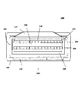
5 Figure 1
shows an example of a portable x-ray source 100 according to aspects of
the present disclosure. Portable x-ray source 100 may comprise a plurality of
x-ray
generators 102, wherein each x-ray generator 102 may be capable of producing a
small
cone (or "conelee') of x-ray radiation. The plurality of x-ray generators 102
may be
arranged as a distributed x-ray generator array 103, and in one preferred
aspect of the
10 present disclosure may comprise a flat-panel, x-ray source (FPS) as
described by
PCT/US2016/014782 to Travish, d al
The use of distributed x-ray generator array 103 provides several improvements
over current systems, and departs from conventional approaches to x-ray
imaging, which
generally focus on a single, tube-based (vacuum tube) source and teach away
from systems
15 utilizing distributed sources.
Figure 2 provides a side-by-side comparison of a conventional single-source,
tube-
based x-ray source 200 (as is typically used in dental imaging) to a portable,
x-ray source
100 in accordance with aspects of the present disclosure. Tube-based x-ray
source 200 is
capable of generating an x-ray cone 201. X-ray cone 201 may impinge object 203
(e.g., the
20 patient's mouth), wherein detector 202 may measure the intensity (and hence
the
attenuation of x-ray cone 201 caused by object 203) to form a two-dimensional
radiograph
(or shadowgram). For a standard intraoral x-ray scan source 200 must typically
be placed
at least 20 cm away from object 203.
Conversely, portable x-ray source 100 may include a distributed x-ray
generator
25 array 103 (e.g., a fixed, two-dimensional array) comprising a moderate
pitch spacing (e.g.,
in the mm to cm range). Each generator 102 may be individually addressable so
as to
enable distributed x-ray generator array 103 to produce multiple, angularly-
diverse
conelets
conelets 204a and conelets 204b), which enables portable x-ray source
100
to image object 203 (or more specifically a region of interest) from
sufficient angles to
30 generate a three-dimensional reconstruction without need for source
movement; in other
words, by motion-free tomosynthesis. Because portable x-ray source 100, unlike
conventional tomographic systems such as CBCT, does not require expensive
precision
1
Date Recue/Date Received 2022-12-14
computer-controlled movers the cost and complexity of portable x-ray source
100 is
significantly reduced as compared to conventional systems.
The design of distributed x-ray generator 103 also enables portable x-ray
source
100 to be placed closer to detector 202, approximately 12 cm away from object
203 as
opposed to the 20 cm in standard intraoral radiography. This reduction in
stand-off
distance (source to object distance) enables the weight and power requirement
of portable
x-ray source 100 to be significantly reduced as compared to conventional
sources, such
as tube-based x-ray source 200. This in turn eliminates the need for costly
power supplies,
which significantly reduces the cost of portable x-ray source 100 as compared
to
conventional systems.
The reduction in weight and input power is partially attributable to the fact
that
radiation declines as a square of distance, as such embodiments of the present
disclosure
may only require approximately 1/4 x-ray intensity (and hence 1/4 input power)
of
conventional systems. The design of distributed x-ray generator array 103
(e.g.,
addressability and moderate pitch spacing) also enables faster acquisition
speeds as
compared to conventional tomographic systems, because unlike conventional
systems
which typically require mechanical movement to achieve sufficient scanning
angles,
embodiments of the present disclosure are capable of movement-free
tomosynthesis.
The elimination of the need for a gantry reduces the size, weight, complexity
and cost of
portable x-ray source 100 as compared to conventional system. Moreover, this
faster
acquisition speed may reduce the amount of time patients need to stay
immobile, and thus
may increase patient comfort.
In one aspect of the present disclosure, portable x-ray source 100 may weigh
approximately 4kg and fit into a standard camera bag. In contrast, a standard,
wall-
mounted dental x-ray unit may weigh nearly 40kg, while a two-dimensional
portable
system typically weights around 6kg.
The cost of portable x-ray source 100 is also reduced by the elimination of
vacuum
tubes, which tend to be fragile, have a short life, and only limited use
outside hospital
settings. In sharp contrast, distributed x-ray generator array 103 may be made
in a
semiconductor foundry, which will reduce cost to manufacture, deploy, and
maintain
portable x-ray source 100.
The cost savings, low-profile, and portability of embodiments of the present
disclosure may lessen the burden on end users, such as hospitals, primary care
facilities,
11
Date Recue/Date Received 2022-12-14
and dental offices, who are often required to make large capital investments
in imaging
systems, and thus may increase the availability of three-dimensional x-ray
imaging. For
example, deploying a portable three-dimensional x-ray imaging source within a
multi-
dentist practice would be transformative given that due to equipment cost many
dentists
do not have access to CBCT or other three-dimensional imaging systems, and
thus are
often forced to rely on limited two-dimensional planar x-ray images.
The shorter stand-off distances achievable by embodiments of the present
disclosure also enable a reduction of radiation scatter (side scatter and
backscatter), which
in turn reduces the risk of x-ray exposure for x-ray operators and clinical
workers. Scatter
radiation may be further reduced by use of a gross collimator 115, which may
absorb x-
rays that are not useful for imaging purposes, such as x-rays outside the
region of interest
of object (ROI) 203, while allowing x-rays useful to imaging to strike the
ROT.
Gross collimator 115 may comprise a structure made of x-ray attenuating
materials (e.g., high-density material(s) with high x-ray absorption). In one
aspect, gross
collimator 115 may comprise an elevated plane (or walls) that limits the area
illuminated
by portable x-ray source 100 to a ROT. In this way, gross collimator 115 may
reduce non-
imaging dosages of x-ray photons by lessening, if not removing, x-ray
backscatter and
side-scatter. Thus, gross collimator 115 may serve to minimize unnecessary,
and
potentially harmful radiation exposure, without impacting x-ray image quality.
The use of such a gross collimator 115 is particularly useful for imaging
where the
activate area of the detectors is small, and in cases where sensitive organs
are adjacent to
the area to be imaged. An example of such a use case is dental imaging: the
intra-oral
detectors are often 2cm x 4cm or smaller and the teeth and jaws of interest
lie in close
proximity to the brain.
Referring back to Figure 1, distributed x-ray generator array 103 may comprise
a
plurality of electron field emitters 104 aligned or otherwise paired with a
plurality of
targets 106. In this way, each generator 102 may be comprised of an electron
field emitter
104 paired with a target 106. Each electron field emitter 104 may be capable
of generating
a beam of electrons that may be directed at a target 106, such as a material
effective at
high-energy bremsstrahlung,, to produce x-rays.
The plurality of electron field emitters 104 (and thus the plurality of
targets 106)
may be arranged as an emitter array 105. Emitter array 105 may comprise any of
several
configurations, including a two-dimensional array forming a square grid; a
triangular grid
12
Date Recue/Date Received 2022-12-14
also known as a "hexagon pack," or electron field emitters 104 may be randomly
spaced.
The spacing and pattern of emitter array 104 may be varied based on, or
determined by,
several factors, including the end-use, the imaging application geometry, or
the desired
image resolution, among others.
Electron field emitters 104 may be fabricated from a variety of conducting
materials including, among others, doped silicon, tungsten or tungsten alloys,
or highly
conductive metals, such as copper or aluminium. Alternatively (or in
conjunction), it may
be desirable to overcoat electron field emitters 104 (or their tips) with a
protective coating
or film of tungsten, titanium-nitride, diamond-like carbon or other robust
conductive
material.
As noted, distributed x-ray generator array 103 may comprise a plurality of
targets
106 made of one or more materials capable of converting incident electrons
through
physical process(es) into x-rays. In one aspect of the present disclosure,
each target 106
may comprise a metal film made of a material(s) effective at high-energy
bremsstrahlung,
such as tungsten, molybdenum, rhenium, gold or other heavy metals. In another
aspect,
each target 106 may be made of two or more metals, or may comprise more than
one
layer of materials, such that each target 106 may include a small area
comprised of an
effective bremsstrahlung material (e.g., tungsten) and an adjacent area made
of a low-Z
material (e.g., silicon).
Targets 106 may be self-supporting, or may be supported by an electrically
conducting substrate, which may serve to complete the electrical circuit
(between the
plurality of electron field emitters 104 and targets 106) and to dissipate the
heat energy
deposited by the electron beam. In one example, target 106 may comprise a thin
film of
tungsten supported by a silicon substrate, or another conductive, light-
element material,
such as aluminium. In yet another embodiment, the substrate may be made of an
insulating material having a conductive coating.
Target 106 may have a variety of geometries, including, among others, a
'doughnut' shape, a circular shape, or may incorporate straight lines. As
would be
understood by a person of skill in the art, the thickness of target 106 may be
varied
depending on the atomic number of the target 106 material, the thermal
properties of the
target 106 material, or the energy of the electron beam that will be incident
on target 106.
In an aspect of the present disclosure, the thickness of each target 106 may
be between 1
and 100 jun.
13
Date Recue/Date Received 2022-12-14
The electron field generated by electron field emitters 104 may be
sufficiently
intense to ionize gas molecules proximate to electron field emitters 104
(e.g., in the +z
and/or ¨z planes). This ionization may prevent the production of useful x-rays
because,
among other things, the ionized gases may scatter the emitted electrons, and
may damage
electron field emitters 104 and targets 106. Accordingly, it may be desirable
to minimize,
if not eliminate, gas molecule ionization by maintaining a vacuum between
electron field
emitters 104 and targets 106 by, among other things, housing emitters 104 and
targets 106
in a vacuum or low-pressure environment.
In an aspect of the present disclosure, such a vacuum (low-pressure)
environment
may be achieved by manufacturing distributed x-ray generator array 103 under
high-
vacuum, and then housing distributed x-ray generator array 103 (and thus
electron field
emitters 104 and targets 106) within a chamber capable of maintaining the
vacuum (low-
pressure) environment. The vacuum environment may be maintained through the
operating life of portable x-ray source 100 by utilizing a vacuum getter,
which may be
capable of chemically combining or absorbing gas molecules. The vacuum getter
may be
coated on an internal surface or attached to the vacuum chamber that houses
distributed
x-ray generator array 103. As would be recognized by a person of skill in the
art in view
of the present disclosure, other mechanisms may be suitable for maintaining a
requisite
vacuum environment, such as mechanical and ion pumps.
Targets 106 may be deposited on the inner surface of the vacuum chamber, which
houses distributed x-ray generator array 103. This configuration provides a
vast
improvement over conventional tube-based sources in that by depositing targets
106 on
the inner surface of the vacuum chamber the heat generated on targets 106 may
be more
easily dissipated by conduction (e.g., excess heat may be dissipated
throughout the
chamber), making portable x-ray source 100 easier to cool than conventional
systems. In
stark contrast, targets or anodes in conventional x-ray sources are housed
within the
source vacuum tube, which makes cooling difficult because it must rely on
radiation to
dissipate heat.
Distributed x-ray generator array 103 may also include a gate, which may be
capable of controlling the emission of electrons from electron field emitters
104. The
gate may comprise a plurality of conducting structures through which electrons
can pass
and a voltage can be applied. Alternatively, the gate may be capable of
suppressing the
electron field generated between electron field emitters 104 and targets 106.
By way of
14
Date Recue/Date Received 2022-12-14
example, the gate may comprise a conducting plate with holes capable of
allowing
electrons from electron field emitters 104 to pass. In another embodiment, the
gate may
comprise an array of individual annular structures, each of which is
associated with an
electron field emitter 104. The gate may also include an insulating substrate
coated with
a conducting material.
In an aspect of the present disclosure, the gate may be powered by high-
voltage
power supply 109 of portable x-ray source 100. In another aspect, voltage to
each portion
of the gate associated with a particular electron field emitter 104, such as a
particular set
of holes or annular structures, may be controlled individually.
The gate may be altogether eliminated such that distributed x-ray generator
array
103 may comprise a diode configuration (e.g., cathode and anode structures
absent a gate).
This diode configuration typically has a limited operating-voltage range
because of the
exponential nature of field emission (e.s., the emitted current depends
exponentially on
the applied voltage), but is simpler to produce and can be more reliable than
a triode
configuration, or a configuration which includes a gate. Nonetheless, triodes
may offer
various benefits, including the ability to independently control the emission
voltage and
accelerating (or final) voltage. Accordingly, based on use and other design
considerations,
it may be desirable to select one configuration over another. By way of
example, in the
case of a dental imaging source, where fixed or nearly fixed voltages are
acceptable, a
diode configuration may be preferable.
Referring to Figure 1, portable, x-ray source 100 may also include spacer 108
disposed between electron field emitters 104 and targets 106. Spacer 108 may
serve to
maintain the requisite separation between electron field emitters 104 and
targets 106, and
to insulate electron field emitters 104 from targets 106.
Spacer 108 may be varied in thickness depending on materials used and voltages
applied (the potential difference between electron field emitters 104 and
targets 106). For
example, larger voltages may require more distance between electron field
emitters 104
and targets 106, and thus may require a thicker spacer 108. Conversely, a
thinner spacer
108 may be used with smaller voltages. In aspects of the present disclosure,
spacer 108
may be between ltrun to 30mm thick. In another aspect, spacer 108 may be
between
5mm and 15mm thick. And in yet another aspect, spacer 108 may be between 15mm
and
30mm thick.
Date Recue/Date Received 2022-12-14
Spacer 108 may be made of glass, borosilicate glass, ceramic, or other
suitable
material as would be understood by a person of skill in the art in view of the
present
disclosure, and may have various configurations. In one aspect of the present
disclosure,
spacer 108 may be substantially cylindrical. In yet another aspect involving a
triode
configuration, spacer 108 may be formed of two parts, such as to allow for the
separation
or removal of the gate from distributed x-ray generator array 103.
Spacer 108 may further serve as part of the vacuum chamber that may house
electron field emitters 104 and targets 106. Alternatively, spacer 108 may not
serve to
form or maintain such a vacuum.
Portable x-ray source 100 may further include a high-voltage power supply 109,
which may be capable of producing a large potential difference (voltage)
between electron
field emitters 104 and targets 106. In an aspect of the present disclosure,
high-voltage
power supply 109 may be capable of converting line voltage, such as the common
voltage
found in a standard outlet, to high voltage. Alternatively, power supply 109
may be
connected to one or more batteries 107, and may be capable of converting
battery voltage
to high voltage.
High-voltage power supply 109 may be capable of producing (but is not be
limited
to) voltages up to -120kV. In another aspect of present disclosure, high-
voltage power
supply 109 may produce voltages between -20 and -120kV. Alternatively, high-
voltage
power supply 109 may produce positive voltages, and in a further aspect, may
operate at
a fixed voltage between 50kV-70kV. In yet another aspect of the present
disclosure,
power supply 109 may be capable of operating at two or more voltages,
sequentially or in
parallel.
High-voltage, power supplies (e.g., -30kV to -80kV), as described in the
present
disclosure, run contrary to conventional x-ray imaging approaches. In
particular,
conventional approaches teach that electron field emitters are to be driven by
low- to
moderate- power supplies, and teach away from use of high-voltage power
supplies 109.
Conventional approaches also teach away from compact power supplies, where the
gap
between the ground and high-tension plane is minimized.
As illustrated in Figure 1, the geometry of high-voltage power supply 109 may
generally follow that of electron field emitters 104 and targets 106, and the
output plane
of high-voltage power supply 109 may touch electron field emitters 104 to form
an
electrical contact. In an aspect of the present disclosure suitable for dental
radiology,
16
Date Recue/Date Received 2022-12-14
high-voltage power supply 109 may be 30mm thick with a transverse size of
150mm by
150tnm. Portable, x-ray source 100 may be packaged in liquid (insulating oils)
and/or
solid (putty, potting), so as to provide the insulation required for the high-
voltage of high-
voltage power supply 109.
In another embodiment (not illustrated), distributed x-ray generator array 103
may
be powered by a plurality of ferroelectric crystals as described in
PCT/US2010/044762.
Alternatively, as would be understood by a person of skill in view of the
present
disclosure, distributed x-ray generator array 103 may be powered by any number
of
devices capable of producing the desired voltage.
Portable x-ray source 100 may further include a plurality of emission controls
110.
The plurality of emission controls 110 may be capable of controlling (e.g.,
defocusing/focusing or deflecting/steering) electrons emitted by electron
field emitters
104. In this way, emission control 110 may be capable of regulating the
emission of x-
rays by each generator 102. In turn, this enables each generator 102 to be
individually
addressable (controllable) and thus enables distributed x-ray generator array
103 to
generate temporally-separated, but physically overlapping x-ray conelets. This
allows for
seamless coverage of an object to be imaged, and maximal use of the available
flux while
maintaining the ability to have minimal stand-off distances.
The plurality of emission controls 110 may be capable of defocusing/focusing
or
deflecting/steering electron beams from an electron field emitter 104,
individually onto
or away from target 106, and thus may affect the production or cessation of x-
rays
respectively. It will be appreciated by one skilled in the art that emission
control is not
limited to one approach, and may be used in combination with one or more
methods,
including through electro-static, magneto-static and electro-magnetic means.
One such
approach is described in PCT/GB2015/050639.
Figure 3 shows an example of a plurality of emission controls 110 comprising a
plurality of selectively-powered coils or magnets (coils/magnets) 301.
Coils/magnets 301
may be capable of preventing electron beams 304 emitted by an electron field
emitter 104
from striking a portion of a target 106 comprised of a material effective at
high-energy
bremsstrahlung, and thus may be capable of controlling x-ray emission.
As illustrated in Figure 3, the output flux (electron beam 304) of electron
field
emitter 104 may be controlled by using electromagnetic fields generated by
coils/magnets
301 such that when energized or "on" 302 the fields produced deflect the
electron beam
17
Date Recue/Date Received 2022-12-14
304 away from the ballistic trajectory/axis 307 and onto target 106. When
coils/magnets
301 are "off" 303, the electron beam 304 continues directly on axis 307 and
strikes
substrate 305, which may be comprised of low atomic number materials which
only
generates low energy photons that produce no signal. Thus, the state of
coils/magnets
301 (on 302 or off 303) serves as a control of the generation of x-rays 306
from a given
emitter.
In an aspect of the present disclosure, target 106 may comprise a discrete
area
made of an effective bremsstrahlung material, such as tungsten, an adjacent
area made of
a low-Z material, such as silicon, and a support made of a conductive
material, such as
aluminum. Lenses or yokes maybe used to elongate the magnetic field in the
beam axis
direction, and compact it off axis.
In a further embodiment of the present disclosure, the individual selectively-
powered coils/magnets 301 may be arranged in clusters of coils/magnets in a
pattern
essentially equivalent to that of emitter array 105. The clusters may comprise
four
coils/magnets 301 capable of creating a dipole magnetic field arranged about
each
electron field emitter 104. Alternatively, the clusters may comprise eight or
more
coils/magnets 301 arranged such that a central set of coils creates a dipole
field for
deflecting the beam trajectory of the electron field emitter 104 and
surrounding
coils/magnets 301 are used to offset the stray field of the central
coils/magnets 301, these
clusters being configured in a pattern essentially equivalent to that of
emitter array 105.
In aspects of the present disclosure, it may be necessary for solenoid coil
301 to
deflect the electron beam a distance of 0.1 mm to 1.25 mm from the nominal
path.
PCT/GB2015/050639 describes methods of achieving such deflection using high-
current
coils. Notably, using clusters of coils, as described herein, similar results
may be achieved
at low currents.
Portable x-ray source 100 may also include a mechanism capable of selectively
controlling x-ray emission from distributed x-ray generator array 103. In an
aspect of the
present disclosure, said mechanism may comprise a circuit(s), such as an
addressing and
timing circuit(s), capable of selectively activating one or more controller
110, such as
solenoid coils 301, in a predetermined sequence. Targets 106 and the plurality
of emission
controllers 110 may be arranged such that portable x-ray source 100 operates
normally in
the on mode. Alternatively, they may be arranged such that portable x-ray
source 100
operates in normally off mode. In addition, the control mechanism (e.g.,
electronic circuit)
18
Date Recue/Date Received 2022-12-14
may be capable of automatically stopping the emission of x-ray photons after a
predetermined detector signal level is achieved.
Portable x-ray source 100 may also include a filter capable of removing or
blocking radiation that does not contribute to x-ray imaging, such as low-
energy x-rays
entirely absorbed by tissue. In this way, the filter may be able to minimize
unnecessary
ray exposure to patients, radiographers, technologists, clinicians, dentists,
etc., without
negatively impacting x-ray imaging. In an aspect of the present disclosure,
the filter may
be removable. The filter may also be encoded such that control electronics can
determine
the specific filter in use.
As would be understood by a person of skill in view of the present disclosure,
the
filter may be made of a variety of materials and may have varying thickness
depending on
the operating voltage or desired end use of portable x-ray source 100. As an
example,
the filter may comprise an aluminium sheet between lmrn to lOmm thick.
Alternatively,
the filter may comprise a copper sheet between 1mrn to 5nun thick. In yet
another aspect,
the filter may comprise a stack of alternating higher-atomic number and lower-
atomic
number materials, such as aluminum and carbon.
Portable x-ray source 100 may include a collimator array, which may serve to
narrow the angle of emitted x-rays, and thus further facilitates fractional
coverage of a
region of interest. The collimator array may be as described in
PCT/GB2015/050637 to
Travish, et al Alternatively, the collimator array may comprise a plate made
of high-
density material having a plurality of holes of appropriate size, wherein the
holes are
capable of allowing x-rays to be transmitted with a specific opening angles.
Such high-
density material may include tungsten, steel, or an alloy made of similar
materials having
a high x-ray attenuation coefficients.
In another aspect of the present disclosure, the collimator array may comprise
a
plurality of tubes, wherein each tube may be capable of controlling the
opening angle of
emitted x-ray cones. For example, the collimator array may comprise a tungsten
plate
having a plurality of aluminium inserts, wherein each aluminium insert serves
to transmit
a portion of each x-ray cone with a well-defined opening angle. In another
embodiment,
the collimator array may comprise two plates arranged one atop of the other
with each
plate having a plurality of holes such that a particular x-ray cone passes
through a hole in
the lower and then in the upper plate.
19
Date Recue/Date Received 2022-12-14
Portable x-ray source 100 may include a housing 101. Housing 101 may comprise
a light, protective casework capable of providing a mechanically rigid
platform for x-ray
source 100 and facffitating portability. Housing 101 may also be capable of
aiding in
thermal control, for example by dissipating heat throughout housing 101. In a
preferred
aspect of the present disclosure, housing 101 is compatible with medical
device
requirements such as sterility, alcohol, wipe down, and cytotoxicity.
Housing 101 may also include a mechanism for aligning portable x-ray source
100
to a detector, such as one or more non-contact sensors capable of providing
the user of
portable x-ray source 100 with an indication that portable x-ray source 100 is
in proper
alignment with a detector. In another aspect of the disclosure, housing 101
may comprise
an inner case designed to enclose high-voltage components, such as distributed
x-ray
generator array 103, and an outer case designed to hold other, non-high
voltage
components. The inner case of housing 101 may be filled with insulating fluid,
or
alternatively with a solid insulator. Housing 101 may also house batteries 107
to power
portable x-ray system 101 without need for wall plug supplied electricity.
Date Recue/Date Received 2022-12-14