Note: Descriptions are shown in the official language in which they were submitted.
CA 03039886 2019-04-09
WO 2018/070936
PCT/SG2017/050509
1
ANTI-CEACAM6 ANTIBODIES AND METHODS OF USE
CROSS-REFERENCE TO RELATED APPLICATIONS
[0001] This application claims the benefit of priority of Singapore
application No.
10201608481W, filed 10 October 2016, the contents of it being hereby
incorporated by
reference in its entirety for all purposes.
FIELD OF THE INVENTION
[0001] The present invention relates generally to antibodies.
Specifically, the invention
.. relates to anti-CEACAM6 monoclonal antibodies and uses thereof.
BACKGROUND OF THE INVENTION
[0002] Carcinoembryonic antigen-related cell adhesion molecule 6
(CEACAM6) belongs
to the carcinoembryonic antigen (CEA) family. CEACAM6 is a glycosyl
phosphatidyl
inositol (GPI) anchored cell surface glycoprotein that has been observed to be
overexpressed
in a variety of cancers including breast, pancreatic, colonic and non-small
cell lung
carcinoma. CEACAM6 acts as an oncogene in tumors and promotes cancer invasion,
metastasis, anoikis resistance and chemoresistance, and inhibits
differentiation.
[0003] To date, there are a very limited number of anti-CEACAM6
monoclonal antibodies
that could be used for antibody therapy or as antibody-drug conjugates for
cancer treatment.
There is therefore a need to develop novel antibodies against CEACAM6 that can
be used for
antibody therapy or as antibody-drug conjugates for cancer treatment.
SUMMARY
[0004] In one aspect, there is provided an antigen-binding protein, or an
antigen-binding
fragment thereof, comprising (i) a heavy chain variable domain comprising a
VHCDR1 having the
amino acid sequence GNTFTSYVMH (SEQ ID NO: 3); a VHCDR2 having the amino acid
sequence
YINPYNDGTKYNEKFKG (SEQ ID NO: 4) and a VHCDR3 having the amino acid sequence
STARATPYFYAMDY (SEQ ID NO: 5); and (ii) a light chain variable domain
comprising a
VLCDR1 having the amino acid sequence KSSQSLLWSVNQNSYLS (SEQ ID NO: 6), a
VLCDR2
having the amino acid sequence GASIRES (SEQ ID NO: 7), and a VLCDR3 having the
amino acid
sequence QHNHGSFLPYT (SEQ ID NO: 8).
CA 03039886 2019-04-09
WO 2018/070936
PCT/SG2017/050509
2
[0005] In another aspect, there is provided a composition comprising a
physiologically
acceptable carrier and a therapeutically effective amount of the antigen-
binding protein, or an
antigen-binding fragment thereof as disclosed herein.
[0006] In another aspect, there is provided a use of an antigen-binding
protein, or an
antigen-binding fragment thereof as disclosed herein, or composition as
disclosed herein in
the manufacture of a medicament for treating or preventing cancer.
[0007] In another aspect, there is provided an antigen-binding protein,
or an antigen-
binding fragment thereof as disclosed herein, or composition as disclosed
herein for use in
treating or preventing cancer.
[0008] In another aspect, there is provided a method of treating or
preventing cancer
comprising administering an antigen-binding protein, or an antigen-binding
fragment thereof
as disclosed herein, or composition as disclosed herein to a subject in need
thereof.
[0009] In another aspect, there is provided a method for detecting
cancer in a subject, the
method comprising: contacting a sample obtained from the subject with an
antigen-binding
protein, or an antigen-binding fragment thereof as disclosed herein in vitro;
detecting the
binding of the antigen-binding protein, or an antigen-binding fragment thereof
in the sample;
correlating the binding with a level of binding in a control sample to
determine the level of
binding in the sample, wherein an increase in the level of binding in the
sample relative to the
control sample is indicative of cancer.
[0010] In another aspect, there is provided a method for identifying a
subject susceptible
to cancer the method comprising: contacting a sample obtained from the subject
with an
antigen-binding protein, or an antigen-binding fragment thereof as disclosed
herein in vitro;
detecting the binding of the antigen-binding protein, or an antigen-binding
fragment thereof
in the sample; correlating the binding with a level of binding in a control
sample to determine
the level of binding in the sample, wherein an increase in the level of
binding in the sample
relative to the control sample indicates that the subject is susceptible to
cancer.
[0011] In one aspect, there is provided a kit when used in the method as
disclosed herein,
comprising an antigen-binding protein, or antigen-binding fragment thereof as
disclosed
herein, together with instructions for use.
CA 03039886 2019-04-09
WO 2018/070936
PCT/SG2017/050509
3
DEFINITIONS
[0012] The following are some definitions that may be helpful in
understanding the
description of the present invention. These are intended as general
definitions and should in
no way limit the scope of the present invention to those terms alone, but are
put forth for a
better understanding of the following description.
[0013] The term "antigen binding protein" as used herein refers to
antibodies, antibody
fragments and other protein constructs, such as domains, which are capable of
binding to
CEACAM6.
[0014] The term "antibody" is used herein in the broadest sense to refer
to molecules with
an immunoglobulin-like domain and includes monoclonal, recombinant,
polyclonal,
chimeric, humanised, bispecific and heteroconjugate antibodies; a chimeric
antigen receptor
(CAR), a single variable domain, a domain antibody, antigen binding fragments,
immunologically effective fragments, single chain Fv, diabodies, TandabsTm,
etc (for a
summary of alternative "antibody" formats see Holliger and Hudson, Nature
Biotechnology,
2005, Vol 23, No. 9, 1126-1136).
[0015] The phrase "single variable domain" refers to an antigen binding
protein variable
domain (for example, VH, VHH, VL) that specifically binds an antigen or
epitope
independently of a different variable region or domain.
[0016] A "domain antibody" or "dAb" may be considered the same as a
"single variable
domain" which is capable of binding to an antigen. A single variable domain
may be a
human antibody variable domain, but also includes single antibody variable
domains from
other species such as rodent (for example, as disclosed in WO 00/29004), nurse
shark and
Camelid VHH dAbs. Camelid VHH are immunoglobulin single variable domain
polypeptides
that are derived from species including camel, llama, alpaca, dromedary, and
guanaco, which
produce heavy chain antibodies naturally devoid of light chains. Such VHH
domains may be
humanised according to standard techniques available in the art, and such
domains are
considered to be "domain antibodies". As used herein VH includes camelid VHH
domains.
[0017] As used herein the term "domain" refers to a folded protein
structure which has
tertiary structure independent of the rest of the protein. Generally, domains
are responsible
for discrete functional properties of proteins, and in many cases may be
added, removed or
transferred to other proteins without loss of function of the remainder of the
protein and/or of
CA 03039886 2019-04-09
WO 2018/070936
PCT/SG2017/050509
4
the domain. A "single variable domain" is a folded polypeptide domain
comprising
sequences characteristic of antibody variable domains. It therefore includes
complete
antibody variable domains and modified variable domains, for example, in which
one or
more loops have been replaced by sequences which are not characteristic of
antibody variable
domains, or antibody variable domains which have been truncated or comprise N-
or C-
terminal extensions, as well as folded fragments of variable domains which
retain at least the
binding activity and specificity of the full-length domain. A domain can bind
an antigen or
epitope independently of a different variable region or domain.
[0018] An antigen binding fragment may be provided by means of arrangement of
one or
more CDRs on non-antibody protein scaffolds such as a domain. The domain may
be a
domain antibody or may be a domain which is a derivative of a scaffold
selected from the
group consisting of CTLA-4, lipocalin, SpA, an Affibody, an avimer, GroEl,
transferrin,
GroES and fibronectin/adnectin, which has been subjected to protein
engineering in order to
obtain binding to an antigen, such as CEACAM6, other than the natural ligand.
[0019] An antigen binding fragment or an immunologically effective fragment
may
comprise partial heavy or light chain variable sequences. Fragments are at
least 5, 6, 8 or 10
amino acids in length. Alternatively the fragments are at least 15, at least
20, at least 50, at
least 75, or at least 100 amino acids in length.
[0020] The term "specifically binds" as used throughout the present
specification in
relation to antigen binding proteins means that the antigen binding protein
binds to
CEACAM6 with no or insignificant binding to other (for example, unrelated)
proteins.
However, the term does not exclude the fact that the antigen binding proteins
may also be
cross-reactive with closely related molecules. The antigen binding proteins
described herein
may bind to CEACAM6 with at least 2, 5, 10, 50, 100, or 1000 fold greater
affinity than they
bind to closely related molecules.
[0021] The term "neutralises" as used throughout the present
specification means that the
biological activity of CEACAM6 is reduced in the presence of an antigen
binding protein as
described herein in comparison to the activity of CEACAM6 in the absence of
the antigen
binding protein, in vitro or in vivo. Neutralisation may be due to one or more
of blocking
CEACAM6 binding to its receptor, preventing CEACAM6 from activating its
receptor, down
regulating CEACAM6 or its receptor, or affecting effector functionality. The
reduction or
CA 03039886 2019-04-09
WO 2018/070936
PCT/SG2017/050509
inhibition in biological activity may be partial or total. A neutralising
antigen binding protein
may neutralise the activity of CEACAM6 by at least 20%, 30% 40%, 50%, 55%,
60%, 65%,
70%, 75%, 80%, 82%, 84%, 86%, 88%, 90%, 92%, 94%, 95%, 96%, 97%, 98%, 99% or
100% relative to CEACAM6 activity in the absence of the antigen binding
protein.
5 Neutralisation may be determined or measured using one or more assays
known to the skilled
person or as described herein. For example, antigen binding protein binding to
CEACAM6
can be assessed in a sandwich ELISA, by BIAcoreTM, FMAT, FORTEbio, or similar
in vitro
assays.
[0022] "CDRs" are defined as the complementarity determining region
amino acid
sequences of an antigen binding protein. These are the hypervariable regions
of
immunoglobulin heavy and light chains. There are three heavy chain and three
light chain
CDRs (or CDR regions) in the variable portion of an immunoglobulin. Thus,
"CDRs" as
used herein refers to all three heavy chain CDRs, all three light chain CDRs,
all heavy and
light chain CDRs, or at least two CDRs.
[0023] As used herein, the term "promoter" is intended to refer to a region
of DNA that
initiates transcription of a particular gene.
[0024] As used herein, the term "cancerous" relates to being affected by
or showing
abnormalities characteristic of cancer.
[0025] As used herein, the term "biological sample" or "sample" is meant
a sample of
tissue or cells from a patient that has been obtained from, removed or
isolated from the
patient.
[0026] The term "obtained or derived from" as used herein is meant to be
used inclusively.
That is, it is intended to encompass any nucleotide sequence directly isolated
from a
biological sample or any nucleotide sequence derived from the sample.
[0027] The method as described herein is suitable for use in a sample of
fresh tissue,
frozen tissue, paraffin- preserved tissue and/or ethanol preserved tissue. The
sample may be
a biological sample. Non-limiting examples of biological samples include whole
blood or a
component thereof (e.g. plasma, serum), urine, saliva lymph, bile fluid,
sputum, tears,
cerebrospinal fluid, bronchioalveolar lavage fluid, synovial fluid, semen,
ascitic tumour fluid,
breast milk and pus. In one embodiment, the sample of nucleic acid is obtained
from blood,
amniotic fluid or a buccal smear. In a preferred embodiment, the sample is a
whole blood
sample.
CA 03039886 2019-04-09
WO 2018/070936
PCT/SG2017/050509
6
[0028] A biological sample as contemplated herein includes cultured
biological materials,
including a sample derived from cultured cells, such as culture medium
collected from
cultured cells or a cell pellet. Accordingly, a biological sample may refer to
a lysate,
homogenate or extract prepared from a whole organism or a subset of its
tissues, cells or
component parts, or a fraction or portion thereof. A biological sample may
also be modified
prior to use, for example, by purification of one or more components,
dilution, and/or
centrifugation.
[0029] As used herein, the term "detectable label" or "reporter" refers
to a detectable
marker or reporter molecules, which can be attached to nucleic acids. Typical
labels include
fluorophores, radioactive isotopes, ligands, chemiluminescent agents, metal
sols and colloids,
and enzymes. Methods for labeling and guidance in the choice of labels useful
for various
purposes are discussed, e.g., in Sambrook et al., in Molecular Cloning: A
Laboratory
Manual, Cold Spring Harbor Laboratory Press (1989) and Ausubel et al., in
Current
Protocols in Molecular Biology, Greene Publishing Associates and Wiley-
Intersciences
(1987).
[0030] As used herein, the term "susceptible to cancer" or
"susceptibility to cancer" refers
to the likelihood of a subject developing cancer. The term does not indicate
that a subject will
develop cancer with 100% certainty. Rather, the term "susceptible to cancer"
refers to an
increased probability that a subject will develop cancer when compared to an
individual who
is not "susceptible to cancer".
[0031] As used herein, the term "about", in the context of
concentrations of components of
the formulations, typically means +/- 5% of the stated value, more typically
+/- 4% of the
stated value, more typically +/- 3% of the stated value, more typically, +/-
2% of the stated
value, even more typically +/- 1% of the stated value, and even more typically
+/- 0.5% of
.. the stated value.
[0032] Throughout this disclosure, certain embodiments may be disclosed
in a range
format. It should be understood that the description in range format is merely
for convenience
and brevity and should not be construed as an inflexible limitation on the
scope of the
disclosed ranges. Accordingly, the description of a range should be considered
to have
specifically disclosed all the possible sub-ranges as well as individual
numerical values
within that range. For example, description of a range such as from 1 to 6
should be
considered to have specifically disclosed sub-ranges such as from 1 to 3, from
1 to 4, from 1
CA 03039886 2019-04-09
WO 2018/070936
PCT/SG2017/050509
7
to 5, from 2 to 4, from 2 to 6, from 3 to 6 etc., as well as individual
numbers within that
range, for example, 1, 2, 3, 4, 5, and 6. This applies regardless of the
breadth of the range.
[0033] Certain embodiments may also be described broadly and generically
herein. Each
of the narrower species and subgeneric groupings falling within the generic
disclosure also
form part of the disclosure. This includes the generic description of the
embodiments with a
proviso or negative limitation removing any subject matter from the genus,
regardless of
whether or not the excised material is specifically recited herein.
[0034] Unless the context requires otherwise or specifically stated to
the contrary,
integers, steps, or elements of the invention recited herein as singular
integers, steps or
elements clearly encompass both singular and plural forms of the recited
integers, steps or
elements.
[0035] The word "substantially" does not exclude "completely" e.g. a
composition which
is "substantially free" from Y may be completely free from Y. Where necessary,
the word
"substantially" may be omitted from the definition of the invention.
[0036] The invention illustratively described herein may suitably be
practiced in the
absence of any element or elements, limitation or limitations, not
specifically disclosed
herein. Thus, for example, the terms "comprising", "including", "containing",
etc. shall be
read expansively and without limitation. Additionally, the terms and
expressions employed
herein have been used as terms of description and not of limitation, and there
is no intention
in the use of such terms and expressions of excluding any equivalents of the
features shown
and described or portions thereof, but it is recognized that various
modifications are possible
within the scope of the invention claimed. Thus, it should be understood that
although the
present invention has been specifically disclosed by preferred embodiments and
optional
features, modification and variation of the inventions embodied therein herein
disclosed may
be resorted to by those skilled in the art, and that such modifications and
variations are
considered to be within the scope of this invention.
[0037] The invention has been described broadly and generically herein.
Each of the
narrower species and subgeneric groupings falling within the generic
disclosure also form
part of the invention. This includes the generic description of the invention
with a proviso or
.. negative limitation removing any subject matter from the genus, regardless
of whether or not
the excised material is specifically recited herein.
CA 03039886 2019-04-09
WO 2018/070936 PCT/SG2017/050509
8
[0038] Other embodiments are within the following claims and non- limiting
examples. In
addition, where features or aspects of the invention are described in terms of
Markush groups,
those skilled in the art will recognize that the invention is also thereby
described in terms of
any individual member or subgroup of members of the Markush group.
BRIEF DESCRIPTION OF THE DRAWINGS
[0039] The invention will be better understood with reference to the
detailed description
when considered in conjunction with the non-limiting examples and the
accompanying
drawings, in which:
[0040] Fig. 1 is a flow diagram depicting the methodology for generating
Gefitinib
resistant clones from PC-9 and cell lines used for immunization and hybridoma
fusion of
generation of monoclonal antibody panel.
[0041] Fig. 2 shows flow cytometry binding of GR6A04 on A) immunizing GR
lines,
CL75, CL86 and CL131, and B) parental PC-9 and another GR PC-9 line. Gating
was
performed at M=2% of negative control for each cell line. Absence of propidium
iodide
staining (FL-3) shows no inherent cytotoxity from mAb GR 6A04 binding.
[0042] Fig. 3 shows flow cytometry screening with A) PC-9 and derived
gefitinib-
resistant clones, B) NSCLC lines, C) HCC827 and derived gefitinib-resistant
clones, D)
breast cancer lines, E) colorectal cancer lines, and F) normal cell lines.
[0043] Fig. 4 shows the characterization of GR 6A04 monoclonal antibody and
its
derivatives. A) GR 6A04 is of mouse IgGl, lc isotype. B) Variable heavy and
light chain
translated sequences with the CDR underlined. C) Direct conjugation of GR6A04
and an
IgG1 isotype control (MG1.45) to Monomethyl auristatin E (MMAE) to obtain
GR6A04-
MMAE and MG1.45 respectively (DAR:Drug-antibody ratio). Flow cytometry binding
is not
affected after conjugation. D) Human chimeric monoclonal antibody for GR 6A04
(Hu-
GR6A04). VH and VL sequences were cloned into expression vector with human
constant
region backbone. Hu-GR6A04 cloned into CHO cells for expression. Coomassie
staining
showed expected protein size and flow cytometry binding was comparable to
GR6A04.
[0044] Fig. 5 shows the characterization of GR 6A04 antigen. A) Western
blot performed
showed the antigen as a smear from 55-90kDa. Antigen smearing reduced after
treatment
with P-mercaptoethanol (reducing agent). B) GR 6A04 binding is PNGase
sensitive. GR
6A04's binding to the antigen is abolished with PNGase treatment (lane 3),
accompanied by a
CA 03039886 2019-04-09
WO 2018/070936
PCT/SG2017/050509
9
drop in molecular weight indicating successful removal of N-glycans from the
antigen. Anti-
actin binding is not affected by the PNGase treatment. In other words, GR 6A04
binding is
dependent on N-glycosylation of antigen.
[0045] Fig. 6 shows A) immunoprecipitation with GR 6A04. Boxed area was
excised for
mass spectrometry. B) Mass spectrometry analysis of excised region identified
the top 5
putative antigens after removal of non-specific hits found in the same region
in the Column
Control lane. The results showed that putative antigens are arranged based on
the overall
score, which is a function of the total protein coverage, and number of unique
peptides found.
The antigen identity was then validated by a pull-down with commercial
antibodies against
the target, and a cross-probe of the immunoprecipitated products with GR 6A04,
and vice-
versa. GRP78 was tested by cross-IP and ruled out as possible antigen for GR
6A04.
[0046] Fig. 7 shows the validation of CEACAM6 as the putative antigen for GR
6A04. A)
Cross-immunoprecipitation was performed with commercial anti-CEACAM6. Both the
commercial anti-CEACAM6, and GR 6A04 recognised the antigen pulled-down by its
.. counterpart. B) siRNA knock down of CEACAM6 was performed. The results
showed that
knockdown of the CEACAM6 expression in PC-9 cells led to a decrease in GR 6A04
binding
observed in Western blot and flow cytometry.
[0047] Fig. 8 shows immunohistology of GR 6A04 in cancer cell lines. A)
FFPE on PC-9
cell pellets show both membrane bound and cytosolic localisation of GR 6A04.
B) FFPE cell
line array screening scored by ImmunoMembrane across duplicate cores.
[0048] Fig. 9 shows immunohistology of GR 6A04 on tumour tissue. FFPE on
tissue
samples (Pantomics TMAs) using a mix of normal and tumour tissues, multiple
organs
(MNT241) and scored by ImmunoMembrane.
[0049] Fig. 10 shows immunohistology of GR 6A04 on tumour tissue. FFPE on
tumour
tissues, multiple organs (MTU481).
[0050] Fig. 11 shows immunohistology of GR 6A04 on tumour tissue. A) FFPE on
tumour tissue, multiple organs (MTU951) and a summary of the cores stained. B)
2+ staining
on two normal cores; esophageal and lung (false positive score). C)
Representative positive
scoring on cancers in multiple organs.
[0051] Fig. 12 shows immunohistology of GR 6A04 on NSCLC tissue samples.
Staining
on lung tumour TMA with adjacent normal (LC 1001 2a) shows positive staining
for 4/27
squamous and 9/18 adenocarcinoma, negative staining for paired adjacent normal
tissue.
CA 03039886 2019-04-09
WO 2018/070936
PCT/SG2017/050509
[0052] Fig. 13 shows core images from the array LC1001 2a.
[0053] Fig. 14 shows negative staining for 93/96 of normal tissue tested
(MN0961). Non-
specific staining was generally found on ductal linings (edge of cores) and/or
necrotic tissue.
[0054] Fig. 15 shows the comparison of GR 6A04 with a commercially
available anti-
5 CEACAM6 antibody. A) GR6A04 affected by Tunicamycin (N-glycosylation
inhibitor) and
PNGase digestion, but not commercial anti-CEACAM6 (Clone 9A6). B) Differences
in role
of glycosylation in mAb binding also leads to differences in specificity in
immunohistochemistry. Cell line array with 1 additional core scored positive
(boxed).
[0055] Fig. 16 shows differences in binding profiles were also observed
for MN0961
10 (multi-normal TMA) where commercial anti-CEACAM6 has more non-specific
binding, with
higher staining intensity. In particular, all 3 normal lung and 2 normal
spleen cores were
scored positive.
[0056] Fig. 17 shows sorting of A549 cells. A) A549 lung adenocarcinoma
cell line
showed heterogeneous binding of GR6A04 and a positive binding tail of 15 ¨
30%. B)
Sorting A549 cells with GR6A04 using Dynabeads Pan Ms-IgG beads followed by
DNase
bead release showed enrichment of GR6A04+ percentages at PO, but loss of
binding was
observed with subsequent passages (Round 1 of sorting from A549 parental cells
(Positive
fraction)). C) Multiple rounds of sorting established stable differential
lines. D) A549-
GR6A04+ single-cell (SC) clones that were generated showed improved
homogeneity of
GR6A04 binding. E) Differences in morphology between A549-GR6A04+ single-cell
(SC)
clones.
[0057] Fig. 18 shows characterization of sorted populations. EGFR was
down-regulated in
A549-GR6A04+ cells and CEACAM1 was up-regulated in A549-GR6A04+ cells.
[0058] Fig. 19 shows the in vivo functionality of GR6A04, using
xenograft models of
A549 lung adenocarcinoma; A549-GR6A04+ and A549 parental, A) A549-GR6A04+
cells
have increased tumorigenicity. B) A549 parental, and Retention of GR6A04
binding at end-
point where A549 parental was selected for GR6A04+ cells in xenograft (20% to
>70%).
[0059] Fig. 20 shows that the antigen of GR 6A04 is leached into
conditioned media.
Media was conditioned for 5 days with cultured cells, known volumes were
blotted onto
membranes in duplicates and probed with Hu-GR6A04. The results showed that
higher
antigen levels could be found in the conditioned media from GR 6A04(+) cells
than from
A549 parental cells and GR6A04 (-) cells.
CA 03039886 2019-04-09
WO 2018/070936
PCT/SG2017/050509
11
[0060] Fig. 21 shows characterization of the antibody drug conjugate of
GR 6A04. A) The
Mab-ZAP Antibody Internalization Kit as was used as proof of concept. GR 6A04
was
indirectly conjugated with saporin using an anti-mouse secondary antibody
linked with the
toxin. B) GR 6A04 was internalized in PC-9 cells. Pre-incubation of GR 6A04,
101.ig/mL at
4 C for 20 min was performed. Cells were fixed at time points and probed with
anti-Ms
AlexaFluor 488. Images were taken at 40x. The results showed that GR 6A04
localised as a
ring (solid arrows) on the cell periphery/membrane at T = 0 min and as a ring
(solid arrows)
and also intracellularly (dashed arrows) at T = 120 min.
[0061] Fig. 22 shows in vitro functionality of GR6A04-MMAE (direct
conjugation of
toxin), with dose-response curves estimating EC50 @ 19.58nM against PC-9; EC50
@
3.33nM against A549-GR6A04+ cells.
[0062] Fig. 23 shows antibody dependent cell cytotoxicity of GR 6A04. Promea
ADCC
kit was used with the Luciferase reporter for FcyR and Cetuximab as a positive
ADCC
control. EC50 for PC-9: 39.6nM; EC50 for A549-GR6A04+: 1.33nM.
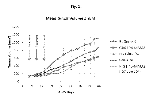
[0063] Fig. 24 shows GR 6A04 proof of concept as ADC in a xenograft model.
Conditions used were GR6A04-MMAE; MG1.45-MMAE isotype Ctrl; Buffer Ctrl;
GR6A04
(unconjugated); Hu-GR6A04 (unconjugated). Treatment regime was 10mg/kg
(-200ug/mouse) and 3 doses, on DO, D4 and D8 (starting at tumor size >150mm3).
The
results indicated that xenografts treated with GR6A04-MMAE showed little
tumour growth
which demonstrated excellent in vivo functionality as an ADC. Xenografts
treated with
unconjugated GR6A04 and Hu-GR6A04 showed slower growth compared to the buffer
control, which indicated some in vivo tumour growth inhibition via ADCC
mechanisms.
DETAILED DESCRIPTION OF THE PRESENT INVENTION
[0064] In a first aspect, there is provided an antigen-binding protein, or
an antigen-binding
fragment thereof, comprising (i) a heavy chain variable domain comprising a
VHCDR1 having the
amino acid sequence GNTFTSYVMH (SEQ ID NO: 3); a VHCDR2 having the amino acid
sequence
YINPYNDGTKYNEKFKG (SEQ ID NO: 4) and a VHCDR3 having the amino acid sequence
STARATPYFYAMDY (SEQ ID NO: 5); and (ii) a light chain variable domain
comprising a
VLCDR1 having the amino acid sequence KSSQSLLWSVNQNSYLS (SEQ ID NO: 6), a
VLCDR2
having the amino acid sequence GASIRES (SEQ ID NO: 7), and a VLCDR3 having the
amino acid
sequence QHNHGSFLPYT (SEQ ID NO: 8).
CA 03039886 2019-04-09
WO 2018/070936
PCT/SG2017/050509
12
[0065]
The antigen-binding protein, or antigen-binding fragment thereof, may comprise
heavy and light chain CDR regions that are about 80%, about 85%, about 90%,
about 95%,
about 96%, about 97%, about 98% or about 99% identical to the heavy and light
chain CDR
regions of (i) and (ii).
[0066] In one embodiment, the heavy chain variable region comprises the
amino acid
sequence
S GPELVKPGASVKMS CKAS GNTFTSYVMHWVKQKPGQGLEWIGYINPYNDGTKYN
EKFKGKATLTSD KS SSTAYMELS SLTSEDSAVYYCARSTARATPYFYAMDYWGQGT
SVTVSS as set forth in SEQ ID NO:l. Alternatively, the heavy chain variable
region may
comprise an amino acid sequence having about 80%, about 85%, about 90%, about
95%,
about 96%, about 97%, about 98% or about 99% identity to the amino acid
sequence set
forth in SEQ ID NO:l.
[0067]
In one embodiment, the light chain variable region comprises the amino acid
sequence
DILMTQSPSSLAVTAGEKVTMRCKSS QSLLWSVNQNSYLSWYQLKQGQPPKLLLYG
ASIRESWVPDRFTGSGS GTDFTLTISNVHVEDLAVYYCQHNHGSFLPYTFGGGTKLEI
K as set forth in SEQ ID NO:2. Alternatively, the antigen-binding protein, or
antigen-binding
fragment thereof, may comprise a light chain variable region which comprises
an amino acid
sequence having about 80%, about 85%, about 90%, about 95%, about 96%, about
97%,
about 98% or about 99% identity to the amino acid sequence set forth in SEQ ID
NO:2.
[0068]
In one embodiment, the antigen-binding protein, or antigen-binding fragment
thereof, as claimed in any one of claims 1 to 6, wherein the antigen binding
protein is
selected from the group consisting of monoclonal, recombinant, polyclonal,
chimeric,
humanised, bispecific and heteroconjugate antibodies; a chimeric antigen
receptor (CAR), a
.. single variable domain, a domain antibody, antigen binding fragments,
immunologically
effective fragments, single chain Fv, a single chain antibody, a univalent
antibody lacking a
hinge region, a minibody, diabodies, and TandabsTm
[0002]
In one embodiment, the antigen-binding protein, or antigen-binding fragment
thereof may be a monoclonal antibody. The monoclonal antibody may be GR 6A04.
In one
embodiment, the monoclonal antibody may be humanised. Alternatively, the
monoclonal
antibody may be chimeric.
CA 03039886 2019-04-09
WO 2018/070936
PCT/SG2017/050509
13
[0069] In one embodiment, the antigen-binding protein, or antigen-
binding fragment
thereof may bind to CEACAM6. In one embodiment, the antigen-binding protein,
or antigen-
binding fragment thereof may bind to a glycan on CEACAM6. The antigen-binding
protein,
or antigen-binding fragment thereof as described herein may bind to an N-
linked glycan on
CEACAM6.
[0070] In another embodiment, the antigen-binding protein, or antigen-
binding fragment
thereof as described herein may comprise a radioisotope or a cytotoxin
conjugated thereto.
The antigen-binding protein, or antigen-binding fragment thereof may be
conjugated with a
cytotoxin selected from the group consisting of monomethyl auristatin E
(MMAE),
mertansine (DM-1), saporin, gemcitabine, irinotecan, etoposide, vinblastine,
pemetrexed,
docetaxel, paclitaxel, platinum agents (for example, cisplatin, oxaliplatin
and carboplatin),
vinorelbine, capecitabine, mitoxantrone, ixabepilone, eribulin, 5-
fluorouracil, trifluridine and
tipiracil.
[0071] In one embodiment, the antigen-binding protein, or an antigen-
binding fragment as
described herein may be internalized into a cell upon binding to CEACAM6.
[0072] In another embodiment, the antigen-binding protein, or an antigen-
binding
fragment as described herein may not be internalized into a cell upon binding
to CEACAM6.
[0073] In one embodiment, the antigen-binding protein, or an antigen-
binding fragment as
described herein may selectively bind to a gefitinib resistant lung cancer
cell, a non-small cell
lung cancer cell, a breast cancer cell and/or a colorectal cancer cell.
[0074] In another aspect, there is provided a composition comprising a
physiologically
acceptable carrier and a therapeutically effective amount of the antigen-
binding protein, or an
antigen-binding fragment thereof as described herein. In one embodiment, the
composition as
disclosed herein may comprise one or more further therapeutic compounds.
[0075] The percentage of the the antigen-binding protein, or an antigen-
binding fragment
thereof, as described herein, in pharmaceutical compositions and preparations
may, of course,
be varied and, for example, may conveniently range from about 2% to about 90%,
about 5%
to about 80%, about 10% to about 75%, about 15% to about 65%; about 20% to
about 60%,
about 25% to about 50%, about 30% to about 45%, or about 35% to about 45%, of
the weight
of the dosage unit. The amount of compound in therapeutically useful
compositions is such
that a suitable dosage will be obtained.
CA 03039886 2019-04-09
WO 2018/070936
PCT/SG2017/050509
14
[0076] The language "physiologically acceptable carrier" is intended to
include solvents,
dispersion media, coatings, anti-bacterial and anti-fungal agents, isotonic
and absorption
delaying agents, and the like. The use of such media and agents for
pharmaceutically active
substances is well known in the art. Except insofar as any conventional media
or agent is
incompatible with the compound, use thereof in the therapeutic compositions
and methods of
treatment and prophylaxis is contemplated. Supplementary active compounds may
also be
incorporated into the compositions according to the present invention. It is
especially
advantageous to formulate parenteral compositions in dosage unit form for ease
of
administration and uniformity of dosage.
[0077] "Dosage unit form" as used herein refers to physically discrete
units suited as
unitary dosages for the individual to be treated; each unit containing a
predetermined quantity
of compound(s) is calculated to produce the desired therapeutic effect in
association with the
required pharmaceutical carrier. The compound(s) may be formulated for
convenient and
effective administration in effective amounts with a suitable pharmaceutically
acceptable
carrier in an acceptable dosage unit. In the case of compositions containing
supplementary
active ingredients, the dosages are determined by reference to the usual dose
and manner of
administration of the said ingredients.
[0078] The composition may be conveniently administered by injection,
for example,
subcutaneous, intravenous, and the like. The composition may also be
administered
parenterally or intraperitoneally. In one embodiment, the compound may be
administered by
injection. In the case of injectable solutions, the carrier can be a solvent
or dispersion
medium containing, for example, water, ethanol, polyol (for example, glycerol,
propylene
glycol, and liquid polyetheylene glycol, and the like), suitable mixtures
thereof, and vegetable
oils. The proper fluidity can be maintained, for example, by the use of a
coating such as
lecithin, by the maintenance of the required particle size in the case of
dispersion and by the
use of surfactants. Prevention of the action of microorganisms can be achieved
by including
various anti-bacterial and/or anti-fungal agents. Suitable agents are well
known to those
skilled in the art and include, for example, parabens, chlorobutanol, phenol,
benzyl alcohol,
ascorbic acid, thimerosal, and the like. In many cases, it may be preferable
to include isotonic
agents, for example, sugars, polyalcohols such as mannitol, sorbitol, and
sodium chloride in
the composition. Prolonged absorption of the injectable compositions can be
brought about
CA 03039886 2019-04-09
WO 2018/070936
PCT/SG2017/050509
by including in the composition an agent which delays absorption, for example,
aluminium
mono stearate and gelatin.
[0079]
Sterile injectable solutions can be prepared by incorporating the analogue in
the
required amount in an appropriate solvent with one or a combination of
ingredients
5
enumerated above, as required, followed by filtered sterilisation. Generally,
dispersions are
prepared by incorporating the analogue into a sterile vehicle which contains a
basic
dispersion medium and the required other ingredients from those enumerated
above.
[0080]
Under ordinary conditions of storage and use, pharmaceutical preparations may
contain a preservative to prevent the growth of microorganisms.
Preferably, the
10 pharmaceutical composition may further include a suitable buffer to
minimise acid
hydrolysis. Suitable buffer agent agents are well known to those skilled in
the art and
include, but are not limited to, phosphates, citrates, carbonates and mixtures
thereof.
[0081]
Single or multiple administrations of the pharmaceutical compositions
according to
the invention may be carried out. One skilled in the art would be able, by
routine
15
experimentation, to determine effective, non-toxic dosage levels of the
compound and/or
composition of the invention and an administration pattern which would be
suitable for
treating the diseases and/or infections to which the compounds and
compositions are
applicable.
[0082]
Further, it will be apparent to one of ordinary skill in the art that the
optimal course
of treatment, such as the number of doses of the compound or composition of
the invention
given per day for a defined number of days, can be ascertained using
convention course of
treatment determination tests.
[0083]
Generally, an effective dosage per 24 hours may be in the range of about
0.0001
mg to about 1000 mg per kg body weight; suitably, about 0.001 mg to about 750
mg per kg
body weight; about 0.01 mg to about 500 mg per kg body weight; about 0.1 mg to
about 500
mg per kg body weight; about 0.1 mg to about 250 mg per kg body weight; or
about 1.0 mg
to about 250 mg per kg body weight. More suitably, an effective dosage per 24
hours may be
in the range of about 1.0 mg to about 200 mg per kg body weight; about 1.0 mg
to about 100
mg per kg body weight; about 1.0 mg to about 50 mg per kg body weight; about
1.0 mg to
about 25 mg per kg body weight; about 5.0 mg to about 50 mg per kg body
weight; about 5.0
mg to about 20 mg per kg body weight; or about 5.0 mg to about 15 mg per kg
body weight.
CA 03039886 2019-04-09
WO 2018/070936
PCT/SG2017/050509
16
[0084] Alternatively, an effective dosage may be up to about 500mg/m2.
For example,
generally, an effective dosage is expected to be in the range of about 25 to
about 500mg/m2,
about 25 to about 350mg/m2, about 25 to about 300mg/m2, about 25 to about
250mg/m2,
about 50 to about 250mg/m2, and about 75 to about 150mg/m2.
[0085] In another aspect, there is provided use of an antigen-binding
protein, or an
antigen-binding fragment thereof as disclosed herein, or the composition as
disclosed herein
in the manufacture of a medicament for treating or preventing cancer.
[0086] In one embodiment, the cancer may be selected from the group
consisting of
gefitinib resistant lung cancer, non-small cell lung cancer, breast cancer,
stomach cancer,
small intestine cancer, esophageal cancer and colorectal cancer.
[0087] In some embodiments, the medicament may be administered with one or
more
further active pharmaceutical ingredients. Alternatively, the medicament may
be
administered with chemotherapy. The further active pharmaceutical ingredients
or
chemotherapy may be administered separately, simultaneously or sequentially.
[0088] In another aspect, there is provided a method for detecting cancer
in a subject, the
method comprising: contacting a sample obtained from the subject with an
antigen-binding
protein, or an antigen-binding fragment thereof as disclosed herein in vitro;
detecting the
binding of the antigen-binding protein, or an antigen-binding fragment thereof
in the sample;
correlating the binding with a level of binding in a control sample to
determine the level of
binding in the sample, wherein an increase in the level of binding in the
sample relative to the
control sample is indicative of cancer.
[0089] In another aspect, there is provided a method for identifying a
subject susceptible
to cancer the method comprising: contacting a sample obtained from the subject
with an
antigen-binding protein, or an antigen-binding fragment thereof as disclosed
herein in vitro;
detecting the binding of the antigen-binding protein, or an antigen-binding
fragment thereof
in the sample; correlating the binding with a level of binding in a control
sample to determine
the level of binding in the sample, wherein an increase in the level of
binding in the sample
relative to the control sample indicates that the subject is susceptible to
cancer.
[0090] In one embodiment, the control sample is from the same subject.
Alternatively, the
control sample may be from a different subject.
[0091] In one embodiment, the antigen-binding protein, or antigen-
binding fragment
thereof as described herein may comprise a detectable label. The detectable
label may be
CA 03039886 2019-04-09
WO 2018/070936
PCT/SG2017/050509
17
selected from the group consisting of a fluorescent label, a chemiluminescent
label, an
enzymatic label and a radionuclide label.
[0092] In one embodiment, the detectable label may be selected from the
group consisting
of biotin, alkaline phosphatase, horseradish peroxidase, FITC, PE and Cy Dyes.
The
detectable label may be detected in an assay selected from flow cytometry,
tissue section,
immunofluorescence, immunocytochemistry or immunohistochemistry.
[0093] In one aspect, there is provided a kit when used in the method as
described herein,
comprising an antigen-binding protein, or antigen-binding fragment thereof as
described
herein, together with instructions for use.
[0094] The invention illustratively described herein may suitably be
practiced in the
absence of any element or elements, limitation or limitations, not
specifically disclosed
herein. Thus, for example, the terms "comprising", "including", "containing",
etc. shall be
read expansively and without limitation. Additionally, the terms and
expressions employed
herein have been used as terms of description and not of limitation, and there
is no intention
in the use of such terms and expressions of excluding any equivalents of the
features shown
and described or portions thereof, but it is recognized that various
modifications are possible
within the scope of the invention claimed. Thus, it should be understood that
although the
present invention has been specifically disclosed by preferred embodiments and
optional
features, modification and variation of the inventions embodied therein herein
disclosed may
be resorted to by those skilled in the art, and that such modifications and
variations are
considered to be within the scope of this invention.
[0095] The invention has been described broadly and generically herein.
Each of the
narrower species and subgeneric groupings falling within the generic
disclosure also form
part of the invention. This includes the generic description of the invention
with a proviso or
negative limitation removing any subject matter from the genus, regardless of
whether or not
the excised material is specifically recited herein.
[0096] Other embodiments are within the following claims and non-
limiting examples. In
addition, where features or aspects of the invention are described in terms of
Markush groups,
those skilled in the art will recognize that the invention is also thereby
described in terms of
any individual member or subgroup of members of the Markush group.
CA 03039886 2019-04-09
WO 2018/070936
PCT/SG2017/050509
18
EXAMPLES
[0097] Non-limiting examples of the invention and comparative examples
will be further
described in greater detail by reference to specific Examples, which should
not be construed
as in any way limiting the scope of the invention.
[0098] Materials and Methods
[0099] Culture and generation of Gefitinib resistant cell lines
[00100] PC-9 were cultured in RPMI (Invitrogen, USA) supplemented with 10%
foetal
bovine serum (HyClone GE Healthscience, South America). To obtain PC-9 clones
with
acquired gefitinib resistance, PC-9 cultures were exposed to increasing
concentration of
gefitinib (Selleckchem, USA), starting from 2nM and gradually increased with
each
subsequent passage to a final concentration of 6.4pM. GR clones, CL75, CL86
and CL131,
were maintained in 6.4pM gefitinib thereafter.
[00101] A549 were cultured in DMEM (Invitrogen, USA) supplemented with 10% FBS
and 20mM L-glutamine (Invitrogen, USA). A549 has primary resistance to
gefitinib, with
IC50 of >10pM.
[00102] Generation of GR mAb panel
[00103] Immunisation of PC-9 GR lines, CL75, CL86 and CL131, was done with 5E6
cells
resuspended 1:1 with Fraund's complete adjuvant. Immunisation was done once
per week for
the first immunisation in week 1-3 with only a single line each, and a mixed
suspension of all
three lines for the subsequent immunisations in week 4-5. After 5 weeks, mice
were
sacrificed, and the B-cells collected for fusion with Sp2/0 mouse myeloma
lines using
STEMCELL Technologies ClonaCellTm-HY kit as per manufacturer's instructions.
Single
hybridoma clones were picked into 96-wells, and the culture supernatant
collected for
screening by flow cytometry.
[00104] Flow cytometry
[00105] Cells were harvested as single cell suspensions using trypsin. 1E5
cells were used
per sample, and incubated with 100111 of mAb culture supernatant for 30 min.
Cells were then
washed with 1% bovine serum albumin in PBS, and further incubated with 100111
of goat
anti-mouse antibody fluorescein isothiocyanate (FITC)-conjugated (1:500, DAKO,
Denmark)
for 15 min at 4 C in the dark. Cells were again washed and resuspended in
200pL of 1%
BSA/PBS for analysis on Guava easyCyte 8HT Benchtop Flow Cytometer (Merck
Millipore, USA). For interrogation of intercellular binding, cells were fixed
with 4%
CA 03039886 2019-04-09
WO 2018/070936
PCT/SG2017/050509
19
PFA/PBS (Affymetrix, USA) at room temperature for 10 min, washed in PBS, and
permeabilised with 0.1% triton/PBS for 5 min at room temperature, before
proceeding with
incubation with mAb supernatant/primary antibody. For staining with propidium
iodide, PI
was added to a final concentration of 5ug/mL for 5 min just prior to analysis
by the flow
cytometer.
[00106] Western blot and immunoprecipitation
[00107] Membrane proteins were extracted from PC-9 cell pellets using the
Membrane
Protein Extraction Kit (BioVision, USA). Briefly, cell pellets of 5E7 cells
were resuspended
in lmL of Homogenize Buffer and cell membranes broken in a dounce homogenizer.
This
was transferred into an Eppendorf tube and centrifuged at 700 x g for 10 min
at 4 C to
remove cell debris. The supernatant was transferred to a new Eppendorf tube
and centrifuged
at 12,000 x g for 30 min at 4 C to pellet the membrane. The membrane was
finally
resuspended in 500p1 of lx Cell Lysis Buffer (Cell Signaling Technology, USA)
containing
protease inhibitors (Pierce ThermoScientific, USA). The membrane protein
solution was
clarified with by centrifugation at 15,000 x g for 5 min at 4 C, to remove any
insoluble
proteins. Protein was quantified using the Pierce 660nM Protein Assay Reagent.
[00108] Immunoprecipitation (IP) was conducted using the automated Phynexus
MEA
system (Phynexus Inc., USA). GR6A04 was captured onto Protein G PhyTip columns
containing Sul of resin bed. The column was then washed with PBS to remove
unbound
proteins, and PC-9 membrane protein extract was introduced to bind to GR6A04
that has
been captured on the column. The column was then washed with Wash Buffer II
(140mM
NaCl, pH7.4) before elution at low pH with Elution Buffer (200 mM NaH2PO4/140
mM
NaCl pH 2.5) and neutralized immediately with 1 M Tris-Cl pH 9Ø The IP
product is then
subjected to analysis by Western blot.
[00109] PC-9 membrane protein extracts, or IP products were denatured by in
protein
loading dye containing SDS at a final concentration of 1%, and heated at 95 C
for 5 min. The
sample was then loaded into pre-cast gradient gel (NuPAGE 4-12% gradient gel,
Invitrogen),
and separated by SDS-PAGE running MOPS Running Buffer (NuPAGE Invitrogen,
USA).
After gel electrophoresis, the resolved proteins were transferred onto a
polyvinylidene
fluoride (PVDF) membrane (BioRad, USA) in a transfer buffer containing 20%
methanol,
10% Tris-Glycine in DI water at constant voltage of 110V for 90 min.
CA 03039886 2019-04-09
WO 2018/070936
PCT/SG2017/050509
[00110] The membrane was then blocked with 5% milk prepared in PBS/0.1% Tween-
20
(PBS-T) for 30 min at room temperature. The membrane was then washed in PBS-T,
followed by overnight incubation of GR6A04 at 2ug/mL in 2.5% milk at 4 C.
Subsequently,
the membrane was washed in PBS-T, before incubation with goat anti-mouse
secondary
5 antibodies horseradish peroxidase-conjugated (1:10000, Dako) for 1 hour at
room
temperature. After a final wash with PBS-T, the binding of HRP-conjugated
secondary
antibodies were visualized by ECL detection (GE Healthcare, Sweden).
[00111] Coomassie blue staining and mass-spectrometry
[00112] A parallel gel was run for the IP products, which was stained with
Coomassie blue
10 staining solution containing 0.1% Coomassie Blue R250 in 10% acetic
acid, 50% methanol
and 40% water for 1 hour at room temperature. The staining solution is then
removed, and
replaced by the de-staining solution containing 10% acetic acid, 50% methanol
and 40%
water. The de-staining solution is replaced with fresh solution, until the
background of the gel
is almost clear. The de-stained gel is then re-hydrated in water. The gel is
then compared with
15 the Western blot of the IP products to determine the position of the
antigen band on the gel.
This region is then excised for LC-MS analysis.
[00113] Glycosylation studies
[00114] PNGase digestion was carried out according to manufacturer's protocol
(New
England Biolabs). Briefly, 20 g of PC-9 membrane protein extract was first
denatured in lx
20 glycoprotein Denaturing Buffer at 95 C for 10 minutes. Subsequently, lx
G7 Reaction Buffer
and 10% NP-40 were added and incubated with PNGase F at 37 C for 1 hour.
Digested
proteins were subsequently analysed by Western blotting as described above.
[00115] Inhibition of N-glycosylation of proteins during cell culture was also
achieved by
addition of 111M Tunicamycin (Sigma Aldrich, USA) in the culture media. PC-9
cells were
seeded at 1E5 cells in a 6-well tissue culture place, and grown in culture
media spiked with
Tunicamycin, or DMSO for 3 days until confluent. The cells were then harvested
for analysis
by flow cytometry and Western blotting.
[00116] Immunohistochemistry staining
[00117] TMA slides containing FFPE tissues were first heated in an oven at 60
C for 30
min to remove any solvents. The slides were then dewaxed and re-hydrated
through
sequential immersion in Histoclear (2x), 100% ethanol (2x), 95% ethanol, 70%
ethanol, and
finally in DI water.
CA 03039886 2019-04-09
WO 2018/070936
PCT/SG2017/050509
21
[00118] Heat-induced epitope retrieval was done in a solution containing 10mM
Tris Base,
1mM EDTA, 0.05% Tween 20 at pH 9.0, and heated at 95 C for 20 min. The
container with
the antigen retrieval solution and slides was then removed and allowed to cool
to room
temperature for an additional 20 min. The slides were then washed in DI water.
Endogenous
peroxidase activity was then blocked by incubation of the slides with 3% H202
in PBS for 30
min at room temperature. The slides were washed in DI water, followed by a
blocking step
with 10% normal goat serum in PBS for 30 min.
[00119] The slides were then incubated with GR6A04 at 5ug/mL in blocking
solution
overnight at 4 C. The slides were then washed an incubated with a polymer-
based anti-mouse
secondary antibody conjugated with HRP (DAKO, USA) for 30 min at room
temperature,
and developed with the recommended DAB chromogen substrate solution for 2 min,
and
counterstained with Gill's Hematoxylin solution.
[00120] The stained slides were subsequently dehydrated through immersion in
50%
ethanol, 70% ethanol, 90% ethanol, 100% ethanol (2x) and Histoclear (2x),
before mounting
with a glass cover slip. The slides were then imaged with the Zeiss AxioScan
Digital Slide
Scanner.
[00121] Enrichment of GR6A04 binding population from A549 cell line
[00122] The CELLectionTM Pan Mouse IgG Kit (ThermoScientific, USA) was used
for the
enrichment of GR6A04- and GR6A04+ sub-populations from the A549 parental lung
adenocarcinoma line, according to manufacturer's instructions. Briefly, A549
cells were
harvested with trypsin to obtain a single-cell solution. Cells were incubated
with GR6A04 at
10m/1E7 cells for 30 min at 4 C. The cells were then centrifuged at 300 x g
for 3 min to
remove unbound GR6A04, before incubating it with the Dynabeads following kit
recommendations. The cell-bead suspension was then applied on the magnetic
rack, and
allowed to separate. The supernatant was collected as the GR6A04- population,
while the
bound beads and cells were collected as the GR6A04+ population. Both fractions
were
washed and subjected to the magnetic rack 3 times. The bound beads and cells
were finally
incubated with DNase Ito release the cells from the Dynabeads. Cells were
seeded into T75
tissue culture flasks at a density of 2.5E6 cells per flask.
[00123] Antibody-dependent cell-mediated cytotoxicity (ADCC) assay
[00124] ADCC activity was measured using a reporter bioassay (Promega; ADCC
Reporter
Bioassay, #G7010). The ADCC bioassay was carried out according to the
manufacturer's
CA 03039886 2019-04-09
WO 2018/070936
PCT/SG2017/050509
22
protocol Briefly, PC-9 and A549 cells were seeded at 5,000 cells per well in a
96-well clear
bottom black tissue culture plates (Corning; #3904) in low 4% IgG-serum
(Promega;
#G711A) media and allowed to attach and spread overnight. Serial dilutions of
Hu-GR6A04
were incubated in triplicate wells for approximately 15 min at 37 C, 5% CO2.
Following
incubation, engineered effector cells were added to the wells at approximately
150,000 cells
per well. After 6 hours, BioGloTM Luciferase Assay Substrate (Promega; #G719A
and
#G720A) was added to the wells and luminescence was measured using the
Infinite 200
microplate reader (Tecan).
[00125] Proliferation- CellTiter-Glo Luminescent Cell Viability (CTG) Assay
[00126] Cells were seeded into a black coated 96 well plate (Grenier Bio-one,
UK) at a
range of density from 1000-5000 cells per well (depending on the cell type
used) and 90uL
per well. The plate was then incubated for 24 hours at 37 C in humidified air
with 5% CO2.
After 24 hours, 10pL of mAb or buffer was added to each well and the plate was
again placed
at 37 C in humidified air with 5% CO2 4 days after addition of mAb or buffer,
100uL of CTG
substrate (Promega, Wisconsin, USA) was added to each well. The plate was then
left in the
dark for 10 minutes, with vigorous shaking. The cell viability of the samples
was then
quantified using Tecan I-control (Tecan, Switzerland).
[00127] Antibody Drug Conjugates (ADCs)
[00128] GR6A04 and a commercial mouse IgG1 isotype antibody (Clone MG1.45 from
BioLegend) were directly conjugated with MMAE toxin (Moradec, San Diego, USA)
at a
DAR of 3.0 and 3.3 respectively. Dose-response curve for the conjugated mAbs
were
established with the CTG assay with a serial dilution range of 0 to 4.5pg/ml.
The cell
viability of the cells was measure 4 days post treatment as described
previously.
[00129] In vivo Xenograft Model
[00130] Xenograft models were established using A549-GR6A04+ cells in NCr Nude
mice. 5E6 cells in DMEM basal media were mixed at a 1:1 ratio with Matrigel,
and injected
subcutaneously at a volume of 200111. Tumours were allowed to form and reach a
size of
>150mm3, before they were randomised into 5 groups of 5 mice each. The groups
were
treated with mAbs as follows: (1) Buffer control; (2) GR6A04-MMAE; (3) Hu-
GR6A04; (4)
GR6A04; (5) MG1.45-MMAE isotype control. mAbs were injected via tail vein
injection at a
total volume of 100p1. Each dose, were applicable, is at 200pg (equivalent to
10mg/kg), and
CA 03039886 2019-04-09
WO 2018/070936
PCT/SG2017/050509
23
treatment was done every 4 days, for a total of 3 doses (Day 0, 4 and 8).
Tumour size was
monitored over 50 days.
[00131] Results and Discussion
[00132] Generation of GR resistant mAb panel
[00133] GR6A04 was identified as part of a monoclonal antibody (mAb) panel
generated
against PC-9 lung adenocarcinoma with acquired resistance against Gefitinib, a
small
molecule inhibitor of epidermal growth factor (EGFR). Gefitinib resistant (GR)
PC-9 clones
were generated through culture in increasing concentration of Gefitinib until
a stable line was
obtained at a Gefitinib concentration of 6.4p.M. This was used as a surrogate
for acquired
resistance against Gefitinib observed in lung cancer patients undergoing
treatment with the
compound.
[00134] Three Gefitinib resistant PC-9 clones (CL75, CL86 and CL131) were used
for
immunisation in Balb/c mice in a semi-cyclic protocol (Fig. 1). The mice were
sacrificed in
Week 6, and the B-cells fused with Sp2/0 mouse myeloma cells using STEMCELL
Technologies ClonaCellTm-HY kit, and mAb-producing hybridoma clones were
obtained.
The mAbs from this GR panel was screened for binding to the immunising lines
by flow
cytometry, of which GR6A04 was identified as one of the binding lead
candidates from this
panel.
[00135] GR6A04 binding on cell lines by flow cytometry
[00136] GR6A04 demonstrated strong reactivity (>50%) towards the three PC-9
Gefitinib
resistant clones used for immunisation, with percentage positive-binding
determined from
FL-1 channel gated at 2% on the secondary-only control (Fig. 2). Propidium
iodide staining
on the FL-3 channel also showed no apparent cell cytotoxicity when cells were
incubated
with the mAb alone. GR6A04 binding was also tested on the Gefitinib sensitive
parental PC-
9 cell line, and on another GR PC-9 clone not used in the immunisation, with
similarly high
binding for both lines. This flow cytometry binding data is summarised in Fig.
3A.
[00137] Flow cytometry binding of GR6A04 was also tested in other NSCLC lines,
with
partial binding on 2 of 4 lines tested (A549 and Calu-3). GR clones from
another lung
adenocarcinoma line, HCC827, were also obtained by culturing in increasing
Gefitinib
concentrations. GR6A04 binding was observed for 2 of 6 of these GR HCC827
clones. In
addition, binding on other cancer indications also showed GR6A04 reactivity in
3 of 13
CA 03039886 2019-04-09
WO 2018/070936
PCT/SG2017/050509
24
breast cancer lines and 1 of 2 colorectal cancer lines tested. GR6A04 binding
on the different
cancer indications is summarised in Fig. 3B-E.
[00138] Importantly, using the same staining method, binding of GR6A04 on the
cell
surface of normal cells was found to be negligible (<8%) when tested with
various normal
cell lines including fibroblasts, endothelial and epithelial cells, and
primary peripheral blood
mononuclear cells (PBMCs), as summarised in Fig. 3F.
[00139] In summary, the flow cytometry binding characteristics of GR6A04 are
a)
GR6A04 binds to the cell surface of PC-9 and their derived gefitinib-resistant
clones based
on flow cytometry, b) GR6A04 demonstrates reactivity to other NSCLC, breast
and
colorectal cancer lines and c) there is no cross-reactivity to normal cell
lines tested.
[00140] Characterisation of GR6A04 mAb and derivatives
[00141] The isotype of GR6A04 mAb was determined to be of a mouse IgG1
subtype, as
determined by PierceTM Rapid Antibody Isotyping Kit in Fig. 4A. The variable
heavy chain
(VH) and light chain (VL) amino acid sequences were determined as Fig. 4B by
use of
degenerate primers for mouse immunoglobulins. GR6A04 was purified from
hybridoma
culture supernatant using CaptureSelectTM IgG-Fc (ms) Affinity Matrix with the
AKTA avant
system.
[00142] Two derivatives of GR6A04 was made and characterised: an antibody drug
conjugate with monomethyl auristatin E (MMAE) (GR6A04-MMAE), and a human
chimeric
mAb with the VH and VL cloned into a human Ig constant region backbone (Hu-
GR6A04).
[00143] GR6A04-MMAE was conjugated with MMAE at a drug-to-antibody ratio (DAR)
of 3.0, and at the same time, a commercial mouse IgG1 antibody (Clone MG1-45
from
Biolegend) at a DAR of 3.3 using the same chemistry (Fig. 4C). Binding of
GR6A04-MMAE
on PC-9 and A549 cells was tested by flow cytometry, and found to be
comparable to
unconjugated GR6A04, while MG1-45-MMAE was determined to have negligible
binding
on these two lines.
[00144] Hu-GR6A04 was expressed in CHO cells, and the antibody purified from
the
culture supernatant using the same system as GR6A04. Purified Hu-GR6A04 was
checked
for correct protein size from the Coomassie blue stain from SDS-PAGE, with the
heavy chain
and light chain at the expected size of 50kDa and 25kDa respectively in the
reducing lane,
and the intact IgG at 150kDa in the non-reducing lane. Similarly, binding of
Hu-GR6A04 on
PC-9 and A549 was comparable to GR6A04 on flow cytometry, shown in Fig. 4E.
CA 03039886 2019-04-09
WO 2018/070936
PCT/SG2017/050509
[00145] GR6A04 binds to N-glycosylated CEACAM6
[00146] Western blotting of PC-9 cell lysates was immunoblotted with GR6A04,
and found
to recognise a smear of between 55 ¨ 90kDa on the non-reducing lane, shown in
Fig. 5.
Antigen binding intensity was also weaker under reducing conditions.
Additionally, treatment
5 of the cell lysate by PNGase to remove N-linked glycans also abolished
binding of GR6A04
to the antigen band, demonstrating the importance of N-glycosylation in the
antibody
recognition site.
[00147] Immunoprecipitation (IP) with GR6A04 against PC-9 cell lysate enriched
for the
antigen, which was excised and sent for identification by mass spectrometry
(Fig. 6A). The
10 protein list was arranged by the overall score based on protein coverage
and number of
unique peptides found, and compared to the protein list from the column
control. Top 5
putative antigens are listed in the table in Fig. 6B and validated by cross-IP
with a
commercial anti-CEACAM6 antibody (Clone 9A6 from Santa Cruz/Abcam) (Fig. 7A),
and
transient siRNA knock-down of CEACAM6 in PC-9 cells (Fig. 7B).
15 [00148] It can be concluded that GR6A04's antigen target is CEACAM6,
whereby the N-
glycan is important for antibody-antigen recognition.
[00149] GR6A04 binding on patient FFPE tissue samples
[00150] Having established that GR6A04 is specific to various cancer
indications on flow
cytometry, and does not bind to normal cells, we proceeded with determining
GR6A04's
20 .. binding on cancer patient tissue samples on formalin fixed paraffin
embedded (FFPE) tissue
microarrays (TMAs). Commercial FFPE TMAs were obtained from Pantomics.
[00151] Binding condition of GR6A04 for FFPE samples was optimised with FFPE
cell
line pellets, and determined to be at 5ug/mL with a pH 9 antigen retrieval
step. GR6A04 was
observed to be localised to both the membrane and cytosol of PC-9 cells. To
extend the
25 binding profile of GR6A04 in other cancer cell lines,
immunohistochemistry (IHC) staining
was conducted on a FFPE cell line arrays covering larger range of cancer
indications.
GR6A04 was found to be reactive in gastric, lung, colorectal and pancreatic
cancer cell lines
(Fig. 8).
[00152] IHC staining on various FFPE TMAs covering cancer tissue samples from
a wide
array of organ origins were also done. GR6A04 staining was scored with the
open source
software: ImmunoMembrane. In a vast majority of tissue cores that was stained
positive (2+
and 3+), binding was localised to the cell membrane. Some non-specific
staining was also
CA 03039886 2019-04-09
WO 2018/070936
PCT/SG2017/050509
26
observed in necrotic regions. IHC staining on multi-tumour TMAs supported what
was
observed in cell line screening, whereby we observed that in addition to lung
cancer,
GR6A04 is also highly reactive to, but not limited to, gastro-intestinal (GI)
and breast cancer
(Fig. 9 to 11). In a focused array for NSCLC, GR6A04 stained positive for 15%
of squamous
cell cancer, and 50% of lung adenocarcinoma cores represented in the TMA.
Importantly, the
adjacent normal tissue did not have any positive staining, demonstrating
GR6A04 specificity
towards cancer tissue (Fig. 12 & 13).
[00153] In addition, GR6A04 was tested against FFPE normal tissues using the
same
staining protocol and scoring system. An FDA recommended TMA was used
(MN0961),
containing 35 different anatomical sites (Fig. 14). No staining of GR6A04 was
observed in 93
of the 96 cores present, but 2 of 3 colon cores, and 1 of 3 prostate cores
were weakly positive
(2+). It should be noted that from the magnified images, the staining in these
cases was
mainly intracellular or in necrotic regions.
[00154] Hence, GR6A04 has demonstrated specificity towards a wide range of
different
cancer indications in patient tissue samples, and has negligible staining on
normal tissue
types, supporting our earlier binding profiles from flow cytometry and cell
based screening.
[00155] Increased specificity of GR6A04 due to glycan recognition site
[00156] The commercial anti-CEACAM6 antibody used for the earlier validation
is one
that is not sensitive to changes in the glycosylation of CEACAM6 (i.e.
Recognition site is not
glycan dependent). This is demonstrated in Fig. 15A, where the binding
intensities remained
unchanged after PNGase and Tunicamycin treatment, unlike GR6A04.
[00157] When the two anti-CEACAM6 (GR6A04 and commercial) were compared in an
FFPE cell line microarray, while staining profiles were largely similar, the
commercial
antibody had one additional staining core in HCC827, while BxPC3 (pancreatic
cancer line)
also showed more intense and intracellular staining (Fig. 15B).
[00158] This apparent non-specificity of the commercial anti-CEACAM6 was also
observed in the TMA with normal tissues (MN0961), whereby 11 of 95 cores
stained
positive, as opposed to only 3 cores with GR6A04. Importantly, all three lung
normal tissues,
and 2 spleen normal tissues, showed staining on the cell membrane (Fig. 16).
[00159] The differences in staining profiles could be attributed to the
differences in epitope
binding sites on CEACAM6, especially that arising from the N-glycans on
CEACAM6. This
allowed for an extra degree of specificity in addition to binding on CEACAM6
protein alone,
CA 03039886 2019-04-09
WO 2018/070936
PCT/SG2017/050509
27
which leads to reduced non-specific binding on normal tissues. This is
important as it
provides an added level of safety if GR6A04 is to be developed for therapy.
[00160] Characteristics of GR6A04+ A549 subpopulations
[00161] The lung adenocarcinoma cell line, A549, is a good biological model to
investigate
the role of glycosylated CEACAM6 expression in cancer due to heterogeneous
binding for
GR6A04, with only a small 15 ¨ 30% GR6A04+ population (Fig. 17A). Through the
use of
magnetic sorting with CELLectionTM Pan Mouse IgG Kit, both the GR6A04- and
GR6A04+
could be enriched for. Although it must be noted that reduction of GR6A04
binding was
observed with increasing passages in culture. Multiple rounds of consecutive
sorting
mitigates this, and after 7 rounds of isolation for GR6A04+ and 5 rounds for
GR6A04- cells,
stable differential lines were obtained, and subsequently referred to as A549-
GR6A04+ and
A549-GR6A04- respectively (Fib. 17B & C). Additionally, single cell clones
from A549-
GR6A04+ cultures were isolated that demonstrated strong, homogeneous binding
of GR6A04
(D5, D7 and S4 clones). These were observed to have different cell morphology
between
clones (Fig. 17D & E). In particular, D5 clones showed dense clusters with
round
morphology, D7 showed spindle-shaped cells and S4 showed SCLC-like morphology
with
attachment independent growth.
[00162] Expression of EGFR and CEACAM1 was measured by flow cytometry on these
sub-populations, and also the parental line. While expression of these two
markers in A549-
GR6A04- cells and the parental A549 were similar, A549-GR6A04+ cells had a
much lower
expression of EGFR (25% decrease in MFI), and an increase in CEACAM1 binding
was also
observed (Fig. 18).
[00163] GR6A04 expression was also found to affect tumorigenicity in vivo. In
an A549
xenograft model in Nude mice, whereby the same initial cell numbers were
injected
subcutaneously, A549-GR6A04+ cells formed larger xenografts than the A549
parental cells
(Fig. 19A). When the xenograft was dissociated at the end of the 50 day study,
it was also
found that percentage of GR6A04 in the xenografts obtained from A549 parental
cells
increased from around 20% at the point of injection, to >70% by Day 50 (Fig.
19B).
[00164] To establish if GR6A04 could have potential application as a serum
biomarker for
lung cancer, conditioned media from A549 subpopulations were spotted on a
membrane and
immunoblotted with GR6A04. The antigen is detectable in the conditioned media
from the
A549-GR6A04+ and A549 parental cell cultures, but not in the A549-GR6A04-
cultures, and
CA 03039886 2019-04-09
WO 2018/070936
PCT/SG2017/050509
28
intensity is proportional to the volume of media spotted (Fig. 20). The
results show that
GR6A04 may be used for antigen quantification in patient serum samples.
[00165] In vitro and in vivo functional assays
[00166] Having established GR6A04's specificity and its possible roles in
cancer biology
in the previous sections, this section focuses primarily on the functionality
of the mAb as a
cancer therapeutic. As an early test for function as an antibody-drug
conjugate (ADC),
GR6A04 was indirectly conjugated with saporin (ribosome inactivating protein)
using an
anti-mouse antibody conjugated with the toxin. Cell growth was inhibited in
both the PC-9
sensitive parental line, and CL75 GR clone by 43% and 28% respectively (Fig.
21A).
GR6A04 was also observed to be able to internalise within 120 min at 37 C,
with the mAb
being localised to the cell periphery after binding at 0 min, and subsequently
found
distributed within the cell cytoplasm (dashed arrows) at 120 min. A proportion
of cells,
however, has not internalised the mAb at 120 min, and the mAb was still
localised on the
surface (solid arrows) (Fig. 21B).
[00167] GR6A04 was subsequently directly conjugated to MMAE (Fig. 4C) for a
more
robust assay. A dose response curve was conducted on PC-9 and A549 binding
cells, and
H1299 non-binding cells (Fig 22). EC50 was established to be 19.8nM for PC-9
cells and
3.33nM for A549-GR6A04+ cells. GR6A04-MMAE did not have an effect on cell
growth for
non-binding cells, H1299 and A549-GR6A04- populations, demonstrating
specificity to
GR6A04 binding cells only.
[00168] GR6A04 was also developed as a chimeric mAb with a human IgG constant
backbone (Fig. 4D). Hu-GR6A04 was tested for antibody-dependent cell
cytotoxicity
(ADCC) using a Promega ADCC kit, which as a Luciferase reporter for FcyR
binding (Fig.
23). A dose-response curve on PC-9 and A549-GR6A04+ was done, and the EC50
established to be 39.6nM and 1.33nM respectively.
[00169] Hence, GR6A04 has demonstrated in vitro functionality on two lung
cancer cell
lines, PC-9 (gefitinib sensitive) and A549 (gefitinib-resistant), as a naked
human chimeric
mAb with ADCC activity, and as an ADC conjugated with MMAE.
[00170] Finally, in vivo functionality was tested using A549-GR6A04+ cells in
a
subcutaneous xenograft model (Fig. 24). Treatment with GR6A04 and their
derivatives was
initiated when the tumour reaches 150mm3, with I.V. mAb injection at 10mg/kg,
every 4 days
for 3 doses. Both the naked mAbs, Gr6A04 and Hu-GR6A04 showed repression of
tumour
CA 03039886 2019-04-09
WO 2018/070936 PCT/SG2017/050509
29
growth for the buffer control, attributed to ADCC mechanisms. Importantly,
GR6A04-
MMAE was extremely effective in controlling tumour size, with little tumour
growth (<2-
fold) throughout the 45 days of study. Hence, GR6A04 has demonstrated good in
vivo
functionality with a gefitinib-resistant A549 xenograft model, congruent with
the in vitro
results.