Note: Descriptions are shown in the official language in which they were submitted.
CA 03040001 2019-04-03
DESCRIPTION
PLASMA SEPARATION APPARATUS AND PLASMA SEPARATION METHOD
Technical Field
[0001]
The present invention relates to an apparatus and method
for separating a liquid component from a bodily fluid or blood,
especially for separating plasma (or serum) from blood. More
particularly, the invention relates to a plasma (or serum)
separation apparatus and a plasma (or serum) separation method
which do not require a centrifuge, or a suctioning or
pressurizing pump, or the like, yet are capable of simply and
conveniently separating plasma (or serum) even from a minute
amount of blood, at a low cost.
Background Art
[0002]
Medical diagnose through blood examination is routinely
conducted because various clinical statuses of patient can be
determined based on absence or presence and/or the
concentration values of certain substances dissolved in the
bodily fluid of patient, especially blood.
After collected from a patient, a blood sample,
especially plasma or serum, can be analyzed to provide a lot
of useful medical information in terms of, for example, chemical
1
CA 03040001 2019-04-03
components are clearly separated in a centrifugal tube such that
red blood cells having a heavy specific gravity would be
separated to a lower layer (about 41% of whole blood), plasma
having a light specific gravity would be separated to a upper
layer (about 55% of whole blood) , and platelets and white blood
cells would be separated to a middle layer (about 4% of whole
blood). When no anticoagulant has been added, whole blood is
separated into serum at the upper layer and blood clots at the
lower layer through a centrifugal operation.
[0004]
Because blood collection is a practice to be performed
by a medical worker, it can impose a burden on both an objective
person (i.e., the subject) and medical establishments, and can
also increase costs. Furthermore, blood examination will
require a centrifuge separator and operating staff therefor,
taking an additional cost and labor.
Furthermore, a regulation of plasma or serum by the
centrifugal separation often results in a problem of hemolysis.
Occurrence of hemolysis is undesirable as it can cause enzymes,
hemoglobin and other pigments, and stroma to be released into
liquid components of blood. If such is the case, multiple
clinical trials can be spoiled.
[0005]
Thus, there have been needs for a plasma or serum
separation apparatus and a plasma or serum separation method
that enable analyzing substances contained in plasma or serum
quickly, readily, and accurately, even if the amount of blood
3
CA 03040001 2019-04-03
is very small and even if a user or a person concerned is not
the medical worker with no training, without requiring a
centrifugal separator method, a suctioning or pressurizing
method, or the like.
There have also been needs for a simple, convenient, and
low-cost plasma or serum separation apparatus and a plasma or
serum separation method capable of dealing with global health
care including developing regions and the like. (Hereinafter,
"plasma or serum" may be simply referred to as "plasma.")
[0006]
As for inventions with such objectives, blood separation
apparatuses capable of separating blood without using a
centrifuge separator have been proposed, for example in Patent
Literature 1 and Patent Literature 2.
[0007]
Patent Literature 1 discloses a configuration composed
of a filter member for moving plasma more quickly than blood
cells and a subsequent serially-connected plasma or serum
separation film, or, a configuration composed of a first filter
member for moving plasma more quickly than blood cells, a second
filter member that is a plasma or serum separation film, and
a third filter member for capturing fibrin and the like, in which
blood components are separated by vacuum filtration, therefore
it requires a system, apparatus, or manpower for
depressurizing.
Patent Literature 2 discloses a configuration in which
a liquid part of blood is separated from cellular components
4
CA 03040001 2019-04-03
of blood as the blood flows through first and second matrixes,
wherein the first, porous separation matrix contains binders
for the cellular components of blood, and the second matrix is
configured to allow for the liquid part of blood to flow into
the first matrix by a capillary effect or chromatographic
separation. Unfortunately, this type of plasma separator is
difficult to be put into practical use because it takes time
for separation, and cellular components such as red blood cells
can be clogged to cause hemolysis and other problems.
[00081
Even if these conventional techniques do not require a
centrifuge separator, they require an apparatus for
depressurizing or pressurizing, or may need a combination of
multiple filter members and porous matrixes, or can rise a risk
of hemolysis, or may require another apparatus, tool, or
manpower, or even if they do not need another apparatus, there
is no guarantees that a liquid component of blood is collected
quickly, simply, and conveniently in an amount just necessary
for inspection at a low cost.
[0009]
Furthermore, in medical diagnoses, it is requested that
analysis is carried out simply and conveniently in a short time
and at a lower cost. In particular, an important issue is to
minimize the amount of specimen needed for analysis. If a system
is established capable of preparing a specimen for biochemical
examination by collecting a small amount of blood with a blood
collection tool usable at home, the specimen could be swiftly
CA 03040001 2019-04-03
sent to a blood examination facility and results could be
received in a short time. Such system can deal with a growing
worldwide need for total health care because it would be
extremely useful not only for home health care but also in
regions where medical establishments are inaccessible and even
in developing counties. Presently, however, any such system
that is practical and useful has not been developed yet.
[0010]
As mentioned above, according to recent social needs,
such the method and apparatus (instrument) has been needed for
clinical care not only in medical institutions but also at home
or in developing countries, etc. that can easily separate a
blood component such as plasma or serum as a sample for blood
examination from even a minute amount of blood, without
requiring a centrifuge separator, a special apparatus or tool
for suctioning or pressurizing, and manpower
Hereinafter, "plasma or serum" may be simply referred to
as "plasma."
Citation List
Patent Literature
[0011]
PTL 1: Japanese Patent Laid-Open No. 2004-344874
PTL 2: Japanese Patent Laid-Open No. 2006-177970
Summary of Invention
Problems to be solved by the invention
6
CA 03040001 2019-04-03
[0012]
Considering the above-mentioned situations of the
conventional arts, an object of the invention is to provide a
plasma separation apparatus and a plasma separation method that
enable quick, reliable, and low-cost separation of plasma from
blood, even a minute amount of blood, at any place, without using
a centrifuge separator, a pressurization/suction pump, or the
like.
[0013]
The above-mentioned object can be achieved by features
defined in the claims.
Here, the term "plasma" means "plasma or serum."
A plasma separation apparatus according to the invention
comprises, a blood separation part having a blood separation
member, and a plasma collection part having a plasma collection
member, wherein the blood separation member is mounted on a
hydrophobic pedestal and includes a blood receiving area and
a plasma separation area that is connected to the plasma
collection part, and the plasma separation part has the
cross-sectional area thereof gradually decreasing toward the
plasma collection part.
The cross-sectional area of the plasma separation area
in the plasma separation apparatus according to the invention
is gradually decreased toward the plasma collection part by the
end cutting.
The blood separation part of the plasma separation
apparatus according to the invention comprises a blood
7
CA 03040001 2019-04-03
reserving part on the upper surface side of the blood separation
member for temporarily reserving blood.
The blood separation part and the plasma collection part
of the plasma separation apparatus according to the invention
constitute a core part, wherein the core part is contained in
a housing part.
The plasma separation apparatus according to the
invention further comprises a support member for separation,
for separating into the blood separation part and the plasma
collection part.
A blood examination kit according to the invention
comprises the plasma separation apparatus and a blood
collection tool.
[0014]
A plasma separation method for separating plasma using
a plasma separation apparatus comprising a blood separation
part having a blood separation member and a plasma collection
part having a plasma collection member, wherein the blood
separation member arranged on a hydrophobic pedestal has a blood
receiving area and a plasma separation area connected to the
plasma collection part, a cross-sectional area of the plasma
separation area is gradually decreased toward the plasma
collection part, the plasma separation method comprising steps
of separating the plasma from the blood received in the blood
receiving area, in the plasma separation area, and collecting
the separated plasma in the plasma collection part.
In the plasma separation method according to the
8
(
CA 03040001 2019-04-03
invention, the cross-sectional area of the plasma separation
part in the plasma separation apparatus is gradually decreased
toward the plasma collection part by the end cutting.
In the plasma separation method according to the
invention, the blood separation part of the plasma separation
apparatus comprises a blood reserving part on the upper surface
side of the blood separation member so that blood is received
in the blood receiving area after temporarily being reserved
in the blood reserving part.
The plasma separation method according to the invention
comprises a step of separating the blood separation part and
the plasma collection part after the plasma separated from the
blood in the plasma separation area of the blood separation
member of the plasma separation apparatus is collected in the
plasma collection part.
Advantageous Effects of Invention
[0015]
The plasma or serum separation apparatus of the invention
does not require a centrifuge separator, a suctioning or
pressurizing apparatus, electric power, or the like, and can
prepare a sample for blood examination easily, conveniently,
quickly, and safely at a low cost, outside a hospital, at home,
or in foreign countries where there is no medical facility
around, and even by any ordinary person without technique
training.
According to the apparatus, by a simple operation and
9
CA 03040001 2019-04-03
without requiring a professional skill, a liquid component
(plasma or serum) can be separated from even a minute amount
of blood easily, quickly, and safely at a low cost without
concern about hemolysis, even in a place where there is no
sophisticated analytic instrument such as the centrifuge
separator and the like, and even by a home user himself or
herself. Not only the separated liquid component but also the
cellular components can be used as a specimen for blood
examination.
[0016]
Here, the term "blood" is referred to as a synonym of whole
blood and includes any combination of cellular components and
liquid (non-cellular) components of blood. Typical cellular
components include, but without limitation, red blood cells
(erythrocytes), white blood cells (leukocytes), and platelets
(thrombocytes), and any combination of these. White blood
cells include mononuclear leukocytes, granulocytes,
agranulocytes, and lymphocytes. When an anticoagulant has
been added, typical liquid components include, but without
limitation, plasma, dissolved salts and inorganics, and plasma
proteins, and the like.
Those samples to be examined, namely, "plasma and serum"
are similar to each other in their components, except that serum
obtained from a coagulated blood specimen is free of fibrinogen
and other certain coagulants lost as a result of coagulation
process.
Moreover, in relation to blood separation, in general,
CA 03040001 2019-04-03
a blood specimen is separated into a liquid component (plasma
or serum) and cellular components (blood cells or blood clots) .
Specifically, a blood specimen with an anticoagulant is
separated into plasma and blood cells, while a blood specimen
without an anticoagulant is separated into serum and blood
clots.
Hereinafter, "plasma or serum" may be simply referred to
as "plasma."
"Plasma collection rate" or "collection rate" is referred
to as a value indicated by percentage of the amount of collected
plasma relative to the total amount of blood dropped onto a blood
separation part. When the separated liquid component is serum,
then it is referred to as "serum collection rate."
Brief Description of Drawings
[0017]
Now preferable embodiments of the invention will be
described with reference to the drawings.
Fig. 1 shows an external view of a core part of a plasma
separation apparatus of an embodiment 1;
Fig. 2A shows an external view of the plasma separation
apparatus of the embodiment 1; Fig. 2B shows a sectional view
thereof;
Figs. 3 is views roughly showing modes of forming of blood
passage in the core part of the embodiment 1: Fig. 3A shows a
mode of dropping blood onto a blood separation member; Fig. 3B
shows a mode of infiltration of the dropped blood into the blood
11
CA 03040001 2019-04-03
separation member, and Fig. 30 shows a mode of trapping blood
cell constituent in the blood separation member and forming a
collection part passage in a collection part by a liquid
component in the blood through a communication/corporation
between a passage forming action of the blood separation member
and a capillary phenomenon of a gap of the collection part;
Figs. 4A to 4E show various modes of the blood separation
member of embodiment 1, having different sizes and/or shapes
of end;
Fig. 5 shows various modes of end working in a thickness
direction of the blood separation member of embodiment 1;
Figs. 6 show modes of end width cutting of the blood
separation member of embodiment 1; Fig. 6A shows a top view and
Figs. 6B, 60 show sectional views respectively;
Fig. 7 shows an external view of a plasma separation
apparatus of embodiment 2;
Fig. 8 shows a sectional view of the plasma separation
apparatus of embodiment 2;
Fig. 9 shows an explosive view of the plasma separation
apparatus of embodiment 2; and
Fig. 10 shows a perspective view of an external view of
a core part (except for a support member for separation) after
the core part of the plasma separation apparatus is assembled
of embodiment 2.
Description of Embodiments
[0018]
12
CA 03040001 2019-04-03
An apparatus according to the invention functions to separate
components in liquid, as shown for example in Figs 1 to 10 in
which its external view and the like are shown. Preferably the
liquid is blood, and the components to be separated are roughly
classified into cellular components (blood cells) such as red
blood cell and white blood cell to be trapped in a blood
separation member, and liquid components such as plasma or serum
which contains substances (including DNA, RNA, and the like)
to be applied to various biochemical examinations.
[0019]
(Embodiment 1)
One embodiment of the invention is a plasma or serum
(hereinafter, simply referred to as "plasma") separation
apparatus 1 comprising at least a core part 2 (Fig. 1) and an
outer housing 5 (Fig. 2) . The housing 5 includes a housing cover
51 and a housing base 52.
[0020]
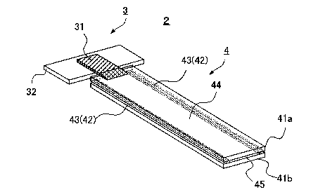
<Structure of core part>
As seen in Fig. 1, the structure of the core part 2 of
the plasma separation apparatus 1 of the invention generally
comprises a blood separation part 3 and a plasma collection part
4.
At first, the blood separation part 3 includes at least
a blood separation member 31 and a pedestal 32 for blood
separation part for mounting (supporting) the blood separation
member 31. In addition, the plasma collection part 4 having
at least two plasma collection members 41 (a, b) includes
13
CA 03040001 2019-04-03
adhesive member 43 sealing the edges 42 of the plasma collection
members 41 in liquid-tight, and a plasma collecting area 44
formed being surrounded by the two plasma collection members
41 and the adhesive member 43.
When a blood sample is applied to the blood separation
part 3, the blood is received in a blood receiving area on the
upper surface of the blood separation member 31, then
infiltrates into the blood separation member 31, so that a
collection part passage 46 from a plasma separation area in the
blood separation member 31 to the plasma collecting area 44 is
formed (Fig. 30) .
Here, the blood receiving area and the plasma separation
area are areas related to the blood separation member, the blood
receiving area refers to an area on the upper surface of the
blood separation member 31, on which blood is dropped for
infiltrating into the member, the plasma separation area means
an area of the blood separation member 31, where the infiltrated
blood is separated into plasma and cellular components, this
area is connected to the plasma collection part.
[0021]
Materials for the blood separation member are not limited
to any of embodiments in particular, for example, synthetic
polymers or fibers made of glass having a small diameter of
fiber, or layered products composed of porous polymeric sheets
or thin nonwoven fabrics and the like. Any material can be used
when they have function/effectiveness as the blood separation
member.
14
CA 03040001 2019-04-03
Nevertheless, when the material adsorbs components to be
measured in blood, it is desirable to apply a surface treatment
to a material constituting a first filter member. As surface
treatment agents, polyether-based or silicone-based
lubricants, hydrophilic polymers such as polyvinyl alcohol or
polyvinylpyrrolidone, or hydrophilic natural polymers, or
polymeric surfactants, can be used, but the materials are not
limited to those.
As the blood separation member, one to which a special
binder such as antibody for capturing cellular components of
blood is coupled, is included therein as well.
As materials for the pedestal for blood separation part,
polyethylene, polystyrene, polycarbonate, polyethylene
terephthalate, polypropylene, dimethylpolysiloxane, Teflon ,
silicone, and ABS, and the like are considered, and it is
preferable that those materials each have a hydrophobic surface
as described below. Here, the term "hydrophobic" generally
refers to a property of small affinity with water molecules.
As a plasma collection member, it is preferable to use
a hydrophilic material such as glass or resin, or a material
to which hydrophilic treatment has been applied, which has an
effect to occur a capillary phenomenon in a gap between the
collecting members (i.e., the plasma collecting area).
As adhesive member, an adhesive tape, an adhesive, and
the like may be considered. Integral molding without using the
adhesive may be carried out, when the adhesive member is made
of the same material as that of the correcting member.
CA 03040001 2019-04-03
[0022]
<External and sectional view>
Fig. 2A shows an external view of the plasma separation
apparatus 1 of the invention; Fig. 2B shows a sectional view
thereof.
The housing part 5 containing the core part 1 is provided
with at least a blood inlet part 6 and a collecting body part
7.
The blood inlet part 6 is provided on an upper part of
the blood separation part 3 contained in the housing and is
provided with a blood inlet 61, a blood reserving part 62, and
a blood deployment observation window 63 and the like as needed.
[0023]
The blood reserving part 62 has an effect of thereby
forming a temporal blood reserving space (blood reservoir) . It
is seated so as to prevent a part of blood after the blood being
dropped from slipping over the upper surface of the blood
separation member and leaking therefrom (while being
unseparated without infiltrating into the blood separation
member) and from flowing into the plasma collection part (from
mixing of blood with the plasma, leaking of blood into the
plasma).
[0024]
In other words, a part of the dropped blood usually flows
while forming a passage owing to a wicking phenomenon in the
blood separation member, but the phenomenon is not a little
occurred that the remaining blood floods over the surface of
16
CA 03040001 2019-04-03
the separation member, and as a result, the part of the blood
flows into the collection part without separation.
By providing the blood reserving part, slightly flooded
blood on the surface of the member can be temporarily reserved
as it is owing to surface tension at the blood reserving part,
in this way a "temporal blood reservoir space" is formed, and
thereafter the blood gradually flows into the separation
member, thus a leaking phenomenon as mentioned above can be
prevented.
Note that, the term "wicking (phenomenon or effect)"
refers to a spontaneous transportation of blood that occurs in
the fibrous or porous structure of the blood separation member,
which transportation may be uni-, bi-, or omni-directional.
This phenomenon allows a liquid to flow against gravity. Such
effect is caused by an intermolecular attraction force between
the liquid and a neighboring solid surface.
[0025]
Although, in the present embodiment, the blood reserving
part is placed in contact with the upper surface of the blood
separation member 31, a close contact with the blood separation
member 31 is not necessary but some gaps may be allowed to be
formed.
The blood reserving part has a function as a guide member
to form the temporal blood reservoir owing to surface tension,
but some gap is allowed to be formed unless its effect as a
temporal blood reservoir forming part (or guiding part for
forming temporal blood reservoir) is lost, and unless blood slip
17
CA 03040001 2019-04-03
over the upper surface of the blood separation member, leak
therefrom, and flow into the collection part because of the gap.
[0026]
Such blood reserving part can be of any shape, can be made
of any material, and can be seated anywhere, when it maintains
its effect as the above mentioned "temporal blood reservoir
forming part (or guiding part for forming temporal blood
reservoir)".
In embodiment 1, the blood reserving part 62 is formed
in a convex part extending along a rectangle shape of the blood
inlet 61 and perpendicularly extending downward therefrom, but
it may be, for example, circular, comb-shaped, U-shaped,
channel-shaped, or other shapes.
Alternatively, as another mode, it may be a bar-shaped
member 33 shown in Fig. 4D. In such mode, the blood reserving
part is preferably provided downstream of the blood dropping
point (the plasma collection part side) to prevent blood from
slipping over the upper surface of the blood separation member
and leaking therefrom too.
As mention above, the blood reserving part maybe provided
on the lower surface of the blood inlet 61 integrally with the
housing 5, like the blood reserving part 62, or it may be made
as an independent structure separate from the blood inlet, when
the effect as the blood reserving part is maintained. For
example, the bar-shaped member 33 as shown in Fig. 4D may be
provided with scaffolds or the like which is in contact with
the pedestal 32 for blood separation part at the both ends and
18
CA 03040001 2019-04-03
can be integrally molded with the pedestal 32 for separating
part in a bridge shape.
The effect as a blood reserving part can be obtained not
only by providing any member, but also by making its
blood-contacting surface hydrophobic, for example. As an
example, the effect of forming temporal blood reservoir can also
be obtained by making hydrophobic the lower surface of the blood
inlet 61 which may come in contact with dropped and flooded
blood.
Note that the blood reserving part not only prevents blood
from leaking over the upper surface of the blood separation
member, but also has a function to more effectively exhibit the
blood separating function of the blood separation member.
[0027]
Whereas the collecting body part 7 is provided with a
plasma deployment observation window 71, an opening 72 or the
like, as needed (see Fig. 2) . The opening 72 may be provided
with a film that allows air to pass therethrough but not liquid,
a valve, a connector to an examination system or the like, or
a liquid (plasma) reserving part. Alternatively, the opening
72 is not necessarily required such that it is a sealed
configuration.
[0028]
<Method for separating plasma from blood without
requiring centrifuge separator, suctioning or pressurizing
apparatus, or the like> See Fig. 3.
A method for separating plasma from blood using a plasma
19
CA 03040001 2019-04-03
separation apparatus of the invention comprises at least the
following steps:
1. A step for providing a plasma separation apparatus,
wherein the end of the blood separation member is inserted into
(arranged in) a gap of the plasma collection part, the blood
separation member being included in the blood separation part
and mounted on a hydrophobic pedestal, and a cross-sectional
area of the end being gradually decreased toward a plasma
collection part (preparation of a blood separation apparatus) ;
2. A step for introducing blood to the blood separation
member through a blood reserving part serving as a temporal
blood reservoir (a step for applying blood);
3. A step for separating blood introduced in the blood
separation member, wherein the blood is separated into blood
cellular components and plasma while forming a passage in the
blood separation member owing to a wicking effect in the blood
separation member (a step for blood deployment and separation) ;
4. A step for collecting plasma in the plasma collection
part, wherein the separated plasma forms a passage in the gap
of the plasma collection part through the
communication/corporation between the wicking effect in the
blood separation member and a capillary phenomenon in the plasma
collection part, so that the plasma is collected in the plasma
collection part (a step for collecting plasma).
The following step is the last for conducting a final
examination.
5. A step for sending the plasma collection part to the
CA 03040001 2019-04-03
examination facilities for examination.
Because this method does not require any special apparatus
such as a centrifuge separator, a suctioning or pressurizing
apparatus (tool) , or the like and can be performed by an ordinary
parson without training who is not a medical worker, it can deal
with needs of a person at home, a resident in a remote place
or in developing countries, or a minority or a majority of
people, plasma separation can be performed easily and quickly
and separated plasma can be send to the examination facilities
as it is. Thus, the apparatus is very useful.
In addition, as mentioned above, the term "wicking
(effect)" refers to a spontaneous transportation of liquid that
occurs in the fibrous or porous structure of the blood
separation member, which transportation may be uni-, bi-, or
omni-directional. This phenomenon allows the liquid to flow
against gravity. Such effect is caused by an intermolecular
attraction force between the liquid and a neighboring solid
surface.
(0029]
<Mechanism for separation of blood >
Blood is dropped through the blood inlet 61 of the blood
inlet part 6 and received in the blood receiving area on the
upper surface of the blood separation member 31 that is arranged
perpendicular to a blood filling direction. Since the blood does
not infiltrate into the blood separation member immediately
after dropping, an overflown part of the blood onto the upper
surface of the blood separation member (i.e., the blood
21
CA 03040001 2019-04-03
receiving part) is temporarily received in the blood reserving
part 62 functioning as a temporal blood reservoir forming part
(or a guiding part for forming temporal blood reservoir).
Meanwhile, the blood gradually infiltrates into and spreads
through the blood separation member 31 owing to the wicking
effect caused by the various matrix structure of the blood
separation part in the blood separation member 31, as a result,
a force to push the blood toward the plasma collection part 4
connected with the end of the blood separation member 31 is
generated, so that a collection part passage 46 is formed from
the blood separation part 3 to the plasma collecting area 44
through the communication/corporation of the force with the
capillary phenomenon of the gap of the plasm collection part
4.
Cellular components in blood (such as red blood cells
and white blood cells) are trapped in the blood separation
member 31. A liquid component of the blood (such as plasma or
serum) flows into the plasma collecting area 44 of the plasma
collection part 4 through the collection part passage 46 formed
through the communication/corporation between the wicking
effect of the blood separation part in the blood separation
member 31 and the capillary phenomenon of the gap of the plasma
collection part, thereby separated from the cellular components
and collected.
Note that, although the reference signs are affixed to
the drawings in order to help understanding the description,
the present mechanism is not limited by the reference signs or
22
CA 03040001 2019-04-03
not limited by any of embodiments.
[0030]
<Experiment 1> (Surface properties of the pedestal)
Object: To prevent blood applied to a blood separation
member from adhering to the pedestal and resulting in a dead
volume.
Method: In the pedestal made of silicon, after a
hydrophobic coating is applied to at least an upper surface in
contact with a planar blood separation member as shown in Fig.
1 by applying a hydrophobic coating agent or a hydrophobic film,
or the like, an angle of contact of a water droplet on the
pedestal is confirmed to be large enough to show that the
pedestal is hydrophobic, thereafter blood is applied to the
blood separation member, and a degree of adhesion of blood
(blood cells and blood clots) , etc . to the pedestal is confirmed.
Not that, coating is performed by applying a
fluorine-based hydrophobic coating agent (available from
NIPPECO Co. Ltd.) to the surface of the pedestal and dying to
give hydrophobicity thereto, this time.
Results: Blood adherence (blood cells and blood clots)
to the pedestal was less than a comparative example and blood
components was more smoothly separated, and collection amount
of the plasma as such was improved. With application of the
hydrophobic coating to the surface of the pedestal, blood was
prevented from adhering thereto and its dead volume was thereby
decreased.
Note that, for hydrophobic coating, a commonly used
23
CA 03040001 2019-04-03
material and method can be adapted when the pedestal is thereby
made hydrophobic and the problem is not occurred that the
coating itself is dissolved out by blood.
The material of the pedestal does not need to be silicon,
and a hydrophobic material, or other materials to which
hydrophobic coating is applicable and by which the pedestal is
formed, will do.
In each of experiments of the embodiments, including
comparative examples, a plasma collection time is generally 3
to 5 minutes, preferably 3 minutes, after blood is dropped.
If plasma collection time is too short, the plasma
collection amount will be too small. If it is too long, an
inflow of blood is likely to occur. A desirable plasma
collection time can be set based on a constitution of each
embodiment, size of each part, material of each member, and the
like.
[0031]
<Experiment 2> (Volume of blood separation part)
Object: To consider the volume of a blood separation member
in order to separate 100 p1 of blood quickly and to collect only
liquid component in the collection part.
Method: A plasma collection rate is calculated by using
MF1 (material: bound glass fiber) available from GE Healthcare
Japan Co., Ltd. as a blood separation member and a relationship
between the plasma collection rate (hereinafter also referred
to as "collection rate" or "yield") and the volume of the member
is examined.
24
CA 03040001 2019-04-03
Note that, in embodiments of the invention, MF1 (product
name) available from GE Healthcare Japan Co., Ltd. is used as
one example of blood separation member, unless otherwise
mentioned.
Results: when the blood separation member had a volume of
74 mm3 (20 mm (length) x 10 mm (width) X 0.37 mm (height
(thickness)): Fig. 4(a)), plasma could be collected from 150
pL of blood; but plasma was hardly collected from 100 pL of blood.
Since a liquid holding capacity of the blood separation
member is tending to depend on the volume. Based on the above
result incase of the volume of 74 mm3, and considering an easily
working shape of the end to be worked in the following experiment
3, the volume of the blood separation member for 100 pL of blood
is 16 mm (length) X 8 mm (width) X 0.37 mm (height) (see Fig.
4B), it is about 47.36 mm3 in volume when converted.
[0032]
<Experiment 3> (Shape of the collection part of the blood
separation member in contact with the collection part: the end
cutting in thickness (direction))
Object: Since the thickness of the blood separation
member used in the experiment 2 is greater than the height of
gap of the collection part, the end of the blood separation
member cannot be inserted into the gap of the collection part,
and the plasma is hard to collect, therefore, the object of the
experiment is to consider "cutting angle of the end in the
thickness direction" (hereinafter, also referred to as "end
thickness cutting angle") required for a quick separation of
CA 03040001 2019-04-03
100 pL of blood and collection of plasma, by decreasing the
"thickness" of the end of the blood separation member, in other
word, by decreasing the "thickness" of the end of the blood
separation member through the end cutting at various acute
angles (angles tapering the end in a shape of a triangle),
Note that the above-mentioned cutting in the thickness
direction at an angle tapering in the shape of a triangle is
hereinafter also referred to as "(end thickness cutting in a
triangle" or simply "(end thickness cutting)."
Method: Since the thickness of the blood separation
member used in experiment 2 is greater than the height of the
gap (space) between the two collecting members of the plasma
collection part (here, 0.15 mm, hereinafter also referred to
as a "gap of the collection part"), in order to improve a
contact between the gap of the collection part and the end of
the blood separation member, the end of the blood separation
member is cut at different acute angles, in other word, the end
is subject to the end thickness cutting, so that the "thickness"
of the end of the blood separation member is decreased.
And with that, the plasma collection rates of the blood
separation member subjected to the end thickness cutting is
compared with one without being subjected thereto.
26
CA 03040001 2019-04-03
[0033]
[Table 1]
End thickness cutting
50 6.70 70 10 20 600 80 90
angle
Plasma collection rate 12% 30% 30% 12% 6% 0% 0% 0%
[0034]
Results: See Table 1.
(1) Comparative example (without end cutting): when the
end was not worked in any way (see Fig. 5A), since the thickness
of the end (0.4 mm) was greater than the gap of the collection
part (0.15 mm), and end of the collection part could not be
inserted into the gap of the collection part, a blood separation
experiment was conducted while the end was in a close contact
with the inlet end of the collection part. However, as a result,
no plasma was collected (see Table 1, the result for 90 ). Unless
at least the end of the blood separation member was inserted
into the gap, the plasm could not be collected due to a contact
failure caused.
(2) Comparative example (end compressing) : To address the
issue above, in an attempt to insert at least the end of the
blood separation member into the gap of the collection part,
a slight pressure (in the perpendicular direction) was applied
to the end of the blood separation member to compress it, so
that the end was inserted into the gap of the collection part
(see Fig. 5B), and this case was considered.
According to an investigation regarding this case, however,
blood was not separated and plasma was not collected (almost
27
CA 03040001 2019-04-03
zero).
(3) Experimental example (end thickness cutting): As one
mode of work for inserting the end of the blood separation member
into the gap of the collection part without compressing the end
of the blood separation member, the end thickness cutting was
carried out at different acute angles by a file, so that the
"thickness" of a part in a length of 3.4 mm of the end of the
blood separation member was decreased as approaching a tip end.
(see Fig. 4C).
The angles totaled in seven: 5 , 6.70, 7 , 10 , 20 , 600
,
80 , and 90 (without cutting ).
As indicated in Table 1, when 100 pL of blood was applied,
the plasma collection rates were: 12% at 50, 30% at 6.7 , 30%
at 70, 12% at 10 , 6% at 20 , 0% at 60 , 80 , and 900 of end
thickness cutting angles (see Table 1).
Surprisingly, the plasma collection rates were
particularly excellent when the end thickness cutting angles
of the blood separation member were 6.7 -7'.
From the above results, it was proved that, in order to
collect plasma from 100 pL of blood using the blood separation
apparatus of the invention, the end thickness cutting angle was
preferably not less than 5 and not more than 20 , more
preferably not less than 5 and not more than 100, even more
preferably not less than 6.7 and not more than 7 .
[0035]
In summary, when plasma is collected from 100 pL of blood,
an optimal plasma collection rate can be obtained by the blood
28
CA 03040001 2019-04-03
separation member having the dimensions of the blood separation
member of 16 mm (length) X 8 mm (width) X 0.37 mm (height
(thickness)), and by applying the work to the end at "the end
cutting angle" of not less than 6.7 and not more than 70, so
that the thickness of the end is gradually decreased toward the
tip end
[0036]
Here, as for the shape of end obtained by the end thickness
cutting for the blood separation member, various variations as
shown in Figs. 5 are thinkable, but a result is given that
patterns A and B produced a low or almost zero plasma collection
rate, as mentioned above.
Note that, in embodiments of the present invention,
experiments were carried out on an assumption that the blood
separation member having dimensions of 16 mm (length) X 8 mm
(width) X 0.37 mm (thickness) and with the end cut at the
thickness cutting angle of 7 (hereinafter, maybe abbreviated
as "the thickness of 0.37 mm, the end thickness cutting angle
of 7 ") was used, unless otherwise mentioned (see Fig. 40).
[0037]
<Experiment 4> (Prevention of blood from slipping over
surface of blood separation member)
Object: As mentioned above, when 100 L of blood was
directly dropped onto the blood separation member, because a
phenomenon was observed that a overflown part of blood slipped
over the upper surface of the blood separation member without
infiltrating into the blood separation member (blood leaking
29
CA 03040001 2019-04-03
phenomenon) and directly flowed into the plasma collection part
as it was, so that the collected plasma and cellular components
such as red blood cells were intermingled.
Accordingly, the object is to consider means for preventing
the slipping of blood on the surface of the blood separation
member.
Method: The blood reserving member (made of
hydrophobically treated glass, this time) having a shape as
shown in Fig. 4D for example, is placed on the upper surface
of a blood separation member, the blood is dropped on an upstream
side of the blood reserving member (opposite side to the plasma
collection part), and results of the plasma collection is
compared.
Results: When the blood reserving member was placed as
mentioned above and blood was dropped, whole blood did not
infiltrate into the blood separation member (whose thickness
was 0.37 mm and the end thickness cutting angle is 7 ), but a
part of it overflowed on the surface of the blood separation
member, nevertheless, such blood was reserved temporarily in
the blood reserving member by surface tension (the blood
reserving member functioned as a temporary reservoir), so the
blood did not leak nor slide into the plasma collection part
from the surface of the blood separation member, whole blood
eventually infiltrated into the blood separation member, so
that the blood components were separated.
When the blood reserving member was absent, blood inflow
(leakage) from the surface of the blood separation member to
CA 03040001 2019-04-03
the plasma collection part was observed, in contrast, when the
blood reserving member was present, blood inflow to the plasma
collection part did not observed, the plasma collection rate
was 30% which showed a good result (see Table 2).
Accordingly, from the above, it is understood that it is
preferable to provide a member which can serve as a temporal
blood reservoir forming part (guiding part for forming temporal
blood reservoir) to prevent the above-mentioned blood leaking
phenomenon on the surface of the blood separation member.
[0038]
[Table 2]
Reservoir presence absence
Plasma collection rate 30%
*: Blood inflow occurred.
In embodiment 1, there is provided the blood reserving
part 62 which is a rectangular protrusion at a lower part of
the blood inlet 61 of the blood inlet part 6 in order to prevent
the above-mentioned leaking phenomenon over the surface of the
blood separation member. The blood reserving part may be any
other shape such as a cylindrical shape, when it is effective
as a temporal blood reservoir forming part (guiding part for
forming temporal blood reservoir) as mentioned above.
Note that a blood reserving part was provided in other
experiments as well (Experiments 1 - 3, 5 - 13).
[0039]
<Experiment 5> (Consideration of thickness of blood
separation member)
31
CA 03040001 2019-04-03
Object: To consider differences in collection rate for
blood separation members of the same material but having
different thicknesses.
Method: With respect to the blood separation member,
differences in collection rates of the plasma from 100 pL of
blood is examined, when its thicknesses vary while its vertical
length of 16 mm and the end thickness cutting angle of 7'is fixed.
In particularly, since it was understood from the
previous experiment that the volume was an important factor,
three types of blood separation members having the thicknesses
of 0.25 mm, 0.37 mm, and 0.78 mm, respectively, are used
(available from GE Healthcare Japan Co.Ltd., Product names:
LF1, MF1, and VF2, respectively), and the widths are set to 12
mm, 8 mm, or 3.7 mm, so that a volume :16 mm (length) x width
x thickness was approximately 47 mm3, which was the same
condition as the case where 16 mm (length) x 8 mm (width), and
the end thickness cutting angle of 7'are adapted except the
width dimension. In other words, the collection rates are
compared by dropping 100 pL of blood on the blood separation
members each having a thickness of 0.25 mm, 0.37 mm, or 0.78
mm and length x width x thickness (in short, the volume) of
approximately 47 mm3.
Results: As indicated in Table 3, when 100 pL of whole
blood was dropped, a high plasma collection rate of 30% was
obtained in case of the thickness of 0.37 mm, but plasma was
not collected in other than the case, that is, the thicknesses
of 0.25 mm or 0.78 mm. Moreover, it was understood that the
32
CA 03040001 2019-04-03
collection rate significantly declined when the amount of the
dropped blood was not near 100 pL.
As above, it was understood that the thickness of 0.37
mm was preferable when the amount of blood was 100 L.
Here, as mentioned above, since the volume of the blood
separation member is related to the collection rate, to collect
plasma from 100 pL of blood, it is proven that, considering in
terms of volume, the volume of 32 mm3 (where the thickness is
0.25 mm; 16 mm (length) x 8 mm (width) x 0.25 mm (thickness) )
or less is small, the volume of 99.84 mm3 (when the thickness
was 0.78 mm; 16 mm (length) x 8 nun (width) x 0.78 mm (thickness) )
or more was too large; but the volume of 47.36 mm3 (when the
thickness was 0.37 mm; 16 mm (length) x 8 mm (width) x 0.37 mm
(thickness)) is the most effective, a large effect being 30%of
the collection rate can be obtained.
[0040]
Note that, here, for the ease of understanding, the volume
of the blood separation member was not the volume after applying
the end thickness cutting at angle 7 , but the volume before
applying the end thickness cutting at angle 7 . The same is true
for the following experiments too. (Incidentally, the volume
after cutting at 7 is 42.9 mm3.)
[0041]
[Table 3]
Thickness of blood separation member 0.25 mm 0.37 mm 0.78 mm
Plasma collection rate 0 30% 0
33
CA 03040001 2019-04-03
[0042]
<Experiment 6> (Relation between volume of blood
separation member and amount of dropped blood in plasma
collection rate - 1)
Object: To consider the relation between the volume of
blood separation member and the amount of dropped blood - 1
Method: the plasma collection rates of the blood
collection member to which the thickness cutting at 7 are
applied, are compared, when amount of dropping blood are varied
as 50 pL, 70 pL, 100 pL, 125 pL, while the volume (16 mm X 8
mm X 0.37 mm = 47.36 mm3) is fixed.
Results: When length x width x height (thickness), namely
volume of the blood separation member was fixed, an optimal
plasma collection rate of 30% was obtained when 100 pL of blood
was dropped, but the plasma collection rate was 6.9% when 75
pL of blood was dropped, and the plasma was not collected and
any effect could not be obtained at all when 50 pL or 125 pL
of blood was dropped (see Table 4).
Accordingly, in the blood separation member to which the
end thickness cutting at 7 was applied, when its volume was set
to be 47 . 36 mm3, it is important and preferable to set the blood
dropping amount to 75 pL - 100 pL, and more preferably to set
to 100 pL. In other words, when the blood dropping amount is
100 pL, it is important that the volume of the blood separation
member is preferably set to near at 47.36 mm3.
Note that, for the ease of understanding, the volume of
the blood separation member referred to here was one before
34
CA 03040001 2019-04-03
applying the end thickness cutting at 70. The same is true for
the following experiments too.
[0043]
[Table 4]
Amount of whole blood 50 pL 75 pL 100 pL 125 pL
Plasma collection amount (pL) 0 5.2 30 0
Plasma collection rate (%) 0 6.9 30 0
[0044]
<Experiment 7> (Relation between volume of blood
separation member and amount of blood - 2)
Object: To consider an optimal volume of a blood separation
member for 150 pL of whole blood, corresponding to the optimal
volume of blood separation member (16 mm x 8mm x 0.37 atm= 47.36
mm3) for 100 pL of whole blood.
Method: Examination is carried out as varying sizes of
length X width, while the thickness is fixed at 0.37 mm. (End
thickness cutting at 7 is adapted as well.)
Results: As a final result, as indicated in Table 5, "16
mm x 8 mm X 0.37 mm - 47.36 mm3" is preferable for 100 pL of
whole blood, whereas "20 mm X 10 mm x 0.37 mm - 74 mm3" is
preferable for 150pL of whole blood.
In other word, when whole blood is 150pL, the volume of
the blood separation member is preferably 74 mm3 - 99.84 mm3,
and more preferably, 74 mm3.
Note that as mentioned above, for the ease of
understanding, the volume referred to here of the blood
separation member was one before applying the thickness end
CA 03040001 2019-04-03
cutting at 70 thereto.
[0045]
[Table 5]
Amount of whole Correction
Area Volume
blood rate
20 X 10 74 mm3 (20 X 10 X
150 pL > 20%
MM2 0.37)
47.36 mm3 (16 X 8 X
16 x 8 mm2 100 pL 30%
0.37)
99.84 mm3 (16 X 8 X
16 x8 mm2 150 pL 14.8%
0.78)
From the results of Experiments 6 and 7, it is understood
that the relation between the amount of applied blood and the
volume of a blood separation member is very important with
respect to plasma collection rates.
[0046]
<Experiment 8> (Consideration of material for plasma
collection member)
Object: To consider the differences in plasma collection
rates depending on the materials for the plasma collection
member.
Method: In the previous experiment, glass was used for
the material of the collecting member, but here the plasma
collection rates are compared when PC, PET, PS, PVC, and PP are
used as alternative material.
As the regular blood separation member, one having the
thickness of 0.37 mm and with the end thickness cutting angle
36
CA 03040001 2019-04-03
of 70 is used, and conditions of members other than the
collecting member are the same.
Results: At first, when the collecting member was made
of glass, good plasma collection rate of 30% could be always
obtained (see Table 6).
In contrast, when the collecting member was made of
material other than glass, its result was that the plasma
collection rates were 10% for PS (polystyrene), 6.3% for PET
(polyethylene terephthalate), 4.5% for PC (polycarbonate),
0.9% for PP (polypropylene), and 0% for PVC (polyvinyl chloride)
(see Table 6).
With that, since the surface of the glass is hydrophilic,
a similar experiment was performed after applying a hydrophilic
treatment to the surface of the plasma collection member made
of PC. After applying the hydrophilic treatment, the plasma
collection rate 20% or more, which was a high value near that
of the glass surface, was obtained, even though the plasma
collection rate was only 4.5% before applying the hydrophilic
treatment.
As mentioned above, it is understood that a high plasma
collection rate is obtained, when the plasma collection member
is made of hydrophilic glass or a PC material to which the
hydrophilic surface treatment is applied. Therefore, it is
preferable to use hydrophilic materials such as glass or resin,
or material to which hydrophilic treatment is applied, and
capable of causing capillary phenomenon in a gap (plasma
collecting area) between the collecting members.
37
CA 03040001 2019-04-03
[0047]
[Table 6]
Collection
Hydrophilized
part Glass PC PET PS PVC PP
PC
material
Plasma
collection 30% 4.5% 20% or more 6.3% 10% 0% 0.9%
rate
[0048]
<Experiment 9> (Consideration of presence/absence of
opening in plasma collection part and end thickness cutting
angle of blood separation member)
Object: To consider an influence of presence/absence of
an opening (vent) in the plasma collection part due to
differences in end thickness cutting angles of the blood
separation member on the plasma collection rate.
Method: The plasma collection rates from 100 pL of blood
is examined
by combining presence/absence of an opening (vent) in the plasma
collection part and blood separation members having various end
thickness cutting angles (angles tapering toward the tip end
in a shape of a triangle).
Here, an absence of a vent (opening 45) means a mode in
which the opening 45 is sealed with some means such as a film
or the like. For example, the opening 45 can be sealed by a
same adhesive as an adhesive 43 for the plasma collection
members or sealed by the same material as the collecting
38
CA 03040001 2019-04-03
members. However, it is needed to secure an air passage. In
the experiment, the opening 45 is sealed with the adhesive, but
the width of the plasma collecting area 44 is made greater than
that of the blood separation member and partially made
hydrophobic in order that an air passage is secured.
Results:
(1)The plasma collection rate was 30% when the "blood
separation member having the thickness of 0.37 mm and the end
thickness cutting angle of 7 " in Experiment 2 was used, and
when the vent was present in the collection part. In contrast,
when the vent was absence in the collection part, the plasma
collection rate was lower to 13.5% but was not 0%, from this,
it was understood that plasma could be satisfactorily collected
to some extent (Table 7).
(2) Further, from a result of an examination regarding
to a relation of presence/absence of the opening (vent) in the
plasma collection part with the blood separation member, which
was performed at different end thickness cutting angles, it was
understood that, as indicated in Table 8, when the end thickness
cutting angle of the blood separation member was preferably
between 50 and 200 (i.e., 5 , 7 , 10 , and 20 ) , then even though
the vent was absent, plasma could be collected to some extent.
It was found that, more preferably, the collection rates were
relatively good when the angle was between 50 and 10 (i.e.,
, 7 , and 10 ), even more preferably, between 50 and 7 .
Note that, when the vent was present, the peak of angles
appeared at 6.7 and 7 at which the collection rate was high,
39
CA 03040001 2019-04-03
but such a peak was not observed when the vent was absent.
[0049]
[Table 7]
Vent Present Absent
Plasma collection rate 30% 13.5%
[0050]
[Table 8]
End thickness cutting angle 5 7.
20 60 80
Plasma collection rate (vent
13.9% 13.5% 13% 4.5% 0% 0%
absent)
Plasma collection rate (vent
12% 30% 12% 6% 0%
present)
[0051]
<Experiment 10> (Consideration of end shape in a width
direction of blood separation member
Object: To consider the plasma collection rate for 100
pL of blood when the end of the blood separation member has a
shape in the width direction tapering toward the tip end in a
shape of a triangle (hereinafter referred to as "width shape
of the end in a shape of a triangle" or simply "width shape of
the end").
Method: A plasma collection rate of the blood for 100 pL
of blood using separation member having a dimensions of 16 mm
(length) x 8 mm (width) is examined, wherein the end of the
separation member is cut so that it is narrowed toward the tip
end (in a shape of triangle) (when seen from above, the end of
the blood separation member is formed in a shape of a triangle
CA 03040001 2019-04-03
with a tip end angle of 600; see Fig. 6A)
Results: As indicated in Table 9, (1) When a cutting was
carried out so as to form a width shape of the end in a shape
of triangle (hereafter referred to "end width cutting in a
shape of triangle" or simply " end width cutting" as well) , but
end width cutting for reducing the thickness was not carried
out (Figs. 6A, 6B) , the correction rate was 0%; (2) When the
end width cutting was carried out and end thickness cutting was
also carried out at 7 (Figs. 6A, 6C) , the plasma collection rate
was 6.5%.
In contrast, (3) When neither end width cutting nor end
thickness cutting was carried out, the correction rate was 0%;
(4) When the end thickness cutting was carried out without
carrying out the end width cutting, the plasma collection rate
was 30% which exhibited a remarkable efficiency, as was the case
in experiment 3 (see Table 8; similar to Table 1 indicating the
results of experiment 3) .
In other word, as for the cutting for shaping the end,
it was reconfirmed that the end thickness cutting at 7"of the
blood separation member alone exhibited a remarkable efficiency
in the plasma collection rate, in contrast, a result came out
that the end width cutting had no effect on the plasma collection
rate (0%) . Moreover, the result showed that, when the end
thickness cutting and the end width cutting were combined (i.e.,
above (2) ) , the remarkable efficiency of the end thickness
cutting was inhibited, resulting in a sharp dropping in the
plasma collection rate to 6.5%.
41
,
CA 03040001 2019-04-03
[0052]
[Table 9]
Amount of End
End width Number of
Collection
whole blood thickness
cutting filters rate (%)
(pL) cutting
60
100 No cut 1 0%
vertex
60
100 Cut (70) 1 6.5%
vertex
100 No cut No cut 1 0%
100 No cut Cut (7 ) 1 30.0%
[0053]
Therefore, from the results of the experiment of the
present embodiments, it is understood that, as for the cutting
for shaping the end of a blood separation member, such end width
cutting for narrowing the width of the blood separation member
in a shape of a triangle has no effect, whereas the end thickness
cutting for reducing the "thickness" of said member in
a shape of a triangle is an important factor for remarkably
improving the plasma collection effect.
[0054]
<Experiment 11> (Biochemical examination results of
collected plasma)
Object: To consider biochemical nature of the plasma
collected in embodiment 1 of the invention.
Method: a general biochemical examination of the blood
plasma collected through methods using an automated biochemical
42
CA 03040001 2019-04-03
analyzer (Hitachi 7180 model; Hitachi High-Technologies
Communication/corporation) is conduct, one of the methods is
the method of collecting the plasma using the embodiment
subjected to the end thickness cutting, the other is a
comparative example of method using centrifugal separation for
collecting plasma (conventional example).
Results: The biochemical examination conducted on eight
items of HDL, LDL, T-CHO, TG, ALP, T-BIL, CRE, and UA. As
indicated in Table 10, it was understood that the values in the
embodiment 1 were substantially approached to those in the
comparative embodiment using the centrifugal separation
method, and there were no problem in the biochemical nature of
the plasma collected through the embodiment of the invention,
thus an excellent results were obtained.
From above, it is shown that the plasma separation
apparatus of the embodiment of the invention is effective as
a sampling apparatus for the biochemical examination of blood.
43
CA 03040001 2019-04-03
[0055]
[Table 10]
Examination
HDL LDL T-CHO TG ALP T-BIL CRE UA
items
mg/ mg/ mg/ mg/d mg/d mg/d mg/d
Units U/L
dL dL dL L
Lower
30 60 128 42 130 0.2 0.47 2.9
limit
(A upper
85 119 250 168 350 1.2 1.09 7.7
limit
AAAAA
( A( A( AA A( AA (A
((A(
Blood
Embodiment
A 49 98 168 275 313 1.29 0.80 4.91
1
% to 98. 97. 98. 107. 101. 101.
108. 100.
centrifuge 3% 4% 6% 7% 2% 0% 9% 5%
Centrifuge
53 86 146 99 132 0.44 0.69 4.43
separation
Blood Embodiment
53 86 145 103 141 0.44 0.72 4.50
1
% to 99. 99. 99. 104. 106. 100.
104. 101.
centrifuge 4% 5% 4% 2% 2% 0% 1% 6%
44
CA 03040001 2019-04-03
[0056]
Here, the dimensions for one mode of the embodiment are
shown in Table 11.
[Table 11]
External dimensions (mm) 80 (W) x 25 (D) x
6 (H)
External material ABS
Separating part
20 (W) x 12 (D) x 1 (H)
pedestal dimensions (mm)
Separating part pedestal material ABS
Blood separation part dimensions (mm) 16 (W) x 8 (D) x 0.4 (H)
Blood separation member material Glass fiber
Collection part passage dimensions
8 (W) X 55 (D) x 0.15 (H)
(mm)
Collection part passage material Glass
[0057]
<Experiment 12> (Consideration of material of the plasma
collection part and shape of the end of blood separation member)
Object: To consider a result of the plasma collection
rates and the effectiveness of the end cutting of a blood
separation member, when material of the plasma collection
member is changed.
Method: the plasma collection rate by using a blood
separation member made of polycarbonate (to which hydrophilic
resin sputtering or plasma hydrophilizing treatment were
applied) and subjected to the end thickness cutting at 70, are
examined, and further the plasma collection rate are examined
when the end of the blood separation member was perpendicularly
CA 03040001 2019-04-03
notched. Note that ten notches were provided.
Results: As indicated in Table 12, when the member of the
collection part was made of hydrophilic-treated polycarbonate,
the collection rate did not extend to one of which the collection
part was made of glass (30%; see Table 1), yet a collection rate
about 20% could still be obtained.
The blood separation member provided with ten notches was
subjected to two experiments. Surprisingly, the collection
rates were increased and their rates of increase were 1.3-times
or 1.5-times, respectively. Especially in the experiment
13-1, the collection rate is 28.7% which was excellent.
From above, in embodiment of the invention, it is shown
again that various resin materials can be adapted provided that
a hydrophilization treatment is applied to the collection part.
Furthermore, it is understood that the collection rate
is improved by further notching at the end in addition to the
end thickness cutting at 70, in the end working of the blood
separation member.
[Table 12]
No notch Ten notches
Experiment 13-1: collection rate (%) 19.8 28.7
Experiment 13-2: collection rate (%) 17.1 21.7
[0058]
<Experiment 13> (Consideration of presence or absence of
hemolysis)
Object: To consider presence or absence of hemolysis in
46
CA 03040001 2019-04-03
a collection part.
Method: Presence or absence of hemolysis with respect to
the plasma collected in the collection part in each of
experiments was visually observed (except Experiments 5 to 13) .
Results: Hemolysis was not observed with respect to the
collected plasma in each of the experiments. Note that, on rare
occasion, however, hemolysis could be caused at the time when
the blood was collected, but such case has been excluded from
the present consideration.
[0059]
(Embodiment 2)
One of other embodiment of the invention is an improved
type of the embodiment 1 and is a plasma separation apparatus
comprises at least a core part 20 and a housing part 50 (Fig.
7 to Fig.10).
The basic structure is the same as that of the embodiment
1, therefore, an identical name in each part indicates the same
technical meaning as that described regarding to the
above-mentioned embodiment 1.
[0060]
<External views and sectional views>
Fig. 7 shows an external view of the plasma separation
apparatus 10 of the invention; Fig. 8 shows a sectional view;
Fig. 9 shows an explosive view; and Fig. 10 shows a perspective
view of the outer of a core part 30 after it is assembled (a
support member 801 for separation not shown).
(0061]
47
CA 03040001 2019-04-03
<Overall external view> (see Fig. 7 to Fig. 10)
The housing part 50 containing the core part 20 is provided
with at least a blood inlet part 60 and a collecting body part
70.
The blood inlet part 60 is provided at an upper part of
a blood separation part 30 contained in the housing, and is
provided with a blood inlet 601, a blood reserving part 602,
and optionally, a blood deployment observation window 603 and
other parts as needed.
[0062]
The blood reserving part 602 has the effect of temporarily
forming a blood reservoir, as mentioned above. With this, after
blood is dropped, a part of it is prevented from slipping over
the upper surface of the blood separation member, leaking
therefrom (in this state, the blood cannot be separated by the
blood separation member) and flowing into the plasma collection
part (the blood is mixed into the plasma) . (Note that, the blood
reserving part also has a function to exhibit the blood
separating effectiveness of the blood separation member more
effectively.)
In other words, a part of the dropped blood usually flows
while forming the passage by the capillary phenomenon in the
blood separation member, but the remaining blood would flood
over the surface of the separation member which phenomenon can
be caused not a few, and as a result, a part of the blood would
flow into the collection part without separating. Therefore,
to address this problem, such blood reserving part is disposed,
48
CA 03040001 2019-04-03
so that the somewhat flooding blood on the member surface is
temporarily reserved as is by the surface tension, and
thereafter gradually flows into the separation member, so that
a leaking phenomenon as mentioned above can be prevented.
Although the blood reserving part 602 is placed to be in
contact with the upper surface of the blood separation member
301, a close contact with the blood separation member 301 is
not necessary. Some gaps are allowed to be unless the blood
reserving part loses its effectiveness as the blood reserving
part (temporal blood reservoir) by surface tension and, unless
the blood slips over the upper surface of the blood separation
member, leaks therefrom, and flows into the collection part
through the gaps.
[0063]
Such the blood reserving part can be of any shape, can
be made of any material, and can be placed anywhere, when its
effect as the above-mentioned "temporal blood reservoir forming
part" (or "guiding part for forming temporal blood reservoir")
is maintained.
In the embodiment, the blood reserving part 602 is formed
in a convex part extending along the shape of the blood inlet
601 and perpendicularly extending downward therefrom, but it
may be, for example, circular, comb-shape, U-shape, or
channel-shape, and may be bar-shaped like 33 shown in Fig. 4D.
Furthermore, like the previous embodiment, it may be provided
on the lower surface of the blood inlet 601 by being integral
molded with the housing 50, or, for example, the bar-shaped like
49
CA 03040001 2019-04-03
a member mentioned above may be integrally molded with a
pedestal for a separating part 302 in a bridge shape. Note that
the blood reserving part also has a function to exhibit the
effectiveness of blood separation of the blood separation
member to the maximum.
[0064]
On the other hand, a collecting body part 70, it is
provided with a plasma deployment observation window 701, an
opening 702 or the like, as needed. The opening 702 may include
a film that allows air to pass there-through but not liquid,
a valve, a connector to an examination system or the like, or
a liquid (plasma) reserving member. Here, as for ventilation,
when any alternative is provided, the opening 702 is not
necessarily required.
[0065]
<Structure of core part>
The structure of a core part 20 of the plasma separation
apparatus 10 of the invention generally includes a blood
separation part 30 and a plasma collection part 40 (Fig. 9) .
The blood separation part 30 includes at least, a blood
separation member 301, a pedestal 302 for blood separation part,
and a receptor 303. The blood separation member 301 is mounted
on the pedestal 302 for blood separation part (Fig. 9) and an
end thereof is inserted into a gap between two plasma collection
members (Fig. 3, Fig. 8) .
The plasma collection part 40 is provided with at least
two sheets of the plasma collection members 401, 402, both ends
CA 03040001 2019-04-03
of those plasma collection members 401, 402 are sealed by such
means as an adhesive or the like, so that a plasma collecting
area 404 surrounded by the plasma collection members 401, 402
and the adhesive part is formed (Fig. 8 to Fig. 10).
As adhesive member, an adhesive tape, an adhesive, and
the like may be considered. Integral molding without using the
adhesive may be carried out, when the adhesive member is made
of the same material as that of the correcting member.
f0066]
When a blood specimen is dropped through the blood inlet
601 of the blood inlet part 60 onto the blood separation part
30, the blood gradually infiltrates into the blood separation
member 301, but it does not immediately infiltrate into the
blood separation member 301, a flooded part of blood specimen
over the upper surface of the blood separation member is
temporarily reserved in the blood reserving part 602, while it
gradually infiltrates into and spreads through the blood
separation member 301 due to the wicking effect caused by a
complex fiber structure of the blood separation member 301,
which eventually generates a force to push it toward the plasma
collection part 40 connected with the end of the blood
separation member 301, so that a collection part passage is
formed from the blood separation part 30 to the plasma
collecting area 404 through the corporation of the force and
a capillary phenomenon of the gap (between the plasma collection
members 401) of the plasma collection part 40.
Cellular components of blood (red blood cells and white
51
CA 03040001 2019-04-03
blood cells and the like) are trapped in the blood separation
member 301, on the other hand, a liquid component of the blood
(plasma or serum and the like) flows into the plasma collecting
area 404 in the plasma collection part 40, while forming a
collection part passage through the communication/corporation
between the wicking effect in the blood separation member 301
and the capillary phenomenon of the gap of the plasma collection
part 40, thereby clearly separated from the cellular components
and collected. (Although the reference signs in the drawings
are affixed for assisting in understanding, the present
embodiment aspect is not limited by these reference signs.)
Note that the plasma collection part 40 may be provided
with an accessory part 90 at a distal end. One mode of the
accessory part 90 may be provided with a connecting part
(connector) for connecting to the examination system, an outer
container or the like, the liquid (plasma) reservoir, valve or
the like.
[0067]
<Separation of plasma collection part by operating the
support for separating part>
As is shown in Fig. 9, the receptors 303 and 403
respectively receive members of a supporting part 80 for
separation to interconnect the pedestal 302 for blood
separation and the plasma collection member 402, and integrate
core part 20 (the blood separation part 30 and the plasma
collection part 40) . After the completion of plasma separating,
the supporting part 80 for separation is removed to disassemble
52
CA 03040001 2019-04-03
the core part 20, so that the plasma c-ollection part 40 can be
separated.
The structure of the supporting part 80 for separation
can be any form when the plasma collection part and the blood
separation part can be safely separated. For example, without
providing the support member for separation, the core part may
be configured that the plasma collection part and the blood
separation part can be cut off.
[00683
Materials for the blood separation member 301 are not
limited to those in the above-mentioned embodiment 1, for
example, fiber composed of glass or synthetic polymers having
a small fiber diameter, laminated porous polymeric sheet or thin
nonwoven fabric and the like can be used when they have a function
and effectiveness as a blood separation member.
On the other hand, As for materials of the pedestal 302
for separating part and the plasma collection members 401, 402,
the same materials as in embodiment 1 may be used, but from
viewpoints of cost and manufacture, it is preferable to design
to make it by using a material such as resin and the like which
may be easily molded.
Nevertheless, in order to fully realize the blood
separation effect of the present embodiment, it should be also
taken into consideration to impart hydrophobicity to a surface
of the pedestal for separating part to prevent blood from
adhering and impart hydrophilicity to a surface of the plasma
collection part with which plasma would come into contact to
53
CA 03040001 2019-04-03
exhibit an optimal effectiveness of the capillary phenomenon,
as in embodiment 1.
[0069]
<Method for separating plasma from blood without
requiring centrifuge separator, suctioning or pressurizing
apparatus, or the like-2> See Fig. 3.
A method for separating plasma from blood using a plasma
separation apparatus of the invention comprises at least the
following steps:
1. A step for providing a plasma separation apparatus,
wherein the end of the blood separation member is inserted into
(arranged in) a gap of the plasma collection part, the blood
separation member being included in the blood separation part
and mounted on a hydrophobic pedestal, and a cross-sectional
area of the end being gradually decreased toward a plasma
collection part (preparation of a blood separation apparatus) ;
2. A step for introducing blood to the blood separation
member through a blood reserving part serving as a temporal
blood reservoir (a step for applying blood);
3. A step for separating blood introduced in the blood
separation member, wherein the blood is separated into blood
cellular components and plasma while forming a passage in the
blood separation member owing to a wicking effect in the blood
separation member (a step for blood deployment and separation) ;
4. A step for collecting plasma in the plasma collection
part, wherein the separated plasma forms a passage in the gap
of the plasma collection part through the
54
CA 03040001 2019-04-03
communication/corporation between the wicking effect in the
blood separation member and a capillary phenomenon in the plasma
collection part, so that the plasma is collected in the plasma
collection part (a step for collecting plasma) .
5. A step for disassembling a housing, extracting a core
part contained therein, and separating the plasma collection
part and the blood separation member by means of a support member
for separation.
The following step is the last for conducting a final
examination.
6. A step for sending the plasma collection part (and/or
the blood separation member) to the examination facilities for
examination.
[00701
Because this method does not require any special apparatus
such as a centrifuge separator, a suctioning or pressurizing
apparatus (tool) , or the like and can be performed by an ordinary
parson without training who is not a medical worker, it can deal
with needs of a person at home, a resident in a remote place
or in developing countries, or a minority or a majority of
people, plasma separation can be performed easily and quickly
and separated plasma can be send to the examination facilities
as it is. Thus, the apparatus is very useful.
[0071]
Note that, although a plasma separation apparatus of the
invention has been particularly described with reference to the
separation of plasma, it can be also used for an examination
CA 03040001 2019-04-03
of blood cells trapped in a blood separation member, in
particular, an examination of HbA1c and the like using red blood
cells, an examination of presence/absence of HIV-B using white
blood cells and an examination regarding to immune cells and
the like.
[0072]
A plasma separation apparatus of the invention may be
formed as a blood examination kit, together with a minutia
amount blood collecting device (blood sampling device).
One embodiment of the invention is a separation apparatus
for blood. It is a plasma separation apparatus when the blood
sampling device includes an anticoagulant, or it is a serum
separation apparatus when the blood sampling device does not
include an anticoagulant.
[0073]
A plasma separation apparatus of the invention does not
require a special apparatus such as a centrifuge separator, a
suctioning or pressurizing apparatus (tool), or the like, and
is easy to handle even the minute amount of blood and available
for an examination for medical care at home or in any remoted
area or in developing countries at a low cost.
Ultimately, one can pack the separated plasma collection
part and/or blood separation part in a proper manner and send
to examination facilities so that it is subjected to blood
examination.
[0074]
Preferable embodiments of the invention have been
56
CA 03040001 2019-04-03
described with reference to the drawings; however, the
invention is not limited by such embodiments but can be modified
without departing from the spirit of the inventive.
It is obvious that those skilled in the art will come to
various modifications and variations within the spirit of the
invention set forth in the attached claims; therefore,
naturally, all those modifications and variations are deemed
to be included in the technical scope of the invention.
[0075]
A part of or all of the above-mentioned embodiments may
be also described as the following supplementary notes, but
without limitation.
[0076]
(Supplementary note A) A plasma separation apparatus,
comprising, a blood separation part having a blood separation
member, and a plasma collection part having a plasma collection
member, wherein the blood separation member is mounted on a
hydrophobic pedestal and includes a blood receiving area and
a plasma separation area that is connected to the plasma
collection part, and the plasma separation part has the
cross-sectional area thereof gradually decreasing toward the
plasma collection part.
[0077]
(Supplementary note B) The plasma separation apparatus
according to Claim A, wherein the cross-sectional area of the
plasma separation area is gradually decreased toward the plasma
collection part by the end cutting.
57
CA 03040001 2019-04-03
[0078]
(Supplementary note C) The plasma separation apparatus
according to claim A or B, wherein the blood separation part
comprises a blood reserving part on the upper surface side of
the blood separation member for temporarily reserving blood.
[0079]
(Supplementary note D) The plasma separation apparatus
according to any one of claims A to C, wherein the blood
separation part and the plasma collection part together
constitute a core part, wherein the core part is contained in
a housing part.
[0080]
(Supplementary note E) The plasma separation apparatus
according to any one of claims A to D, further comprising a
support member for separation, for separating into the blood
separation part and the plasma collection part.
[0081]
(Supplementary note F) A blood examination kit,
comprising the plasma separation apparatus according to anyone
of claims A to E and a blood collection tool.
[0082]
(Supplementary note G) A plasma separation method for
separating plasma using a plasma separation apparatus
comprising a blood separation part having a blood separation
member and a plasma collection part having a plasma collection
member, wherein
the blood separation member arranged on a hydrophobic
58
CA 03040001 2019-04-03
pedestal has a blood receiving area and a plasma separation area
connected to the plasma collection part, a cross-sectional area
of the plasma separation area is gradually decreased toward the
plasma collection part;
the plasma separation method comprising steps of ;
separating the plasma from the blood received in the blood
receiving area, in the plasma separation area, and
collecting the separated plasma in the plasma collection
part.
[0083]
(Supplementary note H) The plasma separation method
according to claim G, wherein the cross-sectional area of the
plasma separation part is gradually decreased toward the plasma
collection part by the end cutting.
[0084]
(Supplementary note I) The plasma separation method
according to claim G or H, wherein the blood separation part
comprises a blood reserving part on the upper-surface side of
the blood separation member so that blood is received in the
blood receiving area after temporarily being reserved in the
blood reserving part.
[0085]
(Supplementary note 3) The plasma separation method
according to any one of claims G to H, comprising a step of;
separating the blood separation part and the plasma collection
part after the plasma separated from the blood in the plasma
separation area is collected in the plasma collection part.
59
CA 03040001 2019-04-03
[0086]
(Supplementary note a) A plasma separation apparatus
comprising a blood separation part and a plasma collection part,
wherein the blood separation part has a blood separation member,
a blood reserving part for temporarily reserving blood on the
upper surface of the blood separation member, and a hydrophobic
pedestal for supporting the blood separation member, the plasma
collection part is formed with a plasma collecting area which
is a gap between collecting members; and an end of the blood
separation member on the plasma collection part side is inserted
into the gap in the plasma collection part, a cross-sectional
area of the end being gradually decreased toward the plasma
collection part.
[0087]
(Supplementary note b) The plasma separation apparatus
according to claim a, wherein, owing to the insertion of the
end of the blood separation member on the plasma collection part
side into the gap in the plasma collection part, the plasma can
be collected in the plasma collection part through the
communication/corporation between a wicking effect in the blood
separation member and a capillary phenomenon of the gap of the
plasma collection part, when blood is dropped onto the blood
separation member.
[00881
(Supplementary note c) The plasma separation apparatus
according to claim a or b, further comprising a support member
for separation, for separating the blood separation part and
CA 03040001 2019-04-03
the plasma collection part.
(C)089]
(Supplementary note d) A plasma separation method
comprising steps of: providing a plasma separation apparatus
in which an end of a blood separation member on a plasma
collection part side is inserted into a gap in the plasma
collection part;
introducing blood to the blood separation member
through a blood reserving part serving as a temporal blood
reservoir from a blood inlet of a blood inlet part;
separating the blood introduced into the blood separation
part into cellular and plasma components, as the blood is
developed while forming a passage in the blood separation part
owing to an wicking effect of the blood separation member; and
collecting the separated plasma component in the plasma
collection part as a liquid component, the separated plasma
component forming the passage in the gap in the plasma
collection part through the communication/corporation between
the wicking effect of the blood separation member and a
capillary phenomenon of the plasma collection part.
[0090]
(Supplementary note e) The plasma separation method
according to claim d comprising; a step of separating the plasma
separation apparatus into the blood separation part and the
plasma collection part after the completion of plasma
collection.
61
CA 03040001 2019-04-03
Industrial Applicability
[00913
The invention is useful in separating blood components
to prepare a sample for blood examination easily, conveniently,
and safely at a low cost even in the absence of a medical worker.
Reference Signs List
[0092]
1 plasma separation apparatus
2 core part
3 blood separation part
31 blood separation member
32 the pedestal for separating part (hydrophobic
surface)
33 blood reserving part (blood reservoir)
4 plasma collection part
41a plasma collection member
41b plasma collection member
42 edge
43 adhesive member
44 plasma collecting area
45 opening (vent)
46 collection part passage
housing
6 blood inlet part
61 blood inlet
62 blood reserving part (blood reservoir forming
62
CA 03040001 2019-04-03
(guiding) part)
63 blood deployment observation window
7 collecting body part
71 plasma deployment observation window
72 opening
plasma separation apparatus
core part
blood separation part
301 blood separation member
302 the pedestal for separating part (hydrophobic
surface)
303 receptor a
plasma collection part
40a collection part cover
40b collection part body
401 plasma collection member
402 edge
403 receptor b
404 plasma collecting area
SO housing
501 housing cover
502 housing base
60 blood inlet part
601 blood inlet
602 blood reserving part (blood reservoir forming
(guiding) part)
603 blood deployment observation window
63
CA 03040001 2019-04-03
70 collecting body part
701 plasma deployment observation window
702 opening
80 supporting part for separation
90 accessory part
64