Note: Descriptions are shown in the official language in which they were submitted.
CA 03041678 2019-04-24
WO 2018/081473
PCT/US2017/058610
RESTIMULATION OF CRYOPRESERVED TUMOR INFILTRATING
LYMPHOCYTES
CROSS REFERENCE TO RELATED APPLICATIONS
[0001] This application claims priority to U.S. Provisional Patent Application
Nos.
62/413,283 and 62/413,387, filed October 26, 2016, entitled "Expansion of
Tumor-
Infiltrating Lymphocytes and Methods of Using the Same," and U.S. Provisional
Patent
Application No. 62/415,452, filed October 31, 2016, entitled "RESTIMULATION OF
CRYOPRESERVED TUMOR INFILTRATING LYMPHOCYTES," which are hereby
incorporated by reference in their entirety.
BACKGROUND OF THE INVENTION
[0002] Treatment of bulky, refractory cancers using adoptive transfer of tumor
infiltrating
lymphocytes (TILs) represents a powerful approach to therapy for patients with
poor
prognoses. Gattinoni, et al., Nat. Rev. Immunol. 2006, 6, 383-393. A large
number of TILs
are required for successful immunotherapy, and a robust and reliable process
is needed for
commercialization. This has been a challenge to achieve because of technical,
logistical, and
regulatory issues with cell expansion. IL-2-based TIL expansion followed by a
"rapid
expansion process" (REP) has become a preferred method for TIL expansion
because of its
speed and efficiency. Dudley, et at., Science 2002, 298, 850-54; Dudley, et
at., J. Clin.
Oncol. 2005, 23, 2346-57; Dudley, et al., J. Cl/n. Oncol. 2008, 26, 5233-39;
Riddell, et al,
Science 1992, 257, 238-41; Dudley, et al., I Immunother. 2003, 26, 332-42. REP
can result
in a 1,000-fold expansion of TILs over a 14-day period, although it requires a
large excess
(e.g., 200-fold) of irradiated allogeneic peripheral blood mononuclear cells
(PBMCs), often
from multiple donors, as feeder cells, as well as anti-CD3 antibody (OKT3) and
high doses of
IL-2. Dudley, et at., J. Immunother. 2003, 26, 332-42.
[0003] TILs that have undergone an REP procedure have produced successful
adoptive cell
therapy following host immunosuppression in patients with melanoma. Current
infusion
acceptance parameters rely on readouts of the composition of TILs (e.g., CD28,
CD8, or CD4
positivity) and on fold expansion and viability of the REP product.
[0004] However, current REP protocols, as well as current TIL expansion
protocols
generally, give little insight into the health of the TIL that will be infused
into the patient. T
cells undergo a profound metabolic shift during the course of their maturation
from naïve to
1
CA 03041678 2019-04-24
WO 2018/081473
PCT/US2017/058610
effector T cells (see Chang, et al., Nat. Immunol. 2016, 17, 364, hereby
expressly
incorporated in its entirety, and in particular for the discussion and markers
of anaerobic and
aerobic metabolism). For example, naïve T cells rely on mitochondrial
respiration to produce
ATP, while mature, healthy effector T cells such as TIL are highly glycolytic,
relying on
aerobic glycolysis to provide the bioenergetics substrates they require for
proliferation,
migration, activation, and anti-tumor efficacy.
[0005] In addition, these expanded cell populations can be cryopreserved,
leading to ease
of use, long-term storage for multiple reinfusions into patients with
recurrent disease, and
other considerations. However, current infusion acceptance parameters rely on
readouts of
the composition of TILs and on fold-expansion and viability of the expanded
TIL based
product These measures give little insight into the health of the TIL that
will be infused into
the patient, and little is known about the effects of cryopreservation on TIL
populations.
[0006] Accordingly, the present invention is directed to methods for expanding
and re-
stimulating TIL populations that lead to improved phenotype and increased
metabolic health
of the TILs and towards methods of assaying for TIL populations to determine
suitability for
more efficacious infusion after re-stimulation.
BRIEF SUMMARY OF THE INVENTION
[0007] The present invention provides methods for expanding TILs in larger,
sometimes
therapeutic, populations in combination with optional cryopreservation.
[0008] According to the present disclosure, a method for expanding tumor
infiltrating
lymphocytes (TILs) into a therapeutic population of TILs comprising the
following steps is
provided:
(i) obtaining a first population of Tits from a tumor resected from a patient;
(ii) performing a first expansion by culturing the first population of TILs in
a cell
culture medium comprising IL-2 to produce a second population of TILs; and
(iii) performing a second expansion by supplementing the cell culture medium
of the
second population of TILs with additional IL-2, OKT-3, and antigen presenting
cells
(APCs), to produce a third population of TILs, wherein the third population of
TILs is
at least 50-fold or 100-fold greater in number than the second population of
TILs, and
wherein the second expansion is performed for at least 14 days in order to
obtain the
third population of TILs, wherein the third population of TILs is a
therapeutic
2
CA 03041678 2019-04-24
WO 2018/081473
PCT/US2017/058610
population of TILs which comprises an increased subpopulation of effector T
cells
and/or central memory T cells relative to the second population of TILs.
[0009] In some embodiments, the method further comprises:
(iv) performing an additional second expansion by supplementing the cell
culture medium
of the third population of TILs with additional IL-2, additional OKT-3, and
additional
APCs, wherein the additional second expansion is performed for at least 14
days to obtain
a larger therapeutic population of TILs than obtained in step (iii), wherein
the larger
therapeutic population of TILs comprises an increased subpopulation of
effector T cells
and/or central memory T cells relative to the third population of TILs.
[0010] In some embodiments, after step (iii), the cells are removed from the
cell culture
and cryopreserved in a storage medium prior to performing step (iv).
[0011] In some embodiments, the cells are thawed prior to performing step
(iv).
[0012] In some embodiments, step (iv) is repeated one to four times in order
to obtain
sufficient TILs in the therapeutic population of TILs for a therapeutically
effective dosage of
the TILs.
[0013] In some embodiments, steps (i) through (iii) or (iv) are performed
within a period of
about 40 days to about 50 days. In some embodiments, steps (i) through (iii)
or (iv) are
performed within a period of about 42 days to about 48 days. In some
embodiments, steps (i)
through (iii) or (iv) are performed within a period of about 42 days to about
45 days. In some
embodiments, steps (i) through (iii) or (iv) are performed within about 44
days.
[0014] In some embodiments, the cells from steps (iii) or (iv) express CD4,
CD8, and TCR
a f3 at levels similar to freshly harvested cells.
[0015] In some embodiments, the antigen presenting cells are peripheral blood
mononuclear cells (PBMCs). In some embodiments, the PBMCs are added to the
cell culture
on any of days 9 through 17 in step (iii).
[0016] In some embodiments, the effector T cells and/or central memory T cells
in the
therapeutic population of TILs in step (iv) exhibit one or more
characteristics selected from
the group consisting of expression of CD27, expression of CD28, longer
telomeres, increased
CD57 expression, and decreased CD56 expression, relative to effector T cells
and/or central
memory T cells in the third population of cells.
3
CA 03041678 2019-04-24
WO 2018/081473
PCT/US2017/058610
[0017] In some embodiments, the effector T cells and/or central memory T cells
exhibit
increased CD57 expression and decreased CD56 expression.
[0018] In some embodiments, the APCs are artificial APCs (aAPCs).
[0019] In some embodiments, the method further comprises the step of
transducing the first
population of TILs with an expression vector comprising a nucleic acid
encoding a high-
affinity T cell receptor.
[0020] In some embodiments, the method further comprises the step of
transducing the first
population of TILs with an expression vector comprising a nucleic acid
encoding a chimeric
antigen receptor (CAR) comprising a single chain variable fragment antibody
fused with at
least one endodomain of a T-cell signaling molecule.
[0021] In some embodiments, the therapeutic population of TILs are infused
into a patient.
[0022] In some embodiments, step (iii) further comprises a step of removing
the cells from
the cell culture medium.
[0023] In some embodiments, step (iii) is repeated one to four times in order
to obtain
sufficient TILs in the therapeutic population of TILs for a therapeutically
effective dosage of
the TILs.
[0024] In some embodiments, the number of TILs sufficient for a
therapeutically effective
dosage is from about 2.3 x101 to about 13.7x
[0025] The present disclosure also provides a population of expanded TILs made
according
to the method of claim 1.
[0026] The present disclosure also provides a population of expanded TILs made
according
to the method of claim 1, wherein the expanded TILs have at least a two-fold
increase in
basal glycolysis as compared to thawed cryopreserved TILs.
[0027] The present disclosure also provides methods for assessing the
metabolic activity of
a TIL cell population made according to the methods described herein,
comprising measuring
the basal glycolysis of the cells.
[0028] The present disclosure also provides methods for assessing the
metabolic activity of
a TIL cell population made according to the methods described herein,
comprising measuring
the basal respiration of the cells.
4
CA 03041678 2019-04-24
WO 2018/081473
PCT/US2017/058610
[0029] The present disclosure also provides methods for assessing the
metabolic activity of
a TIL cell population made according to the methods described herein,
comprising measuring
the spare respiratory capacity (SRC) of the cells.
[0030] The present disclosure also provides methods for assessing the
metabolic activity of
a TIL cell population made according to the methods described herein,
comprising measuring
the glycolytic reserve of the cells.
[0031] The present disclosure also provides a method for expanding tumor
infiltrating
lymphocytes (TILs) into a therapeutic population of TILs comprising:
(i) performing a first expansion by culturing a first population of TILs from
a tumor
resected from a patient in a cell culture medium comprising IL-2 to obtain a
second
population of TILs; and
(ii) performing a second expansion by supplementing the cell culture medium of
the
second population of TILs with additional IL-2, OKT-3, and antigen presenting
cells
(APCs) to obtain a third population of TILs, wherein the third population of
TILs is at
least 50-fold or 100-fold greater in number than the second population of
TILs, and
wherein the second expansion is performed for at least 14 days in order to
obtain the
third population of TILs, wherein the third population of TILs is a
therapeutic
population of TILs which comprises an increased subpopulation of effector T
cells
and/or central memory T cells relative to the second population of TILs.
[0032] In some embodiments, the method further comprises:
(iii) performing an additional second expansion of the third population of
TILs by
supplementing the cell culture medium of the third population of TILs with
additional
IL-2, additional OKT-3, and additional APCs, wherein the additional second
expansion is performed for at least 14 days to obtain a larger therapeutic
population of
TILs than obtained in step (ii), wherein the larger therapeutic population of
TILs
exhibits an increased subpopulation of effector T cells and/or central memory
T cells
relative to the third population of TILs.
[0033] In some embodiments, the cells from the cell culture medium in step
(ii) are
removed and cryopreserved in a storage medium prior to step (iii).
[0034] In some embodiments, the cells are thawed prior to step (iii).
CA 03041678 2019-04-24
WO 2018/081473
PCT/US2017/058610
[0035] In some embodiments, step (ii) is repeated one to four times in order
to obtain
sufficient TILs in the therapeutic population of TILs for a therapeutically
effective dosage of
the TILs.
[0036] In some embodiments, the number of TILs sufficient for a
therapeutically effective
dosage is from about 2.3 x101 to about 13.7x 10m.
[0037] In some embodiments, the APCs are peripheral blood mononuclear cells
(PBMCs).
[0038] In some embodiments, the effector T cells and/or central memory T cells
exhibit
one or more characteristics selected from the group consisting of expression
of CD27,
expression of CD28, longer telomeres, increased CD57 expression, and decreased
CD56
expression, relative to effector T cells and/or central memory T cells in the
third population
of cells.
[0039] In some embodiments, the effector T cells and/or central memory T cells
exhibit
increased CD57 expression and decreased CD56 expression.
[0040] The present disclosure also provides a method for treating a subject
with cancer
comprising administering expanded tumor infiltrating lymphocytes (TILs)
comprising:
(i) obtaining a first population of Tits from a tumor resected from a patient;
(ii) performing a first expansion by culturing the first population of TILs in
a cell
culture medium comprising IL-2 to produce a second population of TILs;
(iii) performing a second expansion by supplementing the cell culture medium
of the
second population of TILs with additional IL-2, OKT-3, and antigen presenting
cells
(APCs), to produce a third population of TILs, wherein the third population of
TILs is
at least 50-fold or 100-fold greater in number than the second population of
Tits, and
wherein the second expansion is performed for at least 14 days in order to
obtain the
third population of TILs, wherein the third population of TILs is a
therapeutic
population of TILs which comprises an increased subpopulation of effector T
cells
and/or central memory T cells relative to the second population of TILs; and
(iv) administering a therapeutically effective dosage of the third population
of TILs to
the patient.
[0041] In some embodiments, the method further comprises prior to step (iv) a
step of
performing an additional second expansion by supplementing the cell culture
medium of the
third population of TILs with additional IL-2, additional OKT-3, and
additional APCs,
6
CA 03041678 2019-04-24
WO 2018/081473
PCT/US2017/058610
wherein the additional second expansion is performed for at least 14 days to
obtain a larger
therapeutic population of TILs than obtained in step (iii), wherein the larger
therapeutic
population of Tits comprises an increased subpopulation of effector T cells
and/or central
memory T cells relative to the third population of TILs.
[0042] In some embodiments, after step (ii) the cells are removed from the
cell culture
medium and cryopreserved in a storage medium prior to the additional second
expansion
according to the methods described herein.
[0043] In some embodiments, the cells are thawed prior to the additional
second expansion
of according to the methods described herein.
[0044] In some embodiments, step (iii) is repeated one to four times in order
to obtain
sufficient TILs in the therapeutic population of TILs for a therapeutically
effective dosage of
the TILs.
[0045] In some embodiments, the number of TILs sufficient for a
therapeutically effective
dosage is from about 2.3 x101 to about 13.7x loto.
[0046] In some embodiments, the APCs are peripheral blood mononuclear cells
(PBMCs).
[0047] In some embodiments, the effector T cells and/or central memory T cells
exhibit
one or more characteristics selected from the group consisting of expression
of CD27,
expression of CD28, longer telomeres, increased CD57 expression, and decreased
CD56
expression, relative to effector T cells and/or central memory T cells in the
third population
of cells.
[0048] In some embodiments, the effector T cells and/or central memory T cells
exhibit
increased CD57 expression and decreased CD56 expression
[0049] In some embodiments, the cancer is selected from the group consisting
of
melanoma, cervical cancer, head and neck cancer, glioblastoma, ovarian cancer,
sarcoma,
pancreatic cancer, bladder cancer, breast cancer, triple negative breast
cancer, and non-small
cell lung carcinoma
[0050] The present disclosure also provides a method for treating a subject
with cancer
comprising administering expanded tumor infiltrating lymphocytes (TILs)
comprising:
(i) performing a first expansion by culturing a first population of TILs from
a tumor
resected from a patient in a cell culture medium comprising IL-2 to obtain a
second
population of TILs;
7
CA 03041678 2019-04-24
WO 2018/081473
PCT/US2017/058610
(ii) performing a second expansion by supplementing the cell culture medium of
the
second population of TILs with additional IL-2, OKT-3, and antigen presenting
cells
(APCs) to obtain a third population of TILs, wherein the third population of
TILs is at
least 50-fold or 100-fold greater in number than the second population of
TILs, and
wherein the second expansion is performed for at least 14 days in order to
obtain the
third population of TILs, wherein the third population of TILs is a
therapeutic
population of TILs which comprises an increased subpopulation of effector T
cells
and/or central memory T cells relative to the second population of TILs; and
(iii) administering a therapeutically effective dosage of the therapeutic
population of
TILs to the patient.
[0051] In some embodiments, the method further comprises prior to step (iii) a
step of
performing an additional second expansion by supplementing the cell culture
medium of the
third population of TILs with additional IL-2, additional OKT-3, and
additional APCs,
wherein the additional second expansion is performed for at least 14 days to
obtain a larger
therapeutic population of TILs than obtained in step (ii), wherein the larger
therapeutic
population of TILs comprises an increased subpopulation of effector T cells
and/or central
memory T cells relative to the third population of TILs.
[0052] In some embodiments, the cells from the cell culture medium in step
(ii) are
removed and cryopreserved in a storage medium prior to the additional second
expansion as
described herein.
[0053] In some embodiments, the cells are thawed prior to the additional
second expansion
as described herein.
[0054] In some embodiments, step (ii) is repeated one to four times in order
to obtain
sufficient TILs in the therapeutic population of TILs for a therapeutically
effective dosage of
the TILs.
[0055] In some embodiments, the number of TILs sufficient for a
therapeutically effective
dosage is from about 2.3 x101 to about 13.7x10to.
[0056] In some embodiments, the APCs are peripheral blood mononuclear cells
(PBMCs)
[0057] In some embodiments, the effector T cells and/or central memory T cells
exhibit
one or more characteristics selected from the group consisting of expression
of CD27,
expression of CD28, longer telomeres, increased CD57 expression, and decreased
CD56
8
CA 03041678 2019-04-24
WO 2018/081473
PCT/US2017/058610
expression, relative to effector T cells and/or central memory T cells in the
third population
of cells.
[0058] In some embodiments, the effector T cells and/or central memory T cells
exhibit
increased CD57 expression and decreased CD56 expression.
[0059] In some embodiments, the cancer is selected from the group consisting
of
melanoma, cervical cancer, head and neck cancer, glioblastoma, ovarian cancer,
sarcoma,
pancreatic cancer, bladder cancer, breast cancer, triple negative breast
cancer, and non-small
cell lung carcinoma.
[0060] The present invention also provides assay methods for determining TIL
viability.
The present disclosure provides methods for assaying TILs for viability by
expanding tumor
infiltrating lymphocytes (TILs) into a larger population of TILs comprising:
(i) obtaining a first population of TILs which has been previously expanded;
(ii) performing a first expansion by culturing the first population of TILs in
a cell
culture medium comprising IL-2 to produce a second population of TILs; and
(iii) performing a second expansion by supplementing the cell culture medium
of the
second population of TILs with additional IL-2, OKT-3, and antigen presenting
cells
(APCs), to produce a third population of TILs, wherein the third population of
TILs is
at least 50-fold or 100-fold greater in number than the second population of
TILs, and
wherein the second expansion is performed for at least 14 days in order to
obtain the
third population of TILs, wherein the third population of TILs comprises an
increased
subpopulation of effector T cells and/or central memory T cells relative to
the second
population of TILs, and wherein the third population is further assayed for
viability.
[0061] In some embodiments, the method further comprises:
(iv) performing an additional second expansion by supplementing the cell
culture
medium of the third population of TILs with additional IL-2, additional OKT-3,
and
additional APCs, wherein the additional second expansion is performed for at
least 14
days to obtain a larger population of TILs than obtained in step (iii),
wherein the
larger population of TILs comprises an increased subpopulation of effector T
cells
and/or central memory T cells relative to the third population of TILs, and
wherein
the third population is further assayed for viability.
[0062] In some embodiments, prior to step (i), the cells are cryopreserved.
9
CA 03041678 2019-04-24
WO 2018/081473
PCT/US2017/058610
[0063] In some embodiments, the cells are thawed prior to performing step (i).
[0064] In some embodiments, step (iv) is repeated one to four times in order
to obtain
sufficient TILs for analysis.
[0065] In some embodiments, steps (i) through (iii) or (iv) are performed
within a period of
about 40 days to about 50 days.
[0066] In some embodiments, steps (i) through (iii) or (iv) are performed
within a period of
about 42 days to about 48 days.
[0067] In some embodiments, steps (i) through (iii) or (iv) are performed
within a period of
about 42 days to about 45 days
[0068] In some embodiments, steps (i) through (iii) or (iv) are performed
within about 44
days
[0069] In some embodiments, the cells from steps (iii) or (iv) express CD4,
CD8, and TCR
a13 at levels similar to freshly harvested cells.
[0070] In some embodiments, the antigen presenting cells are peripheral blood
mononuclear cells (PBMCs).
[0071] In some embodiments, the PBMCs are added to the cell culture on any of
days 9
through 17 in step (iii).
[0072] In some embodiments, the effector T cells and/or central memory T cells
in the
larger population of TILs in step (iv) exhibit one or more characteristics
selected from the
group consisting of expression of CD27, expression of CD28, longer telomeres,
increased
CD57 expression, and decreased CD56 expression, relative to effector T cells,
and/or central
memory T cells in the third population of cells.
[0073] In some embodiments, the effector T cells and/or central memory T cells
exhibit
increased CD57 expression and decreased CD56 expression.
[0074] In some embodiments, the APCs are artificial APCs (aAPCs)
[0075] In some embodiments, the method further comprises the step of
transducing the first
population of TILs with an expression vector comprising a nucleic acid
encoding a high-
affinity T cell receptor.
[0076] In some embodiments, the step of transducing occurs before step (i).
CA 03041678 2019-04-24
WO 2018/081473
PCT/US2017/058610
[0077] In some embodiments, the method further comprises the step of
transducing the first
population of TILs with an expression vector comprising a nucleic acid
encoding a chimeric
antigen receptor (CAR) comprising a single chain variable fragment antibody
fused with at
least one endodomain of a T-cell signaling molecule.
[0078] In some embodiments, the step of transducing occurs before step (i).
[0079] In some embodiments, the TILs are assayed for viability.
[0080] In some embodiments, the TILs are assayed for viability after
cryopreservation.
[0081] In some embodiments, the TILs are assayed for viability after
cryopreservation and
after step (iv).
[0082] According to the present disclosure, a method for assaying TILs for
viability and/or
further use in administration to a subject. In some embodiments, the method
for assay tumor
infiltratitng lymphocytes (TILs) comprises:
(i) obtaining a first population of Tits;
(ii) performing a first expansion by culturing the first population of TILs in
a cell
culture medium comprising IL-2 to produce a second population of TILs; and
(iii) performing a second expansion by supplementing the cell culture medium
of the
second population of TILs with additional IL-2, OKT-3, and antigen presenting
cells
(APCs), to produce a third population of TILs, wherein the third population of
TILs is
at least 50-fold greater in number than the second population of TILs;
(iv) harvesting, washing, and cryopreserving the third population of TILs;
(v) storing the cryopreserved TILs at a cryogenic temperature;
(vi) thawing the third population of TILs to provide a thawed third population
of
TILs; and
(vii) performing an additional second expansion of a portion of the thawed
third
population of TILs by supplementing the cell culture medium of the third
population
with IL-2, OKT-3, and APCs for a reREP period of at least 3 days, wherein the
third
expansion is performed to obtain a fourth population of TILs, wherein the
number of
TILs in the fourth population of TILs is compared to the number of TILs in the
third
population of TILs to obtain a ratio;
(viii) determining based on the ratio in step (vii) whether the thawed
population of
11
CA 03041678 2019-04-24
WO 2018/081473
PCT/US2017/058610
TILs is suitable for administration to a patient;
(ix) administering a therapeutically effective dosage of the thawed third
population of
TILs to the patient when the ratio of the number of TILs in the fourth
population of
TILs to the number of TILs in the third population of TILs is determined to be
greater
than 5:1 in step (viii).
[0083] In some embodiments, the reREP period is performed until the ratio of
the number
of TILs in the fourth population of TILs to the number of TILs in the third
population of TILs
is greater than 50:1.
[0084] In some embodiments, the number of TILs sufficient for a
therapeutically effective
dosage is from about 2.3 x101 to about 13.7x10to.
[0085] In some embodiments, steps (i) through (vii) are performed within a
period of about
40 days to about 50 days. In some embodiments, steps (i) through (vii) are
performed within
a period of about 42 days to about 48 days. In some embodiments, steps (i)
through (vii) are
performed within a period of about 42 days to about 45 days. In some
embodiments, steps (i)
through (vii) are performed within about 44 days.
[0086] In some embodiments, the cells from steps (iii) or (vii) express CD4,
CD8, and TCR
a13 at levels similar to freshly harvested cells. In some embodiments the
cells are TILs.
[0087] In some embodiments, the antigen presenting cells are peripheral blood
mononuclear cells (PBMCs). In some embodiments, the PBMCs are added to the
cell culture
on any of days 9 through 17 in step (iii).
[0088] In some embodiments, the effector T cells and/or central memory T cells
in the
larger population of TILs in steps (iii) or (vii) exhibit one or more
characteristics selected
from the group consisting of expression of CD27, expression of CD28, longer
telomeres,
increased CD57 expression, and decreased CD56 expression, relative to effector
T cells,
and/or central memory T cells in the third population of cells.
[0089] In some embodiments, the effector T cells and/or central memory T cells
exhibit
increased CD57 expression and decreased CD56 expression.
[0090] In some embodiments, the APCs are artificial APCs (aAPCs).
[0091] In some embodiments, the step of transducing the first population of
TILs with an
expression vector comprising a nucleic acid encoding a high-affinity T cell
receptor.
12
CA 03041678 2019-04-24
WO 2018/081473
PCT/US2017/058610
[0092] In some embodiments, the step of transducing occurs before step (i).
[0093] In some embodiments, the step of transducing the first population of
TILs with an
expression vector comprising a nucleic acid encoding a chimeric antigen
receptor (CAR)
comprising a single chain variable fragment antibody fused with at least one
endodomain of a
T-cell signaling molecule.
[0094] In some embodiments, the step of transducing occurs before step (i).
[0095] In some embodiments, the TILs are assayed for viability after step
(vii).
[0096] The present disclosure also provides further methods for assaying TILs.
In some
embodiments, the disclosure provides a method for assaying TILs comprising:
(i) obtaining a portion of a first population of cryopreserved TILs;
(ii) thawing the portion of the first population of cryopreserved TILs;
(iii) performing a first expansion by culturing the portion of the first
population of
TILs in a cell culture medium comprising IL-2, OKT-3, and antigen presenting
cells
(APCs) for a reREP period of at least 3 days, to produce a second population
of TILs,
wherein the portion from the first population of TILs is compared to the
second
population of TILs to obtain a ratio of the number of TILs, wherein the ratio
of the
number of TILs in the second population of TILs to the number of TILs in the
portion
of the first population of TILs is greater than 5:1;
(iv) determining based on the ratio in step (iii) whether the first population
of TILs is
suitable for use in therapeutic administration to a patient;
(v) determining the first population of TILs is suitable for use in
therapeutic
administration when the ratio of the number of TILs in the second population
of TILs
to the number of TILs in the first population of TILs is determined to be
greater than
5:1 in step (iv).
[0097] In some embodiments, the ratio of the number of TILs in the second
population of
TILs to the number of TILs in the portion of the first population of TILs is
greater than 50:1.
[0098] In some embodiments, the method further comprises performing expansion
of the
entire first population of cryopreserved TILs from step (i) according to the
methods as
described in any of the embodiments provided herein.
13
CA 03041678 2019-04-24
WO 2018/081473
PCT/US2017/058610
[0099] In some embodiments, the method further comprises administering the
entire first
population of cryopreserved TILs from step (i) to the patient.
[00100] The present disclosure also provides further methods for assaying
TILs. In some
embodiments, the disclosure provides a method for assaying TILs comprising:
(i) obtaining a portion of a first population of cryopreserved TILs;
(ii) thawing the portion of the first population of cryopreserved TILs;
(iii) performing a first expansion by culturing the portion of the first
population of
TILs in a cell culture medium comprising IL-2, OKT-3, and antigen presenting
cells
(APCs) for a reREP period of at least 3 days, to produce a second population
of TILs,
wherein the portion from the first population of TILs is compared to the
second
population of TILs to obtain a ratio of the number of TILs, wherein the ratio
of the
number of TILs in the second population of TILs to the number of TILs in the
portion
of the first population of TILs is greater than 5:1;
(iv) determining based on the ratio in step (iii) whether the first population
of TILs is
suitable for use in therapeutic administration to a patient; and
(v) therapeutically administering the remainder of the first population of
TILs to the
patient when the ratio of the number of TILs in the second population of TILs
to the
number of TILs in the first population of TILs is determined to be greater
than 5:1 in
step (iv).
1001011 In some embodiments, the ratio of the number of TILs in the second
population of
TILs to the number of TILs in the portion of the first population of TILs is
greater than 50:1.
[00102] In some embodiments, the method further comprises performing expansion
of the
entire first population of cryopreserved TILs from step (i) according to the
methods of any of
the preceding claims.
[00103] In some embodiments, the method further comprises administering the
entire first
population of cryopreserved TILs from step (i) to the patient.
[00104] In some embodiments, the method further comprised the step of
assessing the
metabolic health of the second population of TILs.
[00105] In some embodiments, the method further comprises the step of
assessing the
phenotype of the second population of TILs.
14
CA 03041678 2019-04-24
WO 2018/081473
PCT/US2017/058610
[00106] In some embodiments, the antigen presenting cells are allogeneic
peripherial blood
mononuclear cells.
BRIEF DESCRIPTION OF THE DRAWINGS
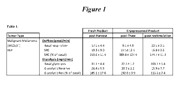
[00107] Figure 1: Shows the results from Example 1. As the Table shows,
following the
antigen restimulation rapid expansion protocol ("reREP"), the TILs exhibit a
marked
enhancement in their glycolytic respiration. SRC = spare respiratory capacity.
[00108] Figure 2: Composition of fresh vs. thawed TIL. TIL were stained for
TCRc43 and
CD56 to define T-cell and NK populations. The data shown are averages of 6
individual
TILs.
[00109] Figure 3: Memory phenotype is defined by CD45RA and CCR7 Expression.
CD4
and CD8 TIL are mainly Effector Memory (EM) This remains the same in the
thawed TIL.
Each point is one sample analyzed. No significant difference is found in a
Wilcoxon
matched-pairs signed rank test.
[00110] Figure 4: Pearson's correlation of CD4, CD8, CD4+CD28+, and CD8+CD28+
frequency between fresh and thawed TIL. Cells were stained with above markers.
Each dot
represents one individual with the fresh value on the x axis and the thawed
value on the y
axis. The fit line was drawn using linear regression analysis.
[00111] Figure 5: Comparable Activation Markers on Fresh and Thawed TILs. No
significant difference in activation status of fresh vs. thawed TIL was found
using a
Wilcoxon Matched-Pairs Rank Test. Each point represents one sample analyzed
and is shown
as mean +1- SEM.
[00112] Figure 6: Maintenance of LAG-3 Staining Following Cryopreservation and
Thaw.
A: LAG-3 staining of CD8 TIL. B: % frequency of regulatory molecules of the
CD4 and
CD8 populations on fresh and thawed TR,. CD8+TIM-3+ and CD8+LAG-3+ thawed TIL
have a lower % than fresh TIL. Mann-Whitney statistical test.
[00113] Figure 7: Remarkably stable tumor-infiltrating lymphocytes (TIL) for
infusion
phenotype following cryopreservation.
CA 03041678 2019-04-24
WO 2018/081473
PCT/US2017/058610
[00114] Figure 8: Scatter plot showing phenotypic characterization of reREP
TILs. Q1
shows 19.0% CD45RA-VCCR7"; Q2 shows 0.066% CD45RA-VCCR7+; Q4 shows 80.6%
CD45RA1CCR7-; and Q3 shows 0.36% CD45RA1CCR7+.
[00115] Figure 9: Diagram and data showing the phenotypic characterization of
reREP
TILs, during the first and second expansion phases 0.08% CD45RA+/CCR7-; 0.03%
CD45RA-VCCR7+; 73.97% CD45RA1CCR7-; and 25.91% CD45RA-/CCR7+ at Day 14, after
the first expansion but prior to the second expansion. Proliferation of CM or
EM TIL in the
repeat ReREP. Central Memory (CM) TIL and Effector Memory (EM) TIL were tested
for
the proliferation capacity using repeat ReREP. Briefly, 1.3 x 10⁶ Post REP TIL
were co-
culture with 1.3 x 10⁷ PBMC feeders (CFSE labelled), OKT3 (30 ng/nl) and rhIL-
2 (3000
IU/ml), culture was incubated for 14 days. On Day 14, central memory TIL and
effector
memory TIL were gated for LID Aqua-/CFSE-/TCRa/I3 +/CD45RA-/CCR7+ and LID Aqua-
/CF SE-/TCRa/I3 +/CD45RA-/CCR7- population respectively and flow cytometry
sorted.
Purity of the cell population was 97%. 1 x 10⁴ flow sorted CM or EM or
unsorted TIL were
then cultured 1 x 10⁶ PBMC feeders, OKT3 (30 ng/nl) and IL-2 (3000 IU/ml) in
triplicates
for 7 days. Cell were counted and recorded. Central memory TIL were more
proliferative
when compared to Effector memory TIL. We are repeating this experiment with
more post
REP TIL lines.
[00116] Figure 10A and 10B: Phenotypic characterization of TILs during ReREP.
Cells
were gated on Aqua-/TCR a/f3+/CD4+ or CD8+ to show Central Memory TILs (CD45RA-
CCR7+) or Effector Memory TILs (CD45RA-CCRT) memory phenotype. Student "t" was
used to calculate statistical significance. *p < 0.05, ns non-significant.
[00117] Figure 11: Exemplary schematic of the TIL preparation process,
sometimes
referred to herein as the IC process.
[00118] Figure 12: Successful expansion of TILs from non-melanoma tumors. Data
shows
the distribution of TIL (CD4+/CD8+) in non-melanoma tumors.
[00119] Figure 13: Non-melanoma TILs expressed CD27 and CD38, consistent with
young
TILs.
[00120] Figure 14: Activated TILs skew towards effector memory population.
[00121] Figure 15: Fresh versus reREP TIL phenotypes.
16
CA 03041678 2019-04-24
WO 2018/081473
PCT/US2017/058610
DETAILED DESCRIPTION OF THE INVENTION
I. Introduction
[00122] Adoptive cell therapy utilizing TILs cultured ex vivo by the Rapid
Expansion
Protocol (REP) has produced successful adoptive cell therapy following host
immunosuppression in patients with melanoma. Current infusion acceptance
parameters rely
on readouts of the composition of TILs (e.g., CD28, CD8, or CD4 positivity)
and on the
numerical folds of expansion and viability of the REP product.
[00123] Current REP protocols give little insight into the health of the TIL
that will be
infused into the patient. T cells undergo a profound metabolic shift during
the course of their
maturation from naïve to effector T cells (see Chang, et al., Nat. Immunot
2016, 17, 364,
hereby expressly incorporated in its entirety, and in particular for the
discussion and markers
of anaerobic and aerobic metabolism). For example, naïve T cells rely on
mitochondrial
respiration to produce ATP, while mature, healthy effector T cells such as TIL
are highly
glycolytic, relying on aerobic glycolysis to provide the bioenergetics
substrates they require
for proliferation, migration, activation, and anti-tumor efficacy.
[00124] Previous papers report that limiting glycolysis and promoting
mitochondrial
metabolism in TILs prior to transfer is desirable as cells that are relying
heavily on glycolysis
will suffer nutrient deprivation upon adoptive transfer which results in a
majority of the
transferred cells dying. Thus, the art teaches that promoting mitochondrial
metabolism might
promote in vivo longevity and in fact suggests using inhibitors of glycolysis
before induction
of the immune response. See Chang et al. (Chang, et al., Nat. Immunol. 2016,
17(364), 574-
582).
[00125] The present invention is directed in preferred aspects to novel
methods of
augmenting REPs with an additional restimulation protocol, sometimes referred
to herein as a
"restimulation Rapid Expansion Protocol" or "reREP", which leads surprisingly
to expanded
memory T cell subsets, including the central memory (CD45RA-CCR7 ) or effector
memory
(CD45RA-CCRT) phenotypes, and/or to marked enhancement in the glycolytic
respiration as
compared to freshly harvested TILs or thawed cryopreserved TILs for the
restimulated TILs
(sometimes referred to herein as "reTILs"). That is, by using a reREP
procedure (i.e., a
procedure comprising a first expansion and a second expansion) on
cryopreserved TILs,
patients can receive highly metabolically active, healthy TILs, leading to
more favorable
outcomes.
17
CA 03041678 2019-04-24
WO 2018/081473
PCT/US2017/058610
[00126] The present invention is further directed in some embodiments to
methods for
evaluating and quantifying this increase in metabolic health. Thus, the
present invention
provides methods of assaying the relative health of a TIL population using one
or more
general evaluations of metabolism, including, but not limited to, rates and
amounts of
glycolysis, oxidative phosphorylation, spare respiratory capacity (SRC) and
glycolytic
reserve.
[00127] Furthermore, the present invention is further directed in some
embodiments to
methods for evaluating and quantifying this increase in metabolic health.
Thus, the present
invention provides methods of assaying the relative health of a TIL population
using one or
more general evaluations of metabolism, including, but not limited to, rates
and amounts of
glycolysis, oxidative phosphorylation, spare respiratory capacity (SRC), and
glycolytic
reserve.
[00128] In addition, optional additional evaluations include, but are not
limited to, ATP
production, mitochondrial mass and glucose uptake.
[00129] In some cases, the reREP cell population with increased metabolic
health are
infused into a patient as is generally known in the art.
II. Definitions
[00130] By "tumor infiltrating lymphocytes" or "TILs" herein is meant a
population of cells
originally obtained as white blood cells that have left the bloodstream of a
subject and
migrated into a tumor. TILs include, but are not limited to, CD8+ cytotoxic T
cells
(lymphocytes), Thl and Th17 CD4+ T cells, natural killer cells, dendritic
cells and M1
macrophages. TILs include both primary and secondary TILs. "Primary TILs" are
those that
are obtained from patient tissue samples as outlined herein (sometimes
referred to as "freshly
harvested"), and "secondary TILs" are any TIL cell populations that have been
expanded or
proliferated as discussed herein, including, but not limited to bulk TILs,
expanded TILs
("REP TILs") as well as "reREP TILs" as discussed herein.
[00131] TILs can generally be defined either biochemically, using cell surface
markers, or
functionally, by their ability to infiltrate tumors and effect treatment. TILs
can be generally
categorized by expressing one or more of the following biomarkers: CD4, CD8,
TCR c43,
CD27, CD28, CD56, CCR7, CD45Ra, CD95, PD-1, and CD25. Additionally, and
alternatively, TILs can be functionally defined by their ability to infiltrate
solid tumors upon
reintroduction into a patient. TILS may further be characterized by potency ¨
for example,
18
CA 03041678 2019-04-24
WO 2018/081473
PCT/US2017/058610
TILS may be considered potent if, for example, interferon (IFN) release is
greater than about
50 pg/mL, greater than about 100 pg/mL, greater than about 150 pg/mL, or
greater than about
200 pg/mL. Interferon can include interferon gamma (IFNy).
[00132] By "cryopreserved TILs" herein is meant that TILs, either primary,
bulk, or
expanded (REP TILs), are treated and stored in the range of about -150 C to -
60 C. General
methods for cryopreservation are also described elsewhere herein, including in
the Examples.
For clarity, "cryopreserved TILs" are distinguishable from frozen tissue
samples which may
be used as a source of primary TILs.
[00133] By "thawed cryopreserved TILs" herein is meant a population of TILs
that was
previously cryopreserved and then treated to return to room temperature or
higher, including
but not limited to cell culture temperatures or temperatures wherein TILs may
be
administered to a patient.
[00134] By "population of cells" (including TILs) herein is meant a number of
cells that
share common traits. In general, populations generally range from 1 X 10⁶ to 1
X 1010 in
number, with different TIL populations comprising different numbers. For
example, initial
growth of primary TILs in the presence of IL-2 results in a population of bulk
TILs of
roughly 1 x 10⁸ cells. REP expansion is generally done to provide populations
of 1.5 x 10⁹ to
1.5 x 10¹⁰ cells for infusion.
[00135] In general, TILs are initially obtained from a patient tumor sample
("primary TILs")
and then expanded into a larger population for further manipulation as
described herein,
optionally cryopreserved, restimulated as outlined herein and optionally
evaluated for
phenotype and metabolic parameters as an indication of TIL health.
[00136] In general, the harvested cell suspension is called a "primary cell
population" or a
"freshly harvested" cell population.
[00137] In general, as discussed herein, the TILs are initially prepared by
obtaining a
primary population of TILs from a tumor resected from a patient as discussed
herein (the
"primary cell population" or "first cell population"). This is followed with
an initial bulk
expansion utilizing a culturing of the cells with IL-2, forming a second
population of cells
(sometimes referred to herein as the "bulk TIL population" or "second
population").
[00138] The term "cytotoxic lymphocyte" includes cytotoxic T (CTL) cells
(including CD8+
cytotoxic T lymphocytes and CD4+ T-helper lymphocytes), natural killer T (NKT)
cells and
natural killer (NK) cells. Cytotoxic lymphocytes can include, for example,
peripheral blood-
19
CA 03041678 2019-04-24
WO 2018/081473
PCT/US2017/058610
derived a/PTCR-positive or a/PTCR-positive T cells activated by tumor
associated antigens
and/or transduced with tumor specific chimeric antigen receptors or T-cell
receptors, and
tumor-infiltrating lymphocytes (TILs).
[00139] The term "central memory T cell" refers to a subset of T cells that in
the human are
CD45R0+ and constitutively express CCR7 (CCR7 hi) and CD62L (CD62 hi). The
surface
phenotype of central memory T cells also includes TCR, CD3, CD127 (IL-7R), and
IL-15R.
Transcription factors for central memory T cells include BCL-6, BCL-6B, MBD2,
and BMII.
Central memory T cells primarily secret IL-2 and CD4OL as effector molecules
after TCR
triggering. Central memory T cells are predominant in the CD4 compartment in
blood, and in
the human are proportionally enriched in lymph nodes and tonsils.
[00140] The term "effector memory T cell" refers to a subset of human or
mammalian T
cells that, like central memory T cells, are CD45R0+, but have lost the
constitutive
expression of CCR7 (CCR71o) and are heterogeneous or low for CD62L expression
(CD62L1o). The surface phenotype of central memory T cells also includes TCR,
CD3,
CD127 (IL-7R), and IL-15R. Transcription factors for central memory T cells
include
BLIMP 1. Effector memory T cells rapidly secret high levels of inflammatory
cytokines
following antigenic stimulation, including interferon-y, IL-4, and IL-5.
Effector memory T
cells are predominant in the CD8 compartment in blood, and in the human are
proportionally
enriched in the lung, liver, and gut. CD8+ effector memory T cells carry large
amounts of
perforin. The term "closed system" refers to a system that is closed to the
outside
environment. Any closed system appropriate for cell culture methods can be
employed with
the methods of the present invention. Closed systems include, for example, but
are not
limited to closed G-containers. Once a tumor segment is added to the closed
system, the
system is no opened to the outside environment until the TILs are ready to be
administered to
the patient.
[00141] The terms "peripheral blood mononuclear cells" and "PBMCs" refers to a
peripheral
blood cell having a round nucleus, including lymphocytes (T cells, B cells, NK
cells) and
monocytes. Preferably, the peripheral blood mononuclear cells are irradiated
allogeneic
peripheral blood mononuclear cells.
[00142] The term "rapid expansion" means an increase in the number of antigen-
specific
TILs of at least about 3-fold (or 4-, 5-, 6-, 7-, 8-, or 9-fold) over a period
of a week, more
preferably at least about 10-fold (or 20-, 30-, 40-, 50-, 60-, 70-, 80-, or 90-
fold) over a period
CA 03041678 2019-04-24
WO 2018/081473
PCT/US2017/058610
of a week, or most preferably at least about 100-fold over a period of a week.
A number of
rapid expansion protocols are described herein.
[00143] In some embodiments, methods of the present disclosure further include
a "pre-
REP" stage in which tumor tissue or cells from tumor tissue are grown in
standard lab media
(including without limitation RPMI) and treated the with reagents such as
irradiated feeder
cells and anti-CD3 antibodies to achieve a desired effect, such as increase in
the number of
TILS and/or an enrichment of the population for cells containing desired cell
surface markers
or other structural, biochemical or functional features. The pre-REP stage may
utilize lab
grade reagents (under the assumption that the lab grade reagents get diluted
out during a later
REP stage), making it easier to incorporate alternative strategies for
improving TIL
production. Therefore, in some embodiments, the disclosed TLR agonist and/or
peptide or
peptidomimetics can be included in the culture medium during the pre-REP
stage. The pre-
REP culture can in some embodiments, include IL-2.
[00144] The present invention is directed in preferred aspects to novel
methods of
augmenting REPs with an additional restimulation protocol, sometimes referred
to herein as a
"restimulation Rapid Expansion Protocol" or "reREP", which leads surprisingly
to expanded
memory T cell subsets, including the memory effector T cell subset, and/or to
marked
enhancement in the glycolytic respiration as compared to freshly harvested
TILs or thawed
cryopreserved TILs for the restimulated TILs (sometimes referred to herein as
"reTILs").
That is, by using a reREP procedure on cryopreserved TILs, patients can
receive highly
metabolically active, healthy TILs, leading to more favorable outcomes. Such
restimulation
protocols, also referred to herein as additional "expansions" of the cell
populations, are
described in further detail herein.
[00145] The terms "fragmenting," "fragment," and "fragmented," as used herein
to describe
processes for disrupting a tumor, includes mechanical fragmentation methods
such as
crushing, slicing, dividing, and morcellating tumor tissue as well as any
other method for
disrupting the physical structure of tumor tissue. The term "in vivo" refers
to an event that
takes place in a subject's body.
[00146] The term "in vitro" refers to an event that takes places outside of a
subject's body. In
vitro assays encompass cell-based assays in which cells alive or dead are
employed and may
also encompass a cell-free assay in which no intact cells are employed.
21
CA 03041678 2019-04-24
WO 2018/081473
PCT/US2017/058610
[00147] The term "anti-CD3 antibody" refers to an antibody or variant thereof,
e.g., a
monoclonal antibody and including human, humanized, chimeric or murine
antibodies which
are directed against the CD3 receptor in the T cell antigen receptor of mature
T cells. Anti-
CD3 antibodies include OKT-3, also known as muromonab, and UCHT-1. Other anti-
CD3
antibodies include, for example, otelixizumab, teplizumab, and visilizumab.
[00148] The term "OKT-3" (also referred to herein as "OKT3") refers to a
monoclonal
antibody or biosimilar or variant thereof, including human, humanized,
chimeric, or murine
antibodies, directed against the CD3 receptor in the T cell antigen receptor
of mature T cells,
and includes commercially-available forms such as OKT-3 (30 ng/mL, MACS GMP
CD3
pure, Miltenyi Biotech, Inc., San Diego, CA, USA) and muromonab or variants,
conservative
amino acid substitutions, glycoforms, or biosimilars thereof. The amino acid
sequences of the
heavy and light chains of muromonab are given in Table 1 (SEQ ID NO:1 and SEQ
ID
NO:2). A hybridoma capable of producing OKT-3 is deposited with the American
Type
Culture Collection and assigned the ATCC accession number CRL 8001. A
hybridoma
capable of producing OKT-3 is also deposited with European Collection of
Authenticated
Cell Cultures (ECACC) and assigned Catalogue No. 86022706.
TABLE 1. Amino acid sequences of muromonab.
Identifier Sequence (One-Letter Amino Acid Symbols)
SEQ ID NO:1 QVQLQQSGAE LARPGASVKM SCKASGYTFT RYTMHWVKQR PGQGLEWIGY
INPSRGYTNY 60
Muromonab heavy
NQKFKDKATL TTDKSSSTAY MQLSSLTSED SAVYYCARYY DDHYCLDYWG QGTTLTVSSA 120
chain KTTAPSVYPL APVCGGTTGS SVTLGCLVKG YFPEPVTLTW NSGSLSSGVH
TFPAVLQSDL 180
YTLSSSVTVT SSTWPSQSIT CNVAHPASST KVDKKIEPRP KSCDKTHTCP PCPAPELLGG 240
PSVFLFPPKP KDTLMISRTP EVTCVVVDVS HEDPEVKFNW YVDGVEVHNA KTKPREEQYN 300
STYRVVSVLT VLHQDWLNGK EYKCKVSNKA LPAPIEKTIS KAKGQPREPQ VYTLPPSRDE 360
LTKNQVSLTC LVKGFYPSDI AVEWESNGQP ENNYKTTPPV LDSDGSFFLY SKLTVDKSRW 420
QQGNVFSCSV MHEALHNHYT QKSLSLSPGK 450
SEQ ID NO:2 QIVLTQSPAI MSASPGEKVT MTCSASSSVS YMNWYQQKSG TSPKRWIYDT
SKLASGVPAH 60
Muromonab light
FRGSGSGTSY SLTISGMEAE DAATYYCQQW SSNPFTFGSG TKLEINRADT APTVSIFPPS 120
chain SEQLTSGGAS VVCFLNNFYP KDINVKWKID GSERQNGVLN SWTDQDSKDS
TYSMSSTLTL 180
TKDEYERHNS YTCEATHKTS TSPIVKSFNR NEC 213
[00149] The term "IL-2" (also referred to herein as "IL2") refers to the T
cell growth factor
known as interleukin-2, and includes all forms of IL-2 including human and
mammalian
forms, conservative amino acid substitutions, glycoforms, biosimilars, and
variants thereof.
IL-2 is described, e.g., in Nelson, J. Immunol. 2004, 172, 3983-88 and Malek,
Annu. Rev.
Immunol. 2008, 26, 453-79, the disclosures of which are incorporated by
reference herein.
The amino acid sequence of recombinant human IL-2 suitable for use in the
invention is
given in Table 2 (SEQ ID NO:3). For example, the term IL-2 encompasses human,
recombinant forms of IL-2 such as aldesleukin (PROLEUKIN, available
commercially from
22
CA 03041678 2019-04-24
WO 2018/081473
PCT/US2017/058610
multiple suppliers in 22 million ILJ per single use vials), as well as the
form of recombinant
IL-2 commercially supplied by CellGenix, Inc., Portsmouth, NH, USA (CELLGRO
GMP) or
ProSpec-Tany TechnoGene Ltd., East Brunswick, NJ, USA (Cat. No. CYT-209-b) and
other
commercial equivalents from other vendors. Aldesleukin (des-alanyl-1, serine-
125 human IL-
2) is a nonglycosylated human recombinant form of IL-2 with a molecular weight
of
approximately 15 kDa. The amino acid sequence of aldesleukin suitable for use
in the
invention is given in Table 2 (SEQ ID NO:4). The term IL-2 also encompasses
pegylated
forms of IL-2, as described herein, including the pegylated IL2 prodrug NKTR-
214, available
from Nektar Therapeutics, South San Francisco, CA, USA. NKTR-214 and pegylated
IL-2
suitable for use in the invention is described in U.S. Patent Application
Publication No. US
2014/0328791 Al and International Patent Application Publication No. WO
2012/065086 Al,
the disclosures of which are incorporated by reference herein. Alternative
forms of
conjugated IL-2 suitable for use in the invention are described in U.S. Patent
Nos. 4,766,106,
5,206,344, 5,089,261 and 4902,502, the disclosures of which are incorporated
by reference
herein. Formulations of IL-2 suitable for use in the invention are described
in U.S. Patent No.
6,706,289, the disclosure of which is incorporated by reference herein.
TABLE 2. Amino acid sequences of interleukins.
Identifier Sequence (One-Letter Amino Acid Symbols)
SEQ ID NO:3 MAPTSSSTKK TQLQLEHLLL DLQMILNGIN NYKNFKLTRM LTEKEYMPKK
ATELKHLQCL 60
recombinant EEELKPLEEV LNLAQSKNFH LRPRDLISNI NVIVLELKGS ETTFMCEYAD
ETATIVEFLN 120
human IL-2 RWITFCQSII STLT 134
(rhIL-2)
SEQ ID NO:4 PTSSSTKKTQ LQLEHLLLDL QMILNGINNY KNPKLTRMLT FKEYMPKKAT
ELKHLQCLEE 60
Aldesleukin ELKPLEEVLN LAQSKNEHLR PRDLISNINV IVLELKGSET TFMCEYADET
ATIVEFLNRW 120
ITFSQSIIST LT 132
SEQ ID NO:5 MHKCDITLQE IIKTLNSLTE QKTLCTELTV TDIFAASKNT TEKETFCRAA
TVLRQFYSHH 60
recombinant EKDTRCLGAT AQQFHRHKQL IRFLKRLDRN LWGLAGLNSC PVKEANQSTL
ENFLERLKTI 120
human IL-4 MREKYSKCSS 130
(rhIL-4)
SEQ ID NO:6 MDCDIEGKDG KQYESVIMVS IDQLLDSMKE IGSNCLNNEF NEFKRHICDA
NKEGMFLFRA 60
recombinant ARKLRQFLKM NSTGDFDLHL LKVSEGTTIL LNCTGQVKGR KPAALGEAQP
THSLEENKSL 120
human IL-7 KEQKKLNDLC FLKRLLQEIK TCWNKILMGT KEN 133
(rhIL-7)
SEQ ID NO:7 MNWVNVISDL KKIEDLIQSM HIDATLYTES DVEPSCKVTA MKCELLELQV
ISLESGDASI 60
recombinant HDTVENLIIL ANNSLSSNGN VTESGCKECE ELEEKNIKEF LQSFVHIVQM FINDS
115
human IL-15
(rhIL-15)
SEQ ID NO:8 MQDRHMIRMR QLIDIVDQLK NYVNDLVPEF LPAPEDVETN CEWSAFSCFQ
KAQLKSANTG 60
recombinant NNERIINVSI KKLKRKPPST NAGRRQKHRL TCPSCDSYEK KPPKEFLERF
KSLLQKMIHQ 120
human IL-21 HLSSRTHGSE DS 132
(rhIL-21)
[00150] The term "IL-4" (also referred to herein as "IL4") refers to the
cytokine known as
interleukin 4, which is produced by Th2 T cells and by eosinophils, basophils,
and mast cells.
IL-4 regulates the differentiation of naïve helper T cells (Th0 cells) to Th2
T cells. Steinke
and Borish, Respir. Res. 2001, 2, 66-70. Upon activation by IL-4, Th2 T cells
subsequently
23
CA 03041678 2019-04-24
WO 2018/081473
PCT/US2017/058610
produce additional IL-4 in a positive feedback loop. IL-4 also stimulates B
cell proliferation
and class II MHC expression, and induces class switching to IgE and IgG1
expression from B
cells. Recombinant human IL-4 suitable for use in the invention is
commercially available
from multiple suppliers, including ProSpec-Tany TechnoGene Ltd., East
Brunswick, NJ,
USA (Cat. No. CYT-211) and ThermoFisher Scientific, Inc., Waltham, MA, USA
(human
IL-15 recombinant protein, Cat. No. Gibco CTP0043). The amino acid sequence of
recombinant human IL-4 suitable for use in the invention is given in Table 2
(SEQ ID NO:5).
[00151] The term "IL-7" (also referred to herein as "IL7") refers to a
glycosylated tissue-
derived cytokine known as interleukin 7, which may be obtained from stromal
and epithelial
cells, as well as from dendritic cells. Fry and Mackall, Blood 2002, 99, 3892-
904. IL-7 can
stimulate the development of T cells. IL-7 binds to the IL-7 receptor, a
heterodimer
consisting of IL-7 receptor alpha and common gamma chain receptor, which in a
series of
signals important for T cell development within the thymus and survival within
the periphery.
Recombinant human IL-4 suitable for use in the invention is commercially
available from
multiple suppliers, including ProSpec-Tany TechnoGene Ltd., East Brunswick,
NJ, USA
(Cat. No. CYT-254) and ThermoFisher Scientific, Inc., Waltham, MA, USA (human
IL-15
recombinant protein, Cat. No. Gibco PHC0071). The amino acid sequence of
recombinant
human IL-7 suitable for use in the invention is given in Table 2 (SEQ ID
NO:6).
[00152] The term "IL-15" (also referred to herein as "IL15") refers to the T
cell growth
factor known as interleukin-15, and includes all forms of IL-2 including human
and
mammalian forms, conservative amino acid substitutions, glycoforms,
biosimilars, and
variants thereof. IL-15 is described, e.g., in Fehniger and Caligiuri, Blood
2001, 97, 14-32,
the disclosure of which is incorporated by reference herein. IL-15 shares p
and y signaling
receptor subunits with IL-2. Recombinant human IL-15 is a single, non-
glycosylated
polypeptide chain containing 114 amino acids (and an N-terminal methionine)
with a
molecular mass of 12.8 kDa. Recombinant human IL-15 is commercially available
from
multiple suppliers, including ProSpec-Tany TechnoGene Ltd., East Brunswick,
NJ, USA
(Cat. No. CYT-230-b) and ThermoFisher Scientific, Inc., Waltham, MA, USA
(human IL-15
recombinant protein, Cat. No. 34-8159-82). The amino acid sequence of
recombinant human
IL-15 suitable for use in the invention is given in Table 2 (SEQ ID NO:7).
[00153] The term "IL-21" (also referred to herein as "IL21") refers to the
pleiotropic
cytokine protein known as interleukin-21, and includes all forms of IL-21
including human
and mammalian forms, conservative amino acid substitutions, glycoforms,
biosimilars, and
24
CA 03041678 2019-04-24
WO 2018/081473
PCT/US2017/058610
variants thereof. IL-21 is described, e.g., in Spolski and Leonard, Nat. Rev.
Drug. Disc. 2014,
13, 379-95, the disclosure of which is incorporated by reference herein. IL-21
is primarily
produced by natural killer T cells and activated human CD4+ T cells
Recombinant human
IL-21 is a single, non-glycosylated polypeptide chain containing 132 amino
acids with a
molecular mass of 15.4 kDa. Recombinant human IL-21 is commercially available
from
multiple suppliers, including ProSpec-Tany TechnoGene Ltd., East Brunswick,
NJ, USA
(Cat. No. CYT-408-b) and ThermoFisher Scientific, Inc., Waltham, MA, USA
(human IL-21
recombinant protein, Cat. No. 14-8219-80). The amino acid sequence of
recombinant human
IL-21 suitable for use in the invention is given in Table 2 (SEQ ID NO:8).
[00154] When "an anti-tumor effective amount", "an tumor-inhibiting effective
amount", or
"therapeutic amount" is indicated, the precise amount of the compositions of
the present
invention to be administered can be determined by a physician with
consideration of
individual differences in age, weight, tumor size, extent of infection or
metastasis, and
condition of the patient (subject). It can generally be stated that a
pharmaceutical composition
comprising the genetically modified cytotoxic lymphocytes described herein may
be
administered at a dosage of 104 to 1011 cells/kg body weight (e.g., 105 to
106, 105 to 1010, 105
town,
106 to 1010, 106 to 1011,107 to 1011, 107 to 1010,
108 to 1011,
108 to 1010, 109 to 1011, or
109 to 1010 cells/kg body weight), including all integer values within those
ranges.
Genetically modified cytotoxic lymphocytes compositions may also be
administered multiple
times at these dosages. The genetically modified cytotoxic lymphocytes can be
administered
by using infusion techniques that are commonly known in immunotherapy (see,
e.g.,
Rosenberg et al., New Eng. J. of Med. 319: 1676, 1988). The optimal dosage and
treatment
regime for a particular patient can readily be determined by one skilled in
the art of medicine
by monitoring the patient for signs of disease and adjusting the treatment
accordingly.
[00155] The term "hematological malignancy" refers to mammalian cancers and
tumors of
the hematopoietic and lymphoid tissues, including but not limited to tissues
of the blood,
bone marrow, lymph nodes, and lymphatic system. Hematological malignancies are
also
referred to as "liquid tumors." Hematological malignancies include, but are
not limited to,
acute lymphoblastic leukemia (ALL), chronic lymphocytic lymphoma (CLL), small
lymphocytic lymphoma (SLL), acute myelogenous leukemia (AML), chronic
myelogenous
leukemia (CML), acute monocytic leukemia (AMoL), Hodgkin's lymphoma, and non-
Hodgkin's lymphomas. The term "B cell hematological malignancy" refers to
hematological
malignancies that affect B cells.
CA 03041678 2019-04-24
WO 2018/081473
PCT/US2017/058610
[00156] The term "solid tumor" refers to an abnormal mass of tissue that
usually does not
contain cysts or liquid areas. Solid tumors may be benign or malignant. The
term "solid
tumor cancer refers to malignant, neoplastic, or cancerous solid tumors. Solid
tumor cancers
include, but are not limited to, sarcomas, carcinomas, and lymphomas, such as
cancers of the
lung, breast, prostate, colon, rectum, and bladder. The tissue structure of
solid tumors
includes interdependent tissue compartments including the parenchyma (cancer
cells) and the
supporting stromal cells in which the cancer cells are dispersed and which may
provide a
supporting microenvironment.
[00157] The term "liquid tumor" refers to an abnormal mass of cells that is
fluid in nature.
Liquid tumor cancers include, but are not limited to, leukemias, myelomas, and
lymphomas,
as well as other hematological malignancies. TILs obtained from liquid tumors
may also be
referred to herein as marrow infiltrating lymphocytes (MILs).
[00158] The term "microenvironment," as used herein, may refer to the solid or
hematological tumor microenvironment as a whole or to an individual subset of
cells within
the microenvironment. The tumor microenvironment, as used herein, refers to a
complex
mixture of "cells, soluble factors, signaling molecules, extracellular
matrices, and mechanical
cues that promote neoplastic transformation, support tumor growth and
invasion, protect the
tumor from host immunity, foster therapeutic resistance, and provide niches
for dominant
metastases to thrive," as described in Swartz, et al., Cancer Res., 2012, 72,
2473. Although
tumors express antigens that should be recognized by T cells, tumor clearance
by the immune
system is rare because of immune suppression by the microenvironment.
[00159] In an embodiment, the invention includes a method of treating a cancer
with a
population of rTILs, wherein a patient is pre-treated with non-myeloablative
chemotherapy
prior to an infusion of rTILs according to the invention. In some embodiments,
the population
of rTILs may be provided with a population of eTils, wherein a patient is pre-
treated with
nonmyeloablative chemotherapy prior to an infusion of rTILs and eTils
according to the
invention. In an embodiment, the non-myeloablative chemotherapy is
cyclophosphamide 60
mg/kg/d for 2 days (days 27 and 26 prior to rTIL infusion) and fludarabine 25
mg/m2/d for 5
days (days 27 to 23 prior to rTIL infusion). In an embodiment, after non-
myeloablative
chemotherapy and rTIL infusion (at day 0) according to the invention, the
patient receives an
intravenous infusion of IL-2 intravenously at 720,000 IU/kg every 8 hours to
physiologic
tolerance.
26
CA 03041678 2019-04-24
WO 2018/081473
PCT/US2017/058610
[00160] Experimental findings indicate that lymphodepletion prior to adoptive
transfer of
tumor-specific T lymphocytes plays a key role in enhancing treatment efficacy
by eliminating
regulatory T cells and competing elements of the immune system ("cytokine
sinks").
Accordingly, some embodiments of the invention utilize a lymphodepletion step
(sometimes
also referred to as "immunosuppressive conditioning") on the patient prior to
the introduction
of the rTILs of the invention.
[00161] The terms "co-administration," "co-administering," "administered in
combination
with," "administering in combination with," "simultaneous," and "concurrent,"
as used
herein, encompass administration of two or more active pharmaceutical
ingredients (in a
preferred embodiment of the present invention, for example, at least one
potassium channel
agonist in combination with a plurality of TILs) to a subject so that both
active
pharmaceutical ingredients and/or their metabolites are present in the subject
at the same
time. Co-administration includes simultaneous administration in separate
compositions,
administration at different times in separate compositions, or administration
in a composition
in which two or more active pharmaceutical ingredients are present.
Simultaneous
administration in separate compositions and administration in a composition in
which both
agents are present are preferred.
[00162] The term "effective amount" or "therapeutically effective amount"
refers to that
amount of a compound or combination of compounds as described herein that is
sufficient to
effect the intended application including, but not limited to, disease
treatment. A
therapeutically effective amount may vary depending upon the intended
application (in vitro
or in vivo), or the subject and disease condition being treated (e.g., the
weight, age and
gender of the subject), the severity of the disease condition, or the manner
of administration.
The term also applies to a dose that will induce a particular response in
target cells (e.g., the
reduction of platelet adhesion and/or cell migration). The specific dose will
vary depending
on the particular compounds chosen, the dosing regimen to be followed, whether
the
compound is administered in combination with other compounds, timing of
administration,
the tissue to which it is administered, and the physical delivery system in
which the
compound is carried.
[00163] The terms "treatment", "treating", "treat", and the like, refer to
obtaining a desired
pharmacologic and/or physiologic effect. The effect may be prophylactic in
terms of
completely or partially preventing a disease or symptom thereof and/or may be
therapeutic in
terms of a partial or complete cure for a disease and/or adverse effect
attributable to the
27
CA 03041678 2019-04-24
WO 2018/081473
PCT/US2017/058610
disease. "Treatment", as used herein, covers any treatment of a disease in a
mammal,
particularly in a human, and includes: (a) preventing the disease from
occurring in a subject
which may be predisposed to the disease but has not yet been diagnosed as
having it;
(b) inhibiting the disease, i.e., arresting its development or progression;
and (c) relieving the
disease, i.e., causing regression of the disease and/or relieving one or more
disease
symptoms. "Treatment" is also meant to encompass delivery of an agent in order
to provide
for a pharmacologic effect, even in the absence of a disease or condition. For
example,
"treatment" encompasses delivery of a composition that can elicit an immune
response or
confer immunity in the absence of a disease condition, e.g., in the case of a
vaccine.
[00164] The term "heterologous" when used with reference to portions of a
nucleic acid or
protein indicates that the nucleic acid or protein comprises two or more
subsequences that are
not found in the same relationship to each other in nature. For instance, the
nucleic acid is
typically recombinantly produced, having two or more sequences from unrelated
genes
arranged to make a new functional nucleic acid, e.g., a promoter from one
source and a
coding region from another source, or coding regions from different sources.
Similarly, a
heterologous protein indicates that the protein comprises two or more
subsequences that are
not found in the same relationship to each other in nature (e.g., a fusion
protein).
[00165] The terms "sequence identity," "percent identity," and "sequence
percent identity"
(or synonyms thereof, e.g., "99% identical") in the context of two or more
nucleic acids or
polypeptides, refer to two or more sequences or subsequences that are the same
or have a
specified percentage of nucleotides or amino acid residues that are the same,
when compared
and aligned (introducing gaps, if necessary) for maximum correspondence, not
considering
any conservative amino acid substitutions as part of the sequence identity.
The percent
identity can be measured using sequence comparison software or algorithms or
by visual
inspection. Various algorithms and software are known in the art that can be
used to obtain
alignments of amino acid or nucleotide sequences. Suitable programs to
determine percent
sequence identity include for example the BLAST suite of programs available
from the U.S.
Government's National Center for Biotechnology Information BLAST web site.
Comparisons between two sequences can be carried using either the BLASTN or
BLASTP
algorithm. BLASTN is used to compare nucleic acid sequences, while BLASTP is
used to
compare amino acid sequences. ALIGN, ALIGN-2 (Genentech, South San Francisco,
California) or MegAlign, available from DNASTAR, are additional publicly
available
software programs that can be used to align sequences. One skilled in the art
can determine
28
CA 03041678 2019-04-24
WO 2018/081473
PCT/US2017/058610
appropriate parameters for maximal alignment by particular alignment software.
In certain
embodiments, the default parameters of the alignment software are used.
[00166] As used herein, the term "variant" encompasses but is not limited to
antibodies or
fusion proteins which comprise an amino acid sequence which differs from the
amino acid
sequence of a reference antibody by way of one or more substitutions,
deletions and/or
additions at certain positions within or adjacent to the amino acid sequence
of the reference
antibody. The variant may comprise one or more conservative substitutions in
its amino acid
sequence as compared to the amino acid sequence of a reference antibody.
Conservative
substitutions may involve, e.g., the substitution of similarly charged or
uncharged amino
acids. The variant retains the ability to specifically bind to the antigen of
the reference
antibody. The term variant also includes pegylated antibodies or proteins.
[00167] The term "in vivo" refers to an event that takes place in a subject's
body.
[00168] The term "in vitro" refers to an event that takes places outside of a
subject's body. In
vitro assays encompass cell-based assays in which cells alive or dead are
employed and may
also encompass a cell-free assay in which no intact cells are employed.
[00169] The term "rapid expansion" means an increase in the number of antigen-
specific
TILs of at least about 3-fold (or 4-, 5-, 6-, 7-, 8-, or 9-fold) over a period
of a week, more
preferably at least about 10-fold (or 20-, 30-, 40-, 50-, 60-, 70-, 80-, or 90-
fold) over a period
of a week, or most preferably at least about 100-fold over a period of a week.
A number of
rapid expansion protocols are outlined below.
Restimulation of Cyropreserved TILs
[00170] As discussed herein, the present invention relates to the
restimulation of
cryopreserved TILs to increase their metabolic activity and thus relative
health prior to
transplant into a patient, and methods of testing said metabolic health. As
generally outlined
herein, TILs are generally taken from a patient sample and manipulated to
expand their
number prior to transplant into a patient. In some embodiments, the TILs may
be optionally
genetically manipulated as discussed below, and then cryopreserved. Once
thawed, they are
then restimulated to increase their metabolism prior to infusion into a
patient.
[00171] The "Step" Designations A, B, C, etc., below are in reference to
Figure 11. The
ordering of the Steps below and in Figure 11 is exemplary and any combination
or order of
29
CA 03041678 2019-04-24
WO 2018/081473
PCT/US2017/058610
steps, as well as additional steps, repetition of steps, and/or omission of
steps is contemplated
by the present application and the methods disclosed herein.
A. STEP A: Obtain Patient Tumor Sample
[00172] In general, TILs are initially obtained from a patient tumor sample
("primary TILs")
and then expanded into a larger population for further manipulation as
described herein,
optionally cryopreserved, restimulated as outlined herein and optionally
evaluated for
phenotype and metabolic parameters as an indication of TIL health.
[00173] A patient tumor sample may be obtained using methods known in the art,
generally
via surgical resection, needle biopsy or other means for obtaining a sample
that contains a
mixture of tumor and TIL cells. In general, the tumor sample may be from any
solid tumor,
including primary tumors, invasive tumors or metastatic tumors. The tumor
sample may also
be a liquid tumor, such as a tumor obtained from a hematological malignancy.
The solid
tumor may be of any cancer type, including, but not limited to, breast,
pancreatic, prostate,
colorectal, lung, brain, renal, stomach, and skin (including but not limited
to squamous cell
carcinoma, basal cell carcinoma, and melanoma). In some embodiments, useful
TILs are
obtained from malignant melanoma tumors, as these have been reported to have
particularly
high levels of TILs.
[00174] The term "solid tumor" refers to an abnormal mass of tissue that
usually does not
contain cysts or liquid areas. Solid tumors may be benign or malignant. The
term "solid
tumor cancer refers to malignant, neoplastic, or cancerous solid tumors. Solid
tumor cancers
include, but are not limited to, sarcomas, carcinomas, and lymphomas, such as
cancers of the
lung, breast, triple negative breast cancer, prostate, colon, rectum, and
bladder. In some
embodiments, the cancer is selected from cervical cancer, head and neck
cancer,
glioblastoma, ovarian cancer, sarcoma, pancreatic cancer, bladder cancer,
breast cancer, triple
negative breast cancer, and non-small cell lung carcinoma. The tissue
structure of solid
tumors includes interdependent tissue compartments including the parenchyma
(cancer cells)
and the supporting stromal cells in which the cancer cells are dispersed and
which may
provide a supporting microenvironment.
[00175] The term "hematological malignancy" refers to mammalian cancers and
tumors of
the hematopoietic and lymphoid tissues, including but not limited to tissues
of the blood,
bone marrow, lymph nodes, and lymphatic system. Hematological malignancies are
also
CA 03041678 2019-04-24
WO 2018/081473
PCT/US2017/058610
referred to as "liquid tumors." Hematological malignancies include, but are
not limited to,
acute lymphoblastic leukemia (ALL), chronic lymphocytic lymphoma (CLL), small
lymphocytic lymphoma (SLL), acute myelogenous leukemia (AML), chronic
myelogenous
leukemia (CML), acute monocytic leukemia (AMoL), Hodgkin's lymphoma, and non-
Hodgkin's lymphomas. The term "B cell hematological malignancy" refers to
hematological
malignancies that affect B cells.
[00176] Once obtained, the tumor sample is generally fragmented using sharp
dissection into
small pieces of between 1 to about 8 mm3, with from about 2-3 mm3 being
particularly
useful. The TILs are cultured from these fragments using enzymatic tumor
digests. Such
tumor digests may be produced by incubation in enzymatic media (e.g., Roswell
Park
Memorial Institute (RPMI) 1640 buffer, 2 mM glutamate, 10 mcg/mL gentamicine,
30
units/mL of DNase and 1.0 mg/mL of collagenase) followed by mechanical
dissociation (e.g.,
using a tissue dissociator). Tumor digests may be produced by placing the
tumor in
enzymatic media and mechanically dissociating the tumor for approximately 1
minute,
followed by incubation for 30 minutes at 37 C in 5% CO2, followed by repeated
cycles of
mechanical dissociation and incubation under the foregoing conditions until
only small tissue
pieces are present. At the end of this process, if the cell suspension
contains a large number
of red blood cells or dead cells, a density gradient separation using FICOLL
branched
hydrophilic polysaccharide may be performed to remove these cells. Alternative
methods
known in the art may be used, such as those described in U.S. Patent
Application Publication
No. 2012/0244133 Al, the disclosure of which is incorporated by reference
herein. Any of
the foregoing methods may be used in any of the embodiments described herein
for methods
of expanding TILs or methods treating a cancer.
[00177] In some embodiments, fragmentation includes physical fragmentation,
including for
example, dissection as well as digestion. In some embodiments, the
fragmentation is physical
fragmentation. In some embodiments, the fragmentation is dissection. In some
embodiments,
the fragmentation is by digestion. In some embodiments, TILs can be initially
cultured from
enzymatic tumor digests and tumor fragments obtained from patients.
[00178] In some embodiments, where the tumor is a solid tumor, the tumor
undergoes
physical fragmentation after the tumor sample is obtained, for example such as
in Step A of
Figure 11 In some embodiments, the fragmentation occurs before
cryopreservation. In some
embodiments, the fragmentation occurs after cryopreservation. In some
embodiments, the
fragmentation occurs after obtaining the tumor and in the absence of any
cryopreservation. In
31
CA 03041678 2019-04-24
WO 2018/081473
PCT/US2017/058610
some embodiments, the tumor is fragmented and 2, 3, or 4 fragments or pieces
are placed in
each container for the first expansion. In some embodiments, the tumor is
fragmented and 3
or 4 fragments or pieces are placed in each container for the first expansion.
In some
embodiments, the tumor is fragmented and 4 fragments or pieces are placed in
each container
for the first expansion,
[00179] In some embodiments, the TILs are obtained from tumor fragments. In
some
embodiments, the tumor fragment is obtained sharp dissection. In some
embodiments, the
tumor fragment is between about 1 mm3 and 10 mm3. In some embodiments, the
tumor
fragment is between about 1 mm3 and 8 mm3. In some embodiments, the tumor
fragment is
about 1 mm3. In some embodiments, the tumor fragment is about 2 mm3. In some
embodiments, the tumor fragment is about 3 mm3. In some embodiments, the tumor
fragment
is about 4 mm3. In some embodiments, the tumor fragment is about 5 mm3. In
some
embodiments, the tumor fragment is about 6 mm3. In some embodiments, the tumor
fragment
is about 7 mm3. In some embodiments, the tumor fragment is about 8 mm3. In
some
embodiments, the tumor fragment is about 9 mm3. In some embodiments, the tumor
fragment
is about 10 mm3.
[00180] In some embodiments, the TILs are obtained from tumor digests. In some
embodiments, tumor digests were generated by incubation in enzyme media, for
example but
not limited to RPMI 1640, 2 mM GlutaMAX, 10 mg/mL gentamicin, 30 U/mL DNase,
and
1.0 mg/mL collagenase, followed by mechanical dissociation (GentleMACS,
Miltenyi
Biotec, Auburn, CA). After placing the tumor in enzyme media, the tumor can be
mechanically dissociated for approximately 1 minute. The solution can then be
incubated
for 30 minutes at 37 C in 5% CO2 and it then mechanically disrupted again for
approximately 1 minute. After being incubated again for 30 minutes at 37 C in
5% CO2, the
tumor can be mechanically disrupted a third time for approximately 1 minute.
In some
embodiments, after the third mechanical disruption if large pieces of tissue
were present, 1
or 2 additional mechanical dissociations were applied to the sample, with or
without 30
additional minutes of incubation at 37 C in 5% CO2. In some embodiments, at
the end of
the final incubation if the cell suspension contained a large number of red
blood cells or
dead cells, a density gradient separation using Ficoll can be performed to
remove these
cells.
32
CA 03041678 2019-04-24
WO 2018/081473
PCT/US2017/058610
[00181] In some embodiments, the harvested cell suspension prior to the first
expansion step
is called a "primary cell population" or a "freshly harvested" cell
population.
[00182] In some embodiments, cells can be optionally frozen after sample
harvest and stored
frozen prior to entry into Step B, which is described in further detail below.
B. STEP B: First Expansion
[00183] In some embodiments, a first expansion of TILs (also referred to as a
first expansion
or first TIL expansion) may be performed using an initial bulk TIL expansion
step (for
example, Step B as indicated in Figure 11 or a first expansion step; this can
include an
expansion step referred to as preREP) as described below and herein, followed
by a second
expansion step (for example, Step D as indicated in Figure 11; which can
include as an
example what is referred to as a rapid expansion protocol (REP) step) as
described below and
herein, followed by optional cryopreservation (for example, after Step D as
indicated in
Figure 11), and followed by an additional second expansion (for example, a
second Step D,
as indicated in Figure 11, which can include what is sometimes referred to as
a restimulation
REP step) as described below and herein. The TILs obtained from this process
may be
optionally characterized for phenotypic characteristics and metabolic
parameters as described
herein. In some embodiments, the TILs are frozen (i.e., cryopreserved) after
the first
expansion (for example, Step B as indicated in Figure 11) and stored until
phenotyped for
selection then thawed prior to proceeding to one or more second expansion
steps (for
example, one or more expansion according to Step D as indicated in Figure 11).
[00184] In some embodiments, where the cells are frozen after obtained from
the tumor
sample (such as, for example, during in Step A as indicated in Figure 11), the
cells are
thawed prior to the first expansion (for example, Step B as indicated in
Figure 11).
[00185] In embodiments where TIL cultures are initiated in 24-well plates, for
example,
using Costar 24-well cell culture cluster, flat bottom (Corning Incorporated,
Corning, NY,
each well can be seeded with lx106 tumor digest cells or one tumor fragment in
2 mL of
complete medium (CM) with IL-2 (6000 IU/mL; Chiron Corp., Emeryville, CA). In
some
embodiments, the tumor fragment is between about 1mm3 and 10 mm3.
[00186] After preparation of the tumor fragments, the resulting cells (i.e.,
fragments) are
cultured in serum containing IL-2 under conditions that favor the growth of
TILs over tumor
and other cells. In some embodiments, the tumor digests are incubated in 2 mL
wells in
33
CA 03041678 2019-04-24
WO 2018/081473
PCT/US2017/058610
media comprising inactivated human AB serum (or, in some cases, as outlined
herein, in the
presence of aAPC cell population) with 6000 IU/mL of IL-2. This primary cell
population is
cultured for a period of days, generally from 10 to 14 days, resulting in a
bulk TIL
population, generally about 1 >< 108 bulk TIL cells. In some embodiments, the
growth media
during the first expansion comprises IL-2 or a variant thereof. In some
embodiments, the IL
is recombinant human IL-2 (rhIL-2). In some embodiments the IL-2 stock
solution has a
specific activity of 20-30x106 IU/mg for a 1 mg vial. In some embodiments the
IL-2 stock
solution has a specific activity of 20-x106111/mg for a 1 mg vial. In some
embodiments the
IL-2 stock solution has a specific activity of 25x106IU/mg for a 1 mg vial. In
some
embodiments the IL-2 stock solution has a specific activity of 30x106IU/mg for
a 1 mg vial.
In some embodiments, the IL- 2 stock solution has a final concentration of 4-
8x10⁶IU/mg of
IL-2. In some embodiments, the IL- 2 stock solution has a final concentration
of 5-7x10⁶
IU/mg of IL-2. In some embodiments, the IL- 2 stock solution has a final
concentration of
6x10⁶IU/mg of IL-2. In some embodiments, the IL-2 stock solution is prepare as
described in
Example 4. In some embodiments, first expansion culture media comprises about
10,000
IU/mL of IL-2, about 9,000 IU/mL of IL-2, about 8,000 IU/mL of IL-2, about
7,000 IU/mL
of IL-2, about 6000 IU/mL of IL-2 or about 5,000 IU/mL of IL-2. In some
embodiments, first
expansion culture media comprises about 9,000 IU/mL of IL-2, to about 5,000
IU/mL of IL-
2. In some embodiments, first expansion culture media comprises about 8,000
IU/mL of IL-2,
to about 6,000 IU/mL of IL-2. In some embodiments, first expansion culture
media comprises
about 7,000 IU/mL of IL-2, to about 6,000 IU/mL of IL-2. In some embodiments,
first
expansion culture media comprises about 6,000 IU/mL of IL-2. In an embodiment,
the cell
culture medium further comprises IL-2. In some embodiments, the cell culture
medium
comprises about 3000 IU/mL of IL-2. In an embodiment, the cell culture medium
comprises
about 1000 IU/mL, about 1500 IU/mL, about 2000 IU/mL, about 2500 IU/mL, about
3000
IU/mL, about 3500 IU/mL, about 4000 IU/mL, about 4500 IU/mL, about 5000 IU/mL,
about
5500 IU/mL, about 6000 IU/mL, about 6500 IU/mL, about 7000 IU/mL, about 7500
IU/mL,
or about 8000 IU/mL of IL-2. In an embodiment, the cell culture medium
comprises between
1000 and 2000 IU/mL, between 2000 and 3000 IU/mL, between 3000 and 4000 IU/mL,
between 4000 and 5000 IU/mL, between 5000 and 6000 IU/mL, between 6000 and
7000
IU/mL, between 7000 and 8000 IU/mL, or between 8000 IU/mL of IL-2.
[00187] In some embodiments, the first expansion culture medium is referred to
as "CM", an
abbreviation for culture media. In some embodiments, it is referred to as CM1
(culture
34
CA 03041678 2019-04-24
WO 2018/081473
PCT/US2017/058610
medium 1). In some embodiments, CM consists of RPMI 1640 with GlutaMAX,
supplemented with 10% human AB serum, 25mM Hepes, and 10 mg/mL gentamicin. In
embodiments where cultures are initiated in gas-permeable flasks with a 40 mL
capacity and
a 10cm2 gas-permeable silicon bottom (for example, G-Rex10; Wilson Wolf
Manufacturing,
New Brighton, MN) (Fig. 1), each flask was loaded with 10-40x 106 viable tumor
digest cells
or 5-30 tumor fragments in 10-40mL of CM with IL-2. Both the G-Rex10 and 24-
well plates
were incubated in a humidified incubator at 37 C in 5% CO2 and 5 days after
culture
initiation, half the media was removed and replaced with fresh CM and IL-2 and
after day 5,
half the media was changed every 2-3 days. In some embodiments, the CM is the
CM1
described in the Examples, see, Example 5. In some embodiments, the first
expansion occurs
in an initial cell culture medium or a first cell culture medium. In some
embodiments, the
initial cell culture medium or the first cell culture medium comprises IL-2.
[00188] In some embodiments, the first TIL expansion can proceed for 11 days,
12 days, 13
days, 14 days, 15 days, 16 days, 17 days, 18 days, 19 days, 20 days, or 21
days. In some
embodiments, the first TIL expansion can proceed for 11 days to 21 days. In
some
embodiments, the first TIL expansion can proceed for 12 days to 21 days. In
some
embodiments, the first TIL expansion can proceed for 13 days to 21 days. In
some
embodiments, the first TIL expansion can proceed for 14 days to 21 days. In
some
embodiments, the first TIL expansion can proceed for 15 days to 21 days. In
some
embodiments, the first TIL expansion can proceed for 16 days to 21 days. In
some
embodiments, the first TIL expansion can proceed for 17 days to 21 days. In
some
embodiments, the first TIL expansion can proceed for 18 days to 21 days. In
some
embodiments, the first TIL expansion can proceed for 19 days to 21 days. In
some
embodiments, the first TIL expansion can proceed for 20 days to 21 days. In
some
embodiments, the first TIL expansion can proceed for 21 days.
C. STEP C: First Expansion to Second Expansion Transition
[00189] In some embodiments, the TILs obtained from the first expansion (for
example,
from Step B as indicated in Figure 11) are stored until phenotyped for
selection. In some
embodiments, the TILs obtained from the first expansion are cryopreserved
after the first
expansion and prior to the second expansion. In some embodiments, the TILs are
cryopreserved as part of the first expansion to second expansion transition.
For example, in
some embodiments, the TILs are cryopreserved after Step B and before Step D as
indicated in
CA 03041678 2019-04-24
WO 2018/081473
PCT/US2017/058610
Figure 11. In some embodiments, the TILs are cryopreserved and thawed as part
of the first
expansion to second expansion transition. For example, in some embodiments,
the TILs are
cryopreserved after Step B then thawed prior to proceeding to Step D (as
provided in Figure
11). In some embodiments, the transition from the first expansion to the
second expansion
occurs at about 22 days, 23, days, 24 days, 25 days, 26 days, 27 days, 28
days, 29 days, or 30
days from when fragmentation occurs. In some embodiments, the transition from
the first
expansion to the second expansion occurs at about 22 days to 30 days from when
fragmentation occurs. In some embodiments, the transition from the first
expansion to the
second expansion occurs at about 24 days to 30 days from when fragmentation
occurs. In
some embodiments, the transition from the first expansion to the second
expansion occurs at
about 26 days to 30 days from when fragmentation occurs. In some embodiments,
the
transition from the first expansion to the second expansion occurs at about 28
days to 30 days
from when fragmentation occurs. In some embodiments, the transition from the
first
expansion to the second expansion occurs at about 30 days from when
fragmentation occurs.
D. STEP D: Second Expansion
[00190] In some embodiments, the second expansion or second TIL expansion
(which can
include expansions sometimes referred to as REP) of TIL can be performed using
any TIL
flasks or containers known by those of skill in the art. In some embodiments,
the second TIL
expansion can proceed for 14 days, 15 days, 16 days, 17 days, 18 days, 19
days, 20 days, 21
days, or 22 days. In some embodiments, the second TIL expansion can proceed
for about 14
days to about 22 days. In some embodiments, the second TIL expansion can
proceed for
about 14 days to about 20 days. In some embodiments, the second TIL expansion
can
proceed for about 14 days to about 18 days. In some embodiments, the second
TIL expansion
can proceed for about 14 days to about 16 days. In some embodiments, the
second TIL
expansion can proceed for about 14 days.
[00191] In some embodiments, the second expansion occurs in a supplemented
cell culture
medium. In some embodiments, the supplemented cell culture medium comprises IL-
2, OKT-
3, and antigen-presenting feeder cells. In some embodiments, the second cell
culture medium
comprises IL-2, OKT-3, and antigen-presenting cells (APCs; also referred to as
antigen-
presenting feeder cells).
36
CA 03041678 2019-04-24
WO 2018/081473
PCT/US2017/058610
[00192] In some embodiments, the second expansion (which can include
expansions referred
to as REP) of TILs can be performed using T-175 flasks and gas-permeable bags
as
previously described (Tran KQ, Zhou J, Durflinger KH, et al., 2008, J
Immunother , 31:742-
751, and Dudley ME, Wunderlich JR, Shelton TE, et al. 2003, J Immunother ,
26:332-342)
or gas-permeable G-Rex flasks. In some embodiments, the second expansion is
performed
using flasks. In some embodiments, the second expansion is performed using gas-
permeable
G-Rex flasks. For TIL the second expansion in T-175 flasks, about 1 x 10⁶ TIL
are suspended
in about 150 mL of media and this is added to each T-175 flask. The TIL are
cultured with
irradiated (50 Gy) allogeneic PBMC as "feeder" cells at a ratio of 1 to 100
and the cells were
cultured in a 1 to 1 mixture of CM and AIM-V medium (50/50 medium),
supplemented
with 3000 IU/mL of IL-2 and 30 ng/mL of anti-CD3. The T-175 flasks are
incubated at
37 C in 5% CO2. In some embodiments, half the media is changed 5 days into the
second
expansion using 50/50 medium with 3000 IU/mL of IL-2. In some embodiments, on
day 7,
cells from 2 T-175 flasks are combined in a 3 L bag and 300 mL of AIM-V with
5%
human AB serum and 3000 IU/mL of IL-2 is added to the 300 mL of TIL
suspension. The
number of cells in each bag can be counted every day or two and fresh media
can be added
to keep the cell count between about 0.5 and about 2.0 x 10⁶ cells/mL.
[00193] In some embodiments, the second expansion (which can include
expansions referred
to as REP) of TIL can be performed in 500 mL capacity gas permeable flasks
with 100 cm2
gas-permeable silicon bottoms (G-Rex 100, commercially available from Wilson
Wolf
Manufacturing Corporation, New Brighton, MN, USA) (Fig. 1), about 5 x 10⁶ or
10 x 106
TIL are cultured with irradiated allogeneic PBMC at a ratio of 1 to 100 in 400
mL of 50/50
medium, supplemented with 3000 IU/mL of IL-2 and 30 ng/ mL of anti- CD3
(OKT3). The
G-Rex100 flasks can be incubated at 37 C in 5% CO2. In some embodiments, 5
days into the
second expansion, 250 mL of supernatant is removed and placed into centrifuge
bottles and
centrifuged at 1500 rpm (491 x g) for 10 minutes. The TIL pellets can then be
resuspended
with 150 mL of fresh medium with 5% human AB serum, 3000 IU per mL of IL-2 and
added
back to the original G-Rex100 flasks. In embodiments where TILs are expanded
serially in
G-Rex100 flasks, on day 7 the TIL in each G-Rex100 are suspended in the 300 mL
of media
present in each flask and the cell suspension was divided into three 100 mL
aliquots that can
be used to seed three G-Rex100 flasks. Then 150 mL of AIM-V with 5% human AB
serum
and 3000 IU per mL of IL-2 can be added to each flask. The G-Rex100 flasks can
be
incubated at 37 C in 5% CO2 and after 4 days in to the second expansion, 150
mL of AIM-V
37
CA 03041678 2019-04-24
WO 2018/081473
PCT/US2017/058610
with 3000 IU per mL of IL-2 can be added to each G-Rex100 flask. In some
embodiments,
the cells are harvested on day 14 of culture.
[00194] In some embodiments, the second expansion (which can include
expansions referred
to as REP) of TIL can be performed in a gas permeable container. For example,
TILs can be
rapidly expanded using non-specific T-cell receptor stimulation in the
presence of
interleukin-2 (IL-2) or interleukin-15 (IL-15). In an embodiment, expansion of
the number of
TILs uses about 1 x 10⁹ to about 1 x 10¹¹ antigen-presenting feeder cells. The
non-specific T-
cell receptor stimulus can include, for example, about 30 ng/ml of OKT3, a
mouse
monoclonal anti-CD3 antibody (commercially available from Ortho-McNeil,
Raritan, NJ or
Miltenyi Biotech, Auburn, CA). TILs can be rapidly expanded further
stimulation of the TILs
in vitro with one or more antigens, including antigenic portions thereof, such
as epitope(s), of
the cancer, which can be optionally expressed from a vector, such as a human
leukocyte
antigen A2 (1-11A-A2) binding peptide, e.g., 0.311M MART-1 :26-35 (27 L) or
gpl 00:209-
217 (210M), optionally in the presence of a T-cell growth factor, such as 300
IU/mL IL-2 or
IL-15. Other suitable antigens may include, e.g., NY-ESO-1, TRP-1, TRP-2,
tyrosinase
cancer antigen, MAGE-A3, SSX-2, and VEGFR2, or antigenic portions thereof. TIL
may
also be rapidly expanded by re-stimulation with the same antigen(s) of the
cancer pulsed onto
HLA-A2-expressing antigen-presenting cells. Alternatively, the TILs can be
further re-
stimulated with, e.g., example, irradiated, autologous lymphocytes or with
irradiated HLA-
A2+ allogeneic lymphocytes and IL-2.
[00195] In some embodiments, the second expansion (which can include
expansions referred
to as REP) of TIL can be performed in 500 mL capacity gas permeable flasks
with 100 cm
gas-permeable silicon bottoms (G-Rex 100, commercially available from Wilson
Wolf
Manufacturing Corporation, New Brighton, MN, USA), 5 x 10⁶ or 10 x 106 TIL may
be
cultured with aAPCs at a ratio of 1 to 100 in 400 mL of 50/50 medium,
supplemented with
5% human AB serum, 3000 IU per mL of IL-2 and 30 ng per ml of anti-CD3 (OKT3).
The
G-Rex 100 flasks may be incubated at 37 C in 5% CO2. On day 5, 250 mL of
supernatant
may be removed and placed into centrifuge bottles and centrifuged at 1500 rpm
(491 x g) for
minutes. The TIL pellets may be re-suspended with 150 mL of fresh medium with
5%
human AB serum, 3000 IU per mL of IL-2, and added back to the original G-Rex
100 flasks.
When TIL are expanded serially in G-Rex 100 flasks, on day 7 the TIL in each G-
Rex 100
may be suspended in the 300 mL of media present in each flask and the cell
suspension may
be divided into 3 100 mL aliquots that may be used to seed 3 G-Rex 100 flasks.
Then 150 mL
38
CA 03041678 2019-04-24
WO 2018/081473
PCT/US2017/058610
of AIM-V with 5% human AB serum and 3000 RI per mL of IL-2 may be added to
each
flask. The G-Rex 100 flasks may be incubated at 37 C in 5% CO2 and after 4
days 150 mL
of AIM-V with 3000 IU per mL of IL-2 may be added to each G-Rex100 flask The
cells may
be harvested on day 14 of culture.
[00196] In one embodiment, the second expansion (including expansions referred
to as REP)
is performed in flasks with the bulk TILs being mixed with a 100- or 200-fold
excess of
inactivated feeder cells, 30 mg/mL OKT3 anti-CD3 antibody and 3000 IU/mL IL-2
in 150 ml
media. Media replacement is done (generally 2/3 media replacement via
respiration with
fresh media) until the cells are transferred to an alternative growth chamber.
Alternative
growth chambers include GRex flasks and gas permeable containers as more fully
discussed
below.
[00197] In another embodiment, the second expansion (including expansions
referred to as
REP) is performed and further comprises a step wherein TILs are selected for
superior tumor
reactivity. Any selection method known in the art may be used. For example,
the methods
described in U.S. Patent Application Publication No. 2016/0010058 Al, the
disclosures of
which are incorporated herein by reference, may be used for selection of TILs
for superior
tumor reactivity.
[00198] Optionally, a cell viability assay can be performed after the second
expansion
(including expansions referred to as the REP expansion), using standard assays
known in the
art. For example, a trypan blue exclusion assay can be done on a sample of the
bulk TILs,
which selectively labels dead cells and allows a viability assessment. In some
embodiments,
TIL samples can be counted and viability determined using a Cellometer K2
automated cell
counter (Nexcelom Bioscience, Lawrence, MA). In some embodiments, viability is
determined according to the Cellometer K2 Image Cytometer Automatic Cell
Counter
protocol described, for example, in Example 2.
[00199] In some embodiments, cells are grown for 7 days, 8 days, 9 days, 10
days, or 11
days of the total second expansion time before being split into more than one
container or
flask.
[00200] In some embodiments, the second expansion culture medium (e.g.,
sometimes
referred to as CM2 or the second cell culture medium), comprises IL-2, OKT-3,
as well as
the antigen-presenting feeder cells (APCs), as discussed in more detail below.
39
CA 03041678 2019-04-24
WO 2018/081473
PCT/US2017/058610
[00201] In some embodiments, the antigen-presenting feeder cells are PBMCs. In
some
embodiments, the antigen-presenting feeder cells are artificial antigen-
presenting feeder cells.
In an embodiment, the ratio of TILs to antigen-presenting feeder cells in the
second
expansion is about 1 to 25, about 1 to 50, about 1 to 100, about 1 to 125,
about 1 to 150,
about 1 to 175, about 1 to 200, about 1 to 225, about 1 to 250, about 1 to
275, about 1 to 300,
about 1 to 325, about 1 to 350, about 1 to 375, about 1 to 400, or about 1 to
500. In an
embodiment, the ratio of TILs to antigen-presenting feeder cells in the second
expansion is
between 1 to 50 and 1 to 300. In an embodiment, the ratio of TILs to antigen-
presenting
feeder cells in the second expansion is between 1 to 100 and 1 to 200.
[00202] In an embodiment, the TIL expansion procedures described herein
require an excess
of feeder cells during the second expansion (including for example, expansions
referred to as
REP TIL expansions). In many embodiments, the feeder cells are peripheral
blood
mononuclear cells (PBMCs) obtained from standard whole blood units from
healthy blood
donors. The PBMCs are obtained using standard methods such as Ficoll-Paque
gradient
separation. In an embodiment, artificial antigen-presenting (aAPC) cells are
used in place of
PBMCs.
[00203] In general, the allogenic PBMCs are inactivated, either via
irradiation or heat
treatment, and used in the REP procedures.
[00204] In some embodiments, the growth media during the first expansion
comprises IL-2
or a variant thereof. In some embodiments, the IL is recombinant human IL-2
(rhIL-2). In
some embodiments the IL-2 stock solution has a specific activity of 20-
30x106IU/mg for a 1
mg vial. In some embodiments the IL-2 stock solution has a specific activity
of 20-x106
IU/mg for a 1 mg vial. In some embodiments the IL-2 stock solution has a
specific activity of
25x106 IU/mg for a 1 mg vial. In some embodiments the IL-2 stock solution has
a specific
activity of 30x106 IU/mg for a 1 mg vial. In some embodiments, the IL- 2 stock
solution has a
final concentration of 4-8x10⁶ IU/mg of IL-2. In some embodiments, the IL- 2
stock solution
has a final concentration of 5-7x10⁶ IU/mg of IL-2. In some embodiments, the
IL- 2 stock
solution has a final concentration of 6x10⁶IU/mg of IL-2. In some embodiments,
the IL-2
stock solution is prepare as described in Example 4. In some embodiments,
first expansion
culture media comprises about 10,000 IU/mL of IL-2, about 9,000 IU/mL of IL-2,
about
8,000 IU/mL of IL-2, about 7,000 IU/mL of IL-2, about 6000 IU/mL of IL-2 or
about 5,000
IU/mL of IL-2. In some embodiments, first expansion culture media comprises
about 9,000
IU/mL of IL-2, to about 5,000 IU/mL of IL-2. In some embodiments, first
expansion culture
CA 03041678 2019-04-24
WO 2018/081473
PCT/US2017/058610
media comprises about 8,000 IU/mL of IL-2, to about 6,000 IU/mL of IL-2. In
some
embodiments, first expansion culture media comprises about 7,000 IU/mL of IL-
2, to about
6,000 IU/mL of IL-2. In some embodiments, first expansion culture media
comprises about
6,000 IU/mL of IL-2. In an embodiment, the cell culture medium further
comprises IL-2. In
some embodiments, the cell culture medium comprises about 3000 IU/mL of IL-2.
In an
embodiment, the cell culture medium comprises about 1000 IU/mL, about 1500
IU/mL,
about 2000 IU/mL, about 2500 IU/mL, about 3000 IU/mL, about 3500 IU/mL, about
4000
IU/mL, about 4500 IU/mL, about 5000 IU/mL, about 5500 IU/mL, about 6000 IU/mL,
about
6500 IU/mL, about 7000 IU/mL, about 7500 IU/mL, or about 8000 IU/mL of IL-2.
In an
embodiment, the cell culture medium comprises between 1000 and 2000 IU/mL,
between
2000 and 3000 IU/mL, between 3000 and 4000 IU/mL, between 4000 and 5000 IU/mL,
between 5000 and 6000 IU/mL, between 6000 and 7000 IU/mL, between 7000 and
8000
IU/mL, or between 8000 IU/mL of IL-2.
[00205] In some embodiments, the second expansion cell culture media also
includes an
anti-CD3 antibody. In some embodiment, the cell culture medium comprises OKT3
antibody.
In some embodiments, the cell culture medium comprises about 30 ng/mL of OKT3
antibody. In an embodiment, the cell culture medium comprises about 0.1 ng/mL,
about 0.5
ng/mL, about 1 ng/mL, about 2.5 ng/mL, about 5 ng/mL, about 7.5 ng/mL, about
10 ng/mL,
about 15 ng/mL, about 20 ng/mL, about 25 ng/mL, about 30 ng/mL, about 35
ng/mL, about
40 ng/mL, about 50 ng/mL, about 60 ng/mL, about 70 ng/mL, about 80 ng/mL,
about 90
ng/mL, about 100 ng/mL, about 200 ng/mL, about 500 ng/mL, and about 1 [tg/mL
of OKT3
antibody. In an embodiment, the cell culture medium comprises between 0.1
ng/mL and 1
ng/mL, between 1 ng/mL and 5 ng/mL, between 5 ng/mL and 10 ng/mL, between 10
ng/mL
and 20 ng/mL, between 20 ng/mL and 30 ng/mL, between 30 ng/mL and 40 ng/mL,
between
40 ng/mL and 50 ng/mL, and between 50 ng/mL and 100 ng/mL of OKT3 antibody.
[00206] In some embodiments, an anti-CD3 antibody in combination with IL-2
induces T
cell activation and cell division in the TIL population. This effect can be
seen with full length
antibodies as well as Fab and F(ab')2 fragments, with the former being
generally preferred;
see, e.g., Tsoukas et at., I Immunot 1985, 135, 1719, hereby incorporated by
reference in its
entirety. As will be appreciated by those in the art, there are a number of
suitable anti-human
CD3 antibodies that find use in the invention, including anti-human CD3
polyclonal and
monoclonal antibodies from various mammals, including, but not limited to,
murine, human,
primate, rat, and canine antibodies. In particular embodiments, the OKT3 anti-
CD3 antibody
41
CA 03041678 2019-04-24
WO 2018/081473
PCT/US2017/058610
is used (commercially available from Ortho-McNeil, Raritan, NJ or Miltenyi
Biotech,
Auburn, CA).
[00207] In some embodiment, the cells in the second expansion are grown in a
culture media
with high doses of a cytokine, in particular IL-2, as is known in the art.
[00208] Alternatively, using combinations of cytokines for the second
expansion of TILS is
additionally possible, with combinations of two or more of IL-2, IL-15 and IL-
21 as is
generally outlined in International Publication No. WO 2015/189356 and
International
Publication No. WO 2015/189357, hereby expressly incorporated by reference in
their
entirety. Thus, possible combinations include IL-2 and IL-15, IL-2 and IL-21,
IL-15 and IL-
21 and IL-2, IL-15 and IL-21, with the latter finding particular use in many
embodiments.
The use of combinations of cytokines specifically favors the generation of
lymphocytes, and
in particular T-cells as described therein.
E. Optional Repeats of Step D: Second Expansion
[00209] In some embodiments, the second expansion is performed one or more
times, i.e.,
the second expansion is repeated. For example, in some embodiments the Step D
second
expansion as indicated in Figure 11 is repeated one or more times. In some
embodiments, the
second expansion is referred to as an additional second expansion. In some
embodiments
where the second expansion is performed more than once (i.e., where the second
expansion is
repeated), this can include procedures referred to as a TIL Rapid Expansion
Protocol. In some
embodiments, the TIL cell population is expanded in number after harvest and
first
expansion. This process is generally referred to in the art as a rapid
expansion process (REP)
and the repeated second expansion can include expansion referred to as reREP.
This overall
protocol can be generally accomplished using culture media comprising a number
of
components, including feeder cells, a cytokine source, and an anti-CD3
antibody, in a gas-
permeable container. In some embodiments, one or more subsequent second
expansion(s) are
performed as described above. In some embodiments, one or more subsequent
second
expansions are performed as provided in under Step D in Figure 11 and prior to
Step E as
provide in Figure 11. In some embodiments, one, two, three, four or more
second expansions
are performed as described above. In some embodiments, one, two, three, four
or more
second expansions are performed as provided in Step D of Figure 11 before Step
E of Figure
11. In some embodiments, two second expansions are performed as described
above. In some
42
CA 03041678 2019-04-24
WO 2018/081473
PCT/US2017/058610
embodiments, two second expansions are performed as provided in Step D of
Figure 11
before Step E of Figure 11. In some embodiments, three second expansions are
performed as
described above. In some embodiments, three second expansions are performed as
provided
in Step D of Figure 11 before Step E of Figure 11. In some embodiments, four
second
expansions are performed as described above. In some embodiments, four second
expansions
are performed as provided in Step D of Figure 11 before Step E of Figure 11.
[00210] In some embodiments, the repeat of the second expansion of the TILS
(such as for
example in Step D of Figure 11) can be referred to as a restimulation of TILs.
In some
embodiments, the present invention includes a restimulation step, i.e., a
repeat of the second
expansion (for example, a repeat of the second expansion from Step D of Figure
11). In some
embodiments, the repeated second expansion (which can include an expansion
referred to as
a restimulation step ("reREP")) is performed on cells that have been
cryopreserved. In some
embodiments, the Tits are cryopreserved after Step D. In some embodiments,
after an initial
second expansion in Step Dõ the cells may be cultured in regular media, e.g. a
"resting"
media, and then one or more second expansions steps are performed. In some
embodiments,
the resting media comprises IL-2. In some embodiments, the resting media does
not comprise
IL-2. In some embodiments, the resting media is a standard cell culture media
known in the
art. In some embodiments, the resting media is AIM-V, DMEM, DMEM/F12, MEM,
RPMI,
OptiMEM, IMDM, or any other standard media that is known in art, including
commercially
available media. In some embodiments, the resting media is AIM-V.
[00211] In general, as discussed herein, the TILs are initially prepared by
obtaining a
primary population of TILs from a tumor resected from a patient as discussed
herein (the
"primary cell population" or "first cell population"). This is followed with
an initial bulk
expansion utilizing a culturing of the cells with IL-2, forming a second
population of cells
(sometimes referred to herein as the "bulk TIL population" or "second
population"). In some
embodiments, this is also referred to as the initial or first expansion.
[00212] The bulk TIE population (for example, the population obtained from for
example
Step A in Figure 11) is then subjected to a REP step, sometimes referred to as
a first
expansion (for example, the first expansion as described in Step B of Figure
11) in a cell
culture media comprising IL-2, OKT-3, and antigen presenting feeder cells
(APCs), wherein
the APCs generally comprise peripheral blood mononuclear cells (PBMCs; or,
alternatively
as discussed herein, using antigen presenting cells), wherein the rapid
expansion (for
example, the second expansion as provide in Step D of Figure 11) is performed
for at least 14
43
CA 03041678 2019-04-24
WO 2018/081473
PCT/US2017/058610
days. As discussed herein, the media may also contain combinations of IL-2, IL-
15 and/or IL-
23 rather than IL-2 alone. In some embodiments, this post second expansion
(for example,
post Step D of Figure 11) expanded TIC, population is at least 50 ¨fold or 100-
fold greater in
number than the second population of TILs (for example, the population of TILs
obtained
from Step B of Figure 11). In some embodiments, the population of TILs
obtained after the
second expansion in Step D of Figure 11 are 50-fold or 100-fold greater in
number than the
TILs obtained from the first expansion in Step B of Figure 11. TILs are
measured by cell
counting methods known in the art, including those methods described in the
Examples
provided herewith, including Examples 1, 2, and 3. In some embodiments, a K2
cell counter
is employed to count the TILs. In some embodiments, a Cellometer IC2 Image
cytometer is
employed to count the TILs.
[00213] In some embodiments, as discussed herein, the TIL population obtained
after the
second expansion (sometimes referred to as a third TIL population or a REP
cell population)
is removed from the supplemented cell culture media (for example, the culture
media used in
Step D of Figure 11 or the media referred to as CM2 in the Examples) and
optionally
cryopreserved in a storage media (for example, media containing 5% DMSO) prior
to
performing and additional second expansion step.
[00214] Optionally, the TILs can be cryopreserved after a second expansion and
before an
additional second expansion. In some embodiments, the TILs are cryopreserved
after
performing Step D of Figure 11 and before performing an additional Step D of
Figure 11. In
some embodiments, the cryopreserved TILs are thawed prior to performing the
additional
second expansion. In some embodiments, the cryopreserved TILs are thawed prior
to
performing the additional Step D as provided in Figure 11. In some
embodiments, the TILs
are cryopreserved in 5% DMSO. In some embodiments, the TILs are cryopreserved
in cell
culture media plus 5% DMSO. Alternatively, the cells are removed from the
supplemented
cell culture media (for example, the culture media used in Step D of Figure
11) and cultured
in a resting media. Such media include those that are described in Examples 1
and 5, as well
as the other Examples provided herewith. In some embodiments, resting media
can include
media with IL-2. In some embodiments, the resting media can be the media
referred to as
CM1 in the examples.
[00215] The additional second expansion (including expansions referred to as
reREP) is
done on either the thawed cells or resting cells, using a supplemented cell
culture medium
(for example, a medium as provide in Step D of Figure 11) comprising IL-2, OKT-
3, and
44
CA 03041678 2019-04-24
WO 2018/081473
PCT/US2017/058610
feeder cells (for example, antigen presenting cells), generally comprising
peripheral blood
mononuclear cells (PBMCs; or, alternatively as discussed herein, using antigen
presenting
cells), wherein the additional second expansion is performed for at least 14
days. As
discussed herein, the media may also contain combinations of IL-2, IL-15
and/or IL-23 rather
than IL-2 alone.
[00216] This results in an expanded population of TILs that are characterized
in that these
expanded TILs exhibits an increased subpopulation of effector T cells and/or
central memory
T cells relative to the second population of TILs (e.g., the bulk starting
TILs). In some
embodiments, these expanded TILs are the TILs obtained from Step D of Figure
11.
[00217] In some embodiments the memory T cells are those cells that
constitutively CCR7
and CD62L. See, Sallusto, et al., Annu. Rev. Immunot, 2004, 22:745-763;
incorporated by
reference herein in its entirety.
[00218] Thus, the present invention provides methods for the restimulation of
cryopreserved
TILs upon thawing, based on post-thaw methods that result in increases of
metabolic health
such as glycolysis and respiration. In some embodiments, method comprises
providing a
population of thawed cryopreserved TILs that are then treated to increase
their metabolic
health to allow for optimal treatment upon infusion into patients.
F. STEP E: Harvest TILS from Step D
[00219] After the second expansion step, cells can be harvested. In some
embodiments the
TILs are harvested after one, two, three, four or more second expansion steps.
In some
embodiments, the TILs are harvested after one, two, three, four or more second
expansion
steps according to Step D as provided in Figure 11.
[00220] TILs can be harvested in any appropriate and sterile manner, including
for example
by centrifugation. Methods for TEL harvesting are well known in the art and
any such know
methods can be employed with the present process.
G. STEP F: Final Formulation and/or Transfer to Infusion Bag
[00221] After Steps A through E as provided in an exemplary order in Figure 11
and as
outlined in detailed above and herein are complete, cells are transferred to a
container for use
in administration to a patient. In some embodiments, once a therapeutically
sufficient number
CA 03041678 2019-04-24
WO 2018/081473 PCT/US2017/058610
of TILs are obtained using the expansion methods described above, they are
transferred to a
container for use in administration to a patient.
[00222] In an embodiment, TILs expanded using APCs of the present disclosure
are
administered to a patient as a pharmaceutical composition. In an embodiment,
the
pharmaceutical composition is a suspension of TILs in a sterile buffer. TILs
expanded using
PBMCs of the present disclosure may be administered by any suitable route as
known in the
art. In some embodiments, the T-cells are administered as a single intra-
arterial or
intravenous infusion, which preferably lasts approximately 30 to 60 minutes.
Other suitable
routes of administration include intraperitoneal, intrathecal, and
intralymphatic.
1. Pharmaceutical Compositions, Dosages, and Dosing Regimens
[00223] In an embodiment, TILs expanded using APCs of the present disclosure
are
administered to a patient as a pharmaceutical composition. In an embodiment,
the
pharmaceutical composition is a suspension of TILs in a sterile buffer. TILs
expanded using
PBMCs of the present disclosure may be administered by any suitable route as
known in the
art. In some embodiments, the T-cells are administered as a single intra-
arterial or
intravenous infusion, which preferably lasts approximately 30 to 60 minutes.
Other suitable
routes of administration include intraperitoneal, intrathecal, and
intralymphatic
administration.
[00224] Any suitable dose of TILs can be administered. In some embodiments, a
therapeutically sufficient number of TILs are needed for a suitable dosage. In
some
embodiments, from about 2.3 x10¹⁰ to about 13.7 x101 TILs are administered,
with an average
of around 7.8 x101 TILs, particularly if the cancer is melanoma. In an
embodiment, about
1.2 x101 to about 4.3 x101 of TILs are administered. In some embodiments,
about 3 x101 to
about 12 x101 TILs are administered. In some embodiments, about 4x 101 to
about 10x 1010
TILs are administered. In some embodiments, about 5 x101 to about 8x101 TILs
are
administered. In some embodiments, about 6x101 to about 8x 10¹⁰ TILs are
administered. In
some embodiments, about 7x101 to about 8x101 TILs are administered. In some
embodiments, the therapeutically effective dosage is about 2.3 x101 to about
13.7 x101 . In
some embodiments, the therapeutically effective dosage is about 7.8 x101
TILs, particularly
of the cancer is melanoma. In some embodiments, the therapeutically effective
dosage is
about 1.2 x 10¹⁰ to about 4.3 x101 of TILs. In some embodiments, the
therapeutically effective
dosage is about 3 x101 to about 12 x101 TILs. In some embodiments, the
therapeutically
effective dosage is about 4 x 10' to about 10x101 TILs. In some embodiments,
the
46
CA 03041678 2019-04-24
WO 2018/081473
PCT/US2017/058610
therapeutically effective dosage is about 5x10¹⁰to about 8x10¹⁰TILs. In some
embodiments,
the therapeutically effective dosage is about 6x101 to about 8x10¹⁰TILs. In
some
embodiments, the therapeutically effective dosage is about 7x101 to about
8x101 TILs.
[00225] In some embodiments, the number of the TILs provided in the
pharmaceutical
compositions of the invention is about lx106, 2x10⁶, 3 x10⁶, 4x10⁶, 5x 10⁶,
6x10⁶, 7x10⁶,
8x10⁶, 9x10⁶, 1x10⁷, 2x10⁷, 3x10⁷, 4x10⁷, 5x10⁷, 6x10⁷, 7x10⁷, 8x10⁷, 9x10⁷,
1x10⁸, 2x10⁸,
3x10⁸, 4x10⁸, 5x10⁸, 6x10⁸, 7x10⁸, 8x10⁸, 9x10⁸, lx109, 2x10⁹, 3x10⁹, 4x10⁹,
5x10⁹, 6x10⁹,
7x10⁹, 8x10⁹, 9x10⁹, 1x10¹⁰, 2x10' , 3x10¹⁰, 4x10¹⁰, 5x101 , 6x10¹⁰, 7x10¹⁰,
8x10¹⁰, 9x10¹⁰,
lx1011, 2x10¹¹, 3x10¹¹, 4x10¹¹, 5x10¹¹, 6x10¹¹, 7x10¹¹, 8x10¹¹, 9x10¹¹,
lx1012, 2x10¹²,
3 x10¹², 4x10¹², 5x10¹², 6x10¹², 7x10¹², 8x10¹², 9x10¹², 1x10¹³, 2x10¹³,
3x10¹³, 4x10¹³,
5x10'3, 6x10¹³, 7x10¹³, 8x10¹³, and 9x10¹³. In an embodiment, the number of
the TILs
provided in the pharmaceutical compositions of the invention is in the range
of lx106 to
5x10⁶, 5x10⁶ to lx107, lx107to 5x10⁷, 5x10⁷ to 1x10⁸, 1x10⁸t0 5x10⁸, 5x10⁸to
1x10⁹,
1x10⁹t0 5x10⁹, 5x10⁹t0 ix101 , lx101 to 5x101 , 5x101 to 1x10", 5x10¹¹ to
lx1012,
lx1012 to 5x10¹², and 5x10'2 to lx1013. In some embodiments, the
therapeutically effective
dosage is about 1x10⁶, 2x10⁶, 3x10⁶, 4x10⁶, 5x10⁶, 6x10⁶, 7x10⁶, 8x10⁶, 9x10⁶,
i><7
2x10⁷, 3x10⁷, 4x10⁷, 5x10⁷, 6x10⁷, 7x10⁷, 8x10⁷, 9x10⁷, 1x10⁸, 2x10⁸ 3x10⁸,
4x10⁸, 5x10⁸,
6x10⁸, 7x10⁸, 8x10⁸, 9x10⁸, lx109, 2x10⁹, 3x10⁹, 4x10⁹, 5x10⁹, 6x10⁹ 7x10⁹,
8x10⁹, 9x10⁹,
lx101 , 2x101 3x101 , 4x101 , 5x101 , 6x10¹⁶, 7x101 , 8x10¹⁰, 9x1010 lx1011,
2x10¹¹,
3x10¹¹, 4x10¹¹ 5x10¹¹, 6x10¹¹, 7x10¹¹, 8x10¹¹, 9x10¹¹, lx1012, 2x10¹² 3x10¹²,
4x10¹²,
5x10¹², 6x10¹² 7x10¹², 8x10¹², 9x10¹², lx1013, 2x10¹³, 3x10", 4x10¹³ 5x10¹³,
6x10¹³,
7x10¹³, 8x10¹³ and 9x10¹³.
[00226] In some embodiments, the concentration of the TILs provided in the
pharmaceutical
compositions of the invention is less than, for example, 100%, 90%, 80%, 70%,
60%, 50%,
40%, 30%, 20%, 19%, 18%, 17%, 16%, 15%, 14%, 13%, 12%, 11%, 10%, 9%, 8%, 7%,
6%,
5%, 4%, 3%, 2%, 1%, 0.5%, 0.4%, 0.3%, 0.2%, 0.1%, 0.09%, 0.08%, 0.07%, 0.06%,
0.05%,
0.04%, 0.03%, 0.02%, 0.01%, 0.009%, 0.008%, 0.007%, 0.006%, 0.005%, 0.004%,
0.003%,
0.002%, 0.001%, 0.0009%, 0.0008%, 0.0007%, 0.0006%, 0.0005%, 0.0004%, 0.0003%,
0.0002% or 0.0001% w/w, w/v or v/v of the pharmaceutical composition.
[00227] In some embodiments, the concentration of the TILs provided in the
pharmaceutical
compositions of the invention is greater than 90%, 80%, 70%, 60%, 50%, 40%,
30%, 20%,
19.75%, 19.50%, 19.25% 19%, 18.75%, 18.50%, 18.25% 18%, 17.75%, 17.50%, 17.25%
17%, 16.75%, 16.50%, 16.25% 16%, 15.75%, 15.50%, 15.25% 15%, 14.75%, 14.50%,
47
CA 03041678 2019-04-24
WO 2018/081473
PCT/US2017/058610
14.25% 14%, 13.75%, 13.50%, 13.25 4 13%, 12.75%, 12.50%, 12.25 4 12%, 11.75%,
11.500o, 11.25 4 11%, 10.75%, 10.500o, 10.25% 10%, 9.75%, 9.50%, 9.25 4 9%,
8.75%,
8.50%, 8.25% 8%, 7.75%, 7.500o, 7.25% 7%, 6.75%, 6.500o, 6.25 4 6 4, 5.750,
5.500o,
5.25 4 5%, 4.75%, 4.50%, 4.25%, 4%, 3.75%, 3.500o, 3.25%, 3%, 2.75%, 2.50%,
2.25%,
2%, 1.75%, 1.50 4, 125%, 10o, 0.5%, 0.4%, 0.3%, 0.2%, 0.10o, 0.09%, 0.08%,
0.07%,
0.06%, 0.05%, 0.04%, 0.03%, 0.02%, 0.010o, 0.009 4, 0.008 4, 0.007%, 0.006 4,
0.005 4,
0.004 4, 0.003 4, 0.002%, 0.001%, 0.0009%, 0.0008%, 0.0007%, 0.0006%, 0.0005%,
0.0004%, 0.0003%, 0.0002% or 0.00010/0 W/VV, W/V, or v/v of the pharmaceutical
composition.
[00228] In some embodiments, the concentration of the TILs provided in the
pharmaceutical
compositions of the invention is in the range from about 0.00010o to about
500o, about
0.001 /0 to about 400o, about 0.010o to about 300o, about 0.02% to about 29%,
about 0.03% to
about 28%, about 0.04 A to about 270/,), about 0.05 A to about 26cY0, about
0.06 A to about
25 /,), about 0.07 A to about 2400, about 0.08 A to about 23%, about 0.09% to
about 22%,
about 0.10o to about 21c1A, about 0.20o to about 200 o, about 0.3 A to about
19%, about 0.4% to
about 18%, about 0.50o to about 17%, about 0.6% to about 16%, about 0.7% to
about 1500,
about 0.8% to about 140o, about 0.9% to about 12% or about 1% to about 10%
w/w, w/v or
v/v of the pharmaceutical composition.
[00229] In some embodiments, the concentration of the TILs provided in the
pharmaceutical
compositions of the invention is in the range from about 0.0010o to about 10%,
about 0.010o
to about 5%, about 0.020o to about 4.5%, about 0.03 A to about 4%, about 0.04%
to about
3.5%, about 0.05% to about 30o, about 0.06 /0 to about 2.5%, about 0.07% to
about 2%, about
0.08% to about 1.5%, about 0.09% to about 1%, about 0.1% to about 0.9% w/w,
w/v or v/v of
the pharmaceutical composition.
[00230] In some embodiments, the amount of the TILs provided in the
pharmaceutical
compositions of the invention is equal to or less than 10 g, 9.5 g, 9.0 g, 8.5
g, 8.0 g, 7.5 g, 7.0
g, 6.5 g, 6.0 g, 5.5 g, 5.0 g, 4.5 g, 4.0 g, 3.5 g, 3.0 g, 2.5 g, 2.0 g, 1.5
g, 1.0 g, 0.95 g, 0.9 g,
0.85 g, 0.8 g, 0.75 g, 0.7 g, 0.65 g, 0.6 g, 0.55 g, 0.5 g, 0.45 g, 0.4 g,
0.35 g, 0.3 g, 0.25 g, 0.2
g, 0.15 g, 0.1 g, 0.09 g, 0.08 g, 0.07 g, 0.06 g, 0.05 g, 0.04 g, 0.03 g, 0.02
g, 0.01 g, 0.009 g,
0.008 g, 0.007 g, 0.006 g, 0.005 g, 0.004 g, 0.003 g, 0.002 g, 0.001 g, 0.0009
g, 0.0008 g,
0.0007 g, 0.0006 g, 0.0005 g, 0.0004 g, 0.0003 g, 0.0002 g, or 0.0001 g.
48
CA 03041678 2019-04-24
WO 2018/081473
PCT/US2017/058610
[00231] In some embodiments, the amount of the TILs provided in the
pharmaceutical
compositions of the invention is more than 0.0001 g, 0.0002 g, 0.0003 g,
0.0004 g, 0.0005 g,
0.0006 g, 0.0007 g, 0.0008 g, 0.0009 g, 0.001 g, 0.0015 g, 0.002 g, 0.0025 g,
0.003 g, 0.0035
g, 0.004 g, 0.0045 g, 0.005 g, 0.0055 g, 0.006 g, 0.0065 g, 0.007 g, 0.0075 g,
0.008 g, 0.0085
g, 0.009 g, 0.0095 g, 0.01 g, 0.015 g, 0.02 g, 0.025 g, 0.03 g, 0.035 g, 0.04
g, 0.045 g, 0.05 g,
0.055 g, 0.06 g, 0.065 g, 0.07 g, 0.075 g, 0.08 g, 0.085 g, 0.09 g, 0.095 g,
0.1 g, 0.15 g, 0.2 g,
0.25 g, 0.3 g, 0.35 g, 0.4 g, 0.45 g, 0.5 g, 0.55 g, 0.6 g, 0.65 g, 0.7 g,
0.75 g, 0.8 g, 0.85 g, 0.9
g, 0.95 g, 1 g, 1.5 g, 2 g, 2.5, 3 g, 3.5, 4 g, 4.5 g, 5 g, 5.5 g, 6 g, 6.5 g,
7 g, 7.5 g, 8 g, 8.5 g, 9
g, 9.5 g, or 10 g.
[00232] The TILs provided in the pharmaceutical compositions of the invention
are effective
over a wide dosage range. The exact dosage will depend upon the route of
administration, the
form in which the compound is administered, the gender and age of the subject
to be treated,
the body weight of the subject to be treated, and the preference and
experience of the
attending physician. The clinically-established dosages of the TILs may also
be used if
appropriate. The amounts of the pharmaceutical compositions administered using
the
methods herein, such as the dosages of TILs, will be dependent on the human or
mammal
being treated, the severity of the disorder or condition, the rate of
administration, the
disposition of the active pharmaceutical ingredients and the discretion of the
prescribing
physician.
[00233] In some embodiments, TILs may be administered in a single dose. Such
administration may be by injection, e.g., intravenous injection. In some
embodiments, TILs
may be administered in multiple doses. Dosing may be once, twice, three times,
four times,
five times, six times, or more than six times per year. Dosing may be once a
month, once
every two weeks, once a week, or once every other day. Administration of TILs
may continue
as long as necessary.
[00234] In some embodiments, an effective dosage of TILs is about lx106,
2x10⁶, 3x10⁶,
4x10⁶, 5x10⁶, 6x10⁶, 7x10⁶, 8x10⁶, 9x10⁶, 1x10, 2x10⁷, 3x10⁷, 4x10⁷, 5x10⁷,
6x10⁷, 7x10⁷,
8x10⁷, 9x10⁷, 1x10⁸, 2x10⁸, 3x10⁸, 4x10⁸, 5x10⁸, 6x10⁸, 7x10⁸, 8x10⁸, 9x10⁸,
lx109, 2x10⁹,
3x10⁹, 4x10⁹, 5x10⁹, 6x10⁹, 7x10⁹, 8x10⁹, 9x10⁹, xl 101 , 2x101 , 3x101 ,
4x10' , 5x101 ,
6x10io,
7x101 , 8x 10¹⁰, 9x101 , lx 1011, 2x10", 3xioii, 4x10",
5x10¹¹, 6x,
iu 7x10¹¹,
8x10",
9x10¹¹, lx 1012, 2x10¹², 3x10¹², 4x10¹², 5x10¹², 6x10t2,
7x10'2 sx
1U 9>< 1012,
1x10'3, 2x10'3, 3 x 10¹³, 4x10'3, 5 x 10¹³, 6x10¹³, 7x10¹³ 8x10", and 9x10¹³.
In some
embodiments, an effective dosage of TILs is in the range of 1x10⁶ to 5x10⁶,
5x10⁶ to 1x10⁷,
49
CA 03041678 2019-04-24
WO 2018/081473
PCT/US2017/058610
1x10⁷ to 5x10⁷, 5x10⁷t0 1x10⁸, 1x10⁸t0 5x10⁸, 5x10⁸ to 1><109, 1x10⁹t0 5x10⁹,
5x10⁹to
lx101 , lx101 to 5x101 , 5x101 to lx1011, 5x10¹¹ to lx1012, ixioi2 to
5x10¹², and 5x1012
tolx10".
[00235] In some embodiments, an effective dosage of TILs is in the range of
about 0.01
mg/kg to about 4.3 mg/kg, about 0.15 mg/kg to about 3.6 mg/kg, about 0.3 mg/kg
to about
3.2 mg/kg, about 0.35 mg/kg to about 2.85 mg/kg, about 0.15 mg/kg to about
2.85 mg/kg,
about 0.3 mg to about 2.15 mg/kg, about 0.45 mg/kg to about 1.7 mg/kg, about
0.15 mg/kg to
about 1.3 mg/kg, about 0.3 mg/kg to about 1.15 mg/kg, about 0.45 mg/kg to
about 1 mg/kg,
about 0.55 mg/kg to about 0.85 mg/kg, about 0.65 mg/kg to about 0.8 mg/kg,
about 0.7
mg/kg to about 0.75 mg/kg, about 0.7 mg/kg to about 2.15 mg/kg, about 0.85
mg/kg to about
2 mg/kg, about 1 mg/kg to about 1.85 mg/kg, about 1.15 mg/kg to about 1.7
mg/kg, about 1.3
mg/kg mg to about 1.6 mg/kg, about 1.35 mg/kg to about 1.5 mg/kg, about 2.15
mg/kg to
about 3.6 mg/kg, about 2.3 mg/kg to about 3.4 mg/kg, about 2.4 mg/kg to about
3.3 mg/kg,
about 2.6 mg/kg to about 3.15 mg/kg, about 2.7 mg/kg to about 3 mg/kg, about
2.8 mg/kg to
about 3 mg/kg, or about 2.85 mg/kg to about 2.95 mg/kg.
[00236] In some embodiments, an effective dosage of TILs is in the range of
about 1 mg to
about 500 mg, about 10 mg to about 300 mg, about 20 mg to about 250 mg, about
25 mg to
about 200 mg, about 1 mg to about 50 mg, about 5 mg to about 45 mg, about 10
mg to about
40 mg, about 15 mg to about 35 mg, about 20 mg to about 30 mg, about 23 mg to
about 28
mg, about 50 mg to about 150 mg, about 60 mg to about 140 mg, about 70 mg to
about 130
mg, about 80 mg to about 120 mg, about 90 mg to about 110 mg, or about 95 mg
to about
105 mg, about 98 mg to about 102 mg, about 150 mg to about 250 mg, about 160
mg to about
240 mg, about 170 mg to about 230 mg, about 180 mg to about 220 mg, about 190
mg to
about 210 mg, about 195 mg to about 205 mg, or about 198 to about 207 mg.
[00237] An effective amount of the TILs may be administered in either single
or multiple
doses by any of the accepted modes of administration of agents having similar
utilities,
including intranasal and transdermal routes, by intra-arterial injection,
intravenously,
intraperitoneally, parenterally, intramuscularly, subcutaneously, topically,
by transplantation,
or by inhalation.
H. Optional Cell Viability Analyses
[00238] Optionally, a cell viability assay can be performed after the Step B
first expansion,
using standard assays known in the art. For example, a trypan blue exclusion
assay can be
CA 03041678 2019-04-24
WO 2018/081473
PCT/US2017/058610
done on a sample of the bulk TILs, which selectively labels dead cells and
allows a viability
assessment. Other assays for use in testing viability can include but are not
limited to the
Alamar blue assay; and the MTT assay.
1. Cell Counts, Viability, Flow Cytometry
[00239] In some embodiments, cell counts and/or viability are measured. The
expression of
markers such as but not limited CD3, CD4, CD8, and CD56, as well as any other
disclosed or
described herein, can be measured by flow cytometry with antibodies, for
example but not
limited to those commercially available from BD Bio-sciences (BD Biosciences,
San Jose,
CA) using a FACSCanto flow cytometer (BD Biosciences). The cells can be
counted
manually using a disposable c-chip hemocytometer (VWR, Batavia, IL) and
viability can be
assessed using any method known in the art, including but not limited to
trypan blue staining.
[00240] In some cases, the bulk TIL population can be cryopreserved
immediately, using the
protocols discussed below. Alternatively, the bulk TIL population can be
subjected to REP
and then cryopreserved as discussed below. Similarly, in the case where
genetically modified
TILs will be used in therapy, the bulk or REP TIL populations can be subjected
to genetic
modifications for suitable treatments.
2. Cell Cultures
[00241] In an embodiment, a method for expanding TILs may include using about
5,000 mL
to about 25,000 mL of cell medium, about 5,000 mL to about 10,000 mL of cell
medium, or
about 5,800 mL to about 8,700 mL of cell medium. In an embodiment, expanding
the number
of TILs uses no more than one type of cell culture medium. Any suitable cell
culture medium
may be used, e.g., AIM-V cell medium (L-glutamine, 50 pM streptomycin sulfate,
and 10 1.1M
gentamicin sulfate) cell culture medium (Invitrogen, Carlsbad CA). In this
regard, the
inventive methods advantageously reduce the amount of medium and the number of
types of
medium required to expand the number of TIL. In an embodiment, expanding the
number of
TIL may comprise adding fresh cell culture media to the cells (also referred
to as feeding the
cells) no more frequently than every third or fourth day. Expanding the number
of cells in a
gas permeable container simplifies the procedures necessary to expand the
number of cells by
reducing the feeding frequency necessary to expand the cells.
[00242] In an embodiment, the cell medium in the first and/or second gas
permeable
container is unfiltered. The use of unfiltered cell medium may simplify the
procedures
51
CA 03041678 2019-04-24
WO 2018/081473
PCT/US2017/058610
necessary to expand the number of cells. In an embodiment, the cell medium in
the first
and/or second gas permeable container lacks beta-mercaptoethanol (BME).
[00243] In an embodiment, the duration of the method comprising obtaining a
tumor tissue
sample from the mammal; culturing the tumor tissue sample in a first gas
permeable
container containing cell medium therein; obtaining TILs from the tumor tissue
sample;
expanding the number of TILs in a second gas permeable container containing
cell medium
therein using aAPCs for a duration of about 14 to about 42 days, e.g., about
28 days.
[00244] In an embodiment, TILs are expanded in gas-permeable containers. Gas-
permeable
containers have been used to expand TILs using PBMCs using methods,
compositions, and
devices known in the art, including those described in U.S. Patent Application
Publication
No. 2005/0106717 Al, the disclosures of which are incorporated herein by
reference. In an
embodiment, TILs are expanded in gas-permeable bags. In an embodiment, TILs
are
expanded using a cell expansion system that expands TILs in gas permeable
bags, such as the
Xuri Cell Expansion System W25 (GE Healthcare). In an embodiment, TILs are
expanded
using a cell expansion system that expands TILs in gas permeable bags, such as
the WAVE
Bioreactor System, also known as the Xuri Cell Expansion System W5 (GE
Healthcare). In
an embodiment, the cell expansion system includes a gas permeable cell bag
with a volume
selected from the group consisting of about 100 mL, about 200 mL, about 300
mL, about 400
mL, about 500 mL, about 600 mL, about 700 mL, about 800 mL, about 900 mL,
about 1 L,
about 2 L, about 3 L, about 4 L, about 5 L, about 6 L, about 7 L, about 8 L,
about 9 L, and
about 10 L. In an embodiment, TILs can be expanded in G-Rex flasks
(commercially
available from Wilson Wolf Manufacturing). Such embodiments allow for cell
populations to
expand from about 5x10⁵ cells/cm2 to between 10x106 and 30x106 cells/cm2. In
an
embodiment this expansion is conducted without adding fresh cell culture media
to the cells
(also referred to as feeding the cells). In an embodiment, this is without
feeding so long as
medium resides at a height of about 10 cm in the GRex flask. In an embodiment
this is
without feeding but with the addition of one or more cytokines. In an
embodiment, the
cytokine can be added as a bolus without any need to mix the cytokine with the
medium.
Such containers, devices, and methods are known in the art and have been used
to expand
TILs, and include those described in U.S. Patent Application Publication No.
US
2014/0377739A1, International Publication No. WO 2014/210036 Al, U.S. Patent
Application Publication No. us 2013/0115617 Al, International Publication No.
WO
2013/188427 Al, U.S. Patent Application Publication No. US 2011/0136228 Al,
U.S. Patent
52
CA 03041678 2019-04-24
WO 2018/081473
PCT/US2017/058610
No. US 8,809,050 B2, International publication No. WO 2011/072088 A2, U.S.
Patent
Application Publication No. US 2016/0208216 Al, U.S. Patent Application
Publication No.
US 2012/0244133 Al, International Publication No WO 2012/129201 Al, U.S.
Patent
Application Publication No. US 2013/0102075 Al, U.S. Patent No. US 8,956,860
B2,
International Publication No. WO 2013/173835 Al, U.S. Patent Application
Publication No.
US 2015/0175966 Al, the disclosures of which are incorporated herein by
reference. Such
processes are also described in Jin et al., J. Immunotherapy, 2012, 35:283-
292. Optional
Genetic Engineering of TILs
[00245] In some embodiments, the TILs are optionally genetically engineered to
include
additional functionalities, including, but not limited to, a high-affinity T
cell receptor (TCR),
e.g., a TCR targeted at a tumor-associated antigen such as MAGE-1, HER2, or NY-
ESO-1, or
a chimeric antigen receptor (CAR) which binds to a tumor-associated cell
surface molecule
(e.g., mesothelin) or lineage-restricted cell surface molecule (e.g., CD19).
I. Optional Cryopreservation of TILs
[00246] As discussed above in Steps A through E, cryopreservation can occur at
numerous
points throughout the TIL expansion process. In some embodiments, the bulk TIL
population
after the first expansion according to Step B or the expanded population of
TILs after the one
or more second expansions according to Step D can be cryopreserved.
Cryopreservation can
be generally accomplished by placing the TIL population into a freezing
solution, e.g., 85%
complement inactivated AB serum and 15% dimethyl sulfoxide (DMSO). The cells
in
solution are placed into cryogenic vials and stored for 24 hours at -80 C,
with optional
transfer to gaseous nitrogen freezers for cryopreservation. See, Sadeghi, et
al., Acta
Oncologica 2013, 52, 978-986. In some embodiments, the TILs are cryopreserved
in 5%
DMSO. In some embodiments, the TILs are cryopreserved in cell culture media
plus 5%
DMSO. In some embodiments, the TILs are cryopreserved according to the methods
provided in Examples 8 and 9.
[00247] When appropriate, the cells are removed from the freezer and thawed in
a 37 C
water bath until approximately 4/5 of the solution is thawed. The cells are
generally
resuspended in complete media and optionally washed one or more times. In some
embodiments, the thawed TILs can be counted and assessed for viability as is
known in the
art.
J. Phenotypic Characteristics of Expanded TILs
53
CA 03041678 2019-04-24
WO 2018/081473
PCT/US2017/058610
[00248] In some embodiment, the TILs are analyzed for expression of numerous
phenotype
markers after expansion, including those described herein and in the Examples.
In an
embodiment, expression of one or more phenotypic markers is examined. In some
embodiments, the phenotypic characteristics of the TILs are analyzed after the
first expansion
in Step B. In some embodiments, the phenotypic characteristics of the TILs are
analyzed
during the transition in Step C. In some embodiments, the phenotypic
characteristics of the
TILs are analyzed during the transition according to Step C and after
cryopreservation. In
some embodiments, the phenotypic characteristics of the TILs are analyzed
after the second
expansion according to Step D. In some embodiments, the phenotypic
characteristics of the
TILs are analyzed after two or more expansions according to Step D. In some
embodiments,
the marker is selected from the group consisting of TCRab, CD57, CD28, CD4,
CD27,
CD56, CD8a, CD45RA, CD8a, CCR7, CD4, CD3, CD38, and FILA-DR. In some
embodiments, the marker is selected from the group consisting of TCRab, CD57,
CD28,
CD4, CD27, CD56, and CD8a. In an embodiment, the marker is selected from the
group
consisting of CD45RA, CD8a, CCR7, CD4, CD3, CD38, and HLA-DR. In some
embodiments, expression of one, two, three, four, five, six, seven, eight,
nine, ten, eleven,
twelve, thirteen, or fourteen markers is examined. In some embodiments, the
expression from
one or more markers from each group is examined. In some embodiments, one or
more of
HLA-DR, CD38, and CD69 expression is maintained (i.e., does not exhibit a
statistically
significant difference) in fresh TILs as compared to thawed TILs. In some
embodiments, the
activation status of TILs is maintained in the thawed TILs.
[00249] In an embodiment, expression of one or more regulatory markers is
measured. In
some embodiments, the regulatory marker is selected from the group consisting
of CD137,
CD8a, Lag3, CD4, CD3, PD1, TIM-3, CD69, CD8a, TIGIT, CD4, CD3, KLRG1, and
CD154. In some embodiments, the regulatory marker is selected from the group
consisting of
CD137, CD8a, Lag3, CD4, CD3, PD1, and TIM-3. In some embodiments, the
regulatory
marker is selected from the group consisting of CD69, CD8a, TIGIT, CD4, CD3,
KLRG1,
and CD154. In some embodiments, regulatory molecule expression is decreased in
thawed
TILs as compared to fresh TILs. In some embodiments, expression of regulatory
molecules
LAG-3 and TIM-3 is decreased in thawed TILs as compared to fresh TILs. In some
embodiments, there is no significant difference in CD4, CD8, NK, TCRc43
expression. In
some embodiments, there is no significant difference in CD4, CD8, NK, TCRc43
expression,
and/or memory markers in fresh TILs as compared to thawed TILs.
54
CA 03041678 2019-04-24
WO 2018/081473
PCT/US2017/058610
[00250] In some embodiments the memory marker is selected from the group
consisting of
CCR7 and CD62L
[00251] In some embodiments, the viability of the fresh TILs as compared to
the thawed
TILs is at least 70%, at least 75%, at least 80%, at least 85%, at least 90%,
at least 95%, or at
least 98%. In some embodiments, the viability of both the fresh and thawed
TILs is greater
than 70%, greater than 75%, greater than 80%, greater than 85%, greater than
90%, greater
than 95%, or greater than 98%. In some embodiments, the viability of both the
fresh and
thawed product is greater than 80%, greater than 81%, greater than 82%,
greater than 83%,
greater than 84%, greater than 85%, greater than 86%, greater than 87%,
greater than 88%,
greater than 89%, or greater than 90%. In some embodiments, the viability of
both the fresh
and thawed product is greater than 86%.
[00252] In an embodiment, restimulated TILs can also be evaluated for cytokine
release,
using cytokine release assays. In some embodiments, TILs can be evaluated for
interferon-7
(IFN-7) secretion in response to stimulation either with OKT3 or co-culture
with autologous
tumor digest. For example, in embodiments employing OKT3 stimulation, TILs are
washed
extensively, and duplicate wells are prepared with 1 x 10⁵ cells in 0.2 mL CM
in 96-well flat-
bottom plates precoated with 0.1 or 1.0 iiig/mL of OKT3 diluted in phosphate-
buffered saline.
After overnight incubation, the supernatants are harvested and IFN-7 in the
supernatant is
measured by ELISA (Pierce/Endogen, Woburn, MA). For the co-culture assay, 1 x
105 TIL
cells are placed into a 96-well plate with autologous tumor cells. (1:1
ratio). After a 24-hour
incubation, supernatants are harvested and IFN-7 release can be quantified,
for example by
ELISA.
[00253] Flow cytometric analysis of cell surface biomarkers: TIL samples were
aliquoted for
flow cytometric analysis of cell surface markers see, for Example see Examples
7, 8, and 9.
[00254] In some embodiments, the TILs are being evaluated for various
regulatory markers.
In some embodiments, the regulatory marker is selected from the group
consisting of TCR
a/f3, CD56, CD27, CD28, CD57, CD45RA, CD45RO, CD25, CD127, CD95, IL-2R-, CCR7,
CD62L, KLRG1, and CD122. In some embodiments, the regulatory marker is TCR
a/f3. In
some embodiments, the regulatory marker is CD56. In some embodiments, the
regulatory
marker is CD27. In some embodiments, the regulatory marker is CD28. In some
embodiments, the regulatory marker is CD57. In some embodiments, the
regulatory marker is
CD45RA. In some embodiments, the regulatory marker is CD45RO. In some
embodiments,
CA 03041678 2019-04-24
WO 2018/081473
PCT/US2017/058610
the regulatory marker is CD25. In some embodiments, the regulatory marker is
CD127. In
some embodiments, the regulatory marker is CD95. In some embodiments, the
regulatory
marker is IL-2R-. In some embodiments, the regulatory marker is CCR7. In some
embodiments, the regulatory marker is CD62L. In some embodiments, the
regulatory marker
is KLRG1. In some embodiments, the regulatory marker is CD122.
K. Metabolic Health of Expanded TILs
[00255] The restimulated TILs are characterized by significant enhancement of
basal
glycolysis as compared to either freshly harvested TILs and/or post-thawed
TILs.
[00256] Spare respiratory capacity (SRC) and glycolytic reserve can be
evaluated for TILs
expanded with aEM3 aAPCs in comparison to PBMC feeders. The Seahorse XF Cell
Mito
Stress Test measures mitochondrial function by directly measuring the oxygen
consumption
rate (OCR) of cells, using modulators of respiration that target components of
the electron
transport chain in the mitochondria. The test compounds (oligomycin, FCCP, and
a mix of
rotenone and antimycin A, described below) are serially injected to measure
ATP production,
maximal respiration, and non-mitochondrial respiration, respectively. Proton
leak and spare
respiratory capacity are then calculated using these parameters and basal
respiration. Each
modulator targets a specific component of the electron transport chain.
Oligomycin inhibits
ATP synthase (complex V) and the decrease in OCR following injection of
oligomycin
correlates to the mitochondrial respiration associated with cellular ATP
production. Carbonyl
cyanide-4 (trifluoromethoxy) phenylhydrazone (FCCP) is an uncoupling agent
that collapses
the proton gradient and disrupts the mitochondrial membrane potential. As a
result, electron
flow through the electron transport chain is uninhibited and oxygen is
maximally consumed
by complex IV. The FCCP-stimulated OCR can then be used to calculate spare
respiratory
capacity, defined as the difference between maximal respiration and basal
respiration. Spare
respiratory capacity (SRC) is a measure of the ability of the cell to respond
to increased
energy demand. The third injection is a mix of rotenone, a complex I
inhibitor, and antimycin
A, a complex III inhibitor. This combination shuts down mitochondrial
respiration and
enables the calculation of nonmitochondrial respiration driven by processes
outside the
mitochondria.
[00257] In some embodiments, the metabolic assay is basal respiration. In
general, second
expansion TILs or second additional expansion TILs (such as, for example,
those described
56
CA 03041678 2019-04-24
WO 2018/081473
PCT/US2017/058610
in Step D of Figure 11, including TILs referred to as reREP TILs) have a basal
respiration
rate that is at least 50%, at least 55%, at least 60%, at least 65%, at least
70%, at least 75%, at
least 80%, at least 85%, at least 90%, at least 95%, at least 97%, at least
98%, or at least 99%
of the basal respiration rate of freshly harvested TILs. In some embodiments,
the basal
respiration rate is from about 50% to about 99% of the basal respiration rate
of freshly
harvested TILs. In some embodiments, the basal respiration rate is from about
60% to about
99% of the basal respiration rate of freshly harvested TILs. In some
embodiments, the basal
respiration rate is from about 70% to about 99% of the basal respiration rate
of freshly
harvested TILs. In some embodiments, the basal respiration rate is from about
80% to about
99% of the basal respiration rate of freshly harvested TILs. In some
embodiments, the basal
respiration rate is from about 90% to about 99% of the basal respiration rate
of freshly
harvested TILs. In some embodiments, the basal respiration rate is from about
95% to about
99% of the basal respiration rate of freshly harvested TILs. In some
embodiments, the second
expansion or second additional expansion TILs (such as, for example, those
described in Step
D of Figure 11, including TILs referred to as reREP TILs) have a basal
respiration rate that is
not statistically significantly different than the basal respiration rate of
freshly harvested
TILs.
[00258] In general, second expansion TILs or additional second expansion TILs,
such as
those in Step D (including, for example, TILs referred to as reREP which have
undergone an
additional second expansion) TILs have a spare respiratory capacity that is at
least is at least
50%, at least 55%, at least 60%, at least 65%, at least 70%, at least 75%, at
least 80%, at
least 85%, at least 90%, at least 95%, at least 97%, at least 98%, or at least
99% of the basal
respiration rate of freshly harvested TILs. In some embodiments, the spare
respiratory
capacity is from about 50% to about 99% of the basal respiration rate of
freshly harvested
TILs. In some embodiments, the spare respiratory capacity is from about 50% to
about 99%
of the basal respiration rate of freshly harvested TILs. In some embodiments,
the spare
respiratory capacity is from about 60% to about 99% of the basal respiration
rate of freshly
harvested TILs. In some embodiments, the spare respiratory capacity is from
about 70% to
about 99% of the basal respiration rate of freshly harvested TILs. In some
embodiments, the
spare respiratory capacity is from about 80% to about 99% of the basal
respiration rate of
freshly harvested TILs. In some embodiments, the spare respiratory capacity is
from about
90% to about 99% of the basal respiration rate of freshly harvested TILs. In
some
embodiments, the spare respiratory capacity is from about 95% to about 99% of
the basal
57
CA 03041678 2019-04-24
WO 2018/081473
PCT/US2017/058610
respiration rate of freshly harvested TILs. In some embodiments, the second
expansion TILs
or second additional expansion TILs (such as, for example, those described in
Step D of
Figure 11, including TILs referred to as reREP TILs) have a spare respiratory
capacity that is
not statistically significantly different than the basal respiration rate of
freshly harvested
TILs.
[00259] In general, the second expansion TILs or second additional expansion
TILs (such
as, for example, those described in Step D of Figure 11, including TILs
referred to as reREP
TILs) have a spare respiratory capacity that is at least is at least 50%, at
least 55%, at least
60%, at least 65%, at least 70%, at least 75%, at least 80%, at least 85%, at
least 90%, at
least 95%, at least 97%, at least 98%, or at least 99% of the basal
respiration rate of freshly
harvested TILs. In some embodiments, the metabolic assay measured is
glycolytic reserve. In
some embodiments, the metabolic assay is glycolytic reserve. In some
embodiments, the
metabolic assay is spare respiratory capacity. To measure cellular
(respiratory) metabolism
cells were treated with inhibitors of mitochondrial respiration and glycolysis
to determine a
metabolic profile for the TIL consisting of the following measures: baseline
oxidative
phosphorylation (as measured by OCR), spare respiratory capacity, baseline
glycolytic
activity (as measured by ECAR), and glycolytic reserve. Metabolic profiles
were performed
using the Seahorse Combination Mitochondrial/Glycolysis Stress Test Assay
(including the
kit commercially available from Agilentg), which allows for determining a
cells' capacity to
perform glycolysis upon blockage of mitochondrial ATP production. In some
embodiments,
cells are starved of glucose, then glucose is injected, followed by a stress
agent. In some
embodiments, the stress agent is selected from the group consisting of
oligomycin, FCCP,
rotenone, antimycin A and/or 2-deoxyglucose (2-DG), as well as combinations
thereof. In
some embodiments, oligomycin is added at 10 mM. In some embodiments, FCCP is
added at
mM. In some embodiments, rotenone is added at 2.5 mM. In some embodiments,
antimycin A is added at 2.5 mM. In some embodiments, 2-deoxyglucose (2-DG) is
added at
500 mM. In some embodiments, glycolytic capacity, glycolytic reserve, and/or
non-glycolytic
acidification are measured. In general, TILs have a glycolytic reserve that is
at least 50%, at
least 55%, at least 60%, at least 65%, at least 70%, at least 75%, at least
80%, at least 85%,
at least 90%, at least 95%, at least 97%, at least 98%, or at least 99% of the
basal respiration
rate of freshly harvested TILs. In some embodiments, the glycolytic reserve is
from about
50% to about 99% of the basal respiration rate of freshly harvested TILs. In
some
embodiments, the glycolytic reserve is from about 60% to about 99% of the
basal respiration
58
CA 03041678 2019-04-24
WO 2018/081473
PCT/US2017/058610
rate of freshly harvested TILs. In some embodiments, the glycolytic reserve is
from about
70% to about 99% of the basal respiration rate of freshly harvested TILs. In
some
embodiments, the glycolytic reserve is from about 80% to about 99% of the
basal respiration
rate of freshly harvested TILs. In some embodiments, the glycolytic reserve is
from about
90% to about 99% of the basal respiration rate of freshly harvested TILs. In
some
embodiments, the glycolytic reserve is from about 95% to about 99% of the
basal respiration
rate of freshly harvested TILs.
[00260] In some embodiments, the metabolic assay is basal glycolysis. In some
embodiments second expansion TILs or additional second expansion TILs, such as
those in
Step D (including, for example, TILs referred to as reREP which have undergone
an
additional second expansion) have an increase in basal glycolysis of at least
two-fold, at least
three-fold, at least four-fold, at least five-fold, at least six-fold, at
least 7-fold, at least eight-
fold, at least nine-fold, or at least ten-fold. In some embodiments, the
second expansion TILs
or additional second expansion, such as those in Step D (including TILs
referred to as reREP
TILs) have an increase in basal glycolysis of about two-fold to about ten-
fold. In some
embodiments, the second expansion TILs or additional second expansion, such as
those in
Step D (including TILs referred to as reREP TILs) have an increase in basal
glycolysis of
about two-fold to about eight-fold. In some embodiments, the second expansion
TILs or
additional second expansion, such as those in Step D (including TILs referred
to as reREP
TILs) have an increase in basal glycolysis of about three-fold to about seven-
fold. In some
embodiments, the second expansion TILs or additional second expansion, such as
those in
Step D (including TILs referred to as reREP TILs) have an increase in basal
glycolysis of
about two-fold to about four-fold. In some embodiments, the second expansion
TILs or
additional second expansion, such as those in Step D (including TILs referred
to as reREP
TILs) have an increase in basal glycolysis of about two-fold to about three-
fold.
[00261] In general, second expansion TILs or additional second expansion, such
as those in
Step D (including, for example, TILs referred to as reREP which have undergone
an
additional second expansion) TILs have a glycolytic reserve that is at least
50%, at least 55%,
at least 60%, at least 65%, at least 70%, at least 75%, at least 80%, at least
85%, at least
90%, at least 95%, at least 97%, at least 98%, or at least 99% of the basal
respiration rate of
freshly harvested TILs. In some embodiments, the glycolytic reserve is from
about 50% to
about 99% of the basal respiration rate of freshly harvested TILs. In some
embodiments, the
glycolytic reserve is from about 60% to about 99% of the basal respiration
rate of freshly
59
CA 03041678 2019-04-24
WO 2018/081473
PCT/US2017/058610
harvested TILs. In some embodiments, the glycolytic reserve is from about 70%
to about
99% of the basal respiration rate of freshly harvested TILs. In some
embodiments, the
glycolytic reserve is from about 80% to about 99% of the basal respiration
rate of freshly
harvested TILs. In some embodiments, the glycolytic reserve is from about 90%
to about
99% of the basal respiration rate of freshly harvested TILs. In some
embodiments, the
glycolytic reserve is from about 95% to about 99% of the basal respiration
rate of freshly
harvested TILs. In some embodiments, the second expansion TILs or second
additional
expansion TILs (such as, for example, those described in Step D of Figure 11,
including TILs
referred to as reREP TILs) have a spare respiratory capacity that is not
statistically
significantly different than the basal respiration rate of freshly harvested
TILs.
[00262] Granzyme B Production: Granzyme B is another measure of the ability of
TIL to
kill target cells. Media supernatants restimulated as described above using
antibodies to CD3,
CD28, and CD137/4-1BB were also evaluated for their levels of Granzyme B using
the
Human Granzyme B DuoSet ELISA Kit (R & D Systems, Minneapolis, MN) according
to the
manufacturer's instructions. In some embodiments, the second expansion TILs or
second
additional expansion TILs (such as, for example, those described in Step D of
Figure 11,
including TILs referred to as reREP TILs) have increased Granzyme B
production. In some
embodiments, the second expansion TILs or second additional expansion TILs
(such as, for
example, those described in Step D of Figure 11, including TILs referred to as
reREP Tits)
have increased cytotoxic activity.
[00263] In some embodiments, the present methods include an assay for
assessing TIL
viability, using the methods as described above. In some embodiments, the TILs
are
expanded as discussed above, including for example as provided in Figure 11.
In some
embodiments, the TILs are cryopreserved prior to being assessed for viability.
In some
embodiments, the viability assessment includes thawing the TILs prior to
performing a first
expansion, a second expansion, and an additional second expansion. In some
embodiments,
the present methods provide an assay for assessing cell proliferation, cell
toxicity, cell death,
and/or other terms related to viability of the TIL population. Viability can
be measured by
any of the TIL metabolic assays described above as well as any methods know
for assessing
cell viability that are known in the art. In some embodiments, the present
methods provide as
assay for assessment of cell proliferation, cell toxicity, cell death, and/or
other terms related
to viability of the TILs expanded using the methods described herein,
including those
exemplified in Figure 11.
CA 03041678 2019-04-24
WO 2018/081473
PCT/US2017/058610
[00264] The present invention also provides assay methods for determining TIL
viability.
The present disclosure provides methods for assaying TILs for viability by
expanding tumor
infiltrating lymphocytes (TILs) into a larger population of TILs comprising:
(i) obtaining a first population of TILs which has been previously expanded;
(ii) performing a first expansion by culturing the first population of TILs in
a cell
culture medium comprising IL-2 to produce a second population of TILs; and
(iii) performing a second expansion by supplementing the cell culture medium
of the
second population of TILs with additional IL-2, OKT-3, and antigen presenting
cells
(APCs), to produce a third population of TILs, wherein the third population of
TILs is
at least 50-fold or 100-fold greater in number than the second population of
TILs, and
wherein the second expansion is performed for at least 14 days in order to
obtain the
third population of TILs, wherein the third population of TILs comprises an
increased
subpopulation of effector T cells and/or central memory T cells relative to
the second
population of TILs, and wherein the third population is further assayed for
viability.
[00265] In some embodiments, the method further comprises:
(iv) performing an additional second expansion by supplementing the cell
culture
medium of the third population of TILs with additional IL-2, additional OKT-3,
and
additional APCs, wherein the additional second expansion is performed for at
least 14
days to obtain a larger population of Tits than obtained in step (iii),
wherein the
larger population of TILs comprises an increased subpopulation of effector T
cells
and/or central memory T cells relative to the third population of TILs, and
wherein
the third population is further assayed for viability.
[00266] In some embodiments, prior to step (i), the cells are cryopreserved.
[00267] In some embodiments, the cells are thawed prior to performing step
(i).
[00268] In some embodiments, step (iv) is repeated one to four times in order
to obtain
sufficient TILs for analysis.
[00269] In some embodiments, steps (i) through (iii) or (iv) are performed
within a period of
about 40 days to about 50 days.
[00270] In some embodiments, steps (i) through (iii) or (iv) are performed
within a period of
about 42 days to about 48 days.
61
CA 03041678 2019-04-24
WO 2018/081473
PCT/US2017/058610
[00271] In some embodiments, steps (i) through (iii) or (iv) are performed
within a period of
about 42 days to about 45 days.
[00272] In some embodiments, steps (i) through (iii) or (iv) are performed
within about 44
days.
[00273] In some embodiments, the cells from steps (iii) or (iv) express CD4,
CD8, and TCR
al3 at levels similar to freshly harvested cells.
[00274] In some embodiments, the antigen presenting cells are peripheral blood
mononuclear cells (PBMCs).
[00275] In some embodiments, the PBMCs are added to the cell culture on any of
days 9
through 17 in step (iii).
[00276] In some embodiments, the effector T cells and/or central memory T
cells in the
larger population of TILs in step (iv) exhibit one or more characteristics
selected from the
group consisting of expression of CD27, expression of CD28, longer telomeres,
increased
CD57 expression, and decreased CD56 expression, relative to effector T cells,
and/or central
memory T cells in the third population of cells.
[00277] In some embodiments, the effector T cells and/or central memory T
cells exhibit
increased CD57 expression and decreased CD56 expression.
[00278] In some embodiments, the APCs are artificial APCs (aAPCs)
[00279] In some embodiments, the method further comprises the step of
transducing the first
population of TILs with an expression vector comprising a nucleic acid
encoding a high-
affinity T cell receptor.
[00280] In some embodiments, the step of transducing occurs before step (i).
[00281] In some embodiments, the method further comprises the step of
transducing the first
population of TILs with an expression vector comprising a nucleic acid
encoding a chimeric
antigen receptor (CAR) comprising a single chain variable fragment antibody
fused with at
least one endodomain of a T-cell signaling molecule.
[00282] In some embodiments, the step of transducing occurs before step (i).
[00283] In some embodiments, the TILs are assayed for viability.
[00284] In some embodiments, the TILs are assayed for viability after
cryopreservation.
62
CA 03041678 2019-04-24
WO 2018/081473
PCT/US2017/058610
[00285] In some embodiments, the TILs are assayed for viability after
cryopreservation and
after step (iv).
[00286] According to the present disclosure, a method for assaying TILs for
viability and/or
further use in administration to a subject. In some embodiments, the method
for assay tumor
infiltratitng lymphocytes (TILs) comprises:
(i) obtaining a first population of TILs;
(ii) performing a first expansion by culturing the first population of TILs in
a cell
culture medium comprising IL-2 to produce a second population of TILs; and
(iii) performing a second expansion by supplementing the cell culture medium
of the
second population of TILs with additional IL-2, OKT-3, and antigen presenting
cells
(APCs), to produce a third population of TILs, wherein the third population of
TILs is
at least 50-fold greater in number than the second population of TILs;
(iv) harvesting, washing, and cryopreserving the third population of TILs;
(v) storing the cryopreserved TILs at a cryogenic temperature;
(vi) thawing the third population of TILs to provide a thawed third population
of
TILs; and
(vii) performing an additional second expansion of a portion of the thawed
third
population of TILs by supplementing the cell culture medium of the third
population
with IL-2, OKT-3, and APCs for a reREP period of at least 3 days, wherein the
third
expansion is performed to obtain a fourth population of TILs, wherein the
number of
TILs in the fourth population of TILs is compared to the number of TILs in the
third
population of Tits to obtain a ratio,
(viii) determining based on the ratio in step (vii) whether the thawed
population of
TILs is suitable for administration to a patient;
(ix) administering a therapeutically effective dosage of the thawed third
population of
TILs to the patient when the ratio of the number of TILs in the fourth
population of
TILs to the number of TILs in the third population of TILs is determined to be
greater
than 5:1 in step (viii).
[00287] In some embodiments, the reREP period is performed until the ratio of
the number
of TILs in the fourth population of TILs to the number of TILs in the third
population of TILs
is greater than 50:1.
63
CA 03041678 2019-04-24
WO 2018/081473
PCT/US2017/058610
[00288] In some embodiments, the number of TILs sufficient for a
therapeutically effective
dosage is from about 2.3 x101 to about 13.7><10m.
[00289] In some embodiments, steps (i) through (vii) are performed within a
period of about
40 days to about 50 days. In some embodiments, steps (i) through (vii) are
performed within
a period of about 42 days to about 48 days. In some embodiments, steps (i)
through (vii) are
performed within a period of about 42 days to about 45 days. In some
embodiments, steps (i)
through (vii) are performed within about 44 days.
[00290] In some embodiments, the cells from steps (iii) or (vii) express CD4,
CD8, and TCR
a 1 at levels similar to freshly harvested cells. In some embodiments the
cells are TILs.
[00291] In some embodiments, the antigen presenting cells are peripheral blood
mononuclear cells (PBMCs). In some embodiments, the PBMCs are added to the
cell culture
on any of days 9 through 17 in step (iii).
[00292] In some embodiments, the effector T cells and/or central memory T
cells in the
larger population of TILs in steps (iii) or (vii) exhibit one or more
characteristics selected
from the group consisting of expression of CD27, expression of CD28, longer
telomeres,
increased CD57 expression, and decreased CD56 expression, relative to effector
T cells,
and/or central memory T cells in the third population of cells.
[00293] In some embodiments, the effector T cells and/or central memory T
cells exhibit
increased CD57 expression and decreased CD56 expression.
[00294] In some embodiments, the APCs are artificial APCs (aAPCs).
[00295] In some embodiments, the step of transducing the first population of
TILs with an
expression vector comprising a nucleic acid encoding a high-affinity T cell
receptor.
[00296] In some embodiments, the step of transducing occurs before step (i).
[00297] In some embodiments, the step of transducing the first population of
TILs with an
expression vector comprising a nucleic acid encoding a chimeric antigen
receptor (CAR)
comprising a single chain variable fragment antibody fused with at least one
endodomain of a
T-cell signaling molecule.
[00298] In some embodiments, the step of transducing occurs before step (i).
[00299] In some embodiments, the TILs are assayed for viability after step
(vii).
64
CA 03041678 2019-04-24
WO 2018/081473
PCT/US2017/058610
[00300] The present disclosure also provides further methods for assaying
TILs. In some
embodiments, the disclosure provides a method for assaying TILs comprising:
(i) obtaining a portion of a first population of cryopreserved TILs;
(ii) thawing the portion of the first population of cryopreserved TILs;
(iii) performing a first expansion by culturing the portion of the first
population of
Tits in a cell culture medium comprising EL-2, OKT-3, and antigen presenting
cells
(APCs) for a reREP period of at least 3 days, to produce a second population
of TILs,
wherein the portion from the first population of TILs is compared to the
second
population of TILs to obtain a ratio of the number of TILs, wherein the ratio
of the
number of TILs in the second population of TILs to the number of TILs in the
portion
of the first population of TILs is greater than 5:1;
(iv) determining based on the ratio in step (iii) whether the first population
of TILs is
suitable for use in therapeutic administration to a patient;
(v) determining the first population of TILs is suitable for use in
therapeutic
administration when the ratio of the number of TILs in the second population
of TILs
to the number of TILs in the first population of TILs is determined to be
greater than
5:1 in step (iv).
[00301] In some embodiments, the ratio of the number of TILs in the second
population of
TILs to the number of Tits in the portion of the first population of TILs is
greater than 50:1.
[00302] In some embodiments, the method further comprises performing expansion
of the
entire first population of cryopreserved TILs from step (i) according to the
methods as
described in any of the embodiments provided herein.
[00303] In some embodiments, the method further comprises administering the
entire first
population of cryopreserved TILs from step (i) to the patient.
[00304] In some embodiments, the cryopreserved TILs are thawed and a second
expansion
performed to determine if the cells expand sufficiently. If the cells expand
to a ratio of at
least 5:1, the TILs are sufficiently viably for administration to the patient.
If the cells expand
to a ratio of at least 10:1, the TILs are sufficiently viably for
administration to the patient. If
the cells expand to a ratio of at least 15:1, the TILs are sufficiently viably
for administration
to the patient. If the cells expand to a ratio of at least 20:1, the TILs are
sufficiently viably for
administration to the patient. If the cells expand to a ratio of at least
25:1, the TILs are
CA 03041678 2019-04-24
WO 2018/081473
PCT/US2017/058610
sufficiently viably for administration to the patient. If the cells expand to
a ratio of at least
30:1, the TILs are sufficiently viably for administration to the patient. If
the cells expand to a
ratio of at least 35:1, the TILs are sufficiently viably for administration to
the patient If the
cells expand to a ratio of at least 40:1, the TILs are sufficiently viably for
administration to
the patient. If the cells expand to a ratio of at least 45:1, the TILs are
sufficiently viably for
administration to the patient. If the cells expand to a ratio of at least 5:1,
the Tits are
sufficiently viably for administration to the patient.
[00305] The present disclosure also provides further methods for assaying
TILs. In some
embodiments, the disclosure provides a method for assaying TILs comprising:
(i) obtaining a portion of a first population of cryopreserved TILs;
(ii) thawing the portion of the first population of cryopreserved TILs;
(iii) performing a first expansion by culturing the portion of the first
population of
TILs in a cell culture medium comprising IL-2, OKT-3, and antigen presenting
cells
(APCs) for a reREP period of at least 3 days, to produce a second population
of TILs,
wherein the portion from the first population of TILs is compared to the
second
population of TILs to obtain a ratio of the number of TILs, wherein the ratio
of the
number of TILs in the second population of TILs to the number of TILs in the
portion
of the first population of TILs is greater than 5:1;
(iv) determining based on the ratio in step (iii) whether the first population
of TILs is
suitable for use in therapeutic administration to a patient; and
(v) therapeutically administering the remainder of the first population of
TILs to the
patient when the ratio of the number of TILs in the second population of TILs
to the
number of TILs in the first population of TILs is determined to be greater
than 5:1 in
step (iv).
[00306] In some embodiments, the ratio of the number of TILs in the second
population of
TILs to the number of TILs in the portion of the first population of TILs is
greater than 50:1.
[00307] In some embodiments, the method further comprises performing expansion
of the
entire first population of cryopreserved TILs from step (i) according to the
methods of any of
the preceding claims.
[00308] In some embodiments, the method further comprises administering the
entire first
population of cryopreserved TILs from step (i) to the patient.
66
CA 03041678 2019-04-24
WO 2018/081473
PCT/US2017/058610
[00309] In some embodiments, the method further comprised the step of
assessing the
metabolic health of the second population of TILs.
[00310] In some embodiments, the method further comprises the step of
assessing the
phenotype of the second population of TILs.
[00311] In some embodiments, the antigen presenting cells are allogeneic
peripherial blood
mononuclear cells.
L. Methods of Treating Patients
[00312] Methods of treatment begin with the initial TIL collection and culture
of TILs. Such
methods have been both described in the art by, for example, Jin et al. (J.
Immunotherapy,
2012, 35(3):283-292), incorporated by reference herein in its entirety. As
well as described
throughout the Examples section below.
[00313] The present invention provides novel methods for TIL generation that
have not been
previously described, e.g., TILs produced according to Steps A through F. The
expanded
TILs produced according to Steps A through F above or as otherwise produced as
described
herein find particular use in the treatment of patients with cancer. General
methods of using
TILs for the treatment of cancer have been described in Goff, et al., J.
Clinical Oncology,
2016, 34(20):2389-239, as well as the supplemental content; incorporated by
reference herein
in its entirety.) Similarly, the TILs produced according to the present
invention can also be
used for the treatment of cancer. In some embodiments, TIL were grown from
resected
deposits of metastatic melanoma as previously described (see, Dudley, et al.,
J Immunother,
2003, 26:332-342; incorporated by reference herein in its entirety). Fresh
tumor can be
dissected under sterile conditions. A representative sample can be collected
for formal
pathologic analysis. Single fragments of 2 mm3 to 3 mm3. In some embodiments,
5, 10, 15,
20, 25 or 30 samples per patient are obtained. In some embodiments, 20, 25, or
30 samples
per patient are obtained. In some embodiments, 20, 22, 24, 26, or 28 samples
per patient are
obtained. In some embodiments, 24 samples per patient are obtained. Samples
can be placed
in individual wells of a 24-well plate, maintained in growth media with high-
dose IL-2 (6,000
IU/mL), and monitored for destruction of tumor and/or proliferation of TIL.
Any tumor with
viable cells remaining after processing can be enzymatically digested into a
single cell
suspension and cryopreserved, as described herein.
67
CA 03041678 2019-04-24
WO 2018/081473
PCT/US2017/058610
[00314] In some embodiments, expanded TILs can be sampled for phenotype
analysis (CD3,
CD4, CD8, and CD56) and tested against autologous tumor when available. TILs
can be
considered reactive if overnight co-culture yielded interferon-gamma (IFN-7)
levels > 200
pg/mL and twice background. (Goff, et al., J Immunother., 2010, 33:840-847;
incorporated
by reference herein in its entirety). In some embodiments, cultures with
evidence of
autologous reactivity or sufficient growth patterns can be selected for a
second expansion (for
example, a second expansion as provided in according to Step D of Figure 11),
including
second expansions that are sometimes referred to as rapid expansion (REP). In
some
embodiments, expanded TILs with high autologous reactivity (for example, high
proliferation
during a second expansion), are selected for an additional second expansion.
In some
embodiments, TILs with high autologous reactivity (for example, high
proliferation during
second expansion as provided in Step D of Figure 11), are selected for an
additional second
expansion according to Step D of Figure 11.
[00315] In some embodiments, the patient is not moved directly to ACT
(adoptive cell
transfer), for example, in some embodiments, after tumor harvesting and/or a
first expansion,
the cells are not utilized immediately. In such embodiments, TILs can be
cryopreserved and
thawed 2 days before the second expansion step (for example, in some
embodiments, 2 days
before a step referred to as a REP step). In such embodiments, TILs can be
cryopreserved and
thawed 2 days before the second expansion step (for example, in some
embodiments, 2 days
before a Step D as provided in Figure 11). As described in various embodiments
throughout
the present application, the second expansion (including processes referred to
as REP) used
OKT3 (anti-CD3) antibody (Miltenyi Biotech, San Diego, CA) and IL-2 (3,000
IU/mL;
Prometheus, San Diego, CA) in the presence of irradiated feeder cells,
autologous when
possible, at a 100:1 ratio (see, Dudley, et al., J Immunother ., 2003, 26:332-
342; incorporated
by reference herein in its entirety). In some embodiments, the TILs can be
cryopreserved and
thawed 5 days before the second expansion step. In some embodiments, the TILs
can be
cryopreserved and thawed 4 days before the second expansion step. In some
embodiments,
the TILs can be cryopreserved and thawed 3 days before the second expansion
step. In some
embodiments, the TILs can be cryopreserved and thawed 2 days before the second
expansion
step. In some embodiments, the TILs can be cryopreserved and thawed 1 day
before the
second expansion step. In some embodiments, the TILs can be cryopreserved and
thawed
immediately before the second expansion step.
68
CA 03041678 2019-04-24
WO 2018/081473
PCT/US2017/058610
[00316] Cell phenotypes of cryopreserved samples of infusion bag TIL can be
analyzed by
flow cytometry (FlowJo) for surface markers CD3, CD4, CD8, CCR7, and CD45RA
(BD
BioSciences), as well as by any of the methods described herein. Serum
cytokines were
measured by using standard enzyme-linked immunosorbent assay techniques. A
rise in serum
IFN-g was defined as >100 pg/mL and greater than 4 3 baseline levels.
1. Optional Lymphodepletion Preconditioning of Patients
[00317] Experimental findings indicate that lymphodepletion prior to adoptive
transfer of
tumor-specific T lymphocytes plays a key role in enhancing treatment efficacy
by eliminating
regulatory T cells and competing elements of the immune system ('cytokine
sinks').
Accordingly, some embodiments of the invention utilize a lymphodepletion step
(sometimes
also referred to as "immunosuppressive conditioning") on the patient prior to
the introduction
of the second expansion Tits or second additional expansion TILs (such as, for
example,
those described in Step D of Figure 11, including TILs referred to as reREP
TILs) of the
invention.
[00318] In general, lymphodepletion is done using fludarabine and/or
cyclophosphamide
(the active form being referred to as mafosfamide) and combinations thereof.
Such methods
are described in Gassner et al. (Cancer Immunol Immunother. . 2011, 60(1):75-
85, Muranski,
et at., Nat Clin Pract Oncol., 2006 3(12):668-681, Dudley, et al., J Clin
Oncol 2008,
26:5233-5239, and Dudley, et al., J Clin Oncol. 2005, 23(10):2346-2357, all of
which are
incorporated by reference herein in their entireties.
[00319] In some embodiments, the fludarabine is at a concentration of 0.5
pg/m1 -10 pg/m1
fludarabine (Sigma-Aldrich, MO, USA). In some embodiments, the fludarabine is
at a
concentration of 1 pg/m1 fludarabine (Sigma-Aldrich, MO, USA). In some
embodiments, the
fludarabine treatment is for 1 day, 2 days, 3 days, 4 days, 5 days, 6 days, or
7 days or more.
In some embodiments, the fludarabine is administered at a dosage of 10
mg/kg/day,
15 mg/kg/day, 20 mg/kg/day 25 mg/kg/day, 30 mg/kg/day, 35 mg/kg/day, 40
mg/kg/day, or
45 mg/kg/day. In some embodiments, the fludarabine treatment is for 2-7 days
at
35 mg/kg/day. In some embodiments, the fludarabine treatment is for 4-5 days
at
35 mg/kg/day. In some embodiments, the fludarabine treatment is for 4-5 days
at
25 mg/kg/day.
[00320] In some embodiments, the mafosfamide, the active form of
cyclophosphamide, is at
a concentration of 0.5 pg/ml -10 pg/ml. In some embodiments, the mafosfamide,
the active
69
CA 03041678 2019-04-24
WO 2018/081473
PCT/US2017/058610
form of cyclophosphamide, is at a concentration of 1 pg/ml. In some
embodiments, the
cyclophosphamide treatment is for 1 day, 2 days, 3 days, 4 days, 5 days, 6
days, or 7 days or
more. In some embodiments, the cyclophosphamide is administered at a dosage of
100 mg/m2/day, 150 mg/m2/day, 175 mg/m2/day, 200 mg/m2/day, 225 mg/m2/day, 250
mg/m2/day, 275 mg/m2/day, or 300 mg/m2/day. In some embodiments, the
cyclophosphamide
is administered intravenously (i.e., i.v.) In some embodiments, the
cyclophosphamide
treatment is for 2-7 days at 35 mg/kg/day. In some embodiments, the
cyclophosphamide
treatment is for 4-5 days at 250 mg/m2/day i.v. In some embodiments, the
cyclophosphamide
treatment is for 4 days at 250 mg/m2/day i.v.
[00321] In some embodiments, the fludarabine and the cyclophosphamide are
administered
together to a patient. In some embodiments, fludarabine is administered at 25
mg/m2/day i.v.
and cyclophosphamide is administered at 250 mg/m2/day i.v. over 4 days.
[00322] This protocol includes administration of fludarabine (25 mg/m2/day
i.v.) and
cyclophosphamide (250 mg/m2/day i.v.) over 4 days.
2. Exemplary Treatment Embodiments
[00323] In some embodiments, the present disclosure provides a method of
treating a cancer
with a population of tumor infiltrating lymphocytes (TILs) comprising the
steps of (a)
obtaining a first population of TILs from a tumor resected from a patient; (b)
performing an
initial expansion of the first population of TILs in a first cell culture
medium to obtain a
second population of TILs, wherein the second population of TILs is at least 5-
fold greater in
number than the first population of TILs, and wherein the first cell culture
medium comprises
IL-2; (c) performing a rapid expansion of the second population of TILs using
a population of
myeloid artificial antigen presenting cells (myeloid aAPCs) in a second cell
culture medium
to obtain a third population of TILs, wherein the third population of TILs is
at least 50-fold
greater in number than the second population of TILs after 7 days from the
start of the rapid
expansion; and wherein the second cell culture medium comprises IL-2 and OKT-
3; (d)
administering a therapeutically effective portion of the third population of
Tits to a patient
with the cancer. In some embodiments, the IL-2 is present at an initial
concentration of about
3000 IU/mL and OKT-3 antibody is present at an initial concentration of about
30 ng/mL in
the second cell culture medium. In some embodiments, first expansion is
performed over a
period not greater than 14 days. In some embodiments, the first expansion is
performed using
a gas permeable container. In some embodiments, the second expansion is
performed using a
CA 03041678 2019-04-24
WO 2018/081473
PCT/US2017/058610
gas permeable container. In some embodiments, the ratio of the second
population of TILs to
the population of aAPCs in the rapid expansion is between 1 to 80 and 1 to
400. In some
embodiments, the ratio of the second population of TILs to the population of
aAPCs in the
rapid expansion is about 1 to 300. In some embodiments, the cancer for
treatment is selected
from the group consisting of melanoma, ovarian cancer, cervical cancer, non-
small-cell lung
cancer (NSCLC), lung cancer, bladder cancer, breast cancer, cancer caused by
human
papilloma virus, head and neck cancer, renal cancer, and renal cell carcinoma.
In some
embodiments, the cancer for treatment is selected from the group consisting of
melanoma,
ovarian cancer, and cervical cancer. In some embodiments, the cancer for
treatment is
melanoma. In some embodiments, the cancer for treatment is ovarian cancer. In
some
embodiments, the cancer for treatment is cervical cancer. In some embodiments,
the method
of treating cancer further comprises the step of treating the patient with a
non-myeloablative
lymphodepletion regimen prior to administering the third population of TILs to
the patient. In
some embodiments, the non-myeloablative lymphodepletion regimen comprises the
steps of
administration of cyclophosphamide at a dose of 60 mg/m2/day for two days
followed by
administration of fludarabine at a dose of 25 mg/m2/day for five days. In some
embodiments,
the high dose IL-2 regimen comprises 600,000 or 720,000 IU/kg of aldesleukin,
or a
biosimilar or variant thereof, administered as a 15-minute bolus intravenous
infusion every
eight hours until tolerance.
3. Methods of co-administration
1003241 In some embodiments, the TILs produced as described herein in Steps
A
through F can be administered in combination with one or more immune
checkpoint
regulators, such as the antibodies described below. For example, antibodies
that target PD-1
and which can be co-administered with the TILs of the present invention
include, e.g., but are
not limited to nivolumab (BMS-936558, Bristol-Myers Squibb; Opdivo0),
pembrolizumab
(lambrolizumab, MK03475 or MK-3475, Merck; Keytrudag), humanized anti-PD-1
antibody
JS001 (ShangHai JunShi), monoclonal anti-PD-1 antibody TSR-042 (Tesaro, Inc.),
Pidilizumab (anti-PD-1 mAb CT-011, Medivation), anti-PD-1 monoclonal Antibody
BGB-
A317 (BeiGene), and/or anti-PD-1 antibody SHR-1210 (ShangHai HengRui), human
monoclonal antibody REGN2810 (Regeneron), human monoclonal antibody MDX-1106
(Bristol-Myers Squibb), and/or humanized anti-PD-1 IgG4 antibody PDR001
(Novartis). In
some embodiments, the PD-1 antibody is from clone: RMP1-14 (rat IgG) -
BioXcell cat#
BP0146. Other suitable antibodies suitable for use in co-administration
methods with TILs
71
CA 03041678 2019-04-24
WO 2018/081473
PCT/US2017/058610
produced according to Steps A through F as described herein are anti-PD-1
antibodies
disclosed in U.S. Patent No. 8,008,449, herein incorporated by reference. In
some
embodiments, the antibody or antigen-binding portion thereof binds
specifically to PD-Li
and inhibits its interaction with PD-1, thereby increasing immune activity.
Any antibodies
known in the art which bind to PD-Li and disrupt the interaction between the
PD-1 and PD-
L1, and stimulates an anti- tumor immune response, are suitable for use in co-
administration
methods with TILs produced according to Steps A through F as described herein.
For
example, antibodies that target PD-Li and are in clinical trials, include BMS-
936559
(Bristol-Myers Squibb) and MPDL3280A (Genentech). Other suitable antibodies
that target
PD-L1 are disclosed in U.S. Patent No. 7,943,743, herein incorporated by
reference. It will be
understood by one of ordinary skill that any antibody which binds to PD-1 or
PD-L1, disrupts
the PD-1/PD-L1 interaction, and stimulates an anti-tumor immune response, are
suitable for
use in co-administration methods with TILs produced according to Steps A
through F as
described herein. In some embodiments, the subject administered the
combination of TILs
produced according to Steps A through F is co-administered with a and anti-PD-
1 antibody
when the patient has a cancer type that is refractory to administration of the
anti-PD-1
antibody alone. In some embodiments, the patient is administered TILs in
combination with
and anti-PD-1 when the patient has refactory melanoma. In some embodiments,
the patient is
administered TILs in combination with and anti-PD-1 when the patient has non-
small cell
lung carcinoma (NSCLC).
4. Adoptive Cell Transfer
[0001] Adoptive cell transfer (ACT) is a very effective form of
immunotherapy and
involves the transfer of immune cells with antitumor activity into cancer
patients. ACT is a
treatment approach that involves the identification, in vitro, of lymphocytes
with antitumor
activity, the in vitro expansion of these cells to large numbers and their
infusion into the
cancer-bearing host. Lymphocytes used for adoptive transfer can be derived
from the stroma
of resected tumors (tumor infiltrating lymphocytes or TILs). TILs for ACT can
be prepared
as described herein. In some embodiments, the TILs are prepared, for example,
according to a
method as described in Figure 11. They can also be derived or from blood if
they are
genetically engineered to express antitumor T-cell receptors (TCRs) or
chimeric antigen
receptors (CARs), enriched with mixed lymphocyte tumor cell cultures (MLTCs),
or cloned
using autologous antigen presenting cells and tumor derived peptides. ACT in
which the
lymphocytes originate from the cancer-bearing host to be infused is termed
autologous ACT.
72
CA 03041678 2019-04-24
WO 2018/081473
PCT/US2017/058610
U.S. Publication No. 2011/0052530 relates to a method for performing adoptive
cell therapy
to promote cancer regression, primarily for treatment of patients suffering
from metastatic
melanoma, which is incorporated by reference in its entirety for these
methods.
[0002] In some embodiments, TILs can be administered as described herein.
In some
embodiments, TILs can be administered in a single dose. Such administration
may be by
injection, e.g., intravenous injection. In some embodiments, TILs and/or
cytotoxic
lymphocytes may be administered in multiple doses. Dosing may be once, twice,
three times,
four times, five times, six times, or more than six times per year. Dosing may
be once a
month, once every two weeks, once a week, or once every other day.
Administration of TILs
and/or cytotoxic lymphocytes may continue as long as necessary.
I. Exemplary Embodiments
[00325] In an embodiment, the invention provides a method for expanding
tumor
infiltrating lymphocytes (TILs) comprising:
(a) obtaining a first population of TILs from a tumor resected from a patient;
(b) performing an initial expansion of the first population of TILs in a first
cell culture
medium to obtain a second population of TILs, wherein the first cell culture
medium
comprises IL-2;
(c) performing a rapid expansion of the second population of TILs, wherein the
third
population of TILs is at least 100-fold greater in number than the second
population
of TILs; and wherein the second cell culture medium comprises IL-2, OKT-3, and
peripheral blood mononuclear cells (PBMCs), wherein the rapid expansion is
performed for at least 14 days;
(d) removing the cells from the second cell culture medium and optionally
cryopreserving the cells in a storage medium to obtain a third population of
cells;
(e) optionally thawing the third population of cells; and
(f) performing a second rapid expansion of the third population of TILs in a
third cell
culture medium, wherein the third cell culture medium comprises IL-2, OKT-3,
and
peripheral blood mononuclear cells (PBMCs), wherein the second rapid expansion
is
performed for at least 14 days, to obtain a fourth population of TILs, wherein
the
73
CA 03041678 2019-04-24
WO 2018/081473
PCT/US2017/058610
fourth population of cells exhibits an increased subpopulation of effector T
cells
and/or central memory T cells relative to the second population of TILs; and
g) optionally, repeating step f) one or more times.
[00326] In an embodiment, the invention provides that said restimulated
cells express
CD4, CD8 and TCR a (3 at levels similar to freshly harvested cells.
[00327] In an embodiment, the invention provides that said reREP medium
comprises
peripheral blood mononuclear cells (PBMCs).
[00328] In an embodiment, the invention provides that said PBMCs are added
to the
TILs on any of days 9 through 17 In some embodiments, the invention provides
that said
PBMCs are added to the TILs on days 9, 10, 11, 12, 13, 14, 15, 16, and/or 17.
[00329] In an embodiment, the invention provides that said reREP medium
comprises
aAPCs.
[00330] In an embodiment, the invention provides that the cryopreserved
TILs were
transduced with an expression vector comprising a nucleic acid encoding a high-
affinity T
cell receptor.
[00331] In an embodiment, the invention provides that the cryopreserved
TILs were
transduced with an expression vector comprising a nucleic acid encoding a
chimeric antigen
receptor (CAR) comprising an immunoglobulin light chain fused with an
endodomain of a T-
cell signaling molecule.
[00332] In an embodiment, the invention provides that restimulated TILs are
infused
into a patient.
[00333] In an embodiment, the invention provides that step d) further
comprises
removing the cells from the second cell culture medium.
[00334] In an embodiment, the invention provides that step f) is repeated a
sufficient
number of times in order to obtain sufficient TILs for a therapeutic dosage of
said TILs.
[00335] In an embodiment, the invention provides a population of
restimulated TILs
made according to the methods described above and herein.
[00336] In an embodiment, the invention provides a population of
restimulated TILs
made according to the method of claim 1 wherein said restimulated TILs have at
least a two-
fold increase in basal glycolysis as compared to said thawed cryopreserved
TILs.
74
CA 03041678 2019-04-24
WO 2018/081473
PCT/US2017/058610
[00337] In an embodiment, the invention provides a method for assessing the
metabolic activity of a TIL cell population comprising measuring the basal
glycolysis of said
cells
[00338] In an embodiment, the invention provides a method for assessing the
metabolic activity of a TIL cell population comprising measuring the basal
respiration of said
cells.
[00339] In an embodiment, the invention provides a method for assessing the
metabolic activity of a TIL cell population comprising measuring the spare
respiratory
capacity (SRC) of said cells.
[00340] In an embodiment, the invention provides a method for assessing the
metabolic activity of a TIL cell population comprising measuring the
glycolytic reserve of
said cells.
[00341] In an embodiment, the invention provides a method of treating
cancer in a
patient with a population of tumor infiltrating lymphocytes (TILs) comprising
the steps of:
a) obtaining a primary TIL population from said patient;
b) rapidly expanding said primary TIL population to form an expanded TIL
population;
c) cryopreserving said expanded population to form a cryopreserved TIL
population,
d) thawing said cryopreserved TIL population;
e) culturing said cryopreserved TIL population in media comprising IL-2 and
anti-
CD3 antibody to form a reREP TIL population; and
f) administering a therapeutically effective amount of reREP TIL cells to said
patient.
[00342] In an embodiment, the invention provides a method for expanding
tumor
infiltrating lymphocytes (TILs) comprising:
(a) obtaining a first population of TILs from a tumor resected from a patient
(b) performing an initial expansion of the first population of TILs in a first
cell culture
medium to obtain a second population of TILs, wherein the first cell culture
medium
comprises IL-2,
CA 03041678 2019-04-24
WO 2018/081473
PCT/US2017/058610
(c) performing a rapid expansion of the second population of TILs, wherein the
third
population of TILs is at least 100-fold greater in number than the second
population
of TILs, and wherein the second cell culture medium comprises IL-2, OKT-3, and
peripheral blood mononuclear cells (PBMCs), wherein the rapid expansion is
performed for at least 14 days;
(d) removing the cells from the second cell culture medium and optionally
cryopreserving the cells in a storage medium to obtain a third population of
cells;
(e) optionally thawing the third population of cells;
(f) performing a second rapid expansion of the third population of TILs in a
third cell
culture medium, wherein the third cell culture medium comprises IL-2, OKT-3,
and
peripheral blood mononuclear cells (PBMCs), wherein the second rapid expansion
is
performed for at least 14 days, to obtain a fourth population of TILs, wherein
the
fourth population of cells exhibits an increased subpopulation of effector T
cells
and/or central memory T cells relative to the second population of TILs; and
(g) administering a therapeutically effective amount of reREP Tit cells to
said
patient.
[00343] In an embodiment, the invention provides that step d) further
comprises
removing the cells from the second cell culture medium.
[00344] In an embodiment, the invention provides that step f) is repeated a
sufficient
number of times in order to obtain sufficient TILs for a therapeutic dosage of
said TILs.
EXAMPLES
EXAMPLE 1: RESTIMULATION PROTOCOL
[00345] As discussed herein, a restimulation protocol and assay were developed
utilizing
fresh antigen restimulation following harvest or thaw of TILs grown in a REP.
[00346] The purpose of this example was to test the proliferation/expansion of
post REP
Tumor Infiltrating Lymphocytes in a Re-stimulation assay. Post REP TIL (post
Step D TIL
according to Figure 11) were be restimulated with allogeneic PBMC feeder
cells, anti-CD3
76
CA 03041678 2019-04-24
WO 2018/081473
PCT/US2017/058610
(clone OKT3) antibody, and interleukin-2 (IL-2). Viable cells were counted on
Day 7 and
recorded.
1003471 The post REP TIL (post Step D TIL according to Figure 11) were infused
into the
patients who were previously lymphodepleted to facilitate TIL survival and
expansion in
vivo. Once the TIL were re-infused into the patient, they encountered antigen,
resulting in the
activation of the TIL, but the TIL were ultimately short-lived. Re-stimulation
of the TIL
through antigen contact together with exposure to IL-2 during ACT may result
in TIL
proliferation and tumor control or may lead to deletion through apoptosis
(activation induced
cell death) or induction of a non-proliferative (anergic) state due to lack of
appropriate co-
stimulation. Without being bound by theory, restimulation of post REP TIL
(restimulation of,
for example post Step D TIL according to Figure 11) with allogeneic PBMC
feeder cells may
mimic the in vivo process by providing antigen stimulation and necessary
cytokines for TIL
expansion. Post REP TIL (post Step D T1L according to Figure 11) were
activated through
membrane receptors on the feeder MNCs that bind to anti-CD3 (clone OKT3)
antibody and
crosslink to TIL in the REP flask, stimulating the TIL to expand.
Proliferation/Expansion of Post REP Tumor Infiltrating Lymphocytes in a Re-
stimulation assay
[00348] Post REP (post Step D TIL according to Figure 11) TIL were
restimulated with
allogeneic PBMC feeder cells, anti-CD3 (clone OKT3) antibody, and interleukin-
2 (IL-2).
Viable cells were counted on Day 7 and recorded.
[00349] In some embodiments, this procedure can also be applied to test or
validate the
current REP protocol.
77
CA 03041678 2019-04-24
WO 2018/081473
PCT/US2017/058610
Table 3: DEFINITIONS AND ABBREVIATIONS
[ti Microliter
AOPI Acridine Orange Propidium Iodide
BSC Biological Safety Cabinet
BSL2 Biosafety Level 2
CM1 Complete Medium for TIL, #1
CM2 Complete Medium for TIL, #2; 50:50 mixture of CM1 and
AIM-V
GMP Good Manufacturing Processing
Gy Gray
IPA Isopropyl alcohol
LN2 Liquid nitrogen
MNC; PBMC Mononuclear Cells; Peripheral Blood Mononuclear Cells
ml Milliliter
NA Not applicable
NR Not required
OKT3 MACS GMP CD3 pure (clone OKT3) antibody
PPE Personal protective equipment
Pre-REP Initial TIL cultures originating from tumor fragments
REP Rapid Expansion Protocol
SDBB San Diego Blood Bank
TIL Tumor Infiltrating Lymphocyte
Table 4: Materials
GibcoTm/Life
AIM-V GNP 087-0112DK 2-8
C
Technologies
Cellometer ViaStainTM
NA Nexcelom CS2-0106 2-8
C
AOPI Staining Solution
Disposable
NA Nexcelom CP2-001 RT
Hemacytometer
Prepared as per
CM1 NA NA 2-8 C
Example 5
GMP recombinant 6 x 10⁶ IU/ml stock
human IL-2 (rhIL-2) solution prepared as per CellGenix
1020-1000 -20 C
Example 4
MACS GMP CD3 pure
GNP Miltenyi Biotec 170-076-116 2-8
C
(clone OKT3) antibody
50m1 conical tubes sterile Any in use RT
transfer pipets sterile Any in use RT
EMD/Millipore SCGPUO5RE or
500m1 filter system sterile RT
or equivalent equivalent
24-well tissue culture Greiner or 662160 or
sterile RT
plates equivalent equivalent
5m1, 10m1 serological sterile Any in use RT
78
CA 03041678 2019-04-24
WO 2018/081473
PCT/US2017/058610
pipets
Pipet tips sterile Any in use RT
Table 5: SPECIMENS
114061611111111111111111111111111111111111111111111111111111$00
Oiiiiii1111111111111111111111111111111Wiii1111111111111111111111111111111111111
111#411111111111111111111111111kiiiiiill
painisismainisismainisismainisitiminiasinisismainisionsignumber
Cryopreserved and Gamma- Stored infreezer SDBB NA NA
irradiated MNC Feeder lots
Post-REP TIL cells Fresh or Frozen in Iovance NA NA
freezer Biotechnologies
[00350] The post REP (post Step D TIL according to Figure 11) TIL were infused
into the
patients who were prior lymphodepleted to facilitate TIL survival and
expansion in vivo.
Once the TIL were re-infused into the patient, they encountered antigen,
resulting in the
activation of the TIL, but the TIL were ultimately short-lived. Re-stimulation
of the TIL
through antigen contact together with exposure to IL-2 during ACT may result
in TIL
proliferation and tumor control or may lead to deletion through apoptosis
(activation induced
cell death) or induction of a non-proliferative (anergic) state due to lack of
appropriate co-
stimulation. Our hypothesis was that restimulation of post REP TIL with
allogeneic PBMC
feeder cells mimicked the in vivo process by providing antigen stimulation and
necessary
cytokines for TIL expansion. Post REP TIL were activated through membrane
receptors on
the feeder MNCs that bind to anti-CD3 (clone OKT3) antibody and crosslink to
TIL in the
REP flask, stimulating the TIL to expand.
PROCEDURE
[00351] Either fresh post-REP (post Step D TIL according to Figure 11) or
frozen post-REP
(post Step D TIL according to Figure 11) TIL that was thawed, was washed once
in CM1
media. The Re-REP (repeat of Step D according to Figure 11) was set up in a 24
well tissue
culture plate with 2 x 10⁶ MNC feeder cells, 30 ng/ml OKT3, 1 x 10⁴ post-REP
TIL plus
3,000 IU/ml rhIL-2 in CM2. The cultures were incubated for seven days in a 5%
CO2, 37 C
humidified incubator at which point viable cell recovery and viability was
determined. The
fold expansion of TIL was calculated based on the viable cell counts.
ReREP- Day 0
Prepare TIL
79
CA 03041678 2019-04-24
WO 2018/081473
PCT/US2017/058610
TILs were obtained from fresh post REP or frozen post REP. TIL cultures were
removed
from the incubator and transferred to the BSC. Next, 200p1 was removed for a
cell count
using the Cellometer K2. Counts were recorded
Prepare Feeder Cells
[00352] For this protocol a minimum of 20 x 106 feeder cells were needed. Each
1 ml vial
frozen by SDBB had 100 x 106 viable cells upon freezing. Assuming a 50%
recovery upon
thaw from LN2 storage, it was recommended to thaw at least two vials of feeder
cells per lot
giving an estimated 100 x 106 viable cells for each REP. Before thawing feeder
cells,
approximately 50m1 of CM2 was pre-warmed without rhIL-2 for each feeder lot
that was
tested. The designated feeder lot vials were removed from LN2 storage and
placed on ice.
Vials were transferred to the tissue culture room. Vials were thawed in a 37 C
water bath.
Vials were transferred to BSC and sprayed or wiped with 70% Et0H or IPA. Using
a transfer
pipette, the contents of feeder vials was immediately transferred into 50 mL
of warm CM2 in
a 50-mL conical tube. 200p1 was removed for cell counting using the Cellometer
K2. Counts
were recorded. Cells were centrifuged at 350 x g for 10 minutes. The
supernatant and
resuspended cells were aspirated in a desired volume at 2 x 10⁶cellsiml in
warm CM2 plus
3000 IU/ml rhIL-2.
Prepare CM2 + 3000 IU/ml working solution
[00353] A sufficient amount of CM2 was prepared for the conditions needed.
Each well
contained 2 ml of CM2. Each well was supplemented the CM2 with 3000 IU/mL of
rhIL-2.
From the stock of 6 x10⁶ IU/mL, 50 [1.1 was needed for each 100 ml of CM2.
Prepare MACS GMP CD3 pure (OKT3) working solution
[00354] Stock solution of OKT3 (1 mg/ml) was taken out of the 4 C
refrigerator. A final
concentration of 30 ng/ml OKT3 was used in the REP. 60 ng of OKT3 were needed
for 2 ml
of CM2 medium in each 24 well. TIL + Feeders, TIL alone and Feeders alone
conditions
were cultured in triplicates. For each feeder lot tested, 1000 pl of a 1:1000
dilution of
lmg/m1 OKT3 for a working concentration of 1 g/m1 (1,000 ng/ml) was made. For
9 wells,
1000 pl of a 1:1000 dilution of lmg/m1 OKT3. 1 p11 mg/ml OKT3 + 999 pl of CM2
with
3000 IU/ml IL-2 was made.
CA 03041678 2019-04-24
WO 2018/081473
PCT/US2017/058610
Prepare 24 Well plate and Coculture.
[00355] Each ReREP tested required 9 wells of 24 well plate.
[00356] Each plate was labeled with Experiment Name, Feeder Lot #, post-REP
TIL
designation, date, and operator initials. Each plate was filled with
components as listed in
Table 8. Each component was added and each well filled with a total of 2 ml
and place the
plates into 37 C incubator. Plates were mixed carefully 3 times using 1 ml
pipette.
Table 6: REP set-up in 24 well plate
Order of addition to single well of 24 TIL + Feeders + TIL
Feeders +
well plate OKT3 +OKT3 OKT3
TIL cells (1 x 10⁴/0.5 ml) in CM2+IL-2 500 [il 500 pl
PBMC feeder cells (2x10⁶/1 ml) in 1000 1.1.1 1000
1.11
CM2+IL-2
OKT3 (1000 ng/ml) in CM2+IL-2 60 [11 60 Ill 60 [11
CM2 +IL-2 440 IA 14400 940 pl
Total Volume 2000 1.1.1 2000 [11 2000
1.11
Media exchange ¨ Day 5
[00357] CM2 was prepared with 3000 IU/ml rhIL-2. 10m1 was needed. 1 ml of the
media
was removed from each well and discarded. With a 1 ml pipette, 1 ml warm CM2
with 3000
IU/mL rhIL-2 was transferred to each well. The plates were returned to the
incubator.
Harvest ¨ Day 7
[00358] Using a
1 ml serological pipet, each well was mixed to break up any clumps of
cells. After thoroughly mixing cell suspension by pipetting, 200 1 was removed
for cell
counting using the Cellometer K2. All the conditions were counted and recorded
for TIL
+Feeders +OKT3, TIL +OKT3, and FEEDERS +OKT3.
[00359] In addition to 24 well ReREP, separate reREP were set up in 4 upright
T25 tissue
culture flasks with 1.3 x 10⁷ MNC feeder cells, 30 ng/ml OKT3, 0.65 x 10 pre-
REP TIL plus
3,000 IU/ml rhIL-2 in CM2. Note: Please refer to Evaluation of Irradiated
Allogeneic Feeder
Cells for Rapid Expansion Protocol of LN-144 (Example 6).
[00360] Allocation of cells for functional assays:
81
CA 03041678 2019-04-24
WO 2018/081473 PCT/US2017/058610
Table 7: Assay
yi].igi].iyi]iyi]igimi]iyi]iy;m;];mminnimmininnimminminiminiu:7:u:]..iaii.....:
,r,.::::...,c:.i.i....,..../:]..õ7,...:::...._1:::.::....4..:_.,,,,õ:::::.:.,..
:...,:,:il
1.0:4]::?.Aif::vit:4*11Kf::::u..1:::&*istiawl....:u:]::n
_rig,upi.lonft,foRy:,:,:,:,:,:,:,:,:,:,:,:,:,:,:,:,:,:,:,:,:,:,::=:m::N:]:u:]:u
:]:u:]:u:]:u:]:u:]:2]mm::?:.].::::::::.:.:.:.:]:n:]:n:]::?.].:?].n:]:::::]::??]
:n:]:n:]:::::::::
Flow Phenotyping 106
Potency ¨ P815effLuc-eGFP 406
For restimulation assay to Granzyme-B, liFN- 56
gamma
Metabolism 26
TCR Sequencing 16
Store culture supernatant of TIL+ feeders and
1 ml
feeders alone for Multiplex ELISA
EVALUATION/ACCEPTANCE CRITERIA
Table 8: Acceptance Criteria Used
i.l!ettlogmagw;;i;i;i;;i;;i;i.Aittopfaitweelitettia:;m5MMENOME03333337357777.17
2E
TIL expansion At least a 50-200-fold expansion of Post REP TIL with
feeders
No expansion and at least 20 /o reduction in the total viable
PBMC Feeders cells alone
number of feeder cells
Reference Procedures ¨ Included in Examples below
Table 9: Reference Procedures
iji:FREENNEENifiiiiiiimmENEENEENENBEEN555itiiiiiiiiiiiitiiiiiiiiiii;!;!;]:;!;!;
!;!;!;!;!:!;!;!;!;!:!;!;!;!;!:!;!;!;!;!:!;!;].;!;!:!;!;];2;mi;
Determination of Cell Count and Example 2
Viability of TIL Cultures Using the
Cellometer K2 Cell Counter
Preparation of IL-2 stock solution Example 4
(CellGenix)
CM Media Formulation Example 5
Evaluation of Irradiated Allogeneic Example 6
Feeder Cells for Rapid Expansion
Protocol of LN-144
Extended Phenotype of Tumor Example 6
infiltrating Lymphocyte after Post REP
Validating the post REP cryofrozen Example 8 and 9
TIL product
82
CA 03041678 2019-04-24
WO 2018/081473
PCT/US2017/058610
EXAMPLE 2: DETERMINATION OF CELL COUNT AND VIABILITY OF TIL
CULTURES USING THE CELLOMETER K2 CELL COUNTER
[00361] This example provides exemplary instructions for how the operation of
the
Cellometer K2 Image Cytometer automatic cell counter was carried out.
[00362] Scope: Determination of the total cell count and viability of cell
cultures.
Table 10. Definitions
11 Microliter
AOPI Acridine Orange Propidium Iodine
BSC Biological Safety Cabinet
DPBS Dulbecco's Phosphate Buffered Saline
ml Milliliter
MNC Mononuclear Blood Cells
NA Not Applicable
PBMC Peripheral Blood Mononuclear Cells
PPE Personal Protective Equipment
Pre-REP Initial TIL culture before Rapid Expansion Protocol of culture
REP Rapid Expansion Protocol
TIL Tumor Infiltrating Lymphocytes
PROCEDURE
Cell suspension preparation
Trypan Blue Preparation
[00363] The final Trypan blue concentration was 0.1%. The manufacturer
recommended
preparing a stock solution of 0.2%. When using Trypan blue on the Cellometer
K2, the stock
(0.4 %) with PBS was diluted to 0.2 %. The Trypan blue was filtered with a 0.2-
0.4 micron
filter and aliquoted in small volumes into labeled, capped tubes. The cell
suspension was
mixed at 1:1 with 0.2 % trypan blue.
AOPI Preparation
[00364] When using AOPI on the Cellometer K2, the AOPI solution was obtained.
Cell
samples were stained at 1:1 with AOPI solution. NOTE: When counting high
concentration
cultures, the cell samples were diluted in cell culture medium prior to the
final 1:1 dilution
with Trypan Blue or AOPI. The manufacturer's suggested range of counting was
used to
determine the best dilution to use.
Cellometer K2 Set-Up
83
CA 03041678 2019-04-24
WO 2018/081473
PCT/US2017/058610
[00365] The Cellometer K2 equipment was turned on. The Cellometer Image
Cytometer
icon was selected on the associated computer monitor. On the main screen of
the software,
one of the Assays listed in the dropdown box was selected. When selecting the
appropriate
Assay, the Cell Type and Image Mode self-populated. Under "Sample" section,
Set
User/Sample ID was clicked to open another screen to input operator's
information for
specimen. "User ID" was entered. This consisted of the user's three letter
initials. Enter
"Sample ID". The sample ID was derived from incoming specimen information.
Set up dilution parameters
[00366] When no other dilution was made besides the 1:1 mixture, the dilution
factor was 2.
When a dilution was made prior to the final 1:1 mixture, the dilution factor
was 2 times of the
prior dilution. The dilution factor was updated according to the mixture used.
Cell Counting
[00367] The plastic backing was removed from both sides of a Cellometer
counting chamber
slide (SD100) and placed on top of a clean, lint-free wipe. After preparing
the cell
suspension, a small aliquot of the sample was removed and transferred into a
well of a
multiwell cell culture plate or tube. When diluting the sample, the dilution
was performed
using cell culture medium. 20 [11 of cell suspension was added into a well of
the multiwell
cell culture plate or tube. 24.1 of 0.2% trypan blue or the AOPI solution was
added to the
241 of cell suspension and the sample mixed thoroughly. 241 of the 1:1
solution was
measured and transferred it into one side of the counting chamber. NOTE:
Touching the clear
area of the slide was avoided. As needed, the samples were repeated on the
other side of the
slide. The chamber was inserted into the slot on the front of the Cellometer.
For the AOPI
cell counting, "Preview Fl" was selected on the main screen to preview the
green fluorescent
image (live cell) image. For Trypan blue counting, "Preview Brightfield" was
selected. The
focusing wheel was used to bring image into optimal focus. Cells that had a
bright center and
a clearly-defined edge. "Count" was selected to begin the counting process.
Results were
displayed in a counting results pop-up box on the computer screen that showed
the results of
the counting process.
84
CA 03041678 2019-04-24
WO 2018/081473
PCT/US2017/058610
EXAMPLE 3: CELLOMETER IC2 IMAGE CYTOMETER AUTOMATIC CELL
COUNTER
[00368] This Example describes the procedure for operation of the Cellometer
K2 Image
Cytometer automatic cell counter.
1. Definitions
ttl Microliter
AOPI Acridine Orange Propidium Iodine
BSC Biological Safety Cabinet
DPBS Dulbecco's Phosphate Buffered Saline
ml Milliliter
MNC Mononuclear Blood Cells
NA Not Applicable
PBMC Peripheral Blood Mononuclear Cells
PPE Personal Protective Equipment
Pre-REP Initial TIL culture before Rapid Expansion Protocol of culture
REP Rapid Expansion Protocol
TIL Tumor Infiltrating Lymphocytes
7. Procedure
7.1 Cell suspension preparation
7.1.1 Trypan Blue Preparation
The final Trypan blue concentration was 0.1%. The manufacturer
recommended preparing a stock solution of 0.2%.
7.1.1.1 When Trypan blue was used on the Cellometer K2, the stock
(0.4 %) was diluted with PBS to 0.2 %.
7.1.1.2 The Trypan blue was filtered with a 0.2-0.4 micron filter and
aliquoted in small volumes into labeled, capped tubes.
7.1.1.3 The cell suspension was mixed at 1:1 with 0.2 % trypan
blue.
7.1.2 AOPI Preparation
7.1.2.1 When AOPI was used on the Cellometer K2, the AOPI
solution was obtained.
7.1.2.2 The cell sample was stained at 1:1 with AOPI solution.
NOTE: When high concentration cultures were counted, the cell samples were
diluted in cell culture medium prior to the final 1:1 dilution with Trypan
Blue
or AOPI.
CA 03041678 2019-04-24
WO 2018/081473
PCT/US2017/058610
The manufacturer's suggested range of counting was used to determine the
best dilution to use.
7.2 Cellometer K2 Set-Up
7.2.1 The Cellometer K2 equipment was turned on.
7.2.2 The Cellometer Image Cytometer icon was selected on the associated
computer monitor.
7.2.3 On the main screen of the software, one of the Assays listed in the
dropdown box was selected.
7.2.3.1 When the appropriate Assay was selected, the Cell Type and
Image Mode self-populated.
7.2.3.2 Under "Sample" section, Set User/Sample ID was selected
to open another screen to input operator's information for
specimen.
7.2.3.2.1 The "User ID" was entered.
7.2.3.2.2 The "Sample ID" was entered. The sample ID
was derived from incoming specimen
information.
7.2.3.3 Dilution parameters were setup.
7.2.3.3.1 When no other dilution was made besides the
1:1 mixture, the dilution factor was 2.
7.2.3.3.2 When a dilution was made prior to the final 1:1
mixture, the dilution factor was 2 times of the
prior dilution.
7.2.3.3.3 The dilution factor was updated according to the
mixture used in the dilution section of the
screen. The pencil icon was selected to bring up
the dialog screens.
7.2.3.3.4 The Fl Image and F2 Image sections were
verified to be identical to each other.
7.2.3.3.5 The "Save" button was selected after set up was
completed.
7.3 Cell Counting
7.3.1 The plastic backing from both sides of a Cellometer counting chamber
slide (SD100) was removed and placed on top of a clean, lint-free
wipe.
7.3.2 After the cell suspension was prepared, a small aliquot of the sample
was removed and transferred into a well of a multiwell cell culture
plate or tube.
7.3.3 When the sample was diluted, the dilution was performed using cell
culture medium.
7.3.4 20 1 of cell suspension was added into a well of the multiwell cell
culture plate or tube.
86
CA 03041678 2019-04-24
WO 2018/081473
PCT/US2017/058610
7.3.5 20 1 of 0.2% trypan blue or the AOPI solution was added to the 20 1
of cell suspension and mix sample thoroughly.
7.3.6 20 1 of the 1:1 solution was measured and transferred it into one side
of the counting chamber.
NOTE: Touching the clear area of the slide was avoided.
7.3.7 When necessary, the sample was repeated on the other side of the slide.
7.3.8. The chamber was inserted into the slot on the front of the
Cellometer.
7.3.8 For the AOPI cell counting, "Preview Fl" was selected on the main
screen to preview the green fluorescent image (live cell) image. For
Trypan blue counting, "Preview Brightfield" was selected.
7.3.9 The focusing wheel was used to bring image into optimal focus. Cells
had a bright center and a clearly-defined edge.
7.3.10 "Count" was selected to begin the counting process.
7.3.11 Results were displayed in a counting results pop-up box on the
computer screen that showed the results of the counting process.
EXAMPLE 4: PREPARATION OF IL-2 STOCK SOLUTION (CELLGENIX)
[00369] This example describes an exemplary preparation procedure for an IL-2
stock
solution.
[00370] Definitions/Abbreviations
!IL: microliter or
BSC: Biological Safety Cabinet
BSL2: Biosafety Level 2
D-PBS: Dulbecco's Phosphate Buffered Saline
G: Gauge
GMP: Good Manufacturing Processing
HAc: Acetic Acid
HSA: Human Serum Albumin
mL: Milliliter
NA: Not applicable
PPE: Personal Protective Equipment
rhIL-2; IL-2: Recombinant human Interleukin-2
COA: Certificate of Analysis
6. Procedure
87
CA 03041678 2019-04-24
WO 2018/081473
PCT/US2017/058610
6.1 Prepared 0.2% Acetic Acid solution (HAc).
6.1.1 Transferred 29 mL sterile water to a 50 mL conical tube.
6.1.2 Added 1 mL 1 N acetic acid to the 50 mL conical tube.
6.1.3 Mixed well by inverting tube 2-3 times.
6.1.4 Sterilized the HAc solution by filtration using a Steriflip filter.
6.1.5 Capped, dated and labeled the solution "Sterile 0.2% Acetic Acid
Solution."
6.1.6 Solution expired after 2 months. Stored at room temperature.
6.2 Prepared 1% HSA in PBS.
6.2.1 Added 4mL of 25% HSA stock solution to 96mL PBS in a 150mL
sterile filter unit.
6.2.2 Filtered solution.
6.2.3 Capped, dated and labeled the solution "1% HSA in PBS."
6.2.4 Solution expired after 2 months. Stored 4 C.
6.3 For each vial of rhIL-2 prepared, document.
6.4 Prepared rhIL-2 stock solution (6x10⁶ IU/mL final concentration)
6.4.1 Each lot of rh1L-2 was different and required information found in the
manufacturer's Certificate of Analysis (COA), such as:
6.4.1.1 Mass of rhIL-2 per vial (mg)
6.4.1.2 Specific activity of rhIL-2 (IU/mg)
6.4.1.3 Recommended 0.2% HAc reconstitution volume (mL)
6.4.2 Calculated the volume of 1% HSA required for rhIL-2 lot by using the
equation below:
( .. , 'ilicti Mass (rig) x Biological Activity
Pi ME k i riAc vol (ra) =1% HSA vo1 erttL) 6x10 6 -
i
6.4.2.1 For example, according to CellGenix's rhIL-2 lot 10200121
COA, the specific activity for the 1 mg vial was 25x106
1U/mg. It recommends reconstituting the rhIL-2 in 2mL
0.2% HAc.
. m ig X .25x1.06
¨H1)
mg
2m1,7----, 2,167mL HSA
- LU
:.. 6x I Ob. mL I:
88
CA 03041678 2019-04-24
WO 2018/081473
PCT/US2017/058610
6.4.3 Wiped rubber stopper of IL-2 vial with alcohol wipe.
6.4.4 Using a 16G needle attached to a 3mL syringe, the recommended
volume of 0.2% HAc was injected into the vial. Care was taken to not
dislodge the stopper as the needle was withdrawn.
6.4.5 Inverted vial 3 times and swirled until all powder was dissolved.
6.4.6 The stopper was carefully removed and set aside on an alcohol wipe.
6.4.7 Added the calculated volume of 1% HSA to the vial.
6.4.8 Capped the vial with the rubber stopper.
6.5 Storage of rhIL-2 solution
6.5.1 For short-term storage (<72hrs), vials were stored at 4 C.
6.5.2 For long-term storage (>72hrs), the vial was aliquoted into smaller
volumes and stored in cryovials at -20 C until ready to use.
Freeze/thaw cycles were avoided. Expired 6 months after date of
preparation.
6.5.3 Rh-IL-2 labels included vendor and catalog number, lot number,
expiration date, operator initials, concentration and volume of aliquot.
EXAMPLE 5: PREPARATION OF MEDIA FOR PRE-REP AND REP PROCESSES
[00371] This Example describes the procedure for the preparation of tissue
culture media for
use in protocols involving the culture of tumor infiltrating lymphocytes (TIL)
derived from
various tumor types including, but not limited to, metastatic melanoma, head
and neck
squamous cell carcinoma, ovarian carcinoma, triple-negative breast carcinoma,
and lung
adenocarcinoma. In many cases, this media was used for preparation of any of
the TILs
described in the present application and Examples.
[00372] Definition
jig microgram
Jim micrometer
jiM micromolar
serum-free tissue culture medium (Thermo Fisher Scientific)
BSC Biological Safety Cabinet
CM1 Complete Medium #1
CM2 Complete Medium #2
CM3 Complete Medium #3
CM4 Complete Medium #4
89
CA 03041678 2019-04-24
WO 2018/081473
PCT/US2017/058610
IU or U International units
ml milliliter
mM millimolar
NA not applicable
PPE personal protective equipment
Pre-REP pre-Rapid Expansion Process
REP Rapid Expansion Process
rhIL-2, IL-2 recombinant human Interleukin-2
RPMI1640 Roswell Park Memorial Institute medium, formulation 1640
SOP Standard Operating Procedure
TIL tumor infiltrating lymphocytes
7. Procedure
7.1 All procedures were done using sterile technique in a BSC (Class II,
Type
A2).
7.1.1 Surface of hood was sprayed with 70% ethanol prior to its use.
7.1.2 All items and reagents were sprayed with 70% ethanol prior to placing
them into tissue culture hood.
7.2 Aliquotting of 200mM L-glutamine
7.2.1 L-glutamine was supplied in larger volumes than needed for the
preparation of serum (e.g., 100m1 or 500m1 volumes).
7.2.2 Thawed bottle of L-glutamine in 37 C water bath.
7.2.3 Mixed L-glutamine well after thawing, as it precipitates after thaw.
Ensure that all precipitates have returned to solution prior to
aliquotting.
7.2.4 Placed 5-10m1 aliquots of L-glutamine into sterile 15m1 conical tubes.
7.2.5 Labeled tubes with concentration, vendor, lot number, date aliquotted,
and expiration date.
7.2.6 Tubes were stored at -20 C and pulled as needed for media
preparation.
7.3 Preparation of CM1
7.3.1 Removed the following reagents from cold storage and warmed them
in a 37 C water bathe:
7.3.1.1 RPMI1640
7.3.1.2 Human AB serum
7.31.3 200mM L-glutamine
7.3.2 Removed the BME from 4 C storage and place in tissue culture hood.
CA 03041678 2019-04-24
WO 2018/081473
PCT/US2017/058610
7.3.3 Placed the gentamycin stock solution from room temperature storage
into tissue culture hood.
7.3.4 Prepared CM1 medium according to Table 1 below by adding each of
the ingredients into the top section of a 0.24tm filter unit appropriate to
the volume that was filtered.
Table 11. Preparation of CM1
Ingredient Final concentration Final Volume 500 ml Final Volume IL
RPMI1640 NA 450 ml 900 ml
Human AB serum, 50m1 100 ml
heat-inactivated 10%
200mM L-glutamine 2mM 5 ml 10 ml
55mM BME 55pM 0.5m1 1 ml
50mg/m1 gentamicin 50ps/m1 0.5 ml 1 ml
sulfate
7.3.5 Labeled the CM1 media bottle with its name, the initials of the
preparer, the date it was filtered/prepared, the two week expiration date
and stored at 4 C until needed for tissue culture. Media was aliquotted
into smalled volume bottles as required.
7.3.6 Any remaining RPMI1640, Human AB serum, or L-glutamine was
stored at 4 C until next preparation of media.
7.3.7 Stock bottle of BME was returned to 4 C storage.
7.3.8 Stock bottle of gentamicin was returned to its proper RT storage
location.
7.3.9 Because of the limited buffering capacity of the medium, CM1 was
discarded no more than two weeks after preparation, or as the phenol
red pH indicator showed an extreme shift in pH (bright red to pink
coloration).
7.3.10 On the day of use, the required amount of CM1 was warmed in a 37 C
water bath and 6000 IU/ml IL-2 was added.
7.3.11 Additional supplementation - as was needed
7.3.11.1 CM1 was supplemented with GlutaMAX
7.3.11.1.1 CM1 was prepared by substituting 2mM
GlutaMAXTM for 2mM glutamine (final
concentration, see Table 2.) When this was
done, the media bottle was labeled adding
"2mM GlutaMAX" to prevent confusion with
the standard formulation of CM1.
7.3.11.2 CM1 was supplemented with extra antibiotic/antimycotic
7.3.11.2.1 Some CM1 formulations required additional
antibiotic or antimycotic to prevent
91
CA 03041678 2019-04-24
WO 2018/081473
PCT/US2017/058610
contamination of pre-REP TIL grown from
certain tumor types.
7.3.11.2.2 Antibiotic/antimycotic was added to the final
concentrations shown in Table 2 below.
7.3.11.2.3 When done, the media bottle was labeled by
adding the name/s of the additional
antibiotic/antimycotic to prevent confusion with
the standard formulation of CM1.
8. Table 12. Additional supplementation of CM1, as was needed.
Supplement Stock concentration Dilution Final concentration
GlutaMAXTm 200mM 1:100 2mM
Penicillin/streptomycin 10,000 U/ml 1:100 100 Um' penicillin
penicillin 100 pg/m1
10,000[1g/m1 streptomycin
streptomycin
Amphotericin B 250[1g/m1 1:100 2.5p.g/m1
8.1 Preparation of CM2
8.1.1 Removed prepared CM1 from refrigerator or prepare fresh CM1 as per
Example above.
8.1.2 Removed AIM-Vg from refrigerator.
8.1.3 Prepared the amount of CM2 needed by mixing prepared CM1 with an
equal volume of AIM-Vg in a sterile media bottle.
8.1.4 Added 3000 IU/ml IL-2 to CM2 medium on the day of usage.
8.1.5 Made sufficient amount of CM2 with 3000 IU/ml IL-2 on the day of
usage.
8.1.6 Labeled the CM2 media bottle with its name, the initials of the
preparer, the date it was filtered/prepared, the two week expiration date
and stored at 4 C until needed for tissue culture. Media was aliquotted
into smalled volume bottles as required.
8.1.7 Returned any CM2 without IL-2 to the refrigerator where it was stored
for up to two weeks, or until phenol red pH indicator showed an
extreme shift in pH (bright red to pink coloration).
8.2 Preparation of CM3
8.2.1 Prepared CM3 on the day it was required for use.
8.2.2 CM3 was the same as AIM-Vg medium, supplemented with 3000
IU/ml IL-2 on the day of use.
8.2.3 Prepared an amount of CM3 sufficient to experimental needs by
adding IL-2 stock solution directly to the bottle or bag of AIM-V.
Mixed well by gentle shaking. Labeled bottle with "3000 IU/ml IL-2"
immediately after adding to the AIM-V. When there was excess CM3,
92
CA 03041678 2019-04-24
WO 2018/081473
PCT/US2017/058610
it was stored in bottles at 4 C labeled with the media name, the initials
of the preparer, the date the media was prepared, and its expiration date
(7 days after preparation).
8.2.4 Discarded media supplemented with IL-2 after 7 days storage at 4 C.
8.3 Preparation of CM4
8.3.1 CM4 was the same as CM3, with the additional supplement of 2mM
GlutaMAXTM (final concentration).
8.3.1.1 For every 1L of CM3, added 10m1 of 200mM
GlutaMAX.
8.3.2 Prepared an amount of CM4 sufficient to experimental needs by
adding IL-2 stock solution and GlutaMAX Tm stock solution directly to
the bottle or bag of AIM-V. Mixed well by gentle shaking.
8.3.3 Labeled bottle with "3000 IL/nil IL-2 and GlutaMAX" immediately
after adding to the AIM-V.
8.3.4 If there was excess CM4, it was stored in bottles at 4 C labeled with
the media name, "GlutaMAX", the initials of the preparer, the date the
media was prepared, and its expiration date (7 days after preparation).
8.3.5 Discarded media supplemented with IL-2 after 7 days storage at 4 C.
EXAMPLE 6: EVALUATION OF IRRADIATED ALLOGENEIC FEEDER CELLS
FOR RAPID EXPANSION PROTOCOL OF LN-144
[00373] This Example describes a novel abbreviated procedure for qualifying
individual lots
of gamma-irradiated peripheral mononuclear cells (PBMCs, also known as MNC)
for use as
allogeneic feeder cells in the exemplary methods described herein.
[00374] Each irradiated MNC feeder lot was prepared from an individual donor.
Each lot or
donor was screened individually for its ability to expand TIL in the REP in
the presence of
purified anti-CD3 (clone OKT3) antibody and interleukin-2 (IL-2). In addition,
each lot of
feeder cells was tested without the addition of TIL to verify that the
received dose of gamma
radiation was sufficient to render them replication incompetent.
Definitions
= AOPI - Acridine Orange/ Propidum Iodide
= BSC - Biological Safety Cabinet
= CD3 - Cluster of Differentiation 3; surface marker protein for T-
lymphocytes
= CF - Centrifugal Force
= CM1 - Complete Medium for T1L, #1
= CM2 - Complete Medium for TIL, #
= CMO - Contract Manufacturing Organization
93
CA 03041678 2019-04-24
WO 2018/081473
PCT/US2017/058610
= CO2 - Carbon Dioxide
= Et0H - Ethyl Alcohol
= GMP - Good Manufacturing Practices
= Gy - Gray
= IL-2 - Interleukin 2
= IU - International Units
= LN2- Liquid Nitrogen
= Mini-REP - Mini-Rapid Expansion Protocol
= ml - Milliliter
= MNC - Mononuclear Cells
= NA - Not Applicable
= OKT3 - MACS GMP CD3 pure (clone OKT3) antibody
= PPE - Personal Protective Equipment
= Pre-REP - Before Rapid Expansion Protocol
= QS - Quantum Satis; fill to this quantity
= REP - Rapid Expansion Protocol
= TIL - Tumor Infiltrating Lymphocytes
= T25 - 25cm2 tissue culture flask
= [tg - Micrograms
= id - Microliter
Equipment, Software, Materials
[00375] Equipment
= BSC (Biological Safety Cabinet)
= Liquid Nitrogen Freezer
= Temperature-controlled water bath
= Centrifuge with swinging bucket rotor
= Humidified tissue culture incubator
= Pipet Aid
= 2-200 Pipettor
= 20-200 1 Pipettor
= 100-10000 Pipettor
= Automated Cell Counter
[00376] Material
= 15m1 conical centrifuge tubes, sterile
= 50m1 conical centrifuge tubes, sterile
= CM1
= CM2
= AIM V Medium CTS (Therapeutic Grade)
= Cell Counter Staining Solution
94
CA 03041678 2019-04-24
WO 2018/081473
PCT/US2017/058610
= IL-2
= MACS GMP CD3 pure (clone OKT3) antibody
= Sterile, disposable serological pipets
= Sterile, disposable transfer pipets
= Sterile, pipet tips
= 24-well tissue culture plate
= T25 flasks (Greiner #690175)
= 5.3.14. Zipper storage bags
PROCEDURE
Background
[00377] Gamma-irradiated, growth-arrested MNC feeder cells were required for
REP (Step
D) of TIL expansion. Membrane receptors on the feeder MNCs bind to anti-CD3
(clone
OKT3) antibody and crosslink to TIL in the REP (Step D) flask, stimulating the
TIL to
expand. Feeder lots were prepared from the leukapheresis of whole blood taken
from
individual donors. The leukapheresis product was subjected to centrifugation
over Ficoll-
Hypaque, washed, irradiated, and cryopreserved under GMP conditions.
[00378] It was important that patients who received TIL therapy not be infused
with viable
feeder cells as this can result in Graft-Versus-Host Disease (GVHD). Feeder
cells were
therefore growth-arrested by dosing the cells with gamma-irradiation, which
resulted in
double strand DNA breaks and the loss of cell viability of the MNC cells upon
reculture.
Evaluation Criteria and Experimental Set-Up
[00379] Feeder lots were evaluated on two criteria: 1) their ability to expand
Tit in co-
culture >100-fold and 2) their replication incompetency.
[00380] Feeder lots were tested in mini-REP format utilizing two primary pre-
REP TIL lines
grown in upright T25 tissue culture flasks. Feeder lots were tested against
two distinct TIL
lines, as each TIL line was unique in its ability to proliferate in response
to activation in a
REP. As a control, a lot of irradiated MNC feeder cells which was historically
been shown to
meet the criteria of 1) and 2): (1) their ability to expand TIL in co-culture
>100-fold and (2)
their replication incompetency was run alongside the test lots.
[00381] To ensure that all lots tested in a single experiment receive
equivalent testing,
sufficient stocks of the same pre-REP TIL lines were used to test all
conditions and all feeder
lots. For each lot of feeder cells tested, there was a total of six T25
flasks:
= Pre-REP TIL line #1 (2 flasks)
CA 03041678 2019-04-24
WO 2018/081473
PCT/US2017/058610
= Pre-REP TIL line #2 (2 flasks)
= Feeder control (2 flasks)
= NOTE: Flasks containing TIL lines #1 and #2 evaluated the ability of the
feeder
lot to expand TIL. The feeder control flasks evaluated the replication
incompetence of the feeder lot.
Experimental Protocol
Day -2/3, Thaw of TIL lines
[00382] Prepared CM2 medium as per Example 5, Pre-REP and REP Media
Preparation.
Warmed CM2 in 37 C water bath. Prepared 40 ml of CM2 supplemented with
30001U/m1 IL-
2. Kept warm until use. Placed 20m1 of pre-warmed CM2 without IL-2 into each
of two 50m1
conical tubes labeled with names of the TIL lines used. Removed the two
designated pre-REP
TIL lines from LN2 storage and transfer the vials to the tissue culture room.
Recorded TIL
line identification form. Thawed vials by placing them inside a sealed zipper
storage bag in a
37 C water bath until a small amount of ice remains. Sprayed or wiped thawed
vials with
70% ethanol and transferred vials to BSC. Used a sterile transfer pipet to
immediately
transfer the contents of vial into the 20m1 of CM2 in the prepared, labeled
50m1 conical tube.
QS (filled to this quantity) to 40m1 using CM2 without IL-2 to wash cells.
Centrifuged at 400
x CF for 5 minutes. Aspirated the supernatant and resuspended in 5m1 warm CM2
supplemented with 3000 IU/ml IL-2. Removed small aliquot (20 1) in duplicate
for cell
counting using an automated cell counter. Recorded the counts. While counting,
placed the
50m1 conical tube with TIL cells into a humidified 37 C, 5% CO2 incubator,
with the cap
loosened to allow for gas exchange. Determined cell concentration and dilute
TIL to 1 x 10⁶
cells/ml in CM2 supplemented with IL-2 at 3000 IU/ml. Cultured in 2m1/well of
a 24-well
tissue culture plate in as many wells as needed in a humidified 37 C incubator
until Day 0 of
the mini-REP. Cultured the different TIL lines in separate 24-well tissue
culture plates to
avoid confusion and potential cross-contamination.
Day 0, initiate Mini-REP
[00383] Prepared enough CM2 medium for the number of feeder lots to be tested.
(e.g., for
testing 4 feeder lots at one time, prepare 800m1 of CM2 medium). Aliquoted a
portion of the
CM2 prepared in Example 5 and supplemented it with 3000 IU/ml IL-2 for the
culturing of
the cells. (e.g., for testing 4 feeder lots at one time, prepare 500m1 of CM2
medium with 3000
IU/ml IL-2). The remainder of the CM2 with no IL-2 was used for washing of
cells as
described below.
96
CA 03041678 2019-04-24
WO 2018/081473
PCT/US2017/058610
Prepared TIL
7.3.2.4.Working with each TIL line separately to prevent cross-contamination,
the 24-
well plate with Tit culture was removed from the incubator and transferred to
the BSC.
7.3.2.5. Using a sterile transfer pipet or 100-1000[11 Pipettor and tip,
removed about lml
of medium from each well of TIL to be used and placed in an unused well of the
24-well tissue culture plate. This was used for washing wells.
7.3.2.6 Using a fresh sterile transfer pipet or 100-10001d Pipettor and tip,
mixed
remaining medium with TIL in wells to resuspend the cells and then transferred
the cell suspension to a 50m1 conical tube labeled with the TIL name and
recorded the volume.
7.3.2.7. Washed the wells with the reserved media and transferred that volume
to the
same 50m1 conical tube.
7.3.2.8. Spun the cells at 400 x CF to collect the cell pellet.
7.3.2.9. Aspirated off the media supernatant and resuspended the cell pellet
in 2-5m1 of
CM2 medium containing 3000 IU/ml IL-2; volume used was based on the
number of wells harvested and the size of the pellet ¨ volume was sufficient
to
ensure a concentration of >1.3 x 10⁶ cells/ml.
7.3.2.10. Using a serological pipet, mixed the cell suspension thoroughly and
recorded the
volume.
7.3.2.11. Removed 200 1 for a cell count using an automated cell counter.
7.3.2.12. While counting, the 50m1 conical tube with TIL cells was placed into
a
humidified, 5% CO2, 37 C incubator, with the cap loosened to allow gas
exchange.
7.3.2.13. Recorded the counts.
7.3.2.14. Removed the 50m1 conical tube containing the TIL cells from the
incubator and
resuspended them cells at a concentration of 1.3 x10⁶ cells/ml in warm CM2
supplemented with 30001U/ml IL-2. Returned the 50m1 conical tube to the
incubator with a loosened cap.
7.3.2.15 When needed, the original 24-well plate was kept to reculture any
residual T1L.
7.3.2.16. Repeated steps 7.3.2.4 - 7.3.2.15 for the second TIL line.
7.3.2.17. Just prior to plating the TIL into the T25 flasks for the
experiment, TIL were
diluted 1:10 for a final concentration of 1.3 x 10⁵ cells/ml as per step
7.3.2.35
below.
Prepare MACS GMT) CD3 pure (OKT3) working solution
7.3.2.18. Took out stock solution of OKT3 (1mg/m1) from 4 C refrigerator and
placed in
BSC.
7.3.2.19. A final concentration of 30ng/m1 OKT3 was used in the media of the
mini-REP.
7.3.2.20. 600ng of OKT3 were needed for 20m1 in each T25 flask of the
experiment; this
is the equivalent of 60[1.1 of a 10[1.g/m1 solution for each 20m1, or 3600 for
all 6
flasks tested for each feeder lot.
7.3.2.21. For each feeder lot tested, 400[11 of a 1:100 dilution of lmg/m1
OKT3 was made
for a working concentration of 10 g/m1 (e.g., for testing 4 feeder lots at one
time, made 1600 1 of a 1:100 dilution of lmg/m1 OKT3: 16[11 of lmg/m1 OKT3
+ 1.584m1 of CM2 medium with 30001U/ml IL-2.)
Prepare T25 flasks
7.3.2.22. Labeled each flask with the name of the TIL line tested, flask
replicate number,
feeder lot number, date, and initials of analyst.
97
CA 03041678 2019-04-24
WO 2018/081473
PCT/US2017/058610
7.3.2.23. Filled flask with the CM2 medium prior to preparing the feeder
cells.
7.3.2.24. Placed flasks into 37 C humidified 5% CO2 incubator to keep media
warm
while waiting to add the remaining components.
7.3.2.25. Once feeder cells were prepared, the components were added to the
CM2 in
each flask as shown in Table 14, Flask Set-up, below.
Table 13: Flask Set-up
COmp.ongotmommommommonommlnaNditunciwevoitioteioottocultuie (feeder only)
flasks
CM2 + 3000 IU/ml IL-2 18m1 19m1
MNC: 1.3 x 10⁷/m1 in CM2 + 3000IU IL-2 (final lml lml
concentration 1.3 x 10⁷ /flask)
OKT3: 10 g/m1 in CM2 + 3000IU IL-2 60111 60111
TIL: 1.3 x 10⁵/m1 in CM2 with 3000IU of IL-2
lml 0
(final concentration 1 3 x 1 05/flask)
Prepared Feeder Cells
7.3.2.26. A minimum of 78 x 106 feeder cells were needed per lot tested for
this protocol.
Each lml vial frozen by SDBB had 100 x 106 viable cells upon freezing.
Assuming a 50% recovery upon thaw from LN2 storage, it was recommended to
thaw at least two lml vials of feeder cells per lot giving an estimated 100 x
106
viable cells for each REP. Alternately, if supplied in 1.8m1 vials, only one
vial
would provide enough feeder cells.
7.3.2.27. Before thawing feeder cells, pre-warmed approximately 50m1 of CM2
without
IL-2 for each feeder lot to be tested.
7.3.2.28. Removed the designated feeder lot vials from LN2 storage, placed in
zipper
storage bag, and place on ice. Transferred vials to tissue culture room.
7.3.2.29. Thawed vials inside closed zipper storage bag by immersing in a 37 C
water
bath.
7.3.2.30. Removed vials from zipper bag, spray or wipe with 70% Et0H and
transferred
vials to BSC.
7.3.2.31. Using a transfer pipet, the contents of feeder vials were
immediately transferred
into 30m1 of warm CM2 in a 50m1 conical tube. Washed vial with a small
volume of CM2 to remove any residual cells in the vial.
7.3.2.32. Centrifuged at 400 x CF for 5 minutes.
7.3.2.33. Aspirated the supernatant and resuspended in 4m1 warm CM2 plus 3000
IU/m1
IL-2.
7.3.2.34. Removed 200 ul for cell counting using the Automated Cell Counter.
Record
the counts.
7.3.2.35. Resuspended cells at 1.3 x 10⁷ce11s/m1 in warm CM2 plus 3000 IU/ml
IL-2.
Setup Co-Culture
7.3.2.36. Diluted TIL cells from 1.3 x 10⁶ cells/ml to 1.3 x 10⁵ cells/ml.
Worked with
each TIL line independently to prevent cross-contamination.
7.3.2.36.1. Added 4.5m1 of CM2 medium to a 15m1 conical tube.
98
CA 03041678 2019-04-24
WO 2018/081473
PCT/US2017/058610
7.3.2.36.2. Removed TIL cells from incubator and resuspended well using a 10m1
serological pipet.
7.3.2.36.3. Removed 0.5m1 of cells from the 1.3 x 10⁶ cells/ml TIL suspension
and
add to the 4.5m1 of medium in the 15m1 conical tube. Returned TIL stock
vial to incubator.
7.3.2.36.4. Mixed well.
7.3.2.36.5. Repeated steps 7.3.2.36.1 ¨ 7.3.2.36.4 for the second TIL line.
7.3.2.36.6. When testing more than one feeder lot at one time, diluted the TIL
to the
lower concentration for each feeder lot just prior to plating the TIL.
7.3.2.37. Transferred flasks with pre-warmed media for a single feeder lot
from the
incubator to the BSC.
7.3.2.38. Mixed feeder cells by pipetting up and down several times with a lml
pipet tip
and transfer 1 ml (1.3 x 10⁷ cells) to each flask for that feeder lot.
7.3.2.39. Added 600 of OKT3 working stock (10 g/m1) to each flask. 7.3.2.40.
Returned
the two control flasks to the incubator.
7.3.2.41. Transferred 1 ml (1.3 x 10⁵) of each TIL lot to the correspondingly
labeled T25
flask.
7.3.2.42. Returned flasks to the incubator and incubated upright. Did not
disturb until Day
5.
7.3.2.43. Repeated 7.3.2.36 ¨ 7.3.2.42 for all feeder lots tested.
7.3.3. Day 5, Media changed
7.3.3.1. Prepared CM2 with 3000 IU/ml IL-2. 10m1 is needed for each flask
7.3.3.2. To prevent cross-contamination, handled the flasks for a single
feeder lot at a
time. Removed flasks from the incubator and transferred to the BSC, and care
was taken not to disturb the cell layer on the bottom of the flask.
7.3.3.3. Gently removed 10m1 of the media from flask and discarded.
7.3.3.4. Repeated for all flasks including control flask.
7.3.3.5. With a 10m1 pipette, transferred 10m1 warm CM2 with 3000 IU/ml IL-2
to each
flask.
7.3.3.6. Returned flasks to the incubator and incubate upright until Day 7.
7.3.3.7.
Repeat 7.3.3.1 - 7.3.3.6 for all feeder lots tested.
7.3.4. Day 7, Harvest
7.3.4.1. To prevent cross-contamination, handled the flasks for a single
feeder lot at a
time.
7.3.4.2. Removed flasks from the incubator and transferred to the BSC, and
care was
taken not to disturb the cell layer on the bottom of the flask.
7.3.4.3. Without disturbing the cells growing on the bottom of the flasks,
removed 10m1
of medium from each test flask and 15m1 of medium from each of the control
flasks.
7.3.4.4. Using a 10m1 serological pipet, resuspended the cells in the
remaining medium
and mixed well to break up any clumps of cells.
7.3.4.5. Recorded the volumes for each flask in Day 7.
7.3.4.6. After thoroughly mixing cell suspension by pipetting, removed 2000
for cell
counting.
7.3.4.7. Counted the TIL using the appropriate standard operating procedure in
conjunction with the automatic cell counter equipment.
7.3.4.8. Recorded counts for Day 7.
7.3.4.9. Repeated 7.3.4.1 ¨ 7.3.4.8 for all feeder lots tested.
7.3.4.10. Feeder control flasks were evaluated for replication incompetence
and flasks
99
CA 03041678 2019-04-24
WO 2018/081473
PCT/US2017/058610
containing TIL were evaluated for fold expansion from Day 0 according to the
criteria listed in Figure 2.
7.3.5. Day 7, Continuation of Feeder Control Flasks to Day 14
7.3.5.1. After completing the Day 7 counts of the feeder control flasks, added
15m1 of
fresh CM2 medium containing 3000 IU/ml IL-2 to each of the control flasks.
7.3.5.2. Returned the control flasks to the incubator and incubated in an
upright position
until Day 14.
7.3.6. Day 14, Extended Non-proliferation of Feeder Control Flasks
7.3.6.1 To prevent cross-contamination, handled the flasks for a single feeder
lot at a
time.
7.3.6.2 Removed flasks from the incubator and transferred to the BSC, and care
was
taken not to disturb the cell layer on the bottom of the flask.
7.3.6.3. Without disturbing the cells growing on the bottom of the flasks,
removed
approximately 17m1 of medium from each control flasks.
7.3.6.4. Using a 5m1 serological pipet, resuspended the cells in the remaining
medium
and mixed well to break up any clumps of cells.
7.3.6.5. Recorded the volumes for each flask.
7.3.6.6. After thoroughly mixing cell suspension by pipetting, removed 200 1
for cell
counting.
7.3.6.7. Counted the TIL using the appropriate standard operating procedure in
conjunction with the automatic cell counter equipment.
7.3.6.8. Recorded counts for Day 14.
7.3.6.9. Repeated 7.3.4.1 ¨ 7.3.4.8 for all feeder lots tested.
Expected Results and Acceptance Criteria
Expected Results
[00384] The dose of gamma irradiation was sufficient to render the feeder
cells replication
incompetent. All lots were expected to meet the evaluation criteria and also
demonstrated a
reduction in the total viable number of feeder cells remaining on Day 7 of the
REP culture
compared to Day 0.
[00385] All feeder lots were expected to meet the evaluation criteria of 100-
fold expansion
of TIL growth by Day 7 of the REP culture.
[00386] Day 14 counts of Feeder Control flasks were expected to continue the
non-
proliferative trend seen on Day 7.
Acceptance criteria
[00387] The following acceptance criteria had to be met for each replicate TIL
line tested for
each lot of feeder cells.
Acceptance was two-fold, as follows (outlined in Figure 2, Acceptance
Criteria):
100
CA 03041678 2019-04-24
WO 2018/081473 PCT/US2017/058610
[00388] Whether the dose of radiation was sufficient to render the MNC feeder
cells
replication incompetent when cultured in the presence of 30ng/m1 OKT3 antibody
and 3000
IU/ml IL-2 was evaluated.
[00389] Replication incompetence was evaluated by total viable cell count
(TVC) as
determined by automated cell counting on Day 7 and Day 14 of the REP.
[00390] Acceptance criteria is "No Growth," meaning the total viable cell
number had not
increased on Day 7 and Day 14 from the initial viable cell number put into
culture on Day 0
of the REP.
Evaluate the ability of the feeder cells to support TIL expansion.
[00391] TIL growth was measured in terms of fold expansion of viable cells
from the onset
of culture on Day 0 of the REP to Day 7 of the REP.
[00392] On Day 7, TIL cultures achieved a minimum of 100-fold expansion,
(i.e., greater
than 100 times the number of total viable TIL cells put into culture on REP
Day 0), as
evaluated by automated cell counting.
[00393] MNC feeder lots that did not meet these two criteria above were
typically excluded.
[00394] Any MNC feeder lots that meet acceptance criteria but are judged to
have poor
performance in regard to the ability to expand TIL relative to other previous
feeder lots tested
in parallel with the same pre-REP TIL lines, as judged by those of skill in
the art could have
been excluded. See Table 15 below for acceptance criteria used.
Table 14: Acceptance Criteria
Irradiation of MNC / Replication Incompetence No growth observed at 7 and
14 days
At least a 100-fold expansion of each TIL
TIL expansion (minimum of 1.3 x 10⁷ viable cells)
[00395] Whether the dose of radiation was sufficient to render the MNC feeder
cells
replication incompetent when cultured in the presence of 30ng/m1 OKT3 antibody
and 3000
IU/ml IL-2 was evaluated.
101
CA 03041678 2019-04-24
WO 2018/081473
PCT/US2017/058610
10.2.2.1.1 Replication incompetence was evaluated by total viable cell count
(TVC)
as determined by automated cell counting on Day 7 and Day 14 of the
REP.
10.2.2.1.2 Acceptance criteria was "No Growth," meaning the total viable cell
number was not increased on Day 7 and Day 14 from the initial viable cell
number put into culture on Day 0 of the REP.
10.2.2.2 The ability of the feeder cells to support TIL expansion was
evaluated.
10.2.2.2.1 TIL growth was measured in terms of fold expansion of viable cells
from
the onset of culture on Day 0 of the REP to Day 7 of the REP.
10.2.2.2.1 On Day 7, TIL cultures achieved a minimum of 100-fold expansion,
(i.e.,
greater than 100 times the number of total viable TIL cells put into culture
on REP Day 0), as evaluated by automated cell counting.
10.2.2.3 When a lot failed to meet the two criteria above, the lot was
retested
according to the contingency plan outlined in Section 10.3 below.
10.2.2.4 Following retesting of a failed lot, any MNC feeder lot that did not
meet
the two acceptance criteria in both the original evaluation and the
contingency testing was excluded.
10.2.2.5 Any MNC feeder lots that met acceptance criteria but were judged to
have
poor performance in regard to the ability to expand TIL relative to other
previous feeder lots tested in parallel with the same pre-REP TIL lines
were excluded as appropriate.
[00396]
Contingency Testing of MNC Feeder Lots that do not meet acceptance criteria
10.3.1 In the event that an MNC feeder lot met either of the acceptance
criteria
outlined in Section 10.2 above, the following steps were taken to retest the
lot to rule out simple experimenter error as its cause.
10.3.2 If there were two or more remaining satellite testing vials of the
lot, then
the lot could be retested. If there were one or no remaining satellite testing
vials of the lot, then the lot was failed according to the acceptance criteria
listed in Section 10.2 above.
10.3.3 Two trained personnel, include the original person who evaluated the
lot in
question, had to both test the lot at the same time.
10.3.4 Repeating Section 7.2 ¨ 7.3 was done to re-evaluate the lot in
question.
10.3.5 Each person would test the lot in question as well as a control lot
(as
defined in Section 7.2.4 above).
10.3.6 In order to be qualified, the lot in question and the control lot
had to
achieve the acceptance criteria of Section 10.2 for both of the personnel
doing the contingency testing.
10.3.7 Upon meeting these criteria, the lot could then be released for CMO
use as
outlined in Section 10.2 above.
102
CA 03041678 2019-04-24
WO 2018/081473
PCT/US2017/058610
EXAMPLE 7: PROCEDURE FOR QUALIFYING INDIVIDUAL LOTS OF GAMMA-
IRRADIATED PERIPHERAL BLOOD MONONUCLEAR CELLS
[00397] This Example describes a novel abbreviated procedure for qualifying
individual lots
of gamma-irradiated peripheral blood mononuclear cells (PBMC) for use as
allogeneic feeder
cells in the exemplary methods described herein. This example provides a
protocol for the
evaluation of irradiated PBMC cell lots for use in the production of clinical
lots of TIL. Each
irradiated PBMC lot was prepared from an individual donor. Over the course of
more than
100 qualification protocols, it has been shown that, in all cases, irradiated
PBMC lots from
SDBB (San Diego Blood Bank) can expand TILs >100-fold on Day 7 of a REP. This
modified qualification protocol is intended to apply to irradiated donor PBMC
lots from
SDBB which must still be tested to verify that the received dose of gamma
radiation was
sufficient to render them replication incompetent. Once demonstrated that they
maintain
replication incompetence over the course of 14 days, donor PBMC lots were
considered
"qualified" for usage to produce clinical lots of TIL.
[00398] Key Terms and Definitions
jig ¨ Microgram
¨ Microliter
AIM-V ¨ commercially available cell culture medium Biological Safety Cabinet
BSC ¨ Cluster of Differentiation
CD ¨ Complete Medium for TIL #2
CM2 ¨ CM2 supplemented with 3000 IU/ml IL-2
CM21L2 ¨ Contract Manufacturing Organization
CO2 ¨ Carbon Dioxide
Et0H ¨ Ethanol
GMP ¨ Good Manufacturing Practices
Gy ¨ Gray
IL ¨ Interleukin
IU ¨ International Units
LN2 ¨ Liquid Nitrogen
MI ¨ Milliliter
NA ¨ Not Applicable
OKT3 ¨ anti-CD3 monoclonal antibody designation
P20 ¨ 2-20111 pipettor
P200 ¨ 20-200 1 pipettor
PBMC ¨ peripheral blood mononuclear cells
P1000 ¨ 100-1000W pipettor
PPE ¨ Personal Protective Equipment
REP ¨ Rapid Expansion Protocol
SDBB ¨ San Diego Blood Bank
TIL ¨ Tumor Infiltrating Lymphocytes
T25 ¨ 25cm2 tissue culture flask
103
CA 03041678 2019-04-24
WO 2018/081473
PCT/US2017/058610
x g ¨ "times gravity" ¨ measure of relative centrifugal force
[00399] Specimens included Irradiated donor PBMC (SDBB).
Procedure
Background
7.1.1 Gamma-irradiated, growth-arrested PBMC were required for current
standard
REP of TIL. Membrane receptors on the PBMCs bind to anti-CD3 (clone
OKT3) antibody and crosslink to TIL in culture, stimulating the TIL to
expand. PBMC lots were prepared from the leukapheresis of whole blood
taken from individual donors. The leukapheresis product was subjected to
centrifugation over Ficoll-Hypaque, washed, irradiated, and cryopreserved
under GMP conditions.
It is important that patients who receive TIL therapy not be infused with
viable
PBMCs as this can result in Graft-Versus-Host Disease (GVHD). Donor
PBMCs were therefore growth-arrested by dosing the cells with gamma-
irradiation, resulting in double strand DNA breaks and the loss of cell
viability
of the PBMCs upon reculture.
Evaluation Criteria
7.2.1 Evaluation criterion for irradiated PBMC lots was their replication
incompetency.
Experimental Set-up
7.3.1 Feeder lots were tested in mini-REP format as if they were to be co-
cultured
with TIL, using upright T25 tissue culture flasks.
7.3.1.1 Control lot: One lot of irradiated PBMCs, which had
historically
been shown to meet the criterion of 7.2.1, was run alongside the
experimental lots as a control.
7.3.2 For each lot of irradiated donor PBMC tested, duplicate flasks were run.
Experimental Protocol
[00400] All tissue culture work in this protocol was done using sterile
technique in a
BSC.
Day 0
7.4.1 Prepared ¨90m1 of CM2 medium for each lot of donor PBMC to be tested.
Kept CM2 warm in 37 C water bath.
7.4.2 Thawed an aliquot of 6 x 10⁶ IU/ml IL-2.
7.4.3 Returned the CM2 medium to the BSC, wiping with 70% Et0H prior to
placing in hood. For each lot of PBMC tested, about 60m1 of CM2 was
removed to a separate sterile bottle. Added IL-2 from the thawed 6 x 10⁶
IU/ml stock solution to this medium for a final concentration of 3000 IU/ml.
104
CA 03041678 2019-04-24
WO 2018/081473
PCT/US2017/058610
Labeled this bottle as "CM2/IL2" (or similar) to distinguish it from the
unsupplemented CM2.
7.4.4 Labeled two T25 flasks for each lot of PBMC to be tested. Minimal label
included:
7.4.4.1 Lot number
7.4.4.2 Flask number (1 0r2)
7.4.4.3 Date of initiation of culture (Day 0)
Prepared OKT3
7.4.5 Took out the stock solution of anti-CD3 (OKT3) from the 4 C refrigerator
and
placed in the BSC.
7.4.6 A final concentration of 30ng/m1 OKT3 was used in the media of the mini-
REP.
7.4.7 Prepared a 10 g/m1 working solution of anti-CD3 (OKT3) from the lmg/m1
stock solution. Placed in refrigerator until needed.
7.4.7.1 For each PBMC lot tested, prepared 1500 of a 1:100 dilution of
the anti-CD3 (OKT3) stock.
E.g., for testing 4 PBMC lots at one time, prepared 600[11 of
g/m1 anti-CD3 (OKT3) by adding 6 1 of the lmg/m1 stock
solution to 594111 of CM2 supplemented with 3000 IU/ml IL-2.
Prepared Flasks
7.4.8 Added 19m1 per flask of CM2/IL-2 to the labeled T25 flasks and place
flasks
into 37 C, humidified, 5% CO2 incubator while preparing cells.
Prepared Irradiated PBMC
7.4.9 Worked with each donor PBMC lot individually to avoid the potential
cross-
contamination of the lots.
7.4.10 Retrieved vials of PBMC lots to be tested from LN2 storage. These were
placed at -80 C or kept on dry ice prior to thawing.
7.4.11 Placed 30m1 of CM2 (without IL-2 supplement) into 50m1 conical tubes
for
each lot to be thawed. Labeled each tube with the different lot numbers of the
PBMC to be thawed. Capped tubes tightly and place in 37 C water bath prior
to use. As needed, returned 50m1 conical tubes to the BSC, wiping with 70%
Et0H prior to placing in the hood.
7.4.12 Removed a vial PBMC from cold storage and place in a floating tube rack
in a
37 C water bath to thaw. Allowed thaw to proceed until a small amount of ice
remains in the vial.
7.4.13 Sprayed or wiped thawed vial with 70% Et0H and transfer to BSC.
7.4.14 Using a sterile transfer pipet, the contents of the vial were
immediately
transferred into the 30m1 of CM2 in the 50m1 conical tube. Removed about
lml of medium from the tube to rinse the vial; returned rinse to the 50m1
conical tube. Capped tightly and swirl gently to wash cells.
105
CA 03041678 2019-04-24
WO 2018/081473
PCT/US2017/058610
7.4.15 Centrifuged at 400 x g for 5min at room temperature.
7.4.16 Aspirated the supernatant and resuspended the cell pellet in lml of
warm
CM2/IL-2 using a 1000 1 pipet tip. Alternatively, prior to adding medium,
resuspended cell pellet by dragging capped tube along an empty tube rack.
After resuspending the cell pellet, bring volume to 4m1 using CM2/IL-2
medium. Recorded volume.
7.4.17 Removed a small aliquot (e.g., 100 1) for cell counting using an
automated
cell counter.
7.4.17.1 Performed counts in duplicate according to the particular
automated cell counter SOP. It was often necessary to perform a
dilution of the PBMC prior to performing the cell counts. A
recommended starting dilution was 1:10, but this could vary
depending on the type of cell counter used.
7.4.17.2 Recorded the counts.
7.4.18 Adjusted concentration of PBMC to 1.3 x 10⁷ cells/ml as per step
7.4.15.2
using CM2/IL-2 medium. Mixed well by gentle swirling or by gently
aspirating up-and-down using a serological pipet.
Set Up Culture Flasks
7.4.19 Returned two labeled T25 flasks to the BSC from the tissue culture
incubator.
7.4.20 Returned the 10 .g/m1 vial of anti-CD3/OKT3 to the BSC.
7.4.21 Added lml of the 1.3 x 10⁷ PBMC cell suspension to each flask.
7.4.22 Added 60 1 of the 10ug/m1 anti-CD3/OKT3 to each flask.
7.4.23 Returned capped flasks to the tissue culture incubators for 14 days of
growth
without disturbance.
7.4.24 The anti-CD3/OKT3 vial was placed back into the refrigerator until
needed for
the next lot.
7.4.25 Repeated steps 7.4.9 ¨ 7.4.24 for each lot of PBMC to be evaluated.
Day 14, Measurement of Non-proliferation of PBMC
7.4.26 Working with each lot independently, carefully returned the duplicate
T25
flasks to the BSC.
7.4.27 For each flask, using a fresh 10m1 serological pipet, removed ¨17ml
from each
of the flasks, then carefully pulled up the remaining media to measure the
volume remaining in the flasks. Recorded volume.
7.4.28 Mixed sample well by pipetting up and down using the same serological
pipet.
7.4.29 Removed a 200u1 sample from each flask for counting.
7.4.30 Counted cells using an automated cell counter.
7.4.31 Repeated steps 7.4.26 ¨ 7.4.31 for each lot of PBMC being evaluated.
RESULTS AND ACCEPTANCE CRITERION
Results
106
CA 03041678 2019-04-24
WO 2018/081473
PCT/US2017/058610
10.1.1 The dose of gamma irradiation was sufficient to render the feeder cells
replication incompetent. All lots were expected to meet the evaluation
criterion and demonstrated a reduction in the total viable number of feeder
cells remaining on Day 14 of the REP culture compared to Day 0.
Acceptance Criterion
10.2.1 The following acceptance criterion was met for each irradiated donor
PBMC
lot tested:
10.2.2 "No growth" ¨ meaning that the total number of viable cells on Day 14
was
less than the initial viable cell number put into culture on Day 0 of the REP.
10.2.3 Should a lot fail to meet the criterion above, the lot was retested per
the
Contingency Testing Procedure outlined in the section 10.4.
10.2.4 Following retesting of a failed lot, any MNC feeder lot that did not
meet the
acceptance criterion in both the original evaluation and the contingency
testing
was excluded.
Contingency Testing of PBMC lots which did not meet acceptance criterion.
10.4.1 In the event than an irradiated donor PBMC lot did not meet the
acceptance
criterion above, the following steps were taken to retest the lot to rule out
simple experimenter error as the cause of its failure.
10.4.2 If there were two or more remaining satellite vials of the lot, then
the lot was
retested. If there were one or no remaining satellite vials of the lot, then
the lot
was failed according to the acceptance criterion of section 10.2 above.
10.4.3 Whenever possible, two trained personnel (preferably including the
original
person who evaluated the lot in question) did the testing of the two separate
vials independently. This was the preferred method of contingency testing.
Aside from the separate vials of PBMC, the same reagents can be used by both
personnel.
10.4.3.1. If two personnel were not available, one person did the testing of
the two PBMC vials for the failed lot, working with each vial
independently.
10.4.4 Repeating of section 7.4 "Experimental Protocol" was done to re-
evaluated the
lot in question.
10.4.5 In addition to the lot in question, a control lot was tested by each
person
carrying out the contingency testing.
10.4.5.1 If two personnel perform contingency testing, both personnel tested
the control lot independently.
10.4.5.2 If only one person was available to perform contingency testing, it
was not necessary for the control lot to be run in duplicate.
10.4.5.3 To be qualified, a PBMC lot going through contingency testing
must have had both the control lot and both replicates of the lot in
question achieve the acceptance criterion of Section 10.2 to pass.
10.4.5.4 Upon meeting this criterion, the lot was then be released for CM0
usage as
outlined in section 10.2.
107
CA 03041678 2019-04-24
WO 2018/081473
PCT/US2017/058610
EXAMPLE 8: COMPARISON OF PRE- AND POST-CRYOPRESERVED TILS
[00401] Antibody cocktails for the samples and the FMO controls were made
before starting
the sample preparation and staining procedure. The cocktails were stored at 4
C in the dark
for up to 60 days. See Cocktail Preparation section below.
Table 15: Staining Procedure:
Step Description
1 Removed Aqua dye aliquot from the freezer.ptdark.
2 Added 3mL 1xPBS to each
sample tube
3 Spun tubes at 300g for 5 minutes.
Prepared Aqua Live/Dead stain. Dilute 1:200 in PBS. 251.iL per sample and
4 FMO control tube is needed.
1:200= iaL Aqua + mL PBS
Aspirated or decanted supernatant from step 3.
6 Added 25 L of Aqua LID to each sample tube. Resuspended cells by
dragging
along rack. Incubated 15min., dark, room temperature.
7 Without washing, added 50 L of appropriate Ab cocktail to each
tube.
8 Incubated tubes for 15 minutes at room temperature.
9 Added 3mLs of FACS Wash buffer
Spun at 330g for 5 min at 4 C.
11 Resuspended tubes by dragging along an empty tube rack.
12 Added 1004 1% PFA/PBS solution at 4 C.
13 Stored samples at 4 C in dark for up to 72 hours.
14 Ran samples on Flow Cytometer
Table 16: Differentiation Panel 1 (DF1):
õ,...
Catalog .
Tirget Format u Cione Supplier ]]] = ]]]]] Titre
Number
==]]]]]]] ]]] .T]]]] ======i]]]] =-m
-
I,22M12µ2M,JILALMEM,211 MIL3WILZL ILJILzaJIL2C1
TCRab PE/Cy7 IP26 BioLegend 306720 3
PerCP-
CD57* FINK-1 BioLegend 359622 2
Cy5.5
CD28* PE CD28.2 BioLegend 302908 2
108
CA 03041678 2019-04-24
WO 2018/081473
PCT/US2017/058610
CD4 FITC OKT4 eBioscience 11-0048-42 2
M-
CD27* APC-H7 T271 BD Biosciences 560222 3
CD56 APC N901 Beckman IM2474U 3
Coulter
RPA-
CD8a PB BioLegend 301033 2
T8
if ACS Buffer
::::::.,...:, ......
Table 17: Differentiation Panel 2 (DF2): _
Target Format¨ Eiii Clone iir Supplier ¨ .. ,,. k Titre
::]]
,:-
:::: ...:.
CD45RA* PE-Cy7 HI100 BD Biosciences 560675 1
CD8a PerCP/Cy5.5 RPA-T8 BioLegend 301032 2
CCR7* PE 150503 BD Biosciences 560765 5
CD4 FITC OKT4 eBioscience 11-0048-
2
42
CD3 APC/Cy7 ElIT3a BioLegend 300318 2
CD38* APC HB-7 BioLegend 356606 1
HLA-DR PB L243 BioLegend 307633 2
,,... -
...........m".....G:g.......m".....G:g.......m".....G......m".....G..- -
,.m?...mr..m?...mr..m?,:m....Nm,:m]?]]
FACS Buffer ,:::::: ,:::::: g g ,:: =-::: ---
E "ia ---E "ia 0 INV--g
,...
*Denotes FMO (Fluorescence Minus One) control should be made.
Table 18: T cell Activation Panel 1 (Tact!)
.. ' .. ' Catalog
Target Format ?: p Clone Supplier * : Titre
.ii... .. I Nuni her '1
CD137* PE/Cy7 4B4-1 BioLegend 309818 2
CD8a PerCP/Cy5.5 RPA-T8 BioLegend 301032 2
Lag3* PE 3DS223H
eBioscience 12-2239-42 5
CD4 FITC OKT4 BioLegend 317408 2
CD3 APC/Cy7 HIT3a BioLegend 300318 1
PD1* APC EH12.2H7 BioLegend
329908 2
Tim-3* BV421 F38-2E2 BioLegend 345008 2
... ...........
......................................................... ....
.....................
109
CA 03041678 2019-04-24
WO 2018/081473 PCT/US2017/058610
Table 19.: . T cell Activation Panel 2 (Tact2)
n "
. Catalog
'Target Format........... Clone n.,õ
Supplier Titre
Number
ug ug
BD
3
CD69* PE-Cy7 FN50 Biosciences 557745
CD8a PerCP/Cy5.5 RPA-T8 BioLegend 301032 2
TIGIT* PE MBSA43
eBioscience 12-9500-42 3
CD4 FITC OKT4 BioLegend 317408 2
CD3 APC/Cy7 HIT3a BioLegend 300318 2
KLRG1* Ax647 SA231A2 BioLegend 367704 1
BD
3
CD154* BV421 TRAP1 Biosciences 563886
FACS Buffer
*Denotes FMO (Fluorescence Minus One) control should be made.
Compensation Controls
1. Added one drop of BD Comp beads toll tubes.
2. Labeled tubes 1 through 7 with the chromophores from DF1
3. Labeled tubes 8 through ten with APCy7, BV421, and Ax647.
4. Tube 11 was for unlabeled beads.
5. Added 5 uL of Antibody to each tube.
6. Incubated 10 to 30 minutes in dark, room temperature.
7. Washed with 3mLs FACS Buffer
8. Resuspended with 500uL 1% PFA.
9. Added one drop of BD Comp negative bead to each tube.
10. Stored at 4 C in dark. Could be used for one week.
Aqua control:
1. Added one drop of Arc positive control to tube labelled Aqua.
2. Added 3 L of thawed aqua solution to tube.
3. Repeated steps 6 ¨ 10 as above. Except used the negative Arc bead for step
9.
Table 20: Setup.
Tube Target qP@ Format Titre
1 TCRab PE/Cy7 5
110
CA 03041678 2019-04-24
WO 2018/081473
PCT/US2017/058610
2 CD57 PerCP-Cy5.5 5
3 CD28 PE 5
4 CD4 FITC 5
CD27 APC-H7 5
6 CD56 APC 5
7 CD8a PB 5
8 CD3 APC/Cy7 5
9 Tim-3 BV421 5
KLRG1 Ax647 5
11 Unlabeled n/a n/a
EXAMPLE 9: REMARKABLY STABLE TUMOR-INFILTRATING
LYMPHOCYTES (TIL) FOR INFUSION PHENOTYPE FOLLOWING
CRYOPRESERVATION
Abstract Background:
[00402] This Example discusses the development of cancer immunotherapies based
on
tumor-infiltrating lymphocytes (TIL) with the ultimate goal of developing
therapeutic
populations of TILs. Cryopreservation of TILs allows the final cell product to
be shipped in a
safe manner with fewer temporal constraints (Axelsson S, Faresjo M, Hedman M,
Ludvigsson
J, Casas R: Cryopreserved peripheral blood mononuclear cells are suitable for
the assessment
of immunological markers in type 1 diabetic children. Cryobiology 2008, 57:201-
8.)
[00403] Here, fresh versus frozen/thawed TIL samples were evaluated for the
expression of
individual phenotypic markers to assess whether phenotypic changes occur with
cryopreserved TILs. (See, for example, Sadeghi A, Ullenhag G, Wagenius G,
TOtterman TH,
Eriksson F: Rapid expansion of T cells: Effects of culture and
cryopreservation and
importance of short-term cell recovery. Acta Oncol. 2013,52:978-86.)
Results:
[00404] No significant differences in CD4, CD8, NK, TCRaf3 expression, or
memory
markers comparing fresh versus thawed TIL were observed. The activation status
of TIL as
defined by HLA-DR, CD38, and CD69 expression was maintained while regulatory
111
CA 03041678 2019-04-24
WO 2018/081473
PCT/US2017/058610
molecules LAG-3 and TIM-3 demonstrated a slight decrease in expression. In
addition, the
viability of both the fresh and thawed product was greater than 86%.
Methods:
[00405] PreREP TILs were obtained by culturing melanoma tumor fragments in IL-
2 (6000
IU/ml).
[00406] Rapid Expansion Protocol (REP) cells were initiated using irradiated
allogeneic
PBMC feeder cells with OKT3 and IL-2 in a GREX-100 flask for 11-14 days.
[00407] Cultured cells were cryopreserved in 5% DMSO.
[00408] Flow cytometric evaluation of fresh and thawed TIL following rest for
1 to 2 hours
in IL-2 was performed using four panels consisting of lineage,
differentiation, activation, and
regulatory markers.
Conclusion:
[00409] Cryopreservation did not affect the measured phenotypic
characteristics of TIL,
with the exception of modest changes in some regulatory molecules. We are
investigating the
possibility of using cryopreserved TIL in a clinical setting.
EXAMPLE 10: MEMORY CELL SUBSETS IN FRESH VERSUS REREP TIL
POPULATIONS
[00410] In previous experiments, no central memory subset was seen with fresh
TIL
populations (see, Figure 8). However, after the ReREP nearly 60% central
memory cells, as
provided in Table 22 below.
[00411] Based on the raw numbers, the rested cells had a slightly higher CD4
population
than the not rested. Overall the CD8 percentage was high as expected. It's
roughly a 60/40
split for CM (central memory¨Q3)/EM (effector memory¨Q4) among the CD8s. The
CD8+CD28+ expression looks interesting. The rested cells have a higher amount.
See also,
Figure 9 and 10A-10B. See, also Figure 15.
112
CA 03041678 2019-04-24
WO 2018/081473
PCT/US2017/058610
EXAMPLE 11: ADMINISTRATION OF AUTOLOGOUS TUMOR INFILTRATING
LYMPHOCYTES (TILS) IN MELANOMA PATIENTS
[00412] Administration of autologous tumor infiltrating lymphocytes (TILs)
in
melanoma patients has shown an overall response of 55% at NCI, 38% at Moffitt
Cancer
Center, 48% at MD Anderson Cancer Center, and 40% in Sheba at the Ella Cancer
Institute,
Israel. The durable responses observed in melanoma patients using ACT may
permit broader
application to other solid tumors. As shown herein, the feasibility of growing
TILs and
developing TIL therapies for other solid tumors is demonstrated. The example
provides data
showing "Successful expansion and characterization of tumor infiltrating
lymphocytes (TILs)
from non-melanoma tumors.", see, Figures 12-14.
[00413] Phenotypic characterization of TILs from bladder, cervical, and
lung cancer
were greater than 60-70% CD8+ T-cells whereas TILs from head and demonstrated
variable
distribution of CD8+ and CD4+ T-cells. TILs propagated from TNBC were greater
than
80% CD4+ T-cells. Regardless of the tumors, most cultures had less than 20%
CD56+
NK cells.
[00414] TILs were prepared by:
a. Washing an obtained tumor in HBSS;
b. Dicing the tumor into fragments (e.g., 2-3 mm3 fragments);
c. Placing the tumor fragments in G-REX 10 cell culture flasks with
medium containing serum and IL-2;
d. Exchanging media on day 7 and every 4-5 days from day 11 until day 21;
and
e. Assessing cell count, viability, and phenotyping followed by
cryopreservation for future purposes including, but not limited to, future
delivery to patients for the treatment of tumors, as described herein
[00415] As demonstrated herein, TILs were grown from lung, bladder, head
and
neck, cervical, and TNBC patient tumors.
[00416] Moreover, as demonstrated herein, lung, bladder, and cervical
tumors
showed greater proportion of CD8+ TILs. Head and neck and TNBC tumors were
mostly
CD4+ TILs. In addition, further characterization of CD4+ and CD8+ TILs
demonstrated
effector memory phenotypic cells that were also CD27+ and CD28+.
113
CA 03041678 2019-04-24
WO 2018/081473
PCT/US2017/058610
[00417] The examples set forth above are provided to give those of ordinary
skill in the
art a complete disclosure and description of how to make and use the
embodiments of the
compositions, systems and methods of the invention, and are not intended to
limit the scope
of what the inventors regard as their invention. Modifications of the above-
described modes
for carrying out the invention that are obvious to persons of skill in the art
are intended to be
within the scope of the following claims. All patents and publications
mentioned in the
specification are indicative of the levels of skill of those skilled in the
art to which the
invention pertains. All references cited in this disclosure are incorporated
by reference to the
same extent as if each reference had been incorporated by reference in its
entirety
individually.
[00418] All headings and section designations are used for clarity and
reference
purposes only and are not to be considered limiting in any way. For example,
those of skill in
the art will appreciate the usefulness of combining various aspects from
different headings
and sections as appropriate according to the spirit and scope of the invention
described
herein.
[00419] All references cited herein are hereby incorporated by reference
herein in their
entireties and for all purposes to the same extent as if each individual
publication or patent or
patent application was specifically and individually indicated to be
incorporated by reference
in its entirety for all purposes.
[00420] Many modifications and variations of this application can be made
without
departing from its spirit and scope, as will be apparent to those skilled in
the art. The specific
embodiments and examples described herein are offered by way of example only,
and the
application is to be limited only by the terms of the appended claims, along
with the full
scope of equivalents to which the claims are entitled.
114