Note: Descriptions are shown in the official language in which they were submitted.
TALAR DOME PROSTHESIS
Field of the Invention
[0001] This disclosure relates to the field of ankle arthroplasty including
methods
and apparatus for supplanting the surface of the talus with a prosthetic
implant adapted
to cooperate with a tibial prosthesis.
Background
[0002] US published application 2012/0271314-Stemniski et al. discloses
aspects
of total ankle replacement arthroplasty based on the coordinated use of a
preoperative
alignment fixture, several associated tool guides coupled to the fixture that
conform the
motion of surgical tools used during a surgical procedure, and prosthetic
members that
are installed to terminate the distal tibia and to engage over the head of the
talus,
articulating with one another as a prosthetic ankle joint.
[0003] More particularly, the fixture is preoperatively adjusted to conform
to the
patient's anatomy while fluoroscopically viewing the tibia and aligning the
fixture. The
distal tibia and the superior talus are resected using a bone saw applied
through an
anterior incision. The saw cutting path is guided along slots in the fixture
while aligned
to the patient's anatomy. Three linear saw cuts in the tibia separate a
trapezoidal piece
of bone that is removed to leave a mortise in the distal tibia, accurately
matched to the
size and shape of a tibial plate prosthesis that will be the proximal part of
a prosthetic
ankle joint. Plural lateral cuts at different inclination angles resect the
dome of the talus
to leave the talus faceted along surfaces that accurately match surfaces on an
underside of a talar dome prosthesis.
[0004] Certain bore holes are drilled, likewise guided by the fixture, to
receive
stabilizing posts or other elongated fasteners that engage with the tibial
plate and/or
talar dome prostheses. If the stabilizing posts and bore holes for one or
another of the
prostheses are parallel and there is sufficient clearance available, the posts
can be
fixedly attached to the prosthesis or integral with the prosthesis, and
inserted into their
associated bore holes when placing the prosthesis.
- 1 -
CA 3042892 2019-05-10
Attorney Docket No. E3383-00581
[0005] In some surgical procedures and embodiments, an elongated post for
the
tibial prosthesis is to extend into the cancellous axial part of the distal
tibia occupies a
substantial diameter as an intramedullary supporting structure. There is
little clearance
for this aspect, but a bore for an intramedullary supporting structure can be
formed via a
plantar incision, drilled through the talus and into the cancellous axial part
of the distal
tibia, once again while precisely guided by the fixture. Anterior access
through saw-cut
mortise permits the tibial bore to be reamed. An intramedullary post structure
is built
and inserted into the tibia in axially short segments that attach to one
another.
[0006] Embodiments of the fixture and technique are used in the Wright
Medical
Technology, Inc. PROPHECY preoperative navigation alignment guides, and the
INBONE and INFINITY total ankle systems. The INBONE and INFINITY systems
each require supporting posts affixed to the talar dome and extending into
post holes
that are bored and reamed in the talus. The supporting posts are surfaced with
a
porous metal coating such as Wright Medial Technology BIOFOAM , a sintered
titanium
alloy material whose rough and porous surface enhances bone ingrowth during
healing.
Summary
[0007] An object of this disclosure is to provide an ankle arthroplasty
talar dome
implant functionally replaces the rounded top of the talus bone in a manner
that is
similar to the function of the talar dome prostheses mentioned above, but is
easier to
manufacture, easier to install and correspondingly effective in a total ankle
arthroplasty.
The implant has a rounded articulating dome on its upper (superior) side for
bearing
against a tibial plate structure as the opposed member of a prosthetic ankle
joint. The
implant has plural angled faces on an underside, for complementary abutment in
surface contact with surfaces of a resected talus. In one embodiment, three
flat faces
are provided on the underside, of which the anterior and posterior faces are
oppositely
inclined toward one another, for example at about 200 relative to a horizontal
central
face, forming a partial enclosure over the talar dome. Preferably, this
partial enclosure
covers over the top of the talus but does not include lateral and medial
sidewall flanges.
[0008] According to one aspect, the implant includes plural affixation
pegs,
preferably integral with the cast surgical alloy of the talar dome, such as
austenitic 316
- 2 -
DM2\499688I
CA 3042892 2019-05-10
Attorney Docket No. E3383-00581
stainless and martensitic 440 and 420 stainless steels or Ti6A14V titanium
alloy. The
pegs preferably have a pyramidal pointed shaped, for example with an
equilateral
triangle cross section. The longitudinal axes of the pegs are parallel to one
another and
perpendicular to the surface of an anterior one of the faces on the underside
of the
implant. Thus the pegs are inclined in a posterior/inferior direction and are
perpendicular to the anterior surface of the resected talus. In a preferred
example, at
least one face of the pegs, such as a posterior-facing side of a peg having an
equilateral
triangle cross section, or both the anterior and posterior faces of a
pyramidal peg having
a square cross section, is oriented perpendicular to the sagittal plane and in
place to
oppose forces arising during flexing of the ankle.
[0009] As so structured, the peg is readily driven into the resected
talus, forming
a complementary opening at which the bone tissue of the talus is compressed
against
the peg. In one embodiment, at least the edges at which the faces of the peg
meat, and
alternatively or additionally the faces themselves, are serrated in the
integral peg
structure, to further secure the talar dome.
[0010] The disclosed implant is compliant in all aspects with ankle
arthroplasty
fixtures including preoperative navigation alignment guides for effecting
resection of the
talus and tibia, boring certain holes for receiving posts of intramedullary or
other
characters, and by which bone surfaces of the ankle are resected accurately to
receive
a tibial plate and talar dome.
[0011] The talar dome element can be affixed to the talus in a surgical
step
comprising anterior insertion of the talar dome element into position anterior
of its final
position (according to the cosine of the angles of the anterior face and the
peg), and
driving the talar dome by one or more impacts applied toward the talus in a
direction
parallel to the longitudinal axis of the pegs. This sets the pegs into talus
and brings the
surfaces of the talar dome into surface abutment with the resected surfaces of
the talus
(preferably with a layer of bone cement).
Brief Description of the Drawings
[0012] These and other objects and aspects will be appreciated by the
following
discussion of preferred embodiments and examples, with reference to the
- 3 -
DIV2\4996881 1
CA 3042892 2019-05-10
Attorney Docket No. E3383-00581
accompanying drawings, and wherein: Fig. 1 is a perspective illustration of a
total ankle
replacement prosthesis according to the present disclosure.
[0013] Fig. 2 is a perspective illustration of the talar dome element of
the total
ankle replacement prosthesis shown in Fig. 1.
[0014] Fig. 3 is a medial side elevation showing a human talus, marked
for
resection.
[0015] Fig. 4 is an anterior side elevation of the human talus as
resected.
[0016] Fig. 5 is a schematic illustration, partly in section, showing the
relationship
of the prosthetic talar dome to the resected talus.
[0017] Fig. 6 is a side elevation of the talar dome prosthesis, with an
inset
showing the shape and orientation of one of the affixation pegs of the
prosthesis.
[0018] Fig. 7 is a schematic illustration of setting the talar dome
prosthesis into
the talus.
[0019] Fig. 8 is a medial side elevation of the installed talar dome
prosthesis.
[0020] Fig. 9 is a series of perspectives showing several advantageous
shapes
for the affixation pegs.
[0021] Fig. 10 is a schematic illustration with an inset detail, showing
serrations at
an edge between adjacent sides of an affixation peg.
[0022] Fig. 11 is a perspective showing serration lines on the sides of
the
affixation peg.
Detailed Description of Exemplary Embodiments
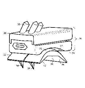
[0023] Fig. 1 is a perspective illustration of a total ankle replacement
prosthesis
including a talar dome prosthetic element 22 according to the present
disclosure. The
total ankle replacement prosthesis includes a tibial prosthesis 24 that
articulates with
the talar dome prosthesis 22. The talar and tibial prostheses 22, 24 slide
over one
another along arched interfacing surfaces 25, 27. Surfaces 25, 27 complement
one
another, each following an arch or curve around a horizontal lateral center
line. The
articulation of the prosthetic tibial and talar prostheses 24, 22 approximates
the
articulation between a natural talus and the tibiofibular joint or
syndesmosis. The
tibiofibular joint functions as a mortise for the talar dome as a tenon,
permitting ranges
- 4 -
DM2 499688 I I
CA 3042892 2019-05-10
of angular displacement. A primary displacement is dorsi-flexion/planter-
flexion (relative
rotation on a lateral horizontal axis of rotation) wherein the tibia and the
foot can be
inclined anteriorly and posteriorly relative to one another during gait.
Additionally,
inversion/eversion is a limited displacement in which the foot and tibia are
rotated
laterally inwardly or outwardly (on a medial horizontal axis) at the ankle.
Abduction/adduction is a displacement wherein the foot is aligned laterally or
medially
relative to a sagittal plane (vertical axis of rotation). It is an object of
the prosthetic
ankle to approximate the degree of freedom of displacement that is
characteristic of the
natural ankle joint.
[0024] The talar dome prosthesis 22 can be an integral forging of
surgical steel,
shaped as shown and polished on its articulating surface 25. The tibial
prosthesis 24
comprises a tibial plate 26 and a wear element 28 received therein. The wear
element
can comprise a high density polyethylene or similar material capable of
withstanding
carrying the patient's weight and sliding smoothly over the talar dome over a
long useful
life.
[0025] The tibial prosthesis 24 and the talar dome need to be permanently
and
rigidly affixed to the tibia and the talus, respectively. US 2012/0271314-
Stemniski et al.
teaches techniques for resecting a tibiofibular joint to receive a tibial
plate and resecting
the talus to receive a talar dome prosthesis, both guided using the same
navigation and
guidance fixture for controlling the paths of surgical saws, drills and
reamers applied
from the anterior and plantar sides. In the Stemniski technique, the
attachments to the
tibia include providing an intramedullar bore in the tibia for receiving an
elongated shaft
element. In the present embodiment as shown in Fig. 1, the tibial plate
carries three
anchoring pins 32 that are embedded in the bores drilled and reamed in the
resected
tibia to securely affix the tibial plate 26 and thereby to securely fix the
tibial prosthesis to
the distal tibia. The tibial plate can carry a sintered porous metal surface
(not shown) to
enhance the attachment by encouraging bone ingrowth.
[0026] It is an aspect of the present invention that the talar dome
prosthesis 22 is
attached to a resected talus by virtue of complementary surface shapes
together with a
plurality of pegs 34 that are embedded in the resected talus to secure the
talar dome
- 5 -
CA 3042892 2019-05-10
Attorney Docket No. E3383-00581
22. As shown in Figs. 1 and 2, the talar dome prosthesis 22 consists
essentially of an
integral body of material, especially surgical steel or titanium alloy. On the
underside
facing the talus, the talar dome prosthesis 22 has a plurality of shaped
surfaces 42, 44,
46 that are arranged to complement that shape of the talus after resection. A
plurality of
pegs 34 extend from the surface and are embedded in the talus to hold the
talar dome
prosthesis 22 securely in place when installed. In this embodiment the pegs
are integral
with the dome body and have a polygonal cross section tapering to a distal
point.
[0027] The total ankle replacement (ankle arthroplasty) prosthesis
comprises a
talar dome prosthesis 22 configured for affixation to a talus bone. The talar
dome 22
has a dome body with an articulating side 25 (the top side in Fig. 1) for
bearing toward
the tibia and a mounting side (shown in Fig. 2) for attachment to the talus
bone, namely
the underside. The mounting side has plural flat sections, the anterior one of
which flat
sections carries at least two pegs 34 rigidly protruding from the dome body.
The pegs
34 are each tapered to a point for embedment in the talus bone for operatively
attaching
the talar dome to the talus bone.
[0028] The mounting side of the talar dome 22 as shown in Fig. 2 has
three flat
surfaces 42, 44, 46 that are angularly inclined relative to one another. The
central flat
surface 44 is aligned substantially horizontal and rests on the top surface of
a talus that
has been re-sected by being sawn off horizontally. The anterior and posterior
surfaces
42, 46 are inclined inferiorly from the horizontal central surface 44, namely
downwardly
toward the front and rear edges, respectively, and rest on complementary
resected flat
surfaces of the talus. The structure forms the underside of the talar dome
prosthesis 22
into a faceted female cup shape that fits in surface contact with the faceted
resected
talus. Due to vertically downward pressure from the weight of the patient on
the ankle,
the talar dome is held against the talus. The inclined anterior and posterior
facet
surfaces 42, 46 contribute to holding the talar dome 22 in place on the talus
and the
pegs 34 further maintain the position of the talar dome 22.
[0029] In the embodiment shown, the pegs 34 extend perpendicularly from
the
anterior-flat section. As a result, the pegs extend are obliquely inclined
relative to
horizontal, downwardly and toward the rear of the talus bone. With a nominal
gait,
pushing off with some degree of dorsi-flexion applies the patient's weight in
a direction
- 6 -
DM2\499688I 1
CA 3042892 2019-05-10
Attorney Docket No. E3383-00581
more or less parallel to the longitudinal axes of the pegs 34. Stepping
forward into
plantar-flexion causes the wear element 28 to slide anteriorly over the talar
dome sliding
surface 25 in a direction corresponding to the insertion direction of pegs 34,
which is
partly posteriorly.
[0030] For providing complementary surfaces on the talus and the talar
dome 22,
the talus is resected during the surgical process ankle arthroplasty. In the
medial side
elevation view of Fig. 3, a human talus 50 is shown with planes marked for
resection.
Three saw cuts are made, preferably guided by an alignment and navigation
fixture with
appropriate guides for the angle and transit of the surgical saw, along the
lines 52, 54
and 56. These lines define planes at angles that complement the angles of
surfaces 42,
44, 46 on the underside of the talar dome prosthesis 22. Each cut removes the
bone
tissue superior to the cut line, leaving a resected talus as shown in an
anterior side
elevation view in Fig. 4, where the bone surfaces 52 and 54 are seen obliquely
or edge-
on. Whereas the saw cuts are controlled by the alignment/navigation feature to
precisely match the dimensions and angles of surfaces 42, 44, 46 of talar dome
22, the
talar dome fits precisely on the talus, but for the pointed pegs 34.
[0031] Fig. 5 is a schematic illustration, the talar dome shown in
section along
line 5-5 in Fig. 2. This figure shows surfaces 42, 44, 46 of the talar dome
prosthesis 22
abutting directly against resected surfaces 52, 54, 56 of talus 50. The
surface contact is
such that the peg 34 is fully embedded in talus 50, up to the inclined surface
42 from
which peg 34 protrudes.
[0032] Fig. 6 is a side elevation of the talar dome prosthesis 22, with
an inset
showing the shape and orientation of one of the affixation pegs 34 of the
prosthesis 22.
In this embodiment the peg 34 is triangular in cross section as shown,
tapering on three
sides to a point. In this embodiment the peg 34 is a regular tetrahedron.
Alternative
shapes are possible, some being shown in Figs. 9-11 including other tapered
pegs with
polygonal cross section. An advantageous aspect of a polygonal cross section,
including the embodiment in Fig. 6, is that the posteriorly facing side 37 of
the peg 34 is
perpendicular to the sagittal plane. This orientation provides maximum
opposition to
forces arising parallel to the sagittal plane in walking. In Fig. 6, the
triangular cross
section defines a posterior flat side and an anterior edge. In other
alternatives shown in
- 7 -
DM2\4996881
CA 3042892 2019-05-10
Attorney Docket No. E3383-00581
Fig. 9, both the anterior and posterior sides can be perpendicular to the
sagittal plane,
e.g., in a peg shaped as a pyramid with a square bottom or in other polygons
with an
even number of sides (four, six, eight, etc.). The triangular cross section of
a
tetrahedron or the square cross section of a square bottom pyramid are the
least
complicated to manufacture.
[0033] For manufacturing, an integral talar dome element is provided in the
general shape shown, for example cast in one piece. The bottom surfaces can be
machine to flat precision at the necessary angles. The sliding upper surface
25 is
polished. The sides of pegs 34 are polished. A porous coating such as sintered
titanium alloy particles as in Wright Medical Technology BIOFOAM (not shown)
can be
applied to surfaces 42, 44, 46 to improve prospects for bone ingrowth. It
would be
possible to likewise apply a porous coating to the peg 34, but in general a
smooth peg is
more readily driven into the talus 50 than a peg thickened by a porous
coating.
[0034] Fig. 7 is a schematic illustration showing driving the talar dome
prosthesis
22 into the talus 50 such that the respective faces 42 etc. of the prosthesis
and 52 etc.
of the talus are brought into surface abutment. A pilot hole can be drilled at
line 62 to
predetermine the point of entry. The talar dome prosthesis 22 is positioned
anteriorly of
its final position by a distance that accounts for the posterior/inferior
orientation of the
peg 34. Preferably, forming the pilot hole at line 62 and the placement and
orientation
of the talar dome prosthesis are determined by the alignment and navigation
fixture.
[0035] The prosthesis 22 is driven home with a mallet 64 or other similar
tool,
which preferably is faced with a polymer material so as not to mar the sliding
surface
25. This places the talar dome prosthesis 22 in its final position shown in
Fig. 8. It may
be noted that the underside of prosthesis 22 conforms exactly to the facing
surfaces of
the resected talus. The prosthesis preferably resides exclusively on top of
the talus 50,
wrapping over the anterior and posterior but not having lateral and/or medial
side
flanges depending downwardly. Omitting the side flanges provides for clear
fluoroscopic visualization and avoids any need to trim the lateral and medial
sides of the
talus to accommodate prosthesis 22.
[0036] In Fig. 6, the pegs 34 form tetrahedral pyramids with a triangular
cross
section and triangular base forming an equilateral triangle. The pegs extend
- 8 -
DM2\4996881
CA 3042892 2019-05-10
Attorney Docket No. E3383-00581
substantially perpendicularly from flat section 42 on the mounting side of the
prosthetic
dome body 22. Variations are possible in peg shape and orientation. For
example, the
pegs can be on an axis that is inclined relative to surface 42, the prosthesis
22 being
driven into place on a line parallel to the peg axis instead of perpendicular
to surface 42.
[0037] In Fig. 9, a series of perspective phantom illustrations show
advantageous
shapes for the affixation pegs 34, including a tetrahedron 72, a square base
pyramid 74
and a polygonal structure with an even number of sides, for example a
hexagonal
pyramid 76. As previously noted, one of the sides is preferably oriented on
the posterior
side perpendicular to the sagittal plane. The peg shapes are shown separately
in Fig.
9, and could comprise one or more parts that are attached to surface 42 of
prosthesis
22. But preferably pegs 34 are formed integrally in one piece with the
remainder of
prosthesis 22 from their bases to their pointed tips. The center axes of the
pegs are
oriented perpendicular to the plane of surface 42.
[0038] As shown in Fig. 10, the pegs are not required to be entirely
smooth. A
trapezoidal peg 72 in Fig. 10 is provided with serrations 77 along the edges
at which the
faces meet. The serrations are useful to lock the prosthesis in place after a
period of
healing, due to ingrowth of bone tissue into serrations 77. In an alternative
embodiment
in Fig. 11, the faces of a peg are shown with grooves 78, forming an
alternative type of
serration that likewise locks the prosthesis 22 in place after a period of
bone ingrowth.
[0039] The pegs are capable of embodiment in shapes other than
polyhedrons,
especially pyramids with sides that meet at three or four edges, at least one
being
perpendicular to the sagittal plane. In the depicted embodiment, the pegs are
obliquely
inclined relative to horizontal, downwardly and toward a rear of the talus
bone, because
the longitudinal axes of the pegs are perpendicular to the plane defined by
the anterior
flat section 42, which is inclined downwardly (inferiorly) toward the anterior
edge of the
talar dome prosthesis.
[0040] Flat section or face 42 is one of a plurality of flat faces on the
underside of
the dome body, configured to abut against a resected surface of the talus
having a
shape that is complementary with the underside, namely cut or machined away to
define the same sequence of flat faces. The underside of the dome body is
fully defined
by the flat faces. That is, the prosthesis lacks depending lateral and medial
flanges,
- 9
DM214996881 I
CA 3042892 2019-05-10
Attorney Docket No. E3383-00581
instead wrapping over the top of the resected talus and defining the smooth
and
rounded articulating upper surface in an arc over the talus, for bearing
against the tibial
prosthesis. The talar dome is formed as an one-piece part with the dome body
and
pegs being integral with one another, being readily manufactured, robust and
structurally uncomplicated.
[0041] The invention has been disclosed in connection with a number of
variations presented as examples. However the invention is not limited to the
exemplary embodiments and is capable of additional variations. Reference
should be
made to the appended claims instead of the foregoing description, to assess
the scope
of exclusive rights in the invention claimed.
- 10 -
DM2\499688I 1
CA 3042892 2019-05-10