Note: Descriptions are shown in the official language in which they were submitted.
CA 03043708 2019-05-10
WO 2018/094194 PCT/US2017/062266
CHIP PLATFORMS FOR MICROARRAY 3D BIOPRINTING
CROSS REFERENCE TO RELATED APPLICATIONS
[0001] This application claims the benefit of Provisional Application No.
62/423,586 filed on
November 17, 2016, and is hereby incorporated by reference in its entirety
into this application.
BACKGROUND
[0002] When developing therapeutic drugs, it is important to determine a
drug's safety and
efficacy. In the relatively early stages of drug development, drug safety and
efficacy is often
tested outside the living organism ("in vitro"). The in vitro assays currently
available, however¨
using 2D cell monolayers or 3D cell spheroids¨do not adequately mimic how
drugs act in the
living organism ("in vivo"). Thus, an in vitro cell/tissue model that can
closely mimic the
corresponding tissues in vivo and systematically simulate diseases is desired.
[0003] 3D bioprinting is a promising technology in this regard. Generally, 3D
bioprinting
refers to robotically dispensing cells layer-by-layer in hydrogels, thus
creating relatively large
scale tissue constructs that more accurately mimic the in vivo environment.
But because the
tissue constructs are generally on a large scale, 3D bioprinting is not ideal
for high throughput
testing, and is thus limited as an alternative to the currently available in
vitro assays. Recently,
however, a method of microarray 3D bioprinting was developed, which allows for
high
throughput testing.
[0004] Microarray 3D bioprinting refers to dispensing very small amounts of
cells along with
other biological samples such as hydrogels, growth factors, extracellular
matrices, biomolecules,
drugs, DNAs, RNAs, viruses, bacteria, growth media, or combinations thereof,
on a
microwell/micropillar chip platform using a microarray spotter and then
incubating the cells to
create a mini-bioconstruct. This technology can potentially revolutionize
tissue engineering and
disease modeling for screening therapeutic drugs and studying toxicology.
[0005] Since microwell/micropillar chip platforms (also known as "microarray
biochips")
contain arrays of up to 5,000 microwells/micropillars, this method is ideal
for high throughput
testing. However, the currently available microwell/micropillar chips are not
ideal for
microarray 3D bioprinting due to the limited space available on the
micropillar chip or limited
control of individual experimental conditions in the microwell chip.
1
CA 03043708 2019-05-10
WO 2018/094194 PCT/US2017/062266
[0006] For example, currently available micropillar chips use pillars with
flat tops, which are
not conducive to dispensing cells layer-by-layer. Thus, it is difficult to
carry out 3D bioprinting
on micropillar chips. In addition, the currently available microwell chips use
wells that trap air
bubbles in the hydrogel as the cell layers are printed. In addition, it is
difficult to control each
bioprinted tissue construct individually in the microwell chip because all
tissue constructs in the
microwell chip should be immersed in a petri dish with a universal growth
medium. Thus, there
is a need for designing a new structure of microwells and micropillars on a
chip that can
facilitate layered cell printing on both the pillar and well, ensure robust
cell spot attachment for
high-content imaging and immunofluorescent assays, and avoid air bubble
entrapment for robust
3D cell/tissue cultures. The new chip design can be compatible with
conventional microtiter
plates, including 96-, 384-, and 1536-well plates.
SUMMARY
[0007] The present invention is directed to a micropillar chip and a microwell
chip that
facilitates layered cell printing on both the pillar and well, ensures robust
cell spot attachment for
high-content imaging and immunofluorescent assays, and avoids air bubble
entrapment. The
present invention is further directed to methods using the micropillar and
microwell chips to
create miniature multicellular biological constructs.
[0008] The micropillar chip comprises a chip base with at least one
micropillar. The
micropillar, rather than having a flat top, has a pillar-microwell at its top
end. The pillar-
microwell comprises a pillar-microwell base and a side wall extending upwardly
from the base.
[0009] The microwell chip comprises at least one microwell that, unlike
conventional
microwells, has an upper and lower microwell.
[0010] The method of creating a miniature multicellular biological construct
comprises
depositing cells into a pillar-microwell, exposing the pillar-microwell to
growth media, and
incubating the cells.
[0011] These and other features, aspects, and advantages of the general
inventive concepts will
become better understood with reference to the following description and
appended claims.
BRIEF DESCRIPTION OF THE DRAWINGS
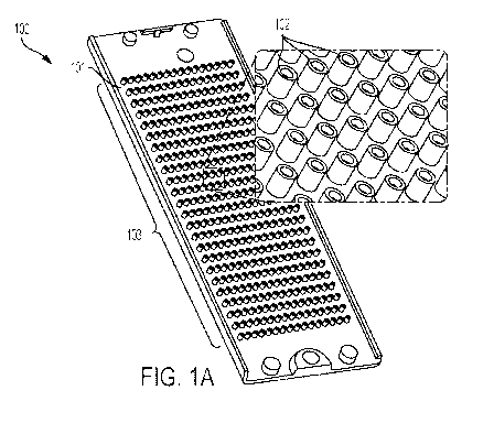
[0012] Fig. 1A shows an embodiment of a micropillar chip.
[0013] Fig. 1B shows an embodiment of a microwell chip.
[0014] Fig. 2A shows a sectional view of embodiments of micropillars.
2
CA 03043708 2019-05-10
WO 2018/094194 PCT/US2017/062266
[0015] Fig. 2B shows a perspective view of embodiments of micropillars.
[0016] Fig. 2C shows a perspective view of embodiments of micropillars.
[0017] Fig. 3 is a sectional view of embodiments of micropillars.
[0018] Fig. 4 is a sectional view of embodiments of microwells.
[0019] Fig. 5A is a sectional view of embodiments of micropillars with pillar-
microwells
containing cells.
[0020] Fig. 5B is a sectional view of embodiments of micropillars with pillar-
microwells
containing cells sandwiched with a microtiter plate containing growth media.
[0021] Figs. 6A shows a sectional view of embodiments of micropillars and a
microtiter plate.
[0022] Fig. 6B shows a sectional view of embodiments of micropillars
sandwiched with a
microtiter plate.
[0023] Fig. 6C shows a sectional view of embodiments of micropillars and a
microtiter plate.
[0024] Fig. 6D shows a sectional view of embodiments of micropillars
sandwiched with a
microtiter plate.
[0025] Fig. 7A shows an embodiment of a perfusion channel chip.
[0026] Fig. 7B shows a blown-up section of Fig. 7A.
[0027] Fig. 7C shows an exploded view of a micropillar paired with a perfusion
channel chip.
[0028] Fig. 7D shows a blown-up section of Fig. 7C.
[0029] Fig. 8 shows embodiments of perfusion channel chips, micropillar chips,
and reservoir
chips.
[0030] Fig. 9A shows a cross-sectional view of an embodiment of a micropillar
chip
sandwiched with an embodiment of a perfusion channel chip.
[0031] Fig. 9B shows a top view of an embodiment of a perfusion channel chip.
[0032] Fig. 10 is an image of a cell-stained mini-bioconstruct.
[0033] Fig. 11 illustrates the surface chemistry of an embodiment of a
functionalized
micropillar.
[0034] Fig. 12 shows a flowchart demonstrating the method used in Example 3.
[0035] Fig. 13A is an image of Hep3B cells in a micropillar/microwell chip.
[0036] Fig. 13B is images of Hep3B cells in a micropillar/microwell chip.
[0037] Fig. 14 are images of Hep3B cells in a micropillar/microwell chip.
[0038] Fig. 15 shows a flowchart demonstrating the method used in Example 5.
3
CA 03043708 2019-05-10
WO 2018/094194 PCT/US2017/062266
[0039] Fig. 16 is images of stained neural progenitor cells.
[0040] Fig. 17A shows a flowchart demonstrating an embodiment of a method of
attaching
antibodies to micropillars.
[0041] Fig. 17B shows a flowchart demonstrating an embodiment of a method of
attaching
antibodies to micropillars.
[0042] Fig. 18A illustrates the surface chemistry of an embodiment of a method
of attaching
antibodies to micropillars.
[0043] Fig. 18B illustrates the surface chemistry of an embodiment of a method
of attaching
antibodies to micropillars.
[0044] 18C illustrates the surface chemistry of an embodiment of a method of
attaching
antibodies to micropillars.
[0045] Fig. 19 shows a flowchart demonstrating an embodiment of a method of
detecting
biomarkers.
[0046] Fig. 20 shows a flowchart demonstrating an embodiment of a method of
measuring
changes in cell surface markers.
[0047] Fig. 21 shows a flowchart demonstrating an embodiment of a method of
quantifying
cancer cell migration and images of stained Hep3B cells.
[0048] Figs. 22A shows a sectional view of an embodiment of a microwell chip.
[0049] and 22B shows a sectional view of an embodiment of a microwell chip.
DETAILED DESCRIPTION
[0050] While various exemplary embodiments and methods are described herein,
other
embodiments, methods, and materials similar or equivalent to those described
herein are
encompassed by the general inventive concepts. All references cited herein,
including published
or corresponding U.S. or foreign patent applications, issued U.S. or foreign
patents, and any
other references, are each incorporated herein by reference in their
entireties, including all data,
tables, figures, and text presented in the cited references.
4
CA 03043708 2019-05-10
WO 2018/094194 PCT/US2017/062266
[0051] Unless defined otherwise, all technical and scientific terms used
herein have the same
meaning as commonly understood by one of ordinary skill in the art to which
the invention
belongs.
[0052] All percentages, parts, and ratios as used herein are by weight of the
total formulation,
unless otherwise specified. All such weights as they pertain to listed
ingredients are based on the
active level and, therefore, do not include solvents or by-products that may
be included in
commercially available materials, unless otherwise specified.
[0053] All references to singular characteristics or limitations of the
present disclosure shall
include the corresponding plural characteristic or limitation, and vice versa,
unless otherwise
specified or clearly implied to the contrary by the context in which the
reference is made.
[0054] The methods and embodiments of the present disclosure can comprise,
consist of, or
consist essentially of the essential elements of the disclosure as described
herein, as well as any
additional or optional element described herein or which is otherwise useful
in carrying out the
general inventive concepts.
[0055] To the extent that the terms "includes," "including," "contains," or
"containing" are
used in the specification or the claims, they are intended to be inclusive in
a manner similar to
the term "comprising" as that term is interpreted when employed as a
transitional word in a
claim. Furthermore, to the extent that the term "or" is employed (e.g., A or
B) it is intended to
mean "A or B or both." When the applicants intend to indicate "only A or B but
not both" then
the term "only A or B but not both" will be employed. Thus, use of the term
"or" herein is the
inclusive, and not the exclusive use. Also, to the extent that the terms "in"
or "into" are used in
the specification or the claims, it is intended to additionally mean "on" or
"onto."
[0056] All combinations of method or process steps as used herein can be
performed in any
order, unless otherwise specified or clearly implied to the contrary by the
context in which the
referenced combination is made.
[0057] All ranges and parameters, including but not limited to percentages,
parts, and ratios,
disclosed herein are understood to encompass any and all sub-ranges assumed
and subsumed
therein, and every number between the endpoints. For example, a stated range
of "1 to 10"
should be considered to include any and all sub-ranges beginning with a
minimum value of 1 or
more and ending with a maximum value of 10 or less (e.g., 1 to 6.1, or 2.3 to
9.4), and to each
integer (1, 2, 3, 4, 5, 6, 7, 8, 9, and 10) contained within the range.
CA 03043708 2019-05-10
WO 2018/094194 PCT/US2017/062266
[0058] The general inventive concepts are directed to micropillar and
microwell chips for
microarray analysis that facilitate layered cell printing on both the
micropillar and in the
microwell. The micropillar and microwell chips ensure robust cell spot
attachment for high-
content imaging and immunofluorescent assays, and avoid air bubble entrapment.
The general
inventive concepts also contemplate methods of creating and analyzing
miniature multicellular
biological constructs ("mini-bioconstructs") using the inventive micropillar
and microwell chips.
[0059] Conventional microarray biochips are designed so that the micropillar
chip mates with
the microwell chip. The micropillars are sized so that they may be inserted
into the
corresponding microwells. The micropillar and microwell chips of this
invention may be
compatible with each other and with conventional micropillar and microwell
chips or microtiter
plates. For example, the inventive micropillar chip may be compatible with a
conventional
microwell chip and conventional microtiter plates and the inventive microwell
plates, and the
inventive microwell chip may be compatible with a conventional micropillar
chip and the
inventive micropillar chip. An exemplary conventional micropillar/microwell
chip is made by
Samsung Electro Mechanics, Co. and MBD Korea (e.g., S+ Microwell Chip).
Exemplary
conventional microtiter plates, including 96-, 384-, 1536-, and 3456-well
plates are made by
Corning and other manufacturers.
[0060] The inventive chips may be made of a biocompatible polymer. The
biocompatible
polymer may be clear or opaque depending on the type of analysis to be
performed. For
example, in some exemplary embodiments, the chip may be made of clear
polystyrene. In some
further exemplary embodiments, the chip may be made of functional poly(styrene-
co-maleic
anhydride). The chip may be manufactured using any conventional manufacturing
process,
including 3D printing.
[0061] Referring to Figure 1, the inventive micropillar chip 100 comprises a
chip base 101 and
at least one micropillar 102. In some exemplary embodiments, the micropillar
chip contains
arrays of micropillars 103, for example, about 90 to about 5,000 micropillars.
The micropillar
102 may be any shape depending on the needs of the test. For example, the
micropillar may be
cylindrical 201 or it may be square 202. An embodiment of a plate containing
an array of 384
pillars is depicted in Figure 1.
[0062] In some exemplary embodiments, the micropillar 102 is from about 0.3 ¨
5 mm in
width, about 0.3 ¨ 5 mm in length, and about 1 ¨ 20 mm in height. In some
further exemplary
6
CA 03043708 2019-05-10
WO 2018/094194 PCT/US2017/062266
embodiments, the micropillar 102 may be from about 0.3 ¨ 5 mm in diameter and
1 ¨ 20 mm in
height. For example, a micropillar 102 may be 2.6 mm in diameter and 13.5 mm
in height.
[0063] Referring to Figure 2, unlike conventional micropillars that have flat
tops, the inventive
micropillar 102 comprises a top end 203 that contains a pillar-microwell 204.
The pillar-
microwell 204 is a reservoir with a pillar-microwell base 205 and at least one
sidewall 206. The
pillar-microwell may extend from the top end 203 of the micropillar to the
pillar-microwell base
205. The pillar-microwell base may be anywhere between the chip base 101 and
pillar top end
203. For example, the pillar-microwell may be capable of holding any volume of
sample,
including 1 ¨ 4 L. The sidewall may be anywhere from about 0.5 ¨ 5 mm in
height and about
0.3 - 1 mm in thickness. The pillar-microwell sidewall 206 facilitates layer-
by-layer cellular
printing and robust cell spot attachment.
[0064] In some exemplary embodiments, the micropillar chip contains a means
for minimizing
air bubble entrapment. For example, in some exemplary embodiments, the pillar-
microwell
sidewall 206 may contain at least one slit 207. The slit 207 is a gap in the
sidewall that extends
at least partway through the width of the sidewall. In some further exemplary
embodiments, the
pillar-microwell sidewall may contain 1 ¨ 5 slits 207, or more.
[0065] Referring to Figure 3, in another exemplary embodiment in which the
micropillar chip
contains a means for minimizing air bubble entrapment, the micropillar 102
contains a bore 301
that extends from the pillar-microwell base 205 at least partially through the
micropillar. In
some exemplary embodiments, the diameter of the bore may be less than the
diameter of the
micropillar. For example, in some exemplary embodiments, the diameter of the
bore may be, but
is not limited to, 0.4 mm for a pillar with a diameter of 2 mm, or the
diameter of the bore may be,
but is not limited to, 1 mm for a pillar with a diameter of 5 mm.
[0066] Further, in some exemplary embodiments, the pillar-microwell base 205
may be plasma
treated or coated with functional polymers to enhance robust cell spot
attachment. Exemplary
functional polymers include, but are not limited to, poly(maleic anhydride-alt-
1-octadecene)
(PMA-OD), poly(maleic anhydride-alt-l-tetradecene) (PMA-TD), polyethylene
oxide-maleic
anhydride copolymers, including ACM1510, ADM1510, AEM1510, AKM0530, and
AKM1510,
poly-L-lysine (PLL), barium chloride (BaC12), calcium chloride (CaCl2)
collagen, PuraMatrix,
fibrinogen, fibronectin, and Matrigel.
7
CA 03043708 2019-05-10
WO 2018/094194 PCT/US2017/062266
[0067] Referring to Figure 4, the inventive microwell chip 104 comprises at
least one
microwell 105. In some exemplary embodiments, the microwell chip may contain
an array of
microwells 106, for example, about 90 to about 5,000 microwells.
[0068] Unlike conventional microwells, the inventive microwell 105 comprises
an upper
microwell 401 and at least one lower microwell 402. The lower microwell may
extend generally
downward from the upper microwell base 405. The upper and lower microwells may
be in fluid
communication.
[0069] The upper 401 and lower 402 microwells may be any shape depending on
the needs of
the test. For example, the microwells may be cylindrical 403 or square 404. In
some exemplary
embodiments, the upper microwell 401 is from about 0.3 ¨ 100 mm in width,
about 0.3 ¨ 100
mm in length, and about 0.3 ¨ 100 mm in height. In some further exemplary
embodiments, the
upper microwell 401 may be from about 0.3 ¨ 100 mm in diameter and 0.3 ¨ 100
mm in height.
In some further exemplary embodiments, the upper microwell may be about 1.2 mm
in diameter
and about 1.5 mm in height. The lower microwell 402 may be smaller than the
upper microwell
in either width, length, or diameter, depending on the shape.
[0070] In some exemplary embodiments, the lower microwell 402 contains a means
for
minimizing air bubble entrapment. For example, in one exemplary embodiment, at
least one
peripheral channel 406 extends vertically along the periphery of the lower
microwell 402. The
dimensions of the peripheral channel 406 may vary in size and shape. For
example, the
peripheral channel may be rectangular or cylindrical. The peripheral channel
may extend from
the upper microwell base 405 to the bottom of the lower microwell.
[0071] In some further exemplary embodiments, the lower microwell may be
plasma treated or
coated with functional polymers to enhance robust cell spot attachment.
[0072] Referring to Figures 5 ¨ 9, the inventive micropillar and microwell
chips enable several
inventive methods for microarray 3D bioprinting. One exemplary method
generally comprises
dispensing cells 501 into at least one pillar-microwell 204 and incubating the
cells to create a
desired mini-bioconstruct. In some exemplary embodiments, the mini-
bioconstructs may be
created to mimic particular tissues such as, but not limited to, a heart,
liver, or brain. For
example, human liver tissue constructs may be created by printing primary
hepatocytes/HepaRG,
hepatic sinusoidal endothelial cells, hepatic stellate cells, and Kupffer
cells layer-by-layer in
collagen to maintain liver-specific functions. Also, for example, human brain
tissues can be
8
CA 03043708 2019-05-10
WO 2018/094194 PCT/US2017/062266
generated by printing neural stem cells in Matrigel and differentiating into
different neural
lineages for several months.
[0073] In some exemplary methods, cells 501 are dispensed into the pillar-
microwell 204 by a
microarray spotter 502. A microarray spotter 502 is a robotic device capable
of dispensing small
amounts of liquid, also known as "spots." In some exemplary methods, the
microarray spotter
502 may be capable of printing spots into multiple pillar-microwells 204 on
the same micropillar
chip 100. The microarray spotter may be capable of printing from about 20 nL
to about 3000 nL
of cells into the pillar-microwells 204. Exemplary microarray spotters include
S+ MicroArrayer,
commercially available from Samsung, and MBD Korea, as well as MicroSys,
PixSys, and
CellJet from DigiLab.
[0074] In some exemplary methods, prior to dispensing cells, a cell suspension
may be made
comprising the cells, at least one hydrogel, and growth media. Optionally, one
or more
biomolecules, drugs, DNAs, RNAs, proteins, bacteria, viruses, or combinations
thereof may be
included in the cell suspension. For example, the biomolecules, drugs, DNAs,
RNAs, proteins,
bacteria, viruses, or combinations thereof may be chosen to mimic a particular
biological
environment, such as particular tissue (liver, heart, brain, etc.).
[0075] A hydrogel is generally a polymer that contains water. For example,
suitable hydrogels
may be alginate, methacrylated alginate, chitosan, hyaluronic acid,
fibrinogen, collagen,
methacrylated collagen, PuraMatrix, Matrigel, PepGel, and polyethylene glycol.
The cells may
be entrapped in a hydrogel using various mechanisms such as, but not limited
to, ionic, photo,
enzymatic, and chemical crosslinking. Crosslinking agents may include salts or
enzymes that
facilitate gelling of the hydrogel. Examples of suitable crosslinking
mechanisms include ionic
crosslinking (e.g., alginate with barium chloride and calcium chloride;
PuraMatrix with salts),
affinity/covalent bonding (e.g., functionalized polymers with streptavidin and
biotin),
photopolymerization (e.g., methacrylated alginate with photoinitiators), and
biocatalysis (e.g.,
fibrinogen with thrombin).
[0076] The cell suspension concentration may be from about 10,000 to about 20
million
cells/mL, about 500,000 to about 5 million cells/mL, or about 1 million to
about 2 million
cells/mL. The growth media may be from about 90 w/v% to about 99.9 w/v% of the
final cell
suspension. The hydrogel may be from about 0.1 w/v% to about 10 w/v% of the
final cell-
suspension.
9
CA 03043708 2019-05-10
WO 2018/094194 PCT/US2017/062266
[0077] Growth media is generally a liquid designed to support cell growth.
Suitable examples
of growth media may include Dulbecco's Modified Eagle Medium (DMEM), Roswell
Park
Memorial Institute Medium (RPMI), and William's E Medium. Biomolecules may
include
molecules that support cellular or tissue growth, such as extracellular
matrices (ECMs), growth
factors, compounds, cytokines, and carbohydrates.
[0078] In some further exemplary methods, prior to dispensing the cells with
the microarray
spotter 502, the pillar-microwells 204 are treated with plasma or coated with
functional polymers
for cell spot attachment and hydrogel gelation.
[0079] Referring to Figures 6A ¨ 6D, in some exemplary methods, rather than
dispense the
cells 501 into pillar-microwells 204 with a microarray spotter 502, pillar-
microwells may be
treated with functional polymers and then submerged in a conventional
microtiter plate 505 that
contains cells suspended in a hydrogel 602, such as alginate. When the pillar-
microwells 204 on
the micropillar chip 100 are submerged 603 into the hydrogel 602, the pillar-
microwells entrap a
portion of the hydrogel 604, so that when the micropillar chip is removed ,
the pillar-microwells
204 contain a portion of the hydrogel 604. In this method, the volume of cells
entrapped in the
pillar-microwell may be controlled by the surface area of the pillar-microwell
base 205, side
walls 206, and slits 207.
[0080] In some exemplary methods, once the pillar-microwell 204 contains the
desired cells,
the micropillar plate 100 may be incubated. In some exemplary methods, the
pillar-microwell
204 may be exposed to growth media 504 for incubation. And in some further
exemplary
methods, the pillar-microwell may be submerged in a conventional microtiter
plate 505 that
contains growth media 504 for cell culture, as shown in Fig. 6D. Submerging
the pillar-
microwells 204 in conventional microtiter plates 505 containing cell growth
media is an
improvement over the current state of the art because it allows for simply
changing the growth
media without disturbing the cell layers with a microplate washer dispenser.
[0081] Referring to Figs. 7 ¨ 9, in some exemplary methods, the micropillar
chip may be
incubated by submerging the pillar-microwells 204 in a perfusion channel chip
701 containing
growth media 504. This method may, for example, be used for long term cultures
and may
mimic circulatory systems to study, for example, organ-organ interactions.
Figures 7A ¨ 7D,
illustrate an embodiment of a perfusion channel chip 701. The perfusion
channel chip may
comprise one or more compartments 702 containing one or more channels 703.
Further, the
CA 03043708 2019-05-10
WO 2018/094194 PCT/US2017/062266
perfusion channel chip 701 may separate one or more of the compartments 702
with a porous
membrane cassette 704. The one or more compartments 702 may contain growth
media 504
containing test compounds, biomolecules, drugs, DNAs, RNAs, proteins,
bacteria, viruses, or
combinations thereof that may flow through or reside in the one or more
channels 703. For
example, an embodiment of the perfusion channel chip 701 may contain one
compartment for
liver co-cultures, one compartment for brain cell co-cultures and a porous
membrane cassette
704 simulating the blood brain barrier. As shown in Fig. 7C and 7D, a
micropillar chip 100
containing pillar-microwells 204 or conventional pillars may be sandwiched
with the perfusion
channel chip 701 so that the contents on the pillar or in the pillar-microwell
204 may be in
contact with the growth media 504 in the channel 703. As shown in Figure 9B,
the perfusion
channel chip may contain pillar insertion holes 901 through which a pillar or
pillar-microwell
204 may be inserted.
[0082] Referring to Figure 8, in some further exemplary embodiments using a
perfusion
channel chip 701, one or more micropumps 804 may be integrated with the
perfusion channel
chip to circulate the growth media 504. In some further exemplary embodiments,
the growth
media may be circulated from reservoirs 803. In some further exemplary
embodiments, a
reservoir chip 802 that contains reservoir wells 803 for growth media may be
integrated with the
perfusion channel chip 701. In some further exemplary embodiments, the
reservoir chip 802
may include a sample injection hole 801 for dispensing any samples that may be
desired,
including, but not limited to, cell-staining reagents, test compounds, growth
media,
biomolecules, drugs, DNAs, RNAs, proteins, bacteria, viruses, or combinations
thereof.
[0083] Referring to Figures 22A and 22B, in some exemplary methods, cells 501
may be
dispensed into the lower microwell 402 of a microwell chip by a microarray
spotter 502. In
some exemplary methods, the cells may be entrapped in a hydrogel, and in some
exemplary
methods, more than one layer or a mixture of cells may be printed into the
lower microwell 402.
In some further exemplary embodiments, the lower microwell 402 may be treated
with
functional polymers for cell spot attachment and hydrogel gelation.
Subsequently, the cells may
be incubated by dispensing cell growth media 504 into the upper microwell 401.
[0084] In some exemplary embodiments, after a mini-bioconstruct is created, at
least one
biosample may be added. Suitable biosamples may include biomolecules, drugs,
DNAs, RNAs,
cells, growth factors, extracellular matrices, proteins, viruses, bacteria, or
combinations thereof
11
CA 03043708 2019-05-10
WO 2018/094194 PCT/US2017/062266
The at least one biosample may be chosen to mimic a particular biological
environment or
condition. In some exemplary embodiments, the at least one biosample may be
printed directly
onto the mini-bioconstruct, whether contained in a pillar-microwell 204 or in
a lower microwell
402, using the microarray spotter 502. In some further exemplary embodiments,
the at least one
biosample may be printed into the wells of a conventional microtiter plate 505
using the
microarray spotter; then the pillar-microwells 204 containing mini-
bioconstructs may be inserted
into the microtiter wells containing biosamples or other mini-bioconstructs.
[0085] In some exemplary embodiments where the mini-bioconstruct is created in
the inventive
microwell plate 104, biosamples or biomolecules may be added by sandwiching
the microwell
plate with a conventional micropillar chip that has been prepared with at
least one biosample or
biomolecule. Likewise, in some further exemplary methods, after the cells are
incubated and a
mini-bioconstruct is created on the inventive micropillar plate 100, at least
one biosample or
biomolecule may be added by sandwiching the micropillar plate 100 with a
conventional
microwell plate that has been prepared with at least one biosample or
biomolecule.
[0086] In some exemplary embodiments, in addition to attaching cell spots on
the inventive
pillar or microwell or conventional pillar or microwell, immobilized
antibodies may be attached
by using functionalization with reactive polymers for measuring soluble
biomarkers. Fig. 17A
illustrates an embodiment of this method. In some exemplary embodiments, the
surface of the
pillar-microwells 204 or conventional pillars may be coated with reactive
polymers, including,
but not limited to poly(maleic anhydride-alt-1-octadecene (PMA-OD) or
poly(styrene-co-maleic
anhydride). Then, ligands, for example, poly-L-lysine (PLL), tagged with
biotin may be
dispensed onto the surface of the coated pillar. Then, after the ligand is
immobilized, the pillars
may be rinsed to remove any unbound ligands. Next, antibodies with affinity
tags, for example,
streptavidin or biotin, may be dispensed onto the surface of the pillar so
that they interact with
the ligands immobilized on the surface of the pillar, achieving attachment of
antibodies on the
surface of the pillar.
[0087] Figs. 18A ¨ 18C illustrate the surface chemistry of an exemplary method
of attaching
immobilized antibodies to the surface of pillars. Fig. 18A demonstrates
surface chemistry of
biotin attachment on PLL. Fig. 18B illustrates the surface chemistry of
attaching biotin-
conjugated antibodies through streptavidin-biotin interactions for sandwich
ELISA assays. Fig.
18C illustrates attachment of maleimide-conjugated antibodies through Sulfo-
SMCC and thiol
12
CA 03043708 2019-05-10
WO 2018/094194 PCT/US2017/062266
reactions for sandwich ELISA assays. Sulfo-SMCC is a water-soluble
heterobifunctional protein
crosslinker. Sulfo-SMCC protein crosslinker contains an amine reactive Sulfo-
NHS ester on one
end, which increases its water solubility, and a maleimide functional group
that can be utilized to
react specifically with cysteines or sulfhydryl (-SH) groups. The maleimide
functional group
does not readily react with lysine or amino groups (-NH2), thus maleimide-
activiated conjugates
can be readily prepared for later utilization. All products in Figs. 18B and
18C are commercially
available from ThermoFisher Scientific.
[0088] Referring to Fig. 19, in some exemplary methods, the inventive or
conventional pillars
with attached immobilized antibodies may be used to detect secreted biomarkers
released by
cells by using sandwich ELISA assays. For example, in some embodiments, cells
may be
entrapped in a hydrogel and dispensed into the pillar-microwell 204 or a
conventional pillar.
Then, a compound capable of releasing soluble biomarkers (for example,
antigens such as
cytokines) in the entrapped cells may be dispensed into wells on a well plate.
Next, the pillars
may be sandwiched with the well plate containing the compound, thus allowing
soluble
biomarkers to be released by the cells. Then, a pillar plate containing the
pillars that has been
prepared with attached immobilized antibodies may be sandwiched with the well
plate
containing the soluble biomarkers. The pillars may be prepared with a variety
of antibodies
corresponding to different pillars. Then, the pillars may be removed and then
sandwiched with
wells on a well plate that contain primary antibodies with fluorescent tags,
allowing for a
sandwich ELISA assay. Sandwich ELISA assay methods are known in the art.
[0089] Referring to Fig. 20, in some exemplary methods, soluble biomarkers may
also be
measured using the inventive pillar chip or conventional pillar chip using
immunofluorescent
assays in situ. Fig. 20 illustrates an exemplary method for modulating 3D-
cultured cells with test
compounds and measuring changes in cell surface markers using
immunofluorescent assays on a
pillar chip. Antibodies with fluorescent tags such as Tyramide signal
amplification kits may be
used for labeling proteins of interest.
[0090] In some further exemplary methods, after the mini-bioconstruct is made,
it may be
examined by imaging the cells. For example, the mini-bioconstruct may be
stained with
fluorescent dyes (e.g., calcein AM, ethidium homodimer-1, Hoechst 33342, YO-
PRO-1,
propidium iodide, TMRM, fluo-4 AM, MCB, a thiol green dye), antibodies with
fluorescent tags
(e.g., Tyramide signal amplification kit), or recombinant viruses carrying
genes for biomarkers
13
CA 03043708 2019-05-10
WO 2018/094194 PCT/US2017/062266
(e.g., BactoBac baculovirus system from ThermoFisher). In some exemplary
embodiments,
the mini-bioconstruct may be imaged using a high-content imaging scanner, for
example.
Suitable imaging devices include the S+ Scanner, commercially available from
Samsung,
GenePix Scanner, commercially available from Molecular Devices, and Cellomics
Arrayscan,
commercially available from Thermo Fisher. In some further exemplary
embodiments, the
various layers of cells may be individually targeted for imaging using
different Z-focus positions.
The small size of the mini-bioconstruct allows for imaging at different Z-
focus positions.
[0091] Cells and mini-bioconstructs may be stained or otherwise prepared to
facilitate imaging,
including high-content imaging, before or after the cell-suspension is made.
For example, the
cells may be stained with fluorescent dyes that indicate certain cellular
processes. Examples of
dyes and the cellular processes that they may indicate are known in the art,
including calcein AM
and ethidium homodimer-1 for cell viability and cytotoxicity; Hoechst 33342
for changes in
nuclear function; YO-PRO-1/propidium iodide for apoptosis or necrosis;
tetramethyl rhodamine
methyl ester (TMRM) for mitochondrial membrane potential; fluo-4 AM for
intracellular
calcium levels; and monochlorobimane (MCB) and thiol green dye for glutathione
levels. Cells
and mini-bioconstructs may also be stained with recombinant viruses carrying
genes for various
fluorescent biomarkers. Exemplary recombinant viruses are baculoviruses, for
example Bac-to-
Bac baculovirus expression system from ThermoFisher. Other suitable staining
methods may
be known in the art. Examples of fluorescent biomarkers include blue
fluorescent protein (BFP),
green fluorescent protein (EGFP), orange fluorescent protein (mOrange), or red
fluorescent
protein (mCherry).
Example 1
[0092] Mini-bioconstructs were generated by printing several layers of human
cell types in
photocrosslinkable alginate with extracellular matrices and growth factors
onto a 384-pillar plate
containing the inventive pillars using a microarray spotter. Hundreds of
different biomimetic
conditions were provided in the array of inventive pillars. After gelation,
the 384-pillar plate was
sandwiched with a 384-well plate containing growth media for rapidly testing
optimum
microenvironments to create human tissue replicates. The mini-bioconstructs
were then tested
with compounds, stained with fluorescent dyes, and scanned with an automated
fluorescent
microscope for high-content imaging (HCI) of organ functions and predictive
assessment of drug
toxicity. Fig. 10 is an example of an image analysis of the mini-
bioconstructs.
14
CA 03043708 2019-05-10
WO 2018/094194 PCT/US2017/062266
Example 2
[0093] Referring to Table 1 below, various inventive pillar structures 204
were tested to
analyze the volume of sample that could be loaded into the pillar-microwells
depending on
sidewall height and number of slits. Inventive pillars of varying sidewall
height and number of
slits were first coated with 0.01% PMA-OD and dried. Next, 0.05 mg/mL
fluorescein
isothiocyanate (FITC) dissolved in Dulbecco's phosphate-buffered saline (DPBS)
was added in a
384-well plate. The pillar-plate was then sandwiched with the well plate and
shaken for 1 hour.
Next, the pillar-plate was removed and inserted into a 384-well plate
containing 50 IAL of DPBS.
Then the fluorescent intensities were measured by a plate reader and the FITC
volume in the
pillar-microwells was back calculated using the calibration curve. The results
are shown in
Table 1.
Table 1
.',,k:s. =,,,-..'s, = ',1=:,,,,,, ,:s:,,,,N
õ:,õ:NN.,,,,,õõ1 ;-,,,,:õ....,,, ::,...:,õ,,M,,,,,,,õ1
%t;4I0037..0i6i.:.:iliiiiiiiiiiiiiiiii:.:4:.:.iii:.:.:4.:.:.:.iiiiiiiii
I fRirT7 in iii:l.:,iii iiingõ..li:iiiiiiiiii]
iiiiiiiiiiklnigqii.:11W7iiigoiiiiiiiiiiiiiihi*IiiiiIiiiiiiiiii
N iiii:''':::* .::iiiiiiiiiiiiiiiiiiiiii
......... .................,.... ili:V4Wiii ............ -Y.%
::1Iiii:i: ........ ..............
"4,5 =:=:=:=:=:=:=:=:=:=:=:=:=:=:=: i0004.iiziw.011W
ii'..ii'4.*ii
-...=== qPittingaiNIPN0iiiiii]
Titmi: ............ .. ........ ........ .
iliiii.miii ............ . :. ... ...
i0.471.).i*ix.f.40. ig.r,,.,:ii
7 ,. c:]iiiiMirrilliiiiiiiiillOPOill itifiliMiliiiiii,01111111
IMIIIIIIIIii111111
I0.:.3.1`.9*0:;210. =0$...X:V.k 4M' .00.10.*00* 40M* '::M'
Ø0.'i...::#.00:4* f#M4i,
. =
ii*x*x*x*iµm.===============================================:.:.:.:.:.:.:......
...............
...............................................................................
.................................... ......... ...:.:.:.:.:.:.:
.
0242iititliiiiiiiiiiiiiiiiiOnWiiiiiiii iiiiiiiiiiii'.iiiii0iii:iiiiiiiiii
i..Ø2fii zi .,0-8.2:.
liA.i:Mki -........... .................. .......
.......................... .Wii ::....:::.....:..:....::. ... .. ....:
::: :.;:....:..:.;..::::::
1.(.;19..*#.4.4.4 =41..;.".* .:¨.:.:.:.:.:.:.: 4..,impw91Fp
.i*f. J.kii .:.:.:.:.:.:.................. iiR41t*ii4:4).!#ii
k.
iAiiiiitkiMiiiiiiiiiNiggiiiiiiiiiiiii
iiiiiiiiiiMMiWigNMEgOMMEN.Iiiiiiiiiii iiiiiiiiiiiiiiiiiiiiiiiiiiiiiiiiiiiiik
...... ..:=:iiiiiiiiiiii
IiiiiiiiiiiiiiiiiiiiiiiiikgROMMEMigiMiiiiiiiiiiiiiii
õ,,,,, ........ imm.744.429ii=i imiosii
0
Example 3
CA 03043708 2019-05-10
WO 2018/094194 PCT/US2017/062266
[0094] Figs. 11 and 12 and Table 2 are referenced in Example 3. In some
embodiments, the
surface of the inventive pillar-microwell 204 or microwell 105 may be
functionalized to facilitate
robust cell-spot attachment. For example, a 384-pillar plate with embodiments
of the inventive
pillars 102 (1.5 mm sidewall height, 0.6 mm slit size, and 4 slits) was coated
with poly(maleic
anhydride-alt-1-octadecene) (PMA-OD) to enable covalent attachment of ligands,
including
poly-L-lysine (PLL). PLL is positively charged, which allows ionic attachment
to the negatively
charged alginate. 1500 nL of 25 mM CaCl2 and 0.0033% PLL were printed in the
pillar-
microwells and allowed to dry. Next, 1500 nL of varying concentrations of cell
suspensions
containing alginate were printed in the pillar-microwells (0, 0.5, 1, 2, 4,
and 6 million cells/mL).
50 !IL of growth media was then dispensed into the microwells of a 384-well
plate. Next, the
pillars containing the varying concentrations of cell suspensions were
sandwiched into the 384-
well plate and left overnight for incubation. After incubation, the pillars
were removed and then
sandwiched with a different 384-well plate containing 5 !IL of Presto Blue
and 45 !IL of growth
media for 1, 2, and 3 hours. The fluorescent intensities were then measured by
a plate reader to
determine cell viability. The calibration curve obtained was y=186. 1x+343.0
(R2=0.974).
Table 2
16
CA 03043708 2019-05-10
WO 2018/094194 PCT/US2017/062266
1800 __________________________________________________________________
1600-
c 1400 -
1200 -
@--) 1000 -
0 800 -
S 600
400 -
200 ___________________________________________________________________
0 2 3 4 5 7
Cell seeding density *10A6 (CellstmL)
Example 4
[0095] Figs. 13A, 13B, and 14 are referenced in Example 4. Various embodiments
of the
inventive pillars 102 were used to model human liver tumors. Hep3B human
hepatoma cells
were suspended in alginate and printed on a 60-pillar plate containing pillar-
microwells with 2,
3, and 4 slits. Fig. 13A is an image of the 60-pillar plate with bioprinted
Hep3B cells in alginate
sandwiched onto a 384-well plate containing cell growth media. Fig. 13B is
images of
bioprinted Hep3B cells in alginate that were cultured over three weeks in
pillar-microwells
containing 2, 3, and 4 slits. The images show a liver tumor-like organoid
culture in the center of
the pillar-microwells.
[0096] Fig. 14 is an image of color-coded bioprinted human tissues cultured in
an embodiment
of the inventive pillars. Hep3B cells were transduced with lentivirus carrying
a gene for red
fluorescent protein (RFP), and the Hep3B cell suspension in alginate was
printed on a 60-pillar
plate to monitor changes in cell morphology over time. Fig. 14 is an image of
the 60-pillar plate
containing bioprinted Hep3B cells in alginate infected with lentiviruses
carrying a gene for RFP
17
CA 03043708 2019-05-10
WO 2018/094194 PCT/US2017/062266
for in situ cell imaging. The red dots indicate live Hep3B cells expressing
RFP in the inventive
pillar.
Example 5
[0097] Figure 15 and Table 3 are referenced in Example 5. In some exemplary
methods, rather
than dispense the cells into pillar-microwells with a microarray spotter,
pillar-microwells 204
may be treated with functional polymers and then submerged in a conventional
microtiter plate
505 that contains cells suspended in a hydrogel 602, such as alginate. To
demonstrate this
embodiment, 1500 nL of 25 mM CaCl2 and 0.0033% PLL were printed into the
pillar-microwells
204 of various embodiments with varying sidewall 206 height and varying number
of slits 207 of
the inventive pillars on a 384-pillar plate. Next, 20 !IL of 1 million cell/mL
suspension in 0.75%
alginate and 1 mg/mL Matrigel was added to the wells of a 384-well plate. The
pillars were then
sandwiched with the well plate containing the cell suspension, taken out, and
put on ice for 4
minutes. Next, 50 of growth media was added to the wells of a different 384-
well plate, and
the pillars were then sandwiched with the wells containing the growth media
for 2-4 hours for
incubation. Next, 5 tL of Presto Blue and 45 !IL of growth media was
dispensed into the wells
of a different 384-well plate. The pillars were then sandwiched with that well
plate containing
the Presto Blue for 10 minutes. Next, the fluorescent intensities were read
by a plate reader.
Table 3 provides the volumes measured in the various embodiments of the
inventive pillar-
mi crowell s.
18
CA 03043708 2019-05-10
WO 2018/094194
PCT/US2017/062266
Table 3
0,8 mm $tit =,=iz 0.7 mm Ot ze . 0:6 mm $1'8 Mzo
.,.. .µ,õ .\\\ .,,,,, ,... õ ,...:. : i':,,,,\::".,\Nõ\..\\. \\ .\ \
...\\\\\\ \ ...\\\ \\..\\ " ..,:,.., , = s 's = ., ...\\\ .\\ L. \\1/4
\ .,. \\ .,..\\ .,\V z . s .. - \ \ ...\\\.\\ ...\\ ,..\\\
I *i:i:i*i.1:i:iii..;=:.i.,:::::::::::
*i:i:i:i:i:i:i:i:i:i:i:i:i:i:i:i:i:i:i:i:i:i:i:i:i:i:::::::::::::::::::::::::::
:::::::::::::::::::::::::
:::::::::::t:Ali0:::::::::::::::::::::::::::::::::::::::i*i:::i*:*::::::::i:i*i
*i*i:i*::::::i*:*:*::i:i:i:i:i:i:i:i i:i:i:i:ia:i:Wiz
:i:i:i:i:i:i:i:::i:i:::::::i:::::i:i:::i:i:::i:i:i:::::::::::i*i:i:i:i:i:i:i:i:
::i:i:::i:::::::::::i:i:i:i:i:i*i:
.i.i.i.i.il':iaMiTi':,A1'..Itii0,..:MSiiiiiiiiiiiiiiiii1Z6%iiiii.t:iiiii
imiiiiiiiiialUiiiiii)AVIliiiiiiiiiiiiiiiii14,1%iiii.:.:ii::i:i:i
i :::::;.:.:::i= ............................................... .;
..........;ii0= astvrw :;u4.%:..... .....' = *6116::*IK I34 ..:2....%/N
=== ..0194*(1AW :41ii] ..... ti2:. twi
......,..õ.....:...õ.....õ......,...:...:...õ: .:...........:...: :.:
j
I 1'mm.' AtitiZialet Ataw 1.'m'f:' AICSISIA=36C SZA%
:IMM. itti=SirtlE211.V akow
0 ::================= ====== =========
=================== ======= ================== ===============
., - ..:'....4.."..: tOrkti:Z:04fit *WM
=A:=:":="4.4238**241V triAilii: A=:".;.=: *MIA:4W 414.W
====== ==::::::::::::::::::::::::::.
Egirliiiiiii#I1411114161111
.............................................. .. . . Iia:Altzn: . ..
. .. ... ... ... .... .. ...
l!.44;....gp:Apc =.,;!=pw ..............
................................ 44.7.xkpmp Amm .....-
.................... .:14.4gAApp, ,41kpfK
-7)
=
iiiiiiiiiiMiNiN=::....:::::::::::::::::::::::::::::::::::::::::::::::::::::::::
::::::::::::::::::::::::::::Iiiiiiiiii]
iiiiiiiiiiiii:MiNai:ii:::::::::::::::::::::::::::::::::::::::::::::Ai:i:i:ii:::
:::::::::::::::::Ai:i:i:i:
:i:i:i:i:i&iiMiMii:i.:ii:::::::::::::::::::::::::::::::::::::::::::::::::.:i:i:
i:i:i:i:i:ii::::::::::::::::::::i:i:i:i:i:i
=:W .----- %%%%%%%%%% .. =====% == *
Ø
$.1.4=22414.4:0 400:4W
[0098] Figure 16 is an image of an immortalized human neural progenitor cell
line (ReNcell
VM from EMD Millipore) encapsulated in a mixture of 0.75% alginate and 1 mg/mL
Matrigel
on a 384-pillar plate containing embodiments of the inventive pillars. The
ReNcell VM cells
were encapsulated in a mixture of 0.75 % alginate and lmg/mL Matrigel,
sandwiched with a
microwell plate containing growth media, and cultured for two weeks. Each cell
spot on the
384-pillar plate initially contained 0.67 million ReNcell VM cells/mL in 1.5
il.L of the alginate-
Matrigel mixture (1,000 cells/spot). The scale bar in Fig. 16 is 400 p.m. The
lighter spots
indicate live neural stem cell spheroids stained with calcein AM.
Example 6
[0099] Figure 21 is referenced in Example 6. In some exemplary methods using
the inventive
pillars, cells may be printed layer-by-layer in the pillar-microwell, not only
to better mimic tissue
structures in vivo, but also to monitor changes in cell viability, function,
migration, and
morphology in situ on the inventive pillar. For example, in one embodiment of
this exemplary
method, the surface of a pillar-microwell 204 may be coated with a functional
polymer, such as
19
CA 03043708 2019-05-10
WO 2018/094194 PCT/US2017/062266
PMA-OD, and crosslinking agents, such as PLL-CaCl2. Next, hydrogels containing
chemoattractants (for example, growth factors and extracellular matrices) may
be printed into the
pillar-microwell 204. Then, cancer cells may be printed (for example, Hep3B
cells) on top of the
chemoattractant layers. Next, the pillar plate may be sandwiched with a well
plate containing
growth media for cell culture. Then, the cells in the mini-bioconstructs may
be imaged with
fluorescent microscopes to assess cancer cell migration in 3D.
[00100] Fig. 21 illustrates an exemplary image analysis procedure for
quantifying cancer cell
migration in 3D using the inventive pillar plate, using Hep3B cells
encapsulated in oxidized,
methacrylated alginate (OMA). Migration of Hep3B cells in 2% OMA towards the
bottom
OMA layer containing 1.5 mg/mL of Matrigel was measured by staining the cells
with calcein
AM and acquiring images using an automated fluorescent microscope and
calculating the
amount of in-focus images obtained and mean Z-position of cells. Out-of-focus
cell images were
processed by finite Fourier transform (FFT), band pass filter, and inverse
finite Fourier transform
(IFFT) to remove out-of-focus cells and obtain in-focus cells. Then, Hue split
was performed to
obtain green fluorescence from the processed cell images. Next, the mean Z-
position of the cells
was calculated to assess cancer cell migration in 3D using the equation
provided in Fig. 22. This
allowed for accurate analysis of in-focus Hep3B cells in the Z-axis. This
method may also
include infecting the cells with lentiviruses carrying genes for fluorescent
proteins, taking images
of the infected cells at various Z-positions over time, and observing their
migration to
chemoattractants in situ.
Example 7
[00101] Fig. 17B illustrates an embodiment of a method of uniformly attaching
antibodies to the
inventive pillar-microwells. First, a pillar plate containing the inventive
pillars 102 may be
coated with 0.01% PMA-OD. Next, 0.005% PLL may be dispensed into the wells of
a well
plate. Then, the pillar plate may be sandwiched with the well plate. Next, the
pillar plate may be
rinsed with distilled water. Then, amine-reactive biotin (sulfo-NHS-biotin)
may be dispensed
into the wells of a different well plate, and the pillar plate may be
sandwiched with this well
plate and then removed. Next, streptavidin may be dispensed into the wells of
another well
plate, and the pillar plate may be sandwiched with this well plate. Next,
biotinylated antibody
may be dispensed into the wells of another well plate, and the pillar plate
may be sandwiched
with this well plate.
CA 03043708 2019-05-10
WO 2018/094194 PCT/US2017/062266
[00102] The inventive aspects have been described with reference to the
exemplary
embodiments. Modification and alterations will occur to others upon a
reading and
understanding of this specification. It is intended to include all such
modifications and
alterations insofar as they come within the scope of the appended claims or
the equivalents
thereof.
21