Note: Descriptions are shown in the official language in which they were submitted.
CA 03044342 2019-05-17
WO 2018/091743 PCT/EP2017/079949
Combined Assay for the Differential Diagnosis of the Alzheimer's Disease
The invention provides a combined immuno-infrared assay for the differential
diagnosis and sub-classification of Alzheimer's disease into different disease
stages.
The method can be applied for assured disease diagnostics and patient
stratification.
The assay considers the label-free detection of the change within the Amyloid-
beta
peptide and Tau protein secondary structure distribution in bodily fluids.
This
secondary structure change from native to 13-sheet enriched isoforms occurs
time-
delayed for AB and Tau, but appears years before clinical disease
manifestation. Now,
the combined method utilizes this shift for diagnostics based on liquid
biopsies.
Background of the invention
Alzheimer's disease (AD) is one of the most frequent neurodegenerative
diseases
which affects over 35 million people worldwide (Prince et al., London, UK
doi:10.1111/j.0963-7214.2004.00293.x (2015)). The differential diagnosis and
sub-
classification of AD, especially into early or prodromal stages of disease, is
still
challenging in clinical routine. The need for reliable biomarkers for the
early detection
of AD is currently in demand. But assured and early differential diagnostics
are
fundamental for future therapeutic interventions (Chiba, Neurodegenerative
Diseases,
edited by Uday Kishore, 181-225. InTech doi:10.5772/55293 (2013); Thorsett and
Latimer, Current Opinion Chem. Biol. 4(4):377-82 (2000)). Therefore,
scientific
research institutes are focusing on simple diagnostic tests preferably based
on liquid
biopsies (Doecke, Arch. Biol. 69(10):1318 doi:10.1001/archneuro1.2012.1282
(2012);
Andreasen et al., J. Neurology, Neurosurgery and Psychiatry 64(3):298-305
(1998);
Fiandaca et al., Frontiers in Neurology 6(Nov):1-13
doi:10.3389/fneur.2015.00237
(2015); Mapstone et al., Nature Medicine 20(4):415-18. doi:10.1038/nm.3466
(2014)).
In Alzheimer's disease a secondary structure change of the mostly intrinsic
disordered
Amyloid-beta (A13) peptide and Tau protein into 13-sheet enriched isoforms is
discussed
as an initiating event during the disease progression (Sarroukh et al., Cell.
Mol. Life
Sci. 68(8):1429-38 doi:10.1007/500018-010-0529-x (2011); Cerf et al., Biochem.
J.
421(3):415-23 doi:10.1042/BJ20090379 (2009); Fandrich, et al., Prion 3(2):89-
93
(2009); Sachse et al., PNAS 105(21):7462-66 doi:10.1073/pnas.0712290105
(2008);
Glabe, J. Biol. Chem. 283(44):29639-43 doi:10.1074/jbc.R800016200 (2008);
CA 03044342 2019-05-17
WO 2018/091743 PCT/EP2017/079949
Cavallucci et al., Mol. Neurobiol. doi:10.1007/s12035-012-8251-3 (2012); Haass
and
Selkoe, Nature Rev. Mol. Cell Biol. 8(2):101-12 doi:10.1038/nrm2101 (2007);
Kolarova et al., Int. J. Alzheimer's Disease doi:10.1155/2012/731526 (2012);
Yang et
al., Devel. Brain Res. 156(2):127-38 doi:10.1016/j.devbrainres.2005.02.004
(2005)).
Thereby, Tau aggregation and deposition into neurofibrillary tangles (NFT) is
suggested to accompany A13 aggregation (Lo et al., Arch. Neurol. 68(10):1257-
66
doi:10.1001/archneuro1.2011.123 (2011); Bennett et al., Arch. Neurol.
61(3):378-84
doi:10.1001/archneur.61.3.378 (2004); Coomaraswamy et al., PNAS 107(17):7969-
74 doi:10.1073/pnas.1001056107 (2010); Braak and Braak, Acta Neuropathologica
82(4):239-59 doi:10.1007/BF00308809 (1991); Thal et al., J. Neuropath. Exp.
Neurol.
59(8):733-48. (2000); Thal et al., Science of Aging Knowledge Environment
2006(6):re1 doi:10.1126/sageke.2006.6.re1 (2006)).
In clinical routine neuropsychological tests and neurochemical quantitative
results of
diverse biomarker levels in cerebrospinal fluid (CSF) are used for state of
the art
differential diagnostics. But biomarker concentrations itself, like A1340,
A1342, the total
Tau or hyperphosphorylated Tau level, might not correlate with AD progression
(Wiltfang et al., J Neurochem.,
101(4):1053-59 doi:10.1111/j.1471-
4159.2006.04404.x (2007), GabeIle et al., J Alzheimers Dis., 26(3):553-63
doi:10.3233/JAD-2011-110515 (2011), Blennow et al., J Nutr Health Aging,
13(3):205-8 doi:10.1007/512603-009-0059-0 (2009)). Moreover, based on these
biomarker quantification differential diagnostics remain challenging. However,
Positron
emission tomography (PET) and Magnetic resonance tomography (MRT) detect
aggregates (accumulated from P-sheet enriched proteins) such as plaques in the
human brain. Nevertheless, PET and MRT are very expensive and time-consuming
techniques, which are not applicable for the detection of prodromal AD stages
and thus
provide only the determination of moderate/late stages of the disease. A
further
disadvantage is in the case of PET the usage of contrast agents, which also
stress the
patients. Besides the already mentioned techniques, fluorescence based immuno
assays are an emerging field, especially Enzyme Linked Immunosorbent Assay
(ELISA)
and surface-based fluorescence intensity distribution analysis (sFIDA). But
these
techniques need fluorescence labeled detection antibodies, which can influence
the
secondary structure of the analyzed biomarker. Moreover, ELISA and sFIDA did
not
reveal any direct information on the protein secondary structure or the
secondary
structure distribution. Furthermore, the secondary structure of the Tau
protein was
2
CA 03044342 2019-05-17
WO 2018/091743 PCT/EP2017/079949
never used for diagnostic or differential purposes to date because of the
missing
conformational sensitivity of the mentioned techniques.
In order to determine such secondary structure change Fourier-transform
infrared-
(FTIR-) difference-spectroscopy is a powerful tool (Kotting and Gerwert,
Chemphyschem 6(5):881-888 doi:10.1002/cphc.200400504 (2005)). The frequency of
the amide I band caused by the C=0 vibration of peptide bond is indicative for
the
secondary structure of the protein backbone. Especially, the increase of 13-
sheet
enriched biomarker isoforms in bodily fluids is reliably detected by a
frequency
downshift to 1630 cm-1 monitored by the surface probing attenuated total
reflection
(ATR) technique. In order to analyze the secondary structure distribution of a
specific
protein in bodily fluids, the protein of interest has to be selectively bound
within the
surface layer, which is achieved with an antibody-functionalized internal
reflection
element (IRE) (Schartner et al., JACS 135(10):4079-87 doi:10.1021/ja400253p
(2013)). This method was applied for the extraction and determination of the
secondary structure distribution of the soluble A13 fraction from CSF and
blood plasma
for moderate AD and disease control differentiation (Nabers et al., J.
Biophotonics
9(3):224-34 doi:10.1002/jbio.201400145 (2016); Nabers et al., Anal. Chem. Doi:
10.1021/acs.analchem.5b04286 (2016)).
In contrast, techniques like surface plasmon resonance (SPR), surface acoustic
waves
or quartz crystal microbalance are used to analyse protein-ligand or protein
drug
interactions. Since, these techniques only provide kinetical information, but
no spectral
resolution, they are not able to reveal a direct secondary structural change
within a
protein. Further techniques like surface enhanced Infrared absorption (SEIRA)
spectroscopy would in theory provide spectral resolution, but the
reproducibility of the
measurements is very challenging due to the preparation of the rough gold
surfaces
and thus does not provide a robust platform for protein secondary structural
transition
analysis.
WO 2015/121339 provides a biosensor for conformation and secondary structure
analysis, notably for the direct non-invasive qualitative secondary structure
analysis of
a single selected protein within a complex mixture, as e.g. a body fluid, by
vibrational
spectroscopic methods. For the analysis it is not required that the selected
substance
is isolated, concentrated, or pretreated by a special preparative procedure.
The
biosensor is suitable for the determination of progression of a disease, in
which a
conformational transitions of a candidate biomarker protein is associated with
disease
3
CA 03044342 2019-05-17
WO 2018/091743 PCT/EP2017/079949
progression, wherein a shift of the amide I band maximum of the biomarker
protein is
a classifier indicative for the progression of the disease. Considering
protein misfolding
diseases as e.g. Alzheimer's disease, Parkinson's disease, Creutzfeldt-Jakob
disease,
or Huntington 's disease, this information is crucially connected to the
disease
progression.
Short Description of the Invention
Alzheimer's disease (AD) is a multifactorial proteopathy including the
misfolding of two
prominent biomarker candidates. Both, the Amyloid-beta peptide (A13) and Tau
protein, show enhanced 13-sheet isoforms during the disease progression.
Previously,
an increased content of 13-sheet A13 isoforms in the total A13 fraction in
cerebrospinal
fluid (CSF) and blood plasma could be applied for AD detection by an immuno-
infrared
sensor. Here, 300 samples from disease control (DC) and Dementia Alzheimer
type
(DAT) patients were analyzed in regard to the secondary structure distribution
of
soluble A13 (CSF, blood plasma) and the Tau protein (CSF), respectively. The
Tau
protein secondary structure distribution proved to be a general marker of
dementia,
not specifically for DAT, but a combined data analysis of A13 and Tau yielded
a
diagnostic assay for DC/DAT differentiation with an accuracy of 93 cYo .
Moreover, the
combined data evaluation showed the potential to subdivide DAT patients in
early and
late stages of DAT and may provide a differential diagnosis of DC subjects.
The
invention thus provides
(1) a method for the differential diagnosis and sub-classification of
Alzheimer's disease
into different disease stages by direct analysis of the secondary structure
distribution
of the soluble Amyloid-beta (A13) peptide fraction and the soluble Tau protein
fraction
in bodily fluids, comprising the steps
(a) conducting, in a first IR cell comprising a first infrared sensor element
having an
internal reflection element with a core of an infrared transparent material
and at least
one receptor for the A13 peptide directly grafted to at least one surface of
said core, at
least one flux of a body fluid with soluble A13 peptide, submitting an IR beam
through
said first IR cell, and obtaining an infrared spectrum therefrom;
(b) conducting, in a second IR cell comprising a second infrared sensor
element having
an internal reflection element with a core of an infrared transparent material
and at
least one receptor for the Tau protein directly grafted to at least one
surface of said
4
CA 03044342 2019-05-17
WO 2018/091743 PCT/EP2017/079949
core, at least one flux of a body fluid with soluble Tau protein, submitting
an IR beam
through said second IR cell, and obtaining an infrared spectrum therefrom;
and
(c) analyzing the obtained infrared spectra to determine the secondary
structure
distribution of the soluble A13 peptide and of the soluble Tau protein in the
bodily fluids
for the differential diagnosis, preferably a downshift of the amide I band of
the A13
peptide and/or of the Tau protein is indicative for the disease stage.
(2) a preferred embodiment of aspect (1) above, wherein said first and second
infrared sensor elements comprise a germanium internal reflection element
being of
trapezoid or parallelogram shape and being transparent in the infrared with
sufficient
signal to noise ratio to detect the amide I band, and at least one receptor
for the A13
peptide or for the Tau protein being antibodies capable of specific and
conformationally
independent binding to the A13 peptide or to the Tau protein, respectively,
and being
directly grafted to at least one surface of said internal germanium reflection
element
by silanization with short silane linkers or by thiolation with short thiol
linkers, reacting
freely accessible amine groups of said at least one receptor with amine-
reactive
groups on the short silane/thiol linkers, and blocking remaining amine-
reactive groups
on the short silane/thiol linkers with a blocking substance not cross-reacting
with the
A13 peptide or the Tau protein, respectively,
(3) a kit for the differential diagnosis and sub-classification of Alzheimer's
disease into
different disease stages comprising a first and second infrared sensor element
as
defined in (1) or (2) above,
(4) a device for the differential diagnosis and sub-classification of
Alzheimer's disease
into different disease stages, said device comprising a first and second
infrared sensor
element as defined in (1) or (2) above, and
(5) the use of the first and second infrared sensor element as defined in (1)
or (2)
above, the kit as defined in (3) above or the device as defined in (4) above
for direct
analysis of the secondary structure distribution of a soluble Amyloid-beta
(A13) peptide
fraction and a soluble Tau protein fraction in bodily fluids.
The present invention is based on the separate detection of A13 and Tau with
two
sensor elements. In this context, the analysis and sub-classification bases on
the
determination of the secondary structure distribution of A13 and Tau both
extracted
separately from bodyly fluids. Up to now, the secondary structure distribution
of the
Tau protein in CSF has never been considered for diagnostic purposes.
Moreover,
CA 03044342 2019-05-17
WO 2018/091743 PCT/EP2017/079949
including the secondary structure change of Tau out of CSF for Alzheimer's
disease
detection provides more than an additive effect on the diagnostic accuracy.
Analyzing
the secondary structure distribution of A13 (e.g. in CSF and/or blood plasma),
and Tau
(e.g. in CSF) enables the sub-classification of Alzheimer's disease in mild to
severe
disease stages, and the differentiation between AD and other dementia types.
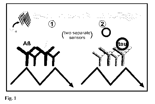
Short Description of the Figures
Figure 1: Scheme of the combined immuno-infrared assay and principle of the
analysis. (A) The total fraction of A13 (1) and Tau (2) present in CSF and/or
plasma
were separately extracted using an antibody functionalized immuno-infrared
sensor.
The detected A13 and Tau secondary structure distribution is indicated by the
infrared
amide I maximum position.
Figure 2: Distribution of the amide I maximum position as displayed in box-
plots for
DC and DAT discrimination based on the analysis of A13 in CSF and plasma, and
Tau in
CSF. Both diagnostic groups showed a high significant difference in the amide
I
maximum of A13 out of CSF (p=2.5*10-il, Kruskal-Wallis ANOVA, confidence level
a=0.05) and EDTA-plasma (p=3.4*10-9), and a moderate significance for Tau out
of
CSF (p=1.6*10-3). In Box-plots 25/50/75% quantiles are displayed as horizontal
lines,
the average band position as square, standard deviation as whiskers, and
observed
minimum/maximum values as cross.
Figure 3: 3D-scatter plot of the amide I maximum position as determined for
Tau in
CSF, A13 in CSF and A13 in EDTA-plasma for 61 DC (grey) and 39 DAT (black)
samples.
Data points within the transparent black box indicate subjects which are
identified as
DAT in all three assays.
Figure 4: ROC-curve analysis for DC (n=61) and DAT (n=39) differentiation
based on
the determination of the A13 secondary structure distribution in CSF (A), A13
in EDTA-
plasma (B), and Tau protein in CSF (C). In this order an AUC of 0.90, 0.85,
and 0.67
was achieved. Thus, a diagnostic accuracy of 92 % (A13, CSF), 85 % (A13,
plasma), and
68 % (Tau, CSF) was calculated (D). Based on these data, the Tau secondary
structure distribution alone seems more to be a general biomarker for dementia
than
for the specific DAT detection.
Figure 5: Diagram of the procedure for the differential diagnosis and disease
stage
classification of DAT and other types of dementia by the combined immuno-
infrared
assay. The assay is based on the determination of the A13 peptide and Tau
protein
6
CA 03044342 2019-05-17
WO 2018/091743 PCT/EP2017/079949
secondary structure distribution in bodily fluids. This distribution is
represented by the
maximum position of the infrared conformation sensitive amide I band of the
extracted
biomarker fraction. A maximum below the discriminative marker band of 1643 cm-
1 is
defined as diseased. This procedure is applied to the extracted A13 fraction
out of CSF
and plasma and to the Tau protein fraction from CSF. No dementia will be
assigned
when all three biomarker values are above or equal to 1643 cm-1. In contrast,
biomarker values below 1643 cm-1 indicate severe DAT. Other types of dementia
will
be identified when only the Tau amide I maximum is below the marker band.
Figure 6: The determination of the amide I maximum representing the secondary
structure distribution of A13 (CSF/plasma) and Tau (CSF) was used for the
differentiation of 61 DC and 39 DAT patients. Thereby, a majority vote (black
=
maximum < 1643 cm-1 and grey = maximum
1643 cm-1) depicted DC and DAT.
Thus, only 3 false positives were observed for the DC group and 4 for the DAT
group.
This results in a specificity of 95 %, sensitivity of 90 %, and thus an
overall diagnostic
accuracy of 93 % for the combined data analysis as compared to the clinical
assessment by gernontopsychiatrists and neurologists.
Detailed Description of the Invention
The immuno-infrared sensors and their production is described in applicant's
previous
patent application WO 2015/121339 and which is now applied for the detection
of the
secondary structure distribution of both A13 and Tau in bodily fluids. The
production of
the IR-sensors includes the direct and intimate immobilization of receptors
for the A13
or Tau, respectively, i.e. antibodies, on the surface of the infrared
transparent material
via silane or thiol chemistry with an optimized, simplified protocol. To
analyze the
liquid (e.g. serum, blood plasma or CSF), it is fed to the sensor in a flow
system. The
macromolecular substance is immobilized by the antibody on the functionalized
sensor
surface. The optical sensor elements are particularly suitable for infrared
analysis and
optionally further for the parallel or alternative analysis by another optical
method
including detection of fluorescence at different wavelengths.
According to the invention, the infrared transparent material of the first and
second IR
cells is independently selected from silicon, germanium, zinc selenide,
gallium
selenide, and diamond, and preferably is germanium.
In a preferred embodiment of the invention, the optical sensor elements has an
internal reflection element comprising a germanium crystal having a trapezoid
or
7
CA 03044342 2019-05-17
WO 2018/091743 PCT/EP2017/079949
parallelogram shape, fiber or rod shaped geometry. It is preferred that the
germanium
crystal is a germanium monocrystal, while a trapezoid cut germanium
monocrystal is
particularly preferred.
It is further preferred that the germanium crystal allows for or provides for
one, more
than one, or more than three reflections of the infrared light through the
reflection
element, particularly preferred are more than five reflections or even more
than
twenty reflections (preferred are 25 reflections with 13 actively sensed
reflections).
For allowing the contact with the candidate biomarker protein in such multiple
reflections, the receptor for the biomarker protein is grafted to the
appropriate number
of surfaces of said internal germanium reflection element.
The silane and thiol linkers that are utilized for coupling the receptor and
hence, the
macromolecule to the internal germanium reflection element include homogenous
silane and thiol linkers, mixtures of silane linkers and mixtures of thiol
linkers. For
allowing a tight and intimate linkage of the receptor/macromolecule short
chained
linkers, preferably linkers having an effective linker chain length (including
carbon
atoms and heteroatoms) of not more than 20 atoms or not more than 15 atoms,
are
utilized.
Such short chained linkers include silane linkers have one of the following
formulas:
X3Si-(CH2)n-Y-(CH2)-Z,
X2R1Si-(CH2)n-Y-(CH2)-Z or
X(R1)2Si-(CH2)n-Y-(CH2)n,-Z,
and the thiol linkers have the following formula:
WS-(CH 2) n -Y-(CH2),-v-Z,
wherein W is R15- or H, X at each occurrence is independently selected from
halogen
and C1_6 alkoxy, n is an integers of 1 to 10, n is an integer of 1 to 5, R1 at
each
occurrence is independently selected from C1_6 alkyl, Y is selected from a
chemical
bond, -0-, -CO-, -SO2-, -NR2-, -S-, -SS-, -NR2C0-, -CONR2-, -NR2502- and -
502NR2-
(wherein R2 is H or C1_6 alkyl), and Z is an amine-reactive group including -
CO2H, -
503H and ester derivatives thereof.
The halogen within the present invention includes a fluorine, chlorine,
bromine and
iodine atom. C1-6 alkyl and C1-6 alkoxy includes straight, branched or cyclic
alkyl or
alkoxy groups having 1 to 6 carbon atoms that may be saturated or unsaturated.
In
case of cyclic alkyl and alkoxy groups, this refers to those having 3 to 6
carbon atoms.
Suitable C1_6 alkyl and C1_6 alkoxy groups include, among others, methyl and
methoxy,
8
CA 03044342 2019-05-17
WO 2018/091743 PCT/EP2017/079949
ethyl and ethoxy, n-propyl and n-propoxy, iso-propyl and iso-propoxy,
cyclopropyl and
cyclopropoxy, n-butyl and n-butoxy, tert-butyl and tert-butoxy, cyclobutyl and
cyclobutoxy, n-pentyl and n-pentoxy, cyclopentyl and cycloppentoxy, n-hexyl
and n-
hexoxy, cyclohexyl and cyclohexoxy, and so on. The amine-reactive group Z
includes
all types of functional groups that are reactive with a free amino group.
Among those,
-CO2H, -S03H and ester derivatives thereof (including active esters) are
particularly
preferred.
The -(CH2)n- and -(CH2)n- structural elements in the above formulas may also
contain
one or more double and/or triple bonds and may be substituted with one or more
halogen atoms such as fluorine or with deuterium.
In a preferred embodiment of the invention, the optical sensor elements are
obtainable by silanization and in the linkers of formulas (i) to (iii) X is
independently
selected from C1_6 alkoxy groups, preferably from methoxy and ethoxy groups, Y
is -
NHCO-, Z is -CO2H or an ester derivative thereof, and n is an integer of 1 to
5 and n is
an integer of 1 to 3, preferably n is 3 and n' is 2.
In another embodiment, the optical sensor elements are obtainable by
thiolation and
in the linkers of formula (iv) W is H, Y is a chemical bond, Z is -CO2H or an
ester
derivative thereof, and n is an integer of 1 to 8 and n' is an integer of 1 to
5,
preferably n is 8 and n' is 4. Particularly preferred is a 12-
mercaptododecanoic acid
NHS ester.
In another preferred embodiment of the optical sensor element, the receptors
for the
A13 peptide and Tau protein are specific antibodies. In case of the A13
peptide, the
antibody is an antibody specifically binding to the central epitope of the A13
peptide,
such as antibody A8978 (Sigma Aldrich) and in case of the Tau protein, the
antibody is
an antibody specifically binding to an epitope present in all Tau variants
(including
phosphorylated and truncated variants, variants with 3 to 4 repeat regions, or
isoforms), such as antibody Tau-5 (AHB0042, Thermo Fisher Scientific).
The blocking substance not cross-reacting with the candidate biomarker protein
includes casein, ethanolamine, L-lysine, polyethylene glycols, albumins, and
derivatives thereof, and preferably is casein.
In the method for preparing the sensor elements, the oxidization is performed
by
treatment with H202/oxalic acid. Further, in the method the silanization with
the short
silane linkers is preferably performed with a silane derivative having the
following
formulas:
9
CA 03044342 2019-05-17
WO 2018/091743 PCT/EP2017/079949
X3Si-(CH2)n-(C1-12)n,-Y,
X2(R1)Si-(CH2)n-(CH2)-Y or
X(R1)(R2)Si-(CH2)n-(0-12)n-Y,
wherein the variables are as defined above. It is particularly preferred that
an ester
derivative of the CO2H or SO3H moiety in the definition of Y be used, which
can be a
simple C1_6 alkyl ester, but can also be an activated ester such as an N-
hydroxysuccinimid ester or any other activated ester derivate. It is also
preferred in
the method that the receptor is an antibody. It is further preferred that the
blocking
substance is casein.
In the method for preparing the sensor elements, the surface activation is
performed
by treatment with HF (49%). Further, in the method the thiolation with the
short thiol
linkers is preferably performed with thiol linkers having the following
formula:
WS-(CH2)n-Y-(CH2)n-Z,
wherein the variables are as defined above. It is particularly preferred that
an ester
derivative of the CO2H or or SO3H moiety in the definition of Y be used, which
can be a
simple C1_6 alkyl ester, but can also be an activated ester such as an N-
hydroxysuccinimid ester or any other activated ester derivate. It is also
preferred in
the method that the receptor is an antibody. It is further preferred that the
blocking
substance is casein.
In the method for preparing the sensor elements, the optical sensor elements
are built
up under room temperature. Every single step can be assessed on the basis of
the IR-
spectra. This validation step is essential for the specific detection and
accurate
secondary structure determination of the analyte.
The device of aspect (4) of the invention has the sensor elements incorporated
in a
suitable IR cell (chamber). It may further include a light (IR) emitting
element, a light
(IR) detecting element and a data processing unit. For parallel detection by
an
additional optical method the device may further include light source and
detector
element for such additional optical method such as light source and detector
elements
for UV/Vis-fluorescence, at different wavelengths.
The method of aspect (1) of the invention comprises the steps of
(a) conducting, in a first IR cell comprising a first infrared sensor element
having an
internal reflection element with a core of an infrared transparent material
and at least
one receptor for the A13 peptide directly grafted to at least one surface of
said core, at
CA 03044342 2019-05-17
WO 2018/091743 PCT/EP2017/079949
least one flux of a body fluid with soluble A13 peptide, submitting an IR beam
through
said first IR cell, and obtaining an infrared spectrum therefrom;
(b) conducting, in a second IR cell comprising a second infrared sensor
element having
an internal reflection element with a core of an infrared transparent material
and at
least one receptor for the Tau protein directly grafted to at least one
surface of said
core, at least one flux of a body fluid with soluble Tau protein, submitting
an IR beam
through said second IR cell, and obtaining an infrared spectrum therefrom; and
(c) analyzing the obtained infrared spectra to determine the secondary
structure
distribution of the soluble A13 peptide and of the soluble Tau protein in the
bodily fluids
by differential diagnosis.
In the method of the invention, the bodily fluids applied in steps (a) and (b)
may be
any complex body fluid comprising the biomarker, including serum, blood plasma
and
CSF. Further suitable bodily fluids are lacrimal fluid and nipple aspirate
fluid.
In a preferred embodiment the method further comprises prior to step (a) and
(b):
installation of said optical sensor element in the IR cell.
Additionally/alternatively the
method may further comprise the step (a') and (b'): regenerating of the
surface of the
optical element by application of a solution of free ligand for the receptor.
The spectrum obtained in steps (a) and (b) have a sufficient signal to noise
ratio to
resolve the amide I band. This allows the analysis of the shift of the amide I
band
maximum of the biomarker protein in step (c) to determine the secondary
structure of
the candidate biomarker proteins and perform the differential diagnosis.
In a further embodiment the step (c) of the method further comprises comparing
the
obtained infrared spectrum with a spectrum of the soluble A13 peptide and/or
of the
soluble Tau with known secondary structure and/or with known concentration.
In another embodiment, the method may comprise, alternative or parallel to the
infrared analysis, detection by another optical method, including UV/Vis-
fluorescence,
at different wavelengths. Notably, a method is preferred that combines immuno-
ATR-
FTIR vibrational spectroscopy with parallel fluorescence spectroscopy.
The method of aspects (1) allows/is suitable for determining the soluble A13
peptide
and the soluble Tau in bodily fluids, notably for directly determining them in
bodily
fluids of mammalian (human, animal) origin, including cerebrospinal fluid,
blood or
serum, without pretreatment (i.e., without a separate preceding enrichment or
purification step). The method is suitable for determination of the candidate
biomarker
protein in a separate (in-vitro) or an online (direct determination of the
body fluid on
11
CA 03044342 2019-05-17
WO 2018/091743 PCT/EP2017/079949
the patient) fashion. In both cases, the method may further comprise the
differential
diagnosis and the assessment of the Alzheimer's disease stages.
The method of aspect (1) are particularly suitable for the determination of
progression
of Alzheimer's disease with Amyloid-beta and Tau as candidate biomarker
proteins,
wherein a shift of the amide I band maximum of the A13 peptide from 1647 cm-1
to
1640 cm-1, preferably with a threshold value of 1643 cm-1 +/- 5 cm-1, (or 1643
cm-1
+/- 3 cm', or 1643 cm-1 +/- 1 cm-1 , or about 1643 cm-1), and a shift of the
amide I
band maximum of the Tau protein from 1647 cm-1 to 1640 cm-1, preferably with a
threshold value of 1643 cm-1 +/- 5 cm-1, (or 1643 cm-1 +/- 3 cm-1, or 1643 cm-
1 +/- 1
cm-1 , or about 1643 cm-1) are indicative for Alzheimer's disease. The method
is also
particularly suitable for the determination of progression of Alzheimer's
disease with
Amyloid-beta and Tau as candidate biomarker proteins. Here the differential
diagnosis
provides for an assured clinical profile of the dementia type, preferably the
method
comprises the detection of the secondary structure distribution of A13 from
CSF (A), A13
from blood plasma (B), and Tau from CSF (C). In particular, the method of the
invention enables the differential diagnosis of Dementia Alzheimer type (DAT)
and
(Disease Control), DAT patients being sub-classified into early, moderate, and
severe
DAT, and DC patients being separated into health controls, other diseases, and
dementia due to another origin than Alzheimer's disease. Notably, for both
biomarkers, (A)/(B) for A13 and (C) for Tau, a discriminative threshold (1643
cm-1
cm-1) separates Alzheimer's disease and DC patients; and/or the combination of
(A),
(B), and (C) provides a biomarker panel applicable for an assured DAT
diagnosis.
A simple threshold classifier is established for both biomarkers similar to
that
described in Nabers et al., Anal. Chem. Doi: 10.1021/acs.analchem.5b04286
(2016)
and WO 2015/121339. Thereby, using a discriminative spectral marker band for
disease control (DC) and the Dementia Alzheimer type (DAT) differentiation,
both
diagnostics groups could be separated with a diagnostic accuracy of 90 % based
on
CSF A13 analysis. The predicitve accuracy observed from blood plasma A13
anaylsis
solely was lower (84 %). Moreover, a separation of both groups only based on
the Tau
protein secondary structure distribution remained insufficient with an
accuracy of
68 %. But combining the determined amide I frequencies of A13 from CSF and
blood
plasma with those of Tau to a marker panel, a simple majority vote classifier
demonstrated significant higher predicitve values. Now, an accuracy of 93 %
and a
specificity of 95 % could be achieved. A high specificity is cruical
especially for
12
CA 03044342 2019-05-17
WO 2018/091743 PCT/EP2017/079949
incurable diseases such as AD, because a false positive diagnosis may have
serious
psychological consequences for the party concerned. But the combined data
analysis
demonstrated a second big advantage. By combining the data of A13 and Tau,
more
information about the disease stage and indications for other types of
dementia can be
provided. The principle for differential diagnostics is simple. In a first
step, the amide I
maximum of the extracted soluble fraction of A13 from CSF was determined as
described in Nabers et al., in Anal. Chem Doi: 10.1021/acs.analchem.5b04286
(2016)
and WO 2015/121339. Thereby, a maximum above or equal to 1643 cm-1 was
indicative for DCs, a maximium below this frequency for DAT. In a second step,
the
same procedure was applied to blood plasma samples. Again, the amide I maxima
of
A13 were determined for each sample. Now, if both values were consistently
above or
below the marker frequency, a differentiation between DC and DAT could be made
with a high accuracy. But for inconsistent A13 results (CSF and plasma) the
Tau protein
secondary structure distribution could be used as decision support. On the
other hand,
the Tau protein secondary structure distribution also demonstrated the
potential to
sub-classify the DC and DAT group into dementia due to another origin than
Alzheimer
or into an early, moderate, or severe stage of the disease. Therewith,
Parkinson
disease or vascular dementia patients could be identified within the DC group
(A@ CSF
1643 cm-1; A13 plasma 1643 cm-1; Tau CSF <1643 cm-1). On the other hand, the
DAT group could be differentiated into early (f.e. A13 CSF <1643 cm-1; A13
plasma
<1643 cm-1; Tau CSF 1643 cm-1), moderate (A@ CSF <1643 cm-1; A13 plasma
1643 cm-1; Tau CSF <1643 cm-1) or (A@ CSF 1643 cm-1; A13 plasma <1643 cm-1;
Tau CSF <1643 cm-1), and severe (A@ CSF <1643 cm-1; A13 plasma <1643 cm-1; Tau
CSF <1643 cm-1) stages of disease.
The invention is further described by the following examples which are,
however, not
to be construed as limiting the invention.
Examples
Materials and Methods: The same experimental set-ups were used as in WO
2015/121339.
Sampling and pretreatment: CSF was drawn by lumbal puncture and aliquoted at
the
university hospital Essen, snap-frozen in liquid nitrogen, shipped and stored
at -80 C.
Samples were not pretreated before the measurement, only thawed at 37 C for
30
seconds and kept on ice until used.
13
CA 03044342 2019-05-17
WO 2018/091743 PCT/EP2017/079949
Patient collective: Details of sample acquisition and diagnosis of Disease
Control (DC)
and Dementia Alzheimer type (DAT) patients have been described in detail
previously
(Nabers et al., Anal. Chem. Doi: 10.1021/acs.analchem.5b04286 (2016)). In the
former study, 141 patient samples were analyzed using the immuno-infrared
sensor.
Out of this collective, 100 patients were randomly selected for the present
study.
Infrared amide I data of A13 extracted from CSF and blood plasma were adopted
from
(Nabers et al., Anal. Chem. Doi: 10.1021/acs.analchem.5b04286 (2016)).
Solutions and reagents:
Phosphate buffered saline (PBS-buffer): 137 mM sodium chloride (NaCI), 2.7 mM
potassium chloride (KCI), 12 mM total-phosphate (in the form of Na2HPO4 and
NaH2PO4), PH 7.4.
Casein blocking-solution: 200 mM sodium hydroxide (NaOH), 1 % (w/v) casein
from
bovine milk (powder), pH adjusted with H3PO4 to 7.4.
Silanization-solution: The used NHS-silane (N-(4,4,4-
triethoxysilanebutyl)succinamic
acid 2,5-dioxopyrrolidin-1-y1 ester) was synthesized and characterized as
described
(Schartner et al., JACS 135(10):4079-4087 (2013)).
Antibody: For the extraction of A13 from the respective body fluid the
antibody A8978
(lot no: 061M4773, Sigma Aldrich) was employed. In case of the Tau protein
detection
the antibody Tau-5 (AHB0042, Thermo Fisher Scientific) was used.
Performing the measurement: The general procedure is identical to the one
described
in WO 2015/121339. IR-measurements were performed on a Vertex 70V spectrometer
(Bruker Optics GmbH, Ettlingen, Germany) with liquid nitrogen cooled mercury-
cadmium-telluride (MCT) detector. Double-sided interferograms were recorded in
forward-backward interferometer movement at a 80 kHz data rate with a spectral
resolution of 2 cm-1, Blackman-Harris-3-Term-apodisation, Mertz-phase
correction and
4 times zero filling. Reference spectra were recorded as an average of 1000,
sample
spectra of 200 interferograms. Recording reference single channel spectra of
the blank
sensor, sensor with 2-propanol, the silanized surface, the buffers, antibody
or casein
coated surface in equilibrium states enabled high sensitivity difference
spectroscopy
based on Lambert-Beer law (E¨log(I/I0). The absorbance of the state change is
the
negative decadic logarithm of the intensity relation before and after the
change.
Workflow: Details of the sensor preparation for the FTIR-spectroscopic
analysis of the
Tau protein in CSF were published previously (WO 2015/121339; Nabers et al.,
Anal.
Chem. Doi:10.1021/acs.analchem.5b04286 (2016); Nabers et al., Journal of
14
CA 03044342 2019-05-17
WO 2018/091743 PCT/EP2017/079949
Biophotonics 9(3):224-34 doi:10.1002/jbio.201400145 (2016)). Briefly, the
total
volume of the flow-cell including all connection tubes amounted to 400 pl. For
each
analysis, one sensor element per sample was freshly functionalized with silane
(Schartner et al., JACS 135(10):4079-87 doi:10.1021/ja400253p (2013)) and
antibody. Before analysis the surface was saturated with a casein blocking
solution.
For A13 detection in CSF and blood plasma, the monoclonal antibody A8978
(Sigma
Aldrich, aa 13-28) was used. Tau capturing was provided by monoclonal Tau-5
antibody (Life Technology, aa 210-230). For the analysis 50 pl CSF or 150 pl
of EDTA-
plasma were added to the circulating buffer with a flow-rate of 1 ml/min,
respectively.
Pretreatment of the spectra: By scaled subtraction of a reference spectrum
water
vapor was removed. Spectra were baseline corrected.
Example 1: The A13 and Tau protein secondary structure distribution for
accurate DC
and DAT differentiation.
The performed study included 300 samples from 61 DC and 39 DAT patients.
Details
about the patients differential diagnosis were described previously (Nabers et
al., Anal.
Chem. Doi: 10.1021/acs.analchem.5b04286 (2016). In general, the patient
collective
was separated into DCs and DAT subjects. The DAT group was further sub-
classified
into early, moderate, and severe states of Alzheimer's disease. For a small
number of
DC patients a complete differential diagnosis was available including patients
suffer
from dementia not due to Alzheimer's disease origin such as Parkinson disease
or
vascular dementia. For the analysis of the secondary structure distribution of
A13 and
Tau in CSF and/or plasma, both biomarker were extracted from the respective
fluid by
an immuno-infrared sensor as described by Nabers et al. (Nabers et al., Anal.
Chem.
Doi: 10.1021/acs.analchem.5b04286 (2016)). Therefore, A13 and Tau were
separately
captured out of the CSF or plasma by the surface immobilized monoclonal
antibody
A8978 (aa13-28 of A13) and Tau-5 (aa210-230), respectively. The secondary
structure
distribution was indicated by the recorded amide I maximum frequency of A13
and Tau.
In the previous study, a simple threshold classifier was established with a
discriminative marker frequency of 1643 cm-1 for DC and DAT differentiation.
The
same marker band was used within the current study. At first, the amide I
maximum
of A13 from CSF was determined for each patient sample. Thereby, a maximum
position
below 1643 cm-1 was indicative for DAT. Next, the amide I maximum of A13 from
blood
EDTA-plasma was identified. Finally, we detected the maximum of the extracted
Tau
CA 03044342 2019-05-17
WO 2018/091743 PCT/EP2017/079949
protein fraction in CSF. The discriminative marker band was identically
defined for
both biomarkers and both bodily fluids at <1643 cm-1 indicative for DAT. The
amide I
maximum distribution of the DC and DAT group showed highly significant
differences
for A13 from CSF (Kruskal-Wallis ANOVA; p=2.5*10-11; confidence level p=0.05)
and
blood plasma (p=3.4*10-9) and significantly differed for Tau (p=1.6*10-3) from
CSF
(Fig. 2). Thus, a smaller shift of the Tau amide I maximum was observed for
DAT
subjects as compared to A13. The mean amide I maximum of Tau was 1644 cm-1 for
the DC and 1642 cm-1 for the DAT group as compared to A13 from CSF with 1645
cm-1
for DC and 1641 cm-1 for DAT subjects. A13 from blood plasma revealed a mean
maximum of 1648 cm-1 for the DC and 1641 cm-1 for the DAT group. Based on
these
distributions, also shown in a 3D-scatter plot in Fig. 3 with a transparent
black box
indicating DAT, the diagnostic performance of each biomarker by itself was
calculated
by giving the accuracy, sensitivity, and specificity. Additionally, Receiver
Operating
Characteristic- (ROC-) curve analyses were performed by scanning the threshold
between 1630.5 cm-1 to 1660.5 cm-1 and determining the sensitivity and
specificity at
each wavenumber. Similar to the results of Nabers et al., the diagnostic
accuracy of
A13 based analysis was highest with 90 % for CSF (Fig. 4A,D; specificity 89
% ,
sensitivity 92 % , AUC 0.90) as compared to the analysis of the A13 secondary
structure
distribution in blood plasma with 85 % (Fig. 4B,D; specificity 90 %,
sensitivity 77 % ,
0.85). In contrast, DC and DAT differentiation based on the Tau protein
secondary
structure distribution in CSF only revealed a diagnostic accuracy of 68 %
(Fig. 4C,D;
specificity 67 % , sensitivity 69 % , AUC 0.67), indicating that Tau alone is
not an
appropriate biomarker for DAT detection. But by adding all three biomarker
values to a
majority vote classifier, thus making a diagnostic decision based the
presented
biomarker panel (two maxima below the threshold = diseased, two maxima above
the
threshold = non DAT), the diagnostic performance could be increased to a
specificity
of 95 %, sensitivity of 90 %, and thus to an overall accuracy of 93 % as
compared to
the clinical diagnosis. Therewith, only 3 false positives out of 61 DCs and 4
false
negatives out of 31 DATs were identified.
Example 2: A combined assay for DC and DAT differential diagnostics.
The combined data analysis provided also the potential to sub classify both
diagnostics
groups. This is schematically shown in Fig. 5. For instance, A13 from CSF and
plasma
demonstrates an amide I maximum above or equal to 1643 cm-1, but the Tau amide
I
16
CA 03044342 2019-05-17
WO 2018/091743 PCT/EP2017/079949
maximum is below 1643 cm-1, in this case another type of dementia might be
potentially indicated by the combined immuno-infrared assay (Fig. 5). In
contrast,
when the amide I maxima of A13 from CSF and plasma are below the marker band
but
the Tau maximum is above, an early state of DAT will be displayed. This
procedure
was applied to both diagnostics groups within our study. The amide I maximum
of A13
from CSF demonstrated in 69 % of all DC cases a higher maximum value than Tau
from CSF. This effect may be explained by higher disordered properties of the
Tau
protein as compared to A13. On the other hand, in 25 % of all DC cases the
maximum
was lower for A13 and only in 6 % of all cases the maxima were identically.
This
relation completely changed for the DAT group. Therein only 31 % of all DAT
patients
yielded an amide I maximum for A13 greater than for Tau, 62 % showed a lower
A13
maximum (Fig. 6). This supports the hypothesis that A13 accumulation is in
early and
initiating event in Alzheimer's disease progression and accompanied by Tau
protein
aggregation.
17