Note: Descriptions are shown in the official language in which they were submitted.
JB15139 WOPCT
SAFE AND EFFECTIVE METHOD OF TREATING LUPUS WITH ANTI-
IL12/IL23 ANTIBODY
SEQUENCE LISTING
[00011 The instant application contains a Sequence Listing which has been
submitted
electronically in ASCII format. Said ASCII copy, created on 06 September 2018,
is named
.113I5139W0PCTSEQLIST.txt and is 13/382 bytes in size.
FIELD OF THE INVENTION
[ 0002 ] The present invention relates to methods for treating lupus with an
antibody that
binds human IL-12 and/or human IL-23 proteins. In particular, the present
invention relates
to methods of treating active Systemic Lupus Erythematosus (SLE) in a patient
by
administering a clinically proven safe and clinically proven effective amount
of an anti-IL-
12/1L-23p40 antibody or an anti-1L-23 antibody, e.g., the anti-1L-12/1L-23p40
antibody
ustekinumab, and specific pharmaceutical compositions of the antibody.
BACKGROUND OF THE INVENTION
[ 0003 ] Interleukin (IL)-12 is a secreted heterodimeric cytokine comprised of
2 disulfide-
linked glycosylated protein subunits, designated p35 and p40 for their
approximate
molecular weights. IL-12 is produced primarily by antigen-presenting cells and
drives
cell-mediated immunity by binding to a two-chain receptor complex that is
expressed on the
surface of T cells or natural killer (NK) cells. The IL-12 receptor beta-1 (IL-
12R131) chain
binds to the p40 subunit of IL-12, providing the primary interaction between
IL-12 and its
receptor. However, it is IL-12p35 ligation of the second receptor chain, IL-
1211132, that
confers intracellular signaling (e.g. STAT4 phosphorylation) and activation of
the receptor-
bearing cell (Presky et al, 1996). IL-12 signaling concurrent with antigen
presentation is
thought to invoke T cell differentiation towards the T helper 1 (Thl)
phenotype,
characterized by interferon gamma (IFNy) production (Trinchieri, 2003). Thl
cells are
CA 3044777 2019-10-22
CA 03044777 2019-05-23
WO 2019/058345 PCT/1B2018/057368
believed to promote immunity to some intracellular pathogens, generate
complement-fixing
antibody isotypes, and contribute to tumor immunosurveillance. Thus, IL-12 is
thought to be
a significant component to host defense immune mechanisms.
[ 0004 ] It was discovered that the p40 protein subunit of IL-12 can also
associate with a
separate protein subunit, designated p19, to form a novel cytokine, IL-23
(Oppman et al,
2000). IL-23 also signals through a two-chain receptor complex. Since the p40
subunit is
shared between IL-12 and IL-23, it follows that the IL-12RM chain is also
shared between
IL-12 and IL-23. However, it is the IL-23p19 ligation of the second component
of the IL-23
receptor complex, IL-23R, that confers IL-23 specific intracellular signaling
(e.g., STAT3
phosphorylation) and subsequent IL-17 production by T cells (Parham et al,
2002; Aggarwal
et al. 2003). Recent studies have demonstrated that the biological functions
of IL-23 are
distinct from those of IL-12, despite the structural similarity between the
two cytokines
(Langrish et al, 2005).
[ 0005 ] Abnormal regulation of IL-12 and Thl cell populations has been
associated with
many immune-mediated diseases since neutralization of IL-12 by antibodies is
effective in
treating animal models of psoriasis, multiple sclerosis (MS), rheumatoid
arthritis,
inflammatory bowel disease, insulin-dependent (type 1) diabetes mellitus, and
uveitis
(Leonard et al, 1995; Hong et al, 1999; Malfait et al, 1998; Davidson et al,
1998). IL-12 has
also been shown to play a critical role in the pathogenesis of SLE in two
independent mouse
models of systemic lupus erythematosus (Kikawada et al. 2003; Dai et al. 2007.
[ 0006 ] Systemic lupus erythematosus (SLE) is a complex, chronic,
heterogeneous
autoimmtme disease of unknown etiology that can affect almost any organ
system, and
which follows a waxing and waning disease course. Systemic lupus erythematosus
occurs
much more often in women than in men, up to 9 times more frequently in some
studies, and
often appears during the child-bearing years between ages 15 and 45. This
disease is more
prevalent in Afro-Caribbean, Asian, and Hispanic populations. In SLE, the
immune system
attacks the body's cells and tissue, resulting in inflammation and tissue
damage which can
harm the heart, joints, skin, lungs, blood vessels, liver, kidneys and nervous
system. About
half of the subjects diagnosed with SLE present with organ-threatening
disease, but it can
take several years to diagnose subjects who do not present with organ
involvement. Some of
2
CA 03044777 2019-05-23
WO 2019/058345 PCT/1B2018/057368
the primary complaints of newly diagnosed lupus patients are arthralgia (62%)
and
cutaneous symptoms (new photosensitivity; 20%), followed by persistent fever
and
malaise." The estimated annual incidence of lupus varies from 1.8 to 7.6 cases
per 100,000
and the worldwide prevalence ranges from 14 to 172 cases per 100,000 people."
Patients
with mild disease have mostly skin rashes and joint pain and require less
aggressive therapy;
regimens include nonsteroidal anti-inflammatory drugs (NSAIDs), anti-malarials
(e.g., hydroxychloroquine, chloroquine, or quinacrine) and/or low dose
corticosteroids. With
more severe disease patients may experience a variety of serious conditions
depending on
the organ systems involved, including lupus nephritis with potential renal
failure,
endocarditis or myocarditis, pneumonitis, pregnancy complications, stroke,
neurological
complications, vasculitis and cytopenias with associated risks of bleeding or
infection.
Common treatments for more severe disease include immunomodulatory agents,
such as
methotrexate (MTX), azathioprine, cyclophosphamide, cyclosporine, high dose
corticosteroids, biologic B cell cytotoxic agents or B cell modulators, and
other
immunomodulators. Patients with serious SLE have a shortening of life
expectancy by 10 to
30 years, largely due to the complications of the disease, of standard of care
therapy, and/or
accelerated atherosclerosis. In addition, SLE has a substantial impact on
quality of life, work
productivity, and healthcare expenditures. Existing therapies for SLE are
generally either
cytotoxic or immunomodulatory, and may have notable safety risks.. Newer
treatments for
SLE have provided only modest benefits over standard of care therapy. Thus,
there is a large
unmet need for new alternative treatments that can provide significant benefit
in this disease
without incurring a high safety risk.
SUMMARY OF THE INVENTION
[ 0007 ] The general and preferred embodiments are defined, respectively, by
the
independent and dependent claims appended hereto, which for the sake of
brevity are
incorporated by reference herein. Other preferred embodiments, features, and
advantages of
the various aspects of the invention will become apparent from the detailed
description
below taken in conjunction with the appended drawing figures.
3
CA 03044777 2019-05-23
WO 2019/058345 PCT/1B2018/057368
[ 0008 ] In certain embodiments, the present invention provides a clinically
proven safe
and clinically proven effective method of treating lupus in a patient
comprising
intravenously (IV) and/or subcutaneously (SC) administering to the patient an
anti-IL-12
and/or anti-IL-23 antibody.
[ 0009 ] In certain embodiments, the invention provides a clinically proven
safe and
clinically proven effective method of treating lupus in a patient comprising
intravenously
(IV) and/or subcutaneously (SC) administering to the patient an anti-IL-12
and/or anti-IL-23
antibody, wherein the anti-IL-12 and/or anti-IL-23 antibody is an anti-IL-
12/23p40
antibody.
[ 0010 ] In certain embodiments, the invention provides a clinically proven
safe and
clinically proven effective method of treating lupus in a patient comprising
intravenously
(IV) and/or subcutaneously (SC) administering to the patient an anti-IL-12
and/or anti-IL-23
antibody, wherein the anti-IL-12 and/or anti-IL-23 antibody is an anti-IL-
12/23p40.
[ 0011 ] In certain embodiments, the invention provides a clinically proven
safe and
clinically proven effective method of treating lupus in a patient comprising
intravenously
(IV) and/or subcutaneously (SC) administering to the patient an anti-IL-12
and/or anti-IL-23
antibody, wherein the anti-IL-12 and/or anti-IL-23 antibody is an anti-IL-
12/23p40 antibody
comprising: (i) the heavy chain CDR amino acid sequences of SEQ ID NO:!, SEQ
ID NO:2,
and SEQ ID NO:3; and (ii) the light chain CDR amino acid sequences of SEQ ID
NO:4,
SEQ ID NO:5, and SEQ ID NO:6 (corresponding to ustekintunab (Stelara of
Janssen
Biotech, Inc.)).
[ 0012 ] In certain embodiments, the invention provides a clinically proven
safe and
clinically proven effective method of treating lupus in a patient comprising
intravenously
(IV) and/or subcutaneously (SC) administering to the patient an anti-IL-12
and/or anti-IL-23
antibody, wherein the anti-IL-12 and/or anti-IL-23 antibody is an anti-IL-
12/23p40 antibody
comprising: (i) the heavy chain variable domain amino acid sequence of SEQ TD
NO:7; and
(ii) the light chain variable domain amino acid sequence of SEQ ID NO:8
(corresponding to
ustekinumab (Stelara of Janssen Biotech, Inc.)).
[ 0013 ] In certain embodiments, the invention provides a clinically proven
safe and
clinically proven effective method of treating lupus in a patient comprising
intravenously
4
CA 03044777 2019-05-23
WO 2019/058345 PCT/1B2018/057368
(IV) and/or subcutaneously (SC) administering to the patient an anti-IL-12
and/or anti-IL-23
antibody, wherein the anti-IL-12 and/or anti-IL-23 antibody is the anti-IL-
12/23p40
antibody ustekinumab (Stelara0), comprising: (i) the heavy chain amino acid
sequence of
SEQ ID NO:10; and (ii) the light chain amino acid sequence of SEQ ID NO:11
(corresponding to ustekinumab (Stelara of Janssen Biotech, Inc.)).
[ 0014 ] In certain embodiments, the present invention provides a composition
comprising
an anti-IL-12 and/or anti-IL-23 antibody for use in a clinically proven safe
and clinically
proven effective method of treating lupus in a patient comprising
intravenously (IV) and/or
subcutaneously (SC) administering to the patient the pharmaceutical
composition
comprising the anti-IL-12 and/or anti-1L-23 antibody.
[ 0015 ] In certain embodiments, the present invention provides a composition
comprising
an anti-1L-12 and/or anti-IL-23 antibody for use in a clinically proven safe
and clinically
proven effective method of treating lupus in a patient comprising
intravenously (IV) and/or
subcutaneously (SC) administering to the patient the pharmaceutical
composition
comprising an anti-IL-12 and/or anti-IL-23 antibody, wherein the anti-IL-12
and/or anti-IL-
23 antibody is an anti-IL-12/23p40 antibody.
[ 0016 ] In certain embodiments, the present invention provides a composition
comprising
an anti-IL-12 and/or anti-IL-23 antibody for use in a clinically proven safe
and clinically
proven effective method of treating lupus in a patient comprising
intravenously (IV) and/or
subcutaneously (SC) administering to the patient the pharmaceutical
composition
comprising an anti-IL-12 and/or anti-IL-23 antibody, wherein the anti-IL-12
and/or anti-IL-
23 antibody is an anti-IL-12/23p40 antibody.
[ 0017 ] In certain embodiments, the present invention provides a composition
comprising
an anti-IL-12 and/or anti-IL-23 antibody for use in a clinically proven safe
and clinically
proven effective method of treating lupus in a patient comprising
intravenously (IV) and/or
subcutaneously (SC) administering to the patient the pharmaceutical
composition
comprising an anti-IL-12 and/or anti-IL-23 antibody, wherein the anti-IL-12
and/or anti-IL-
23 antibody is an anti-IL-12/23p40 antibody comprising: (i) the heavy chain
CDR amino
acid sequences of SEQ ID NO:1, SEQ ID NO:2, and SEQ ID NO:3; and (ii) the
light chain
CDR amino acid sequences of SEQ ID NO:4, SEQ NO:5, and SEQ ID NO:6.
CA 03044777 2019-05-23
WO 2019/058345 PCT/1B2018/057368
[ 0018 ] In certain embodiments, the present invention provides a composition
comprising
an anti-IL-12 and/or anti-IL-23 antibody for use in a clinically proven safe
and clinically
proven effective method of treating lupus in a patient comprising
intravenously (IV) and/or
subcutaneously (SC) administering to the patient the pharmaceutical
composition
comprising an anti-IL-12 and/or anti-IL-23 antibody, wherein the anti-1L-12
and/or anti-IL-
23 antibody is an anti4L-12/23p40 antibody comprising: (i) the heavy chain
variable
domain amino acid sequence of SEQ ID NO:7; and (ii) the light chain variable
domain
amino acid sequence of SEQ ID NO:8.
[ 0019 ] In certain embodiments, the present invention provides a composition
comprising
an anti-1L-12 and/or anti-IL-23 antibody for use in a clinically proven safe
and clinically
proven effective method of treating lupus in a patient comprising
intravenously (IV) and/or
subcutaneously (SC) administering to the patient the pharmaceutical
composition
comprising an anti-IL-12 and/or anti-IL-23 antibody, wherein the anti-IL-12
and/or anti-IL-
23 antibody is the anti-IL-12123p40 antibody ustekinumab (StelaraO),
comprising: (i) the
heavy chain amino acid sequence of SEQ ID NO:10; and (ii) the light chain
amino acid
sequence of SEQ ID NO: Ii.
[ 0020 ] In certain embodiments, the present invention provides a
pharmaceutical
composition for intravenously (IV) administration comprising an anti-IL-12/IL-
23p40
antibody comprising: (i) the heavy chain CDR amino acid sequences of SEQ ID
NO: I, SEQ
ID NO:2, and SEQ ID NO:3; and (ii) the light chain CDR amino acid sequences of
SEQ ID
NO:4, SEQ ID NO:5, and SEQ ID NO:6; in a solution comprising 10 mM L-
histidine, 8.5%
(w/v) sucrose, 0.04% (w/v) polysorbate 80, 0.4 mg/mL L methionine, and 20
gg/mL EDTA
disodium salt, dehydrate, at pH 6Ø
[ 0021 ] In certain embodiments, the present invention provides a
pharmaceutical
composition for subcutaneous (SC) administration comprising an anti-IL-12/IL-
23p40
antibody comprising: (i) the heavy chain CDR amino acid sequences of SEQ ID
NO: I, SEQ
ID NO:2, and SEQ ID NO:3; and (ii) the light chain CDR amino acid sequences of
SEQ ID
NO:4, SEQ ID NO:5, and SEQ ID NO:6; in a solution comprising 6.7 mM L-
histidine, 7.6%
(w/v) sucrose, 0.004% (w/v) polysorbate 80, at pH 6Ø
6
CA 03044777 2019-05-23
WO 2019/058345 PCT/1B2018/057368
[ 0022 ] In certain embodiments, the present invention provides a
pharmaceutical
composition for intravenously (IV) administration comprising an anti-IL-12/IL-
23p40
antibody comprising: (i) the heavy chain variable domain amino acid sequence
of SEQ ID
NO:7; and (ii) the light chain variable domain amino acid sequence of SEQ ID
NO:8; in a
solution comprising 10 mM L-histidine, 8.5% (w/v) sucrose, 0.04% (w/v)
polysorbate 80,
0.4 mg/mL L methionine, and 20 g/inL EDTA disodium salt, dehydrate, at pH
6Ø
[0023] In certain embodiments, the present invention provides a pharmaceutical
composition for subcutaneous (SC) administration comprising an anti-IL-12/IL-
23p40
antibody comprising: (i) the heavy chain variable domain amino acid sequence
of SEQ ID
NO:7; and (ii) the light chain variable domain amino acid sequence of SEQ ID
NO:8; in a
solution comprising 6.7 inM L-histidine, 7.6% (w/v) sucrose, 0.004% (w/v)
polysorbate 80, at
pH 6Ø
[ 0024 ] In certain embodiments, the present invention provides a
pharmaceutical
composition for intravenously (IV) administration comprising the anti-IL-
12/23p40 antibody
ustekinumab (Stelarag), comprising: (i) the heavy chain amino acid sequence of
SEQ ID
NO:10; and (ii) the light chain amino acid sequence of SEQ ID NO:11; in a
solution
comprising 10 rriM L-histidine, 8.5% (w/v) sucrose, 0.04% (w/v) polysorbate
80, 0.4 mg/mL
L methionine, and 20 tig/mL EDTA disodium salt, dehydrate, at pH 6Ø
[ 0025 ] In certain embodiments, the present invention provides a
pharmaceutical
composition for subcutaneous (SC) administration comprising the anti-IL-
12/23p40
antibody ustekinumab (Stelara0), comprising: (i) the heavy chain amino acid
sequence of
SEQ TD NO:10; and (ii) the light chain amino acid sequence of SEQ ID NO:11; in
a solution
comprising 6.7 mM L-histidine, 7.6% (w/v) sucrose, 0.004% (w/v) polysorbate
80, at pH 6Ø
[ 0026 ] In certain embodiments, the present invention provides a method of
treating lupus
in a patient comprising subcutaneously administering an anti-IL-23 specific
antibody (also
referred to as IL-23p19 antibody), e.g., guselkumab and risankizumab (BI-
655066),
tildrakizumab (MK-322).
[ 0027 ] In certain embodiments, the composition used in the method of the
invention
comprises a pharmaceutical composition comprising: an anti-IL-23 specific
antibody in an
amount from about 1.0 jig/m1 to about 1000 mg/ml, specifically at 50 mg or 100
mg. In a
7
CA 03044777 2019-05-23
WO 2019/058345 PCT/1B2018/057368
preferred embodiment, the anti-IL-23 specific antibody is guselkumab at 100
mg/mL; 7.9%
(w/v) sucrose, 4.0mM Histidine, 6.9 mM L-Histidine monohydrochloride
monohydrate;
0.053% (w/v) Polysorbate 80 of the pharmaceutical composition; wherein the
diluent is
water at standard state.
[ 0028 ] In certain embodiments, the composition used in the method of the
invention
comprises an isolated anti-IL23 specific antibody, e.g., guselkumab, at 100
mg/mL; 7.9%
(w/v) sucrose, 4.0mM Histidine, 6.9 mM L-Histidine monohydrochloride
monohydrate;
0.053% (w/v) Polysorbate 80 of the pharmaceutical composition; wherein the
diluent is
water at standard state.
[ 0029 ] In certain embodiments, method of the invention comprises
administering a
pharmaceutical composition comprising an isolated anti-IL-23 specific
antibody, e.g.,
guselkumab, at 100 mg/mL; 7.9% (w/v) sucrose, 4.0mM Histidine, 6.9 mM L-
Histidine
monohydrochloride monohydrate; 0.053% (10.7) Polysorbate 80 of the
pharmaceutical
composition; wherein the diluent is water at standard state.
[ 0030 ] In certain embodiments, the present invention provides a method of
treating active
Systemic Lupus Erythematosus (SLE) in a patient, comprising administering an
anti-1L-
12/1L-23p40 antibody to the patient in a clinically proven safe and clinically
proven
effective amount, wherein the antibody comprises a heavy chain variable region
and a light
chain variable region, said heavy chain variable region comprising: a
complementarity
determining region heavy chain 1 (CDRH1) amino acid sequence of SEQ ID NO:1; a
CDRH2 amino acid sequence of SEQ ID NO:2; and a CDRH3 amino acid sequence of
SEQ
TD NO:3; and said light chain variable region comprising: a complementarity
determining
region light chain 1 (CDRL1) amino acid sequence of SEQ ID NO:4; a CDRL2 amino
acid
sequence of SEQ ID NO:5; and a CDRL3 amino acid sequence of SEQ ID NO:6.
[ 0031 ] In certain embodiments, the present invention provides a method of
treating active
Systemic Lupus Erythematosus (SLE) in a patient, comprising administering an
anti-IL-
12/IL-23p40 antibody to the patient in a clinically proven safe and clinically
proven
effective amount, wherein the antibody comprises a heavy chain variable region
and a light
chain variable region, said heavy chain variable region comprising: a
complementarity
determining region heavy chain 1 (CDRHI) amino acid sequence of SEQ ID NO:1; a
8
CA 03044777 2019-05-23
WO 2019/058345 PCT/1B2018/057368
CDRH2 amino acid sequence of SEQ ID NO:2; and a CDRH3 amino acid sequence of
SEQ
ID NO:3; and said light chain variable region comprising: a complementarity
determining
region light chain 1 (CDRL1) amino acid sequence of SEQ ID NO:4; a CDRL2 amino
acid
sequence of SEQ ID NO:5; and a CDRL3 amino acid sequence of SEQ ID NO:6,
wherein
the antibody is administered with an initial intravenous (IV) dose at week 0,
followed by
administrations of a subcutaneous (SC) dose every 8 weeks (q8w) or wherein the
antibody is
administered as an initial subcutaneous (SC) dose, followed by administrations
of a SC dose
every 8 weeks (q8w).
[ 0032 ] In certain embodiments, the present invention provides a method of
treating active
Systemic Lupus Erythematosus (SLE) in a patient, comprising administering an
anti-IL-
12/1L-23p40 antibody to the patient in a clinically proven safe and clinically
proven
effective amount, wherein the antibody comprises a heavy chain variable region
and a light
chain variable region, said heavy chain variable region comprising: a
complementarity
determining region heavy chain 1 (CDRH1) amino acid sequence of SEQ ID NO:1; a
CDRH2 amino acid sequence of SEQ ID NO:2; and a CDRH3 amino acid sequence of
SEQ
ID NO:3; and said light chain variable region comprising: a complementarity
determining
region light chain 1 (CDRL1) amino acid sequence of SEQ ID NO:4; a CDRL2 amino
acid
sequence of SEQ ID NO:5; and a CDRL3 amino acid sequence of SEQ ID NO:6,
wherein
the antibody is administered with an initial intravenous (IV) dose at week 0,
followed by
administrations of a subcutaneous (SC) dose every 8 weeks (q8w) or wherein the
antibody is
administered as an initial subcutaneous (SC) dose, followed by administrations
of a SC dose
every 8 weeks (q8w), and wherein the initial IV dose is 6.0 mg/kg 1.5
mg,/kg.
[ 0033 ] In certain embodiments, the present invention provides a method of
treating active
Systemic Lupus Erythematosus (SLE) in a patient, comprising administering an
anti-IL-
12/IL-23p40 antibody to the patient in a clinically proven safe and clinically
proven
effective amount, wherein the antibody comprises a heavy chain variable region
and a light
chain variable region, said heavy chain variable region comprising: a
complementarity
determining region heavy chain 1 (CDRH1) amino acid sequence of SEQ ID NO:1; a
CDRH2 amino acid sequence of SEQ ID NO:2; and a CDRH3 amino acid sequence of
SEQ
TD NO:3; and said light chain variable region comprising: a complementarity
determining
region light chain 1 (CDRL1) amino acid sequence of SEQ ID NO:4; a CDRL2 amino
acid
9
CA 03044777 2019-05-23
WO 2019/058345 PCT/1B2018/057368
sequence of SEQ ID NO:5; and a CDRL3 amino acid sequence of SEQ ID NO:6,
wherein
the antibody is administered with an initial intravenous (IV) dose at week 0,
followed by
administrations of a subcutaneous (SC) dose every 8 weeks (q8w) or wherein the
antibody is
administered as an initial subcutaneous (SC) dose, followed by administrations
of a SC dose
every 8 weeks (q8w), and wherein the initial IV dose is 260 mg for patients
with body
weight ?35 kg and =E55 kg, 390 mg for patients with body weight >55 kg and
.L85 kg, and
520 mg for patients with body weight >85 kg.
[ 0034 ] In certain embodiments, the present invention provides a method of
treating active
Systemic Lupus Erythematosus (SLE) in a patient, comprising administering an
anti-IL-
12/1L-23p40 antibody to the patient in a clinically proven safe and clinically
proven
effective amount, wherein the antibody comprises a heavy chain variable region
and a light
chain variable region, said heavy chain variable region comprising: a
complementarity
determining region heavy chain 1 (CDRH1) amino acid sequence of SEQ ID NO:1; a
CDRH2 amino acid sequence of SEQ ID NO:2; and a CDRH3 amino acid sequence of
SEQ
ID NO:3; and said light chain variable region comprising: a complementarity
determining
region light chain 1 (CDRL1) amino acid sequence of SEQ ID NO:4; a CDRL2 amino
acid
sequence of SEQ ID NO:5; and a CDRL3 amino acid sequence of SEQ ID NO:6, and
wherein the antibody is administered with an initial intravenous (IV) dose at
week 0,
followed by administrations of a subcutaneous (SC) dose every 8 weeks (q8w) or
wherein
the antibody is administered as an initial subcutaneous (SC) dose, followed by
administrations of a SC dose every 8 weeks (q8w), wherein the SC dose is 90
mg.
[ 0035 ] In certain embodiments, the present invention provides a method of
treating active
Systemic Lupus Erythematosus (SLE) in a patient, comprising administering an
anti-IL-
12/IL-23p40 antibody to the patient in a clinically proven safe and clinically
proven
effective amount, wherein the antibody comprises a heavy chain variable region
and a light
chain variable region, said heavy chain variable region comprising: a
complementarity
determining region heavy chain 1 (CDRH1) amino acid sequence of SEQ ID NO:1; a
CDRH2 amino acid sequence of SEQ ID NO:2; and a CDRH3 amino acid sequence of
SEQ
ID NO:3; and said light chain variable region comprising: a complementarity
determining
region light chain 1 (CDRL1) amino acid sequence of SEQ ID NO:4; a CDRL2 amino
acid
sequence of SEQ ID NO:5; and a CDRL3 amino acid sequence of SEQ ID NO:6,
wherein
CA 03044777 2019-05-23
WO 2019/058345 PCT/1B2018/057368
the antibody is administered with an initial intravenous (IV) dose at week 0,
followed by
administrations of a subcutaneous (SC) dose every 8 weeks (q8w) or wherein the
antibody is
administered as an initial subcutaneous (SC) dose, followed by administrations
of a SC dose
every 8 weeks (q8w), and wherein the patient is a responder to the treatment
with the
antibody and is identified as having an improvement beginning at 12 weeks of
treatment and
a statistically significant improvement in disease activity as determined by
an improvement
in the Systemic Lupus Eiythematosus Disease Activity Index 2000 (SLEDAI-2K)
score of?
4 (SRI-4 response) by week 24 of treatment with the antibody, with the
response sustained
out to week 48.
[ 0036 ] In certain embodiments, the present invention provides a method of
treating active
Systemic Lupus Erythematosus (SLE) in a patient, comprising administering an
anti-IL-
12/IL-23p40 antibody to the patient in a clinically proven safe and clinically
proven
effective amount, wherein the antibody comprises a heavy chain variable region
and a light
chain variable region, said heavy chain variable region comprising: a
complementarity
determining region heavy chain 1 (CDRH1) amino acid sequence of SEQ ID NO:1; a
CDRH2 amino acid sequence of SEQ ID NO:2; and a CDRH3 amino acid sequence of
SEQ
Ill NO:3; and said light chain variable region comprising: a complementarity
determining
region light chain 1 (CDRL1) amino acid sequence of SEQ ID NO:4; a CDRL2 amino
acid
sequence of SEQ ID NO:5; and a CDRL3 amino acid sequence of SEQ ID NO:6,
wherein
the antibody is administered with an initial intravenous (IV) dose at week 0,
followed by
administrations of a subcutaneous (SC) dose every 8 weeks (q8w) or wherein the
antibody is
administered as an initial subcutaneous (SC) dose, followed by administrations
of a SC dose
every 8 weeks (q8w), and wherein the patient is a responder to the treatment
with the
antibody and is identified as having an improvement beginning at 12 weeks of
treatment and
a statistically significant reduction in the risk of a new British Isles Lupus
Assessment
Group (BILAG) flare, defined as >1 new BILAG A domain score or >2 new BILAG B
domain score, by week 24 of treatment with the antibody.
[ 0037 ] In certain embodiments, the present invention provides a method of
treating active
Systemic Lupus Erythematosus (SLE) in a patient, comprising administering an
anti-IL-
12/1L-23p40 antibody to the patient in a clinically proven safe and clinically
proven
effective amount, wherein the anti body comprises a heavy chain variable
region and a light
11
CA 03044777 2019-05-23
WO 2019/058345 PCT/1B2018/057368
chain variable region, said heavy chain variable region comprising: a
complementarity
determining region heavy chain 1 (CDRH1) amino acid sequence of SEQ ID NO:1 ;
a
CDRH2 amino acid sequence of SEQ ID NO:2; and a CDRH3 amino acid sequence of
SEQ
ID NO:3; and said light chain variable region comprising: a complementarity
determining
region light chain 1 (CDRL1) amino acid sequence of SEQ ID NO:4; a CDRL2 amino
acid
sequence of SEQ ID NO:5; and a CDRL3 amino acid sequence of SEQ ID NO:6,
wherein
the antibody is administered with an initial intravenous (IV) dose at week 0,
followed by
administrations of a subcutaneous (SC) dose every 8 weeks (q8w) or wherein the
antibody is
administered as an initial subcutaneous (SC) dose, followed by administrations
of a SC dose
every 8 weeks (q8w), and wherein the patient is a responder to the treatment
with the
antibody and is identified as having an improvement beginning at 12 weeks
after start of
treatment and there is a statistically significant increase in the proportion
of patients with a
50% improvement from baseline in Cutaneous Lupus Erythematosus Disease Area
and
Severity Index (CLASI) score for patients that received treatment with the
antibody
compared to patients treated with a placebo.
[ 0038 ] In certain embodiments, the present invention provides a method of
treating active
Systemic Lupus Erythematosus (SLE) in a patient, comprising administering an
anti-IL-
12/IL-23p40 antibody to the patient in a clinically proven safe and clinically
proven
effective amount, wherein the antibody comprises a heavy chain variable region
and a light
chain variable region, said heavy chain variable region comprising: a
complementarity
determining region heavy chain 1 (CDRH1) amino acid sequence of SEQ ID NO:1; a
CDRH2 amino acid sequence of SEQ ID NO:2; and a CDRI-13 amino acid sequence of
SEQ
ID NO:3; and said light chain variable region comprising: a complementarity
determining
region light chain 1 (CDRL I ) amino acid sequence of SEQ ID NO:4; a CDRL2
amino acid
sequence of SEQ ID NO:5; and a CDRL3 amino acid sequence of SEQ ID NO:6,
wherein
the antibody is administered with an initial intravenous (IV) dose at week 0,
followed by
administrations of a subcutaneous (SC) dose every 8 weeks (q8w) or wherein the
antibody is
administered as an initial subcutaneous (SC) dose, followed by administrations
of a SC dose
every 8 weeks (q8w), and wherein the patient is a responder to the treatment
with the
antibody and is identified as having an improvement beginning at 12 weeks of
treatment and
12
CA 03044777 2019-05-23
WO 2019/058345 PCT/1B2018/057368
a statistically significant improvement in disease activity as determined by a
50%
improvement from baseline joint disease activity by week 24 of treatment with
the antibody.
[ 0039 ] In certain embodiments, the present invention provides a method of
treating active
Systemic Lupus Erythematosus (SLE) in a patient, comprising administering an
anti-IL-
12/IL-23p40 antibody to the patient in a clinically proven safe and clinically
proven
effective amount, wherein the antibody comprises a heavy chain variable region
and a light
chain variable region, said heavy chain variable region comprising: a
complementarity
determining region heavy chain 1 (CDRH1) amino acid sequence of SEQ ID NO:1; a
CDRH2 amino acid sequence of SEQ ID NO:2; and a CDRH3 amino acid sequence of
SEQ
ID NO:3; and said light chain variable region comprising: a complementarity
determining
region light chain 1 (CDRL1) amino acid sequence of SEQ ID NO:4; a CDRL2 amino
acid
sequence of SEQ ID NO:5; and a CDRL3 amino acid sequence of SEQ ID NO:6,
wherein
the antibody is administered with an initial intravenous (IV) dose at week 0,
followed by
administrations of a subcutaneous (SC) dose every 8 weeks (q8w) or wherein the
antibody is
administered as an initial subcutaneous (SC) dose, followed by administrations
of a SC dose
every 8 weeks (q8w), and wherein the patient is a responder to the treatment
with the
antibody and is identified as having a statistically significant improvement
in disease
activity by week 24 of treatment that is sustained through 1 year of
treatment, wherein
disease activity is determined by one or more criteria selected from the group
consisting of:
a decrease from baseline in the Systemic Lupus Erythematosus Disease Activity
Index 2000
(SLEDAI-2K) score of? 4 (SRI-4 response), proportion of patients with a 50%
improvement from baseline in Cutaneous Lupus Erythematosus Disease Area and
Severity
Index (CLASI) score, and a 50% improvement from baseline joint disease
activity.
[ 0040 ] In certain embodiments, the present invention provides a method of
treating active
Systemic Lupus Erythematosus (SLE) in a patient, comprising administering an
anti-IL-
12/IL-23p40 antibody to the patient in a clinically proven safe and clinically
proven
effective amount, wherein the antibody comprises a heavy chain variable region
and a light
chain variable region, said heavy chain variable region comprising: a
complementarity
determining region heavy chain 1 (CDRH1) amino acid sequence of SEQ ID NO: I;
a
CDRH2 amino acid sequence of SEQ ID NO:2; and a CDRH3 amino acid sequence of
SEQ
ID NO:3; and said light chain variable region comprising: a complementarity
determining
13
CA 03044777 2019-05-23
WO 2019/058345 PCT/1B2018/057368
region light chain 1 (CDRL1) amino acid sequence of SEQ ID NO:4; a CDRL2 amino
acid
sequence of SEQ ID NO: 5; and a CDRL3 amino acid sequence of SEQ ID NO:6,
wherein
the antibody is administered with an initial intravenous (IV) dose at week 0,
followed by
administrations of a subcutaneous (SC) dose every 8 weeks (q8w) or wherein the
antibody is
administered as an initial subcutaneous (SC) dose, followed by administrations
of a SC dose
every 8 weeks (q8w), and wherein the antibody for use with IV administration
is in a
pharmaceutical composition comprising a solution comprising 10 mM L-histidine,
8.5%
(w/v) sucrose, 0.04% (w/v) polysorbate 80, 0.4 mg/mL L methionine, and 20
ps/mL EDTA
disodium salt, dehydrate, at pH 6Ø
[ 0041 ] In certain embodiments, the present invention provides a method of
treating active
Systemic Lupus Erythematosus (SLE) in a patient, comprising administering an
anti-IL-
12/IL-23p40 antibody to the patient in a clinically proven safe and clinically
proven
effective amount, wherein the antibody comprises a heavy chain variable region
and a light
chain variable region, said heavy chain variable region comprising: a
complementarity
determining region heavy chain 1 (CDRH1) amino acid sequence of SEQ ID NO:1; a
CDRH2 amino acid sequence of SEQ ID NO:2; and a CDRH3 amino acid sequence of
SEQ
Ill NO:3; and said light chain variable region comprising: a complementarity
determining
region light chain 1 (CDRL1) amino acid sequence of SEQ ID NO:4; a CDRL2 amino
acid
sequence of SEQ ID NO:5; and a CDRL3 amino acid sequence of SEQ ID NO:6,
wherein
the antibody is administered with an initial intravenous (IV) dose at week 0,
followed by
administrations of a subcutaneous (SC) dose every 8 weeks (q8w) or wherein the
antibody is
administered as an initial subcutaneous (SC) dose, followed by administrations
of a SC dose
every 8 weeks (q8w), and wherein the antibody for use with SC administration
is in a
pharmaceutical composition comprising a solution comprising 6.7 mM L-
histidine, 7.6%
(w/v) sucrose, 0.004% (w/v) polysorbate 80, at pH 6Ø
[ 0042 ] In certain embodiments, the present invention provides a method of
treating active
Systemic Lupus Erythematosus (SLE) in a patient, comprising administering an
anti-IL-
12/IL-23p40 antibody to the patient in a clinically proven safe and clinically
proven
effective amount, wherein the antibody comprises a heavy chain variable region
and a light
chain variable region, said heavy chain variable region comprising: a
complementarity
determining region heavy chain 1 (CDRH1) amino acid sequence of SEQ ID NO:1 ;
a
14
CA 03044777 2019-05-23
WO 2019/058345 PCT/1B2018/057368
CDRH2 amino acid sequence of SEQ ID NO:2; and a CDRH3 amino acid sequence of
SEQ
ID NO:3; and said light chain variable region comprising: a complementarity
determining
region light chain 1 (CDRL1) amino acid sequence of SEQ ID NO:4; a CDRL2 amino
acid
sequence of SEQ ID NO:5; and a CDRL3 amino acid sequence of SEQ ID NO:6,
wherein
the method further comprises administering to the patient one or more
additional drugs used
to treat lupus.
[ 0043 ] In certain embodiments, the present invention provides a method of
treating active
Systemic Lupus Erythematosus (SLE) in a patient, comprising administering an
anti-IL-
12/1L-23p40 antibody to the patient in a clinically proven safe and clinically
proven
effective amount, wherein the antibody comprises a heavy chain variable region
and a light
chain variable region, said heavy chain variable region comprising: a
complementarity
determining region heavy chain 1 (CDRH1) amino acid sequence of SEQ ID NO:1; a
CDRH2 amino acid sequence of SEQ ID NO:2; and a CDRH3 amino acid sequence of
SEQ
ID NO:3; and said light chain variable region comprising: a complementarity
determining
region light chain 1 (CDRL1) amino acid sequence of SEQ ID NO:4; a CDRL2 amino
acid
sequence of SEQ ID NO:5; and a CDRL3 amino acid sequence of SEQ ID NO:6,
wherein
the method further comprises administering to the patient one or more
additional drugs used
to treat lupus, and wherein the additional drug is selected from the group
consisting of:
immunosuppressive agents, non-steroidal anti-inflammatory drugs (NSAIDs),
methotrexate
(MTX), anti-B-cell surface marker antibodies, angiotensin converting enzyme
inhibitors,
angiotensin receptor blockers, anti-malarials, mycophenolate mofetil,
mycophenolic acid,
azathioprine,6-mercaptopurine, belimumab, anti-CD20 antibodies, rituximab,
corticosteroids, and co-stimulatory modifiers.
[ 0044 ] In certain embodiments, the present invention provides a method of
treating active
Systemic Lupus Erythematosus (SLE) in a patient, comprising administering an
anti-IL-
12/1L-23p40 antibody to the patient in a clinically proven safe and clinically
proven
effective amount, wherein the antibody comprises a heavy chain variable region
of the
amino acid sequence of SEQ ID NO:7 and a light chain variable region of the
amino acid
sequence of SEQ ID NO:8.
CA 03044777 2019-05-23
WO 2019/058345 PCT/1B2018/057368
[ 0045 ] In certain embodiments, the present invention provides a method of
treating active
Systemic Lupus Erythematosus (SLE) in a patient, comprising administering an
anti-IL-
12/1L-23p40 antibody to the patient in a clinically proven safe and clinically
proven
effective amount, wherein the antibody comprises a heavy chain variable region
of the
amino acid sequence of SEQ ID NO:7 and a light chain variable region of the
amino acid
sequence of SEQ ID NO:8, and wherein the antibody is administered with an
initial
intravenous (IV) dose at week 0, followed by administrations of a subcutaneous
(SC) dose
every 8 weeks (q8w) or wherein the antibody is administered as an initial
subcutaneous (SC)
dose, followed by administrations of a SC dose every 8 weeks (q8w).
[ 0046 ] In certain embodiments, the present invention provides a method of
treating active
Systemic Lupus Erythematosus (SLE) in a patient, comprising administering an
anti-IL-
12/IL-23p40 antibody to the patient in a clinically proven safe and clinically
proven
effective amount, wherein the antibody comprises a heavy chain variable region
of the
amino acid sequence of SEQ ID NO:7 and a light chain variable region of the
amino acid
sequence of SEQ ID NO:8, and wherein the antibody is administered with an
initial
intravenous (IV) dose at week 0, followed by administrations of a subcutaneous
(SC) dose
every 8 weeks (q8w) or wherein the antibody is administered as an initial
subcutaneous (SC)
dose, followed by administrations of a SC dose every 8 weeks (q8w), wherein
the initial IV
dose is 6.0 mg/kg 1.5 mg/kg.
[ 0047 ] In certain embodiments, the present invention provides a method of
treating active
Systemic Lupus Erythematosus (SLE) in a patient, comprising administering an
anti-IL-
12/IL-23p40 antibody to the patient in a clinically proven safe and clinically
proven
effective amount, wherein the antibody comprises a heavy chain variable region
of the
amino acid sequence of SEQ ID NO:7 and a light chain variable region of the
amino acid
sequence of SEQ ID NO:8, and wherein the antibody is administered with an
initial
intravenous (IV) dose at week 0, followed by administrations of a subcutaneous
(SC) dose
every 8 weeks (q8w) or wherein the antibody is administered as an initial
subcutaneous (SC)
dose, followed by administrations of a SC dose every 8 weeks (q8w), wherein
the initial IV
dose is 260 mg for patients with body weight ?:35 kg and Z55 kg, 390 mg for
patients with
body weight >55 kg and 5_85 kg, and 520 mg for patients with body weight >85
kg.
16
CA 03044777 2019-05-23
WO 2019/058345 PCT/1B2018/057368
[ 0048 ] In certain embodiments, the present invention provides a method of
treating active
Systemic Lupus Erythematosus (SLE) in a patient, comprising administering an
anti-IL-
12/1L-23p40 antibody to the patient in a clinically proven safe and clinically
proven
effective amount, wherein the antibody comprises a heavy chain variable region
of the
amino acid sequence of SEQ ID NO:7 and a light chain variable region of the
amino acid
sequence of SEQ ID NO:8, and wherein the antibody is administered with an
initial
intravenous (IV) dose at week 0, followed by administrations of a subcutaneous
(SC) dose
every 8 weeks (q8w) or wherein the antibody is administered as an initial
subcutaneous (SC)
dose, followed by administrations of a SC dose every 8 weeks (q8w), wherein
the SC dose is
90 mg.
[ 0049 ] In certain embodiments, the present invention provides a method of
treating active
Systemic Lupus Erythematosus (SLE) in a patient, comprising administering an
anti-IL-
12/IL-23p40 antibody to the patient in a clinically proven safe and clinically
proven
effective amount, wherein the antibody comprises a heavy chain variable region
of the
amino acid sequence of SEQ ID NO:7 and a light chain variable region of the
amino acid
sequence of SEQ ID NO:8, and wherein the antibody is administered with an
initial
intravenous (IV) dose at week 0, followed by administrations of a subcutaneous
(SC) dose
every 8 weeks (q8w) or wherein the antibody is administered as an initial
subcutaneous (SC)
dose, followed by administrations of a SC dose every 8 weeks (q8w), wherein
the patient is
a responder to the treatment with the antibody and is identified as having an
improvement
beginning at 12 weeks of treatment and a statistically significant improvement
in disease
activity as determined by a decrease from baseline in the Systemic Lupus
Erythematosus
Disease Activity Index 2000 (SLEDAI-2K) score of? 4 (SRI-4 response) by week
24 of
treatment with the antibody.
[ 0050 ] In certain embodiments, the present invention provides a method of
treating active
Systemic Lupus Erythematosus (SLE) in a patient, comprising administering an
anti-IL-
12/1L-23p40 antibody to the patient in a clinically proven safe and clinically
proven
effective amount, wherein the antibody comprises a heavy chain variable region
of the
amino acid sequence of SEQ ID NO:7 and a light chain variable region of the
amino acid
sequence of SEQ ID NO:8, and wherein the antibody is administered with an
initial
intravenous (IV) dose at week 0, followed by administrations of a subcutaneous
(SC) dose
17
CA 03044777 2019-05-23
WO 2019/058345 PCT/182018/057368
every 8 weeks (q8w) or wherein the antibody is administered as an initial
subcutaneous (SC)
dose, followed by administrations of a SC dose every 8 weeks (q8w), wherein
the patient is
a responder to the treatment with the antibody and is identified as having an
improvement
beginning at 12 weeks of treatment and a statistically significant reduction
in the risk of a
new British Isles Lupus Assessment Group (BILAG) flare, defined as 21 new
BILAG A
domain score or 22 new BILAG B domain score, by week 24 of treatment with the
antibody.
[ 0051 ] In certain embodiments, the present invention provides a method of
treating active
Systemic Lupus Erythematosus (SLE) in a patient, comprising administering an
anti-IL-
12/IL-23p40 antibody to the patient in a clinically proven safe and clinically
proven
effective amount, wherein the antibody comprises a heavy chain variable region
of the
amino acid sequence of SEQ ID NO:7 and a light chain variable region of the
amino acid
sequence of SEQ ID NO:8, wherein the antibody is administered with an initial
intravenous
(IV) dose at week 0, followed by administrations of a subcutaneous (SC) dose
every 8
weeks (q8w) or wherein the antibody is administered as an initial subcutaneous
(SC) dose,
followed by administrations of a SC dose every 8 weeks (q8w), and wherein the
patient is a
responder to the treatment with the antibody and is identified as having an
improvement
beginning at 12 weeks of treatment and there is a statistically significant
increase in the
proportion of patients with a 50% improvement from baseline in Cutaneous Lupus
Erythematosus Disease Area and Severity Index (CLASI) score for patients that
received
treatment with the antibody compared to patients treated with a placebo.
[ 0052 ] In certain embodiments, the present invention provides a method of
treating active
Systemic Lupus Erythematosus (SLE) in a patient, comprising administering an
anti-IL-
12/IL-23p40 antibody to the patient in a clinically proven safe and clinically
proven
effective amount, wherein the antibody comprises a heavy chain variable region
of the
amino acid sequence of SEQ ID NO:7 and a light chain variable region of the
amino acid
sequence of SEQ ID NO:8, wherein the antibody is administered with an initial
intravenous
(IV) dose at week 0, followed by administrations of a subcutaneous (SC) dose
every 8
weeks (q8w) or wherein the antibody is administered as an initial subcutaneous
(SC) dose,
followed by administrations of a SC dose every 8 weeks (q8w), and wherein the
patient is a
responder to the treatment with the antibody and is identified as having an
improvement
18
CA 03044777 2019-05-23
WO 2019/058345 PCT/IB2018/057368
beginning at 12 weeks of treatment and a statistically significant improvement
in disease
activity as determined by a 50% improvement from baseline joint disease
activity by week
24 of treatment with the antibody.
[ 0053 ] In certain embodiments, the present invention provides a method of
treating active
Systemic Lupus Erythematosus (SLE) in a patient, comprising administering an
anti-IL-
12/IL-23p40 antibody to the patient in a clinically proven safe and clinically
proven
effective amount, wherein the antibody comprises a heavy chain variable region
of the
amino acid sequence of SEQ ID NO:7 and a light chain variable region of the
amino acid
sequence of SEQ ID NO:8, wherein the antibody is administered with an initial
intravenous
(IV) dose at week 0, followed by administrations of a subcutaneous (SC) dose
every 8
weeks (q8w) or wherein the antibody is administered as an initial subcutaneous
(SC) dose,
followed by administrations of a SC dose every 8 weeks (q8w), and wherein the
patient is a
responder to the treatment with the antibody and is identified as having a
statistically
significant improvement in disease activity by week 24 of treatment that is
sustained through
1 year of treatment, wherein disease activity is determined by one or more
criteria selected
from the group consisting of: a decrease from baseline in the Systemic Lupus
Erythematosus
Disease Activity Index 2000 (SLEDAI-2K) score of? 4 (SR1-4 response),
proportion of
patients with a 50% improvement from baseline in Cutaneous Lupus Erythematosus
Disease
Area and Severity Index (CLASI) score, and a 50% improvement from baseline
joint disease
activity.
[ 0054 ] In certain embodiments, the present invention provides a method of
treating active
Systemic Lupus Erythematosus (SLE) in a patient, comprising administering an
anti-IL-
12/IL-23p40 antibody to the patient in a clinically proven safe and clinically
proven
effective amount, wherein the antibody comprises a heavy chain variable region
of the
amino acid sequence of SEQ ID NO:7 and a light chain variable region of the
amino acid
sequence of SEQ ID NO:8, and wherein the antibody is administered with an
initial
intravenous (IV) dose at week 0, followed by administrations of a subcutaneous
(SC) dose
every 8 weeks (q8w) or wherein the antibody is administered as an initial
subcutaneous (SC)
dose, followed by administrations of a SC dose every 8 weeks (q8w), wherein
the antibody
for use with IV administration is in a pharmaceutical composition comprising a
solution
19
CA 03044777 2019-05-23
WO 2019/058345 PCT/1B2018/057368
comprising 10 mM L-histidine, 8.5% (w/v) sucrose, 0.04% (w/v) polysorbate 80,
0.4 mg/mL
L methionine, and 20 I.tg/mL EDTA disodium salt, dehydrate, at pH 6Ø
[ 0055 ] In certain embodiments, the present invention provides a method of
treating active
Systemic Lupus Erythematosus (SLE) in a patient, comprising administering an
anti-IL-
12/1L-23p40 antibody to the patient in a clinically proven safe and clinically
proven
effective amount, wherein the antibody comprises a heavy chain variable region
of the
amino acid sequence of SEQ ID NO:7 and a light chain variable region of the
amino acid
sequence of SEQ 113 NO:8, and wherein the antibody is administered with an
initial
intravenous (Iv) dose at week 0, followed by administrations of a subcutaneous
(SC) dose
every 8 weeks (q8w) or wherein the antibody is administered as an initial
subcutaneous (SC)
dose, followed by administrations of a SC dose every 8 weeks (q8w), wherein
the antibody
for use with SC administration is in a pharmaceutical composition comprising a
solution
comprising 6.7 mM L-histidine, 7.6% (w/v) sucrose, 0.004% (w/v) polysorbate
80, at pH
6Ø
[ 0056 ] In certain embodiments, the present invention provides a method of
treating active
Systemic Lupus Erythematosus (SLE) in a patient, comprising administering an
anti-IL-
12/IL-23p40 antibody to the patient in a clinically proven safe and clinically
proven
effective amount, wherein the antibody comprises a heavy chain variable region
of the
amino acid sequence of SEQ ID NO:7 and a light chain variable region of the
amino acid
sequence of SEQ ID NO: 8, wherein the method further comprises administering
to the
patient one or more additional drugs used to treat lupus.
[ 0057 ] In certain embodiments, the present invention provides a method of
treating active
Systemic Lupus Erythematosus (SLE) in a patient, comprising administering an
anti-IL-
12/IL-23p40 antibody to the patient in a clinically proven safe and clinically
proven
effective amount, wherein the antibody comprises a heavy chain variable region
of the
amino acid sequence of SEQ ID NO:7 and a light chain variable region of the
amino acid
sequence of SEQ ID NO: 8, wherein the method further comprises administering
to the
patient one or more additional drugs used to treat lupus, wherein the
additional drug is
selected from the group consisting of: immunosuppressive agents, non-steroidal
anti-
inflammatory drugs (NSAIDs), methotrexate (M'TX), anti-B-cell surface marker
antibodies,
CA 03044777 2019-05-23
WO 2019/058345 PCT/IB2018/057368
angiotensin converting enzyme inhibitors, angiotensin receptor blockers, anti-
malarials,
mycophenolate mofetil, mycophenolic acid, azathioprine,6-mercaptopurine,
belimumab,
anti-CD20 antibodies, rituximab, corticosteroids, and co-stimulatory
modifiers.
[ 0058 ] In certain embodiments, the present invention provides a method of
treating active
Systemic Lupus Erythematosus (SLE) in a patient, comprising administering an
anti-IL-
12/IL-23p40 antibody to the patient in a clinically proven safe and clinically
proven
effective amount, wherein the antibody comprises the anti-IL-12/23p40 antibody
ustekinumab (Stelara0), comprising: (i) the heavy chain amino acid sequence of
SEQ ID
NO:10; and (ii) the light chain amino acid sequence of SEQ ID NO:11.
[ 0059 ] In certain embodiments, the present invention provides a method of
treating active
Systemic Lupus Erythematosus (SLE) in a patient, comprising administering an
anti-IL-
12/IL-23p40 antibody to the patient in a clinically proven safe and clinically
proven
effective amount, wherein the antibody comprises the anti-IL-12/23p40 antibody
ustekinumab (Stelaral0), comprising: (i) the heavy chain amino acid sequence
of SEQ ID
NO:10; and (ii) the light chain amino acid sequence of SEQ ID NO:11, wherein
the antibody
is administered with an initial intravenous (IV) dose at week 0, followed by
administrations
of a subcutaneous (SC) dose every 8 weeks (q8w) or wherein the antibody is
administered as
an initial subcutaneous (SC) dose, followed by administrations of a SC dose
every 8 weeks
(q8w).
[ 0060 ] In certain embodiments, the present invention provides a method of
treating active
Systemic Lupus Erythematosus (SLE) in a patient, comprising administering an
anti-IL-
12/IL-23p40 antibody to the patient in a clinically proven safe and clinically
proven
effective amount, wherein the antibody comprises the anti-IL-12/23p40 antibody
ustekinumab (Stelarae), comprising: (i) the heavy chain amino acid sequence of
SEQ ID
NO:10; and (ii) the light chain amino acid sequence of SEQ ID NO:11. wherein
the antibody
is administered with an initial intravenous (IV) dose at week 0, followed by
administrations
of a subcutaneous (SC) dose every 8 weeks (q8w) or wherein the antibody is
administered as
an initial subcutaneous (SC) dose, followed by administrations of a SC dose
every 8 weeks
(q8w), and wherein the initial IV dose is 6.0 mg/kg 1.5 mg/kg.
21
CA 03044777 2019-05-23
WO 2019/058345 PCT/1B2018/057368
[ 0061 ] In certain embodiments, the present invention provides a method of
treating active
Systemic Lupus Erythematosus (SLE) in a patient, comprising administering an
anti-IL-
12/1L-23p40 antibody to the patient in a clinically proven safe and clinically
proven
effective amount, wherein the antibody comprises the anti-IL-12/23p40 antibody
ustekinumab (Stelaral)), comprising: (i) the heavy chain amino acid sequence
of SEQ
NO:10; and (ii) the light chain amino acid sequence of SEQ ID NO:11, wherein
the antibody
is administered with an initial intravenous (IV) dose at week 0, followed by
administrations
of a subcutaneous (SC) dose every 8 weeks (q8w) or wherein the antibody is
administered as
an initial subcutaneous (SC) dose, followed by administrations of a SC dose
every 8 weeks
(q8w), and wherein the initial IV dose is 260 mg for patients with body weight
235 kg and
<55 kg, 390 mg for patients with body weight >55 kg and =185 kg , and 520 mg
for patients
with body weight >85 kg.
[ 0062 ] In certain embodiments, the present invention provides a method of
treating active
Systemic Lupus Erythematosus (SLE) in a patient, comprising administering an
anti-IL-
12/1L-23p40 antibody to the patient in a clinically proven safe and clinically
proven
effective amount, wherein the antibody comprises the anti-IL-12/23p40 antibody
ustekinumab (Stelara0), comprising: (i) the heavy chain amino acid sequence of
SEQ ID
NO:10; and (ii) the light chain amino acid sequence of SEQ ID NO:11, and
wherein the
antibody is administered with an initial intravenous (IV) dose at week 0,
followed by
administrations of a subcutaneous (SC) dose every 8 weeks (q8w) or wherein the
antibody is
administered as an initial subcutaneous (SC) dose, followed by administrations
of a SC dose
every 8 weeks (q8w), wherein the SC dose is 90 mg.
[ 0063 ] In certain embodiments, the present invention provides a method of
treating active
Systemic Lupus Erythematosus (SLE) in a patient, comprising administering an
anti-IL-
12/IL-23p40 antibody to the patient in a clinically proven safe and clinically
proven
effective amount, wherein the antibody comprises the anti-IL-12/23p40 antibody
ustekinumab (StelaraC), comprising: (i) the heavy chain amino acid sequence of
SEQ ID
NO:10; and (ii) the light chain amino acid sequence of SEQ TD NO:11, wherein
the antibody
is administered with an initial intravenous (IV) dose at week 0, followed by
administrations
of a subcutaneous (SC) dose every 8 weeks (q8w) or wherein the antibody is
administered as
an initial subcutaneous (SC) dose, followed by administrations of a SC dose
every 8 weeks
22
CA 03044777 2019-05-23
WO 2019/058345 PCT/1B2018/057368
(q8w), and wherein the patient is a responder to the treatment with the
antibody and is
identified as having a statistically significant improvement in disease
activity as determined
by a decrease from baseline in the Systemic Lupus Erythematosus Disease
Activity Index
2000 (SLEDAI-2K) score of?: 4 (SRI-4 response) by week 24 of treatment with
the
antibody.
[ 0064 ] In certain embodiments, the present invention provides a method of
treating active
Systemic Lupus Erythematosus (SLE) in a patient, comprising administering an
anti-IL-
12/IL-23p40 antibody to the patient in a clinically proven safe and clinically
proven
effective amount, wherein the antibody comprises the anti-IL-12/23p40 antibody
ustekinumab (Stelara0), comprising: (i) the heavy chain amino acid sequence of
SEQ ID
NO:10; and (ii) the light chain amino acid sequence of SEQ ID NO:11, wherein
the antibody
is administered with an initial intravenous (IV) dose at week 0, followed by
administrations
of a subcutaneous (SC) dose every 8 weeks (q8w) or wherein the antibody is
administered as
an initial subcutaneous (SC) dose, followed by administrations of a SC dose
every 8 weeks
(q8w), and wherein the patient is a responder to the treatment with the
antibody and is
identified as having a statistically significant reduction in the risk of a
new British Isles
Lupus Assessment Group (BILAG) flare, defined as 21 new BILAG A domain score
or 22
new BILAG B domain score, by week 24 of treatment with the antibody.
[ 0065 ] In certain embodiments, the present invention provides a method of
treating active
Systemic Lupus Erythematosus (SLE) in a patient, comprising administering an
anti-IL-
12/IL-23p40 antibody to the patient in a clinically proven safe and clinically
proven
effective amount, wherein the antibody comprises the anti-M-12/23p40 antibody
ustekinumab (StelaraT), comprising: (i) the heavy chain amino acid sequence of
SEQ ID
NO:10; and (ii) the light chain amino acid sequence of SEQ ID NO:11, wherein
the antibody
is administered with an initial intravenous (IV) dose at week 0, followed by
administrations
of a subcutaneous (SC) dose every 8 weeks (q8w) or wherein the antibody is
administered as
an initial subcutaneous (SC) dose, followed by administrations of a SC dose
every 8 weeks
(q8w), and wherein there is a statistically significant increase in the
proportion of patients
with a 50% improvement from baseline in Cutaneous Lupus Erythematosus Disease
Area
and Severity Index (CLASI) score for patients that received treatment with the
antibody
compared to patients treated with a placebo.
23
CA 03044777 2019-05-23
WO 2019/058345 PCT/1B2018/057368
[ 0066 ] In certain embodiments, the present invention provides a method of
treating active
Systemic Lupus Erythematosus (SLE) in a patient, comprising administering an
anti-IL-
12/IL-23p40 antibody to the patient in a clinically proven safe and clinically
proven
effective amount, wherein the antibody comprises the anti-IL-12/23p40 antibody
ustekinumab (Stelaral)), comprising: (i) the heavy chain amino acid sequence
of SEQ ID
NO:10; and (ii) the light chain amino acid sequence of SEQ ID NO:11, wherein
the antibody
is administered with an initial intravenous (IV) dose at week 0, followed by
administrations
of a subcutaneous (SC) dose every 8 weeks (q8w) or wherein the antibody is
administered as
an initial subcutaneous (SC) dose, followed by administrations of a SC dose
every 8 weeks
(q8w), and wherein the patient is a responder to the treatment with the
antibody and is
identified as having a statistically significant improvement in disease
activity by week 24 of
treatment that is sustained through 1 year of treatment, wherein disease
activity is
determined by one or more criteria selected from the group consisting of: a
decrease from
baseline in the Systemic Lupus Erythematosus Disease Activity Index 2000
(SLEDAI-2K)
score of? 4 (SRI-4 response), proportion of patients with a 50% improvement
from baseline
in Cutaneous Lupus Erythematosus Disease Area and Severity Index (CLASI)
score, and a
50% improvement from baseline joint disease activity.
[ 0067 ] In certain embodiments, the present invention provides a method of
treating active
Systemic Lupus Erythematosus (SLE) in a patient, comprising administering an
anti-IL-
12/IL-23p40 antibody to the patient in a clinically proven safe and clinically
proven
effective amount, wherein the antibody comprises the anti-IL-12/23p40 antibody
ustekinumab (Stelarat), comprising: (i) the heavy chain amino acid sequence of
SEQ ID
NO:10; and (ii) the light chain amino acid sequence of SEQ 1:13 NO:11, wherein
the antibody
is administered with an initial intravenous (IV) dose at week 0, followed by
administrations
of a subcutaneous (SC) dose every 8 weeks (q8w) or wherein the antibody is
administered as
an initial subcutaneous (SC) dose, followed by administrations of a SC dose
every 8 weeks
(q8w), and wherein the antibody for use with IV administration is in a
pharmaceutical
composition comprising a solution comprising 10 mM L-histidine, 8.5% (w/v)
sucrose,
0.04% (w/v) polysorbate 80, 0.4 mg/mL L methionine, and 20 jtg/mL EDTA
disodium salt,
dehydrate, at pH 6Ø
24
CA 03044777 2019-05-23
WO 2019/058345 PCT/1B2018/057368
[ 0068 ] In certain embodiments, the present invention provides a method of
treating active
Systemic Lupus Erythematosus (SLE) in a patient, comprising administering an
anti-IL-
12/1L-23p40 antibody to the patient in a clinically proven safe and clinically
proven
effective amount, wherein the antibody comprises the anti-IL-12/23p40 antibody
ustekinumab (Stelaral)), comprising: (i) the heavy chain amino acid sequence
of SEQ ID
NO:10; and (ii) the light chain amino acid sequence of SEQ ID NO:11, wherein
the antibody
is administered with an initial intravenous (IV) dose at week 0, followed by
administrations
of a subcutaneous (SC) dose every 8 weeks (q8w) or wherein the antibody is
administered as
an initial subcutaneous (SC) dose, followed by administrations of a SC dose
every 8 weeks
(q8w), and wherein the antibody for use with SC administration is in a
pharmaceutical
composition comprising a solution comprising 6.7 mM L-histidine, 7.6% (vv/v)
sucrose,
0.004% (w/v) polysorbate 80, at pH 6Ø
[ 0069 ] In certain embodiments, the present invention provides a method of
treating active
Systemic Lupus Erythematosus (SLE) in a patient, comprising administering an
anti-IL-
12/1L-23p40 antibody to the patient in a clinically proven safe and clinically
proven
effective amount, wherein the antibody comprises the anti-IL-12/23p40 antibody
ustekinumab (Stelara0), comprising: (i) the heavy chain amino acid sequence of
SEQ ID
NO:10; and (ii) the light chain amino acid sequence of SEQ ID NO:11, wherein
the method
further comprises administering to the patient one or more additional drugs
used to treat
lupus.
[ 0070 ] In certain embodiments, the present invention provides a method of
treating active
Systemic Lupus Erythematosus (SLE) in a patient, comprising administering an
anti-IL-
12/1L-23p40 antibody to the patient in a clinically proven safe and clinically
proven
effective amount, wherein the antibody comprises the anti-IL-12/23p40 antibody
ustekinumab (StelaraT), comprising: (i) the heavy chain amino acid sequence of
SEQ ID
NO:10; and (ii) the light chain amino acid sequence of SEQ ID NO:11, wherein
the method
further comprises administering to the patient one or more additional drugs
used to treat
lupus, and wherein the additional drug is selected from the group consisting
of
immunosuppressive agents, non-steroidal anti-inflammatory drugs (NSAIDs),
methotrexate
(MTX), anti-B-cell surface marker antibodies, angiotensin converting enzyme
inhibitors,
angiotensin receptor blockers, anti-malarials, mycophenolate mofetil,
mycophenolic acid,
CA 03044777 2019-05-23
WO 2019/058345 PCT/1B2018/057368
azathioprine,6-mercaptopurine, belimumab, anti-CD20 antibodies, rituximab,
corticosteroids, and co-stimulatory modifiers.
BRIEF DESCRIPTION OF THE DRAWINGS
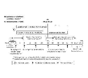
[ 0071 ] Figure I: Shows a Schematic Overview of the Main Study (Screening
through
16-Week Safety Follow-Up. Abbreviations: DBL=database lock; FU=follow-up;
IV=intravenous; PE=primary endpoint; PL=placebo; q8w=every 8 weeks;
SC=subcutaneous; SLE=systemic lupus erythematosus; SRI=SLEDAI-2K Responder
Index;
Wks=weeks.
[ 0072 ] Figure 2: Shows a Schematic Overview of the Study Including the Study
Extension. Abbreviations: DBL=database lock; FU=follow-up; IV=intravenous;
PE=primary endpoint; PL=placebo; q8w=every 8 weeks; SC=subeutaneous;
SLE=systemic
lupus erythematosus; SRI=SLEDAI-2K Responder Index; Wks=weeks.
[ 0073 ] Figure 3: Shows a Kaplan Meier Plot of BILAG Flare Free Time from
Week 12
Through Week 24; Full Analysis Set. BILAG flare defined as at least 1 new
BILAG A or 2
new BILAG B scores (from scores <B). Counts include subjects available for
analysis at a
given visit. Values for subjects meeting treatment failure criteria are set to
missing from the
point of treatment failure forward. *Test for greater treatment effect in
ustekinumab over
placebo performed using a log-rank test.
DETAILED DESCRIPTION OF THE PREFERRED EMBODIMENTS
[ 0074 ] As used herein the method of treatment of lupus comprises
administering isolated,
recombinant and/or synthetic anti-IL-12, IL-23 and ILI 2/23p40 human
antibodies and
diagnostic and therapeutic compositions, methods and devices.
[ 0075 ] As used herein, an "anti-IL-12 antibody," "anti-IL-23 antibody,"
"anti-IL-
12/23p40 antibody," "IL-12/23p40 antibody," "antibody portion," or "antibody
fragment"
and/or "antibody variant" and the like include any protein or peptide
containing molecule
that comprises at least a portion of an immunoglobulin molecule, such as but
not limited to,
at least one complementarity determining region (CDR) of a heavy or light
chain or a ligand
26
CA 03044777 2019-05-23
WO 2019/058345 PCT/1B2018/057368
binding portion thereof, a heavy chain or light chain variable region, a heavy
chain or light
chain constant region, a framework region, or any portion thereof, or at least
one portion of
an IL-12 and/or IL-23 receptor or binding protein, which can be incorporated
into an
antibody of the present invention. Such antibody optionally further affects a
specific ligand,
such as but not limited to, where such antibody modulates, decreases,
increases, antagonizes,
agonizes, mitigates, alleviates, blocks, inhibits, abrogates and/or interferes
with at least one
1L-12/23 activity or binding, or with 1L-12/23 receptor activity or binding,
in vitro, in situ
and/or in vivo. As a non-limiting example, a suitable anti-IL-12/23p40
antibody, specified
portion or variant of the present invention can bind at least one IL-12/23
molecule, or
specified portions, variants or domains thereof. A suitable anti-IL-12/23p40
antibody,
specified portion, or variant can also optionally affect at least one of IL-
12/23 activity or
function, such as but not limited to, RNA, DNA or protein synthesis, IL-12/23
release, IL-
12/23 receptor signaling, membrane IL-12/23 cleavage, IL-12/23 activity, IL-
12/23
production and/or synthesis.
[ 0076 ] The term "antibody" is further intended to encompass antibodies,
digestion
fragments, specified portions and variants thereof, including antibody
mimetics or
comprising portions of antibodies that mimic the structure and/or function of
an antibody or
specified fragment or portion thereof, including single chain antibodies and
fragments
thereof Functional fragments include antigen-binding fragments that bind to a
mammalian
IL-12/23. For example, antibody fragments capable of binding to IL-12/23 or
portions
thereof, including, but not limited to, Fab (e.g., by papain digestion), Fab'
(e.g., by pepsin
digestion and partial reduction) and F(ab)2 (e.g., by pepsin digestion), facb
(e.g., by plasmin
digestion), pFc' (e.g., by pepsin or plasmin digestion), Fd (e.g., by pepsin
digestion, partial
reduction and reaggregation), Fv or say (e.g., by molecular biology
techniques) fragments,
are encompassed by the invention (see, e.g., Colligan, Immunology, supra).
[ 0077 ] Such fragments can be produced by enzymatic cleavage, synthetic or
recombinant
techniques, as known in the art and/or as described herein. Antibodies can
also be produced
in a variety of truncated forms using antibody genes in which one or more stop
codons have
been introduced upstream of the natural stop site. For example, a combination
gene
encoding a F(a1:02 heavy chain portion can be designed to include DNA
sequences encoding
the CH1 domain and/or hinge region of the heavy chain. The various portions of
antibodies
27
CA 03044777 2019-05-23
WO 2019/058345 PCT/1B2018/057368
can be joined together chemically by conventional techniques, or can be
prepared as a
contiguous protein using genetic engineering techniques.
[ 0078 ] As used herein, the term "human antibody" refers to an antibody in
which
substantially every part of the protein (e.g., CDR, framework, CL, CH domains
(e.g., CH1,
CH2, CH3), hinge, (VL, VH)) is substantially non-immunogenic in humans, with
only minor
sequence changes or variations. A "human antibody" may also be an antibody
that is derived
from or closely matches human germline immunoglobulin sequences. Human
antibodies
may include amino acid residues not encoded by germline immunoglobulin
sequences (e.g.,
mutations introduced by random or site-specific mutagenesis in vitro or by
somatic mutation
in vivo). Often, this means that the human antibody is substantially non-
immunogenic in
humans. Human antibodies have been classified into groupings based on their
amino acid
sequence similarities. Accordingly, using a sequence similarity search, an
antibody with a
similar linear sequence can be chosen as a template to create a human
antibody. Similarly,
antibodies designated primate (monkey, baboon, chimpanzee, etc.), rodent
(mouse, rat,
rabbit, guinea pig, hamster, and the like) and other mammals designate such
species, sub-
genus, genus, sub-family, and family specific antibodies. Further, chimeric
antibodies can
include any combination of the above. Such changes or variations optionally
and preferably
retain or reduce the immunogenicity in humans or other species relative to non-
modified
antibodies. Thus, a human antibody is distinct from a chimeric or humanized
antibody.
[ 0079 ] It is pointed out that a human antibody can be produced by a non-
human animal
or prokaryotic or eukaryotic cell that is capable of expressing functionally
rearranged human
immunoglobulin (e.g., heavy chain and/or light chain) genes. Further, when a
human
antibody is a single chain antibody, it can comprise a linker peptide that is
not found in
native human antibodies. For example, an Fv can comprise a linker peptide,
such as two to
about eight glycine or other amino acid residues, which connects the variable
region of the
heavy chain and the variable region of the light chain. Such linker peptides
are considered to
be of human origin.
[ 0080 ] Anti-IL-12/23p40 antibodies (also termed IL-12/23p40 antibodies) (or
antibodies
to IL-23) useful in the methods and compositions of the present invention can
optionally be
characterized by high affinity binding to IL-12/23p40 (or to IL-23) and,
optionally and
28
preferably, having low toxicity. In particular, an antibody, specified
fragment or variant of
the invention, where the individual components, such as the variable region,
constant region
and framework, individually and/or collectively, optionally and preferably
possess low
immunogenicity, is useful in the present invention. The antibodies that can be
used in the
invention are optionally characterized by their ability to treat patients for
extended periods
with measurable alleviation of symptoms and low and/or acceptable toxicity.
Low or
acceptable immunogenicity and/or high affinity, as well as other suitable
properties, can
contribute to the therapeutic results achieved. "Low immunogenicity" is
defined herein as
raising significant HAHA. HACA or HAMA responses in less than about 75%, or
preferably
less than about 50% of the patients treated and/or raising low titres in the
patient treated
(less than about 300, preferably less than about 100 measured with a double
antigen enzyme
immunoassay) (Elliott et al., Lancet 344:1125-1127 (1994)). "Low
immunogenicity" can
also be defined as the incidence of titrable levels of antibodies to the anti-
IL-12 antibody in
patients treated with anti-IL-12 antibody as occurring in less than 25% of
patients treated,
preferably, in less than 10% of patients treated with the recommended dose for
the
recommended course of therapy during the treatment period.
[ 0081 ] The terms " clinically proven efficacy" and "clinically proven
effective" as used
herein in the context of a dose, dosage regimen, treatment or method refer to
the
effectiveness of a particular dose, dosage or treatment regimen. Efficacy can
be measured
based on change in the course of the disease in response to an agent of the
present invention.
For example, an anti-IL12/23p40 or anti-1L23 antibody of the present invention
(e.g., the
anti-IL12/23p40 antibody usetkinumab) is administered to a patient in an
amount and for a
time sufficient to induce an improvement, preferably a sustained improvement,
in at least
one indicator that reflects the severity of the disorder that is being
treated. Various indicators
that reflect the extent of the subject's illness, disease or condition may be
assessed for
determining whether the amount and time of the treatment is sufficient. Such
indicators
include, for example, clinically recognized indicators of disease severity,
symptoms, or
manifestations of the disorder in question. The degree of improvement
generally is
determined by a physician, who may make this determination based on signs,
symptoms,
biopsies, or other test results, and who may also employ questionnaires that
are administered
29
CA 3044777 2019-10-22
CA 03044777 2019-05-23
WO 2019/058345 PCT/1B2018/057368
to the subject, such as quality-of-life questionnaires developed for a given
disease. For
example, an anti-IL12/23p40 or anti-11,23 antibody of the present invention
may be
administered to achieve an improvement in a patient's condition related to
Systemic Lupus
Erythematosus (SLE). Improvement may be indicated by an improvement in an
index of
disease activity, by amelioration of clinical symptoms or by any other measure
of disease
activity. One such index of disease is the Systemic Lupus Erythematosus
Disease Activity
Index 2000 (SLEDAI-2K) score. The SLEDAI-2K is an established, validated
disease
activity index for Systemic Lupus Erythematosus (SLE) that is based on the
presence of
24 features in 9 organ systems and measures disease activity in SLE patients
in the previous
30 days. Features are scored if present within the last 30 days with more
severe features
having higher scores and the scores are added to determine the total SLEDAI-2K
score,
which ranges from 0 to 105. Other disease activity indexes for systemic lupus
erythematosus
(SLE) disease activity assessment include, for example, the Cutaneous Lupus
Erythematosus
Disease Area and Severity Index (CLAS1) and the British Isles Lupus Assessment
Group
(BILAG) index. The CLASI index consists of 2 scores; the first summarizes the
activity of
the disease while the second is a measure of the damage done by the disease.
The scores are
calculated by simple addition based on the extent of the symptoms. Higher
activity and
damage scores indicate worse disease activity. The BILAG index is a measure of
disease
activity consisting of 97 questions in 9 organ systems, each put into 1 of 5
categories (A, B,
C, D, E) depending on presence of items. Higher scores indicate more disease
involvement
[ 0082 ] The term "clinically proven safe", as it relates to a dose, dosage
regimen,
treatment or method with an anti-IL12/23p40 or anti-IL23 antibody of the
present invention
(e.g., the anti-IL12/23p40 antibody usetkinumab), refers to a favorable risk:
benefit ratio with
an acceptable frequency and/or acceptable severity of treatment-emergent
adverse events
(referred to as AEs or TEAEs) compared to the standard of care or to another
comparator.
An adverse event is an untoward medical occurrence in a patient administered a
medicinal
product. In particular, safe as it relates to a dose, dosage regimen or
treatment with an anti-
.1L12/23p40 or anti-IL23 antibody of the present invention refers to with an
acceptable
frequency and/or acceptable severity of adverse events associated with
administration of the
antibody if attribution is considered to be possible, probable, or very likely
due to the use of
the anti-1L12/23p40 or anti-IL23 antibody.
[ 0083 ] As used herein, unless otherwise noted, the term "clinically proven"
(used
independently or to modify the terms "safe" and/or "effective") shall mean
that it has been
proven by a clinical trial wherein the clinical trial has met the approval
standards of U.S.
Food and Drug Administration, EMEA or a corresponding national regulatory
agency. For
example, the clinical study may be an adequately sized, randomized, double-
blinded study
used to clinically prove the effects of the drug.
Utility
[ 0084 ] The isolated nucleic acids of the present invention can be used for
production of
at least one anti-IL-12/23p40 (or anti-IL-23) antibody or specified variant
thereof, which can
be used to measure or effect in an cell, tissue, organ or animal (including
mammals and
humans), to diagnose, monitor, modulate, treat, alleviate, help prevent the
incidence of, or
reduce the symptoms of, at least one IL-12/23 condition, selected from, but
not limited to, at
least one of an immune disorder or disease, a cardiovascular disorder or
disease, an
infectious, malignant, and/or neurologic disorder or disease, or other known
or specified IL-
12/23 related condition.
[ 0085 ] Such a method can comprise administering an effective amount of a
composition
or a pharmaceutical composition comprising at least one anti-IL-12/23p40 (or
anti-IL-23)
antibody to a cell, tissue, organ, animal or patient in need of such
modulation, treatment,
alleviation, prevention, or reduction in symptoms, effects or mechanisms. The
effective
amount can comprise an amount of about 0.001 to 500 mg/kg per single (e.g..
bolus),
multiple or continuous administration, or to achieve a serum concentration of
0.01-5000
ii,g/m1 serum concentration per single, multiple, or continuous
administration, or any
effective range or value therein, as done and determined using known methods,
as described
herein or known in the relevant arts.
Citations
[ 0086 ] Applicant refers to the following references: Ausubel, et al., ed.,
Current
Protocols in Molecular Biology, John Wiley & Sons, Inc., NY, NY (1987-2001);
Sambrook,
et al., Molecular Cloning: A Laboratory Manual, 2nd Edition, Cold Spring
Harbor, NY
(1989); Harlow and Lane, antibodies, a Laboratory Manual, Cold Spring Harbor,
NY
31
CA 3044777 2019-10-22
(1989); Colligan, et al., eds., Current Protocols in Immunology, John Wiley &
Sons, Inc.,
NY (1994-2001); Colligan et al., Current Protocols in Protein Science, John
Wiley & Sons,
NY, NY, (1997-2001).
Antibodies of the Present Invention ¨ Production and Generation
[ 0087 ] At least one anti-IL-12/23p40 (or anti-IL-23) used in the method of
the present
invention can be optionally produced by a cell line, a mixed cell line, an
immortalized cell
or clonal population of immortalized cells, as well known in the art. See,
e.g., Ausubel, et
al., ed., Current Protocols in Molecular Biology, John Wiley & Sons, Inc., NY,
NY (1987-
2001); Sambrook, et al., Molecular Cloning: A Laboratory Manual, 2nd Edition,
Cold Spring
Harbor, NY (1989); Harlow and Lane, antibodies, a Laboratory Manual, Cold
Spring
Harbor, NY (1989); Colligan, et al., eds., Current Protocols in Immunology,
John Wiley &
Sons, Inc., NY (1994-2001); Colligan et al., Current Protocols in Protein
Science, John
Wiley & Sons, NY, NY, (1997-2001).
[ 0088 ] A preferred anti-IL-12/23p40 antibody is ustekinumab (Stelara0)
having the
heavy chain variable region amino acid sequence of SEQ ID NO:7 and the light
chain
variable region amino acid sequence of SEQ ID NO:8 and having the heavy chain
CDR
amino acid sequences of SEQ ID NO:I, SEQ ID NO:2, and SEQ ID NO: 3; and the
light
chain CDR amino acid sequences of SEQ ID NO:4, SEQ ID NO:5. and SEQ ID NO:6. A
preferred anti-IL-23 antibody is guselkumab (also referred to as CNT01959).
Other anti-IL-
23 antibodies have sequences listed herein and are described in U.S. Patent
No. 7,935,344).
[ 0089 ] Human antibodies that are specific for human 1L-12/23p40 or IL-23
proteins or
fragments thereof can be raised against an appropriate immunogenic antigen,
such as an
isolated IL-12/23p40 protein, IL-23 protein and/or a portion thereof
(including synthetic
molecules, such as synthetic peptides). Other specific or general mammalian
antibodies can
32
CA 3044777 2019-10-22
[ 0090 ] In one approach, a hybridoma is produced by fusing a suitable
immortal cell line
(e.g., a myeloma cell line, such as, but not limited to, Sp2/0, Sp2/0-AG14,
NSO, NS1, NS2,
AE-1, L.5, L243, P3X63Ag8.653, Sp2 SA3, Sp2 MA!, Sp2 SS1, Sp2 SA5, U937, MLA
144,
ACT IV, MOLT4, DA-1, JURKAT, WEHI, K-562, COS, RAH, NIH 313, HL-60, MLA
144, NAMALWA, NEURO 2A, or the like, or heteromylomas, fusion products
thereof, or
any cell or fusion cell derived therefrom, or any other suitable cell line as
known in the art)
(see, e.g., www. atcc.org, www. lifetech.com., and the like), with antibody
producing cells,
such as, but not limited to, isolated or cloned spleen, peripheral blood,
lymph, tonsil, or
other immune or B cell containing cells, or any other cells expressing heavy
or light chain
constant or variable or framework or CDR sequences, either as endogenous or
heterologous
nucleic acid, as recombinant or endogenous, viral, bacterial, algal,
prokaryotic, amphibian,
insect, reptilian, fish, mammalian, rodent, equine, ovine, goat, sheep,
primate, eukaryotic,
genomic DNA, cDNA, rDNA, mitochondrial DNA or RNA, chloroplast DNA or RNA,
hnRNA, mRNA, tRNA, single, double or triple stranded, hybridized, and the like
or any
combination thereof. See, e.g., Ausubel, supra, and Colligan. Immunology,
supra, chapter 2.
[ 0091 ] Antibody producing cells can also be obtained from the peripheral
blood or,
preferably, the spleen or lymph nodes, of humans or other suitable animals
that have been
immunized with the antigen of interest. Any other suitable host cell can also
be used for
expressing heterologous or endogenous nucleic acid encoding an antibody,
specified
fragment or variant thereof, of the present invention. The fused cells
(hybridomas) or
recombinant cells can be isolated using selective culture conditions or other
suitable known
methods, and cloned by limiting dilution or cell sorting, or other known
methods. Cells
which produce antibodies with the desired specificity can be selected by a
suitable assay
(e.g., EL1SA).
[ 0092 ] Other suitable methods of producing or isolating antibodies of the
requisite
specificity can be used, including, but not limited to, methods that select
recombinant
antibody from a peptide or protein library (e.g., but not limited to, a
bacteriophage,
33
CA 3044777 2019-10-22
ribosome, oligonucleotide, RNA, cDNA, or the like, display library; e.g., as
available from
Cambridge antibody Technologies, Cambridgeshire, UK; MorphoSys,
Martinsreid/Planegg,
DE; Biovation, Aberdeen, Scotland, UK; BioInvent, Lund, Sweden; Dyax Corp.,
Enzon,
Affymax/Biosite; Xoma, Berkeley, CA; Ixsys. See, e.g., EP 368.684,
PCT/GB91/01134;
PCT/GB92/01755; PCT/GB92/002240; PCT/GB92/00883; PCT/GB93/00605; US
08/350260(5/12/94); PCT/GB94/01422; PCT/GB94/02662; PCT/GB97/01835;
(CAT/MRC); W090/14443; W090/14424; W090/14430; PCT/US94/1234; W092/18619;
W096/07754; (Scripps); W096/13583. W097/08320 (MorphoSys); W095/16027
(Biolnvent); W088/06630; W090/3809 (Dyax); US 4,704,692 (Enzon);
PCT/US91/02989
(Affymax); W089/06283; EP 371 998; EP 550 400; (Xoma); EP 229 046;
PCT/US91/07149
(Ixsys); or stochastically generated peptides or proteins - US 5723323,
5763192, 5814476,
5817483, 5824514, 5976862, WO 86/05803, EP 590 689 (Ixsys, predecessor of
Applied
Molecular Evolution (AME), each entirely incorporated herein by reference)) or
that rely
upon immunization of transgenic animals (e.g., SCID mice, Nguyen et al.,
Microbiol.
Immunol. 41:901-907 (1997); Sandhu et al., Crit. Rev. Biotechnol. 16:95-118
(1996); Eren
et al., Immunol. 93:154-161 (1998), as well as related patents and
applications) that are
capable of producing a repertoire of human antibodies, as known in the art
and/or as
described herein. Such techniques, include, but are not limited to, ribosome
display (Hanes
et al., Proc. Natl. Acad. Sci. USA, 94:4937-4942 (May 1997): Hanes et al.,
Proc. Natl. Acad.
Sci. USA, 95:14130-14135 (Nov. 1998)); single cell antibody producing
technologies (e.g.,
selected lymphocyte antibody method ("SLAM-) (US pat. No. 5,627,052, Wen et
al., J.
Immunol. 17:887-892 (1987); Babcook etal., Proc. Natl. Acad. Sci. USA 93:7843-
7848
(1996)); gel microdroplet and flow cytometry (Powell et al., Biotechnol. 8:333-
337 (1990);
One Cell Systems, Cambridge, MA; Gray et al., J. Imm. Meth. 182:155-163
(1995); Kenny
et al., Bio/Technol. 13:787-790 (1995)); B-cell selection (Steenbakkers etal.,
Molec. Biol.
Reports 19:125-134 (1994); Jonak etal., Progress Biotech, Vol. 5, In Vitro
Immunization in
Hybridoma Technology, Borrebaeck, ed., Elsevier Science Publishers B.V.,
Amsterdam,
Netherlands (1988)).
[ 0093 ] Methods for engineering or humanizing non-human or human antibodies
can also
be used and are well known in the art. Generally, a humanized or engineered
antibody has
one or more amino acid residues from a source that is non-human, e.g., but not
limited to.
34
CA 3044777 2019-10-22
mouse, rat, rabbit, non-human primate or other mammal. These non-human amino
acid
residues are replaced by residues often referred to as "import" residues,
which are typically
taken from an "import" variable, constant or other domain of a known human
sequence.
[ 0094 ] Known human Ig sequences are disclosed, e.g.,
www. ncbi.nlm.nih.gov/entrez/query.fcgi;
www. ncbi.nih.gov/igblast;
www. atcc.org/phage/hdb.html;
www. mrc-epe.cam.ac.uk/ALIGNMENTS.php;
www. kabatdatabase.com/top.html; ftp.ncbi.nih.gov/repository/kabat;
www. sciquest.com;
www. abcam.com;
www. antibodyresource.comionlinecomp.html;
www. public.iastate.edui-pedro/research_tools.html;
www. whfreeman.com/immunology/CH05/kuby05.html;
www. hhmi .org/grant s/lectures/1996/v lab;
www. path.cam.ac.uk/-mrc7/mikeimages.html;
www. mcb.harvard.edu/BioLinks/Immunology.html;
www. immunologylink.com; pathbox.wustl.edu/-hcenter/index.html;
www. appliedbiosystems.com;
www. nal.usda.gov/awic/pubs/antibody;
www. m.ehime-u.ac.jp/-yasuhito/Elisa.html;
www. biodesign.com;
www. cancerresearchuk.org;
www. biotech.ufl.edu;
www. isac-net.org; baserv.uci.kun.n1/-jraats/linksl.html;
www. recab.uni-hd.de/immuno.bme.nwu.edu;
www. mrc-cpe.cam.ac.uk;
www. ibt.unam.mx/virN_mice.html;
www. bioinf.org.uk/abs; antibody.bath.ac.uk;
www. unizh.ch;
www. cryst.bbk.ac.uk/-ubcg07s;
www. n i mr.mrc.ac.uk/CC/ccaewg/ccaewg.html;
www. path.cam.ac.uk/-mrc7/humanisation/TAHHP.html;
www. ibt.unam.mx/vir/structure/stat_aim.html;
www. biosci.missouri.edu/smithgp/index.html;
www. jerini.de;
Kabat et al., Sequences of Proteins of Immunological Interest, U.S. Dept.
Health
(1983)
[ 0095 ] Such imported sequences can be used to reduce immunogenicity or
reduce,
enhance or modify binding, affinity, on-rate, off-rate, avidity, specificity,
half-life, or any
other suitable characteristic, as known in the art. In general, the CDR
residues are directly
CA 3044777 2019-10-22
CA 03044777 2019-05-23
WO 2019/058345 PCT/1B2018/057368
and most substantially involved in influencing antigen binding. Accordingly,
part or all of
the non-human or human CDR sequences are maintained while the non-human
sequences of
the variable and constant regions may be replaced with human or other amino
acids.
[ 0096 ] Antibodies can also optionally be humanized or human antibodies
engineered
with retention of high affinity for the antigen and other favorable biological
properties. To
achieve this goal, humanized (or human) antibodies can be optionally prepared
by a process
of analysis of the parental sequences and various conceptual humanized
products using
three-dimensional models of the parental and humanized sequences. Three-
dimensional
immunoglobulin models are commonly available and are familiar to those skilled
in the art.
Computer programs are available which illustrate and display probable three-
dimensional
conformational structures of selected candidate immunoglobulin sequences.
Inspection of
these displays permits analysis of the likely role of the residues in the
functioning of the
candidate immunoglobulin sequence, i.e., the analysis of residues that
influence the ability
of the candidate immunoglobulin to bind its antigen. In this way, framework
(FR) residues
can be selected and combined from the consensus and import sequences so that
the desired
antibody characteristic, such as increased affinity for the target antigen(s),
is achieved.
[ 0097 ] In addition, the human anti-IL-12/23p40 (or anti-IL-23) specific
antibody used in
the method of the present invention may comprise a human germline light chain
framework.
In particular embodiments, the light chain germline sequence is selected from
human VK
sequences including, but not limited to, Al, A10, All, A14, A17, A18, A19, A2,
A20, A23,
A26, A27, A3, A30, A5, A7, B2, B3, Li, Lb. L11, L12, L14, L15, L16, L18, L19,
L2, L20,
L22, L23, L24, L25, L4/18a, L5, L6, L8, L9, 01, 011, 012, 014, 018, 02, 04,
and 08. In
certain embodiments, this light chain human germline framework is selected
from V1-11,
V1-13, V1-16, V1-17, V1-18, V1-19, V1-2, V1-20, V1-22, V1-3, V1-4, V1-5, V1-7,
V1-9,
V2-1, V2-11, V2-13, V2-14, V2-15, V2-17, V2-19, V2-6, V2-7, V2-8, V3-2, V3-3,
V3-4,
V4-1, V4-2, V4-3, V4-4, V4-6, V5-1, V5-2, V5-4, and V5-6.
[ 0098 ] In other embodiments, the human anti-IL-12/23p40 (or anti-IL-23)
specific
antibody used in the method of the present invention may comprise a human
germline heavy
chain framework. In particular embodiments, this heavy chain human germline
framework is
selected from VI-Ii-18, VH1-2, VH.1-24, VH1-3, VH1-45, VH1-46, VH1-58, VH1-69,
VH1-
36
8, VH2-26, VH2-5, VH2-70, VH3-11, VH3-13, VH3-15, VH3-16, VH3-20, VH3-21, VH3-
23, VH3-30, VH3-33. VH3-35, VH3-38, VH3-43, VH3-48, VH3-49, VH3-53, V113-64,
VH3-66, VH3-7, VH3-72, VH3-73, VH3-74, VH3-9, VH4-28, VI14-31, VH4-34, VH4-39,
VH4-4, VH4-59, VH4-6I, VHS-51, VH6-1. and VH7-81.
[ 0099 1 In particular embodiments, the light chain variable region and/or
heavy chain
variable region comprises a framework region or at least a portion of a
framework region
(e.g., containing 2 or 3 subregions, such as FR2 and FR3). In certain
embodiments, at least
FRL I, FRL2, FRL3, or FRL4 is fully human. In other embodiments, at least
FRH1, FRH2,
FRH3, or FRH4 is fully human. In some embodiments, at least FRL1, FRL2, FRL3,
or
FRL4 is a germline sequence (e.g., human germline) or comprises human
consensus
sequences for the particular framework (readily available at the sources of
known human Ig
sequences described above). In other embodiments, at least FRHI, FRH2, FRH3,
or FRH4
is a germline sequence (e.g., human germline) or comprises human consensus
sequences for
the particular framework. In preferred embodiments, the framework region is a
fully human
framework region.
[ 00100 ] Humanization or engineering of antibodies of the present invention
can be
performed using any known method, such as but not limited to those described
in, Winter
(Jones et al., Nature 321:522 (1986); Riechmann et al., Nature 332:323 (1988);
Verhoeyen
et al., Science 239:1534 (1988)), Sims et al., J. Immunol. 151: 2296 (1993);
Chothia and
Lesk, J. Mol. Biol. 196:901 (1987), Carter et al., Proc. Natl. Acad. Sci.
U.S.A. 89:4285
(1992); Presta et al., J. lmmunol. 151:2623 (1993), US Patent Nos: 5723323,
5976862,
5824514, 5817483, 5814476, 5763192, 5723323, 5,766886, 5714352, 6204023,
6180370,
5693762, 5530101, 5585089, 5225539; 4816567, PCT/: US98/16280, US96/18978,
US91/09630, US91/05939, US94/01234, GB89/01334, GB91/01134, GB92/01755;
W090/14443, W090/14424, W090/14430, EP 229246.
[ 00101 ] In certain embodiments, the antibody comprises an altered (e.g.,
mutated) Fe
region. For example, in some embodiments, the Fe region has been altered to
reduce or
enhance the effector functions of the antibody. In some embodiments, the Fe
region is an
isotype selected from IgM, IgA, IgG. IgE, or other isotype. Alternatively, or
additionally, it
may be useful to combine amino acid modifications with one or more further
amino acid
37
CA 3044777 2019-10-22
modifications that alter Clq binding and/or the complement dependent
cytotoxicity function
of the Fc region of an IL-23 binding molecule. The starting polypeptide of
particular interest
may be one that binds to C I q and displays complement dependent cytotoxicity
(CDC).
Polypeptides with pre-existing Cl q binding activity, optionally further
having the ability to
mediate CDC may be modified such that one or both of these activities are
enhanced. Amino
acid modifications that alter Clq and/or modify its complement dependent
cytotoxicity
function are described, for example, in W00042072.
[ 00102 ] As disclosed above, one can design an Fc region of the human anti-IL-
12/23p40
(or anti-IL-23) specific antibody of the present invention with altered
effector function, e.g.,
by modifying Clq binding and/or FcyR binding and thereby changing complement
dependent cytotoxicity (CDC) activity and/or antibody-dependent cell-mediated
cytotoxicity
(ADCC) activity. "Effector functions" are responsible for activating or
diminishing a
biological activity (e.g., in a subject). Examples of effector functions
include, but are not
limited to: Clq binding; CDC; Fc receptor binding; ADCC; phagocytosis; down
regulation
of cell surface receptors (e.g., B cell receptor; BCR), etc. Such effector
functions may
require the Fc region to be combined with a binding domain (e.g., an antibody
variable
domain) and can be assessed using various assays (e.g., Fc binding assays,
ADCC assays,
CDC assays, etc.).
[ 00103 ] For example, one can generate a variant Fc region of the human anti-
1L-
12/23p40 (or anti-IL-23) antibody with improved Clq binding and improved
FcyRIllbinding
(e.g., having both improved ADCC activity and improved CDC activity).
Alternatively, if it
is desired that effector function be reduced or ablated, a variant Fc region
can be engineered
with reduced CDC activity and/or reduced ADCC activity. In other embodiments,
only one
of these activities may be increased, and, optionally, also the other activity
reduced (e.g., to
generate an Fc region variant with improved ADCC activity, but reduced CDC
activity and
vice versa).
[ 00104 ] Fc mutations can also be introduced in engineer to alter their
interaction with the
neonatal Fc receptor (FeRn) and improve their pharmacokinetic properties. A
collection of
38
CA 3044777 2019-10-22
CA 03044777 2019-05-23
WO 2019/058345 PCT/1B2018/057368
human Fc variants with improved binding to the FcRn have been described
(Shields et al.,
(2001). High resolution mapping of the binding site on human IgG1 for FcyRI,
FcyRII,
FcyRIII, and FcRn and design of TgG1 variants with improved binding to the
FcyR, J. Biol.
Chem. 276:6591-6604).
[ 00105 ] Another type of amino acid substitution serves to alter the
glycosylation pattern
of the Fc region of the human anti-IL-12/23p40 (or anti-IL-23) specific
antibody.
Glycosylation of an Fc region is typically either N-linked or 0-linked. N-
linked refers to the
attachment of the carbohydrate moiety to the side chain of an asparagine
residue. 0-linked
glycosylation refers to the attachment of one of the sugars N-
aceylgalactosamine, galactose,
or xylose to a hydroxyamino acid, most commonly serine or threonine, although
5-
hydroxyproline or 5-hydroxylysine may also be used. The recognition sequences
for
enzymatic attachment of the carbohydrate moiety to the asparagine side chain
peptide
sequences are asparagine-X-serine and asparagine-X-threonine, where X is any
amino acid
except proline. Thus, the presence of either of these peptide sequences in a
polypeptide
creates a potential glycosylation site.
[ 00106 ] The glycosylation pattern may be altered, for example, by deleting
one or more
glycosylation site(s) found in the polypeptide, and/or adding one or more
glycosylation sites
that are not present in the polypeptide. Addition of glycosylation sites to
the Fc region of a
human IL-23 specific antibody is conveniently accomplished by altering the
amino acid
sequence such that it contains one or more of the above-described tripeptide
sequences (for
N-linked glycosylation sites). An exemplary glycosylation variant has an amino
acid
substitution of residue Asn 297 of the heavy chain. The alteration may also be
made by the
addition of, or substitution by, one or more serine or threonine residues to
the sequence of
the original polypeptide (for 0-linked glycosylation sites). Additionally, a
change of Asn
297 to Ala can remove one of the glycosylation sites.
[ 00107 ] In certain embodiments, the human anti-IL-12/23p40 (or anti-IL-23)
specific
antibody of the present invention is expressed in cells that express beta
(1,4)-N-
acetylglucosaminyltransferase III (GnT III), such that GnT III adds GlcNAc to
the human
anti-IL-12/23p40 (or anti-1L-23) antibody. Methods for producing antibodies in
such a
fashion are provided in WO/9954342, WO/03011878, patent publication
20030003097A1,
39
and Umana etal., Nature Biotechnology, 17:176-180, Feb. 1999.
[ 00108 ] The human anti-IL-12/23p40 (or anti-1L-23) antibody can also be
optionally
generated by immunization of a transgenic animal (e.g., mouse, rat, hamster,
non-human
primate, and the like) capable of producing a repertoire of human antibodies,
as described
herein and/or as known in the art. Cells that produce a human anti-IL-12/23p40
(or anti-IL-
23) antibody can be isolated from such animals and immortalized using suitable
methods,
such as the methods described herein.
[ 00109 ] Transgenic mice that can produce a repertoire of human antibodies
that bind to
human antigens can be produced by known methods (e.g., but not limited to,
U.S. Pat. Nos:
5,770,428, 5,569.825, 5,545,806, 5,625,126, 5,625,825, 5,633,425, 5,661,016
and 5,789,650
issued to Lonberg et al.; Jakobovits et at. WO 98/50433, Jakobovits etal. WO
98/24893,
Lonberg et al. WO 98/24884, Lonberg etal. WO 97/13852, Lonberg etal. WO
94/25585,
Kucherlapate et al. WO 96/34096, Kucherlapate et al. EP 0463 151 B 1,
Kucherlapate et al.
EP 0710 719 Al, Surani etal. US. Pat. No. 5,545,807, Bruggemann etal. WO
90/04036,
Bruggemann et al. EP 0438 474 Bl, Lonberg et al. EP 0814 259 A2, Lonberg et
al. GB 2
272 440 A, Lonberg etal. Nature 368:856-859 (1994), Taylor etal., Int.
Immunol. 6(4)579-
591 ( 1 994), Green et al, Nature Genetics 7:13-21(1994), Mendez et at.,
Nature Genetics
15:146-156 (1997), Taylor etal., Nucleic Acids Research 20(23):6287-6295
(1992),
Tuaillon etal., Proc Natl Acad Sci USA 90(8)3720-3724 (1993), Lonberg etal.,
Int Rev
Immunol 13(1):65-93 (1995) and Fishwald et al., Nat Biotechnol 14(7):845-851
(1996)).
Generally, these mice comprise at least one transgene comprising DNA from at
least one
human immunoglobulin locus that is functionally rearranged, or which can
undergo
functional rearrangement. The endogenous immunoglobulin loci in such mice can
be
disrupted or deleted to eliminate the capacity of the animal to produce
antibodies encoded
by endogenous genes.
[ 00110 ] Screening antibodies for specific binding to similar proteins or
fragments can be
conveniently achieved using peptide display libraries. This method involves
the screening of
large collections of peptides for individual members having the desired
function or structure.
Antibody screening of peptide display libraries is well known in the art. The
displayed
CA 3044777 2019-10-22
peptide sequences can be from 3 to 5000 or more amino acids in length,
frequently from 5-
100 amino acids long, and often from about 8 to 25 amino acids long. In
addition to direct
chemical synthetic methods for generating peptide libraries, several
recombinant DNA
methods have been described. One type involves the display of a peptide
sequence on the
surface of a bacteriophage or cell. Each bacteriophage or cell contains the
nucleotide
sequence encoding the particular displayed peptide sequence. Such methods are
described in
PCT Patent Publication Nos. 91/17271, 91/18980, 91/19818, and 93/08278.
[ 00111 ] Other systems for generating libraries of peptides have aspects of
both in vitro
chemical synthesis and recombinant methods. See, PCT Patent Publication Nos.
92/05258,
92/14843, and 96/19256. See also, U.S. Patent Nos. 5,658,754; and 5,643.768.
Peptide
display libraries, vector, and screening kits are commercially available from
such suppliers
as Invitrogen (Carlsbad, CA), and Cambridge antibody Technologies
(Cambridgeshire, UK).
See. e.g., U.S. Pat. Nos. 4704692, 4939666, 4946778, 5260203, 5455030,
5518889,
5534621, 5656730, 5763733, 5767260, 5856456, assigned to Enzon; 5223409,
5403484,
5571698, 5837500, assigned to Dyax, 5427908, 5580717, assigned to Affymax;
5885793,
assigned to Cambridge antibody Technologies; 5750373, assigned to Genentech,
5618920,
5595898, 5576195, 5698435, 5693493, 5698417, assigned to Xoma, Colligan,
supra;
Ausubel, supra; or Sambrook, supra, each of the above patents and
publications.
[ 00112 ] Antibodies used in the method of the present invention can also be
prepared
using at least one anti-IL-12/23p40 (or anti-1L-23) antibody encoding nucleic
acid to
provide transgenic animals or mammals, such as goats, cows, horses, sheep,
rabbits, and the
like, that produce such antibodies in their milk. Such animals can be provided
using known
methods. See, e.g., but not limited to, US Patent Nos. 5,827,690; 5,849,992;
4,873,316;
5,849,992; 5,994,616; 5,565,362; 5,304,489, and the like.
[ 00113 ] Antibodies used in the method of the present invention can
additionally be
prepared using at least one anti-IL-12/23p40 (or anti-IL-23) antibody encoding
nucleic acid
to provide transgenic plants and cultured plant cells (e.g., but not limited
to, tobacco and
maize) that produce such antibodies, specified portions or variants in the
plant parts or in
cells cultured therefrom. As a non-limiting example, transgenic tobacco leaves
expressing
recombinant proteins have been successfully used to provide large amounts of
recombinant
41
CA 3044777 2019-10-22
proteins, e.g., using an inducible promoter. See, e.g., Cramer et al., Curr.
Top. Microbol.
Immunol. 240:95-118 (1999) and references cited therein. Also, transgenic
maize have been
used to express mammalian proteins at commercial production levels, with
biological
activities equivalent to those produced in other recombinant systems or
purified from natural
sources. See, e.g., Hood et al., Adv. Exp. Med. Biol. 464:127-147 (1999) and
references
cited therein. Antibodies have also been produced in large amounts from
transgenic plant
seeds including antibody fragments, such as single chain antibodies (scFv's),
including
tobacco seeds and potato tubers. See, e.g., Conrad et al., Plant Mol. Biol.
38:101-109 (1998)
and references cited therein. Thus, antibodies of the present invention can
also be produced
using transgenic plants, according to known methods. See also, e.g., Fischer
et al.,
Biotechnol. App!. Biochem. 30:99-108 (Oct., 1999), Ma et al., Trends
Biotechnol. 13:522-7
(1995); Ma et al., Plant Physiol. 109:341-6 (1995); Whitelam et al., Biochem.
Soc. Trans.
22:940-944 (1994); and references cited therein.
[ 00114 ] The antibodies used in the method of the invention can bind human 1L-
12/1L-
23p40 or IL-23 with a wide range of affinities (KO. In a preferred embodiment,
a human
mAb can optionally bind human IL-12/IL-23p40 or IL-23 with high affinity. For
example, a
human mAb can bind human IL-12/1L-23p40 or 1L-23 with a KD equal to or less
than about
10-7M, such as but not limited to, 0.1-9.9 (or any range or value therein) X
10-7, 10-8, i09,
10-10, 1011. r-12,
u 1 0-13or any range or value therein.
[ 00115 ] The affinity or avidity of an antibody for an antigen can be
determined
experimentally using any suitable method. (See, for example, Berzofsky, et
al., "Antibody-
Antigen Interactions," In Fundamental Immunology, Paul, W. E., Ed., Raven
Press: New
York, NY (1984); Kuby, Janis Immunology, W. H. Freeman and Company: New York,
NY
(1992); and methods described herein). The measured affinity of a particular
antibody-
antigen interaction can vary if measured under different conditions (e.g.,
salt concentration,
pH). Thus, measurements of affinity and other antigen-binding parameters
(e.g., Kn, Ka, Kd)
are preferably made with standardized solutions of antibody and antigen, and a
standardized
buffer, such as the buffer described herein.
42
CA 3044777 2019-10-22
CA 03044777 2019-05-23
WO 2019/058345 PCT/IB2018/057368
Nucleic Acid Molecules
[ 00116 ] Using the information provided herein, for example, the nucleotide
sequences
encoding at least 70-100% of the contiguous amino acids of at least one of the
light or heavy
chain variable or CDR regions described herein, among other sequences
disclosed herein,
specified fragments, variants or consensus sequences thereof, or a deposited
vector
comprising at least one of these sequences, a nucleic acid molecule of the
present invention
encoding at least one IL-12/IL-23p40 or IL-23 antibody can be obtained using
methods
described herein or as known in the art.
[ 00117 ] Nucleic acid molecules of the present invention can be in the form
of RNA, such
as mRNA, hnRNA, tRNA or any other form, or in the form of DNA, including, but
not
limited to, cDNA and genomic DNA obtained by cloning or produced
synthetically, or any
combinations thereof. The DNA can be triple-stranded, double-stranded or
single-stranded,
or any combination thereof Any portion of at least one strand of the DNA or
RNA can be
the coding strand, also known as the sense strand, or it can be the non-coding
strand, also
referred to as the anti-sense strand.
[ 00118 ] Isolated nucleic acid molecules used in the method of the present
invention can
include nucleic acid molecules comprising an open reading frame (ORF),
optionally, with
one or more introns, e.g., but not limited to, at least one specified portion
of at least one
CDR, such as CDR1, CDR2 and/or CDR3 of at least one heavy chain or light
chain; nucleic
acid molecules comprising the coding sequence for an anti-IL-1211L-23p40 or IL-
23
antibody or variable region; and nucleic acid molecules which comprise a
nucleotide
sequence substantially different from those described above but which, due to
the
degeneracy of the genetic code, still encode at least one anti-IL-12/IL-23p40
or IL-23
antibody as described herein and/or as known in the art. Of course, the
genetic code is well
known in the art. Thus, it would be routine for one skilled in the art to
generate such
degenerate nucleic acid variants that code for specific anti-IL-12/IL-23p40 or
11L-23
antibodies used in the method of the present invention. See, e.g., Ausubel, et
al., supra, and
such nucleic acid variants are included in the present invention. Non-limiting
examples of
isolated nucleic acid molecules include nucleic acids encoding HC CDR1, HC
CDR2, HC
CDR3, LC CDR1, LC CDR2, and LC CDR3, respectively.
43
CA 03044777 2019-05-23
WO 2019/058345 PCT/1B2018/057368
[ 00119 ] As indicated herein, nucleic acid molecules which comprise a nucleic
acid
encoding an anti-IL-12/IL-23p40 or IL-23 antibody can include, but are not
limited to, those
encoding the amino acid sequence of an antibody fragment, by itself; the
coding sequence
for the entire antibody or a portion thereof; the coding sequence for an
antibody, fragment or
portion, as well as additional sequences, such as the coding sequence of at
least one signal
leader or fusion peptide, with or without the aforementioned additional coding
sequences,
such as at least one intron, together with additional, non-coding sequences,
including but not
limited to, non-coding 5' and 3' sequences, such as the transcribed, non-
translated sequences
that play a role in transcription, mRNA processing, including splicing and
polyadenylation
signals (for example, ribosome binding and stability of mRNA); an additional
coding
sequence that codes for additional amino acids, such as those that provide
additional
functionalities. Thus, the sequence encoding an antibody can be fused to a
marker sequence,
such as a sequence encoding a peptide that facilitates purification of the
fused antibody
comprising an antibody fragment or portion.
Polynucleotides Selectively Hybridizing to a Polynucleotide as Described
Herein
[ 00120 ] The method of the present invention uses isolated nucleic acids that
hybridize
under selective hybridization conditions to a polynucleotide disclosed herein.
Thus, the
polynucleotides of this embodiment can be used for isolating, detecting,
and/or quantifying
nucleic acids comprising such polynucleotides. For example, polynucleotides of
the present
invention can be used to identify, isolate, or amplify partial or full-length
clones in a
deposited library. In some embodiments, the polynucleotides are genomic or
cDNA
sequences isolated, or otherwise complementary to, a cDNA from a human or
mammalian
nucleic acid library.
[ 00121 ] Preferably, the cDNA library comprises at least 80% full-length
sequences,
preferably, at least 85% or 90% full-length sequences, and, more preferably,
at least 95%
full-length sequences. The cDNA libraries can be normalized to increase the
representation
of rare sequences. Low or moderate stringency hybridization conditions are
typically, but
not exclusively, employed with sequences having a reduced sequence identity
relative to
complementary sequences. Moderate and high stringency conditions can
optionally be
employed for sequences of greater identity. Low stringency conditions allow
selective
44
hybridization of sequences having about 70% sequence identity and can be
employed to
identify orthologous or paralogous sequences.
[ 00122 ] Optionally, polynucleotides will encode at least a portion of an
antibody. The
polynucleotides embrace nucleic acid sequences that can be employed for
selective
hybridization to a polynucleotide encoding an antibody of the present
invention. See, e.g.,
Ausubel, supra; Colligan, supra.
Construction of Nucleic Acids
[ 00123 ] The isolated nucleic acids can be made using (a) recombinant
methods, (b)
synthetic techniques, (c) purification techniques, and/or (d) combinations
thereof, as well-
known in the art.
[ 00124 ] The nucleic acids can conveniently comprise sequences in addition to
a
polynucleotide of the present invention. For example, a multi-cloning site
comprising one or
more endonuclease restriction sites can be inserted into the nucleic acid to
aid in isolation of
the polynucleotide. Also, translatable sequences can be inserted to aid in the
isolation of the
translated polynucleotide of the present invention. For example, a hexa-
histidine marker
sequence provides a convenient means to purify the proteins of the present
invention. The
nucleic acid of the present invention, excluding the coding sequence, is
optionally a vector,
adapter, or linker for cloning and/or expression of a polynucleotide of the
present invention.
[ 00125 ] Additional sequences can be added to such cloning and/or expression
sequences
to optimize their function in cloning and/or expression, to aid in isolation
of the
polynucleotide, or to improve the introduction of the polynucleotide into a
cell. Use of
cloning vectors, expression vectors, adapters, and linkers is well known in
the art. (See, e.g.,
Ausubel, supra; or Sambrook, supra)
Recombinant Methods for Constructing Nucleic Acids
[ 00126 ] The isolated nucleic acid compositions, such as RNA, cDNA, genomie
DNA, or
any combination thereof, can be obtained from biological sources using any
number of
cloning methodologies known to those of skill in the art. In some embodiments,
oligonucleotide probes that selectively hybridize, under stringent conditions,
to the
polynucleotides of the present invention are used to identify the desired
sequence in a cDNA
CA 3044777 2019-10-22
CA 03044777 2019-05-23
WO 2019/058345 PCT/1B2018/057368
or genomic DNA library. The isolation of RNA, and construction of cDNA and
genomic
libraries, are well known to those of ordinary skill in the art. (See, e.g.,
Ausubel, supra; or
Sambrook, supra)
Nucleic Acid Screening and Isolation Methods
[ 00127 ] A cDNA or genomic library can be screened using a probe based upon
the
sequence of a polynucleotide used in the method of the present invention, such
as those
disclosed herein. Probes can be used to hybridize with genomic DNA or cDNA
sequences to
isolate homologous genes in the same or different organisms. Those of skill in
the art will
appreciate that various degrees of stringency of hybridization can be employed
in the assay;
and either the hybridization or the wash medium can be stringent. As the
conditions for
hybridization become more stringent, there must be a greater degree of
complementarity
between the probe and the target for duplex formation to occur. The degree of
stringency
can be controlled by one or more of temperature, ionic strength, pH and the
presence of a
partially denaturing solvent, such as formamide. For example, the stringency
of
hybridization is conveniently varied by changing the polarity of the reactant
solution
through, for example, manipulation of the concentration of formamide within
the range of
0% to 50%. The degree of complementarity (sequence identity) required for
detectable
binding will vary in accordance with the stringency of the hybridization
medium and/or
wash medium. The degree of complementarity will optimally be 100%, or 70-100%,
or any
range or value therein. However, it should be understood that minor sequence
variations in
the probes and primers can be compensated for by reducing the stringency of
the
hybridization and/or wash medium.
[ 00128 ] Methods of amplification of RNA or DNA are well known in the art and
can be
used according to the present invention without undue experimentation, based
on the
teaching and guidance presented herein.
[ 00129 ] Known methods of DNA or RNA amplification include, but are not
limited to,
polymerase chain reaction (PCR) and related amplification processes (see,
e.g., U.S. Patent
Nos. 4,683,195, 4,683,202, 4,800,159,4,965,188, to Mullis, et al.; 4,795,699
and 4,921,794
to Tabor, et al; 5,142,033 to Innis; 5,122,464 to Wilson, et at.; 5,091,310 to
Innis; 5,066,584
to Gyllensten, et al; 4,889,818 to Gelfand, et al; 4,994,370 to Silver, et al;
4,766,067 to
46
Biswas; 4,656,134 to RingoId) and RNA mediated amplification that uses anti-
sense RNA to
the target sequence as a template for double-stranded DNA synthesis (U.S.
Patent No.
5,130,238 to Malek, et al, with the tradename NASBA). (See, e.g., Ausubel,
supra; or
Sambrook, supra.)
[ 00130 ] For instance, polymerase chain reaction (PCR) technology can be used
to
amplify the sequences of polynucleotides used in the method of the present
invention and
related genes directly from genomic DNA or cDNA libraries. PCR and other in
vitro
amplification methods can also be useful, for example, to clone nucleic acid
sequences that
code for proteins to be expressed, to make nucleic acids to use as probes for
detecting the
presence of the desired mRNA in samples, for nucleic acid sequencing, or for
other
purposes. Examples of techniques sufficient to direct persons of skill through
in vitro
amplification methods are found in Berger, supra, Sambrook, supra, and
Ausubel, supra, as
well as Mullis, et al., U.S. Patent No. 4,683,202 (1987); and Innis, et al..
PCR Protocols A
Guide to Methods and Applications, Eds., Academic Press Inc., San Diego, CA
(1990).
Commercially available kits for genomic PCR amplification are known in the
art. See, e.g.,
Advantage-GC Genomic PCR Kit (Clontech). Additionally, e.g., the T4 gene 32
protein
(Boehringer Mannheim) can be used to improve yield of long PCR products.
Synthetic Methods for Constructing Nucleic Acids
[ 00131 ] The isolated nucleic acids used in the method of the present
invention can also
be prepared by direct chemical synthesis by known methods (see, e.g., Ausubel,
et al..
supra). Chemical synthesis generally produces a single-stranded
oligonucleotide, which can
be converted into double-stranded DNA by hybridization with a complementary
sequence,
or by polymerization with a DNA polymerase using the single strand as a
template. One of
skill in the art will recognize that while chemical synthesis of DNA can be
limited to
sequences of about 100 or more bases, longer sequences can be obtained by the
ligation of
shorter sequences.
47
CA 3044777 2019-10-22
Recombinant Expression Cassettes
[ 00132 ] The present invention uses recombinant expression cassettes
comprising a
nucleic acid. A nucleic acid sequence, for example, a cDNA or a genomic
sequence
encoding an antibody used in the method of the present invention, can be used
to construct a
recombinant expression cassette that can be introduced into at least one
desired host cell. A
recombinant expression cassette will typically comprise a polynucleotide
operably linked to
transcriptional initiation regulatory sequences that will direct the
transcription of the
polynucleotide in the intended host cell. Both heterologous and non-
heterologous (i.e.,
endogenous) promoters can be employed to direct expression of the nucleic
acids.
[ 00133 ] In some embodiments, isolated nucleic acids that serve as promoter,
enhancer, or
other elements can be introduced in the appropriate position (upstream,
downstream or in
the intron) of a non-heterologous form of a polynucleotide of the present
invention so as to
up or down regulate expression of a polynucleotide. For example, endogenous
promoters
can be altered in vivo or in vitro by mutation, deletion and/or substitution.
Vectors and Host Cells
[ 00134 ] The present invention also relates to vectors that include isolated
nucleic acid
molecules, host cells that are genetically engineered with the recombinant
vectors, and the
production of at least one anti-IL-23 antibody by recombinant techniques, as
is well known
in the art. See, e.g., Sambrook, et al., supra; Ausubel, et al., supra.
[ 00135 ] The polynucleotides can optionally be joined to a vector containing
a selectable
marker for propagation in a host. Generally, a plasmid vector is introduced in
a precipitate,
such as a calcium phosphate precipitate, or in a complex with a charged lipid.
If the vector is
a virus, it can be packaged in vitro using an appropriate packaging cell line
and then
transduced into host cells.
[ 00136 ] The DNA insert should be operatively linked to an appropriate
promoter. The
expression constructs will further contain sites for transcription initiation,
termination and,
in the transcribed region, a ribosome binding site for translation. The coding
portion of the
mature transcripts expressed by the constructs will preferably include a
translation initiating
48
CA 3044777 2019-10-22
at the beginning and a termination codon (e.g., UAA, UGA or UAG) appropriately
positioned at the end of the mRNA to be translated, with UAA and UAG preferred
for
mammalian or eukaryotic cell expression.
[ 00137 1 Expression vectors will preferably but optionally include at least
one selectable
marker. Such markers include, e.g., but are not limited to, methotrexate
(MTX),
dihydrofolate reductase (DHFR, US Pat.Nos. 4,399,216; 4,634,665; 4,656,134;
4,956,288;
5,149,636; 5,179,017, ampicillin, neomycin (G418), mycophenolic acid, or
glutamine
synthetase (GS, US Pat.Nos. 5,122,464; 5,770,359; 5,827,739) resistance for
eukaryotic cell
culture, and tetracycline or ampicillin resistance genes for culturing in E.
coli and other
bacteria or prokaryotics. Appropriate culture mediums and conditions for the
above-
described host cells are known in the art. Suitable vectors will be readily
apparent to the
skilled artisan. Introduction of a vector construct into a host cell can be
effected by calcium
phosphate transfection, DEAE-dextran mediated transfection, cationic lipid-
mediated
transfection, electroporation, transduction, infection or other known methods.
Such methods
are described in the art, such as Sambrook, supra, Chapters 1-4 and 16-18;
Ausubel, supra,
Chapters 1, 9, 13, 15, 16.
[ 00138 ] At least one antibody used in the method of the present invention
can be
expressed in a modified form, such as a fusion protein, and can include not
only secretion
signals, but also additional heterologous functional regions. For instance, a
region of
additional amino acids, particularly charged amino acids, can be added to the
N-terminus of
an antibody to improve stability and persistence in the host cell, during
purification, or
= during subsequent handling and storage. Also, peptide moieties can be
added to an antibody
of the present invention to facilitate purification. Such regions can be
removed prior to final
preparation of an antibody or at least one fragment thereof. Such methods are
described in
many standard laboratory manuals, such as Sambrook, supra, Chapters 17.29-
17.42 and
18.1-18.74: Ausubel, supra, Chapters 16, 17 and 18.
[ 00139 ] Those of ordinary skill in the art are knowledgeable in the numerous
expression
systems available for expression of a nucleic acid encoding a protein used in
the method of
the present invention. Alternatively, nucleic acids can be expressed in a host
cell by turning
on (by manipulation) in a host cell that contains endogenous DNA encoding an
antibody.
49
CA 3044777 2019-10-22
Such methods are well known in the art, e.g., as described in US patent Nos.
5,580,734,
5,641,670, 5,733,746. and 5,733,761.
[ 00140 ] Illustrative of cell cultures useful for the production of the
antibodies, specified
portions or variants thereof, are mammalian cells. Mammalian cell systems
often will be in
the form of monolayers of cells although mammalian cell suspensions or
bioreactors can
also be used. A number of suitable host cell lines capable of expressing
intact glyeosylated
proteins have been developed in the art, and include the COS-1 (e.g., ATCC CRL
1650),
COS-7 (e.g., ATCC CRL-1651), HEK293, BHK21 (e.g., ATCC CRL-10), CHO (e.g.,
ATCC CRL 1610) and BSC-1 (e.g., ATCC CRL-26) cell lines. Cos-7 cells, CHO
cells, hep
G2 cells, P3X63Ag8.653, SP2/0-Ag14, 293 cells, HeLa cells and the like, which
are readily
available from, for example, American Type Culture Collection, Manassas, Va (w-
ww.
atcc.org). Preferred host cells include cells of lymphoid origin, such as
myeloma and
lymphoma cells. Particularly preferred host cells are P3X63Ag8.653 cells (ATCC
Accession
Number CRL-1580) and SP2/0-Ag14 cells (ATCC Accession Number CRL-1851). In a
particularly preferred embodiment, the recombinant cell is a P3X63Ab8.653 or a
SP2/0-
Ag14 cell.
[ 00141 ] Expression vectors for these cells can include one or more of the
following
expression control sequences, such as, but not limited to, an origin of
replication; a promoter
(e.g., late or early SV40 promoters, the CMV promoter (US Pat.Nos. 5,168,062;
5,385,839),
an HSV tk promoter, a pgk (phosphoglycerate kinase) promoter, an EF-1 alpha
promoter
(US Pat.No. 5,266,491), at least one human immunoglobulin promoter; an
enhancer, and/or
processing information sites, such as ribosome binding sites, RNA splice
sites,
polyadenylation sites (e.g., an SV40 large T Ag poly A addition site), and
transcriptional
terminator sequences. See, e.g., Ausubel et al., supra; Sambrook, et al.,
supra. Other cells
useful for production of nucleic acids or proteins of the present invention
are known and/or
available, for instance, from the American Type Culture Collection Catalogue
of Cell Lines
and Hybridomas (www. atcc.org) or other known or commercial sources.
[ 00142 ] When eukaryotic host cells are employed, polyadenlyation or
transcription
terminator sequences are typically incorporated into the vector. An example of
a terminator
sequence is the polyadenlyation sequence from the bovine growth hormone gene.
Sequences
CA 3044777 2019-10-22
for accurate splicing of the transcript can also be included. An example of a
splicing
sequence is the VP1 intron from SV40 (Sprague, et al., J. Virol. 45:773-781
(1983)).
Additionally, gene sequences to control replication in the host cell can be
incorporated into
the vector, as known in the art.
Purification of an Antibody
[ 00143 ] An anti-1L-12/1L-23p40 or 1L-23 antibody can be recovered and
purified from
recombinant cell cultures by well-known methods including, but not limited to,
protein A
purification, ammonium sulfate or ethanol precipitation, acid extraction,
anion or cation
exchange chromatography, phosphocellulose chromatography, hydrophobic
interaction
chromatography, affinity chromatography, hydroxylapatite chromatography and
lectin
chromatography. High performance liquid chromatography ("HPLC") can also be
employed
for purification. See, e.g., Colligan, Current Protocols in Immunology, or
Current Protocols
in Protein Science, John Wiley & Sons, NY, NY, (1997-2001), e.g., Chapters 1,
4, 6, 8, 9,
10.
[ 00144 1 Antibodies used in the method of the present invention include
naturally purified
products, products of chemical synthetic procedures, and products produced by
recombinant
techniques from a eukaryotic host, including, for example, yeast, higher
plant, insect and
mammalian cells. Depending upon the host employed in a recombinant production
procedure, the antibody can be glycosylated or can be non-glycosylated, with
glycosylated
preferred. Such methods are described in many standard laboratory manuals,
such as
Sambrook, supra, Sections 17.37-17.42; Ausubel, supra, Chapters 10, 12, 13,
16, 18 and 20,
Colligan, Protein Science, supra, Chapters 12-14.
Anti-IL-12/IL-23p40 or IL-23 Antibodies
[ 00145 ] An anti-IL-12/1L-23p40 or IL-23 antibody according to the present
invention
includes any protein or peptide containing molecule that comprises at least a
portion of an
immunoglobulin molecule, such as but not limited to, at least one ligand
binding portion
(LBP), such as but not limited to, a complementarity determining region (CDR)
of a heavy
or light chain or a ligand binding portion thereof, a heavy chain or light
chain variable
51
CA 3044777 2019-10-22
CA 03044777 2019-05-23
WO 2019/058345 PCT/1B2018/057368
region, a framework region (e.g., FRI , FR2, FR3, FR4 or fragment thereof,
further
optionally comprising at least one substitution, insertion or deletion), a
heavy chain or light
chain constant region, (e.g., comprising at least one Cal, hinge], hinge2,
hinge3, hinge4,
CH2, or CH3 or fragment thereof, further optionally comprising at least one
substitution,
insertion or deletion), or any portion thereof, that can be incorporated into
an antibody. An
antibody can include or be derived from any mammal, such as but not limited
to, a human, a
mouse, a rabbit, a rat, a rodent, a primate, or any combination thereof, and
the like.
[ 00146 ] The isolated antibodies used in the method of the present invention
comprise the
antibody amino acid sequences disclosed herein encoded by any suitable
polynucleotide, or
any isolated or prepared antibody. Preferably, the human antibody or antigen-
binding
fragment binds human IL-12fIL-23p40 or IL-23 and, thereby, partially or
substantially
neutralizes at least one biological activity of the protein. An antibody, or
specified portion or
variant thereof, that partially or preferably substantially neutralizes at
least one biological
activity of at least one IL-12/IL-23p40 or IL-23 protein or fragment can bind
the protein or
fragment and thereby inhibit activities mediated through the binding of IL-
12/IL-23p40 or
IL-23 to the IL-12 and/or 1L-23 receptor or through other IL-12/1L-23p40 or IL-
23-
dependent or mediated mechanisms. As used herein, the term "neutralizing
antibody" refers
to an antibody that can inhibit an IL-12/IL-23p40 or IL-23-dependent activity
by about 20-
120%, preferably by at least about 10, 20, 30, 40, 50, 55, 60, 65, 70, 75, 80,
85, 90, 91, 92,
93, 94, 95, 96, 97, 98, 99, 100% or more depending on the assay. The capacity
of an anti-IL-
12/IL-23p40 or IL-23 antibody to inhibit an IL-12/IL-23p40 or IL-23-dependent
activity is
preferably assessed by at least one suitable IL-12/11.-23p40 or IL-23 protein
or receptor
assay, as described herein and/or as known in the art. A human antibody can be
of any class
(TgG, IgA, TgM, IgE, IgD, etc.) or isotype and can comprise a kappa or lambda
light chain.
In one embodiment, the human antibody comprises an IgG heavy chain or defined
fragment,
for example, at least one of isotypes, IgGl, IgG2, IgG3 or IgG4 (e.g., y I ,
y2, y3, y4).
Antibodies of this type can be prepared by employing a transgenic mouse or
other trangenic
non-human mammal comprising at least one human light chain (e.g., IgG, IgA,
and IgM)
transgenes as described herein and/or as known in the art. In another
embodiment, the anti-
IL-23 human antibody comprises an IgG1 heavy chain and an IgG1 light chain.
52
CA 03044777 2019-05-23
WO 2019/058345 PCT/1B2018/057368
[ 00147 ] An antibody binds at least one specified epitope specific to at
least one IL-12/IL-
23p40 or IL-23 protein, subunit, fragment, portion or any combination thereof.
The at least
one epitope can comprise at least one antibody binding region that comprises
at least one
portion of the protein, which epitope is preferably comprised of at least one
extracellular,
soluble, hydrophillic, external or cytoplasmic portion of the protein.
[ 00148 ] Generally, the human antibody or antigen-binding fragment will
comprise an
antigen-binding region that comprises at least one human complementarity
determining
region (CDR1, CDR2 and CDR3) or variant of at least one heavy chain variable
region and
at least one human complementarity determining region (CDR1, CDR2 and CDR3) or
variant of at least one light chain variable region. The CDR sequences may be
derived from
human germline sequences or closely match the germline sequences. For example,
the
CDRs from a synthetic library derived from the original non-human CDRs can be
used.
These CDRs may be formed by incorporation of conservative substitutions from
the original
non-human sequence. In another particular embodiment, the antibody or antigen-
binding
portion or variant can have an antigen-binding region that comprises at least
a portion of at
least one light chain CDR (i.e., CDR1, CDR2 and/or CDR3) having the amino acid
sequence of the corresponding CDRs 1, 2 and/or 3.
[ 00149 ] Such antibodies can be prepared by chemically joining together the
various
portions (e.g., CDRs, framework) of the antibody using conventional
techniques, by
preparing and expressing a (i.e., one or more) nucleic acid molecule that
encodes the
antibody using conventional techniques of recombinant DNA technology or by
using any
other suitable method.
[ 00150 ] The anti-IL-12/IL-23p40 or IL-23 specific antibody can comprise at
least one of
a heavy or light chain variable region having a defined amino acid sequence.
For example,
in a preferred embodiment, the anti-IL-12/IL-23p40 or IL-23 antibody comprises
an anti-IL-
12/IL-23p40 antibody with a heavy chain variable region comprising the amino
acid
sequence of SEQ ID NO:7 and a light chain variable region comprising the amino
acid
sequence of SEQ ID NO: 8. The anti-IL-12/1L-23p40 or IL-23 specific antibody
can also
comprise at least one of a heavy or light chain having a defined amino acid
sequence. In
another preferred embodiment, the anti-IL-12/IL-23p40 or IL-23 antibody
comprises an
53
CA 03044777 2019-05-23
WO 2019/058345 PCT/1B2018/057368
anti-IL-12/1L-23p40 antibody with a heavy chain comprising the amino acid
sequence of
SEQ ID NO:10 and a light chain comprising the amino acid sequence of SEQ ID
NO: ii.
Antibodies that bind to human IL-12/1L-23p40 or 1L-23 and that comprise a
defined heavy
or light chain variable region can be prepared using suitable methods, such as
phage display
(Katsube, Y., etal., Int J Mot. Med,1(5):863-868 (1998)) or methods that
employ transgenic
animals, as known in the art and/or as described herein. For example, a
transgenic mouse,
comprising a functionally rearranged human immunoglobulin heavy chain
transgene and a
transgene comprising DNA from a human immunoglobulin light chain locus that
can
undergo functional rearrangement, can be immunized with human IL-12/1L-23p40
or IL-23
or a fragment thereof to elicit the production of antibodies. If desired, the
antibody
producing cells can be isolated and hybridomas or other immortalized antibody-
producing
cells can be prepared as described herein and/or as known in the art.
Alternatively, the
antibody, specified portion or variant can be expressed using the encoding
nucleic acid or
portion thereof in a suitable host cell.
[ 00151 ] The invention also relates to antibodies, antigen-binding fragments,
immunoglobulin chains and CDRs comprising amino acids in a sequence that is
substantially the same as an amino acid sequence described herein. Preferably,
such
antibodies or antigen-binding fragments and antibodies comprising such chains
or CDRs can
bind human IL-12/1L-23p40 or IL-23 with high affinity (e.g., KD less than or
equal to about
10-9M). Amino acid sequences that are substantially the same as the sequences
described
herein include sequences comprising conservative amino acid substitutions, as
well as amino
acid deletions and/or insertions. A conservative amino acid substitution
refers to the
replacement of a first amino acid by a second amino acid that has chemical
and/or physical
properties (e.g., charge, structure, polarity, hydrophobicity/hydrophilicity)
that are similar to
those of the first amino acid. Conservative substitutions include, without
limitation,
replacement of one amino acid by another within the following groups: lysine
(K), arginine
(R) and histidine (H); aspartate (D) and glutamate (E); asparagine (N),
glutamine (Q), serine
(S), threonine (T), tyrosine (Y), K, R, H, D and E; alanine (A), valine (V),
leucine (L),
isoleucine (I), proline (P), phenylalanine (F), tryptophan (W), methionine
(M), cysteine (C)
and glycine (G); F, W and Y; C, S and T.
54
CA 03044777 2019-05-23
WO 2019/058345
PCT/1B2018/057368
Amino Acid Codes
[ 00152 ] The amino acids that make up anti-IL-12/IL-23p40 or IL-23 antibodies
of the
present invention are often abbreviated. The amino acid designations can be
indicated by
designating the amino acid by its single letter code, its three letter code,
name, or three
nucleotide codon(s) as is well understood in the art (see Alberts, B., et al.,
Molecular
Biology of The Cell, Third Ed., Garland Publishing, Inc., New York, 1994):
SINGLE THREE NAME THREE
LETTER LETTER NUCLEOTIDE
CODE CODE CODON(S)
A * Ala Alanine GCA, GCC, GCG,
GCU
Cys Cysteine UGC, UGU
Asp Aspartic acid GAC, GAU
Glu Glutamic acid GAA, GAG
Phe Phenylanine UUC, UUU
Gly Glycine GGA, GGC, GGG,
GGU
His Histidine CAC, CAU
Ile Isoleucine AUA, AUC, AUU
Lys Lysine AAA, AA.G
Leu Leucine UUA, UUG, CUA,
CUC, CUG, CUU
Met Methionine AUG
Asn Asparagine AAC, AAU
CA 03044777 2019-05-23
WO 2019/058345
PCT/1B2018/057368
Pro Proline CCA, CCC, CCG,
CCU
Gin Glutamine CAA, CACi
Arg Arginine AGA, AGG, CGA,
CGC, CGG, CGU
Set Serine AGC, AGU, UCA,
UCC, UCG, UCU
Thr Threonine ACA, ACC, ACG,
ACU
V Val Valine GUA, GUC, GUG,
GUU
Trp Tryptophan UGG
Tyr Tyrosine UAC, UAU
Sequences
Example anti-IL-12/IL-23p40 antibody sequences - STELARA (ustekinumab)
[ 00153 ] Amino acid sequence of anti-IL-12/IL-23p40 antibody connplementarity
determining region heavy chain 1 (CDRHI): (SEQ ID NO:
TriolLG
[ 00154 ] Amino acid sequence of anti-IL-12/1L-23p40 antibody complementarity
determining region heavy chain 2 (CDRH2): (SEQ ID NO:2)
IMSPVDSDIRYSPSFQG
[ 00155 ] Amino acid sequence of anti-IL-12/1L-23p40 antibody complementarity
determining region heavy chain 3 (CDRH3): (SEQ ID NO:3)
RRPGQGYFDF
56
CA 03044777 2019-05-23
WO 2019/058345 PCT/IB2018/057368
[ 00156 ] Amino acid sequence of anti-IL-12/1L-23p40 antibody complementarity
determining region light chain 1 (CDRL1.): (SEQ ID NO:4)
RASQGISSWLA
[ 00157 ] Amino acid sequence of anti-1L-12/IL-23p40 antibody complementarity
determining region light chain 2 (CDRL2): (SEQ 1D NO:5)
AASSLQS
[ 00158 1 Amino acid sequence of anti-IL-12/TL-23p40 antibody complementarity
determining region light chain 3 (CDRL3): (SEQ ID NO:6)
QQYNIYPYT
[ 00159 ] Amino acid sequence of anti-IL-12/IL-23p40 antibody variable heavy
chain
region (CDRs underlined): (SEQ ID NO: 7)
1 EVQLVQSGAE VKKPGESLKI SCKGSGYSFT TYWLGWVRQM PGKGLDWIGI MSPVDSDIRY
61 SPSFQGQVTM SVDKSITTAY LQWNSLKASD TAMYYCARRR PGQGYFDFWG QGTLVTVSS
[ 00160 1 Amino acid sequence of anti-IL-12/IL-23p40 antibody variable light
chain
region (CDRs underlined): (SEQ ID NO:8)
1 DIQMTQSPSS LSASVGDRVT ITCRASQGIS SWLAWYQQKP EKAPKSLIYA ASSLQSGVPS
61 RFSGSGSGTD FTLTISSLQP EDFATYYCQQ YNIYPYTFGQ GTKLEIKR
[ 00161 ] Amino acid sequence of anti-IL-12/IL-23p40 antibody heavy chain
(CDRs
underlined): (SEQ ID NO:10)
1 EVQLVQSGAE VKKPGESLKI SCKGSGYSFT TYWLGWVRQM PGKGLDWIGI MSPVDSDIRY
61 SPSFQGQVTM SVDKSITTAY LQWNSLKASD TAMYYCARRR PGQGYFDFWG QGTLVTVSSS
121 STKGPSVFPL APSSKSTSGG TAALGCLVKD YFPEPVrVSW NSGALTSGVH TFPAVLQSSG
181 LYSLSSVVTV PSSSLGTQTY ICNVNHKPSN TKVDKRVEPK SCDKTHTCPP CPAPELLGGP
241 SVFLFPPKPK DTLMISRTPE VTCVVVDVSH EDPEVKFNWY VDGVEVHNAK TKPREEQYNS
301 TYRVVSVLTV LHQDWLNGKE YKCKVSNKAL PAPTEKTISK AKGQPREPQV YTLPPSRDEL
361 TKNQVSLTCL VKGFYPSDIA VEWESNGQPE NNYKTTPPVL DSDGSFFLYS KLTVDKSRWQ
421 QGNVESCSVM HEALHNHYTQ KSLSLSPGK
[ 00162 1 Amino acid sequence of anti-IL-12/1L-23p40 antibody light chain
(CDRs
underlined): (SEQ ID NO:11)
1 DIQMTQSPSS LSASVGDRVT ITCRASQGIS SWLAWYQQKP EKAPKSLIYA ASSLQSGVPS
61 RFSGSGSGTD FTLTISSLQP EDFATYYCQQ YNIYPYTFGQ GTKLEIKRTV AAPSVFIFPP
121 SDEQLKSGTA SVVCLLNNFY PREAKVQWKV DNALQSGNSQ ESVTEQDSKD STYSLSSTLT
57
CA 03044777 2019-05-23
WO 2019/058345 PCT/IB2018/057368
181 LSKADYEKHK VYACEVTHQG LSSPVTKSFN RGEC
Amino acid sequence IL-12
[ 00163 ] Amino acid sequence of human interleukin (IL)-12 with alpha and beta
subunits:
(SEQ ID NO:9)
1 RNLPVATPDP GMFPCLHHSQ NLLRAVSNML QKARQTLEFY PCTSEEIDHE DITKDKTSTV
61 EACLPLELTK NESCLNSRET SFITNGSCLA SRKTSFMMAL CLSSIYEDLK MYQVEFKTMN
121 AKLLMDPKRQ IFLDQNMLAV IDELMQALNF NSETVPQKSS LEEPDFYKTK IKLCILLHAF
181 RIRAVTIDRV MSYLNASIWE LKKDVYVVEL DWYPDAPGEM VVLTCDTPEE DGITWTLDQS
241 SEVLGSGKTL TIQVKEFGDA GQYTCHKGGE VLSHSLLLLH KKEDGIWSTD 1LKDQKEPKN
301 KTFLRCEAKN YSGRFTCWWL TTISTDLTFS VKSSRGSSDP QGVTCGAATL SAERVRGDNK
361 EYEYSVECQE DSACPAAEES LPIEVMVDAV HKLKYENYTS SFFIRDIIKP DPPKNLQLKP
421 LKNSRQVEVS WEYPDTWSTP HSYFSLTFCV QVQGKSKREK KDRVFTDKTS ATVICRKNAS
481 ISVRAQDRYY SSSWSEWASV PCS
[ 00164 ] An anti-IL-12/IL-23p40 or IL-23 antibody used in the method of the
present
invention can include one or more amino acid substitutions, deletions or
additions, either
from natural mutations or human manipulation, as specified herein.
[ 00165 ] The number of amino acid substitutions a skilled artisan would make
depends on
many factors, including those described above. Generally speaking, the number
of amino
acid substitutions, insertions or deletions for any given anti-IL-1211L-23p40
or IL-23
antibody, fragment or variant will not be more than 40, 30, 20, 19, 18, 17,
16, 15, 14, 13, 12,
11, 10, 9, 8, 7, 6, 5, 4, 3, 2, 1, such as 1-30 or any range or value therein,
as specified herein.
[ 00166 ] Amino acids in an anti-IL-1211L-23p40 or IL-23 specific antibody
that are
essential for function can be identified by methods known in the art, such as
site-directed
mutagenesis or alanine-scanning mutagenesis (e.g., Ausubel, supra, Chapters 8,
15;
Cunningham and Wells, Science 244:1081-1085 (1989)). The latter procedure
introduces
single alanine mutations at every residue in the molecule. The resulting
mutant molecules
are then tested for biological activity, such as, but not limited to, at least
one IL-12/1L-23p40
or 1L-23 neutralizing activity. Sites that are critical for antibody binding
can also be
identified by structural analysis, such as crystallization, nuclear magnetic
resonance or
photoaffinity labeling (Smith, et al., J. Mol. Biol. 224:899-904(1992) and de
Vos, et al.,
Science 255:306-312 (1992)).
58
CA 03044777 2019-05-23
WO 2019/058345 PCT/1B2018/057368
[ 00167 ] Anti-IL-1211L-23p40 or IL-23 antibodies can include, but are not
limited to, at
least one portion, sequence or combination selected from 5 to all of the
contiguous amino
acids of at least one of SEQ ID NOs 1, 2, 3,4, 5, 6, 7, 8, 10, or 11.
[ 00168 ] IL-1211L-23p40 or IL-23 antibodies or specified portions or variants
can include,
but are not limited to, at least one portion, sequence or combination selected
from at least 3-
contiguous amino acids of the SEQ ID NOs above; 5-17 contiguous amino acids of
the
SEQ ID NOs above, 5-10 contiguous amino acids of the SEQ ID NOs above, 5-11
contiguous amino acids of the SEQ ID NOs above, 5-7 contiguous amino acids of
the SEQ
ID NOs above; 5-9 contiguous amino acids of the SEQ ID NOs above.
[ 00169 ] An anti-IL-12/IL-23p40 or IL-23 antibody can further optionally
comprise a
polypeptide of at least one of 70-100% of 5, 17, 10, 11, 7, 9, 119, 108, 449,
or 214
contiguous amino acids of the SEQ ID NOs above. In one embodiment, the amino
acid
sequence of an immunoglobulin chain, or portion thereof (e.g., variable
region, CDR) has
about 70-100% identity (e.g., 70, 71, 72, 73, 74, 75, 76, 77, 78, 79, 80, 81,
82, 83, 84, 85,
86, 87, 88, 89, 90, 91, 92, 93, 94, 95, 96, 97, 98, 99, 100 or any range or
value therein) to the
amino acid sequence of the corresponding chain of at least one of the SEQ ID
NOs above.
For example, the amino acid sequence of a light chain variable region can be
compared with
the sequence of the SEQ ID NOs above, or the amino acid sequence of a heavy
chain CDR3
can be compared with the SEQ ID NOs above. Preferably, 70-100% amino acid
identity
(i.e., 90, 91, 92, 93, 94, 95, 96, 97, 98, 99, 100 or any range or value
therein) is determined
using a suitable computer algorithm, as known in the art.
[ 00170 ] "Identity," as known in the art, is a relationship between two or
more
polypeptide sequences or two or more polynucleotide sequences, as determined
by
comparing the sequences. In the art, "identity" also means the degree of
sequence
relatedness between polypeptide or polynucleotide sequences, as determined by
the match
between strings of such sequences. "Identity" and "similarity" can be readily
calculated by
known methods, including, but not limited to, those described in Computational
Molecular
Biology, Lesk, A. M., ed., Oxford University Press, New York, 1988;
Biocomputing:Informatics and Genome Projects, Smith, D. W., ed., Academic
Press, New
York, 1993; Computer Analysis of Sequence Data, Part I, Griffin, A. M., and
Griffin, H. G.,
59
CA 03044777 2019-05-23
WO 2019/058345 PCT/1B2018/057368
eds., Humana Press, New Jersey, 1994; Sequence Analysis in Molecular Biology,
von
Heinje, G., Academic Press, 1987; and Sequence Analysis Primer, Gribskov, M.
and
Devereux, J., eds., M Stockton Press, New York, 1991; and Carillo, H., and
Lipman, D.,
Siam J. Applied Math., 48:1073 (1988). In addition, values for percentage
identity can be
obtained from amino acid and nucleotide sequence alignments generated using
the default
settings for the AlignX component of Vector NT! Suite 8.0 (Informax,
Frederick, MD).
[ 00171 ] Preferred methods to determine identity are designed to give the
largest match
between the sequences tested. Methods to determine identity and similarity are
codified in
publicly available computer programs. Preferred computer program methods to
determine
identity and similarity between two sequences include, but are not limited to,
the GCG
program package (Devereux, J., et al., Nucleic Acids Research 12(1): 387
(1984)),
BLASTP, BLAS'TN, and FASTA (Atschul, S. F. et al., J. Molec. Biol. 215:403-410
(1990)).
The BLAST X program is publicly available from NCBI and other sources (BLAST
Manual, Altschul, S., et al., NCBINLM NTH Bethesda, Md. 20894: Altschul, S.,
et al., J.
Mol. Biol. 215:403-410 (1990). The well-known Smith Waterman algorithm may
also be
used to determine identity.
[ 00172 ] Preferred parameters for polypeptide sequence comparison include the
following:
(1) Algorithm: Needleman and Wunsch, J. Mol Biol. 48:443-453 (1970) Comparison
matrix: BLOSSUM62 from Hentikoff and Hentikoff, Proc. Natl. Acad. Sci, USA.
89:10915-10919(1992)
Gap Penalty: 12
Gap Length Penalty: 4
A program useful with these parameters is publicly available as the "gap"
program from
Genetics Computer Group, Madison Wis. The aforementioned parameters are the
default
parameters for peptide sequence comparisons (along with no penalty for end
gaps).
[ 00173 ] Preferred parameters for polynucleotide comparison include the
following:
(1) Algorithm: Needleman and Wunsch, J. Mol Biol. 48:443-453 (1970)
Comparison matrix: matches------4-10, mismatch=0
Gap Penalty: 50
CA 03044777 2019-05-23
WO 2019/058345 PCT/1B2018/057368
Gap Length Penalty: 3
Available as: The "gap" program from Genetics Computer Group, Madison Wis.
These
are the default parameters for nucleic acid sequence comparisons.
[ 00174 ] By way of example, a polynucleotide sequence may be identical to
another
sequence, that is 100% identical, or it may include up to a certain integer
number of
nucleotide alterations as compared to the reference sequence. Such alterations
are selected
from the group consisting of at least one nucleotide deletion, substitution,
including
transition and transversion, or insertion, and wherein the alterations may
occur at the 5' or 3'
terminal positions of the reference nucleotide sequence or anywhere between
those terminal
positions, interspersed either individually among the nucleotides in the
reference sequence
or in one or more contiguous groups within the reference sequence. The number
of
nucleotide alterations is determined by multiplying the total number of
nucleotides in the
sequence by the numerical percent of the respective percent identity (divided
by 100) and
subtracting that product from the total number of nucleotides in the sequence,
or:
n<sub>n</sub>.ltorsim.x<sub>n</sub> -(x<sub>n</sub>.y),
wherein n<sub>n</sub> is the number of nucleotide alterations, x<sub>n</sub> is the total
number of
nucleotides in sequence, and y is, for instance, 0.70 for 70%, 0.80 for 80%,
0.85 for 85%,
0.90 for 90%, 0.95 for 95%, etc., and wherein any non-integer product of
x<sub>n</sub> and y is
rounded down to the nearest integer prior to subtracting from x<sub>n</sub>.
[ 00175 ] Alterations of a polynucleotide sequence encoding the the SEQ ID NOs
above
may create nonsense, missense or frameshift mutations in this coding sequence
and thereby
alter the polypeptide encoded by the polynucleotide following such
alterations. Similarly, a
polypeptide sequence may be identical to the reference sequence of the SEQ ID
NOs above,
that is be 100% identical, or it may include up to a certain integer number of
amino acid
alterations as compared to the reference sequence such that the percentage
identity is less
than 100%. Such alterations are selected from the group consisting of at least
one amino
acid deletion, substitution, including conservative and non-conservative
substitution, or
insertion, and wherein the alterations may occur at the amino- or carboxy-
terminal positions
of the reference polypeptide sequence or anywhere between those terminal
positions,
interspersed either individually among the amino acids in the reference
sequence or in one
or more contiguous groups within the reference sequence. The number of amino
acid
61
CA 03044777 2019-05-23
WO 2019/058345 PCT/1B2018/057368
alterations for a given % identity is determined by multiplying the total
number of amino
acids in the SEQ ID NOs above by the numerical percent of the respective
percent identity
(divided by 100) and then subtracting that product from the total number of
amino acids in
the SEQ ID NOs above, or: n<sub>a</sub>.ltorsim.x<sub>a</sub> -(x<sub>a</sub>.y), wherein
n<sub>a</sub> is the
number of amino acid alterations, x<sub>a</sub> is the total number of amino acids
in the SEQ ID
NOs above, and y is, for instance 0.70 for 70%, 0.80 for 80%, 0.85 for 85%
etc., and
wherein any non-integer produce of x<sub>a</sub> and y is rounded down to the
nearest integer
prior to subtracting it from x<sub>a</sub>.
[ 00176 ] Exemplary heavy chain and light chain variable regions sequences and
portions
thereof are provided in the SEQ ID NOs above. The antibodies of the present
invention, or
specified variants thereof, can comprise any number of contiguous amino acid
residues from
an antibody of the present invention, wherein that number is selected from the
group of
integers consisting of from 10-100% of the number of contiguous residues in an
anti-IL-
12/IL-23p40 or IL-23 antibody. Optionally, this subsequence of contiguous
amino acids is at
least about 10, 20, 30, 40, 50, 60, 70, 80, 90, 100, 110, 120, 130, 140, 150,
160, 170, 180,
190, 200, 210, 220, 230, 240, 250 or more amino acids in length, or any range
or value
therein. Further, the number of such subsequences can be any integer selected
from the
group consisting of from 1 to 20, such as at least 2, 3, 4, or 5.
[ 00177 ] As those of skill will appreciate, the present invention includes at
least one
biologically active antibody of the present invention. Biologically active
antibodies have a
specific activity at least 20%, 30%, or 40%, and, preferably, at least 50%,
60%, or 70%, and,
most preferably, at least 80%, 90%, or 95%-100% or more (including, without
limitation, up
to 10 times the specific activity) of that of the native (non-synthetic),
endogenous or related
and known antibody. Methods of assaying and quantifying measures of enzymatic
activity
and substrate specificity are well known to those of skill in the art.
[ 00178 ] In another aspect, the invention relates to human antibodies and
antigen-binding
fragments, as described herein, which are modified by the covalent attachment
of an organic
moiety. Such modification can produce an antibody or antigen-binding fragment
with
improved pharmacokinetic properties (e.g., increased in vivo serum half-life).
The organic
moiety can be a linear or branched hydrophilic polymeric group, fatty acid
group, or fatty
62
CA 03044777 2019-05-23
WO 2019/058345 PCT/1B2018/057368
acid ester group. In particular embodiments, the hydrophilic polymeric group
can have a
molecular weight of about 800 to about 120,000 Daltons and can be a polyalkane
glycol
(e.g., polyethylene glycol (PEG), polypropylene glycol (PPG)), carbohydrate
polymer,
amino acid polymer or polyvinyl pyrolidone, and the fatty acid or fatty acid
ester group can
comprise from about eight to about forty carbon atoms.
[ 00179 ] The modified antibodies and antigen-binding fragments can comprise
one or
more organic moieties that are covalently bonded, directly or indirectly, to
the antibody.
Each organic moiety that is bonded to an antibody or antigen-binding fragment
of the
invention can independently be a hydrophilic polymeric group, a fatty acid
group or a fatty
acid ester group. As used herein, the term "fatty acid" encompasses mono-
carboxylic acids
and di-carboxylic acids. A "hydrophilic polymeric group," as the term is used
herein, refers
to an organic polymer that is more soluble in water than in octane. For
example, polylysine
is more soluble in water than in octane. Thus, an antibody modified by the
covalent
attachment of polylysine is encompassed by the invention. Hydrophilic polymers
suitable
for modifying antibodies of the invention can be linear or branched and
include, for
example, polyalkane glycols (e.g., PEG, monomethoxy-polyethylene glycol
(mPEG), PPG
and the like), carbohydrates (e.g., dextran, cellulose, oligosaccharides,
polysaccharides and
the like), polymers of hydrophilic amino acids (e.g., polylysine,
polyarginine, polyaspartate
and the like), polyalkane oxides (e.g., polyethylene oxide, polypropylene
oxide and the like)
and polyvinyl pyrolidone. Preferably, the hydrophilic polymer that modifies
the antibody of
the invention has a molecular weight of about 800 to about 150,000 Daltons as
a separate
molecular entity. For example, PEGDoo and PEG2o,000, wherein the subscript is
the average
molecular weight of the polymer in Daltons, can be used. The hydrophilic
polymeric group
can be substituted with one to about six alkyl, fatty acid or fatty acid ester
groups.
Hydrophilic polymers that are substituted with a fatty acid or fatty acid
ester group can be
prepared by employing suitable methods. For example, a polymer comprising an
amine
group can be coupled to a carboxylate of the fatty acid or fatty acid ester,
and an activated
carboxylate (e.g., activated with N, N-carbonyl diimidazole) on a fatty acid
or fatty acid
ester can be coupled to a hydroxyl group on a polymer.
[ 00180 ] Fatty acids and fatty acid esters suitable for modifying antibodies
of the
invention can be saturated or can contain one or more units of unsaturation.
Fatty acids that
63
CA 03044777 2019-05-23
WO 2019/058345 PCT/1B2018/057368
are suitable for modifying antibodies of the invention include, for example, n-
dodecanoate
(C12, laurate), n-tetradecanoate (C14, myristate), n-octadecanoate (Cis,
stearate), n-
eicosanoate (C20, arachidate), n-docosanoate (C22, behenate), n-triacontanoate
(C3o), n-
tetracontanoate (C40), cis-A9-octadecanoate (Cis, oleate), all cis-A5,8,11,14-
eicosatetraenoate (C20, arachidonate), octanedioic acid, tetradecanedioic
acid,
octadecanedioic acid, docosanedioic acid, and the like. Suitable fatty acid
esters include
mono-esters of dicarboxylic acids that comprise a linear or branched lower
alkyl group. The
lower alkyl group can comprise from one to about twelve, preferably, one to
about six,
carbon atoms.
[ 00181 ] The modified human antibodies and antigen-binding fragments can be
prepared
using suitable methods, such as by reaction with one or more modifying agents.
A
"modifying agent" as the term is used herein, refers to a suitable organic
group (e.g.,
hydrophilic polymer, a fatty acid, a fatty acid ester) that comprises an
activating group. An
"activating group" is a chemical moiety or functional group that can, under
appropriate
conditions, react with a second chemical group thereby forming a covalent bond
between the
modifying agent and the second chemical group. For example, amine-reactive
activating
groups include electrophilic groups, such as tosylate, mesylate, halo (chloro,
bromo, fluor ,
iodo), N-hydroxysuccinimidyl esters (NHS), and the like. Activating groups
that can react
with thiols include, for example, maleimide, iodoacetyl, acrylolyl, pyridyl
disulfides, 5-
thio1-2-nitrobenzoic acid thiol (TNB-thiol), and the like. An aldehyde
functional group can
be coupled to amine- or hydrazide-containing molecules, and an azide group can
react with a
trivalent phosphorous group to form phosphoramidate or phosphorimide linkages.
Suitable
methods to introduce activating groups into molecules are known in the art
(see for example,
Hermanson, G. T., Bioconjugate Techniques, Academic Press: San Diego, CA
(1996)). An
activating group can be bonded directly to the organic group (e.g.,
hydrophilic polymer,
fatty acid, fatty acid ester), or through a linker moiety, for example, a
divalent CJ-C12 group
wherein one or more carbon atoms can be replaced by a heteroatom, such as
oxygen,
nitrogen or sulfur. Suitable linker moieties include, for example,
tetraethylene glycol, -
(CH2)3-, -NH-(CH2)6-NH-, -(CH2)2-NH- and -CH2-0-CH2-CH2-0-CH2-CH2-0-CH-NH-.
Modifying agents that comprise a linker moiety can be produced, for example,
by reacting a
mono-Boc-alkyldiamine (e.g., mono-Boc-ethylenediamine, mono-Boc-diaminohexane)
with
64
a fatty acid in the presence of 1-ethyl-3-(3-dimethylaminopropyl) carbodiimide
(EDC) to
form an amide bond between the free amine and the fatty acid carboxylate. The
Boc
protecting group can be removed from the product by treatment with
trifluoroacetic acid
(TFA) to expose a primary amine that can be coupled to another carboxylate, as
described,
or can be reacted with maleic anhydride and the resulting product cyclized to
produce an
activated maleimido derivative of the fatty acid. (See, for example, Thompson,
etal., WO
92/1622L)
[ 00182 ] The modified antibodies can be produced by reacting a human antibody
or
antigen-binding fragment with a modifying agent. For example, the organic
moieties can be
bonded to the antibody in a non-site specific manner by employing an amine-
reactive
modifying agent, for example, an NHS ester of PEG. Modified human antibodies
or antigen-
binding fragments can also be prepared by reducing disulfide bonds (e.g.,
intra-chain
disulfide bonds) of an antibody or antigen-binding fragment. The reduced
antibody or
antigen-binding fragment can then be reacted with a thiol-reactive modifying
agent to
produce the modified antibody of the invention. Modified human antibodies and
antigen-
binding fragments comprising an organic moiety that is bonded to specific
sites of an
antibody of the present invention can be prepared using suitable methods, such
as reverse
proteolysis (Fisch et al., Bioconjugate Chem., 3:147-153 (1992); Werlen et
al., Bioconjugate
Chem., 5:411-417 (1994); Kumaran etal., Protein Sci. 6(10):2233-2241 (1997);
hob etal.,
Bioorg. Chem., 24(1): 59-68 (1996); Capellas etal., Biotechnol. Bioeng.,
56(4):456-463
(1997)), and the methods described in Hermanson, G. T., Bioconjugate
Techniques,
Academic Press: San Diego, CA (1996).
[ 00183 ] The method of the present invention also uses an anti-IL-12/1L-23p40
or IL-23
antibody composition comprising at least one, at least two, at least three, at
least four, at
least five, at least six or more anti-IL-12/IL-23p40 or IL-23 antibodies
thereof, as described
herein and/or as known in the art that are provided in a non-naturally
occurring composition,
mixture or form. Such compositions comprise non-naturally occurring
compositions
comprising at least one or two full length, C- and/or N-terminally deleted
variants, domains,
fragments, or specified variants, of the anti-IL-12/IL-23p40 or IL-23 antibody
amino acid
sequence selected from the group consisting of 70-100% of the contiguous amino
acids of
the SEQ ID NOs above, or specified fragments, domains or variants thereof.
Preferred anti-
CA 3044777 2019-10-22
IL-12/IL-23p40 or IL-23 antibody compositions include at least one or two full
length,
fragments, domains or variants as at least one CDR or LBP containing portions
of the anti-
IL-12/IL-23p40 or IL-23 antibody sequence described herein, for example, 70-
100% of the
SEQ ID NOs above, or specified fragments, domains or variants thereof. Further
preferred
compositions comprise, for example, 40-99% of at least one of 70-100% of the
SEQ ID NOs
above, etc., or specified fragments, domains or variants thereof. Such
composition
percentages are by weight, volume, concentration, molarity, or molality as
liquid or dry
solutions, mixtures, suspension, emulsions, particles, powder, or colloids, as
known in the
art or as described herein.
Antibody Compositions Comprising Further Therapeutically Active Ingredients
[ 00184 ] The antibody compositions used in the method of the invention can
optionally
further comprise an effective amount of at least one compound or protein
selected from at
least one of an anti-infective drug, a cardiovascular (CV) system drug, a
central nervous
system (CNS) drug, an autonomic nervous system (ANS) drug, a respiratory tract
drug, a
gastrointestinal (G1) tract drug, a hormonal drug, a drug for fluid or
electrolyte balance, a
hematologic drug, an antineoplastic, an immunomodulation drug, an ophthalmic,
otic or
nasal drug, a topical drug, a nutritional drug or the like. Such drugs are
well known in the
art, including formulations, indications, dosing and administration for each
presented herein
(see, e.g., Nursing 2001 Handbook of Drugs, 21st edition, Springhouse Corp.,
Springhouse,
PA, 2001; Health Professional's Drug Guide 2001, ed., Shannon. Wilson, Stang,
Prentice-
Hall, Inc, Upper Saddle River, NJ; Pharmcotherapy Handbook, Wells et al., ed.,
Appleton &
Lange, Stamford, CT).
[ 00185 ] By way of example of the drugs that can be combined with the
antibodies for the
method of the present invention, the anti-infective drug can be at least one
selected from
amebicides or at least one antiprotozoals, anthelmintics, antifungals,
antimalarials,
antituberculotics or at least one antileprotics, aminoglycosides, penicillins,
cephalosporins,
tetracyclines, sulfonamides, fluoroquinolones, antivirals, macrolide anti-
infectives, and
miscellaneous anti-infectives. The hormonal drug can be at least one selected
from
corticosteroids, androgens or at least one anabolic steroid, estrogen or at
least one progestin,
gonadotropin, antidiabetic drug or at least one glucagon, thyroid hormone,
thyroid hormone
66
CA 3044777 2019-10-22
CA 03044777 2019-05-23
WO 2019/058345 PCT/1B2018/057368
antagonist, pituitary hormone, and parathyroid-like drug. The at least one
cephalosporin can
be at least one selected from cefaclor, cefadroxil, cefazolin sodium,
cefdinir, cefepime
hydrochloride, cefixime, cefmetazole sodium, cefonicid sodium, cefoperazone
sodium,
cefotaxime sodium, cefotetan disodium, cefoxitin sodium, cefpodoxime proxetil,
cefprozil,
ceftazidime, ceftibuten, ceftizoxime sodium, ceftriaxone sodium, cefuroxime
axetil,
cefuroxime sodium, cephalexin hydrochloride, cephalexin monohydrate,
cephradine, and
loracarbef.
[ 00186 ] The at least one coricosteroid can be at least one selected from
betamethasone,
betamethasone acetate or betamethasone sodium phosphate, betamethasone sodium
phosphate, cortisone acetate, dexamethasone, dexamethasone acetate,
dexamethasone
sodium phosphate, fludrocortisone acetate, hydrocortisone, hydrocortisone
acetate,
hydrocortisone cypionate, hydrocortisone sodium phosphate, hydrocortisone
sodium
succinate, methylprednisolone, methylprednisolone acetate, methylprednisolone
sodium
succinate, prednisolone, prednisolone acetate, prednisolone sodium phosphate,
prednisolone
tebutate, prednisone, triamcinolone, triamcinolone acetonide, and
triamcinolone diacetate.
The at least one androgen or anabolic steroid can be at least one selected
from danazol,
fluoxymesterone, methyltestosterone, nandrolone decanoate, nandrolone
phenpropionate,
testosterone, testosterone cypionate, testosterone enanthate, testosterone
propionate, and
testosterone transdermal system.
[ 00187 ] The at least one immunosuppressant can be at least one selected from
azathioprine, basiliximab, cyclosporine, daclizumab, lymphocyte immune
globulin,
muromonab-CD3, mycophenolate mofetil, mycophenolate mofetil hydrochloride,
sirolimus,
6-mercaptopurine, methotrexate, mizoribine, and tacrolimus.
[ 00188 ] The at least one local anti-infective can be at least one selected
from acyclovir,
amphotericin B, azelaic acid cream, bacitracin, butoconazole nitrate,
clindamycin phosphate,
clotrimazole, econazole nitrate, erythromycin, gentamicin sulfate,
ketoconazole, mafenide
acetate, metronidazole (topical), miconazole nitrate, mupirocin, naftifine
hydrochloride,
neomycin sulfate, nitrofurazone, nystatin, silver sulfadiazine, terbinafine
hydrochloride,
terconazole, tetracycline hydrochloride, tioconazole, and tolnaftate. The at
least one
scabicide or pediculicide can be at least one selected from crotatniton,
lindane, pertnethrin,
67
and pyrethrins. The at least one topical corticosteroid can be at least one
selected from
betamethasone dipropionate, betamethasone valerate, clobetasol propionate,
desonide,
desoximetasone, dexamethasone, dexamethasone sodium phosphate, diflorasone
diacetate,
fluocinolone acetonide, fluocinonide, flurandrenolide, fluticasone propionate,
halcionide,
hydrocortisone, hydrocortisone acetate, hydrocortisone butyrate, hydrocorisone
valerate,
mometasone furoate. and triamcinolone acetonide. (See, e.g., pp. 1098-1136 of
Nursing
2001 Drug Handbook.)
[ 00189 ] Anti-IL-12/IL-23p40 or IL-23 antibody compositions can further
comprise at
least one of any suitable and effective amount of a composition or
pharmaceutical
composition comprising at least one anti-IL-12/1L-23p40 or IL-23 antibody
contacted or
administered to a cell, tissue, organ, animal or patient in need of such
modulation, treatment
or therapy, optionally further comprising at least one selected from at least
one TNF
antagonist (e.g., but not limited to a TNF chemical or protein antagonist, TNF
monoclonal
or polyclonal antibody or fragment, a soluble TNF receptor (e.g., p55, p70 or
p85) or
fragment, fusion polypeptides thereof, or a small molecule TNF antagonist,
e.g., TNF
binding protein I or II (TBP-I or TBP-II), nerelimonmab, infliximab,
eternacept, CDP-571,
CDP-870, afelimomab, lenercept, and the like), an antirheumatic (e.g.,
methotrexate,
auranofin, aurothioglucose, azathioprine, etanercept, gold sodium thiomalate,
hydroxychloroquine sulfate, leflunomide, sulfasalzine), an immunization, an
immunoglobulin, an immunosuppressive (e.g., basiliximab, cyclosporine,
daclizumab), a
cytokine or a cytokine antagonist. Non-limiting examples of such cytokines
include, but are
not limited to, any of IL-1 to IL-23 et al. (e.g., IL-1, IL-2, etc.). Suitable
dosages are well
known in the art. See, e.g., Wells et al., eds., Pharmacotherapy Handbook, 2nd
Edition,
Appleton and Lange, Stamford, CT (2000); PDR Pharmacopoeia, Tarascon Pocket
Pharmacopoeia 2000, Deluxe Edition, Tarascon Publishing, Loma Linda, CA
(2000).
[ 00190 ] Anti-IL-12/1L-23p40 or IL-23 antibody compounds, compositions or
combinations used in the method of the present invention can further comprise
at least one
of any suitable auxiliary, such as, but not limited to, diluent, binder,
stabilizer, buffers, salts,
lipophilic solvents, preservative, adjuvant or the like. Pharmaceutically
acceptable
auxiliaries are preferred. Non-limiting examples of, and methods of preparing
such sterile
68
CA 3044777 2019-10-22
CA 03044777 2019-05-23
WO 2019/058345 PCT/1B2018/057368
solutions are well known in the art, such as, but limited to, Gennaro, Ed.,
Remington's
Pharmaceutical Sciences, 18th Edition, Mack Publishing Co. (Easton, PA) 1990.
Pharmaceutically acceptable carriers can be routinely selected that are
suitable for the mode
of administration, solubility and/or stability of the anti-IL-23 antibody,
fragment or variant
composition as well known in the art or as described herein.
[ 00191 ] Pharmaceutical excipients and additives useful in the present
composition
include, but are not limited to, proteins, peptides, amino acids, lipids, and
carbohydrates
(e.g., sugars, including monosaccharides, di-, tri-, tetra-, and
oligosaccharides; derivatized
sugars, such as alditols, aldonic acids, esterified sugars and the like; and
polysaccharides or
sugar polymers), which can be present singly or in combination, comprising
alone or in
combination 1-99.99% by weight or volume. Exemplary protein excipients include
serum
albumin, such as human serum albumin (HSA), recombinant human albumin (rHA),
gelatin,
casein, and the like. Representative amino acid/antibody components, which can
also
function in a buffering capacity, include alanine, glycine, arginine, betaine,
histidine,
glutamic acid, aspartic acid, cysteine, lysine, leucine, isoleucine, valine,
meth ionine,
phenylalanine, aspartame, and the like. One preferred amino acid is glycine.
[ 00192 ] Carbohydrate excipients suitable for use in the invention include,
for example,
monosaccharides, such as fructose, maltose, galactose, glucose, D-mannose,
sorbose, and
the like; disaccharides, such as lactose, sucrose, trehalose, cellobiose, and
the like;
polysaccharides, such as raffinose, melezitose, maltodextrins, dextrans,
starches, and the
like; and alditols, such as mannitol, xylitol, maltitol, lactitol, xylitol
sorbitol (glucitol),
myoinositol and the like. Preferred carbohydrate excipients for use in the
present invention
are mannitol, trehalose, and raffinose.
[ 00193 ] Anti-IL-12/1L-23p40 or IL-23 antibody compositions can also include
a buffer
or a pH adjusting agent; typically, the buffer is a salt prepared from an
organic acid or base.
Representative buffers include organic acid salts, such as salts of citric
acid, ascorbic acid,
gluconic acid, carbonic acid, tartaric acid, succinic acid, acetic acid, or
phthalic acid; Tris,
tromethamine hydrochloride, or phosphate buffers. Preferred buffers for use in
the present
compositions are organic acid salts, such as citrate.
69
[ 00194 ] Additionally, anti-IL-12/IL-23p40 or IL-23 antibody compositions can
include
polymeric excipients/additives, such as polyvinylpyrrolidones, ficolls (a
polymeric sugar),
dextrates (e.g., cyclodextrins, such as 2-hydroxypropyl-fl-cyclodextrin),
polyethylene
glycols, flavoring agents, antimicrobial agents, sweeteners, antioxidants,
antistatic agents,
surfactants (e.g., polysorbates, such as "TWEEN 20" and "TWEEN 80"), lipids
(e.g.,
phospholipids, fatty acids), steroids (e.g., cholesterol), and chelating
agents (e.g., EDTA).
[ 00195 ] These and additional known pharmaceutical excipients and/or
additives suitable
for use in the anti-IL-12/1L-23p40 or IL-23 antibody, portion or variant
compositions
according to the invention are known in the art, e.g., as listed in
"Remington: The Science
& Practice of Pharmacy," 19t1 e
a Williams & Williams, (1995), and in the "Physician's
Desk Reference," 52nd ed., Medical Economics, Montvale, NJ (1998). Preferred
carrier or
excipient materials are carbohydrates (e.g., saccharides and alditols) and
buffers (e.g.,
citrate) or polymeric agents. An exemplary carrier molecule is the
mucopolysaccharide,
hyaluronic acid, which may be useful for intraarticular delivery.
Formulations
[ 00196 ] As noted above, the invention provides for stable formulations,
which preferably
comprise a phosphate buffer with saline or a chosen salt, as well as preserved
solutions and
formulations containing a preservative as well as multi-use preserved
formulations suitable
for pharmaceutical or veterinary use, comprising at least one anti-IL-12/IL-
23p40 or IL-23
antibody in a pharmaceutically acceptable formulation. Preserved formulations
contain at
least one known preservative or optionally selected from the group consisting
of at least one
phenol, m-cresol, p-cresol, o-cresol, chlorocresol, benzyl alcohol.
phenylmercuric nitrite,
phenoxyethanol, formaldehyde, chlorobutanol, magnesium chloride (e.g.,
hexahydrate),
alkylparaben (methyl, ethyl, propyl, butyl and the like), benzalkonium
chloride,
benzethonium chloride, sodium dehydroacetate and thimerosal, or mixtures
thereof in an
aqueous diluent. Any suitable concentration or mixture can be used as known in
the art, such
as 0.001-5%, or any range or value therein, such as, but not limited to 0.001,
0.003. 0.005,
0.009, 0.01, 0.02, 0.03, 0.05, 0.09, 0.1, 0.2, 0.3, 0.4, 0.5, 0.6, 0.7, 0.8,
0.9, 1.0, 1.1, 1.2, 1.3,
1.4, 1.5, 1.6, 1.7, 1.8, 1.9, 2.0, 2.1, 2.2, 2.3, 2.4, 2.5, 2.6, 2.7, 2.8,
2.9, 3.0, 3.1, 3.2, 3.3, 3.4,
CA 3044777 2019-10-22
CA 03044777 2019-05-23
WO 2019/058345 PCT/1B2018/057368
3.5, 3.6, 3.7, 3.8, 3.9, 4.0, 4.3, 4.5, 4.6, 4.7, 4.8, 4.9, or any range or
value therein. Non-
limiting examples include, no preservative, 0.1-2% m-cresol (e.g., 0.2, 0.3.
0.4, 0.5, 0.9,
1.0%), 0.1-3% benzyl alcohol (e.g., 0.5, 0.9, 1.1, 1.5, 1.9, 2.0, 2.5%), 0.001-
0.5% thimerosal
(e.g., 0.005, 0.01), 0.001-2.0% phenol (e.g., 0.05, 0.25, 0.28, 0.5, 0.9,
1.0%), 0.0005-1.0%
alkylparaben(s) (e.g., 0.00075, 0.0009, 0.001, 0.002, 0.005, 0.0075, 0.009,
0.01, 0.02, 0.05,
0.075, 0.09, 0.1, 0.2, 0.3, 0.5, 0.75, 0.9, 1.0%), and the like.
[ 00197 ] As noted above, the method of the invention uses an article of
manufacture,
comprising packaging material and at least one vial comprising a solution of
at least one
anti-IL-12/1L-23p40 or IL-23 antibody with the prescribed buffers and/or
preservatives,
optionally in an aqueous diluent, wherein said packaging material comprises a
label that
indicates that such solution can be held over a period of 1, 2, 3, 4, 5, 6, 9,
12, 18, 20, 24, 30,
36, 40, 48, 54, 60, 66, 72 hours or greater. The invention further uses an
article of
manufacture, comprising packaging material, a first vial comprising
lyophilized anti-IL-
12/IL-23p40 or IL-23 antibody, and a second vial comprising an aqueous diluent
of
prescribed buffer or preservative, wherein said packaging material comprises a
label that
instructs a patient to reconstitute the anti-IL-12/IL-23p40 or IL-23 antibody
in the aqueous
diluent to form a solution that can be held over a period of twenty-four hours
or greater.
[ 00198 ] The anti-IL-12/IL-23p40 or IL-23 antibody used in accordance with
the present
invention can be produced by recombinant means, including from mammalian cell
or
transgenic preparations, or can be purified from other biological sources, as
described herein
or as known in the art.
[ 00199 ] The range of the anti-IL-12/IL-23p40 or 1L-23 antibody includes
amounts
yielding upon reconstitution, if in a wet/dry system, concentrations from
about 1.0 gglml to
about 1000 mg/ml, although lower and higher concentrations are operable and
are dependent
on the intended delivery vehicle, e.g., solution formulations will differ from
transdermal
patch, pulmonary, transmucosal, or osmotic or micro pump methods.
[ 00200 ] Preferably, the aqueous diluent optionally further comprises a
pharmaceutically
acceptable preservative. Preferred preservatives include those selected from
the group
consisting of phenol, m-cresol, p-cresol, o-cresol, chlorocresol, benzyl
alcohol, alkylparaben
(methyl, ethyl, propyl. butyl and the like), benzalkonium chloride,
benzethonium chloride,
71
CA 03044777 2019-05-23
WO 2019/058345 PCT/1B2018/057368
sodium dehydroacetate and thimerosal, or mixtures thereof. The concentration
of
preservative used in the formulation is a concentration sufficient to yield an
anti-microbial
effect. Such concentrations are dependent on the preservative selected and are
readily
determined by the skilled artisan.
[ 00201] Other excipients, e.g., isotonicity agents, buffers, antioxidants,
and preservative
enhancers, can be optionally and preferably added to the diluent. An
isotonicity agent, such
as glycerin, is commonly used at known concentrations. A physiologically
tolerated buffer is
preferably added to provide improved pH control. The formulations can cover a
wide range
of pHs, such as from about pH 4 to about pH 10, and preferred ranges from
about pH 5 to
about pH 9, and a most preferred range of about 6.0 to about 8Ø Preferably,
the
formulations of the present invention have a pH between about 6.8 and about
7.8. Preferred
buffers include phosphate buffers, most preferably, sodium phosphate,
particularly,
phosphate buffered saline (PBS).
[ 00202 ] Other additives, such as a pharmaceutically acceptable solubilizers
like Tween
20 (polyoxyethylene (20) sorbitan monolaurate), Tween 40 (polyoxyethylene (20)
sorbitan
monopalmitate), Tween 80 (polyoxyethylene (20) sorbitan monooleate), Pluronic
F68
(polyoxyethylene polyoxypropylene block copolymers), and PEG (polyethylene
glycol) or
non-ionic surfactants, such as polysorbate 20 or 80 or poloxamer 184 or 188,
Pluronic
polyls, other block co-polymers, and chelators, such as EDTA and EGTA, can
optionally be
added to the formulations or compositions to reduce aggregation. These
additives are
particularly useful if a pump or plastic container is used to administer the
formulation. The
presence of pharmaceutically acceptable surfactant mitigates the propensity
for the protein
to aggregate.
[ 00203 ] The formulations can be prepared by a process which comprises mixing
at least
one anti-IL-1211L-23p40 or IL-23 antibody and a preservative selected from the
group
consisting of phenol, m-cresol, p-cresol, o-cresol, chlorocresol, benzyl
alcohol,
alkylparaben, (methyl, ethyl, propyl, butyl and the like), benzalkonium
chloride,
benzethonium chloride, sodium dehydroacetate and thimerosal or mixtures
thereof in an
aqueous diluent. Mixing the at least one anti-IL-12/IL-23p40 or IL-23 specific
antibody and
preservative in an aqueous diluent is carried out using conventional
dissolution and mixing
72
CA 03044777 2019-05-23
WO 2019/058345 PCT/1B2018/057368
procedures. To prepare a suitable formulation, for example, a measured amount
of at least
one anti-IL-12/1L-23p40 or IL-23 antibody in buffered solution is combined
with the desired
preservative in a buffered solution in quantities sufficient to provide the
protein and
preservative at the desired concentrations. Variations of this process would
be recognized by
one of ordinary skill in the art. For example, the order the components are
added, whether
additional additives are used, the temperature and pH at which the formulation
is prepared,
are all factors that can be optimized for the concentration and means of
administration used.
[ 00204 ] The formulations can be provided to patients as clear solutions or
as dual vials
comprising a vial of lyophilized anti-1L-12/1L-23p40 or 1L-23 specific
antibody that is
reconstituted with a second vial containing water, a preservative and/or
excipients,
preferably, a phosphate buffer and/or saline and a chosen salt, in an aqueous
diluent. Either a
single solution vial or dual vial requiring reconstitution can be reused
multiple times and can
suffice for a single or multiple cycles of patient treatment and thus can
provide a more
convenient treatment regimen than currently available.
[ 00205 1 The present articles of manufacture are useful for administration
over a period
ranging from immediate to twenty-four hours or greater. Accordingly, the
presently claimed
articles of manufacture offer significant advantages to the patient.
Formulations of the
invention can optionally be safely stored at temperatures of from about 2 C to
about 40 C
and retain the biologically activity of the protein for extended periods of
time, thus allowing
a package label indicating that the solution can be held and/or used over a
period of 6, 12,
18, 24, 36, 48, 72, or 96 hours or greater. If preserved diluent is used, such
label can include
use up to 1-12 months, one-half, one and a half, and/or two years.
[ 00206 ] The solutions of anti-IL-12/1L-23p40 or IL-23 specific antibody can
be prepared
by a process that comprises mixing at least one antibody in an aqueous
diluent. Mixing is
carried out using conventional dissolution and mixing procedures. To prepare a
suitable
diluent, for example, a measured amount of at least one antibody in water or
buffer is
combined in quantities sufficient to provide the protein and, optionally, a
preservative or
buffer at the desired concentrations. Variations of this process would be
recognized by one
of ordinary skill in the art. For example, the order the components are added,
whether
73
CA 03044777 2019-05-23
WO 2019/058345 PCT/1B2018/057368
additional additives are used, the temperature and pH at which the formulation
is prepared,
are all factors that can be optimized for the concentration and means of
administration used.
[ 00207 ] The claimed products can be provided to patients as clear solutions
or as dual
vials comprising a vial of lyophilized at least one anti-IL-12/IL-23p40 or IL-
23 specific
antibody that is reconstituted with a second vial containing the aqueous
diluent. Either a
single solution vial or dual vial requiring reconstitution can be reused
multiple times and can
suffice for a single or multiple cycles of patient treatment and thus provides
a more
convenient treatment regimen than currently available.
[ 00208 ] The claimed products can be provided indirectly to patients by
providing to
pharmacies, clinics, or other such institutions and facilities, clear
solutions or dual vials
comprising a vial of lyophilized at least one anti-IL-12/1L-23p40 or IL-23
specific antibody
that is reconstituted with a second vial containing the aqueous diluent. The
clear solution in
this case can be up to one liter or even larger in size, providing a large
reservoir from which
smaller portions of the at least one antibody solution can be retrieved one or
multiple times
for transfer into smaller vials and provided by the pharmacy or clinic to
their customers
and/or patients.
[ 00209 ] Recognized devices comprising single vial systems include pen-
injector devices
for delivery of a solution, such as BD Pens, BD Autojector , Humaject .
NovoPen , B-
D Pen, AutoPen , and OptiPen , GenotropinPen , Genotronorm Pen , Humatro Pen ,
Reco-Pen , Roferon Pen , Biojector , Iject , J-tip Needle-Free Injector ,
Intraject , Medi-
Ject , Smartject e.g., as made or developed by Becton Dickensen (Franklin
Lakes, NJ,
www. bectondickenson.com), Disetronic (Burgdorf, Switzerland, www.
disetronic.com;
Bioject, Portland, Oregon (www. bioject.com); National Medical Products,
Weston Medical
(Peterborough, UK, www. weston-medical.com), Medi-Ject Corp (Minneapolis, MN,
www.
mediject.com), and similary suitable devices. Recognized devices comprising a
dual vial
system include those pen-injector systems for reconstituting a lyophilized
drug in a cartridge
for delivery of the reconstituted solution, such as the HumatroPen . Examples
of other
devices suitable include pre-filled syringes, auto-injectors, needle free
injectors, and needle
free IV infusion sets.
74
CA 03044777 2019-05-23
WO 2019/058345 PCT/1B2018/057368
[ 00210 ] The products may include packaging material. The packaging material
provides,
in addition to the information required by the regulatory agencies, the
conditions under
which the product can be used. The packaging material of the present invention
provides
instructions to the patient, as applicable, to reconstitute the at least one
anti-IL-12/1L-23p40
or IL-23 antibody in the aqueous diluent to form a solution and to use the
solution over a
period of 2-24 hours or greater for the two vial, wet/dry, product. For the
single vial,
solution product, pre-filled syringe or auto-injector, the label indicates
that such solution can
be used over a period of 2-24 hours or greater. The products are useful for
human
pharmaceutical product use.
[ 00211 ] The formulations used in the method of the present invention can be
prepared by
a process that comprises mixing an anti-1L-12/1L-23p40 or IL-23 antibody and a
selected
buffer, preferably, a phosphate buffer containing saline or a chosen salt.
Mixing the anti-IL-
23 antibody and buffer in an aqueous diluent is carried out using conventional
dissolution
and mixing procedures. To prepare a suitable formulation, for example, a
measured amount
of at least one antibody in water or buffer is combined with the desired
buffering agent in
water in quantities sufficient to provide the protein and buffer at the
desired concentrations.
Variations of this process would be recognized by one of ordinary skill in the
art. For
example, the order the components are added, whether additional additives are
used, the
temperature and pH at which the formulation is prepared, are all factors that
can be
optimized for the concentration and means of administration used.
[ 00212 ] The method of the invention provides pharmaceutical compositions
comprising
various formulations useful and acceptable for administration to a human or
animal patient.
Such pharmaceutical compositions are prepared using water at "standard state"
as the
diluent and routine methods well known to those of ordinary skill in the art.
For example,
buffering components such as histidine and histidine monohydrochloride
hydrate, may be
provided first followed by the addition of an appropriate, non-final volume of
water diluent,
sucrose and polysorbate 80 at "standard state." Isolated antibody may then be
added. Last,
the volume of the pharmaceutical composition is adjusted to the desired final
volume under
"standard state" conditions using water as the diluent. Those skilled in the
art will recognize
a number of other methods suitable for the preparation of the pharmaceutical
compositions.
CA 03044777 2019-05-23
WO 2019/058345 PCT/1B2018/057368
[ 00213 ] The pharmaceutical compositions may be aqueous solutions or
suspensions
comprising the indicated mass of each constituent per unit of water volume or
having an
indicated pH at "standard state." As used herein, the term "standard state"
means a
temperature of 25 C +/- 2 C and a pressure of 1 atmosphere. The term "standard
state" is
not used in the art to refer to a single art recognized set of temperatures or
pressure, but is
instead a reference state that specifies temperatures and pressure to be used
to describe a
solution or suspension with a particular composition under the reference
"standard state"
conditions. This is because the volume of a solution is, in part, a function
of temperature and
pressure. Those skilled in the art will recognize that pharmaceutical
compositions equivalent
to those disclosed here can be produced at other temperatures and pressures.
Whether such
pharmaceutical compositions are equivalent to those disclosed here should be
determined
under the "standard state" conditions defined above (e.g. 25 C +/- 2 C and a
pressure of 1
atmosphere).
[ 00214 ] Importantly, such pharmaceutical compositions may contain component
masses
"about" a certain value (e.g. "about 0.53 mg L-histidine") per unit volume of
the
pharmaceutical composition or have pH values about a certain value. A
component mass
present in a pharmaceutical composition or pH value is "about" a given
numerical value if
the isolated antibody present in the pharmaceutical composition is able to
bind a peptide
chain while the isolated antibody is present in the pharmaceutical composition
or after the
isolated antibody has been removed from the pharmaceutical composition (e.g.,
by dilution).
Stated differently, a value, such as a component mass value or pH value, is
"about" a given
numerical value when the binding activity of the isolated antibody is
maintained and
detectable after placing the isolated antibody in the pharmaceutical
composition.
[ 00215 ] Competition binding analysis is performed to determine if the IL-
12/IL-23p40 or
IL-23 specific mAbs bind to similar or different epitopes and/or compete with
each other.
Abs are individually coated on ELISA plates. Competing mAbs are added,
followed by the
addition of biotinylated hrTL-12 or IL-23. For positive control, the same mAb
for coating
may be used as the competing mAb ("self-competition"). IL-I 2/1L-23p40 or IL-
23 binding
is detected using streptavidin. These results demonstrate whether the mAbs
recognize
similar or partially overlapping epitopes on IL-12/IL-23p40 or IL-23.
76
CA 03044777 2019-05-23
WO 2019/058345 PCT/1112018/057368
[ 00216 ] One aspect of the method of the invention administers to a patient a
pharmaceutical composition comprising
[ 00217 ] In one embodiment of the pharmaceutical compositions, the isolated
antibody
concentration is from about 77 to about 104 mg per ml of the pharmaceutical
composition.
In another embodiment of the pharmaceutical compositions the pH is from about
5.5 to
about 6.5.
[ 00218 ] The stable or preserved formulations can be provided to patients as
clear
solutions or as dual vials comprising a vial of lyophilized at least one anti-
IL-23 antibody
that is reconstituted with a second vial containing a preservative or buffer
and excipients in
an aqueous diluent. Either a single solution vial or dual vial requiring
reconstitution can be
reused multiple times and can suffice for a single or multiple cycles of
patient treatment and
thus provides a more convenient treatment regimen than currently available.
[ 00219 ] Other formulations or methods of stabilizing the anti-EL-23 antibody
may result
in other than a clear solution of lyophilized powder comprising the antibody.
Among non-
clear solutions are formulations comprising particulate suspensions, said
particulates being a
composition containing the anti-1L-23 antibody in a structure of variable
dimension and
known variously as a microsphere, microparticle, nanoparticle, nanosphere, or
liposome.
Such relatively homogenous, essentially spherical, particulate formulations
containing an
active agent can be formed by contacting an aqueous phase containing the
active agent and a
polymer and a nonaqueous phase followed by evaporation of the nonaqueous phase
to cause
the coalescence of particles from the aqueous phase as taught in U.S.
4,589,330. Porous
microparticles can be prepared using a first phase containing active agent and
a polymer
dispersed in a continuous solvent and removing said solvent from the
suspension by freeze-
drying or dilution-extraction-precipitation as taught in U.S. 4,818,542.
Preferred polymers
for such preparations are natural or synthetic copolymers or polymers selected
from the
group consisting of gleatin agar, starch, arabinogalactan, albumin, collagen,
polyglycolic
acid, polylactic aced, glycolide-L(-) lactide poly(episilon-caprolactone,
poly(epsilon-
caprolactone-CO-lactic acid), poly(epsilon-caprolactone-CO-glycolic acid),
poly(B-hydroxy
butyric acid), polyethylene oxide, polyethylene, poly(alky1-2-cyanoacrylate),
poly(hydroxyethyl methacrylate), polyamides, poly(amino acids), poly(2-
hydroxyethyl DL-
77
CA 03044777 2019-05-23
WO 2019/058345 PCT/1B2018/057368
aspartamide), poly(ester urea), poly(L-phenylalanine/ethylene glyco1/1,6-
diisoc.:yanatohexane) and poly(methyl methacrylate). Particularly preferred
polymers are
polyesters, such as polyglycolic acid, polylactic aced, glycolide-L(-) lactide
poly(episilon-
caprolactone, poly(epsilon-caprolactone-CO-lactic acid), and poly(epsilon-
caprolactone-
CO-glycolic acid. Solvents useful for dissolving the polymer and/or the active
include:
water, hexafluoroisopropanol, methylenechloride, tetrahydrofuran, hexane,
benzene, or
hexafluoroacetone sesquihydrate. The process of dispersing the active
containing phase with
a second phase may include pressure forcing said first phase through an
orifice in a nozzle to
affect droplet formation.
[ 00220 ] Dry powder formulations may result from processes other than
lyophilization,
such as by spray drying or solvent extraction by evaporation or by
precipitation of a
crystalline composition followed by one or more steps to remove aqueous or
nonaqueous
solvent. Preparation of a spray-dried antibody preparation is taught in U.S.
6,019,968. The
antibody-based dry powder compositions may be produced by spray drying
solutions or
slurries of the antibody and, optionally, excipients, in a solvent under
conditions to provide a
respirable dry powder. Solvents may include polar compounds, such as water and
ethanol,
which may be readily dried. Antibody stability may be enhanced by performing
the spray
drying procedures in the absence of oxygen, such as under a nitrogen blanket
or by using
nitrogen as the drying gas. Another relatively dry formulation is a dispersion
of a plurality of
perforated microstructures dispersed in a suspension medium that typically
comprises a
hydrofluoroalkane propellant as taught in WO 9916419. The stabilized
dispersions may be
administered to the lung of a patient using a metered dose inhaler. Equipment
useful in the
commercial manufacture of spray dried medicaments are manufactured by Buchi
Ltd. or
Niro Corp.
[ 00221 ] An anti-IL-23 antibody in either the stable or preserved
formulations or solutions
described herein, can be administered to a patient in accordance with the
present invention
via a variety of delivery methods including SC or IM injection; transdermal,
pulmonary,
transmucosal, implant, osmotic pump, cartridge, micro pump, or other means
appreciated by
the skilled artisan, as well-known in the art.
78
CA 03044777 2019-05-23
WO 2019/058345 PCT/1B2018/057368
Therapeutic Applications
[ 00222 ] The present invention also provides a method for modulating or
treating lupus, in
a cell, tissue, organ, animal, or patient, as known in the art or as described
herein, using at
least one IL-23 antibody of the present invention, e.g., administering or
contacting the cell,
tissue, organ, animal, or patient with a therapeutic effective amount of IL-
12/1L-23p40 or
IL-23 specific antibody.
[ 00223 ] Any method of the present invention can comprise administering an
effective
amount of a composition or pharmaceutical composition comprising an anti-IL-23
antibody
to a cell, tissue, organ, animal or patient in need of such modulation,
treatment or therapy.
Such a method can optionally further comprise co-administration or combination
therapy for
treating such diseases or disorders, wherein the administering of said at
least one anti-1L-23
antibody, specified portion or variant thereof, further comprises
administering, before
concurrently, and/or after, at least one selected from at least one TNF
antagonist (e.g., but
not limited to, a TNF chemical or protein antagonist, TNF monoclonal or
polyclonal
antibody or fragment, a soluble TNF receptor (e.g., p55, p70 or p85) or
fragment, fusion
polypeptides thereof, or a small molecule TNF antagonist, e.g., TNF binding
protein I or H
(TBP-1 or TBP-II), nerelimonmab, infliximab, etemacept (Enbrelm), adalimulab
(Humiraml), CDP-571, CDP-870, afelimomab, lenercept, and the like), an
antirheumatic
(e.g., methotrexate, auranofin, aurothioglucose, azathioprine, gold sodium
thiomalate,
hydroxychloroquine sulfate, leflunomide, sulfasalzine), a muscle relaxant, a
narcotic, a non-
steroid anti-inflammatory drug (NSAID), an analgesic, an anesthetic, a
sedative, a local
anesthetic, a neuromuscular blocker, an antimicrobial (e.g., aminoglycoside,
an antifungal,
an antiparasitic, an antiviral, a carbapenem, cephalosporin, a
flurorquinolone, a macrolide, a
penicillin, a sulfonamide, a tetracycline, another antimicrobial), an
antipsoriatic, a
corticosteriod, an anabolic steroid, a diabetes related agent, a mineral, a
nutritional, a thyroid
agent, a vitamin, a calcium related hormone, an antidiarrheal, an antitussive,
an antiemetic,
an antiulcer, a laxative, an anticoagulant, an erythropoietin (e.g., epoetin
alpha), a filgrastim
(e.g., G-CSF, Neupogen), a sargramostim (GM-CSF, Leukine), an immunization, an
immunoglobulin, an immunosuppressive (e.g., basiliximab, cyclosporine,
daclizumab), a
growth hormone, a hormone replacement drug, an estrogen receptor modulator, a
mydriatic,
a cycloplegic, an alkylating agent, an antimetabolite, a mitotic inhibitor, a
79
radiopharmaceutical, an antidepressant, antimanic agent, an antipsychotic, an
anxiolytic, a
hypnotic, a sympathomimetic, a stimulant, donepezil, tacrine, an asthma
medication, a beta
agonist, an inhaled steroid, a leukotriene inhibitor, a methylxanthine, a
cromolyn, an
epinephrine or analog, dornase alpha (Pulmozyme), a cytokine or a cytokine
antagonist.
Suitable dosages are well known in the art. See, e.g., Wells et at., eds..
Pharmacotherapy
Handbook, 2nd Edition, Appleton and Lange, Stamford, CT (2000); PDR
Pharmacopoeia,
Tarascon Pocket Pharmacopoeia 2000, Deluxe Edition, Tarascon Publishing, Loma
Linda,
CA (2000); Nursing 2001 Handbook of Drugs, 21' edition, Springhouse Corp.,
Springhouse, PA, 2001; Health Professional's Drug Guide 2001, ed.. Shannon,
Wilson,
Stang, Prentice-Hall, Inc, Upper Saddle River, NJ.
Therapeutic Treatments
[ 00224 ] Typically, treatment of lupus is affected by administering an
effective amount or
dosage of an anti-1L-12/23p40 or anti-IL-23 antibody composition that total,
on average, a
range from at least about 0.01 to 500 milligrams of an anti-IL-12/23p40 or
anti-IL-23
antibody per kilogram of patient per dose, and, preferably, from at least
about 0.1 to 100
milligrams antibody/kilogram of patient per single or multiple administration,
depending
upon the specific activity of the active agent contained in the composition.
Alternatively, the
effective serum concentration can comprise 0.1-5000 g/ml serum concentration
per single
or multiple administrations. Suitable dosages are known to medical
practitioners and will, of
course, depend upon the particular disease state, specific activity of the
composition being
administered, and the particular patient undergoing treatment. In some
instances, to achieve
the desired therapeutic amount, it can be necessary to provide for repeated
administration,
i.e., repeated individual administrations of a particular monitored or metered
dose, where the
individual administrations are repeated until the desired daily dose or effect
is achieved.
[ 00225 ] Preferred doses can optionally include 0.1, 0.2, 0.3, 0.4, 0.5, 0.6,
0.7, 0.8, 0.9, 1,
2, 3,4, 5, 6, 7, 8, 9, 10, 11, 12, 13, 14, 15, 16, 17, 18, 19, 20, 21, 22, 23,
24, 25, 26, 27, 28,
29, 30, 31, 32, 33, 34, 35, 36, 37, 38, 39, 40, 41, 42, 43, 44, 45, 46, 47,
48, 49, 50, 51, 52,
53. 54, 55, 56, 57, 58, 59, 60, 62, 63, 64, 65, 66, 67, 68, 69, 70, 71, 72,
73, 74, 75, 76, 77,
78, 79, 80, 81, 82, 83, 84, 85, 86, 87, 88, 89, 90, 91, 92, 93, 94, 95, 96,
97, 98, 99 and/or
CA 3044777 2019-10-22
CA 03044777 2019-05-23
WO 2019/058345 PCT/1B2018/057368
100-500 mg/kg/administration, or any range, value or fraction thereof, or to
achieve a serum
concentration of 0.1, 0.5, 0.9, 1.0, 1.1, 1.2, 1.5, 1.9, 2.0, 2.5, 2.9, 3.0,
3.5, 3.9, 4.0, 4.5, 4.9,
5.0, 5.5, 5.9, 6.0, 6.5, 6.9, 7.0, 7.5, 7.9, 8.0, 8.5, 8.9, 9.0, 9.5, 9.9, 10,
10.5, 10.9, 11, 11.5,
11.9, 20, 12.5, 12.9, 13.0, 13.5, 13.9, 14.0, 14.5,4.9, 5.0, 5.5., 5.9, 6.0,
6.5, 6.9, 7.0, 7.5, 7.9,
8.0, 8.5, 8.9, 9.0, 9.5, 9.9, 10, 10.5, 10.9, 11, 11.5, 11.9, 12, 12.5, 12.9,
13.0, 13.5, 13.9, 14,
14.5, 15, 15.5, 15.9, 16, 16.5, 16.9, 17, 17.5, 17.9, 18, 18.5, 18.9, 19,
19.5, 19.9,20, 20.5,
20.9, 21, 22, 23, 24, 25, 26, 27, 28, 29, 30, 35, 40, 45, 50, 55, 60, 65, 70,
75, 80, 85, 90, 96,
100, 200, 300, 400, 500, 600, 700, 800, 900, 1000, 1500, 2000, 2500, 3000,
3500, 4000,
4500, and/or 5000 pg/m1 serum concentration per single or multiple
administration, or any
range, value or fraction thereof.
[ 00226 ] Alternatively, the dosage administered can vary depending upon known
factors,
such as the pharmacodynamic characteristics of the particular agent, and its
mode and route
of administration; age, health, and weight of the recipient; nature and extent
of symptoms,
kind of concurrent treatment, frequency of treatment, and the effect desired.
Usually a
dosage of active ingredient can be about 0.1 to 100 milligrams per kilogram of
body weight.
Ordinarily 0.1 to 50, and, preferably, 0.1 to 10 milligrams per kilogram per
administration or
in sustained release form is effective to obtain desired results.
[ 00227 ] As a non-limiting example, treatment of humans or animals can be
provided as a
one-time or periodic dosage of at least one antibody of the present invention
0.1 to 100
mg/kg, such as 0.5, 0.9, 1.0, 1.1, 1.5, 2, 3, 4, 5, 6, 7, 8,9, 10, 11, 12, 13,
14, 15, 16, 17, 18,
19, 20, 21, 22, 23, 24, 25, 26, 27, 28, 29, 30, 40, 45, 50, 60, 70, 80, 90 or
100 mg/kg, per
day, on at least one of day 1, 2, 3, 4, 5, 6, 7, 8, 9, 10, 11, 12, 13, 14, 15,
16, 17, 18, 19, 20,
21, 22, 23, 24, 25, 26, 27, 28, 29, 30, 31, 32, 33, 34, 35, 36, 37, 38, 39, or
40, or,
alternatively or additionally, at least one of week 1, 2, 3, 4, 5, 6, 7, 8, 9,
10, 11, 12, 13, 14,
15, 16, 17, 18, 19, 20, 21, 22, 23, 24, 25, 26, 27, 28, 29, 30, 31, 32, 33,
34, 35, 36, 37, 38,
39, 40, 41, 42, 43, 44, 45, 46, 47, 48, 49, 50, 51, or 52, or, alternatively
or additionally, at
least one of 1, 2, 3,4, 5, 6õ 7, 8, 9, 10, 11, 12, 13, 14, 15, 16, 17, 18, 19,
or 20 years, or any
combination thereof, using single, infusion or repeated doses.
[ 00228 ] Dosage forms (composition) suitable for internal administration
generally
contain from about 0.001 milligram to about 500 milligrams of active
ingredient per unit or
81
CA 03044777 2019-05-23
WO 2019/058345 PCT/1B2018/057368
container. In these pharmaceutical compositions, the active ingredient will
ordinarily be
present in an amount of about 0.5-99.999% by weight based on the total weight
of the
composition.
[ 00229 ] For parenteral administration, the antibody can be formulated as a
solution,
suspension, emulsion, particle, powder, or lyophilized powder in association,
or separately
provided, with a pharmaceutically acceptable parenteral vehicle. Examples of
such vehicles
are water, saline, Ringer's solution, dextrose solution, and 1-10% human serum
albumin.
Liposomes and nonaqueous vehicles, such as fixed oils, can also be used. The
vehicle or
lyophilized powder can contain additives that maintain isotonicity (e.g.,
sodium chloride,
mannitol) and chemical stability (e.g., buffers and preservatives). The
formulation is
sterilized by known or suitable techniques.
[ 00230 ] Suitable pharmaceutical carriers are described in the most recent
edition of
Remington's Pharmaceutical Sciences, A. Osol, a standard reference text in
this field.
Alternative Administration
[ 00231 ] Many known and developed modes can be used according to the present
invention for administering pharmaceutically effective amounts of an anti-IL-
23 antibody.
While pulmonary administration is used in the following description, other
modes of
administration can be used according to the present invention with suitable
results. 1L-12/IL-
23p40 or IL-23 antibodies of the present invention can be delivered in a
carrier, as a
solution, emulsion, colloid, or suspension, or as a dry powder, using any of a
variety of
devices and methods suitable for administration by inhalation or other modes
described here
within or known in the art.
Parenteral Formulations and Administration
[ 00232 ] Formulations for parenteral administration can contain as common
excipients
sterile water or saline, polyalkylene glycols, such as polyethylene glycol,
oils of vegetable
origin, hydrogenated naphthalenes and the like. Aqueous or oily suspensions
for injection
can be prepared by using an appropriate emulsifier or humidifier and a
suspending agent,
according to known methods. Agents for injection can be a non-toxic, non-
orally
administrable diluting agent, such as aqueous solution, a sterile injectable
solution or
82
suspension in a solvent. As the usable vehicle or solvent, water, Ringer's
solution, isotonic
saline, etc. are allowed; as an ordinary solvent or suspending solvent,
sterile involatile oil
can be used. For these purposes, any kind of involatile oil and fatty acid can
be used,
including natural or synthetic or semisynthetic fatty oils or fatty acids;
natural or synthetic or
semisynthtetic mono- or di- or tri-glycerides. Parental administration is
known in the art and
includes, but is not limited to, conventional means of injections, a gas
pressured needle-less
injection device as described in U.S. Pat. No. 5,851,198. and a laser
perforator device as
described in U.S. Pat. No. 5,839,446.
Alternative Delivery
[ 00233 ] The invention further relates to the administration of an anti-IL-
12/1L-23p40 or
1L-23 antibody by parenteral, subcutaneous, intramuscular, intravenous,
intrarticular,
intrabronchial, intraabdominal, intracapsular, intracartilaginous,
intracavitary. intracelial,
intracerebellar, intracerebroventricular, intracolic, intracervical,
intragastric, intrahepatic,
intramyocardial, intraosteal, intrapelvic, intrapericardiac, intraperitoneal,
intrapleural,
intraprostatic, intrapulmonary, intrarectal, intrarenal, intraretinal,
intraspinal, intrasynovial,
intrathoracic, intrauterine, intravesical, intralesional, bolus, vaginal,
rectal, buccal,
sublingual, intranasal, or transdermal means. An anti-IL-12/1L-23p40 or IL-23
antibody
composition can be prepared for use for parenteral (subcutaneous,
intramuscular or
intravenous) or any other administration particularly in the form of liquid
solutions or
suspensions; for use in vaginal or rectal administration particularly in
semisolid forms, such
as, but not limited to, creams and suppositories; for buccal, or sublingual
administration,
such as, but not limited to, in the form of tablets or capsules; or
intranasally, such as, but not
limited to, the form of powders, nasal drops or aerosols or certain agents; or
transdermally,
such as not limited to a gel, ointment, lotion, suspension or patch delivery
system with
chemical enhancers such as dimethyl sulfoxide to either modify the skin
structure or to
increase the drug concentration in the transdermal patch (Junginger, et al. In
"Drug
Permeation Enhancement" Hsieh, D. S., Eds., pp. 59-90 (Marcel Dekker, Inc. New
York
1994), or with oxidizing agents that enable the application of formulations
containing
proteins and peptides onto the skin (WO 98/53847), or applications of electric
fields to
create transient transport pathways, such as
83
CA 3044777 2019-10-22
electroporation, or to increase the mobility of charged drugs through the
skin, such as
iontophoresis, or application of ultrasound, such as sonophoresis (U.S. Pat.
Nos. 4,309,989
and 4.767.402).
[ 00234 1 Having generally described the invention, the same will be more
readily
understood by reference to the following Examples, which are provided by way
of
illustration and are not intended as limiting. Further details of the
invention are illustrated
by the following non-limiting Examples.
Examples
A Multicenter, Randomized, Double-blind, Placebo-controlled, Proof-of-Concept
Study of Ustekinumab in Subjects with Active Systemic Lupus Erythematosus
SYNOPSIS
[ 00235 ] STELARA (ustekinumab) is a fully human GI kappa monoclonal antibody
that
binds with high affinity and specificity to the shared p40 subunit of human
interleukin (IL)-
12 and IL-23 cytokines. The binding of ustekinumab to the IL-12/23p40 subunit
blocks the
binding of IL-12 or IL-23 to the IL-12R131 receptor on the surface of natural
killer and CD4+
T cells, inhibiting IL-12- and 1L-23-specific intracellular signaling and
subsequent activation
and cytokine production. Abnormal regulation of IL-12 and IL-23 has been
associated with
multiple immune-mediated diseases including Systemic Lupus Erythernatosus
(SLE).
Therefore, inhibition of 1L-12 and IL-23 has the potential to be effective in
the treatment of
SLE.
OBJECTIVE AND HYPOTHESIS
Primary Objective
[ 00236 ] The primary objective is to evaluate the efficacy of ustekinumab as
measured by
a reduction in disease activity for subjects with active SLE.
84
CA 3044777 2019-10-22
CA 03044777 2019-05-23
WO 2019/058345 PCT/1B2018/057368
Secondary Objectives
[ 00237 ] The secondary objectives are to evaluate:
= The safety and tolerability of ustekinumab in subjects with SLE.
= The effect of ustekinumab administration on health-related quality of
life in subjects
with SLE.
= The effects of ustekinumab on cutaneous manifestations of SLE.
= Pharmacokinetics and immunogenicity of ustekinumab in subjects with SLE.
Exploratory Objective
[ 00238 1 The exploratory objectives are to evaluate:
= Safety and efficacy during long-term administration of ustekinumab.
= Reduction in corticosteroid dosing during long-term administration of
ustekimunab.
= Additional composite clinical endpoints or methods of calculation of
clinical response
with potential for greater sensitivity to improvement and/or worsening of SLE.
= Biomarkers related to lupus disease (genetic, systemic, and skin-
related).
Hypothesis
[ 00239 1 The hypothesis is that dosing with ustekinumab is significantly
superior to
placebo as measured by the Systemic Lupus Erythematosus Disease Activity Index
2000
(SLEDAI-2K) Responder Index (SRI-4) composite measure at Week 24.
OVERVIEW OF STUDY DESIGN
[ 00240 ] CNT01275SLE2001 is a Phase 2a, proof-of-concept, multicenter,
randomized,
double-blind, placebo-controlled study of the efficacy and safety of
ustekinumab added to
standard of care background in subjects with active SLE. Subjects to be
enrolled must have
SLE according to Systemic Lupus International Collaborating Clinics (SLICC)
criteria and
Systemic Lupus Erythematosus Disease Activity Index 2000 (SLEDAI-2K) score >6,
despite conventional treatment (e.g., immunomodulators, antimalarial drugs,
corticosteroids,
nonsteroidal anti-inflammatory drugs, anti-hypertensive drugs, and/or topical
medications).
In addition, subjects must have at least 1 positive autoantibody test
(antinuclear antibodies
[ANA], anti-double stranded deoxyribonucleic acid (anti-dsDNA) antibodies,
and/or
anti-Smith antibodies) observed during screening, as well as a well-documented
positive
autoantibody test in medical history. Subjects must also demonstrate at least
1 British Isles
CA 03044777 2019-05-23
WO 2019/058345 PCT/1B2018/057368
Lupus Assessment Group (BTLAG) A and/or 2 BILAG B domain scores observed
during
screening. In addition, subjects must have a clinical SLEDAT-2K score 2:4
(excluding
laboratory results) at week 0, prior to randomization.
[ 00241 ] Approximately 100 subjects will be randomly assigned in a 3:2 ratio
to receive
either ustekinumab or placebo through Week 24. Following randomization at Week
0,
subjects will receive an initial body weight-range based IV dose approximating
6 mg/kg of
ustekinumab (ustekinumab 260 mg [weight kg to 55
kg]; ustekinumab 390 mg [weight
>55 kg and kg]; ustekinumab 520 mg [weight >85 kg]) followed by 90 mg SC
administered every 8 weeks (q8w).
[ 00242 ] At Week 24, subjects receiving placebo will cross-over and all
subjects will
receive ustekinumab 90 mg SC at Weeks 24, 32, and 40 followed by safety follow-
up
through Week 56 in a blinded fashion for 16 weeks (i.e., approximately 5 half-
lives) after
last study agent SC administration.
[ 00243 ] A placebo comparator (added to standard of care background therapy)
will be
used through Week 24 for the evaluation of the efficacy and safety of
ustekinumab in
subjects with SLE. From Week 24 through Week 40, the placebo group will cross-
over to
receive ustekinumab 90 mg SC q8w. This cross-over design will permit placebo
subjects to
receive study agent and provide experience with ustekinumab 90 mg SC without
the IV
loading dose in subjects with SLE. The 40-Week dosing period will be useful to
understand
the longer-term safety and time course of potential clinical response of
ustekinumab in the
SLE population.
[ 00244 ] Every reasonable effort should be made to keep concomitant
medications stable
as defined in the protocol. All concomitant therapies must be recorded
throughout the study
beginning at entry into screening and any changes must be recorded throughout
the study.
[ 00245 ] All subjects with cutaneous disease will be evaluated using
Cutaneous Lupus
Erythematosus Disease Area and Severity Index (CLASI) scoring. Additionally,
subjects
with cutaneous disease who consent to participate in the cutaneous lupus
substudy will have
other assessments including collection of skin biopsies (optional consent)
and/or
photographs of a cutaneous lesion or area of active disease (optional
consent). There will not
86
CA 03044777 2019-05-23
WO 2019/058345 PCT/IB2018/057368
be any restrictions on the number of subjects with cutaneous disease who can
enroll into
either the main study or the cutaneous lupus substudy.
[ 00246 ] Interim analyses (IA) will be conducted when approximately 1/3 and
2/3 of
subjects reach Week 24. In the first IA, only an assessment of notable
efficacy will be
performed. In the second IA, evidence for notable efficacy as well as
treatment futility will
be analyzed. Database locks (DBLs) will occur at Weeks 24 and following the
last subject's
Week 56 visit, or the final subject's Week 16 safety follow-up visit from the
main study. In
addition, an independent data monitoring committee (DMC) will review interim
safety data
periodically including a formal review when approximately 1/3 and 2/3 of
subjects reach
Week 24, as well as at the Week 24 DBL. The DMC will make a recommendation to
the
Sponsor committee whether the study should be stopped for futility or for
safety concerns or
if data meet prespecified criteria demonstrating notable efficacy. The content
of the
summaries, the DMC role and responsibilities, and the general procedures
(including
communications) will be defined in the DMC charter.
[ 00247] The amended study design will continue to provide open-label
ustekintunab 90
mg Ow SC administration through Week 104. Subjects will be eligible to
continue study
treatment through Week 104 if they meet the study inclusion criteria (Section
4.1.3)
including:
= must not have permanently discontinued study treatment on or before their
Week 40
visit, and
= are able to continue q8 week study treatment at approximately 8 weeks ( 2
weeks) after
their Week 40 visit
or
= are able to resume study treatment with no more than 16 weeks ( 2 weeks)
since their
Week 40 visit.
[ 00248 ] In addition to the DBL planned after the final subject's Week 56
visit, or after
the last subject's Week 16 safety follow-up visit from the main study, there
will be an
additional DBL at the end of the study extension (following Study Extension 16-
week safety
follow-up visit).
87
CA 03044777 2019-05-23
WO 2019/058345 PCT/1B2018/057368
SUBJECT POPULATION
[ 00249 ] Screening for eligible subjects must be performed no more than 6
weeks prior to
the randomization visit (Week 0). The target study population is subjects with
SLE
according to SLICC criteria and SLEDAI-2K score >6, despite conventional
treatment (e.g.,
immunomodulators, antimalarial drugs, corticosteroids, nonsteroidal anti-
inflammatory
drugs, anti-hypertensive drugs, and/or topical medications). In addition,
subjects must have
at least 1 positive autoantibody test (ANA, anti-dsDNA antibodies, and/or anti-
Smith
antibodies) observed during screening, as well as a well-documented positive
autoantibody
test in medical history. Subjects must also have at least 1 BILAG A and/or 2
BILAG B
domain scores observed during screening prior to first administration of study
agent.
[ 00250 ] In addition, to be eligible for study participation, subjects must
have a clinical
SLEDAI-2K score A. (excluding laboratory results) for clinical features at
Week 0 (prior to
randomization) and have received approval for study randomization following
review and
adjudication of screening lupus assessments by the Sponsor and/or Sponsor-
selected
independent reviewer(s).
[ 00251 ] SLE subjects enrolling into the main study with active cutaneous
lupus
(including subjects with discoid lupus erythematosus, subacute cutaneous lupus
erythematosus, alopecia or SLE malar rash or other SLE skin lesions
characterized by
erythema and or scale) will be evaluated using CLASI scoring. In addition,
subjects who
provide consent will be enrolled in the cutaneous lupus substudy evaluating
the histology of
cutaneous biopsies and/or skin photographs. Subjects participating in the
cutaneous lupus
substudy are not required to undergo biopsies, and may allow only photographs
to document
changes in an identified lesion or area of active disease.
DOSAGE AND ADMINISTRATION
[ 00252 ] All subjects will receive a body weight range-based IV
administration of study
agent (placebo or ustekinumab) at Week 0 and then SC administration of placebo
or
ustekinumab at Weeks Sand 16, followed by all subjects receiving ustekinumab
dosing at
Weeks 24, 32, and 40. Every reasonable effort should be made to keep
concomitant
medications stable at least through Week 28, with some adjustments allowed
beyond Week
88
CA 03044777 2019-05-23
WO 2019/058345 PCT/1B2018/057368
28 through the 8-Week Safety Follow-Up or study extension as defined in the
protocol. A
concomitant medication may be reduced or medication temporarily discontinued
because of
abnormal laboratory values, side effects, concurrent illness, or the
performance of a surgical
procedure, but the change and reason for the medication change should be
clearly
documented in the subject's medical record. If concomitant medications have
been adjusted
after randomization as allowed per protocol, every effort should be made to
return subject
back to the baseline (Week 0) dose level by the Week 12 visit; or increased
medication use
may render a subject to be considered a treatment failure.
[ 00253 ] Subjects who are enrolled in the study extension will continue to
receive
ustekinumab 90 mg SC administration every 8 weeks through Week 104. With the
exception
of corticosteroids, concomitant medications should be maintained at stable
doses through the
study extension.
Week 0 up to Week 24 (Blinded Study Agent Administration Phase)
[ 00254 1 Group 1: Subjects will receive weight-range based IV dosing of
approximately 6
mg/kg of ustekinumab at Week 0 followed by ustekinumab 90 mg SC
administrations at
Weeks 8 and 16.
[ 00255 ] Group 2: Subjects will receive weight-range based IV dosing of
placebo at Week
0 followed by placebo SC administrations at Weeks 8 and 16.
Week 24 to Week 40 (Cross-over Administration Phase)
[ 00256 ] Group 1: Subjects will receive an ustekinumab 90 mg SC
administration at
Week 24 followed by q8w administrations through Week 40.
[ 00257 ] Group 2: Subjects in the placebo dosing group will cross-over to
ustekinumab 90
mg SC administrations at Week 24 followed by q8w administrations through Week
40.
After Week 40 to 16-Week Safety Follow-Up (Safety Follow-up Phase)
[ 00258] Groups 1 and 2: Subjects who do not participate in the study
extension are
expected to return for safety follow-up visits at Week 44 and for 8- and 16-
weeks safety
follow up.
Study Extension (Week 48/Week 56 through Week 120)
89
CA 03044777 2019-05-23
WO 2019/058345 PCT/1B2018/057368
[ 00259 ] Subjects who meet the study extension inclusion criteria (Section
4.1.3) will
receive an additional 1 year of open label ustekinumab administration for the
purpose of
expanding the safety experience and maintenance of efficacy in lupus patients
exposed to
ustekinumab 90 mg q8w. Subjects who continue dosing in the extended study
starting at
Week 48 or at Week 56 will receive open-label ustekinumab SC dosing through
Week 104.
If the development of ustekinumab in SLE is terminated, then the study
extension will also
be discontinued.
EFFICACY EVALUATIONS
[ 00260 ] The primary efficacy endpoint of this study is to compare the
proportion of
subjects with a composite SRI-4 response at Week 24 for subjects receiving
ustekinumab as
compared to placebo treatment.
[ 00261 ] Efficacy evaluations and patient reported quality of life measures
include:
= SLEDAI-2K
= S2K RI-50
= B1LAG
= CLASI
= Physician's Global Assessment of Disease Activity
= Patient's Global Assessment of Disease Activity
= Short-form 36 questionnaire
= Fatigue Severity Scale
= Patient's Assessment of Pain
PHARMACOKINE17C AND IMMUNOGENICITY EVALUATIONS
[ 00262 ] Serum samples will be used to evaluate the pharmacokinetics of
ustekinumab, as
well as the immunogenicity of ustekinumab (antibodies to ustekinumab).
BIOMARKER EVALUATIONS AND SEROLOGIC MARKERS
[ 00263 ] The collection, preparation, storage and shipment of skin biopsies,
blood, serum
and urine are detailed in the Laboratory Manual. Biomarkers may include, but
are not
limited to, inflammatory markers, ribonucleic acid (RNA), cell surface
markers,
CA 03044777 2019-05-23
WO 2019/058345 PCT/1132018/057368
autoantibodies, T cell and B cell repertoire, target specific markers, and
other categories of
biomarkers potentially involved in the development and the progression of
lupus.
Serum Analyses
[ 00264 ] Serum will be analyzed for levels of specific proteins including but
not limited to
soluble CD4Oligand (sCD154), interleuk in (IL)-6, IL-12p40, IL-17, IL-21, 1L-
22, IL-23p19,
C-X-C motif chemokine 10 (CXCL10), B cell activating factor (BAFF),
interferons,
autoantibodies and other inflammation-related molecules.
Skin Biopsy Analyses
[ 00265 ] Skin biopsies will be utilized for cellular, molecular, and gene
expression
analyses.
Whole Blood Gene Expression Analyses
[ 00266 ] Whole blood will be collected from all subjects for RNA, flow
cytometry, T cell
and B cell repertoire and epigenetics analysis (e.g., deoxyribonucleic acid
[DNA]
methylation).
Serologic Markers
[ 00267 ] Autoantibodies (e.g., ANA, anti-dsDNA, etc.), complement C3 and C4
will be
collected as described in the Table of Events (Table 1).
PHARMACOGENOMIC (DNA) EVALUATIONS
[ 00268 1 DNA samples will be used for research related to this study
(CNT01275SLE2001). Specific genomic testing will be undertaken for consenting
subjects
(subjects participating in this portion of the study must sign a separate
informed consent
form. The procedure will involve taking a blood sample that may be analyzed
for specific
target genes that may play a role in lupus. Any genomic assessments will be
performed in
strict adherence to current subject confidentiality standards for genetic
testing. Refusal to
participate in genomics testing will not result in ineligibility for
participation in the rest of
the clinical study.
91
CA 03044777 2019-05-23
WO 2019/058345 PCT/1B2018/057368
CUTANEOUS LUPUS SUBSTUDY
[ 00269 ] All subjects with cutaneous disease will be evaluated using CLASI
scoring.
Additionally, subjects with cutaneous disease who consent to participate in
the cutaneous
lupus substudy will have other assessments including collection of skin
biopsies (optional
consent) and/or photographs of an identified cutaneous lesion or area of
active disease
(optional consent). There will not be any restrictions on the number of
subjects with
cutaneous disease who can enroll into either the main study or the cutaneous
lupus substudy.
[ 00270 ] Subjects who provide consent will be enrolled in the cutaneous lupus
substudy
evaluating the histology of cutaneous biopsies and/or skin photographs. Biopsy
samples
(2 samples, 4 mm size) from consenting subjects will be collected prior to
dosing at Week 0
and at Week 24 from a single lesion or area of active cutaneous disease.
Photographs and
skin biopsies can target a different area of active disease, but the follow-up
photographs or
biopsies should re-evaluate the same area of active disease as originally
assessed at week 0.
Subjects participating in the cutaneous lupus substudy are not required to
undergo biopsies,
and may allow only photographs to document changes in an identified lesion or
area of
active disease. Subjects with cutaneous lupus deemed unsuitable for biopsy
(e.g., malar rash
or alopecia) can also be enrolled in the substudy, and may be evaluated by
photography.
[002711 Independent of cutaneous biopsy collection, subjects who participate
in the
cutaneous lupus substudy will be requested to provide consent for photographs
to be
collected from an identified lesion or area of active disease. The photographs
are for
exploratory purposes only. The photographs will be used to assist in a
qualitative evaluation
of clinical response. Confidentiality of the subjects involved in this study
will be
maintained; specifically photographs of subjects in this study will not be
published or
otherwise made public without blocking adequate portions of the subject's face
or body so
that the individual cannot be identified.
SAFETY EVALUATIONS
[ 00272 ] Safety assessments include vital signs, general physical exam and
skin
evaluations, adverse events (AE), serious AEs, concomitant medication review,
pregnancy
testing, infusion reactions, chemistry and hematology laboratory tests, and
antibodies to
92
CA 03044777 2019-05-23
WO 2019/058345 PCT/1B2018/057368
ustekinumab. Chest x-ray and tuberculosis, human immunodeficiency virus,
hepatitis B, and
hepatitis C testing will be required at time of screening. Any clinically
significant
abnormalities persisting at the end of the study will be followed by the
investigator until
resolution or until a clinically stable endpoint is reached. Subject diary
cards will be used to
capture medication changes that occur in between study visits during the main
portion of
this study. Safety data collected up to 16 weeks after the final
administration of study agent
will be evaluated.
STATISTICAL METHODS
Sample Size Determination
[ 00273 ] Approximately 100 subjects will be randomly assigned in a 3:2 ratio
to receive
either ustekinumab or placebo through Week 24. Approximately sixty subjects
treated with
ustekinumab and approximately 40 subjects with placebo is projected to give
approximately
80% power to detect a significant difference in response rate compared with
placebo
(assume 35% and 60% response rates in placebo and ustekinumab respectively,
which
translates to 25% absolute increase over placebo or an odds ratio of 2.79)
with an alpha level
of 0.1.
Efficacy Analyses
[ 00274 ] The primary endpoint of this study is the proportion of subjects
with a composite
measure of SLE disease activity (SLE Responder Index (SRI]-4 response) at Week
24. The
primary analysis will be based upon the primary endpoint and will be conducted
on the
modified intent-to-treat (mITT) population, which includes all randomized
subjects who
receive at least 1 dose of study agent, have at least 1 measurement prior to
the
administration, and have at least 1 post-baseline SRI-4 measurement.
[ 00275 ] Last observation carried forward (LOCF) procedure will be used to
impute the
missing SRI-4 component if the subjects have data for at least 1 SRI-4
component at Week
24. If the subjects do not have data for any SRI components at Week 24, the
subjects will be
considered not to have achieved the SRI-4 response.
93
CA 03044777 2019-05-23
WO 2019/058345 PCT/1B2018/057368
[ 00276 ] In addition, subjects who meet any of a variety of treatment failure
criteria, such
as receiving a dose of immunomodulator that is higher at Week 24 than at
baseline, or
initiated prohibited treatment (dose or timing) with corticosteroids, or
discontinued study
agent due to a lack of efficacy will be considered to have not achieved the
primary endpoint,
SRI-4 response at Week 24.
[ 00277 ] Logistic regression, adjusting for baseline stratifications and
baseline SLEDAI,
will be used to analyze the primary endpoint The baseline SLEDAI value is
defined as the
closest non-missing measurement taken prior to the Week 0 infusion. If
significant non-
normality is observed, appropriate nonparametric tests will be used to
evaluate the
differences between treatments.
[ 00278 ] The study will be considered positive if the primary analysis
achieves statistical
significance at a significance level of 0.1 (2-sided) and ustekinumab shows a
positive
treatment effect relative to placebo treatment.
Safety Analyses
[ 00279 ] Safety will be assessed by analyses of the incidence and type of
AEs, SAEs,
reasonably related AEs, infections, and infusion reactions. Safety assessments
will also
include analyses of laboratory parameters and change from baseline in
laboratory parameters
(hematology and chemistry) and incidence of abnormal laboratory parameters
(hematology
and chemistry).
94
CA 03044777 2019-05-23
WO 2019/058345 PCT/IB2018/057368
Table 1: Time and Events Schedule for Main Study (Screening through S-Week/16-
Week Safety Follow-up)
Bit:tiled Study Agent AdtuircistfatiO. ?base Cr 2'
Oefety Fellow-up
8-Week v`Ve'l's
Safety
Week. Safety -
0 4 8 32 30 20 24 20 32 36 40 44 = r
crow
r onnw- .
Fina3
Xiat".
incincionleacitinien criterr X Ns7,
Medical renter-7 and tianaograplatra
SLE CdasaitIcation by SLICE csitefia X
Randomization X
Study agent adminianuticte. X X X X
Mg:00ga:
Trani on diary card and dirtribute
Cullect review and eittribute diedw .z.ardss X XXX X X X X
X X X X X X
X X X
V. 380'J, and i-ICV
PRAia;APN*.,TE Gok; teat X
Ads ,Snt.
TB evalpgiv.L. X X It; X X X X X X
Serum preniaocctttfX
Urine pregnancy tgeõ X X X X X X X X
Vitg3 signs X X XXX X X X X X X X X
X X
:Height
Weight X X
Chett n43V X
Conconingrt therapy X X XXX X X X X X X X X
X X
X X XXX X X X X X X X X X X
CA 03044777 2019-05-23
WO 2019/058345 PCT/IB2018/057368
Table I (Continued): Time and Events Schedule for Main Study (Screening
through 8-WeekI16-Week Safety Follow-up)
3Iinded Study Agent Administration Phase Crass
SS'arp'µI.4:1:t6'3'n'tmn :Safety FoIsbasy-up
14-
Woek
S-Week
Safetv
Safety
Week ';':"41.Aentte.. D 4 & 3 2 36 2.6
24 28 32 36 4.8 .44
Fo
Study rog,rsplittser.'
Urinalysis (dirrstick, all study roott x X XXX X X X X
X X X X X X
Urine sample for biomarken X X X X X X X X X X
Tistemtreatnime astice U X XXX X X X X X X X
X X X
Microscopy of urine. saigient`I X, X XXX X X XX XI
X X X X X
=. =.
Sea:am I thk155f5k f4lI 112 2.7,..7X X X X X
X X X X X
Antibsildiea to study A.K83.:l: X X X X X X ,
X X
Whole blecd DNA U ¨
............ .............
iiiiiIi3838383ii3i3S383SK838383:3838383S38383
Sams Nariple X X X X X X 2C K U X
X
Whole blood fer RNik gene expression U X X U X
X X X
T cell and cell repertoire X X U X
44e:sett:es X X
Flow surcrtue4,Y.: X U X X
a. Screening visit must be performed no store Than 6 weeks prior to the
ratdonsization visit CiVeek To be eligible for study participation,
subjects roast haye SLED Ai
score 4 (excluding laboratory results) La clinical features at Week 6 and haye
rectis...ed approve l for study raadons.m3 aticsi following reciew aid act
tidira6Oes of
screening, lupus essessnaesiOs 'Uri the Sponsor sindim Sponsor-selected
indepv..1destt revimm(s).
b. Subjects, who discontinue study agent .admiisisations one cr before. the
Week 44 7tsit,mast rut= approximately 8 and 12 weeks after lest sturl!..-
ar.smt administration for
safely Milow-up visits. The 8-week anth'or l&week.sefety follow-up visits ere
not required fm subjects who confinue treatment in the saidy extension within
S
weeks) or le (a,-.2 weeks) weeks, repectively, of their Week 40- visit (refer
to Table 2).
c. All assessments (except for injection-site etaluetion,l am to be
completed prior to study agent administration; unless otherwise speeified.
4. Intravetiote administration of study agent at Week Et all other doses
will be SC.
e. Only required if gp. 4ak&g..-.,i-a-B is not registerediapproved locally
or Me tuberculin skin test (1ST) is mandated by local health authorities.
/.. If TB is suspected at any time during ffin study, a chest x-ray
.(Ectal), end ,3:bR4hf,f,..klaN9-12A Gold test slosid be pmformed. A -1ST is
arldionany required if the
Q.1.143).ttff.F.07817Ã'-TB Gold test is not segistetd.,2ppro-,-ett locallT or
the TST is mandated by local health authorities.
96
CA 03044777 2019-05-23
WO 2019/058345 PCT/IB2018/057368
Table I (Continued): Time and Events Schedule for Main Study (Screening
through 8-WeekI16-Week Safety Follow-up)
Cross-over Adrinimstration
Blinded Study Apint AdiniMstratich Phase Safety Fib
Phase
16-
-Weak
Safety
Week $111ellilke, 0 4 S 12 36 20 24 28 32 36 40 44 SifetY
FC117+;
Follow-
Fiat
StudiVrIt9:824.1.10-
E. LS oddities to the tureen:mg eyaluation, the plug-ear:my test may be
repeated at shy time at the discretion GT Zsuestqator or subject
h. May quirdrici urine prear&itcy text more frequently czeg, tuusithly
basis) if 'required by local regulations.
PosterinUaniericir &Ed iatefaf 1:iews must be taken within 3 months prior to
the..fint administratioss of study agent frtr TB detection.
j. Subject', should be .monitcred for the. occoffence of Mins its or
injection-cite reactiom. :,531- 30 tam:nes after the info:ion (IV
administration) or in_iectiost
k. Only for subjeutt who consented so participate recta cutaneous lupus
attgu4). for biopsy anther photograph collection_
l.All visit-tpecific patient reported outcome assessments should be conducted
before any tests, procerlares, or other consultations for fiat-resit to
prevent isifbaencine
subject percephons.
sn Complete SLEDAI-21( (Baseline) will be evaluated during screening and at
Week 0, althouall at V,Feek 0 only the clinical (non- laboratorv). features
will be considered to
confute elvgibility for study enrollment The photographs and skin biopsies can
target a different location of active disease, but the follow-up photographs
or biopnes
should re-eyahate the same area of active disease as originally assessed at
week CL
n. CLASI scoring will he obtained for alt enrolled nbject with CULP-MEM
lupus regardless of era-ailment in the Cirtill2e0175 lupus WM&
o. blip perform3 oell analyses at smeeninz for subjects- previQualy
exiamierrite 3 pt.:31 depleting -therapies.
p. If abnonmal test res4ts is nor obtained as rOlt-trriEg or at Week 0, no
additional follow-up testing is required. However, additional testing may he
pm-lormed if needed.
q.. Thme test will be performed ou-site or .at local labs).
E Anti-dt,DNA she:add be :analyzed at maw specified visit If the other
autoa,stibcely tests are negative at both the screening and Week 0 visits,
then those aatoantibucly-
tesib need only be analysed again at Weeks 24 and U. However, if the caber
antoantibucties tests are positive at either screening or Week 0, than they
should be analyzed
at all visits.
is. The ,enne blood draw will be need for the measurement of Kfrokawat
concentration and detection of antibodies to .uslakimssat:. For visits with
study agent
adininEhation, blo.cul samples the asseesina pre-dose
tis...0;10rnmeataconcestration and antshodies to mektp.gnek MUST be collected
BEFORE the adininishation of the
study agent.
t. At Week it it 2 separate samples for serum ,,t4r,Kiatwarg,
concentrations (hidicated hy "2X^ in the Schedule above) will be collected (I
sample will be collected prior
to IV infesion and the other collected I hour after the end of the infusion)
for all subjects.
as. Orly for subjects who umlaut: to allow gmuaraic analyses.
Flow cylearretry- samples will be analyzed from aubjects at selected sites.
a. Biopsies are. ello:,:yodp:oecair 1 - 2 days prior tu ramicrinization and at
the Week 14 visit
Photunraphe do not need to be token at the same area of active disease as the
biopsy; however, follow-up photographs or biopsies ahoold re-evaluate the same
area of
active disease as CEigillally assessed a/ week 0.
97
CA 03044777 2019-05-23
WO 2019/058345
PCT/IB2018/057368
Table 2: Time and Events Schedule hi Study Extension (Week 48/56 through
Extension Sarety Follow-tip)
.F.,..taly E'rtensizn Extension
Safety F611,7.-x-Rt ,
_
Extensica s Emensicn 16-
7,1. 741. WIc WI-r. :n n -
...,.:57, -,,,y.k. ,_ 1, ,.f.,..,, Week Safety
Wed:
4S -36 ,54 72 ,F....:i SS 96 1E14
- el-t ----I Follow-
-....--ino
study 'gnu:du:W. ..
=:::-8:6iiiiiii4,,,,Adiiiiiii*.t.i.i.i.5i:is:.:.;;,:m::;;;.*----- - -
¨1,::.::,,5.:; 555: =:555-.5..:.:.:::::::::::.:::
::::::::::::.:.:.::::::::::::::---::::::::: ::::::::: --::.::::
:::::::::::;:.::::::::5:.:::::::::::::.:.:.:::::
::::.:.:.:::::::::::::;:.:::::::::::.:.:1
Infimmed K.P.14el-4:-. X X
Stady agent administration ..,7.,', X:: ( X X X X
X .....i
:Tgas-eti7AS*?..:is,-2eintC%=.2:::::::::::iiii:',i',Z:i.niii:i::::ngi=n= g=],g
gg ::==== =U:=Ug gnn]M:':ii.niii:i::ii.i::gn .=,n,A:;i:;Z:i.:.:::',M],
Piryn.,csi, examinabon , X X X , X X X
TB g.Katwg,i.:0, X X X X X X_X X X X
Urine pregnancy test' X X X X X X X X X X
1.-'ital i..grµs X X X X X X X X X X
Concomitant tiaexapy X X X X X X X X X X
Ansverse Events X X X X X X X X X X
kJ ectic-n-site reaction wak4.a.:424:=:. ,,,, X X X X X X
X X
Eie-ii,..:AW-Mt:Olt,;,::, ,,-,_ ,-
,%.,;;i;T;,;::;3::::::::.:L:::::]:::?, -:::::::i:::::::]:::: -p:::i::-
;:i.,i.:i:.::i-,;:i:.y::,-*-?]:j:i?-?:=.:*??*-?::
S2X.K1-5C, X X 7 X 1' X X
cIASk X X X X X
BILAG X X X X X
Physician's Global Assessment of X X X X X
Disease Activity
Patient's C4,-_,ba! Asse,ssinenL. {Pain -and x X X X X
Disease Activity)
SF-36 X X X X
Fatigue Se:7efity Scale X 4. X i .1 X X
_ kter,A,A.q.gt X X X X X X X X X X
C3, C4 X X X X X X X X X X
Comnbs § las neeAed) X X X X X
Conp.latien E...2bs i;as needed) kl, X X X X X
c.&=t,i4j#7.. X X X X X X X X X X
Anti-ci.s.DNA X X X X X X X X X X
OthoL x x, x
98
CA 03044777 2019-05-23
WO 2019/058345 PCT/IB2018/057368
Table 2 (Continued): Time and Events Schedule in Study Extension (Week 48/56
through Extension Safety Follow-up)
St15,6y Extensiot Extemion Safer Fonow-pe
Extetz.674a 16-
Extem io.ri 8-
Week 7.0-; wk. a. al
42 56 64 72 SO SS 96 124
' o''''''''': 'IP 1.93,1,i2,-.3.1
Visit
StudY P.M:P.tiMA!
As-Ai-pho,sph.A.p.id git,k0p3,-,W, X X X
Ig iscoipe psE.I.e X 3:
',A,M0AAW;40i..=Wii...Wi' W::::;, == "' =.',iN-:-.:õ
i',i':i',..M. 1 ...:.':::::;:;:;:
Urinslysiis (dipAick, all stud': antaciii):' 7., X X 26 X X
X X X 3c X
Urine nrnpie for biotnarken (211 X X X X X
_ subiect)
_ Protein:Cmatiaine.-mtip:, X , X X , X _ X _ X , X X X
X
... Mictrcz!cor of 4.1:me nAtirsigp,t, X X X X X X X
X X X
= a:X.:.,':::::',,,W*R;',7g.:',7,;',;',
Serum Z,4.k:.*?.54143k0c:AMtrx,:At.i.Ue. X X X X X
Antiborlie,... ti., study aµgmt,'.i. X X X
........ ..i....X........
....................................................................
7U.ii:Ii&O....M :::::'.= .. :::',5;K:i
K:i::::::,i*K:i:K.::i*i:i:::::::ii*K::-:::-
K:i*i:i:::K:i:K::.:::=K:i:i*::::::K:i:K::-:::-K5.?:::
Ser.= sAmple X X X X X
Wade b1,0Ã11 for RNA .2eile. ea-preszl]on X X X X X
99
CA 03044777 2019-05-23
WO 2019/058345 PCT/IB2018/057368
Table 2 (Continued): Time and Events Schedule in Study Extension (Week 48/56
through Extension Safety Follow-up)
Study Extension Extension Safety Follow-
pie
'Extens!on. 115.-
Ex elision s
WI. Week Safe
3.4; WI :a. SA ty
Week Week Safetv
4g 56. 64 72 3C.., SS 9,5 1 CN1
- Follow-
up :Final test
V
&at,' g!.T.U.O.Al::?e
.gubjects, who c.ampletecit eche..duled doses or discontinue study .agent
adminisation before the end .of the study exasion, must
reara. at appresmately 3 and 16 weeks aftet last study agent administration
for safety follow-up ,Fisits.
b.. All assessments (except for injectiest-rate evaluation) are to be
co.mpleted pr.:0;th study agent administation..
= Prior to dosing in the study extension. subjects must sign a revised I.CF
indicating aareement ta narcipate in the extended study.
A. T.B el,aluaos ludes en as.se,sntent of recent exp., 7,sure or risk of
TB incluIng new or Chronic cough, 'fever, night sAveatk
unintentional weiaht boss er recent contact with ?.?z,ineone with active TB.
If TB is suspected at way time &rine the study, a chest
li-ske peal); and gRmag.,&Q.N7'-13. Gold test should be performed. A T:ST is
.3fIltionany reqhned if the Qapu4ffargte-TE
Gold t'S.4. is tact 2-egi.Siered.pfti,c,ed locally or the 1ST is mandated by
local health authorities_
e Ia additon to scheduled urine dipstick testing, a serum or urine
pregnancy test may he conducted at any time at the discretion of
investigator or subject, or if required by local reEnlations.
f. Subjects should he inciiiicred for the occurrence of injection-site
reechoes for 3.0 minutes after the injection.
g_ All visit-specific :patent reported outcome assessments should be
conducted before any tests, procedures or other consultation
for that visit to prevent influencing siesta' perceptions.
Is C.LASI scoring will be obtained for all enodled subjects who have
cutaneous lupus
i. If clinical cotK,eras or abnormal results flints pike visit observed Us
these assessments, then strong m.lesideration should be giveu
to more frequent testing (at least q4 week assessments) until normal reed
If histar.y c:f abnormal test result was obsereed in main study, tires follow
scheduled assessments. Additional testing inay be
performed if fie&led.
k. These tests will be perfonmed on-site cc at local lab(s).
L If the "other autoant23o.dy" teats terse routinely negative prior to Week
45, then those autoantibody tests need only be analyzed
anamolly.. HOWEVET, if the ether automhbodies tests were positive at either
scletning or Week Cl. then they -should be analyzed
every t months as shown.
In_ Urine sediment analyses to he =.,:erfonmed at szaly site or local lab
lipossible. if necessary; with astreemas from study 4.10tIVOT,
urine sediment analyses can be condueted at the Central Lab for specific sites
that ,7annot arrange locai analyses.
n . The same blood, dimlys ill he used for the measurement ofusielcianmA,
coni..-soiLation and detection 2.f antibodies to Idzkinasb..
All hleed samples co:llected assesU..ng
pre-dese mel4r.kurn0.coricentratitm. and antibe.dies to uslekkurn.4,7,.,11.7s-
r collected
BEFORE the admini4ration of the study a,gent_
100
CA 03044777 2019-05-23
WO 2019/058345
PCT/I132018/057368
ABBREVIATIONS
ACE angiotensin-converting enzyme
AE adverse event
ANA antinuclear antibodies
ANCOVA analysis of covariance
anti-dsDNA anti-double stranded deoxyribonucleic acid
anti-HBc total HBV core antibody total
anti HBs HBV surface antibody
ARB angiotensin II receptor blocker
AZA /6 MP azathioprine/6 mercaptopurine
BAFF B cell activating factor, also known as B lymphocyte stimulator
(BLyS)
BCG Bacille Calmette-Guerin
P-hCG P human chorionic gonadotropin
BICLA BILAG-based Combined Lupus Assessment
BILAG British Isles Lupus Assessment Group
BLyS B lymphocyte stimulator, also known as B cell activating factor
(BAFF)
CLASI Cutaneous Lupus Erythematosus Disease Area and Severity Index
CLE cutaneous lupus erythematosus
CNS central nervous system
COX-2 cyclooxygenase-2
CD Crohn's disease
CTCAE Common Terminology Criteria for Adverse Events
CXCLIO C-X-C motif chemokine 10
DMC data monitoring committee
DNA deoxyribonucleic acid
eDC Electronic Data Capture
EDTA ethylenediaminetetraacetic acid
ELISA enzyme-linked immunosorbent assay
FSS Fatigue Severity Scale
FVP Final Vialed Product
GCP Good Clinical Practice
HBsAg HBV surface antigen
HBV hepatitis B virus
HCV hepatitis C virus
HIV human immunodeficiency virus
IA interim analyses
ICF informed consent form
ICH International Conference on Harmonisation
TEC Independent Ethics Committee
Ig Immunoglobulin
IL Interleukin
IM Intramuscular
IP Investigative Product
IRB Institutional Review Board
101
CA 03044777 2019-05-23
WO 2019/058345 PCT/IB2018/057368
IV Intravenous
IWRS interactive web response system
JAK janus kinase
mITT modified intent to-treat
MMF mycophenolate mofetil
MPA mycophenolic acid
MTX Methotrexate
NAbs neutralizing antibodies
NSAIDs nonsteroidal anti inflammatory drugs
PFS prefilled syringe
PGA Physician's Global Assessment of Disease Activity
PK Pharmacokinetic
PQC product quality complaint
PROs patient reported outcomes
PsA psoriatic arthritis
PtGA Patient's Global Assessment of Disease Activity
q8w every 8 weeks
RA rheumatoid arthritis
RNA ribonucleic acid
RNP Ribonucleoprotein
S2K RI-50 SLEDAI-2K Responder Index
SAE serious AE
SAP statistical analysis plan
SC Subcutaneous
SF Short-form
SLE Systemic Lupus Erythematosus
SLEDAI Systemic Lupus Erythematosus Disease Activity Index
SLEDAI-2K Systemic Lupus Erythematosus Disease Activity Index 2000
SLICC Systemic Lupus International Collaborating Clinics
SRI-4 SLE Responder Index
SSA anti-Sjogren's-syndrome-related antigen A
SSB anti-Sjogren's-syndrome-related antigen B
TB Tuberculosis
Th T helper
TNFoc tumor necrosis factor alpha
ULN upper limit of normal
VAS visual analogue scale
WBC white blood cells
1. INTRODUCTION
[ 00280 ] STELARA* (ustekinumab) is a fully human GI kappa monoclonal antibody
that
binds with high affinity and specificity to the shared p40 subunit of human
interleukin (IL)-
102
CA 03044777 2019-05-23
WO 2019/058345 PCT/1132018/057368
12 and IL-23 cytokines. The binding of ustekinumab to the TL-12/23p40 subunit
blocks the
binding of IL-12 or IL-23 to the IL-12R131 receptor on the surface of natural
killer and CD4-
T cells, inhibiting IL-12- and IL-23-specific intracellular signaling and
subsequent activation
and cytokine production. Abnormal regulation of IL-12 and IL-23 has been
associated with
multiple immune-mediated diseases including systemic lupus erythematosus
(SLE).
Therefore, inhibition of IL-12 and IL-23 has the potential to be effective in
the treatment of
SLE.
[ 00281 ] Systemic 1 upus erythematosus is a complex, chronic heterogeneous
autoimmune
disease of unknown etiology that can affect almost any organ system, and which
follows a
waxing and waning disease course. Systemic lupus erythematosus occurs much
more often
in women than in men, up to 9 times more frequently in some studies, and often
appears
during the child-bearing years between ages 15 and 45. This disease is more
prevalent in
Afro-Caribbean, Asian, and Hispanic populations. In SLE, the immune system
attacks the
body's cells and tissue, resulting in inflammation and tissue damage which can
harm the
heart, joints, skin, lungs, blood vessels, liver, kidneys and nervous system.
About half of the
subjects diagnosed with SLE present with organ-threatening disease, but it can
take several
years to diagnose subjects who do not present with organ involvement. Some of
the primary
complaints of newly diagnosed lupus patients are arthralgia (62%) and
cutaneous symptoms
(new photosensitivity; 20%), followed by persistent fever and malaise." The
estimated
annual incidence of lupus varies from 1.8 to 7.6 cases per 100,000 and the
worldwide
prevalence ranges from 14 to 172 cases per 100,000 people.39 Patients with
mild disease
have mostly skin rashes and joint pain and require less aggressive therapy;
regimens include
nonsteroidal anti-inflammatory drugs (NSAIDs), anti-malarials (e.g.,
hydroxychloroquine,
chloroquine, or quinacrine) and/or low dose corticosteroids. With more severe
disease
patients may experience a variety of serious conditions depending on the organ
systems
involved, including lupus nephritis with potential renal failure, endocarditis
or myocarditis,
pneumonitis, pregnancy complications, stroke, neurological complications,
vasculitis and
cytopenias with associated risks of bleeding or infection. Common treatments
for more
severe disease include immunomodulatory agents, such as methotrexate (MTX),
azathioprine, cyclophosphamide, cyclosporine, high dose corticosteroids,
biologic B cell
cytotoxic agents or B cell modulators, and other immunomodulators. Patients
with serious
103
CA 03044777 2019-05-23
WO 2019/058345 PCT/1132018/057368
SLE have a shortening of life expectancy by 10 to 30 years, largely due to the
complications
of the disease, of standard of care therapy, and/or accelerated
atherosclerosis. In addition,
SLE has a substantial impact on quality of life, work productivity, and
healthcare
expenditures. Existing therapies for SLE are generally either cytotoxic or
immunomodulatory, and may have notable safety risks. Newer treatments for SLE
have
provided only modest benefits over standard of care therapy. Thus, there is a
large unmet
need for new alternative treatments that can provide significant benefit in
this disease
without incurring a high safety risk.
[ 00282 ] The long-term outcome for patients with lupus depends on a variety
of factors
including whether they have organ involvement, the presence of certain
laboratory measures
(such as anti-phospholipid antibodies), race, gender, age of consent, access
to health care,
adherence to treatment, education and other comorbidities. Only about 50/0 of
patients who
are diagnosed with SLE will demonstrate a spontaneous remission without
treatment. A
variety of new therapeutic agents are being evaluated for the treatment of
subjects with
refractory lupus, however to date very few have demonstrated notable clinical
efficacy
beyond those medications currently considered standard of care for patients
with this
disease.
28:1 1 In this study, the target population is subjects with SLE according to
Systemic Lupus
International Collaborating Clinics (SLICC) criteria and Systemic Lupus
Erythematosus
Disease Activity Index (SLEDAI)" score 26, despite conventional treatment
(e.g.,
immunomodulators, antimalarial drugs, corticosteroids, NSAIDs, anti-
hypertensive drugs,
and/or topical medications). In addition, subjects must have at least 1
positive autoantibody
test (antinuclear antibodies [ANA], anti-double stranded deoxyribonucleic acid
[anti-dsDNA] antibodies, and/or anti-Smith antibodies) observed during
screening, as well
as a well-documented positive autoantibody test in medical history. Subjects
must also
demonstrate at least 1 British Isles Lupus Assessment Group (BILAG)38 A and/or
2 BILAG
B domain scores during screening. In addition, subjects must have a SLEDA1
score 24 at
Week 0 (prior to randomization) for clinical features (excluding laboratory
results). This
level of disease activity is consistent with prior studies that have
investigated an
experimental therapy for systemic lupus.36
104
CA 03044777 2019-05-23
WO 2019/058345 PCT/1132018/057368
1.1. Background
[ 00284 ] To date, ustekinumab has received marketing approval globally,
including
countries in North America, Europe, South America, and the Asia-Pacific
region, for the
treatment of adult patients including those with chronic moderate to severe
plaque psoriasis
and/or active psoriatic arthritis. Ustekinumab is also being evaluated in a
Phase 3 studies for
Crohn's disease (CD).
1.2. Overall Rationale for the Study
1.2.1. Scientific Rationale for Use of Anti-1L-12/23p40 Therapy in Systemic
Lupus
Erythematosus
[ 00285 ] Systemic lupus erythematosus is a complex, immune-mediated
inflammatory
disorder exhibiting dysregulated B lymphocytes that produce destructive
autoantibodies. B
cell targeted therapies (e.g., belimumab) for SLE, however, have shown only
modest clinical
results beyond a limited standard of care contro1,22 suggesting that
additional immune
pathways play an important role in SLE pathogenesis. Chronic immune activation
in SLE
leads to the increased production of inflammatory cytokines that contribute
actively to local
inflammation and to processes that mediate tissue damage. Many SLE patients,
for example,
have a characteristic type I interferon signature observed in their blood
cells.' Interferon
signatures have also been observed to occur more frequently in lupus families
and may be a
risk factor for development of SLE.23 Several studies have also reported an
elevation of
IL-12, IL-6, and 1L-23 in both serum and tissues of patients,4,20,24.2630,44
suggesting that the
inflammatory environment in SLE is prone to induce T helper (Th)1 and 'Th17
cells.
Increased levels of IL-17 in the serum have been observed in SLE
patients,331=44.4546 but
the correlation of IL-17 levels to disease activity is not strong.7.46 No
direct genetic links
have been established in SLE to the 1L-12/1L-23/Th17 pathway,i8=28=29 although
genome-
wide association studies in SLE have identified STAT4, which mediates IL-12
signaling, as
a susceptibility gene in both the Caucasian and Asian populations." In
patients with active
SLE, messenger RNA levels of p19, p40, and p35 were significantly higher
compared with
those in the inactive SLE patients.' 4 Targeting IL-12/23p40 with ustekinumab
has been
shown in 3 separate case reports to be associated with a marked improvement of
cutaneous
105
CA 03044777 2019-05-23
WO 2019/058345 PCT/1132018/057368
lupus.5'6'43 Taken together, there is accumulating evidence to demonstrate the
importance of
the IL-12 and IL-23 cytokine pathways in SLE pathogenesis, warranting further
clinical
investigation of ustekinumab as an interventional therapy in this disease.
[ 00286 ] In addition, 2 disease-related groups, the Alliance for Lupus
Research and Lupus
Research Institute, independently commissioned a scientific review of a large
set of
commercially available lupus drug candidates, from which ustekinumab was
recommended
to be evaluated in SLE based on its molecular mechanism, which further
supports the
scientific rationale for a placebo-controlled clinical study to evaluate the
efficacy and safety
of ustekinumab in subjects with active SLE.
1.1.2.1. Subgroup of Subjects with Active Cutaneous Manifestations of Systemic
Lupus
Erythematosus
[ 00287 ] The above-mentioned case reports of patients with refractory
cutaneous lupus
responding to ustekinumab treatment prompts an evaluation of the effects of
ustekinumab on
cutaneous lesions. Given the relatively common occurrence of cutaneous
manifestations in
SLE, the feasibility of repeated punch biopsy and/or photographs of an
identified lesion or
area of active disease, and the availability of cutaneous lupus erythematosus
(CLE)-specific
disease assessment tools, this patient population may provide useful data
regarding the
effects of ustekinumab on SLE and the symptoms of cutaneous disease. All
subjects with
cutaneous disease will be evaluated using CLASI scoring. Additionally,
subjects with
cutaneous disease who consent to participate in the cutaneous lupus substudy
will be
requested to provide potential collection of skin biopsies (optional consent)
and/or
photographs of an identified lesion or area of active disease (optional
consent). There are no
pre-specified numbers of subjects to be enrolled with cutaneous disease for
either the main
study or the cutaneous lupus substudy.
1.3. Justification for Dosing Regimen
[ 00288 ] The dosing regimen for this study was selected based on experience
with the use
of ustekinumab in the treatment of subjects with moderately to severely active
CD
(C0743T26, CNT01275CRD3001, and CNT01275CRD3002). Both CD and SLE are
immune-mediated inflammatory diseases, which are commonly treated with
106
CA 03044777 2019-05-23
WO 2019/058345 PCT/1132018/057368
immunomodulators, such as methotrexate (MIX), azathioprine and
corticosteroids, and thus
this indication serves as a useful model for risk assessment of ustekinumab in
lupus.
Although the dosing rationale has not changed, additional safety and efficacy
information
has become available from the ustekinumab Phase 3 CD (UNITI) studies which
supports
amending the protocol to further extend treatment with ustekinumab 90 mg SC
q8w for an
additional year. These results from the UNITI CD studies are summarized later
in this
section.
[ 00289 ] Although the dosing rationale has not changed, some additional
safety and
efficacy information has become available from the ustekinumab Phase 3 CD
(UNITI)
studies which supports the treatment extension planned for this study. These
results from the
UNITI CD studies are summarized later in this section (Section 1.3).
[ 00290 ] in the Phase 2b dose ranging study C0743T26, a single IV ustekinumab
dose of
6 mg/kg was the highest loading dose tested in subjects with CD. In this
study, the 6 mg/kg
IV dose was shown to be effective in inducing clinical response through Week 8
and was
well tolerated with a safety profile generally comparable to the other
treatment groups.
Results from ustekinumab CD studies also suggest that an IV loading dose may
provide a
rapid onset of clinical response following IL-12 and IL-23 inhibition. In the
Phase 3 studies
CNT01275CRD3001 and CNT01275CRD3002, body weight-range dosing approach
(ustekinumab 260 mg [weight -1-55 kg]; ustekinumab 390 mg [weight >55 kg and
85 kg];
ustekinumab 520 mg [weight >85 kg]) was used to approximate the IV loading
dose of 6
mg,/kg. The body weight-range based dosing allows administration of complete
vials to
patients to simplify dose calculation and reduce the potential for errors in
dosing. This
weight range dosing is intended to achieve drug exposure similar to that
observed with 6
mg/kg weight-adjusted dosing. Thus, in this study, a strategy of IV loading
dose based on
body weight range at Week 0 will be evaluated to assess the ability of the
drug to rapidly
reduce the disease activity of SLE without causing significant concern for
increased safety
risk based on data obtained from previous studies.
[ 00291 ] The ustekinumab maintenance dosing regimen of 90 mg SC every 8 weeks
(q8w) was studied in subjects with CD (C0743T26). The results from C0743126
study
suggest that ustekinumab 90 mg SC q8w was safe and effective in maintaining
subjects in
107
CA 03044777 2019-05-23
WO 2019/058345 PCT/IB2018/057368
clinical remission. The q8w dosing frequency is selected to maintain
sufficient ustekinuinab
exposure to determine if treatment with ustekinumab can provide sustained
clinical
response. In addition, SC administration is considered more convenient
compared with IV
administration. A 16-week follow-up period following last ustekinumab study
dose was
selected to allow more than 5 half-lives for drug elimination and adequate
safety follow-up.
[ 00292 ] In addition, there were also 3 Phase 3 studies in subjects with CD
initiated in
2011 that have recently provided additional safety and efficacy data; UNITI-1,
UNIT! 2, and
IM-UNITI. UNM-1 and UNITI-2 were 8-week induction studies and were identical
in
design but studied distinct patient populations. UNM-1 studied subjects who
had failed or
were intolerant to anti-Ti, agents while UNITI-2 studied subjects who had not
failed a
TNF antagonist but who had failed conventional immunomodulator or steroid
therapies. The
IM-UNITI study evaluated maintenance treatment for patients enrolled from both
UNITI-1
and UNITI-2 studies. The UNITI studies randomized 1,367 subjects to either
placebo, 130
mg IV or approximately 6 mg/kg IV. After Week 8 of therapy, subjects in both
UNITI-1 and
UNITI-2 studies could enter into IM-UNITI, which primarily evaluated two
maintenance
regimens of 90 mg every 8 or 12 weeks compared to placebo in induction
responders. While
the IM-UNITI study is still ongoing in long-term extension phase, the primary
results of all
3 studies have been published,' and the results supported the approval of
ustekinumab in
patients with active moderate to severe CD. The approved dose in induction is
a single IV
weight-based dose approximating 6 mg/kg and the approved maintenance dose is
90 mg
either every 8 or 12 weeks depending on the approval region. The results of
these studies are
particularly relevant to the CNT01275SLE2001 SLE study in that a similar dose
is being
evaluated. In addition, similar to the SLE population, about 1/3 of the CD
patients enrolled
into the UNITI studies were using concomitant immunomodulators (e.g MTX, AZA,
6-MP)
and approximately 46% were on concomitant glucocorticoids. The results of
these studies
are reviewed in detail, in the primary publication' and the highlights are
presented below:
= In the 2 UNM induction studies, the primary endpoint and all major
secondary
endpoints were met for both doses studied including the 6 mg/kg dose.
= In the im-uNrn maintenance study, both the 90 mg every 8 or every 12 week
regimens
were superior to placebo in maintaining response or achieving remission
compared to
placebo at Week 44.
108
CA 03044777 2019-05-23
WO 2019/058345 PCT/H32018/057368
= Importantly, the safety profiles of both maintenance doses were
comparable to placebo
over 44 weeks and no new safety signals were identified. The safety profile
was similar
to that seen in the psoriatic indications.
[ 00293 ] In summary, these CD studies support the dosing regimen planned for
this proof-
of concept SLE study including body weight-range based IV loading dose
approximating 6
mg/kg followed by 90 mg SC q8w to ensure a high level of systemic exposure of
ustekinumab to inhibit the actions of IL-12/23.
[ 00294 ] Open label 90 mg SC q8w ustekinumab dosing will be provided to
subjects
starting at Week 24 though Week 40. Per the amended study design, subjects who
are able
to continue q8w study treatment at approximately 8 weeks ( 2 weeks) after
their Week 40
visit, or are able to resume study treatment with no more than 16 weeks ( 2
weeks) since
their Week 40 visit will be eligible for continued 90 mg SC q8w ustekinumab
treatment
through Week 104, followed by an additional 16-week safety follow-up period.
2. OBJECTIVES AND HYPOTHESIS
2.1. Objectives
Primary Objective
[ 00295 ] The primary objective is to evaluate the efficacy of ustekinumab as
measured by
a reduction in disease activity for subjects with active SLE.
Secondary Objectives
The secondary objectives are to evaluate:
= The safety and tolerability of ustekinumab in subjects with SLE.
= The effect of ustekinumab administration on health-related quality of
life in subjects
with SLE.
= The effects of ustekinumab on cutaneous manifestations of SLE.
= Pharmacokinetics and immunogenicity of ustekinumab in subjects with SLE.
Exploratory Objectives
The exploratory objectives are to evaluate:
109
CA 03044777 2019-05-23
WO 2019/058345 PCT/1B2018/057368
= Safety and efficacy during long-term administration of ustekinumab.
= Reduction in corticosteroid dosing during long-term administration of
ustekinumab.
= Additional composite clinical endpoints or methods for calculation of
response with
potential for greater sensitivity to improvement and/or worsening of SLE.
= Biomarkers related to lupus disease (genetic, systemic, and skin-
related).
2.2. Hypothesis
[ 00296 ] The hypothesis is that ustekinumab is significantly superior to
placebo as
measured by the Systemic Lupus Erythematosus Disease Activity Index 2000
(SLEDAI-2K)
Responder Index (SRI-4) composite measure at Week 24.
3. STUDY DESIGN AND RATIONALE
[ 00297 ] A complete list describing all efficacy evaluations and endpoints,
and which
evaluations are included in the composite endpoints is provided in Appendix 1.
The main
study is defined from the original protocol as screening through the Main
Study 8-week and
16-week safety follow-up visits. Note that the Main Study 8-week and 16-week
safety
follow-up visits were previously described in the original protocol as the
Week 48 and Week
56 visits. However, with this amendment, the Week 48 and Week 56 visits will
only be used
to describe treatment visits for those subjects who are participating in the
study extension.
The study extension (applicable to subjects meeting the inclusion criteria) is
defined as the
Week 48 or Week 56 visits through the Study Extension 16-week safety follow-up
visit.
3.1. Overview of Study Design
[ 00298 ] CNT01275SLE2001 is a Phase 2a, proof-of-concept, multicenter,
randomized,
double-blind, placebo-controlled study of the efficacy and safety of
ustekinumab added to
standard of care background therapy in subjects with active SLE. Subjects
between 18 and
75 years of age must have SLE according to SLICC criteria and SLEDAI-2K score
>6,
despite conventional treatment (e.g., immunomodulators, antimalarial drugs,
corticosteroids,
NSAIDs, anti-hypertensive drugs, and/or topical medications). In addition,
subjects must
have at least 1 positive autoantibody test (ANA, anti-dsDNA antibodies, and/or
anti-Smith
antibodies) observed during screening, as well as a well-documented positive
autoantibody
test in their medical history. Subjects must also demonstrate at least 1 BILAG
A and/or 2
110
CA 03044777 2019-05-23
WO 2019/058345 PCT/1132018/057368
BILAG B domain scores observed during screening. In addition, subjects must
have a
clinical SLEDAI-2K score >4 (excluding laboratory results) at week 0, prior to
randomization.
[ 00299 ] Subject randomization will be stratified according to consent for
skin biopsy
collection (yin), and other features (e.g., presence of lupus nephritis [yin],
baseline SLE
medications and SLEDAI score), site/region, and race, or concomitant
medications as
described in Section 8.
[ 00300 ] Approximately 100 subjects will be randomly assigned by 3:2 ratio to
receive
either ustekinumab or placebo through Week 24. Following randomization at Week
0,
subjects will receive an initial body weight-range based IV dose approximating
6 mg/kg of
ustekinumab (ustekinumab 260 mg [weight >35 kg to <55 kg]; ustekinumab 390 mg
[weight
>55 kg and L:85 kg]; ustekinumab 520 mg [weight >85 kg]) followed by 90 mg SC
administered q8w (Section 6). At Week 24, subjects receiving placebo will
cross-over and
all subjects will receive ustekinumab 90 mg SC at Weeks 24, 32, and 40
followed by safety
follow-up through Week 56 in a blinded fashion for 16 weeks (i.e.,
approximately 5 half-
lives) after last study agent SC administration.
[ 00301 ] A placebo comparator (added to standard of care background therapy)
will be
used through Week 24 for the evaluation of the efficacy and safety of
ustekinumab in
subjects with SLE. From Week 24 through Week 40, the placebo group will cross-
over to
ustekinumab 90 mg SC q8w. This cross-over design will permit placebo subjects
to receive
study agent and provide experience with ustekinumab 90 mg SC without the IV
loading
dose in subjects with SLE. The 40-Week dosing period will be useful to
understand the
longer-term safety and time course of potential clinical response of
ustekinumab in the SLE
population.
[ 00302 ] Every reasonable effort should be made to keep concomitant
medications stable
as defined in the protocol. All concomitant therapies must be recorded
throughout the study
beginning at entry into screening and any changes must be recorded throughout
the study.
[ 00303 ] All subjects with cutaneous disease will be evaluated using CLAST
scoring.
Additionally, subjects with cutaneous disease who consent to participate in
the cutaneous
lupus substudy will have other assessments including collection of skin
biopsies (optional
1[111
CA 03044777 2019-05-23
WO 2019/058345 PCT/H32018/057368
consent) and/or photographs of an identified cutaneous lesion or area of
active disease
(optional consent). There will not be any restrictions on the number of
subjects with
cutaneous disease who can enroll into either the main study or the cutaneous
lupus substudy.
[ 00304 ] Interim analyses (IA) will be conducted when approximately 1/3 and
2/3 of
subjects reach Week 24. In the first IA, only evidence for notable efficacy
will be assessed.
In the second IA, evidence for notable efficacy as well as treatment futility
will be analyzed.
Variations in placebo effect across regions will be incorporated into the
interim analyses.
Database locks (DBLs) will occur at Weeks 24 and after the final subject's
Week 56 visit or
following the last subject's 16-week safety follow-up visit from the main
study. In addition,
an independent data monitoring committee (DMC) will review interim safety data
periodically including a formal review when approximately 1/3 and 2/3 of
subjects reach
Week 24, as well as at the Week 24 DBL. The DMC will make a recommendation to
the
Sponsor committee whether the study should be stopped for futility or for
safety concerns or
if data meet prespecified criteria demonstrating notable efficacy. The content
of the
summaries, the DMC role and responsibilities, and the general procedures
(including
communications) will be defined in the DMC charter.
[ 00305 ] The amended study design will continue to provide open-label
ustekinumab 90
mg q8w SC administration through Week 104 (study extension). Subjects will be
eligible to
continue study treatment through Week 104 if they meet the study inclusion
criteria (Section
4.13):
= must not have permanently discontinued study treatment on or before their
Week 40
visit, and
= are able to continue q8 week study treatment at approximately 8 weeks (12
weeks) after
their Week 40 visit
= are able to resume study treatment with no more than 16 weeks ( 2 weeks)
since their
Week 40 visit
[ 00306 ] In addition to the DBL planned following the last subject's Week 56
visit or the
final 16-week safety follow-up visit from the main study, there will be an
additional DBL
following the Extension 16-Week Safety Follow-up period.
112
CA 03044777 2019-05-23
WO 2019/058345 PCT/1132018/057368
[ 00307 ] A diagram of the main study design is provided in Figure 1, and a
diagram of the
extended study is provided in Figure 2.
3.2. Study Design Rationale
Blinding, Control, Study Phase/Periods, Treatment Groups
[ 00308 ] A placebo control will be used to establish the frequency and
magnitude of
changes in clinical endpoints that may occur in the absence of active
treatment.
Randomization will be used to minimize bias in the assignment of subjects to
treatment
groups, to increase the likelihood that known and unknown subject attributes
(e.g., demographic and baseline characteristics) are evenly balanced across
treatment
groups, and to enhance the validity of statistical comparisons across
treatment groups.
Blinded treatment will be used to reduce potential bias during data collection
and evaluation
of clinical endpoints.
DNA and Biomarker Collection
[ 00309 ] It is recognized that genetic variation can be an important
contributory factor to
interindividual differences in drug distribution and response and can also
serve as a marker
for disease susceptibility and prognosis. Pharmacogenomic research may help to
explain
interindividual variability in clinical outcomes and may help to identify
population
subgroups that respond differently to a drug. The goal of the pharmacogenomic
component
is to collect deoxyribonucleic acid (DNA) to allow the identification of
genetic factors that
may influence the pharmacokinetics, pharmacodynamics, efficacy, safety, or
tolerability of
ustekinumab and to identify genetic factors associated with SLE.
[ 00310] Biomarker samples will be collected to evaluate the mechanism of
action of
ustekintunab or help to explain inter-individual variability in clinical
outcomes or may help
to identify population subgroups that respond differently to a drug. The goal
of the
biomarker analyses is to evaluate the pharmacodynamics of ustekinumab and aid
in
evaluating the drug-clinical response relationship.
[ 00311 ] DNA and Biomarker samples may be used to help address emerging
issues and
to enable the development of safer, more effective, and ultimately
individualized therapies.
113
CA 03044777 2019-05-23
WO 2019/058345 PCT/1132018/057368
4. SUBJECT POPULATION
[ 00312 ] The target study population is subjects with SLE according to SLTCC
criteria
and SLEDAI-2K score >6, despite conventional treatment (e.g.,
immunomodulators,
antimalarial drugs, corticosteroids, NSAIDs, anti-hypertensive drugs, and/or
topical
medications). Subjects must have at least 1 BILAG A and/or 2 BILAG B domain
scores
observed during screening. In addition, subjects must have at least 1 positive
autoantibody
test (ANA, anti-dsDNA antibodies, and/or anti-Smith antibodies) observed
during screening,
as well as a well-documented positive autoantibody test in their medical
history, and they
must also have a clinical SLEDAI-2K score (excluding laboratory results) prior
to
randomization at week 0.
[ 00313 ] The inclusion and exclusion criteria for enrolling subjects in this
study are
described in the following 2 subsections. If there is a question about the
inclusion or
exclusion criteria, the investigator should consult with the appropriate
Sponsor
representative before enrolling a subject in the study.
[ 00314 ] Subjects with SLE enrolling into the main study with active
cutaneous lupus
(including subjects with discoid lupus erythematosus, subacute cutaneous lupus
erythematosus, or SLE malar rash or other SLE skin lesions characterized by
erythema
and/or scale) will be evaluated using CLAS1 scoring. In addition, subjects who
provide
consent will be enrolled in the cutaneous lupus substudy evaluating the
histology of
cutaneous biopsies and/or skin photographs. Biopsy samples (2 samples, 4 mm
size) from
consenting subjects will be collected prior to dosing at Week 0 and at Week 24
from a lesion
demonstrating active cutaneous disease. Subjects participating in the
cutaneous lupus
substudy are not required to undergo biopsies, and may allow only photographs
to document
changes in an identified cutaneous lesion or area of active disease. Subjects
with cutaneous
lupus deemed unsuitable for biopsy (e.g., malar rash or alopecia) can also be
enrolled in the
substudy, and may be evaluated by photography.
[ 00315 ] If a subject has failed screening and investigator wishes to
rescreen the subject,
this should be discussed with the study Sponsor and/or their designee. Only 1
rescreening is
allowed per subject (also see Section 9.1.2).
114
CA 03044777 2019-05-23
WO 2019/058345 PCT/1132018/057368
[ 00316 ] The study extension population will be comprised of those subjects
who have
not permanently discontinued study treatment before or at the Week 40 dose and
for whom
the investigators judge that there is a potential benefit that outweighs the
potential risks to
continued ustekinumab treatment.
[ 00317 ] For a discussion of the statistical considerations of subject
selection, refer to
Section 11.2, Sample Size Determination.
4.1. Inclusion Criteria
4.1.1. Inclusion Criteria Applicable to All Subjects
[ 00318 ] Each potential subject must satisfy all of the following criteria to
be enrolled in
the study.
I. Subject must be between 18 (or older as per local requirements) and 75
years of age,
inclusive, and weigh at least 35 kg.
2. Subjects must have documented medical history to meet SLICC classification
criteria for SLE for a minimum of 3 months prior to first dose (Table 3).
Subjects eligible for enrollment in this study must qualify as having SLE by
meeting the
SLICC classification criteria for SLE25 based upon 1 or both of the following:
= Meeting 4 criteria with at least 1 clinical criterion and at least 1
immunologic
criterion, or
= A diagnosis of lupus nephritis with presence of at least 1 of the
immunological
variables
Table 3: Clinical and Immunological Criteria Used in the SLICC Classification
Criteria*25
Clinical Criteria Specific Criteria
1. Acute Cutaneous Lupus = Bullous lupus
including lupus malar rash (do = Toxic epidermal necrolysis variant of
not count if malar discoid) SLE
= Maculopapular lupus rash
= Photosensitive lupus rash (in absence of
dermatomyositis)
115
CA 03044777 2019-05-23
WO 2019/058345
PCT/I132018/057368
= Subacute cutaneous lupus (nonindurated
psoriaform and/or annular polycyclic
lesions that resolve without scarring,
although occasionally with
postinflammatory dyspigmentation or
telangiectasias)
2. Chronic cutaneous lupus = Localized (above the neck)
including classical discoid rash = Generalized (above and below the neck)
= Hypertrophic (verrucous) lupus
= Lupus panniculitis (profundus)
= Mucosa] lupus
= Lupus erythematosus tumidus
= Chilblains lupus
= Discoid lupus/ lichen planus overlap
3. Oral ulcers: palate = Buccal
= Tongue
= Nasal
In the absence of other causes such as
vasculitis, Behcets, infection (herpes),
inflammatory bowel disease, reactive
arthritis, and acidic .loods
4. Non-scarring alopecia (diffuse In the absence of other causes such as
thinning or hair fragility with alopecia areata, drugs, iron deficiency and
visible broken hairs) androgenic alopecia
5. Synovitis involving two or Characterized by swelling or effusion OR
more joints tenderness in 2 or more joints and thirty
minutes or more of morning stiffness
6. Serositis = Typical pleurisy for more than 1 day
o Or pleural effusions
o Or pleural rub
= Typical pericardial pain (pain with
recumbency improved by sitting
forward) for more than 1 day
o Or pericardial effusion
o Or pericardial rub
o Or pericarditis by EKG
In the absence of other causes such as
infection, uremia and Dressler's pericarditis
7. Renal = Urine protein/creatinine (or 24-hour
urine protein) representing 500 mg of
protein/24 hour, or
= Red blood cell casts
116
CA 03044777 2019-05-23
WO 2019/058345 PCT/I132018/057368
8. Neurologic = Seizures
= Psychosis
= Mononeuritis multiplex (in the absence
of other known causes such as primary
vasculitis)
= Myelitis
= Peripheral or cranial neuropathy (in the
absence of other known causes such as
primary vasculitis, infection and diabetes
mellitus)
= Acute confusional state (in the absence
of other causes including toxic-
metabolic, uremia, drugs)
9. Hemolytic anemia = Presence
I 0a. Leukopenia (<4000/mm3 In the absence of other known causes such as
at least once), or Felty's, drugs, and portal hypertension
.......
10b. Lymphopenia In the absence of other known causes such as
(<1000/mm3 at least once) corticosteroids, drugs, and infection
11. Thrombocytopenia in the absence qf other known causes such as
(<100,000/mm3 at least once) drugs, portal hypertension, and TTP
Immunological Criteria Specific Criteria
1. ANA above laboratory reference range
2. Anti -dsDNA above laboratory reference range, except
ELISA; twice above laboratory reference
range
3. Anti-Smith = Presence
Anti-phospholipid antibody = Lupus anticoagulant
(any shown to right) = False-positive RPR
= Medium or high titer anticardiolipin
(IgA, IgG or IgM)
= Anti-132 glycoprotein 1 (IgA, IgG or IgM)
5. LOW Complement = Low C3
= Low C4
= Low CH50
6. Direct Coombs test In the absence of hemolytic anemia
*Criteria are cumulative and do not need to be present concurrently
117
CA 03044777 2019-05-23
WO 2019/058345 PCT/H32018/057368
3. To be eligible for study enrollment, subjects must have:
= At least 1 well-documented (subject file, referring physician letter, or
laboratory result) unequivocally positive, documented test for autoantibodies
in medical history including either of the following: ANA, and/or anti
dsDNA antibodies, and/or anti Smith antibodies (Section 9.1.2).
= At least 1 unequivocally positive autoantibody test including ANA and/or
anti dsDNA antibodies and/or anti Smith antibodies (Section 9.1.2) detected
during screening.
= At least 1 BILAG A and/or 2 BILAG B domain scores observed during
screening prior to first administration of study agent.
4. Demonstrate active disease based on SLEDAI-2K score 26 observed during
screening
and assessed approximately 2 to 6 weeks prior to randomization. Must also have
SLEDAI-2K 24 for clinical features (i.e., SLEDAI excluding laboratory results)
at Week
0 prior to the first administration of study agent.
5. Data from the SLICC, SLEDAI and BILAG evaluations will be reviewed and
adjudicated by the Sponsor and/or the Sponsor-selected independent
reviewer(s). For
subjects to receive their first administration of study agent, approval must
be received by
the Sponsor and/or Sponsor-selected independent reviewers.
6. If using oral corticosteroids, subjects must be receiving this medication
for at least 6
weeks and on a stable dose equivalent to an average dose of 520 mg/day of
prednisone
for at least 4 weeks prior to the first administration of study agent. If
currently not using
corticosteroids, must have not received oral corticosteroids for at least 6
weeks prior to
the first administration of study agent.
7. If using antimalarials (e.g., chloroquine, hydroxychloroquine, or
quinacrine), subjects
must have used the medication for 28 weeks and be on a stable dose for at
least 6 weeks
prior to the first administration of study agent.
8. If using immunomodulatory drugs (mycophenolate mofetil [MMF]/mycophenolic
acid
[MPA] 5_2 g/day, azathioprine/6 mercaptopurine (AZA /6 NIP) 5_2 mg/kg/day
and/or
MTX 5_25 mg/v.& with concomitant folic acid [recommend 25 mg/wk]), subjects
must be
118
CA 03044777 2019-05-23
WO 2019/058345 PCT/1B2018/057368
receiving a stable dose for at least 6 weeks prior to the first administration
of study
agent.
9. If receiving regular treatment with NSAIDs or other analgesics, subjects
must be
receiving stable dosing for at least 2 weeks prior to first administration of
study agent.
10. Before randomization, a woman must be either:
Not of childbearing potential: premenarchal; postmenopausal (>45 years of age
with
amenorrhea for at least 12 months); permanently sterilized (e.g., tubal
occlusion,
hysterectomy, bilateral salpingectomy); or otherwise be incapable of
pregnancy.
Of childbearing potential and practicing a highly effective method of birth
control
consistent with local regulations regarding the use of birth control methods
for subjects
participating in clinical studies: e.g., established use of oral, injected or
implanted
hormonal methods of contraception associated with inhibition of ovulation;
placement of
an intrauterine device or intrauterine system; male partner sterilization (the
vasectomized partner should be the sole partner for that subject); true
abstinence (when
this is in line with the preferred and usual lifestyle of the subject).
Note: If the childbearing potential changes after start of the study (e.g.,
woman who is
not heterosexually active becomes active, premenarchal woman experiences
menarche) a
woman must begin a highly effective method of birth control, as described
above.
11. A woman of childbearing potential must have a negative serum pregnancy
test 13-human
chorionic gonadotropin [P-hCG]) at screening, and a negative urine pregnancy
test at
Week 0 before the first administration of study agent.
12. Women of childbearing potential must be willing to remain on a highly
effective method
of birth control during the study and for 4 months after receiving the last
study agent.
Also, women of childbearing potential must agree to not donate eggs (ova,
oocytes) for
the purposes of assisted reproduction during the study and for 4 months after
receiving
the last dose of study agent.
13. A man who is sexually active with a woman of childbearing potential and
has not had a
vasectomy must agree to use a barrier method of birth control e.g., either
condom with
spermicidal foam/gel/film/cream/suppository or partner with occlusive cap
(diaphragm
119
CA 03044777 2019-05-23
WO 2019/058345 PCT/1B2018/057368
or cervical/vault caps) with spermicidal foam/gel/film/cream/suppository, and
all men
must also not donate sperm during the study and for 4 months after receiving
the last
dose of study agent.
14. Are considered eligible according to the following tuberculosis (TB)
screening criteria:
a. Have no history of latent or active TB prior to screening. An exception
is made for
subjects who have a history of latent TB and are currently receiving treatment
for
latent TB, will initiate treatment for latent TB prior to first administration
of study
agent, or have documentation of having completed appropriate treatment for
latent
TB within 3 years prior to the first administration of study agent It is the
responsibility of the investigator to verify the adequacy of previous anti-
tuberculous treatment and provide appropriate documentation.
b. Have no signs or symptoms suggestive of active TB upon medical history
and/or
physical examination.
c. Have had no recent close contact with a person with active TB or, if
there has been
such contact, will be referred to a physician specializing in TB to undergo
additional evaluation and, if warranted, receive appropriate treatment for
latent TB
prior to the first administration of study agent.
d. Within 6 weeks prior to the first administration of study agent, have a
negative
QuantiFEROIslt-TB Gold test result, or have a newly identified positive
QuantiFERONt-TB Gold test result in which active TB has been ruled out and for
which appropriate treatment for latent TB has been initiated prior to the
first
administration of study agent. Within 6 weeks prior to the first
administration of
study agent, a negative tuberculin skin test, or a newly identified positive
tuberculin skin test in which active TB has been ruled out and for which
appropriate treatment for latent TB has been initiated prior to the first
administration of study agent, is additionally required if the QuantiFEROW-TB
Gold test is not approved/registered in that country or the tuberculin skin
test is
mandated by local health authorities.
i. Subjects with persistently indeterminate QuaritiFERONkTB Gold
test
results may be enrolled without treatment for latent TB, if active TB is
120
CA 03044777 2019-05-23
WO 2019/058345 PCT/1B2018/057368
ruled out, their chest radiograph shows no abnormality suggestive of TB
(active or old, inactive TB), and the subject has no additional risk factors
for TB as determined by the investigator. This determination must be
promptly reported to the Sponsor's medical monitor and recorded in the
subject's source documents and initialed by the investigator.
ii. The QuantiFERONe-TB Gold test and the tuberculin skin test are
not
required at screening for subjects with a history of latent TB and ongoing
treatment for latent TB or documentation of having completed adequate
treatment as described above; Subjects with documentation of having
completed adequate treatment as described above are not required to
initiate additional treatment for latent TB.
e. Subjects who test positive for TB by a TB test other than QuantiFERONkTB
Gold
and TB skin test and who have no evidence of TB on chest radiograph will in
the
context of this protocol be considered latent TB positive and be required to
undergo evaluation by a TB specialist and receive treatment for TB to be
eligible
for this study.
f. Have a chest radiograph (both posterior-anterior and lateral views) taken
within
3 months prior to the first administration of study agent and read by a
qualified
radiologist or pulmonologist, with no evidence of current, active TB or old,
inactive TB.
15. Have laboratory test results within the following parameters at
screening:
Hemoglobin 28.5 gidL (SI: 285 g/L)
Lymphocytes 20.5 x 10³/111, (SI: 20.5 GI/L)
Neutrophils 21.0 x 10³/1AL (SL 21.0 GI/L)
Platelets 275 x 103/4 (SI: 275 GI/L)
Serum creatinine 5_1.8 mg/dL (SI: ..1.591.tmol/L)
White blood cells 22.0 x 10³/4 (SI: 22.0 GI/L)
[ 00319 ] The aspartate aminotransferase, alanine aminotransferase, and
alkaline
phosphatase levels must be within 2 x upper limit of normal (ULN) range for
the laboratory
121
CA 03044777 2019-05-23
WO 2019/058345 PCT/1B2018/057368
conducting the test. For subjects within the range of 1.5 to 2 x ULN for
transaminases, the
subject may be included only if the investigator judges the abnormalities or
deviations from
normal to not be clinically significant or to be appropriate and reasonable
for the population
under study. This determination must be promptly reported to the Sponsor's
medical
monitor and recorded in the subject's source documents and initialed by the
investigator.
[ 00320 ] Subjects with other marked disease-associated laboratory
abnormalities may be
included only if the investigator judges the abnormalities or deviations from
normal to be
not clinically significant or to be appropriate and reasonable for the
population under study.
This determination must be promptly reported to the Sponsor's medical monitor
and
recorded in the subject's source documents and initialed by the investigator.
16. Subject must be willing and able to adhere to the prohibitions and
restrictions specified
in this protocol.
17. Each subject must sign an informed consent form (ICE) indicating that he
or she
understands the purpose of and procedures required for the study and are
willing to
participate in the study.
18. Each subject must sign a separate informed consent form if he or she
agrees to provide
an optional DNA sample for research (where local regulations permit). Refusal
to give
consent for the optional DNA research sample does not exclude a subject from
participation in the study.
4.1.2. Additional Inclusion Criteria for the Cutaneous Lupus Substudy
To be enrolled in the cutaneous lupus substudy, an SLE subject must satisfy
all previously
listed inclusion criteria (Section 4.1.1) in addition to the criteria listed
below:
1. Have diagnosis of active CLE at screening as well as documented
cutaneous disease
prior to study enrollment, including subjects with discoid lupus
erythematosus,
subacute cutaneous lupus erythematosus, or SLE malar rash or other SLE skin
lesions including those characterized by erythema and/or scale.
2. Subjects taking systemic, topical, or intra-lesional medications for CLE
must be on a
stable dose or treatment regimen for 4 weeks prior to first study agent
administration.
122
CA 03044777 2019-05-23
WO 2019/058345 PCT/1B2018/057368
3. Subjects who consent to participate in the cutaneous lupus substudy will be
asked to
provide biopsies of an active CLE target lesion prior to dosing at Weeks 0 and
24.
An active CLE lesion is characterized by scale and/or erythema, excluding
previously scarred tissue. In addition, separate consent will be obtained to
collect
photographs of a cutaneous lesion or area of active disease according to the
schedule
defined in Table 1.
4. Subjects with cutaneous lupus deemed unsuitable for biopsy (e.g., malar
rash or
alopecia) can also be enrolled in the substudy, and may be evaluated by
photography.
4.1.3. Inclusion Criteria Applicable to All Subjects Entering into the Study
Extension
(Week 48 or Week 56 visits)
Any subjects who do not meet the inclusion criteria for the study extension
must follow the
Time and Events schedule for the main study design (Table 1), and have safety
follow-up
visits conducted at 8 and 16 weeks following their Week 40 or final study
dose.
1. Subjects must not have permanently discontinued study treatment on or
before their
Week 40 visit, and are able to either continue q8w SC dosing at approximately
8 weeks
( 2 weeks) after their Week 40 visit, or are able to resume dosing at Week 56
with no
more than 16 weeks ( 2 weeks) since their Week 40 visit.
2. In the judgment of the study investigator, the potential benefit of
continuing
ustekinumab long-term treatment outweighs the potential risks for the subject
3. Each subject must sign a revised informed consent indicating agreement to
participate
in the extended study.
4.2. Exclusion Criteria
Any potential subject who meets any of the following criteria will be excluded
from
participating in the study.
1. Have other inflammatory diseases that might confound the evaluations of
efficacy,
including but not limited to rheumatoid arthritis (RA), psoriatic arthritis
(PsA), RA/lupus
overlap, psoriasis, or active Lyme disease.
2. Are pregnant, nursing, or planning a pregnancy or fathering a child
while enrolled in
the study or within 4 months after receiving the last administration of study
agent.
123
CA 03044777 2019-05-23
WO 2019/058345 PCT/1B2018/057368
3. Have received systemic or topical cream/ointment preparations of
cyclosporine A or
other systemic immunomodulatoty agents other than those described in inclusion
criteria
within the past 3 months prior to first administration of study agent (Section
4.1).
Corticosteroids are not included in this criterion; see Sections 4.3 and 8.3
regarding
corticosteroids.
4. Have received a single B cell targeting agent within 3 months prior to
first study
agent administration; or received more than 1 previous B cell targeting
therapy including
belimumab or epratuzamab within 6 months prior to first administration of the
study agent;
or received B cell depleting therapy (e.g., rituximab) within 12 months prior
to first
administration of the study agent or have evidence of continued B cell
depletion following
such therapy.
5. Have ever received ustekinumab.
6. Have received prior immunomodulatory biologic therapy for lupus not
described in
Exclusion Criterion #4 including, but not limited to, tocilizumab, alefacept,
efalizumab,
natalizumab, abatacept, anakinra, brodalumab, secukinumab, ixekizumab, or
inhibitors of
TNF, IL-1, IL-6, IL-17, or interferon pathways, less than 5 half-lives or 3
months,
whichever is longer, prior to first administration of the study agent.
7. Have a known hypersensitivity to human immunoglobulin (Ig) proteins
(e.g.,
intravenous Ig).
8. Have used oral cyclophosphamide within 90 days or IV cyclophosphamide
within
180 days of starting screening.
9. Have a history of active granulomatous infection, including
histoplasmosis, or
coccidioidomycosis, prior to screening. Refer to inclusion criteria for
information regarding
eligibility with a history of latent TB.
10. Have had a Bacille Calmette-Guerin (BCG) vaccination within 12 months
of
screening.
124
CA 03044777 2019-05-23
WO 2019/058345 PCT/1B2018/057368
11. Have a chest radiograph within 3 months prior to the first
administration of study
agent that shows an abnormality suggestive of a malignancy or current active
infection,
including TB.
12. Have had a nontuberculous mycobacterial infection or opportunistic
infection (e.g.,
cytomegalovirus, pneumocystosis, aspergillosis) within 6 months prior to
screening.
13. Have received, or are expected to receive, any live virus or bacterial
vaccination
within 3 months before the first administration of study agent, during the
study, or within 3
months after the last administration of study agent. For BCG vaccination
criterion, see
Exclusion Criterion 10 and Prohibition/Restriction Criterion 8.
14. Have had a serious infection (including but not limited to, hepatitis,
pneumonia,
sepsis, or pyelonephritis), or have been hospitalized for an infection, or
have been treated
with intravenous antibiotics for an infection within 2 months prior to first
administration of
study agent. Less serious infections (e.g., acute upper respiratory tract
infection, simple
urinary tract infection) need not be considered exclusionary at the discretion
of the
investigator.
15. Have a history of, or ongoing, chronic or recurrent infectious disease,
including but
not limited to, chronic renal infection, chronic chest infection (e.g.,
bronchiectasis), sinusitis,
recurrent urinary tract infection (e.g., recurrent pyelonephritis), an open,
draining, or
infected skin wound, or an ulcer.
16. Subject has a history of human immunodeficiency virus (HIV) antibody
positive, or
tests positive for HIV at screening.
17. Has a hepatitis B infection. Subjects must undergo screening for
hepatitis B virus
(HBV). At a minimum, this includes testing for HBsAg (HBV surface antigen),
anti Iffis
(HBV surface antibody), and anti-HBc total (HBV core antibody total).
18. Subjects who are seropositive for antibodies to hepatitis C virus
(HCV), unless they
have 2 negative HCV RNA test results 6 months apart prior to screening and
have a third
negative HCV RNA test result at screening.
125
CA 03044777 2019-05-23
WO 2019/058345 PCT/1B2018/057368
19. Subjects having experienced a recent single dermatomal herpes zoster
eruption
within the past 4 months are excluded. Those with multi-dermatomal herpes
zoster or central
nervous system (CNS) zoster within the past 5 years are excluded.
20. Subjects with a history or suspected occurrence of drug-induced lupus.
21. Have urinary protein >4 g/day or protein/creatinine ratio >4.
22. Have inherited complement deficiency or combined variable
immunodeficiency.
23. Have end-stage renal disease, or severe or rapidly progressive
giomerulonephritis,
including severe, active lupus nephritis reported in recent biopsy and/or
other assessments
such as active urinary sediment, rapidly increasing creatinine, or other
factors that suggest
severe or rapidly progressing nephritis (see also limits on serum creatinine
in Inclusion
Criterion #15).
24. Have severe CNS lupus including but not limited to seizures, psychosis,
transverse
myelitis, CNS vasculitis and optic neuritis.
25. Have severe, progressive, or uncontrolled hepatic, hematological,
gastrointestinal,
endocrine, pulmonary, cardiac, neurologic/ cerebral, or psychiatric disease,
or current signs
and symptoms thereof.
26. Have a known history of lymphoproliferative disease, including
lymphoma, or signs
and symptoms suggestive of possible lymphoproliferative disease, such as
lymphadenopathy
of unusual size or location, clinically significant splenomegaly, or history
of monoclonal
gammopathy of undetermined significance.
27. Subject has a history of malignancy within 5 years before screening
(exceptions are
squamous and basal cell carcinomas of the skin that has been treated with no
evidence of
recurrence for at least 3 months before the first study agent administration
and carcinoma in
situ of the cervix that has been surgically cured).
28. Has known allergies, hypersensitivity, or intolerance to ustekinumab,
its excipients
or latex (contained in the syringe needle cover, see Section 14.1).
29. Are currently receiving venom immunotherapy (honeybee, wasp, yellow
jacket,
hornet, or fire ant).
126
CA 03044777 2019-05-23
WO 2019/058345 PCT/1B2018/057368
30. Has received an investigational drug that is not previously defined in
other exclusion
criteria (including investigational vaccines or other medications specified in
section 4.3,
Prohibition/Restriction No. 3) within 5 half lives or 3 months, whichever is
longer, or used
an invasive investigational medical device within 3 months before the planned
first dose of
study drug, or is currently enrolled in an interventional study.
31. Has any condition for which, in the opinion of the investigator and/or
Sponsor,
participation would not be in the best interest of the subject (e.g.,
compromise the well
being) or that could prevent, limit, or confound the protocol-specified
assessments including
a previous pattern of non-compliance with medical follow-up or being deemed
unlikely to
be compliant with a study visit schedule.
32. Has had major surgery, (e.g., requiring general anesthesia) within 1
month before
screening, or will not have fully recovered from surgery, or has major surgery
(e.g.,
requiring general anesthesia) planned during the time the subject is expected
to participate in
the study or within 1 month after the last dose of study drug administration.
Note: Subjects with planned minor surgical procedures to be conducted under
local
anesthesia may participate.
33. Have a transplanted organ (with the exception of a corneal transplant
perforined >3
months prior to first administration of study agent).
34. Have or have had a substance abuse (drug or alcohol) problem within the
previous 3
years.
35. Are unwilling or unable to undergo multiple venipunctures because of
poor
tolerability or lack of easy venous access.
36. Subject is an employee of the investigator or study site (i.e.
personnel to whom the
investigator has delegated a role or responsibility for conducting the study),
with direct
involvement in the proposed study or other studies under the direction of that
investigator or
study site, as well as family members of the employees or the investigator.
37. Lives in an institution on court or authority order, unless permitted
by local
regulations.
127
CA 03044777 2019-05-23
WO 2019/058345 PCT/1B2018/057368
NOTE: Investigators should ensure that all study enrollment criteria have been
met at
screening. If a subject's status changes (including laboratory results or
receipt of
additional medical records) after screening but before the first dose of study
drug is
given such that he or she no longer meets all eligibility criteria, then the
subject should
be excluded from participation in the study. Sponsor reserves the right to
discontinue the
subject for any operational or safety reasons.
4.3. Prohibitions and Restrictions
Potential subjects must be willing and able to adhere to the following
prohibitions and
restrictions during the course of the study (including the study extension) to
be eligible for
continued dosing in the study:
1. If a woman is capable of pregnancy, she must remain on a highly
effective method of
birth control during the study and for 4 months after receiving the last study
agent. The
exception to this restriction is if the subject or her male partner is
sterilized; this situation
does not require birth control. A woman must not donate eggs (ova, oocytes)
for the
purposes of assisted reproduction during the study and for 4 months after
receiving the last
dose of study agent.
2. If a man, he is to use an effective method of birth control and not
donate sperm
during the study and for 4 months after receiving the last dose of study
agent. The exception
to this is if the subject or his female partner is sterilized; this situation
does not require birth
control.
3. Use of additional immunosuppressants or immunomodulators, other than
those
explicitly allowed in the inclusion/exclusion criteria, are prohibited
including but not limited
to the following:
= Biologic agents targeted at reducing TNFL (including but not limited to
infliximab,
golimumab, certolizuinab pegol, etanercept, yisaipu, CT-P13 [Remsimag] and
adalimumab)
= B cell depleting agents (anti-CD20 [e.g., rituximat], anti-B cell
activating factor
[BAIT], also known as B lymphocyte stimulator [BLyS], [e.g., belimumab], or
anti CD22
[e.g., epratuzumab])
128
CA 03044777 2019-05-23
WO 2019/058345 PCT/IB2018/057368
= Interleukin-1 inhibitors (e.g., canakinumab)
= Interferon inhibitors
= IL-Ira (e.g., anakinra)
= Tocilizumab or any other biologic targeting IL-6 or IL-6 receptor
= Tofacitinib or any other janus kinase (JAK) inhibitor
= A batacept
= Anti-IL-17 agents (e.g., brodalumab, secukinumab, and ixekizumab)
= Leflunomide
= Cyclosporine A (oral or topical ointment/cream preparations)
= Tacrolimus or picrolimus, oral or topical preparations
= Toll-like receptor inhibitors
= Thalidomide or lenalidomide
= Dapsone
= Adrenocorticotropic hormone (ACTH) by injection
4. Use of cytotoxic drugs is prohibited including, but not limited to,
cyclophosphamide,
chlorambucil, nitrogen mustard, or other alkylating agents.
5. Multiple administrations of high doses of corticosteroids, and
initiation of medium or
high potency topical corticosteroids, are prohibited during the study as
defined in Section
8.3.
6. The initiation of a new permitted immunomodulatory agent (MIX,
azathioprine, 6-
mercaptopurine, mycophenolate mofetil/mycophenolic acid) in addition to an
ongoing
immunomodulatory therapy is prohibited.
7. Initiation of new angiotensin II receptor blocker (ARB) or angiotensin-
converting
enzyme (ACE) inhibitor therapy after first dose of study agent is not
permitted for the
treatment of lupus-related disease through Week 28.
129
CA 03044777 2019-05-23
WO 2019/058345 PCT/1B2018/057368
8. Must agree not to receive a live virus or live bacterial vaccination
during the study.
Subjects must also agree not to receive BCG vaccination for 12 months after
last dose of
study agent, or any other live vaccine for 3 months after receiving the last
administration of
study agent.
9. Must agree not to receive an investigational medical device or an
investigational
drug other than study agent for the duration of this study.
10. The use of complementary therapies that may trigger activation of lupus
or mitigate
the symptoms of SLE, including but not limited to, traditional medicine (e.g.,
herbal/alternative preparations [e.g., Echinacea], Chinese, acupuncture,
ayurvedic) is
prohibited through Week 40.
11. Study subjects should avoid excessive sun exposure and may not
participate in
commercial ultraviolet tanning or ultraviolet phototherapy during the study.
12. Skin concealers or topical tan preparations should be avoided due to
their potential to
obscure skin disease activity.
13. Sulfa-based antibiotics, where reasonable, should generally be avoided.
5. TREATMENT ALLOCATION AND BLINDING
5.1. Procedures for Randomization
Dynamic central randomization will be implemented in conducting this study.
Subjects will
be assigned to 1 of 2 treatment groups based on a minimization randomization
algorithm
implemented in the interactive web response system (IWRS) before the study.
Dynamic
central randomization targets to balance the distribution of subjects to
achieve the
randomization ratio (3:2) at the study level and within the levels of each
individual
stratification factor: skin biopsy (yin, when n<16 for y), presence of lupus
nephritis (yin),
baseline SLE medications and SLEDAI-2K score (combined factor)*, site, region
(approximately 4 categories), and race (3 categories). Based on the algorithm,
each subject
will be assigned to the treatment group which will produce minimum total
imbalance score
with a high probability, where the total imbalance score is a weighted average
of the
130
CA 03044777 2019-05-23
WO 2019/058345 PCT/1B2018/057368
imbalance scores for each stratification factor and for the whole study. The
IWRS will the
assign a unique treatment code, which will dictate the treatment assignment
for the subject.
* The baseline SLE medications and SLEDAI-2K score will be calculated as a
combined
factor, including:
= SLEDAI-2K score (<10 or >1.0) combined with
= Baseline medications:
¨ High medications defined as ?:.15 mg/wk MTX, or ?1.5 mg/kg/day AZA/6-MP,
or >1.5 g/day MMF/MPA, and/or >15 mg/day prednisone.
¨ Low medications defined as <15 mg/wk MTX, or <1.5 mg/kg/day AZA/6-MP, or
<1 5 g/day MMFAVA, and/or <15 mg/day prednisone.
5.2. Blinding
The investigator will not be provided with randomization codes. The codes will
be
maintained within the IWRS, which has the functionality to allow the
investigator to break
the blind for an individual subject.
Under normal circumstances, the blind should not be broken until all subjects
have completed
the study at Week 56 or terminated study participation, and the database is
finalized.
Otherwise, the blind should be broken only if specific emergency
treatment/course of action
would be dictated by knowing the treatment status of the subject. In such
cases, the
investigator may in an emergency determine the identity of the treatment by
contacting
IWRS. It is recommended that the investigator contact the Sponsor or its
designee if possible
to discuss the particular situation, before breaking the blind. Telephone
contact with the
Sponsor or its designee will be available 24 hours per day, 7 days per week.
In the event the
blind is broken, the Sponsor must be informed as soon as possible. The date
and reason for
the unblinding must be documented by the 1WRS. The documentation received from
the
IWRS indicating the code break must be retained with the subject's source
documents in a
secure manner.
Subjects who have had their treatment assignment unblinded may be discontinued
from
further administration of study agent and should return for safety follow-up.
In general, randomization codes will be disclosed fully only if the study is
completed and the
clinical database is closed. The Sponsor will be blinded through the Week 24
evaluation and
131
CA 03044777 2019-05-23
WO 2019/058345 PCT/1B2018/057368
until the database is cleaned and finalized for planned analyses. The clinical
site, subjects,
investigators, and site personnel will remain blinded through the end of the
study until Week
56 data are finalized. Data that may potentially unblind the treatment
assignment will be
handled with special care.
6. DOSAGE AND ADMINISTRATION
6.1. IV administration
For IV administration, the study agent will be administered to each subject
over a period of
not less than 1 hour.
Ustekinumab 5 mg/mL Final Vialed Product (FVP) (IV) is supplied as a single-
use, sterile
solution in 30 mL vials with 1 dose strength (i.e., 130 nig in 26 mL nominal
volume). In
addition to ustekinumab, the solution contains 10 inM L-histidine, 8.5% (w/v)
sucrose,
0.04% (w/v) polysorbate 80, 0.4 mg/mL L-methionine, and 20 tig/mL
ethylenediaminetetraacetic acid (EDTA) disodium salt dihydrate at pH 6Ø No
preservatives
are present
Placebo for FVP (IV) is supplied as single-use, sterile solution in 30 mL
vials with a 26 mL
nominal volume. The composition of the placebo is 10 mM L-histidine, 8.5%
(w/v) sucrose,
0.04% (w/v) polysorbate 80, 0.4 mg/mL L-methionine, and 20 tig/mL EDTA
disodium salt
dihydrate at pH 6Ø No preservatives are present.
Body weight-range based dosing will allow administration of complete vials to
patients to
simplify dose calculation and reduce the potential for errors in dosing. This
body weight-
range based IV dosing is intended to achieve drug exposure similar to that
observed with
weight adjusted 6 mg/kg dosing. Comparable numbers of vials will be
administered to
subjects receiving placebo based on their body weight-range. The body weight-
range doses
are based on the following:
= Body weight >35 kg and <55 kg: 260 mg ustekinumab (2 vials)
= Body weight >55 kg and 85 kg: 390 mg ustekinumab (3 vials)
= Body weight >85 kg: 520 mg ustekinumab (4 vials)
132
CA 03044777 2019-05-23
WO 2019/058345 PCT/1B2018/057368
6.2. Sc administration
Ustekinumab will also be supplied as a single-use latex-free prefilled syringe
(PFS) in a
strength of 90 mg in 1 mL nominal volume for SC administration. Each 1 mL of
ustekinumab
solution in the PFS contains 90 mg ustekinumab with nominal excipient
concentrations of 6.7
mM L-histidine, 7.6% (w/v) sucrose, 0.004% (w/v) polysorbate 80, at pH 6Ø No
preservatives are present. The needle cover on the PFS contains dry natural
rubber (a
derivative of latex), which may cause allergic reactions in individuals
sensitive to latex.
Placebo administrations will have the same appearance as the respective
ustekinumab
administrations. Liquid placebo will also be supplied in a 1 mL PFS, and have
a composition
mM L-histidine, 8.5% (w/v) sucrose, 0.004% (w/v) polysorbate 80, at pH 6Ø No
preservatives are present The needle cover on the PFS contains thy natural
rubber (a
derivative of latex), which may cause allergic reactions in individuals
sensitive to latex.
Week 0 up to Week 24 (Blinded Study Agent Administration Phase)
[ 00321 ] Group 1: Subjects will receive weight-range based IV dosing of
approximately
6 mg/kg of ustekinumab at Week 0 followed by ustekinumab 90 mg SC
administrations at
Weeks 8 and 16.
[ 00322 ] Group 2: Subjects will receive weight-range based IV dosing of
placebo at
Week 0 followed by placebo SC administrations at Weeks 8 and 16.
Week 24 to Week 40 (Cross-over Administration Phase)
[ 00323 ] Group I: Subjects will receive an ustekinumab 90 mg SC
administration at
Week 24 followed by q8w administrations through Week 40.
[ 00324 ] Group 2: Subjects will cross-over to ustekinumab 90 mg SC
administrations at
Week 24 followed by q8w administrations through Week 40.
After Week 40 to 16-Week safety Follow-up (Safety Follow-up Phase)
[ 00325 ] Croups 1 and 2: Subjects who do not participate in the study
extension are
expected to return for safety follow-up visits at Weeks 44 and for 8- and 16-
weeks safety
follow-up.
133
CA 03044777 2019-05-23
WO 2019/058345 PCT/1B2018/057368
Study Extension (Week 48/Week 56 through Week 129)
[ 00326 ] Subjects who meet the study extension inclusion criteria will
receive open-label
ustekinumab administration for the purpose of expanding the safety experience
and
maintenance of efficacy in lupus patients continuously exposed to ustekinumab
90 mg Ow.
Subjects who continue dosing in the extended study starting at Week 48 or at
Week 56 will
receive open-label ustekinumab SC dosing through Week 104. If the development
of
ustekinumab in SLE is terminated, then the study extension will also be
discontinued.
7. TREATMENT COMPLIANCE
Study personnel will maintain a log of all study agent administrations. Study
agent supplies
for each subject will be inventoried and accounted for. All ongoing therapies
administered at
the time of screening must be recorded.
Compliance with the treatment schedule is strongly encouraged. It is
understood that
treatment may be interrupted for health-related or safety reasons. The Weeks
0, 24, and 48
visits are essential for assessing efficacy and safety of ustekinumab as
therapy for active SLE.
Therefore, if for any reason a subject cannot receive a dose of study agent at
the scheduled
visits, the subjects must make every effort to come for scheduled assessments.
Through the
Week 32 visit, the visit and study agent administration should occur within
7 days of the
scheduled visit day (relative to Week 0). Following the Week 32 visit, the
study agent
administrations are allowed to occur within 2 weeks of the scheduled visit
day (relative to
Week 0). The study agent administrations are scheduled to occur approximately
8 weeks
apart, and cannot occur <14 days apart. If there is a delay in treatment, the
subject should
resume the normal study schedule relative to the baseline visit (Week 0).
All subjects will be monitored by a site monitor designated by the Sponsor.
During these
monitoring visits, all procedures will be evaluated for compliance with the
protocol. Subject
charts will be reviewed and compared with earlier data entries on the to
ensure accuracy. The
Sponsor must be contacted for any deviation to the timeframes above.
134
CA 03044777 2019-05-23
WO 2019/058345 PCT/IB2018/057368
8. CONCOMITANT THERAPY
All prestudy therapies administered up to 90 days before entry into screening
must be
recorded at screening. Modification of an effective preexisting therapy should
not be made
for the explicit purpose of entering a subject into the study. All concomitant
therapies must
be recorded throughout the study beginning at entry into screening and any
changes must be
recorded throughout the study.
Every reasonable effort should be made to keep concomitant medications stable
at least
through Week 28, and if possible also through the main study 8-week safety
follow-up or
through the study extension (if applicable). With the exception of
corticosteroids (see Section
8.3 regarding corticosteroid tapering), all other concomitant medications
should be
maintained at stable doses throughout the study. A concomitant medication may
be reduced
or medication temporarily discontinued because of abnormal laboratory values,
side effects,
concurrent illness, or the performance of a surgical procedure, but the change
and reason for
the medication change should be clearly documented in the subject's medical
record. If
concomitant medications have been adjusted after randomization as allowed per
protocol,
every effort should be made to return subject back to the baseline (Week 0)
dose level by the
Week I 2 visit; or increased medication use (relative to baseline) may render
a subject to be
considered a treatment failure. Corticosteroid adjustments for cause are
permitted as defmed
in Section 8.3.
The Sponsor must be notified in advance (or as soon as possible thereafter) of
any instances
in which prohibited therapies are administered.
All pharmacologic therapies (prescription or over-the-counter medications,
including
vaccines, vitamins, herbal supplements) different from the study agent must be
recorded.
Subject diary cards will be used to capture changes in subject-administered
medications that
occur in between study visits during the main portion of this study, and these
changes must
also be recorded.
8.1. Immunomodulators
If receiving immunomodulators, subjects should be receiving stable dosing from
screening
through Week 28. Subjects can be receiving MMF/MF'A (<2 g/day),
135
CA 03044777 2019-05-23
WO 2019/058345 PCT/1B2018/057368
azathioprine/6-mercaptopurine mg/kg/day) and/or MTX (5_25 mg/wk) with
concomitant
folic acid (recommend >5 mg,/wk), during screening and through Week 28. A
reduction in
immunomodulators from Week 12 through Week 28 is allowed only if the subject
develops
unacceptable side effects, with the implication that this may affect
interpretation of the
subjects' clinical data. A higher dose of an immunomodulator (relative to the
baseline dose)
or the addition of a new immunomodulator to the existing treatment regimen
between the
Week 12 and 24 visit will cause subjects to be considered a treatment failure
for the purposes
of the primary endpoint analysis. Permanent discontinuation of the study
treatment must be
considered for subjects receiving an increase (relative to baseline) in their
immunomodulator
dose. Beyond Week 28, immunomodulators should remain as stable as possible
through the
8-week safety follow-up or through the study extension (if applicable);
however, dose
adjustment is allowed for unacceptable side effects.
8.2. Antimalarial Medications
Stable treatment with hydroxychloroquine, chloroquine, or quinacrine is
permitted through
the 8-week safety follow-up. Beyond Week 28, it is permitted to introduce or
adjust dosing of
antimalarials. Antimalarials produced by a licensed compounding pharmacy
(e.g., quinacrine)
in the country of administration and using pharmacaceutical grade components
are allowed.
8.3. Corticosteroid Therapy
Unnecessary dose changes are discouraged, and any dose adjustments should be
made in
increments. Changes in corticosteroids through the 8-week safety follow-up or
through the
study extension (if applicable) are allowed for medical necessity, but the
degree and timing of
the adjustment should be carefully considered as this may have an impact on
the study
results, especially during the period between 12 and 28 weeks.
Oral Corticosteroids*
If using oral corticosteroids, must be receiving this medication for at least
6 weeks and on a
stable dose equivalent to an average dose of <20 mg of prednisone/day for at
least 4 weeks
prior to the first administration of study agent. Corticosteroid dose
adjustment (increase or
decrease) of no more than 5 mg prednisone (equivalent/day) to a maximum dose
of 25
mg/day is permitted through Week 6. From Week 6 through Week 12, no
corticosteroid dose
136
CA 03044777 2019-05-23
WO 2019/058345 PCT/1B2018/057368
increases are permitted, and within this window only a gradual decrease of up
to 5.0 mg
prednisone (equivalent/day) adjustment towards the baseline dose are allowed
up to the Week
12 visit. No further adjustments in doses of corticosteroid for the treatment
of SLE disease
are permitted between Weeks 12 and 28. Following Week 28, changes in
corticosteroid
dosing through the 8-week safety follow up is allowed for medical necessity,
but the degree
and timing of the adjustment should be carefully considered as this may have
an impact on
the study. Dose increases of oral corticosteroids of 40 mg/day or more should
be discussed
with the medical monitor and may result in discontinuation of study agent
administration.
Subjects may receive short courses (2 weeks or less) of oral corticosteroids
for reasons such
as prophylactic therapy before surgery (stress-dose corticosteroids) or
therapy for limited
infections, exacerbation of asthma, or chronic obstructive pulmonary disease.
Subjects likely to require multiple courses of steroids for reasons other than
SLE should be
excluded from study participation.
Gradual tapering of oral corticosteroid dosing in the study extension
(recommended
reductions of no more than 10 to 20% of the original dose per week) is
encouraged starting
after the Week 48 dose at the discretion of the study investigator. Tapering
to the lowest
possible maintenance dose of corticosteroids is recommended, including
complete weaning
off of corticosteroids if possible. It is recommended that subjects should be
educated and
monitored by study staff for symptoms of steroid deficiency (e.g., Addisonian
symptoms)
during periods of steroid tapering, as appropriate.
If subjects experience a worsening in their disease activity while tapering
corticosteroids,
further dose decreases may be suspended, and/or their oral corticosteroid dose
may be
temporarily increased if deemed necessary by the investigator. For subjects
whose
corticosteroid taper is interrupted, investigators are encouraged to resume
tapering within 4
weeks.
In the event of increased corticosteroid dosing, it is recommended that the
average dose
should not be increased above the baseline dose unless medically necessary.
Discretion
should be used as any corticosteroid increases may render a subject to be
considered a
treatment or steroid tapering failure. Sustained oral corticosteroid doses of
40 mg/day or
higher may result in discontinuation of study agent.
137
CA 03044777 2019-05-23
WO 2019/058345 PCT/IB2018/057368
*Rectal administration of corticosteroids, if necessary, should be short-term
and using topical
preparations.
Epidural, Intravenous, Intramuscular, Intra-articular, and Intra-lesional
Corticosteroids
Epidural, IV, TM, IA, or intra-lesional administration of corticosteroids is
strongly
discouraged within 4 weeks prior to the first administration of study agent
and is not allowed
for the treatment of SLE through Week 28. Drugs that induce release of
endogenous steroids
such as ACTH administered by injection are not allowed within 3 months prior
to the first
administration of study agent and throughout the study. Short-term (<2 weeks)
epidural, IV,
IM, IA, or intra-lesional corticosteroid use for the treatment of indications
other than SLE
should be limited to situations where, in the opinion of the treating
physician, there are no
adequate alternatives. If clinically necessary, a total of 1 or 2 IA
injections may be permitted
up to the Week 16 dosing, however this would render those joints unevaluable
for subsequent
assessments. For conditions other than SLE, corticosteroid therapy should be
limited to
situations in which, in the opinion of the treating physician, there are no
adequate
alternatives. Intravenous corticosteroids of >625 mg prednisone equivalent/day
for 2 or more
days total in the 24-week period will be evaluated for treatment failure as
per the statistical
analysis plan (SAP).
Inhalation Corticosteroids
Corticosteroids administered by bronchial or nasal inhalation for treatment of
conditions
other than SLE may be given as needed.
Corticosteroid Use in Cutaneous Lupus Substudy
For subjects in the cutaneous lupus substudy, the initiation of, or an
increase from baseline in,
the use of potent topical corticosteroids, or intra-lesional corticosteroid
injections, is not
allowed and should be avoided through the 8-week safety follow-up or in the
study extension.
8.4. Nonsteroidal Anti-inflammatory Drugs
Subjects treated with NSAIDs, including aspirin and selective cyclooxygenase-2
(COX-2) inhibitors, and other analgesics should receive the usual marketed
doses approved
138
CA 03044777 2019-05-23
WO 2019/058345 PCT/IB2018/057368
in the country in which the study is being conducted. Prescriptions of NSAIDs
and other
regularly administered analgesics should not be adjusted for at least 2 weeks
prior to the first
administration of the study drug and through Week 28, and may be changed only
if the
subject develops unacceptable side effects. After Week 16 and through Week 28
the addition
of new NSAIDs to the treatment regimen is not permitted. Minor adjustments in
NSAID
therapy are allowed after Week 28 although it is recommended that the use of
any NSAMS
remain as stable as possible, and any notable changes should be recorded.
8.5. Anti-hypertensive Medications
Subjects are permitted to receive stable doses of ARB or ACE inhibitors for
the treatment of
hypertension and lupus. Initiation of new ARB or ACE inhibitor therapy after
first dose of
study agent is not permitted for the treatment of lupus-related disease
through Week 28.
Subjects should not initiate any new ARB or ACE inhibitor therapy between
randomization
and Week 28. New or adjusted ARB or ACE inhibitor therapy is allowed beyond
Week 28.
8.6. Topical Medications
Topical medications are permitted; however, topical compounds cannot include a
prohibited
medication. Topical ointments or creams of cyclosporine A are prohibited
through Week 28;
however ophthalmic use is permitted. Low potency topical steroids are allowed
except on day
of study visit. Medium to high potency topical corticosteroids are disallowed
for all subjects
through the 8-week safety follow-up, and high potency topical corticosteroids
are not allowed
during the study extension. For subjects in the cutaneous lupus substudy,
topical treatment of
target lesions should remain stable during the cutaneous lupus substudy
period. For 72 hours
prior to study visit, topical medications should not be applied to lesions
under evaluation.
9. STUDY EVALUATIONS
9.1. Study Procedures
9.1.1. Overview
The Time and Events Schedule summarizes the frequency and timing of efficacy,
pharmacokinetics, antibodies to ustekinutnab, pharmacodynamics,
pharmacogenomics,
health-related quality of life, safety, and other measurements applicable to
this study.
139
CA 03044777 2019-05-23
WO 2019/058345 PCT/1B2018/057368
Additional serum or urine pregnancy tests may be performed, as determined
necessary by the
investigator or required by local regulation, to establish the absence of
pregnancy at any time
during the subject's participation in the study.
The total blood volume to be collected from each subject over the course of
the main portion
of the study will be approximately 640 mL. The total blood volume to be
collected in the
study extension between Weeks 48 and 120 will be approximately 250 mL.
Repeat or unscheduled samples may be taken for safety reasons or for technical
issues with
the collection or analysis of specific samples.
A blood sample will be collected from subjects who have consented to
participate in the
pharmacogenomics component of the study. In the event of DNA extraction
failure, a
replacement pharmacogenomics blood sample may be requested from the subject. A
separate
informed consent would not be required to obtain a replacement sample.
Subjects who have consented to participate in the cutaneous lupus substudy
will be requested
to allow collection of skin biopsy samples at Week 0 and at Week 24. In
addition,
photographs will be taken of a target cutaneous lesion or area of active
disease as noted in the
Time and Events Schedule (Table 1). For additional detail regarding the
cutaneous lupus
substudy, refer to Section 9.7.
9.1.2. Screening Phase
9.1.2.1. Screening Procedures
Written informed consent must be obtained and reviewed by investigator before
any
screening data is collected.
Screening procedures will be performed as indicated in the Time and Events
Schedule (Table
1). The screening visit must be performed no more than 6 weeks prior to the
randomization
visit (Week 0). In addition, to be eligible for study participation, subjects
must have SLEDAI
score L. 4 for clinical features at Week 0 and have received approval for
study randomization
following review and adjudication of screening lupus assessments by the
Sponsor and/or
Sponsor-selected independent reviewer(s).
Subjects will be trained on how to complete the Diary cards. Diary cards will
be distributed
to subjects for completion during the screening period.
140
CA 03044777 2019-05-23
WO 2019/058345 PCT/1B2018/057368
Women of childbearing potential must have a negative serum 13-hCG pregnancy
test at
screening and a negative urine 13-hCG pregnancy test before randomization.
Women of
childbearing potential and men must consent to use highly effective methods of
contraception
(see inclusion criteria, Section 4.1) and continue to use contraception for
the duration of the
study and for 4 months after the last study agent administration. The
method(s) of
contraception used by each subject must be documented.
All screening evaluations establishing subject eligibility will be performed
and reviewed by
investigator before subject can be randomized. Although the SLICC criteria may
not have
been formally assessed, to be eligible for enrollment subjects must have
demonstrated
symptoms (documented in subject file) of SLE sufficient to meet SLICC criteria
for a
minimum of 3 months prior to first dose of study agent. Subjects eligible for
enrollment in
this study must qualify as having SLE by meeting the SLICC classification
criteria for SLE
based upon 1 or both of the following (as described in Inclusion Criterion
#2):
= Meeting 4 criteria with at least 1 clinical criterion and at least 1
immunologic
criterion, or
= A diagnosis of lupus nephritis with presence of at least 1 of the
immunological
variables,
Subjects must also have 1 well-documented (subject file, referring physician
letter, or
laboratory result) medical historical value for unequivocally positive ANA,
anti-dsDNA
antibodies, and/or anti-Smith antibodies. Medical historical documentation of
a positive test
of ANA (e.g., ANA by HEp-2 titer, ANA by enzyme-linked immunosorbent assay) or
anti-dsDNA (e.g., anti-dsDNA by Farr assay or ELTSA) must include the date and
type of the
test, the testing laboratory name, numerical reference range, and a key that
explains that the
values provided are positive versus negative/equivocal or borderline. Only
unequivocally
positive values as defined in the laboratory's reference range are acceptable;
borderline
values will not be accepted.
In addition, in order to assess the stability of SLE disease activity,
subjects must demonstrate
SLEDAI-2K score >6, despite conventional treatment (e.g., immunomodulators,
antimalarial
drugs, corticosteroids, NSAIDs, anti-hypertensive drugs, and/or topical
medications). In
addition, subjects must have at least 1 positive autoantibody test (ANA, anti-
dsDNA
141
CA 03044777 2019-05-23
WO 2019/058345 PCT/1B2018/057368
antibodies, and/or anti-Smith antibodies) observed during screening. Subjects
must also
demonstrate at least 1 BILAG A and/or 2 BILAG B domain scores observed prior
to first
administration of study agent
9.1.2.2. Retesting
If a subject has signed the ICF and failed to meet at least 1 entry
requirement, a one-time
retest of screening laboratory test(s) will be allowed in the event of
suspected error in sample
collection or analysis performance, or a study entry procedure may be repeated
once during
the screening period if needed. A request to use a local test to replace the
central lab test
should be discussed with the medical monitor prior to retesting. This is
inclusive of only 1
additional blood draw to be completed for retesting, regardless of whether an
additional
laboratory value is found to be out of range. The goal of the retest procedure
is to assess if the
subject is eligible for randomization within the screening window or should be
screen failed.
Subjects that have laboratory values that do not meet entry criteria following
the retest or do
not meet disease activity criteria following the repeat procedure are to be
deemed a screen
failure. Exceptions to this are positive QuantiFERON 14-TB Gold, hepatitis C
or B, or HIV
tests; unless there is a suspected error in sample collection or analysis
performance, these
tests may not be repeated to meet eligibility criteria.
9.1.2.3. Rescreening
If a subject has failed screening and investigator wishes to rescreen the
subject, this should be
discussed with the study Sponsor and/or their designee. Only 1 rescreening is
allowed per
subject. Subjects who are rescreened will be assigned a new subject number,
undergo the
informed consent process, and then restart a new screening phase.
9.13. Double-Blind Treatment Phase
9.13.1. Week 0/Day of Randomization
At Week 0, eligible subjects will be randomly assigned by the IWRS in a 3:2
ratio to receive
either ustekinumab or placebo in a blinded manner. Assessments will be
performed as
indicated in the Time and Events Schedule (Table 1). Subjects participating in
the cutaneous
lupus substudy will have baseline, pre-treatment photographs and/or skin
biopsies collected.
Subject's diary card which was distributed during screening will be reviewed
at Week 0, and
142
CA 03044777 2019-05-23
WO 2019/058345 PCT/IB2018/057368
a new card will be provided at each study visit to record medication changes
during the
subsequent 4 weeks through the main portion of the study.
9.13.2. Placebo-controlled Treatment Period (Through Week 24)
After randomization and the first administration of study agent by IV
infusion, subjects will
have blinded study agent administrations SC q8w through the Week 24 visit.
Assessments
will be performed as indicated in the Time and Events Schedule (Table 1).
9.1.4. Cross-over Treatment (Through Week 40)
At Week 24, subjects in the placebo group will cross-over to receive
ustekinumab dosing,
and all subjects will continue to receive SC administrations q8w through Week
40. All
subjects will continue to remain blinded to study treatment received during
the placebo-
controlled treatment period as described in Section 9.1.3.2.
9.1.5. Study Extension (Week 48/Week 56 through Week 104)
Subjects who qualify for participation in the study extension through Week 104
will continue
ustekinumab 90 mg q8w SC dosing at approximately 8 weeks ( 2 weeks) after
their Week 40
visit, or resume ustekinumab dosing at Week 56 with no more than 16 weeks ( 2
weeks)
since their Week 40 visit.
9.1.6. Subjects Withdrawing from Study Participation
Subjects who withdraw from study participation will not be required to return
for any follow-
up assessments.
9.1.7. Post-treatment Safety Follow-up
Subjects who permanently discontinue study agent at or before Week 40, or
permanently
discontinue at or before Week 104 if they are participating in the study
extension, but do not
withdraw from study participation, should be followed for approximately 16
weeks (5 half-
lives) after the last study agent administration according to the visit
schedule and assessments
indicated in the appropriate Time and Events Schedules (Table 1 and Table 2).
Follow-up
visits should occur approximately 8 weeks and 16 weeks after the last study
agent
administration. Subjects who permanently discontinue study agent before or at
Week 40 will
not be eligible to participate in the study extension.
143
CA 03044777 2019-05-23
WO 2019/058345 PCT/1B2018/057368
Telephone contact will be made to determine reasons for study discontinuation
for up to
16 weeks after the last dose of study drug, unless the subject is lost to
follow-up, or has
withdrawn consent. If the information on reason for discontinuation is
obtained via telephone
contact, written documentation of the communication must be available for
review in the
source documents. If the subject has died, the date and cause of death will be
collected and
documented.
9.2. Efficacy
All efficacy evaluations should be consistently performed by the study
investigator or
sub-investigator to achieve comparable measures over time. Independent
adjudication by
Sponsor or Sponsor-designated independent reviewer(s) will be performed for
key lupus
assessments (e.g., SLEDAI, BILAG, and CLASI). These data will be reviewed at
every visit
that these data are collected and may require reconciliation of
inconsistencies across
assessments.
9.2.1. Evaluations
A complete list describing all efficacy evaluations and endpoints, and which
evaluations are
included in the composite endpoints is provided in Appendix I.
9.2.1.1. SLEDAI-2K and S2K RI-50
The SLE disease activity index 2000 (SLEDAI-2K / S2K RI-50 [Baseline]) is an
established,
validated SLE activity index. It is based on the presence of 24 features in 9
organ systems
and measures disease activity in SLE patients in the previous 30 days. It is
weighted
according to the feature. At screening, features are scored by the assessing
physician if
present within the last 30 days with more severe features having higher
scores, and then
simply added to determine the total SLEDAI-2K score, which ranges from 0 to
105.'3 At
baseline, the features assessed in the SLEDAI-2K are used for comparison to
the S2K RI-50
index described below.
The SLEDAI-2K has been adapted and developed into the SLED AI-2K Responder
Index
(S2K RI-50 [Follow-up])35, a measure that can document partial improvement in
the
24 disease features between SLEDAI-2K assessments.34 A threshold of 50%
improvement
was judged to reflect clinically significant improvement and is scored as half
the weight for
144
CA 03044777 2019-05-23
WO 2019/058345 PCT/1B2018/057368
the feature. "When a descriptor is recorded as present at the initial visit, 1
of 3 situations can
follow: (1) the descriptor achieves complete remission at follow-up, in which
case the score
would be "0"; (2) the descriptor does not achieve a minimum of 50% improvement
at follow-
up, in which case the score would be identical to its corresponding SLEDA1-2K
value; or (3)
the descriptor improves by >50% (according to the S2K R1-50 definition) but
has not
achieved complete remission, in which case the score is evaluated as one-half
the score that
would be assigned for SLEDAI-2K."32 The S2K R1-50 score is the sum of the 24
scored
items, which ranges from 0 to 105.
9.2.1.2. BILAG
The BILAG13.17index scores subjects based on the need for alterations or
intensification of
therapy. The assessing physician will evaluate 97 items divided into the
following
9 organ/systems domains.
= Constitutional
= Mucocutaneous
= Neuropsychiatric
= Musculoskeletal
= Cardiorespiratory
= Gastrointestinal
= Ophthalmic
= Renal
= Hematological
The assessing physician ought to consider each item as to its presence in the
past 4 weeks,
and answer ICnot present, 1= improving, 2=same, 3=worse, or 4=new as compared
with a
specified reference visit Each organ/system domain is classified as B1LAG A,
B, C, D, or E
based upon organ/system specific items and criteria specific to the domain.
9.2.1.3. CLASI
Cutaneous lupus erythematosus disease activity will be measured by the CLASI.
The CLASI
is an instrument the assessing physician will use to assess the disease
activity and damage
caused to the skin for CLE patients with or without systemic involvement The
CLASI
145
CA 03044777 2019-05-23
WO 2019/058345 PCT/1B2018/057368
consists of 2 scores; the first summarizes the activity of the disease while
the second is a
measure of the damage done by the disease. Activity is scored on the basis of
erythema,
scalethyperkeratosis, mucous membrane involvement, acute hair loss and non-
scarring
alopecia. Damage is scored in terms of dyspigmentation and scarring, including
scarring
alopecia. The scores are calculated by simple addition based on the extent of
the symptoms.'
Higher activity and damage scores indicate worse disease activity.
9.2.1.4. Physician Global Assessment of Disease Activity
The physician must complete the Physician Global Assessment of Disease
Activity'
independent of subjects' assessment. The assessments will be recorded on a
visual analogue
scale (VAS; 0 to 10 cm). The scale for the assessment ranges from "no Lupus
activity" (0) to
'extremely active Lupus" (10).
The physician assessor should preferably be the same person at every study
visit for a given
subject.
9.2.1.5. Patient Global Assessments
The subject must complete the Patient Global Assessment of Disease Activity
and Patient's
Assessment of Pain independent of the Physician's Global Assessment of Disease
Activity.
9.2.1.5.1 Patient Global Assessment of Disease Activity
The Global Assessment of Disease Activity will be recorded on a visual
analogue scale (VAS;
0 to 10 cm). The scale for the assessment ranges from "very well" (0) to "very
poor" (10).
9.2.1.5.2. Patient Assessment of Pain
The Patient's Assessment of Pain is used to assess the patient reported pain
intensity. The
patient's will be asked to assess their average pain during the past week on a
visual analogue
scale (VAS; 0 to 10 cm). The anchors of the instrument include 0 to represent
'no pain' and
to represent 'the worst possible pain'.
9.2.1.6. Short-Form-36
The RAND short-form (SF)-36 questionnaire is a self-administered multi-domain
scale with
36 items. Eight health domains cover a range of functioning:
= Limitations in physical function
146
CA 03044777 2019-05-23
WO 2019/058345 PCT/1B2018/057368
= Limitations in usual role activities
= Bodily pain
= General mental health (psychological distress and well-being)
= Vitality (energy and fatigue)
= Limitations in social functioning due to physical or mental health
problems
= Limitations in usual role activities due to personal or emotional
problems
= General health perception
The subscales are scored from 0 to 100. The scoring yields a Physical
Component Summary
score and a Mental Component Summary score, a total score, and subscale
scores. Higher
scores represent better outcomes. It is appropriate for persons over the age
of 14 and may be
completed in 5 to 10 minutes. Translations are available in most languages;
the instrument
has undergone extensive linguistic and cultural validation. Version 2 acute
will be used in the
study.
The concepts measured by the SF-36 are not specific to any age, disease, or
treatment group,
allowing comparison of relative burden of different diseases and the benefit
of different
treatments.' A change of 3 points in any of the subscales or 5 points for the
component score
is associated with clinically meaningful change.27,41,40 The SF-36 has been
used extensively
in clinical trials providing evidence of psychometric properties. Reliability
estimates for
physical and mental component summary scores exceeded 0.90 in early studies21
and have
been further confirmed in later studies. Construct validation was established
through
comparison to several other generic health surveys.
9.2.1.7. Fatigue Severity Scale
The Fatigue Severity Scale (FSS) is a 9-item questionnaire designed to assess
the severity of
fatigue and its impact on daily living using 7 response options (1=Completely
Disagree,
7ompletely Agree) during a recall period of the past week. It can be completed
within 5
minutes by the subject Scores above 36 of the total possible score of 63
reflect increasing
severity of fatigue. The scale was developed for use in SLE.' The scores on
the scale
correlate with patient reported pain, sleep, depression, and with each
subscale of the SF-36.
The FSS has shown a high internal consistency, and differentiates patients
from controls in
147
CA 03044777 2019-05-23
WO 2019/058345 PCT/1132018/057368
studies with SLE subjects. The instrument was translated from the original
English version
and is available in several languages.
9.2.2. Definitions
A complete list describing all efficacy evaluations and endpoints, and which
evaluations are
included in the composite endpoints is provided in Appendix 1.
9.2.2.1. SRI-4
Systemic Lupus Erythematosus Disease Activity Index 2000 SRI-4 response is
defined as a
composite endpoint requiring at least a 4 point reduction in SLEDAI 2K score
(Section
9.2.1.1), no worsening (<10 mm increase) from baseline in the Physician's
Global
Assessment of Disease Activity score (PGA) (Section 9.2.1.4), and no new BILAG
Domain
A and no more than 1 new BILAG Domain B scores (Section 9.2.1.2).9 SRI-5 and
SRI-6 are
similarly defined with response requiring a >5 point reduction or >6 point
reduction in
SLEDAI 2K, respectively. SRI-5 and SRI-6 are similarly defined with response
requiring a >5
point reduction or >6 point reduction in SLEDAI-2K, respectively.
9.2.2.2. BILAG-based Combined Lupus Assessment
The BILAG-based Combined Lupus Assessment (BICLA) requires patients to meet
response
criteria across 3 assessment tools: (1) the BILAG-2004 index (2) the SLEDAI
index and (3) a
PGA.. Patients are identified as responders or non-responders based upon the
following
requirements:39
Requirements for BICLA Response
BILAG BILAG improvement classified as:
= All BILAG A. scores at baseline improved to either
BILAG B, C or D
= All BILAG B scores at baseline improved to either
BILAG C or D
= No worsening in disease activity defined as no new
BILAG A scores and <1 new BILAG B score
ST,EDAI-2K No worsening of total SLEDAI-2K from baseline (change <0)
P(iA No significant deterioration (<10 mm increase) in 100 mm
visual
analogue PGA
Treatment Failure No treatment failure (see SAP for definition of treatment
failure)
148
CA 03044777 2019-05-23
WO 2019/058345 PCT/IB2018/057368
9.2.2.3. Flares
Flares for this study will be defined as:
= SLEDAI Flare: At least a 4+ point increase in SLEDAI-2K score (includes
severe
flares)
= Severe SLEDAI flare: At least a 7+ point increase in SLEDAI-2K score
= BILAG flare: At least 1 new BILAG A or 2 new BILAG B scores (from scores
<B)
9.2.2.4. S2K RI-50 Response
S2K RI-50 response is defined as a decrease of at least 6 points from baseline
in the
SLEDAI-2K score.
9.2.2.5. No Worsening in PGA
No worsening in PGA is defined as less than a 10 mm increase on 100 mm VAS.
9.2.3. Endpoints
Primary Endpoint
The primary endpoint of this study is the proportion of subjects with a
composite SRI-4
response at Week 24.
Major Secondary Endpoints
The major secondary endpoints are listed in order of importance as specified
below:
1. The change from baseline in SLEDAI-2K at Week 24.
2. The change from baseline in PGA at Week 24.
3. The proportion of subjects with BICLA response at Week 24.
Other Endpoints
Flares:
4. Time to first flare (SLEDAI flare, Severe SLEDAI flare, BILAG flare) from
Week
12 through Week 24 and from Week 24 through Main Study 8-week Safety Follow-
up Visit/Week 48 as well as from Week 48 through Week 104.
5. Number of flare (SLEDAI flare, Severe SLEDAI flare, BILAG flare) free
visits from
Week 12 through Week 24 and from Week 24 through Main Study 8-week Safety
Follow-up Visit/Week 48 as well as from Week 48 through Week 104.
149
CA 03044777 2019-05-23
WO 2019/058345 PCT/1B2018/057368
SLE Disease activity:
6. The proportion of subjects with responses in SRI-4, SRI-5, SRI-6, S2K RI-
50 response
and BICLA over time.
7. The proportion of subjects with no worsening in SLEDAI, BILAG, PGA, and
Patient's
Global Assessment of Disease Activity (PtGA) over time.
8. The proportion of subjects with improvement in SLED AI (4, 5, and 6,
points), BILAG,
and PGA over time.
9. The absolute change from baseline in SLEDAI-2K, S2K RI-50, PGA over time.
10. The percent change in serological activity (e.g., ANA, anti-dsDNA, other
autoantibodies, C3, C4) or SLEDAI feature measurements over time.
11. Shift table of BILAG by organ domain over time.
12. The percent change in CLASI scores (activity and damage) in subjects with
cutaneous
disease over time.
PRO outcomes:
13. The change in patient reported outcomes (PROs) (Pain VAS scale, FSS, SF-36
physical and mental component summary scores and individual domains) over
time.
14. The proportion of subjects with clinically (the minimally clinical
important difference)
in PROs (i.e., FSS, improvement in SF-36) over time.
15. The change from baseline in PtGA at Week 24.
Medications:
16. The proportion of subjects with meaningful changes in selected SLE
medications from
Week 12 through Main Study 8-week Safety Follow-up Visit/Week 48.
17. Change in corticosteroid dose from Week 48 through Week 104 for subjects
who
participate in the study extension.
Development and analyses of the new endpoint(s) will be included in a
separated technical
report
9.3. Pharmacokinetics and Immunogenicity
Serum samples will be used to evaluate the pharmacokinetics (PK) of
ustekinumab, as well as
the immunogenicity of ustekinumab (antibodies to ustekinumab). Serum collected
for PK and
immunogenicity analyses may additionally be used to evaluate safety or
efficacy aspects that
address concerns arising during or after the study period. Genetic analyses
will not be
performed on these serum samples. Subject confidentiality will be maintained.
150
CA 03044777 2019-05-23
WO 2019/058345 PCT/1B2018/057368
9.3.1. Serum Collection and Handling
Venous blood samples will be collected at the time points shown in the Time
and Events
Schedule for the determination of serum ustekinumab concentrations and
antibodies to
ustekinumab. Serum samples will also be collected at the final visit from
subjects who
terminate study participation early. At visits where PK and immunogenicity
will be
evaluated, 1 blood draw of sufficient volume can be used. Each sample will be
split into 3
aliquots (1 aliquot for serum ustekinumab concentration, 1 aliquot for
antibodies to
ustekinumab, and 1 aliquot as a back-up). Samples must be collected before
study drug
administration at visits when study drug administration is scheduled. The
exact dates and
times of blood sample collection must be recorded in the laboratory
requisition form.
93.2. Analytical Procedures
Serum samples will be analyzed to determine ustekinumab concentrations using a
validated,
specific, and sensitive immunoassay method by Sponsor's bioanalytical facility
or under the
supervision of the Sponsor. The Sponsor, or its designee, under conditions in
which the
subjects' identity remains blinded, will assay these samples.
933. Immunogenicity Assessments
Antibodies to ustekinumab will be detected using a validated immunoassay
method in serum
samples collected from all subjects. Serum samples that test positive for
antibodies to
ustekinumab will be further characterized to determine if antibodies to
ustekinumab could
neutralize the biological effects of ustekinumab in vitro (i.e., neutralizing
antibodies [NAbs]
to ustekinumab). All samples will be tested by the Sponsor or Sponsor's
designee.
9.4. Biomarkers
The collection, preparation, storage and shipment of skin biopsies, blood,
serum and urine are
detailed in the Time and Events schedule (Table 1) and the Laboratory Manual.
Biomarkers
may include, but are not limited to, inflammatory markers, RNA, cell surface
markers,
auto-antibodies, T cell and B cell repertoire, target specific markers, and
other categories of
biomarkers potentially involved in the development and the progression of
lupus.
151
CA 03044777 2019-05-23
WO 2019/058345 PCT/1B2018/057368
Serum Analyses
Serum will be analyzed for levels of specific proteins including but not
limited to soluble
CD40 ligand (sCD154), interleukin (IL)-6, IL-12p40, IL-17, 1L-21, 1L-22, IL-
23p19, C-X-C
motif chemokine 10 (CXCL10), BAFF, interferons, auto-antibodies and other
inflammation-
related molecules.
Urine Samples
Urine samples will be evaluated for excreted proteins or other markers
believed to have
relevance in SLE.
Skin Biopsy Analyses
Skin biopsies will be utilized for cellular, molecular, and gene expression
analyses.
Whole Blood Gene Expression Analyses
Whole blood will be collected from all subjects for RNA, flow cytometry
(samples from
selected sites will be analyzed at central laboratory or other analytical
laboratory), T cell and
B cell repertoire (nucleic acid analyses [RNA and DNA] for specific T and B
cell receptors
only) and epigenetics analysis (e.g., DNA methylation).
9.5. Pharmacogenomic Evaluations
The DNA samples will be used for research related to this study
(CNT01275SLE2001).
Specific genomic testing will be undertaken for consenting subjects (subjects
participating in
this portion of the study must sign a separate ICF). The procedure will
involve taking a blood
sample that may be analyzed for specific target genes that may play a role in
lupus. Any
genomic assessments will be performed in strict adherence to current subject
confidentiality
standards for genetic testing. Refusal to participate in genomics testing will
not result in
ineligibility for participation in the rest of the clinical study.
9.6. Serologic Markers
Sample for autoantibodies (including ANA, anti-dsDNA, anti-Smith), complement
C3, C4,
and other analytes will be collected as described in the Table of Events
(Table 1) and Section
9.8 Safety Evaluations (Clinical Laboratory Tests).
152
CA 03044777 2019-05-23
WO 2019/058345 PCT/1B2018/057368
9.7. Cutaneous Lupus Substudy
Subjects with cutaneous disease will be evaluated using CLASI scoring.
Additionally,
subjects with cutaneous disease who consent to participate in the cutaneous
lupus substudy
will have additional assessments including collection of skin biopsies
(optional consent) prior
to study agent administration at Week 0 and at Week 24 and/or photographs of a
cutaneous
lesion or an area of active disease (optional consent) to be performed as
shown in the Table
of Events (Table 1). There will not be any restrictions on the number of
subjects with
cutaneous disease who can enroll into either the main study or the cutaneous
lupus substudy.
Subjects who consent to the optional biopsy collection will have 2 skin
biopsies (4 mm)
excised from an active target lesion at Week 0, followed by 2 additional
biopsies of the same
lesion (regardless of cutaneous disease activity) at Week 24 (Cutaneous Lupus
Substudy
Manual). Skin biopsies will be utilized for cellular, molecular, and gene
expression analyses.
Independent of cutaneous biopsy collection, subjects who participate in the
cutaneous lupus
substudy will be requested to provide consent for photographs to be collected
from an
identified cutaneous lesion or an area of active disease. Consenting subjects
with cutaneous
lupus unsuitable for biopsy (e.g., malar rash or alopecia) may be evaluated by
photography.
The photographs are for exploratory purposes only. The photographs will be
used to assist in
a qualitative evaluation of clinical response. The photographs and skin
biopsies can target a
different area of active disease, but the follow-up photographs or biopsies
should re-evaluate
the same area of active disease as originally assessed at week 0.
Confidentiality of the
subjects involved in this study will be maintained; specifically photographs
of subjects in this
study will not be published or otherwise made public without blocking adequate
portions of
the subject's face or body so that the individual cannot be identified.
9.8. Safety Evaluations
Safety assessments include vital signs, general physical examinations and skin
evaluations
(assessed during S2K RI-50 and CLASI evaluations), adverse events, concomitant
medication review, pregnancy testing (refer to Section 12.3.3), administration
reactions,
chemistry and hematology laboratory tests, and antibodies to ustekinumab.
Chest x-ray and
TB, HIV, hepatitis B, and hepatitis C testing will be required at time of
screening (Table 1).
153
CA 03044777 2019-05-23
WO 2019/058345 PCT/1B2018/057368
Refer to Section 4.1 for tuberculosis screening criteria. Subject diary cards
will be used to
capture medication changes that occur in between study visits through the main
portion of the
study.
Any clinically significant abnormalities persisting at the end of the study
will be followed by
the investigator until resolution or until a clinically stable endpoint is
reached.
The study will include the following evaluations of safety and tolerability
according to the
time points provided in Table 1 and Table 2 for the extended study.
Adverse Events
Adverse events (AE) will be reported by the subject (or, when appropriate, by
a caregiver) for
the duration of the study, and will be followed by the investigator.
Infections
Subjects will be provided an alert card of signs and symptoms for infections,
and will be
instructed to contact the site between scheduled visits should any signs and
symptoms occur.
At each site visit, investigators or other site personnel are required to
evaluate subjects for
any signs or symptoms of infection, and ask about symptoms of infection or
other AEs that
may have occurred in between site visits.
Study agent should not be administered to a subject with a clinically
important, active
infection. Treatment with study agent should be withheld until serious and/or
severe
infections are completely resolved. If a subject develops a serious or severe
infection,
including but not limited to sepsis or pneumonia, discontinuation of study
treatment must be
considered. Treatment must be permanently discontinued for subjects who
develop an
opportunistic infection. For active varicella-zoster infection or a
significant exposure to
varicella zoster infection in a subject without history of chickenpox, the
subject should be
evaluated for symptoms of infection and if the subject has received
appropriate treatment
and/or recovered or no symptoms of infection, may continue study
administration after
discussion with the study Sponsor.
154
CA 03044777 2019-05-23
WO 2019/058345 PCT/1B2018/057368
Clinical Laboratory Tests
Blood samples for serum chemistry and hematology will be collected according
to the Time
and Events Schedule (Table I and Table 2 for the extended study). The
investigator must
review the laboratory report immediately upon availability, document this
review, and record
any clinically relevant changes occurring during the study. Coomb's direct
test, urine
dipstick, urine sediment microscopy and urine pregnancy test will be performed
by site staff
or the local laboratory. With the approval of the study Sponsor, the use of
local laboratories
may also be allowed in cases where initiation of treatment or safety follow-up
is time-critical
and the central laboratory results are not expected to be available before the
need to provide
study agent treatment or if actions need to be taken for safety reasons.
A one-time retest of screening laboratory test(s) analyzed by the central
laboratory will be
allowed in the event of suspected error in sample collection or analysis
performance.
= Hematology Panel
- hemoglobin
- hematocrit
- white blood cell (WBC) count with differential (basophils, eosinophils,
lymphocytes, monocytes, neutrophils)
- platelet count
- CD 19 B-cell analyses during screening only if needed for subjects
previously
exposed to B-cell depleting therapies (Section 4.1.3)
- Coomb's direct test (local laboratories, if available)
= Serology Laboratory
- Ig isotype profile (IgG, IgM, IgA levels)
- C3 and C4 Complement
-ANA
- anti-dsDNA
- anti-phospholipid antibodies including lupus anticoagulant, anti-
cardiolipin, and
anti-02-glycoprotein-I antibodies
- other autoantibodies including anti-Smith, anti-Sjogren's-syndrome-related
antigen A (SSA [anti-Ro], and B (SSB [anti-La]), anti-ribonucleoprotein (anti-
RNP)
= Coagulation Labs
- Prothrombin Time
- Partial Thromboplastin Time
- International Normalized Ratio
= Serum Chemistry Panel
- sodium - alkaline phosphatase
- potassium - calcium
155
CA 03044777 2019-05-23
WO 2019/058345 PCT/1B2018/057368
- chloride - phosphorous
- bicarbonate - albumin
- blood urea nitrogen - total protein
- creatinine - creatinine kinase
- glucose - aspartate aminotransferase
- aldolase (if creatine kinase is elevated - alanine aminotransferase
at screening then aldolase test at Week 0 - total bilirubin, and if total
bilirubin is
and follow-up as needed) abnormally elevated, then direct
bilirubin, and indirect bilirubin
= Urine Analyses - Fresh spot urine
= Urinalysis using urine dipstick. Urine sample will be further analyzed at
Central laboratory.
= Urinary protein/creatinine ratio9 will be analyzed at the central
laboratory
using an aliquot of spot urine collected from subjects.
= Urine Sediment Microscopy (Local Laboratory Assessment using spot urine
samples)
- Red blood cells
- WBC, with note if urinary tract infection is present/absent
- epithelial cells
- crystals
- Red blood cells, WBC, or heme-granular casts
- bacteria
= Serum and urine pregnancy testing for women of childbearing potential
only
= Viral serology (HIV antibody, HBsAg, anti-HBs, anti-HBc total, and
hepatitis C
virus antibody)
Vital Signs
Weight and temperature will be assessed. Blood pressure and heart rate
measurements will be
assessed.
Physical Examination
A full body physical examination will be perfonrned pre-treatment and during
the study as
shown in Table 1 and Table 2 for the extended study.
9.9. Sample Collection and Handling
The actual dates and times of sample collection must be recorded on the
laboratory
requisition form.
Refer to the Time and Events Schedule (Table l and Table 2 for the extended
study) for the
timing and frequency of all sample collections.
156
CA 03044777 2019-05-23
WO 2019/058345 PCT/IB2018/057368
Instructions for the collection, handling, and shipment of samples are found
in the laboratory
manual that will be provided for sample collection and handling.
10. SUBJECT COMPLETION/WITHDRAWAL
10.1. Completion
A subject who does not enter into the study extension will be considered to
have completed
the main study if he or she has completed assessments through 16-week safety
follow-up of
the main study. A subject who has enrolled into the study extension will be
considered to
have completed the main portion of this study if he or she has completed
assessments through
the 8-week safety follow-up visit of the main study. Subjects who prematurely
discontinue
study treatment for any reason before the Week 8 or Week 16 safety follow-up
visits (from
the main study), will not be considered to have completed the main portion of
the study. A
subject who has enrolled into the study extension will be considered to have
completed the
study extension if he or she has completed assessments through Week 120.
Discontinuation of Study Treatment
If a subject's study treatment must be discontinued before or at Week 40 (for
subjects who do
not participate in the study extension) or before Week 104 (for subjects who
do participate in
the study extension), this will not result in automatic withdrawal of the
subject from the study
and follow-up assessments should be obtained approximately 8 and 16 weeks
following the
last dose of study agent.
A subject's study treatment must be permanently discontinued if any of the
following
occur:
1. An AE temporally associated with study agent infusion or injection,
resulting in
bronchospasm with wheezing and/or dyspnea requiring ventilatory support, or
symptomatic hypotension with a greater than 40 nun Hg decrease in systolic
blood
pressure.
2. The subject withdraws consent for administration of study agent.
3. Pregnancy or planning to become pregnant within the study period or within
16 weeks
after the last study agent injection.
4. The initiation of prohibited medications or treatments (as per Section
4.3).
5. Malignancy, with the exception of no more than 2 localized basal cell
skin cancers that
are treated with no evidence of recurrence or residual disease.
157
CA 03044777 2019-05-23
WO 2019/058345 PCT/1B2018/057368
6. An opportunistic infection.
7. The investigator or Sponsor's medical monitor deems it is in the
subject's best interest.
8. The subject is deemed ineligible according to the following TB criteria:
= A diagnosis of active TB is made.
= A subject has symptoms suggestive of active TB based on follow-up
assessment
questions and/or physical examination, or has had recent close contact with a
person with active TB, and cannot or will not continue to undergo additional
evaluation.
= A subject undergoing continued screening has a chest radiograph with
evidence
of current active TB and/or a positive QuantiFEROW-TB Gold test and/or a
positive tuberculin skin test result in countries in which the QuantiFEROW-TB
Gold is not approved/registered result and/or an indeterminate QuantiFEROW-
TB Gold test result on repeat testing, unless active TB can be ruled out and
appropriate treatment for latent TB can be initiated either prior to or
simultaneously with the next administration of study agent and continued to
completion.
= A subject receiving treatment for latent TB discontinues this treatment
prematurely or is noncompliant with the therapy.
9. Significant worsening of SLE disease activity from baseline or having high
disease
activity for 2 or more consecutive visits starting at Week 16 based on overall
clinical
assessments; or if a subject requires the addition of a new immunomodulator to
the
existing treatment regimen after Week 16.
In addition, permanent discontinuation of study agent treatment must he
considered for
subjects who:
= Receive an increase (relative to baseline) in their immunomodulator dose.
= Develop any of the following adverse events that are reported as serious
or severe:
study agent infusion reaction, injection-site reaction, or infection.
10.3. Withdrawal from the Study
A subject will be withdrawn from the study for any of the following reasons:
= Lost to follow-up
= Withdrawal of consent
= Death
If a subject is lost to follow-up, every reasonable effort must be made by the
study site
personnel to contact the subject and determine the reason for
discontinuation/withdrawal. The
measures taken to follow-up must be documented.
158
CA 03044777 2019-05-23
WO 2019/058345 PCT/1B2018/057368
When a subject withdraws before completing the study, the reason for
withdrawal is to be
documented. Study drug assigned to the withdrawn subject may not be assigned
to another
subject. Subjects who withdraw from this study will not be replaced.
A subject who withdraws from the study will have the following options
regarding the
optional research samples:
= The collected samples will be retained and used in accordance with the
subject's
original informed consent for optional research samples.
= The subject may withdraw consent for optional research samples, in which
case the
samples will be destroyed and no further testing will take place. To initiate
the sample
destruction process, the investigator must notify the Sponsor study site
contact (or
appropriate designee) of withdrawal of consent for the optional research
samples and
to request sample destruction. The Sponsor study site contact will, in turn,
contact the
biomarker representative to execute sample destruction. If requested, the
investigator
will receive written confirmation from the Sponsor that the samples have been
destroyed.
Withdrawal from the Optional Research Samples While Remaining in the Main
Study
The subject may withdraw consent for optional research samples while remaining
in the
study. In such a case, the optional research samples will be destroyed. The
sample destruction
process will proceed as described above.
Withdrawal from the Use of Samples in Future Research
The subject may withdraw consent for use of samples for research (refer to
Section 16.2.5,
Long-Term Retention of Samples for Additional Future Research). In such a
case, samples
will be destroyed after they are no longer needed for the clinical study.
Details of the sample
retention for research are presented in the main ICF and in the separate ICF
for optional
research samples.
II. STATISTICAL METHODS
Statistical analysis will be done by the Sponsor or under the authority of the
Sponsor. A
general description of the statistical methods to be used to analyze the
efficacy and safety
data is outlined below. Specific details will be provided in the Statistical
Analysis Plan.
159
CA 03044777 2019-05-23
WO 2019/058345 PCT/1B2018/057368
11.1. Subject Information
For all subjects who receive at least 1 dose of study drug descriptive
statistics will be
provided for demographic data and baseline characteristics, including prior
and background
SLE therapies. All subjects who are randomized and received at least I dose of
study agent
will be included in the efficacy analyses according to their assigned
treatment group. The
safety analysis population will include those subjects who received at least 1
dose of study
agent, and will be analyzed according to the actual study agent received.
11.2. Sample Size Determination
The sample size calculation is based upon the primary endpoint, proportion of
SRI-4
responders at Week 24. Approximately 60 subjects treated with ustekinumab and
approximately 40 subjects with placebo is projected to give approximately 80%
power to
detect a significant difference in response rate compared with placebo (assume
35% and 60%
response rates in placebo and ustekinumab respectively, which translates to
25% absolute
increase over placebo or an odds ratio of 2.79) with an alpha level of 0.1.
The assumption of
a 35% responder rate for placebo is based upon a previous study in which a
similar SLE
population was treated.' Recent studies have shown very high placebo rates in
certain
regions, thus the power for the study could be reduced."
The power to detect a significant treatment difference at a-0.1 (2-sided) is
calculated under
various assumptions (see Table 4).
160
CA 03044777 2019-05-23
WO 2019/058345 PCT/IB2018/057368
Table 4: Power to Detect a Significant Treatment Difference in the Proportion
of
Subjects with SRI-4 Response at Week 24
Proportion of Placebo Absolute Increase Proportion of Odds
Ratio Power
Group with Response (%) in Response (%) Ustekinumab Group
with Response (%)
20 20 40 2.67 70%
25 45 3.27 85%
30 50 4.00 94%
25 20 45 2.45 67%
25 50 3.00 82%
30 55 3.67 92%
30 20 50 2.33 64%
25 55 2.85 80%
30 60 3.50 91%
3520 55 L 2.27 62%
25 60 2.79 79%
30 65 3.45 91%
40 20 60 2.25 62%
25 65 279 79%
30 70 3.50 91%
*Note: SRI-4 response is defined as a? 4-point reduction in SLEDAI-2K score,
no new
domain scores in either BILAG A or BILAG B and no worsening (<10 mm increase)
from
baseline in the PGA.'
11.3. Efficacy Analyses
All efficacy analyses will be performed on the modified intent-to-treat (mITT)
analysis set.
The mITT analysis set will include all subjects who are randomized and
received at least 1
dose of study agent. The efficacy analyses will be calculated according to
their assigned
treatment group.
11.3.1. Primary Endpoint Analysis
The primary endpoint of this study is the proportion of subjects with a
composite measure of
SLE disease activity (SRI-4 response) at Week 24 (Section 9.2.2.1). The
primary analysis
will be based upon the primary endpoint and will be conducted on the mITT
population,
which includes all randomized subjects who receive at least 1 dose of study
agent, have at
least 1 measurement prior to the administration, and have at least 1 post-
baseline SRI-4
measurement.
161
CA 03044777 2019-05-23
WO 2019/058345 PCT/1B2018/057368
Last observation carried forward procedure will be used to impute the missing
SRI-4
component if the subjects have data for at least 1 SRI-4 component at Week 24.
If the
subjects do not have data for any SRI components at Week 24, the subjects will
be
considered not to have achieved the SRI-4 response. In addition, subjects who
meet any 1 of
the following criteria will be considered to have not achieved the primary
endpoint, SRI-4
response at Week 24 (full details will be provided in the SAP):
= Between the Week 12 visit and the Week 24 visit, either the dose of an
immunomodulator is higher than at baseline, or a new immunomodulator has been
added to the existing treatment regimen.
= The addition of a new immunomodulator to the existing treatment regimen
before
Week 12 and subject still was receiving that immunomodulator after Week 12.
= Initiate treatment with disallowed dose or disallowed use of oral, IV or
IM or other
type of corticosteroid administration for SLE, or increase the dose of oral
corticosteroids for SLE above baseline between the Week 12 and 24 visits.
= Subjects who were not receiving ARB or ACE inhibitor therapy who then
initiated a
new ARB or ACE inhibitor therapy between Week 12 and Week 24. Subjects who
substitute an ARB or ACE inhibitor for a comparable medication would not be
considered treatment failures.
= Discontinue study agent due to lack of efficacy for an AE of worsening of
SLE prior
to Week 24.
For subjects who use systemic corticosteroids for another indication, the
efficacy
measurement will be carried forward from the last observation prior to the
initiation of the
treatment, for the period of 2 weeks after initiation of the treatment. After
the 2 week period,
the subject's calculated value will be as measured.
Other situations may confound the primary endpoint, such as a subject
initiating NSAIDs
after Week 16, or using epidural, IV, IM, IA, or intra-lesional, inhaled
corticosteroids, and
topical medication. Data handling rules will be specified in the Statistical
Analysis Plan.
Logistic regression, adjusting for baseline stratifications and baseline
SLEDAI, will be used
to analyze the primary endpoint. The baseline SLEDAI value is defined as the
closest non-
162
CA 03044777 2019-05-23
WO 2019/058345 PCT/1B2018/057368
missing measurement taken prior to the Week 0 infusion. If significant non-
normality is
observed, appropriate nonparametric tests will be used to evaluate the
differences between
treatments.
The study will be considered positive if the primary analysis achieves
statistical significance
at a significance level of 0.1 (2-sided) and ustekinumab shows a positive
treatment effect
relative to placebo treatment.
In addition to the primary analysis, sensitivity analyses will be performed to
explore the
effects with different data handling rules. If it is deemed necessary, the
primary endpoint will
be analyzed on the per protocol population. Details of the inclusion/exclusion
rules for per
protocol population will be provided in the SAP.
Subgroup analysis based on region will be performed. This is due to potential
regional
differences in evaluating efficacy, and high placebo response rates in certain
regions.
Subgroup analysis of the primary endpoint by other selected baseline
characteristics will be
presented. Details will be outlined in the SAP.
11.3.2. Major Secondary Analyses
= The change from baseline in SLEDAI-2K at Week 24.
= The change from baseline in PGA at Week 24.
= The proportion of subjects with BICLA response at Week 24.
Continuous responses will be analyzed using an analysis of covariance model
with treatment
group as a fixed factor and baseline stratifications (e.g., regions) as a
covariate.
Nonparametric methods will be adopted when the normality assumption is
violated.
11.3.3. Other Planned Efficacy Analyses
For the other efficacy endpoints listed in Section 9.2.3, the following
statistical methods will
be applied:
Binary data will be analyzed using the same statistical method as in the
primary efficacy
analysis. Continuous responses will be analyzed using an analysis of
covariance model with
treatment group as a fixed factor and baseline stratifications (e.g., regions)
as a covariate.
Nonparametric methods will be adopted when the normality assumption is
violated. Log-rank
tests will be used to compare endpoints defined by time to an event.
163
CA 03044777 2019-05-23
WO 2019/058345 PCT/1B2018/057368
11.3.4. Efficacy Analyses in the Study Extension
Long-term evaluations of efficacy including SRI-4, SLEDAI-2K, PGA, reduction
in
corticosteroid dosing, and evaluations of flare over time will also be
performed for those
subjects who participate in the study extension.
11.4. Interim Analyses
Interim analyses (IA) will be conducted when approximately 1/3 and 2/3 of
subjects reach
Week 24. In the first IA, only evidence for notable efficacy will be assessed.
In the second
IA, evidence for notable efficacy as well as treatment futility will be
analyzed. Variations in
placebo effect across regions will be incorporated into the interim analyses.
Details
concerning the IAs are described in the IA Statistical Analysis Plan.
11.5. Pharmacokinetic Analyses
Serum ustekinumab concentrations will be summarized for each treatment group
over time.
Descriptive statistics, including arithmetic mean, standard deviation, median,
interquartile
range, minimum, and maximum will be calculated at each sampling time point.
If feasible, a population PK analysis using nonlinear mixed effects modeling
may be used to
characterize the disposition characteristics of ustekinumab in the current
study. The influence
of important variables such as body weight and antibodies to ustekinumab
status on the
population PK parameter estimates may be evaluated. Details will be given in a
population
PK analysis plan, and results of the population PK analysis will be presented
in a separate
technical report.
11.6. Immunogenicity Analyses
The incidence and titers of antibodies to ustekinumab will be summarized for
subjects who
received at least 1 administration of ustekinumab and have appropriate samples
for detection
of antibodies to ustekinumab (i.e., subjects with at least 1 sample obtained
after their first
dose of ustekinumab).
The incidence of NAbs to ustekinumab will be summarized for subjects who are
positive for
antibodies to ustekinumab and have samples evaluable for NAbs.
164
CA 03044777 2019-05-23
WO 2019/058345 PCT/1B2018/057368
11.7. Biomarker Analyses
The following results from treated and untreated SLE subjects will be
summarized:
= The concentration of individual serum and urine markers.
= Results from selected biomarkers in skin biopsy tissue by RNA-sequencing
and
immunohistochemistry.
= Results from whole blood gene expression profiling, flow cytoinetry, T
cell and B cell
repertoire, and epigenetics.
= Additional exploratory analyses may be performed following evaluation of
the data.
The samples collected from other ongoing clinical studies may also be included
in the
biomarker data analyses. Results of biomarker analyses may be presented in a
separate report.
11.8. Pharmacogenetics Analyses
The DNA research may consist of the analysis of 1 or more candidate genes or
of the analysis
of genetic markers throughout the genome (as appropriate) in relation to this
study.
Results of genomic analyses will be presented in a separate report once the
overall number of
samples including those collected from other sources is appropriate.
11.9. Pharmacokinetic and Pharmacodynamic Analysis
If data permit, the relationships between serum ustekinumab concentration and
efficacy or
pharmacodynamic measures may be analyzed graphically.
11.10. Safety Analyses
Safety analyses will be based on the population of subjects who received at
least 1 dose of
either study agent; subjects will be summarized by the treatment they actually
received.
Adverse Events (AEs)
The verbatim terms used to identify AEs will be coded using the Medical
Dictionary for
Regulatory Activities. All reported AEs with onset during the treatment phase
(i.e.,
treatment-emergent AEs, and AEs that have worsened since baseline) will be
included in the
analysis. For each AE, the percentage of subjects who experience at least 1
occurrence of the
given event will be summarized by treatment group. Routine safety evaluations
will be
performed. Adverse events, serious AEs (SAEs), reasonably related AEs, and AEs
by
severity will be summarized by treatment group.
165
CA 03044777 2019-05-23
WO 2019/058345 PCT/1B2018/057368
The incidence and types of infections, infusion reaction, and inject site
reactions will be
analyzed for this study. An infusion reaction is defined as an AE that occurs
during or within
1 hour following the infusion of study agent, with the exception of laboratory
abnormalities.
Special attention will be given to those subjects who died, or who
discontinued treatment due
to an adverse event, or who experienced a severe or a serious adverse event
(e.g., summaries,
listings, and narrative preparation may be provided, as appropriate).
Clinical Laboratory Tests
Laboratory data will be summarized by the type of laboratory test. Reference
ranges and
Common Terminology Criteria for Adverse Events (CTCAE) will be used in the
summary of
laboratory data. Descriptive statistics will be calculated for each laboratory
analyte at
baseline and at each scheduled time point. Changes from baseline results will
be presented in
pre-versus post-treatment cross-tabulations (with classes for below, within,
and above normal
ranges based on laboratory reference ranges). The baseline is defined as the
last measurement
prior to the first dose of the randomized treatment The ntunber and percentage
of subjects by
Maximum CTCAE Grade will be summarized for each treatment group for each
laboratory
analyte. The laboratory parameters and change from baseline in selected
laboratory
parameters (hematology and chemistry), and the number of subjects with
abnormal
laboratory parameters (hematology and chemistry) based on CTCAE toxicity
grading will be
summarized treatment group. Listings of SAEs will also be provided. All safety
analyses will
be based on the population of subjects who received at least 1 dose of either
study agent;
subjects will be summarized by the treatment they actually received.
Urine protein and creatinine measurements will be used to calculate the urine
protein to
creatinine ratio. Descriptive statistics will be calculated for these ratios
at baseline and at each
scheduled time point.
Vital Signs
Vital sign measures at each scheduled time point and their changes from
baseline will be
summarized using descriptive statistics. The baseline is defined as the last
measurement prior
to the first dose of the randomized treatment.
166
CA 03044777 2019-05-23
WO 2019/058345 PCT/1B2018/057368
11.11. Data Monitoring Committee
An independent DMC will be established to monitor data on an ongoing basis to
ensure the
continuing safety of the subjects enrolled in this study and to conduct
interim efficacy
analysis. The committee will meet at least twice to review interim data,
including when 1/3
and 2/3 of subjects reach Week 24. After each review, the DMC will make a
recommendation
to the Sponsor committee whether the study should be stopped for safety
concerns. In the
first IA, Sponsor will also be notified for notable efficacy in order to
advance to next trial. In
the second IA, Sponsor will be notified for notable efficacy as well as
futility. The details
will be provided in a separate DMC charter and in the IA Statistical Plan.
The DMC will have 3 to 6 members who are independent of the Sponsor. The DMC
will
consist of at least 1 medical expert in the relevant therapeutic area and at
least 1 statistician.
The DMC responsibilities, authorities, and procedures will be documented in
its charter.
The DMC will no longer be active after the assessment of the primary endpoint
in this study.
12. ADVERSE EVENT REPORTING
Timely, accurate, and complete reporting and analysis of safety information
from clinical
studies are crucial for the protection of subjects, investigators, and the
Sponsor, and are
mandated by regulatory agencies worldwide. The Sponsor has established
Standard
Operating Procedures in conformity with regulatory requirements worldwide to
ensure
appropriate reporting of safety information; all clinical studies conducted by
the Sponsor or
its affiliates will be conducted in accordance with those procedures.
12.1. Definitions
12.1.1. Adverse Event Definitions and Classifications
Adverse Event
An adverse event is any untoward medical occurrence in a clinical study
subject administered
a medicinal (investigational or non-investigational) product. An adverse event
does not
necessarily have a causal relationship with the treatment. An adverse event
can therefore be
any unfavorable and unintended sign (including an abnormal finding), symptom,
or disease
temporally associated with the use of a medicinal (investigational or non-
investigational)
167
CA 03044777 2019-05-23
WO 2019/058345 PCT/1B2018/057368
product, whether or not related to that medicinal (investigational or non-
investigational)
product. (Definition per International Conference on Harmonisation [ICH])
This includes any occurrence that is new in onset or aggravated in severity or
frequency from
the baseline condition, or abnormal results of diagnostic procedures,
including laboratory test
abnormalities.
Note: The Sponsor collects adverse events starting with the signing of the ICF
(refer to
Section 12.3.1, All Adverse Events, for time of last adverse event recording).
Serious Adverse Event
A serious adverse event based on ICH and EU Guidelines on Pharmacovigilance
for
Medicinal Products for Human Use is any untoward medical occurrence that at
any dose:
= Results in death
= Is life-threatening
(The subject was at risk of death at the time of the event It does not refer
to an event
that hypothetically might have caused death if it were more severe.)
= Requires inpatient hospitalization or prolongation of existing
hospitalization
= Results in persistent or significant disability/incapacity
= Is a congenital anomaly/birth defect
= Is a suspected transmission of any infectious agent via a medicinal
product
= Is Medically Important*
*Medical and scientific judgment should be exercised in deciding whether
expedited
reporting is also appropriate in other situations, such as important medical
events that may
not be immediately life threatening or result in death or hospitalization but
may jeopardize
the subject or may require intervention to prevent 1 of the other outcomes
listed in the
definition above. These should usually be considered serious.
If a serious and unexpected adverse event occurs for which there is evidence
suggesting a
causal relationship between the study drug and the event (e.g., death from
anaphylaxis), the
event must be reported as a serious and unexpected suspected adverse reaction.
168
CA 03044777 2019-05-23
WO 2019/058345 PCT/I132018/057368
Unlisted (Unexpected) Adverse Event/Reference Safety Information
An adverse event is considered unlisted if the nature or severity is not
consistent with the
applicable product reference safety information.
Adverse Event Associated With the Use of the Drug
An adverse event is considered associated with the use of the drug if the
attribution is
possible, probable, or very likely by the definitions.
12.1.2. Attribution Definitions
Not Related
An adverse event that is not related to the use of the drug.
Doubtful
An adverse event for which an alternative explanation is more likely, e.g.,
concomitant
drug(s), concomitant disease(s), or the relationship in time suggests that a
causal relationship
is unlikely.
Possible
An adverse event that might be due to the use of the drug. An alternative
explanation,
e.g., concomitant drug(s), concomitant disease(s), is inconclusive. The
relationship in time is
reasonable; therefore, the causal relationship cannot be excluded.
Probable
An adverse event that might be due to the use of the drug. The relationship in
time is
suggestive (e.g., confirmed by dechallenge). An alternative explanation is
less likely,
e.g., concomitant drug(s), concomitant disease(s).
Very Likely
An adverse event that is listed as a possible adverse reaction and cannot be
reasonably
explained by an alternative explanation, e.g., concomitant drug(s),
concomitant disease(s).
169
CA 03044777 2019-05-23
WO 2019/058345 PCT/1B2018/057368
The relationship in time is very suggestive (e.g., it is confirmed by
dechallenge and
rechallenge).
12.1.3. Severity Criteria
An assessment of severity grade will be made using the following general
categorical
descriptors:
Mild:
Awareness of symptoms that are easily tolerated. causing minimal discomfort
and not
interfering with everyday activities.
Moderate:
Sufficient discomfort is present to cause interference with normal activity.
Severe:
Extreme distress, causing significant impairment of functioning or
incapacitation. Prevents
normal everyday activities.
The investigator should use clinical judgment in assessing the severity of
events not directly
experienced by the subject (e.g., laboratory abnormalities).
12.2. Special Reporting Situations
Safety events of interest on a Sponsor study drug that may require expedited
reporting and/or
safety evaluation include, but are not limited to:
= Overdose of a Sponsor study drug
= Suspected abuse/misuse of a Sponsor study drug
= Inadvertent or accidental exposure to a Sponsor study drug
= Medication error involving a Sponsor product (with or without
subject/patient
exposure to the Sponsor study drug, e.g., name confusion)
= Adverse events of special interest: any newly identified malignancy,
opportunistic
infection (i.e., infection by an organism that normally is not pathogenic or
does not
cause invasive infection in immunocompetent hosts), or case of active TB
occurring
after the first administration of study agent in subjects participating in
this clinical trial
must be reported by the investigator following procedures. Investigators are
also
170
CA 03044777 2019-05-23
WO 2019/058345 PCT/1B2018/057368
advised that active TB is considered a reportable disease in most countries.
These
events are to be considered serious only if they meet the definition of an
SAE.
Special reporting situations should also be recorded. Any special reporting
situation that
meets the criteria of a serious adverse event should be recorded.
12.3. Procedures
12.3.1. All Adverse Events
All adverse events and special reporting situations, whether serious or non-
serious, will be
reported from the time a signed and dated ICF is obtained until completion of
the subject's
last study-related procedure (which may include contact for follow-up of
safety). Serious
adverse events, including those spontaneously reported to the investigator
within 16 weeks
after the last dose of study drug, must be reported using the Serious Adverse
Event Form.
The Sponsor will evaluate any safety information that is spontaneously
reported by an
investigator beyond the time frame specified in the protocol.
All events that meet the definition of a serious adverse event will be
reported as serious
adverse events, regardless of whether they are protocol-specific assessments.
All adverse events, regardless of seriousness, severity, or presumed
relationship to study
drug, must be recorded using medical terminology in the source document
Whenever
possible, diagnoses should be given when signs and symptoms are due to a
common etiology
(e.g., cough, runny nose, sneezing, sore throat, and head congestion should be
reported as
"upper respiratory infection"). Investigators must record their opinion
concerning the
relationship of the adverse event to study therapy. All measures required for
adverse event
management must be recorded in the source document and reported according to
Sponsor
instructions.
The Sponsor assumes responsibility for appropriate reporting of adverse events
to the
regulatory authorities. The Sponsor will also report to the investigator (and
the head of the
investigational institute where required) all serious adverse events that are
unlisted
(unexpected) and associated with the use of the study drug. The investigator
(or Sponsor
where required) must report these events to the appropriate Independent Ethics
Committee/Institutional Review Board (IEC/IRB) that approved the protocol
unless
otherwise required and documented by the IECARB.
171
CA 03044777 2019-05-23
WO 2019/058345 PCT/1B2018/057368
The subject must be provided with a "wallet (study) card" and instructed to
carry this card
with them for the duration of the study indicating the following:
= Study number
= Statement, in the local language(s), that the subject is participating in
a clinical study
= Investigator's name and 24-hour contact telephone number
= Local Sponsor's name and 24-hour contact telephone number (for medical
staff only)
= Site number
= Subject number
= Any other information that is required to do an emergency breaking of the
blind
12.3.2. Serious Adverse Events
All serious adverse events occurring during the study must be reported to the
appropriate
Sponsor contact person by study-site personnel within 24 hours of their
knowledge of the
event.
Information regarding serious adverse events will be transmitted to the
Sponsor using the
Serious Adverse Event Form, which must be completed and signed by a physician
from the
study site, and transmitted to the Sponsor within 24 hours. The initial and
follow-up reports
of a serious adverse event should be made by facsimile (fax).
All serious adverse events that have not resolved by the end of the study, or
that have not
resolved upon discontinuation of the subject's participation in the study,
must be followed
until any of the following occurs:
= The event resolves
= The event stabilizes
= The event returns to baseline, if a baseline value/status is available
= The event can be attributed to agents other than the study drug or to
factors unrelated
to study conduct
= It becomes unlikely that any additional information can be obtained
(subject or health
care practitioner refusal to provide additional information, lost to follow-up
after
demonstration of due diligence with follow-up efforts)
Suspected transmission of an infectious agent by a medicinal product will be
reported as a
serious adverse event. Any event requiring hospitalization (or prolongation of
hospitalization)
172
CA 03044777 2019-05-23
WO 2019/058345 PCT/IB2018/057368
that occurs during the course of a subject's participation in a study must be
reported as a
serious adverse event, except hospitalizations for the following:
= Hospitalizations not intended to treat an acute illness or adverse event
(e.g., social
reasons such as pending placement in long-term care facility)
= Surgery or procedure planned before entry into the study (must be
documented).
The cause of death of a subject in a study within 16 weeks of the last dose of
study drug,
whether or not the event is expected or associated with the study drug, is
considered a serious
adverse event.
12.3.3. Pregnancy
All initial reports of pregnancy must be reported to the Sponsor by the study-
site personnel
within 24 hours of their knowledge of the event using the appropriate
pregnancy notification
form. This includes subject report of a positive home over-the-counter
pregnancy test.
Abnormal pregnancy outcomes (e.g., spontaneous abortion, stillbirth, and
congenital
anomaly) are considered serious adverse events and must be reported using the
Serious
Adverse Event Form. Any subject who becomes pregnant during the study must
discontinue
further study treatment, and followed for 4 months after last study dose.
Because the effect of the study drug on sperm is unknown, pregnancies in
partners of male
subjects included in the study will be reported by the study-site personnel
within 24 hours of
their knowledge of the event using the appropriate pregnancy notification
form.
Follow-up information regarding the outcome of the pregnancy and any postnatal
sequelae in
the infant will be required.
13. PRODUCT QUALITY COMPLAINT HANDLING
A product quality complaint (PQC) is defined as any suspicion of a product
defect related to
manufacturing, labeling, or packaging, i.e., any dissatisfaction relative to
the identity, quality,
durability, or reliability of a product, including its labeling or package
integrity. A PQC may
have an impact on the safety and efficacy of the product. Timely, accurate,
and complete
reporting and analysis of PQC information from studies are crucial for the
protection of
subjects, investigators, and the Sponsor, and are mandated by regulatory
agencies worldwide.
The Sponsor has established procedures in conformity with regulatory
requirements
173
CA 03044777 2019-05-23
WO 2019/058345 PCT/1B2018/057368
worldwide to ensure appropriate reporting of PQC information; all studies
conducted by the
Sponsor or its affiliates will be conducted in accordance with those
procedures.
13.1. Procedures
All initial PQCs must be reported to the Sponsor by the study-site personnel
within 24 hours
after being made aware of the event.
If the defect is combined with a serious adverse event, the study-site
personnel must report
the PQC to the Sponsor according to the serious adverse event reporting
timelines (refer to
Section 12.3.2, Serious Adverse Events). A sample of the suspected product
should be
maintained for further investigation if requested by the Sponsor.
14. STUDY DRUG INFORMATION
14.1. Physical Description of Study Drug
14.1.1. IV administration
Ustekinumab 5 mg/mL FVP (IV) is supplied as a single-use, sterile solution in
30 mL vials
with 1 dose strength (i.e., 130 mg in 26 mL nominal volume). In addition to
ustekinumab, the
solution contains 10 mM L-histidine, 8.5% (w/v) sucrose, 0.04% (w/v)
polysorbate 80, 0.4
mg/mL L-methionine, and 20 ag/mL EDTA disoditun salt, dihydrate at pH 6Ø No
preservatives are present
Placebo for FVP (IV) is supplied as single-use, sterile solution in 30 mL
vials with a 26 mL
nominal volume. The composition of the placebo is 10 mM L-histidine, 8.5%
(w/v) sucrose,
0.04% (w/v) polysorbate 80, 0.4 mg/mL L-methionine, and 20 ag/mL EDTA disodium
salt,
dihydrate at pH 6Ø No preservatives are present.
14.1.2. SC administration
Ustekinumab will also be supplied as a single-use latex-free PFS in a strength
of 90 mg in 1
mL nominal volume for SC administration. Each 1 mL of ustekinumab solution in
the PFS
contains 90 mg ustekinumab with nominal excipient concentrations of 6.7 mM L-
histidine,
7.6% (w/v) sucrose, 0.004% (w/v) polysorbate 80, at pH 6Ø No preservatives
are present.
174
CA 03044777 2019-05-23
WO 2019/058345 PCT/1B2018/057368
The needle cover on the PFS contains dry natural rubber (a derivative of
latex), which may
cause allergic reactions in individuals sensitive to latex.
[ 00327 ] Placebo administrations will have the same appearance as the
respective
ustekinumab administrations. Liquid placebo will also be supplied in a 1 mL
PFS, and have
a composition 10 mM L-histidine, 8.5% (w/v) sucrose, 0.004% (w/v) polysorbate
80, at pH
6Ø No preservatives are present. The needle cover on the PFS contains dry
natural rubber
(a derivative of latex), which may cause allergic reactions in individuals
sensitive to latex.
CONCLUSION
Safety and Efficacy of Ustekinumab in Patients with Systemic Lupus
Erythematosus:
Results of a Phase 2, Randomized, Placebo-Controlled, Study
Background/purpose:
[ 00328 ] The IL-12/23 pathway has been implicated in the pathogenesis of
Systemic
Lupus Erythematosus (SLE). The anti-IL-12/IL-23p40 antibody ustekinumab is
used in the
treatment of psoriasis, psoriatic arthritis, and Crohn's disease. Here, the
safety and efficacy
of usetkinumab was evaluated in patients with active SLE.
Methods:
[ 00329 ] A phase 2, placebo-controlled study, was conducted in 102 adults
with
seropositive (ANA, anti-dsDNA, and/or anti-Smith antibodies) SLE by SLICC
criteria and
active disease (SLEDAI-2K ?fi and ?..1 BILAG A and/or ?..2 BILAG B scores)
despite
conventional therapy. Patients (n=102) were randomized (3:2) to receive
ustekinumab
intravenous (IV) at -6 mg/kg or placebo at week 0, then subcutaneous (SC)
injections of
ustekinumab 90mg q8w or placebo, both added to standard care; stratification
factors were
consent for skin biopsy (yes/no), disease features, (e.g., presence of LN,
baseline
concomitant SLE medications, SLEDAI score), site/region, and race. At week 24,
placebo
patients crossed over to ustekinumab (90 mg SC q8w). Primary endpoint was SLE
response
index (SRI-4) response at week 24. Major secondary endpoints at week 24
included change
from baseline in SLEDAI-2K, change from baseline in Physician's Global
Assessment
(PGA), and proportion of patients with BICLA response. Endpoint analyses
included all
175
CA 03044777 2019-05-23
WO 2019/058345 PCT/1B2018/057368
patients who received >1 dose of study agent, had >1 measurement prior to
administration,
and had >1 post-baseline measurement Modified intention-to-treat (mITT)
analyses across
SLE disease activity measures were performed to evaluate for maintenance of
response with
ustekinumab between week 24 and week 48. Subjects crossing over from placebo
to SC
ustekinumab were also assessed for de novo clinical responses across disease
activity
measures. Safety was assessed through week 56. Patients with missing data and
treatment
failures were imputed as nonresponders.
Results:
[ 00330 ] Patient demographic and disease characteristics were well-balanced
between
treatment groups (female=91%; mean age=41 (18-66) years; mean SLEDAI-2K=
10.9). At
week 24, 61.7% of patients in the ustekinumab group had an SRI-4 response vs
33.3% in the
placebo group (p=0.0057), with a treatment effect favoring ustekinumab
beginning at week
12. Patients in the ustekinumab group had greater median improvements from
week 0 to
week 24 in SLEDAI-2K and PGA vs placebo (Table 5). Furthermore, rates of
SLEDAI-2K
(65% at week 24 vs 66.7% at 1 year), PGA (67.9% at week 24 vs 75% at 1 year),
and active
joint (86.5% at week 24 vs 86.5% at 1 year) responses were also sustained from
week 24 to
1 year in the ustekinumab group (Table 6). CLASI response rate plateaued by
week 28
(53.1% at week 24 vs 67.7% at week 28) and was maintained through 1 year in
the
ustekinumab group (68.6%) (Table 6). No difference was observed in the
proportion of
patients achieving a BICLA composite response at week 24, although a notable
difference in
the proportion of patients with no BILAG worsening among BICLA nonresponders
was
observed. The risk of a new BILAG flare (>1 new BILAG A or >2 new BILAG B) was
significantly lower in the ustekinumab group vs. placebo (FIR 0.12 [95% CI
0.01-0.94];
p.0119). Ustekinumab also demonstrated improvement in musculoskeletal and
mucocutaneous disease features vs placebo. Improvements in anti-dsDNA and C3
levels
were also noted through week 24 with ustekinumab. Through week 24, 78% of
ustekinumab
patients and 67% of placebo patients had 2:1 adverse event (Table 5). Among
placebo
patients who crossed over to SC ustekinumab at week 24 (n=33), 54.5% achieved
an SRI-4
response at 1 year. Placebo patients who crossed over to SC ustekinumab at
week 24 also
demonstrated greater response rates across multiple efficacy measures
including proportion
176
CA 03044777 2019-05-23
WO 2019/058345 PCT/1B2018/057368
of patients with point improvement from baseline SLEDA1-2K (46% at 24 weeks vs
55%
at 1 year), proportion of patients with 30% improvement from baseline PGA (56%
at 24
weeks vs 77% at I year), proportion of patients with ?ISO% improvement in the
number of
active joints at baseline (61% at week 24 vs 82% at 1 year), and proportion of
patients with
50% improvement from baseline CLAS1 Activity Score (35% at Wk 24 vs. 47% at I
year).
Of ustekinumab-exposed patients, 81.7% had >1 TEAE, 15.1% had >1 SAE, and 7.5%
had
>I serious infection through 1 year (Table 7). There were no deaths,
malignancies,
opportunistic infections, or tuberculosis cases observed in the study. The
ustekinumab safety
profile was consistent with earlier studies in other diseases.
Conclusion:
[ 00331 ] Ustekinumab showed significantly better efficacy in many clinical
and
laboratory parameters in active SLE compared to placebo and comparable safety
at 24
weeks. Ustekinumab also provided sustained clinical benefit in global and
organ-specific
SLE activity measures through I year. De novo increases in response rates
across disease
activity measures were observed in patients who crossed over from placebo to
SC
ustekinumab at week 24. The safety profile of ustekinumab was also consistent
with other
indications. Thus, ustekinumab is a clinically proven safe and clinically
proven effective
therapy with a novel mechanism of action for the treatment of SLE.
Table 5: Efficacy and Safety Results at Week 24.
Placebo Ustekinumab
Patients randomized, n 42 60
Efficacy
Proportion with SRI-4 response, n (%) 14 (33.3%) 37 (61.7%)
P value 0.0057
Median change from baseline in
SLEDAI-2K -2.0 -6.0
P value 0.0265a
Median change from baseline in PGA -1.6 -2.5
P value 0.2110a
Proportion with B1CLA response 14 (33.3) 21 (35.0)
P value 0.9939
Proportion with no BILAG worsening,
n/N (%) 11/42(26.2) 29/60 (48)
P value .3 0.0281
177
CA 03044777 2019-05-23
WO 2019/058345
PCT/IB2018/057368
Proportion with 50% improvement from
baseline joint disease activityb 61 86
P value 0.0100d
Proportion with 50% improvement from
baseline CLASI activity score' 29.9 64.1
P value 0.03194
Mean (SD) change from baseline in
anti-dsDNA (kIU/L) -3.7(96.8) -226.6
(686.5)
P value 0.2482
Complement C3 (mg/dL) 3.6 (10.7) 8.3 (15.1)
P value 0.2749
Adverse events
Proportion with 21 TEAE, n (%) 29 (69.0) 47 (78.3)
Most Common TEAEs, n (%)
Upper respiratory tract infection 9 (21.4) 5 (8.3%)
Urinary tract infection 5 (11.9) 6(10.0%)
Nasopharyngitis 3 (7.1) 6(10.0%)
Headache 5 (11.9) 4(6.7%)
Proportion with 21 SAE, n (%) 4(9.5) 5 (8.3%)
'One-sided test for no difference between two treatment groups based upon a
Wilcoxon
non-parametric median test for difference of location.
bPatient subpopulation (-70% of total population) with at least 4 joints with
pain and signs
of inflammation at baseline
Patient subpopulation (-60% of total population) with CLA.SI activity score of
at least 4 at
baseline
SRI-4, SLE Response Index; SLEDAI 2K, Systemic Lupus Erythematosus Disease
Activity Index; PGA, physician's global assessment; BICLA, BILAG-based
Combined
Lupus Assessment; BILAG, British Isles Lupus Assessment Group; TEAE, treatment
emergent adverse event
dProportions of responders and p values based on a modified intention to treat
analysis
using a multiple imputation model for missing data from weeks 16 to 24
Table 6: Efficacy results at 24 weeks and 1 year in patients initially
randomized to
ustekinumab
Ustekinumab
Week 24 Week 48
Randomized patients (m1TT) 60 60
178
CA 03044777 2019-05-23
WO 2019/058345
PCT/IB2018/057368
SRI-4 response", n/randomized (%) 37/60 (61.7) 38/60 (63.3)
Improvement from baseline in SLEDAI-
2K scoreb, nfrandomized (%) 39/60(65.0) 40/60(66.7)
>30P/o improvement from baseline in
PGA, n/evaluablec (%) 38/56 (67.9) 39/52 (75.0)
>50% improvement from baseline in the
number of joints with pain and signs of
inflammation, n/evaluable" (%) 32/37 (86.5) 32/37 (86.5)
>50% improvement from baseline
CLASI activity score, n/evaluablee ( /0) 17/32 (53.1) 24/35 (68.6)
"SRI-4 response was defined as a >4-point reduction in SLEDAI-2K total score,
no new
BILAG A and no more than 1 new BILAG B domain score, and no worsening (<10%
increase) from baseline in the PGA of disease activity score
bSLEDAI-2K response defined as >4-point improvement from baseline score
Values for patients meeting treatment failure criteria are set to missing from
the point of
treatment failure forward
dPatient subpopulation (67% of total population) with joints with pain and
signs of
inflammation at baseline
ePatient subpopulation (60% of total population) with CLASI activity score of
_?.4 at
baseline
CLASI, Cutaneous Lupus Erythematosus Disease Area and Severity Index; mITT,
modified intention-to-treat; PBO, placebo; PGA, Physician Global Assessment;
SLEDAI-2K, Systemic Lupus Erythematosus Disease Activity Index 2000; SRI-4,
SLE
Responder Index-4; UST, ustekinumab
Table 7: Safety results at 24 weeks and 1 year
Placebo-controlled Exposed to ustekinumab
through Week 24 through 1 year
Randomized All UST
PBO UST to UST (UST + PBO-UST)
Treated patients 42 60 60 93
Patients with >I
TEAE 29 (69.0) 47 (78.3) 54 (90.0) 76 (81.7)
Patients with >1 SAE 4(9.5) 5(8.3) 10 (16.7) 14 (15.1)
179
CA 03044777 2019-05-23
WO 2019/058345
PCT/132018/057368
Patients with 21
infectiona 21 (50.0) 29 (48.3) 40 (66.7) 56 (60.2)
Patients with 21
serious infection' 0 (0) 2(3.3) 6(10.0) 7 (7.5)
Patients with 21
DCAE 4(9.5) 4(6.7) 5 (8.3) 6(65)
All data are presented as n (%).
'Based on infection system organ class
DCAE, adverse event leading to discontinuation; PBO, placebo; PBO-UST,
patients
who crossed over from PBO to UST at week 24; SAE, serious adverse event; TEAE,
treatment-emergent adverse event; UST, ustekinumab
Table 8: Comprehensive Summary of Efficacy Results at Week 24.
Placebo Ustekinuniab Difference P value
Patients randomized, a 42 60
Primary Endpoint
SRI-4 response, a (%) 14 (33%) 37 (62%) 28.4% (9.5 to 47.2)
0.0057"
Major Secondary
Endpoints
Change from baseline in
SLEDAI-2K, mean (SD) -3.8 (5.4) -4.4 (2.9) -0.63 (-2.4 to 1.17)
0.0929a
Change from baseline in
PGA, mean (SD) -1.9(2.2) -2.2(1.9) -0.24 (-
1.13 to 0.64) 0.3944"
B1CLA response, a (/0) 14(33%) 21(35%) 1.7% (-17.0
to 20.3) 0.99390
Additional Endpoints
SRI-5 response, a (%) 9 (21%) 26 (43%) 21.90/0
(4.3 to 39.5) 0.0218"
SRI-6 response, n (%) 8 (19%) 26 (43%) 24.3% (7.0
to 41.6) 0.0122"
SLEDAT-2K response"
Patients, n/N (')/o) 15/31 (48%) 38/53(72%) 23.3% (4.4 to 42.2)
Mean response rate, %
(95% CI) 49.1% (48.2 to 50.0) 76.8% (76.4 to 77.2) 0.0071 a'b
Modified SLEDAI-2K
responseca
Patients, n/N (%) 18/32 (56%) 40/56 (71%) 15.2% (-3.7 to 34.0)
Mean response rate, %
(95% Cl) 51.6% (35.4 to 67.4) 75.0% (61.4 to 85.0) 0.0162b
PGA improvement from
baseline? 30%, n (%) 18 (43%) 37 (62%) 18.8% (-0.6
to 38.2) 0.0815b
180
CA 03044777 2019-05-23
WO 2019/058345 PCT/IB2018/057368
No worsening in PGAc
Patients, n/N (%) 29/32(91%) 51/55(93%) 2.1%(-8.9 to 13.1)
Mean response rate, %
(95% CI) 88.9% (73.4 to 95.9) 92.4% (81.4 to 97.1) 0.3121 '
No worsening hi B1LAG
score, n (%) 11(26%) 29 (48%) 22.1% (3.8
to 40.5) 0.0281
250% improvement from
baseline joint disease
activityel.% (95% CI)
Patients, n/N ( /0) 14/23 (61%) 32/37 (86%) 25.6% (8.5 to 42.7)
Mean response rate, 'A
(95% CI) 65.5% (44.6 to 81.7) 90.1%(75.2 to 96.5) 0 .0100b
250% improvement from
baseline CLASI activity
scores, % (95% CI)
Patients, n/N (%) 6/17 (35%) 17/32(53%) 17.8% (-1.4 to 37.0)
Mean response rate, %
(95% Cl) 29.9% (12.0 to 57.0) 64.1% (43.0 to 80.9) 0.0319"
a Prespecified analyses; all other analyses shown here were post-hoc.
"Nominal p value; not adjusted for multiplicity.
Proportion of patients with response am reported as observed values at Week 24
and mean response rates
using multiple imputation for missing data.
d SLEDAI-2K response is the proportion of patients with at least 4-point
improvement from baseline
SLED Al score.
e Modified SLEDAI-2K response is the proportion of patients with SLEDA1-2K
response excluding
serologic markers of disease activity (C3, C4, and anti-double-stranded DNA
antibodies).
f Patient subpopulation (67% of total population) with? 4 joints with pain and
signs of inlialimiati on at
baseline.
g Patient subpopulation (58% of total population) with CLASI activity score >4
at baseline.
BICLA = BILAG-based Combined Lupus Assessment. BILAG = British Isles Lupus
Assessment Group.
CI= confidence interval. CLASI Cutaneous Lupus Erythematosus Disease Area and
Severity Index. PGA
= physician's global assessment. SD- standard deviation. SLEDAT-2K - Systemic
Lupus Erythemalosus
Disease Activity Index 2000. SRI = Systemic Lupus Erythematosus Disease
Activity Index 2000 Responder
Index.
181
CA 03044777 2019-05-23
WO 2019/058345 PCT/1B2018/057368
REFERENCES
1. Albrecht J, Taylor L, Berlin JA, et al. The CLASI (Cutaneous Lupus
Erythematosus
Disease Area and Severity Index): an outcome instrument for cutaneous lupus
erythematosus. J Invest DermatoL 2005 ;125(5): 889-894.
2. Bennett L; Palucka AK, Arce E, et al. Interferon and granulopoiesis
signatures in
systemic lupus erythematosus blood. J Exp Med. 2003;197:711-723.
3. Chen XQ, Yu YC, Deng HH, et al. Plasma IL-17A is increased in new-onset
SLE
patients and associated with disease activity. J Clin ImmunoL 2010;30:221-225.
4. Crispin JC, Oukka M, Bayliss G, et al. Expanded double negative T cells
in patients
with systemic lupus erythematosus produce IL-17 and infiltrate the kidneys. J
Immunol. 2008;181:8761-8766.
5. Dahl C, Johansen C, Kragballe K, Olesen AB. Ustekinumab in the treatment
of
refractory chronic cutaneous lupus erythematosus: a case report. Acta Derm
VenereoL 2013;93:368-369.
6. De Souza A, Ali-Shaw T, Strober BE, Franks Jr AG. Successful treatment
of subacute
lupus erythematosus with ustekinumab. Arch Dermatol. 2011;147: 896-898.
7. Feagan, BG, Sandborn WJ, Gasink C, et al. Ustekinumab as Induction and
Maintenance Therapy for Crohn's Disease. N Engl J Med. 2016;375(20):1946-
1960.
8. Felson DT, Anderson JJ, Boers M. et al. American College of Rheumatology
preliminary definition of improvement in rheumatoid arthritis. Arthritis
Rheum.
1995;38(6):727-735.
9. Fine DM, Ziegenbein M, Petri M, et al. A prospective study of protein
excretion using
short-interval timed urine collections in patients with lupus nephritis.
Kidney Int.
2009;76(12): 1284-1288.
10. Furie RA, Petri MA, Wallace DJ, et al. Novel evidence-based systemic
lupus
erythematosus responder index. Arthritis & Rheumatism. 2009;61(9):1143-1151.
11. Gladman DD, Ibanez D, Urowitz MB. Systemic lupus erythematosus disease
activity
index 2000. J RheumatoL 2002;29(2):288-291.
12. Han JW, Zheng HF, Cui Y, et al. Genome-wide association study in a
Chinese Han
population identifies nine new susceptibility loci for systemic lupus
erythematosus.
Nat Genet. 2009;41:1234e7.
13. Hay EM, Bacon PA, Gordon C, et al. The BILAG index: a reliable and
valid
instrument for measuring clinical disease activity in systemic lupus
erythematosus.
Quart J Medicine. 1993;86:447-458.
14. Huang X, Hua J, Shen N, Chen S. Dysregulated expression of interleukin-
23 and
interleukin-12 subunits in systemic lupus erythematosus patients. Mod
RheumatoL
2007;17(3):220-223.
182
CA 03044777 2019-05-23
WO 2019/058345 PCT/IB2018/057368
15. Illei, G., Wang, L., Greth, W., & Khamashta, M. (2015). The effect of
geography on
the efficacy of sifalimumab, an anti-interferon alpha monoclonal antibody, in
moderate to severe systemic lupus erythematosus. Gaithersburg: MedImmune. Clin
E.xp Rheumatol. 2015; 33(3 Suppl.90):abstr P5.10 (11th International Congress
on
Systemic Lupus Erythematosus, 2-6 September 2015, Vienna, Austria).
16. International Consortium for Systemic Lupus Erythematosus Genetics
(SLEGEN),
Harley J, Alarcon-Riquelme M, et al. Genome-wide association scan in women
with systemic lupus erythematosus identifies susceptibility variants in ITGAM,
PXK, KIAA1542 and other loci. Nat Genet. 2008;40(2):204-210.
17. Isenberg DA, Rahman A, Allen E, etal. BILAG 2004. Development and
initial
validation of an updated version of the British Isles Lupus Assessment Group's
disease activity index for patients with systemic lupus erythematosus.
Rheunuttulogy. 2005;44:902-906.
18. Kim HS, Kim I, Kim JO, Bae JS, Shin HD, Bae SC, No association between
interleukin 23 receptor gene polymorphisms and systemic lupus erythematosus.
Rheumatol Int. 2009;30: 33-38.
19. Krupp LB, LaRocca NG, Muir-Nash J, Steinberg AD. The fatigue severity
scale:
application to patients with multiple sclerosis and systemic lupus
erythematosus.
Arch Neurol. 1989;46(10);1121-1123.
20. Linker-Israeli M, Deans RJ, Wallace DJ, et al. Elevated levels of
endogenous IL-6 in
systemic lupus erythematosus. A putative role in pathogenesis. J ImmunoL
1991;147:117-123.
21. McHomey CA, Ware JE Jr, Lu JF, Sherbourne CD. The MOS 36-item Short-
Form
Health Survey (SF-36): III. Tests of data quality, scaling assumptions, and
reliability across diverse patient groups. Med Care. 1994;32(1):40-66.
22. Navarra SV, Guzman RM, Gallacher AE, et al. Efficacy and safety of
belimumab in
patients with active systemic lupus erythematosus: a randomised, placebo-
controlled, phase 3 trial. Lancet. 2011; 377:721-731.
23. Niewold TB, Hua J, Lehman TJA, Harley JB, Crow MK. High serum IFN-a
activity is
a heritable risk factor for systemic lupus erythematosus. Genes Immuno. 2007,
8(6):492-502.
24. Oh Ski, Roh Kwon JE, et al. Expression of interleukin-17 is
correlated with
interferon-a expression in cutaneous lesions of lupus erythematosus. Clin Exp
Dermatol. 2011;36:512-520.
25. Petri M, Orbai AM, Alarcon GS, et al. Derivation and validation of the
Systemic
Lupus International Collaborating Clinics classification criteria for systemic
lupus
erythematosus. Arthritis Rheum. 2012;64(8):2677-2686.
26. Qiu F, Song L, Yang N, Li X. Glucocorticoid downregulates expression of
IL-12
family cytokines in systemic lupus erythematosus patients. Lupus.
2013;22(10): 1011-1016.
183
CA 03044777 2019-05-23
WO 2019/058345 PCT/1B2018/057368
27. Samsa G, Edelman D. Rothman ML, et al. Determining clinically important
differences in health status measures: a general approach with illustration to
the
Health Utilities Index Mark II. Phannacoeconomics. 1999;15(2):141-155.
28. Sanchez E, Rueda B, Callejas JL, et al. Analysis of interleukin-23
receptor (IL23R)
gene polymorphisms in systemic lupus erythematosus. Tissue Antigens.
2007;70:233-237.
29. Sestak AL, Fiirnrohr BG, Harley JB, Merrill JT, Namjou B. The genetics
of systemic
lupus erythematosus and implications for targeted therapy. Ann Rheum Dis.
2011;70(S1): i37-i43.
30. Shah K, Lee WW, Lee SH, et al. Dysregulated balance of Th17 and Thi
cells in
systemic lupus erythematosus. Arthritis Res Ther. 2010;12:R53.
31. Tanasescu CE, Balanescu P, Balanescu R, et al. 1L-17 in cutaneous lupus
erythematosus. Eur J Intern Med. 2010;21:202-207.
32. Touma Z, Gladman DD, Ibanez D, Urowitz MB. Development and initial
validation of
the systemic lupus erythematosus disease activity index 2000 responder index
50. J
Rheumatol. 2011; 38:2; doi:10.3899/jrheum.100724.
33. Touma Z, Gladman DD, Urowitz MB. SLEDAI-2K for a 30 day window. Lupus.
2010a;19(1):49-51. Epub 2009 Nov 12.
34. Touma Z, Urowitz M., Ibanez D, Gladman D. SLEDAI-2K 10 days versus
SLE.DAI-
2K 30 days in a cross-sectional and longitudinal evaluation. Lupus. The 9th
International Congress on SLE June 24-27 2010c, Vancouver, Canada. A.bstract
P02.D.6.
35. Touma Z; Urowitz M, Gladman D. SLEDAI-2K Responder Index-50 (SRI-50).
Lupus.
The 9th International Congress on SLE June 24-27 2010b, Vancouver, Canada.
Abstract P02.D.7.
36. Van Vollenhoven RF, Petri MA, Cervera R. et al. Belimumab in the
treatment of
systemic lupus erythematosus: high disease activity predictors of response.
Ann
Rheum Dis. 2012;71(8):1343-1349.
37. Vincent FB, Northcott M, Hoi A, etal. Clinical associations of serum
interleukin-17 in
systemic lupus erythematosus. Arthritis Res Ther. 2013;15: R97.
38. Wallace DJ, Strand D, Furie V, et al. Evaluation of Treatment Success
in Systemic
Lupus Erythematosus Clinical Trials: Development of the British Isles .Lupus
Assessment Group-based Composite Lupus Assessment Endpoint. Arthritis
Rheum. 2011;63 (S10):S885.
39. Wallace DJ. Lupus: The essential clinician's guide. New York, NY:
Oxford University
Press, Inc; 2008.
40. Ware JE. SF-36 Health Survey Update. Spine. 2000;25(24):3130-3139.
41. Ware JE, Kosinski M., Keller SK. SF-36 Physical and Mental Health
Summary Scales:
A User's Manual. Boston MA. The Health Institute, 1994.
184
CA 03044777 2019-05-23
WO 2019/058345
PCT/1B2018/057368
42. Ware JE Jr, Sherbourne CD. The MOS 36 item short-form health survey (SF
36), I:
conceptual framework and item selection. Med Care. 1992;30(6):473-483.
43. Winchester D, Duffin KC, Hansen C. Response to ustekinumab in a patient
with both
severe psoriasis and hypertrophic cutaneous lupus. Lupus. 2012; 21:1007-1010.
44. Wong CK, Lit LCW, Tam LS, et al. Hyperproduction of IL-23 and IL-17 in
patients
with systemic lupus erythematosus: implications for Thl 7-mediated
inflammation
in auto-immunity. Chn Immunol. 2008;127: 385-393.
45. Yang X, Wang H, Zhao X, et al. Th22, but not Th17 might be a good index
to predict
the tissue involvement of systemic lupus erythematosus. J am Immunol. 2013;
33:767-774.
46. Zhao XF, Pan HF, Yuan H. et al. Increased serum interleukin 17 in
patients with
systemic lupus erythematosus. Mol Biol Rep. 2010;37:81-85.
185
CA 03044777 2019-05-23
WO 2019/058345 PCT/1B2018/057368
Appendix 1: EFFICACY EVALUATIONS AND ENDPOINTS
Efficacy Evaluations Description Composed of
Other
Assessments
BILAG British Isles Lupus Measure of alterations to therapy consisting
of 97 questions
Assessment Group in 9 organ systems, each put into 1 of 5
categories
(A;B,C,D,E) depending on presence of items. Higher scores
indicate more disease involvement.
BICLA BILAG-based Combined Composite
requiring subjects to meet response criteria BILAG
Lupus Assessment across tlw BILAG, PGA and SLEDAI-2K index. PGA
_____________________________________________________________ SLEDAI-2K
CLASI Cutaneous Lupus Assesses the disease activity and damage caused
to the skin
Erythcmatosus Disease for CLE patients. Scored 0-70 for activity and 0-
56 for
Area and Severity Index damage with higher scores indicating extremely
active
Lupus.
Flares SLEDAI flare SLEDAI flare: At least a 4+ point increase in
SLEDAI-2K BILAG
Severe SLEDAI flare score (includes severe flares). Severe SLEDAI
flare: At SLEDAI-2K
BILAG flare least a 7+ point increase in SLEDA1.-2K score.
BILAG
flare: At least 1 new BILAG A or 2 new BILAG B scores
areal scores < B)
FSS Fatigue Severity Scale A 9-item questionnaire designed to assess
the severity of
fatigue and its impact on daily living. Each item scored
from 1-7 with higher score indicating more severe impact.
Scored 9-63.
Pain VAS Patients Numeric Rating Measures the patient's assessment of
pain on a visual
Scale of Pain analogue scale WAS; 0 to 10 cm). The anchors of
the
instrument include 0 to represent no pain' and 10 to
represent 'the worstpain.'
186
CA 03044777 2019-05-23
WO 2019/058345 PCT/1132018/057368
PGA Physician's Global Measures the PGA on a VAS scale. Each scored
from 0-10
Assessment of Disease with higher scores indicating worse activity.
Activity
PtGA Patient's Global Assessment Measures the PtGA on a VAS scale. Each
scored from 0-10
of Disease Activity with higher scores indicating worse activity.
SF-36 RAND Short-Form-36 Measures 36 items within 8 health domains.
Scored 0-100
Health Survey for each health concept with higher scores
indicating an
improved health slate. In addition, health concepts can be
combined into either a physical or mental component, also
scored 0-100.
SLEDAI-2K Systemic Lupus Measures 24 features in 9 organ domains over
the previous
(Baseline) Erythematosus Disease 30 days. Scored 0-105 with higher
scores indicating more
Activity Index 2000 disease activity.
S2K RI-50 SLEDAI-2K Responder Measures
clinically important 50% reduction in SLEDA1- SLEDAI-2K
(Follow-up) Index 50 2K score.
SRI-4 SLE Responder Index-4 Composite endpoint requiring at least a 4
point reduction in SLEDA1-2K
SLEDAI 2K, no worsening (<10 mm increase) from PGA
baseline in PGA and no new BILAG Domain A and no BILAG
more than I new BILAG Domain!) scores (see Section
9.2.2.1.).
SRI-5 and SRI- SLEDA1 2-K SLE Same criteria
as SR1-4 however the SRI-5 and SRI-6 SLEDAI-2K.
6 Responder Index-5 and require at least a 5 point or 6 point
!eduction in SLEDA1-2K PGA
SLEDAI 2-K SLE respectively. BILAG
Respo.nder Index-6
187
CA 03044777 2019-05-23
WO 2019/058345 PCT/1132018/057368
Appendix 2: QuantiFERONle-TB Gold Testing
[ 00332 ] The QuantiFERON"-TB Gold test is one of the interferon-y (IFN-y)
based blood
assays for TB screening (Cellestis, 2009). It utilizes the recently identified
M tuberculosis-
specific antigens ESAT-6 and CFP-10 in the standard format, as well as TB7.7
(p4) in the
In-Tube format, to detect in vitro cell-mediated immune responses in infected
individuals.
The QuantiFEROM-TB Gold assay measures the amount of IFNI produced by
sensitized T
cells when stimulated with the synthetic M. tuberculosis-specific antigens. In
M. tuberculosis-infected persons, sensitized T lymphocytes will secrete IFN-y
in response to
stimulation with the M. tuberculosis-specific antigens and, thus, the
QuantiFEROW-TB
Gold test should be positive. Because the antigens used in the test are
specific to
M. tuberculosis and not found in BCG, the test is not confounded by BCG
vaccination,
unlike the tuberculin skin test. However, there is some cross-reactivity with
the
3 Mycobacterium species, M. kansasii, M. marinum, and M. szu/gai. Thus, a
positive test
could be the result of infection with one of these 3 species of Mycobacterium,
in the absence
of M. tuberculosis infection.
[ 00333 ] In a study of the QuantiFEROW-TB Gold test (standard format) in
subjects with
active TB, sensitivity has been shown to be approximately 89% (Mori et al,
2004).
Specificity of the test in healthy BCG-vaccinated individuals has been
demonstrated to be
more than 98%. In contrast, the sensitivity and specificity of the tuberculin
skin test was
noted to be only about 66% and 35% in a study of Japanese patients with active
TB and
healthy BCG-vaccinated young adults, respectively. However, sensitivity and
specificity of
the tuberculin skin test depend on the population being studied, and the
tuberculin skin test
performs best in healthy young adults who have not been BCG-vaccinated.
[ 00334 ] Data from a limited number of published studies examining the
performance of
the QuantiFERON"-TB Gold assay in immunosuppressed populations suggest that
the
sensitivity of the QuantiFERON&TB Gold test is better than the tuberculin skin
test even in
immunosuppressed patients (Ferrara et al, 2005; Kobashi et at, 2007; Matulis
et al, 2008).
The ability of IFN-y-based tests to detect latent infection has been more
difficult to study
due to the lack of a gold standard diagnostic test; however, several TB
outbreak studies have
188
CA 03044777 2019-05-23
WO 2019/058345 PCT/1132018/057368
demonstrated that the tests correlated better than the tuberculin skin test
with the degree of
exposure that contacts had to the index TB case (Brock et al, 2004; Ewer et
al, 2003). In
addition, TB contact tracing studies have shown that patients who had a
positive
QuantiFEROW-TB Gold test result and were not treated for latent TB infection
were much
more likely to develop active TB during longitudinal follow-up than those who
had a
positive tuberculin skin test and a negative QuantiFERONe-TB Gold test result
(Higuchi et
al, 2007; Diel et al, 2008).
[ 00335 ] Although the performance of the new 1FN-y-based blood tests for
active or latent
M. tuberculosis infection have not been well validated in the immunosuppressed
population,
experts believe these new tests will be at least as, if not more, sensitive,
and definitely more
specific, than the tuberculin skin test (Barnes, 2004; personal communication,
April, 2008
TB Advisory Board).
Performing the QuantiFEROM-TB Gold In Tube Test
[ 00336 ] The QuantiFEROW-TB Gold test In-Tube format will be provided for
this
study. The In-Tube format contains 1 additional M. tuberculosis-specific
antigen, TB7.7
(p4), which is thought to increase the specificity of the test.
[ 00337 ] To perform the test using the In-Tube format, blood is drawn through
standard
venipuncture into supplied tubes that already contain the M. tuberculosis-
specific antigens.
Approximately 3 tubes will be needed per subject, each requiring 1 mL of
blood. One tube
contains the M. tuberculosis-specific antigens, while the remaining tubes
contain positive
and negative control reagents. Thorough mixing of the blood with the antigens
is necessary
prior to incubation. The blood is then incubated for 16 to 24 hours at 37 C,
after which tubes
are centrifuged for approximately 15 minutes at 2000 to 3000g. Following
centrifugation,
plasma is harvested from each tube, frozen, and shipped on dry ice to the
central laboratory.
The central laboratory will perform an ELISA to quantify the amount of 1FN-y
present in the
plasma using spectrophotometry and computer software analysis.
[ 00338 ] The central laboratory will analyze and report results for each
subject, and sites
will be informed of the results. Subjects who have an indeterminate result
should have the
test repeated.
189
CA 03044777 2019-05-23
WO 2019/058345 PCT/I132018/057368
Adherence to Local Guidelines
[ 00339 ] Local country guidelines for immunocompromised patients should be
consulted
for acceptable anti-tuberculous treatment regimens for latent TB. If no local
country
guidelines for immunocompromised patients exist, US guidelines must be
followed.
[ 00340 ] In countries in which the QuantiFEROM-TB Gold test is not considered
approved/registered, a tuberculin skin test is additionally required.
References
Barnes PF. Diagnosing latent tuberculosis infection: Turning glitter to gold
[editorial].
Amer J Respir Crit Care Med. 2004;170:5-6.
Brock I, Weldingh K, Lillebaek T, et al. Comparison of tuberculin skin test
and new specific
blood test in tuberculosis contacts. Am J Respir Crit Care Med. 2004;170:65-
69.
Cellestis. QuantiFERON-TB Gold clinicians guide and QuantiFERON-TB Gold In-
Tube
Method package insert. Downloaded from www. cellestis.com, February 2009.
Diel R, Loddenkemper R, Meywald-Walter K, Niemann S, Nienhaus A. Predictive
value of
a whole blood IFN-X assay for the development of active tuberculosis disease
after
recent infection with mycobacterium tuberculosis. Am J Respir Crit Care Med.
2008;177:1164-1170.
Ewer K, Deeks J, Alvarez L, et al. Comparison of T-cell-based assay with
tuberculin skin
test for diagnosis of Mycobacterium tuberculosis infection in a school
tuberculosis
outbreak. Lancet. 2003;361:1168-73.
Ferrara G, Losi M, Meacci M, et al. Routine hospital use of a new commercial
whole blood
interferon-y assay for the diagnosis of tuberculosis infection. Am J Respir
Crit Care
Med. 2005; 172:631-635.
Higuchi K, Nobuyuki H, Mori T, Sekiya Y. Use of QuantiFERON-TB Gold to
investigate
tuberculosis contacts in a high school. Respiralogy. 2007;12:88-92.
Kobashi Y, Mouri K, Obase Y, et al. Clinical evaluation of QuantiFERON-TB-2G
test for
immunocompromised patients. Eur Respir J. 2007; 30:945-950.
Matulis G, Juni P. Villiger PM, Gadola SD. Detection of latent tuberculosis in
immunosuppresseci patients with autoimmune diseases: performance of a
Mycobacterium tuberculosis antigen-specific interferon X. assay. Ann Rheum
Dis.
2008;67:84-90
Mori T, Sakatani M, Yamagishi F, et al. Specific detection of tuberculosis
infection: An
interferon-y-based assay using new antigens. Am J Respir Grit Care Med.
2004;170:59-64.
190
CA 03044777 2019-05-23
WO 2019/058345 PCT/162018/057368
Appendix 3: Tuberculin Skin Testing
Administering the Mantoux Tuberculin Skin Test
The Mantoux tuberculin skin test (CDC, 2000) is the standard method of
identifying persons
infected with Mycobacterium tuberculosis. Multiple puncture tests (Tine and
Heat) should
not be used to determine whether a person is infected because the amount of
tuberculin
injected intradermally cannot be precisely controlled. Tuberculin skin testing
is both safe and
reliable throughout the course of pregnancy. The Mantoux tuberculin test is
performed by
placing an intradermal injection of 0.1 mL of tuberculin into the inner
surface of the forearm.
The test must be performed with tuberculin that has at least the same strength
as either 5
tuberculin units (TU) of standard purified protein derivative (PPD) S or 2 TU
of PPD RT 23,
Statens Seruminstitut, as recommended by the World Health Organization. PPD
strengths of
1 TU or 250 'RI are not acceptable (Menzies, 2000). Using a disposable
tuberculin syringe
with the needle bevel facing upward, the injection should be made just beneath
the surface of
the skin. This should produce a discrete, pale elevation of the skin (a wheal)
6 mm to 10 mm
in diameter. To prevent needle-stick injuries, needles should not be recapped,
purposely bent
or broken, removed from disposable syringes, or otherwise manipulated by hand.
After they
are used, disposable needles and syringes should be placed in puncture-
resistant containers
for disposal. Institutional guidelines regarding universal precautions for
infection control
(e.g., the use of gloves) should be followed. A trained health care worker,
preferably the
investigator, should read the reaction to the Mantoux test 48 to 72 hours
after the injection.
Subjects should never be allowed to read their own tuberculin skin test
results. If a subject
fails to show up for the scheduled reading, a positive reaction may still be
measurable up to 1
week after testing. However, if a subject who fails to return within 72 hours
has a negative
test, tuberculin testing should be repeated. The area of induration (palpable
raised hardened
area) around the site of injection is the reaction to tuberculin. For
standardization, the
diameter of the induration should be measured transversely (perpendicular) to
the long axis of
the forearm. Erythema (redness) should not be measured. All reactions should
be recorded in
millimeters, even those classified as negative.
191
CA 03044777 2019-05-23
WO 2019/058345 PCT/1132018/057368
Interpreting the Tuberculin Skin Test Results
In the US and many other countries, the most conservative definition of
positivity for the
tuberculin skin test is reserved for immunocompromised patients, and this
definition is to be
applied in this study to maximize the likelihood of detecting latent TB, even
though the
subjects may not be immunocompromised at baseline.
In the US and Canada, an induration of 5 mm or greater in response to the
intradermal
tuberculin skin test is considered to be a positive result and evidence for
either latent or active
TB.
In countries outside the US and Canada, country-specific guidelines for
immunocompromised patients should be consulted for the interpretation of
tuberculin skin
test results. If no local country guidelines for immunocompromised patients
exist, US
guidelines must be followed.
Treatment of Latent Tuberculosis
Local country guidelines for immunocompromised patients should be consulted
for
acceptable anti-tuberculous treatment regimens for latent TB. If no local
country guidelines
for immunocompromised patients exist, US guidelines must be followed.
References
Centers for Disease Control and Prevention. Core curriculum on tuberculosis:
What the
clinician should know (Fourth Edition). Atlanta, GA: Department of Health and
Human Services; Centers for Disease Control and Prevention; National Center
for
I-EIV, STD, and TB Prevention; Division of Tuberculosis Elimination; 2000:25-
86.
Menzies RI. Tuberculin skin testing. In: Reichman LB, Hershfield ES (eds).
Tuberculosis,
a comprehensive international approach. 2nd ed. New York, NY: Marcel Dekker,
Inc; 2000:279-322.
192
CA 03044777 2019-05-23
WO 2019/058345 PCT/IB2018/057368
Appendix 4: [113V Screening and Monitoring
Subjects must undergo screening for hepatitis B virus (HBV). At a minimum,
this includes
testing for liBsAg (HBV surface antigen), anti-HI-3s (HBV surface antibody),
and anti-FIBc
total (HBV core antibody total):
1) Subjects who test negative for all HBV screening tests (i.e., HBsAg-, anti-
HBc-, and
anti-HBs-) are eligible for this study.
2) Subjects who test negative for surface antigen (FIBsAg-) and test positive
for core
antibody (anti-HBe-F-) and surface antibody (anti-I-Ms-e) are eligible for
this study.
3) Subjects who test positive only for surface antibody (anti-I-Ms-1) are
eligible for this
study.
4) Subjects who test positive for surface antigen (11-BsAg+) are NOT eligible
for this
study, regardless of the results of other hepatitis B tests.
5) Subjects who test positive only for core antibody (anti-I-Mc+) must undergo
further
testing for the presence of hepatitis B virus deoxyribonucleic acid (1-1131v7
DNA test).
If the Ha's/ DNA test is positive, the subject is NOT eligible for this study.
If the I-1BV
DNA. test is negative, the subject is eligible for this study. In the event
the HBV DNA
test cannot be performed, the subject is NOT eligible for this study.
For subjects who are not eligible for this study due to HBV test results,
consultation with
a physician with expertise in the treatment of hepatitis B virus infection is
recommended
Eligibility based on hepatitis B Virlls test results
Hepatitis B test result
Hepatitis B Hepatitis B Hepatitis B
core
Action surface antigen surface antibody antibody
_________________________ .(-.1PsA14) (anti-HBA) ______________ canti-Ips
totalL
Include
Exclude ¨ or + ¨ or +
rorro:*' -
:""",mi!i'"Ti!i!",mi!i"""mi!"""5"""mr:'=""T!a"mi"""mnw",mi!"""mi""mimr',.im"""m
i""mi'mmi""'"Ti!i!i=
Require testing for
presence HBV DNA*
* If HBV DNA is detectable, exclude from the clinical study. If HBV DNA
testing
cannot be performed, or there is evidence of chronic liver disease, exclude
from the
clinical study.
193
CA 03044777 2019-05-23
WO 2019/058345 PCT/1132018/057368
Further embodiments of the invention
Set out below are certain further embodiments of the invention according to
the
disclosures elsewhere herein. Features from embodiments of the invention set
out above
described as relating to the invention disclosed herein also relate to each
and every one of
these further numbered embodiments.
1. An anti-IL-12 and/or IL-23 antibody, or anti-IL-12/23p40 antibody, for use
in
treating a subject or patient having lupus, such as SLE, in a clinically
proven safe and
clinically proven effective amount administered intravenously ( IV) and/or
subcutaneously
(SC).
2. An anti-IL-12/1L-23p40 antibody for use in treating a subject or patient
having
SLE in a clinically proven safe and clinically proven effective amount,
wherein the antibody
comprises a heavy chain and a light chain with heavy and light chain variable
regions
comprising:
(i) said heavy chain variable region comprising: a complementarity determining
region
heavy chain 1 (CDRH1) amino acid sequence of SEQ ID NO:1; a CDRH2 amino acid
sequence of SEQ ID NO:2; and a CDRH3 amino acid sequence of SEQ ID NO:3; and
said
light chain variable region comprising: a complementarity determining region
light chain 1
(CDRL1) amino acid sequence of SEQ ID NO:4; a CDRL2 amino acid sequence of SEQ
ID
NO:5; and a CDRL3 amino acid sequence of SEQ ID 10:6; or
(ii) said heavy chain variable region comprising the amino acid sequence of
SEQ ID NO:7
and said light chain variable region comprising the amino acid sequence of SEQ
ID NO: 8;
or
(iii) said heavy chain comprising the amino acid sequence of SEQ ID NO:10 and
said light
chain comprising the amino acid sequence of SEQ ID NO:11.
3. The use of embodiment 2, wherein the initial administration is an
intravenous (IV)
dose at week 0, followed by administrations of a subcutaneous (SC) dose every
8 weeks
(q8w) or an initial SC administration dose, followed by administrations of a
SC dose every 8
weeks (q8w).
4. The use of embodiment 3, wherein the initial IV dose is 6.0 mg/kg 4: 1.5
mg/kg
and the SC dose is 90 mg.
194
CA 03044777 2019-05-23
WO 2019/058345 PCT/I132018/057368
5. The use of embodiment 4, wherein the initial IV dose is 260 mg for patients
with
body weight 235 kg and L:55 kg, 390 mg for patients with body weight >55 kg
and S85 kg,
and 520 mg for patients with body weight >85 kg.
6. The use of any of embodiments 1-5, wherein the patient is a responder to
the
treatment with the antibody and is identified as having a statistically
significant
improvement in disease activity as determined by a decrease from baseline in
the Systemic
Lupus Erythematosus Disease Activity Index 2000 (SLEDAI-2K) score of? 4 (SRI-4
response) by week 24 of treatment with the antibody.
7. The use of any of embodiments 1-5, wherein the patient is a responder to
the
treatment with the antibody and is identified as having a statistically
significant reduction in
the risk of a new British Isles Lupus Assessment Group (BILAG) flare, defined
as 21 new
BILAG A domain score or 22 new BILAG B domain score, by week 24 of treatment
with
the antibody.
8. The use of any of embodiments 1-5, wherein there is a statistically
significant
increase in the proportion of patients with a 50% improvement from baseline in
Cutaneous
Lupus Erythematosus Disease Area and Severity Index (CLASI) score for patients
that
received treatment with the antibody compared to patients treated with a
placebo.
9. The use of any of embodiments 1-5, wherein the patient is a responder to
the
treatment with the antibody and is identified as having a statistically
significant
improvement in disease activity as determined by a 50% improvement from
baseline joint
disease activity by week 24 of treatment with the antibody.
10. The use of any of embodiments 1-5, wherein the patient is a responder to
the
treatment with the antibody and is identified as having a statistically
significant
improvement in disease activity by week 24 of treatment that is sustained
through 1 year of
treatment, wherein disease activity is determined by one or more criteria
selected from the
group consisting of: a decrease from baseline in the Systemic Lupus
Erythematosus Disease
Activity Index 2000 (SLEDAI-2K) score of 24 (SRI-4 response), proportion of
patients
with a 50% improvement from baseline in Cutaneous Lupus Erythematosus Disease
Area
and Severity Index (CLASI) score, and a 50% improvement from baseline joint
disease
activity.
195
CA 03044777 2019-05-23
WO 2019/058345 PCT/1132018/057368
11. The use of any of embodiments 1-5, wherein the antibody for use with IV
administration is in a pharmaceutical composition comprising a solution
comprising 10 mM
L-histidine, 8.5% (w/v) sucrose, 0.04% (w/v) polysorbate 80, 0.4 mg/mL L
methionine, and
20 lig/mL EDTA disodium salt, dehydrate, at pH 6Ø
12. The use of any of embodiments 1-5, wherein the antibody for use with SC
administration is in a pharmaceutical composition comprising a solution
comprising 6.7 mM
L-histidine, 7.6% (w/v) sucrose, 0.004% (w/v) polysorbate 80, at pH 6Ø
13. The use of any of embodiments 1-5, wherein the use further comprises one
or
more additional drugs used to treat lupus.
14. The use of any of embodiments 1-5, wherein the additional drug is selected
from
the group consisting of: immunosuppressive agents, non-steroidal anti-
inflammatory drugs
(NSAIDs), methotrexate (MIX), anti-B-cell surface marker antibodies,
angiotensin
converting enzyme inhibitors, angiotensin receptor blockers, anti-malarials,
mycophenolate
mofetil, mycophenolic acid, azathioprine,6-mercaptopurine, belimurnab, anti-
CD20
antibodies, rituximab, corticosteroids, and co-stimulatory modifiers
196