Note: Descriptions are shown in the official language in which they were submitted.
INSULIN-FC FUSIONS AND METHODS OF USE
[0001]
TECHNICAL FIELD
[0002] The present technology relates to compositions of insulin-Fc (e.g.,
proinsulin-Fc)
fusion proteins and their use to treat autoimmune disease, e.g., autoimmune
diabetes, e.g., Type 1
diabetes.
BACKGROUND
[0003] The following description of the background of the present
technology is provided
simply as an aid in understanding the present technology and is not admitted
to describe or
constitute prior art to the present technology.
[0004] Autoimmune diabetes, e.g., Type 1 diabetes (T1D), is a form of
diabetes in which the
immune system attacks and destroys the insulin producing 0-cells of the
pancreas. The resulting
lack of insulin leads to increased levels of glucose in the blood and urine of
patients, which
contributes to a number of serious long term complications including heart
disease, kidney
disease, stroke, neuropathy, skin ulcers, and blindness (Diabetes Care (2013)
36, 1033-1044).
The global prevalence of diabetes is estimated to be roughly 9% among adults
over 18 years of
age, and is expected to rise to 10% of the worldwide adult population by 2030
(Diabetes Voice,
Global Perspective on Diabetes, 2011). In the United States, more than 15,000
children and
15,000 adults are diagnosed with Type I diabetes each year. It has been
estimated that T1D
accounts for nearly 9% of the economic burden of diagnosed diabetes including
Type 1 and Type
2 in the United States (Dall, T. M. et al, Popul Health Manag (2009) 12, 103-
110), or
approximately $20 billion per year in direct medical and indirect costs
(Diabetes Care 36 (2013),
1033-1044).
[0005] Current treatment fails to normalize blood glucose levels, leading
to a host of diabetic
complications. Therefore, there is a need for more cost effective and less
burdensome treatment
options for this disease.
SUMMARY OF THE PRESENT TECHNOLOGY
[0006] In one aspect, the present disclosure provides an insulin-Fe fusion
protein comprising
an insulin polypeptide fused to a Fe domain, wherein the insulin polypeptide
comprises a B-
1
CA 3046337 2020-02-12
chain peptide, a C-chain peptide, and an A-chain peptide, and wherein the
amino acid sequence
of the C-chain peptide is AAK. In some embodiments, the insulin-Fc fusion
protein binds
human insulin receptor at an ICso >5,000 nM in a competitive binding assay.
Additionally or
alternatively, in some embodiments, the insulin-Fc fusion protein inhibits in
vitro binding of
insulin + B cell receptors to insulin at an ICso 100 nM. Additionally or
alternatively, in some
embodiments, the insulin-Fc fusion protein activates T-cells to secrete IL-2
levels that are
reduced compared to that observed in T-cells activated by recombinant human
insulin. In some
embodiments, the the insulin-Fc fusion protein activates T-cells to secrete IL-
2 levels that are
less than 3,000 pg/ml.
100071 In certain embodiments, the insulin polypeptide is a proinsulin
polypeptide or a
preproinsulin polypeptide. In some embodiments of the insulin-Fc fusion
protein, the amino acid
sequence of the A-chain peptide comprises SEQ ID NO: 19. The insulin
polypeptide may be
fused to the Fc fragment via a peptide linker. Examples of peptide linkers
include SEQ ID NO:
20 and SEQ ID NO: 21. Alternatively, no peptide linker may be present between
the insulin
polypeptide and the Fc domain of the insulin-Fc fusion protein (e.g., the C-
terminal region of the
insulin polypeptide is covalently linked (e.g., via a peptide bond) to the N-
terminal region of the
Fc domain or the N-terminal region of the insulin polypeptide is covalently
linked (e.g., via a
peptide bond) to the C-terminal region of the Fc domain, e.g., SEQ ID NO: 7).
Additionally or
alternatively, in some embodiments of the insulin-Fc fusion protein, the Fc
domain comprises a
wild-type Fc fragment of human IgGi. In certain embodiments, the amino acid
sequence of the
Fc domain comprises SEQ ID NO: 22.
100081 Additionally or alternatively, in any of the above embodiments of
the insulin-Fc
fusion protein, the orientation of the insulin polypeptide from N- to C-
termini is: (N-terminus)-
B-chain peptide--C-chain peptide--A-chain peptide-(C-terminus). The insulin
polypeptide may
be located at the N-terminus or C-terminus of the Fc domain.
100091 Additionally or alternatively, in any of the above embodiments of
the insulin-Fc
fusion protein, the B-chain peptide comprises the amino acid sequence
FVNQHLCGSHLVX1ALX2LVCGEX3GFFYTPK (SEQ ID NO: 28), wherein Xi is E or Q, X2 is
Y or A, and X3 is R or E. In certain embodiments, X2 is A.
2
CA 3046337 2020-02-12
[0010] In one aspect, the present disclosure provides an insulin-Fe fusion
protein comprising
an insulin polypeptide fused to a Fe domain, wherein the insulin polypeptide
comprises a B-
chain peptide, a C-chain peptide, and an A-chain peptide, wherein the B-chain
peptide comprises
the amino acid sequence FVNQHLCGSHLVXIALX2LVCGEX3GFFYTPK (SEQ ID NO: 28),
wherein Xi is E or Q, X2 is Y or A, and X3 is R or E; and wherein the amino
acid sequence of the
C-chain peptide is AAK. In some embodiments, X2 is A.
[0011] In certain embodiments of the insulin-Fe fusion protein, the insulin
polypeptide is a
proinsulin polypeptide or a preproinsulin polypeptide. In some embodiments of
the insulin-Fe
fusion protein, the amino acid sequence of the A-chain peptide comprises SEQ
ID NO: 19. The
insulin polypeptide may be fused to the Fe fragment via a peptide linker.
Examples of peptide
linkers include SEQ ID NO: 20 and SEQ ID NO: 21. Alternatively, no peptide
linker may be
present between the insulin polypeptide and the Fe domain of the insulin-Fe
fusion protein.
Additionally or alternatively, in some embodiments of the insulin-Fe fusion
protein, the Fe
domain comprises a wild-type Fe fragment of human IgGi. In certain
embodiments, the amino
acid sequence of the Fe domain comprises SEQ ID NO: 22.
[0012] Additionally or alternatively, in some embodiments, the amino acid
sequence of the
insulin-Fe fusion protein is SEQ ID NO: 2, SEQ ID NO: 3, SEQ ID NO: 4, SEQ ID
NO: 5, SEQ
ID NO: 6, SEQ ID NO: 7, or SEQ ID NO: 8.
[0013] Additionally or alternatively, in any of the above embodiments of
the insulin-Fe
fusion protein, the orientation of the insulin polypeptide from N- to C-
termini is: (N-terminus)-
B-chain peptide--C-chain peptide--A-chain peptide-(C-terminus). The insulin
polypeptide may
be located at the N-terminus or C-terminus of the Fc domain.
[0014] In another aspect, the present disclosure provides a recombinant
nucleic acid
sequence (e.g., mRNA, cDNA, DNA) encoding an insulin-Fe fusion protein
selected from the
group consisting of SEQ ID NO: 2, SEQ ID NO: 3, SEQ ID NO: 4, SEQ ID NO: 5,
SEQ ID NO:
6, SEQ ID NO: 7, SEQ ID NO: 8, SEQ ID NO: 9, and SEQ ID NO: 10 or the nucleic
acid
sequence of SEQ ID NO: 1, SEQ ID NO: 29, SEQ ID NO: 30, SEQ ID NO: 31, SEQ ID
NO: 32,
SEQ ID NO: 33, SEQ ID NO: 34, SEQ ID NO: 35, and SEQ ID NO: 36.
[0015] In one aspect, the present disclosure provides vectors comprising
the recombinant
nucleic acid sequences disclosed herein, as well as engineered eukaryotic
cells that comprise
3
CA 3046337 2020-02-12
such vectors (e.g., transfected with a recombinant nucleic acid sequence
(e.g., mRNA, cDNA,
DNA) encoding an insulin-Fc fusion protein described herein.
[0016] In another aspect, the present disclosure provides methods for
treating or preventing
autoimmune diabetes in a subject in need thereof comprising administering to
the subject an
effective amount of the insulin-Fc fusion proteins of the present technology.
Autoimmune
diabetes may comprise Type 1 diabetes, juvenile diabetes, insulin-dependent
diabetes, or latent
autoimmune diabetes.
[0017] In some embodiments of the methods disclosed herein, the subject has
been
diagnosed with or is at risk for autoimmune diabetes. In some embodiments, the
subject has
been diagnosed with autoimmune diabetes for less than 3 months, less than 6
months, less than 9
months, less than 1 year, or less than 1.5 years.
[0018] Additionally or alternatively, in some embodiments of the methods
disclosed herein,
the subject has detectable levels of at least one autoimmune antibody but does
not have
hyperglycemia. In some embodiments, the at least one autoimmune antibody is
selected from
the group consisting of an insulin autoantibody (IAA), an anti-glutamic acid
decarboxylase
(GAD) antibody, and an anti-islet antigen-2 (IA-2) antibody. In other
embodiments, the subject
lacks detectable levels of insulin autoantibody (IAA), anti-glutamic acid
decarboxylase (GAD)
antibody, and anti-islet antigen-2 (IA-2) antibody.
[0019] Additionally or alternatively, in some embodiments of the methods
disclosed herein,
the subject has detectable levels of a pathogenic B cell population or a
disease-causing B cell
population (e.g., anti-insulin B cells, insulin-specific B cells, or insulin +
B cells). In other
embodiments, the subject lacks detectable levels of a pathogenic B cell
population or a disease-
causing B cell population (e.g., anti-insulin B cells, insulin-specific B
cells, or insulin B cells).
[0020] Additionally or alternatively, in some embodiments of the methods
disclosed herein,
the subject harbors one or more human leukocyte antigen (HLA) haplotypes
selected from the
group consisting of: (a) DRB1*0301-DQA1*0501-DQB1*0201; (b) DRB1*0405-
DQA1*0301-
DQB1*0302; (c) DRB1*0401-DQA1*0301-DQB*0302; (d) DRB1*0402-DQA1*0301-
DQB1*0302; (e) DRB1*0404-DQA1*0301-DQB1*0302; and (f) DRB1*0801-DQB1*0401-
DQB1*0402.
4
CA 3046337 2020-02-12
[0021] Additionally or alternatively, in some embodiments of the methods
disclosed herein,
the insulin-Fe fusion protein is administered parenterally, intravenously or
subcutaneously. In
some embodiments, the insulin-Fe fusion protein is administered as an
injectable depot
formulation. In other embodiments, the insulin-Fc fusion protein is
administered as a bolus
infusion or an intravenous push. In certain embodiments, the insulin-Fe fusion
protein is
administered through syringe injection, pump, pen, needle, or indwelling
catheter. The insulin-
Fc fusion protein may be administered as a single dose or in multiple doses.
In certain
embodiments, the insulin-Fe fusion protein is administered daily, twice daily,
twice weekly, or at
least weekly to the subject.
[0022] Additionally or alternatively, in some embodiments of the methods
disclosed herein,
administration of the insulin-Fe fusion protein results in a reduced number of
anti-insulin B cells
in the subject (e.g., in blood or spleen) compared to that observed in the
subject prior to
administration (e.g., reduction by at least 5%, e.g., at least 5%, 10%, 15%,
20%, 30%, 40%,
50%, 60%, 70%, 80%, 90%, 100%, 2-fold, 3-fold, 4-fold, 5-fold, 10-fold, or
more). The insulin-
Fc fusion protein may be administered once or multiple times. In certain
embodiments,
administration of the insulin-Fc fusion protein does not substantially reduce
the number of B
cells other than anti-insulin B cells. In some embodiments of the methods
disclosed herein, the
subject displays a reduction in the number of anti-insulin B cells 1 day, 2
days, 3 days, 4 days, 5
days, 6 days, 7 days, 8 days, 9 days, 10 days, 11 days, 12 days, 13 days, 2
weeks, 3 weeks, or
more than 3 weeks after administration of the insulin-Fc fusion protein
compared to that
observed in the subject prior to administration.
[0023] Additionally or alternatively, in some embodiments of the methods
disclosed herein,
administration of the insulin-Fc fusion protein results in decreased levels of
insulin autoantibody
in the subject (e.g., circulating IAA) compared to that observed in the
subject prior to
administration (e.g., a decrease of at least 5%). In some embodiments of the
methods disclosed
herein, the subject displays decreased levels of insulin autoantibody 1 day, 2
days, 3 days, 4
days, 5 days, 6 days, 7 days, 8 days, 9 days, 10 days, 11 days, 12 days, 13
days, 2 weeks, 3
weeks, or more than 3 weeks after administration of the insulin-Fc fusion
protein compared to
that observed in the subject prior to administration. The insulin-Fc fusion
protein may be
administered once or multiple times.
CA 3046337 2020-02-12
[0024] Additionally or alternatively, in some embodiments of the methods
disclosed herein,
the blood glucose levels of the subject after administration of the insulin-Fc
fusion protein are
comparable to that observed in the subject prior to administration. In some
embodiments of the
methods disclosed herein, the blood glucose levels of the subject 1 day, 2
days, 3 days, 4 days, 5
days, 6 days, 7 days, 8 days, 9 days, 10 days, 11 days, 12 days, 13 days, 2
weeks, 3 weeks, or
more than 3 weeks after administration of the insulin-Fe fusion protein are
comparable to that
observed in the subject prior to administration. The insulin-Fc fusion protein
may be
administered once or multiple times.
[0025] In any of the above embodiments, the subject has an endogenous C-
peptide level,
e.g., before the administration of the insulin-Fe fusion protein, that is (i)
greater than or equal to
0.25 nmol/L (e.g., greater than or equal to 0.4, 0.6, 1, 1.5 nmol/L or
greater); and/or (ii) greater
than or equal to about 90%, 50%, 25%, or 10% relative to a reference standard
e.g., before
treatment with an insulin-Fc fusion protein described herein. In some
embodiments, the glucose
lowering activity of the insulin-Fe fusion protein is lower than a reference
standard, such as
human insulin.
[0026] Also disclosed herein are kits comprising the insulin-Fe fusion
protein of the present
technology, and instructions for use.
BRIEF DESCRIPTION OF THE DRAWINGS
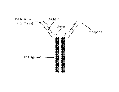
[0027] FIG. lA shows a schematic representation of an exemplary insulin-Fe
fusion protein.
Each insulin-Fe fusion protein comprises a proinsulin-like insulin molecule
containing an insulin
B chain and an insulin A chain that are optionally connected between the B
chain-C-terminal
region and the A chain-NH2 terminus with a linker peptide, and the A chain-C-
terminal region
and Fe-chain amino terminus with a linker, and the insulin-Fe fusion protein
sequence
terminating in the Fc-CH3-C-terminal region.
[0028] FIG. 1B shows an illustration depicting an insulin-Fe fusion protein
that does not
interact with the insulin-hormone receptor but is capable of binding insulin-
specific B cell
receptors and directing their destruction through antibody-dependent cell-
mediated cytotoxicity
(ADCC).
6
CA 3046337 2020-02-12
[0029] FIG. 2 shows a graph depicting the titer concentrations for the
insulin-Fe fusion
proteins of the present technology, manufactured in HEK cells and in CHO
cells. The titers
shown are titers observed after the Protein A purification step.
[0030] FIG. 3 shows a graph depicting the homodimer percentage for the
insulin-Fe fusion
proteins of the present technology, calculated using size-exclusion
chromatography (SEC-
HPLC).
[0031] FIG. 4A shows a graph depicting the insulin-Fe fusion protein
concentration over
time when an exemplary insulin-Fe fusion protein (SEQ ID NO: 3) was dosed at 2
mg/kg i.p.
into BALB/c mice.
[0032] FIG. 4B is a graph depicting serum fasting blood glucose levels
(FBGL) over time
when an exemplary insulin-Fe fusion protein (SEQ ID NO: 3) was dosed i.p. into
BALB/c mice.
[0033] FIG. 5 shows a graph depicting the inhibition of 125I-labeled
recombinant insulin
(RHI) binding to insulin autoantibodies (IAAs) in the serum of a pre-diabetic,
IAA-positive
human subject via radioimmunoassay for Rh I (IC50 = 3 nM) and an exemplary
insulin-Fe
protein (SEQ ID NO: 3) (IC50=10 nM).
[0034] FIG. 6A shows a graph depicting representative FACS dot plots for %
insulin + B cells
from 125Tg splenocyte/rat AM co-cultures treated with vehicle.
[0035] FIG. 6B shows a graph depicting representative FACS dot plots for %
insulin + B cells
from 125Tg splenocyte/rat AM co-cultures treated with an exemplary insulin-Fe
fusion protein
(SEQ ID NO: 3).
[0036] FIG. 6C shows a dose response curve of an exemplary insulin-Fe
protein (SEQ ID
NO: 3) and its corresponding activity in deleting insulin + B cells from 125Tg
splenocyte/rat AM
co-cultures.
[0037] FIGs. 7A-7H are a series of graphs showing in vivo cell reduction
data following a
two-week dosing regimen of an exemplary insulin-Fe fusion protein (SEQ ID NO:
3) in VH125
NOD mice. FIG. 7A is a graph showing the insulin + B cells in blood as a
percent of vehicle-
treated controls; FIG. 7B is a graph showing the insulin(-) B cells in blood
as a percent of
controls; FIG. 7C is a graph showing the insulin + B cells in spleen (all
splenic compartments) as
a percent of controls; FIG. 7D shows the insulin + B cells in the marginal
zone spleen population
7
CA 3046337 2020-02-12
(CD21High CD23High); FIG. 7E shows the insulin + B cells in the follicular
spleen population
(IgMmid CD21mid); FIG. 7F shows the insulin + B cells in the Ti spleen
population (CD211-'w
CD231-0); FIG. 7G shows the insulin + B cells in the T2 spleen population
(IgMfhg" CD21"d);
and FIG. 7H shows the insulin + B cells in the pre-marginal zone spleen
population (IgMlligh
CD21High).
[0038] FIGs. 8A-8B are graphs showing in vivo insulin B cell reduction
data. FIG. 8A
shows in vivo IgMHI insulin + B cell reduction data in the bone marrow
compartment following a
34-week dosing regimen of an exemplary insulin-Fe fusion protein (SEQ ID NO:
3) in VH125
NOD mice; FIG. 8B show in vivo IgMHI insulin + B cell reduction data in the
lymph node
compartment following a 34-week dosing regimen of an exemplary insulin-Fc
fusion protein
(SEQ ID NO: 3) in VH125 NOD mice.
[0039] FIG. 9 is a graph showing IL-2-mediated 5KC-3-4 T cell line
stimulation ELISA data
for several insulin-Fc fusion proteins of the present technology (SEQ ID NOs:
2, 3, 4, 5, 6, 7, 8,
9, and 10) and a recombinant human insulin (RHI) control.
[0040] FIG. 10 is a graph showing competitive inhibition of IL-2 secretion
induced by a T
cell stimulatory compound for an exemplary insulin-Fc fusion protein (SEQ ID
NO: 3) and
various contrasting insulin-Fe fusions proteins (SEQ ID NOs: 9 and 10) and a
control over
multiple concentrations.
[0041] FIG. 11 is a graph showing qualitative scoring of insulin-specific
125Tg B cell
deletion effectiveness for several insulin-Fe fusion proteins (SEQ ID NOs: 2,
3, 4, 5, 6, 7, 8, 9,
and 10) in addition to RHI as a control.
[0042] FIG. 12 is a graph showing the inhibition of biotin labelled-insulin
binding to an
antibody form of a cloned insulin-specific B cell receptor (mAb125) for
several insulin-Fe fusion
proteins (SEQ ID NOs: 2, 3, 4, 5, 6, 7, 8, 9, and 10) in addition to RHI as a
control.
[0043] FIG. 13 shows a Kaplan-Meier survival curve for the development of
T1D in female
3-week old wild type NOD mice (n=15 in each treatment group) treated with an
exemplary
insulin-Fe fusion protein (SEQ ID NO: 3).
8
CA 3046337 2020-02-12
[0044] FIG. 14 shows a Kaplan-Meier survival curve for the development of
T1D in female
3-week old wild type NOD mice (n=20 in each treatment group) treated with an
exemplary
insulin-Fc fusion protein (SEQ ID NO: 3).
[0045] FIG. 15 shows a Kaplan-Meier survival curve for the development of
T1D in female
3-week old wild type NOD mice (n=20 in each treatment group) treated with an
exemplary
insulin-Fc fusion protein (SEQ ID NO: 2).
[0046] FIG. 16 shows a Kaplan-Meier survival curve for the development of
T1D in female
3-week old wild type NOD mice (n=18 in each treatment group) treated with an
exemplary
insulin-Fc fusion protein (SEQ ID NO: 4).
DETAILED DESCRIPTION
[0047] It is to be appreciated that certain aspects, modes, embodiments,
variations and
features of the present methods are described below in various levels of
detail in order to provide
a substantial understanding of the present technology.
[0048] In practicing the present methods, many conventional techniques in
molecular
biology, protein biochemistry, cell biology, immunology, microbiology and
recombinant DNA
are used. See, e.g., Sambrook and Russell eds. (2001) Molecular Cloning: A
Laboratory
Manual, 3rd edition; the series Ausubel et al. eds. (2007) Current Protocols
in Molecular
Biology; the series Methods in Enzymology (Academic Press, Inc., N.Y.);
MacPherson et al.
(1991) PCR 1: A Practical Approach (IRL Press at Oxford University Press);
MacPherson et al.
(1995) PCR 2: A Practical Approach; Harlow and Lane eds. (1999) Antibodies, A
Laboratory
Manual; Freshney (2005) Culture of Animal Cells: A Manual of Basic Technique,
5th edition;
Gait ed. (1984) Oligonucleotide Synthesis; U.S. Patent No. 4,683,195; Hames
and Higgins eds.
(1984) Nucleic Acid Hybridization; Anderson (1999) Nucleic Acid Hybridization;
Hames and
Higgins eds. (1984) Transcription and Translation; Immobilized Cells and
Enzymes (IRL Press
(1986)); Perbal (1984)A Practical Guide to Molecular Cloning; Miller and Cabs
eds. (1987)
Gene Transfer Vectors for Mammalian Cells (Cold Spring Harbor Laboratory);
Makrides ed.
(2003) Gene Transfer and Expression in Mammalian Cells; Mayer and Walker eds.
(1987)
Immunochemical Methods in Cell and Molecular Biology (Academic Press, London);
and
Herzenberg et al. eds (1996) Weir 's Handbook of Experimental Immunology.
9
CA 3046337 2020-02-12
[0049] The present disclosure relates to compositions of insulin-Fc fusion
proteins (e.g.,
proinsulin-Fc fusion proteins) and their use to treat or prevent autoimmune
disease, e.g.,
autoimmune diabetes, e.g., type 1 diabetes. As described herein, the insulin-
Fe fusion proteins of
the present technology selectively bind to autoantigen-specific B cells (e.g.,
insulin-specific B
cells), thus avoiding drawbacks associated with the non-specific global
elimination of all B cells
(e.g., immunocompromisation). Additionally, the insulin-Fe fusion proteins of
the present
technology avoid non-specifically deleting all cells that express the insulin
hormone receptor as
they lack binding affinity for insulin that is bound to an insulin hormone
receptor. Without
wishing to be bound by theory, it is believed that in some embodiments, the
fusion proteins
described herein bind to autoantigen-specific BCRs (e.g., insulin-specific
BCRs).
[0050] The insulin-Fc fusion proteins of the present technology do not
interfere with the
binding of biotin labelled-insulin to the IM-9 insulin-hormone receptor, and
therefore bind the
insulin receptor present on IM-9 cells very weakly or not at all, which
minimizes their chances of
lowering blood sugar in vivo. This is an advantageous property for treating
patients with an
autoimmune disease (e.g., pre-diabetic patients, patients with insulin
autoantibodies, or recent-
onset type 1 diabetic patients), who may have normal or slightly elevated
blood sugar levels and
would be susceptible to the risk of potential hypoglycemia (e.g. low blood
sugar) induced by
therapy with insulin-Fc fusion proteins that are able to bind the insulin
receptor with IC50 values
<3,000 nM or proteins with even higher binding affinities with IC50 values <
1,000 nM in the in
vitro binding assay described herein). Accordingly, the insulin-Fe fusion
proteins of the present
technology are useful for treating or preventing autoimmune Type 1 diabetes in
subjects without
lowering their in vivo blood glucose levels.
Definitions
[0051] Unless defined otherwise, all technical and scientific terms used
herein generally have
the same meaning as commonly understood by one of ordinary skill in the art to
which this
technology belongs. As used in this specification and the appended claims, the
singular forms
"a", "an" and "the" include plural referents unless the content clearly
dictates otherwise. For
example, reference to "a cell" includes a combination of two or more cells,
and the like.
Generally, the nomenclature used herein and the laboratory procedures in cell
culture, molecular
genetics, organic chemistry, analytical chemistry and nucleic acid chemistry
and hybridization
described below are those well-known and commonly employed in the art.
CA 3046337 2020-02-12
[0052] The terms "insulin', "Ins", "insulin-specific" and "anti-insulin"
are used
interchangeably herein.
[0053] As used herein, EC50 refers to the concentration of an insulin-Fe
fusion protein at
which half-maximal response for in vitro insulin-specific B cell deletion is
observed (e.g.,
concentration at which the insulin + B cell receptors are reduced by half).
[0054] As used herein, IC50 refers to the concentration of an insulin-Fe
fusion protein at
which a given biological function or biochemical process (e.g., binding) is
inhibited by half. In
some embodiments, IC50 refers to the concentration of an insulin-Pc fusion
protein where the
binding of insulin to the human insulin receptor is reduced by half. In some
embodiments, the
ICsorefers to the concentration of an insulin-Fe fusion protein where the
binding of insulin to
insulin-specific B cell is reduced by half. In some embodiments, the IC50 is
the concentration of
an insulin-Fe fusion protein where the T cell activation induced by a
reference standard is
reduced by half.
[0055] As used herein, the term "about" in reference to a number is
generally taken to
include numbers that fall within a range of 1%, 5%, 10%, or 20% in either
direction (greater than
or less than) of the number unless otherwise stated or otherwise evident from
the context (except
where such number would be less than 0% or exceed 100% of a possible value).
[0056] As used herein, the "administration" of an agent or drug to a
subject includes any
route of introducing or delivering to a subject a compound to perform its
intended function.
Administration can be carried out by any suitable route, including but not
limited to, orally,
intranasally, parenterally (intravenously, intramuscularly, intraperitoneally,
or subcutaneously),
rectally, intrathecally, transdermally, or topically. Administration includes
self-administration
and the administration by another.
[0057] As used herein, the term "analog" refers to a compound or conjugate
(e.g., a
compound, conjugate as described herein, e.g., insulin) having a chemical
structure similar to
that of another compound or conjugate, but differing from it in at least one
aspect.
[0058] As used herein, the term "autoantibody" refers to an antibody that
targets and/or
reacts with one or more of an individual's own proteins, cells, tissues, or
organs. The term
"autoantigen" as used herein refers to an antigen comprised of normal tissue,
cells, protein,
11
CA 3046337 2020-02-12
peptides, or DNA that is the target of an immune response (e.g., a humoral or
cell-mediated
immune response). An autoantigen may be targeted by or react with an
autoantibody in the case
of an autoimmune disease.
[0059] As used herein, "autoimmune diabetes" refers to diabetes that is
characterized by the
destruction of the insulin-producing 13-cells of the pancreas.
[0060] As used herein, the term "cell surface receptor" refers to a
molecule such as a protein,
generally found on the external surface of a cell membrane and which interacts
with soluble
molecules, e.g., that circulate in the blood supply. Cell surface receptors
may also be secreted in
a soluble form into the extracellular space or may be shed from the external
surface of a cell. In
some embodiments, a cell surface receptor may include an antigen, or an
antigen receptor. In
other embodiments, B lymphocytes, also termed B cells, have cell surface
receptors that are
referred to as "B cell receptors", or "BCR", or in some cases "IgM" receptor.
[0061] As used herein, a "control" is an alternative sample used in an
experiment for
comparison purpose. A control can be "positive" or "negative." For example,
where the purpose
of the experiment is to determine a correlation of the efficacy of a
therapeutic agent for the
treatment for a particular type of disease, a positive control (a compound or
composition known
to exhibit the desired therapeutic effect) and a negative control (a subject
or a sample that does
not receive the therapy or receives a placebo) are typically employed.
[0062] As used herein, the term "effective amount" refers to a quantity
sufficient to achieve a
desired therapeutic and/or prophylactic effect, e.g., an amount which results
in the prevention of,
or a decrease in a disease or condition described herein or one or more signs
or symptoms
associated with a disease or condition described herein. In the context of
therapeutic or
prophylactic applications, the amount of a composition administered to the
subject will vary
depending on the composition, the degree, type, and severity of the disease
and on the
characteristics of the individual, such as general health, age, sex, body
weight and tolerance to
drugs. The skilled artisan will be able to determine appropriate dosages
depending on these and
other factors. The compositions can also be administered in combination with
one or more
additional therapeutic compounds. In the methods described herein, the
pharmaceutical
compositions may be administered to a subject having one or more signs or
symptoms of an
autoimmune disease (e.g., autoimmune diabetes, e.g., Type 1 diabetes). As used
herein, a
12
CA 3046337 2020-02-12
"therapeutically effective amount" of a composition refers to composition
levels in which the
physiological effects of a disease or condition described herein are
ameliorated or eliminated. A
therapeutically effective amount can be given in one or more administrations.
As used herein, a
"prophylactically effective amount" of a composition refers to composition
levels that prevent or
delay the onset of at least one symptom of a disease or condition described
herein. A
prophylactically effective amount can be given in one or more administrations.
[0063] As used herein, the term "endogenous C-peptide" level refers to the
level of C-
peptide in the subject prior to a treatment, e.g., an insulin-Fe fusion
protein treatment described
herein.
[0064] As used herein, the term "fusion protein", e.g., "insulin-Fe fusion"
protein refers to a
protein comprising more than one domain, e.g., typically from different
sources (e.g., different
proteins, polypeptides, cells, etc.), that are covalently linked through
peptide bonds. In some
embodiments, a fusion protein is produced recombinantly. In some embodiments,
the domains
of a fusion protein are covalently linked by connecting the gene sequences
that encode each
domain into a single nucleic acid molecule. In some embodiments, an insulin-Fe
fusion protein
is a protein, e.g., a single polypeptide, comprising an insulin polypeptide
(e.g., proinsulin
polypeptide) and an Fe fragment polypeptide, where the insulin and Fe fragment
polypeptides
are joined by peptide bonds to form a single polypeptide.
[0065] As used herein, the term "insulin" encompasses mature insulin,
preproinsulin, and
proinsulin, as well as naturally occurring insulin or analogs thereof (e.g.,
proinsulin analogs). In
some embodiments, an insulin polypeptide, e.g., proinsulin polypeptide, can be
a full-length
insulin (e.g., full-length proinsulin) polypeptide or a fragment thereof In
some embodiments, an
insulin polypeptide (e.g., proinsulin polypeptide) comprises one or more
fragments or domains
from a naturally occurring insulin (e.g., proinsulin) and/or one or more
fragments or domains
from a non-naturally occurring insulin (e.g., proinsulin).
[0066] As used herein, the terms "individual", "patient", or "subject" can
be an individual
organism, a vertebrate, a mammal, or a human. In some embodiments, the
individual, patient or
subject is a human. Exemplary human subjects include a human patient having a
disorder, e.g., a
disorder described herein, or a normal subject.
13
CA 3046337 2020-02-12
[0067] The terms "parenteral administration" and "administered
parenterally" as used herein
refer to modes of administration other than enteral and topical
administration, usually by
injection, and includes, without limitation, intravenous, intramuscular,
intraarterial, intrathecal,
intracapsular, intraorbital, intracardiac, intradermal, intraperitoneal,
transtracheal, subcutaneous,
subcuticular, intraarticular, subcapsular, subarachnoid, intraspinal and
intrasternal injection and
infusion.
[0068] The term "pharmaceutically acceptable" as used herein refers to
those compounds,
materials, compositions, and/or dosage forms which are, within the scope of
sound medical
judgment, suitable for use in contact with the tissues of human beings and
animals without
excessive toxicity, irritation, allergic response, or other problem or
complication, commensurate
with a reasonable benefit/risk ratio.
[0069] The term "pharmaceutically acceptable carrier" as used herein means
a
pharmaceutically acceptable material, composition or vehicle, such as a liquid
or solid filler,
diluent, excipient, solvent or encapsulating material, involved in carrying or
transporting the
insulin-Fc fusion proteins of the present technology from one organ, or
portion of the body, to
another organ, or portion of the body. Each carrier must be "acceptable" in
the sense of being
compatible with the other ingredients of the formulation and not injurious to
the patient.
[0070] As used herein, "prevention" or "preventing" of a disorder or
condition refers to a
compound that, in a statistical sample, reduces the occurrence of the disorder
or condition in the
treated sample relative to an untreated control sample, or delays the onset of
one or more
symptoms of the disorder or condition relative to the untreated control
sample. As used herein,
preventing an autoimmune disease (e.g., autoimmune diabetes, e.g., Type 1
diabetes), includes
preventing or delaying the initiation of symptoms of an autoimmune disease
(e.g., autoimmune
diabetes, e.g., Type 1 diabetes). As used herein, prevention of an autoimmune
disease (e.g.,
autoimmune diabetes, e.g., Type 1 diabetes) also includes preventing a
recurrence of one or more
signs or symptoms of an autoimmune disease (e.g., autoimmune diabetes, e.g.,
Type 1 diabetes).
[0071] As used herein, the term "sample" means biological sample material
derived from
living cells of a subject. Biological samples may include tissues, cells,
protein or membrane
extracts of cells, and biological fluids (e.g., ascites fluid or cerebrospinal
fluid (C SF)) isolated
14
CA 3046337 2020-02-12
from a subject, as well as tissues, cells and fluids (blood, plasma, saliva,
urine, serum, etc.)
present within a subject.
[0072] As used herein, the term "separate" therapeutic use refers to an
administration of at
least two active ingredients at the same time or at substantially the same
time by different routes.
[0073] As used herein, the terms "sequence identity" or "identical" in the
context of an
amino acid or nucleotide sequence mean that the same nucleotides or amino acid
residues are
found within a particular query sequence and a reference sequence when a
specified, contiguous
segment of the nucleotide sequence or amino acid sequence of the query
sequence is aligned and
compared to the nucleotide sequence or amino acid sequence of the reference
sequence.
Methods for sequence alignment and for determining identity between sequences
are known in
the art. See, e.g., Ausubel et al., eds. (1995) Current Protocols in Molecular
Biology, Chapter 19
(Greene Publishing and Wiley-Interscience, New York); and the ALIGN program
(Dayhoff
(1978) in Atlas of Polypeptide Sequence and Structure 5: Suppl. 3 (National
Biomedical
Research Foundation, Washington, D.C.)). With respect to optimal alignment of
two nucleotide
sequences, the contiguous segment of the query nucleotide sequence may have
additional
nucleotides or deleted nucleotides with respect to the reference nucleotide
sequence. Likewise,
for purposes of optimal alignment of two amino acid sequences, the contiguous
segment of the
query amino acid sequence may have additional amino acid residues or deleted
amino acid
residues with respect to the reference amino acid sequence. In some
embodiments, the
contiguous segment used for comparison to the reference nucleotide sequence or
reference amino
acid sequence will comprise at least 6, 10, 15, or 20 contiguous nucleotides,
or amino acid
residues, and may be 30, 40, 50, 100, or more nucleotides or amino acid
residues. Corrections
for increased sequence identity associated with inclusion of gaps in the query
nucleotide
sequence or amino acid sequence can be made by assigning gap penalties.
Methods of sequence
alignment are known in the art.
[0074] In certain embodiments, the determination of percent identity
between two sequences
is accomplished using a mathematical algorithm. For example, the percent
identity of an amino
acid sequence is determined using the Smith-Waterman homology search algorithm
using an
affine 6 gap search with a gap open penalty of 12 and a gap extension penalty
of 2, BLOSUM
matrix 62. The Smith-Waterman homology search algorithm is described in Smith
and
CA 3046337 2020-02-12
Waterman (1981) Adv. AppL Math 2:482-489. In some embodiments, the percent
identity of a
nucleotide sequence is determined using the Smith-Waterman homology search
algorithm using
a gap open penalty of 25 and a gap extension penalty of 5. Such a
determination of sequence
identity can be performed using, for example, the DeCypher Hardware
Accelerator from
TimeLogic.
[0075] As used herein, the term "sequential" therapeutic use refers to
administration of at
least two active ingredients at different times, the administration route
being identical or
different. More particularly, sequential use refers to the whole
administration of one of the
active ingredients before administration of the other or others commences. It
is thus possible to
administer one of the active ingredients over several minutes, hours, or days
before
administering the other active ingredient or ingredients. There is no
simultaneous treatment in
this case.
[0076] As used herein, the term "simultaneous" therapeutic use refers to
the administration
of at least two active ingredients by the same route and at the same time or
at substantially the
same time.
100771 As used herein, "specifically binds" or "selectively binds" refers
to the non-covalent
interactions of the type which occur between (i) an immunoglobulin molecule
(e.g., anti-insulin
immunoglobulin) and an insulin or an insulin-Fc fusion protein of the present
technology, (ii) a B
cell receptor (e.g., anti-insulin immunoglobulin) and an insulin or insulin-Fc
fusion protein of the
present technology, or (iii) a B cell expressing a B cell receptor (e.g., anti-
insulin
immunoglobulin) and an insulin or insulin-Fe fusion protein of the present
technology. The
strength, or affinity of the binding interactions, e.g., immunological binding
interactions or
specific binding interactions, can be expressed in terms of the dissociation
constant (1(d) of the
interaction, wherein a smaller Kd represents a higher affinity. Immunological
or specific binding
properties of selected polypeptides can be quantified using methods known in
the art. One such
method entails measuring the rates of ligand/ligand-receptor complex (e.g.,
antigen/antigen
receptor complex; insulin antibody/ insulin complex; or insulin
antibody/insulin-Fe fusion
protein complex) formation and dissociation, wherein those rates depend on the
concentrations
of the complex partners, the affinity of the interaction, and geometric
parameters that equally
influence the rate in both directions. Thus, both the "on rate constant" (kon)
and the "off rate
16
CA 3046337 2020-02-12
constant" (kw) can be determined by calculation of the concentrations and the
actual rates of
association and dissociation. See, e.g., Nature 361:186-87 (1993). The ratio
of koff/kon enables
the cancellation of all parameters not related to affinity, and is equal to
the dissociation constant
Ka. (See, generally, Davies et al. (1990) Annual Rev Biochem 59:439-473). In
some
embodiments, a fusion protein described herein specifically binds an anti-
insulin antibody
immunoglobulin, a BCR (e.g., a BCR comprising an anti-insulin immunoglobulin),
and/or a B
cell, e.g., autoantigen-specific B cell such as an insulin-specific B cell,
when the equilibrium
binding constant (Ka) is less than or equal to 1 M, e.g., less than or equal
to 100 nM, less than
or equal to 10 nM, less than or equal to 100 pM, or less than or equal to
about 1 pM, e.g., as
measured by assays such as radioligand binding assays, ELISAs, surface plasmon
resonance,
equlibrium binding assays, or similar assays known to those skilled in the
art.
[0078] The terms "systemic administration," "administered systemically,"
"peripheral
administration" and "administered peripherally" as used herein mean the
administration of the
insulin-Fe fusion protein other than directly into the central nervous system,
such that it enters
the patient's system and, thus, is subject to metabolism and other like
processes, for example,
subcutaneous administration.
[0079] "Treating" or "treatment" as used herein covers the treatment of a
disease or disorder
described herein, in a subject, such as a human, and includes: (i) inhibiting
a disease or disorder,
i.e., arresting its development; (ii) relieving a disease or disorder, i.e.,
causing regression of the
disorder; (iii) slowing progression of the disorder; and/or (iv) inhibiting,
relieving, or slowing
progression of one or more symptoms of the disease or disorder. In some
embodiments,
treatment means that the symptoms associated with the disease are, e.g.,
alleviated, reduced,
cured, or placed in a state of remission.
[0080] It is also to be appreciated that the various modes of treatment of
a disease or disorder
as described herein are intended to mean "substantial," which includes total
but also less than
total treatment, and wherein some biologically or medically relevant result is
achieved. The
treatment may be a continuous prolonged treatment for a chronic disease or a
single, or few time
administrations for the treatment of an acute condition.
17
CA 3046337 2020-02-12
Management of Type I Diabetes
100811 There are three main approaches to reducing or eliminating the
hardships associated
with T1D: 1) better disease management, e.g., improved insulins, smart pumps,
and continuous
glucose monitors; 2) disease reversal, e.g., pancreas or islet transplants, 0-
cell regeneration,
systemic or T cell specific immunomodulation; and 3) disease prevention, e.g.,
avoidance of
environmental triggers, antigen-specific vaccination, non-antigen specific
immunomodulation
therapy). As T1D patients develop autoimmunity, their 0-cell function declines
and so does the
potential therapeutic benefit of intervention (Rewers, M and Gottlieb, P.
Diabetes Care (2009),
32, 1769-1782). Additionally, once the autoimmune process has begun it might
become more
progressively difficult to alter. For these reasons and the estimated cost
benefit relative to late
stage intervention, disease prevention at the earliest possible stage of T1D
is ideal for the long
term.
10082] Effective disease prevention requires an in-depth understanding of
the T1D
autoimmune process as well as tools that can accurately diagnose or predict
the risk of
developing T1D well before the onset of overt hyperglycemia. After initiation
of islet
autoimmunity, most T1D patients have a long preclinical period that offers an
opportunity for
treatments to halt progression to clinical diabetes. A major hallmark of the
onset of islet
autoimmunity is the presence of circulating antibodies specific for islet-
autoantigens including
insulin, isoform 65 of glutamate decarboxylase (anti-GAD65), protein tyrosine
phosphatase-like
protein (IA2), and the zinc transporter 8 (ZnT8). In fact, at diagnosis
greater than 90% of T1D
patients present at least one islet-specific antibody, and in the prospective
Diabetes
Autoimmunity Study in the Young (DAISY) cohort, 89% of children who progressed
to diabetes
expressed two or more islet-specific autoantibodies. Although CD4+ and CD8+ T
cells
contribute to the ultimate attack on 0-cells, the pathogenic role of B cells
(e.g., anti-insulin B
cells, insulin-specific B cells, or insulin + B cells) has emerged in recent
years, which may help
explain why antibodies and T cells are specific for the same islet-specific
autoantigens, as well as
the lag in timing between the appearance of islet-specific autoantibodies and
complete T cell
mediated 0-cell destruction. B cells can take up islet antigens, present them
to helper T cells, and
differentiate into antibody secreting plasma cells which enhance antigen
uptake by antigen-
presenting cells ultimately leading to the activation of cytotoxic T cells for
13-cell destruction.
18
CA 3046337 2020-02-12
[0083] Global B cell depletion, e.g., by a B cell antigen antibody such as
rituximab, has been
proposed as a treatment for autoimmune disease, e.g., T 1 D. See, e.g.,
Pescovitz etal., N
England J. Med. 361.22(2009):2143-52. However, global B cell depletion has
been shown to
cause immunocompromisation in subjects due to the nonspecific elimination of
healthy/non-
autoimmune B cells that are normally required by the immune system for normal
function (e.g.,
clearance of pathogens).
[0084] Thus, there is a need for treatments and prophylaxes for autoimmune
diseases such as
T1D that avoid adverse effects caused by the destruction of healthy cells.
Fe Domains
[0085] The term "Fe region", "Fe domain", or "Fe fragment" as used herein
refers to a C-
terminal region of an imrnunoglobulin heavy chain, which is capable of binding
to a mammalian
Fc(gamma) or Fc(Rn) receptor, e.g., human Fc(gamma) or Fc(Rn) receptor. An Fe
receptor
(FcR) refers to a receptor that binds to an Fe fragment or the Fe region of an
antibody. In certain
embodiments, the FcR is a native human FcR sequence. In some embodiments, the
FcR binds an
IgG antibody (a gamma receptor) and includes receptors of the FcyRI, FcyRII,
and FcyRIII
subclasses, including allelic variants and alternatively spliced forms of
these receptors. FcyRII
receptors include FcyRIIA (an "activating receptor") and FcyRIIB (an
"inhibiting receptor"),
which have similar amino acid sequences that differ primarily in the
cytoplasmic domains
thereof. FcRs are described in Ravetch and Kinet, 1991, Ann. Rev. Immunol.,
9:457-92; Capel et
al., 1994, Immunomethods, 4:25-34; and de Haas etal., 1995,1 Lab. Clin. Med.,
126:330-41.
"FcR" also includes the neonatal receptor, FcRn, which is responsible for the
transfer of maternal
IgGs to the fetus (Guyer etal., 19761 Immunol., 117:587; and Kim et al.,
1994,1 Immunol.,
24:249) and contributes to the prolonged in vivo elimination half-lives of
antibodies and Fe-
fusion proteins in vivo.
[0086] The Fe fragment, region, or domain may be a native sequence Fe
region. Although
the boundaries of the Fe region of an immunoglobulin heavy chain might vary,
the human IgG
heavy chain Fe region is usually defined to stretch from an amino acid residue
at position
Cys226, or from Pro230, to the carboxyl-terminus thereof. The numbering of the
residues in the
Fe region is that of the EU index as in Kabat. Kabat et al., Sequences of
Proteins of
Immunological Interest, 5th Ed. Public Health Service, National Institutes of
Health, Bethesda,
19
CA 3046337 2020-02-12
Md., 1991. The Fc region of an immunoglobulin generally comprises two constant
domains,
CH2 and CH3.
[0087] In some embodiments, the Fc fragment comprises or consists of the Fc
region (e.g.,
CH2 domain and CH3 domain) of a mammalian IgG, e.g., human IgG. In certain
embodiments,
the Fc fragment comprises or consists of the Fc region (e.g., CH2 domain and
CH3 domain) of
human IgGi. In some embodiments, the Fc fragment comprises or consists of an
amino acid
sequence having at least 80% (e.g., at least 80%, 85%, 90%, 95%, 97%, 99%, or
more) identity
to the Fc region (e.g., CH2 domain and CH3 domain) of human IgGi.
[0088] In some embodiments, the Fc region of a human IgGi comprises the
following amino
acid sequence:
DKTHTCPPCPAPELLGGPSVFLFPPKPKDTLMISRTPEVTCVVVDVSHEDPEVKFNWYV
DGVEVHNAKTKPREEQYNSTYRVVSVLTVLHQDWLNGKEYKCKVSNICALPAPIEKTIS
KAKGQPREPQVYTLPPSRDELTKNQVSLTCLVKGFYPSDIAVEWESNGQPENNYKTTPP
VLDSDGSFFLYSKLTVDKSRWQQGNVFSCSVMHEALHNHYTQKSLSLSPG (SEQ ID NO:
22).
[0089] In certain embodiments, the Fc region of a human IgGi comprises an
additional
amino acid at one or both termini. In some embodiments, this additional amino
acid comprises a
charged side chain (e.g., a positively charged amino acid, e.g., lysine or
arginine). In certain
embodiments, the Fc region of a human IgGi comprises the following amino acid
sequence:
DKTHTCPPCPAPELLGGPSVFLFPPKPKDTLMISRTPEVTCVVVDVSHEDPEVKFNWYV
DGVEVHNAKTKPREEQYNSTYRVVSVLTVLHQDWLNGKEYKCKVSNKALPAPIEKTIS
KAKGQPREPQVYTLPPSRDELTKNQVSLTCLVKGFYPSDIAVEWESNGQPENNYKTTPP
VLDSDGSFFLYSKLTVDKSRWQQGNVFSCSVMHEALHNHYTQKSLSLSPGK (SEQ ID
NO: 23).
Insulin and Insulin Analogs
100901 Insulin is a peptide hormone produced by 13-cells in the islets of
Langerhans within
the pancreas. Insulin functions by regulating the absorption of glucose from
the blood. When
exposed to a stimulus, such as increased protein and glucose levels, insulin
is released from 13-
cells and binds to the insulin receptor, initiating a signaling cascade that
affects many aspects of
human metabolism. Disruption of this process is directly related to several
diseases,
CA 3046337 2020-02-12
autoimmune diabetes (e.g., Type 1 diabetes), insulinoma, insulin resistance,
metabolic
syndromes, and polycystic ovary syndrome. The amino acid sequence of insulin
is strongly
conserved throughout evolution, particularly in vertebrates, and consists of
two polypeptide
chains, termed the A and B chains, that are linked through disulfide bonds.
The sequence of
human proinsulin is represented by the amino acid sequence:
FVNQHLCGSHLVEALYLVCGERGFFYTPKTRREAEDLQVGQVELGGGPGAGSLQPLAL
EGSLQKRGIVEQCCTSICSLYQLENYCN (SEQ ID NO: 24).
[0091] Insulin is initially synthesized as an inactive precursor called
preproinuslin. Through
a series of highly coordinated, enzyme-regulated steps, preproinsulin is
converted into mature
insulin. Cleavage of the signal peptide of preproinsulin in the endoplasmic
reticulum followed
by oxidation and chaperone-assisted folding yields proinsulin, which is
transported to the trans-
Golgi network. Proinsulin is then subjected to further proteolytic processing
steps, resulting in
the release of a fragment called the C-peptide and formation of mature
insulin, which is stored
within zinc (Zn2+) and calcium (Ca2+)-rich secretory vesicles in 13-cells as
an inactive hexamer.
After exposure to a stimulus, the secretory vesicles fuse with the plasma
membrane, releasing the
insulin and promoting the dissociation of the hexamers into active insulin
monomers. In some
embodiments, the insulin of the present disclosure is a monomer. In some
embodiments, the
insulin is a non-covalent multimer (e.g., a dimer, tetramer, hexamer, or
higher order multimer,
e.g., a trimer of dimers). In some embodiments, the insulin may be a monomer
or a non-covalent
multimer (e.g., a dimer, tetramer, hexamer, or higher order multimer, e.g., a
trimer of dimers).
[0092] In some embodiments, the insulin described herein is a single chain
insulin. In some
embodiments, the insulin is a preproinsulin or a proinsulin, e.g., a
prohormone precursor to
mature insulin. All salt forms and non-salt forms of insulin and insulin
analogs (e.g. proinsulin
and proinsulin analogs) are encompassed by the scope of the present
disclosure.
[0093] In some embodiments, the insulin of the present disclosure comprises
an insulin
analog (e.g., proinsulin analog). Several analogs of human insulin are
commercially available
for therapeutic use. In some embodiments, the insulin analog of the present
technology is a
monomer. In some embodiments, the insulin analog is a non-covalent multimer
(e.g., a dimer,
tetramer, hexamer, or higher order multimer, e.g., a trimer of dimers).
21
CA 3046337 2020-02-12
[0094] The insulin analogs may be closely related to the structure of human
insulin, yet
contain a modification (e.g. a structural modification) to enhance a certain
functional aspect. In
some embodiments, the insulin analog may differ from the structure of human
insulin by amino
acid substitutions only. In some embodiments, the insulin analog may differ
from the structure
of human insulin by amino acid deletions only. In some embodiments, the
insulin analog may
differ from the structure of human insulin by amino acid additions only. In
some embodiments,
the insulin analog comprises a variant or mutant of insulin (e.g., the
sequence of insulin as
described by SEQ ID NO: 24). In some embodiments, the insulin analog comprises
an amino
acid substitution, deletion, or addition relative to insulin (e.g., the
sequence of insulin as
described by SEQ ID NO: 24). In some embodiments, the insulin analog comprises
at least 2, at
least 3, at least 4, at least 5, at least 6, at least 7, at least 8, at least
9, at least 10, at least 12, at
least 15, at least 20, at least 25, at least 30, at least 40, or at least 50
amino acid substitutions,
deletions, or additions relative to insulin (e.g., the sequence of insulin as
described by SEQ ID
NO: 24).
[0095] In some embodiments, the insulin or insulin analog is a three-chain
peptide
comprising elements of an A chain, a B chain, and a C chain. In some
embodiments, the insulin
or insulin analog comprises a wild-type insulin B, A, and/or C chain peptide,
e.g., from a
mammal (e.g., human or mouse).
[0096] The sequences of the human insulin A chain and B chain are
represented by SEQ ID
NO: 19 and SEQ ID NO: 25, respectively: Human insulin A chain:
GIVEQCCTSICSLYQLENYCN (SEQ ID NO: 19); Human insulin B chain:
FVNQHLCGSHLVEALYLVCGERGFFYTPKT (SEQ ID NO: 25).
[0097] In some embodiments, modifications to the sequence of the insulin or
insulin analog
(e.g., amino acid substitutions, deletions, or additions or chemical
modifications) may be to
either the A chain of insulin, the B chain of insulin, or any combination
thereof. In some
embodiments, when the insulin or insulin analog is a non-covalent multimer
comprising more
than one A chain, B chain, and/or C chain, modifications to the sequence of
insulin (e.g., amino
acid substitutions, deletions, or additions or chemical modifications) may be
to either the A
chain, B chain, or both in the non-covalent multimer.
22
CA 3046337 2020-02-12
Insulin-Fc Fusion Proteins of the Present Technology
[0098] In one aspect, the present disclosure provides an insulin-Fc fusion
protein comprising
an insulin polypeptide fused to a Fe domain, wherein the insulin polypeptide
comprises a B-
chain peptide, a C-chain peptide, and an A-chain peptide, and wherein the
amino acid sequence
of the C-chain peptide is AAK. Additionally or alternatively, in some
embodiments, the insulin-
Fe fusion protein binds human insulin receptor at an IC50 >5,000 nM in a
competitive binding
assay. In some embodiments, the insulin-Fe fusion proteins described herein
have low
bioactivity or are substantially metabolically inactive, e.g., they do not
substantially lower blood
glucose levels in a subject upon administration.
[0099] Additionally or alternatively, in some embodiments, the insulin-Fe
fusion protein
inhibits in vitro binding of insulin + B cell receptors to insulin at an IC50
< 300 nM, <200 nM, <
150 nM, < 100 nM, or < 75 nM. Additionally or alternatively, in some
embodiments, the
insulin-Fe fusion protein activates T-cells to secrete IL-2 levels that are
reduced compared to that
observed in T-cells activated by recombinant human insulin. In some
embodiments, the the
insulin-Fe fusion protein activates T-cells to secrete IL-2 levels that are
less than 3,000 pg/ml,
less than 1,000 pg/mL, less than 500 pg/mL, less than 300 pg/mL, or less than
100 pg/ml.
1001001 In certain embodiments, the insulin polypeptide is a proinsulin
polypeptide or a
preproinsulin polypeptide. In some embodiments of the insulin-Fe fusion
protein, the amino acid
sequence of the A-chain peptide comprises SEQ ID NO: 19. The insulin
polypeptide may be
fused to the Fe fragment via a peptide linker. Examples of peptide linkers
include SEQ ID NO:
20 and SEQ ID NO: 21. Alternatively, no peptide linker may be present between
the insulin
polypeptide and the Fe domain of the insulin-Fe fusion protein (e.g., the C-
terminal region of the
insulin polypeptide is covalently linked (e.g., via a peptide bond) to the N-
terminal region of the
Fe domain or the N-terminal region of the insulin polypeptide is covalently
linked (e.g., via a
peptide bond) to the C-terminal region of the Fe domain). Additionally or
alternatively, in some
embodiments of the insulin-Fe fusion protein, the Fe domain comprises a wild-
type Fe fragment
of human IgGi. In certain embodiments, the amino acid sequence of the Fe
domain comprises
SEQ ID NO: 22.
[00101] Additionally or alternatively, in any of the above embodiments of the
insulin-Fe
fusion protein, the orientation of the insulin polypeptide from N- to C-
termini is: (N-terminus)-
23
CA 3046337 2020-02-12
B-chain peptide--C-chain peptide--A-chain peptide-(C-terminus). The insulin
polypeptide may
be located at the N-terminus or C-terminus of the Fe domain.
[00102] Additionally or alternatively, in any of the above embodiments of the
insulin-Fe
fusion protein, the B-chain peptide comprises the amino acid sequence
FVNQHLCGSHLVX1ALX2LVCGEX3GFFYTPK (SEQ ID NO: 28), wherein Xi is E or Q, X2 is
Y or A, and X3 is R or E. In certain embodiments, X2 is A.
[00103] In one aspect, the present disclosure provides an insulin-Fe fusion
protein comprising
an insulin polypeptide fused to a Fe domain, wherein the insulin polypeptide
comprises a B-
chain peptide, a C-chain peptide, and an A-chain peptide, wherein the B-chain
peptide comprises
the amino acid sequence FVNQHLCGSHLVX1ALX2LVCGEX3GFFYTPK (SEQ ID NO: 28),
wherein Xi is E or Q, X2 is Y or A, and X3 is R or E; and wherein the amino
acid sequence of the
C-chain peptide is AAK. In some embodiments, X2 is A.
[00104] In certain embodiments of the insulin-Fe fusion protein, the insulin
polypeptide is a
proinsulin polypeptide or a preproinsulin polypeptide. In some embodiments of
the insulin-Fe
fusion protein, the amino acid sequence of the A-chain peptide comprises SEQ
ID NO: 19. The
insulin polypeptide may be fused to the Fe fragment via a peptide linker.
Examples of peptide
linkers include SEQ ID NO: 20 and SEQ ID NO: 21. Alternatively, no peptide
linker may be
present between the insulin polypeptide and the Fe domain of the insulin-Fe
fusion protein.
Additionally or alternatively, in some embodiments of the insulin-Fe fusion
protein, the Fe
domain comprises a wild-type Fe fragment of human IgGi. In certain
embodiments, the amino
acid sequence of the Fe domain comprises SEQ ID NO: 22.
[00105] Additionally or alternatively, in some embodiments, the amino acid
sequence of the
insulin-Fe fusion protein is SEQ ID NO: 2, SEQ ID NO: 3, SEQ ID NO: 4, SEQ ID
NO: 5, SEQ
ID NO: 6, SEQ ID NO: 7, or SEQ ID NO: 8.
[00106] FIG. lA shows a schematic representation of an exemplary insulin-Fe
fusion protein
of the present technology. The insulin-Fe fusion proteins described herein
have an advantage of
specifically binding to one or more of: (i) soluble anti-insulin antibodies
(e.g., anti-insulin
antibodies not bound to a cell); (ii) anti-insulin immunoglobulins bound to a
B cell receptor
(BCR), e.g., on a B cell (e.g., an anti-insulin B cell); and/or (iii) anti-
insulin B cells, but do not
interact with the insulin-hormone receptor. See FIG. 1B.
24
CA 3046337 2020-02-12
[00107] In one aspect, the present disclosure provides an insulin-Fc fusion
protein comprising
an insulin polypeptide fused to an Fc domain. In certain embodiments, the
insulin polypeptide of
the insulin-Fc fusion protein of the present technology comprises domains in
the following
orientation from N- to C- termini: (N-terminus)-B-chain peptide--C-chain
peptide--A-chain
peptide-(C-terminus). Additionally or alternatively, in some embodiments, the
insulin-Fc fusion
protein comprises domains in the following orientation from N- to C-termini:
(N-terminus)-
insulin polypeptide¨optional linker¨Fc domain-(C-terminus) (e.g., (N-terminus)-
B-chain
peptide--C-chain peptide--A-chain peptide--optional linker¨Fc domain-(C-
terminus)). In
certain embodiments, a linker (e.g., a peptide linker described herein) is
located between the
insulin polypeptide and the Fc domain. In other embodiments, no linker (e.g.,
peptide linker) is
present between the insulin polypeptide and the Fc domain. Exemplary linkers
(e.g., peptide
linkers) are described in greater detail in the Linkers section herein.
[00108] Exemplary insulin-Fc fusion proteins (e.g., proinsulin-Fc fusion
proteins) and their
domain sequences are shown in Table A. In some embodiments, the insulin-Fc
fusion proteins
include modified mutants, e.g., that lead to properties such as anti-insulin B
cell removal and/or
inhibition of insulin-specific T cell activation.
[00109] The insulin-Fc fusion proteins of the present technology comprise a C
chain peptide
that is about 3-5 amino acids in length and comprises amino acids selected
from among alanine
and lysine. In some embodiments, the C chain peptide of the insulin-Fc fusion
proteins does not
comprise amino acids other than alanine and lysine. In certain embodiments,
the C chain peptide
of the insulin-Fc fusion proteins comprises or consists of the amino acid
sequence of: AAK. See
Table A.
[00110] Additionally or alternatively, in some embodiments, the A chain
peptide of the
insulin-Fc fusion protein comprises or consists of the amino acid sequence of
the A chain peptide
of wild type human proinsulin, or an amino acid sequence having at least 80%
(e.g., at least 80%,
85%, 90%, 95%, 97%, 99% or more) identity to the amino acid sequence of the A
chain peptide
of wild type human proinsulin. In some embodiments, the A chain peptide of the
insulin-Fc
fusion protein comprises or consists of the amino acid sequence of
GIVEQCCTSICSLYQLENYCN (SEQ ID NO: 19) or an amino acid sequence having at
least
80% (e.g., at least 80%, 85%, 90%, 95%, 97%, 99% or more) identity to SEQ ID
NO: 19.
CA 3046337 2020-02-12
[00111] Additionally or alternatively, in some embodiments, the B chain
peptide of the
insulin-Fc fusion protein comprises or consists of the amino acid sequence of
the B chain peptide
of wild type human proinsulin or an amino acid sequence having at least 80%
(e.g., at least 80%,
85%, 90%, 95%, 97%, 99% or more) identity to the amino acid sequence of the B
chain peptide
of wild type human proinsulin. In some embodiments, the B-chain peptide of the
insulin-Fc
fusion protein comprises or consists of the amino acid sequence of a B-chain
peptide listed in
Table A or an amino acid sequence having at least 80% (e.g., at least 80%,
85%, 90%, 95%,
97%, 99%, or more) identity to a B-chain peptide listed in Table A. In some
embodiments, the B
chain peptide of the insulin-Fc fusion protein comprises or consists of the
amino acid sequence
of FVNQHLCGSHLVEALYLVCGERGFFYTPKT (SEQ ID NO: 25) or an amino acid
sequence having at least 80% (e.g., at least 80%, 85%, 90%, 95%, 97%, 99% or
more) identity to
SEQ ID NO: 25.
[00112] Additionally or alternatively, in some embodiments, mutations may be
introduced to
the B chain peptide of the insulin-Fe fusion protein, e.g., to vary the
sequence and/or length.
Mutations at specific amino acid residues of the B chain of insulin have been
shown to abrogate
T-cell stimulation in non-obese diabetic (NOD) mice (Nakayama, M. et al.
Science (2005)
435:220-223; Nakayama, M. et al Ann NY Acad Sci (2006) 1079:122-129). Without
being bound
by theory, it is believed that certain insulin mutants may disrupt the
recognition of the insulin
peptide epitope-MHC-II complex by the T-cell receptor, which in turn prevents
T cell activation
toward the pancreatic islet cells. Exemplary modifications to the sequence of
insulin or an
insulin analog may include for example, an amino acid substitution at residue
16 (e.g., a Y16A
substitution) of SEQ ID NO: 25. Thus, in some embodiments, an insulin-Fc
fusion protein
comprising at least 1 amino acid substitution, deletion, or addition (e.g.,
relative to SEQ ID NO.
25) on the B chain of insulin or an insulin analog may result in reduced
affinity of T cells for an
MHC-II complex bearing the insulin fragment or insulin analog fragment, and/or
reduced T cell
activation in vivo. Accordingly, in certain embodiments, the B-chain peptide
of the insulin-Fe
fusion protein comprises a mutated B-chain having a mutation at amino acid
residue 16 (e.g., a
Y16A substitution). In certain embodiments, the B-chain peptide of the insulin-
Fe fusion protein
has the sequence of any one of SEQ ID NOs: 11-15.
[00113] Provided herein are insulin-Fe fusion proteins comprising an insulin
polypeptide
operably linked to an Fe domain. In certain embodiments, the insulin-fusion
protein comprises
26
CA 3046337 2020-02-12
an Fe domain described herein. In some embodiments of the insulin-Fe fusion
protein, the Fe
domain comprises or consists of the amino acid sequence of SEQ ID NO: 22; or
an amino acid
sequence having at least 80% (e.g., at least 80%, 85%, 90%, 95%, 97%, 99%, or
more) identity
to SEQ ID NO: 22. In other embodiments of the insulin-Fe fusion protein, the
Fe domain
comprises or consists of the amino acid sequence of SEQ ID NO: 23; or an amino
acid sequence
having at least 80% (e.g., at least 80%, 85%, 90%, 95%, 97%, 99%, or more)
identity to SEQ ID
NO: 23.
[00114] The full length sequences of the insulin-Fe fusion proteins of the
present technology
are provided below:
SEQ ID NO: 2
FVNQHLCGSHLVEALYLVCGERGFFYTPKAAKGIVEQCCTSICSLYQLENYCNGGGGAG
GGGDKTHTCPPCPAPELLGGPSVFLFPPKPKDTLMISRTPEVTCVVVDVSHEDPEVKFN
WYVDGVEVHNAKTKPREEQYNSTYRVVSVLTVLHQDWLNGKEYKCKVSNKALPAPIE
KTISKAKGQPREPQVYTLPPSRDELTKNQVSLTCLVKGFYPSDIAVEWESNGQPENNYK
TTPPVLDSDGSFFLYSKLTVDKSRWQQGNVFSCSVMHEALHNHYTQKSLSLSPG
SEQ ID NO: 3
FVNQHLCGSHLVEALALVCGERGFFYTPKAAKGIVEQCCTSICSLYQLENYCNGGGGAG
GGGDKTHTCPPCPAPELLGGPSVFLFPPKPKDTLMISRTPEVTCVVVDVSHEDPEVKFN
WYVDGVEVHNAKTKPREEQYNSTYRVVSVLTVLHQDWLNGKEYKCKVSNKALPAPIE
KTISKAKGQPREPQVYTLPPSRDELTKNQVSLTCLVKGFYPSDIAVEWESNGQPENNYK
TTPPVLDSDGSFFLYSKLTVDKSRWQQGNVFSCSVMHEALHNHYTQKSLSLSPG
SEQ ID NO: 4
FVNQHLCGSHLVQALYLVCGERGFFYTPKAAKGIVEQCCTSICSLYQLENYCNGGGGA
GGGGDKTHTCPPCPAPELLGGPSVFLFPPKPKDTLMISRTPEVTCVVVDVSHEDPEVKFN
WYVDGVEVHNAKTKPREEQYNSTYRVVSVLTVLHQDWLNGKEYKCKVSNKALPAPIE
KTISKAKGQPREPQVYTLPPSRDELTKNQVSLTCLVKGFYPSDIAVEWESNGQPENNYK
TTPPVLDSDGSFFLYSKLTVDKSRWQQGNVFSCSVMHEALHNHYTQKSLSLSPG
SEQ ID NO: 5
FVNQHLCGSHLVEALYLVCGEEGFFYTPKAAKGIVEQCCTSICSLYQLENYCNGGGGAG
GGGDKTHTCPPCPAPELLGGPSVFLFPPKPKDTLMISRTPEVTCVVVDVSHEDPEVKFN
WYVDGVEVHNAKTKPREEQYNSTYRVVSVLTVLHQDWLNGKEYKCKVSNKALPAPIE
KTISKAKGQPREPQVYTLPPSRDELTKNQVSLTCLVKGFYPSDIAVEWESNGQPENNYK
TTPPVLDSDGSFFLYSKLTVDKSRWQQGNVFSCSVMHEALHNHYTQKSLSLSPG
27
CA 3046337 2020-02-12
SEQ ID NO: 6
FVNQHLCGSHLVEALALVCGEEGFFYTPKAAKGIVEQCCTSICSLYQLENYCNGGGGAG
GGGDKTHTCPPCPAPELLGGPSVFLFPPKPKDTLMISRTPEVTCVVVDVSHEDPEVKFN
WYVDGVEVHNAKTKPREEQYNSTYRVVSVLTVLHQDWLNGKEYKCKVSNKALPAPIE
KTISKAKGQPREPQVYTLPPSRDELTKNQVSLTCLVKGFYPSDIAVEWESNGQPENNYK
TTPPVLDSDGSFFLYSKLTVDKSRWQQGNVFSCSVMHEALHNHYTQKSLSLSPG
SEQ ID NO: 7
FVNQHLCGSHLVEALALVCGERGFFYTPKAAKGIVEQCCTSICSLYQLENYCNDKTHTC
PPCPAPELLGGPSVFLFPPKPKDTLMISRTPEVTCVVVDVSHEDPEVKFNWYVDGVEVH
NAKTKPREEQYNSTYRVVSVLTVLHQDWLNGKEYKCKVSNKALPAPIEKTISKAKGQP
REPQVYTLPPSRDELTKNQVSLTCLVKGFYPSDIAVEWESNGQPENNYKTTPPVLDSDG
SFFLYSKLTVDKSRWQQGNVFSCSVMHEALHNHYTQKSLSLSPG
SEQ ID NO: 8
FVNQHLCGSHLVEALALVCGERGFFYTPKAAKGIVEQCCTSICSLYQLENYCNGGGGSG
GGGDKTHTCPPCPAPELLGGPSVFLFPPKPKDTLMISRTPEVTCVVVDVSHEDPEVKFN
WYVDGVEVHNAKTKPREEQYNSTYRVVSVLTVLHQDWLNGKEYKCKVSNKALPAPIE
KTISKAKGQPREPQVYTLPPSRDELTKNQVSLTCLVKGFYPSDIAVEWESNGQPENNYK
TTPPVLDSDGSFFLYSKLTVDKSRWQQGNVFSCSVMHEALHNHYTQKSLSLSPG
SEQ ID NO: 9
FVNQHLCGSHLVEALALVCGERGFFYTPKAAAKGIVEQCCTSICSLYQLENYCNGGGGA
GGGGDKTHTCPPCPAPELLGGPSVFLFPPKPKDTLMISRTPEVTCVVVDVSHEDPEVKFN
WYVDGVEVHNAKTKPREEQYNSTYRVVSVLTVLHQDWLNGKEYKCKVSNKALPAPIE
KTISKAKGQPREPQVYTLPPSRDELTKNQVSLTCLVKGFYPSDIAVEWESNGQPENNYK
TTPPVLDSDGSFFLYSKLTVDKSRWQQGNVFSCSVMHEALHNHYTQKSLSLSPG
SEQ ID NO: 10
FVNQHLCGSHLVEALALVCGERGFFYTPKAAAAKGIVEQCCTSICSLYQLENYCNGGGG
AGGGGDKTHTCPPCPAPELLGGPSVFLFPPKPKDTLMISRTPEVTCVVVDVSHEDPEVKF
NWYVDGVEVHNAKTICPREEQYNSTYRVVSVLTVLHQDWLNGKEYKCKVSNKALPAPI
EKTISKAKGQPREPQVYTLPPSRDELTKNQVSLTCLVKGFYPSDIAVEWESNGQPENNY
KTTPPVLDSDGSFFLYSKLTVDKSRWQQGNVFSCSVMHEALHNHYTQKSLSLSPG
Table A
Sequence B Chain C Peptide A Chain Linker Fc Domain
SEQ ID SEQ ID NO: 22
SEQ ID SEQ ID SEQ ID SEQ ID
NO: 11 DKTHTCPPCPAPELL
NO: 2 NO: 16 NO: 19 NO: 20
FVNQHLCG GGPSVFLFPPICPKDTL
28
CA 3046337 2020-02-12
SHLVEALY AAK
GIVEQCCT GGGGAG MISRTPEVTCVVVDV
LVCGERGF SICSLYQL GGG
SHEDPEVKFNWYVD
FYTPK ENYCN GVEVHNAKTKPREEQ
YNSTYRVVSVLTVLH
QDWLNGKEYKCKVS
NKALPAPIEKTISKAK
GQPREPQVYTLPPSR
DELTKNQVSLTCLVK
GFYPSDIAVEWESNG
QPENNYKTTPPVLDS
DGSFFLYSKLTVDKS
RWQQGNVFSCSVMH
EALHNHYTQKSLSLS
PG
SEQ ID NO: 22
DKTHTCPPCPAPELL
GGPSVFLFPPKPKDTL
MISRTPEVTCVVVDV
SHEDPEVKFNWYVD
GVEVHNAKTKPREEQ
SEQ ID
SEQ ID
YNSTYRVVSVLTVLH
NO: 12 SEQ ID
SEQ ID NO: 19
QDWLNGKEYKCKVS
SEQ ID NO: 20
NO:
FVNQHLCG NO: 16 GIVEQCCT
NKALPAPIEKTISKAK
3
SHLVEALA GGGGAG
GQPREPQVYTLPPSR
AAK SICSLYQL
LVCGERGF ENYCN
GGG DELTKNQVSLTCLVK
FYTPK
GFYPSDIAVEWESNG
QPENNYKTTPPVLDS
DGSFFLYSKLTVDKS
RWQQGNVFSCSVMH
EALHNHYTQKSLSLS
PG
SEQ ID NO: 22
SEQ ID SEQ ID SEQ ID SEQ ID SEQ ID
DKTHTCPPCPAPELL
NO: 4 NO: 13 NO: 16 NO: 19 NO: 20
GGPSVFLFPPKPKDTL
29
CA 3046337 2020-02-12
FVNQHLCG AAK
GIVEQCCT GGGGAG MISRTPEVTCVVVDV
SHLVQALY SICSLYQL GGG
SHEDPEVKFNWYVD
LVCGERGF ENYCN GVEVHNAKTKPREEQ
FYTPK
YNSTYRVVSVLTVLH
QDWLNGKEYKCKVS
NKALPAPIEKTISKAK
GQPREPQVYTLPPSR
DELTKNQVSLTCLVK
GFYPSDIAVEWESNG
QPENNYKTTPPVLDS
DGSFFLYSKLTVDKS
RWQQGNVFSCSVMH
EALHNHYTQKSLSLS
PG
SEQ ID NO: 22
DKTHTCPPCPAPELL
GGPSVFLFPPKPKDTL
MISRTPEVTCVVVDV
SHEDPEVKFNWYVD
GVEVHNAKTKPREEQ
SEQ ID
SEQ ID
YNSTYRVVSVLTVLH
NO: 14 SEQ ID
SEQ ID NO: 19
QDWLNGKEYKCKVS
SEQ ID NO: 20
F'VNQHLCG NO: 16
NKALPAPIEKTISKAK
NO: 5 GIVEQCCT
SHLVEALY GGGGAG
GQPREPQVYTLPPSR
AAK SICSLYQL
LVCGEEGF ENYCN
GGG DELTKNQVSLTCLVK
FYTPK
GFYPSDIAVEWESNG
QPENNYKTTPPVLDS
DGSFFLYSKLTVDKS
RWQQGNVFSCSVMH
EALHNHYTQKSLSLS
PG
SEQ ID NO: 22
SEQ ID SEQ ID SEQ ID SEQ ID SEQ ID
DKTHTCPPCPAPELL
NO: 6 NO: 15 NO: 16 NO: 19 NO: 20
GGPSVFLFPPKPKDTL
CA 3046337 2020-02-12
FVNQHLCG AAK
GIVEQCCT GGGGAG MISRTPEVTCVVVDV
SHLVEALA SICSLYQL GGG
SHEDPEVKFNWYVD
LVCGEEGF ENYCN
GVEVHNAKTKPREEQ
FYTPK
YNSTYRVVSVLTVLH
QDWLNGKEYKCKVS
NKALPAPIEKTISKAK
GQPREPQVYTLPPSR
DELTKNQVSLTCLVK
GFYPSDIAVEWESNG
QPENNYKTTPPVLDS
DGSFFLYSKLTVDKS
RWQQGNVFSCSVMH
EALHNHYTQKSLSLS
PG
SEQ ID NO: 22
DKTHTCPPCPAPELL
GGPSVFLFPPKPKDTL
MISRTPEVTCVVVDV
SHEDPEVKFNWYVD
GVEVHNAKTKPREEQ
SEQ ID
SEQ ID
YNSTYRVVSVLTVLH
NO: 12
SEQ ID NO: 19
QDWLNGKEYKCKVS
SEQ ID
FVNQHLCG NO: NO: 16 GIVEQCCT
NKALPAPIEKTISKAK
7
SHLVEALA AAK SICSLYQL
GQPREPQVYTLPPSR
LVCGERGF ENYCN
DELTKNQVSLTCLVK
FYTPK GFYPSDIAVEWESNG
QPENNYKTTPPVLDS
DGSFFLYSKLTVDKS
RWQQGNVFSCSVMH
EALI-INHYTQKSLSLS
PG
SEQ ID NO: 22
SEQ ID SEQ ID SEQ ID SEQ ID SEQ ID
DKTHTCPPCPAPELL
NO: 8 NO: 12 NO: 16 NO: 19 NO: 21
GGPSVFLFPPKPKDTL
31
CA 3046337 2020-02-12
FVNQHLCG AAK
GIVEQCCT GGGGSG MISRTPEVTCVVVDV
SHLVEALA SICSLYQL GGG
SHEDPEVICFNWYVD
LVCGERGF ENYCN
GVEVHNAKTKPREEQ
FYTPK
YNSTYRVVSVLTVLH
QDWLNGKEYKCKVS
NKALPAPIEKTISKAK
GQPREPQVYTLPPSR
DELTKNQVSLTCLVK
GFYPSDIAVEWESNG
QPENNYKTTPPVLDS
DGSFFLYSKLTVDKS
RWQQGNVFSCSVMH
EALHNHYTQKSLSLS
PG
SEQ ID NO: 22
DKTHTCPPCPAPELL
GGPSVFLFPPKPICDTL
MISRTPEVTCVVVDV
SHEDPEVICFNWYVD
GVEVHNAKTKPREEQ
SEQ ID
SEQ ID
YNSTYRVVSVLTVLH
NO: 12 SEQ ID
SEQ ID NO: 19
QDWLNGKEYKCKVS
SEQ ID NO: 20
FVNQHLCG NO: 17
NKALPAPIEKTISKAK
NO: 9 GIVEQCCT
SHLVEALA GGGGAG
GQPREPQVYTLPPSR
AAAK SICSLYQL
LVCGERGF ENYCN
GGG DELTKNQVSLTCLVK
FYTPK
GFYPSDIAVEWESNG
QPENNYKTTPPVLDS
DGSFFLYSICLTVDKS
RWQQGNVFSCSVMH
EALHNHYTQKSLSLS
PG
SEQ ID NO: 22
SEQ ID SEQ ID SEQ ID SEQ ID SEQ ID
DKTHTCPPCPAPELL
NO: 10 NO: 12 NO: 18 NO: 19 NO: 20
GGPSVFLFPPKPKDTL
32
CA 3046337 2020-02-12
FVNQHLCG AAAAK GIVEQCCT GGGGAG MISRTPEVTCVVVDV
SHLVEALA SICSLYQL GGG SHEDPEVICFNWYVD
LVCGERGF ENYCN GVEVHNAKTKPREEQ
FYTPK YNSTYRVVSVLTVLH
QDWLNGKEYKCKVS
NKALPAPIEKTISKAK
GQPREPQVYTLPPSR
DELTKNQVSLTCLVK
GFYPSDIAVEWESNG
QPENNYKTTPPVLDS
DGSFFLYSKLTVDKS
RWQQGNVFSCSVMH
EALHNHYTQKSLSLS
PG
[00115] Table A does not include a leader sequence. In some embodiments, an
insulin-Fc
fusion protein described herein does not include a leader sequence at the N-
terminus. In other
embodiments, an insulin-Fc fusion protein described herein includes a leader
sequence, e.g., at
the N-terminus. An exemplary leader sequence includes the amino acid sequence
MEWS WVFLFFLSVTTGVHS (SEQ ID NO: 26). In some embodiments, an insulin-Fc
fusion
protein described herein is encoded by a nucleic acid molecule comprising a
leader sequence,
e.g., for expression (e.g., recombinant expression) in cells (e.g.,
eukaryotic, e.g., mammalian
cells). In certain embodiments, the leader sequence is cleaved off, e.g., in
the cell culture, during
expression. An exemplary nucleic acid sequence encoding a leader sequence
includes the
nucleic acid sequence
ATGGAATGGAGCTGGGTCTTTCTCTTCTTCCTGTCAGTAACGACTGGTGTCCACTCC
(SEQ ID NO: 27). In other embodiments, a fusion protein described herein is
encoded by a
nucleic acid molecule not comprising a leader sequence.
[00116] Also disclosed herein are nucleic acid sequences (e.g., mRNA, cDNA,
DNA)
encoding the insulin-Fc fusion proteins of SEQ ID NOs: 2-10.
33
CA 3046337 2020-02-12
[00117] In some embodiments, the nucleic acid sequence is:
ATGGAATGGAGCTGGGTCTTTCTCTTCTTCCTGTCAGTAACGACTGGTGTCCACTCCT
TCGTGAAGCAGCACCTGTGCGGCCCTCACCTGGTGGAAGCTCTGTATCTCGTGTGCG
GC GAGCGGGGCTTC TTCTACACCCCCAAGTCTCGGAGAGAGGTGGAAGATCCCCAG
GTGGAACAGCTGGAACTGGGCGGCTCTCCTGGCGATCTGCAGACACTGGCCCTGGA
AGTGGCCCGGCAGAAACGGGGCATCGTGGACCAGTGCTGCACCTCCATCTGCTCCCT
GTACCAGCTGGAAAACTACTGCAATGGTGGAGGCGGTGGAGTGCCCAGAGATTGTG
GATGTAAGCCTTGCATATGTACAGTCCCAGAAGTATCATCTGTCTTCATCTTCCCCCC
AAAGCCCAAGGATGTGCTCACCATTACTCTGACTCCTAAGGTCACGTGTGTTGTGGT
AGACATCAGCAAGGATGATCCCGAGGTCCAGTTCAGCTGGTTTGTAGATGATGTGG
AGGTGCACACAGC TCAGACGCAAC CC CGGGAGGAGCAGTTCAACAGCACTTTCCGC
TCAGTCAGTGAACTTCCCATCATGCACCAGGACTGGCTCAATGGCAAGGAGTTCAAA
TGCAGGGTCAACAGTGCAGCTTTC CCTGC CC CCATCGAGAAAAC CATCTCCAAAAC C
AAAGGCAGACCGAAGGCTCCACAGGTGTACACCATTCCACCTCCCAAGGAGCAGAT
GGCCAAGGATAAAGTCAGTCTGACCTGCATGATAACAGACTTCTTCCCTGAAGACAT
TACTGTGGAGTGGCAGTGGAATGGGCAGCCAGCGGAGAACTACAAGAACACTCAGC
CCATCATGGACACAGATGGCTCTTACTTCGTCTACAGCAAGCTCAATGTGCAGAAGA
GCAACTGGGAGGCAGGAAATACTTTCACCTGCTCTGTGTTACATGAGGGCCTGCACA
ACCACCATACTGAGAAGAGCCTCTCCCACTCTCCTGGTTAG (SEQ ID NO: 1).
[00118] In some embodiments, the nucleic acid sequence is:
ATGGAATGGAGCTGGGTCTTTCTCTTCTTCCTGTCAGTAACGACTGGTGTCCACTCCT
TCGTGAACCAGCACCTGTGCGGCTCCCACCTGGTGGAAGCTCTGGCTCTCGTGTGCG
GCGAGCGGGGCTTCTTCTACACCCCCAAGGCCGCTAAAGGCATCGTGGAACAGTGC
TGCACCTCCATCTGCTCCCTGTACCAGCTGGAAAACTACTGCAATGGCGGAGGTGGT
GCAGGAGGCGGTGGAGACAAAACTCACACATGCCCACCGTGCCCAGCACCTGAACT
CCTGGGGGGACCGTCAGTCTTC CTCTTCCC CC CAAAACCCAAGGACACCCTCATGAT
CTCCCGGACCCCTGAGGTCACATGCGTGGTGGTGGACGTGAGCCACGAAGACCCTG
AGGTCAAGTTCAACTGGTACGTGGACGGCGTGGAGGTGCATAATGCCAAGACAAAG
CCGCGGGAGGAGCAGTACAACAGCACGTACCGTGTGGTCAGCGTCCTCACCGTCCT
GCAC CAGGACTGGCTGAATGGCAAGGAGTACAAGTGCAAGGTCTC CAACAAAGC CC
TCCCAGCCCCCATCGAGAAAACCATCTCCAAAGCCAAAGGGCAGCCCCGAGAACCA
34
CA 3046337 2020-02-12
CAGGTGTACACCCTGCCCCCATCCCGGGATGAGCTGACCAAGAACCAGGTCAGCCT
GAC CTGCCTGGTCAAAGGC TTCTATCC CAGC GACATC GC CGTGGAGTGGGAGAGCA
ATGGGCAGCCGGAGAACAACTACAAGACCACGCCTCCCGTGCTGGACTCCGACGGC
TCCTTCTTCCTCTACAGCAAGCTCACCGTGGACAAGAGCAGGTGGCAGCAGGGGAA
CGTCTTCTCATGCTCCGTGATGCATGAGGCTCTGCACAACCACTACACGCAGAAGAG
CCTCTCCCTGTCTCCGGGTTAG (SEQ ID NO: 29).
[00119] In some embodiments, the nucleic acid sequence is:
ATGGAATGGAGCTGGGTCTTTCTCTTCTTC CTGTCAGTAAC GAC TGGTGTC CACTC CT
TCGTGAACCAGCACCTGTGCGGCTCCCACCTGGTGCAAGCTCTGTATCTCGTGTGCG
GCGAGCGGGGCTTCTTCTACACCCCCAAGGCCGCTAAAGGCATCGTGGAACAGTGC
TGCACCTCCATCTGCTCCCTGTACCAGCTGGAAAACTACTGCAATGGCGGAGGTGGT
GCAGGAGGCGGTGGAGACAAAACTCACACATGCCCACCGTGCCCAGCACCTGAACT
CCTGGGGGGACCGTCAGTCTTCCTCTTCCCCCCAAAACCCAAGGACACCCTCATGAT
CTC CC GGACCCCTGAGGTCACATGCGTGGTGGTGGACGTGAGC CAC GAAGACCCTG
AGGTCAAGTTCAACTGGTACGTGGACGGCGTGGAGGTGCATAATGCCAAGACAAAG
CCGCGGGAGGAGCAGTACAACAGCACGTACCGTGTGGTCAGCGTCCTCACCGTCCT
GCAC CAGGACTGGCTGAATGGCAAGGAGTACAAGTGCAAGGTCTCCAACAAAGC CC
TCCCAGCCCCCATCGAGAAAACCATCTCCAAAGCCAAAGGGCAGCCCCGAGAACCA
CAGGTGTACACCCTGCCCCCATCCCGGGATGAGCTGACCAAGAACCAGGTCAGCCT
GACCTGCCTGGTCAAAGGCTTCTATC CCAGCGACATC GC CGTGGAGTGGGAGAGCA
ATGGGCAGCCGGAGAACAACTACAAGAC CAC GCCTCCC GTGCTGGACTC CGACGGC
TCCTTCTTCCTCTACAGCAAGCTCACCGTGGACAAGAGCAGGTGGCAGCAGGGGAA
CGTCTTCTCATGCTCCGTGATGCATGAGGCTCTGCACAACCACTACACGCAGAAGAG
CCTCTCCCTGTCTCCGGGTTAG (SEQ ID NO: 30).
[00120] In some embodiments, the nucleic acid sequence is:
ATGGAATGGAGCTGGGTCTTTCTCTTCTTCCTGTCAGTAACGACTGGTGTCCACTCCT
TCGTGAACCAGCACCTGTGCGGCTCCCACCTGGTGGAAGCTCTGTATCTCGTGTGCG
GCGAGGAGGGCTTCTTCTACACCCCCAAGGCCGCTAAAGGCATCGTGGAACAGTGC
TGCACCTCCATCTGCTCCCTGTACCAGCTGGAAAACTACTGCAATGGCGGAGGTGGT
GCAGGAGGCGGTGGAGACAAAAC TCACACATGCC CAC CGTGCCCAGCAC CTGAACT
CCTGGGGGGAC CGTCAGTCTTC CTCTTCCC CC CAAAAC C CAAGGACACCCTCATGAT
CA 3046337 2020-02-12
CTC CC GGACC CCTGAGGTCACATGCGTGGTGGTGGACGTGAGCCACGAAGACC CTG
AGGTCAAGTTCAACTGGTACGTGGACGGCGTGGAGGTGCATAATGCCAAGACAAAG
C C GCGGGAGGAGC AGTACAACAGCACGTAC C GTGTGGTCAGC GTC CTCAC CGTC CT
GCACCAGGACTGGCTGAATGGCAAGGAGTACAAGTGCAAGGTCTCCAACAAAGCCC
TCCCAGCC CC CATCGAGAAAACCATCTC CAAAGCCAAAGGGCAGC C CC GAGAACCA
CAGGTGTACAC CCTGCC C C CATCC CGGGATGAGCTGACCAAGAACCAGGTCAGC CT
GACCTGCCTGGTCAAAGGCTTCTATCCCAGCGACATCGCCGTGGAGTGGGAGAGCA
ATGGGCAGCC GGAGAACAACTACAAGAC CACGC CTC CC GTGCTGGACTCC GACGGC
TCCTTCTTCCTCTACAGCAAGCTCACCGTGGACAAGAGCAGGTGGCAGCAGGGGAA
CGTCTTCTCATGCTCCGTGATGCATGAGGCTCTGCACAACCACTACACGCAGAAGAG
CCTCTCCCTGTCTCCGGGTTAG (SEQ ID NO: 31).
[00121] In some embodiments, the nucleic acid sequence is:
ATGGAATGGAGCTGGGTCTTTCTCTTCTTCCTGTCAGTAACGACTGGTGTCCACTCCT
TCGTGAACCAGCACCTGTGCGGCTCCCACCTGGTGGAAGCTCTGGCTCTCGTGTGCG
GC GAGGAGGGCTTCTTCTACACC CC CAAGGCC GCTAAAGGCATCGTGGAACAGTGC
TGCACCTCCATCTGCTCCCTGTACCAGCTGGAAAACTACTGCAATGGCGGAGGTGGT
GCAGGAGGCGGTGGAGACAAAACTCACACATGCCCACCGTGCCCAGCACCTGAACT
CCTGGGGGGACCGTCAGTCTTCCTCTTCCCCCCAAAACCCAAGGACACCCTCATGAT
CTC CC GGACC CCTGAGGTCACATGCGTGGTGGTGGAC GTGAGC CACGAAGACC CTG
AGGTCAAGTTCAACTGGTACGTGGACGGCGTGGAGGTGCATAATGCCAAGACAAAG
CC GCGGGAGGAGCAGTACAACAGCACGTAC C GTGTGGTCAGCGTCCTCACC GTCCT
GCACCAGGACTGGCTGAATGGCAAGGAGTACAAGTGCAAGGTCTCCAACAAAGCCC
TCCCAGCCCCCATCGAGAAAACCATCTCCAAAGCCAAAGGGCAGCCCCGAGAACCA
CAGGTGTACAC CCTGCC CCCATCCC GGGATGAGC TGAC CAAGAAC CAGGTCAGC CT
GACCTGCCTGGTCAAAGGCTTCTATCCCAGCGACATCGCCGTGGAGTGGGAGAGCA
ATGGGCAGC CGGAGAACAACTACAAGAC CAC GCCTC CCGTGCTGGACTC CGACGGC
TCCTTCTTCCTCTACAGCAAGCTCACCGTGGACAAGAGCAGGTGGCAGCAGGGGAA
CGTCTTCTCATGCTCCGTGATGCATGAGGCTCTGCACAACCACTACACGCAGAAGAG
CCTCTCCCTGTCTCCGGGTTAG (SEQ ID NO: 32).
[00122] In some embodiments, the nucleic acid sequence is:
ATGGAATGGAGCTGGGTCTTTCTCTTCTTC CTGTCAGTAACGACTGGTGTC CAC TC CT
36
CA 3046337 2020-02-12
TCGTGAACCAGCACCTGTGCGGCTCCCACCTGGTGGAAGCTCTGGCTCTCGTGTGCG
GCGAGCGGGGCTTCTTCTACACCCCCAAGGCCGCTAAAGGCATCGTGGAACAGTGC
TGCACCTCCATCTGCTCCCTGTACCAGCTGGAAAACTACTGCAATGACAAAACTCAC
ACATGCCCACCGTGCCCAGCACCTGAACTCCTGGGGGGACCGTCAGTCTTCCTCTTC
C C CC CAAAACC CAAGGACACC CTCATGATCTC C C GGACC CCTGAGGTCACATGC GTG
GTGGTGGAC GTGAGC CAC GAAGACC CTGAGGTCAAGTTCAACTGGTAC GTGGACGG
CGTGGAGGTGCATAATGCCAAGACAAAGCCGCGGGAGGAGCAGTACAACAGCACG
TACCGTGTGGTCAGC GTCCTCACCGTCCTGCACCAGGACTGGCTGAATGGCAAGGA
GTACAAGTGCAAGGTCTCCAACAAAGCCCTCCCAGCCCCCATCGAGAAAACCATCT
CCAAAGCCAAAGGGCAGCCCCGAGAACCACAGGTGTACACCCTGCCCCCATCCCGG
GATGAGCTGACCAAGAACCAGGTCAGCCTGACCTGCCTGGTCAAAGGCTTCTATCCC
AGCGACATC GC C GTGGAGTGGGAGAGCAATGGGCAGC CGGAGAACAACTACAAGA
CCACGCCTCCCGTGCTGGACTCCGACGGCTCCTTCTTCCTCTACAGCAAGCTCACCG
TGGACAAGAGCAGGTGGCAGCAGGGGAACGTCTTCTCATGCTCCGTGATGCATGAG
GCTCTGCACAACCACTACACGCAGAAGAGCCTCTCCCTGTCTCCGGGTTAG (SEQ ID
NO: 33).
[00123] In some embodiments, the nucleic acid sequence is:
ATGGAATGGAGCTGGGTCTTTCTCTTCTTCCTGTCAGTAACGACTGGTGTCCACTCCT
TCGTGAACCAGCACCTGTGCGGCTCCCACCTGGTGGAAGCTCTGGCTCTCGTGTGCG
GC GAGCGGGGCTTCTTCTACACC CC CAAGGCC GCTAAAGGCATCGTGGAACAGTGC
TGCACCTCCATCTGCTCCCTGTACCAGCTGGAAAACTACTGCAATGGCGGAGGTGGT
TCAGGAGGCGGTGGAGACAAAACTCACACATGCCCACCGTGCCCAGCACCTGAACT
CCTGGGGGGACC GTCAGTCTTCCTC TTCCC CC CAAAACC CAAGGACAC CCTCATGAT
CTCC CGGACC CC TGAGGTCACATGC GTGGTGGTGGAC GTGAGC CAC GAAGACC CTG
AGGTCAAGTTCAACTGGTACGTGGACGGCGTGGAGGTGCATAATGCCAAGACAAAG
CC GCGGGAGGAGCAGTACAACAGCACGTACCGTGTGGTCAGC GTCCTCAC CGTCCT
GCACCAGGACTGGCTGAATGGCAAGGAGTACAAGTGCAAGGTCTCCAACAAAGCCC
TCCCAGCCCCCATCGAGAAAACCATCTCCAAAGCCAAAGGGCAGCCCCGAGAACCA
CAGGTGTACACCCTGCCCCCATCCCGGGATGAGCTGACCAAGAACCAGGTCAGCCT
GACCTGCCTGGTCAAAGGCTTCTATCCCAGCGACATCGCCGTGGAGTGGGAGAGCA
ATGGGCAGCCGGAGAACAACTACAAGACCACGCCTCCCGTGCTGGACTCCGACGGC
37
CA 3046337 2020-02-12
TCCTTCTTCCTCTACAGCAAGCTCACCGTGGACAAGAGCAGGTGGCAGCAGGGGAA
CGTCTTCTCATGCTCCGTGATGCATGAGGCTCTGCACAACCACTACACGCAGAAGAG
CCTCTCCCTGTCTCCGGGTTAG (SEQ ID NO: 34).
[00124] In some embodiments, the nucleic acid sequence is:
ATGGAATGGAGCTGGGTCTTTCTCTTCTTCCTGTCAGTAACGACTGGTGTC CACTC CT
TCGTGAACCAGCACCTGTGCGGCTCCCACCTGGTGGAAGCTCTGGCTCTCGTGTGCG
GCGAGC GGGGCTTCTTC TACAC CC CCAAGGCC GCTGCAAAAGGCATCGTGGAACAG
TGCTGCACCTCCATCTGCTCCCTGTACCAGCTGGAAAACTACTGCAATGGCGGAGGT
GGTGCAGGAGGCGGTGGAGACAAAACTCACACATGCCCACCGTGCCCAGCACCTGA
ACTC CTGGGGGGAC CGTCAGTCTTCCTCTTC CC CCCAAAACCCAAGGACACCCTCAT
GATCTCCCGGACCCCTGAGGTCACATGCGTGGTGGTGGACGTGAGCCACGAAGACC
CTGAGGTCAAGTTCAACTGGTACGTGGACGGCGTGGAGGTGCATAATGCCAAGACA
AAGCC GC GGGAGGAGCAGTACAACAGCACGTACCGTGTGGTCAGC GTCCTCACC GT
CCTGCACCAGGACTGGCTGAATGGCAAGGAGTACAAGTGCAAGGTCTCCAACAAAG
CCCTCCCAGCC C CCATC GAGAAAACCATC TC CAAAGCCAAAGGGCAGC CC CGAGAA
CCACAGGTGTACACCCTGCCCCCATCCCGGGATGAGCTGACCAAGAACCAGGTCAG
CCTGACCTGCCTGGTCAAAGGCTTCTATCCCAGCGACATCGCCGTGGAGTGGGAGA
GCAATGGGCAGCCGGAGAACAACTACAAGACCACGCCTCCCGTGCTGGACTCCGAC
GGCTCCTTCTTCCTCTACAGCAAGCTCACCGTGGACAAGAGCAGGTGGCAGCAGGG
GAACGTCTTCTCATGCTCCGTGATGCATGAGGCTCTGCACAACCACTACACGCAGAA
GAGCCTCTCCCTGTCTCCGGGTTAG (SEQ ID NO: 35).
[00125] In some embodiments, the nucleic acid sequence is:
ATGGAATGGAGCTGGGTCTTTCTCTTCTTC CTGTCAGTAACGACTGGTGTC CACTC CT
TCGTGAACCAGCACCTGTGCGGCTCCCACCTGGTGGAAGCTCTGGCTCTCGTGTGCG
GCGAGCGGGGCTTCTTCTACACCCCCAAGGCCGCTGCAGCTAAAGGCATCGTGGAA
CAGTGCTGCACCTCCATCTGCTCCCTGTACCAGCTGGAAAACTACTGCAATGGCGGA
GGTGGTGCAGGAGGCGGTGGAGACAAAACTCACACATGCCCACCGTGCCCAGCACC
TGAACTCCTGGGGGGACCGTCAGTCTTCCTCTTCCCCCCAAAACCCAAGGACACCCT
CATGATCTC CC GGAC CCCTGAGGTCACATGCGTGGTGGTGGACGTGAGC CACGAAG
AC CCTGAGGTCAAGTTCAAC TGGTACGTGGAC GGCGTGGAGGTGCATAATGC CAAG
ACAAAGCC GC GGGAGGAGCAGTACAACAGCAC GTACC GTGTGGTCAGCGTCCTCAC
38
CA 3046337 2020-02-12
CGTCCTGCACCAGGACTGGCTGAATGGCAAGGAGTACAAGTGCAAGGTCTCCAACA
AAGCCCTCCCAGCCCCCATCGAGAAAACCATCTCCAAAGCCAAAGGGCAGCCCCGA
GAACCACAGGTGTACACCCTGCCCCCATCCCGGGATGAGCTGACCAAGAACCAGGT
CAGCCTGACCTGCCTGGTCAAAGGCTTCTATCCCAGCGACATCGCCGTGGAGTGGGA
GAGCAATGGGCAGCCGGAGAACAACTACAAGACCACGCCTCCCGTGCTGGACTCCG
ACGGCTCCTTCTTCCTCTACAGCAAGCTCACCGTGGACAAGAGCAGGTGGCAGCAG
GGGAACGTCTTCTCATGCTCCGTGATGCATGAGGCTCTGCACAACCACTACACGCAG
AAGAGCCTCTCCCTGTCTCCGGGTTAG (SEQ ID NO: 36).
Linkers
[00126] In some embodiments, an insulin-Fc fusion protein described herein
comprises one or
more linkers, e.g., between one or more domains of the insulin-Fc fusion
protein, or between the
insulin-Fc fusion protein and a conjugated molecule/moiety. For example, an
insulin-Fc fusion
protein comprises a linker between the insulin polypeptide and the Fc
fragment. In other
embodiments, an insulin-Fc fusion protein comprises a linker between one or
more peptides
(e.g., A-chain, B-chain, and/or C-chain peptides) of the insulin polypeptide.
[00127] Peptide linkers may comprise natural or unnatural amino acids. In some
embodiments, peptide linkers can be encoded by a nucleic acid molecule, e.g.,
such that a single
nucleic acid molecule can encode the various peptides within an insulin
polypeptide as well as
the peptide linker(s); or can encode the insulin polypeptide, the Fc fragment,
and the peptide
linker.
[00128] In some embodiments, the peptide linker comprises at least 5 amino
acid residues,
e.g., at least 5, 6, 7, 8, 9, 10, 11, 12, 13, 14, 15, 16, 17, 18, 19, 20 or
more amino acid residues.
In some embodiments, the peptide linker comprises 5-9 amino acid residues. In
some
embodiments, the peptide linker comprises four or less amino acids or six or
more amino acids in
length. In some embodiments, the peptide linker comprises 0 amino acids (e.g.
no peptide
linker). In some embodiments, the peptide linker comprises 4 or more glycines
(e.g., 4, 5, 6, 7,
8, or more glycines). In some embodiments, the peptide linker comprises 4 or
more consecutive
glycines (e.g., 5 or more consecutive glycines). In some embodiments, the
peptide linker
comprises the amino acid sequence of GGGGAGGGG (SEQ ID NO: 20) or GGGGSGGGG
(SEQ ID NO: 21). In some embodiments, the peptide linker comprises the linker
in the
39
CA 3046337 2020-02-12
etanercept fusion protein. See, e.g., US 8063182 Bl. In some embodiments, the
peptide linker
does not comprise the "hinge" region (or a fragment thereof) of a human
immunoglobulin (e.g.,
an IgG, e.g., IgGI).
[00129] The "hinge" region of a human immunoglobulin is often divided into
three regions:
the upper, middle, and lower hinge. In some embodiments, in an immunoglobulin,
the upper
hinge is the number of amino acids between the end of the first domain of the
heavy chain (CH1)
and the first cysteine forming an interheavy chain disulfide bridge. The
middle hinge is high in
proline and contains the inter-heavy chain cysteine disulfide bridges. The
lower hinge connects
the middle hinge to the CH2 domain. See Sandlie, I. and Michaelsen, T.,
Chapter 3: Engineering
the Hinge Region to Optimize Complement-induced Cytolysis, In Antibody
Engineering: A
Practical Guide, W. H. Freeman and Co., New York, N.Y.; see also Hamers-
Casterman, C.,
Naturally Occurring Antibodies Devoid of Light Chains, 363 Nature 446 (1993)
and Terskikh,
A. V., "Peptabody ": A New Type of High Avidity Binding Protein, 94 Proc.
Natl. Acad. Sci.
USA 1663 (1997).
Functional Features of the Insulin-Fe Fusion Proteins of the Present
Technology
[00130] Described herein are methods for treating or preventing an autoimmune
disease, e.g.,
autoimmune diabetes comprising the administration of an insulin-Fc fusion
protein described
herein to a subject, wherein the insulin-Fe fusion protein specifically binds
to an anti-insulin
antibody, binds to an anti-insulin B cell, decreases T cell interactions (as
determined by IL-2
secretion), and/or exhibit weak binding to insulin receptor. Additionally or
alternatively, in some
embodiments, the insulin-Fc fusion protein binds human insulin receptor at an
IC50 >3,000 nM
(e.g., 4,000 nM, 5,000 nM, or higher) in a competitive binding assay.
[00131] Additionally or alternatively, in some embodiments, administration of
an insulin-Fc
fusion protein described herein does not have a substantial effect (e.g.,
substantial lowering
effect) on glucose levels (e.g., blood glucose levels) in a subject. In some
embodiments, the
glucose lowering activity of the insulin-Fc fusion protein is lower (e.g., at
least 10% lower, 20%
lower, 30% lower, 40% lower, 50% lower, or at least 2-fold lower, e.g., at
least 5-fold lower, 10-
fold lower, 15-fold lower, 20-fold lower, 30-fold lower, 40-fold lower, 50-
fold lower, etc.) than a
reference standard. A reference standard can be a naturally occurring insulin
(e.g., proinsulin or
CA 3046337 2020-02-12
mature insulin) from a mammal, e.g., human or mouse. A reference standard can
also be a
fusion protein described in US 2013/0028918 Al or CN 103509118A.
[00132] In some embodiments, the insulin-Fc fusion proteins described herein
specifically
target B cells that react with a particular islet autoantigen, e.g., the
insulin-Fc fusion proteins do
not target B cells that do not react with that particular islet autoantigen.
For example, the
insulin-Fc fusion proteins described herein specifically target B cells that
react with insulin (also
called insulin-specific B cells, insulin + B cells, insulieB220+ cells, or
anti-insulin B cells). In
some embodiments, the insulin-Fe fusion proteins of the present technology do
not target non-
insulin-specific B cells. Thus, the insulin-Fe fusion proteins described
herein advantageously
have high specificity for particular B cells, e.g., autoimmune B cells
expressing anti-insulin
receptors (e.g., anti-insulin BCRs, anti-insulin B cells, or insulin + B
cells).
[00133] In some embodiments, the insulin-Fe fusion proteins described herein
bind to specific
types of B cells, e.g., autoantigen-specific B cells or autoimmune B cells
(e.g., anti-insulin B
cells, insulin-specific B cells, or insulin + B cells), and target them for
elimination, e.g., via
phagocytosis by macrophages or dendritic cells, or via antibody dependent cell-
mediated
cytotoxicity (ADCC) or complement mediated cytotoxicity (CDC). In some
embodiments, the
insulin-Fe fusion proteins described herein do not target non-autoantigen-
specific B cells or non-
autoimmune B cells (e.g., cells other than anti-insulin B cells) for
elimination. Eliminating
autoimmune B cells or autoantigen-specific B cells, e.g., insulin-specific B
cells, may decrease
the autoimmune response, level of autoantibodies, and ultimately treats or
prevents an
autoimmune disease, such as autoimmune (e.g., Type I) diabetes.
[00134] In some embodiments, the insulin-Fe fusion protein may function by
targeting a
specific entity, e.g., a B cell, an autoimmune antibody, a specific
immunoglobulin B cell
receptor, or other protein or cell. In some embodiments, the insulin-Fe fusion
protein functions
by targeting a B cell, e.g., a B cell that is specific for an islet
autoantigen, e.g., a B cell
containing a B cell receptor that binds insulin. In some embodiments, the
insulin-Fe fusion
protein functions by targeting an anti-insulin B cell, e.g., a B cell
containing a B cell receptor
that binds insulin. In some embodiments, the insulin-Fe fusion protein is a
homodimer
containing two proinsulin or proinsulin analogs, and is able to simultaneously
bind to multiple
(e.g. more than one) anti-insulin B cell receptors on the same B cell, which
causes the insulin-Fe
41
CA 3046337 2020-02-12
fusion protein to be endocytosed. In some embodiments, the insulin-Fc fusion
protein is a
homodimer containing two proinsulin or proinsulin analogs, and is able to
simultaneously bind to
multiple (e.g. more than one) anti-insulin B cell receptors on the B same
cell, which causes a
signaling event within the B cell. In some embodiments, the insulin-Fe fusion
protein is a
homodimer containing two proinsulin or proinsulin analogues, and is able to
simultaneously bind
to multiple (e.g. more than one) anti-insulin B cell receptors on the same B
cell, which causes the
B cell to undergo apoptosis. In some embodiments, the insulin-Fe fusion
protein is a homodimer
containing two proinsulin or proinsulin analogues, and is able to
simultaneously bind to multiple
(e.g. more than one) anti-insulin B cell receptors on the same B cell, while
epitopes on the Fc
region of the insulin-Fe fusion protein interact with immune effector cells to
elicit an effect on
the B cell (e.g. apoptosis through ADCC). In some embodiments, the insulin-Fc
fusion protein
binds to an anti-insulin B cell that is an early-stage B cell and causes the
early B cell to modify
its B cell receptor through a process known as receptor editing.
[00135] In one aspect, provided herein is a method of treating an autoimmune
disease (e.g.,
autoimmune diabetes), comprising administering to a subject an insulin-Fc
fusion protein
described herein, wherein administration of the insulin-Fe fusion protein
results in a decrease in
autoantigen-specific B cell levels in the subject relative to a reference
standard or reference
treatment. In another aspect, provided herein is a method of treating an
autoimmune disease
(e.g., autoimmune diabetes), comprising administering to a subject an insulin-
Fe fusion protein
described herein, wherein administration of the insulin-Fe fusion protein
induces autoantigen-
specific B cells in the subject to undergo increased receptor editing relative
to a reference
standard or reference treatment. In some embodiments, the B cell is specific
for an autoantigen
(e.g., the B cell comprises a BCR comprising an immunoglobulin that binds an
autoantigen, e.g.,
insulin).
[00136] In some embodiments, the B cell comprises a disease-causing B cell or
a pathogenic
B cell (e.g., anti-insulin B cells, insulin-specific B cells, or insulin + B
cells). In some
embodiments, the B cell comprises an anti-insulin B cell. In some embodiments,
the B cell
presents a specific cell surface receptor. In some embodiments, the anti-
insulin B cell surface
receptor comprises B220, CD19, CD20, CD22, and other similar cell surface
receptors and
isoforms thereof. In some embodiments, the anti-insulin B cell presents a
combination of cell
surface receptors comprising B220, CD19, CD20, CD22, and other similar cell
surface receptors
42
CA 3046337 2020-02-12
and isoforms thereof. In some embodiments, the anti-insulin B cell presents a
B cell receptor
(BCR) specific to insulin, e.g., the BCR comprises an immunoglobulin specific
for insulin, e.g.,
an IgM receptor.
[00137] In some embodiments, administration of the insulin-Fe fusion protein
leads to
elimination of greater than 10% (e.g., greater than 10%, 20%, 30%, 40%, 50%,
60%, 70%, 80%,
90% or more) of the insulin-specific B cells present in the subject (e.g., in
blood or spleen) prior
to treatment with the insulin-Fe fusion protein. Additionally or
alternatively, in some
embodiments, administration of the insulin-Fe fusion protein leads to
elimination of fewer than
30% (e.g., fewer than 30%, 25%, 20%, 15%, 10%, 5%, 2.5%, 1% or fewer) of the
non-insulin-
specific/reactive B cells present in the subject prior to treatment with the
insulin-Fe fusion
protein. In some embodiments, administration of the insulin-Fe fusion protein
leads to
elimination of fewer than 30% (e.g., fewer than 30%, 25%, 20%, 15%, 10%, 5%,
2.5%, 1% or
fewer) of the total number of B cells (which includes both insulin-specific B
cells and non-
insulin-specific B cells) present in the subject prior to treatment with the
insulin-Fe fusion
protein.
[00138] In some embodiments, administration of the insulin-Fe fusion protein
leads to a
reduction (e.g., by at least 2-fold, e.g., at least 2, 4, 6, 8, 10, 15, 20,
25, 30, 35, 40, 45, 50, 100,
150, 200, 250, 300, 400, 500, 1000, or 10,000-fold or more) of the number of
insulin-specific B
cells (compared to that observed prior to treatment with the insulin-Fe fusion
protein) for at least
1 day (e.g., at least 1, 2, 3, 4, 5, 6, 7, 8, 9, 10, 11, 12, 13, 14, 15, 16,
17, 18, 19, 20, 21, 22, 23,
24, 25, 30, 35, 40, 45, 50, 55, 60 days or more, e.g., at least 1, 2, 3, 4, 5,
6, 7, 8, 9, 10 weeks or
more) after treatment.
[00139] In some embodiments, the insulin-Fe fusion protein, e.g., when
administered
chronically to a subject, leads to a reduction (e.g., by at least 2-fold,
e.g., at least 2, 4, 6, 8, 10,
15, 20, 25, 30, 35, 40, 45, 50, 100, 150, 200, 250, 300, 400, 500, 1000, or
10,000-fold or more)
of the number of insulin-specific B cells (compared to that observed prior to
treatment with the
insulin-Fe fusion protein) during the course of the treatment and, in some
cases, for a period of
time after cessation of the treatment, e.g., at least 1 day (e.g., at least 1,
2, 3, 4, 5, 6, 7, 8, 9, 10,
11, 12, 13, 14, 15, 16, 17, 18, 19, 20, 21, 22, 23, 24, 25, 30, 35, 40, 45,
50, 55, 60 days or more,
e.g., at least 1, 2, 3, 4, 5, 6, 7, 8, 9, 10 weeks or more) after cessation of
the treatment.
43
CA 3046337 2020-02-12
[00140] Exemplary methods of determining the number of cells eliminated by
insulin-Fc
fusion protein treatment are described in the Examples section. For example, a
determination of
the amount of cells eliminated can be performed using an in vitro co-culture
of macrophages
(e.g., rat alveolar macrophages) with splenocytes that contain a transgene
expressing an anti-
insulin antibody fragment (e.g., splenocytes from a VH-125 mouse). See, e.g.,
Hulbert, et al., J.
Immunol. 167(2001):5535-38. In another example, a determination of the amount
of cells
eliminated can be performed by measuring the level of B cells (e.g.,
autoantigen-specific) in
peripheral blood of a subject. For example, a reduction in the number of
autoantigen-specific B
cells after treatment with an insulin-Fc fusion protein compared to that
observed prior to
treatment indicates that administration of the insulin-Pc fusion protein leads
to elimination of the
autoantigen-specific B cells. A small (or no) reduction in the number of total
B cells or non-
insulin specific B cells indicates that the insulin-Pc fusion protein does not
lead to global non-
specific elimination of B cells, i.e., the insulin-Pc fusion protein is
specific for the autoantigen-
specific B cells.
[00141] In some embodiments, the insulin-Fc fusion protein has an affinity for
an anti-insulin
antibody (e.g., soluble, e.g., circulating, or bound to receptor)
characterized by a Kd of 1 [tM or
lower, e.g., 1 [IM, 900 nM, 800 nM, 700 nM, 600 nM, 500 nM, 400 nM, 300 nM,
200 nM, 100
nM, 50 nM, 10 nM, 5 nM, 1 nM, 0.1 nM, 0.01 nM, 0.001 nM, or lower). In some
embodiments,
the insulin-Pc fusion protein has an affinity for an anti-insulin
immunoglobulin or a B cell
receptor (BCR) (e.g., on an insulin-specific B cell) greater than (e.g., at
least 2-fold greater than,
e.g., at least 2, 4, 6, 8, 10, 15, 20, 25, 30, 35, 40, 45, 50, 100, 150, 200,
250, 300, 400, 500, 1000,
or 10,000-fold or more greater than) that of the insulin hormone receptor.
[00142] In some embodiments, the insulin-Pc fusion protein has an affinity for
an insulin-
specific B cell, characterized by a Kd of 1 1.1.M or lower, e.g., 1 [tM, 900
nM, 800 nM, 700 nM,
600 nM, 500 nM 400 nM, 300 nM, 200 nM, 100 nM, 50 nM, 10 nM, 5 nM, 1 nM, 0.1
nM, 0.01
nM, 0.001 nM, or lower). In some embodiments, the insulin-Pc fusion protein
has an affinity for
the insulin-specific B cell that is lower than (e.g., at least 2-fold greater
than, e.g., at least 2, 4, 6,
8, 10, 15, 20, 25, 30, 35, 40, 45, 50, 100, 150, 200, 250, 300, 400, 500,
1000, or 10,000-fold or
more lower than) its affinity to a non-insulin specific/reactive B cell.
44
CA 3046337 2020-02-12
[00143] Additionally or alternatively, in some embodiments, the insulin-Fe
fusion protein
inhibits in vitro binding of insulin + B cell receptors to insulin at an IC50
< 300 nM (e.g., 250 nM,
200 nM, 150 nM, 100 nM, 95 nM, 90 nM, 85 nM, 80 nM, 75 nM, 70 nM, 65 nM, 60
nM, 55 nM,
50 nM, 45 nM, 40 nM, 35 nM, 30 nM, 25 nM, 20 nM, 15 nM, 10 nM, 5 nM, 4 nM, 3
nM, 2 nM,
1 nM or lower).
[00144] Additionally or alternatively, in some embodiments, the insulin-Fe
fusion protein
deletes insulin-specific B cells in vitro at an EC50 of about 70 pM.
[00145] In some embodiments, the insulin-Fc fusion protein is capable of
binding to insulin
autoantibodies (e.g., in vivo or in vitro) with a binding affinity that is at
least 5% of that observed
with human insulin. In some embodiments, the insulin-Fc fusion protein is
capable of binding to
insulin autoantibodies (e.g., in vivo or in vitro) with a binding affinity
that is about 5%, about
10%, about 15%, about 20%, about 25%, about 30%, about 35%, about 40%, about
50%, about
55%, about 60%, about 65%, about 70%, about 75%, about 80%, about 85%, about
90%, about
95%, about 99% or about 100% of that observed with human insulin.
[00146] In some embodiments, the insulin-Fe fusion protein is capable of
binding to a B cell
receptor (e.g., in vivo or in vitro) with a binding affinity that is at least
5% of that observed with
human insulin. In some embodiments, the insulin-Fe fusion protein is capable
of binding to a B
cell receptor (e.g., in vivo or in vitro) with a binding affinity that is
about 5%, about 10%, about
15%, about 20%, about 25%, about 30%, about 35%, about 40%, about 50%, about
55%, about
60%, about 65%, about 70%, about 75%, about 80%, about 85%, about 90%, about
95%, about
99% or about 100% of that observed with human insulin.
[00147] An antigen presenting cell (APC) is a cell that displays an antigen
complexed with a
major histocompatibility complex molecule (MHC), e.g., an MHCII molecule, on
its cell surface
through the process of antigen presentation.
[00148] In some embodiments, the insulin-Fe fusion proteins of the present
technology are
capable of being processed by antigen presenting cells (APCs) into peptides.
These APC-
processed peptides can then be presented onto an APC MHCII receptor in the
form of peptide-
MHCII complexes that are capable of binding to T cell receptors. In some
embodiments, these
APC-processed peptide-MHC II complexes are capable of binding to a T cell
receptor (e.g., in
vivo or in vitro) with a binding affinity that is about 5%, about 10%, about
15%, about 20%,
CA 3046337 2020-02-12
about 25%, about 30%, about 35%, about 40%, about 50%, about 55%, about 60%,
about 65%,
about 70%, about 75%, about 80%, about 85%, about 90%, about 95%, about 99% or
about
100% higher than that observed with human insulin peptide-MHC II complexes,
e.g., human
insulin B9:23 peptide-MHC II complexes.
[00149] In some embodiments, administration of the insulin-Fe fusion protein
results in a
decrease in the level of insulin autoantibody (IAA) (e.g., circulating IAA)
levels in a subject after
administration, e.g., a decrease of at least 2-fold, e.g., at least 2, 4, 6,
8, 10, 15, 20, 25, 30, 35, 40,
45, 50, 100, 150, 200, 250, 300, 400, 500, 1000, or 10,000-fold greater. In
some embodiments,
administration of the insulin-Fc fusion protein results in a reduction in the
insulin autoantibody
(IAA) (e.g., circulating IAA) levels in a subject compared to that observed
with treatment with
an insulin or insulin analog that is not fused to an Fc fragment.
[00150] In some embodiments, an insulin-Fe fusion protein described herein
results in
decreased T cell activation in vivo and/or in vitro, e.g., relative to a
naturally occurring human
insulin, e.g., human proinsulin or human mature insulin. In some embodiments,
an insulin-Fe
fusion protein described herein, when contacted with B cells in vitro or in
vivo, is processed by a
B cell and presented as an antigen (e.g., at least the B-chain peptide or a
portion of the B-chain
peptide is presented) in a MHCII complex to T cells.
[00151] In still further embodiments, an insulin-Fe fusion protein described
herein, when
contacted with antigen presenting cells (e.g., dendritic cells or macrophages)
in vitro or in vivo,
is processed by an antigen presenting cell and presented as an antigen (e.g.,
at least the B-chain
peptide or a portion of the B-chain peptide is presented) in a MHCII complex
to T cells. In some
embodiments, where the insulin-Fe fusion protein comprises a B-chain peptide
comprising a
Y16A mutation, the extent of recognition of this B-chain peptide:MHCII complex
by T cells is
reduced relative to an insulin-Fe fusion protein comprising a B-chain peptide
from wild-type
human insulin (e.g., which comprises Y16), where the numbering of the mutation
refers to the
position in the insulin B chain relative to the N-terminus. In some
embodiments, an insulin-Fe
fusion protein described herein results in less T cell activation in vivo
and/or in vitro compared to
a fusion protein described in US 2013/0028918 Al or CN 103509118A.
[00152] Additionally or alternatively, in some embodiments, the insulin-Fe
fusion protein
activates T-cells to secrete IL-2 levels that are reduced compared to that
observed in T-cells
46
CA 3046337 2020-02-12
activated by recombinant human insulin. In some embodiments, the the insulin-
Fe fusion protein
activates T-cells to secrete IL-2 levels that are less than 3,000 pg/ml (e.g.,
2750 pg/mL, 2500
pg/mL, 2250 pg/mL, 2000 pg/mL, 1750 pg/mL, 1500 pg/mL, 1250 pg/mL, 1000 pg/mL,
750
pg/mL, 500 pg/mL, 250 pg/mL, 200 pg/mL, 175 pg/ml, 150 pg/ml, 125 pg/ml, 100
pg/ml, 95
pg/ml, 90 pg/ml, 85 pg/ml, 80 pg/ml, 75 pg/ml, 70 pg/ml, 65 pg/ml, 60 pg/ml,
55 pg/ml, 50
pg/ml, 45 pg/ml, 40 pg/ml, 35 pg/ml or lower).
[00153] In some embodiments, the insulin-Fc fusion protein inhibits T-cell
activation induced
by a reference standard at an IC50 of 100 nM or less (e.g., 100 nM, 95 nM, 90
nM, 85 nM, 80
nM, 75 nM, 70 nM, 65 nM, 60 nM, 55 nM, 50 nM, 45 nM, 40 nM, 35 nM, 30 nM, 25
nM, 20
nM, 15 nM, 10 nM, 9 nM, 8 nM, 7 nM, 6 nM, 5 nM, 4 nM, 3 nM, 2 nM, 1 nM, 0.9
nM, 0.8 nM,
0.7 nM, 0.6 nM, 0.5 nM, 0.4 nM, 0.3 nM, 0.2 nM, 0.1 nM, 0.01 nM, 0.001 nM, or
lower),
wherein the reference standard is an insulin-Fe protein having the amino acid
sequence of SEQ
ID NO: 5. In some embodiments, the insulin-Fc fusion protein inhibits T-cell
activation induced
by a reference standard at an ICsoof 5nM or less, wherein the reference
standard is an insulin-Fc
protein having the amino acid sequence of SEQ ID NO: 5.
[00154] In some embodiments, an insulin-Fc fusion protein described herein,
when
administered to a subject, decreases the incidence of an autoimmune disease,
e.g., autoimmune
diabetes, compared to a reference standard. A reference standard can be a
naturally occurring
insulin (e.g., proinsulin or mature insulin) from a mammal, e.g., human or
mouse.
[00155] In some embodiments, an insulin-Fe fusion protein described herein has
a serum half-
life of at least 2 h (e.g., at least 2 h, 5 h, 10 h, 15 h, 20 h, 24 h, 36 h, 1
day, 1.5 days, 2 days, 2.2
days, 2.5 days, 3 days, 5 days, 7 days, or more) when administered to a
subject. In some
embodiments, an insulin-Fe fusion protein described herein has a longer serum
half-life than a
reference standard. A reference standard can be a naturally occurring insulin
(e.g., proinsulin or
mature insulin) from a mammal, e.g., human or mouse. A reference standard can
also be a
peptide (e.g. an insulin B-chain peptide, or an insulin B-chain peptide
containing one or more
amino acid mutations). A reference standard can also be a fusion protein
described in US
2013/0028918 Al or CN 103509118A.
[00156] Additionally or alternatively, in some embodiments of the methods
disclosed herein,
administration of the insulin-Fe fusion protein results in a reduced number of
anti-insulin B cells
47
CA 3046337 2020-02-12
in the subject (e.g., in blood or spleen) compared to that observed in the
subject prior to
administration (e.g., reduction by at least 5%, e.g., at least 5%, 10%, 15%,
20%, 30%, 40%,
50%, 60%, 70%, 80%, 90%, 100%, 2-fold, 3-fold, 4-fold, 5-fold, 10-fold, or
more). In certain
embodiments, administration of the insulin-Fc fusion protein does not
substantially reduce the
number of B cells other than anti-insulin B cells. In some embodiments of the
methods disclosed
herein, the subject displays a reduction in the number of anti-insulin B cells
1 day, 2 days, 3
days, 4 days, 5 days, 6 days, 7 days, 8 days, 9 days, 10 days, 11 days, 12
days, 13 days, 2 weeks,
3 weeks, or more than 3 weeks after administration of the insulin-Fe fusion
protein compared to
that observed in the subject prior to administration.
[00157] Additionally or alternatively, in some embodiments of the methods
disclosed herein,
administration of the insulin-Fe fusion protein results in decreased levels of
insulin autoantibody
in the subject (e.g., circulating IAA) compared to that observed in the
subject prior to
administration (e.g., a decrease of at least 5%). In some embodiments of the
methods disclosed
herein, the subject displays decreased levels of insulin autoantibody 1 day, 2
days, 3 days, 4
days, 5 days, 6 days, 7 days, 8 days, 9 days, 10 days, 11 days, 12 days, 13
days, 2 weeks, 3
weeks, or more than 3 weeks after administration of the insulin-Fe fusion
protein compared to
that observed in the subject prior to administration.
[00158] Additionally or alternatively, in some embodiments of the methods
disclosed herein,
the blood glucose levels of the subject after administration of the insulin-Fe
fusion protein are
comparable to that observed in the subject prior to administration. In some
embodiments of the
methods disclosed herein, the blood glucose levels of the subject 1 day, 2
days, 3 days, 4 days, 5
days, 6 days, 7 days, 8 days, 9 days, 10 days, 11 days, 12 days, 13 days, 2
weeks, 3 weeks, or
more than 3 weeks after administration of the insulin-Fe fusion protein are
comparable to that
observed in the subject prior to administration.
Insulin-Fe Fusion Protein Production
[00159] Various procedures may be used for the production of the insulin-Fe
fusion proteins
described herein. (See, for example, Antibodies: A Laboratory Manual, Harlow
E, and Lane D,
1988, Cold Spring Harbor Laboratory Press, Cold Spring Harbor, N.Y.
[00160] Vectors. An insulin-Fe fusion protein can be expressed by a vector
comprising any of
the DNA sequences encoding an insulin-Fe fusion protein of the present
technology as described
48
CA 3046337 2020-02-12
herein. These can include nucleic acid vectors, liposomes, naked DNA, adjuvant-
assisted DNA,
gene gun, catheters, etc. Vectors can include chemical conjugates such as
described in WO
93/64701, which has targeting moiety (e.g. a ligand to a cellular surface
receptor) and a nucleic
acid binding moiety (e.g. polylysine), viral vectors (e.g. a DNA or RNA viral
vectors), plasmids,
phages, etc. The vectors can be chromosomal, non-chromosomal or synthetic.
[00161] Exemplary vectors include viral vectors, fusion proteins and chemical
conjugates.
Retroviral vectors include moloney murine leukemia viruses. In some
embodiments, the viral
vector is a DNA viral vector. Exemplary DNA vectors include pox vectors such
as orthopox or
avipox vectors, herpesvirus vectors such as a herpes simplex I virus (HSV)
vector (see Geller, A.
I. etal., J Neurochem, 64:487 (1995); Lim, F., et al., in DNA Cloning:
Mammalian Systems, D.
Glover, Ed. (Oxford Univ. Press, Oxford England) (1995); Geller, A. I. etal.,
Proc Natl. Acad.
Sc.: US.A. 90:7603 (1993); Geller, A. I., etal., Proc Natl. Acad. Sci. USA
87:1149 (1990),
Adenovirus Vectors (see LeGal LaSalle etal., Science, 259:988 (1993);
Davidson, et al., Nat.
Genet 3:219 (1993); Yang, et al., I Virol. 69:2004 (1995) and Adeno-associated
Virus Vectors
(see Kaplitt, M. G. et al., Nat. Genet. 8:148 (1994).
[00162] Pox viral vectors introduce the gene into the cell cytoplasm. Avipox
virus vectors
result in only a short term expression of the nucleic acid. In some
embodiments, adenovirus
vectors, adeno-associated virus vectors and herpes simplex virus (HSV) vectors
are used for
introducing the nucleic acid into cells. The adenovirus vector results in a
shorter term expression
(about 2 months) than adeno-associated virus (about 4 months), which in turn
is shorter than
HSV vectors. The particular vector chosen will depend upon the target cell and
the condition
being treated. The introduction can be by standard techniques, e.g. infection,
transfection,
transduction or transformation. Examples of modes of gene transfer include
e.g., naked DNA,
CaPO4precipitation, DEAE dextran, electroporation, protoplast fusion,
lipofection, cell
microinjection, and viral vectors. These vectors can be used to express the
insulin-Fc fusion
proteins described herein. In some embodiments, an insulin-Fe fusion protein
can be expressed
by a vector described in the Examples section.
[00163] Cell Lines, Expression and Purification. Also disclosed herein are
host cells that
express an insulin-Fe fusion protein of the present technology or a vector
comprising any of the
DNA sequences encoding an insulin-Fe fusion protein of the present technology.
49
CA 3046337 2020-02-12
[00164] In some embodiments, an insulin-Fe fusion protein can be expressed
recombinantly,
e.g., in a eukaryotic cell, e.g., mammalian cell or non-mammalian cell.
Exemplary mammalian
cells used for expression include HEK cells, e.g., HEK293 cells, or CHO cells,
among other cell
lines available in the art, e.g., cell lines used for expression of antibody
fragments or Fe
containing proteins. In some embodiments, non-mammalian cells, such as insect
cells are used
for expression of the insulin-Fe fusion proteins of the present technology,
e.g., SF9 or S2 cells,
among other cell lines available in the art, e.g., cell lines used for
expression of antibody
fragments or Fe containing proteins. In some embodiments, cells are
transfected with a nucleic
acid molecule, e.g., vector, encoding the insulin-Fe fusion protein (e.g.,
where the entire insulin-
Fe fusion protein is encoded by a single nucleic acid molecule). In other
embodiments, cells are
transfected with more than one nucleic acid molecule, where each nucleic acid
molecule encodes
a different domain of the insulin-Fe fusion protein. For example, one nucleic
acid molecule can
encode the insulin polypeptide, and a different nucleic acid molecule can
encode the Fe
fragment. Cells can be cultured using standard methods in the art.
[00165] In some embodiments, the insulin-Fe fusion protein is purified or
isolated from the
cells (e.g., by lysis of the cells). In other embodiments, the insulin-Fe
fusion protein is secreted
by the cells and, e.g., the fusion protein is purified or isolated from the
cell culture media in
which the cells were grown. Purification of the insulin-Fe fusion protein can
include using
column chromatography, e.g., affinity chromatography, or using other
separation methods that
involve size, charge, and/or affinity for certain molecules. In some
embodiments, purification of
the insulin-Fe fusion protein involves selecting/enriching for proteins with
an Fe fragment, e.g.,
by using Protein A beads or a Protein A column. Other affinity separation
methods can be used,
e.g., using anti-insulin antibodies or fragments thereof. Additionally or
alternatively, other
separation methods such as ion exchange chromatography and/or gel filtration
chromatography
can also be employed. In some embodiments, purification of the insulin-Fe
fusion protein
further comprises filtering or centrifuging the protein preparation.
[00166] The purified fusion protein can be characterized, e.g., for purity,
yield, structure,
and/or activity, using a variety of methods, e.g., absorbance at 280 nm (e.g.,
to determine yield),
size exclusion or capillary electrophoresis (e.g., to determine the molecular
weight and/or
purity), mass spectrometry (MS) and/or liquid chromatography (LC)-MS (e.g., to
determine
purity and/or glycosylation), and/or ELISA (e.g., to determine extent of
binding, e.g., affinity, to
CA 3046337 2020-02-12
an anti-insulin antibody). Exemplary methods of characterization are also
described in the
Examples section.
[00167] In some embodiments, expression of an insulin-Fc fusion protein in a
cell, e.g., cell
culture, generates a yield of at least 50 mg of the insulin-Fc fusion protein
(e.g., purified fusion
protein) per liter of culture (e.g., at least 50 mg/L, 60 mg/L, 70 mg/L, 80
mg/L, 90 mg/L, 100
mg/L, 110 mg/L, 120 mg/L, or more). In some embodiments, a purified insulin-Fe
fusion
protein has a purity of at least 80% (e.g., at least 80%, 85%, 90%, 95%, 97%,
99% by weight),
e.g., as determined by standard methods.
[00168] In some embodiments, expression of an insulin-Fe fusion protein
described herein in
a cell generates a yield of the insulin-Fc fusion protein (e.g., purified
insulin-Fc fusion protein)
that is greater than (e.g., at least 1.5-fold, 2-fold, 3-fold, 4-fold, 5-fold,
10-fold, 15-fold, 20-fold,
30-fold, 40-fold, 50-fold, or more) a fusion protein described in US
2013/0028918 Al or CN
103509118A.
Therapeutic and Prophylactic Methods
[00169] The following discussion is presented by way of example only, and is
not intended to
be limiting.
[00170] Described herein are methods for treating or preventing an autoimmune
disease (e.g.,
autoimmune diabetes, e.g., Type 1 diabetes), wherein the method comprises
administering to a
subject an insulin-Fe fusion protein described herein. In some embodiments of
the methods
disclosed herein, the autoimmune disease is autoimmune diabetes, e.g.,
diabetes mellitus type 1
(i.e., Type 1 diabetes (Ti D), juvenile diabetes, or insulin-dependent
diabetes), or latent
autoimmune diabetes of adults (LADA). LADA, also referred to as slow onset
Type 1 diabetes,
is a form of diabetes mellitus type 1 that occurs in adults and presents with
a slower course of
onset. It is estimated that up to about 50% of adults diagnosed with non-
obesity related Type 2
diabetes may have LADA. In some embodiments of the methods disclosed herein,
the
autoimmune disease comprises a decreased number of insulin-producing n-cells
of the pancreas
in a subject relative to a reference standard or normal control subject.
[00171] In some embodiments of the methods disclosed herein, the subject has
been
diagnosed with an autoimmune disease (e.g., autoimmune diabetes, e.g., Type 1
diabetes). In
certain embodiments, the subject has been diagnosed with an autoimmune disease
(e.g.,
51
CA 3046337 2020-02-12
autoimmune diabetes, e.g., Type 1 diabetes) for less than 3 months, less than
6 months, less than
9 months, less than 1 year, or less than 1.5 years.
[00172] Additionally or alternatively, in some embodiments of the methods
disclosed herein,
the subject has detectable levels of autoimmune antibody but has not developed
symptoms of
autoimmune disease (e.g., autoimmune diabetes, e.g., Type 1 diabetes), such as
hyperglycemia.
The autoimmune antibody may be specific for an islet-autoantigen. For example,
in some
embodiments, the islet-autoantigen comprises insulin, glutamate decarboxylase
(e.g., isoform 65,
e.g., anti-GAD65), protein tyrosine phosphatase-like protein (IA2), or zinc
transporter 8 (ZnT8).
In other embodiments, the autoimmune antibody is an anti-insulin antibody.
[00173] In certain embodiments of the methods disclosed herein, the subject
has no detectable
levels of insulin autoantibody and has not developed symptoms of autoimmune
disease (e.g.,
autoimmune diabetes, e.g., Type 1 diabetes), e.g., has not developed
hyperglycemia.
[00174] In some embodiments of the methods disclosed herein, the subject has
no detectable
levels of a pathogenic B cell population or a disease-causing B cell
population (e.g., anti-insulin
B cells, insulin-specific B cells, or insulin + B cells) and has not developed
symptoms of
autoimmune disease (e.g., autoimmune diabetes, e.g.. Type 1 diabetes), e.g.,
has not developed
hyperglycemia.
[00175] In certain embodiments of the methods disclosed herein, the subject
has detectable
levels of a pathogenic B cell population or a disease-causing B cell
population (e.g., anti-insulin
B cells, insulin-specific B cells, or insulin + B cells) but has not developed
symptoms of
autoimmune disease (e.g., autoimmune diabetes, e.g., Type 1 diabetes), e.g.,
has not developed
hyperglycemia. In some embodiments, the B cell population comprises an insulin-
specific B
cell. In some embodiments, the insulin-specific B cell presents a specific
cell surface receptor
such as B220, CD19, CD20, CD22, or other similar cell surface receptors and
isoforms thereof.
In certain embodiments, the insulin-specific B cell expresses two or more cell
surface receptors
such as B220, CD19, CD20, CD22, and other similar cell surface receptors and
isoforms thereof.
[00176] Additionally or alternatively, in some embodiments of the methods
disclosed herein,
the level of endogenous C-peptide in a subject is assessed to aid in the
diagnosis and extent of
autoimmune diabetes (e.g., Type 1 diabetes or latent autoimmune diabetes). C-
peptide is
frequently utilized as a biomarker for residual P. cell function, as it is
produced in equal amounts
52
CA 3046337 2020-02-12
to insulin and may therefore represent the total amount of insulin secretion
in a subject (Jones,
A.G. and Hattersley, A.T., Diabet Med (2013) 30, 803-817). Measurement of C-
peptide in a
subject may be useful in directing determination of insulin levels, as any
exogenous insulin
present in a subject will be also detected in direct insulin assays. A healthy
subject (e.g., a
subject without autoimmune diabetes) has an endogenous C-peptide level that
ranges from about
0.6 nmol/L to about 0.8 nmol/L (e.g., about 0.65 nmol/L). In contrast, a
subject with
autoimmune diabetes (e.g., Type 1 diabetes or LADA) has an endogenous C-
peptide level
ranging from undetectable to about 0.6 nmol/L (e.g., about 0.05 nmol/L). In
some embodiments,
the endogenous C-peptide level does not include any C-peptide that is derived
from the C-
peptide of an insulin-Fc fusion protein described herein, e.g., after
administration of an insulin-
Fc fusion protein described herein.
[00177] In some embodiments, the subject has been diagnosed with autoimmune
diabetes
(e.g., Type 1 diabetes) and has an endogenous C-peptide level of equal to or
less than about 0.01
nmol/L, about 0.02 nmol/L, about 0.03 nmol/L, about 0.04 nmol/L, about 0.05
nmol/L, about
0.06 nmol/L, about 0.07 nmol/L, about 0.08 nmol/L, about 0.09 nmol/L, about
0.1 nmol/L, about
0.125 nmol/L, about 0.15 nmol/L, about 0.175 nmol/L, about 0.2 nmol/L, about
0.3 nmol/L,
about 0.4 nmol/L, or about 0.5 nmol/L, or less than 0.6 nmol/L e.g., before
treatment with an
insulin-Fe fusion protein described herein.
[00178] In some embodiments, the subject has been diagnosed with autoimmune
diabetes
(e.g., Type 1 diabetes) and has an endogenous C-peptide level of less than or
equal to about 0.1
nmol/L, about 0.09 nmol/L, about 0.08 nmol/L, about 0.07 nmol/L, about 0.06
nmol/L, about
0.05 nmol/L, about 0.04 nmol/L, about 0.03 nmol/L, about 0.02 nmol/L, about
0.01 nmol/L, or
less, e.g., before treatment with an insulin-Fc fusion protein described
herein. In some
embodiments, the subject has been diagnosed with autoimmune diabetes (e.g.,
Type 1 diabetes)
and has an endogenous C-peptide level of less than or equal to about 0.01
nmol/L, about 0.009
nmol/L, about 0.008 nmol/L, about 0.007 nmol/L, about 0.006 nmol/L, about
0.005 nmol/L,
about 0.004 nmol/L, about 0.003 nmol/L, about 0.002 nmol/L, about 0.001
nmol/L, or less, e.g.,
before treatment with an insulin-Fc fusion protein described herein. In some
embodiments, the
subject has been diagnosed with autoimmune diabetes (e.g., Type 1 diabetes)
and has an
endogenous C-peptide level of less than or equal to about 0.001 nmol/L, about
0.1 pmol/L, about
0.01 pmol/L, about 0.001 pmol/L, or less, e.g., before treatment with an
insulin-Fc fusion protein
53
CA 3046337 2020-02-12
described herein. In certain embodiments, the subject has been diagnosed with
autoimmune
diabetes (e.g., Type 1 diabetes) and has an undetectable level of endogenous C-
peptide, e.g.,
before treatment with an insulin-Fc fusion protein described herein.
[00179] In some embodiments, the subject has been diagnosed with autoimmune
diabetes
(e.g., Type 1 diabetes) and has an endogenous C-peptide level of about 95% or
less relative to a
reference standard, e.g., before treatment with an insulin-Fe fusion protein
described herein. In
some embodiments, the subject has been diagnosed with autoimmune diabetes
(e.g., Type 1
diabetes) and has an endogenous C-peptide level of about 95% or less, about
90% or less, about
85% or less, about 80% or less, about 75% or less, about 70% or less, about
65% or less, about
60% or less, about 55% or less, about 50% or less, about 45% or less, about
40% or less, about
35% or less, about 30% or less, about 25% or less, about 20% or less, about
15% or less, about
10% or less, about 5% or less, or about 1% or less relative to a reference
standard.
[00180] In some embodiments, the endogenous C-peptide level may be measured in
a subject
in a fasting (e.g., deprived of glucose) or fed (e.g., stimulated with
glucose) state. By way of
example, a subject in a fasting state may abstain from food for about 30
minutes, about 1 hour,
about 2 hours, about 4 hours, about 6 hours, about 8 hours, about 12 hours,
about 18 hours, about
20 hours, or about 24 hours prior to analysis of the endogenous C-peptide
level. In another
example, a subject in a fed state may consume food within 12 hours, within 10
hours, within 6
hours, within 4 hours, within 3 hours, within 2 hours, within 1.5 hours,
within 1 hour, within 30
minutes, within 15 minutes, or concurrent with analysis of the endogenous C-
peptide level.
[00181] In some embodiments, the subject is in a fasted (e.g., deprived of
glucose) state and
has an undetectable level of endogenous C-peptide, e.g., prior to treatment
with an insulin-Fe
fusion protein described herein. In some embodiments, the subject is in a
fasted (e.g., deprived
of glucose) state and has an endogenous C-peptide level less than or equal to
about 0.06 nmol/L,
e.g., prior to treatment with an insulin-Fe fusion protein described herein.
In some embodiments,
the subject is in a fasted (e.g., deprived of glucose) state and has an
endogenous C-peptide level
less than or equal to about 0.06 nmol/L, about 0.07 nmol/L, about 0.08 nmol/L,
about 0.09
nmol/L, about 0.1 nmol/L, about 0.125 nmol/L, about 0.15 nmol/L, about 0.175
nmol/L, about
0.2 nmol/L, about 0.3 nmol/L, about 0.4 nmol/L, about 0.5 nmol/L, about 0.6
nmol/L, about 0.7
nmol/L, about 0.8 nmol/L, about 0.9 nmol/L, about 1.0 nmol/L or more, e.g.,
prior to treatment
54
CA 3046337 2020-02-12
with an insulin-Fe fusion protein described herein. In some embodiments, the
subject is in a
fasted state and has an endogenous C-peptide level of less than or equal to
about 0.2 nmol/L, or
less than or equal to about 0.5 nmol/L, or less than or equal to about 1.0
nmol/L, e.g., prior to
treatment with an insulin-Fe fusion protein described herein.
[00182] In some embodiments, the subject is in a fed (e.g., stimulated with
glucose) state and
has an undetectable level of endogenous C-peptide, e.g., prior to treatment
with an insulin-Fe
fusion protein described herein. In some embodiments, the subject is in a fed
(e.g., stimulated
with glucose) state and has an endogenous C-peptide level less than or equal
to about 0.2
nmol/L, e.g., prior to treatment with an insulin-Fe fusion protein described
herein. In other
embodiments, the subject is in a fed state (e.g., stimulated with glucose)
state and has an
endogenous C-peptide level less than or equal to about 0.2 nmol/L, about 0.25
nmol/L, about 0.3
nmol/L, about 0.4 nmol/L , about 0.5 nmol/L, about 0.6 nmol/L, about 0.7
nmol/L, about 0.8
nmol/L, about 0.9 nmol/L, about 1.0 nmol/L, or more, e.g., prior to treatment
with an insulin-Fe
fusion protein described herein. In certain embodiments, the subject is in a
fed state and has an
endogenous C-peptide level of less than or equal to about 0.6 nmol/L, or less
than or equal to
about 0.75 nmol/L, or less than or equal to about 1.0 nmol/L, e.g., prior to
treatment with an
insulin-Fe fusion protein described herein.
[00183] In some embodiments, the insulin-Fe fusion protein is administered
prophylactically.
In some embodiments, the subject has no detectable levels of an autoimmune
antibody (e.g., an
insulin autoantibody) and the insulin-Fe fusion protein is administered
prophylactically. In some
embodiments, the subject has no detectable levels of a pathogenic B cell
population or a disease-
causing B cell population (e.g., anti-insulin B cells, insulin-specific B
cells, or insulin + B cells)
and the insulin-Fe fusion protein is administered prophylactically. In some
embodiments, the
subject has no detectable levels of an autoimmune antibody (e.g., an insulin
autoantibody) and
no detectable levels of a pathogenic B cell population or a disease-causing B
cell population
(e.g., anti-insulin B cells, insulin-specific B cells, or insulin + B cells)
and the insulin-Fe fusion
protein is administered prophylactically. In some embodiments, the subject has
not been
diagnosed with an autoimmune disease (e.g., autoimmune diabetes, e.g., Type 1
diabetes). In
some embodiments, the subject has not been diagnosed with an autoimmune
disease (e.g.,
autoimmune diabetes, e.g., Type 1 diabetes) and the insulin-Fe fusion protein
is administered
prophylactically. In some embodiments, the subject is at risk for developing
Ti D, e.g., the
CA 3046337 2020-02-12
subject has a first degree relative who has been diagnosed with Ti D. The
subject may be an
adult or a child.
[00184] In some embodiments, the subject is at risk for developing Ti D, e.g.,
the subject has
one or more alleles at the DRB1, DQAI, and/or DQB1 loci (e.g., DR-DQ
haplotypes) that are
associated with higher risk for developing TI D, e.g., as described in Erlich,
et al., Diabetes.
2008 April; 57(4): 1084-1092. In some embodiments, the subject has one or more
of the
following human leukocyte antigen (HLA) haplotypes:
(a) DRB1*0301-DQA1*0501-DQB1*0201
(b) DRB1*0405-DQA1*0301-DQB1*0302
(c) DRB1*0401-DQA1*0301-DQB*0302
(d) DRB1*0402-DQA1*0301-DQB1*0302
(e) DRB1*0404-DQA1*0301-DQB1*0302; or
(f) DRB1*0801-DQB1*0401-DQB1*0402.
[00185] In some embodiments, upon administration of the insulin-Fc fusion
protein, the
subject does not develop symptoms of an autoimmune disease (e.g., autoimmune
diabetes, e.g.,
Type 1 diabetes). In some embodiments, upon administration of the insulin-Fc
fusion protein,
the subject does not develop symptoms of an autoimmune disease (e.g.,
autoimmune diabetes,
e.g., Type 1 diabetes) for at least about 3 months, at least about 6 months,
at least about 9
months, at least about 1 year, at least about 1.5 years, at least about 2
years, at least about 3
years, at least about 4 years, at least about 5 years, at least about 10
years, at least about 15 years,
at least about 20 years, at least about 25 years, at least about 30 years, at
least about 40 years, at
least about 50 years or more.
[00186] In some embodiments, upon administration of the insulin-Pc fusion
protein, the
subject does not develop an autoimmune disease (e.g., autoimmune diabetes,
e.g., Type 1
diabetes). In some embodiments, upon administration of the insulin-Fc fusion
protein, the
subject does not develop an autoimmune disease (e.g., autoimmune diabetes,
e.g., Type 1
diabetes) for at least about 3 months, at least about 6 months, at least about
9 months, at least
about 1 year, at least about 1.5 years, at least about 2 years, at least about
3 years, at least about 4
years, at least about 5 years, at least about 10 years, at least about 15
years, at least about 20
56
CA 3046337 2020-02-12
years, at least about 25 years, at least about 30 years, at least about 40
years, at least about 50
years or more.
[00187] In some embodiments, upon administration of the insulin-Fe fusion
protein, the
subject has a delayed rate of onset of the symptoms of autoimmune disease
(e.g., autoimmune
diabetes, e.g., Type 1 diabetes) compared with a reference standard or
reference treatment. In
some embodiments, upon administration of the insulin-Fe fusion protein, the
rate of onset of
autoimmune disease (e.g., autoimmune diabetes, e.g., Type 1 diabetes) is
delayed by at least 3
months, at least 6 months, at least 9 months, at least 1 year, at least 1.5
years, at least 2 years, at
least 3 years, at least 4 years, at least 5 years, at least 10 years, at least
15 years, at least 20 years,
at least 25 years, at least 30 years, at least 40 years, at least 50 years or
more, compared with a
reference standard or reference treatment. In some embodiments, upon
administration of the
insulin-Fc fusion protein, the rate of onset of the symptoms of autoimmune
disease (e.g.,
autoimmune diabetes, e.g., Type 1 diabetes) is delayed by about 2%, about 3%,
about 4%, about
5%, about 7%, about 10%, about 15%, about 20%, about 25%, about 30%, about
35%, about
40%, about 45%, about 50%, about 55%, about 60%, about 65%, about 70%, about
75%, about
80%, about 85%, about 90%, about 95%, about 99% or more compared with a
reference standard
or reference treatment.
1001881 In some embodiments, upon administration of the insulin-Fe fusion
protein, the
subject has a delayed rate of onset of autoimmune disease (e.g., autoimmune
diabetes, e.g., Type
1 diabetes) compared with a reference standard or reference treatment. In some
embodiments,
upon administration of the insulin-Fe fusion protein, the rate of onset of
autoimmune disease
(e.g., autoimmune diabetes, e.g., Type 1 diabetes) is delayed by at least 3
months, at least 6
months, at least 9 months, at least 1 year, at least 1.5 years, at least 2
years, at least 3 years, at
least 4 years, at least 5 years, at least 10 years, at least 15 years, at
least 20 years, at least 25
years, at least 30 years, at least 40 years, at least 50 years or more,
compared with a reference
standard or reference treatment. In some embodiments, upon administration of
the insulin-Fc
fusion protein, the rate of onset of autoimmune disease (e.g., autoimmune
diabetes, e.g., Type 1
diabetes) is delayed by about 2%, about 3%, about 4%, about 5%, about 7%,
about 10%, about
15%, about 20%, about 25%, about 30%, about 35%, about 40%, about 45%, about
50%, about
55%, about 60%, about 65%, about 70%, about 75%, about 80%, about 85%, about
90%, about
95%, about 99% or more compared with a reference standard or reference
treatment.
57
CA 3046337 2020-02-12
[00189] In some embodiments, the insulin-Fe fusion protein is administered
therapeutically.
In some embodiments, the subject has detectable levels of an autoimmune
antibody (e.g., an
insulin autoantibody) and the insulin-Fe fusion protein is administered
therapeutically. In some
embodiments, the subject has detectable levels of a pathogenic B cell
population or a disease-
causing B cell population (e.g., anti-insulin B cells, insulin-specific B
cells, or insulin + B cells)
and the insulin-Fe fusion protein is administered therapeutically. In some
embodiments, the
subject has detectable levels of an autoimmune antibody (e.g., an insulin
autoantibody) and
detectable levels of a pathogenic B cell population or a disease-causing B
cell population (e.g.,
anti-insulin B cells, insulin-specific B cells, or insulin + B cells) and the
insulin-Fe fusion protein
is administered therapeutically. In some embodiments, the subject has been
diagnosed with an
autoimmune disease (e.g., autoimmune diabetes, e.g.. Type 1 diabetes). In some
embodiments,
the subject has been diagnosed with an autoimmune disease (e.g., autoimmune
diabetes, e.g.,
Type 1 diabetes) and the insulin-Fe fusion protein is administered
therapeutically.
[00190] In some embodiments, administration of the insulin-Fe fusion protein
treats, reverses,
or ameliorates the symptoms of autoimmune disease (e.g., autoimmune diabetes,
e.g., Type 1
diabetes) in a subject compared to that observed in the subject prior to
administration. In some
embodiments, upon administration of the insulin-Fe fusion protein, the
symptoms of
autoimmune disease (e.g., autoimmune diabetes, e.g., Type 1 diabetes) in a
subject are treated,
reversed, or ameliorated by at least 3 months, at least 6 months, at least 9
months, at least I year,
at least 1.5 years, at least 2 years, at least 3 years, at least 4 years, at
least 5 years, at least 10
years, at least 15 years, at least 20 years, at least 25 years, at least 30
years, at least 40 years, at
least 50 years or more, compared to that observed in the subject prior to
administration. In
certain embodiments, upon administration of the insulin-Fe fusion protein, the
symptoms of
autoimmune disease (e.g., autoimmune diabetes, e.g., Type 1 diabetes) in a
subject are treated,
reversed, or ameliorated by about 2%, about 3%, about 4%, about 5%, about 7%,
about 10%,
about 15%, about 20%, about 25%, about 30%, about 35%, about 40%, about 45%,
about 50%,
about 55%, about 60%, about 65%, about 70%, about 75%, about 80%, about 85%,
about 90%,
about 95%, about 99% or more compared to that observed in the subject prior to
administration.
[00191] In some embodiments, administration of the insulin-Fe fusion protein
treats, reverses,
or ameliorates an autoimmune disease (e.g., autoimmune diabetes, e.g., Type 1
diabetes) in a
subject compared to that observed in the subject prior to administration. In
some embodiments,
58
CA 3046337 2020-02-12
upon administration of the insulin-Fe fusion protein, the autoimmune disease
(e.g., autoimmune
diabetes, e.g., Type 1 diabetes) in the subject is treated, reversed, or
ameliorated by at least 3
months, at least 6 months, at least 9 months, at least 1 year, at least 1.5
years, at least 2 years, at
least 3 years, at least 4 years, at least 5 years, at least 10 years, at least
15 years, at least 20 years,
at least 25 years, at least 30 years, at least 40 years, at least 50 years or
more, compared to that
observed in the subject prior to administration. In certain embodiments, upon
administration of
the insulin-Fc fusion protein, the autoimmune disease (e.g., autoimmune
diabetes, e.g., Type 1
diabetes) in the subject is treated, reversed, or ameliorated by about 2%,
about 3%, about 4%,
about 5%, about 7%, about 10%, about 15%, about 20%, about 25%, about 30%,
about 35%,
about 40%, about 45%, about 50%, about 55%, about 60%, about 65%, about 70%,
about 75%,
about 80%, about 85%, about 90%, about 95%, about 99% or more compared to that
observed in
the subject prior to administration.
[00192] In some embodiments, the subject receives one course of treatment of
an insulin-Fe
fusion protein described herein. A course of treatment as used herein refers
to a particular
dosage amount or regimen as determined by a suitable practitioner provided to
a subject until the
autoimmune disease (e.g., autoimmune diabetes e.g., Type 1 diabetes) is
treated, cured,
alleviated, or the symptoms are reduced. In other embodiments, the subject
receives more than
one course of treatment of a fusion protein. In other embodiments, the subject
receives a
plurality of courses of treatment of a fusion protein. In still other
embodiments, the subject
receives a plurality of courses of treatment of a fusion protein, and each
course of treatment is
separated by a specific length of time (e.g., about 1 day, about 1 week, about
2 weeks, about 1
month, about 2 months, about 3 months, about 6 months, about 1 year, about 1.5
years, about 2
years, about 3 years, about 4 years, about 5 years, about 7.5 years, about 10
years, about 12.5
years, about 15 years, about 20 years or more).
[00193] In some embodiments, the subject is an adult (e.g., at least 18 years
of age, e.g., at
least 19, 20, 21, 22, 23, 24, 25, 25-30, 30-35, 35-40, 40-50, 50-60, 60-70, 70-
80, or 80-90 years
of age). In some embodiments, the subject is a child (e.g., less than 18 years
of age, e.g., less
than 17, 16, 15, 14, 13, 12, 11, 10,9, 8, 7, 6, 5, 4, 3, 2, 1 or less years of
age). In some
embodiments, the subject is a male or a female.
59
CA 3046337 2020-02-12
[00194] In some embodiments, a reference treatment used in any method
described herein
includes but is not limited to an insulin or insulin analog, e.g., an insulin
or insulin analog
described herein; islet cell transplantation; pancreas transplantation; or
antibody (e.g., cytotoxic
antibody) against a pan-B cell antigen (e.g., anti-CD20 antibody, anti-CD22
antibody, or anti-
CD19 antibody). In some embodiments, a reference treatment can include a
naturally occurring
insulin (e.g., proinsulin or mature insulin) from a mammal, e.g., human or
mouse or
compositions described in US 2013/0028918 Al or CN 103509118A.
[00195] In some embodiments, a reference standard used in any method described
herein
includes an outcome, e.g., outcome described herein, of an autoimmune disease
therapy, e.g.,
type 1 diabetes therapy. In some embodiments, a reference standard is a level
of a marker (e.g.,
blood glucose or level of C peptide) in the subject prior to initiation of a
therapy, e.g., an insulin-
Fe fusion protein therapy described herein, e.g., where the subject is at risk
for developing T1D
(e.g., subject is a first degree relative of a T1D patient); where the subject
is pre-diabetic (e.g.,
subject is autoantibody positive); where the subject has experienced a recent
onset of T1D (e.g.,
time from onset of less than 12 months); where the subject has long-standing
T1D (e.g., time
from onset greater than or equal to 12 months); or where the subject is a
healthy subject (e.g.,
healthy age and/or sex-matched subject). In some embodiments, a reference
standard is a
measure of presence of/progression of/severity of disease or presence
of/severity of symptoms of
disease prior to initiation of a therapy, e.g., an insulin-Fe fusion protein
therapy described herein,
e.g., where the subject is at risk for developing T1D (e.g., subject is a
first degree relative of a
T1D patient); where the subject is pre-diabetic (e.g., subject is autoantibody
positive); where the
subject has experienced a recent onset of T1D (e.g., time from onset of less
than 12 months);
where the subject has long-standing T1D (e.g., time from onset greater than or
equal to 12
months); or where the subject is a healthy subject (e.g., healthy age and/or
sex-matched subject).
Pharmaceutical Compositions
[00196] Provided herein are pharmaceutical compositions containing a fusion
protein
described herein that can be used to treat or prevent an autoimmune disease,
e.g., autoimmune
diabetes, e.g., Type 1 diabetes.
[00197] The amount and concentration of the fusion protein in pharmaceutical
compositions,
as well as the quantity of the pharmaceutical composition administered to a
subject, can be
CA 3046337 2020-02-12
selected based on clinically relevant factors, such as medically relevant
characteristics of the
subject (e.g., age, weight, gender, other medical conditions, and the like),
the solubility of
compounds in the pharmaceutical compositions, the potency and activity of the
compounds, and
the manner of administration of the pharmaceutical compositions. For further
information on
Routes of Administration and Dosage Regimes, see Chapter 25.3 in Volume 5 of
Comprehensive
Medicinal Chemistry (Corwin Hansch; Chairman of Editorial Board), Pergamon
Press 1990.
[00198] Provided herein are pharmaceutical compositions containing an insulin-
Fe fusion
protein described herein that can be used to treat or prevent an autoimmune
disease, e.g.,
autoinunune diabetes, e.g., Type 1 diabetes.
[00199] While it is possible for an insulin-Fc fusion protein described herein
to be
administered alone, in some embodiments, the insulin-Fc fusion protein of the
present
technology may be administered as a pharmaceutical formulation (composition),
where the
insulin-Fe fusion protein is combined with one or more pharmaceutically
acceptable diluents,
excipients or carriers. The pharmaceutical compositions having one or more
insulin-Fe fusion
proteins disclosed herein can include a carrier, which can be a solvent or
dispersion medium
containing, for example, water, ethanol, polyol (e.g., glycerol, propylene
glycol, and liquid
polyethylene glycol, and the like), and suitable mixtures thereof. The proper
fluidity can be
maintained, for example, by the use of a coating such as lecithin, by the
maintenance of the
required particle size in the case of dispersion and by the use of
surfactants. Prevention of the
action of microorganisms can be achieved by various antibacterial and
antifungal agents, for
example, parabens, chlorobutanol, phenol, ascorbic acid, thiomerasol, and the
like. Glutathione
and other antioxidants can be included to prevent oxidation. In many cases, it
will be
advantageous to include isotonic agents, for example, sugars, polyalcohols
such as mannitol,
sorbitol, or sodium chloride in the composition. Prolonged absorption of the
injectable
compositions can be brought about by including in the composition an agent
that delays
absorption, for example, aluminum monostearate or gelatin.
[00200] The insulin-Fe fusion protein may be formulated for administration in
any convenient
way for use in human medicine. In certain embodiments, the insulin-Fe fusion
protein included
in the pharmaceutical preparation may be active itself, or may be a prodrug,
e.g., capable of
being converted to an active compound in a physiological setting. Regardless
of the route of
61
CA 3046337 2020-02-12
administration selected, the insulin-Fe fusion protein of the present
technology, which may be
used in a suitable hydrated form, and/or the pharmaceutical compositions of
the present
technology, is formulated into a pharmaceutically acceptable dosage form
described herein or by
other conventional methods known to those of skill in the art.
[00201] In another aspect, the present technology provides pharmaceutically
acceptable
compositions comprising a therapeutically effective amount or prophylactically
effective amount
of an insulin-Fe fusion protein described herein, formulated together with one
or more
pharmaceutically acceptable carriers (additives) and/or diluents. The
pharmaceutical
compositions described herein can be specially formulated for administration
in solid or liquid
form, including those adapted for parenteral administration, for example, by
subcutaneous,
intramuscular or intravenous injection as, for example, a sterile solution or
suspension. In
certain embodiments, the pharmaceutical compositions can be simply dissolved
or suspended in
sterile water. In some embodiments, the pharmaceutical preparation is non-
pyrogenic, i.e., does
not elevate the body temperature of a patient.
[00202] Some examples of materials which can serve as pharmaceutically
acceptable carriers
include: (1) sugars, such as lactose, glucose and sucrose; (2) starches, such
as corn starch and
potato starch; (3) cellulose, and its derivatives, such as sodium
carboxymethyl cellulose, ethyl
cellulose and cellulose acetate; (4) powdered tragacanth; (5) malt; (6)
gelatin; (7) talc; (8)
excipients, such as cocoa butter and suppository waxes; (9) oils, such as
peanut oil, cottonseed
oil, safflower oil, sesame oil, olive oil, corn oil and soybean oil; (10)
glycols, such as propylene
glycol; (11) polyols, such as glycerin, sorbitol, mannitol and polyethylene
glycol; (12) esters,
such as ethyl oleate and ethyl laurate; (13) agar; (14) buffering agents, such
as magnesium
hydroxide and aluminum hydroxide; (15) alginic acid; (16) pyrogen-free water;
(17) isotonic
saline; (18) Ringer's solution; (19) ethyl alcohol; (20) phosphate buffer
solutions; (21)
cyclodextrins such as Captisol0; and (22) other non-toxic compatible
substances employed in
pharmaceutical formulations. In some embodiments, the carrier includes
phosphate buffered
saline (PBS).
[00203] Wetting agents, emulsifiers and lubricants, such as sodium lauryl
sulfate and
magnesium stearate, as well as coloring agents, release agents, coating
agents, sweetening,
62
CA 3046337 2020-02-12
flavoring and perfuming agents, preservatives and antioxidants can also be
present in the
compositions.
[00204] As described herein, certain embodiments of the insulin-Fc fusion
proteins of the
present technology can contain a basic functional group, such as an amine, and
are thus capable
of forming pharmaceutically acceptable salts with pharmaceutically acceptable
acids. The term
"pharmaceutically acceptable salts" in these instances, refers to the
relatively non-toxic,
inorganic and organic acid addition salts of the insulin-Fc fusion proteins of
the present
technology. These salts can be prepared in situ during the final isolation and
purification of the
insulin-Fe fusion protein, or by separately reacting a purified insulin-Fe
fusion protein described
herein in its free base form with a suitable organic or inorganic acid, and
isolating the salt thus
formed. Representative salts include the hydrobromide, hydrochloride, sulfate,
bisulfate,
phosphate, nitrate, acetate, valerate, oleate, palmitate, stearate, laurate,
benzoate, lactate,
phosphate, tosylate, citrate, maleate, fumarate, succinate, tartrate,
napthylate, mesylate,
glucoheptonate, lactobionate, and laurylsulphonate salts and the like (see,
for example, Berge et
al. (1977) "Pharmaceutical Salts", J. Pharm. Sci. 66:1-19).
[00205] In other cases, certain embodiments of the insulin-Fc fusion proteins
of the present
technology can contain one or more acidic functional groups and, thus, are
capable of forming
pharmaceutically acceptable salts with pharmaceutically acceptable bases. The
term
"pharmaceutically acceptable salts" in these instances refers to the
relatively non-toxic, inorganic
and organic base addition salts of the insulin-Fe fusion protein. These salts
can likewise be
prepared in situ during the final isolation and purification of the insulin-Fc
fusion protein, or by
separately reacting the purified insulin-Fc fusion protein in its free acid
form with a suitable
base, such as the hydroxide, carbonate or bicarbonate of a pharmaceutically
acceptable metal
cation, with ammonia, or with a pharmaceutically acceptable organic primary,
secondary or
tertiary amine. Representative alkali or alkaline earth salts include the
lithium, sodium,
potassium, calcium, magnesium, and aluminum salts and the like. Representative
organic
amines useful for the formation of base addition salts include ethylamine,
diethylamine,
ethylenediamine, ethanolamine, diethanolamine, piperazine and the like (see,
for example, Berge
et al., supra).
63
CA 3046337 2020-02-12
[00206] Examples of pharmaceutically acceptable antioxidants include: (1)
water soluble
antioxidants, such as ascorbic acid, cysteine hydrochloride, sodium bisulfate,
sodium
metabisulfite, sodium sulfite and the like; (2) oil-soluble antioxidants, such
as ascorbyl palmitate,
butylated hydroxyanisole (BHA), butylated hydroxytoluene (BHT), lecithin,
propyl gallate,
alpha-tocopherol, and the like; and (3) metal chelating agents, such as citric
acid,
ethylenediamine tetraacetic acid (EDTA), sorbitol, tartaric acid, phosphoric
acid, and the like.
[00207] Pharmaceutical compositions are typically formulated to be compatible
with its
intended route of administration. Examples of routes of administration include
parenteral (e.g.,
intravenous, intradermal, intraperitoneal or subcutaneous), oral, inhalation,
transdermal (topical),
intraocular, iontophoretic, and transmucosal administration. Solutions or
suspensions used for
parenteral, intradermal, or subcutaneous application can include the following
components: a
sterile diluent such as water for injection, saline solution, fixed oils,
polyethylene glycols,
glycerine, propylene glycol or other synthetic solvents; antibacterial agents
such as benzyl
alcohol or methyl parabens; antioxidants such as ascorbic acid or sodium
bisulfite; chelating
agents such as ethylenediaminetetraacetic acid; buffers such as acetates,
citrates or phosphates
and agents for the adjustment of tonicity such as sodium chloride or dextrose.
pH can be
adjusted with acids or bases, such as hydrochloric acid or sodium hydroxide.
The parenteral
preparation can be enclosed in ampoules, disposable syringes or multiple dose
vials made of
glass or plastic. For convenience of the patient or treating physician, the
dosing formulation can
be provided in a kit containing all necessary equipment (e.g., vials of drug,
vials of diluent,
syringes and needles) for a treatment course (e.g., 7 days of treatment).
[00208] Formulations of the present disclosure include those suitable for
parenteral
administration. The formulations may conveniently be presented in unit dosage
form and may
be prepared by any methods well known in the art of pharmacy. The amount of
active ingredient
which can be combined with a carrier material to produce a single dosage form
will vary
depending upon the subject being treated and the particular mode of
administration. The amount
of active ingredient that can be combined with a carrier material to produce a
single dosage form
will generally be that amount of the compound which produces a therapeutic
effect. Generally,
out of one hundred percent, this amount will range from about 1 percent to
about 99 percent of
active ingredient, from about 5 percent to about 70 percent, or from about 10
percent to about 30
percent.
64
CA 3046337 2020-02-12
[00209] Pharmaceutical compositions of the present technology suitable for
parenteral
administration comprise insulin-Fc fusion proteins described herein in
combination with one or
more pharmaceutically acceptable sterile isotonic aqueous or nonaqueous
solutions, dispersions,
suspensions or emulsions, or sterile powders which may be reconstituted into
sterile injectable
solutions or dispersions just prior to use, which may contain antioxidants,
buffers, bacteriostats,
solutes which render the formulation isotonic with the blood of the intended
recipient or
suspending or thickening agents.
[00210] Examples of suitable aqueous and nonaqueous carriers that may be
employed in the
pharmaceutical compositions of the present technology include water, ethanol,
polyols (such as
glycerol, propylene glycol, polyethylene glycol, and the like), and suitable
mixtures thereof,
vegetable oils, such as olive oil, and injectable organic esters, such as
ethyl oleate. Proper
fluidity can be maintained, for example, by the use of coating materials, such
as lecithin, by the
maintenance of the required particle size in the case of dispersions, and by
the use of surfactants,
e.g., Tween-like surfactants. In some embodiments, the pharmaceutical
composition (e.g., as
described herein) comprises a Tween-like surfactant, e.g., Tween-80. In some
embodiments, the
pharmaceutical composition (e.g., as described herein) comprises a Tween-like
surfactant, e.g.,
Tween-80, at a concentration between about 0.001% and about 2%, or between
about 0.005%
and about 0.1%, or between about 0.01% and about 0.5%.
[00211] In some embodiments, in order to prolong the effect of the insulin-Fc
fusion protein,
it may be desirable to slow the absorption of the drug from the subcutaneous
or intramuscular
injection site. This may be accomplished by the use of a liquid suspension of
crystalline or
amorphous material having poor water solubility. The rate of absorption of the
drug then
depends upon its rate of dissolution, which, in turn, may depend upon crystal
size and crystalline
form. Alternatively, delayed absorption of a parenterally administered form of
the insulin-Fc
fusion protein is accomplished by dissolving or suspending the insulin-Fe
fusion protein in an oil
vehicle. Alternatively, absorption of the drug may be delayed through the use
of a concentrated
form of the insulin-Fe fusion protein.
[00212] In some embodiments, the insulin-Fc fusion protein is administered as
a bolus
infusion or an intravenous push. In some embodiments, the insulin-Fc fusion
protein is
administered through syringe injection, pump, pen, needle, or indwelling
catheter.
CA 3046337 2020-02-12
[00213] Pharmaceutical compositions suitable for injectable use can include
sterile aqueous
solutions (where water soluble) or dispersions and sterile powders for the
extemporaneous
preparation of sterile injectable solutions or dispersion. For intravenous
administration, suitable
carriers include physiological saline, bacteriostatic water, CREMOPHOR ELTM
(BASF,
Parsippany, N.J.) or phosphate buffered saline (PBS). In all cases, a
composition for parenteral
administration must be sterile and should be fluid to the extent that easy
syringability exists. It
should be stable under the conditions of manufacture and storage and must be
preserved against
the contaminating action of microorganisms such as bacteria and fungi.
[00214] Sterile injectable solutions can be prepared by incorporating the
active compound in
the required amount in an appropriate solvent with one or a combination of
ingredients
enumerated above, as required, followed by filtered sterilization. Generally,
dispersions are
prepared by incorporating the active compound into a sterile vehicle, which
contains a basic
dispersion medium and the required other ingredients from those enumerated
above. In the case
of sterile powders for the preparation of sterile injectable solutions,
typical methods of
preparation include vacuum drying and freeze drying, which can yield a powder
of the active
ingredient plus any additional desired ingredient from a previously sterile-
filtered solution
thereof
[00215] Oral compositions generally include an inert diluent or an edible
carrier. For the
purpose of oral therapeutic administration, the active compound can be
incorporated with
excipients and used in the form of tablets, troches, or capsules, e.g.,
gelatin capsules. Oral
compositions can also be prepared using a fluid carrier for use as a
mouthwash.
Pharmaceutically compatible binding agents, and/or adjuvant materials can be
included as part of
the composition. The tablets, pills, capsules, troches and the like can
contain any of the
following ingredients, or compounds of a similar nature: a binder such as
microcrystalline
cellulose, gum tragacanth or gelatin; an excipient such as starch or lactose,
a disintegrating agent
such as alginic acid, Primogel, or corn starch; a lubricant such as magnesium
stearate or Sterotes;
a glidant such as colloidal silicon dioxide; a sweetening agent such as
sucrose or saccharin; or a
flavoring agent such as peppermint, methyl salicylate, or orange flavoring.
[00216] For administration by inhalation, the compounds can be delivered in
the form of an
aerosol spray from a pressurized container or dispenser, which contains a
suitable propellant,
66
CA 3046337 2020-02-12
e.g., a gas such as carbon dioxide, or a nebulizer. Such methods include those
described in U.S.
Pat. No. 6,468,798.
[00217] Systemic administration of a therapeutic compound as described herein
can also be by
transmucosal or transdermal/topical means. For transmucosal or
transdermal/topical
administration, penetrants appropriate to the barrier to be permeated are used
in the formulation.
Such penetrants are generally known in the art, and include, for example, for
transmucosal
administration, detergents, bile salts, and fusidic acid derivatives.
Transmucosal administration
can be accomplished through the use of nasal sprays or inhalants. For
transdermal/topical
administration, the active compounds are formulated into powders, solutions,
ointments, lotions,
gels, patches, pastes, salves, or creams as generally known in the art. The
active compound can
be mixed under sterile conditions with a pharmaceutically acceptable carrier,
and with any
preservatives, buffers, or propellants that may be required. In one
embodiment, transdermal
administration may be performed by iontophoresis.
[00218] Methods of introduction may also be provided by rechargeable or
biodegradable
devices. Various slow release polymeric devices have been developed and tested
in vivo in
recent years for the controlled delivery of drugs, including proteinacious
biopharmaceuticals. A
variety of biocompatible polymers (including hydrogels), including both
biodegradable and non-
degradable polymers, can be used to form an implant for the sustained release
of a compound at
a particular target site.
[00219] The present technology contemplates formulation of the insulin-Fe
fusion protein in
any of the aforementioned pharmaceutical compositions and preparations.
Furthermore, the
present technology contemplates administration via any of the foregoing routes
of
administration. One of skill in the art can select the appropriate formulation
and route of
administration based on the condition being treated and the overall health,
age, and size of the
patient being treated.
Effective Dosages
[00220] Dosage, toxicity and therapeutic efficacy of any therapeutic agent can
be determined
by standard pharmaceutical procedures in cell cultures or experimental
animals, e.g., for
determining the LD50 (the dose lethal to 50% of the population) and the ED50
(the dose
therapeutically effective in 50% of the population). The dose ratio between
toxic and therapeutic
67
CA 3046337 2020-02-12
effects is the therapeutic index and it can be expressed as the ratio
LD50/ED50. Compounds that
exhibit high therapeutic indices are advantageous. While compounds that
exhibit toxic side
effects may be used, care should be taken to design a delivery system that
targets such
compounds to the site of affected tissue in order to minimize potential damage
to unaffected cells
and, thereby, reduce side effects.
[00221] The data obtained from the cell culture assays and animal studies can
be used in
formulating a range of dosage for use in humans. The dosage of such compounds
may be within
a range of circulating concentrations that include the ED50 with little or no
toxicity. The dosage
may vary within this range depending upon the dosage form employed and the
route of
administration utilized. For any compound used in the methods, the
therapeutically effective
dose can be estimated initially from cell culture assays. A dose can be
formulated in animal
models to achieve a circulating plasma concentration range that includes the
ICso as determined
in cell culture. Such information can be used to determine useful doses in
humans accurately.
Levels in plasma may be measured, for example, by high performance liquid
chromatography.
[00222] Actual dosage levels of the insulin-Fc fusion protein in the
pharmaceutical
compositions described herein can be varied so as to obtain an amount of the
active ingredient
that is effective to achieve the desired therapeutic or prophylactic response
for a particular
patient, composition, and mode of administration, without being toxic to the
patient.
[00223] The selected dosage level will depend upon a variety of factors
including the
activity of the particular insulin-Fe fusion protein employed, or the ester,
salt or amide thereof,
the route of administration, the time of administration, the rate of excretion
of the particular
insulin-Fe fusion protein being employed, the duration of the treatment, other
drugs, compounds
and/or materials used in combination with the particular insulin-Fe fusion
protein employed, the
severity of the disease or disorder, previous treatments, the age, sex,
weight, condition, general
health and prior medical history of the patient being treated, and like
factors well known in the
medical arts. Moreover, treatment of a subject with a therapeutically or
prophylactically
effective amount of the pharmaceutical compositions described herein can
include a single
treatment or a series of treatments.
[00224] A physician having ordinary skill in the art can readily determine and
prescribe the
effective amount of the pharmaceutical composition required. For example, the
physician could
68
CA 3046337 2020-02-12
start doses of the insulin-Fe fusion protein employed in the pharmaceutical
composition at levels
lower than that required in order to achieve the desired therapeutic or
prophylactic effect and
gradually increase the dosage until the desired effect is achieved. When the
insulin-Fc fusion
protein is administered as a pharmaceutical, to a subject, it can be given per
se or as a
pharmaceutical composition containing, for example, 0.1 to 99.5% (or 0.5 to
90%) of active
ingredient in combination with a pharmaceutically acceptable carrier.
[00225] In general, a suitable daily dose of an insulin-Fc fusion protein will
be that amount of
the insulin-Fc fusion protein that is the lowest dose effective to produce a
therapeutic effect.
Such an effective dose will generally depend upon the factors described above.
Typically, an
effective amount of the one or more insulin-Fe fusion proteins disclosed
herein sufficient for
achieving a therapeutic or prophylactic effect, range from about 0.000001 mg
per kilogram body
weight per day to about 10,000 mg per kilogram body weight per day. Suitably,
the dosage
ranges are from about 0.0001 mg per kilogram body weight per day to about 100
mg per
kilogram body weight per day. For example dosages can be 1 mg/kg body weight
or 10 mg/kg
body weight every day, every two days or every three days or within the range
of 1-10 mg/kg
every week, every two weeks or every three weeks. In one embodiment, a single
dosage of the
therapeutic compound ranges from 0.001-10,000 micrograms per kg body weight.
In one
embodiment, one or more insulin-Fc fusion protein concentrations in a carrier
range from 0.2 to
2000 micrograms per delivered milliliter. An exemplary treatment regime
entails administration
once per day or once a week. In therapeutic applications, a relatively high
dosage at relatively
short intervals is sometimes required until progression of the disease is
reduced or terminated, or
until the subject shows partial or complete amelioration of symptoms of
disease. Thereafter, the
patient can be administered a prophylactic regime. In some embodiments, a
therapeutically
effective amount of one or more insulin-Fc fusion proteins may be defined as a
concentration of
insulin-Fe fusion protein at the target tissue of 10-32 to 10-6 molar, e.g.,
approximately 10-7 molar.
This concentration may be delivered by systemic doses of 0.001 to 100 mg/kg or
equivalent dose
by body surface area. The schedule of doses would be optimized to maintain the
therapeutic
concentration at the target tissue, such as by single daily or weekly
administration, but also
including continuous administration (e.g., parenteral infusion or transdermal
application).
[00226] Generally, intravenous and subcutaneous doses of the insulin-Fe fusion
protein for a
patient will range from about 0.0001 to about 100 mg per kilogram of body
weight per day, e.g.,
69
CA 3046337 2020-02-12
about 0.0001-about 0.001 mg/kg/day, about 0.001-about 0.01 mg/kg/day, about
0.01-about 0.1
mg/kg/day, about 0.1-about 1 mg/kg/day, about 1-about 10 mg/kg/day, or about
10-about 100
mg/kg/day. In some embodiments, the insulin-Fe fusion protein is administered
at a dose greater
than or equal to 60 nmol/kg/day. In some embodiments, the insulin-Fe fusion
protein is
administered at a dose greater than or equal to 75 nmol/kg/day, greater than
or equal to 100
nmol/kg/day, greater than or equal to 150 nmol/kg/day, or greater than or
equal to 200
nmol/kg/day. In certain embodiments, the insulin-Fe fusion protein is
administered at a dose
greater than or equal to 1 mg/kg/day, e.g., 2 mg/kg/day, 4 mg/kg/day, 8
mg/kg/day, 16
mg/kg/day, 32 mg/kg/day, 64 mg/kg/day, 100 mg/kg/day, 200 mg/kg/day or
greater.
[00227] The insulin-Fc fusion protein may be present at a concentration of
about 100 mg/mL
or less (e.g., 100 mg/mL or less, e.g., 90 mg/mL, 80 mg/mL, 70 mg/mL, 60
mg/mL, 50 mg/mL,
40 mg/mL, 30 mg/mL, 20 mg/mL, 10 mg/mL, 5 mg/mL, 2.5 mg/mL, 1 mg/mL, 0.5
mg/mL, 0.25
mg/mL, 0.1 mg/mL, 0.05 mg/mL, 0.01 mg/mL, or less). In some embodiments, the
insulin-Fe
fusion protein is present at a concentration of about 0.25 mg/mL to about 1
mg/mL, e.g., about
0.25 mg/mL, about 0.5 mg/mL (e.g., 0.5 mg/mL), about 0.75 mg/mL, or about 1
mg/mL.
[00228] If desired, the effective daily dose of the insulin-Fe fusion protein
can be
administered as two, three, four, five, six or more sub-doses administered
separately at
appropriate intervals throughout the day, optionally, in unit dosage forms. In
some
embodiments, the insulin-Fe fusion protein is administered once daily. In some
embodiments,
the insulin-Fe fusion protein is administered at least twice a week. In some
embodiments, the
insulin-Fe fusion protein is administered at least once a week. In certain
embodiments, the
insulin-Fe fusion protein is administered twice weekly.
[00229] The insulin-Fe fusion protein can be administered as such or in
admixtures with
pharmaceutically acceptable and/or sterile carriers and can also be
administered in conjunction
with antimicrobial agents such as penicillins, cephalosporins, aminoglycosides
and
glycopeptides. Conjunctive therapy thus includes sequential, simultaneous and
separate
administration of the insulin-Fe fusion protein in a way that the therapeutic
or prophylactic
effects of the first administered therapy are still detectable when the
subsequent therapy is
administered.
CA 3046337 2020-02-12
Combination Therapy
[00230] In some embodiments, one or more insulin-Fe fusion proteins disclosed
herein may
be combined with one or more additional therapies for the prevention or
treatment of an
autoimmune disease (e.g., autoimmune diabetes, e.g., Type 1 diabetes).
[00231] In any case, the multiple therapeutic agents may be administered in
any order or even
simultaneously. If simultaneously, the multiple therapeutic agents may be
provided in a single,
unified form, or in multiple forms (by way of example only, either as a single
pill or as two
separate pills). One of the therapeutic agents may be given in multiple doses,
or both may be
given as multiple doses. If not simultaneous, the timing between the multiple
doses may vary
from more than zero weeks to no more than four weeks. In addition, the
combination methods,
compositions and formulations are not to be limited to the use of only two
agents.
[00232] In some embodiments, the at least one additional therapy is
administered in a
formulation comprising a combination with an insulin-Fe fusion protein
described herein to treat
or prevent an autoimmune disease (e.g., autoimmune diabetes, e.g., Type 1
diabetes). In certain
embodiments, the at least one additional therapy is administered
simultaneously with the insulin-
Fe fusion protein described herein. In certain embodiments, the at least one
additional therapy is
administered sequentially (at a different time) than the insulin-Fe fusion
protein described herein.
In an example, the at least one additional therapy is administered about 5
minutes, about 10
minutes, about 30 minutes, about 1 hour, about 1.5 hours, about 2 hours, about
4 hours, about 6
hours, about 12 hours, about 18 hours, about 24 hours, about 2 days, about 3
days, about 4 days,
about 5 days, about 6 days, about 1 week, about 2 weeks, about 3 weeks, about
4 weeks apart
from the insulin-Fe fusion protein described herein. The at least one
additional therapy may or
may not be administered by the same route as the insulin-Fe fusion protein. In
an example, the
insulin-Fe fusion protein may be administered in one manner (e.g.,
intravenously or
subcutaneously), while the at least one additional therapy may be separately
administered in
another manner (e.g., orally).
[00233] In some embodiments, the at least one additional therapy is an insulin
sensitizer.
Insulin sensitizers (e.g., biguanides (e.g., metformin) and glita7ones (e.g.,
rosiglitazone and
pioglitazone)) act by increasing the response of a subject to a given amount
of insulin (or insulin
analog). A patient receiving an insulin sensitizer may therefore require a
lower dose of an
insulin-Fe fusion protein described herein compared with a patient not
receiving said insulin
71
CA 3046337 2020-02-12
sensitizer. Thus, in certain embodiments, the insulin-Fc fusion protein is
administered to a
subject in combination with an insulin sensitizer. In some embodiments, the
insulin-Fc fusion
protein may be administered at about 95% of the standard dose required in the
absence of the
insulin sensitizer, e.g., at about 90%, at about 85%, about 80%, at about 75%,
at about 70%, at
about 65%, at about 60%, at about 55%, at about 50%, at about 50%, at about
45%, at about
40%, at about 35%, at about 30%, at about 25%, at about 20%, at about 15%, at
about 10%, at
about 5% or less of the standard dose required in the absence of the insulin
sensitizer.
[00234] In some embodiments, the at least one additional therapy is
administered to prevent
full onset of an autoimmune disease (e.g., autoimmune diabetes, e.g., Type 1
diabetes) or the
symptoms thereof and results in maintenance of at least about 90%13-cell mass
compared with
that of a healthy subject. In some embodiments, the at least one additional
therapy is
administered to prevent full onset of an autoimmune disease (e.g., autoimmune
diabetes, e.g.,
Type 1 diabetes) and results in maintenance of at least about 80%, at least
about 70%, at least
about 60%, at least about 50%, at least about 40%, at least about 30%, at
least about 20%, at
least about 10%, or at least about 5%13-cell mass compared with that of a
healthy subject. In
some embodiments, the insulin-Fc fusion protein is administered to a subject
in combination
with a therapy that prevents the symptoms of an autoimmune disease (e.g.,
autoimmune diabetes,
e.g., Type 1 diabetes) and results in maintenance of at least about 90%13-cell
mass compared
with that of a healthy subject. In some embodiments, the insulin-Fe fusion
protein is
administered to a subject in combination with a therapy that prevents the
symptoms of an
autoimmune disease (e.g., autoimmune diabetes, e.g., Type 1 diabetes)and
results in maintenance
of at least about 80%, at least about 70%, at least about 60%, at least about
50%, at least about
40%, at least about 30%, at least about 20%, at least about 10%, or at least
about 5%13-cell mass
compared with that of a healthy subject.
[00235] In some embodiments, the at least one additional therapy is
administered to prevent
full onset of an autoimmune disease (e.g., autoimmune diabetes, e.g., Type 1
diabetes) or the
symptoms thereof and results in maintenance of at least about 90% endogenous C-
peptide level
compared with that of a healthy subject. In some embodiments, the at least one
additional
therapy is administered to prevent full onset of an autoimmune disease (e.g.,
autoimmune
diabetes, e.g., Type 1 diabetes) or the symptoms thereof and results in
maintenance of at least
about 80%, at least about 70%, at least about 60%, at least about 50%, at
least about 40%, at
72
CA 3046337 2020-02-12
least about 30%, at least about 20%, at least about 10%, or at least about 5%
endogenous C-
peptide level compared with that of a healthy subject. In some embodiments,
the insulin-Fc
fusion protein is administered to a subject in combination with a therapy that
prevents the
symptoms of an autoimmune disease (e.g., autoimmune diabetes, e.g., Type 1
diabetes)and
results in maintenance of at least about 90% endogenous C-peptide level
compared with that of a
healthy subject. In some embodiments, the insulin-Fc fusion protein is
administered to a subject
in combination with a therapy that prevents the symptoms of an autoimmune
disease (e.g.,
autoimmune diabetes, e.g., Type 1 diabetes)and results in maintenance of at
least at least about
80%, at least about 70%, at least about 60%, at least about 50%, at least
about 40%, at least
about 30%, at least about 20%, at least about 10%, or at least about 5%
endogenous C-peptide
level compared with that of a healthy subject.
Kits
[00236] The present disclosure also provides kits for the prevention and/or
treatment of an
autoimmune disease (e.g., autoimmune diabetes, e.g., Type 1 diabetes)
comprising one or more
of the insulin-Fc fusion proteins described herein. Optionally, the above
described components
of the kits of the present technology are packed in suitable containers and
labeled for the
prevention and/or treatment of an autoimmune disease (e.g., autoimmune
diabetes, e.g., Type 1
diabetes).
[00237] The above-mentioned components may be stored in unit or multi-dose
containers, for
example, sealed ampoules, vials, bottles, syringes, and test tubes, as an
aqueous, preferably
sterile, solution or as a lyophilized, preferably sterile, formulation for
reconstitution. The kit
may further comprise a second container which holds a diluent suitable for
diluting the
pharmaceutical composition towards a higher volume. Suitable diluents include,
but are not
limited to, the pharmaceutically acceptable excipient of the pharmaceutical
composition and a
saline solution. Furthermore, the kit may comprise instructions for diluting
the pharmaceutical
composition and/or instructions for administering the pharmaceutical
composition, whether
diluted or not. The containers may be formed from a variety of materials such
as glass or plastic
and may have a sterile access port (for example, the container may be an
intravenous solution
bag or a vial having a stopper which may be pierced by a hypodermic injection
needle). The kit
may further comprise more containers comprising a pharmaceutically acceptable
buffer, such as
phosphate-buffered saline, Ringer's solution and dextrose solution. It may
further include other
73
CA 3046337 2020-02-12
materials desirable from a commercial and user standpoint, including other
buffers, diluents,
filters, needles, syringes, culture medium for one or more of the suitable
hosts. The kits may
optionally include instructions customarily included in commercial packages of
therapeutic
products, that contain information about, for example, the indications, usage,
dosage,
manufacture, administration, contraindications and/or warnings concerning the
use of such
therapeutic or products.
1002381 The kit can also comprise, e.g., a buffering agent, a preservative or
a stabilizing agent.
The kit can also contain a control sample or a series of control samples,
which can be assayed
and compared to the test sample. Each component of the kit can be enclosed
within an
individual container and all of the various containers can be within a single
package, along with
instructions for interpreting the results of the assays performed using the
kit. The kits of the
present technology may contain a written product on or in the kit container.
The written product
describes how to use the reagents contained in the kit. In certain
embodiments, the use of the
reagents can be according to the methods of the present technology.
EXAMPLES
1002391 The present technology is further illustrated by the following
Examples, which should
not be construed as limiting in any way.
Example 1: Synthesis and Methods of Making an Insulin-Fc Fusion Protein in
HEK293 Cells
1002401 Insulin-Fc fusion proteins were synthesized as follows. A gene
sequence of interest
was constructed using proprietary software (LakePharma, Belmont, CA) and were
cloned into a
high expression mammalian vector. HEK293 cells were seeded in a shake flask 24
hours before
transfection, and were grown using serum-free chemically defined media. A DNA
expression
construct that encodes the insulin-Fc fusion protein of interest was
transiently transfected into a 2
L suspension of 11EK293 cells using the Syd Labs (Natick, MA) standard
operating procedure
for transient transfection. After 20 hours, cells were counted to determine
the viability and
viable cell count, and titer was measured by ForteBio Octet (Pall ForteBio
LLC, Fremont,
CA). Additional readings were taken throughout the transient transfection
production run. The
culture was harvested on or after day 5.
74
CA 3046337 2020-02-12
[00241] As shown in Table 1, exemplary insulin-Fc fusion proteins of the
present technology
(SEQ ID NO: 2; SEQ ID NO: 3; SEQ ID NO: 4; SEQ ID NO: 5; SEQ ID NO: 6; SEQ ID
NO: 7;
and SEQ ID NO: 8) that were synthesized in 11EK293 cells exhibited a
significantly higher titer
than other insulin-Fe fusion proteins that do not contain the `AAK' C-chain
sequence (SEQ ID
NO: 9 and SEQ ID NO: 10).
Table 1: Titer (mg/L) for Insulin-Fc Fusion Proteins
Manufactured in HEK293 Cells
Sequence Titer (mg/L) % of Maximum Titer
SEQ ID NO: 2 139 100%
SEQ ID NO: 3 93 67%
SEQ ID NO: 4 117 84%
SEQ ID NO: 5 93 67%
SEQ ID NO: 6 88 63%
SEQ ID NO: 7 83 60%
SEQ ID NO: 8 138 99%
SEQ ID NO: 9 29 21%
SEQ ID NO: 10 33 24%
Example 2: Synthesis and Methods of Making an Insulin-Fc Fusion Protein in CHO
Cells
[00242] A CHO cell line was originally derived from CHO-K1 (LakePharma,
Belmont, CA),
and the endogenous glutamine synthetase (GS) genes were knocked out by
recombinant
technology using methods known in the art. Stable expression DNA vectors were
designed and
optimized for CHO expression and GS selection and incorporated into a high
expression
mammalian vector (LakePharma, Belmont, CA). The sequence of each completed
construct was
confirmed prior to initiating scale up experiments. The suspension CHO cells
were cultured in a
humidified 5% CO2 incubator at 37 C in a chemically defined media (CD
OptiCHOTM;
Invitrogen, Carlsbad, CA). No serum or other animal-derived products were used
in culturing
the CHO cells.
[00243] Approximately 80 million suspension-adapted CHO cells, growing in CD
OptiCHOTM
media during the exponential growth phase, were transfected by electroporation
using
CA 3046337 2020-02-12
MaxCytee STX system (MaxCyte, Inc., Gaithersburg, MD) with 80 lig DNA to a
create a
stable CHO cell line for each insulin-Fe fusion protein (DNA construct
contains the full-length
sequence of the insulin-Fc fusion protein). After twenty-four hours, the
transfected cells were
counted and placed under selection for stable integration of the insulin-Fe
fusion genes. The
transfected cells were seeded into CD OptiCHOTM selection media containing 100
M
methionine sulfoximine (MSX) at a cell density of 0.5x10⁶ cells/mL in a shaker
flask and
incubated at 37 C with 5% CO2. During a selection process, the cells were spun
down and
resuspended in fresh selection media every 2-3 days until the pool recovered
its growth rate and
viability. The cell culture was monitored for growth and titer.
[00244] The cells were grown to 2.5x10⁶ cells per mL. At the time of harvest
for cell
banking, the viability was above 95%. The cells were then centrifuged, and the
cell pellet was
resuspended in the CD OptiCHOTM media with 7.5% dimethyl sulfoxide (DMSO) to a
cell count
of 15x106 cells per mL per vial. Vials were cryopreserved for storage in
liquid nitrogen.
[00245] A small-scale-up production was performed using the CHO cells as
follows. The
cells were scaled up for production in CD OptiCHOTM growth medium containing
100 iiM MSX
at 37 C and fed every 2-4 days as needed, with CD OptiCHOTM growth medium
supplemented
with glucose and additional amino acids as necessary for approximately 14-21
days. The
conditioned media supernatant harvested from the stable pool production run
was clarified by
centrifuge spinning. The protein was run over a Protein A (MabSelect, GE
Healthcare, Little
Chalfont, United Kingdom) column and eluted using a pH gradient. Filtration
using a 0.2 iiM
membrane filter was performed.
[00246] The cell line was optionally further subcloned to monoclonality and
optionally further
selected for high titer insulin-Fe-fusion protein-expressing clones using the
method of limiting
dilution, a method known to those skilled in the art. After obtaining a high
titer, monoclonal
insulin-Fe fusion protein-expressing cell line, production of the insulin-Fe
fusion protein was
accomplished as described above in growth medium without MSX, or optionally in
growth
medium containing MSX, to obtain a cell culture supernatant that containing
the recombinant,
CHO-made, insulin-Fe fusion protein.
76
CA 3046337 2020-02-12
Example 3: Purification of an Insulin-Fc Fusion Protein
[00247] Purification of an insulin-Fe fusion protein was performed as follows.
Conditioned
media supernatants containing the secreted insulin-Fe fusion protein were
harvested from the
transiently or stably transfected HEK or CHO production runs and were
clarified by
centrifugation. The supernatant containing the desired insulin-Fe fusion
protein was run over a
Protein A column and eluted using a low pH gradient. Afterwards, the eluted
desired protein
fractions were pooled and buffer exchanged into 200 mM HEPES, 100 mM NaC1, 50
mM
Na0Ac, pH 7.0 buffer. A final filtration step was performed using a 0.2 pm
membrane filter.
The final protein concentration was calculated from the solution optical
density at 280 nm.
Further optional purification by ion-exchange chromatography, gel filtration
chromatography, or
other methods was performed as necessary. The titer results (mg/L) obtained
after Protein A
column purification are displayed in FIG. 2.
Example 4: Structure Confirmation by Non-Reducing and Reducing CE-SDS
[00248] CE-SDS analysis was performed in a LabChip GXII (Perkin Elmer,
Waltham, MA)
on a solution of a purified insulin-Fc fusion protein dissolved in 200 mM
HEPES, 100 mM
NaCl, 50 mM Na0Ac, pH 7.0 buffer, and the electropherogram was plotted. Under
non-
reducing conditions, the sample was run against known molecular weight (MW)
protein
standards, and the eluting peak represented the 'apparent' MW of the insulin-
Fc fusion protein
homodimer.
[00249] Under reducing conditions (using beta-mercaptoethanol to break
disulfide bonds of
the insulin-Fe fusion protein homodimer), the apparent MW of the resulting
insulin-Fe fusion
protein monomer is compared against half the molecular weight of the insulin-
Fe fusion protein
homodimer as a way of determining that the structural purity of the insulin-Fe
fusion protein is
likely to be correct.
[00250] The non-reducing and reducing main peak found via CE-SDS analysis for
insulin-Fe
fusion proteins synthesized in HEK293 cells are shown in Table 2. The non-
reducing and
reducing main peak found via CE-SDS analysis for insulin-Fe fusion proteins
synthesized in
CHO cells are shown in Table 3, and 2x the apparent MW of the resulting
insulin-Fe fusion
protein monomer was compared the molecular weight of the insulin-Fe fusion
protein
77
CA 3046337 2020-02-12
homodimer. The results in Table 2 and Table 3 illustrate that the structural
purities of the
insulin-Fc fusion proteins are likely to be correct.
Table 2: CE-SDS Non-Reducing and Reducing Main Peak for insulin-Fe
fusion proteins synthesized in HEK293 cells
Sequence Non-reducing Reducing (kDa) MWhomodinier
(kDa) Peak 1 Peak 1 X MW
SEQ ID NO: 2 94.0 46.1 1.0
SEQ ID NO: 3 87.7 42.3 1.1
SEQ ID NO: 4 87.1 42.8 1.0
SEQ ID NO: 5 93.7 44.0 1.1
SEQ ID NO: 6 95.5 44.0 1.1
SEQ ID NO: 7 95.6 43.4 1.1
SEQ ID NO: 8 97.3 43.3 1.1
SEQ ID NO: 9 88.0 44.5 1.0
SEQ ID NO: 10 89.1 46.3 1.0
Table 3: CE-SDS Non-Reducing and Reducing Main Peak for insulin-Fe
fusion proteins synthesized in CHO cells
Sequence Non-reducing Reducing (kDa) non - reducing
(kDa) Peak 1 Peak 1 2 X reducing
SEQ ID NO: 2 78.8 40.4 1.0
SEQ ID NO: 3 83.9 42.8 1.0
SEQ ID NO: 4 88.3 43.0 1.1
SEQ ID NO: 5 84.7 42.4 1.0
SEQ ID NO: 6 86.1 41.4 1.1
SEQ ID NO: 7 DNS DNS DNS
SEQ ID NO: 8 DNS DNS DNS
SEQ ID NO: 9 DNS DNS DNS
SEQ ID NO: 10 DNS DNS DNS
78
CA 3046337 2020-02-12
*DNS = did not synthesize.
Example 5: Sequence Identity by LC-MS with Glycan Removal
1002511 To obtain an accurate estimate of the insulin-Fc mass via mass
spectroscopy (MS),
the sample was first treated to remove naturally occurring glycan that might
interfere with the
MS analysis. 100 tit of a 2.5 mg/mL insulin-Fc fusion protein dissolved in 200
mM HEPES,
100 mM NaC1, 50 mM Na0Ac, pH 7.0 buffer solution was first buffer exchanged
into 0.1 M
Tris, pH 8.0 buffer containing 5 mM EDTA using a Zeba desalting column
(Pierce,
ThermoFisher Scientific, Waltham, MA). 1.67 [it of PNGase F enzyme (Prozyme N-
glycanase)
was added to this solution in order to remove N-linked glycan present in the
fusion protein, and
the mixture was incubated at 37 C overnight in an incubator. The sample was
then analyzed via
LC-MS (NovaBioassays, Woburn, MA) resulting in a molecular mass of the
molecule which
corresponds to the desired homodimer without the glycan. This mass was then
further corrected
since the enzymatic process used to cleave glycan from asparagine, also
deaminates the
asparagine side chain to form an aspartic acid, and in doing so the
enzymatically treated
homodimer gains 2 Da overall, corresponding to a mass of 1 Da for each chain
present in the
homodimer. Therefore the actual molecular mass is the measured mass minus 2 Da
to correct for
the enzymatic modification of the insulin-Fc fusion protein structure in the
analytical sample.
The LC-MS molecular mass data, corrected mass data, and theoretical molecular
masses
(obtained via Expasy MW/pI tool) for exemplary insulin-Fc fusion proteins is
shown in Table 4.
Table 4: Molecular Mass Determined by MS Compared to Theoretical
Sequence Measured Measured Mass, Desired Homodimer
Molecular Mass Corrected for N to Molecular Mass
(Da) D transformation (theoretical, from AA
(subtract 2 Da) sequence, Da)
SEQ ID NO: 2¨ HEK 63,767.5 63,765.5 63,764.3
SEQ ID NO: 3 ¨ HEK 63,582.8 63,580.8 63,580.1
SEQ ID NO: 3 ¨ CHO 63,583.1 63,581.1 63,580.1
SEQ ID NO: 9¨ HEK 63,727.8 63,725.8 63,722.3
SEQ ID NO: 10 ¨ HEK 63,865.6 63,863.6 63,864.4
79
CA 3046337 2020-02-12
Example 6: % Homodimer by Size-Exclusion Chromatography (SEC-HPLC)
1002521 Size-exclusion chromatography (SEC-HPLC) of insulin-Fe fusion proteins
was
carried out using a Waters 2795HT HPLC (Waters Corporation, Milford, MA)
connected to a
Waters 2998 Photodiode array at a wavelength of 280 nm. 100 L or less of a
sample containing
an insulin-Fe fusion protein of interest was injected into a MAbPac SEC-1, 5
pm, 4 x 300 mm
column (ThermoFisher Scientific, Waltham, MA) operating at a flow rate of 0.2
mL/min and
with a mobile phase comprising 50 mM sodium phosphate, 300 mM NaC1, and 0.05%
w/v
sodium azide, pH 6.2. The MAbPac SEC-1 column operates on the principle of
molecular size
separation. Therefore, larger soluble insulin-Fe aggregates (e.g. multimers of
insulin-Fe fusion
protein homodimers) eluted at earlier retention times, and the non-aggregated
homodimers eluted
at later retention times. In separating the mixture of homodimers from
aggregated multimeric
homodimers via analytical SEC-HPLC, the purity of the insulin-Fe fusion
protein solution in
terms of the percentage of non-aggregated homodimer was ascertained. FIG. 3
shows the
homodimer percentage of insulin-Fe fusion proteins manufactured in HEK293
cells and in CHO
cells. FIG. 3 demonstrates that the insulin-Fe fusion proteins of the present
technology ((SEQ ID
NO: 2; SEQ ID NO: 3; SEQ ID NO: 4; SEQ ID NO: 5; SEQ ID NO: 6; SEQ ID NO: 7;
and SEQ
ID NO: 8) showed relatively high homodimer % when manufactured in one or both
of HEK293
and CHO cells. In contrast, SEQ ID NO: 9 and SEQ ID NO: 10 showed relative low
homodimer
% when manufactured in HEK293 cells.
Example 7: Biotin Conjugates of Insulin-Fc Fusion Proteins
1002531 Insulin-Fe fusion proteins were conjugated to biotin as follows: 1 mL
of insulin-Fe
fusion protein at 0.5 mg/mL in PBS, pH 7.4 was added to a small vial equipped
with a magnetic
stir bar. Separately, a 30 mM solution of biotinidase-resistant, biotin-BR-
PEG4-NHS reagent
(Quanta BioDesign, Union County, Ohio) was made in dimethyl sulfoxide (DMSO).
A portion
of the biotin reagent stock solution was then diluted by 10x in unbuffered, pH
4.0 deionized
water just prior to addition of the diluted DMSO/water biotin reagent solution
to the insulin-Fe
protein solution. An amount of diluted biotin DMSO/water solution was added
portion-wise to
the insulin-Fe protein solution with intermittent stirring such that the molar
ratio of biotin reagent
to insulin-Fe protein is between 1-100 mol/mol. Typical biotin conjugation
reactions target a
biotin reagent to insulin-Fe fusion protein molar ratio of 12:1. The combined
biotin reagent-
insulin-Fe fusion protein solution was allowed to react for 2 hours or more at
room temperature
CA 3046337 2020-02-12
(RT) after which time the unconjugated biotin reagent and DMSO were removed
via gel
filtration using ZebaTM 10 mL, 7 kDa PierceTM spin columns (ThermoFisher
Scientific, Waltham,
MA) into endotoxin-free phosphate buffered saline (PBS). The final
concentration of the
solution was determined by absorbance at 280 nm.
[00254] Proof of biotin conjugation was obtained via an ELISA assay as
follows. Insulin-Fe
fusion protein and a non-biotinylated mouse IgGi control antibody were diluted
to 10 pg/mL in
0.1 M sodium carbonate buffer, and 100 L/well of each solution was added
separately to wells
of a 96-well assay Nunc MaxiSorpTM microplate (ThermoFisher Scientific,
Waltham, MA), and
allowed to incubate for 1 hour at RT to coat plate wells. The plates were then
washed with a
plate washer (Biotek , Winnoski, VT) with PBS containing 0.1% v/v Tween-20
(PBST), and
the well surfaces were blocked with 250 4/well of PierceTM SuperBlockTM
(ThermoFisher
Scientific, Waltham, MA) for 1 hour at RT. The plate was then washed with
PBST, and samples
were incubated separately with 100 L/well with a Streptavidin-HRP conjugate
(Abeam,
Cambridge, MA) diluted between 1:8000 to 1:15,000 from stock. The plate was
allowed to
incubate for 1 hour at RT. The plate was then washed a final time with a plate
washer using
PBST, and then again with deionized water 2x. Finally, the plate was incubated
with 100
L/well TMB (Life Technologies (ThermoFisher Scientific), Carlsbad, CA) for an
appropriate
amount of time (typically 5 minutes) to develop the plate, followed by 100
pt/well ELISA stop
solution (Boston BioProducts, Ashland, MA). The absorbance of the plate was
quantified by
0D450 nm on a SpectraMax 190 microplate reader (Molecular Devices, Sunnyvale,
California).
Successful biotin conjugated insulin-Fc fusion protein coated wells typically
demonstrated A450
values that were greater with 1.5 OD or more than that of the mouse IgGi
unconjugated control
or blank wells.
Example 8: Measurement of Pharmacokinetic (PK) and Pharmocodvnamic (PD)
Parameters In
Vivo
[00255] Biotin conjugated insulin-Fe fusion protein and unconjugated insulin-
Fe fusion
protein were assessed for their in vivo pharmacokinetics as follows. N=4
balb/c mice per group
(The Jackson Laboratory (JAX), Bar Harbor, ME) were weighed and two baseline
blood glucose
measurements were taken using a glucometer (Abbott Laboratories, Abbott Park,
Illinois) and
one baseline blood sample was collected via submandibular vein for later serum
analysis. Mice
were then dosed intraperitoneally (i.p.) with 2 mg/kg of insulin-Fe fusion
protein. Blood glucose
81
CA 3046337 2020-02-12
measurements and blood collections for serum analysis were taken in intervals
between t=0 and
t=21 days. Blood was allowed to clot, and after centrifugation of the clotted
samples, serum was
obtained and frozen immediately in a polypropylene 96-well plate and stored at
-20 C until
analysis was conducted. Separate control experiments were conducted with
recombinant human
insulin (RHI) (Sigma-Aldrich Corporation, ST. Louis, MO) to demonstrate the
difference in
glucose-lowering activity of insulin-Fc fusion protein versus the RHI control
groups.
[00256] ELISA-Based PK Assay. The pharmacokinetic data was obtained via ELISA
analysis
as follows. 96-well Nunc MaxiSorpTM plates (ThermoFisher Scientific, Waltham,
MA) were
coated with 10 [tg/mL of PierceTM NeutrAvidinTM (ThermoFisher Scientific,
Waltham, MA) for 1
hour at RT, after which time the plates were washed and were blocked with 250
4/well of
PierceTM SuperBlockTM (ThermoFisher Scientific, Waltham, MA) overnight at 4 C.
Next, 100
pt of diluted in vivo serum samples (typically 2-200x dilution factor or more)
and, in separate
wells, biotin-conjugated insulin-Fc fusion protein standards with known
concentrations in
PBST/SB were loaded onto the plate to construct a standard curve (3x serial
dilutions) and were
incubated for 1 hour at RT. Plates were washed using a plate washer (Biotek ,
Winnoski, VT)
and 100 !IL of the appropriate secondary antibody Rabbit-anti-human IgG(H+L)-
HRP (Bethyl
Laboratories, Montgomery, TX)) diluted 1:10,000 from stock into PBST/SB, was
added to the
plate and incubated for 1 hour at RT. Plates were washed using a plate washer,
and 100 1 of
TMB solution (Life Technologies (ThermoFisher Scientific), Carlsbad, CA) was
loaded into
each well of the plate. Once the plate was developed for the appropriate
amount of time, 100 pi,
of stop solution (Boston BioProducts, Ashland, MA) was added to each well.
Plates were read
on SpectraMax 190 microplate reader (Molecular Devices, Sunnyvale, California)
at 0D450 nm,
and concentrations of biotin-conjugated insulin-Fe in serum were obtained by
comparing the
0D450 nm of the diluted serum samples against the 0D450 nm curve obtained for
the standards.
The data was then analyzed via Prism (GraphPad Software, Inc., La Jolla, CA)
as described
further below.
[00257] The pharmacokinetic data was also optionally obtained through
detection of the
proinsulin portion of insulin-Fe fusion protein using Mercodia Mouse Insulin
ELISA kits
(Mercodia, Uppsala, Sweden) according to the manufacturer's protocol and with
the
manufacturer's standard curve to report values of insulin-Fe protein
concentrations in serum
82
CA 3046337 2020-02-12
samples in mouse insulin equivalent units. Concentrations for each sample were
determined,
back multiplied by their dilution factor and analyzed via Excel (Microsoft,
Seattle, WA) and
Prism (GraphPad Software, Inc., La Jolla, CA) software to calculate Cmax,
tmax, AUC, and
terminal phase elimination rate (half-life).
[00258] FIG. 4A illustrates the serum concentration in ng/mL as a function of
time in days
after an exemplary insulin-Fc fusion protein (SEQ ID NO: 3) was dosed i.p. As
shown in FIG.
4A, the serum half-life of the tested insulin-Fc fusion protein was about 1
day.
[00259] The results of the fasting blood glucose (%FBGL) measurements
demonstrate that
there was no observed glucose lowering activity of the tested biotin
conjugated insulin-Fe fusion
protein. See FIG. 4B.
Example 9: Determination of Binding Affinity to Insulin Autoantibodies (IAAs)
in Human Serum
Samples
[00260] The binding affinity of insulin-Fc fusion proteins to circulating
human serum insulin
autoantibodies (IAAs) was determined as follows. The determination was used as
a proxy for
the affinity of the insulin-Fe fusion proteins to insulin + B cells from which
the IAAs originated,
as antibodies are the secreted forms of B cell receptors in mammals. Human
serum samples
from pre-diabetic, IAA-positive subjects (Barbara Davis Center for Childhood
Diabetes) were
mixed in vitro with radiolabeled insulin and serially-diluted concentrations
of either unlabeled
recombinant human insulin (RHI) or insulin-Fc fusion proteins (e.g., SEQ ID
NO: 3) to measure
inhibition of 125I-radiolabeled RHI binding to IAAs. After incubating mixtures
for 1 hour at RT,
the mixtures were added to a suspension of anti-human IgG beads
(CaptureSelect, GE
Healthcare, Little Chalfont, United Kingdom). The beads were washed, and the
resulting beads
were analyzed using a scintillation counter (gamma radiation counter). Strong
binding of the
RHI control or insulin-Fc fusion protein to the human serum IAAs, inhibits IAA
binding to 1251..
labeled insulin, thus resulting in lower gamma radiation counts. Weaker
binding of RHI control
or insulin-Fe fusion protein, results in higher gamma counts. The data was
used to construct a
binding curve and analyzed using Prism (GraphPad Software, La Jolla, CA) to
determine the
concentration to inhibit 50% of the radiolabeld insulin binding to the IAAs
(IC50). As shown in
FIG. 5, SEQ ID NO: 3 insulin-Fe fusion protein (IC50 = 10 nM) had a similar
affinity compared
to RHI (IC50 = 3 nM) for the pooled, pre-diabetic human IAA samples,
demonstrating that SEQ
ID NO: 3 insulin-Fc fusion protein is useful in targeting anti-insulin B cell
clones in a subject.
83
CA 3046337 2020-02-12
[00261] These results demonstrate that the insulin-Fc fusion proteins of the
present
technology are useful in methods for treating or preventing an autoimmune
disease (e.g.,
autoimmune diabetes, e.g., Type 1 diabetes).
Example 10: In Vitro Reduction of Anti-Insulin B Cells ¨ 125Tg NOD mice
[00262] To assess the ability of the insulin-Fc fusion proteins of the present
technology to
specifically delete insulin + B cells, an in vitro assay was developed using
125Tg NOD mouse
splenocytes and primary rat alveolar macrophages (AMs, lung lavage) or mouse
macrophages
(MMs from bone marrow) from animals bred at Akston Biosciences (Beverly, MA).
The spleens
were harvested, the erythrocytes were lysed, and the splenocytes purified by
Ficoll prep to obtain
a purified mixture of T cells and B cells. The advantage of the 125Tg NOD
model is that
approximately 50-80% of the splenocytes are insulin + B cells, which
facilitates experimental
testing. The purified splenocyte mixture was then co-cultured with rat AMs or
MMs over a
period of three days (-5x10⁵ splenocytes with 5x10⁴ AMs or MMs per 96-
microtiter well) along
with varying concentrations of an exemplary insulin-Fc fusion protein and
control. After
incubation, the cells were washed repeatedly and incubated overnight in fresh
medium to ensure
complete removal of the exemplary insulin-Fc fusion protein. Cells were then
harvested and
labeled with a cocktail of 1 lig anti-B220-PE mAb, 0.7 pg anti-IgM-PECy7 mAb,
and 5 1_, of
RHI- Beads (made using RHI (Sigma-Aldrich Corporation, St. Louis, MO) and a
NHS-activated
microbead kit (Miltenyi Biotec, Cambridge, MA)). Next, beads are additionally
labeled with
anti-pead-APC mAb (Miltenyi Biotec, Cambridge, MA). Cells were fixed after
washing and
analyzed by FACS.
[00263] FACS analysis was performed in a four-color 2-laser FACSCalibure flow
cytometer
using CellQuest Pro software (BD Biosciences, San Jose, CA). Live lymphocytes
were gated in
a FSC vs. SSC scatter and the gated lymphocytes were analyzed in FL2 vs. FL4
dot plot to
enumerate the B220+ B cells and B220+ insulin + B cells (insulin-specific B
cells) using quadrant
stats. The quadrant gating of insulin + and insulin(-) populations were set
based on the B220(-)
insulin(-) population levels and on the inhibition control samples (inhibited
by adding unlabeled
RHI prior to labeling of the cells). The B cell receptor density on the B
cells was also estimated
using the median fluorescent intensity (MFI) of anti-IgM-PECy7 in a FL2 vs.
FL3 dot plot.
84
CA 3046337 2020-02-12
[00264] The results of the FACS analysis are shown in FIG. 6A-6B. B220 is a B
cell surface
marker that allows quantification of total B cells in a mixture of multiple
cell types. B220+ refers
to the fraction of cells that are labeled with anti-B220-fluorophore conjugate
that bind and
fluoresce as B220+ B cells in a flow cytometer. Ins + refers to the fraction
of cells that bind a
labeled insulin (or insulin conjugated bead) of all types that are quantified
in a flow cytometer.
By assessing the Ins+ and B220+ fraction, it is possible to ascertain the
percentage of cells that are
both B cells and capable of binding insulin.
[00265] The targeted cell removal with SEQ ID NO: 3 was highly specific for
B220+ insulin+
B cells, leaving both the B220+ insulin(-) B cells (lower right quadrants,
FIGS. 6A and 6B) and
B220(-) insulin(-) cell populations (lower left quadrants, FIGS. 6A and 6B)
indistinguishable
from controls. The overall dose response curve for SEQ ID NO: 3-mediated
deletion of insulin+
B cells is depicted in FIG. 6C. These data demonstrate that an exemplary
insulin-Fc fusion
protein (SEQ ID NO: 3) caused a >95% reduction in B220+ insulin + B cells from
the co-culture
at a concentration of 80 nM and that the effect was dose dependent with an
EC50 of about 70 pM.
[00266] These results demonstrate that the insulin-Fc fusion proteins of the
present
technology are useful in methods for treating or preventing an autoimmune
disease (e.g.,
autoimmune diabetes, e.g., Type 1 diabetes).
Example 11: In Vivo Reduction of Anti-Insulin B Cells - VH125 NOD Mice
[00267] Experiments were carried out using an exemplary insulin-Fc fusion
protein, SEQ ID
NO: 3, or a vehicle control formulation via twice-weekly i.p. injections at
0.4 mg/kg for two
weeks in male and female VH125 NOD mice (N = 3-6 mice per group). After two
weeks of
dosing, mice were anesthetized using isoflurane during blood collection
(submandibular vein),
followed by carbon dioxide asphyxiation and collection of lymph nodes, bone
marrow, and/or
spleens was conducted under sterile conditions. Blood and spleen samples were
processed and
analyzed as described in Examples 12-13. The results of these experiments are
shown in FIGS.
7A-7H, which demonstrate the ability of the exemplary insulin-Fc fusion
protein (SEQ ID NO:
3), to achieve removal of insulin + B cells in V11125 NOD mice in blood and
spleen while
preserving the insulin(-) B cell population.
CA 3046337 2020-02-12
[00268] These results demonstrate that the insulin-Fc fusion proteins of the
present
technology are useful in methods for treating or preventing an autoimmune
disease (e.g.,
autoimmune diabetes, e.g., Type 1 diabetes).
Example 12: In Vivo Reduction of Anti-Insulin B Cells ¨ Whole Blood Analysis
(VH125 NOD
Mice)
[00269] A procedure was developed for analyzing the insulin-specific B cells
in peripheral
blood collected from VH125 NOD mice following treatment with exemplary insulin-
Fc fusion
proteins described herein. Approximately 200-400 pt of blood was collected
from treated mice
by sub-mandibular (S MD) venipuncture into 5 mL microcentrifuge with 2 mL of 2
mM
EDTA/HBSS/Gentamycin, immediately mixed by inverting to prevent clotting.
Tubes were then
centrifuged at 500xg for 7 minutes and washed twice with 5 mL of HBSS/heparin
buffer at
500xg for 7 minutes. Blood pellet was resuspended in complete IMDM medium (10%
FBS, 5
mg/mL gentamycin and 2-ME) with heparin and incubated in 10 well plates
overnight at 37 C to
allow turnover of B cell receptors (BCR) so that they were freed from any
endogenous mouse
insulin or any residual insulin-Fc fusion protein present from the in vivo
experiments. Blood
cells were then harvested into 5 mL tubes, centrifuged at 500xg for 7 minutes,
cell pellets were
resuspended in 5 mL of RBC-Lysis buffer and kept for 5 minutes at room
temperature. After
lysing RBCs, leukocytes were washed twice with HBSS/2%FBS at 500xg for 7
minutes and
finally suspended in 100 1_, of cold FACS staining medium containing 10 1_,
of mouse FcR
block (Miltenyi Biotech, Cambridge, MA), 1 i.tg of mAb rat anti-mouse B220-
Alexa Fluor 488
(BioLegend , San Diego, CA), 0.7 pg of rat anti-mouse IgM-PE/Cy7 mAb and 100
equivalent
volume (12.5 [IL) of RHI-conjugated micro beads (Miltenyi Biotech, Cambridge,
MA) and kept
for 30 minutes at 4 C. Blood cells were washed once by adding 2 mL of ice-cold
FACS wash
buffer and centrifuging at 500xg for 7 minutes. Cells were finally labeled
with 6 pi, of anti-
pead-APC (Miltenyi Biotech, Cambridge, MA) in 100 1_, of FACS staining buffer
for 20
minutes at 4 C, washed once with 2 mL of FACS wash buffer and resuspended in
150 1.t1_, of 2%
paraformaldehyde buffer for FACS analysis. For V11125 transgenic mice, due to
low frequency
of insulin-specific B cells in blood, an optional enrichment procedure was
conducted before
FACS analysis. For the enrichment procedure, approximately 500-600 !IL blood
was required
which necessitated pooling of N=3 or more individual mouse blood samples for a
given
treatment group. The blood labeling procedure was combined with the insulin-
specific B cell
86
CA 3046337 2020-02-12
enrichment protocol described in Example 12. The labeled blood cells after
fixing and washing
steps could be enriched using a MS column as described above for VH125 NOD
spleen cells
before FACS analysis.
[00270] FIG. 7A is a graph showing the insulin+ B cells in blood as a percent
of vehicle-
treated controls; FIG. 7B is a graph showing the insulin(-) B cells in blood
as a percent of
controls. It can be seen from FIG. 7A and FIG. 7B that the exemplary insulin-
Fc fusion protein
(SEQ ID NO: 3) significantly reduced Ins+ B cells, while not significantly
reducing Ins(-) B cells
(* means that the result is statistically significant, p value <0.05 (student
t-test) and, n.s. means
that the result is not statistically significant, with a p value > 0.05
(student t-test)).
[00271] These results demonstrate that the insulin-Fc fusion proteins of the
present
technology are useful in methods for treating or preventing an autoimrnune
disease (e.g.,
autoimmune diabetes, e.g., Type 1 diabetes).
Example 13: In Vivo Reduction of Anti-Insulin B Cells ¨ Spleen Analysis (VH125
NOD Mice)
[00272] Enrichment of insulin-specific B cells from mouse splenocytes for FACS
Analysis.
This procedure can be applied to analyze insulin-specific B cells in freshly
isolated splenocytes
fromVH125 NOD mice. Since the frequency of insulin-specific B cells in VII 125
NOD mice is
2-5% of all B cells, a magnetic activated cell sorting (MACS) column
enrichment procedure was
used to enrich the insulin-specific B cells from total splenocytes to properly
quantitate the extent
of in vivo insulin-specific B cell reduction. Approximately, ¨10x106 spleen
cells were labeled for
each enrichment procedure. When freshly isolated VH125 NOD spleen cells were
enriched, the
cells were incubated with complete culture medium (IMDM or DMEM medium with
10% FBS,
mg/mL gentamycin and 2-mercaptoethanol) in a 37 C, 5% CO2 incubator overnight
to allow
turnover of B cell receptors (BCR) so that they are freed from any endogenous
mouse insulin or
any residual insulin-Fc fusion protein present from the in vivo experiments.
Cells were harvested
into 15 mL tubes and Ficoll purified by underlaying the cells with 2.5 ml of
Ficoll and
centrifuging at 800xg for 15 minutes.
[00273] After washing twice with 1-1BSS/2%FBS buffer at 500xg for 10 minutes,
cells were
resuspended in 250 [LI, FACS staining buffer (HBSS/2mM EDTA/0.1% Na-azide plus
4% horse
serum) and kept at 4 C for 20-30 minutes for blocking. Cells were counted and
adjusted to 107
cells in 250 pt volume. Cells were labeled with 3.5 lig of mAb rat anti-mouse
B220-Alexa
87
CA 3046337 2020-02-12
Fluor 488 (BioLegend), 2.5 g of rat anti-mouse IgM-PE/Cy7 mAb, 1.6 g of
BV510-anti-
mouse CD23, 0.7 g of BV421-anti-mouse CD21, 1.6 jig of PE-anti-mouse CD43 and
200 ng
equivalent (25 L) RHI-conjugated microbeads (described above) for 30 minutes
at 4 C. Cells
were washed once by adding 5 mL of ice-cold FACS wash buffer and centrifuging
at 500xg for
7 minutes. Cells were then resuspended in 100 jiL FACS staining buffer
containing 10 !IL of
anti- Bead-APC (Miltenyi Biotec, Cambridge, MA) for 20 minutes at 4 C, and
washed once
with 2-4 mL of ice-cold FACS wash buffer at 500xg for 7 minutes. Cells were
then fixed with
200 mL of 21% freshly prepared ultra-pure paraformaldehyde at RT for 5
minutes. After
fixation, cells were washed twice with FACS wash buffer and finally
resuspended in 600 L of
MACS buffer (HBSS/0.5% horse serum/0.1% azide/2 mM EDTA). MS columns (Miltenyi
Biotec, Cambridge, MA) were placed in the magnetic separator, washed once with
500 jiL of
MACS buffer and the cell suspension was then added to the column to pass
through.
Approximately 100 IA of labeled cell suspension was kept without passing
through the MS
column for analyzing the un-enriched total cells. MS columns were then washed
three times
with 3x500 I MACS buffer. Microbead labeled insulin-specific B cells held in
the column
were then eluted by removing the columns from the magnet, adding 1 mL of MACS
buffer and
plunging them using the plunger provided with MS column into a 1.8 mL
microcentrifuge tube.
Enriched cells were then centrifuged at 500xg for 7 minutes and resuspended in
250 jiL of
MACS buffer for FACS analysis.
[00274] FIG. 7C is a graph depicting the insulin + B cells in all spleen
compartments.
According to FIG. 7C, the exemplary insulin-Fc fusion protein (SEQ ID NO: 3)
significantly
reduced insulin + B cells in all spleen compartments (** means p value <0.01
(statistically
significant)).
[00275] FIG. 7D shows the insulin + B cells in the marginal zone spleen
compartment
(CD21 High CD23 High). As shown in FIG. 7D, the exemplary insulin-Fe fusion
protein (SEQ ID
NO: 3) significantly reduced insulin + B cells in the marginal zone spleen
compartment (* means
p value <0.05 (statistically significant)).
[00276] FIG. 7E shows the insulin + B cells in the follicular spleen
compartment (IgMmid
CD21m'd). According to FIG. 7E, the exemplary insulin-Fc fusion protein (SEQ
ID NO: 3)
88
CA 3046337 2020-02-12
significantly reduced insulin B cells in the follicular spleen compartment (*
means p value
<0.05 (statistically significant)).
[00277] FIG. 7F shows the insulin B cells in the Ti spleen compartment (CD211-
0VD231-0v).
As shown in FIG. 7F, the exemplary insulin-Fe fusion protein (SEQ ID NO: 3)
significantly
reduced insulin + B cells in the Ti spleen compartment (* means p value <0.05
(statistically
significant)).
[00278] FIG. 7G shows the insulin+ B cells in the T2 spleen compartment (IgM
High CD21mid).
According to FIG. 7G, the exemplary insulin-Fe fusion protein (SEQ ID NO: 3)
has significantly
reduced insulin + B cells in the T2 spleen compartment (* means p value <0.05
(statistically
significant)).
[00279] FIG. 7H shows the insulin + B cells in the pre-marginal zone spleen
compartment
(IgM High CD21 High). As shown in FIG. 7G, the exemplary insulin-Fe fusion
protein (SEQ ID
NO: 3) significantly reduced insulin + B cells in the pre-marginal zone spleen
compartment (*
means p value < 0.05 (statistically significant)).
[00280] These results demonstrate that the insulin-Fe fusion proteins of the
present
technology are useful in methods for treating or preventing an autoimmune
disease (e.g.,
autoimmune diabetes, e.g., Type 1 diabetes).
Example 14: In Vivo Reduction of Anti-Insulin B Cells ¨ Bone Marrow Analysis
(VH125 NOD
Mice)
[00281] Sample processing and enrichment (Bone marrow). An in vivo experiment
similar to
the one described in Example 11 was conducted, except in this case the test
articles were dosed
twice weekly from 6 weeks of age through 34 weeks of age. VII 125 NOD tibias
and femurs
were dissected from the surrounding muscles and tendons after conducting the
in vivo
experiment described in Example 11. After muscles and tissue debris were
scraped off from
bones using sterile scalpels, bones were washed once with 70% isopropanol and
twice with
sterile PBS. Both ends of each bone were cut with sterile scissors and the
marrow was flushed
by plunging approximately 8-10 mL of sterile HBSS/2% FBS buffer filled in a 20
mL syringe
and 25G needle inserted through one end of the bone. Bone marrow suspension
was filtered
through 20 pm cell strainer and then centrifuged at 500xg for 10 minutes.
After aspirating the
supernatant, red blood cells were lysed using RBC Lysis buffer for 5 minutes
at room
89
CA 3046337 2020-02-12
temperature and then washed twice with HBSS/2% FBS buffer. The bone marrow
cell
suspension was resuspended in complete medium (DMEM, 5% BSA, 1 mM sodium
pyruvate, 50
pg/mL gentamycin, 5x10⁻⁵ M beta mercaptoethanol) and incubated overnight at 37
C to allow
for BCR turnover. At the end of the incubation period, Ficoll density gradient
separation was
used to clear the bone marrow mononuclear cells from debris.
[00282] Cells were then suspended in FACS staining buffer (PBS, 2% BSA, 0.1%
sodium
azide) and Fc Block (Mitenyi Biotech, Cambridge, MA). Biotinylated insulin and
other B cell
markers were added to the cells on ice. After 30 minutes, cells were washed
and re-suspended in
HBSS buffer with 2% PFA. Cells were washed again and a subsequent staining
step in FACS
staining buffer with Streptavidin-Alexa Fluor 647 was completed. Miltenyi
anti-Alexa Flour
647 microbeads (Miltenyi Biotec, Cambridge, MA) were then used, for an
optional positive
enrichment step (according to the manufacturer protocol). A BD FACSCaliburTM
(BD
Biosciences, San Jose, CA) was used for sample acquisition with gating of the
high MFI insulin+
B cells. Voltages and compensation were set using single stain controls. Data
were analyzed
using FlowJo software (FlowJo, LLC, Ashland, OR).
[00283] The bone marrow analysis from study week 34 is illustrated in FIG. 8A,
which shows
significant reduction of anti-insulin B cells in both male and female VH125
mice after treatment
with SEQ ID NO:3.
[00284] These results demonstrate that the insulin-Fe fusion proteins of the
present
technology are useful in methods for treating or preventing an autoimmune
disease (e.g.,
autoimmune diabetes, e.g., Type 1 diabetes).
Example 15: In Vivo Reduction of Anti-Insulin B Cells ¨ Lymph Node Analysis
(VH125 NOD
Mice)
[00285] The in vivo experiment of Example 14 was conducted, and lymph nodes
and resulting
lymph node cell suspensions were processed and collected in a similar manner
to the procedure
previously described for splenocytes, except that for a given animal two to
four lymph nodes
were pooled together as one sample, and no Ficoll purification and no lysis
buffer steps were
performed. Resulting lymph node cell suspensions were labeled in a similar
manner to that
described for the bone marrow cell suspensions described above. Lymph node
analysis from
CA 3046337 2020-02-12
study week 34 is depicted in FIG. 8B, which shows significant reduction of
anti-insulin B cells in
both male and female VH125 mice after treatment with SEQ ID NO:3.
[00286] These results demonstrate that the insulin-Fe fusion proteins of the
present
technology are useful in methods for treating or preventing an autoimmune
disease (e.g.,
autoimmune diabetes, e.g., Type 1 diabetes).
Example 16: T-cell Activation Assay - Single Concentration
[00287] The ability of insulin-Fc fusion proteins to activate T cells was
assessed by measuring
IL-2 secretion by a 5KC-3-4 mouse IAg7 T cell hybridoma with a reactive T cell
receptor (TCR)
for the insulin B-chain epitope (positions 9 through 23 on the insulin B-
chain). Insulin-specific
B cells were isolated from 125Tg NOD spleens for use as antigen presenting
cells. Spleens were
isolated aseptically, and cells were isolated in sterile buffer containing 2%
FBS, filtered through
40 pm sterile cell strainers. Red blood cells lysed using ACK Lysing Buffer
(ThermoFisher
Scientific, Waltham, MA) followed by two washes with 2% FBS in sterile buffer.
B cells were
isolated from the spleen cell mixture using a no-touch B cell isolation kit
according to
manufacturer's directions (Mouse Pan B Cell Isolation Kit II, Miltenyi Biotec,
Cambridge, MA).
Cells were then used as antigen presenting cells without activation or
fixation. 5KC-3-4 cells
and B cells were mixed and resuspended in FBS at 6.67x10⁶/mL and 1.33x10⁷/mL
respectively.
Test compounds were buffer exchanged into IMDM containing 0.002% Tween-80 1 mM
pyruvate 55 M beta-mercaptoethanol, and Gentamicin using a 7 kDa MWCO Zeba
spin
column, and then diluted to 0.2 mg/mL. As a positive control, recombinant
human insulin (RHI)
at 0.018 mg/mL was prepared in the same medium so that the RHI (MW ¨ 5.8 kDa)
is compared
at an equimolar concentration to that of the insulin-Fe fusion proteins (MW ¨
63.5 kDa). 15 pL
cell suspension (containing lx i05 5KC-3-4 and 2x10⁵B cells) and 150 piL test
compound were
combined in a 96-well tissue culture plate. Following an overnight incubation
at 37 C, culture
medium was collected and centrifuged at 3500 rpm, 5 minutes at 4 C.
Supernatant was then
assayed for IL-2 via a mouse IL-2 Quantikine ELISA Kit (R&D Systems,
Minneapolis, MN)
according to the manufacturer's protocol.
[00288] FIG. 9 shows the results of the T cell stimulation experiment (as
indicated by IL-2
secretion) ELISA in pg/mL. FIG. 9 demonstrates that some insulin-Fe fusion
proteins exhibit
very low or non-stimulatory behavior with respect to the 5KC-3-4 cells (SEQ ID
NO: 3 ¨ T cell
91
CA 3046337 2020-02-12
stimulation = 32 pg/mL IL-2), while some insulin-Fc fusion proteins are T cell
stimulatory (SEQ
ID NO: 5 ¨ T cell stimulation = 21,635 pg/mL IL-2). RHI control was also
somewhat
stimulatory (RHI ¨ T cell stimulation = 5,503 pg/mL IL-2).
[00289] These results demonstrate that the insulin-Fc fusion proteins of the
present
technology are useful in methods for treating or preventing an autoimmune
disease (e.g.,
autoimmune diabetes, e.g., Type 1 diabetes).
Example 17: Inhibition of T-cell Activation Assay ¨ Multiple Concentrations
[00290] Insulin B9-23 reactive 5KC-3-4 T cells were mixed with 125Tg B cells,
which acted
as the antigen presenting cells in the assay. To this cell suspension was
added a fixed
concentration (10 ng/mL) of insulin-Fc fusion protein SEQ ID NO: 5 as
stimulatory control that
was premixed with various serially diluted concentrations of test compounds
between 130 and
10,000 ng/mL, and the assay plate was incubated overnight at 37 C. IL-2
secretion was
measured, and ICso concentrations were determined from the data.
[00291] A more detailed description of the assay is as follows. Insulin
specific B cells were
isolated from 125Tg NOD spleens for use as antigen presenting cells. Spleens
were isolated
aseptically, and cells were isolated in sterile buffer containing 2% FBS,
filtered through 40 gm
sterile cell strainers. Red blood cells lysed using ACK Lysing Buffer
(ThermoFisher Scientific,
Waltham, MA) followed by two washes with 2% FBS in sterile buffer. B cells
were isolated
from the spleen cell mixture using a no-touch B cell isolation kit according
to manufacturer
directions (Mouse Pan B Cell Isolation Kit II, Miltenyi Biotec, Cambridge,
MA). Cells were
then used as antigen presenting cells without activation or fixation. 5KC-3-4
and 125Tg B cells
were resuspended in 40% FBS (in serum free medium) at 2.67x10⁶ and 3.6 x10⁶
cells per mL,
respectively. Test compounds were diluted to 2x(20,000 ng/mL) in IMDM
containing 1 mM
pyruvate, 125 p.M P-mercaptoethanol, and 0.002% Tween-80, and seven further
1:5 serial
dilutions were prepared in the same medium. In addition to tested insulin-Fe
fusion proteins,
purified human IgG was also used as a non-inhibitory control. Insulin-Fe
fusion protein SEQ ID
NO: 5 was diluted to 4x (10 ng/mL) in the same medium. 37.5 pt cell suspension
(containing
lx105 5KC-3-4 and 1.4x10⁵ 125Tg B cells), 37.5 L SEQ ID NO: 5 (or medium) and
75 tit test
compound (or medium) were combined in a 96-well tissue culture plate.
Following an overnight
incubation at 37 C, culture medium was collected and centrifuged at 3500 rpm,
5 minutes at
92
CA 3046337 2020-02-12
4 C. Supernatants were then assayed for IL-2 levels via a mouse IL-2
Quantikine ELISA Kit
(R&D Systems, Minneapolis, MN) according to the manufacturer's protocol.
[00292] FIG. 10 shows the results of the competitive inhibition of IL-2
secretion in the
presence of a fixed amount of insulin-specific T cell stimulatory compound
(SEQ ID NO: 5).
Table 5 shows the competitive inhibition of IL-2 secretion induced by a T cell
stimulatory
compound for an exemplary insulin-Fc fusion protein (SEQ ID NO: 3) and various
contrasting
insulin-Fc fusions proteins (SEQ ID NO: 9 and SEQ ID NO: 10) and a hIgG
negative control at
the ICso (hIgG). Lower ICso values indicate more potent inhibitors of IL-2
secretion.
_
Table 5: Competitive Inhibition of IL-2 Secretion Induced
by a T Cell Stimulatory Compound at the IC50
Identifier IC50 (nM)
SEQ ID NO: 3 ¨ HIEK 4.6 x 10
SEQ ID NO: 9 ¨ HEK 5.0 x 10³
SEQ ID NO: 10 ¨ I-IEK 6.2x 10⁵
hIgG > 1.0 x 10⁶
[00293] Table 5 shows that SEQ ID NO: 3 exhibited significantly greater
inhibition of T cell
activation compared to SEQ ID NO: 9 and SEQ ID NO: 10. Without wishing to be
bound by
theory, it is believed that the C-chain of SEQ ID NO: 9 and SEQ ID NO: 10 (i)
reduces their
ability to bind to the insulin + B cell receptor (mAb125) in vitro (see
Example 18), and (ii)
because of their relatively weak interaction with the B cell receptors, are
likely unable to be as
efficiently be processed and presented by 125Tg B cells, thus accounting for
the decreased
inhibition of insulin peptide-specific T cells co-incubated with SEQ ID NO: 5
compared to that
observed with SEQ ID NO: 3.
[00294] These results demonstrate that the insulin-Fe fusion proteins of the
present
technology are useful in methods for treating or preventing an autoimmune
disease (e.g.,
autoimmune diabetes, e.g., Type 1 diabetes).
93
CA 3046337 2020-02-12
Example 18: In Vitro Insulin+ B Cell Deletion Assay (125Tg NOD Cells)
[00295] To assess the ability of the insulin-Fc fusion proteins of the present
technology to
specifically delete insulin + B cells, an in vitro assay was developed using
125Tg NOD mouse
splenocytes and primary rat alveolar macrophages (AMs, lung lavage) or mouse
macrophages
(MMs from bone marrow) from animals bred at Akston Biosciences (Beverly, MA).
The spleens
were harvested, the erythrocytes were lysed, and the splenocytes purified by
Ficoll prep to obtain
a purified mixture of T cells and B cells. The advantage of the 125Tg NOD
model is that
approximately 30-80% of the splenocytes are insulin + B cells, which
facilitates experimental
testing. The purified splenocyte mixture was then co-cultured with rat AMs or
MMs over a
period of three days (-5x10⁵ splenocytes with 5x10⁴ AMs or MMs per 96-
microtiter well) along
with varying concentrations of an exemplary insulin-Fc fusion protein and
control. After
incubation, the cells were washed repeatedly and incubated overnight in fresh
medium to ensure
complete removal of the exemplary insulin-Fc fusion protein. Cells were then
harvested and
labeled with a cocktail of 1 gg anti-B220-PE mAb, 0.7 gg anti-IgM-PECy7 mAb,
and 5 I.LL of
RHI-gBeads (made using RHI (Sigma-Aldrich Corporation, St. Louis, MO) and a
NHS-activated
microbead using a standard kit (Miltenyi Biotec, Cambridge, MA)) followed by
labeling with
anti-gBead-APC mAb (Miltenyi Biotec, Cambridge, MA). Cells were fixed after
washing and
analyzed by FACS.
[00296] FACS analysis was performed in a four-color 2-laser FACSCalibure flow
cytometer
using CellQuest Pro software (BD Biosciences, San Jose, CA). Live lymphocytes
were gated in
a FSC vs. SSC scatter and the gated lymphocytes were analyzed in FL2 vs. FL4
dot plot to
enumerate the B220+ B cells and B220-Vinsulin+ B cells (insulin-specific B
cells) using quadrant
stats. The quadrant gating of insulin + and insulin(-) populations were set
based on the B220(-)
insulin(-) population levels and on the inhibition control samples (inhibited
by adding unlabeled
RHI prior to labeling of the cells). The B cell receptor density on the B
cells was also estimated
using the median fluorescent intensity (MFI) of anti-IgM-PECy7 in a FL2 vs.
FL3 dot plot.
[00297] Table 6 describes the qualitative scoring scheme used. RHI was used
as a negative
control.
Table 6: Qualitative Scoring Rubric for In Vitro Insulin + B Cell Deletion
Qualitative Scoring Number of test compound serial dilutions that result
in >50% of
94
CA 3046337 2020-02-12
insulin + B cell deletion (as % of total B cells) for given test compound
concentration
Zero
1
++ 2
+++ 3
++++ 4 or greater
[00298] FIG. 11 shows the effectiveness of various insulin-Fc fusion proteins
(SEQ ID NOs:
2-10) in deleting insulin-specific 125Tg B cells. As shown in FIG. 11, the
insulin-Fc fusion
proteins of the present technology (SEQ ID NOs: 2-8) were significantly more
effective at
deleting insulin-specific B cells than SEQ ID NO: 9 and SEQ ID NO: 10 (which
contain longer
C-peptide chains, i.e. "AAAK" and "AAAAK" versus "AAK").
[00299] These results demonstrate that the insulin-Fc fusion proteins of the
present
technology are useful in methods for treating or preventing an autoimmune
disease (e.g.,
autoimmune diabetes, e.g., Type 1 diabetes).
Example 19: Insulin-specific B Cell Receptor Binding ELISA
[00300] Monoclonal antibody mAb125 (Clone AE9D6 ATCC# HB-125) is a soluble
version
of surface IgM (B Cell Receptor) of 125Tg transgenic mice, and can therefore
be used for
binding insulin or insulin-Fc fusion proteins to measure their binding
properties to insulin-
specific B cell receptors. mAb125 is made in vivo using ascites production in
nude mice and
purified via Protein G affinity chromatography using methods known in the art.
mAb125 coated
on ELISA plates was used to bind serial dilutions of insulin-Fc fusion
proteins against a fixed
amount of biotin-labeled recombinant human insulin (RHI) in a competitive-
inhibition ELISA
format to determine the relative efficiency of binding to mAb125 (a surrogate
B cell receptor
(BCR)) which was quantified by their IC50 values (calculated by plotting the
data and fitting the
resulting curves using GraphPad Prism (GraphPad Software, La Jolla, CA)). 96-
well MaxiSorp
plates were coated with 10 g/mL of mAb125 for ?..1 hour at room temperature
(RT), and then
washed and blocked with SuperBlock blocking solution (ThermoFisher, Waltham,
MA) 250
uL/well overnight at 4 C. Eight serial dilutions of insulin-Fc fusion proteins
were prepared
CA 3046337 2020-02-12
ranging from 75 [tg/mL to 34 ng/mL for testing in PBS buffer containing 0.1%
Tween-80 and
10% v/v SuperBlock (PBST/SB10). 120 4 of serial dilutions of each compound was
mixed
with 6 4 of 1.5 ng/mL biotin-RHI (20x) in 1.2 mL tubes and 100 [IL of these
mixed solutions
were quickly added to mAb125 coated plates using a multichannel pipettor.
Plates were
incubated for 1 hour at RT and then washed using a plate washer (Biotek,
Winooski, VT) to
remove unbound reagents. 100 4/well 1:20,000 diluted Streptavidin-HRP (Thermo
Fisher
Scientific, Waltham, MA) was added to the wells and incubated for 1 hour.
Plates were washed
again using plate washer, and 100 pt/well TMB solution (Life Technologies,
Carlsbad, CA) was
added to the plate. After 5-15 min, the color development was stopped by
adding 100 [IL of
ELISA stop solution (Boston Bioproducts, Ashland, MA) to all wells. Plates
were read on
microplate reader (SpectraMax190, Molecular Devices, Sunnyvale, CA) at 0D450
nm. The OD
values were used to calculate % inhibition of binding of biotin-RHI, and IC50
values were
calculated using GraphPad Prism.
[00301] FIG. 12 is a graph showing the inhibition of biotin labelled-insulin
binding to an
antibody form of a cloned insulin + B cell receptor (mAb125) for several
insulin-Fe fusion
proteins (SEQ ID NOs: 2, 3, 4, 5, 6, 7, 8, 9, and 10 in addition to MI as a
control). As shown in
FIG. 12, the insulin-Fe fusion proteins of the present technology (SEQ ID NOs:
2-8)
significantly inhibited the binding of biotin labelled-insulin to the insulin-
specific B cell receptor
and in some instances exhibited inhibitory activity that was comparable to
that observed with
RHI (positive control). In contrast, SEQ ID NOs: 9, and 10 were less effective
in inhibiting the
binding of biotin labelled-insulin to the insulin-specific B cell receptor.
[00302] These results demonstrate that the insulin-Fe fusion proteins of the
present
technology are useful in methods for treating or preventing an autoimmune
disease (e.g.,
autoimmune diabetes, e.g., Type 1 diabetes).
Example 20: Insulin Receptor Binding Assay
[003031 The insulin receptor binding assay is a competitive binding assay
designed to assess
the ability of the insulin-Fe fusion proteins disclosed herein to bind to
insulin receptors present
on the surface of human IM-9 cells (ATCC# CCL-159). Seven serial dilutions of
insulin-Fe
fusion proteins and control recombinant human insulin (RHI) diluted in FACS
staining buffer
were pre-mixed with a fixed concentration of biotinylated-RHI and then added
to IM-9 cells and
96
CA 3046337 2020-02-12
incubated on ice to allow competitive binding to occur with insulin receptors
present on the cells.
Resulting bound biotinylated-RHI was then labeled by streptavidin-PE reagent
and fluorescent
intensity of labeled receptors was analyzed in a FACSCalibur flow cytometer.
IM-9 cells were
grown in complete RPMI-10 medium (RPMI with 10% fetal bovine serum, 25 mM
HEPES, and
50 pz/mL Gentamycin) in log growth phase in T75 culture flasks, harvested on
the day of culture
in 50 mL tubes, centrifuged at 250xg for 10 min and resuspended in cold FACS
staining medium
(HBSS/2mMEDTA/Na-azide/4% horse serum) to a concentration of 2x10⁶ cells/mL
and kept on
ice.
[00304] Biotinylated-RHI was prepared at 10 g/mL in cold FACS staining medium
and 5 1,
of this solution was added per well in a V-bottom 96 well plate placed on ice.
The test
compounds were serially diluted (1:3) in cold FACS staining medium to seven
molar
concentrations (784 nM, 261 nM, 87 nM, 29 nM, 9.7 nM, 3.2 nM and 1.1 nM) in
tubes. RI-II
was serially diluted from 192 nM to 0.26 nM. 50 tit of the serial dilutions of
each test
compound was added to wells containing the biotinylated-RHI and the contents
were mixed in a
plate shaker and then placed on ice. 5041, of IM-9 cell suspension at 2x10⁶
cells/mL was then
added using a multichannel pipettor to all wells and contents were mixed again
on a plate shaker
and incubated on ice for 30 minutes. Cells were then washed twice with cold
MACS buffer
(HBSS/2mM EDTA/Na-azide/0.5% horse serum) by centrifuging at 3000 rpm for 3
minutes at
4 C and aspirating the supernatant. Cells were resuspended again in 50
[LL/well cold FACS
medium containing 1:100 diluted streptavidin-PE and incubated on ice for 20
mm. Cells were
finally washed once with cold MACS buffer and then fixed with 3%
paraformaldehyde. Cells
were analyzed in a FACSCalibur flow cytometer and FL-2 MFI for each sample
tube was
analyzed. The percent (%) inhibition by test compounds of biotinylated-RHI
binding to insulin
receptors on IM-9 cells was plotted against log concentrations of each test
compounds and IC50
values were calculated in GraphPad Prism (GraphPad Software, La Jolla, CA).
Lower IC50
values of test compounds were reflective of stronger binding to insulin
receptors.
[00305] Table 7 shows the inhibition of biotin labelled-insulin binding to IM-
9 insulin
receptor (IC50; nM) for several insulin-Fe fusion proteins (SEQ ID NOs: 2, 3,
4, 5, 6, 7, 8, 9, and
10) and RHI (control). As shown in Table 7, the insulin-Fe fusion proteins SEQ
ID NO: 2, SEQ
ID NO: 3, SEQ ID NO: 4, SEQ ID NO: 5, SEQ ID NO: 6, SEQ ID NO: 7, and SEQ ID
NO: 8 do
97
CA 3046337 2020-02-12
not interfere with the binding of biotin labelled-insulin to the IM-9 insulin-
hormone receptor, and
therefore bind the insulin receptor present on IM-9 cells very weakly or not
at all, which
minimizes their chances of lowering blood sugar in vivo. This is an
advantageous property for
treating patients with an autoimmune disease (e.g., pre-diabetic patients,
patients with insulin
autoantibodies, or recent-onset type 1 diabetic patients), who may have normal
or slightly
elevated blood sugar levels and would be susceptible to the risk of potential
hypoglycemia (e.g.
low blood sugar) induced by therapy with insulin-Fc fusion proteins that are
able to bind the
insulin receptor with IC50 values < 3,000 nM in this assay (e.g. SEQ ID NO: 9,
SEQ ID NO: 10,
or proteins with even higher binding affinities with IC50 values < 1,000 nM in
this assay).
Table 7: Inhibition of biotin labelled-insulin binding to IM-9 Insulin
Receptor
Identifier Cell Type ICso (nM)
SEQ ID NO: 2 HEK > 5000
CHO >5000
SEQ ID NO: 3 HEK > 5000
CHO >5000
SEQ ID NO: 4 HEK DNM
CHO >5000
SEQ ID NO: 5 HEK > 5000
CHO >5000
SEQ ID NO: 6 HEK >5000
CHO >5000
SEQ ID NO: 7 HEK > 5000
CHO DNM
SEQ ID NO: 8 HEK > 5000
CHO DNM
SEQ ID NO: 9 HEK 2560
CHO DNM
SEQ ID NO: 10 HEK 1160
CHO DNM
98
CA 3046337 2020-02-12
RHI N/A 18
DNM = Did not measure
[00306] These results demonstrate that the insulin-Fe fusion proteins of the
present
technology are useful in methods for treating or preventing an autoimmune
disease (e.g.,
autoimmune diabetes, e.g., Type 1 diabetes).
Example 21: SEQ ID NOs:2-4 Prevent Diabetes in Wild Type NOD Mice
[00307] This Example demonstrates that the insulin-Fe fusion proteins of the
present
technology are useful in methods for preventing an autoimmune disease (e.g.,
autoimmune
diabetes, e.g., Type 1 diabetes).
[00308] The study was initiated with 3-week old, wild type NOD female mice
(n=15 per
group) as follows. NOD female mice were treated twice-weekly (2 mg/kg,
intraperitoneal
injection (i.p.)) with an insulin-Fe fusion protein (SEQ ID NO: 3 - treatment
group) or with
vehicle control (saline plus 0.02% Tween-80), and in some cases, a mouse IgGI
isotope control
(secreted and purified from cell line #CC9C10). Treatment duration was noted
in the test study
results. Blood glucose levels were measured at least one time per week using a
handheld
AlphaTRAK glucometer (Abbott, Abbott Park, IL). T1D was diagnosed after two
successive
weekly blood glucose readings > 240 mg/dL.
[00309] The diabetes incidence data was converted into a Kaplan-Meier survival
curve format
to perform a statistical comparison of diabetes incidence between the
treatment group and
control groups (vehicle and IgGI isotype control), using the log-rank test
with p < 0.05 indicating
statistical significance.
[00310] FIG. 13 shows a statistically significant (p < 0.05 versus IgGi
isotype control; p
<0.001 versus vehicle control) decrease in T1D development at 49 weeks of age
in the treatment
group which received SEQ ID NO: 3 (22% conversion) compared to the control
group (55%
conversion). Dosing was stopped at 34 weeks of age. These results were
confirmed in a
subsequent blinded study. See FIG. 14 (showing a statistically significant
difference (p <
0.0001) in diabetes prevention between the treatment group which received SEQ
ID NO: 3 (56%
conversion) vs. the control group (94% conversion) at 26 weeks of age).
99
CA 3046337 2020-02-12
[00311] FIG. 15 shows a statistically significant difference (p < 0.0001) in
diabetes prevention
between the treatment group which received SEQ ID NO: 2 (40% conversion) vs.
the control
group (94% conversion) at 26 weeks of age. FIG. 16 shows a statistically
significant (p <0.0001)
decrease in T1D development at 68 weeks of age in the treatment group which
received SEQ ID
NO: 4 (22% conversion) vs. the control group (85% conversion). Dosing was
stopped at 18
weeks of age in this study.
[00312] These results demonstrate that the insulin-Fc fusion proteins of the
present
technology are useful in methods for preventing an autoimmune disease (e.g.,
autoimmune
diabetes, e.g., Type 1 diabetes).
Example 22: SEQ ID NOs: 5-8 Prevent Diabetes in Wild Type NOD Mice
[00313] This Example demonstrates that the insulin-Fc fusion proteins of the
present
technology are useful in methods for preventing an autoimmune disease (e.g.,
autoimmune
diabetes, e.g., Type 1 diabetes).
[00314] A further study will be conducted with 3-week old, wild type NOD
female mice
(n=15 per group) to evaluate other exemplary sequences. NOD female mice are
treated twice-
weekly (2 mg/kg, intraperitoneal injection (i.p.)) with an insulin-Fe fusion
protein (one of SEQ
ID NO: 5, SEQ ID NO: 6, SEQ ID NO: 7, SEQ ID NO: 8, SEQ ID NO: 9 or SEQ ID NO:
10) or
with saline or vehicle controls. Treatment duration is carried out to between
26 and 60 weeks of
age. Blood glucose levels are measured at least one time per week using a
handheld
AlphaTRAK glucometer (Abbott, Abbott Park, IL). T1D is diagnosed after two
successive
weekly blood glucose readings > 240 mg/dL. The diabetes incidence data will be
converted into
a Kaplan-Meier survival curve format to perform a statistical comparison of
diabetes incidence
between the treatment group and control groups (vehicle or saline controls),
using the log-rank
test with p < 0.05 indicating statistical significance.
[00315] It is predicted that SEQ ID NO: 5, SEQ ID NO: 6, SEQ ID NO: 7 and SEQ
ID NO: 8
will prevent T1D phenotypes similar to that observed with SEQ ID NO: 2 (FIG.
15), SEQ ID
NO: 3 (FIG. 13 and FIG. 14) and SEQ ID NO: 4 (FIG. 16). It is also predicted
that SEQ ID NO:
9 and SEQ ID NO: 10 will be less effective in preventing T1D.
100
CA 3046337 2020-02-12
[00316] These results demonstrate that the insulin-Fc fusion proteins of the
present
technology are useful in methods for preventing an autoimmune disease (e.g.,
autoimmune
diabetes, e.g., Type 1 diabetes).
EQUIVALENTS
[00317] The present technology is not to be limited in terms of the particular
embodiments
described in this application, which are intended as single illustrations of
individual aspects of
the present technology. Many modifications and variations of this present
technology can be
made without departing from its spirit and scope, as will be apparent to those
skilled in the art.
Functionally equivalent methods and apparatuses within the scope of the
present technology, in
addition to those enumerated herein, will be apparent to those skilled in the
art from the
foregoing descriptions. Such modifications and variations are intended to fall
within the scope
of the present technology. It is to be understood that this present technology
is not limited to
particular methods, reagents, compounds compositions or biological systems,
which can, of
course, vary. It is also to be understood that the terminology used herein is
for the purpose of
describing particular embodiments only, and is not intended to be limiting.
[00318] In addition, where features or aspects of the disclosure are described
in terms of
Markush groups, those skilled in the art will recognize that the disclosure is
also thereby
described in terms of any individual member or subgroup of members of the
Markush group.
[00319] As will be understood by one skilled in the art, for any and all
purposes, particularly
in terms of providing a written description, all ranges disclosed herein also
encompass any and
all possible subranges and combinations of subranges thereof. Any listed range
can be easily
recognized as sufficiently describing and enabling the same range being broken
down into at
least equal halves, thirds, quarters, fifths, tenths, etc. As a non-limiting
example, each range
discussed herein can be readily broken down into a lower third, middle third
and upper third, etc.
As will also be understood by one skilled in the art all language such as "up
to," "at least,"
"greater than," "less than," and the like, include the number recited and
refer to ranges which can
be subsequently broken down into subranges as discussed above. Finally, as
will be understood
by one skilled in the art, a range includes each individual member. Thus, for
example, a group
having 1-3 cells refers to groups having 1, 2, or 3 cells. Similarly, a group
having 1-5 cells refers
to groups having 1, 2, 3, 4, or 5 cells, and so forth.
101
CA 3046337 2020-02-12