Note: Descriptions are shown in the official language in which they were submitted.
CA 03046397 2019-06-06
WO 2018/122318 PCT/EP2017/084728
1
NANORESERVOIRS
The present invention relates to nanoreservoirs, methods of making such
nanoreservoirs and the uses thereof, in particular the use of nanoreservoirs
suited for
coating medical devices.
Medical devices and biomaterial implants are clinically used in a variety of
applications with their performance being critical to a patient's overall
health and
quality of life.
Most medical devices raise biocompatibility issues. Importantly, implantation
of
foreign materials in blood vasculature activates the contact pathway of
coagulation,
which may lead to thrombotic complications. In particular, there is a medical
need to
improve the biocompatibility and durability of prosthetic heart valves which
are
currently among the most widely used cardiovascular devices. Mechanical
prosthetic
heart valves have a substantial risk of thromboemboli and thrombotic
obstruction
often requiring chronic anti-coagulation therapy which is in turn associated
with an
increased risk of haemorrhagic complications. In contrast, despite bio-
prosthetic heart
valves having a lower risk of thromboembolism without anti-coagulation, their
durability is limited by calcific or non-calcific tissue deterioration.
Medical devices and biomaterials may also become infected and treatment for
such
infections generally requires removal of the entire component/system and
administration of antibiotics targeting the causative bacteria.
As highlighted above, there is a need for the creation of an anti-bacterial,
anti-biofilm,
anti-thrombotic, anti-inflammatory and/or anti-calcification product that can
be
anchored or attached onto the surface of a biomaterial or medical device.
Previous attempts have been made to modify the surfaces of biomaterials and
medical
devices in order to improve the biocompatibility of blood contacting devices.
Surface
modification strategies have also been adopted to prevent biomaterial
contamination
with bacteria such as coating surfaces of biomaterials and medical devices
with silver
which has antimicrobial properties. However, none of these surface
modifications
CA 03046397 2019-06-06
WO 2018/122318 PCT/EP2017/084728
2
provide the combined properties of, for example, antibacterial, anti-biofilm,
anti-
thrombotic and anti-calcification.
The present invention relates to nanoreservoirs such as nanogels which provide
for a
larger cargo space which may be used to incorporate bioactive compounds. Such
nanoreservoirs can be anchored or attached onto the surface of any biomaterial
or
medical device, be it metallic or polymeric, or on a bioprosthesis and thereby
reduce
or prevent infection and improve biocompatibility and hemocompatibility of
transiently or permanently implanted materials to help maintain their
functionality and
increase their durability.
According to a first aspect of the invention, there is provided a
nanoreservoir
comprising a first polymer and a second polymer, the first polymer bearing one
or
more catechol moieties; and the second polymer comprising a hydrophilic
backbone
with one or more reactive moieties.
The nanoreservoir may refer to a nanoparticle, preferably comprising a
nanogel, which
is composed of a hydrogel with a cross-linked hydrophilic polymer network.
Nanogels
are most often composed of synthetic polymers or biopolymers which are
chemically
or physically crosslinked. The pores in nanogels can be filled with small
molecules or
macromolecules, and their properties, such as swelling, degradation, and
chemical
functionality, can be controlled.
In an embodiment of the invention, the nanoreservoir comprises nanoparticles
formed
from a nanogel. A nanoparticle is any particle wherein the longest dimension
is less
than 1000nm, e.g. about 1 Onm to 300nm. For example, the nanoreservoir may
have a
longest dimension of less than about 500nm, less than about 300nm, less than
about
200nm, less than about 150nm, less than about 100nm, less than about 50nm,
less than
about 30nm, less than about 1 Onm or less than about 3nm. In particular
embodiments,
the nanoreservoir of the present invention comprises nanoparticles which have
a
diameter of about 150nm to about 250nm. In particular embodiments, the
nanoreservoir of the present invention comprises nanoparticles which have a
diameter
of about 100 to about 250nm.
CA 03046397 2019-06-06
WO 2018/122318 PCT/EP2017/084728
3
Preferably the nanoparticles in the nanoreservoir of the invention are made of
the first
and second polymer. The first and second polymers may be crosslinked.
The crosslinking between the first and second polymer may involve an amine-
quinone
reaction. Preferably the crosslinking does not use radical polymerisation.
The first polymer may have the structure as defined in Formula 1:
Hydrophilic (co)polymer backbone
[ ] DP-x
1 with DP=polymerization degree of the main
chain
OH and x=can be 1 to DP-1
OH
Formula 1
The catechol moiety may have the structure:
OH
SOH
also known as benzene 1,2 diol.
The linker may be an alkyl ester, an N-alkylamide, an alkyl, or an alkoxy
group.
The hydrophilic polymer or copolymer backbone may comprise one or more of
polyallylamine, polyvinylamines, polyvinylamides,
polyvinylalcohol,
poly(metha)acrylates, poly(meth)acrylamide, polyurethane or PEG or a
polyelectrolyte
(cationic, anionic or zwitterionic) or a hydrophilic biopolymer such as a
polysaccharide such as chitosan or hyaluronan.
CA 03046397 2019-06-06
WO 2018/122318 PCT/EP2017/084728
4
The first polymer may be a polyDOPA, for example poly( N-methacryloyl 3,4-
dihydroxy-L-phenylalanine methyl ester) also referred to as P(mDOPA) as
illustrated
below:
Before use P(mDOPA) may be oxidised to form Pox(mDOPA), as illustrated below:
(.4
6
The first polymer may be P(mDOPA), or polyDOPA, or their copolymers with
hydrophilic monomers (cationic, anionic, zwitterionic monomers, or neutral
hydrosoluble monomer). In a preferred embodiment, the first polymer is a
P(mDOPA)
copolymer.
In the second polymer the hydrophilic backbone may comprise one or more of a
polyallylamine, a polyvinylamine, a polyvinylamide, a polyvinylalcohol, a
poly(meth)acrylate, a poly(meth)acrylamide, a polyurethane, a polyethylene
glycol
(PEG), a polyelectrolyte (cationic, anionic or zwitterionic) with reactive
groups such
as primary or secondary amines or thiol; or a hydrophilic biopolymer such as a
polysaccharide such as chitosan or hyaluronan.
The reactive moiety may be a primary or secondary amine or a thiol.
CA 03046397 2019-06-06
WO 2018/122318 PCT/EP2017/084728
The second polymer may be a natural or a synthetic polymer with a primary
amine
function, for example polyvinyl amine, chitosan or a protein. In a preferred
embodiment, the second polymer may comprise a polyallylamine, such as
5 poly(allylamine hydrochloride) also known as PAH, as illustrated below:
In a preferred embodiment the nanoreservoir comprises nanoparticles formed
from
crosslinked P(mDOPA) and PAH, which comprise one or both of the following
bonds:
OH
OH
4111
HN
PAH
Polymer
HAP\
0
Polymer
In a preferred embodiment, the nanoreservoir contains one or more bioactive
molecules, therapeutic molecules or drugs, in addition to nanoparticles of
crosslinked
first and second polymers.
The nanoreservoir may be loaded with one or more bioactive agents such as
bioactive
molecules, therapeutic molecules or drugs including antibiotics, anti-biofilm
CA 03046397 2019-06-06
WO 2018/122318 PCT/EP2017/084728
6
formation agents, anti-platelet agents, anti-coagulants, anti-thrombotic
agents, and
anti-calcification agents. The bioactive agents may be located within the
nanoparticles
in the nanoreservoir and/or betwecn the nanoparticles in the nanoreservoir.
Bioactive agents may include any agent which is desired to be delivered to
molecules,
cells, tissues or organs for modulating or otherwise modifying molecule or
cell
function, including for therapeutic effects. Bioactive agents include, but are
not
limited to, pharmaceutically active compounds or diagnostic compounds.
Bioactive
compounds include, but are not limited to, nucleotides (aptamers, RNAi,
antisense
oligonucleotides), peptides, oligopeptides, proteins, apoproteins,
glycoproteins,
antigens and antibodies or antibody fragments thereto, receptors and other
membrane
proteins, retro-inverso oligopeptides, protein analogs in which at least one
non-
peptide linkage replaces a peptide linkage, enzymes, coenzymes, enzyme
inhibitors,
amino acids and their derivatives, hormones, lipids, phospholipids, liposomes,
ricin or
ricin fragments; toxins such as aflatoxin, digoxin, xanthotoxin, rubratoxin;
analgesics
such as aspirin, ibuprofen and acetaminophen; bronchodilators such as
theophylline
and albuterol; beta-blockers such as propranolol, metoprolol, atenolol,
labetolol,
timolol, penbutolol and pindolol; antimicrobial agents such as those described
above
and ciprofloxacin, cinoxacin and norfloxacin; antihypertensive agents such as
clonidine, methyldopa, prazosin, verapamil, nifedipine, aptopril and
enalapril;
cardiovascular agents including antiarrhythmics, cardiac glycosides,
antianginals and
vasodilators; central nervous system agents including stimulants,
psychotropics,
antimanics and depressants; antiviral agents; antihistamines such as
chlorphenirmine
and brompheniramine; cancer drugs including chemotherapeutic agents, such as
chlorambucil, carboplatin, derivatives of busulfan, doxorubicin, etoposide,
topotecan
(TPT); tranquilizers such as diazepam, chordiazepoxide, oxazepam, alprazolam
and
triazolam, anti-depressants such as fluoxetine, amitriptyline, nortriptyline
and
imipramine; H-2 antagonists such as nizatidine, cimetidine, famotidine and
ranitidine;
anticonvulsants; antinauseants; prostaglandins; muscle relaxants; anti-
inflammatory
substances; stimulants; decongestants; antiemetics; diuretics; antispasmodics;
antiasthmatiics; anti-Parkinson agents; expectorants; cough suppressants;
mucolytics;
vitamins; and mineral and nutritional additives. Other molecules include
nucleotides;
oligonucleotides; polynucleotides; and their art-recognized and biologically
functional
analogs and derivatives including, for example, methylated polynucleotides and
nucleotide analogs having phosphorothioate linkages; plasmids, cosmids,
artificial
CA 03046397 2019-06-06
WO 2018/122318 PCT/EP2017/084728
7
chromosomes, other nucleic acid vectors; antisense polynucleotides including
those
substantially complementary to at least one endogenous nucleic acid or those
having
sequences with a sense opposed to at least portions of selected viral or
retroviral
genomes; promoters; enhancers; inhibitors; other ligands for regulating gene
transcription and translation.
The bioactive agent may be an anti-infective agent. Anti-infective agents
include, but
are not limited to antibiotics, such as amikacin, gentamicin, kanamycin,
neomycin,
netilmicin, tobramycin, paromomycin, streptomycin, spectinomycin,
geldanamycin,
herbimycin, rifaximin, loracarbef, ertapenem, dorpenem, imipenem/cilastatin,
meropenem, cefadroxil, cefazolin, cefalotin, cephalexin, cefaclor,
cefamandole,
cefoxitin, cefproxil, cefuroxime, cefixime, cefdinir, cedfitoren,
cefoperazone,
cefotaxime, cefpodoxime, ceftazidime, ceftibuten, ceftizoxime, ceftriaxone,
cefepime,
ceftaroline fosamil, ceftobiprole, teicoplanin, vancomycin, telavancin,
daibavancin,
oritavancin, clindamycin, lincomycin, daptomycin, azithromycin,
clarithromycin,
dirithromycin, erythromycin, roxithromycin, troleandomycin, telithromycin,
spiramycin, aztreonam, furazolidone, nitrofurantoin, linezolid, amoxicillin,
ampicillin,
piperacillin, ticarcillin, bacitracin, colistin, polymyxin B, ciprofloxacin,
enoxacin,
gatifloxacin, gemifloxacin, levoflaxicin, lomefloxacilin, moxifloxacin,
nalidixic acid,
norfloxacin, ofloxacin, mafenide, sulfacetamide, sulfadizine, silver
sulfadizine,
sulfadimethoxine, sulfamethizole, sulfamethoxazole, sulfanilimide,
sulfisoxazole,
trimethoprim-sulfamethoxazole, sulfonamidochrysoidine, demeclocyline,
doxycycline,
minocycline, oxytetracycline, tetracycline, clofazimine, dapsone, rifampicin,
rifabutin,
arspehnamine, chloramphenicol, fosfomycin,
metronidazo le, thiamphenicol,
tigecycline, tinidazole, and trimethoprim.
Anti-biofilm formation agents include, but are not limited to naturally
occurring
peptides such as human cathelicidin LL-37 or the bovine peptide indolicidin,
or
synthetic peptides such as 1018, natural compounds with 2-aminoimidazole
moiety, 2-
aminoimidazole based inhibitors, benzimidazoles analogs, indole-triazo-amide
analogs, plant-derived biofilm inhibitors such as emodin, phloretin, casbane
diterpene,
resveratrol and its oligomers, sulphur derivatives, brominated furanone
analogs,
bromopyrrole alkaloids, skyllamycins and (-)-ageloxime D structures,
cembranoids,
N-acyl homoserine lactone analogs, carolacton, molecules that interfere with
the
formation of amyloid-like fibres, fatty acids, nitric oxide donors, ionic
liquids as 1-
CA 03046397 2019-06-06
WO 2018/122318 PCT/EP2017/084728
8
alkyl-3-methyl imidazolium chloride, 1-alkylquinolinium bromide, all these
agents
can be used in combination with conventional antibiotics.
Anti-platelet agents include, but are not limited to, irreversible
cyclooxygenase
inhibitors such as aspirin and triflusal (Disgren), adenosine diphosphate
(ADP)
receptor inhibitors such as clopidogrel (Plavix), prasugrel (Effient),
ticagrelor
(Brilinta), ticlopidine (Ticlid), Phosphodiesterase inhibitors such as
cilostazol (Pletal),
Protease-activated receptor-1 (PAR-1) antagonists such as vorapaxar
(Zontivity),
glycoprotein IIB/IIIA inhibitors (intravenous use only) such as abciximab
(ReoPro),
eptifibatide (Integrilin), tirofiban (Aggrastat), Adenosine reuptake
inhibitors such as
dipyridamole (Persantine), thromboxane inhibitors, thromboxane synthase
inhibitors
and thromboxane receptor antagonists such as terutroban, glycoprotein VI
inhibitors
such as Revacept, glycoprotein lb inhibitors, and von Willebrand factor
inhibitors.
Anti-coagulants include, but are not limited, to acenocoumarol, coumatetralyl,
dicoumarol, ethyl biscoumacetate, phenprocoumon, warfarin, clorindione,
dipjenadione, phenindione, ticlomarol, bemiparin, certoparin, ardeparin,
dalteparin,
enoxaparin, nadroparin, parnaparin, reviparin, dabigatran, apixaban,
betrixabaan,
darexaban, edoxaban, otamixaban, rivaroxaban, alteplase, danaparoid,
tinzaparin, and
fondaparinux.
Thrombolytic agents include, but are not limited to, alteplase, reteplase,
tenecteplase,
saruplase, urokinase, anistreplase, monteplase, streptokinase, ancrod, brinase
and
fibrinolysin.
Anti-calcification agents include, but are not limited to, bisphosphonates,
aluminium
salts, glutaraldehyde, amino oleic acid, and metalloproteinase inhibitors.
In a preferred embodiment a nanoreservoir of the invention comprises
nanoparticles of
crosslinked first and second polymers, and at least one, preferably at least
two,
bioactive molecules, therapeutic molecules and/or drugs. The nanoreservoir may
comprise an anti-platelet agent and/or an antibiotic. The
bioactive molecules,
therapeutic molecules or drugs may be encapsulated in the nanoparticles and/or
may
be covalently bound to reactive moieties of nanoparticles.
CA 03046397 2019-06-06
WO 2018/122318 PCT/EP2017/084728
9
The nanoreservoir may comprise an antibiotic and an anti-platelet agent in a
ratio of
between about 1 part antibiotic and about 5 parts anti-platelet agent, or
between about
2 parts antibiotic and about 3 parts anti-platelet agent.
In a further embodiment, hydrophilic functionalised ligands may be grafted
onto the
assembled crosslinked nanogel nanoparticles. The hydrophilic ligands may
comprise
thiol or vinyl end functionalised ligands. The functionalised ligands may
comprise
one or more PEG (polyethylene glycol) molecule and/or one or more vinyl end
functionalised PEG ligand, such as PEG-acrylate molecules. Where PEG is used
the
PEG may be PEG 1.5 (Methoxy-PEG-(CH2)2-SH, Mw 2,000), PEG2 (Methoxy-PEG-
(CH2)2-SH, Mw 2,000), PEGS (Methoxy-PEG-(CH2)2-SH, Mw 5,000) or PEG10
(Methoxy-PEG-(CH2)2-SH, Mw 10,000). Where PEG-Acrylate (APEG) is used the
molecule may be: APEG (polyethylene glycol methyl ether acrylate, Mw 480) or
APEG1 (polyethylene glycol methyl ether acrylate, MW 1,000). The
functionalised
ligands may also include polybetaines. In a preferred embodiment at least
PEG2, or a
PEG with a higher molecular weight is used as the functionalised ligand.
Preferably nanoreservoirs comprising nanoparticles carrying hydrophilic
ligands
display anti-adhesive properties against platelets and bacteria when compared
to
nanogel nanoparticles without hydrophilic ligands.
Preferably the hydrophilic ligands are added after the formation of the
nanogel
nanoparticles and are attached on the nanoparticle surface.
Accordingly, the invention provides a nanoreservoir comprising nanoparticles
comprising a first polymer and a second polymer, wherein the first polymer
bearing
one or more catechol moieties is crosslinked to the second polymer comprising
a
hydrophilic backbone with one or more reactive moieties, and wherein the
nanoparticles are surface decorated with hydrophilic ligands.
According to another aspect, the invention provides a nanoreservoir comprising
two or
more layers of nanogel. Each layer of nanogel may comprise nanoparticles as
described herein. Each layer may be the same or different. For example, one
layer
may comprise bioactive agents. One layer may comprise nanogels loaded with
different bioactive molecules. Alternatively different layers may comprise
different
CA 03046397 2019-06-06
WO 2018/122318 PCT/EP2017/084728
bioactive agents. The nanoreservoir may comprise 2, 3, 4, 5 or more layers of
nanoparticles of nanogel. By using multi-layered nanoreservoirs the anti-
thrombotic
and/or anti-biofilm properties of the nanoreservoir may be improved. The
presence of
multiple layers of nanogel may prolong the release of bioactive agents from
within the
5 nanoreservoir.
In a preferred embodiment, a nanoreservoir of the invention comprises at least
5
layers of nanoparticles of nanogel, wherein the nanoparticles in at least the
upper most
layer carry functionalised ligands. The nanoreservoir may comprise at least 2,
3, 4, 5
10 or more layers, wherein at least 1, 2, 3, 4 or 5 layers contain
bioactive molecules,
therapeutic molecules or drugs, such as an anti-bacterial and/or an anti-
platelet agent,
and wherein the uppermost later carries functionalised ligands. The anti-
bacterial
agent may be minocycline. The anti-platelet agent may be ticagrelor. The
functioanlised ligand may be PEG2 or a PEG molecule with a MW of about 2000 or
more.
Nanoreservoirs of the invention may have anti-bacterial and/or anti-
thrombotic/anti-
platelet properties conferred by bioactive agents incorporated into the
nanoreservoir
and/or as a result of the chemical compositions used to produce the
nanoparticles.
In a further aspect of the invention the nanoreservoir of the invention may be
used as
a coating, for example to coat the surface of a biomaterial implant, medical
device or
a bioprosthesis.
In a yet further aspect, the invention provides a biomaterial implant, medical
device or
a bioprosthesis coated, at least on a part of its surface, with a
nanoreservoir according
to the invention.
A biomaterial implant may be any implantable foreign material for clinical use
in host
mammals such as for prosthetic joints, pacemakers, implantable cardioverter-
defibrillators, catheters including intravascular or urinary catheters or
materials, stents
including coronary stents, mechanical and biological prosthetic heart valves,
intraocular lens, dental implants and the like. In a preferred embodiment, the
biomaterial implant is a bioprosthesis.
CA 03046397 2019-06-06
WO 2018/122318 PCT/EP2017/084728
11
A medical device includes, but is not limited to, any device, tool,
instrument, implant,
or the like, relating to medicine or the practice of human or veterinary
medicine, or
intended for use to heal or treat a disease or condition. A medical device may
include
all natural and synthetic materials and both fibrous and non-fibrous
materials. For
example, the materials may be comprised of a metal, plastic, paper, glass,
ceramic,
textile, rubber, polymer, composite material or any other material or
combination of
materials. Exemplary medical devices include, but are not limited to, any kind
of
catheter; cannulae; needles; stents of any size, shape, or placement; coils of
any size,
shape, or placement; contact lenses; IUDs; peristaltic pump chambers;
endotracheal
tubes; gastroenteric feeding tubes; arteriovenous shunts; condoms; oxygenator
and
kidney membranes; gloves; pacemaker leads; wound dressings; metallic pins,
plates
and screws; metallic artificial hips; artificial knees; and gels; creams and
ointments.
A bioprosthesis includes, but is not limited to, a prosthesis made of
biological
material. Examples include heart valves, pericardium, vascular grafts, urinary
bladder
prostheses, tendon prostheses, hernia patches, surgical mesh and skin
substitutes. In
an embodiment, the nanoreservoir of the invention may be used to coat a
bioprosthetic
heart valve, for example a decellularized porcine heart valve or a bovine
pericardium.
In another aspect the invention provides a bioprosthetic heart valve, for
example a
decellularized porcine heart valve or a bovine pericardium coated with a
nanoreservoir
on the invention.
In an embodiment of the invention, the nanoreservoir of the present invention
may be
anchored or attached onto the surface of a medical device, biomaterial implant
or
bioprosthesis using various physical or chemical strategies, such as
electrografting
(electroinitiation of the polymerization by polarizing the metallic surface in
the
presence of the monomer), surface irradiation, layer-by-layer (LbL) assembly,
spin
coating, chemical vapor deposition (CVD), laser deposition, blood proteins,
mussel-
inspired coatings, and plant phenols (Qiang Wei and Rainer Haag., 2005, Mater.
Horiz. 2015, 2: 567-577 Universal polymer coatings and their representative
biomedical applications).
According to another aspect of the invention, there is provided a
nanoreservoir formed
by cross-linking a first polymer and a second polymer, wherein the first
polymer bears
one or more catechol moieties; and the second polymer comprises a hydrophilic
CA 03046397 2019-06-06
WO 2018/122318 PCT/EP2017/084728
12
backbone with one or more reactive moieties. The polymers may be crosslinked
in the
presence of one or more bioactive agents so as to produce nanoparticles
comprising
the first and second polymers with the bioactive agent entrapped within.
In a yet further aspect, there is provided a method of forming a nanoreservoir
comprising crosslinking a first polymer which bears one or more catechol
moieties
with a second polymer comprising a hydrophilic backbone with one or more
reactive
moieties. The nanoreservoir produced preferably comprises nanoparticles
of
crosslinked polymers.
In a preferred embodiment, the method of making a nanoreservoir comprises the
steps
of:
i) obtaining P(mDOPA)
vH
ii) oxidising P(mDOPA) to form an aqueous solution of Pox(mDOPA):
O
113
iii) adding a PAH solution to the aqueous solution of Pox(mDOPA)
CA 03046397 2019-06-06
WO 2018/122318 PCT/EP2017/084728
13
iv) crosslinking the compounds of ii) and iii) to from a nanogel solution of
nanoparticles of the formula:
OH
OH
411111
HN
PAH
Polymer
HAP\
0
Polymer
In a preferred embodiment, the invention provides a method of making a
nanoreservoir comprising at least two bioactive molecules, therapeutic
molecules or
drugs, the method comprising the steps of:
i) mixing Pox(mDOPA) in an aqueous solution with one bioactive
molecule, therapeutic molecule or drug:
ii) adding a PAH solution to the resulting aqueous solution of
Pox(mDOPA) obtained in i) to form a first nanogel solution;
iii) repeating steps i) and ii) with a second bioactive molecule, therapeutic
molecule or drug to form a second nanogel solution;
CA 03046397 2019-06-06
WO 2018/122318 PCT/EP2017/084728
14
iv) mixing the first and second nanogel solutions to obtain a
nanoreservoir with two bioactive molecules, therapeutic molecules or drugs.
The nanoreservoir may comprise a ratio of nanogels loaded with a first
bioactive
.. molecule, therapeutic molecule or drug to nanogels loaded with a second
bioactive
molecule, therapeutic molecule or drug of between about 1 part first and about
5 parts
second bioactive molecule, therapeutic molecule or drug; or about 2 parts
first and
about 3 parts second bioactive molecule, therapeutic molecule or drug.
.. The first bioactive molecule, therapeutic molecule or drug may be an
antibiotic. The
second bioactive molecule, therapeutic molecule or drug may be an anti-
platelet agent.
According to another aspect the invention provides a method of producing a
medical
device, a biomaterial implant or a bioprosthesis with a coated surface
comprising
coating a surface of the medical device, biomaterial implant or bioprosthesis
with a
nanoreservoir according to the invention.
The method may comprise the steps of
i) dippping the surface to be coated in a solution of a first polymer;
ii) oxidising the first polymer;
iii) dipping the resulting surface in a second polymer solution;
iv) dipping the surface in a solution of a nanoreservoir as described herein
to
produce a coating on the surface; and
v) optionally dipping the coated surface in a solution of hydrophilic
functionalized ligand.
The first polymer may be P(mDOPA). The oxidised form of the first polymer may
be
Pox(mDOPA). The second polymer may be PAH.
The nanoreservoir in step iv) may comprises nanoparticles of a nanogel formed
by
crosslinking a first polymer and a second polymer. The first polymer may be
P(mDOPA) and the second polymer may be PAH.
CA 03046397 2019-06-06
WO 2018/122318 PCT/EP2017/084728
The nanoreservoir in step iv) may comprise one or more, preferably two or more
bioactive molecules, therapeutic molecules and/or drugs. The bioactive
molecules may
include an antibiotic and/or an anti-platelet agent.
5 A medical device, a biomaterial implant or a bioprosthesis with a surface
coated with
a nanoreservoir comprising two or more layers of nanoparticles may be produced
by
repeating steps iii) and iv) of the above described method. A
nanoreservoir
comprising 2, 3, 4, 5 or more layers may be produced.
10 The bioprosthesis may a prosthetic heart valve.
The method of the invention may be used to coat just a part of the surface of
a medical
device, a biomaterial implant or a bioprosthesis, or substantially the whole
or the
whole surface of a medical device, a biomaterial implant or a bioprosthesis.
The invention further provides a coated medical device, a biomaterial implant
or a
bioprosthesis according to the invention or produced by the method of the
invention
for use in the prevention or reduction of infection when the medical device, a
biomaterial implant or a bioprosthesis is implanted in a subject.
The invention further provides a nanoreservoir according to the invention or
produced
by the method of the invention for use in the prevention or reduction of
infection
when a medical device, a biomaterial implant or a bioprosthesis is implanted
in a
subject. The subject may be a mammal, preferably a human.
According to another aspect of the invention, there is provide a method of
coating a
surface of a medical device, a biomaterial implant or a bioprosthesis with a
nanoreservoir, the method comprising the steps of
i) dipping the surface to be coated in a solution of P(mDOPA);
ii) oxidising the P(mDOPA) to form Pox(mDOPA);
iii) dipping the resulting surface in a PAH solution;
iv) dipping the surface in a solution of a nanoreservoir according to the
invention; and
v) optionally dipping the resulting coated surface in a solution of
hydrophilic
functionalized ligand.
CA 03046397 2019-06-06
WO 2018/122318 PCT/EP2017/084728
16
According to another aspect of the invention, there is provide a method of
coating a
surface of a medical device, a biomaterial implant or a bioprosthesis with a
nanoreservoir comprising two or more different bioactive molecules,
therapeutic
molecules or drugs, wherein the method comprises the steps of
i) dipping the surface in a solution of p(mDOPA);
ii) oxidising the p(mDOPA) to form Pox(mDOPA);
iii) dipping the resulting surface in a PAH solution;
iv) dipping the surface in a solution of nanoreservoirs according to the
invention
containing two or more bioactive molecules, therapeutic molecules or drugs;
v) repeating steps iii) and iv) at least 3, 4 or 5 times to build a
multilayer
coating; and optionally
vi) dipping the coated surface in a solution of hydrophilic
functionalized ligand
According to a further aspect, there is provided a biomaterial implant, a
medical
device or a bioprosthesis, such as a prosthetic heart valve, coated at least
in part with
a nanoreservoir comprising at least 2, 3, 4, 5, 6 or more layers according to
the
invention. At least one of the layers of the nanoreservoir preferably
comprises at least
least 2 bioactive molecules, therapeutic molecules or drugs. Preferably the
outermost
layer of the nanoreservoir carries functionalised groups according to the
invention.
According to a further aspect of the invention, there is provided a
nanoreservoir
formed by cross-linking a first polymer and a second polymer, the first
polymer
bearing one or more catechol moieties; and the second polymer comprising a
hydrophilic backbone with one or more reactive moieties; for use as a coating
on a
biomaterial implant, medical device or bioprosthesis; optionally wherein the
nanoreservoir contains one or more bioactive molecule, therapeutic molecule or
drug.
According to another aspect of the invention, there is provided a
nanoreservoir
comprising a nanogel comprising at least polymers of Formula I:
CA 03046397 2019-06-06
WO 2018/122318 PCT/EP2017/084728
17
Hydrophilic (co)polymer backbone
[ I DP-x
with DP=polymerization degree of the main chain
OH and x=can be 1 to DP-1
OH
Formula I ;
wherein the hydrophilic polymer backbone comprises one or more of
polyallylamine,
polyvinylamines, polyvinylamides, polyvinylalcohol,
poly(metha)acrylates,
poly(meth)acrylamide, polyurethane or PEG or a polyelectrolyte (cationic,
anionic or
zwitterionic) or a hydrophilic biopolymer such as a polysaccharide such as
chitosan or
hyaluronan.
According to a further aspect of the invention, there is provided a
nanoreservoir
comprising a nanogel formed by cross-linking polymers of Formula I:
Hydrophilic (co)polymer backbone
[ I DP-x
with DP=polymerization degree of the main chain
OH and x=can be 1 to DP-1
OH
Formula I ;
wherein the hydrophilic polymer backbone comprises one or more of
polyallylamine,
polyvinylamines, polyvinylamides, polyvinylalcohol,
poly(metha)acrylates,
poly(meth)acrylamide, polyurethane or PEG or a polyelectrolyte (cationic,
anionic or
zwitterionic) or a hydrophilic biopolymer such as a polysaccharide such as
chitosan or
hyaluronan.
CA 03046397 2019-06-06
WO 2018/122318 PCT/EP2017/084728
18
In a further aspect of the invention, the nanoreservoir according to the
invention may
be used in a coating composition on a medical device, a biomaterial implant or
a
bioprosthesis.
In a further aspect of the invention, there is provided the use of a
nanoreservoir
according to the invention as a coating on a medical device, a biomaterial
implant or a
bioprosthesis.
In a further aspect of the invention, there is provided the use of a
nanoreservoir
according to the invention as a coating composition on a medical device, a
biomaterial
implant or a bioprosthesis.
In another aspect of the invention, the nanoreservoir may be used in a coating
composition on a medical device, a biomaterial implant or bioprosthesis,
wherein the
nanoreservoir comprises one or more bioactive compounds or drugs.
In a further aspect, the invention provides a medical device, a biomaterial
implant or a
bioprosthesis with nanoreservoirs of the invention on the surface. The surface
of the
medical device, biomaterial implant or bioprosthesis may be coated with a
nanoreservoir of the invention.
A biomaterial implant, medical device or bioprosthesis, or part thereof, may
be coated
with a nanoreservoir of the invention by dipping the biomaterial implant,
medical
device or bioprosthesis into a solution comprising nanoreservoirs of the
invention, or
by spraying the biomaterial implant, medical device or bioprosthesis with a
solution
comprising nanoreservoirs of the invention and then drying the coated
biomaterial
implant, medical device or bioprosthesis.
In the present invention, the nanoreservoir may be configured to be anti-
adhesive
against both platelets and bacteria, and for storing and/or delivering
therapeutic and/or
active molecules such as biological and non-biological active molecules (e.g.
drugs,
biologics) with or without an associated coating that controls the rate of
delivery of
the therapeutic or active molecule to the surrounding tissue. The rate of
delivery may
be for a period of release of at leastl day, 2 days, 3 days, 7 days, whilst
the anti-
CA 03046397 2019-06-06
WO 2018/122318 PCT/EP2017/084728
19
adhesive efficacy will be maintained for at least one month, at least two
months, at
least 3 months, at least 4 months, at least 5 months, or at least 6 months.
In a preferred embodiment, the nanoresevoir contains an antibiotic and an anti-
platelet
agent. In a further preferred embodiment, the nanoreservoir contains
minocycline and
ticagrelor. In a preferred
embodiment, the nanoresevoir contains an anti-biofilm
formation agent and an anti-coagulant. In a preferred embodiment, the
nanoresevoir
contains an anti-biofilm formation agent and an anti-platelet agent.
According to another aspect of the invention, there is provided a method of
making a
nanoreservoir comprising the steps of:
(i) mixing in an aqueous solution a quinone functionalized polymer in the
presence of one or more bioactive molecules or drugs;
(ii) adding a polymer solution to the aqueous solution to form a nanogel
solution
wherein the polymer is selected from one or more of polyallylamine,
polyvinylamines, polyvinylamides, polyvinylalcohol, poly(metha)acrylates,
poly(meth)acrylamide, polyurethane or PEG or a polyelectrolyte (cationic,
anionic or zwitterionic) or a hydrophilic biopolymer such as a polysaccharide
such as chitosan or hyaluronan.
According to another aspect of the invention, there is provided a method of
making a
nanoreservoir comprising the steps of:
(i) mixing a quinone
functionalized polymer Pox(mDOPA) in an aqueous
solution with the one or more bioactive molecule, therapeutic molecule or
drug;
(ii) adding a PAH
solution to the aqueous solution of Pox(mDOPA) to form
a nanogel solution.
In a preferred embodiment of the invention, there is an initial step of
oxidising
p(mDOPA) to form the Pox(mDOPA).
In further embodiment of the invention, there is an additional step of
lyophilising the
nanogel solution.
In a preferred embodiment of the invention, the aqueous solution of Pox(mDOPA)
.. and/or the PAH solution has a pH of at least 8, preferably at least 10.
CA 03046397 2019-06-06
WO 2018/122318 PCT/EP2017/084728
The invention also provides a method of making a nanogel, comprising the steps
of:
(i) dissolving the first polymer, e.g. methacrylamide bearing 3,4-dihydroxy-
L-
phenylalanine (P(mDOPA)) in distilled water to produce oxidized P(mDOPA),
5 (ii) adding NaOH (0.1M) to the aqueous solution of oxidized P(mDOPA)
at room
temperature;
(ii) adding a solution of PAH at about pH 10 to the aqueous solution of
oxidized
P(mDOPA) under vigorous stirring for at least an 1 hour at room temperature.
10 Preferably in step (ii) the NaOH and oxidized P(mDOPA) are mixed for at
least 6
hours, preferably overnight. Preferably the pH of the solution is 10 or above.
According to a further aspect of the invention, there is provided a method of
making a
nanogel, comprising the steps of:
15 (i) dissolving methacrylamide bearing
3 ,4 - dihydroxy-L-phenylalanine
(P(mDOPA)) in distilled water in the presence of a bioactive molecule or drug
to produce oxidized P(mDOPA) for 1 hour at 6 C,
(ii) adding NaOH (0.1M) to the aqueous solution of oxidized P(mDOPA) at
room
temperature overnight;
20 (ii) adding a solution of PAH at pH 10 to the aqueous solution of
oxidized
P(mDOPA) under vigorous stirring overnight at 6 C.
According to another aspect of the invention, there is provided a nanogel
obtained by
any method of the invention.
It will be appreciated that optional features applicable to one aspect or
embodiment of
the invention can be used in any combination, and in any number. Moreover,
they can
also be used with any of the other aspects or embodiments of the invention in
any
combination and in any number. This includes, but is not limited to, the
dependent
claims from any claim being used as dependent claims for any other claims of
this
application.
The invention will be further described, by means of non-limiting example
only, with
reference to the following figures and experimental examples.
CA 03046397 2019-06-06
WO 2018/122318 PCT/EP2017/084728
21
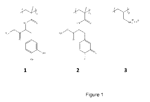
Figure 1 - shows the chemical structures of (1) P(mDOPA), (2) Pox(mDOPA)
(3) PAH
Figure 2 - shows the strategy for the formation of nanogels of
Pox(mDOPA)/PAH.
Figure 3 ¨ shows TEM analysis of a nanogel according to the invention
without (a) and with lyophilisation (b).
Figure 4 ¨ shows DLS analysis of nanogel solution.
Figure 5 ¨ shows DLS analysis of nanogel, nanogel(ABTs) and
nanogel(ABTs-Tica).
Figure 6 ¨ shows the modification of Ti surfaces with polydopamine (PDOPA)
layers, PAH and nanogels are self-crosslinked through Schiff-base bond on
PDOPA coated Ti substrates.
Figure 7 ¨ shows an image of a Ti surface before and after PDOPA
modification.
Figure 8 ¨ shows field emission scanning electron microscopy (SEM)
observations.
Figure 9 ¨ shows nanoreservoir build-up analyzed by Quartz Crystal
Microbalance.
Figure 10 - shows the modification of a Ti surface with nanogels according to
the invention.
Figure 11 ¨ shows SEM analysis of a nanogel according to the invention
deposited on a Ti surface following the 2nd approach
Figure 12 ¨ shows pictures of a biological valve before and after PDOPA
modification.
CA 03046397 2019-06-06
WO 2018/122318 PCT/EP2017/084728
22
Figure 13- shows SEM images of a biological valve surface before (above left)
and after nanogel deposition with (above right) or without PDOPA coating
(bottom left) (magnification=5000 X).
Figure 14 ¨ shows SEM images of a biological valve surface before (above
left) and after nanogel deposition with (above right) or without PDOPA
coating (bottom left) (magnification=30000 X).
Figure 15 ¨ shows a dynamic hemocompatibility test using an Impact-R
system at a constant shear rate of 18005-1 for 2min. The effect of shear
stress
on platelets adherence (left graph) and aggregate formation (right graph) at
different time points with two polymer coatings PEG or APEG is shown.
Median values on N=4 healthy donors are reported in both graphs. Statistical
analysis was performed using Graph Pad software with grouped, two-way
ANOVA and Bonferroni post-test. Moreover, platelets adherence correlated
with water contact angles of coatings on glass support: surfaces with lower
contact angle (i.e. the more hydrophilic PEG surface) gave lower platelets
adherence.
Figure 16 ¨ shows a dynamic Impact-R test at a constant shear rate of 18005-1
for 2min (A and B) or for 4 min (C). A. SC and AS median values determined
on 3 healthy donors (duplicates per condition per donor). B. Representative
pictures of the test shown in A.
Figure 17 ¨ shows a dynamic Impact-R test at a constant shear rate of 18005-1
for 4min. Platelet count was measured after shear stress challenge. Test was
carried on one donor in quadruplicate.
Figure 18 ¨ shows a static test which analyses blood cells that have adhered
to
nanogel-modified surfaces. Blood was incubated on 24-well polystyrene non-
coated (NC) or coated with Nanogel (NG) or Nanogel Peg (NP) or Nanogel
Antibiotics Peg (NAP). Cells that adhered to the surface were stained with
crystal violet. Images of cells fixed on the polystyrene surface were
processed.
Images were taken with an optical microscope Olympus CKX41 (20X
CA 03046397 2019-06-06
WO 2018/122318 PCT/EP2017/084728
23
magnification). Number of cells counted in each field using Fiji Software
(Particle Analysis plug-in). Numbers represent average of 8 fields per
conditions.
Figure 19 ¨ shows platelets rich plasma (PRP) at a concentration of
250000platelets/ 1 incubated in static condition at 37 C for lmin (A) or 45min
(B) on PS NC (polystyrene non-coated) or NAP (nanogel antibiotic PEG)-
coated wells. After 3 washes with NaCl 0,9% platelets were stained with May-
Grunwald solution and were observed at an Olympus CKX41 optical
microscope (10X magnification).
Figure 20 ¨ shows S. aureus biofilm quantification with crystal violet.
Bacteria were grown for 24 hours on a polystyrene surface modified by
nanogels loaded or not with antibiotics as indicated.
Figure 21 ¨ shows SEM analysis of a biological valve surface after 24-hour
incubation with S. aureus. Non-coated and nanogel-modified surfaces are
compared (magnification=2000x).
Figure 22 ¨ shows E. Faecalis bacteria grown for 24 hours on modified
polystyrene surfaces under static or shaking conditions. CFU counting of
planktonic bacteria plated on TSB agar plates is also shown.
Figure 23 ¨ shows the results of a study of nanoreservoir anti-thrombotic
property in solution by platelet aggregation assays. (A) PRP was pre-incubated
for 10 min with a solution of nanoreservoir containing increasing
concentrations of ticagrelor or with free ticagrelor, as indicated. Platelet
aggregation was induced by adding 10[tM ADP at 37 C under stirring
conditions (1200 rpm). Percentages of maximum aggregation recorded by light
transmission aggregometry are shown. Data represent means SD (*p<0.05,
**p<0.01, ***p<0.001). (B) Maximum platelet aggregation obtained in the
presence of different ratios of ticagrelor-loaded nanogels and minocycline-
loaded nanogels. NG: nanogel bearing PEG 1500; NGT: ticagrelor-loaded
nanogel bearing PEG 1500; NGM: minocycline-loaded nanogel bearing PEG
1500. Data represent means SD.
CA 03046397 2019-06-06
WO 2018/122318 PCT/EP2017/084728
24
Figure 24 ¨ illustrates the results of a study of the effect of immobilized
nanoreservoir on the activation of the contact phase of coagulation in human
plasma. Standard plasma was incubated on the coated and non-coated PS
surfaces for 10 min. Mixed minocycline- and ticagrelor-loaded nanogels in a
ratio of 2/3 (NGT60:NGM40) is compared to nanogels loaded with the two
drugs (NGTM). Basal: plasma that has not been in contact with the test
surfaces. NC: non-coated. Data represent means SD.
Figure 25 ¨ illustrates the results of a study of multilayer nanogel assembly
on
S. aureus biofilm formation. (A) Bacteria were let to adhere on coated and
non-coated PS surface for 24h before crystal violet staining. Data represent
means SD. (B) Bacteria were let to adhere on coated and non-coated titanium
surface for 24h before crystal violet staining. Data are representative of two
independent experiments. NC: non-coated; NG: non-loaded nanogel bearing
PEG 1500; LBL-1,3,5: 1, 3, and 5 layers of a mixture of nanogels loaded with
minocycline and ticagrelor in a ratio of 2/3.
Figure 26 ¨ illustrates the results of a study of multilayer nanogel assembly
on
antiplatelet effect of nanoreservoirs. NC: non-coated; NG: non-loaded nanogel
bearing PEG 1500; LBL-1,3,5: 1, 3, and 5 layers of a mixture of nanogels
loaded with minocycline and ticagrelor in a ratio of 2/3. Data represent
means SD.
Figure 27 - shows that cross-linking of the 5-layer nanogels with dopamine
sustains the antibiotic efficiacy against S. aureus biofilm formation beyond
48h. (A) Surfaces were incubated for two times 24h with fresh medium before
adding S. aureus bacteria. Biofilm formation was quantified by crystal violet
staining after 24h. (B) Anti-biofilm effect of medium removed after the second
24h of contact with the test surfaces. NC: non-coated; NG: 5-layer nanogel
with PEG 1500; NTMV: 5-layer nanogel with PEG 1500, loaded with
ticagrelor, minocycline and vancomycin; D-NTMV: dopamine cross-linked S-
layer nanogel with PEG 1500, loaded with ticagrelor, minocycline and
vancomycin. Data represent means SD.
CA 03046397 2019-06-06
WO 2018/122318 PCT/EP2017/084728
Figure 28 ¨ shows the results of in vivo analysis of the anti-biofilm efficacy
of
nanoreservoir immobilized on titanium implants. (A) S. aureus pre-infected
titanium discs were implanted subcutaneously in mice (n=3) and left for 3h
before analysis of live bacteria on the implants by CFU counting. (B) Surface
5 analysis of the implants by SEM (magnification 2000X).
Figure 29 ¨ shows the 1H NMR spectrum of thiol end functionalized PEG1.5
recorded in CDC13 with peak assignments.
10 Figure 30 ¨ shows the (A) 1H NMR spectrum of polymerized (2-
(Methacryloyloxy)ethyl Phosphorylcholine) recorded in D20 with peak
assignments. (B) Aqueous size exclusion chromatogram of four different
molecular weights of these polybetaines.
15 Figure 31 ¨ shows the LDH activity assay of platelet adhesion on
polystyrene
surfaces. PRP was incubated for 45 min on the indicated coated and non-coated
surfaces. NC: non-coated; NG: 5-layer nanogel with PEG 1500; NTM:
minocycline and ticagrelor containing nanreservoir (5-layer nanogels-PEG
1500 in a ratio 2/3); NTM-APEG1: minocycline and ticagrelor containing
20 nanreservoir (5-layer nanogels-PEG acrylate 1000 in a ratio 2/3); NTM-
APEG2: minocycline and ticagrelor containing nanreservoir (5-layer nanogels-
PEG acrylate 2000 in a ratio 2/3). Data represent means SD.
Figure 32 ¨ shows S. aureus biofilm formation on titanium implants coated or
25 not with with 5-layer nanogels bearing different ligands with thiol or
vinyl
functionalized ends. Bacteria were let to adhere for 3h before CFU counting.
NC: non-coated; NG-PB15: 5-layer nanogels with polybetaine 15 kD as last
layer; NG-P1.5, NG-P2: 5-layer nanogels with PEG 1.5 kD, or 2 kD as last
layer. NG-APEGO.5: 5-layer nanogels-PEG acrylate 500; NG-APEG1: 5-layer
nanogels-PEG acrylate 1000. Data represent means SD.
Figure 33 - shows S. aureus biofilm formation on PS surface coated or not
with with 5-layer nanogels bearing PEG of different molecular weights.
Bacteria were let to adhere for 24h before biofilm quantification by crystal
violet staining. Ctrl: non-coated; NG: 5-layer nanogel without grafted
polymer;
CA 03046397 2019-06-06
WO 2018/122318 PCT/EP2017/084728
26
NG-P1.5, NG-P2, NG-P5, NG-P10: 5-layer nanogels with PEG 1.5 kD, 2 kD, 5
kD, 10kD as last layer. Data represent means SD.
Figure 34 - illustrates the results of a study of the effect of thiol end PEG
of
different molecular weight on platelet adhesion under flow using Impact-R
under flow using Impact-R. Citrated whole blood was added to PS wells before
applying 780rpm for 4 min. Surface coverage (SC) and aggregate size (AS)
were determined. NC: non-coated; NG: 5-layer nanogel without grafted
polymer; NG-P1.5, NG-P2, NG-P5, NG-P10: 5-layer nanogels with PEG 1.5
kD, 2 kD, 5 kD, 10kD as last layer. Data represent means SD.
Figure 35 - illustrates the results of a study of the effect of 5-layer
nanogels
bearing PEG thiol of different molecular weights on the activation of the
contact phase of coagulation. Standard human plasma was incubated for 10
min at 37 C before clotting time analysis in the presence of the Nodia Non
Activated Partial Thromboplastin Time (NaPTT) reagent. Kaolin is used as
positive control. CTI: corn trypsin inhibitor; NC: non-coated; NG: 5-layer
nanogel without grafted polymer; NG-P1.5, NG-P2, NG-P5, NG-P10: 5-layer
nanogels with PEG 1.5 kD, 2 kD, 5 kD, 10kD as last layer. Data represent
means SD.
Figure 36 ¨ shows S. aureus Xen-29 biofilm formation on PS surfaces coated
with 5-layer nanogels bearing or not PEG thiol or polybetaines of different
molecular weight. Bacteria were let to adhere for 4h before removing the
medium and quantifying photon emission, which is directly proportional to
bacteria adhesion, either immediately (top panel) or after 2h (bottom panel).
NC: non-coated; NG: 5-layer nanogel without grafted polymer; NG-PB7, NG-
PB15, NG-PB44, NG-PB70: 5-layer nanogels with polybetaine 7 kD, 15 kD, 44
kD, 70 kD as last layer; NG-P2: 5-layer nanogels with PEG 2 kD as last layer.
Data represent means SD.
Figure 37 - illustrates the results of a study of the effect of thiol end PEG
and
polybetaines on platelet adhesion under flow using Impact-R. Citrated whole
blood was added to PS wells before applying 780rpm for 4 min. Surface
coverage (SC) and aggregate size (AS) were determined. NC: non-coated; NG:
CA 03046397 2019-06-06
WO 2018/122318 PCT/EP2017/084728
27
5-layer nanogel without grafted polymer; NG-PB7, NG-PB15, NG-PB44, NG-
PB70: 5-layer nanogels with polybetaine 7 kD, 15 kD, 44 kD, 70 kD as last
layer; NG-P2: 5-layer nanogels with PEG 2 kD as last layer. Data represent
means SD.
Figure 38 ¨ shows the results of a pNPP assay of platelet adhesion on
polystyrene surfaces. PRP was incubated for 45 min on the indicated coated
and non-coated surfaces. NC: non-coated; NG-PB7, NG-PB15: 5-layer
nanogels with polybetaine 7, or 15 kD as last layer; NG-P1.5, NG-P5: 5-layer
nanogels with PEG 1.5 kD, or 5 kD as last layer. NG-APEGO.5: 5-layer
nanogels-PEG acrylate 500; NG-APEG1: 5-layer nanogels-PEG acrylate 1000.
Data represent means SD.
Figure 39 - illustrates the results of a study of the effect of 5-layer
nanogels
bearing PEG thiol or polybetaine of different molecular weight on the
activation of the contact phase of coagulation. Standard human plasma was
incubated for 10 min at 37 C before clotting time analysis in the presence of
the Nodia Non Activated Partial Thromboplastin Time (NaPTT) reagent.
Kaolin is used as positive control. NC: non-coated; NG: 5-layer nanogel
without grafted polymer; NG-PB7, NG-PB15, NG-PB44: 5-layer nanogels with
polybetaine 7 kD, 15 kD, 44 kD as last layer; NG-P1.5, NG-P2, NG-P5: S-
layer nanogels with PEG 1.5 kD, 2 kD, 5 kD as last layer. Data represent
means SD.
Figure 40 - shows the quantification of plasma proteins adhered onto coated
and non-coated polystyrene surface after incubation of standard human plasma
for 10 min at 37 C. NC: non-coated; NG: 5-layer nanogel without grafted
polymer; NG-PB7, NG-PB15, NG-PB44: 5-layer nanogels with polybetaine 7
kD, 15 kD, 44 kD as last layer; NG-P1.5, NG-P2, NG-P5: 5-layer nanogels
with PEG 1.5 kD, 2 kD, 5 kD as last layer. Data represent means SD.
The invention now being generally described, it will be more readily
understood by
reference to the following examples which are included merely for purpose of
illustration of certain aspects and embodiments of the present invention, and
are not
intended to limit the invention in any way.
CA 03046397 2019-06-06
WO 2018/122318 PCT/EP2017/084728
28
Materials and Methods
Materials
Reagents
Antibiotics (minocycline, vancomycin) were purchased from Sigma, the
antiplatelet
drug Ticagrelor was from Cayman Chemicals.
Blood collection tubes: sodium citrate Vacutainer tubes (3.2% Sodium Citrate)
were
.. from BD Biosciences.
Bacteria strains and culture media: S. epidermidis, strain RP62A (#35984) was
from
ATCC; S. aureus, E. faecalis were purchased from ATCC (25904 and 29212); S.
aureus - Xen29 bioluminescent pathogenic bacteria were from Perkin Elmer
(#119240). Tryptic Soy Broth (TSB) and agar powder was from Sigma-Aldrich.
Sample preparation
Sterilization of surfaces before in vitro testing: all coated or non-coated
surfaces were
sterilized in 100% absolute ethanol for 10min followed by 2 to 5-minutes-
incubation
washes in distillate water and one wash in 0.9% NaCl.
Blood from healthy donors (under no medication and that did not take any
aspirin or
other anti-coagulant drug in the last 20 days prior the drawing) was drawn
using a 18g
needle and directly let flow in a 50mL polypropylene tube containing 3.2%
sodium
citrate (1 volume citrate for 9 volumes of blood) for the static test or using
a 21 g
needle and citrate vacutainers tubes for dynamic tests. The study was approved
by the
Ethics Committee of the University Hospital of Liege, Belgium. An informed
consent
was signed by the donors.
Experimentations using S. aureus were conducted in a biosafety level 2 room of
the
GIGA-R.
Dynamic in vitro Impact-R test
The blood was rested in the tube for 45 min before any processing at the cone
& plate
device Impact-R system (Matis Medical). Blood was then mixed at 1 Orpm for
lmin at
room temperature before the application of shear stress. For each test 130 1
of blood
CA 03046397 2019-06-06
WO 2018/122318 PCT/EP2017/084728
29
was carefully deposited on the well. The shear stress applied was 1800s-1 for
4 min
(corresponding to 780rpm bell speed): this speed and incubation time simulates
the
laminar arterial blood flow over a polystyrene surface and is useful to
analyze platelet
function under shear stress.
After the 4 min application of shear stress, blood was collected and analyzed
by Cell
Dyn for single platelet count, while the PS wells coated or not were gently
washed
with distilled water 4 times and stained with May-Grunwald stain solution for
lmin at
RT. Platelets that adhered on the surface were visualized with an optical
microscope
and quantified using the Impact-R software. Two parameters were obtained from
the
analysis (i) the surface coverage (SC %) and (ii) the aggregate size of the
platelets
(AS p.m). Every condition was repeated in duplicate per each donor.
Static in vitro test
Platelet adhesion on surfaces was analysed by using one of the following
photometric
assays: the LDH (Lactate Dehydrogenase Test) and the p-NitroPhenil Phosphate
(pNPP) test. Values obtained from both tests are directly proportional to the
number
of platelets adhering to the surface. LDH released upon platelet lysis is
indirectly
analysed by measuring the conversion of NAD to NADH, detected at 450nm. The
pNPP test measures the levels of alkaline and acid phosphatases released upon
platelet
lysis. The hydrolysis of pNPP, a substrate of these phosphatases, produces p-
nitrophenol, which has a maximal absorbance at 405nm and is proportional to
the
amount of platelets bound to the surface.
Platelet poor plasma (PPP) or platelet rich plasma (PRP) was layered on coated
polystyrene surfaces for 45min at 37 C. Surfaces were washed 3 times with NaCl
0.9%. Lysis of adhered platelets was achieved by adding 1% Triton in PBS for
the
LDH test, or in 1% Triton in a sodium citrate buffer (0.05M Citrate pH 5.4)
containing
5mM pNPP for the pNPP assay. Background readings from the PPP incubation was
subtracted from the PRP reads.
Platelet aggregation tests
Platelet-rich-plasma (PRP) was prepared by centrifugation of citrate
anticoagulated
human blood at 100xg for 15 min at room temperature. Platelet aggregation
CA 03046397 2019-06-06
WO 2018/122318 PCT/EP2017/084728
experiments were performed on PRP aliquots under stirring (1200 rpm) at 37 C
using
light aggregometry (Chrono-Log Model 700 aggregometer, Kordia).
Plasma-biomaterial interaction: coagulation assay and plasma protein adhesion
5 Clotting tests were performed using the Stago STart0 4 Hemostasis
Analyzer and the
Nodia Non Activated Partial Thromboplastin Time (NaPTT) reagent. The NaPTT
reagent is a synthetic phospholipid platelet substitute intended for the study
of
activation of contact phase of coagulation. The Stago Analyzer is a semi-
automated
system integrated with an electro-mechanical clot detection method (Viscosity-
based
10 detection system). Clot formation in citrated human standard plasma
(Stago) is
catalyzed by the addition of Ca2+ ions as well as by phospholipids. Kaolin was
used as
a positive control for contact phase activation, while Corn Trypsin Inhibitor
(CTI), a
specific inhibitor of factor XIIa, which is the factor initiating the contact
activation
pathway, was used to determine clotting time independently of this pathway.
Standard
15 human plasma was defrost in a water bath at 37 C, and added into coated
and non-
coated polystyrene (PS) wells for 10 min at 37 C without stirring. Plasma was
then
snap frozen in a dry ice/ acetone bath (-78 C) and stored at -80 C until
analysis. On
the day of the test, plasma was defrost at 37 C and immediately processed in
the Stago
STart0 4 apparatus. The 37 C pre-warmed Nodia Reagent was added to 100 1 of
pre-
20 warmed plasma in a cuvette containing a coated metal bead before
initiating
coagulation by addition of a pre-warmed calcium solution (8.3mM). The clotting
end
point is measured by the pendular movement of the bead alimented by an
electromagnetic field. Such movement, which is influenced by the viscosity of
the
plasma, stops when the viscosity becomes maximal, i.e. when plasma coagulation
25 occurs.
Protein adherence was measured using the Pierce Micro BCA (Bicinchonic Acid)
Assay, a highly sensitive method detecting down to 5ng/m1 of protein.
Bicinchonic
acid binds to Cu+ ions with a 2:1 stoichiometry delivering high sensitivity.
The assay
30 is based on the conversion of Cu2+ ions into Cu+ by proteins in basic
environment.
120111 of plasma (Stago Standard Plasma) pre-warmed at 37 C was added in
coated
and non-coated wells of a 48-well polystyrene (PS) plate (11mm diameter wells)
and
incubated for 10 min at 37 C. After 3 washes with NaCl 0.9%, the adhered
proteins
were detached by adding 250111 of a solution of SDS 1% in PBS for 10 min at
RT. The
undiluted protein solution was used in the Micro BCA Assay.
CA 03046397 2019-06-06
WO 2018/122318 PCT/EP2017/084728
31
Bacteria adhesion and biofilm formation analysis
One colony of S. epidermidis or S.aureus or E. faecalis was grown 0/N at 37 C
in
TSB medium under agitation (220rpm), the following day a dilution 1:100 was
performed in TSB fresh medium and the suspension was grown for 4 hours until
the
logarithmic phase (0D595=0.5) was reached. Bacteria were then diluted 1/20 in
sterile
NaCl 0.9% to have around 200000 cfu/ 1 and 500[LL were incubated in static or
dynamic condition for 24hr at 37 C in coated or non-coated polystyrene 24-well
plates. Bacteria suspensions were analyzed by agar plating and CFU (colony
formation
unit) counting, while biofilms were analyzed by crystal violet staining. The
surface
was first washed 3 times with NaCl 0.85% to eliminate planktonic bacteria and
then
stained with 1% crystal violet for 40min. After 3 washes with water the
crystal violet
dye retained in the bacteria was released using 10% Acetic Acid for 10min.
Intensities
were measured at 595nm in a 96-well plate using a spectrophotometer plate
reader.
Conditions were in duplicates and reads were in triplicate. Kinetics of
bacteria
adhesion and biofilm formation on surfaces were also assessed by using
bioluminescent S. aureus bacteria with the IVIS Lumina system (Perkin Elmer).
Bacteria were let adhere for 3h before washing the wells, and luminescence
signals,
directly proportional to bacteria density, were then recorded for increasing
times.
Biofilms were imaged using the IVIS camera system. Total photon emission from
selected wells was quantified using the LivingImage software package.
Mouse model of biomaterial-associated infection mouse model: subcutaneous
implantation of a pre-infected titanium device and in vivo biofilm formation
Staphylococcus aureus was grown for 2h at 37 C in TSB medium to reach
logarithmic
phase. Bacteria were diluted 1:10000 in TSB supplemented with 2% NaCl + 1%
Glucose and a 800 I aliquot (corresponding to 20000 CFU/disk) was layered on
Titanium 0.2 cm diameter disks (Biotronik). Bacteria were let adhere on all
disks for
3h at 37 C under static conditions. Bacteria suspension was removed, and the
surface
was gently washed 3 times in PBS. To determine the number of bacteria that
adhered
on the titanium disks, half of the disks were sonicated for 5min in a Fisher
waterbath
sonicator. The detached bacteria were plated on a TSB agar plate to determine
the
number of colony forming units (CFU) per disk before implantation. The other
half of
the disks was implanted in 8-weeks old male BALB/cJRj mice (Janvier
Laboratories)
as follows. Two hours prior anesthesia, mice were injected subcutaneously with
0.05
CA 03046397 2019-06-06
WO 2018/122318 PCT/EP2017/084728
32
mg/kg buprenorphine analgesic (Temgesic). Fifteen minutes before the
implantation
mice were anesthetized by intraperitoneal injection of a ketamine
(125mg/kg)/xylazine
(12.5mg/kg) mixture. Mice were shaved on the lower ventral side below the rib
cage
and the area was sterilized with betadine followed by 70% ethanol solution.
Using a
sterile scalpel an incision was made on the skin and the S. aureus infected or
non-
infected disks were inserted between the skin and the muscles. After 4h
incubation,
mice were sacrificed by cervical dislocation and the devices were analysed to
determine CFU/disk (by sonication, like previously described). The protocol
was
approved by the ethical committee of the ULiege University (# 16-1774). SEM
images
of explanted titanium disks were taken with the Quanta Microscope
(magnification is
4000X). Titanium disks were gently washed and fixed in 2.5 glutaraldehyde in
Sorensen buffer for lh at 4 C, followed by 3 washes in Sorensen buffer and
fixation in
2% 0s04 for lh at 4 C. The disks were dehydrated in increasing ethanol
concentrations, dried under CO2 atmosphere (critical point drying) to keep
biological
structures and then metallised.
Results and discussion
Examples
Example 1. Preparation of cross-linked nanogel and loading with bioactive
molecules
A homopolymer of methacrylamide bearing 3,4-dihydroxy-L-phenylalanine
(P(mDOPA), 1, Figure 1) was specifically designed to prepare nanogels and to
immobilize active (bio)molecules by physical entrapment or covalent
conjugation.
Cross-linked reactive nanogels can be directly deposited onto a surface pre-
coated
with a bio-inspired polydopamine layer. This strategy has several advantages
over
existing methods: i) there is no use of an external cross-linking agent, ii)
coupling
reactions are fast at room temperature in water, iii) no undesirable side
products are
formed and released out of the film, and iv) active biomolecules can be
covalently
grafted to the surface.
A fast and water based cross-linking process was used to exploit the redox
properties
of DOPA molecules in order to provide reactive function available for nanogel
formation and for nanogel functionalization.
CA 03046397 2019-06-06
WO 2018/122318 PCT/EP2017/084728
33
Stable solutions of nanogels in water were prepared by adequately controlling
both the
redox state of the P(mDOPA) polymer and the pH of the PAH solutions.
Preparation
conditions are crucial for the success of the nanogel formation. First,
P(mDOPA) is
oxidized in aqueous media under basic conditions for 12 hours to form the
hydrosoluble Pox(mDOPA). Oxidized DOPA moieties of Pox(mDOPA) are necessary
for the covalent interaction of PAH through amine/quinone reaction and/or
Schiff base
formation at room temperature, and consequently for the preparation of stable
cross-
linked nanogel (Figure 2).
The formation of nanogels Pox(mDOPA)/PAH was first performed by the slow
addition of a solution of PAH to an aqueous solution of Pox(mDOPA) at room
temperature. Weight ratios and addition modes of the two partners were
controlled to
form stable dispersions of nanogels (Pox(mDOPA)/ PAH). For that purpose, the
slow
addition of PAH to an aqueous solution of Pox(mDOPA) resulted in the
spontaneous
formation of a stable and clear light brown solution of cross-linked nanogels
at room
temperature. The presence of the nanogels was confirmed by transmission
electron
microscopy (TEM) performed after lyophilisation that showed nanogels with a
diameter ranging from 100 to 200 nm (Figure 3b). It is important to note that
the
nanogels solution had to be lyophilized on the TEM grid prior to analysis. If
the
solution was simply dropped onto the grid and slowly dried at room temperature
under
atmospheric conditions, nanogels strongly aggregated (Figure 3a).
Analysis by dynamic light scattering (DLS) without filtration showed nanogels
agglomeration with an average hydrodynamic diameter equal to 130 nm with a
rather
high polydispersity (PDI = 0.2) (Figure 4). The nanogel solutions were stable
for at
least one month when stored at room temperature and at pH around 10 without
stirring. The solution remained clear without any precipitation and the
hydrodynamic
diameter distribution obtained by DLS remained almost unchanged after one
month of
storage.
The ability of the nanogels to be loaded with and deliver multiple drugs was
further
explored. Different bioactive agents were incorporated through the combination
of
covalent conjugation, electrostatic and hydrophobic interactions as well as
hydrogen-
bond formation. Covalent conjugation was carried out by exploiting the
reactivity of
quinone groups of Pox(mDOPA) towards amine function. For that purpose,
CA 03046397 2019-06-06
WO 2018/122318 PCT/EP2017/084728
34
Vancomycin (V), a glycopeptide antibiotic that contains primary amine in its
sequence, was used for the conjugation, and physical entrapment was employed
for the
incorporation of Minocycline (M), a tetracyclin-class antibiotic, and
ticagrelor (T), an
antiplatelet agent with antithrombotic properties.
Pox(mDOPA)-VMT/PAH) were then prepared in a similar way by the addition of a
PAH solution to an aqueous solution of VMT loaded Pox(mDOPA), resulting in the
appearance of a yellow-brown suspension. DLS measurement without filtration
evidenced the presence of nanogels with an average hydrodynamic diameter of
200 nm
slightly higher than the previous one due probably to the presence of VMT
(bio)molecules (Figure 5).
All synthetic steps are performed in mild conditions and in aqueous media,
which
make the building-block synthesis pathways relevant for the development of an
environment-safe process.
Oxidation of P(mDOPA) in Basic Medium : Oxidation was carried out according to
a previous study (Faure et al., Biofouling. 2012: 28(7):719-28). P(mDOPA) (20
mg)
were dissolved in distilled water (20 mL) and a NaOH solution (0.1 M) was
slowly
added in order to raise the pH above 10. This oxidation step lasted at least
one night
under air.
Preparation of Pox(mDOPA)/PAH Cross-Linked Nanogels was as follows:
Nanogel preparation: P(mDOPA) (2.5 mg) was dissolved in distilled water (5 mL)
and NaOH (0.1 M) was slowly added in order to raise the pH above 10 and to
promote
the oxidation of catechol groups of P(mDOPA). After one night at room
temperature,
an aqueous solution of PAH (0.5 mL; 0.5 g/L) at pH 10 was slowly added to the
solution of Pox(mDOPA) under vigorous stirring. The solution was lead to react
for
one hour at room temperature under vigorous stirring. Nanogels with a diameter
ranging from 150nm to 250 nm were observed by DLS.
Nanogel antibiotics (ABTs) preparation: The procedure is identical as the one
described above except that Pox(mDOPA) was solubilized in the presence of ABTs
(Minocycline and Vancomycine) to increase the interaction between polymer
chains
CA 03046397 2019-06-06
WO 2018/122318 PCT/EP2017/084728
and drugs. After lh at 6 C, an aqueous solution of PAH (0.5 mL; 0.5 g/L) at pH
10
was slowly added to the solution of Pox(mDOPA) under vigorous stirring. The
solution was lead to react for one night at 6 C under vigorous stirring before
nanogel
deposition. The final concentration of the ABTs in the nanogels solution was
5 .. 0.5mg/ml.
Nanogel Ticagrelor (T) preparation: The procedure is identical as the one
described
above except that Pox(mDOPA) was solubilized in the presence of lml of
Ticagrelor
solution (1mg/m1 in DMSO) to increase the interaction between polymer chains
and
10 drugs. After lh at 6 C, an aqueous solution of PAH (0.5mL; 0.5 g/L) at
pH 10 was
slowly added to the solution of Pox(mDOPA)/T under vigorous stirring. The
solution
was lead to react for one night at 6 C under vigorous stirring before nanogel
deposition.
15 .. Example 2. Nanogel immobilization on titanium substrate using
polydopamine
coating
A first strategy consisted of a first immersion of the substrate in a Tris
buffer solution
of DOPA to strongly anchor the first layer to the surface by DOPA/metal
interactions
20 (Figure 6). The next layers were then built by the successive dipping of
the surface
into an aqueous solution of a polymer bearing primary amines, polyallylamine
(PAH),
and then in a solution of a P(mDOPA) based nanogel. Poly(methacrylamide)
bearing
oxidized DOPA moieties on each monomer unit (Figure 1, formula (2)).
(Pox(mDOPA)) were used in combination with PAH to prepare stable solutions of
25 .. nanogels in water at room temperature that can be easily deposited to
titanium (Ti)
surface.
Polydopamine coating
Polydopamine (PDOPA) has been used to modify bio-inert surfaces because it can
30 adhere on various material surfaces. Dopamine molecules have 3,4-
dihydroxy-l-
phenylalanine-lysine motif, which can polymerize to form PDOPA layers on
material
surfaces at mild conditions. Incorporating PDOPA as primer films provides an
alternative route to functionalize those biomaterials with non-fouling
surfaces, and
can further enhance their desirable biological, chemical, and therapeutic
properties for
35 biomedical applications.
CA 03046397 2019-06-06
WO 2018/122318 PCT/EP2017/084728
36
Dopamine (2 mg/mL) was dissolved in 10 mM Tris-HC1 (pH 8.5), and substrates
were
dipped into the solution. pH-induced oxidation changes the solution colour to
dark
brown, resulted in spontaneous deposition of a thin adherent polymer film
(Figure 7).
To avoid the microparticle deposition lower dopamine concentration can be used
0.125mg/m1 and/or vertical sample orientation were necessary. The coated
surfaces
were rinsed with ultrapure water and dried by N2 gas before storage or treated
as
described below for ad-layer formation.
.. Polydopamine-assisted self-assembled PAH monolayer
After modification of titanium surface using PDOPA as first layer, covalent
grafting
of PAH occur through amine/quinone reaction and/or Schiff base formation at
room
temperature, and consequently for the interlayer cross-linking (Scheme 1).
Importantly, the next solution of PAH was deposited at pH above 10 in order to
obtain
the polymer in the deprotonated state. The reaction between primary amines and
the
quinone groups of Pox(mDOPA) was therefore made possible. In acidic media,
amine
groups were protonated and did not react with Pox(mDOPA), such that
crosslinking
and growth of the film cannot occur. For PAH ad-layer formation, 1 mg/ml of
PAH
solution was dissolved in ultrapure water which was equilibrated at pH abovel0
by
adding 0.1M NaOH solution. Polydopmaine-coated substrates were subsequently
immersed in the solution. After 4hrs or more (typically overnight reaction for
18 hrs),
the substrates were rinsed by ultrapure water.
Contact angles were measured on a dry titanium surface after dropping 10 [LL
of
ultrapure water. The static contact angles on the pure titanium and
polydopamine-
coated titanium surfaces were 85 2 and 47 6 , respectively proving the
modification of the surface.
Nanogel assembly
Covalent grafting of nanogels was performed through the same reaction and/or
Schiff
base formation between primary amines of PAH monolayer and the quinone groups
of
Pox(mDOPA) in nanogel. The surface coated by a layer of the PDOPA, and a layer
of
PAH was then incubated with the aqueous solutions of the nanogels
Pox(mDOPA)/PAH following the protocol described below. Contact angles were
measured on a modified titanium surface with PAH and nanogel. The contact
angle of
CA 03046397 2019-06-06
WO 2018/122318 PCT/EP2017/084728
37
the PAH was about 65 ; its amplitude was higher than of the one measured on
polydopamine-coated surface, probably due to the presence of non-polar amine
group.
After deposition of nanogel presenting the same catechol groups as
polydopamine, the
contact angle decreased to about 42 .
PEG grafting monolayer
Poly(ethylene glycol) (PEG) is one of the most commonly used synthetic polymer
to
impart protein resistance to a surface. Several strategies to modify
substrates with that
kind of polymers can be found in the literature such as electrografting, self-
assembled
monolayers (SAMs), copolymers adsorption to cite only a few. The PEG grafting
was
carried out by exploiting the reactivity of quinone groups of Pox(mDOPA)
towards
thiols. For that purpose, thiol end-functionalized PEG (PEG-SH) were
considered for
the conjugation. This thiol-based strategy allows specific grafting under mild
conditions and without pH constrain in contrast to the amino-based strategy
that
requires pH > 10. For PEG grafting, 5 mg/mL of methoxy-poly(ethylene glycol)-
thiol
(mPEG-SH, 1.5 kDa) was dissolved in 10 mM Tris pH 8Ø The buffer used for
mPEG-
SH was vacuum degassed for ¨1 hr to prevent oxidation (-S-S-) between thiol
groups.
PEG was reacted into the PDOPA layer (nanogel layer) through Michael-type
addition
and Schiff base reactions to inhibit non-specific interactions and increase
hydrophilicity in physiological conditions. The water contact angles verified
the
anchoring of PEO chains to modified Ti surfaces. Surprisingly a PEO layer
exhibiting
contact angle of 43 1 was found.
Briefly, nanogel deposition using PDOPA as first layer was conducted at room
temperature in five steps:
Step 1: Ti discs (1 cm diameter) were sonicated in tetrahydrofuran (THF),
acetone,
ethanol, and water, 10 min for each step.
Step 2: Ti were immediately dipped into the DOPA Tris buffer solution (0.125 g
L -1)
for 18h.
step 3: After rinsing twice with 5m1 water, the modified substrates were
dipped into a
solution of PAH (pH>10) for 18h and rinsed twice with 5m1 water.
step 4: After rinsing twice with 5m1 water, the modified substrates were
dipped into a
solution of nanogels (loaded with ABTs and T) for 18h and rinsed twice with
5m1
water.
CA 03046397 2019-06-06
WO 2018/122318 PCT/EP2017/084728
38
step 5: After rinsing twice with 5m1 water, the modified substrates were
dipped into a
solution of PEG-SH for lh and rinsed twice with 5m1 water.
The surface morphology is an important parameter for biomedical devices that
may
affect the interface energy and also the interaction between the bio-
components and
the material surfaces. To gain further information and understand the
microstructures
of the nanogel coated Ti surfaces, field emission scanning electron microscopy
(SEM)
observations were carried out (Figure 8). The pristine Ti surface exhibited a
relatively
smooth morphology (left panels). In contrast, after nanogel deposition,
numerous
well-distributed nanogels were observed, ranging in size from 80 nm to 120 nm
(right
panels). The results indicated that the size of deposited nanogels,
corroborate the sizes
of nanogels measured by TEM in solution (Figure 2b).
Layer-by-layer (LbL) assembly
LbL deposition was conducted in the same conditions, using similar procedure
as the
one described above except that steps 2 to 4 were repeated five times to
obtain five
layers.
Quartz Crystal Microbalance
As a first evidence of the multilayer film build-up, Quartz Crystal
Microbalance
coupled with Dissipation (QCM-D) was used to follow the film growth in real
time on
gold sensors by measuring the variation of the resonant frequency (Af) vs.
time. A
decrease in Af indicates polymer deposition. Figure 9 shows that all
components were
successfully deposited according to the selected deposition protocol and
redox/pH
conditions and remain on the substrate even after rinsing with water.
First, a tris buffer solution of DOPA (0.125g/L) was flowed through the cell
at room
temperature, leading to the first anchoring layer. The f shift continued to
decrease,
until the DOPA was rinsed from the sample chamber. Second, a solution of PAH
(pH>10) was injected leading to an immediate negative shift was observed
indicating
an increase in mass at the interface, as the polymer adsorbed to the gold
surface. After
removing the excess unbounded polymer by rinsing with water, a stable baseline
was
attained during the rinsing step, demonstrating that the polymer ad-layer
strongly
tethered onto the surface. Once the reversibly adsorbed polymer was removed in
the
rinsing step, nanogel solution in milli-Q water (pH E10) was introduced into
the
CA 03046397 2019-06-06
WO 2018/122318 PCT/EP2017/084728
39
sensor chamber, at the same flow rate and temperature. The deposition of the
nanogel
solution was observed by the important decrease of the frequency vibration.
Importantly, this layer of nanogels was stable since it cannot be removed
after rinsing
with water. In the last step, irreversibly bound polymer can be seen from the
raw Af
after injection of PEG-SH solution due to the reaction of thiol function with
residual
quinone groups present in the nanogels (Figure 9).
Example 3. Direct nanogel immobilization on titanium substrate
In a second approach, titanium surface was directly immersed in the nanogel
solution
to obtain nanogel-modified surface without the need of a primer coating. In
this case,
the assembly mechanism is based on the adhesive property of the catechol and
quinone group present in the surface of the nanogel (Figure 10).
Using SEM analysis, the formation of a monolayer of nanogels Pox(mDOPA)/PAH on
Ti was observed (Figure 11). Estimated diameters of the nanogels were found to
vary
between 80 nm to 120 nm.
Example 4: Immobilization of nanogels onto valve bioprosthesis
The same approach reported above was used to modify a biological valve.
Respecting
the same steps, biological valves were first immersed in a Tris buffer
solution of
DOPA (10 mM Tris-HC1 (pH 8.5)) to strongly anchor the first layer of
polydopamine.
The implant became dark brown as a result of deposition of a thin adherent
PDOPA
film (Figure 12).
The next layers were then built by successive dipping of the valve into an
aqueous
solution of a polyallylamine (PAH), and then in a solution of a P(mDOPA) based
nanogel.
The following steps were conducted at room temperature:
Step 1: Biological discs (1 cm diameter) were dipped three times in 10m1 of
water, for
10 min.
Step 2: Biological discs were immediately dipped into the DOPA Tris buffer
solution
(0.125 g L-1) for 18h.
Step 3: After rinsing twice with 5m1 water, the modified tissue was dipped
into a
solution of PAH (pH>10) for 18h and rinsed twice with 5m1 water.
CA 03046397 2019-06-06
WO 2018/122318 PCT/EP2017/084728
Step 4: After rinsing twice with 5m1 water, the modified tissue was dipped
into a
solution of nanogel for 18h and rinsed twice with 5m1 water.
Step 5: After rinsing twice with 5m1 water, the modified tissue was dipped
into a
solution of PEG-SH for lh and rinsed twice with 5m1 water.
5 Scanning electron microscopy SEM was used to analyse the surface
morphology of
modified biological tissue. It was observed that the surface of biological
tissue before
modification exhibited a relatively smooth and ordered collagen nanofibers.
After
nanogel deposition, collagen fibers appeared more compact (Figure 13), and
well-
distributed nanogels were observed on the surface of collagen fibers (Figure
14). The
10 PDOPA primer coating augmented nanogel deposition.
Film growths (PDOPA and nanogels) were followed in real time using quartz
crystal
microbalance coupled with dissipation technique (QCM-D). A Q-Sense E4 was used
in
this study. The stainless steel-coated AT-cut resonator (fundamental
frequency: 5
15 MHz) was used as received. First, distilled water was introduced in the
cell and the
flow was maintained until a stable baseline was obtained. LbL deposition was
then
initiated by switching the liquid exposed to the crystal from distilled water
to the
DOPA solution 0.125g U1 with 0.15 M NaC1 at a flow rate of 200 [LL min 1,
temperature of 25 C. After 10 min, the substrate was rinsed by distilled water
to
20 .. remove the excess of unbounded DOPA. Then, the alternative deposition of
PAH (1 g
L -1) and nanogels solutions was carried out (about 10 min for each step) with
rinsing
steps with distilled water between each layer. PEG-SH solution was finally
introduced
in the system as the last layer and further rinsed when a stable signal is
obtained.
25 A Delsa Nano-C Particle Analizer (Beckman Coulter) equipped with a laser
diode
source (wavelength 658 nm; power 30 mW) was used for measuring the
hydrodynamic
diameter of the aqueous nanogels solutions. Scattering data were collected for
at least
individual measurements at a constant scattering angle and averaged for each
sample. The obtained scattering data were fitted using a volume-weighted
cumulative
30 analysis to estimate the diffusion coefficient of the nanogels in
solution. The
hydrodynamic diameter of the samples (DH) was obtained using Stokes-Einstein
relationship.
The samples for scanning electron microscopy (SEM) were analysed by Field
35 Emission Gun Scanning Electron Microscope (FEG-SEM) MEB ULTRA55 operating
CA 03046397 2019-06-06
WO 2018/122318 PCT/EP2017/084728
41
at 3 kV was used for sample observation after a thin layer (10 nm) of Au-Pd to
increase the contrast.
Synthesis of N-methacryloyl 3,4-dihydroxy-L-phenylalanine methyl ester
DOPA methyl ester hydrochloride (9 g, 0.0363m01) in dry CH2C12 (350 mL) was
added to a two-necked round-bottomed flask equipped with a dropping funnel and
a
magnetic stirrer, and placed under nitrogen. Freshly distilled Et3N (17.7mL,
0.127
mol) was then added, and the flask was cooled to OC. A solution of
methacryloyl
chloride (3.51 mL, 0.0363 mol) in CH2C12 (70 mL) was added dropwise through a
dropping funnel with vigorous stirring under nitrogen. The final mixture was
maintained under stirring at room temperature for 48 h. After reaction, the
triethylammonium chloride, formed as a by-product, was removed by filtration
and the
excess of reagents was removed under reduced pressure. The product was
recovered as
a sticky solid with a yield of 90%.
P(mDOPA) synthesis
Prior to polymerization, the catechol group of the mDOPA must be protected in
order
to avoid side reactions during the radical polymerization from its -OH groups.
2 g of
mDOPA with protected catechol groups (see below for the protection step) (2.6
mmoL) were placed under nitrogen in a one-necked round-bottomed flask equipped
with a magnetic stirrer. At the same time, 19 mg (0.067 mmoL) of V501
initiator was
dissolved in 7 mL of distilled water. The pH solution was adjusted above 9
with
Na2CO3 until complete dissolution of the white powder. The solution was then
degassed by bubbling nitrogen through it for 15 minutes. Then, the aqueous
solutions
of the V501 initiator was transferred with a capillary under nitrogen in the
glass flask
containing protected mDOPA. The reactor was heated in an oil bath thermostated
at
70 C during 24 hours. Then, catechol groups were deprotected by adjusting the
pH
around 2 with concentrated HC1. The resulting mixture was dialyzed (membrane
porosity 1000 Da) against water during 48 hours, followed by lyophilization.
The
copolymer was recovered as a white powder with a 88 % yield.
Synthesis of a-methoxy-(o-mercapto-poly(ethylene oxide) (MPEG-SH)
a-Methoxy-x-mercapto-poly(ethylene oxide) (MPEG-SH) was synthesized by
esterification of the hydroxyl end-group of the monomethoxy poly(ethylene
oxide)
(MPEG-OH) (Mn = 1500 g/moL) with mercaptoacetic acid. A typical reaction was
CA 03046397 2019-06-06
WO 2018/122318 PCT/EP2017/084728
42
carried out as follows. MPEG-OH (10 g; 5 mmoL) was added into a 100 ml two-
necked flask equipped with a stirrer and a Dean-Stark device. The MPEG-OH was
dried by three azeotropic distillations with toluene and finally dissolved in
50 ml of
toluene. Mercaptoacetic acid (3.5 ml, 50 mmoL) and concentrated sulfuric acid
(two
drops) were then added. The flask was heated in an oil bath at 110 C
overnight.
MPEG-SH was collected by precipitation in ether at 0 C and then dried at 40 C
under
vacuum for 24 h.
Example 5: Anti-thrombotic properties of bioactive nanogels
Dynamic Impact-R Test
To study blood-material interaction in dynamic conditions, the Impact-R
apparatus
was used. This is a close-to-physiological in vitro system mimicking the
laminar flow
of the blood in circulation and is used to study platelets function and
thrombus
formation under flow. The laminar flow created by the rotation at constant
speed of a
piston (the bell and the cone) on a thin blood layer deposited on a well,
activates
platelets by shear stress, platelets adhere on the polystyrene surface of the
well and
then are stained with May-Grunwald stain. The system allows variation of the
speed:
the higher the speed ¨ corresponding to higher heart rate - the more thrombi
form on
the surface.
By inspection of the polystyrene surface at an optical microscope and by image
analysis, it is possible to obtain two parameters: the percentage of surface
covered by
platelets aggregates (SC, %), and the average size of the aggregates (thrombi)
(AS,
lim2).
The test was performed on citrated blood of healthy donors. The SC and AS
obtained
were proportional to the speed rotation of the cone at a specific well radius
(speed
increases with radius).
The effect of different polymer coatings at different time points was tested
(Figure
15). The polymers (PEG and reticulated PEG, APEG) were covalently bound to a
thin
polydopamine layer, DOPA, which was also tested alone, and which had a slight,
but
not significant, effect on the reduction of platelet activation and adherence.
Increasing
the time of shear stress applied to the blood, the two parameters SC and AS
increased
CA 03046397 2019-06-06
WO 2018/122318 PCT/EP2017/084728
43
significantly. A significant difference was found at all time points between
non-coated
(NC) and PEG-coated surface for both SC and AS parameters.
The following coatings were then tested, Polydopamine (DOPA), DOPA-PEG (PEG),
DOPA-Nanogel (NGEL) and DOPA-Nanogel-PEG (NGEL-PEG) on 3 healthy donors
in vitro at the Impact-R using 18005-1 shear stress for 2min (Figure 16A,B).
Significant differences in SC values were observed for all coatings compared
to NC,
except for DOPA. PEG-coated PS wells gave the lowest SC and AS values.
Platelets
could not form aggregates on PEG-coated surface. Importantly, nanogel
deposition did
not show platelet adhesive properties.
The test in Figure 17 was performed to study the effect of ticagrelor alone
(NG-T-
PEG) or in combination with the two antibiotics (NG-TA-PEG) on flow-induced
platelet consumption. A test on a blood sample from one donor was carried out
at
1800s-1 for 4 min. Binding of platelets was not observed for any conditions
except for
the non-coated (NC) wells. The values reported in Figure 17 represent single
platelet
count in the blood after the test, and thus depict platelet aggregate
formation on the
surface and/or in blood. A significant increase in platelet count was observed
for both
nanogels, indicating that platelets remained in suspension as single platelets
(i.e. non
activated) and did not adhere to the surface.
Static Test
The hemocompatibility of the coating in static conditions (no shear stress
applied) was
studied. Static test was performed by incubating 500[LL of fresh blood under
gentle
agitation (60rpm) for 2h at 37 C and 5%CO2 in a 24-well non-treated plate and
observing cell adherence at the optical microscope in presence or absence of
the
coating. Cells were stained using Crystal Violet dye. In Figure 18 optical
microscope
images of cells stained with crystal violet highlighted inhibition of blood
cell adhesion
after PS surface modification. Conditions were: Non Coated (NC), Nanogel (NG),
Nanogel Peg (NP) and Nanogel antibiotics Peg (NAP) bound to a polydopamine
layer.
A platelet suspension (PRP: platelet rich plasma) was also incubated with NAP-
coated
PS wells for lmin (Figure 19A) or for 45 min (Figure 19B). A clear difference
was
observed in surface coverage compared to NC-PS well. NC-lmin: 90% surface
coverage (SC); NAP-lmin: 5% SC; NC-45min: 99,5% SC; NAP-45min: 40% SC.
CA 03046397 2019-06-06
WO 2018/122318 PCT/EP2017/084728
44
Thus, all these data indicate that surface modification with the nanogel
preparation of
the present invention does not activate platelets under static and dynamic
conditions.
Example 6: Anti-biofilm properties of bioactive nanogels: Staphylococcus (S)
aureus
Biofilms of S. aureus grown for 24h in a 24-well plate under static conditions
were
analyzed by crystal violet staining (Figure 20). A reduction in biofilm was
observed
when nanogels were loaded with minocycline. In contrast, covalently bound
vancomycin does not reduce biofilm formation. Accordingly, the Ng-Mino-Peg
condition led to a 95% reduction in planktonic bacteria compared to all other
conditions.
In the case of biological valve disks incubated in static condition with S.
aureus for
24h in a 24-well plate, planktonic bacteria were completely killed when
incubated
with Ng-ABsPeg modified disk, while viable bacteria were present in the non-
coated
disk condition.
Interestingly, SEM analysis of biological valve surface modified with
vancomycin,
minocyclin, and ticagrelor-loaded nanogels did not detect any bacteria, while
non-
modified surface revealed large biofilms (Figure 21). Thus, the bioactive
nanogel of
the present invention confers potent anti-biofilm properties to biological
valves.
Example 8: Anti-biofilm properties of bioactive nanogels: Enterococcus (E)
faecalis
Figure 22 shows the effect of PS surface modification on E. faecalis biofilm
formation. Antibacterial activity was observed by assessing viability of
bacteria in
suspension. Vancomycin-loaded nanogels presented antibacterial activity under
static
conditions, demonstrating that covalent binding of the antibiotic in nanogels
preserved
its activity. Minocyclin was effective in both shaking and static conditions.
Example 9: Defining the optimal ratio of minocycline and ticagrelor to create
a
nanoreservoir with anti-thrombotic properties
In order to produce a nanoreservoir with both antibacterial and antiplatelet
activity
that would exhibit optimal anti-thrombotic properties, the optimal
concentration of the
CA 03046397 2019-06-06
WO 2018/122318 PCT/EP2017/084728
antiplatelet drug ticagrelor was first determined. For this purpose, lml of
nanogel
suspensions loaded with increasing concentrations of ticagrelor was
centrifuged at
12000g for 15minutes, washed 2 times in PBS and resuspended in 300 1- of PBS.
260 L of platelet-rich-plasma (PRP) was incubated with 10 1._, of purified
nanogel
5 suspensions for 10 min. Platelet aggregation was then induced by adding
10[tM ADP
at 37 C under stirring conditions. The efficacy of the nanogel suspensions to
inhibit
the ADP-induced platelet aggregation was compared to that of the IC50
concentration
of free ticagrelor. It was observed that nanogels loaded with 112 g/mL
ticagrelor
were as potent as ticagrelor 1.8 g/mL to inhibit platelet aggregation (Figure
23A).
10 This loading concentration was used for subsequent studies.
It was identified that the antiplatelet nanogels (loaded with 112 g/mL
ticagrelor)
formed more easily in the absence of minocycline. Therefore, a strategy was
adopted
in which the two nanogel solutions were mixed in a x/y ratio to obtain a
15 nanoreservoir containing minocycline and ticagrelor. A ratio of x
(minocycline)/y
(ticagrelor) between 1/5 and 2/3 provided optimal inhibition of ADP-induced
platelet
aggregation (Figure 23B). Furthermore, immobilization of the nanogel mixture
prepared in a 2/3 ratio on a polystyrene surface did not activate coagulation
of human
plasma as compared to non-coated surface (Figure 24).
Example 10: Multilayer assembly of nanogels improves the nanoreservoir anti-
biofilm and anti-thrombotic efficacy
It was then assessed whether multilayer assembly could improve the
antibacterial and
antiplatelet efficacy of the nanoreservoir. One, three, or five layer-by-layer
nanogels
were immobilized on polystyrene or titanium surfaces. Surfaces were then
incubated
with S. aureus, and biofilm formation was quantified as described in Methods.
It was
found that increasing the number of nanogel layers augmented the nanoreservoir
anti-
biofilm action (Figure 25).
In order to study the antiplatelet effect of immobilized multilayer
nanoreservoirs, a
test using the Impact R apparatus (see Methods) was set up. Whole blood was
pre-
activated or not with 2.8[04 ADP for lmin at RT under gentle agitation before
applying a rotation of 720rpm for 4 min. Surface coverage and aggregate size
were
determined on each tested surface, and the drop of platelet count was analysed
in
CA 03046397 2019-06-06
WO 2018/122318 PCT/EP2017/084728
46
supernatant. It was anticipated that ticagrelor released from the
nanoreservoir can
revert ADP effect since it has a higher affinity for the platelet P2Y12
receptor.
The effect of 1 layer ticagrelor/minocyline-loaded nanogel was compared with 5-
layer
.. loaded nanogels (Figure 26). Non coated surface and surface coated with non-
loaded
nanogels were also included. Pre-activation with ADP induced the formation of
micro-
aggregates in solution, which resulted in reduced surface coverage (Figure
26A),
slightly increased aggregate size (Figure 26B), and translated into a loss of
single
platelets (Figure 26C) as compared to non-activated blood upon incubation on
non-
coated surface (NC) or on surface coated with non-loaded nanogel (NG). Blood
incubation on surface coated with loaded nanogels inhibited the effect of ADP,
as a
result of ticagrelor release from the nanoreservoir. The efficacy of the 5-
layer
nanoreservoir was superior to that of the 1-layer one in terms of surface
coverage and
aggregate size. Both nanoreservoirs recovered the ADP-induced loss of single
platelets in solution.
Example 11: Reticulation of multilayer nanogel assembly delays bioactive
molecule release and prolongs efficacy
The release of the bioactive molecules was observed to slow down by
reticulating the
5-layer nanogel assembly with dopamine at the last step of nanoreservoir
formation.
By adjusting the crosslinking density of the nanogel coating, the diffusion
rate of the
loaded molecules can indeed be tuned. Dopamine acts as crosslinking agent to
slow
down the diffusion of bioactive molecules from nanoreservoirs. This dopamine
treatment can affect the diffusion of the molecules in two different ways.
First,
dopamine can penetrate within the different layers of the LBL nanogel
assembly, react
with the nanogels, and thereby increase their cross-linking density. Second,
dopamine
can polymerize in the solution and deposit by precipitation as a thin layer on
top of
the surface of the LBL, and act as an additional barrier to the diffusion of
the
bioactive molecules.
After the deposition steps of the different layers, the resulting LBL assembly
was
dipped in 0.125mg/m1 dopamine solution during one hour. The surface was washed
before grafting PEG 1500 onto the surface. Coated and non-coated surfaces were
incubated with medium alone for 48h. The medium was changed twice before the
CA 03046397 2019-06-06
WO 2018/122318 PCT/EP2017/084728
47
bacteria were allowed to adhere as previously described. Figure 27A shows that
non-
reticulated nanoreservoir loaded with minocycline and vancomycin (NTMV) could
not
inhibit S. aureus biofilm formation, while reticulated nanoreservoir (D-NTMV)
was
still active after 48h. Figure 27B depicts the antibacterial effect of medium
removed
after the second 24h of contact with the two nanoreservoirs, demonstrating the
release
of higher concentration of antibiotics from reticulated nanoreservoir during
this
contact period.
Example 12: In vivo demonstration of antibacterial efficacy of nanoreservoir
immobilized on Ti implants
In order to demonstrate the anti-biofilm efficacy of the nanoreservoir in
vivo, a mouse
model of titanium implant S. aureus infection was used. Pre-infected titanium
devices
were implanted subcutaneously and in vivo biofilm formation was assessed after
4h.
Figure 28A shows the number of CFU per explanted titanium disk after
correcting for
the initial CFU per disk at the time of implantation. The nanoreservoir fully
prevented
biofilm formation on titanium implants as compared to non-loaded nanogels.
This
result was further confirmed by scanning electron microscopy of titanium
implants
(Figure 28B). Bacteria were visible on titanium coated with non-loaded
nanogels only.
Immune cells were observed on nanoreservoir-coated implants.
Example 13: Preparation of LBL assembled cross-linked nanogels covered by
different thiol or vinyl end functionalized ligands
In order to identify a nanogel formulation that would exert intrinsic anti-
adhesive
properties against platelets and bacteria, different thiol or vinyl end
functionalized
ligands were added as the last layer of LBL assembled nanogels.
oi-methoxy-w-mercapto-poly(ethylene oxide) (MPEG-SH), referred to as PEG1.5,
was
synthetized by esterification of the hydroxyl end-group of the monomethoxy
poly(ethylene oxide) (MPEG-OH) (Mn = 1500 g/mol) with mercaptoacetic acid as
follows:
0
HOOC-CH2-S1-1
H2 H2
H3C4 H2 H2 -0----C -C 4-0H _____ 111 H3C+0 C C -.)-Ci'' ___________
SH
u n
-1-120
CA 03046397 2019-06-06
WO 2018/122318 PCT/EP2017/084728
48
MPEG-OH (10 g; 5 mmol) was added into a 100 ml two-necked flask equipped with
a
stirrer and a Dean-Stark device. The MPEG-OH was dried by three azeotropic
distillations with toluene and finally dissolved in 50 ml of toluene.
Mercaptoacetic
acid (3.5 ml, 50 mmol) and concentrated sulfuric acid (two drops) were then
added.
The flask was heated in an oil bath at 110 C overnight. MPEG-SH was collected
by
precipitation in ether at 0 C and then dried at 40 C under vacuum for 24 h.
PEG-SH
was characterized by 1H NMR (Figure 29).
PEG2 (Methoxy-PEG-(CH2)2-SH, Mw 2,000, Chemical Name: oi-Mercaptoethyl-w-
methoxy, polyoxyethylene, ref. SUNBRIGHTO ME-0205H), PEGS (Methoxy-PEG-
(CH2)2-SH, Mw 5,000, Chemical Name: oi-Mercaptoethyl-w-methoxy,
polyoxyethylene, ref. SUNBRIGHTO ME-0505H), and PEG10 (Methoxy-PEG-
(CH2)2-SH, Mn 10,000, Chemical Name: oi-Mercaptoethyl-w-methoxy,
polyoxyethylene, ref. SUNBRIGHTO ME-100SH) were obtained from NOF
corporation.
The chemical formula of MPEG-SH from NOF corporation is:
H3CfõuO.-SH
n
The following vinyl end functionalized PEG ligands (PEG-Acrylate) were used:
Polyethylene glycol methyl ether acrylate, Mn 480, Sigma-Aldrich, ref. 454990
(APEGO.5), Polyethylene glycol methyl ether acrylate, Mn 1,000, Alfa Aezar.
ref.
46537 (APEG1). Their chemical formula is:
_ 0
H3C10OCH2.)..L'."'
- n
Synthesis method of polybetaines is illustrated below. Polymerization of 2-
(Methacryloyloxy) ethyl Phosphorylcholine (MPC) was achieved by adding MPC
(0.5
g, 1.7 mmol), 4-cyanopentanoic acid dithiobenzoate (CTP; 10 mg, 0.05 mmol),
AIBN
(1.2 mg, 7.3 x 10⁻³ mmol), and deionized H20:Me0H 3:1 (5.0 ml) in a Schlenk
flask
equipped with a magnetic stir bar. The mixture was then stirred in an ice-bath
to
ensure complete dissolution of CTP and AIBN. The solution was then purged with
nitrogen prior to immersion in a preheated oil-bath at 70 C. After 12 h, the
CA 03046397 2019-06-06
WO 2018/122318 PCT/EP2017/084728
49
polymerization was stopped via rapid cooling and exposure to air. The
polymerization
solution was then dialyzed against deionized water for 12 h with 3 changes of
the
deionized water. Homopolymer was then recovered by freeze-drying. Polybetaines
were characterized by 1H NMR (Figure 30).
crt
AIBN s
s
______________________________________________ . 2
H20, 70 c Hoc
, 3
0
0
0¨P=0 e
)
0
¨N C)
Stable solutions of nanogels in water were prepared by adequately controlling
both the
redox state of the P(mDOPA) polymer and the pH of the PAH solutions. First,
P(mDOPA) is oxidized in aqueous media under basic conditions for 12 h to form
the
hydrosoluble Pox(mDOPA). Oxidized DOPA moieties of Pox(mDOPA) are necessary
for the covalent interaction of PAH through amine/quinone reaction and/or
Schiff base
formation at room temperature, and consequently for the preparation of stable
cross-
linked nanogels.
Immobilisation of cross-linked nanogels on a surface of a substrate was
achieved by a
first immersion/dipping of the substrate in a Tris buffer solution of DOPA to
strongly
anchor the first layer to the surface. The next layers are then built by the
successive
dipping of the surface into an aqueous solution of a polymer bearing primary
amines,
polyallylamine (PAH), and then in a solution of nanogel followed by two washes
with
deionized water. The layer-by-layer (LBL) assembly was obtained by repeating
the
above deposition and washing steps. Solutions of thiol and vinyl end
functionalized
ligands (5 mg/ml in 10 mM Tris pH 8.0) were then added as a last layer by
exploiting
the reactivity of quinone groups of Pox(mDOPA) towards thiols, and through
Michael-type addition and Schiff base reactions with amine group of PAH,
respectively.
CA 03046397 2019-06-06
WO 2018/122318 PCT/EP2017/084728
The table below shows contact angles measured on a surface coated with
nanogels
bearing different ligands with thiol or vinyl functionalized ends, as compared
to
polydopamine.
COATIN PDOP PEGlk PEG2k PEG5k APEGO.5k APEGlk PMPC15k
G A D D D D D D
Contact 60 40 38 37 55 42 35
angle ( )
PDOPA: polydopamine; PEG: PEG-SH; APEG: PEG-acrylate; PMPC: polybetaine
5 In the following examples, the anti-adhesive (plasma proteins, platelets
and bacteria)
properties of surface immobilized 5-layer nanogels grafted with the different
ligands
as last layer were tested. The effect on the activation of the contact phase
of
coagulation was also studied.
10 Example 14: Ligands with thiol functionalized ends are superior to those
with
vinyl functionalized ends to prevent platelet adhesion on immobilized
nanoreservoirs
LDH activity assays were performed in order to compare platelet adhesion on
polystyrene surface coated or not with 5-layer nanogels or nanoreservoirs
bearing a
15 .. top layer of PEG with thiol or vinyl functionalized ends (Figure 31).
After 45 min
incubation of PRP on the surfaces, it was observed that nanoreservoirs made of
minocycline- and ticagrelor-loaded PEG-SH nanogels (in a ratio of 2/3, see
above)
efficiently inhibited platelet adhesion as compared to non-loaded nanogels,
while the
same nanoreservoirs made of nanogels bearing PEG-acrylate were less efficient.
Example 15: Superiority of PEG2000 ligand with thiol functionalized ends to
prevent S. aureus biofilm formation on nanogel-coated Titanium implants.
To determine if ligand grafting onto nanogels could confer by themselves anti-
adhesive properties to surfaces, S. aureus biofilm formation was compared on
medical
grade titanium implants were coated or not with 5-layer nanogels bearing
different
ligands with thiol or vinyl functionalized ends. Biofilm formation was
evaluated by
detaching and plating bacteria that adhered after 3h on the implants. CFU
counts were
then determined. It appeared that the thiol end PEG2000 ligand could confer
the most
CA 03046397 2019-06-06
WO 2018/122318 PCT/EP2017/084728
51
efficient anti-biofilm property to titanium as compared to polybetaine 15000
and PEG-
acrylate 500 and 1000 (Figure 32).
Example 16: PEG ligands with thiol functionalized ends: effect of molecular
weight on bacteria adhesion
Further comparisons of S. aureus anti-biofilm efficacy of nanogels bearing PEG
thiol
of higher molecular weight than 2000 were made. Figure 33 indicates that the
anti-
biofilm effect of PEG thiol-nanogels increases with PEG molecular weight.
Example 17: PEG ligands with thiol functionalized ends: effect of molecular
weight on platelet adhesion under flow
With the aim to create a surface with both anti-biofilm and antiplatelet
properties,
experiments were conducted of platelet adhesion under flow using the Impact-R
system, the same PEG thiol ligands as in example 16 (Figure 34) were compared.
In
contrast to biofilm formation, the results indicate that nanogels without
grafted
polymers already conferred antiplatelet effect to surfaces, as shown by
reduced
percentages of surface coverage by platelets and reduced aggregate size as
compared
to non-coated surface. Differences were also observed between anti-biofilm and
antiplatelet effects in terms of PEG thiol molecular weights. Indeed, the
optimal
antiplatelet coating was achieved by using PEG 1500 or PEG 2000, while
increasing
molecular weight did not improve the effect.
Example 18: PEG ligands with thiol functionalized ends: effect of molecular
weight on coagulation
Clotting time of plasma that has been in contact with coated and non-coated
surfaces
was compared. The reference was the plasma in basal state, i.e. plasma that
has not
been in contact with the surface (except the surface of the tube and tips)
(Figure 35).
Figure 35 shows a shortening of clotting time obtained when negatively charged
kaolin is immobilized on the surface, and the opposite effect of the FXIIa
inhibitor
CTI. It was observed that surfaces coated with 5-layer nanogels bearing PEG
ligands
slightly prolonged clotting times as compared to non-coated surface. When
comparing
PEG of different molecular weights, it was observed that the longest clotting
times
were produced by nanogels bearing PEG 2000 and 5000. Thus, although not
reaching
CA 03046397 2019-06-06
WO 2018/122318 PCT/EP2017/084728
52
full inhibition of contact phase of coagulation, these nanogels were able to
improve
the effect of non-coated surfaces.
Example 19: PEG thiol ligands are superior to polybetaines to prevent biofilm
formation
The data presented here indicates that grafting of PEG 2000 onto nanogel
assembly
could confer anti-adhesive properties to surfaces against both platelets and
bacteria.
A further comparison was made taking PEG 2000 as a reference, to polybetaines
of
different chain size and molecular weight. Biofilm formation was first
assessed using
the IVIS Lumina system and bioluminescent S. aureus bacteria (Figure 36). This
technique enabled us the kinetics of bacteria adhesion on a test surface to be
followed.
As shown in Figure 36, PEG 2000 remained the best anti-biofilm ligand among
all
polybetaines tested.
Example 20: PEG thiol ligands are superior to polybetaines to prevent platelet
adhesion under flow conditions
Similarly as above, the efficacy of polybetaine ligands of different chain
size and
molecular weight to prevent platelet adhesion under flow conditions was
assessed(Figure 37). It was observed that PEG 2000 was superior to any of the
polybetaines in terms of surface coverage and size of platelet aggregates
formed on
the surface. In contrast, no difference was observed between thiol end PEG and
polybetaines when assessing platelet adhesion under static conditions (Figure
38).
Example 21: PEG ligands with thiol functionalized ends are superior to
polybetaines in terms of coagulation
It was shown that among thiol end PEG ligands, PEG 2000 produced the lowest
activation of coagulation (Figure 35). PEG 2000 with thiol end polybetaines of
increasing chain size and molecular weight were also compared. Figure 39 shows
no
effect of the size of the chain with the polybetaine polymer, while it was
confirmed
that PEG 2000 did not produce more contact phase activation than basal plasma
that
has not been in contact with the surface of the PS wells.
CA 03046397 2019-06-06
WO 2018/122318 PCT/EP2017/084728
53
Example 22: PEG ligands with thiol functionalized ends are superior to
polybetaines to prevent plasma protein adhesion
When plasma or blood comes in contact with a foreign surface the first event
is
protein adsorption, which promotes platelet adhesion, and leads to the
activation of
the intrinsic cascade of coagulation. Therefore, testing plasma protein
adherence is an
important step in the development of hemocompatible devices.
The surface of polystyrene wells was coated or not with 5-layer nanogels
bearing or
not PEG or polybetaine of different molecular weights as last top layer. This
test is in
line with the general idea that the length of a grafted polymer could prevent
non-
specific adsorption of proteins. Our results indicate that PEG 2000 (NG-P2)
and 5000
(NG-P5) improve antifouling property of the surface as compared to nanogels
without
grafted polymer (NG), whilst polybetaines (NG-PB7, 15, 44) could not (Figure
40).
It will be appreciated that reference in the examples to nanogels can also be
replaced
with reference to nanoreservoirs. The reader will appreciate that the term
nanogels
can be used to refer to a nanoreservoir comprising nanoparticles made from
nanogel..
Other embodiments are intentionally within the scope of the invention as
defined by
the appended claims.