Note: Descriptions are shown in the official language in which they were submitted.
CA 03046632 2019-06-10
WO 2018/107175 PCT/US2017/065646
- 1 -
BRUSH BIOPSY DEVICE, KIT AND METHOD
FIELD OF THE INVENTION
The present invention provides a system and method for performing a biopsy of
the
uterus. More particularly, it is a device that disrupts and samples cells from
the endometrium,
and simultaneously takes a sample with an abrasive brush and an aspirate.
BACKGROUND OF THE INVENTION
The present technology represents improvements over US patent Nos. 9,351,712,
8,920,336, 8,517,956, and 8,348,856, each of which are incorporated herein by
reference in their
entirety. Those patents, in turn, represent an improvement over the Cook
Medical Tao BrushTM
I.U.M.C. Endometrial Sampler, and the Pipelle endometrial biopsy device (See,
Sierecki AR,
Gudipudi DK, Montemarano N, Del Priore G., "Comparison of endometrial
aspiration biopsy
techniques: specimen adequacy." J Reprod Med. 53(10):760-4, 2008 Oct),
expressly
incorporated herein by reference.
As shown in Figs. 1A and 1B, the Tao Brush TM has a bead at the tip, to reduce
trauma
when the brush reaches the fundus of the uterus. Fig. 1A shows the brush
extended from the
sheath, while Fig. 1B shows the brush retracted. Proximal to the brush, on the
guidewire, is an
inner sleeve provided to center the wire, but this does not provide an
interference fit, and does
not draw a vacuum when the guidewire is retracted. The sample taken by the Tao
Brush TM
represents the cells swept or abraded from the endometrium, by the bristles.
See also, US
4,227,537, 3,877,464; 9,078,642; 5,916,175; 5,954,670; 6,059,735; 6,610,005;
7,767,448;
8,827,923; 8,439,847; 8,251,918; 7,749,173; 5,546,265; 3,881,464; 4,108,162;
8,968,213;
8,323,211; D658,388; 5,713,369; 5,546,265; 4,235,244; 4,754,764; 4,763,670;
4,966,162;
5,146,928; 5,253,652; 4,662,381; 5,217,024; 5,279,307; 6,336,905; each of
which is expressly
incorporated herein by reference in its entirety.
Figs. 2A and 2B show a Tao Brush TM, with the handle at the opposite end from
the brush
visible.
Figs. 3A-3D show the use of the Tao Brush TM . The manufacturer (Cook Medical)
provides the following instructions for use:
1. Position screw-cap test tube containing 8 ml of CytoRich0 Brush Cytology
Preservative (AutoCyte, Inc., Elon College, NC) in a test-tube rack at the
site of the
procedure.
CA 03046632 2019-06-10
WO 2018/107175 PCT/US2017/065646
-2-
2. Place patient in lithotomy position.
3. Retract the brush sampler completely into the outer sheath. (Figure 2)
4. Gently insert the device to the level of the fundus. (Figure 3A)
5. Pull back the outer sheath all the way to the handle. Amply rotate the
brush sampler.
(Figure 3B)
Two methods are suggested:
1) Rotate brush sampler in a clockwise manner until reference mark on the
handle
indicates completion of a 360 turn, then rotate counterclockwise the
(opposite direction)
until the reference mark on the handle indicates completion of a 360 turn;
2) Rotate the brush sampler in only one direction by completing 4 or 5 360
rotations.
NOTE: Reference mark on handle indicates completion of a 360 rotation.
6. In order to trap endometrial material in situ, push the outer sheath over
the brush to
the tip and remove the device. (Figure 3C). The normal endometrial cavity is
in a collapsed
state, so the brush will have direct contact with the entire endometrial
surface.
7. Immediately immerse the device into 8 ml of CytoRich0 Brush Cytology
Preservative.
8. Retract the sheath to expose the brush to preservative solution.
9. Hold the sheath firmly and move the brush in and out of the sheath to clean
it of
adherent cells and tissue. (Figure 3C) NOTE: Collections are stable in
preservative for
periods of up to several weeks.
10. Remove the brush assembly from the test-tube, replace the screw cap, and
submit the
tube to the laboratory for processing.
To Obtain Uncontaminated Endometrial Cultures
1. After insertion of a sterile, nonlubricated vaginal speculum, swab the
ectocervix and
the endocervical canal with povidone iodine solution. NOTE: Insert the swab
about 1.5 cm
into the endocervical canal to ensure adequate swabbing of the endocervix with
the
povidone.
2. Insert the brush into the endometrial cavity following steps 3-6 from the
section
preceding these instructions. The reference mark on the handle indicates
completion of a
360 turn.
3. Remove sampler.
4. Wipe the rounded tip of the brush with 95% alcohol gauze.
CA 03046632 2019-06-10
WO 2018/107175 PCT/US2017/065646
-3-
5. Pull back the sheath. Prepare morphologic evaluation (if required) by
preparing a
direct smear on a sterile glass slide and spray-fix immediately.
6. For culture studies, place the brush into sterile Stuarts Transportation
Medium and
agitate for 5 seconds
Figs. 4A and 4B show a Pipelle biopsy tool, which aspirates a sample into a
sheath, as
shown in Figs. 5A-5C, but does not have an exposed brush.
Figs. 6-7 show the design according to US patent Nos. 9,351,712, 8,920,336,
8,517,956,
and 8,348,856, expressly incorporated herein by reference, which improve the
Tao Brush TM
design by implementing an aspiration biopsy in addition to an abrasive tissue
sampling biopsy.
This is achieved by providing an interference fitting plunger proximal to the
biopsy brush, which
draws in a fluid sample from the uterus as the brush is withdrawn into the
sheath.
However, according to the Tao Brush TM design and that of US patent Nos.
9,351,712,
8,920,336, 8,517,956, and 8,348,856, the brush is inserted either an arbitrary
or estimated
distance, or until resistance is encountered by the tip of the brush pushing
against the fundus of
the uterus, which risks unnecessary tissue damage, and in some cases,
complications.
US patent Nos. 9,351,712, 8,920,336, 8,517,956, and 8,348,856 discuss an
intrauterine
biopsy sampling device having a narrow cylindrical tube with a guidewire and
biopsy sampling
device at the end at the end of the guidewire, similar to a Cook Medical
(Bloomington, IN) Tao
Brush TM I.U.M.C. Endometrial Sampler, modified such that disposed within the
sheath, is a
piston-like structure which, when the wire is withdrawn through the sheath,
draws a vacuum and
sucks fluid surrounding the guidewire into the sheath. A vacuum biopsy
sampling device, such
as the known Pipelle endometrial suction curette produces a vacuum and draws
it into the sheath
by a similar principle, but lacks the brush or other biopsy sampling device at
its distal end.
The device is a 1-3 mm diameter by 30-40 cm long coaxial "straw" 1 that can
easily pass
into the uterus endometrial cavity with little or no discomfort. It is
malleable but rigid enough to
apply sufficient force to pass through the cervix. In the center of the outer
sheath, which is an
impermeable tube, a thinner inner insert 2 can be extended beyond the end of
the tube 3 into the
uterus. Proximal from the biopsy brush is a suction element 4, which draws
liquid into the
sheath when the guidewire is withdrawn. The inner obturator disrupts the
uterus to loosen and
collect a biopsy sample of the uterus. The tissue sampling device includes a
spirally twisted
flexible wire with opposed proximal and distal ends. Also included is a
plastic tube covering a
CA 03046632 2019-06-10
WO 2018/107175 PCT/US2017/065646
- 4 -
significant portion of the wire to provide additional rigidity without making
the overall brush
stiff.
Along the distal end portion of the wire is a brush that includes bristles
that were used for
collecting a tissue sample. The bristles are fixed within the spirally twisted
wire near the distal
end and are tapered from smaller to larger towards the distal end of the wire.
Tapering of the
bristles from the distal end of the device allows for more global tissue
collection of the
endometrium because of the shape of the endometrial cavity. An atraumatic bulb
is located on
the extreme distal end of the twisted wire. The plastic tube and twisted wire
are contained within
a sheath of shorter length than the twisted wire, such that the sheath can be
moved along the
plastic tube to the atraumatic bulb on the distal end of the twisted wire,
thereby covering the
brush during insertion and removal after tissue collection.
Before insertion, the sheath can be moved into position over the distal end of
the twisted
wire to protect the brush during insertion. Having the brush covered during
insertion also
increases comfort for the patient and protects the brush from collecting
tissue from unintended
areas. The sheath is moved back toward the proximal end of the twisted wire
after the device has
been inserted to the proper collection depth, exposing the brush and allowing
for collection of a
tissue sample. The sheath may be moved to completely uncover the brush or may
be moved in
gradients to uncover portions of the brush. This allows the practitioner to
adjust the effective
collection area of the brush based on the anatomy of the patient.
The plastic tube covering the wire is scored in centimeter gradations along
the plastic
tube with markings indicating the exact length of the brush inserted into the
uterus, starting from
the distal tip of the brush to the proximal end of the plastic tube. This
allows the clinician to
know how deeply the brush is inserted into the uterus. The sheath is
approximately the same
length as the plastic tube and in position to cover the brush bristles prior
to insertion. The sheath
may be formed of a clear material such that the gradations on the plastic tube
may be viewed
through the sheath. The ability to measure insertion depth increases the
certainty that the tissue
sample collected is from the correct area. After a tissue sample is collected
from the proper area,
while the tissue sampling device remains inserted, the sheath can be moved
back along the distal
end of the twisted wire to cover the brush bristles before removing the brush.
This allows for the
tissue sample to be protected on the brush within the sheath during removal.
Additionally, the gradations along the flexible tube allow the practitioner to
measure the
length of bristles exposed. As the practitioner pulls the sheath from its
insertion position towards
CA 03046632 2019-06-10
WO 2018/107175 PCT/US2017/065646
- 5 -
the handle, the further the sheath is pulled the more bristles are exposed.
The gradations (ruler)
provide a visual confirmation of this measurement and allow the practitioner
to be precise in
exposing only a certain length of the brush bristles. This measurement allows
the practitioner to
have better control of where the tissue is sampled and allows the practitioner
to adjust the length
of brush based on patient specific parameters; such as uterine size measured
during previous
tests or inferred based on patient history. Control of brush exposure
increases sampling precision
and patient comfort.
Simultaneously with withdrawal of the inner obturator back into the narrow
cylindrical
tube, the device creates a weak suction to collect the disrupted sample into
the outer tube. The
entire apparatus is then withdrawn from the uterus and the sample is collected
by reversing the
process outside the body.
Combining two or more biopsy methods into one device, eliminates pain,
discomfort,
and inconvenience, e.g., a second procedure to obtain an adequate and accurate
specimen. The
multiple methods of specimen collection, e.g., disruption by physical means,
and suction, used
together, allows a gentler application of the individual methods, e.g. a
gentle disruption and
gentle suction applied simultaneously can replace a vigorous disruption, e.g.
D&C, and a
powerful suction. The combination of multiple gentler methods in one device is
safer and more
effective than any method alone.
See (each of which is expressly incorporated here by reference in its
entirety):
Yang GC, Wan LS, Del Priore G. Factors influencing the detection of uterine
cancer by
suction curettage and endometrial brushing. J Reprod Med 2002; 47:1005-10.
Ries LAG, Melbert D, Krapcho M, Mariotto A, Miller BA, Feuer EJ, Clegg L,
Homer MJ, Howlader N, Eisner MP, Reichman M, Edwards BK (eds). SEER Cancer
Statistics
Review, 1975-2004, National Cancer Institute. Bethesda, MD,
seer.cancer.govicsr/1975_2004/,
based on November 2006 SEER data submission, posted to the SEER web site,
2007.
McCluggage WG. My approach to the interpretation of endometrial biopsies and
curettings. J Clin Pathol. 2006;59:801-12.
Dijkhuizen FP, Mol BW, Brolmann HA, Heintz AP. The accuracy of endometrial
sampling in the diagnosis of the patients with endometrial carcinoma and
hyperplasia: a meta¨
analysis. Cancer 2000; 89(8):1765-72.
Feldman S, Berkowitz RS, Tosteson AN. Cost-effectiveness of strategies to
evaluate
postmenopausal bleeding. Obstet Gynecol 1993; 81(6):968-75.
CA 03046632 2019-06-10
WO 2018/107175
PCT/US2017/065646
- 6 -
Grimes DA. Diagnostic dilation and curettage: A reappraisal. Am J Obstet
Gynecol
1982; 142:1-6.
Ong S, Duffy T, Lenehan P, Murphy J. Endometrial pipelle biopsy compared o
conventional dilatation and curettage. Jr J Med Sci 1997; 166:47-9.
Tahir MM, Bigrigg MA, Browning JJ, Brookes ST, Smith PA. A randomized
controlled
trial comparing transvaginal ultrasound, outpatient hysteroscopy and
endometrial biopsy with
inpatient hysteroscopy and curettage. Br J Obstet Gynecol 1999; 106(12):1259-
64.
Ferry J, Farnsworth A, Webster M, Wren B. The efficacy of the pipelle
endometrial
biopsy in detecting endometrial carcinoma. Aust N Z J Obstet Gynecol 1993;
33:1-76.
Guido RS, Kanbour-Shakir A, Rulin M, Christopherson WA. Pipelle endometrial
sampling: sensitivity in the detection of endometrial cancer. J Reprod Med
1995; 40:553-5.
Stovall TG, Photopulos GJ, Poston WM, Ling FW, Sandles LG. Pipelle endometrial
sampling in patients with known endometrial carcinoma. Obstet Gynecol 1991;
77:954-6.
Van den Bosch T, Vandendael A, Wranz PA, Lombard CJ. Endopap-versus Pipelle-
sampling in the diagnosis of postmenopausal endometrial disease. Eur J Obstet
Gynecol Reprod
Biol 1996; 64:91-4.
Huang GS, Gebb JS, Einstein MH, et al. Accuracy of preoperative endometrial
sampling
for the detection of high-grade endometrial tumors. Am J Obstet Gynecol 2007;
196:243.e1-
243.e5.
Kozuka T. Patch testing to exclude allergic contact dermatitis caused by
povidone-
iodine. Dermatology 2002; 204 Suppl 1:96-8.
Borja JM, Galindo PA, Gomez E, Feo F. Contact dermatitis due to povidone-
iodine:
allergic or irritant?. J Investig Allergol Clin Immunol 2003; 13(2):131-2.
Naim NM, Mandy ZA, Ahmad S, Razi ZRM. The Vabra aspirator versus the pipelle
device for outpatient endometrial sampling. Aust N Z J Obstet Gynecol 2007;
47(2):132-6.
Phillips V, McCluggage WG. Results of a questionnaire regarding criteria for
adequacy
of endometrial biopsies. J Clin Pathol. 2005; 58:417-9.
CA 03046632 2019-06-10
WO 2018/107175 PCT/US2017/065646
- 7 -
SUMMARY OF THE INVENTION
A preferred embodiment of the present invention provides a narrow cylindrical
tube with
a guidewire and biopsy sampling device at the end, similar to a Cook Medical
(Bloomington,
IN) Tao Brush TM I.U.M.C. Endometrial Sampler, modified such that surrounding
the cylindrical
tube, a cervical stop is provided that limits insertion of the tube to a fixed
distance past the
external os of the uterus.
This feature may be combined with a suction device to draw a liquid sample in
to the
lumen of the narrow cylindrical tube, per US patent Nos. 9,351,712, 8,920,336,
8,517,956, and
8,348,856.
The device is intended to collect tissue samples from the lining of the uterus
(endometrium). The device has a brush at the distal end of the catheter. The
brush is intended to
gently sample the endometrium. The proximal end of the device has a handle for
ease of
physician handling. The device has a relatively rigid, outer sheath, that can
be move along the
length of the device (with respect to the handle), to cover or expose the
brush at the distal end.
The device has a skirt stopper around the distal end of the outer sheath. The
skirt is
intended to locate the device in relation to the cervix, and may be fixed in
position or manually
slidable along the outer sheath (with a sufficiently tight fit to remain
axially fixed in position
after placement, such that when abutting the cervix, manipulation of the
guidewire and the
sheath will not reposition the skirt stopper). A series of axial markings are
preferably provided
to allow quantitative alignment of the skirt stopper along the sheath. The
skirt stopper is
preferably made of an elastomer with rounded edges, such as rubber, silicone,
or plastic, having
sufficient elasticity to provide the desired characteristics and avoid
unintended traumatic injury.
The device is intended to be advanced into the patient with the brush covered
by the
outer sheath until the skirt encounters the cervix and can advance no further.
After the skirt is
stopped against the cervix, the brush is advanced past the end of the sheath
by moving the
guidewire with respect to the sheath, to expose the brush inside the uterus,
to allow tissue
sampling.
The device also has an 0-ring secured to the main shaft of the catheter. The
outer sheath
and 0-ring create a seal against each other, and create suction (vacuum) at
the distal end of the
catheter for securing tissue samples when the guidewire and brush are
withdrawn into the sheath.
The device is removed from the patient with the brush covered by the outer
sheath. An
atraumatic bulb may be provided at the end of the brush, which can seal
against the distal end of
CA 03046632 2019-06-10
WO 2018/107175 PCT/US2017/065646
- 8 -
the sheath when the guidewire is withdrawn into the sheath. A stop may be
provided to limit
withdrawal of the guidewire into the sheath. For example, a toroidal or
cylindrical member
attached in fixed position inside the sheath may interfere with the 0-ring,
and thus limit
retraction.
The device preferably sterile, and intended for single-use only.
In accordance with another embodiment of the invention, a multiple sample
biopsy
device is provided, capable of obtaining and segregating a plurality of biopsy
samples taken in a
single session. In accordance with this embodiment, the biopsy instrument is
placed at an
anatomical orifice, such as a cervical os or anus. Advantageously, a
protrusion provides a
positional reference with respect to the outer portion of the orifice, similar
to the aforementioned
skirt. This protrusion may be part of the design, or an added element to
achieve the desired
depth-of-insertion reference function.
The biopsy device according to this embodiment provides a plurality of biopsy
sampling
tools, which may each be the same or different, e.g., an endocervical sampler,
an endometrial
sampler, a punch sampler, and an endometrial sampler with suction. Each tool
is provided as a
device inside a sheath, such as a 1.5-4mm tube, which is operable by a
guidewire to extend the
tool sampling head beyond the end of the sheath, twist with respect to the
sheath, and retract the
tool sampling head back within the sheath.
In addition to providing control over advancing the biopsy tool with respect
to the sheath,
each sheath is controllable to be selectively inserted into the orifice, and
advance into the organ
with the biopsy tool retracted into the sheath, and to be removed from the
organ with the biopsy
tool retracted into the sheath.
In some cases, the sheath itself may be articulable or angularly guidable to
direct the
biopsy tool to a desired region. The articulable sheath may be a single axis,
i.e., a curvature of
the end of the sheath, typically as a result of a tension on a tensile element
such as cable,
guidewire or filament attached to the wall of the sheath. By controlling the
angle of curvature,
and the rotational angle of the sheath with respect to the organ, a reasonable
range of control is
provided.
Similarly, a punch, or snare, or encapsulating biopsy device may also be
controlled by a
tension, which may be a wire or polymer filament. Thus, the case of a single
guidewire with a
single degree of freedom (advance/retract) is a simplest case, and additional
controls and degrees
of freedom may be provided.
CA 03046632 2019-06-10
WO 2018/107175 PCT/US2017/065646
- 9 -
In some cases, "blind" sampling may be accomplished, for example within a
short canal,
or at a distal portion of the organ with respect to the orifice.
In other cases, e.g., within a lumen of a larger organ, some imaging guidance
is
preferred. Therefore, the device may be used with an endoscope, and/or include
an endoscopic
camera, such as a 1-3mm endoscopic camera. Typically, such devices rely on
fiber optics from
the tip to the imager, for both illumination and imaging. However, according
to one
embodiment of the technology, the imager circuit and lens are present at the
tip of the scope,
which in turn is disposed proximate to the end of the biopsy sampling device,
to provide direct
and real-time imaging of the biopsy procedure.
For example, On Semiconductor provides various suitable devices, such as the
MT9V115 1/13" VGA, 0V6922 1/18" 1/4 VGA imager, and 0VM6946 1/18" 400x400
imager,
which may be included as part of a subminiature module that transmits the
image as a data
stream over an electrical interconnection (or wirelessly). The imager is
typically provided with a
field of view facing the biopsy tool, with a set of LEDs, or LED illuminated
fibers, illuminating
the field. While the camera is not required in all modes of operation, i.e.,
all sampling
procedures, if provided, it may remain inserted into the orifice throughout
the procedure. The
camera may be present near the end of the sheath and advanced with the
respective sheath of the
biopsy tool into the organ during the procedure.
Advantageously, the video signal from the imager may be carried using the
guidewire(s)
which control the biopsy tool as electrical power and/or signal carriers. Note
that the operating
voltage is typically low, e.g., <3.3V, so a dangerous condition for the
patient would not be
present in case of electrical leakage. However, the power carrying members may
be insulated to
further reduce risk and enhance signal integrity. A wireless transmission may
also be provided,
for example to a nearby wireless receiver, avoiding the need for wired
transmission. In that
case, the device may have a self-contained battery, or receive operating power
over a conductor
which advantageously may include the guidewire. Since the preferred guidewire
is multi-
stranded, power and ground, and even signal, may be transmitted if the strands
are mutually
insulated. There is no compelling reason why a guidewire needs to be
uninsulated, so this
permits enhanced use of an existing structure, at low added cost and
complexity.
The biopsy device according to this embodiment provides a barrel cartridge
with the
various biopsy tools in angularly displaced positions. One was to selectively
activate certain
tools is to provide the barrel with a single active position, in which the
manipulator controlled by
CA 03046632 2019-06-10
WO 2018/107175 PCT/US2017/065646
- 10 -
the user provides functional control over a single one of the plurality of
biopsy tips, e.g.,
extension and retraction of the sheath, and extension, retraction and rotation
of the guidewire.
As discussed above, a function for articulation of the sheath by tension on
another actuation
filament may also be provided. The remaining biopsy tools in the barrel may
remain restrained
in their undeployed positions, e.g., clamped in position.
Because the barrel has a larger diameter than the sheath, the barrel is
maintained outside
of the orifice, and a mechanism for engaging and disengaging each respective
biopsy tool is also
outside the orifice, which, for example, may rotate into position to release
one tool while locking
the others in retracted position. Thus, a relatively large barrel, e.g., 8-
20mm, may be provided
with 2-12 biopsy tools in reserve. The end of the barrel mechanism
advantageously serves as the
skirt, to limit insertion distance of the sheath into the organ, and provide a
well-defined
positional reference.
According to a one embodiment, each biopsy tool in the device is separate,
with no
changeover in control. Thus, for a biopsy device with four deployable biopsy
tools, there are
four separate sheaths with respective guidewires extending from the cartridge.
This permits a
physician to select the appropriate biopsy tools for a respective procedure,
from generic or
custom designs. The unused tools remain outside of the organ, while an active
tool is in use. In
some cases, multiple tools may be advanced into the organ, for example where
an endoscope is
provided as one of the available tools, and not linked to a particular or
single biopsy tool.
On the other hand, in a second embodiment, a mechanism may be provided to
mechanically separately engage the sheath, guidewire, and articulation wire
for each separate
biopsy tool, with a single control system extending from the cartridge. For
example, a multi-
way clamp, bayonet socket, quick-release, or magnetic mechanism may be
provided to
individually engage the respective biopsy tool in the active position. The
cartridge is typically
round, and centered at the orifice during the procedure, so that the non-
deployed biopsy devices
are eccentric within the cartridge when not in use. As they are brought into
the active position,
such as by rotation of a lockout/clamp control, and centering, the controls
for that respective
biopsy tool are also connected and made active. The camera may also be
attached to the active
biopsy tool at this time. Alternately, the camera is inserted in advance of
the biopsy tool, and is
separately positioned from the biopsy tools.
In some cases, an electrical mechanism may be provided in the cartridge, for
example to
latch the mechanical controls, extend the sheath to a desire depth of
insertion, rotate the brush,
CA 03046632 2019-06-10
WO 2018/107175 PCT/US2017/065646
- 11 -
and retract the sheath and/or biopsy brush into the sheath. Typically, the
extension of the biopsy
brush and axial manipulation are user controlled, and not automated, though a
completely
automated biopsy is possible.
It is preferred that each biopsy tool have a mechanical limiter to control and
constrain the
movements within a predetermined range, wherein the predetermined range may
differ for the
various biopsy tools depending on their intended use of application.
Advantageously, axial
control limits are referenced to the exterior surface surrounding the orifice
of insertion, and the
end of the barrel, a ring or protrusion surrounding the barrel, used to
maintain this position
reference without slipping into the orifice.
For example, the endocervical brush will typically have the sheath extend 0-2
cm past the
orifice, and an endometrial brush will typically have the sheath extend 2-10
cm past the cervix,
into the uterus, and a brush biopsy tool will extend 1-3 cm beyond the end of
the sheath. The
endocervical and endometrial brushes may be provided with or without suction,
which may be
provided by mechanical action of a plunger as the guidewire controlling the
brush is withdrawn
into the sheath, or by a vacuum provided through the sheath from the cartridge
or beyond.
A puncher or cup biopsy tool are typically used under visual observation with
the video
imager, and may be less mechanically constrained in this circumstance, since
the user is
presumed to have control over the device during use.
Therefore, the present design permits multiple biopsies to be taken in a
single session,
from different regions of the organ, and maintained segregated from each
other. From a patient
perspective, this is advantageous, because the sampling procedure is
facilitated, and the
combined time and economic burden will typically be less than if separate
biopsy tools are
employed. Further, compatibility with a single imager used for a plurality of
biopsy procedures
is also efficient. Finally, in the case of a cartridge that disconnects from a
standard handle, the
cartridge provides an efficient way to organize and label the samples from a
single patient, and
makes pathological examination of the various samples from the same patient
and same organ
more efficient. Finally, because each sample is accurately depth labelled with
respect to the
orifice, clinically important information is obtained, as compared to
traditional biopsy tools
which do not provide an accurate depth reference. It is noted that a memory
card, such as a
micro-SD card, may be associated with the cartridge, which includes video
and/or manipulation
history information for each biopsy tool, which is automatically recorded and
maintained, and
which may be readily passed to the pathologist or made part of the patient's
record.
CA 03046632 2019-06-10
WO 2018/107175 PCT/US2017/065646
- 12 -
It is therefore an object to provide a tissue sampling device, comprising: a
flexible sheath
having at least a distal portion configured to maintain an internal vacuum; a
skirt stopper
configured to maintain the sheath at a fixed insertion depth through the
cervix within the uterus;
and a displaceable structure within the sheath, to form a coaxial structure;
the displaceable
structure having a first end extending from a proximal end of the sheath and
second end
configured to, in a first state, extend from a distal end of the sheath, and
in a second state, to be
retracted into the distal end of the sheath; the second end of the
displaceable structure having a
cellular sampling structure, preceded by a suction element; and the coaxial
structure being
configured such that a tension on the first end of the displaceable structure
at the proximal end of
the sheath results in a retraction of the displaceable structure from the
first state to the second
state, to generate the suction to cause a displacement of media external to
the sheath into the
sheath distal to the piston.
The displaceable structure may terminate at the second end in an atraumatic
bulb.
The cellular sampling structure may comprise a brush.
The brush may comprise a plurality of bristles extending radially from the
displaceable
structure. The brush may have a cross section which tapers with respect
distance from the
second end. The brush may have a helical cross sectional profile.
The coaxial structure may be configured for insertion to a predetermined depth
into the
cervical os of a uterus of a human, to retrieve an endometrial biopsy sample,
and to be
withdrawn from the cervical os of the uterus.
The coaxial structure may be further configured to be: inserted into the
cervical os with
the displaceable structure in the second state to a predetermined depth;
extended into the first
state with the cellular sampling structure within the uterus; manipulated by a
user by movement
of the first end of the displaceable structure to dislodge cells within the
uterus; retracted into the
second state within the uterus, to cause the vacuum to withdraw a liquid
sample surrounding the
cellular sampling structure in to the distal end of the sheath; and retracted
from the cervical os
with the displaceable structure in the second state.
The displaceable structure may comprise a spirally twisted flexible guidewire.
The sheath may have an outer diameter of between 1 and 3 mm and a length
between 20
and 50 cm.
It is also an object to provide a tissue sampling method, comprising:
providing a coaxial
structure, comprising a flexible sheath having at least a distal portion
configured to maintain an
CA 03046632 2019-06-10
WO 2018/107175 PCT/US2017/065646
- 13 -
internal vacuum, a skirt around the flexible sheath, configured to limit an
insertion depth of the
flexible sheath into a human cervix; and a displaceable structure within the
sheath, to form a
coaxial structure, the displaceable structure having a first end extending
from a proximal end of
the sheath and second end configured to, in a first state, extend from a
distal end of the sheath,
and in a second state, to be retracted into the distal end of the sheath, and
the second end of the
displaceable structure having a cellular sampling structure, preceded by a
piston; and applying a
tension on the first end of the displaceable structure at the proximal end of
the sheath to case
retraction of the displaceable structure from the first state to the second
state, generating the
vacuum.
The coaxial structure may be configured for insertion into the cervical os of
uterus of a
human to the predetermined insertion depth, to retrieve an endometrial biopsy
sample, and to be
withdrawn from the cervical os of the uterus.
The method may, further comprise: inserting the distal portion of the coaxial
structure
into the cervical os of a uterus, with the displaceable structure in the
second state to the
predetermined depth; extending the distal portion of the coaxial structure
into the first state with
the cellular sampling structure within the uterus; manipulating the first end
of the displaceable
structure to dislodge cells within the uterus; retracting the coaxial
structure into the second state
within the uterus, to cause the vacuum to withdraw a liquid sample surrounding
the cellular
sampling structure in to the distal end of the sheath; and retracting the
distal portion of the
coaxial structure from the cervical os with the displaceable structure in the
second state.
The cellular sampling structure may comprise a brush having a plurality of
radially
extending bristles from the displaceable structure and terminating in an
atraumatic bulb.
The displaceable structure may comprise a spirally twisted flexible guidewire,
further
comprising twisting the guidewire to rotate the cellular sampling structure.
It is a still further object to provide a flexible coaxial biopsy device,
comprising: a
tubular sheath having a wall configured to maintain an internal vacuum with
respect to an
exterior of the tubular sheath; a flanged element on an outer surface of the
tubular sheath,
configured to limit a depth of insertion of the tubular sheath into a cervix;
a displaceable wire
within the tubular sheath; and a cellular sampling device configured to
disrupt a surface of a
tissue, mounted on the displaceable structure distal to the element,
configured to protrude from a
distal end of the tubular sheath when the displaceable element is disposed in
a first state, and to
CA 03046632 2019-06-10
WO 2018/107175 PCT/US2017/065646
- 14 -
be contained within the distal end of the tubular sheath when the displaceable
element is
disposed in a second state.
The cellular sampling device may comprise a plurality of bristles extending
outwards
from the displaceable wire, terminating at the distal end in an atraumatic
bulb.
The device may be configured for insertion into the cervical os of a uterus of
a human to
the predetermined depth, to retrieve an endometrial biopsy sample from inside
the uterus, and to
be withdrawn from the cervical os of the uterus after the endometrial biopsy
sample is obtained.
The device may be further configured to be: inserted into the cervical os with
the
displaceable wire in the second state to the predetermined depth; extended
into the first state
with the cellular sampling device within the uterus; manipulated by movement
of the first end of
the displaceable wire to dislodge endometrial cells; retracted into the second
state within the
uterus, to draw the vacuum to withdraw a liquid sample surrounding the
cellular sampling device
in to the distal end of the tubular sheath; and retracted from the cervical os
with the displaceable
wire in the second state.
The device may further comprise an element that creates a negative pressure
within the
tubular sheath when the displaceable wire is withdrawn into the tubular
sheath.
The biopsy brush described above may also be revised for use as an anal biopsy
brush,
and an endometrial biopsy brush and anal biopsy brush may be provided together
as a kit,
optionally alone with a vial of preservative solution (for a single brush), or
a plurality of vials of
preservative for a kit. The kit is preferably a sterile package, which may be
double wrapped,
containing the biopsy brush or bushes, a vial or vials of preservative, and
optionally an
acceptable lubricant for cytological sampling, and optionally a disposable
sterile sheet or drape.
The anal biopsy brush differs from an intrauterine biopsy brush in that it
will be shorter,
since the working distance between the physician or caregiver and patient
orifice is less. The,
for intrauterine use, the sheath is typically 20-25 cm long, with a 4 cm long
brush and 2 cm
exposed guidewire, such that the wire is 26-31 cm long, past the end of the
handle to which it is
bound, with a skirt on the sheath about 4 cm from the distal end.
An anal biopsy brush sheath will typically be 8-12 cm long, with the skirt
about 4 cm
from the distal end. For example, an anal biopsy brush may have a sheath 8 cm
long with the
skirt located 4 cm from the distal end, having a guidewire 14-18 cm long for
sampling in the
rectum up to 6 cm past the end of the sheath.
CA 03046632 2019-06-10
WO 2018/107175 PCT/US2017/065646
- 15 -
A kit may therefore include a long intrauterine biopsy device having a sheath
length of
about 20 cm, a short anal biopsy device having a sheath length of about 8 cm,
two vials of
cytological preservative, a packet of water-based cytologically acceptable
lubricant (e.g.,
surgilube0, which preferably does not include carbomers), a sterile drape, and
package insert
labelling instructions (which may be imprinted on the packaging as
appropriate). Any lubricant
should be applied on the exterior of the sheath, between the skirt or flange
and distal tip, with the
brush in the retracted position, with care taken to avoid getting lubricant on
the end of the sheath
or brush.
BRIEF DESCRIPTION OF THE DRAWINGS
Figs. 1A, 1B, 2A and 2B, show a prior art Tao Brush s in the extended and
retracted state
with respect to the sheath, respectively, the inner obturator and disrupting
elements, which may
be, for example, a brush, tapered helical screw, loop or loop with brush
elements, or the like;
Figs. 3A-3D show illustrations of use of the Tao Brush TM .
Figs. 4A and 4B show a Pipelle endometrial biopsy device of the prior art, in
the
extended and retracted states, respectively.
Figs. 5A-5C shows use of Pipelle device in a biopsy procedure.
Figs. 6 and 7 show an improved endometrial biopsy brush with suction,
according to US
patent Nos. 9,351,712, 8,920,336, 8,517,956, and 8,348,856.
Fig. 8 shows a guidewire and biopsy brush according to the present invention.
Fig. 9 shows a shirt stopper according to the present invention.
Fig. 10 shows a narrow sheath with skirt stopper installed according to the
present
invention.
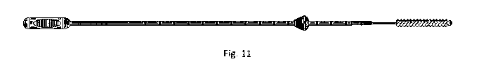
Fig 11 shows a complete biopsy device with manual handle, skirt stopper,
sheath,
guidewire, brush, and 0-ring, according to the present invention.
Fig. 12 shows an arrangement of an independently controllable, biopsy multiple
sample,
biopsy device showing four similar biopsy sampling tools.
Fig. 13 shows an arrangement of an independently controllable, biopsy multiple
sample,
biopsy device showing four different biopsy sampling tools.
Fig. 14 shows a detail of a selector which permits manipulation of a single
biopsy
sampling tool in a barrel cartridge.
CA 03046632 2019-06-10
WO 2018/107175 PCT/US2017/065646
- 16 -
DETAILED DESCRIPTION OF THE PREFERRED EMBODIMENTS
Example 1
A preferred embodiment of the present invention consists of an intrauterine
biopsy
device having an outer thin walled tube of approximately 2.25 mm outside
diameter and 1.2 mm
inside diameter; length is between 20-50 cm, e.g., 22 cm. This tube may be a
clear, bendable but
self-supporting plastic tube, made e.g., of nylon. The guidewire is preferably
a twisted stainless
steel wire of approximately 0.1-0.2 mm diameter, having sufficient mechanical
properties to
convey the forces for extension and retraction of the brush during use. At the
distal end of the
guidewire is a biopsy brush, shown in Figs. 8 and 11, tipped with an
atraumatic bulb. The brush
may be about 4 cm long, and extend about 2 cm past the end of the sheath when
extended. The
0-ring preferably remains within the sheath over the entire range of travel,
to avoid problems re-
engaging the end of the sheath. For example, the 0-ring (or more generally,
plunger attached to
the wire) may be, for example, 2-5 mm from the end of the sheath when
extended.
An anal biopsy device may also be provided, having an outer thin walled tube
of
approximately 2.25 mm outside diameter and 1.2 mm inside diameter; length is
between 8-12,
e.g., 8 cm. This tube may be a clear, bendable but self-supporting plastic
tube, made e.g., of
nylon. The guidewire is preferably a twisted stainless steel wire of
approximately 0.1-0.2 mm
diameter, having sufficient mechanical properties to convey the forces for
extension and
retraction of the brush during use. At the distal end of the guidewire is a
biopsy brush, shown in
Figs. 8 and 11, tipped with an atraumatic bulb. The brush for the anal biopsy
device may also be
4 cm long, with the 0-ring or plunger 2-5 mm from the end of the sheath when
the brush is
extended.
The wire may be periodically marked, such as in 1 cm increments, so that the
physician
or biopsy device operator can estimate the brush insertion with respect to the
proximal end of the
sheath.
At one end, the one that enters the uterus or anus, the biopsy brush is
formed. A tight
fitting 0-ring around the guidewire, shown in Fig. 11, acts as a piston and
creates the suction as
the obturator is withdrawn through the outer thin walled tube.
In another embodiment, the 0-ring may be disposed about 2.5 cm from the tip,
with the
brush extending about 1.5 cm from the tip, with 1 cm of bare wire between
them.
As shown in Figs. 9, 10 and 11, a skirt stopper is provided about the exterior
of the thin
walled tube, near the distal end, which may be in fixed position or manually
slidable. The skirt is
CA 03046632 2019-06-10
WO 2018/107175 PCT/US2017/065646
- 17 -
approximately 1 cm in diameter, and may be formed of nylon, polyurethane,
silicone, neoprene,
or other medically acceptable plastic or rubber. Typically, the skirt is fixed
in position, and may
be glued (e.g., UV activated methyl-methacrylate adhesive) or molded to the
sheath in position.
The biopsy device is use as follows:
The brush is retracted completely into the outer sheath.
The sheath is inserted, through the vagina, into the cervix, until the skirt
stopper meets
the external os of the cervix. The tip of the brush should be displaced from
the fundus.
The outer sheath is pulled back until it stops, i.e., abuts the handle. The
brush is then
rotated by holding the sheath still and turning the handle. For example, the
brush may be rotated
in a clockwise manner until a reference mark on the handle indicates
completion of a 360 turn,
and then rotated counterclockwise until the reference mark on the handle
indicates completion of
a -360 turn. Alternately, the brush may be rotated in only one direction by
completing 4 or 5
360 rotations. In some cases, the brush may be repositioned axially, though
it should not be
withdrawn into the sheath until the sampling is completed.
After sampling with the brush, the guidewire is pulled at the handle, until
the sheath hits
the stop (e.g., the edge of the handle), thereby suctioning fluid surrounding
the tip into the
sheath, and then withdrawing the brush into the sheath.
After withdrawal of the device from the vagina, the brush and fluids in the
sheath are
immersed in a cytology preservative, such as formalin, and the sample is
washed from the brush
into the preservative by moving the brush in and out of the sheath immersed in
the fluid.
The invention may be used, for example, to sample the inside of the uterus to
diagnose
abnormalities. It can detect or exclude a cancer. It can obtain an adequate
tissue sample to
determine infertility causes.
The anal brush is similarly employed. Such a biopsy tool typically has a
shorter sheath
and guidewire than an endocervical brush biopsy tool, because of the easier
anatomical access.
For example, the sheath may be 10-15 cm long, and the brush may extend 2-6 cm
beyond the
end of the sheath. As with the endocervical brush biopsy tool described above,
a skirt is
preferably provided which prevents insertion of the sheath into the anus
beyond the sheath, to
provide a physical reference distance for insertion. In some cases, the skirt
may be repositioned
on the sheath, to permit the physician the ability to determine at what depth
of insertion the
sample should be acquired. Advantageously, the readjustment requires more
force than would
be available by applying an unconstrained compression of the sheath against
the skirt stopper, so
CA 03046632 2019-06-10
WO 2018/107175
PCT/US2017/065646
- 18 -
that the position is maintained during use, but the stiction force can be
overcome when the
biopsy tool is external to the body.
Example 2
According to a second embodiment, a multiple sample biopsy device is provided,
capable of obtaining and segregating a plurality of biopsy samples taken in a
single session.
This therefore requires a plurality of biopsy brushes or tools, and a
plurality of sheaths in which
the tools are extended and retracted.
As discussed above, a depth of insertion positional reference, such as a skirt
stopper may
be provided. However, where the multiple biopsy tool system has a mechanism
maintained
outside of the orifice, the diameter of the tool may be sufficiently large to
act as the stopper
without additional structures.
According to one design, each biopsy tools is separate, including a sheath and
guidewire
control. A set of biopsy tools are aggregated in an outer tube housing. The
tube has a conical
internal profile at the distal end, so that a single biopsy tool may be
advanced past the end of the
housing, into the orifice or canal from which a biopsy is to be taken. In some
cases, endoscopic
guidance of the biopsy is desired, and in that case, a second sheath which
supports the
endoscope and lighting may be advanced as well. He endoscope sheath may also
inject saline
for visualization, though in the case of a brush biopsy, this is disfavored,
since the saline will
wash away the dislodged cells, and reduce the positional accuracy of sampling.
An inert gas,
such as CO2 may also be injected through the sheath, in known manner.
For example, the biopsy brush may be provided in a 3 mm tube, with 6 separate
brushes
provided within a housing. A stop may be provided at the proximal end of each
sheath within
the housing, to prevent over-withdrawal. Markings may be provided on each
sheath, to inform
the physician about the depth of insertion. In some cases, the physician may
intend gradated
sampling at a series of depths in the orifice, and advantageously, each
respective sheath may
have a stopper which limits its depth of insertion, and provides the physician
with haptic
feedback when that depth is achieved. This stopper may be a simple 0-ring or
clamp, which is
adjusted by the physician for each biopsy sampling tool, before the procedure.
The guidewire
for each sampling tool may also have depth limits. Of course, the retracted
position with the
biopsy tool fully withdrawn into the sheath represents one extreme, and a
clamp or limit may be
provided on the manipulation end to control how far the guidewire may be
extended beyond the
end of the sheath.
CA 03046632 2019-06-10
WO 2018/107175 PCT/US2017/065646
- 19 -
In this first design, each biopsy brush may be of known type, with the
optional addition
of the insertion and retraction limiters, and indeed, the housing for
arranging a multiple biopsy
sample session may itself may be provided independent of the biopsy brushes.
In general, the housing avoids the need for a separate skirt stopper, though
the housing
may terminate in a skirt stopper.
Example 3
According to a second design of the multiple sample biopsy device, a single
manipulator
extends from a housing, which itself contains a plurality of biopsy tools.
As discussed above, a depth of insertion positional reference, such as a skirt
stopper may
be provided. However, where the multiple biopsy tool system has a mechanism
maintained
outside of the orifice, the diameter of the tool may be sufficiently large to
act as the stopper
without additional structures.
Thus, a selectively engageable coupling is provided between a single guidewire
and the
various tools. The coupling thus links the guidewire, that extends to a
physician manipulation
interface, such as a grasping element, a handle, or a pivotal mechanism, to
the individual
guidewire for each tool. Advantageously, the plurality of tools are provide in
a rotating barrel,
which serves as the housing. Each biopsy tool, when engaged with the
manipulation guidewire,
can be advanced with its respective sheath an insertion distance, and then the
biopsy head
advanced beyond the sheath, and twisted or otherwise manipulated to obtain a
biopsy sample.
The biopsy head is then withdrawn back into the sheath, the sheath with biopsy
head covers then
withdrawn back into the cartridge, and the barrel twisted so another biopsy
tool may then be
engaged.
Therefore, the coupling is a coaxial coupling, which separately links and
controls the
sheath and the guidewire within each respective sheath. For example, within
the cartridge, the
end of the sheath may terminate in a steel ring, which is magnetically
permeable. Thus, a
magnetic coupling can be used to connect and disconnect the sheaths. Further,
the inactive
biopsy tools may also be held in place by another magnet, which is typically
an electromagnet,
or a permanent magnet with an electromagnetic release. The guidewire may be
selectively
connected to the external manipulation guidewire with a spring-loaded clamp.
As the barrel is
turned, the spring loaded clamp releases, and re-engages as it reaches the
next detent position
with the next biopsy tool aligned with the spring clamp. Within the barrel,
the guidewire from
the biopsy tool extends beyond the proximal end of the respective sheath.
CA 03046632 2019-06-10
WO 2018/107175 PCT/US2017/065646
- 20 -
The barrel is typically at least as long as the desired depth of insertion of
the sheath into
the patient. Thus, if it is desired to have a 12 cm depth of insertion, the
barrel mechanism may
be 13-16 cm long.
As shown in Fig. 12, a plurality of similar brushes are provided in a
cartridge. In Fig. 13
a plurality of different brushes are provided in the cartridge. The cartridge
has an exit port for
the engaged biopsy tool. Each brush has its own associated sheath, which may
be independently
advanced into the patient, depending on which tool is engaged. A mechanism at
the proximal
end of the housing controls the selection of the barrel position by an angle
of rotation, the
latching of the sheath of the respective active tool to the tool advancement
control, the clamping
of the guidewire of the respective active tool to the guidewire control for
manipulation by the
physician, and in some cases, other controls, such as deflection angle of the
sheath. Fig. 12
shows a bulb provided just proximal to each sampling brush, which is provided
to draw a
sampling vacuum when the respective brush is withdrawn back into the sheath.
Fig. 14 shows an end view of a portion of the mechanism in the barrel, wherein
one
guidewire is free to be manipulated by the physician, while access for
manipulation of the other
guidewires is locked out. In Fig. 13, only one biopsy tool has such a feature.
The biopsy
sampling tools, may be, for example, an endocervical sampler, an endometrial
sampler, a punch
sampler, and an endometrial sampler with suction.
In some cases, the sheath itself may be articulable or angularly guidable to
direct the
biopsy tool to a desired region. The articulable sheath may be a single axis,
i.e., a curvature of
the end of the sheath, typically as a result of a tension on a tensile element
such as cable,
guidewire or filament attached to the wall of the sheath, not shown in in the
figures... By
controlling the angle of curvature, and the rotational angle of the sheath
with respect to the
organ, a reasonable range of control is provided. Similarly, a punch, or
snare, or encapsulating
biopsy device may also be controlled by a tension, which may be a wire or
polymer filament.
Thus, the case of a single guidewire with a single degree of freedom
(advance/retract) is a
simplest case, and additional controls and degrees of freedom may be provided.
The controls for
these tools may also be selectively engaged through a mechanism, or provided
individually to
the user. An endoscopic imager (not shown in the figures) may be provided,
preferably as a
feature of the housing, so that it may be used with various biopsy tools
within the housing. For
example, a 1-3mm endoscopic camera with fiber optic lighting, may be provided,
e.g., the On
Semiconductor 0VM6946 1/18" 400x400 imager.