Note: Descriptions are shown in the official language in which they were submitted.
CA 03048210 2019-06-21
WO 2018/119437
PCT/US2017/068302
ELECTROPHORESIS DIAGNOSTIC METHODS AND KITS
CROSS-REFERENCE TO RELATED APPLICATIONS
This international application claims the benefit of priority to U.S.
Provisional Application 62/438,583, filed December
23, 2016, the entirety of which is incorporated herein by reference.
FIELD OF THE INVENTION
The application relates to methods, compositions, and kits for detecting
analytes in biological samples.
BACKGROUND
The detection of analytes has various clinical and non-clinical applications
in industries ranging from medicine,
biological research, to environmental science and beyond. Traditional methods
for analyte detection involve assays
such as enzyme-linked immunosorbent assays (ELISA), mass spectrometry, and
high pressure liquid chromatography
(HPLC). While HPLC and mass spectrometry may be used to detect analytes on the
basis of charge and/or size, ELISA
may be used to detect an analyte based on antigens on the analyte that are
recognizable by capture and detection
agents (e.g., antibodies, aptamers, etc.). In particular, ELISA assay has
become a relatively common detection method
utilized in the life sciences. However, conventional ELISA may be time-
consuming as it involves various incubation and
washing steps and may not provide sufficient sensitivity for various
applications. Further, the parameters for carrying
out ELISA assays are highly variable thus rendering the assay difficult to
develop as a universal platform, particularly
as home diagnostics for individual users.
Accordingly, there remains a need for improved methods for detecting an
analyte in a sample that takes less time and
input to perform compared to conventional ELISA, while maintaining or
improving the sensitivity of detection.
SUMMARY OF THE INVENTION
As described herein, the application is directed to nanoswitch compositions,
methods of using and preparing the
same, and kits that meet the needs in the art for improved methods for
detecting analytes (or biomarkers) in a
sample such as a bodily fluid or other sample derived from a subject or
patient.
In an embodiment, the application is directed to a nanoswitch for detecting a
biomarker (or an analyte). In some
embodiments, the nanoswitch may include a nucleic acid scaffold (i.e., a
scaffold nucleic acid) that is hybridized to
one or more oligonucleotides. In some embodiments, the nanoswitch may include
a set of binding partners
configured to bind the biomarker, wherein the set of binding partners are
linked to the nucleic acid scaffold or the one
or more oligonucleotides and include a first binding partner and a second
binding partner. The binding of the first
binding partner and the second binding partner to a biomarker (or analyte) of
interest may be sequential, or
1
CA 03048210 2019-06-21
WO 2018/119437
PCT/US2017/068302
simultaneous, or in any order (e.g., first then second, second then first,
first simultaneous with second, etc.). In some
embodiments, a binding partner (e.g., a first or second binding partner) may
be, for example, an antibody or an
antigen. As described herein, binding of the biomarker by the set of binding
partners causes the nucleic acid scaffold
to form a loop. In some embodiments described herein, a loop refers to a
looped nanoswitch that may be detectable
by gel electrophoresis. In some embodiments, the loop may be a detectable loop
that may be detected or quantified
by one or more of the analytical methods described herein. In some
embodiments, the loop may be detachable or
undetachable, as described herein.
In an embodiment, the application is directed to a nanoswitch for detecting a
biomarker (or an analyte) where, in
some embodiments, the nanoswitch includes a nucleic acid scaffold that
includes an M13 scaffold or a fragment
thereof that may be hybridized to one or more oligonucleotides. In some
embodiments, the nanoswitch may include
a set of binding partners configured to bind the biomarker, wherein the set of
binding partners are linked to the
nucleic acid scaffold or the one or more oligonucleotides and include a first
binding partner and a second binding
partner.
In an embodiment, the application is directed to a nanoswitch for detecting a
biomarker (or an analyte) where, in
some embodiments, the nanoswitch includes a nucleic acid scaffold that
includes single strand DNA that may be
hybridized to one or more oligonucleotides, wherein the single strand DNA
optionally includes p8064 single strand
DNA. In some embodiments, the nanoswitch may include a set of binding partners
configured to bind the biomarker,
wherein the set of binding partners are linked to the nucleic acid scaffold or
the one or more oligonucleotides and
include a first binding partner and a second binding partner.
In an embodiment, the application is directed to a nanoswitch for detecting a
biomarker (or an analyte) that may be
considered a megaloop nanoswitch. In some embodiments, the nanoswitch includes
a nucleic acid scaffold
hybridized to one or more oligonucleotides. In some embodiments, the
nanoswitch includes a set of binding partners
configured to bind the biomarker, wherein the set of binding partners are
linked to the nucleic acid scaffold or the one
or more oligonucleotides and include a first binding partner and a second
binding partner. In some embodiments, the
nanoswitch includes a latch having latch oligonucleotides hybridized to the
nucleic acid scaffold, wherein the latch
oligonucleotides may include streptavidin, desthiobiotin, biotin, or a
combination thereof.
In an embodiment, the application is directed to a nanoswitch for detecting a
biomarker (or an analyte) that may be
considered a megaloop nanoswitch. In some embodiments, the nanoswitch includes
a nucleic acid scaffold
hybridized to one or more oligonucleotides. In some embodiments, the
nanoswitch includes a set of binding partners
configured to bind the biomarker, wherein the set of binding partners are
linked to the nucleic acid scaffold or the one
or more oligonucleotides and include a first binding partner and a second
binding partner. In some embodiments, the
nanoswitch includes single strand extension oligonucleotides hybridized to the
nucleic acid scaffold and configured to
hybridize a key oligonucleotide.
2
CA 03048210 2019-06-21
WO 2018/119437
PCT/US2017/068302
In an embodiment, the application is directed to a nanoswitch for detecting a
biomarker (or an analyte) that may be
considered a megaloop nanoswitch. In some embodiments, the nanoswitch includes
a nucleic acid scaffold
hybridized to one or more oligonucleotides, wherein the nucleic acid scaffold
includes at least two regions available
to hybridize a bridge oligonucleotide. In some embodiments, the nanoswitch
includes a set of binding partners
configured to bind the biomarker, wherein the set of binding partners are
linked to the nucleic acid scaffold or the one
or more oligonucleotides and include a first binding partner and a second
binding partner.
In an embodiment, the application is directed to a biomarker or analyte test
system that may be configured to receive
a sample and determine the presence of a biomarker or analyte in the sample.
In some embodiments, the test system
includes a case, a nanoswitch source disposed in the case including a
nanoswitch as described herein that forms a
loop in when binding the biomarker. In some embodiments, the test system
includes a sample receiver (e.g., a sponge
or porous element) connected to the case and configured to receive sample. In
some embodiments, the test system
includes an electrophoretic medium (e.g., a gel electrophoresis medium)
disposed in the case and connected to the
nanoswitch source and the sample receiver. In some embodiments, the test
system includes an electrophoretic
medium (e.g., a gel electrophoresis medium) disposed in the case in fluid
communication with the nanoswitch source
and the sample receiver.
In an embodiment, the application is directed to a method of preparing a
nanoswitch as described herein. In some
embodiments, the method includes the step of preparing the nucleic acid
scaffold. In some embodiments, the method
includes the step of coupling an oligonucleotide to the nucleic acid scaffold.
In some embodiments, the method
includes the step of functionalizing one or more of the nucleic acid scaffold
and the oligonucleotide with a set of binding
partners.
In an embodiment, the application is directed to a method of preparing a
nanoswitch as described herein. In some
embodiments, the method includes the step of preparing the nucleic acid
scaffold. In some embodiments, the method
includes the step of functionalizing an oligonucleotide with a set of binding
partners to provide a functionalized
oligonucleotide. In some embodiments, the method includes the step of coupling
the functionalized oligonucleotide to
the nucleic acid scaffold.
In an embodiment, the application is directed to a method of detecting a
biomarker (or an analyte) in a sample provided
by a subject. In some embodiments, the method includes the step of contacting
the sample with nanoswitches
described herein to bind the biomarkers in the sample. In some embodiments,
the method includes the step of
separating the nanoswitches bound to the biomarkers in the sample from unbound
nanoswitches by gel electrophoresis
in an electrophoretic medium (e.g., a gel electrophoresis medium).
3
CA 03048210 2019-06-21
WO 2018/119437
PCT/US2017/068302
DESCRIPTION OF THE FIGURES
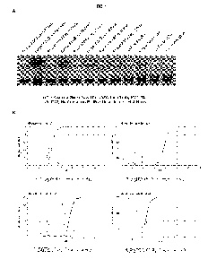
FIG. 1, panel A depicts results from a gel electrophoresis of crude and
purified urine using methods described herein.
Panel B provides various plots of the distribution of various micro-species
and fully deprotonated species in the
presence of different chelators.
FIG. 2 depicts results of an LH detection using the nanoswitch described
herein. 06 and 07 were the baseline days,
C11 was the detected surge day prior to collection, and the concentrations
refer to the final concentration of the
nanoswitch.
FIG. 3, panel A, shows PSA detection using crude PSA nanoswitch standard
characterization. Panel B shows post-
purified PSA nanoswitch with low PSA detection testing.
FIG. 4, panel A shows a schematic diagram of HSV-2 detection using the
nanoswitch described herein. Panel B shows
results using HSV-2 4.44 4.19 crude nanoswitch. Panel C shows results using
BluePippin-purified HSV-2 antibody
oligonucleotide ("oligo") crude construct. Panel D shows results using
BluePippin purified HSV-2 antibody-oligos with
BluePippin purified construct.
FIG. 5, panel A shows coupling of streptococcus pyo genes (Strep-A) antibody
to oligonucleotides tested in 0.5X KBB
electrophoresis buffer. Panel B shows detection of Strep-A antigen using Strep-
poly construct. Panels C and D show
issues of false positive bands. Panel E shows elimination of the false
positive band through use of passivating agents.
Panels F and G show detection of Strep-A in the presence of 0.1% BSA or 0.01%
Tween20, respectively. Panel H
depicts loop yields for the two conditions with different concentrations of
the Strep-A antigen. Panel I shows
repeatability of Strep-A detection using Strep-poly construct. Specifically,
the construct (150 pM final concentration)
was incubated with 0.01% Tween followed by incubation with different dilutions
of the Strep-A antigen. The detection
signal was analyzed for different dilutions of the Strep-A antigen as the
intensity of the looped band (panel J) and as
the % looped material (panel K). Panel L shows specificity of the nanoswitch
in detecting Strep-A derived from the
QuickVue Dipstick Strep-A test. Panels M and N shows detection of serially
diluted Strep-A derived from the QuickVue
Dipstick Strep-A test using the nanoswitch described herein compared to the
QuickVue Dipstick. Panel 0 shows
.. detection of Strep-A derived from the QuickVue Dipstick Strep-A test
without any pH adjustment.
FIG. 6 shows characterization of a peptide coupled to two different oligo
sequences as described in Example 3. The
peptide conjugated oligonucleotides ran higher in the gel than the
unconjugated oligo.
FIG. 7, panel A, depicts a schematic of NHS-based coupling strategy for
hydrazone linkage of antibodies to
oligonucleotides. Panel B depicts common antibody glycosylation patterns. The
glycosylation pattern of the antibody
determines which residues can be targeted for activation. Panel C shows
activation of reactive aldehydes through the
4
CA 03048210 2019-06-21
WO 2018/119437
PCT/US2017/068302
oxidation of glycosylation sites. Panel D shows hydrazone-linkage coupling
strategy. Panel E depicts a gel
electrophoresis of the hydrizide-oligo formed by methods described herein.
FIG. 8 is a schematic of a test strip that combines both ovulation and
pregnancy testing.
FIG. 9, panels A and B, show improvement of gel size in order to reduce
background using SYBR Gold and GELRed
pre-stained gels.
FIG. 10 provides an illustration of flipping the voltage in a direction
orthogonal to the original (x) run direction to reduce
background.
FIG. 11, panel A, shows a comparison of 1% agarose gel versus 1% agarose gel
with incorporation of 0.4%
hydroxyethyl cellulose (HEC). Panel B provide a series of plots showing the
effects of various percentage of agarose
and HEC on running conditions.
FIG. 12 shows the effect of buffer level (5/8 inches above gel or 3/8 inches
above gel) on DNA running distance.
FIG. 13 shows a comparison of gels ran at constant current voltage (top gel)
versus constant current (bottom gel).
Improvement in sharpness of the looped bands was seen with gels ran at
constant current.
FIG. 14 depicts exemplary tiny gels (approximately 1-5 cm) that were used as
described in Example 5. The exemplary
gels shown in FIG. 14 are 2.5 cm.
FIG. 15, panels A and B, depict gels that were not pre-stained (panel A) or
pre-stained with 1X SYBR Gold (panel B).
FIG. 16, panels A and B, provides a comparison of gels ran without pre-
staining the sample (panel A) versus gel ran
with a pre-stained sample (panel B). Pre-staining of sample appears to yield
straighter and sharper bands.
FIG. 17, panel A, shows a typical DNA nanoswitch including a single pair of
antibodies which can simultaneously bind
to an analyte. Panel B shows an improved version of an antibody DNA nanoswitch
that includes 3 pairs of antibodies
with each type grouped in a closely spaced group. Panel C shows loop yield
Increases as more antibodies are added
to the scaffold. The gel on the left shows a nanoswitch with two antibodies
has a 20% maximum loop yield. The middle
gel shows a nanoswitch with 4 antibodies has a 30% maximum loop yield. The gel
on the right shows a nanoswitch
with 12 antibodies has a 90% maximum loop yield.
FIG. 18, panels A and B, show an example of a false positive signal. Panel A
shows a gel lane with no analyte present
containing a looped DNA band. Panel B shows a gel lane with low amount of
analyte (10 pM) containing a more intense
5
CA 03048210 2019-06-21
WO 2018/119437
PCT/US2017/068302
looped DNA band. Panel C shows the effect of pH on false positive signal. All
of the lanes lacked analyte, but have
been incubated in buffers with differing pH. For this nanoswitch, high pH
eliminated the false positive signal.
FIG. 19, panels A-D provide schematics of the megaloop design described in
Example 6. Panel E shows the
construction and testing of the latch. The gel shows loop formation tests at
different concentrations of streptavidin.
Panel F shows characterization of the off-rate for the biotin-SA-desthiobiotin
latch. Panel G shows the design of
megaloop using key and bridge oligonucleotides. Panel H shows a
characterization of the megaloop using key and
bridge oligonucleotides. Panel I shows a characterization of the megaloop with
antigen-antibody interaction.
FIG. 20, panel A depicts loop migration comparison of alternative cut sites
using various restriction enzymes. Panel B
shows migration of oligonucleotide bridge loops at different restriction
enzyme cut sites and DNA sources. Panel C
shows loop migration of alternative restriction enzyme cut sites on M13 and
p8064 DNA.
FIG. 21, panel A, shows antibody oligo conjugates ran on an agarose gel. Lane
1 is a DNA ladder, Lane 2 is mix of
conjugated and unconjugated Antibody-oligo. The uncoupled oligo runs
significantly lower on an agarose gel. Panel B
shows selection of appropriate Blue Pippin cutoffs. Lane 1: DNA Ladder. Lane
2: An unpurified antibody oligo conjugate
contains uncoupled oligo which runs below lkbp, and an antibody-oligo
conjugate which runs similarly to where a DNA
nanoswitch. Lane 3: A purified antibody which has been Blue Pippin purified
using 1-3kbp cutoff region. Lane 4: A DNA
nanoswitch purified using a 5-9kbp. If the antibody conjugates were not
purified using the 1-3kbp cutoffs there would
still be excess antibody-oligo conjugate that could compete with the DNA
nanoswitch for antigen binding.
FIG. 22 demonstrates a functional testing (i.e., HOG binding) of individual
antibody-oligos that were either dried using
Speed Vac or control antibody-oligos.
.. FIG. 23 depicts circular DNA purity. Between 70-80% circular purity was
typically obtained when purchasing
commercial M13.
FIG. 24, panels A-G provides a schematic of the sequential affinity
purification protocol describe herein. Panels H-K
provides a schematic of the temporal affinity purification protocol described
herein.
FIG. 25, panel A, provides image of a gel lane. The dotted line indicates a
row along which the intensity was analyzed
to determine if the background is inhomogeneous. Panel B provides a plot of
the intensity profile of the row as a function
of the pixel location. The dotted line indicates the slope of the background
signal. This line was subtracted from the
profile before any further analysis was done.
FIG. 26 provides an illustration of an exemplary nanoswitch (NS) synthesis and
method of use.
6
CA 03048210 2019-06-21
WO 2018/119437
PCT/US2017/068302
DETAILED DESCRIPTION OF THE INVENTION
The application is directed improved methods, compositions, and kits for
detecting analytes, including, for example,
detecting analytes and/or complex formation, monitoring binding interactions,
measuring association and/or
dissociation kinetics, and the like. In various embodiments, polymers (i.e.,
nanoswitches) may be used that change
conformation upon analyte binding, and are then separated and distinguished
from each other via gel electrophoresis.
The improved methods may be performed at home, in a laboratory setting, or
along with a high-throughput analyzer
for medical or scientific applications. The methods described here may provide
significant advantages including speed,
sensitivity, and accuracy compared to prior art detection techniques. Further
still, the methods described herein may
be relatively inexpensive and easy to perform thereby offering a distinct
advantage for use as home diagnostics.
Basic Nanoswitch Approach
In various embodiments, the application relates to a nanoswitch-based
detection of an analyte including, for example,
detecting analytes and/or complex formation, monitoring binding interactions,
measuring association and/or
dissociation kinetics, and the like, as described herein and various means of
improvement as described herein.
FIG. 26 provides an illustration of exemplary nanoswitch (NS) synthesis and
method of use. For example, in some
embodiments, DNA nanoswitches may be made by hybridizing one or more
oligonucleotides to a longer scaffold nucleic
acid, wherein at least one such oligonucleotide is conjugated to a binding
partner of an analyte of interest. In some
embodiments, a plurality of oligonucleotides is hybridized to the scaffold. In
other embodiments, the method described
herein may be performed with a single oligonucleotide or with no
oligonucleotide and simply conjugation of the binding
partner to the scaffold itself. The use of hybridizing oligonucleotides
facilitates specific positioning of the binding partner
along the length of the scaffold. Further, use of the hybridizing
oligonucleotides renders the nanoswitch technology
more versatile and universal since a plurality of oligonucleotides may be
created that differ with respect to the binding
partners conjugated to them.
Various methods may be used to generate a nanoswitch as is known in the art.
Exemplary methods are described for
example, in WO 2013/067489, WO 2016/089588, and Koussa, etal. Nature Methods
12, 123-126 (2015), the entire
contents of which are hereby incorporated by reference. In some embodiments,
nanoswitches are generated by single-
strand nicking of a double stranded nucleic acid, followed by hybridization to
the nicked nucleic acid with one or more
oligonucleotides conjugated to a binding partner of interest. The nicking
action may be sequence-independent or
sequence-dependent. The binding partners may be but are not limited to
antibodies and antigen binding antibody
fragments. The scaffold nucleic acid may be scaffold DNA such as linearized
M13 DNA.
In the presence of an analyte of interest, the two binding partners of the
nanoswitch bind to a single analyte thereby
causing the nanoswitch to form a loop. In some embodiments, such loop
formation requires the action of at least two
binding partners. In other embodiments, nanoswitches described herein may
comprise two or more binding partners,
7
CA 03048210 2019-06-21
WO 2018/119437
PCT/US2017/068302
with each binding interaction involving two or more binding partners rendering
a different conformation that can be
distinguished from other conformations using gel electrophoresis. In some
embodiments involving multiple binding
partners, a loop is formed when only one pair of binding partner bind to a
single analyte. In still other embodiments,
the method may involve two physically separate polymers such as two physically
separate nucleic acids, each
conjugated to a binding partner, and in the presence of the analyte the two
binding partners bind to the analyte, thereby
causing the physical interaction of the two polymers. The result is a complex
comprising the two polymers joined
together via a common analyte and at two binding partners to such analyte.
In various embodiments, the resulting mixture is run on a gel, such as an
agarose gel, and imaged so as to distinguish
the nanoswitches with different conformations.
In various embodiments, the invention described herein relates to an improved
method for analyte detection of an
analyte including, for example, detecting analytes and/or complex formation,
monitoring binding interactions,
measuring association and/or dissociation kinetics, and the like, by placing a
nucleic acid complex, which comprises a
single-stranded scaffold nucleic acid hybridized to one or more single-
stranded oligonucleotides, where a first single-
stranded oligonucleotide is linked to a first binding partner and a second
single-stranded oligonucleotide in the plurality
is linked to a second binding partner, under conditions that allow for binding
of binding partners to each other, and
detecting a change in the nucleic acid complex using gel electrophoresis, e.g.
a change in the apparent length of the
nucleic acid complex as determined from migration through a gel when binding
between the partners occurs as
compared to the absence of binding (e.g. a change in migration distance as the
result of a change in nucleic acid
topology, e.g. detecting the presence or absence of a looped structure, e.g. a
looped linker structure, being formed
when binding between the partners occurs, e.g. comparing a looped structure
(indicating that binding between the
partners occurred) to a linear structure (indicating that binding between the
partners has not occurred) and observing
a "gel shift"). In various embodiments, the binding partners may be entities
that measurably interact with each other,
e.g. an antibody (or functional fragment thereof, e.g. containing a relevant
paratope) and an antigen (or epitope-
containing fragment thereof), a receptor and a ligand, etc. In various
embodiments, either of the binding partners may
be an analyte for which detection is desired. For example, in various
embodiments, the analyte for which detection is
desired is an antigen (or epitope-containing fragment thereof) that is
recognized by an antibody (or functional fragment
thereof, e.g. containing a relevant paratope).
Throughout the disclosure, various improvements of the nanoswitch methods and
compositions are provided. Such
improvements are separate embodiments described herein, but also may be
combined to perform the methods or
make the compositions (e.g. this disclosure envisions using the various
improvements individually or in combination).
Improvements Related to Nanoswitches
8
CA 03048210 2019-06-21
WO 2018/119437
PCT/US2017/068302
In various embodiments, the nanoswitches comprise a scaffold or backbone
nucleic acid comprising one or more
binding partners. The scaffold nucleic acid may be of any length sufficient to
allow association and dissociation of
binding partners to occur, to be detected, and to be distinguished from other
events. In some embodiments, the scaffold
nucleic acid is at least about 100 nucleotides in length. In some embodiments,
the scaffold nucleic acid is about 100 to
about 200,000 nucleotides in length. For example, the scaffold nucleic acid
may be about 100, about 200, about 300,
about 400, about 500, about 600, about 700, about 800, about 900, about 1,000,
about 2,000, about 3,000, about
4,000, about 5,000, about 6,000, about 7,000, about 8,000, about 9,000, about
10,000, about 11,000, about 12,000,
about 13,000, about 14,000, about 15,000, about 16,000, about 17,000, about
18,000, about 19,000, about 20,000,
about 30,000, about 40,000, about 50,000, about 60,000, about 70,000, about
80,000, about 90,000, about 100,000,
about 125,000, about 150,000, about 175,000, or about 200,000 nucleotides in
length. In some embodiments, the
scaffold nucleic acid may be greater than about 100, about 200, about 300,
about 400, about 500, about 600, about
700, about 800, about 900, about 1,000, about 2,000, about 3,000, about 4,000,
about 5,000, about 6,000, about 7,000,
about 8,000, about 9,000, about 10,000, about 11,000, about 12,000, about
13,000, about 14,000, about 15,000, about
16,000, about 17,000, about 18,000, about 19,000, about 20,000, about 30,000,
about 40,000, about 50,000, about
60,000, about 70,000, about 80,000, about 90,000, about 100,000, about
125,000, about 150,000, about 175,000, or
about 200,000 nucleotides in length. In some embodiments, the scaffold nucleic
acid may be less than about 100,
about 200, about 300, about 400, about 500, about 600, about 700, about 800,
about 900, about 1,000, about 2,000,
about 3,000, about 4,000, about 5,000, about 6,000, about 7,000, about 8,000,
about 9,000, about 10,000, about
11,000, about 12,000, about 13,000, about 14,000, about 15,000, about 16,000,
about 17,000, about 18,000, about
19,000, about 20,000, about 30,000, about 40,000, about 50,000, about 60,000,
about 70,000, about 80,000, about
90,000, about 100,000, about 125,000, about 150,000, about 175,000, or about
200,000 nucleotides in length.
In some embodiments, the scaffold nucleic acid may be a naturally occurring
nucleic acid. In an embodiment, the
scaffold nucleic acid comprises a M13 scaffold such as M13mp18. The M13
scaffolds are disclosed by Rothemund
(2006) Nature 440:297-302, the entire contents of which are hereby
incorporated by reference. In an embodiment, the
scaffold nucleic acid may be lambda DNA. In other embodiments, the scaffold
nucleic acid may also be non-natural or
synthetic nucleic acids such as polymerase chain reaction (PCR)-generated
nucleic acids, rolling circle amplification
(RCA)-generated nucleic acids, etc.
In some embodiments, the binding partners are positioned along the scaffold
nucleic acid to yield loops and thus length
changes that are detectable (for example, by gel electrophoresis). The
scaffold may be at least partially or fully single-
stranded, or at least partially or fully double-stranded, or at least
partially or fully triple-stranded, or at least partially or
fully quadruple-stranded, or more (e.g., comprising at least five strands, six
strands, seven strands, eight strands, nine
strands, or ten strands, or more). The complex may comprise varying lengths of
double-stranded regions. The scaffold
nucleic acid may comprise DNA, RNA, DNA analogs, RNA analogs, or a combination
thereof. In some embodiments,
9
CA 03048210 2019-06-21
WO 2018/119437
PCT/US2017/068302
the binding partners are conjugated to a scaffold nucleic acid via
hybridization of oligonucleotides to the scaffold,
wherein such oligonucleotides are themselves conjugated to a binding partner.
In an embodiment, the scaffold nucleic
acid is a DNA.
In some embodiments, the scaffold nucleic acid may be hybridized to one, two,
or more, including a plurality, of
oligonucleotides. Each of the plurality of oligonucleotides may hybridize to
the scaffold nucleic acid in a sequence-
specific and non-overlapping manner (i.e., each oligonucleotide hybridizes to
a distinct sequence in the scaffold). In
other embodiments, the plurality of oligonucleotides may hybridize to the
scaffold nucleic acid in a sequence-specific
and overlapping manner (e.g., so as to allow certain oligonucleotides to peel
off). The number of oligonucleotides
hybridized to a particular scaffold may vary depending on the application. In
various embodiments, there may be about
2 or more oligonucleotides hybridized to the scaffold, including about 3,
about 4, about 5, about 6, about 7, about 8,
about 9, about 10, about 20, about 30, about 40, about 50, about 60, about 70,
about 80, about 90, about 100, about
200, about 300, about 400, about 500, about 600, about 700, about 800, about
900, or about 1000 or more
oligonucleotides.
In some embodiments, the one or more oligonucleotides hybridized to the
scaffold nucleic acid are unmodified.
Unmodified oligonucleotides include oligonucleotides that are not linked to
binding partners such as binding partners
being tested (e.g., an antibody or an antigen). In other embodiments, the one
or more oligonucleotides hybridized to
the scaffold are modified. Modified oligonucleotides include those that are
linked to binding partners being tested (e.g.,
a receptor and/or its ligand, an antibody and/or its antigen, etc.). Modified
oligonucleotides may also include those that
are modified and thus used to immobilize the nanoswitch to a solid support
such as but not limited to a bead. Such
modified oligonucleotides include, for example, biotinylated oligonucleotides
or oligonucleotides modified with any of
the tags described herein. Modified oligonucleotides may be referred to herein
as "variable" or "functionalized"
oligonucleotides since these oligonucleotides may be modified by linking to a
variety of binding partners depending on
the method of use.
Regions comprising scaffold hybridized to modified oligonucleotides may be
referred to herein as "variable" regions
and the remaining scaffold regions may be referred to as "fixed" regions.
The scaffold-binding partner construct may be made in a number of ways
including through nicking of a double stranded
nucleic acid to which binding partners are conjugated (to one strand), or by
hybridization of one or more
oligonucleotides to the scaffold, as described herein. In some embodiments,
the binding partners may be conjugated
to the scaffold nucleic acid itself rather than to an oligonucleotide that is
hybridized to the scaffold.
The spacing of binding partners, and thus in some instances of the modified
(or variable) oligonucleotides, along the
length of the scaffold nucleic acid may vary. In some embodiments, the
nanoswitch may comprise about 2, 3, or 4, or
more binding partners. In some embodiments, the nanoswitch may comprise about
2, about 3, about 4, about 5, about
CA 03048210 2019-06-21
WO 2018/119437
PCT/US2017/068302
6, about 7, about 8, about 9, about 10, about 11, about 12, about 13, about
14, about 15, about 16, about 17, about
18, about 19, about 20, about 25, about 30, about 35, about 40, about 45,
about 50, about 55, about 60, about 65,
about 70, about 75, about 80, about 85, about 90, about 95, about 100, about
110, about 120, about 130, about 140,
about 150, about 160, about 170, about 180, about 190, about 200, about 250,
about 300, about 350, about 400, about
450, or about 500, or more binding partners. As an example, a nucleic acid
nanoswitch may comprise two internal
modified oligonucleotides. The modified oligonucleotides internal to the
nanoswitch may be linked individually to
members of a binding pair (i.e., each of the two oligonucleotides is linked to
a member of the binding pair such that the
nanoswitch comprises the binding pair, with each member of the pair on a
different oligonucleotide). The internal
modified oligonucleotides may be symmetrically or quasi-symmetrically located
around the center of the scaffold. In
other words, they may be positioned equi-distant from the center of the
scaffold. In some embodiments, the distance
between the binding pair members may be about 100 to about 200,000 base pairs
in length. For example, the distance
between the binding pair members may be about 100, about 200, about 300, about
400, about 500, about 600, about
700, about 800, about 900, about 1,000, about 2,000, about 3,000, about 4,000,
about 5,000, about 6,000, about 7,000,
about 8,000, about 9,000, about 10,000, about 11,000, about 12,000, about
13,000, about 14,000, about 15,000, about
16,000, about 17,000, about 18,000, about 19,000, about 20,000, about 30,000,
about 40,000, about 50,000, about
60,000, about 70,000, about 80,000, about 90,000, about 100,000, about
125,000, about 150,000, about 175,000, or
about 200,000 base pairs in length.
In various embodiments, the distance between the binding partners is used to
distinguish association and dissociation
between binding partners linked to the nanoswitches. This is because when the
binding partners are associated with
each other, a loop will be formed comprising the nucleic acid sequence that
exists between the binding partners. When
the binding partners are not associated to each other (i.e., unbound), then
the loop does not form and the complex
length is different (e.g., longer). In some embodiments, the invention
described herein comprises multiple binding
partners as described herein, and a loop is formed when at least one pair of
binding partners bind to a single analyte.
The nanoswitch configuration can be determined by analyzing the migration of
the nanoswitch through a matrix such
as a gel in a gel electrophoretic system. The unbound, linear form travels
more rapidly than does the bound, looped
form. Thus, in various embodiments, presence of an analyte of interest, to
which the binding partners on a single
nanoswitch bind, will trigger the formation of a bound and looped nanoswitch.
In various embodiments, the bound,
looped nanoswitch will be distinguished from its unbound, linear counterpart
based on the difference in their migration
distances through a gel or other pore-containing matrix. The invention
described herein contemplates different
variations on the nucleic acid nanoswitches described herein. In various
embodiments, these variations commonly
comprise a nucleic acid nanoswitch having two or more binding partners. The
binding partners typically have binding
specificity for a common analyte. Methods described herein rely on the
association and/or dissociation of binding
partners. A change in conformation of the nanoswitch (e.g., from an open to a
closed conformation) provides
11
CA 03048210 2019-06-21
WO 2018/119437
PCT/US2017/068302
information about the presence of the analyte. The binding partners may be non-
covalently or covalently bound to the
scaffold.
In some embodiments, the nucleic acid complex comprises two binding partners
having binding specificity for a
common analyte. The binding partners are physically separate and thus spaced
apart from each other (when not bound
to the common analyte). When bound to the common analyte, the nucleic acid
nanoswitch assumes a looped (or closed
or bound) conformation having a different conformation and thus a different
"apparent" length (as for example
measured using migration through a gel electrophoresis system), compared to
the nucleic acid nanoswitch in an open
(or unbound) conformation.
In other embodiments, the invention further contemplates a nucleic nanoswitch
comprising more than two conjugated
binding partners. The number of binding partners may be about 2, about 3,
about 4, or more. In some embodiments,
pairs of binding partners are provided, with each pair having binding
specificity for a particular analyte. A single
nanoswitch may comprise a binding pair for a first analyte, which may be a
test analyte, and a second binding pair for
a second analyte, which may be a control analyte. In this way, the nanoswitch
may have a control reading as well as
a test reading. For example, a first binding pair may bind to a marker of
interest and a second binding pair may bind to
a control protein or other moiety that will always be present in the sample
being tested (e.g., the urine) in order to
establish to the end user that a sufficient quantity of sample was applied to
the system. The location or arrangement
of the binding partners may vary and may include serially positioned binding
pairs or nested binding pairs, or
combinations thereof. Alternatively, the test and control analytes may be
assayed using different nanoswitches that are
nevertheless still run through the same gel system.
In various embodiments, the nanoswitches comprise binding partners such as,
for example, an antibody or an antigen.
The linkage between the nucleic acid and the binding partner may be covalent
or noncovalent depending on the
strength of binding required for a particular application. They may be
generated by first incorporating a reactive group
(or moiety) into the nucleic acid (or into an oligonucleotide hybridized to
the nucleic acid), and then reacting this group
(or moiety) with the binding partner of interest which may or may not be
modified itself. Suitable reactive groups are
known in the art. Examples of reactive groups that can covalently conjugate to
other reactive groups (leading to an
irreversible conjugation) include but are not limited to amine groups (which
react to, for example, esters to produce
amides), carboxylic acids, amides, carbonyls (such as aldehydes, ketones, acyl
chlorides, carboxylic acids, esters and
amides) and alcohols. Those of ordinary skill in the art will be familiar with
other "covalent" reactive groups. Examples
of reactive groups that non-covalently conjugate to other molecules (leading
to a reversible conjugation) include biotin
and avidin or streptavidin reactive groups (which react with each other),
antibody (or antibody fragment) reactive groups
and antigens, receptors and receptor ligands, aptamers and aptamer ligands,
nucleic acids and their complements,
and the like. Virtually any reactive group is amenable to the methods
described herein, provided it participates in an
interaction of sufficient affinity to prevent dissociation of the binding
partner from the nucleic acid nanoswitch.
12
CA 03048210 2019-06-21
WO 2018/119437
PCT/US2017/068302
In various embodiments, multiple binding partners may be used including
multiple antibodies (see Example 6 provided
herein). In some embodiments, the methods described herein utilizes at least
about 2, about 3, about 4, about 5, about
6, about 7, about 8, about 9, about 10, about 11, about 12, about 13, about
14, about 15, about 16, about 17, about
18, about 19, about 20, about 21, about 22, about 23, about 24, about 25,
about 26, about 27, about 28, about 29,
about 30, about 31, about 32, about 33, about 34, about 35, about 36, about
37, about 38, about 39, about 40, about
45, about 50, about 55, about 60, about 65, about 70, about 75, about 80,
about 85, about 90, about 95, about 100,
about 110, about 120, about 130, about 140, about 150, about 160, about 170,
about 180, about 190, about 200, about
250, about 300, about 350, about 400, about 450, or about 500 antibodies. In
embodiments, the methods described
herein utilizes at least about 2, about 3, about 4, about 5, about 6, about 7,
about 8, about 9, about 10, about 11, about
12, about 13, about 14, about 15, about 16, about 17, about 18, about 19,
about 20, about 25, about 30, about 40,
about 45, about 50, about 55, about 60, about 65, about 70, about 75, about
80, about 85, about 90, about 95, about
100, about 110, about 120, about 130, about 140, about 150, about 160, about
170, about 180, about 190, about 200,
or about 250 antibody pairs. In some embodiments, the antibodies are located
in clusters of close groups so that all
loop combinations run to a similar location in a gel. In other embodiments,
the antibodies are not clustered in close
groups. In such embodiments, a decrease of bands in a gel corresponding to
linear conformations may be monitored
instead of an increase of bands corresponding to looped conformations.
In some embodiments, methods described herein may involve coupling of
multivalent antibodies. For example, the
methods may involve coupling of bi-valent or trivalent single-chain variable
fragment antibodies (e.g., each of which
can contain about 4 or about 6 analyte, or more, binding sites, respectively).
In other embodiments, methods described
herein may involve chemically forming aggregates of multiple antibodies which
can be coupled to a single
oligonucleotide. This could be performed with a variety of multifunctional
linkers.
It is contemplated that use of multiple binding partners allows for
improvements in the speed and sensitivity of analyte
detection. For example, in some embodiments, methods described herein
significantly reduce incubation time (e.g.,
during low analyte detection assays). In some embodiments, methods described
herein allow for incubation times of
about 1 minute to about 60 minutes, e.g., about 10 minutes, about 15 minutes,
about 20 minutes, about 25 minutes, or
about 30 minutes. In some embodiments, use of multiple binding partners
increase loop yields including maximum loop
yields (e.g., so as to achieve a maximum loop yield of about 20%, about 30%,
about 40%, about 50%, about 60%,
about 70%, about 80%, about 90%, about 91%, about 92%, about 93%, about 94%,
about 95%, about 96%, about
97%, about 98%, about 99%, or about 100%).
The production of DNA nanoswitches with multiple antibodies is one method for
increasing detection limits of DNA
nanoswitches. However, the purification of multiple antibodies in parallel can
be time-consuming and expensive. In
various embodiments, the invention described herein provides a pooling method
for ensuring an equal-molar ratio of
13
CA 03048210 2019-06-21
WO 2018/119437
PCT/US2017/068302
material as well as the separation of cross-reacting antibodies. In an
embodiment, the method involves pooling of
antibody-oligonucleotide as described in Example 7.
When using DNA nanoswitches that are functionalized with antibodies, a common
problem that can occur is the
formation of a DNA loop in the absence of the antigen. This is commonly
referred to as a false positive signal. In various
embodiments, methods described herein minimize such false positive signals. In
some embodiments, the methods
reduce signals by controlling solution pH. In some embodiments, the solution
pH is controlled by the use of appropriate
buffers, which can be specific for the antibodies used. In some embodiments,
Tris/Borate/EDTA buffer and/or buffers
with EDTA are utilized. Various buffers may be utilized in the invention
described herein. Exemplary buffers that may
be utilized for running gels in the invention described herein include, but
are not limited to, single buffers systems such
as Sodium Borate, Sodium Acetate, Sodium Citrate, Lithium Borate, Tris/Acetic
Acid/EDTA, Tris/Acetic Acid, Iris-
Acetate, Tris Acetate EDTA, Tris/TAPS/EDTA Buffer, Bis-Tris/HCI buffer, Tris-
Acetate SDS, MOPS,
MOPS/Tris/SDS/EDTA, MOPS/Tris/EDTA, MOPS/Tris/SDS, MOPS/Tris, MES,
MES/Tris/SDS/EDTA,
MES/Tris/EDTA, MES/Tris/SDS, MES/Tris, Tris-glycine, or dual buffer systems
such as Tris EDTA on one side and
Boric Acid on the other side of the gel. Additional exemplary buffers that may
be utilized for stabilizing pH include, but
are not limited to, Sodium Borate, Sodium Acetate, Sodium Citrate, Lithium
Borate, Tris-HCI, TAPS, Tris/Acetic
Acid/EDTA, Tris-Acetate, Tris Acetate EDTA, Tris/TAPS/EDTA Buffer, Ammonium
Bicarbonate, Sodium Bicarbonate,
Phosphate buffer, Guanidine Hydrochloride, Guanidine Thiocyanate, Bis-Tris/HCI
buffer, Tris-Acetate SDS, MOPS,
MOPS/Tris/EDTA, MOPS/Tris/SDS, MOPS/Tris, MES, MES/Tris/SDS/EDTA,
MES/Tris/SDS, MES/Tris, and Tris-
glycine.
In some embodiments, passivating agents such as Tween, BSA, poly ethylene
glycol, or casein are used. Additional
exemplary passivating agents that may be utilized in the invention described
herein include, but are not limited to,
Glycerol, Sucrose, Glucose, TritonX, SDS, LDS, Sigmacoat, DNA oligos, Fish
Gelatin, Whole sera, Polyvinyl alcohol,
polyvinylpyrrolidone, salmon-sperm DNA, Silanes, and Silica.
In various embodiments, the methods involve detecting low concentration of
analyte. In such embodiments, the
methods may involve use of increased concentrations of the nanoswitch in
solution (thereby resulting in, for example,
increased sensitivity and decrease in incubation time). In some embodiments,
the methods may involve decreasing
the loop size, and/or increasing the number of antibodies on the construct.
In various embodiments, the methods utilize a megaloop design (i.e., a loop
with a latch as described in Example 6
and depicted in FIG. 19, panels A-D). Without wishing to be bound by any one
theory of the invention, it is believed
that the megaloop provides a larger loop size thus allowing for better
separation between looped and linear bands.
Additionally, use of megaloop involves formation of a latch that can increase
local concentration of antibodies on the
construct thereby increasing the amount of analyte bound in the looped
geometry. The high local concentration also
decreases the amount of DNA nanoswitches which have two analytes bound
(capped). This is important for solutions
14
CA 03048210 2019-06-21
WO 2018/119437
PCT/US2017/068302
which have very high concentrations of analyte. If the concentration of
analyte is too high, the majority of DNA
nanoswitches will be capped and unable to loop. Megaloop ensures the closest
possible distance between
functionalized oligos and thus significantly increases the highest
concentration of detectable analyte. This strategy
leads to improved sensitivity of detection and incubation times required for
the read out. In some embodiments, the
latch is made using streptavidin-desthiobiotin, streptavidin-biotin, DNA
overhangs, or any other binding partner (e.g.,
one with revisable binding). In some embodiments, the megaloop is generated
using "key" and bridge" oligonucleotides
(as described in Example 6 and depicted in FIG. 19, panel G). In such
embodiments, hybridization of either a "key" or
a "bridge" oligonucleotide results in loop formation. Hybridization using a
"key" oligonucleotide involves a megaloop
construct with single-stranded extensions at locations where the antibody-
oligo conjugate would be placed. Loop
formation is achieved by the addition of a DNA strand ("key") whose sequence
is partially complementary to the single-
stranded extensions. Specifically, hybridization of this "key" oligonucleotide
leads to loop formation. In some
embodiments, the "key" oligonucleotide may be from about 1 to about 500
nucleotides in length. For example, the
"key" oligonucleotide may be about 1, about 2, about 3, about 4, about 5,
about 6, about 7, about 8, about 9, about 10,
about 11, about 12, about 13, about 14, about 15, about 16, about 17, about
18, about 19, about 20, about 25, about
30, about 35, about 40, about 45, about 50, about 55, about 60, about 65,
about 70, about 75, about 80, about 85,
about 90, about 95, about 100, about 125, about 150, about 175, about 200,
about 225, about 250, about 275, about
300, about 325, about 350, about 375, about 400, about 425, about 450, about
475, or about 500 nucleotides in length.
Hybridization using a "bridge" oligonucleotide involves a megaloop construct
with single-stranded regions on the
scaffold nucleic acid (e.g., M13) at locations where the antibody-oligo
conjugate will be placed. In some embodiments,
this is achieved by omitting specific backbone oligonucleotides that bind to
those regions on the scaffold. Loop
formation is induced by the addition of a DNA strand ("bridge") whose sequence
is partially complementary to the
single-stranded regions on the scaffold. Hybridization of this "bridge"
oligonucleotide leads to loop formation. In some
embodiments, the "bridge" oligonucleotide may be from about 1 to about 200
nucleotides in length. For example, the
"bridge" oligonucleotide may be about 1, about 2, about 3, about 4, about 5,
about 6, about 7, about 8, about 9, about
10, about 11, about 12, about 13, about 14, about 15, about 16, about 17,
about 18, about 19, about 20, about 25,
about 30, about 35, about 40, about 45, about 50, about 55, about 60, about
65, about 70, about 75, about 80, about
85, about 90, about 95, about 100, about 125, about 150, about 175, or about
200 nucleotides in length.
All phases of DNA nanoswitch construction require effective purification
techniques. For example, purification
techniques are particularly useful for the antibody conjugation step and/or
the DNA nanoswitch hybridization step.
The purification of antibody conjugates from unconjugated oligonucleotides is
important in DNA nanoswitch
construction. Uncoupled oligonucleotides (also referred to as "oligos") can
compete with the antibody-oligos when
hybridizing onto the DNA scaffold. For example, unconjugated oligonucleotides
can hybridize to a nanoswitch resulting
in un-loopable nanoswitches. This leads to a drop in nanoswitch functionality.
CA 03048210 2019-06-21
WO 2018/119437
PCT/US2017/068302
In some embodiments, methods described herein utilize gel purification. In
various embodiments, gel purification is
utilized to extract and purify antibody-oligonucleotides conjugates and/or to
remove uncoupled oligonucleotides. The
purified antibody conjugate can then be used for hybridization to a DNA
nanoswitch.
In some embodiments, the invention described herein provides methods for
purifying conjugated oligonucleotides using
.. protein G and/or protein A beads. Without wishing to be bound by theory, it
is believed that the Protein G and Protein
A bind to the Fc region of an antibody, allowing for removal of any
oligonucleotides that lacks and antibody thus
enriching the conjugated oligonucleotides. In other embodiments, beads coated
with antigens (which would also bind
to the antibody) may be used. In further embodiments, a small molecule or
protein tag may be added to the antibody
which could be bound by the beads.
Purification is also important for the DNA nanoswitch hybridization step. In
order to improve (e.g. maximize) the
hybridization efficiency of the antibody-oligo conjugate, in some embodiments,
the antibody-oligo conjugate is added
in vast excess to the DNA nanoswitch. This leaves unhybridized antibody in the
solution which can compete with the
DNA nanoswitch for analyte binding. In various embodiments, the ratio of
antibody-oligo conjugates to DNA nanoswitch
used for hybridization may be at least about 1, about 2, about 3, about 4,
about 5, about 10, about 11, about 12, about
13, about 14, about 15, about 16, about 17, about 18, about 19, about 20,
about 25, about 30, about 40, about 50,
about 60, about 70, about 80, about 90, or about 100 antibody-oligo conjugates
per DNA nanoswitch. Removal of
these excess oligos is especially important when detecting analyte at low
concentrations because high concentrations
of DNA nanoswitch are needed.
In some embodiments, gel purification is used to purify excess antibody (e.g.,
unhybridized antibody) which runs lower
.. than the DNA nanoswitch. In some embodiments, gel purification is also used
to remove un-hybridized backbone oligos
which fill in the single stranded DNA scaffold to make it fully double
stranded. Without wishing to be bound by theory,
it is believed that this has the additional benefit of leading to sharper DNA
bands, and improved quality. This is because
when running DNA nanoswitch in a pre-stained gel the presence of excess oligo
can alter the run conditions leading
to poor band quality.
In some embodiments, antibodies with many conjugated or attached
oligonucleotides are also removed prior to
hybridization to the DNA scaffold.
During the production of DNA nanoswitches, it is also important to be able to
purify away excess oligonucleotides as
excess functional oligonucleotides can bind to antigen thus blocking their
ability to close a nanoswitch. Accordingly, in
various embodiments, the invention described herein provides methods for
purifying the nanoswitches of the invention
from excess oligonucleotides. In some embodiments, this is achieved by adding
a tag to the nanoswitch that can be
bound by a functionalized bead. In some embodiments, the nanoswitch can be
modified with a protein tag, small
molecule tag, or a string of additional bases in the form of a single stranded
region (e.g., this can be an overhang at
16
CA 03048210 2019-06-21
WO 2018/119437
PCT/US2017/068302
the 5' or 3' end of the nanoswitch, or an unhybridized region anywhere along
the Nanoswitch). In such embodiments,
the nanoswitch with the protein or small molecule tag can be run through a
purification resin with the complementary
binder to the tag on the resin. For example, a biotinylated oligo can be bound
to the nanoswitch. The Nanoswitch could
then be purified with a resin made up of streptavidin beads. In another
example, an end oligo can also be modified to
have additional bases to include a polyA tail. The polyA tail would allow for
purification using dT oligo beads. In various
embodiments, the nanoswitch can be eluted by the addition of either binding
partner excess biotin, excess streptavidin,
excess polyA, or excess polyT. Additional protein, small molecule or
nucleotide modifications may also be used which
include, but are not limited to Biotin, digoxigenin, amines, sulfhydryls,
click reagents (alkynes, azides), snap tags,
antigens, antibodies, protein G, protein a, Streptavidin, sugars, lipids,
alkyl-halides, aldehydes, and sulphates.
In some embodiments, purification of nanoswitches is achieved by temporal
elutions to purify fully functional
nanoswitches. When assembling DNA nanoswitches with two antibodies on them, 3
species can form: 1) a
species with two antibodies on it (the desired product "Loopables"); 2) a
species with only one antibody on it (undesired
products "Heifers"); 3) a species with no antibodies on it (undesired products
"Unfunctionalized"). In various
embodiments, the invention described herein provides methods to drive the
production of loopables over the heifers
and/or the unfunctionalized products. In various embodiments, the invention
described herein provides methods for
selectively enriching and purifying the loopables. In some embodiments,
methods described herein involve sequential
affinity purification of the loopables as described in Example 7. In other
embodiments, methods described herein
involve temporal affinity purification of the loopables as described in
Example 7.
Antibody conjugates and DNA nanoswitches can be stored in solution at 4 C for
up to 1 month. However, if stored at
room temperature, the stability of the nanoswitch degrades much faster.
Without wishing to be bound by theory, it is
believed that storage of the antibody conjugate or the DNA nanoswitch in a dry
form can increase the shelf life and
allow significant improvement of the final nanoswitch concentration in the
bodily fluid (which helps with the kinetics and
fraction of analyte bound). Additionally, the concentration of the antibodies
is very important to the nanoswitch
hybridization, where the reaction efficiency depends directly on how dilute or
concentrated the antibodies are. In various
embodiments, drying of the nanoswitches is achieved using a Speed Vac or a
lyophilizer. The various embodiments,
drying of the nanoswitches does not denature the antibodies or the
nanoswitches or reduce the functionality of the
antibodies or the nanoswitches.
When hybridizing oligonucleotides to the nanoswitch scaffold, they usually
need to be heated to remove secondary
structure. However, antibody/protein/peptide oligo conjugates are often not
tolerant of heating, as heating can denature
proteins. This can lead to loss of functionality leading to decreased
sensitivity, a complete lack of functionality, or
aggregation which can cause false positive readings. Additionally, heating can
lead to hydrolysis of the linker especially
in the case of hydrazone linkages. Accordingly, in various embodiments, the
methods use oligonucleotides which lack
secondary structure so that they can be hybridized at room temperature. An
exemplary protocol for low temperature
17
CA 03048210 2019-06-21
WO 2018/119437
PCT/US2017/068302
hybridization is provided in Example 7. In various embodiments, methods
described herein enable efficient
hybridization of oligonucleotides without the risk of denaturing the proteins
on the functionalized oligos.
Improvements Related to Polymers
In various embodiments, the invention described herein contemplates the use of
polymers that change configuration
upon binding to an analyte. Exemplary polymers include naturally occurring
polymers or non-naturally occurring
polymers. In various embodiments, the polymer may include nucleic acids,
peptides, proteins, polysaccharides, lipids,
nylon, PEG, PE, PET, neoprene, polyvinyl chloride (PVC or vinyl), polystyrene,
polyethylene, polypropylene,
polyacrylonitrile, PVB, silicone or combinations thereof.
In some embodiments, the polymers comprise nucleic acids. In some embodiments,
the polymers comprise naturally
.. occurring nucleotides and/or non-naturally occurring nucleotides. In some
embodiments, the polymer comprises DNA,
RNA, DNA analogs, RNA analogs, PNA, LNA and combinations thereof, provided it
is able to hybridize in a sequence-
specific manner to oligonucleotides and/or to be conjugated to a binding
partner.
In some embodiments, the polymers are single-stranded nucleic acids. Such
nucleic acids may be modified to include
one or more binding partners at particular positions. The polymers may be
single stranded nucleic acids hybridized to
one or more modified oligonucleotides that are conjugated to one or more
binding partners. Such nucleic acids may
be referred to herein as scaffold nucleic acids. They may also be referred to
as "single-stranded" and it is to be
understood that this refers to their state prior to hybridization to the one
or more oligonucleotides. In various
embodiments, the scaffold nucleic acid may be hybridized to one or more
including about 2, about 3, about 4, about 5,
about 6, about 7, about 8, about 9, about 10, about 11, about 12, about 13,
about 14, about 15, about 16, about 17,
about 18, about 19, about 20, about 21, about 22, about 23, about 24, about
25, about 26, about 27, about 28, about
29, about 30, about 31, about 32, about 33, about 34, about 35, about 36,
about 37, about 38, about 39, about 40,
about 45, about 50, about 55, about 60, about 65, about 70, about 75, about
80, about 85, about 90, about 95, about
100, about 110, about 120, about 130, about 140, about 150, about 160, about
170, about 180, about 190, about 200,
about 250, about 300, about 350, about 400, about 450, or about 500, or more
oligonucleotides. Each oligonucleotide
may comprise one or more binding partners, depending on their length.
In some embodiments, the polymer may be a single-stranded nucleic acid, a
partially double-stranded nucleic acid, or
a completely double-stranded nucleic acid.
In some embodiments, the nucleic acid may be a naturally occurring nucleic
acid (e.g., M13 DNA such as M13mp18).
Use of M13 DNA as a scaffold nucleic acid is disclosed by Rothemund (2006)
Nature 440:297-302, the entire contents
.. are hereby incorporated by reference. In some embodiments, either the full
length M13 DNA or fragments of M13 DNA
are used. In some embodiments, nucleic acids to be used as polymers may be
naturally occurring and thus harvested
from a naturally occurring source. Alternatively, they may be non-naturally
occurring nucleic acids such as polymerase
18
CA 03048210 2019-06-21
WO 2018/119437
PCT/US2017/068302
chain reaction (PCR)-generated nucleic acids, rolling circle amplification
(RCA)-generated nucleic acids, etc. Without
wishing to be bound by theory, it is believed that larger ssDNA scaffolds
yield greater signal/molecule as the number
of dye molecules is directly proportional to the length of the DNA. A larger
scaffold also allows for larger loop sizes,
increased separation, and more options for multiplexing interactions. In some
embodiments, the nucleic acid used is
p8064 single strand DNA (ssDNA) by Tilibit.
In various embodiments, the nucleic acid may also comprise a plurality of
nicks that are typically located between
bound oligonucleotides. The length and the number of oligonucleotides used may
vary. In some instances, the length
and sequence of the oligonucleotides is chosen so that each oligonucleotide is
bound to the scaffold nucleic acid at a
similar strength. In some embodiments, the oligonucleotides are designed to be
of approximately equal length. The
oligonucleotides may be about 10, about 15, about 20, about 30, about 40,
about 50, about 60, about 70, about 80,
about 90 or about 100 nucleotides in length. In various embodiments, a
plurality of nucleotides may be used, which
include about 1, about 2, about 3, about 4, about 5, about 6, about 7, about
8, about 9, about 10, about 15, about 20,
about 30, about 40, about 50, about 60, about 70, about 80, about 90, about
100, without limitation. The number of
oligonucleotides hybridized to a particular scaffold may vary depending on the
application.
In various embodiments, methods described herein involve the use of various
enzymes for linearizing circular DNA
such as circular single stranded DNA. In some embodiments, methods described
herein utilize various restriction
enzymes including, but not limited to, Btscl, EcoR1, and HindIII. Additional
restriction enzymes that may be utilized
include, but are not limited to, Type 1 enzymes (EC 3.1.21.3), Type 11 enzymes
(EC 3.1.21.4), Type III enzymes (EC
3.1.21.5), and Type IV enzymes. Illustrative restriction enzymes include Acll,
HindIII, Sspl , MluCI, Tsp5091, Pcil, Agel
, BspMI BfuAl, SexAl, Mlul , BceAl, HpyCH4IV, HpyCH4111, Bael, BsaXI, AfIIII,
Spel , Bsrl, Bmrl, BgIII, Afel, Alul, Stul,
Scal , Clal BspDI, PI-Scel, Nsil , Asel, Swal, CspCI, Mfel , BssSI BssSal,
Nb.BssSI, BmgBI, Prn11, Drain, Alel, EcoP15I,
Pvull , AlwNI, Btsl Mutl, TspRI, Ndel, NIalll, CviAll, Fatl, Msll, FspEl,
Xcml, BstXI, PfIMI, Bccl, Ncol , BseYI, Faul, Smal,
Xmal TspMI, Nt.CviPII, LpnPl, Acil, Sad, BsrBI, Mspl Hpall, ScrFl, BssKI
StyD4I, BsaJI, Bsll, Btgl, Ncil, AvrII, MnII,
BbyCl, Nb.BbyCl, Nt.BbyCl, Sbfl , Bpu101, Bsu36I, EcoNI, HpyAV, BstNI, PspGI,
Styl , Bcgl, Pvul, BstUI, Eagl , Rsrll,
BsiEl, BsiWI, BsmBI, Hpy99I, MspA1I, MspJI, SgrAl, Bfal, BspCNI, Xhol PaeR7I
Tlil, Earl, Acul, Pstl , Bpml, Ddel,
Sfcl, AfIll, BpuEl, SmII, Aval, BsoBI, Mboll, Bbsl, Xmnl, Bsml, Nb.Bsml, EcoRI
, Hgal, Aatll, Zral, Tth111I, PfIFI, PshAl,
Ahdl, Drdl, Eco53k1, Sad, BseRI, Plel, Nt.BstNBI, Mlyl, Hinfl, EcoRV , Mbol
Sau3A1 Dpnll BfuCI, Dpnl, BsaBl, Tfil,
BsrDI, Nb.BsrDI, Bbyl, Btsl Btsal, Nb.Btsl, BstAPI, SfaNI, Sphl , Srfl,
NmeAIII, Nael, NgoMIV, Bgll, AsiSI, BtgZI, HinP1I,
Hhal, BssHII, Notl , Fnu4HI, Cac8I, Mwol, Nhel , Bmtl , Sapl BspQI, Nt.BspQI,
Blpl, Tsel ApeKI, Bsp1286I, Alwl,
Nt.Alwl, BamHI , Fokl, BtsCI, Haelll Phol, Fsel, Sfil, Nan, Kasl, Sfol, PluTI,
Ascl, Ecil, BsmFI, Apal, PspOMI, Sau96I,
NIalV, Kpnl , Acc65I, Bsal , Hphl, BstEll , Avail, Banl, BaeGI, BsaHl, Banll,
Rsal, CviQl, BstZ17I, BciVI, Sail, Nt.BsmAl,
BsmAl BcoDI, ApaLl, Bsgl, Accl, Hpy1661I, Tsp45I, Hpal, Pmel, Hincll, BsiHKAI,
Apol Apol-HF, Nspl, BsrFI BsrFal,
BstYl, Haell, CviKI-1, Eco01091, PpuMI, I-Ceul, SnaBl, I-Scel, BspHI, BspEl,
Mmel, Taqal, Nrul , Hpy188I, Hpy18811I,
19
CA 03048210 2019-06-21
WO 2018/119437
PCT/US2017/068302
Xbal, Boll, HpyCH4V, Fspl, PI-Pspl, Mscl, BsrGI , Msel, Pad, Psil, BstBI,
Dral, PspXI, BsaWl, BsaAl, and Eael.
Illustrative restriction enzymes include EcoRI, EcoRII, Btscl, BamHI, HindIII,
Taql, Notl, HinFl, Sau3A1, Pvull, Smal,
HaeIII, Hgal, Alul, EcoRV, EcoP15I, Kpnl, Pstl, Sad, Sall, Scal, Spel, Sphl,
Stul, and Xbal.
In some embodiments, one or more restriction enzymes may be used for
linearization. In an embodiment, the single
stranded DNA is first subjected to Btscl treatment followed by EcoRI
treatment.
For DNA nanoswitches that use circular plasmids, the successful linearization
of the plasmid prior to hybridizing the
functionalized oligos is important for nanoswitch performance. Inefficient
linearization can lead to a reduction in DNA
nanoswitch yield. Additionally, the circular DNA runs close to the looped DNA
nanoswitch providing false signal which
can contaminate true signal. This is especially important when trying to
detect analyte at low concentrations. In various
embodiments, the methods described herein provide an improved linearization
process to ensure linear DNA purity by
adding in excess of restriction enzyme (see, e.g., Example 7).
For DNA nanoswitches that use circular ssDNA as scaffold sources, the circular
purity of the source is important for
functional nanoswitch yield and band quality. Circular DNA is usually
converted to linear DNA through the addition of
a cut-site oligo and digestion enzyme. Any DNA which is already linear will be
further cut. This leads to a continuous
distribution of shorter DNA fragments. This distribution manifests as a
leading smear that runs below the linear DNA
band. Fragments which have been cut between the two antibodies sites will lead
to pieces of DNA which have only 1
antibody. These can bind up antigen in solution, but will not adopt a loop
geometry, leading to a loss in analyte detection
sensitivity. Other cuts result in loopable DNA nanoswitches which run to other
locations thus diluting signals. In various
embodiments, the invention described herein contemplates the use of ssDNA
source with high circular purity. In some
embodiments, the circular purity is at least about 50%, about 55%, about 60%,
about 65%, about 70%, about 75%,
about 80%, about 85%, about 90%, about 91%, about 92%, about 93%, about 94%,
about 95%, about 96%, about
97%, about 98%, about 99% or 100%. In an embodiment, the invention described
herein uses M13 DNA from Tilibit
which is at least 99% circular.
Improvements Related to Samples and Analytes
In various embodiments, the sample being tested for the presence of the one or
more analytes may be a biological
sample. Exemplary biological samples include, but are not limited to bodily
fluids such as a blood sample, a urine
sample, a sputum sample, a saliva sample, a stool sample, a biopsy, and the
like. For example, the biological samples
may include, but are not limited to, serum, cerebrospinal fluid (CSF), lymph,
mucus, cervical mucus, vaginal discharge,
semen, menstrual blood, tears, sweat, ear wax, skin oil, skin cells, cheek
swab samples, and throat swab samples.
The sample may be complex. As used herein, a complex sample refers to a sample
comprising a plurality of known
and unknown components.
CA 03048210 2019-06-21
WO 2018/119437
PCT/US2017/068302
In an embodiment, the biological sample is urine. Human urine inherently
contains DNA fragments, which can cause
darkened backgrounds and unwanted bands to appear when run on a DNA staining
gel. For example, running a urine
sample through a gel may cause vertical streaking. In some embodiments, an
urine sample is purified prior to analysis.
In an embodiment, a urine sample is purified using hydroxyapatite, which can
bind to DNA and remove DNA
contaminates from the sample. For example, a urine sample can be initially
spiked with phosphate and then purified
using hydroxyapatite packed columns prior to analysis.
Bodily fluids often contain DNA degrading enzymes that can degrade the DNA
nanoswitches of the invention. Such
DNA degrading enzymes often require divalent ions (such as, without limitation
magnesium, iron, calcium) to function.
Accordingly, in various embodiments, methods described herein involve the use
of metal chelators for reducing the
degradation of DNA nanoswitches in samples that are bodily fluids. Exemplary
metal chelators that may be used
include, but are not limited to, EDTA, NTA, EGTA, DCTA, DTPA, BAPTA,
ethylenediamine, porphine, heme,
dimercaprol, DMPS, DMSA, DTPA, DEG, EDG, Dow Chemical VERSENE CSI, Dow
Chemical VERSENE, DAPTA,
Glycolic Acid, CyDTA, EDTA-OH, GEDTA, DHEG, IDA, DTPA-OH, NIP, Me-EDTA, HIDA,
EDDP, EDTPO, NTPO,
and the like.
In various embodiments, the analyte to be detected may be virtually any
analyte provided that binding partners specific
for the analyte are available. In various embodiments, the analyte can be
bound by at least two binding partners
simultaneously. In various embodiments, the analyte is bound by the binding
partners at the same epitope or at different
epitopes. In various embodiments, the analytes may be or may comprise nucleic
acids, peptides or proteins,
carbohydrates, lipids, or any combination thereof.
In various embodiments, the invention described herein contemplates the
detection of Human chorionic gonadotropin
(hCG), for example, as part of a pregnancy test. Fully intact hCG includes a
dimer formed between two hCG subunits,
alpha-hCG and beta-hCG. In some embodiments, the hCG is detected using
antibodies, for example, a pair of
antibodies that recognize one or more epitopes on alpha-hCG and beta-hCG. In
an embodiment, the hCG is detected
using antibodies that recognize beta-hCG. In various embodiments, any of the
antibodies provided in Example 2 below
may be utilized as a binding partner to detect hCG. In various embodiments,
any known antibodies directed against
alpha-hCG or beta-hCG may be utilized in the invention described herein. In
some embodiments, the antibodies include
INN-hCG-2, INN-hCG-2, 5008-5P5, 5008-5P5, and 5011 SPRN-1, or functional
variants thereof. In some
embodiments, the methods described herein can detect hCG earlier and with
greater accuracy than conventional
pregnancy tests on the market such as those pregnancy tests developed by First
Response.
In various embodiments, the invention described herein contemplates the
detection of luteinizing hormone
(LH)/Lutropin, for example, as part of a test for identifying ovulation.
Exemplary antibodies that recognize LH that may
be used in methods described herein include, but are not limited to,
Fitzgerald 10-L15A and 10-L15B, or functional
variants thereof. In some embodiments, the invention described herein further
contemplates the detection of estrone-
21
CA 03048210 2019-06-21
WO 2018/119437
PCT/US2017/068302
3-glucoronide (E3G) as another biomarker for identifying ovulation. In some
embodiments, the methods described
herein can detect LH or E3G earlier and with greater accuracy than
conventional ovulation tests on the market such
as the ClearBlue Digital Ovulation Test or other ovulation tests developed by
ClearBlue. In some embodiments, the
methods described herein are particularly suited for predicting ovulation in
women with polycystic ovary syndrome
(Kos) who cannot use the ovulation tests currently on the market due to their
high LH baseline.
In various embodiments, the invention described herein contemplates the
detection of Prostate Specific Antigen (psA).
Exemplary antibodies that recognize LH that may be used in methods described
herein include, but are not limited to,
anti-PSA 5001 (Medix), anti-PsA 5012 (Medix), or functional variants thereof.
In various embodiments, the invention described herein contemplates the
detection of Herpes Simplex Virus (HSV) or
antibodies against HSV present in the blood or serum. In some embodiments, the
methods described herein relate to
the detection of HSV-1 (oral herpes) or antibodies against HSV-1 present in
the blood or serum. In other embodiments,
the methods described herein relate to the detection of HSV-2 (genital herpes)
or antibodies against HSV-2. For
example, an HSV-1 or HSV-2 antigen may be used as binding partners to detect
the presence of antibodies against
HSV-1 or HSV-2 in the blood or serum.
In various embodiments, the invention described herein contemplates the
detection of Streptococcus Pyogenes
(referred to herein as Strep-A). Exemplary antibodies that may be utilized for
the detection of Strep-A include, but are
not limited to, polyclonal antibodies targeting the Strep-A antigen or
monoclonal Strep-A 2601 SPTN-5 or 2603 SPTN-
5 antibodies, or functional variants thereof manufactured by Biospacific. In
some embodiments, the methods described
herein can detect Strep-A antigen earlier and with greater accuracy than
conventional tests such as the QuickVue
Dipstick Strep-A test. In an embodiment, the methods described herein are at
least about 10 times, about 20 times,
about 30 times, about 40 times, about 50 times, about 60 times, about 70
times, about 80 times, about 90 times, or at
least about 100 times more sensitive than other rapid tests currently on the
market e.g., the Quick Vue Dipstic Strep-A
test. In various embodiments, the methods described herein have enhanced
specificity to Strep-A compared to other
Streptococcus bacteria such as Strep-B, Strep-C or Strep-G.
In various embodiments, the invention described herein contemplates the
detection of various infections, including
gonorrhea and chlamydia. Exemplary antigens that may be detected using methods
described herein include, but are
not limited to, chlamydial LPS KDO-trisaccharide, chlamydial major outer
membrane protein, all antigens of Neisseria
gonorrhea including any major outer membrane protein.
In various embodiments, the invention described herein contemplates the
detection of various diseases or conditions
including diabetes and inflammation. Exemplary antigens that may be detected
using methods described herein
include, but are not limited to, Hemoglobin A1C and C-reactive protein.
Additional antigens that may be detected by
22
CA 03048210 2019-06-21
WO 2018/119437
PCT/US2017/068302
methods described herein include any known antigen that may be detected by
ELISA or sandwich ELISA
immunoassays currently on the market.
In some embodiments, the antigen described herein may be any biomarker for a
biological event. In some
embodiments, the biological events may include a disease event (i.e., disease
biomarker), an inflammation event (i.e.,
an inflammation biomarker), a reproduction event (i.e., a reproduction
biomarker), and/or an aging event (i.e., an aging
biomarker).
Disease antigens/biomarkers may include one or more disease biomarkers related
to or associated with the onset of
disease, the offset of disease, and/or the presence of a disease state in a
patient. Disease antigens/biomarkers may
include one or more of a viral biomarker, a bacterial biomarker, a cancer
biomarker, or a symptom biomarker. Viral
antigens/biomarkers may include, but are not limited to biomarkers for common
cold (e.g. rhinovirus), influenza, herpes,
Zika, and/or HIV. In some embodiments, viral antigens/biomarkers may include
one or more rhinovirus proteins, one
or more influenza A/B/C proteins, one or more HSF-1/2 proteins, and/or one or
more HIV virus proteins. Bacterial
antigens/biomarkers may include, but are not limited to, biomarkers for strep
throat, biomarkers for chlamydia, and/or
biomarkers for gonorrhea. In some embodiments, bacterial antigens/biomarkers
may include, but are not limited to,
one or more streptococcus proteins, one or more chlamydia trachomatis
proteins, and/or one or more neisseria
gonorrhoeae proteins. Symptom antigens/biomarkers may include, but are not
limited to, biomarkers for coughing,
wheezing, runny nose, nausea, cramps, tightness of the chest, light-
headedness, sore throat, and/or chest pain.
Disease antigens/biomarkers may also include, but are not limited to,
biomarkers for cardiac distress and/or diabetes.
In some embodiments, disease biomarkers may include troponin, CRP, and/or
halo.
Cancer antigens/biomarkers may include biomarkers for breast cancer,
colorectal cancer, gastric cancer, GIST,
leukemia/lymphoma, lung cancer, melanoma, and or pancreatic cancer. In some
embodiments, breast cancer
biomarkers may include one or more of ER/PR and HER-2/neu. In some
embodiments, colorectal cancer biomarkers
may include one or more of EGFR, KRAS, and UGT1A1. In some embodiments,
gastric cancer biomarkers may
include HER-2/neu. In some embodiments GIST biomarkers may include c-KIT. In
some embodiments,
leukemia/lymphoma biomarkers may include one or more of CD20 antigen, CD30,
FIP1L1-PDGRFalpha, PDGFR,
PML/RAR alpha, TPMT, and UGT1A1. In some embodiments, lung cancer biomarkers
may include one or more of
ALK, EGFR, and KRAS. In some embodiments melanoma biomarkers may include BRAF.
Inflammatory antigens/biomarkers, which may include anti-inflammatory
biomarkers, may include one or more
inflammatory biomarkers described in U.S. Patent Application Publication No.
2010/0275282, the entirety of which is
incorporated herein by reference.
Reproduction antigens/biomarkers may include biomarkers for ovulation,
fertilization, implantation, and/or embryo
development. In some embodiments, ovulation biomarkers may include leutenizing
hormone. In some embodiments,
23
CA 03048210 2019-06-21
WO 2018/119437
PCT/US2017/068302
fertilization biomarkers may include early pregnancy factor (EPF) and/or pre
implantation factor. In some embodiments,
implantation biomarkers may include beta HOG and/or hyperglycosylated HOG. In
some embodiments, embryo
development biomarkers may include beta HOG.
Aging antigens/biomarkers or age-related antigens/biomarkers include one or
more biomarkers described in U.S.
Patent Application Publication No. 2008/0124752, the entirety of which is
incorporated herein by reference.
Additional antigens/biomarkers of interest include, but are not limited to,
any known antigens/biomarkers associated
with SARS, Hand foot and mouth disease, cardiac biomarkers, thyroid hormone,
obesity biomarkers, biomarkers
relating to bleeding disorders such as vWF, Factor 8, Factor 10, fifths
disease, cold, flu, Ebola, E coli, listeria, and
salmonella.
Improvements Related to Binding Partners
In various embodiments, the binding partners described herein may include,
without limitation, antibodies including but
not limited to single chain antibodies, antigen-binding antibody fragments,
antigens (to be used to bind to their
antibodies, for example), receptors, ligands, aptamers, aptamer receptors,
nucleic acids, small molecules, and the like.
In various embodiments, the linkage between the polymer (e.g., nucleic acid)
and the binding partner may be covalent
or non-covalent depending on the strength of binding required for a particular
application.
Methods for covalently linking and/or conjugating a polymer (e.g., nucleic
acid) to a binding partner are known in the
art. For example, commercial kits including the Solulink antibody-
oligonucleotide conjugation kit, Thunder-link,
Thunder-Link Plus may be utilized.
Methods for non-covalently linking and/or conjugating a polymer (e.g., nucleic
acid) to a binding partner are known in
the art. In some embodiments, a biotin-streptavidin system may be used to non-
covalently link the polymer to the
binding partner. For example, a streptavidin molecule may be attached to a
binding partner, using, for example, the
Lightning-Link Streptavidin kit. The streptavidin attached binding partner may
subsequently be mixed with a biotin-
functionalized polymer (e.g., oligonucleotide).
In some embodiments, the invention described herein contemplates the use of a
coupling method utilizing copper-free
click chemistry to functionalize antibodies, peptides, or proteins to a
polymer (e.g., oligonucleotide). Exemplary
methods are described in Example 3 below. In brief, the antibody, peptide, or
protein can be activated with a ring-
strained alkyne such as Dibenzocyclooctyne (DBCO) utilizing a NHS-DBCO linker
to target free amines. DBCO can
then be used to react with an azide-functionalized oligonucleotide.
In some embodiments, the invention described herein involves the use of a DNA
nanoswitch that is functionalized with
a pair of antibodies that can simultaneously bind to an antigen. In other
embodiments, the detection of an antibody
(e.g., antibody against HSV-1 or HSV-2) is required. In such embodiments, the
DNA nanoswitch should be
24
CA 03048210 2019-06-21
WO 2018/119437
PCT/US2017/068302
functionalized with two antigens (typically proteins) that can bind to a
single antibody. This can be problematic if the
protein contains few/no surface exposed lysines. One solution to this issue is
to synthesize a short peptide of the
antibody binding region on the protein. During peptide synthesis a functional
residue can be added to the peptide. This
ensures that each peptide can be coupled to an oligo. Another advantage of the
use of peptides is that they can be
suspended at extremely high molar concentration as compared to larger protein
resulting in better reaction kinetics and
yields. Accordingly, in various embodiments, methods described herein involve
the creation of a peptide oligo conjugate
where the peptide has been synthesized with a terminal azide. This can be
couple to a DBCO-modified oligo via copper
free click chemistry. An exemplary method for coupling azide-modified peptide
to DBCO-functionalize oligonucleotides
is described in Example 3.
In various embodiments, an N-hydroxysuccinimide (NHS)-based coupling strategy
for hydrazone linkage of antibodies
to a polymer (e.g., an oligonucleotide) is utilized. These methods rely on NHS
coupling of reactive linkers. As NHS
coupling can react with any exposed lysine residue, this method has the risk
of chemically modifying the binding pocket
of the antibody resulting in steric hindrance that may reduce or abolish the
antibodies ability to bind to its antigen.
However, by targeting antibody-glycosylation sites in the CH2 heavy-chain
region of an IgG, one can be sure that the
binding pocket remains free of chemical modification. Accordingly, in some
embodiments, the invention described
herein utilizes a coupling strategy that employs the oxidation of sugar
residues, including terminal sialic acid and
internal mannose residues. As described in Example 3, in such embodiments, the
method involves the activation of
hydrizide-oligonucleotides, followed by oxidation of IgG glycosylation sites,
to achieve hydrazone linkage of oxidized
IgG to hydrizide-oligonucleotides.
In various embodiments, the sample being tested is combined with a polymer
pair or with a polymer (conjugated to
analyte-specific binding partners), such as a nanoswitch, under conditions
that allow binding of analyte-specific binding
partners to their respective analytes if present in the sample. Those
conditions may vary depending on the nature of
the analyte and the binding partner. Those conditions may also take into
consideration the stability of the polymer,
binding partner and/or analyte. In some embodiments, the conditions may
comprise inhibitors such as DNase inhibitors,
RNase inhibitors, or protease inhibitors.
Improvements Related to Products Including Kits
In various embodiments, the invention described herein relate, in part, to
kits comprising nanoswitches, polymers or
polymer pairs along with specific reagents for analyte detection. The polymers
may be conjugated to binding partners
of interest or they may be provided with binding partners of interest with or
without the reagents required to conjugate
the two. Thus, for example, in some embodiments, a polymer conjugated to two
binding partners which bind to the
same analyte is provided. In some embodiments, two polymers each conjugated to
a binding partner, wherein both
binding partners bind specifically to the same analyte. In still other
embodiments, provided are oligonucleotides that
are bound to binding partners of interest and scaffold nucleic acids to which
such oligonucleotides hybridize to form
CA 03048210 2019-06-21
WO 2018/119437
PCT/US2017/068302
one version of the polymers of this disclosure. Similarly, nanoswitches may be
provided fully assembled or scaffolds
and oligonucleotides with or without conjugated binding partners may be
provided.
In various embodiments, instructions for conjugation of binding partners to
polymers such as nucleic acids may also
be provided. In some embodiments, instructions for incubating nanoswitches or
polymers with samples including
complex samples may also be provided.
In some embodiments, the invention described herein provides a pregnancy
detection kit which involve the detection
of hCG (e.g., 13-hCG).
In some embodiments, the invention described herein provides an ovulation
detection kit which involves the detection
of LH.
In some embodiments, the invention described herein provides a kit that can
provide ovulation and pregnancy testing
in one test stick, as described, for example in Example 4.
In some embodiments, the invention described herein provides a kit for
detecting PSA.
In some embodiments, the invention described herein provides a kit for
detecting HSV (e.g., HSV-1 and/or HSV-2). In
an embodiment, the kits described herein are specific for HSV-2 over HSV-1.
In some embodiments, the invention described herein provides a kit for
detecting Strep-A.
In other embodiments, the invention described herein provides a kit for
detecting infection by, for example, gonorrhea
and chlamydia.
In other embodiments, the invention described herein provides a kit for
detecting a disease or condition, such as, but
not limited to, inflammation and diabetes.
In various embodiments, methods and kits described herein allows for personal
baselining for a biomarker of interest
(e.g., as described in Example 4). In such embodiments, the methods described
herein allow for a determination of a
normal range for each individual user. In some embodiments, the user is
alerted if there is any deviation from the
individual's personal normal range.
Improvements Related to Gel Running, Gel Processing, Gel Analysis and Band
Quantification
To determine the presence and/or concentration of the analyte in a sample, the
methods described herein are
performed and the intensity of the looped band after gel electrophoresis is
determined. The invention described herein
contemplates various improvements in gel analysis and band quantification.
In various embodiments, the methods described herein provide gels with reduced
backgrounds. In some embodiments,
the gels are pre-stained gels such as those pre-stained with SYBR Gold or
GELRed. Additional non-limiting examples
of pre-stained gels include those gels pre-stained with, ethidium bromide,
actinomycin D, psoralen, 4'-aminomethyl-
26
CA 03048210 2019-06-21
WO 2018/119437
PCT/US2017/068302
4,5',8-trimethylpsoralen (AMT), Hoechst 33258, EvaGreen dye, GelRed, GelGreen,
SYBR Green I, SYBR Green II,
OliGreen, RiboGreen SYBR GreenEr, SYBR Gold, SYBR Safe, gel red, LC Green, LC
Green Plus, BOXTO, BEBO,
SYBR DX, SYT09, SYTOX Blue, SYTOX Green, SYTOX Orange, SYTO dyes, POPO-1, POPO-
3, BOBO-1, BOBO-
3, YOYO-1, YOYO-3, 1010-1, 1010-3, P0¨PRO-1, BO-PRO-1, YO-PRO-1, TO-PRO-1, JO-
PRO-1, PO¨PRO-3,
LO-PRO-1, BO-PRO-3, YO-PRO-3, TO-PRO-3, TO-PRO-5, Ethidium Homodimer-1,
Ethidium Homodimer-2, Ethidium
Homodimer-3, propidium iodide, various Hoechst dyes, DAPI, ResoLight,
Chromofy, and acridine homodimer, or
combinations or mixtures thereof.
In some embodiments, methods described herein involve a slicing technology
(see Example 5) that provides the benefit
of reducing gel background and/or increasing running distance within the gel
by reducing the resistance along the
electron pathway. In some embodiments, such methods involve reducing the size
of the gel that is used. In some
embodiments, gels of the sizes less than about 7 cm, about 3.5 cm, about 2 cm,
or about 1.5 cm are used. In an
embodiment, gels of about 2 cm are used. In some embodiments, fluorescent DNA
binding dyes are run out of the gel
prior to gel analysis.
When running gels that have been pre-stained with fluorescent DNA binding
dyes, the residual dye left in the gel can
sometimes make gel analysis problematic. Accordingly, in some embodiments,
methods described herein utilize a Flip
technology (see Example 5) involving running the dye out of the gel in a
direction orthogonal to the run direction. In
such embodiments, the gel can be rotated or separate sets of electrodes may be
used.
In various embodiments, methods described herein utilize agarose gels for gel
electrophoresis. In some embodiments,
additives may be added to the agarose gel to change the running conditions. In
an embodiment, the additive is a
polymer such as hydroxyethyl cellulose (HEC). Without wishing to be bound by
theory, it is believed that adding HEC
to agarose gels improves the separation between the linear and looped NS bands
compared to agarose alone. This
allows gels to be run for less time but maintain good separation for later
analysis. In various embodiments, the amount
of HEC in the gel range from about 0.01%, about 0.02%, about 0.03%, about
0.04%, about 0.05%, about 0.06%, about
0.07%, about 0.08%, about 0.09%, about 0.1%, about 0.2%, about 0.3%, about
0.4%, about 0.5%, about 0.6%, about
0.7%, about 0.8%, about 0.9%, to about 1%. In various embodiments, other
additives such as polyethylene oxide
(pEo) or locust bean gum (LBG) may be utilized.
In various embodiments, buffer levels are improved to enhance resolution of
bands, to provide sharper bands, and/or
to better distinguish looped confirmation from the linear conformation. In
some embodiments, buffer level refers to the
height of electrophoresis buffer that sits above the gel. When there are
increased amounts of buffer above the gel,
current can travel through the buffer rather than through the gel.
Accordingly, in various embodiments, the buffer level
is about 1 inch, about 7/8 inch, about 6/8 inch, about 5/8 inch, about 4/8
inch, about 3/8 inch, about 2/8 inch, or about
1/8 inch above the gel. In an embodiment, the buffer level may be about 5/8
inches above the gel. In another
embodiment, the buffer level may be about 2/8 inches above the gel. In some
embodiments, the buffer level may be
27
CA 03048210 2019-06-21
WO 2018/119437
PCT/US2017/068302
below the gel. In such embodiments, the buffer level may be about 1 inch,
about 7/8 inch, about 6/8 inch, about 5/8
inch, about 4/8 inch, about 3/8 inch, about 2/8 inch, or about 1/8 inch below
the gel.
In various embodiments, the methods described herein involve running a gel
using a constant voltage. Without wishing
to be bound by theory, it is believed that a constant voltage does reduce the
amount of heat generated as it runs, and
the current does reduce over time. This makes for a safer running environment
for long gel runs.
In other embodiments, methods described herein involve running a gel using
constant current. This is applicable to, for
example, methods involving quick gel runs where heat generation is more
manageable. Without wishing to be bound
by theory, it is believed that running a gel using constant current provides
improved sharpness of the bands, e.g.,
looped bands. In various embodiments, methods described herein involve running
a gel under a constant current of
about 1 mA to about 500 mA. For example, the current may be about 1 mA, about
2 mA, about 3 mA, about 4 mA,
about 5 mA, about 6 mA, about 7 mA, about 8 mA, about 9 mA, about 10 mA, about
20 mA, about 30 mA, about 40
mA, about 50 mA, about 60 mA, about 70 mA, about 80 mA, about 90 mA, about 100
mA, about 150 mA, about 200
mA, about 250 mA, about 300 mA, about 350 mA, about 400 mA, about 450 mA, or
about 500 mA.
In various embodiments, methods described herein involve running a gel under
increased electric field as measured
in volts/centimeter. Without wishing to be bound by theory, it is believed
that such methods result in better separation
of looped DNA from unlooped DNA. Further, it is also believed that such
methods result in shorter test run time for the
end user. In such embodiments, the method may involve running a gel using high
voltage. In various embodiments,
the gels are run using a voltage of about 1V to about 500V. For example, the
gels may be running using a voltage of
about 1V, about 2V, about 3V, about 4V, about 5V, about 6V, about 7V, about
8V, about 9V, about 10V, about 15V,
about 20V, about 30V, about 40V, about 50V, about 60V, about 70V, about 80V,
about 90V, about 100V, about 110V,
about 120V, about 130V, about 140V, about 150V, about 160V, about 170V, about
180V, about 190V, about 200V,
about 210V, about 220V, about 230V, about 240V, about 250V, about 260V, about
270V, aobut 280V, about
290V,about 300V, about 310V, about 320V, about 330V, about 340V, about 350V,
about 360V, about 370V, about
380V, or about 390V, about 400V, about 450V, or about 500V. In other
embodiments, the method may involve the use
of small gels. In various embodiments, the gels involve using gel boxes with a
size of about 1 cm, about 2 cm, about 3
cm, about 4 cm, about 5 cm, about 6 cm, about 7 cm, about 8 cm, about 9 cm,
about 10 cm, about 11 cm, about 12
cm, about 13 cm, about 14 cm, about 15 cm, about 16 cm, about 17 cm, about 18
cm, about 19 cm, about 20 cm,
about 25 cm, or about 30 cm.
In various embodiments, methods described herein involve pre-staining a gel.
Without wishing to be bound by theory,
it is believed that pre-staining a gel increases the separation of looped and
linear DNA and/or allows shorter gel running
time. Further still, pre-staining a gel allows for a gel to be analyzed
immediately after running. In various embodiments,
the gels may be pre-stained using SYBR Gold or GELRed. Additional non-limiting
examples of pre-stained gels include
those gels pre-stained with, ethidium bromide, actinomycin D, psoralen, 4'-
aminomethy1-4,5',8-trimethylpsoralen
28
CA 03048210 2019-06-21
WO 2018/119437
PCT/US2017/068302
(AMT), Hoechst 33258, EvaGreen dye, GelRed, GelGreen, SYBR Green I, SYBR Green
II, OliGreen, RiboGreen SYBR
GreenEr, SYBR Gold, SYBR Safe, gel red, LC Green, LC Green Plus, BOXTO, BEBO,
SYBR DX, SYT09, SYTOX
Blue, SYTOX Green, SYTOX Orange, SYTO dyes, POPO-1, POPO-3, BOBO-1, BOBO-3,
YOYO-1, YOYO-3, 1010-
1, 1010-3, P0¨PRO-1, BO-PRO-1, YO-PRO-1, TO-PRO-1, JO-PRO-1, PO¨PRO-3, LO-PRO-
1, BO-PRO-3, YO-
PRO-3, TO-PRO-3, TO-PRO-5, Ethidium Homodimer-1, Ethidium Homodimer-2,
Ethidium Homodimer-3, propidium
iodide, various Hoechst dyes, DAPI, ResoLight, Chromofy, and acridine
homodimer, or combinations or mixtures
thereof.
In various embodiments, methods described herein involve strict regulation of
the gel running buffer composition to
ensure running results. In some embodiments, the running buffers described
herein are produced using precision
graduated cylinders and/or volumetric flasks.
In various embodiments, methods described herein involve adding a florescent
DNA stain to the gel sample prior to
gel loading. Without wishing to be bound by theory, it is believed that adding
a DNA stain to the sample prior to gel
loading improves the clarity of the linear and looped DNA bands and/or make
the bands sharper.
Methods described herein also contemplate improvements in gel image
processing. In some embodiments, methods
described herein may involve non-homogenous backgrounds. In such embodiments,
the background is corrected as
described in Example 8 to ensure accuracy of the gel analysis. In some
embodiments, methods described herein
involve analysis of each individual column in a gel lane as described, for
example, in Example 8. For example, rather
than analyzing a gel lane by taking the mean/median or some other ranked
filter of each row to form a 1D intensity
profile, each individual column in a gel lane can be analyzed, and the area of
the looped band measured. Without
wishing to be bound by theory, it is believed that using this as a population
of measurements rather than a single
measurement, one can fit the population and estimate the error in analyte
detection measurement.
Exemplary Oliqonucleotide Sequences
The following tables provide various oligonucleotides that may be used in the
methods disclosed herein. In various
embodiments, these sequences have been identified to have low secondary
structures thus allowing nanoswitch
assembly at room temperature.
Identifier SEQ Sequence
ID
NO:
4.44 1 CTCAAATATCAAACCCTCAATCAATATCTGGTCAGTTGGC
4.44_29 4 CTCAAATATCAAACCCTCAATCAATATCT
4.13 5 TTGGCAAATCAACAGTTGAAAGGAATTG
4.08 F20 8 CACCTTGCTGAACCTCAAAT
4.08 M20 9 ATCAAACCCTCAATCAATAT
29
CA 03048210 2019-06-21
WO 2018/119437 PCT/US2017/068302
4.08 L20 10 CTGGTCAGTTGGCAAATCAA
4.09 F20 11 CACCTTGCTGAACCTCAAAT
4.09 M20 12 CAGTTGAAAGGAATTGAGGA
4.09 L20 13 AGGTTATCTAAAATATCTTT
5.09_10 14 GAGAAGAGTCAATAGTGAAT
5.10_1 15 TTATCAAAATCATAGGTCTG
5.10_2 16 AGAGACTACCTTTTTAACC
5.10_3 17 AGAGACTACCTTTTTAACC
5.10_4 18 TCCGGCTTAGGTTGGGTTAT
L3 20 CAATATATGTGAGTGAATAACCTTGCTTCTGTAAATCGTCGCTATTAATTAATTTTCCCT
4.19 M48 21 ATAACTATATGTAAATGCTGATGCAAATCCAATCGCAAGACAAAGAAC
4.19_1 6 ATAACTATATGTAAATGCTGATGC
4.19_3 7 AAATCCAATCGCAAGACAAAGAAC
L4 22 CTGAACAAGAAAAATAATATCCCATCCTAATTTACGAGCATGTAGAAACCAATCAATAAT
L5 23 TTGTTTAACGTCAAAAATGAAAATAGCAGCCTTTACAGAGAGAATAACATAAAAACAGGG
EcoR1 cut 24 TACCGAGCTCGAATTCGTAATCATG
oligo
Hind III 25 GGCCAGTGCCAAGCTTTCAGAGGTG
8064 Tilibit
M13 cut
Hind III 26 GGCCAGTGCCAAGCTTGCATGCCTG
7249 NEB
M13 cut
Attorney Docket No. 116931-5003-WO
Type Identifier # SEQ ID NO Sequence
Sequence Length
0
t.)
BB 1.01 27
AGAGCATAAAGCTAAATCGGTTGTACCAAAAACATTATGACCCTGTAATACTTTTGCGGG 60
o
1-,
oe
BB 1.02 28
AGAAGCCTTTATTTCAACGCAAGGATAAAAATTTTTAGAACCCTCATATATTTTAAATGC 60
1-,
BB 1.03 29
AATGCCTGAGTAATGTGTAGGTAAAGATTCAAAAGGGTGAGAAAGGCCGGAGACAGTCAA 60
.6.
c.,.)
--.1
BB 1.04 30
ATCACCATCAATATGATATTCAACCGTTCTAGCTGATAAATTAATGCCGGAGAGGGTAGC 60
BB 1.05 31
TATTTTTGAGAGATCTACAAAGGCTATCAGGTCATTGCCTGAGAGTCTGGAGCAAACAAG 60
BB 1.06 32
AGAATCGATGAACGGTAATCGTAAAACTAGCATGTCAATCATATGTACCCCGGTTGATAA 60
BB 1.07 33
TCAGAAAAGCCCCAAAAACAGGAAGATTGTATAAGCAAATATTTAAATTGTAAACGTTAA 60
BB 1.08 34
TATTTTGTTAAAATTCGCATTAAATTTTTGTTAAATCAGCTCATTTTTTAACCAATAGGA 60
BB 1.09 35
ACGCCATCAAAAATAATTCGCGTCTGGCCTTCCTGTAGCCAGCTTTCATCAACATTAAAT 60
....*:..::.,,i:i:i:i:i:i:i:i:i:i:i:i:i:i:i:i:i:i:i:i:i*:::,.:.,,i:i:i:i:i:i:i:i
:i:i:i:i:i:i:i:i:i:i:i:i:i:i:i:i:i:i:i:i*,,i*i:i:i:i:i:i:i:i:i:i:i:i:i:i:i:i:i:
i:i:i:i:i:i:i:i:i:i:i:i:i:i:i:i:i:i:i:i:i:i:i:i:i:i:i:i:i:i:i:i:i:i:i:i:i:i:i:i
:i:i:i:i:i:i:i:i:i:i:i:i:i:i:i:i:i:i:i:i:i:i:i:i:i:i:i:i:i:i:i:i:i:i:i:i:i:i:i:
i:i:i:i:i:i:i:i:i:i:i:i:i:i:i:i:i:i:i:i:i:i:i:i:i:i:i:i:i:i:i:i:i:i:i:i:i:i:i:i
:i:i:i:i:i:i:i:i:i:i:i:i:i:i:i:i:i:i:i:i:i:i:i:i:i:i:i:i:i:i:i:i:i:i:i:i:,.:.,,
i:i:i:i:i:i:i:i:i:i:i:i:i:i:i:i:i:i*:*m,,i:i:i:i:i:i:i:i:i:i:i:i:i:i:i:i:i:i:i:
i:i:i:i:i:i:i:i:i:i:i:i:i:i:i
wniumitiviiiiiiiiiiiiiiiiiiiiinigniiiiiiiiiiiiiiiiiiiiiiinignignigniniiiiiiiiii
iiiiiiiiiiiiiiiiiiiiiiiiiiiiiiiiiiiiiiiiiiiiiiiiiiiiiiiiiiiiiiiiiiiiiiiiiiiiiii
iiiiiiiiiiiiiiiiiiiiiiiiiiiiiiiiiiiiiiiiiiiiiiiiiiiiiiiiiiiiiiiiiiiiiiiiiiiiiii
iiiiiiiiiiiiiiiiiiiiiiiiiiiiiiiiiiiiiiiiiiiiiiiiiiiiiiiiiiiiiiiiiiiiiiiiiiiiiii
iiiiiiiiiiiiiiiiiiiiiiiiiiiiiiiiiiiiiiiiiiiiiiiiiiiiiiiiiiiiiiiiiiiiiiiiiiiiiii
iiiiiiiiiiiiiiiiiiiiiiiiiiiiimignmiiiiiiiiiiiiiiiimunignignign P
:.:.:..,,....f,:::.:.:.:.n..,..f,::::,,,:::::::::::::::::::::::::::::::::::::::
:::::::::::::::::::::::::::::::::::::::::::::::::::::::::::::::::::::::::::::::
:::::::::::::::::::::::::::::::::::::::::::::::::::::::::::::::::::::::::::::::
:::::::::::::::::::::::::::::::::::::::::::::::::::::::::::::::::::::::::::::::
:::::::::::::::::::::::::::::::::::::::::::::::::::::::::::::::::::::::::::::::
:::::::::::::::::::::::::::::::::::::::::::::::::::::::::::::::::::::::::::::::
:::::::::::::::::::::::::::::::::::::::::::::::::::::::::::::::::::::::::::::::
:::::::::::::::::::::::::::::::::::::::::::::::::::::::::::::::::::::::::::::::
:: .
BB 2.01 36
GGATAGGTCACGTTGGTGTAGATGGGCGCATCGTAACCGTGCATCTGCCAGTTTGAGGGG 60
.
.3
r.,
1-, BB 2.02 37
ACGACGACAGTATCGGCCTCAGGAAGATCGCACTCCAGCCAGCTTTCCGGCACCGCTTCT 60
.
r.,
BB 2.03 38
GGTGCCGGAAACCAGGCAAAGCGCCATTCGCCATTCAGGCTGCGCAACTGTTGGGAAGGG 60
,
,
BB 2.04 39
CGATCGGTGCGGGCCTCTTCGCTATTACGCCAGCTGGCGAAAGGGGGATGTGCTGCAAGG 60
.
,
r.,
,
BB 2.05 40
CGATTAAGTTGGGTAACGCCAGGGTTTTCCCAGTCACGACGTTGTAAAACGACGGCCAGT 60
BB 2.06 41
GCCAAGCTTGCATGCCTGCAGGTCGACTCTAGAGGATCCCCGGGTACCGAGCTCGAATTC 60
BB 2.07 42
GTAATCATGGTCATAGCTGTTTCCTGTGTGAAATTGTTATCCGCTCACAATTCCACACAA 60
BB 2.08 43
CATACGAGCCGGAAGCATAAAGTGTAAAGCCTGGGGTGCCTAATGAGTGAGCTAACTCAC 60
BB 2.09 44
ATTAATTGCGTTGCGCTCACTGCCCGCTTTCCAGTCGGGAAACCTGTCGTGCCAGCTGCA 60
BB 2.10 45
TTAATGAATCGGCCAACGCGCGGGGAGAGGCGGTTTGCGTATTGGGCGCCAGGGTGGTTT 60
Iv
n
,-i
IIIIIIIIIIIIEIIIIIIIIIIIIEMEIIIIIIIIIIIIIIIIIIIIIIIIIIIIIIIIIIIIIIIIIIIIIIIIIII
IIIIIIIIIIIIIIIIIIIIIIIIIIIIIIIIIIIIIIIIIIIIIIIIIIIIMIEIIIN
cp
BB 3.01 46
GTTGCAGCAAGCGGTCCACGCTGGTTTGCCCCAGCAGGCGAAAATCCTGTTTGATGGTGG 60
t.)
o
1-,
BB 3.02 47
TTCCGAAATCGGCAAAATCCCTTATAAATCAAAAGAATAGCCCGAGATAGGGTTGAGTGT 60
--.1
o
c:
BB 3.03 48
TGTTCCAGTTTGGAACAAGAGTCCACTATTAAAGAACGTGGACTCCAACGTCAAAGGGCG 60
oe
c.,.)
o
BB 3.04 49
AAAAACCGTCTATCAGGGCGATGGCCCACTACGTGAACCATCACCCAAATCAAGTTTTTT 60
t.)
BB 3.05 50
GGGGTCGAGGTGCCGTAAAGCACTAAATCGGAACCCTAAAGGGAGCCCCCGATTTAGAGC 60
DB1/ 94863100.1 3 1
Attorney Docket No. 116931-5003-WO
BB 3.06 51
TTGACGGGGAAAGCCGGCGAACGTGGCGAGAAAGGAAGGGAAGAAAGCGAAAGGAGCGGG 60
BB 3.07 52
CGCTAGGGCGCTGGCAAGTGTAGCGGTCACGCTGCGCGTAACCACCACACCCGCCGCGCT 60 0
w
BB 3.08 53
TAATGCGCCGCTACAGGGCGCGTACTATGGTTGCTTTGACGAGCACGTATAACGTGCTTT 60 12
oe
BB 3.09 54
CCTCGTTAGAATCAGAGCGGGAGCTAAACAGGAGGCCGATTAAAGGGATTTTAGACAGGA 60 It
BB 3.10 55
ACGGTACGCCAGAATCCTGAGAAGTGTTTTTATAATCAGTGAGGCCACCGAGTAAAAGAG 60
ii.N.4.4111$v4.:I10siliiiiiiiiiiiiiiiiiiiiiiiiiiiiiiiiiiiiiiiiiiiiiiiiiiiiiiiii
iiiiiiiiiiiiiiiiiiiiiiiiiiiiiiiiiiiiiiiiiiiiiiiiiiiiiiiiiiiillitliiiiiiiiiiiiii
iiiiiiii
BB 4.01 56 '
TTGCCTGAGTAGAAGAACTCAAACTATCGGCCTTGCTGGTAATATCCAGAACAATATTAC 60
BB 4.02 57
CGCCAGCCATTGCAACAGGAAAAACGCTCATGGAAATACCTACATTTTGACGCTCAATCG 60
BB 4.03 58
TCTGAAATGGATTATTTACATTGGCAGATTCACCAGTCACACGACCAGTAATAAAAGGGA 60
BB 4.04 59
CATTCTGGCCAACAGAGATAGAACCCTTCTGACCTGAAAGCGTAAGAATACGTGGCACAG 60
BB 4.05 60
ACAATATTTTTGAATGGCTATTAGTCTTTAATGCGCGAACTGATAGCCCTAAAACATCGC 60
P
BB 4.06 61
CATTAAAAATACCGAACGAACCACCAGCAGAAGATAAAACAGAGGTGAGGCGGTCAGTAT 60 2
BB 4.07 62
TAACACCGCCTGCAACAGTGCCACGCTGAGAGCCAGCAGCAAATGAAAAATCTAAAGCAT 60 2
BB 4.08 63
CACCTTGCTGAACCTCAAATATCAAACCCTCAATCAATATCTGGTCAGTTGGCAAATCAA 60
r.,
i-9
BB 4.09 64
CAGTTGAAAGGAATTGAGGAAGGTTATCTAAAATATCTTTAGGAGCACTAACAACTAATA 60 1
BB 4.10 65
GATTAGAGCCGTCAATAGATAATACATTTGAGGATTTAGAAGTATTAGACTTTACAAACA 60
,
ppligiwociiiiiiiiiiiiiiiiiiiiiiiiiiiiiiimmmiiiiiiiiiiiiiiiiiiiiiiiiiiiimmEmEmEm
EmEmEmEmEmEmEmEmEmEmiiiiiimmmiiiiiiiiiiiiiiiiiiiiiiiiiiiiiiiiiiiiiiiiiiiiiiiiii
iii
BB 5.01 66
CATTATCATTTTGCGGAACAAAGAAACCACCAGAAGGAGCGGAATTATCATCATATTCCT 60
BB 5.02 67
GATTATCAGATGATGGCAATTCATCAATATAATCCTGATTGTTTGGATTATACTTCTGAA 60
BB 5.03 68
TAATGGAAGGGTTAGAACCTACCATATCAAAATTATTTGCACGTAAAACAGAAATAAAGA 60
BB 5.04 69
AATTGCGTAGATTTTCAGGTTTAACGTCAGATGAATATACAGTAACAGTACCTTTTACAT 60
BB 5.05 70
CGGGAGAAACAATAACGGATTCGCCTGATTGCTTTGAATACCAAGTTACAAAATCGCGCA 60 rl
1-3
BB 5.06 71
GAGGCGAATTATTCATTTCAATTACCTGAGCAAAAGAAGATGATGAAACAAACATCAAGA 60
cp
BB 5.07 72
AAACAAAATTAATTACATTTAACAATTTCATTTGAATTACCTTTTTTAATGGAAACAGTA 60
ow
-1
BB 5.08 73
CATAAATCAATATATGTGAGTGAATAACCTTGCTTCTGTAAATCGTCGCTATTAATTAAT 60
BB 5.09 74
TTTCCCTTAGAATCCTTGAAAACATAGCGATAGCTTAGATTAAGACGCTGAGAAGAGTCA 60 caw
2
BB 5.10 75
ATAGTGAATTTATCAAAATCATAGGTCTGAGAGACTACCTTTTTAACCTCCGGCTTAGGT 60
1
..10
*::::::::m.,iiiiiiiiiiiiiiiiiiiiiiiiiiiiiiiiiiiiiiiiiiiiii:iii::::::::iiiiiiiii
iiiiiiiiiiiiiiiiiiiiiiiiiiiiiiiiiiiiiiiiiiiiiiiiiikiiiiiiiiiiiiiiiiiiiiiiiiiiii
iiiiiiiiiiiiiiiiiiiiiiiiiiiiiiiiiiiiiiiiiiiiiiiiiiiiiiiiiiiiiiiiiiiiiiiiiiiiiii
iiiiiiiiiiiiiiiiiiiiiiiiiiiiiiiiiiiiiiiiiiiiiiiiiiiiiiiiiiiiiiiiiiiiiiiiiiiiiii
iiiiiiiiiiiiiiiiiiiiiiiiiiiiiiiiiiiiiiiiiiiiiiiiiiiiiiiiiiiiiiiiiiiiiiiiiiiiiii
iiiiiiiiiiiiiiiiiiiiiiiiiiiiiiiiiiiiiiiiiiiiiiiiiiiiiiiiiiiiiiiiiiiiiiiiiiiiiii
iniiiiiiiiiiiiiiiiiiiiiiiiiiiiiiiiim::::::::::::::iiiiiiiiiiiiiiiiiiiiiiiiiiiii
iiiiiiiiiiiiiiiiiiiiiiiiiiiiiiiiiiiii
DB1/ 94863100.1 32
Attorney Docket No. 116931-5003-WO
BB 6.01 76
GAAAACTTTTTCAAATATATTTTAGTTAATTTCATCTTCTGACCTAAATTTAATGGTTTG 60
BB 6.02 77
AAATACCGACCGTGTGATAAATAAGGCGTTAAATAAGAATAAACACCGGAATCATAATTA 60
0
n.)
BB 6.03 78
CTAGAAAAAGCCTGTTTAGTATCATATGCGTTATACAAATTCTTACCAGTATAAAGCCAA 60
o
1-,
oe
BB 6.04 79
CGCTCAACAGTAGGGCTTAATTGAGAATCGCCATATTTAACAACGCCAACATGTAATTTA 60
1-,
o
BB 6.05 80
GGCAGAGGCATTTTCGAGCCAGTAATAAGAGAATATAAAGTACCGACAAAAGGTAAAGTA 60
.6.
c.,.)
--.1
BB 6.06 81
ATTCTGTCCAGACGACGACAATAAACAACATGTTCAGCTAATGCAGAACGCGCCTGTTTA 60
BB 6.07 82
TCAACAATAGATAAGTCCTGAACAAGAAAAATAATATCCCATCCTAATTTACGAGCATGT 60
BB 6.08 83
AGAAACCAATCAATAATCGGCTGTCTTTCCTTATCATTCCAAGAACGGGTATTAAACCAA 60
BB 6.09 84
GTACCGCACTCATCGAGAACAAGCAAGCCGTTTTTATTTTCATCGTAGGAATCATTACCG 60
BB 6.10 85
CGCCCAATAGCAAGCAAATCAGATATAGAAGGCTTATCCGGTATTCTAAGAACGCGAGGC 60
1..II.,Ifeli=jnnili=ii=ii=ii=IIII,Mnlli=ii=ii=ii=ii=EIIIIIIIIIIIIIIIIIIIIIIIIII
IIIIIIIIIIIIIIIIIIIIIIIIIIIIIIIIIIIIIIIIIIIIIIIIIAIII=J=J=J=.IIIIMMMMMM
P
BB 7.01 86
ATTTTGCACCCAGCTACAATTTTATCCTGAATCTTACCAACGCTAACGAGCGTCTTTCCA 60
.
BB 7.02 87
GAGCCTAATTTGCCAGTTACAAAATAAACAGCCATATTATTTATCCCAATCCAAATAAGA 60
.
2
c.,.) BB 7.03 88
AACGATTTTTTGTTTAACGTCAAAAATGAAAATAGCAGCCTTTACAGAGAGAATAACATA 60
.
r.,
,
BB 7.04 89
AAAACAGGGAAGCGCATTAGACGGGAGAATTAACTGAACACCCTGAACAAAGTCAGAGGG 60
.
,
BB 7.05 90
TAATTGAGCGCTAATATCAGAGAGATAACCCACAAGAATTGAGTTAAGCCCAATAATAAG 60
,
BB 7.06 91
AGCAAGAAACAATGAAATAGCAATAGCTATCTTACCGAAGCCCTTTTTAAGAAAAGTAAG 60
BB 7.07 92
CAGATAGCCGAACAAAGTTACCAGAAGGAAACCGAGGAAACGCAATAATAACGGAATACC 60
BB 7.08 93
CAAAAGAACTGGCATGATTAAGACTCCTTATTACGCAGTATGTTAGCAAACGTAGAAAAT 60
BB 7.09 94
ACATACATAAAGGTGGCAACATATAAAAGAAACGCAAAGACACCACGGAATAAGTTTATT 60
BB 7.10 95
TTGTCACAATCAATAGAAAATTCATATGGTTTACCAGCGCCAAAGACAAAAGGGCGACAT 60
vaolwailoiiiiiiiiiignignimiiiiiiiiiiiiiiiiiiiiiiiignignigniggiiiiiiiiiiiiiiiiii
iiiiiiiiiiiiiiiiiiiiiiiiiiiiiiiiiiiiiiiiiiiiiiiiiiiiiiiiiiiiiiiiiiiiiiiiiiiiiii
iiiiiiiiiiiiiiiiiiiiiiiiiiiiiiiiiiiiiiiiiiiiiiiiiiiiiiiiiiiiiiiiiiiiiiiiiiiiiii
iiiiiiiiiiiiiiiiiiiiiiiiiiiiiiiiiiiiiiiiiiiiiiiiiiiiiiiiiiiiiiiiiiiiiiiiiiiiiii
iiiiiiiiiiiiiiiiiiiiiiiiiiiiiiiiiiiiiiiiiiiiiiiiiiiiiiiiiiiiiiiiiiiiiiiiiiiiiii
iiiiiiiiiiiiiiiiiiiiiiiiiiiignigniiiiiiiiiiiiiiiiiiiiignignignignm n
1-3
BB 8.01 96
TCACCGTCACCGACTTGAGCCATTTGGGAATTAGAGCCAGCAAAATCACCAGTAGCACCA 60
cp
n.)
BB 8.02 97
TTACCATTAGCAAGGCCGGAAACGTCACCAATGAAACCATCGATAGCAGCACCGTAATCA 60
o
1-,
--.1
BB 8.03 98
GTAGCGACAGAATCAAGTTTGCCTTTAGCGTCAGACTGTAGCGCGTTTTCATCGGCATTT 60
o
c:
oe
BB 8.04 99
TCGGTCATAGCCCCCTTATTAGCGTTTGCCATCTTTTCATAATCAAAATCACCGGAACCA 60
w
o
n.)
BB 8.05 100
GAGCCACCACCGGAACCGCCTCCCTCAGAGCCGCCACCCTCAGAACCGCCACCCTCAGAG 60
BB 8.06 101
CCACCACCCTCAGAGCCGCCACCAGAACCACCACCAGAGCCGCCGCCAGCATTGACAGGA 60
DB1/ 94863100.1 33
Attorney Docket No. 116931-5003-WO
BB 8.07 102
GGTTGAGGCAGGTCAGACGATTGGCCTTGATATTCACAAACAAATAAATCCTCATTAAAG 60
BB 8.08 103
CCAGAATGGAAAGCGCAGTCTCTGAATTTACCGTTCCAGTAAGCGTCATACATGGCTTTT 60
0
n.)
BB 8.09 104
GATGATACAGGAGTGTACTGGTAATAAGTTTTAACGGGGTCAGTGCCTTGAGTAACAGTG 60
o
1-,
oe
BB 8.10 105
CCCGTATAAACAGTTAATGCCCCCTGCCTATTTCGGAACCTATTATTCTGAAACATGAAA 60
1-,
o
vgifilioaimi:i:i:i:i:i:i:i:i:i:i:i:i:i:i:i:i:i:i:i:i:i:i:i:i:i:i:i:i:i:i:i:i:i:
i:i:i:i:i:i:i:i:i:i:i:i:i:i:i:i:i:i:i:i:i:i:i:i:i:i:i:i:i:i:i:i:i:i:i:i:i:i:i:i
:i:i:i:i:i:i:i:i:i:i:i:i:i:i:i:i:i:i:i:i:i:i:i:i:i:i:i:i:i:i:i:i:i:i:i:i:i:i:i:
i:i:i:i:i:i:i:i:i:i:i:i:i:i:i:i:i:i:i:i:i:i:i:i:i:i:i:i:i:i:i:i:i:i:i:i:i:i:i:i
:i:i:i:i:i:i:i:i:i:i:i:i:i:i:i:i:i:i:i:i:i:i:i:i:i:i:i:i:i:i:i:i:i:i:i:i:i:i:i:
i:i:i:i:,.:.,*i*i:i:i:i:i:i:i:i:i:i:i:i:i:i:i:i:i:i:i:i:i*i:i:i:i:i:i:i:i:i:i:i
:i:i:i:i:i:i:i:i:i:i:i:i:i:i:i:i .6.
--.1
BB 9.01 106
CCAGGCGGATAAGTGCCGTCGAGAGGGTTGATATAAGTATAGCCCGGAATAGGTGTATCA 60
BB 9.02 107
CCGTACTCAGGAGGTTTAGTACCGCCACCCTCAGAACCGCCACCCTCAGAACCGCCACCC 60
BB 9.03 108
TCAGAGCCACCACCCTCATTTTCAGGGATAGCAAGCCCAATAGGAACCCATGTACCGTAA 60
BB 9.04 109
CACTGAGTTTCGTCACCAGTACAAACTACAACGCCTGTAGCATTCCACAGACAGCCCTCA 60
BB 9.05 110
TAGTTAGCGTAACGATCTAAAGTTTTGTCGTCTTTCCAGACGTTAGTAAATGAATTTTCT 60
BB 9.06 111
GTATGGGATTTTGCTAAACAACTTTCAACAGTTTCAGCGGAGTGAGAATAGAAAGGAACA 60
P
BB 9.07 112
ACTAAAGGAATTGCGAATAATAATTTTTTCACGTTGAAAATCTCCAAAAAAAAGGCTCCA 60
.
BB 9.08 113
AAAGGAGCCTTTAATTGTATCGGTTTATCAGCTTGCTTTCGAGGTGAATTTCTTAAACAG 60
.
2
.6. BB 9.09 114
CTTGATACCGATAGTTGCGCCGACAATGACAACAACCATCGCCCACGCATAACCGATATA 60
.
r.,
,
BB 9.10 115
TTCGGTCGCTGAGGCTTGCAGGGAGTTAAAGGCCGCTTTTGCGGGATCGTCACCCTCAGC 60
.
,
viinipituviniiiiiiiiiiiiiiiiiiiiiiiiiiiiiiiiiiiiiiiiiiiiiiiiiiiiiiiiiiiiiiiiiii
iiiiiiiiiiiiiiiiiiiiiiiiiiiiiiiiiiiiiiiiiiiiiiiiiiiiiiiiiiiiiiiiiiiiiiiiiiiiiii
iiiiiiiiiiiiiiiiiiiiiiiiiiiiiiiiiiiiiiiiiiiiiiiiiiiiiiiiiiiiiiiiiiiiiiiiiiiiiii
iiiiiiiiiiiiiiiiiiiiiiiiiiiiiiiiiiiiiiiiiiiiiiiiiiiiiiiiiiiiiiiiiiiiiiiiiiiiiii
iiiiiiiiiiiiiiiiiiiiiiiiiiiiiiiiiiiiiiiiiiiiiiiiiiiiiiiiiiiiiiiiiiiiiiiiiiiiiii
iiiiiiiiiiiiiiiiiiiiiiiiiiiiiiiiiiiiiiiiiiiiiiiiiiiiiiiiiiiiiiiiiiiiiiiiiiiiiii
iiiiiiiiiiiiiiiiiiiiiiiiiiiiiiiiiiiiiiiiiiiiiiiiiiiiiiiiiiiiiiiiiiiiiiiiiiiiiii
iiiiiiiiiiiiiiiiiiiiiiiiiiiiiiiiiiiiiiiiiiiiiiiiiiii ,
r.,
:.:.:.:,..:.:.:.!,:::.:n.:,...:.:.:,..:.:.:::,..,:.:::,.:::::::::::::::::::::::
:::::::::::::::::::::::::::::::::::::::::::::::::::::::::::::::::::::::::::::::
:::::::::::::::::::::::::::::::::::::::::::::::::::::::::::::::::::::::::::::::
:::::::::::::::::::::::::::::::::::::::::::::::::::::::::::::::::::::::::::::::
:::::::::::::::::::::::::::::::::::::::::::::::::::::::::::::::::::::::::::::::
:::::::::::::::::::::::::::::::::::::::::::::::::::::::::::::::::::::::::::::::
:::::::::::::::::::::::::::::::::::::::::::::::::::::::::::::::::::::::::::::::
:::::::::::::::::::::::::::::::::::::::::::::::::::::::::::::::::::::::::::::::
:::::::::::::::::::: ,
BB 10.01 116
CTTTTTCATGAGGAAGTTTCCATTAAACGGGTAAAATACGTAATGCCACTACGAAGGCAC 60
BB 10.02 117
CAACCTAAAACGAAAGAGGCAAAAGAATACACTAAAACACTCATCTTTGACCCCCAGCGA 60
BB 10.03 118
TTATACCAAGCGCGAAACAAAGTACAACGGAGATTTGTATCATCGCCTGATAAATTGTGT 60
BB 10.04 119
CGAAATCCGCGACCTGCTCCATGTTACTTAGCCGGAACGAGGCGCAGACGGTCAATCATA 60
BB 10.05 120
AGGGAACCGAACTGACCAACTTTGAAAGAGGACAGATGAACGGTGTACAGACCAGGCGCA 60
Iv
BB 10.06 121
TAGGCTGGCTGACCTTCATCAAGAGTAATCTTGACAAGAACCGGATATTCATTACCCAAA 60
n
1-3
BB 10.07 122
TCAACGTAACAAAGCTGCTCATTCAGTGAATAAGGCTTGCCCTGACGAGAAACACCAGAA 60
cp
BB 10.08 123
CGAGTAGTAAATTGGGCTTGAGATGGTTTAATTTCAACTTTAATCATTGTGAATTACCTT 60
n.)
o
1-,
--.1
BB 10.09 124
ATGCGATTTTAAGAACTGGCTCATTATACCAGTCAGGACGTTGGGAAGAAAAATCTACGT 60
o
o
BB 10.10 125
TAATAAAACGAACTAACGGAACAACATTATTACAGGTAGAAAGATTCATCAGTTGAGATT 60
oe
o
:,,,,,,,,,:::::::::::::::::::::::::::::,,,,,::,,,,,,,,,,,::::::::::::::::::::::
:::::::::::::::::::::::::::::::::::::::::::::::::::::::::::::::::::::::::::::::
:::::::::::::::::::::::::::::::::::::::::::::::::::::::::::::::::::::::::::::::
:::::::::::::::::::::::::::::::::::::::::::::::::::::::::::::::::::::::::::::::
:::::::::::::::::::::::::::::::::::::::::::::::::::::::::::::::::::::::::::::::
:::::::::::::::::::::,,,,,,,:::::::::::::::,,,,,,,:::::::::::::::::::::::::,,,,
,,,::::::::::::::::::::::::::::::::::::::::::::::::::::::::: n.)
iiiiiiiiiiiiiiiiiiiiiiiiiiiiiiiiiiiiiiiiiiiiiiiiiiiiiiiiiiiiiiiiiiiiiiiiiiiiiii
iiiiiiiiiiiiiiiiiiiiiiiiiiiiiiiiiiiiiiiiiiiiiiiiiiiiiiiiiiiiiiiiiiiiiiiiiiiiiii
iiiiiiiiiiiiiiiiiiiiiiiiiiiiiiiiiiiiiiiiiiiiiiiiiiiiiiiiiiiiiiiiiiiiiiiiiiiiiii
iiiiiiiiiiiiiiiiiiiiiiiiiiiiiiiiiiiiiiiiiiiiiiiiiiiiiiiiiiiiiiiiiiiiiiiiiiiiiii
iiiiiiiiiiiiiiiiiiiiiiiiiiiiiiiiiiiiiiiiiiiiiiiiiiiiiiiiiiiiiiiiiiiiiiiiiiiiiii
iiiiiiiiiiiiiiiiiiiiiiiiiiiiiiiiiIiiiiiiiiiiiiiiiiiiiiiiiiiiiiiiiiiiiiiiiiiiiii
iii'iiiiiiiiiiiiiiiiiiiiiiiiiiiiiiiiiiiiiiiiiiiiiiiiiiiiii
BB 11.01 126
TAAGAGCAACACTATCATAACCCTCGTTTACCAGACGACGATAAAAACCAAAATAGCGAG 60
DB1/ 94863100.1 34
Attorney Docket No. 116931-5003-WO
BB 11.02 127
AGGCTTTTGCAAAAGAAGTTTTGCCAGAGGGGGTAATAGTAAAATGTTTAGACTGGATAG 60
BB 11.03 128
CGTCCAATACTGCGGAATCGTCATAAATATTCATTGAATCCCCCTCAAATGCTTTAAACA 60
0
w
BB 11.04 129
GTTCAGAAAACGAGAATGACCATAAATCAAAAATCAGGTCTTTACCCTGACTATTATAGT 60
=
1¨
oe
BB 11.05 130
CAGAAGCAAAGCGGATTGCATCAAAAAGATTAAGAGGAAGCCCGAAAGACTTCAAATATC 60
1-
1¨
o
BB 11.06 131
GCGTTTTAATTCGAGCTTCAAAGCGAACCAGACCGGAAGCAAACTCCAACAGGTCAGGAT 60
.6.
--.1
BB 11.07 132
TAGAGAGTACCTTTAATTGCTCCTTTTGATAAGAGGTCATTTTTGCGGATGGCTTAGAGC 60
BB 11.08 133
TTAATTGCTGAATATAATGCTGTAGCTCAACATGTTTTAAATATGCAACTAAAGTACGGT 60
BB 11.09 134
GTCTGGAAGTTTCATTCCATATAACAGTTGATTCCCAATTCTGCGAACGAGTAGATTTAG 60
BB 11.10 135
TTTGACCATTAGATACATTTCGCAAATGGTCAATAACCTGTTTAGCTAT 49
Type Identifier # SEQ ID NO Sequence
Sequence Length
BB 4.01 56
TTGCCTGAGTAGAAGAACTCAAACTATCGGCCTTGCTGGTAATATCCAGAACAATATTAC 60
P
0
BB 4.02 57
CGCCAGCCATTGCAACAGGAAAAACGCTCATGGAAATACCTACATTTTGACGCTCAATCG 60
.
0
r.,
BB 4.03 58
TCTGAAATGGATTATTTACATTGGCAGATTCACCAGTCACACGACCAGTAATAAAAGGGA 60
,
vi
0
r.,
BB 4.04 59
CATTCTGGCCAACAGAGATAGAACCCTTCTGACCTGAAAGCGTAAGAATACGTGGCACAG 60
,
,
BB 4.05 60
ACAATATTTTTGAATGGCTATTAGTCTTTAATGCGCGAACTGATAGCCCTAAAACATCGC 60
0
0
,
r.,
BB 4.06 61
CATTAAAAATACCGAACGAACCACCAGCAGAAGATAAAACAGAGGTGAGGCGGTCAGTAT 60
,
BB 4.07 62
TAACACCGCCTGCAACAGTGCCACGCTGAGAGCCAGCAGCAAATGAAAAATCTAAAGCAT 60
BB 4.08 63
CACCTTGCTGAACCTCAAATATCAAACCCTCAATCAATATCTGGTCAGTTGGCAAATCAA 60
BB 4.09 64
CAGTTGAAAGGAATTGAGGAAGGTTATCTAAAATATCTTTAGGAGCACTAACAACTAATA 60
BB 4.10 65
GATTAGAGCCGTCAATAGATAATACATTTGAGGATTTAGAAGTATTAGACTTTACAAACA 60
BB 5.01 66
CATTATCATTTTGCGGAACAAAGAAACCACCAGAAGGAGCGGAATTATCATCATATTCCT 60
1-d
n
BB 5.02 67
GATTATCAGATGATGGCAATTCATCAATATAATCCTGATTGTTTGGATTATACTTCTGAA 60
BB 5.03 68
TAATGGAAGGGTTAGAACCTACCATATCAAAATTATTTGCACGTAAAACAGAAATAAAGA 60
cp
w
o
BB 5.04 69
AATTGCGTAGATTTTCAGGTTTAACGTCAGATGAATATACAGTAACAGTACCTTTTACAT 60
1-
--.1
BB 5.05 70
CGGGAGAAACAATAACGGATTCGCCTGATTGCTTTGAATACCAAGTTACAAAATCGCGCA 60
o
o
oe
BB 5.06 71
GAGGCGAATTATTCATTTCAATTACCTGAGCAAAAGAAGATGATGAAACAAACATCAAGA 60
c,.)
o
w
BB 5.07 72
AAACAAAATTAATTACATTTAACAATTTCATTTGAATTACCTTTTTTAATGGAAACAGTA 60
BB 5.08 73
CATAAATCAATATATGTGAGTGAATAACCTTGCTTCTGTAAATCGTCGCTATTAATTAAT 60
DB1/ 94863100.1 35
Attorney Docket No. 116931-5003-WO
BB 5.09 74
TTTCCCTTAGAATCCTTGAAAACATAGCGATAGCTTAGATTAAGACGCTGAGAAGAGTCA 60
BB 5.10 75
ATAGTGAATTTATCAAAATCATAGGTCTGAGAGACTACCTTTTTAACCTCCGGCTTAGGT 60
0
w
BB 6.01 76
GAAAACTTTTTCAAATATATTTTAGTTAATTTCATCTTCTGACCTAAATTTAATGGTTTG 60
=
1¨
oe
BB 6.02 77 AAATAC C GACC GTGTGATAAATAAG GC
GTTAAATAAGAATAAACAC C GGAATCATAATTA 60 1-
1¨
o
BB 6.03 78
CTAGAAAAAGCCTGTTTAGTATCATATGCGTTATACAAATTCTTACCAGTATAAAGCCAA 60
.6.
--.1
BB 6.04 79
CGCTCAACAGTAGGGCTTAATTGAGAATCGCCATATTTAACAACGCCAACATGTAATTTA 60
BB 6.05 80 G G CAGAG G CATTTTC GAG C
CAGTAATAAGAGAATATAAAGTAC C GACAAAAGGTAAAGTA 60
BB 6.06 81 ATTCTGTC CAGACGACGACAATAAACAACATGTTCAG
CTAATG CAGAAC GC G CCTGTTTA 60
BB 6.07 82 TCAACAATAGATAAGTCCTGAACAAGAAAAATAATATCC
CATC CTAATTTAC GAG CATGT 60
BB 6.08 83
AGAAACCAATCAATAATCGGCTGTCTTTCCTTATCATTCCAAGAACGGGTATTAAACCAA 60
BB 6.09 84 GTAC C GCACTCATCGAGAACAAG CAAG CC
GTTTTTATTTTCATCGTAGGAATCATTAC C G 60
BB 6.10 85 C G CC CAATAGCAAGCAAATCAGATATAGAAG G
CTTATC CG GTATTCTAAGAACG C GAG GC 60 P
0
Var 1 136
AACATCCAATAAATCATACAGGCAAGGCAAAGAATTAGCAAAATTAAGCAATAAAGCCTC 60
.
0
Var 2 137 GTGAG C GAGTAACAAC C CGTC G GATTCTC C
GTG GGAACAAAC G GC G GATTGAC C GTAATG 60 ,
o 0
Var 3 138 TTCTTTTCACCAGTGAGAC GG G CAACAG CTGATTG
CC CTTCAC CG C CTG GC C CTGAGAGA 60 ,
,
Var 4 139
TCTGTCCATCACGCAAATTAACCGTTGTAGCAATACTTCTTTGATTAGTAATAACATCAC 60
0
0
,
Var 5 140 ATTCGACAACTC GTATTAAATC CTTTG C CC GAAC
GTTATTAATTTTAAAAGTTTGAGTAA 60 ,
Var 6 141
TGGGTTATATAACTATATGTAAATGCTGATGCAAATCCAATCGCAAGACAAAGAACGCGA 60
Var 7 142 GTTTTAG C GAACCTCC C GACTTGC GG GAG
GTTTTGAAG CCTTAAATCAAGATTAGTTG CT 60
Var 8 143 TCAACC GATTGAGG GAG GGAAG GTAAATATTGAC
G GAAATTATTCATTAAAG GTGAATTA 60
Var 9 144
GTATTAAGAGGCTGAGACTCCTCAAGAGAAGGATTAGGATTAGCGGGGTTTTGCTCAGTA 60
Var 10 145 AG CGAAAGACAG CATC GGAAC GAG G
GTAGCAACG GCTACAGAGG CTTTGAGGACTAAAGA 60 1-d
n
Var 11 146 TAG GAATAC CACATTCAACTAATG CAGATACATAAC
GC CAAAAGGAATTAC GAG GCATAG 60
Var 12 147 ATTTTCATTTGG GG C GC GAGCTGAAAAGGTGG
CATCAATTCTACTAATAGTAGTAG CATT 60 cp
w
o
1-
--.1
o
o
oe
o
n.)
DB1/ 94863100.1 36
CA 03048210 2019-06-21
WO 2018/119437
PCT/US2017/068302
EXAMPLES
Example 1. Preparation of Biological Fluids
A. Hydroxyapatite (HA) Urine Purification:
.. Human urine inherently contains DNA fragments, which can cause darkened
backgrounds and unwanted bands to
appear when run on a DNA staining gel. This effect is most noticeable in the
vertical streaking that can be seen when
urine samples are run. Hydroxyapatite packed columns can be used to purify DNA
since it can loosely bind to DNA,
the strength of this interaction can be adjusted by altering the phosphate
concentration or pH levels in the eluting
solutions. By running urine spiked with phosphate through these columns the
problematic DNA fragments can removed
with only minor losses of protein.
The following protocol was developed to prepare and purify urine samples prior
to electrophoresis:
Column Prep: Add 1mL of Sigmacote to clean and dry column. (water reacts with
Sigmacote to form HOD. Spin and
shake column to make sure it is evenly coated. Open stopcock and allow
remaining Sigmacote to flow out. Let sit at
least 15 minutes to allow the column to dry completely. Rinse the column 2x
with a column full of DI-H20. Add 1mL of
10% w/v hydroxyapatite slurry gently to the bottom of the column using a P-
1000 pipette. Let hydroxyapatite settle to
the bottom with the stopcock closed. (approximately 5 minutes) There should be
a clear distinction between the packed
hydroxyapatite and the water above. Run a column-full of DI-H20 through the
column 2x and let the column clear
slowly by gravity, make sure the run off is clear (wash again if the run off
is still cloudy).
Urine Purification: Add appropriate amount of 4M potassium phosphate (pH = 7)
solution to bring the final phosphate
concentration in the urine to 0.1M. Add the urine to the column (200 pL
minimum to keep the HA wet), with the stopcock
closed. Open stopcock and apply a gentle pressure to the column to start flow.
Collect in a protein low bind tube. (More
pressure may be needed to collect the last of the solution in the column, and
at low volumes pressure will likely be
needed for the entire elution process). Clean column with bleach once done and
hang upside down to dry completely
before next use. An exemplary gel of urine samples purified using methods
described herein is provided in FIG. 1,
panel A.
B. Use of metal chelators:
DNA nanoswitches can degrade in bodily fluids due to the presence of DNA
degrading enzymes. Many of these
enzymes require divalent ions such as Mg2 to function. Chelating metal ions
(specifically magnesium) can thus help
reduce the degradation of DNA nanoswitches in bodily fluids (e.g. blood,
saliva, and urine). EDTA is a commonly used
chelator in most biological contexts. Metal chelators such as EDTA, however,
are highly pH dependent, as the chelators
can only complex with metal ions when they are fully deprotonated. As seen in
Equation 1 below, the chelating capacity
37
CA 03048210 2019-06-21
WO 2018/119437
PCT/US2017/068302
of EDTA is dependent on both the formation constant of the specific metal ion
and the fraction of EDTA in the fully
deprotonated form, where ay4- refers to the fraction in the fully deprotonated
form and Kf is the formation constant
of a chelator to a specific metal ion, Mn+. Each protonation state is referred
to as a micro species.
[myn-4]
a4-Kf = ___________________________________________
[Mn+][EDTA]
The fraction of fully deprotonated chelator is directly dependent on the pH of
the solution and the pKa's of the chelator
in question. This results in different distributions of micro species of each
chelator as a function of pH. In FIG. 1, panel
B, the various micro species and the fully deprotonated species are plotted.
While EDTA has proven very effective in urine and serum, different chelators
can be used to chelate in solutions of
lower pH when the fully deprotonated fraction of EDTA approaches zero. These
micro species distributions of various
chelators as well as their formation constants with Ca2+ and Mg2+ (two of the
more prominent ions in urine and blood)
are used to decide what chelators to be used in the context of different tests
and test conditions in which pH may vary.
Example 2. Characterization of Different Biological Markers
A. Detection of human chorionic gonadrtropin (hCG) as a pregnancy test
hCG is produced by the placenta following implantation, and is a widely known
biomarker for the indication of
pregnancy. Fully intact hCG consists of a dimer formed between two hCG
subunits, beta-hCG and alpha-hCG. A DNA
Nanoswitch (DNA NS) was developed to detect the presence of endogenous hCG in
human urine using an antibody
sandwich motif. A pair of antibodies which can simultaneously bind to hCG are
functionalized to oligos which are
hybridized onto the DNA NS scaffold. When hCG is present in solution, both
antibodies can bind to an hCG molecule
forming looped DNA NS's. This signal is detected using gel electrophoresis.
Listed below are exemplary antibody pairs that can be used to detect hCG (e.g.
selecting one or two of the following
to make a pair):
ISOBMii Ab Codes Owner Owner Codes
382 Stenman F16-6G5
383 Medix 5501 SP-1
384 Stenman F52-3F8
385 Medix 5503 SRI
386 Stenman F94-8F8
387 Medix 5009 SP-5
388 Medix 5006 SP-5
389 Stenman F140-1105
390 Medix 5008 SP-5
391 Medix 6601 SPR-5
392 Stenman F20-6E11
38
CA 03048210 2019-06-21
WO 2018/119437
PCT/US2017/068302
ISOBMii Ab Codes Owner Owner Codes
393 Stenman F52-3C11
394 Medix 5014 SPTN-5
395 Abbott 71752
396 Stenman F132-3010
397 Stenman F142-7F3
398 Stenman F26-2G11
399 Stenman F95-504
400 Abbott 95658
401 Stenman F95-1E8
402 Medix 5004 SP-1
403 Roche M-INN2
404 Stenman F26-7E10
405 Stenman F95-162
406 Medix 5011 SPRN-1
407 Stenman F19-9011
408 Medix 5016 SPRN-5
409 Medix 5012 SPRN-1
410 Roche M-BCG005
411 Roche M-1F7.9
412 Siemens 34/25.2.2
413 Mologic D101
414 Paus E26
415 Siemens 3A11
416 Paus E30
417 Roche M-INN22
418 Siemens 2F11
419 Paus E27
420 Roche M-94.139
421 Siemens 411/100.1.1.200.4.2
422 Paus E28
423 Mologic D102
424 Siemens 1G4
425 Siemens 5.00E+05
426 Siemens 16 E 2
427 Siemens 34A8.1.1
432 Medix 41-3-9
433 Medix 45A10
428 sheep Mologic 8F11 sheep
429 sheep Mologic 9F10 sheep
430 sheep Mologic 8G5 sheep
431 sheep Mologic 618 sheep poly
434a INN hCG111
435a INN hCG2
436a INN hCG40
437a INN hCG64
438a INN hCG53
439a INN hCG68
440a INN hCG26
39
CA 03048210 2019-06-21
WO 2018/119437
PCT/US2017/068302
ISOBMii Ab Codes Owner Owner Codes
441a INN bLH1
442a INN hCG58
443a INN hCG112
444a INN hCG106
445a INN hCG24
446a INN hCG45
447a INN hCG10
448a INN hCG103
449a INN hCG22
450a Stahl i h54
Several important factors have been identified for choosing antibodies pairs
which work successfully in urine. For
example, the ability to simultaneously bind is a prerequisite. In addition,
the antibodies should be able to bind to fully
intact hCG as well as the beta subunit. Antibodies which have high affinities,
but low on-rates, work but require long
incubations. The following antibodies have been successfully used to detect
endogenous hCG in human urine: I NN-
hCG-2, INN-hCG-22, 5008-5P5, 5014-SPTN5, and 5011 SPRN-1. Accordingly, an
embodiment of the invention
pertains to the use or one, or two of INN-hCG-2, INN-hCG-22, 5008-5P5, 5014-
SPTN5, and 5011 SPRN-1, or
functional fragments thereof.
B. Detection of Luteinizing hormone for identification of ovulation
One of the biomarkers for the nanoswitch design is Luteinizing
Hormone(LH)/Lutropin. LH is the primary biomarker
used for identifying ovulation. This is due in large part to the surge that
occurs approximately 1-3 days before ovulation.
This LH Surge increases the baseline LH levels ten-fold and is the primary
indicator for ovulation throughout industry.
The Luteinizing Hormone Nanoswitch is comprised of anti-LH antibodies
(Fitzgerald 10-L15A and 10-L15B) conjugated
to the 4.19M48 (L15B) and 4.44M40 (L15A) oligonucleotides through the Solulink
process. The antibody-oligo
conjugates are purified using the mag-bead process, and added to linear DNA to
form the LH nanoswitch. This
nanoswitch can be further purified using the Improved Recovery 5k-9k process
on the SageScience BluePippin. The
purified nanoswitch detected the LH Surge earlier than ClearBlue's Digital
Ovulation Test according the following
protocol:
1. Thaw one urine aliquot per test day
2. Invert multiple times and spin for 10 mins at 1,000xG
3. Pipet 30pL of each sample into new tubes without disturbing pellet
4. Mix 1.77pL BPIR NS + 1pL 5.4xTBE with EDTA + 6pL sample (500pM NS)
5. 30 min incubation
6. 2.5pL SYBR Load (2pL Promega Loading Dye + 0.5pL 100x diluted SYBR Gold
in TBE)
CA 03048210 2019-06-21
WO 2018/119437
PCT/US2017/068302
7. Load 10.6pL into 0.7% agarose in 0.5xTBE with lx SYBR Gold pre-stain
8. Run 30 mins at 200V in 0.5xTBE buffer
9. Image at 450 and 600 PMT.
An exemplary result of LH detection using the nanoswitch described herein is
provided in FIG. 2.
It is contemplated that E3G (estrone-3-glucoronide) can also be used as a
biomarker alongside LH. This could be used
alongside or in place of the LH nanoswitch either on the same nanoswitch,
mixed in the same lane, or run in a
completely different lane.
C. Detection of Prostate Specific Antigen (PSA)
A PSA detector was synthesized by creating a standard antibody-oligo DNA
Nanoswitch in which the DNA is
functionalized with two antibodies that bind to two epitopes of a single
antigen. The two antibodies used in this construct
were: Anti-PSA 5001 (Medix) and Anti-PSA 5012 (Medix).
PSA antibodies were linked to oligos using the following protocol: Anti-PSA
5001 and Anti-PSA 5012 were conjugated
to the following oligonucleotides: CTCAAATATCAAACCCTCAATCAATATCTGGTCAGTTGG
(4.44; SEQ ID NO:1)
and ACTATATGTAAATGCTGATGCAAATCCAATCGCAAGACAAAGAAC (4.19; SEQ ID NO:2) using
the SolulinkTM
Antibody-oligonucleotide conjugation kit
(store.solulink.com/collections/antibody-oligonucleotide-products).
Anti-PSA-Oligo conjugates were purified with Protein A beads. A volume of
Protein A beads equal to the volume of
Antibody-oligo conjugate were washed 4 times with 10X volume of PBS (e.g., If
processing 50pL of Antibody-oligo
conjugate 50pL of Protein A beads would be washed 4 times with 500pL of PBS).
The antibody-oligo was applied to
the beads and allowed to incubate for 40 minutes at room temperature. The
beads were mixed every 10 minutes during
the 40 minute incubation to prevent settling. The protein A beads were then
washed three times with a 10X volume of
PBS. An equal volume of Gentle Elution buffer was added to the Protein A beads
and mixed every 5 minutes for 15
minutes. This elution step was then repeated again, and the fractions were
combined and buffer exchanged into 10
mM sodium phosphate, 150 mM sodium chloride, 1mM EDTA, 0.05% sodium azide, pH
7.2 buffer. Magnetic beads
were used for faster processing as they can be easily pulled down with a
magnet. However, this protocol could also be
applied to centrifuged beads, or a flow column.
In order to hybridize anti-PSA 4.44 4.19 construct, 1.19pL of 119 backbone mix
(-4.44 -4.19) was added to 5pL of
20nM linearized M13. The reaction mixture was heated to 95 C for 2 minutes and
brought down to 35 C at 1 C/min.
5pL of each PSA-Oligo conjugate was added and the mix was incubated at 35 C
for 10 mins and then 25 C for 1 Hr
Mins and then held at 4 C until use.
41
CA 03048210 2019-06-21
WO 2018/119437
PCT/US2017/068302
Purification of anti-PSA 4.44 4.19 construct was achieved by the following:
15pL of KBB electrophoresis buffer and
10pL of BluePippinTM loading solution were added to 15pL of Anti-PSA 4.44 4.19
nanoswitch. The resulting mixture
was BluePippinTM purified with the 0.75% Agarose Si Improved Recovery with a
Cut-off range of 5000-9000bp.
Exemplary results from PSA detection using methods described herein is
provided in FIG. 3, panels A and B.
-- D. Detection of Herpes Simplex Virus (HSV)
A coupling strategy was developed for coupling small peptides where ordering a
custom azide-modified peptide is
possible. Specifically, the ring-strained alkyne, DBCO is functionalized onto
oligonucleotides to enable copper-free
click chemistry with an azide-functionalized peptide. This is particularly
useful when making DNA nanoswitches for the
detection of antibodies with known peptide antigens. Antibody detection is
often done for serology testing in which one
-- is looking to detect whether a person has ever been exposed to a certain
pathogen (as the body creates antibodies
against the pathogen resulting in a permeant record of the exposure).
In this example a nanoswitch was developed to detect the presence of
antibodies against Herpes Simplex Virus 2
(HSV-2) (genital herpes) selectively over antibodies against Herpes Simplex
Virus 1 (HSV-1) (oral herpes). The method
of detection of Herpes Simplex Virus can be done by the detection of anti-HSV
antibodies in blood or serum. By coupling
-- 2 copies of an HSV antigen to the DNA nanoswitch, a single antibody which
has two binding arms, can cause a
nanoswitch to loop. See FIG. 4, panel A.
To prepare DBCO-functionalized oligos, 50pL of 1mM amine-functionalized oligo
was buffer exchanged into 150 mM
sodium chloride, 100 mM sodium phosphate (pH 8.0). A 2mg vial of dry NHS-PEG4-
DBCO, pulled from the freezer,
was allowed to equilibrate to room temperature (this reduces risk of premature
hydration resulting from condensation
-- of ambient moisture on the cold walls of the container). The 2mg of NHS-
PEG4-DBCO was dissolved with 30.7pL of
anhydrous DM F resulting in 100mM NHS-PEG4-DBCO. 5pL of the 100mM NHS-PEG4-
DBCO was added to 50pL of
buffer exchanged amine-oligo and allowed to incubate for 1Hr 30min at ambient
temperature. The resultant reaction
mix was buffer exchanged into PBS resulting in ¨1mM DBCO-oligo. This was
stored on ice until ready for use. The
A309 and A260 were measured from stock and a 1:200 dilution and the molar
substitution ratio (MSR) was
-- approximated. A MSR <0.5 should be rejected from use. All buffer exchange
steps were performed with Zeba Columns.
To prepare HSV-2 oligo conjugates using click-chemistry, azide-functionalized
HSV-2 peptide ((Lys-N3; SEQ ID NO:3)
RGTARTPPTDPKTHPHGPADAPPGSPAPPPPEHRGGPEEFEGAGDGEPPEDDDS) (synthesized by
LifeTein) was
diluted to a final concentration 13.33mM in PBS. 5pL of 13.33mM Azide-HSV-2
was added to 5pL of 1mM DBCO-4.44
and 5pL of 13.33mM Azide-HSV-2 was added to 5pL of 1mM DBCO-4.19 allowed to
incubate overnight at ambient
-- temperature. Purification of the HSV-2 oligo conjugates was next performed.
Specifically, 1:100 dilution of HSV-2-01igo
conjugate was prepared. 30pL each HSV-2-01igo conjugate was BluePippinTM
purified with a collection time range of
00Hr:00min:01sec - 01Hr:40min:00sec.
42
CA 03048210 2019-06-21
WO 2018/119437
PCT/US2017/068302
To prepare the HSV-2 4.44 4.19 nanoswitch, 1.19pL of 119 backbone mix (-4.44 -
4.19) was added to 5pL of 20nM
linearized M13. The reaction mixture was heated to 95 C for 2 minutes and
brought down to 35 C at 1 C/min. 5pL of
each purified HSV-2-01igo conjugate was added and the mix was incubated at 35
C for 10 mins and then 25 C for 1Hr
30 Mins and held at 4 C. Purification of the nanoswitch was next performed.
Specifically, 15pL of KBB electrophoresis
buffer and 10pL of BluePippinTM loading solution was added to 15pL of HSV-2
4.44 4.19 nanoswitch. The resulting
mixture was BluePippinTM purified with a 0.75% Agarose Dye-Free Marker 51 High-
Pass 6-10kb vs3 with a collection
range set from 4,000bp-50,000bp.
FIG. 4, panels B, C, and D show exemplary results for HSV-2 testing using the
nanoswitch described herein.
E. Detection of Streptococcus Pyo genes (Strep-A)
A nanoswitch against the group A streptococcus pyogenes bacteria (Strep-A) was
developed. For antibody-
oligonucleotide coupling, rabbit anti-Strep-A, affinity purified polyclonal
antibodies targeting the Strep-A antigen was
purchased from Biospacific (www.biospacific.com/products/data/G47010.pdf). The
antibodies were coupled to specific
oligonucleotides (4.44 and 4.19) on the DNA nanoswitch. Monoclonal Anti-StrepA
2601 SPTN-5
(www. biospacific. com/products/data/2601-100341. pdf) and Anti-
StrepA 2603 SPTN-5
(www.biospacific.com/products/data/2603-100343.pdf) were conjugated on to 4.44
and 4.19, respectively. The
antibodies are specific to Group A Streptococcus Pyo genes with specificity to
the cell-wall bound Strep-A antigen.
Antibody oligonucleotide conjugates were prepared using the SoluLink Antibody-
Oligonucleotide All-in-One
Conjugation Kit. In brief, specific amine-modified oligonucleotides were
modified using an excess of the Sulfo-S-4FB
linker, while the polyclonal antibody (100 pg) was modified using the S-HyNic
linker, and the two molecules reacted
together. The conjugates were then purified using magnetic-affinity solid
phase strategy or using a BluePippin. The
conjugates were tested on a 0.5X KBB electrophoresis buffer as indicated in
FIG. 5, panel A. For detection of Step-A
antigen using the strep-poly construct, the constructs were tested using a
commercially available Strep-A antigen
(Biospacific, Catalog number: J47000501 containing 2 x 10⁸ organism/ml). Blue
pippin purified constructs were
incubated with different amounts of the Strep-A antigen for 2 hours at room
temperature (see FIG. 5, panel B).
In some cases, aggregates lead to a "false positive" band (that migrates
similar to a looped band) and a "fiducial band"
that migrates slower than the looped band, when the constructs were incubated
with Strep-A antigen for 2 hours at
room temperature (See FIG. 5, panels C and D). To eliminate the false positive
band, constructs were incubated with
passivating agents to check for elimination of the false positive signal.
Different concentrations of Tween, Bovine Serum
Albumin (BSA) and poly ethylene glycol (PEG) were used to check for false
positive elimination. 5 pL of the passivating
agent was first added into protein lo-bind tubes followed by the addition of 5
pL of the construct. The mixture was left
to incubate at room temperature for 30 mins. Out of these conditions, 0.1% BSA
and 001% Tween20 (indicated in
black boxes) resulted in complete elimination of false positives (See FIG. 5,
panel E).
43
CA 03048210 2019-06-21
WO 2018/119437
PCT/US2017/068302
The functionality of the nanoswitch in passivating agents was tested.
Specifically, the strep-poly construct was used to
detect Strep-A antigen in the presence of the best passivating conditions.
Detection of Strep-A (as a looped band)
occurred in the presence of BSA, 0.1% (FIG. 5, panel F) and Tween20, 0.01%
(FIG. 5, panel G). Loop yields for the
two conditions with different concentrations of the Strep-A antigen is shown
in (FIG. 5, panel H). In each case, 5 pL of
the passivating agent was first added into protein lo-bind tubes followed by
the addition of 5 pL of the construct. The
mixture was left to incubate at room temperature for 30 minutes, followed by
the addition of 10 pL of different dilutions
of the Strep-A antigen and incubation at room temperature for 1 hour.
Construct concentration used was 150 pM.
Repeatability of Strep-A detection using Strep-poly construct was also
assessed. FIG. 5, panel I, shows the construct
(150 pM final concentration) was incubated with 0.01% Tween followed by
incubation with different dilutions of the
Strep-A antigen. The detection signal was analyzed for different dilutions of
the Strep-A antigen as the intensity of the
looped band (FIG. 5, panel J) and as the % looped material (FIG. 5, panel K).
In each case, 5 pL of the passivating
agent was first added into protein lo-bind tubes followed by the addition of 5
pL of the construct. The mixture was left
to incubate at room temperature for 30 minutes, followed by the addition of 10
pL of different dilutions of the Strep-A
antigen and incubation at room temperature for 1 hour.
A study was conducted to compare the nanoswitch to commercially available
Strep detection kit. A commercially
available kit, the QuickVue Dipstick Strep-A Test (see website located at
www.quidel.com/immunoassays/rapid-strep-
tests/quickvue-dipstick-strep-test), was used for detecting Group A
streptococcal antigen from throat swabs. The kit
contains two extraction reagents (reagent A: 4 M sodium nitrite and reagent B:
0.2 M acetic acid) that are mixed to
form the lysis mixture'. The kit also contains a positive control (heat
inactivated Group A streptococcus pyogenes) and
a negative control (heat inactivated Group C streptococcus). Addition of
positive or negative controls to the lysis mixture
breaks open the cell wall releasing antigens. The strep-poly nanoswitch was
used to detect Strep-A antigen from this
mixture.
For preparation of the lysis mixture and positive/negative controls, 150 pL
reagent A + 150 pL reagent B followed by
addition of 50 pL positive or negative control solution. The pH was adjusted
to be around 7 using 1M NaOH and 25X
PBS. In each case, 5 pL of the passivating agent was first added into protein
lo-bind tubes followed by the addition of
5 pL of the construct. The mixture was left to incubate at room temperature
for 30 mins, followed by the addition of 10
pL of different dilutions of the QuickVue positive/negative controls and
incubated at room temperature for 1 hour.
Detection was observed (as a looped band) only for the positive control and
not the negative control demonstrating the
specificity of the nanoswitch as shown in FIG. 5, panel L.
A dilution series for Quickvue controls was analyzed. The positive control
from the Quickvue kit was diluted in PBS to
test for limit of detection using the nanoswitch. The positive control mixture
was prepared as described in FIG. 5 and
was diluted to 1/5, 1/10, 1/100 and 1/1000 from the stock mixture. In each
case, 5 pL of the passivating agent was first
added into protein lo-bind tubes followed by the addition of 5 pL of the
construct. The mixture was left to incubate at
44
CA 03048210 2019-06-21
WO 2018/119437
PCT/US2017/068302
room temperature for 30 mins, followed by the addition of 10 pL of different
dilutions of the Quick Vue positive/negative
controls and incubated at room temperature for 1 hour. The same dilutions were
also tested on the Quickvue Dipstick
for comparison. Similar dilutions were used, but a higher volume (40 uL) was
placed in a tube and the dipstick was
placed into the solution. Appearance of a blue line indicates that the
dipstick is functional and appearance of a red line
indicates Strep-A detection. The nanoswitch can detect up to 1:100 dilution of
the positive control while the dipstick
test shows the read out only up to 1:10 dilution. Results are shown in FIG. 5,
panels M and N.
Detection of Quickvue controls without pH adjustment was carried out. The
positive and negative controls from the
Quickvue kit were prepared and buffer exchanged in to a 0.02% Tween solution
(in PBS) and used as such without pH
adjustment for detection. In each case, 5 pL of the passivating agent was
first added into protein lo-bind tubes followed
by the addition of 5 pL of the construct. 10 pL of different dilutions of the
buffer exchanged QuickVue positive/negative
controls were then added and incubated at room temperature for 1 hour. The
lysis mixture (reagent A + B) without the
positive or negative controls was used to check for any false positives. FIG.
5, panel 0.
The DNA nanoswitches described herein (e.g., in an antibody-sandwich
configuration ¨ two antibodies on a nanoswitch
binding to a single antigen) can be used to detect additional diseases and
conditions. Specifically, the following pairs
of antibodies are used to detect infection with Gonorrhea and Chlamydia as
well as diseases/conditions including
diabetes and inflammation:
Chlamydia:
Company Product Antigen
Medix Anti-Chlamydia 6701 5P-5 Chlamydial LPS KDO-
trisaccharide
Medix Anti-Chlamydia 6703 SPRN-5 Chlamydial LPS KDO-
trisaccharide
Medix Anti-Chlamydia 6703 SPRN-5 Chlamydial LPS KDO-
trisaccharide
Abcam Ab20881 major outer membrane protein
Abcam ab20767 major outer membrane protein
Abcam ab41193 major outer membrane protein
Gonorrhea:
Company Product Antigen
Abcam ab19962 all antigens of Neisseria
gonorrhoeae
Abcam ab62964 major outer membrane
protein
Abcam ab40998 N/A
Abcam ab21096 N/A
Diabetes
Company 1 Product 1 Antigen
CA 03048210 2019-06-21
WO 2018/119437
PCT/US2017/068302
Abcam ab31152 Hemoglobin A1C
Abcam ab31151 Hemoglobin A1C
Abcam ab33847 Hemoglobin A1C
Abcam ab131229 Hemoglobin A1C
Abcam (ab130119) Hemoglobin A1C
Abcam (ab33615) Hemoglobin A1C
Inflammation
Company Product Antigen
Medix CRP 6402 SPTN-5 C-Reactive Protein
Medix CRP 6403 SPTN-5 C-Reactive Protein
Medix CRP 6404 SP-2 C-Reactive Protein
Medix CRP 6404 SP-6 C-Reactive Protein
Medix CRP 6405 SPTN-5 C-Reactive Protein
Medix CRP 6407 SPTN-5 C-Reactive Protein
Example 3. Improvement of Coupling Chemistry
For coupling of various antibodies to oligonucleotides, the Solulink antibody-
oligonucleotide conjugation kit (Cat. No.
A-9202-001) was used in accordance with manufacturer's instructions. In an
embodiment, the purification is performed
using BluePippin instead of magnetic beads as recommended by the manufacturer.
Specifically, use of this kit involved
four mains steps for coupling:
1. An amine-modified, 20 to 60-mer oligonucleotide is modified using an
excess of the Sulfo-S-4FB-
linker. This reactive NHS-ester incorporates a 4FB (aromatic aldehyde
functional group, formylbenzamide) at
the desired terminus of the oligonucleotide.
2. Polyclonal or monoclonal antibody (100 pg) is modified using the S-HyNic
linker. This NHS-ester
reacts with lysine residues, incorporating HyNic functional groups (hydrazino-
nicotinamide) onto the antibody.
3. The two modified biomolecules are mixed together in the presence of the
TurboLinkTm catalyst,
aniline, leading to rapid and efficient conversion of the antibody to
conjugate through formation of stable bis-
arylhydrazone bonds,
4. Magnetic-affinity, solid phase purification.
Alternatively, coupling was carried out using the Innova antibody-
oligonucleotide conjugation kit (i.e., Thunder-Link
Plus or Thunder-Link) in accordance with manufacturer's instructions. Use of
this kit involved four main steps:
1. Oligo Activation
2. Antibody Activation
46
CA 03048210 2019-06-21
WO 2018/119437
PCT/US2017/068302
3. Oligo-Antibody Coupling
4. Purification
An alternative to covalently coupling a protein to an oligo is the use of
biotin-streptavidin, one of the strongest non-
covalent interactions. In this regard, the Innova Lightning-Link Streptavidin
kit was used, in accordance with
manufacturer's instructions, which can attach a streptavidin molecule to an
antibody or protein. The protein can
subsequently be mixed with a biotin functionalized oligo, to create a
protein/Antibody oligo conjugate. This also allowed
for quick and modular assembly as any streptavidinated antibody can be added
to any biotinylated oligonucleotide.
In addition to the commercial kits, additional coupling methods were developed
for coupling antibodies to oligos.
DBCO-Functionalized IgG coupling to Azide-Modified Oligo
This coupling method utilizes copper-free click chemistry to functionalize
antibodies, peptides, or proteins to oligos.
First, the antibody, peptide, or protein can be activated with a ring-strained
alkyne such as Dibenzocyclooctyne (DBCO)
utilizing a NHS-DBCO linker to target free amines. DBCO can then be used to
react with an azide-functionalized oligo.
To prepare DBCO-functionalized IgG, 50pL of 5mg/mL antibody were buffer
exchanged into Activation Buffer (150 mM
sodium chloride, 100 mM sodium phosphate, pH 8.0). A 2mg vial of dry NHS-PEG4-
DBCO, pulled from the freezer,
was allowed to equilibrate to room temperature (this reduces risk of premature
hydration resulting from condensation
of ambient moisture on the cold walls of the container). The 2mg of NHS-PEG4-
DBCO was dissolved with 50pL of
anhydrous DMF. These 50pL were immediately added to 250pL Activation Buffer
resulting in 300pL of 10mM NHS-
PEG4-DBCO. 5pL of the 10mM NHS-PEG4-DBCO was added to 50pL of the buffer
exchanged IgG (from above), and
allowed to incubate for 1Hr 30min at room temperature. This results in 30 DBCO
molecules for each antibody, for
antibodies of at lower concentrations the concentration DBCO can be scaled
accordingly. The resultant reaction mix
was buffer exchanged into PBS and stored at 4 C until ready for use. All
buffer exchange steps were performed using
Zeba 7K MW cutoff columns.
To synthesize IgG-oligo conjugates, azide-modified oligo was diluted to 1mM in
nuclease-free water. 5.5pL of 1mM
azide oligo was added to 55pL of DBCO-functionalized IgG. The reaction was
allowed to incubate overnight at room
temperature. The resultant reaction mixture was buffer-exchanged into 150 mM
sodium chloride, 1mM EDTA, 0.05%
sodium azide, 10 mM sodium phosphate (pH 7.2).
Azide-Modified Peptide Coupling to DBCO-Functionalized Oligo
A DNA nanoswitch that is used to detect an antigen is functionalized with of a
pair of antibodies that can simultaneously
bind to the antigen. However, if the detection of an antibody is required the
DNA nanoswitch should be functionalized
with two antigens (typically proteins) that can bind to a single antibody. The
creation of protein-oligo conjugates typically
utilizes NHS-based chemistry to append reactive groups to the protein. In some
instances, this can be problematic if
47
CA 03048210 2019-06-21
WO 2018/119437
PCT/US2017/068302
the protein contains few/no surface exposed lysines. One solution to this
issue is to synthesize a short peptide of the
antibody binding region on the protein. During peptide synthesis a functional
residue can be added to the peptide. This
ensures that each peptide can be coupled to an oligo. Another advantage of the
use of peptides is that they can be
suspended at extremely high molar concentration as compared to larger protein
resulting in better reaction kinetics and
yields. A method was developed which resulted in creation of a peptide oligo
conjugate where the peptide was
synthesized with a terminal azide. This could be coupled to a DBCO-modified
oligo via copper free click chemistry.
Specifically, to prepare DBCO-functionalized oligo, 50pL of 1mM amine-
functionalized oligo was buffer exchanged into
150 mM sodium chloride, 100 mM sodium phosphate (pH 8.0). A 2mg vial of dry
NHS-PEG4-DBCO, pulled from the
freezer, was allowed to equilibrate to room temperature (this reduces risk of
premature hydration resulting from
-- condensation of ambient moisture on the cold walls of the container). The
2mg of NHS-PEG4-DBCO was dissolved
with 30.7pL of anhydrous DMF resulting in 100mM NHS-PEG4-DBCO. 5pL of the
100mM NHS-PEG4-DBCO was
added to 50pL of buffer exchanged amine-oligo and allowed to incubate for 1Hr
30min at ambient temperature. The
resultant reaction mix was buffer exchanged into PBS resulting in ¨1mM DBCO-
oligo. This was stored at 4 C until
ready for use. The A309 and A260 of a 1:200 dilution can be used to estimate
the fraction of oligo which has a DBCO
successfully coupled. All buffer exchange steps were performed with Zeba
Columns.
Preparation of peptide-oligo conjugates for click chemistry was performed in
which azide-functionalized peptide was
diluted to a final concentration 13.33 mM in PBS. 5pL of 13.33mM Azide-Peptide
was added to 5pL of 1mM DBCO-
oligo and allowed to incubate overnight at ambient temperature.
In order to purify the peptide-oligo conjugates, a 1:100 dilution of Peptide-
Oligo conjugate was prepared. 30pL each
Peptide-Oligo conjugate was BluePippinTM purified using the 0.75%DF 3-10 kb
Marker 51 Improved recovery cassette
with a time range of 00Hr:00min:01sec - 01Hr:40min:00sec. Conjugates were
characterized on a 4-20% polyacrylamide
gel as shown in FIG. 6.
Glycosylation-targeting Oligo-IgG Coupling Strategy
The coupling strategy that was used most for oligo-IgG coupling was a purely
amine-based targeting strategy that
relies on NHS coupling of reactive linkers as provided in FIG. 7, panel A. As
NHS coupling can react with any exposed
lysine residue, this method has the risk of chemically modifying the binding
pocket of the antibody resulting in steric
hindrance that may reduce or abolish the antibodies ability to bind to its
antigen. However, by targeting antibody-
glycosylation sites in the CH2 heavy-chain region of an IgG, it is believed
that the binding pocket may remain free of
chemical modification. FIG. 7, panel B provides a schematic of common antibody
glycosylation patterns.
A new coupling strategy was developed which employed the oxidation of sugar
residues, specifically terminal sialic
acid and internal mannose residues. Using a gentle oxidizer (such as sodium
meta-periodate) allowed for the
48
CA 03048210 2019-06-21
WO 2018/119437
PCT/US2017/068302
conversion of cis-diols to aldehydes groups that can react with hydrizides to
create a hydrazine (see FIG. 7, panels C
and D).
The first step of the coupling strategy involved activation of the hydrizide-
oligo. Specifically, lyophilized oligo was
suspended to 1mM in Oligo Activation Buffer (150 mM sodium chloride, 100mM
sodium phosphate pH 8.0). If the oligo
was already suspended it was buffer exchanged into Oligo Activation Buffer.
One half volume equivalent of anhydrous
DMF was added to the oligo. NHS-Hydrizide (Solulink, S-1002-105) was dissolved
in Anhydrous DMF to a
concentration of 137 mM (1mg HyNic (290.27 g/mol) in 25pL DMF). This was
diluted into the oligo solution such that
there were 25 mole-equivalents of NHS-Hydrizide per oligo. For example: If
using 20 pL of 1 mM oligo (20nm01e5),
10p1 of DMF would be added, followed by 3.65 pL of 137mM HyNic (500nm01e5).
The reaction was allowed to incubate
for two hours at ambient temperature. The resulting reaction mixture was
buffer-exchanged into Coupling Buffer (150
mM sodium chloride, 30 mM aniline, 100 mM sodium phosphate (pH 6.0) using a
Zeba column. This resulted in
approximately 600uM oligo that can be stored for later use at -20 C for up to
2 weeks.
Oxidation of IgG glycosylation sites was then carried out. Stock IgG (1-5
mg/ml) solution was buffer exchanged into
Antibody Oxidation Buffer (0.1M sodium acetate buffer, pH 5.5). Sodium
metaperiodate was suspended, in a dark tube,
to a concentration of 100mM in Antibody Oxidation Buffer. The sodium
metaperiodate solution was added to the IgG
solution to a final concentration of 10 mM sodium metaperiodate (e.g. 1 pl of
100mM sodium metaperiodate + 9 pl of
IgG). The reaction was allowed to incubate at room temperature in the dark for
30 minutes. The final reaction mixture
was buffer-exchanged into 150 mM sodium chloride, 100 mM sodium phosphate (pH
6.0).
Lastly, hydrazone linkage of oxidized IgG to hydrizide-oligo was performed.
10X volume of oxidized IgG was added to
1X volume of hydrizide oligo (e.g. 10plof oxidized IgG (-6-30 pM) could be
added to 1p1 of hydrazide oligo (-600uM)).
The reaction was incubated at room temperature for 2 hours. The final reaction
was buffer-exchanged into 10 mM
sodium phosphate, 150 mM sodium chloride, 1mM EDTA, 0.05% sodium azide, pH
7.2. FIG. 7, panel E, depicts a gel
electrophoresis of the hydrizide-oligo formed by methods described herein.
Example 4. Improvement of End User Tests
Combining ovulation and pregnancy testing into one test stick
Currently people purchase ovulation predictor kits (OPKs) and pregnancy tests
(PTs) as separate tests. OPKs measure
Luteinizing Hormone (LH) as ovulation follows shortly after spikes in LH. Thus
OPKs are used to help time intercourse
to maximize chances of conceiving. An example of OPK is ClearBlue made by
Swiss Precision Diagnostics. After
having intercourse people then purchase PTs to determine if they were
successful in having a fertilized embryo implant.
PTs measure beta-human Chorionic Gonadotropin (8-hCG) which is produced by the
developing embryo. 8-hCG
levels as much as quadruple daily making it easy to detect 4-5 days prior to a
woman's expected missed period. An
49
CA 03048210 2019-06-21
WO 2018/119437
PCT/US2017/068302
exemplary PT is First Response which is a very sensitive test (the most
sensitive on the market) that can detect 13-
hCG in urine 6 days prior to a woman's expected missed period.
A test was developed that is extremely sensitive and enables detection of 8-
hCG in urine 8 days prior to a woman's
expected missed period. When reaching these sensitivities, cross reactivity
with LH becomes an issue such that high
LH levels can give a signal comparable to very low 8-hCG levels. To overcome
this, a test strip which tests for both
LH and 8-hCG at the same time from the same urine sample was developed. With
both test results in hand, one can
ignore the 8-hCG signal when there is high LH thus eliminating false positives
resulting from LH cross reactivity. An
exemplary test strip is shown in FIG. 8.
The newly developed test has the added benefit of reducing end-user
complexity. Users currently may find the process
of testing ovulation then pregnancy to be cumbersome and stressful. Part of
this stress comes from remembering what
test to take when. By combining LH and 8-hCG tests in one stick, the confusion
and hassle are eliminated. The test
results and/or information about menstrual cycle can be used to determine
which test to focus on. Additionally, the test
results can be used to determine the woman's menstrual cycle without the need
for user input.
Other combined testing kits on the market involve separate tests and the user
should manually coordinate when to
take each and how to interpret the results.
Personal baselining
The majority of diagnostics today are reported as numerical value. To put that
value into context the number is often
reported with a population average and a range of values around that average
that are considered "normal". Being
above or below that population distribution indicates something is different
from normal (e.g. presence of a condition
(pregnancy, inflammation); infection (strep, herpes); or a disease (diabetes,
prostate cancer)). These population
means and ranges come from relatively small trials on populations that are
notorious for not being representative of
the diversity of race and gender.
The system developed and described herewith provides several unique advantages
that provides more personalized
baselining and range determination. Specifically, the system provides clinical
grade diagnostics sensitivity and
quantification in a form factor that enables at home use. Additionally, there
is a user facing application and a cloud
system that log and chronical user data. These things together mean that an
individual can test often enough personal
baselines for each biomarker can be developed. Accordingly, the "normal range"
for each person as an individual can
be developed, and the individual can be alerted when there are deviations from
their "normal range".
In some embodiments, the system would be smart and make decisions about what
data to use. In some embodiments,
published clinical guidelines could be used. In some embodiments, aggregated
user data (which will eventually be
CA 03048210 2019-06-21
WO 2018/119437
PCT/US2017/068302
more representative than the studies done by clinicians simply due to the
reach) could be used, or data from that
individual (personal baseline) could be used.
In some embodiments, when a user is new to the platform, clinical or aggregate
distributions are used. However, once
the system has enough information to confidently build a personal baseline, it
will transition to using the personal
.. baseline to provide better results.
Example 5. Improvement of Gel Electrophoresis
Gel size improvement
Slice Technology provides the benefit of reducing background within a pre-
stained gel as well as the benefit of slightly
increasing running distance within the gel by reducing the resistance along
the electron pathway. FIG. 9, panels A and
.. B, show the reduction in background with slight increase in running
distance/separation within both the SYBR pre-
stained gel and the GELRed pre-stained gel as the size of the gel decreases.
In some embodiments, 2 cm gels were used as slightly different running
conditions could cause linear DNA to approach
the cut and possibly run off the gel (e.g., as seen in the GELRed 1.5 cm show
in FIG. 9, panel B). To achieve this, a
full-length gel was poured with two sets of wells, one at the top and the
other half-way, and that gel was measured and
cut to create two 2 cm gels.
For different running conditions, the length of the gels could be adjusted and
changed to reduce the total distance
between the end of the gel and the linear DNA band.
FLIP technology
When running gels that have been pre-stained with fluorescent DNA binding
dyes, the residual dye left in the gel can
.. sometimes make gel analysis problematic. One solution to this problem is to
cut the gel so that the dye runs out of the
gel during the running process (see above). This method works because the dye
is positively charged and moves more
quickly through the gel towards the negative electrode than does the DNA
towards the positive electrode.
An alternative is to run the dye out of the gel in a direction orthogonal to
the run direction as depicted in FIG. 10. First,
the voltage was applied in the standard orientation so that the DNA migrated
into the gel. Subsequently, either the
voltage or the gel was 'flipped' such that the stain migrates out of the gel
vertically (in the z direction as shown in FIG.
10), while the DNA migrated towards the bottom. This procedure was done by
either rotating the gel, or by using a
separate set of electrodes to apply a voltage in the z direction. This flip
technique also alleviated a problem that could
arise with the slice method described above, i.e., cutting the gel sometimes
resulted in the DNA running too close to
the end of the gel, which can hinder analysis.
.. Hydroxyethyl cellulose gels
51
CA 03048210 2019-06-21
WO 2018/119437
PCT/US2017/068302
While plain agarose gels are widely used for native gel electrophoresis,
additives can be added to change the running
conditions. Generally, these consist of another polymer that can change the
gel's properties once it has formed. One
polymer of interest is hydroxyethyl cellulose (HEC), a thickening and gelling
agent generally used as an additive in
cosmetics, drug capsules, and cleaning products. Adding HEC to agarose gels
improves the separation between the
.. linear and looped NS bands compared to agarose alone. This allows gels to
be run for less time but maintain good
separation for later analysis.
Specifically, the following experiment was performed: 0.5 g of agarose and 0.2
g of HEC (2-Hydroxyethyl cellulose,
average My ¨90,000) were added to an Erlenmeyer flask. 50mL of 0.5X TBE buffer
was then added. The mixture was
heated and mixed well until boiling while making sure all materials were
dissolved and well mixed. The mixture was
then gently cooled in a water bath, and stain was added if desired (e.g., SYBR
Gold or GelRed). The mixture was
poured into gel mold, and any bubbles were removed. The gel was cooled
completely before use. HEC gels cold be
run for less time than similar agarose gels.
FIG. 11, panel A, shows a 1% agarose gel compared to a 1% agarose gel with
0.4% HEC added. With the incorporation
of HEC, the average separation improved from 0.3 cm to 0.39 cm, i.e., a 26%
increase in separation despite the same
material and running conditions. FIG. 11, panel B shows plots of the effect of
various percentages of HEC and Agarose
and their effect on the running conditions. In the first plot agarose was held
constant at 1.0% and HEC was changed,
which showed the most improvement at 0.4% HEC. When HEC was held constant at
0.7%, and agarose changed, the
best results were at 1.0% agarose and above. The last plot shows that the
travel distance of the first band was inversely
correlated to the agarose band, so 1.0% agarose was selected as for its good
separation while maintaining some
distance from the well.
Buffer level control
Buffer Level Control (BLC) provides the benefit of standardizing run distance
between many different gels by controlling
the total current pass-through within the gel. For gel electrophoresis, a
voltage drop is generated across buffer and an
agarose gel with the current in the gel moving linear DNA and other
constituents through the gel. The resistance for
where buffer and the gel exist in parallel is modeled by the equations below.
L L 1 1 1 _W
(Nei +hb)
Rflet ¨ ___________________________ hbW hgel w * Pgel Rb = - * Pb
ill ilgel Pgel PO
Where L is the length of the gel, W is the width of the gel, hgei and hb are
the heights of the gel and the buffer level
above the gel, respectively, and p gel and Pb are the respective electrolytic
resistivities of gel and buffer where Pb is
smaller than p gel. There is also buffer after the gel, which causes the
overall resistance to be:
52
CA 03048210 2019-06-21
WO 2018/119437
PCT/US2017/068302
L2 1 L2
R2 = ____________________________________ *pb R = R1+ R __ + 2 = __ * Pb
(hb + hflei)W W (hgel + hb) (hb + hgei)W
L V),gei Pb)
Ohm's Law states that current is related to resistance and voltage by the
equation:
V
/ = ¨R
and voltage is held constant by the power supplies used for electrophoresis.
This leaves the only variable within BLC
to be the buffer height which causes the overall resistance to approach zero
as it increases infinitely thus increasing
the overall current. But, since the buffer and gel are in parallel, a greater
percentage of current will be through the
buffer opposed to gel as shown by:
R- 'gel
=
R9 el lb
As shown in FIG. 12, two halves of the same gel, in the same buffer, for the
same run-time, and same batch when
tested with different buffer levels, can result in different DNA running
distances.
In various embodiments, buffer levels are improved for gel electrophoresis.
Constant Current Gels:
Typically, native agarose gels are run using a constant voltage. This allows
the current to fluctuate in order to maintain
the voltage, since the resistance of the gel can change, mostly due to changes
in temperature resulting from joule
heating during the running process.
A constant voltage does reduce the amount of heat generated as it runs, and
the current does reduce over time. This
makes for a safer running environment for long gel runs. However, since the
nanoswitch tests being developed involve
quick gel runs, heat generation is more manageable. Since current is the
measure of ion/charged molecule movement,
holding current constant leads to DNA traveling in a tighter band. Overall,
there is some sacrifice in travel distance, but
there are large gains in the sharpness of the looped bands.
To hold the current constant, when running gel, the voltage was set to maximum
and the power supply was set to
constant current at 40mA, which was roughly equivalent to 200V. A comparison
of constant voltage versus constant
current is shown in FIG. 13. By holding the current constant, gels may be run
for less time and still maintain resolvability
of the two bands due to the improved sharpness.
Tiny gels (high V/cm)
Being able to separate looped DNA from unlooped DNA is key to the detection
technology described herein. The ability
to do so quickly becomes extremely important for two reasons. First, the
technical limitation of needing to ensure that
53
CA 03048210 2019-06-21
WO 2018/119437
PCT/US2017/068302
the gel run time is shorter than the lifetime of the looped nanoswitches (as
they can fall-apart/unloop during gel running
resulting in a smear rather than a band). Second, ensuring a test run time is
tolerable to the end user.
For these reasons methods for reducing the gel run time was developed. One
such method is increasing the electric
field measured in Volts/centimeter (V/cm). This can be achieved by either
increasing the voltage applied across the gel
or decreasing the size of the gel and thus the separation between the
electrodes.
In an embodiment, a system was developed which utilized that provided 4 V/cm
and took lhr and 40 minutes to provide
separation. Alternatively, smaller gel boxes (14cm vs 30cm) and larger
voltages (300V vs 100V) were used to reduce
the gel runtime needed to see separation to 20 minutes. Additionally, very
small gels (approximately 2-3 cm long) were
developed enabling gel running in under 5 minutes. At such small scales,
bubbles begin to interfere with the consistency
of current through the gel. Using sponges soaked with electrolyte eliminated
this issue. Exemplary small gels are shown
in FIG. 14, which provided a clear differentiation between looped and unlooped
DNA.
Gel pre-staining
DNA stains bind to DNA. This tends to alter the migration of DNA through gels
during electrophoresis. For this reason,
agarose gels are often stained after running to reduce potential issues with
the dyes causing issues in the running
conditions.
It was surprisingly discovered that although pre-staining did alter DNA
mobility (overall reducing mobility), it actually
increased the separation of looped and linear DNA (see FIG. 15). Without
wishing to be bound by theory, it is believed
that pre-staining slowed the looped DNA more than it slowed the linear DNA
thus enabling shorter run times. This also
had the added benefit that pre-stained gels allowed for the gel to be analyzed
immediately after running.
Thus pre-staining the gels resulted in advantage including reducing the gel
running time required to resolve the looped
DNA signal from the linear signal, reduced the number of steps required to run
the assay by eliminating the staining
step, and reduced the time to run the assay by eliminating the staining step.
To stain gels with either SYBR Gold or GELRed, gels were made by boiling 350
mg of agarose in 50 mL of 0.5xTBE
followed by cooling to approximately 40 C before adding 5pL of 10,000x SYBR
Gold or GELRed. The solution was
.. mixed and poured into a gel box in the fridge.
Buffer consistency
It was discovered that gel running conditions were extremely sensitive to the
gel running buffer composition. This was
especially important when running gels in high electric fields (Volts/cm).
Once a voltage and run time condition had
been chosen for a particular running buffer, changes in the buffer composition
could alter the migration of the DNA
nanoswitch, and the heating of the running buffer. Altered migration of the
DNA could cause changes in the band
54
CA 03048210 2019-06-21
WO 2018/119437
PCT/US2017/068302
quality and band separation rendering a gel unanalyzable. Heating could cause
the looped DNA nanoswitches to fall
apart during the running of the gel, resulting in both a loss of band quality
and reduced signal.
As such, tight regulation of the running buffer composition ensured reliable
running results. All running buffers were
therefore made using precision graduated cylinders and volumetric flasks such
as the protocol described below for
making 0.5x TBE running buffer:
= Measure out 100mL of 10X TBE in a 100mL graduated cylinder;
= Add the 100mL 10X TBE to a 2L volumetric flask;
= Top the volumetric flask with milipore filtered water until the meniscus
reaches the fill line.
Gel Preparation: Sybr Load and Red Load protocols for band sharpness and
separation
When running gels at high voltage to achieve short run times, band quality can
be significantly degraded. It was
discovered that adding the fluorescent DNA stain to the sample prior to
loading in the gel can improve the clarity of the
linear and looped DNA bands by both straightening and making the bands sharper
(see FIG. 16, panels A and B). This
yielded more reliable gel bands.
The procedure can be done with both SYBR Gold DNA stain and Gel Red. For SYBR
Gold, this was done by adding
0.5pL of 100x SYBR Gold and 2pL of 6x Promega Loading Dye to 8.77pL of
material before being loaded onto a lx
pre-stained gel. For GELRed, this is done by adding 0.5pL of 215x SYBR Gold
and 2pL of 6x Promega Loading Dye
to 8.77pL of material before being loaded onto a lx pre-stained gel. Both of
these loading conditions were altered to
work best in urine but also worked well in buffer.
Example 6. Improvement of DNA Nanoswitch
Quad/dodecas (antibody multiples)
When the concentration of the analyte is lower than that of the DNA nanoswitch
(typical working concentrations of DNA
nanoswitch range from 0.1 to 1nM), it is the concentration of the nanoswitch
not the concentration of the analyte that
determines the fraction of analyte bound in equilibrium, and the rate at which
the analyte binding reaches equilibrium.
To significantly improve the fraction of analyte bound, and minimize the time
to reach equilibrium the concentration of
the DNA nanoswitch can be increased. This, however, cannot be increased
without end. If the concentration of DNA
loaded in a lane is too high, there will be significant distortions in the
lane profile, thus hindering analysis. A means of
improving the nanoswitch performance was developed without increasing the
total amount of DNA that needs to be
loaded.
Typical DNA nanoswitches form a looped conformation via a pair of antibodies
that are functionalized to oligos which
are complementary to two different sites on the DNA nanoswitch scaffold (FIG.
17, panel A). By coupling multiple pairs
CA 03048210 2019-06-21
WO 2018/119437
PCT/US2017/068302
of antibodies to oligos, DNA nanoswitches were created that composed of up to
6 pairs of antibodies (12 total
antibodies). FIG. 17, panel B shows an example with three of each antibody (6
total antibodies). A design consideration
when coupling such a high amount of antibodies is the location of the
antibody. They should be located in clusters of
close groupings so that all loop combinations run to a similar location in the
gel.
.. Improvements in nanoswitch performance were observed as more antibodies
were added to the DNA scaffold (FIG.
17, panel C). It was also observed that the kinetic performance of the
nanoswitch was significantly improved, reducing
1 hour incubation time to 15 minutes when performing standard low analyte
detection assays, when going from just 2
to 4 antibodies.
This concept could also be executed by coupling bi-valent or trivalent single-
chain variable fragment antibodies, each
of which can contain 4 or 6 analyte binding sites respectively. Another method
is by chemically forming aggregates of
multiple antibodies which can be coupled to a single oligo. This could be
performed with a variety of multifunctional
linkers.
Through experiments, the following M13 sites were identified as good locations
to place clusters of antibodies
For a 4 antibody DNA nanoswitch
Cluster 1:
= 4.44_29: CTCAAATATCAAACCCTCAATCAATATCT (SEQ ID NO:4)
= 4.13: TTGGCAAATCAACAGTTGAAAGGAATTG (SEQ ID NO:5)
Cluster 2:
= 4.19_1: ATAACTATATGTAAATGCTGATGC (SEQ ID NO:6)
= 4.19_3: AAATCCAATCGCAAGACAAAGAAC (SEQ ID NO:7)
For DNA nanoswitch with 6 to 12 antibodies:
Cluster 1:
= 4.08 F20: CACCTTGCTGAACCTCAAAT (SEQ ID NO:8)
= 4.08 M20: ATCAAACCCTCAATCAATAT (SEQ ID NO:9)
= 4.08 L20: CTGGTCAGTTGGCAAATCAA (SEQ ID NO:10)
= 4.09 F20: CACCTTGCTGAACCTCAAAT (SEQ ID NO:11)
= 4.09 M20: CAGTTGAAAGGAATTGAGGA (SEQ ID NO:12)
= 4.09 L20: AGGTTATCTAAAATATCTTT (SEQ ID NO:13)
56
CA 03048210 2019-06-21
WO 2018/119437
PCT/US2017/068302
Cluster 2:
= 5.09_10: GAGAAGAGTCAATAGTGAAT (SEQ ID NO:14)
= 5.10_1: TTATCAAAATCATAGGTCTG (SEQ ID NO:15)
= 5.10_2: AGAGACTACCTTTTTAACC (SEQ ID NO:16)
= 5.10_3: AGAGACTACCTTTTTAACC (SEQ ID NO:17)
= 5.10_4: TCCGGCTTAGGTTGGGTTAT (SEQ ID NO: 18)
= 4.19_1: ATAACTATATGTAAATGCTGATGC (SEQ ID NO:6)
= 4.19_3: AAATCCAATCGCAAGACAAAGAAC (SEQ ID NO:7)
False positive signals
When using DNA nanoswitches that are functionalized with antibodies, a common
problem that can occur is the
formation DNA loop in the absence of the antigen. This is commonly referred to
as a false positive signal (see, for
example, FIG. 18, panels A and B).
Analyte can still be detected when there is false positive signal by looking
at differential measurements between a
negative control lane, and the analyte containing lane. However, creating a
true negative lane can be difficult when
.. working to detect analyte in urine specimens as subjects often have
baseline levels of analyte and the bodily fluid can
add other signal.
Several solutions were developed to eliminate/reduce false positive signals.
One method for reducing false positive
signals was the control of solution pH. A person's urine can fluctuate in pH
values from pH 5-8. It was discovered that
buffer pH affected the false positive signal of a DNA nanoswitch (see FIG. 18,
panel C). As seen in FIG. 18, panel C,
the pH of the buffer had a significant effect on the false positive signal. It
was observed that this was dependent on the
antibody being used. Therefore, this should be studied for each pair of
antibodies. Once the optimal pH was found, the
appropriate buffers could be selected to regulate pH to the desired range in
the bodily fluid of interest.
It was also discovered that buffering with TBE and EDTA helped mitigate false
positives in urine for bHCG
nanoswitches. Additionally, it was discovered that the use of passivating
agents such as, tween, BSA, and poly ethylene
glycol, or casein could also help to reduce false positive signals.
Nanoswitch concentration to Enable Low Concentration Antigen detection
When the concentration of the antigen being detected, Atat, is considerably
lower than the concentration of DNA
nanoswitch, the fraction of antigen which is bound to the nanoswitch, and in
the looped conformation, ¨A , is determined
Atot
by the concentration of the DNA nanoswitch Cdna, the effective concentration
of the two antibodies to one another (as
57
CA 03048210 2019-06-21
WO 2018/119437
PCT/US2017/068302
these antibodies are tethered by the Nanoswitch, the effective concentration
is proportional to the loop size or length
of DNA between them), CL, and dissociation constant of the receptor-antigen
interaction Kd The fraction ¨,,A as a
Atot
function of Cdõ CL, and Kd are described by the following equation:
A 1
Atot Kd2 ___________________________________ + 21(d
Cdna CL CL
This model illustrates the three important factors to consider when detecting
low concentration of analyte, Cdõ, CL, and
Kd
If one increases Ocilla or CL and/or decrease Kd then improvements in
sensitivity can be achieved. Furthermore,
increases in Ocilla will also lead to a decrease in the binding time for the
analyte. Increasing Ocilla can be achieved by
simply increasing the concentration of Nanoswitch in solution. It was observed
that sensitivity could be increased and
incubation time could be decreased by increasing Ocilla.
Increasing CL can be achieved by decreasing loop size, or increasing the
number of antibodies on the construct.
Decreasing Kd could be achieved by using different antibodies.
In various embodiments, the concentration of the nanoswitch is
changed/improved.
Megaloop design (loop with a latch)
A megaloop design for the DNA nanoswitch was developed. Without wishing to be
bound by theory, it is believed that
the megaloop design can achieve two purposes: (i) a larger loop size for
better separation and (ii) includes a latch to
increase local concentration of antibodies on the construct which
significantly improves the amount of analyte bound
in the looped geometry. The latch can be made using streptavidin-
desthiobiotin, streptavidin-biotin, DNA overhangs,
or any other binding partner (preferably one with revisable binding). As an
exemplary embodiment, a streptavidin-
desthiobiotin (dbio), streptavidin-biotin (bio) latch is described.
The high local concentration also decreases the amount of DNA nanoswitch which
has two analytes bound. This is
especially important for solutions which have very high concentrations of
analyte. If the concentration of analyte is too
high, the majority of DNA nanoswitch will be capped and unable to loop.
Megaloop ensures the closest possible
distance between functionalized oligos and thus significantly improves the
highest concentration of detectable analyte.
This strategy leads to improved sensitivity of detection and incubation times
required for the read out.
In an exemplary design shown in FIG. 19, panels A-D, the bio/dbio latch forms
a smaller (inner) loop bringing the
antibodies in close proximity (FIG. 19, panel B) than they were originally in
the linear conformation of the construct
(FIG. 19, panel A). Next, addition of the target of interest triggers the
formation of the outer loop by the binding of the
58
CA 03048210 2019-06-21
WO 2018/119437
PCT/US2017/068302
two antibodies to the antigen (FIG. 19, panel C). The latch can be released by
addition of excess biotin, and the looped
detection band of the antigen can be readout (FIG. 19, panel D).
Particularly, the latch is closed by binding of streptavidin (SA) to the
biotin and desthiobiotin on the construct thereby
forming a loop. Construction and testing of the latch using different
concentrations of streptavidin is shown in FIG. 19,
panel E.
The off-rate for biotin-SA-desthiobiotin latch was characterized. After
detecting the antigen of interest, the latch can be
released while the outer loop with the antigen-antibody interaction remained
bound. For the latch to be released, excess
biotin (as saturated biotin) was added to the solution which released the
streptavidin bound to biotin/desthiobiotin on
the construct and saturated all the available binding sites on the
streptavidin molecule (one streptavidin can bind to
four biotin moieties). FIG. 19, panel F shows how long it takes for the latch
to be released. Specifically, release was
tested at different time points on addition of biotin (lanes 1-6). It was
determined that the latch was released between
30-60 minutes.
A megaloop using "key" and "bridge" oligonucleotides was designed as shown in
FIG. 19, panel G. In this design, loop
formation was tested using DNA strands instead of antibodies. Two versions of
the construct were used for testing.
In the first version (top panel of FIG. 19, panel G), the megaloop construct
had single-stranded extensions (seq-a and
seq-b) at locations where the antibody-oligo conjugate was placed. Loop
formation was tested by the addition of a DNA
strand whose sequence was partially complementary to the single-stranded
extensions (region seq-a* is
complementary to extension seq-a and seq-b* is complementary to seq-b).
Hybridization of this "key" oligonucleotide
led to loop formation.
In the second version (bottom panel of FIG. 19, panel G), the megaloop
construct had single-stranded regions on the
scaffold M13 at locations where the antibody-oligo conjugate was placed. This
was done by omitting specific backbone
oligonucleotides that bind to those regions on the M13. Loop formation was
tested by the addition of a DNA strand
whose sequence was partially complementary to the single-stranded regions on
the scaffold M13. For example, region
4.44* and L9* are complementary to single-stranded regions 4.44 and L9 on the
M13 scaffold. Hybridization of this
"bridge" oligonucleotide led to loop formation.
The megaloop using "key" and "bridge" oligonucleotides was characterized as
shown in FIG. 19, panel H. Specifically,
the stability of the megaloop using "key" and "bridge" oligonucleotides to
close the loop, with and without bio/dbio/SA
latch, was tested. Design and working principle is shown in FIG. 19, panel H,
top and middle gels, which show loop
formation using 'key' oligonucleotides of different lengths (30-nucleotides
and 20-nucleotides respectively). Both these
key oligonucleotides were designed to bind to the same single-stranded
extension on the construct (30-nucleotides
long). FIG. 19, panel H, bottom gel shows loop formation by the hybridization
of a 40-nucleotide 'bridge' oligonucleotide
59
CA 03048210 2019-06-21
WO 2018/119437
PCT/US2017/068302
to 20-nucleotide single-stranded regions on the M13 scaffold. It can be seen
that the megaloop (-3900 bp, indicated
by white arrow) migrated slower than a smaller loop (-900 bp, indicated by
black arrow).
The megaloop with antigen-antibody interaction was also characterized (FIG.
19, panel l). For detection of 8HCG,
antibody-oligonucleotide conjugates were annealed on to the construct
(antibody 5011 on 4.44 and antibody 5008 on
Var L9). Different amounts of the antibody-oligo conjugate were added to the
annealed M13/backbone oligo mixture
(1:1, 1:3 and 1:5) with the highest amounts yielding a construct that did not
have false positive bands. Detection of
8HCG using this construct was also indicated by loop formation.
Alternative ssDNA linearization and source DNA
Nanoswitch loop size has been observed to have an effect on the migration
distance of looped constructs. Increasing
loop size, while fixing gel run time, tends to increase the separation between
the looped and linear bands (see, for
example, FIG. 20, panel B).
In addition to changing the size of the loop, changing the way the originally
circular ssDNA was linearized, as well as
changing the DNA itself, was also tested.
Currently, circular M13 DNA is linearized using the Btscl restriction enzyme.
Restriction enzymes have differing targets
and specificities for those targets. Linearizing the DNA at different
locations means that the loop will (in most cases)
stay the same size, but shift along the nanoswitch backbone. A variety of
restriction enzymes was tested while holding
constant the functional oligos (and thus the loop size) to determine which
provided the cleanest, most reliable cut, and
which significantly improves separation of the looped and linear bands (see
FIG. 20, panels A and C).
Furthermore, different starting material for the DNA was also tested. Larger
ssDNA scaffolds yielded greater
signal/molecule as the number of dye molecules is directly proportional to the
length of the DNA (see FIG. 20, panel
B). A larger scaffold also allowed for larger loop sizes, increased
separation, and more options for multiplexing
interactions.
For linearization of M13 ssDNA using BtsCI, 10pL of circular M13 ssDNA (NEB)
were mixed with 5pL of NEB buffer
2.1, 26pL of nuclease-free water, and 1.0pL of BtsCI complementary oligo. The
mixture was brought up to 95 C for 30
seconds and ramped down to 37 C. 8pL of BtsCI enzyme (NEB) was added to the
tube and allowed to incubate for
1Hr at 37 C and then heat inactivated at 90 C for 1 minute.
For linearization of M13 ssDNA using EcoRI, 5pL of circular M13 ssDNA (NEB)
were mixed with 2.5pL of NEB buffer
2.1, 13pL of nuclease-free water, and 0.5pL of EcoRI complementary oligo. The
mixture was brought up to 95 C for
seconds and ramped down to 50 C. 4pL of EcoRI HF enzyme (NEB) were added to
the tube and allowed to incubate
30 for 1Hr at 50 C and then heat inactivated at 80 C for 20 minutes.
CA 03048210 2019-06-21
WO 2018/119437
PCT/US2017/068302
M13 ssDNA was also linearized using BtsCI followed by EcoRI (BtsEco).
Specifically, the BtsCI protocol was used
followed by addition of 0.5pL of EcoRI complementary oligo to 25pL of BtsCI
linearized M13 ssDNA. The mixture was
brought up to 95 C for 30 seconds and ramped down to 50 C. 4pL of EcoRI HF
enzyme (NEB) were added to the tube
and allowed to incubate for 1 hour at 50 C and then heat inactivated at 80 C
for 20 minutes. This yielded a shorter
ssDNA scaffold cut at both the BtsCI and EcoRI restriction site.
For linearization of M13 ssDNA using HindlIl, 5pL of circular M13 ssDNA (NEB)
were mixed with 2.5pL of NEB buffer
2.1, 13pL of nuclease-free water, and 0.5pL of HindlIl complementary oligo.
The mixture was brought up to 95 C for
30 seconds and ramped down to 50 C. 4pL of HindlIl HF enzyme (NEB) were then
added to the tube and allowed to
incubate for 1Hr at 50 C and then heat inactivated at 80 C for 20 minutes.
Linearization of the p8064 circular ssDNA was also performed. 5pL of circular
p8064 ssDNA (Tilibit) were mixed with
2.5pL of NEB buffer 2.1, 13pL of nuclease-free water, and 0.5pL of restriction-
site complementary oligo(BtsCI, EcoRI,
or HindlIl). The ramping procedure was followed for each enzyme as described
for M13 above to yield linearized p8064
ssDNA scaffold.
Formation of bridge loops was induced. Specifically, a fill 119 scaffold fill-
in mix was prepared and hybridized to the
linearized DNA scaffold. An additional p8064 fill-in mix was introduced to
linearized p8064 ssDNA scaffolds to fill in the
remaining DNA. Bridging oligos (a single oligo that bridges the two
unhybridized regions of the scaffold to form a loop)
were then introduced to induce specific loop sizes (L4-L5 (500bp loop size) L3-
L5 (1000-bp loop size), or L3-L4(600bp
loop size) conformations). Results are shown in FIG. 20, panels B and C.
Example 7. Process Improvements
Gel Purification
All phases of DNA nanoswitch construction require effective purification
techniques. Particularly, it was observed that
gel extraction is a highly effective tool for many purification processes. The
Blue pippin device is an automated gel
extraction system that simplifies the purification process. Gel extraction is
particularly useful at two stages of DNA
nanoswitch construction: the antibody conjugation step, and the DNA nanoswitch
hybridization step.
The purification of Antibody conjugates from unconjugated oligo is important
in DNA nanoswitch construction.
Uncoupled oligo can compete with the Antibody-oligo when hybridizing onto the
DNA scaffold. This leads to a drop in
nanoswitch functionality. As seen in FIG. 21, panel A, uncoupled oligo runs
much faster on an agarose gel then
Antibody-oligo conjugates.
In practice an agarose gel can be run, and the antibody oligo conjugates can
be extracted from bands cut out of the
gel. The use of a Sage BluePippin simplifies this process. It was observed
that the 0.75%DF 3-10kb Marker 51
Improved Recovery Cassette Definition was highly effective at removing
uncoupled oligos. The Blue Pippin allows the
user to select when to collect based on where the material runs relative to a
DNA ladder. Based on FIG. 21, panel A,
61
CA 03048210 2019-06-21
WO 2018/119437
PCT/US2017/068302
the user could enter cutoff selection of 1-9K to remove the uncoupled oligo
and retain all the conjugated antibodies.
The antibody conjugate can then be hybridized to a DNA nanoswitch.
In order to significantly improve the hybridization efficiency of the antibody-
oligo conjugate, it is typically added in vast
excess to the DNA nanoswitch. This leaves unhybridized antibody in the
solution which can compete with the DNA
nanoswitch for analyte binding. Removal of these excess oligos is especially
important when detecting analyte at low
concentrations because high concentrations of DNA nanoswitch are needed. The
BluePippin can be used to purify out
any excess antibody which runs lower than the DNA nanoswitch (7249bp).
However, as seen in FIG. 21, panel A, there
were sometimes antibody conjugates which run in the same location as the DNA
nanoswitch. This required different
cutoff selections to be used to remove Antibody conjugates which run near the
DNA nanoswitch. It was determined
that a reliable cutoff to choose is 1-3Kbp, when purifying antibody-oligos
conjugates (FIG. 21, panel B). These purified
conjugates can then be hybridized the DNA nanoswitch, and sent through a
second round of Blue Pippin Purification
selecting a cutoff of 5k-9k.
Lastly, the BluePippin purification of the DNA nanoswitch also removed un-
hybridized backbone oligos which fill in the
single stranded DNA scaffold to make it fully double stranded. This has an
additional benefit of leading to sharper DNA
bands, and improved quality. When running DNA nanoswitch in a pre-stained gel
the presence of excess oligo can
alter the run conditions leading to poor band quality.
Nanoswitch pulldown
When producing Nanoswitches it is important to be able to purify away excess
oligonucleotides as excess functional
oligonucleotides can bind to antigen thus blocking their ability to close a
Nanoswitch.
One method for achieving this is to add a tag to the Nanoswitch that can be
bound by a functionalized bead. The
nanoswitch can be modified with a protein tag, small molecule tag, or a string
of additional bases in the form of a single
stranded region (e.g. an overhang at the 5' or 3' end of the Nanoswitch, or an
unhybridized region anywhere along the
nanoswitch). A nanoswitch with the protein or small molecule tag can be run
through purification resin with the
complementary binder to the tag on the resin. For example, a biotinylated
oligo can be bound to the Nanoswitch. The
Nanoswitch could then be purified with a resin made up of streptavidin beads.
An end oligo can also be modified to
have additional bases to include a polyA tail. The polyA tail would allow for
purification using dT oligo beads. The
nanoswitches can be eluted by the addition of binding partner excess biotin,
excess streptavidin, excess polyA, or
excess polyT. Other protein, small molecule, or nucleotide modifications can
be used, as well.
Provided herewith is a protocol for the polyA purification with dT oligo beads
for a Nanoswitch with two biotins on it.
The protocol is also applicable to for the purification of Nanoswitches with
two antibodies or two antigens, or any
Nanoswitch.
Preparation of Magnetic dT Oligo Beads:
62
CA 03048210 2019-06-21
WO 2018/119437
PCT/US2017/068302
Beads to sample ratio
Step 1 - Prep MS. with F30A and L30 oligos to add polyA strands:
10Var (-4.19, -Var12) was made with 10pL of each Var excluding 4.19 and Var12.
BB (-4.44) mix was made with 10pL
of each backbone excluding BB4.08 which overlaps with 4.44. Make a 119 Mix
with:
= 10pL 10Var (-4.19, -Var12)
= 108pL BB mix (-4.44)
= 1pL F30 stock
= 2pL [PolyA L30 + ToeHold] mix stock
Mix a diluted 4.19 and 4.44 mix with 1pL 4.19-bio, 1pL 4.44-bio, and 13pL NF
water. Make NS solution:
Solution uL 1
M13 40 1
119Mix 9.52 1
1
4.19/4.44 1.19
,
10X NEB Buffer 2-- 1.19
Heat in thermocycler with ANNEAL method (depends on the application)
Step 2: Solution prep
The following solutions were made:
Binding Buffer 200pL + (500 *X washes) pL used:
20mM Tris-HCI (pH 7.5)
500mM LiCI
1mM EDTA
Low Salt Buffer (decreased LiCI) 500pL used:
20mM Tris-HCI (pH 7.5)
200mM LiCI
1mM EDTA
10pM polyA comp buffer
63
CA 03048210 2019-06-21
WO 2018/119437
PCT/US2017/068302
Stocks:
100mM Tris-HCI (pH 7.5) (made already)
2.5M LiCI (3.18g in 30mL)
50mM 1mM EDTA (need a 10-fold dilution)
100uM polyA comp buffer (in green box in Chill-es Darwin)
For Binding Buffer
10mL Iris HCI
10mL LiCI
1mL EDTA
29mL water
For Low Salt Buffer
10mL Iris HCI
4mL LiCI
1mL EDTA
35mL water
Elution buffer
10pM polyA complement buffer (This concentration can be titrated to improve
for different situations)
10-fold dilution of 100uM polyA comp stock (in protein lo-bind tube)
Step 3 - Prep magnetic beads
Pipette 100pL of Oligo d(T) magnetic beads into 2mL DNA low-bind tube. Add
200pL of Lysis binding buffer, vortex
briefly and mix with agitation (shaken by hand) for 2min.
Step 4 - Binding
Place magnetic bead tube on magnetic rack for 1 minute (whenever magnetic bead
is on rack, be sure not to rotate
tube as it may cause loss of magnetic bead into solution). Remove Binding
Buffer (should be ¨300pL) from tube. Add
5pL crude polyA N.S. (decreasing the bead amount). Bring the volume up to
100pL by adding 95pL PBS. (NS volume
can be changed/improved for different situations). Remove from magnetic rack,
suspend by vortexing or shaking to get
beads off tube wall, and agitate for 10 minutes. Place in magnetic rack for 1
minute before removing supernatant.
64
CA 03048210 2019-06-21
WO 2018/119437
PCT/US2017/068302
Step 5 - Washes:
Add 500pL Binding buffer. Mix and agitate for 1min. Place tube in magnetic
rack for 1 minute then remove wash
solution (Potentially keep wash solutions to analyze any NS loss). Repeat
steps of step 510 times with Binding Buffer.
Repeat previous steps once with Low Salt Buffer in place of Binding Buffer.
Step 6- Elute:
Add 100pL of polyA comp buffer, and vortex/shake to suspend beads. Mix and
agitate for 30 minutes. Place tube in
magnetic rack for 1 minute then remove and keep supernatant. Repeat steps of
step 6 two more times.
Step 7- Prep and run in gel:
Dilute the crude polyA nanoswitch 100-fold. Mix a negative control of 5pL 100-
fold diluted polyA nanoswitch + 5pL NF
water. Mix a 5pL 100-fold diluted polyA nanoswitch + 5pL 60nM SA. Dilute the
elutions 5-fold. (to have even amounts
in each gel). For each 10-fold dilution, mix 5pL of the elution 10-fold
dilution + 5pL of water. The wash buffer solutions
were not diluted. 2pL of loading dye were added to 10pL of each the new mixes,
then the 12pL were loaded onto a
7% 0.5X TBE gel. All the gel inputs were set to 2%. The gel is run at 200V for
30 minutes.
Conjugated oligo purification
When making antibody-oligo conjugates, it is important to be able to remove
unconjugated oligonucleotide, as these
can hybridize to a Nanoswitch resulting in un-loopable nanoswitches.
Purification of oligo conjugated with antibody can be accomplished with
protein G and protein A beads. The Protein G
and Protein A bind to the Fc region of an antibody, allowing for removal of
any oligo that lacks and antibody thus
enriching the conjugated oligo. Alternatively, beads coated with antigen
(which would also bind to the antibody) may
be used, or a small molecule or protein tag could be added to the antibody
which could be bound by the beads.
The following protocol was used for protein G or protein A magnetic bead
purification (adapted from Promega):
Materials to Be Supplied by the User
= bind/wash buffer: 1X PBS
= Pierce Gentle elution buffer AG/Ab
(www.thermofisher.com/order/catalog/product/21030)
= magnetic stand
= mixing platform
Gently vortex or invert the beads to obtain a uniform suspension. Keep the
suspension uniform when aliquoting
beads. Add 50p1 of bead slurry to a 1.5m1 microcentrifuge tube. Place in the
magnetic stand for 10 seconds. Remove
and discard the storage buffer. Add 500p1 of 1X PBS. Mix and place in the
magnetic stand for 10 seconds. Remove
CA 03048210 2019-06-21
WO 2018/119437
PCT/US2017/068302
and discard the 1X PBS. Combine 50p1 of 1X PBS and 50p1 of sample, then add to
the equilibrated beads. Mix sample
for 45 minutes at room temperature. Make sure the beads remain in suspension
by using a tube shaker or end-over-
end mixer. Place tube in the magnetic stand for 10 seconds. Remove the
supernatant, and save for analysis if
desired. Wash beads by adding 500p1 of 1X PBS and mix for 5 minutes. Place in
the magnetic stand for 10 seconds.
Remove and save for analysis if desired. Repeat the previous washing step for
a total of two washes. Wash beads by
adding 200p1 of 1X PBS. Mix and place in the magnetic stand for 10 seconds.
Remove and save for analysis if
desired. Add 50p1 of Gentle Elution Buffer to the beads. Mix for 5 minutes at
room temperature. Repeat the elution
steps. Eluted samples can be combined. Buffer exchange the samples using a
Zeba column into desired storage buffer
(e.g. 10 mM sodium phosphate, 150 mM sodium chloride, 1mM EDTA, 0.05% sodium
azide, pH 7.2.)
Lyophilization/SpeedVac/Drying of nanoswitches (to increase shelf life)
Antibody conjugates and DNA nanoswitches can be stored in solution at 4 C for
up to 1 month. However, if stored at
room temperature, the nanoswitch degrades much faster. Without wishing to be
bound by theory, it is believed that
storage of the Antibody conjugate or the DNA nanoswitch in a dry form can
increase the shelf life and allow significant
improvement of the final Nanoswitch concentration in the bodily fluid (which
helps with the kinetics and fraction of
analyte bound).
The concentration of the antibodies is very important to the nanoswitch
hybridization, where the reaction efficiency
depends directly on how dilute or concentrated the antibodies are. The drying
procedure can be done using a Speed
Vac or Lyophilizer. One potential problem associated with the use of a
SpeedVac is that samples are dried at an
elevated temperature, which has the potential of denaturing antibodies.
SpeedVac'd antibody-oligo conjugates were
run against control antibody-oligos to look for any loss of functionality.
Specifically, 5pL purified (by BluePippin) 4.44-5011 and 5pL purified 4.19-
5008 were SpeedVac'd at medium
temperature for the initial functionality test. All liquid was evaporated in
the Speed Vac before re-suspending to the
same volume with 5pL PBS. To test functionality of the individual antibody-
oligos, a polyacrylamide gel comparing
HCG binding between the SpeedVac'd antibody-oligos and the control antibody-
oligos were run (FIG. 22). Since HCG
concentration in the positive lane should have been enough to saturate the
antibodies, any unbound antibody band
remaining would represent antibodies that had lost functionality and did not
bind HCG.
As seen in FIG. 22, both the SpeedVac'd and non-SpeedVac'd Antibody-oligo
conjugates showed an equal shift upon
addition of HCG, suggesting no loss of functionality resulted from the Speed
Vac drying procedure.
Room temperature hybridization protocol
When hybridizing oligonucleotides to the Nanoswitch scaffold, they usually
need to be heated to remove secondary
structure. Antibody/Protein/Peptide oligo conjugates are often not tolerant of
heating, as heating can denature proteins.
This can lead to loss of functionality leading to decreased sensitivity, a
complete lack of functionality, or aggregation
66
CA 03048210 2019-06-21
WO 2018/119437
PCT/US2017/068302
which can cause false positive readings. Additionally, heating can lead to
hydrolysis of the linker especially in the case
of hydrazone linkages.
For this reason, when conjugating proteins to oligos, oligonucleotides which
lack secondary structure were used so
that they can hybridize at room temperature. A protocol was developed for
optimal low temperature hybridization.
First all non-functionalized oligos were hybridized to the M13 scaffold by
heating to 95 C for 2 min and brought down
to 35 C at 1 C/min). Once at 35 C the Antibodies-oligo conjugates were added
to the scaffold, the temperature is held
at 35 C for 10 minutes, then dropped to 20 C for 1 hour, and finally dropped
to 4 C for storage. This protocol enabled
efficient hybridization of all oligos without the risk of denaturing the
proteins on the functionalized oligos.
DNA linearization
.. For DNA nanoswitches that use circular plasmids, the successful
linearization of the plasmid prior to hybridizing the
functionalized oligos is important for nanoswitch performance. Inefficient
linearization can lead to a reduction in DNA
nanoswitch yield. Additionally, the circular DNA runs close to the looped DNA
nanoswitch providing false signal which
could contaminate true signal.
This is especially important when trying to detect analyte at low
concentrations. In this case, a high concentration of
DNA nanoswitch is loaded in the gel. For example, if 500pM of DNA nanoswitch
is loaded, and the analyte is at 5pM
concentration, a 98% linear DNA nanoswitch, will have 10pM of circular DNA,
this can introduce significant noise into
the readout.
Accordingly, an improved linearization process was developed to ensure linear
DNA purity by adding in vast excess of
restriction enzyme. Initially, the following agents were added to a clean PCR
tube:
= 20pL Single-Stranded M13mp18 (240pg/mL)
= 2pL BTSCI oligo
= 10pL 10x NEBuffer2
= 52pL NF H20
The mixture was placed in a thermocycler, brought up to 90 C for 1 minute, and
dropped down to and held at 50 C.
Subsequently, 16pL BtsCI Enzyme (NEB R06475) was added. The mixture was held
at 50 C for 1 hour. Heat
inactivation of the enzyme was performed by heating the mixture to 95 C for 1
minute. For this protocol, the BTSCI cut
site oligo comprises the sequence: CTACTAATAGTAGTAGCATTAACATCCAATAAATCATACA
(SEQ ID NO:19).
Circular DNA purity
67
CA 03048210 2019-06-21
WO 2018/119437
PCT/US2017/068302
For DNA nanoswitches that use circular ssDNA as scaffold sources, the circular
purity of the source is important for
functional nanoswitch yield and band quality. When purchasing circular M13
from a variety of sources, it was observed
that the circular purity varied between 70-80% circular (see FIG. 23)
Circular DNA is usually converted to linear DNA through the addition of a cut-
site oligo and digestion enzyme. Any
DNA which is already linear will be further cut. This leads to a continuous
distribution of shorter DNA fragments. This
distribution manifests as a leading smear that runs below the linear DNA band.
Fragments which have been cut
between the two antibodies sites will lead to pieces of DNA which have only 1
antibody. These can bind up antigen in
solution, but will not adopt a loop geometry, leading to a loss in analyte
detection sensitivity. Other cuts result in loopable
DNA nanoswitches which run to other locations thus diluting signals.
It was observed that M13 DNA from Tilibit was 99% circular. This resulted in a
maximum yield of functional DNA
nanoswitches.
Streamlining of purification and assembly processes
The production of DNA nanoswitches with multiple antibodies is a possible next
step in increasing detection limits of
DNA nanoswitches. However, the purification of multiple antibodies in parallel
can be time-consuming and expensive.
The pooling method described herein ensures an equal-molar ratio of material
as well as the separation of cross-
reacting antibodies. Specifically, a protocol was developed for antibody-oligo
pooling. The concentration of each
antibody-oligo conjugate was calculated using a KBB gel electrophoresis with a
1pM oligo standard from the intensity
profile. Antibodies coupled to the same oligo were pooled into equimolar
concentrations to create a Var-specific master
mix. These antibody-oligo master mixes were then purified using protein A
bead. Further purification included
BluePippin improved recovery purification from 500-4000bp range.
Affinity based purification: temporal elution to purify only fully functional
nanoswitches
When assembling DNA nanoswitches with two antibodies on them, 3 species can
form.
1) a species with two antibodies on it (The desired product - "Loopables")
2) a species with only one antibody on it (Antibody 1 or Antibody 2)
(undesired products - "Heifers")
3) a species with no antibodies on it (Undesired products -
"Unfunctionalized")
A method was developed for selectively purifying out loopables to enrich the
population of the desired species from a
crude mixture (See FIG. 24, panels A-K). Specifically, the following two
methods were developed:
Sequential affinity purification in which the nanoswitches could be incubated
with beads functionalized with the epitope
that binds to Antibody 1. Removal of the supernatant and washing of the beads
would eliminate all unfunctionalized
constructs, and half of the Heifers (those that only contain Antibody 2). Once
the nanoswitches were released from the
beads they could be incubated with beads with functionalized epitope that
binds to antibody 2. Removal of the
68
CA 03048210 2019-06-21
WO 2018/119437
PCT/US2017/068302
supernatant and washing of the beads would eliminate all the remaining Heifers
(those that only contain Antibody 1).
See FIG. 24, panels A-G.
Temporal affinity purification is similar to the sequential purification, but
rather than requiring two different epitopes,
this method used the fact that nanoswitches with two antibodies would bind
more tightly to an antigen functionalized
bead than will nanoswitches with only one antibody. Incubate the nanoswitches
with antigen-functionalized beads,
followed by removal of the supernatant and washing of the beads, which would
eliminate all unfunctionalized
constructs. Placing these beads in a column and applying flow would allow
removal of the Heifers which will unbind
long before the Loopables. The Loopables could then be eluted from the beads
and collected. See FIG. 24, panels H-
K.
Example 8. Improvement of Imaging
Background subtraction
The intensity at each point in a gel image is the sum of the intensity
resulting from the DNA present at that location and
any background signal. Typically, when analyzing a gel, the intensity in each
row is either summed or averaged to
obtain a 1D Intensity profile. Background is then subtracted from the
resulting 1D profile. This is appropriate if the
background signal is homogeneous. However, this is often not the case (see
FIG. 25, panel A). The background
typically has a characteristic slope to it. Without correcting for this, the
resulting analysis can be biased. As shown in
FIG. 25, panel B, background can be subtracted from the gel images prior to
analysis so as to achieve a more unbiased
analysis of DNA gels.
Analysis by column for statistical support
A typical method of analyzing a gel lane is to take the mean/median, or some
other ranked filter of each row to form a
1D intensity profile. Following this the area of the looped band is calculated
to estimate the amount of analyte detected
in the looped band. Rather than analyze this average profile, each individual
column in a gel lane can be analyzed,
and the area of the looped band measured. By using this as a population of
measurements rather than a single
measurement, one can fit the population and estimate the error in analyte
detection measurement.
Definitions
As used herein, "a," "an," or "the" can mean one or more than one.
Further, the term "about" when used in connection with a referenced numeric
indication means the referenced numeric
indication plus or minus up to 10% of that referenced numeric indication. For
example, the language "about 50%"
covers the range of 45% to 55%.
As used herein, something is "decreased" if a read-out of activity and/or
effect is reduced by a significant amount, such
as by at least about 10%, at least about 20%, at least about 30%, at least
about 40%, at least about 50%, at least
69
CA 03048210 2019-06-21
WO 2018/119437
PCT/US2017/068302
about 60%, at least about 70%, at least about 80%, at least about 90%, at
least about 95%, at least about 97%, at
least about 98%, or more, up to and including at least about 100%, in the
presence of an agent or stimulus relative to
the absence of such modulation. As will be understood by one of ordinary skill
in the art, in some embodiments, activity
is decreased and some downstream read-outs will decrease but others can
increase.
Conversely, activity is "increased" if a read-out of activity and/or effect is
increased by a significant amount, for example
by at least about 10%, at least about 20%, at least about 30%, at least about
40%, at least about 50%, at least about
60%, at least about 70%, at least about 80%, at least about 90%, at least
about 95%, at least about 97%, at least about
98%, or more, up to and including at least about 100% or more, at least about
2-fold, at least about 3-fold, at least
about 4-fold, at least about 5-fold, at least about 6-fold, at least about 7-
fold, at least about 8-fold, at least about 9-fold,
at least about 10-fold, at least about 50-fold, at least about 100-fold, in
the presence of an agent or stimulus, relative
to the absence of such agent or stimulus.
As referred to herein, all compositional percentages are by weight of the
total composition, unless otherwise specified.
As used herein, the word "include," and its variants, is intended to be non-
limiting, such that recitation of items in a list
is not to the exclusion of other like items that may also be useful in the
compositions and methods of this technology.
Similarly, the terms "can" and "may" and their variants are intended to be non-
limiting, such that recitation that an
embodiment can or may comprise certain elements or features does not exclude
other embodiments of the technology
described herein that do not contain those elements or features.
Although the open-ended term "comprising," as a synonym of terms such as
including, containing, or having, is used
herein to describe and claim the invention, the invention described herein, or
embodiments thereof, may alternatively
be described using alternative terms such as "consisting of' or "consisting
essentially of."
As used herein, the words "preferred" and "preferably" refer to embodiments of
the technology that afford certain
benefits, under certain circumstances. However, other embodiments may also be
preferred, under the same or other
circumstances. Furthermore, the recitation of one or more preferred
embodiments does not imply that other
embodiments are not useful, and is not intended to exclude other embodiments
from the scope of the technology.
EQUIVALENTS
While the invention has been described in connection with specific embodiments
thereof, it will be understood that it is
capable of further modifications and this application is intended to cover any
variations, uses, or adaptations of the
invention following, in general, the principles of the invention and including
such departures from the disclosure as
come within known or customary practice within the art to which the invention
pertains and as may be applied to the
essential features hereinbefore set forth and as follows in the scope of the
appended claims.
CA 03048210 2019-06-21
WO 2018/119437
PCT/US2017/068302
Those skilled in the art will recognize, or be able to ascertain, using no
more than routine experimentation, numerous
equivalents to the specific embodiments described specifically herein. Such
equivalents are intended to be
encompassed in the scope of the following claims.
INCORPORATION BY REFERENCE
All patents and publications referenced herein are hereby incorporated by
reference in their entireties.
The publications discussed herein are provided solely for their disclosure
prior to the filing date of this application.
Nothing herein is to be construed as an admission that the invention described
herein is not entitled to antedate such
publication by virtue of prior invention.
As used herein, all headings are simply for organization and are not intended
to limit the disclosure in any manner. The
content of any individual section may be equally applicable to all sections.
71