Note: Descriptions are shown in the official language in which they were submitted.
METHODS OF DEPOSITING SILVER NANOSTRUCTURES ON TO IMPLANT
SURFACES
CLAIM OF PRIORITY
[0001] This application claims the benefit of U.S. Provisional Patent
Application Serial No.
62/694,600, filed on July 6, 2018, the benefit of priority of which is claimed
hereby, and which is
incorporated by reference herein in its entirety.
FIELD OF THE DISCLOSURE
[0002] The present disclosure relates to methods of depositing
nanostructures onto implant
surfaces, and the implants produced by such methods.
BACKGROUND
[0003] Implant associated infections can present a significant burden to
both the patient and
the economy. In oral implantology, inflammation and infection of the pen-
implant tissue is a
major concern, as it can lead to progressive bone loss around the implant and
subsequent implant
failure. In a study which evaluated rates of dental implant loss and peri-
implantitis, it was found
14.5% of patients exhibited moderate/severe peri-implantitis at the 9-year
examination. (See
e.g., Derks, J. (2015). Effectiveness of implant therapy in Sweden, University
of Gothenburg,
Gothenburg, Sweden).
[0004] Bacterial infections are the result of bacterial adhesion and
proliferation on the
implant surface. The competition between osseointegration with healthy tissue
and colonization
of bacteria onto the implant surface is critical to the long-term success of
the implant. For a
successful implantation, bacterial colonization must be prevented before
tissue integration.
[0005] To prevent bacterial colonization, both "passive" and "active"
methods can be
considered. For example, the physiochemical properties of surfaces can be
modified to form
unfavorable environment for bacterial adhesion. However, the effectiveness of
such "passive"
methods strongly depend on bacteria species, and therefore may not be
applicable to all bacteria
types.
[00061 Clinically, to reduce the incidence of bacterial film formation on
implant surfaces,
local delivery of antibiotics can be desirable. Compared with systemic
treatments, local delivery
can have numerous advantages in terms of therapeutic efficiency and tolerance.
Local release
can result in a high and sustained concentration of antibiotics to prevent the
risk of recurrence.
1
CA 3048565 2019-07-03
Furthermore, in local delivery, the high doses administered can be better
tolerated by the patient.
However, one of the major concerns with the use of antibiotics is the
relatively rapid rate that
bacteria can become resistant to treatment. There is evidence that local
delivery of antibiotics
significantly increases the likelihood of infection with antibiotic resistant
bacteria following
implant revision surgery. Moreover, many antibiotics operate specifically and
show limited
efficacy against certain bacterial strains. Thus, other antibacterial agents
that act more broadly
against a wide range of bacteria have been pursued as an alternative strategy.
[0007] The present disclosure provides improved deposition structures and
methods to
prevent bacterial colonization, while allowing osseointegration at the
appropriate time.
SUMMARY
[0008] Various deposition methods are disclosed.
According to one example an
electrochemical deposition process is disclosed, which reduces silver ions in
the electrolyte
solution to metallic silver particles. Simultaneously with this reduction of
silver ions, the silver
particles can be deposited onto an underlying substrate. According to another
example, an
electroless deposition process is disclosed, which can generate a silver
coated surface by
immersing the substrate into silver-rich environment. This process can include
an ion exchange
that happens between silver ions in the solution and elements on a surface of
the substrate.
According to yet another example, silver compounds can be incorporated into
drug carriers, such
as a pH sensitive polymer. Then the release of the compounds can be triggered
by the conditions
of the surrounding environment and can be controlled by the degradation of the
carriers.
[0009] To better illustrate the apparatuses, methods and systems disclosed
herein, a non-
limiting list of examples is provided here:
[0010] In Example 1, a dental implant optionally can comprise: a body
including an external
surface positioned to contact a jawbone of a patient when the dental implant
is implanted, a
majority of the external surface can be coated with silver nanoparticles,
wherein the silver
nanoparticles can be configured to provide antimicrobial properties to the
dental implant.
[0011] In Example 2, the dental implant of Example 1, wherein the
nanoparticles can be
between 0.1 nm and 100 nm in at least one dimension, inclusive.
[0012] In Example 3, the dental implant of any one or combination of
Examples 1-2, wherein
substantially all of the external surface can be coated with the silver
nanoparticles.
2
CA 3048565 2019-07-03
[0013] In Example 4, the dental implant of Example 3, wherein substantially
all exposed
surfaces of the dental implant can be coated with the silver nanoparticles.
[0014] In Example 5, the dental implant of any one or combination of
Examples 1-4, wherein
an amount by weight percentage of the silver nanoparticles can be between 0.1
wt% and 25.0
wt%, inclusive.
[0015] In Example 6, the dental implant of any one or combination of
Examples 1-5, wherein
the silver nanoparticles can have broad spectrum antimicrobial properties
against Gram-negative
and Gram-positive bacteria, and a low microorganism resistance probability.
[0016] In Example 7, a method of depositing nanoparticles on a dental
implant that can
optionally comprise: preparing an electrolyte solution comprising an aqueous
solution having
silver nitrate (AgNO3); positioning the dental implant within the electrolyte
solution; and
applying an electric current to the electrolyte solution for a duration of
time, thereby causing
deposition of silver nanoparticles on a surface of the dental implant, the
silver nanoparticles
providing antimicrobial properties to the dental implant.
[0017] In Example 8, the method of Example 7, wherein the aqueous solution
can have a
concentration of between about .01 mM to about 50 mM, inclusive, AgNO3.
[0018] In Example 9, the method of any one or any combination of Examples 7-
8, can
further comprise preparing the electrolyte solution so that the aqueous
solution additionally has
NaNO3.
[0019] In Example 10, the method of Example 9, wherein the aqueous solution
can have a
concentration of between about 0.1 mM to about 1000 mM, inclusive, NaNO3.
[0020] In Example 11, the method of any one or any combination of Examples
7-10, can
further comprise maintaining a temperature of the electrolyte solution at
anywhere between
about 18-30 C during the deposition step.
[0021] In Example 12, the method of Example 9, can further comprise
stirring the aqueous
solution at a rate of anywhere between about 100-1000 RPM prior to the
deposition step.
[0022] In Example 13, the method of any one or any combination of Examples
7-12, can
further comprise placing the dental implant through an ultrasonication process
to remove
nanoparticles over a certain size threshold from the surface of the dental
implant.
[0023] In Example 14, the method of Example 9, can further comprise
controlling one or
more of the following deposition parameters to control the size, quantity,
and/or deposition
3
CA 3048565 2019-07-03
location of silver nanoparticles on the surface of the dental implant: (i)
deposition time, (ii)
electrolyte solution temperature, (iii) the amount of electric current applied
to the electrolyte
solution, (iv) AgNO3 concentration, and (v) NaNO3 concentration.
[0024] In Example 15, a method implanting a dental implant can optionally
comprise:
forming a bore in a jaw of a patient; and implanting the dental implant into
the bore at an
implantation location, the dental implant including a coating of silver
nanoparticles on a surface
of the dental implant, the silver nanoparticles being configured to dissolve
over time and provide
an antimicrobial effect within the patient at the implantation location.
[0025] In Example 16, the method of Example 15, wherein the silver
nanoparticles can have
broad spectrum antimicrobial properties against Gram-negative and Gram-
positive bacteria, and
a low microorganism resistance probability.
[0026] In Example 17, the method of any one or any combination of Examples
15-16,
wherein a majority of the dental implant can be coated with the silver
nanoparticles.
[0027] In Example 18, the method of any one or any combination of Examples
15-17,
wherein an amount by weight percentage of the silver nanoparticles can be
between 0.1 wt% and
25.0 wt%, inclusive.
[0028] In Example 19, the method of any one or any combination of Examples
15-18,
wherein the nanoparticles can be between 0.1 nm and 100 nm in at least one
dimension,
inclusive.
[0029] In Example 20, the method of any one or any combination of Examples
15-19,
wherein coating of silver nanoparticles on the surface of the dental implant
can include coating
substantially all of the external surface with the silver nanoparticles.
BRIEF DESCRIPTION OF THE FIGURES
[0030] The above-mentioned and other features and advantages of this
disclosure, and the
manner of attaining them, will become more apparent and the disclosure itself
will be better
understood by reference to the following description of examples taken in
conjunction with the
accompanying drawings, wherein:
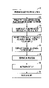
[0031] Fig. 1 depicts a flow chart illustrating a method of depositing
nanostructures on an
implant surface.
4
CA 3048565 2019-07-03
[0032] Figs. 2A-E illustrate a first example of the deposition method of
Fig. 1, where the
concentration of silver nitrate (AgNO3) is varied.
[0033] Figs. 3A-C illustrate a substrate processed according to a second
example of the
deposition method of Fig. 1, where the voltage applied during the deposition
process was varied.
[0034] Figs. 4A-C illustrate a substrate processed according to a third
example of the
deposition method of Fig. 1, where the deposition time was varied.
[0035] Figs. 5A-D illustrate a substrate processed according to a fourth
example of the
deposition method of Fig. 1, where the deposition time was varied and the
substrate includes a
nanotube structure.
[0036] Fig. 6 are several close-up views of Fig. 5B.
[0037] Figs. 7A-B illustrate a substrate processed according to a fifth
example of the
deposition method of Fig. 1, where the deposition temperature was varied.
[0038] Corresponding reference characters indicate corresponding parts
throughout the
several views. The exemplifications set out herein illustrate examples of the
disclosure, and such
exemplifications are not to be construed as limiting the scope of the
disclosure any manner.
DETAILED DESCRIPTION
[0039] In describing the examples of the disclosure illustrated and to be
described with
respect to the drawings, specific terminology will be used for the sake of
clarity. However, the
examples are not intended to be limited to any specific terms used herein, and
it is to be
understood that each specific term includes all technical equivalents.
[0040] The present disclosure is directed to methods for depositing
nanostructures onto
implant surfaces, and the implants resulting from such methods. The methods
can involve
depositing silver or silver compound nanostructures on implant surfaces to,
for example, improve
the antimicrobial characteristics thereof, while keeping any osseointegration
characteristics
intact. The silver or silver compound nanostructures can alleviate a number of
downsides to
current antimicrobial techniques, as detailed more fully below.
[0041] Fig. 1 illustrates a flow chart depicting a method for depositing
nanostructures onto
an implant surface. In a merely exemplary embodiment, first an electrolytic
solution 10 can be
prepared. The electrolyte solution can comprise an aqueous solution having
about .01 mM to
about 50 mM silver nitrate (AgNO3). In an example, about 0 mM NaN011 to about
1000 mM
CA 3048565 2019-07-03
NaNOLI can also be added to the solution to, for instance, increase the
conductivity of the
electrolyte solution. NaNOLI is not needed in all cases, and thus, can be 0 nM
in some cases.
According to one example, 138 mM NaN011 can be utilized. The solution can be
kept at or
near room temperature (anywhere between about 18-30 C), although it is to be
appreciated that
other temperatures can be employed, as described below. In some examples, the
electrolyte
solution might only comprise silver nitrate (AgNO3) and not NaNO LI.
[0042] As illustrated at step 20, the electrolyte solution can then be
transferred to a beaker or
other container. The beaker or container can, in an example, be placed on a
magnetic stirrer or
another agitation device can be used. At step 30, an anode (e.g., Pt) and a
cathode (e.g., Ti (i.e.
commercially pure Ti, Ti alloy, TiAl alloy (e.g., Ti6A14V)), stainless steel,
tantalum, etc. ) can be
placed onto a fixture (e.g., Teflon). In an example, the anode and cathode can
be separated by
some predetermined distance (e.g., anywhere between about .25-3 cm) by the
fixture.
[0043] At step 30, the anode and cathode can be immersed into the
electrolyte solution, and
the stirrer or agitation device can be activated to stir or mix the solution
at a rate of anywhere
between about 100-1000 RPM, in a particular embodiment anywhere between about
300-500
RPM. At step 40, a power supply connected to the anode and cathode can be
adjusted to a
desired voltage. In an example, the power supply can be set to anywhere
between about 2-25V
with a 2 +/- .2 AMP maximum output. In addition, the power supply can be shut
down at step 40
so that, at step 50, the anode and cathode can be connected to the power
supply (e.g., with silver
and platinum wires, respectively).
[0044] Step 60 can commence the electrochemical deposition process. First,
a substrate can
be placed in the electrolyte solution. The substrate can include a surface
that can be machined,
acid etched, grit blasted and acid etched, and/or with nano-complexity such as
nanotubes or
Discrete Crystalline Deposition (DCDe), as provided by the Applicant. Certain
exemplary
substrates are described in more detail below.
[0045] In an example, the deposition process can next involve setting the
temperature of the
electrolyte solution to anywhere between about 0-100 C, turning the power
supply on, and
applying an electric current to the system (e.g., anode and cathode, immersed
in the electrolyte
solution). The electric current can be applied for anywhere between about 30
seconds to 1 hour,
in an example. Other time durations are possible. Application of an electric
current within the
ranges mentioned above can be applied to the electrolyte solution for the
aforementioned time,
6
CA 3048565 2019-07-03
causing deposition of silver nanoparticles onto the substrate immersed in the
electrolytic
solution.
[0046] In an example, the substrate can be an implant. In a further
example, the substrate
can be a dental implant designed to be implanted into a patient's jawbone. For
instance, the
dental implant can include any of the features or characteristics of U.S.
Patent Pub. No.
2014/0272791, filed by Zimmer Dental, Inc., the disclosure of which is hereby
incorporated by
reference herein in its entirety. Of course, the substrate can be a dental
implant that has different
features than the dental implant of the '791 Publication.
[0047] The deposition of silver nanoparticles can occur on an external
surface of the implant
that is arranged to contact the patient's jawbone (e.g., outer surface of
implant body 17 of the
'791 Publication). Alternatively, or in addition, the silver nanoparticles can
be deposited on a
majority of the aforementioned external surface of implant body 17, or a
portion of the external
surface that is less than a majority. In a further alternative, the silver
nanoparticles can be
deposited on substantially all of the surfaces of implant body 17 that are
exposed within the
electrolyte solution. In an example, this can mean that substantially all of
the outer surfaces of
implant body 17 can include deposited silver nanoparticles, according to the
above process. It is
to be appreciated that the application of an electric current in the
electrolytic solution detailed
above can reduce silver ions to nanostructures and deposit the silver
nanostructures onto surfaces
of the substrate (e.g., dental implant). In yet further examples, the silver
nanoparticles can be
deposited on various other components and/or other surfaces such as an
implant/abutment
junction, a provisional abutment external surface, a definitive abutment
external surface, internal
surfaces that can be accessed, etc.).
[0048] By "nanostructures" or "nanoparticles", it is meant that such
particles have either one,
two or all three dimensions in the nanoscale range of 0.1 nm to 100 nm,
inclusive. The
nanostructures or nanoparticles can either be in a single particulate or
agglomerate to form a
cluster.
[0049] At step 70, an ultrasonication process can take place. In an
example, step 70 can be
omitted. The ultrasonication process can optimize the size distribution of
silver nanostructures
by removing some large particles from the surface of the substrate. In other
words, using an
ultrasonication process, particles over a certain size threshold (e.g., 50 nm
to 500 nm, inclusive)
7
CA 3048565 2019-07-03
can be removed from the substrate by applying ultrasonic energy to the
substrate. This can have
the effect of ensuring an optimal size distribution of silver nanostructures
on the substrate.
[0050] In step 80, the substrate can be rinsed in a reverse
osmosis/deionized water (RO/DI
water). The substrate can then be dried (e.g., in an oven) at a temperature of
about 100 +/- 5 C
for about 30 +/- 10 minutes.
[0051] Deposition of silver nanoparticles onto the surface of a substrate
(e.g., dental implant)
can have a number of benefits. Silver ions, compounds, and particles have
antibacterial
properties that can be used as an alternative to antibiotic therapy. Bacteria
can be killed upon
surface contact with metallic silver nanoparticles, and through the extended
release of low
concentrations of silver ions through oxidative dissolution of the
nanoparticles. The advantages
of using silver as an antimicrobial agent can include: (1) it has broad
spectrum antimicrobial
activities against Gram-negative and Gram-positive bacteria, (2) it has high
efficiency and low
toxicity for long-term use, and (3) as an alternative to traditional
antibiotic molecules, silver has
a low probability of a microorganism developing resistance. The foregoing is a
non-exhaustive
list of the benefits of using silver nanoparticles in the context of the
present invention.
[0052] In addition to the aforementioned benefits, an additional positive
to the deposition
method of the present disclosure is that the antimicrobial agent (silver
nanostructures) can be
coated onto the substrate (e.g., dental implant) without requiring any
specific pre-treatment of the
target surface of the substrate. Further, the silver nanostructures are in
nanoscale. Due to the
high specific surface area of nanostructures, bacteria can be killed more
effectively.
Furthermore, since the coating can be deposited by an electrochemical
procedure, as described
above, a uniform distribution of silver nanostructures can be created while
the size and quantity
of the nanostructures can be well controlled. Therefore, the amount of silver
can be maintained
between minimum effective level and maximum safety level. As disclosed herein,
the minimum
effective level for silver ion concentration can be as low as 1.43 ppm (ppm
and ug/ml are
equivalent). This minimum effective level is sufficient to kill or inhibit a
wide range of
microorganisms. As disclosed herein, the maximum safety level can be as high
as 112.03 ug/ml.
[0053] Applicant sets forth several particular examples of the deposition
process detailed
above using different substrates and different deposition parameters. Such
examples are
illustrated in Figs. 2A-7B, and disclosed in more detail below. It is to be
understood that the
8
CA 3048565 2019-07-03
examples are non-limiting, and are illustrative of how different parameters of
the deposition
process and/or the substrate can affect the ultimate result.
EXAMPLES 1A-E
[0054]
Figs. 2A-E illustrate Examples 1A-E of the present disclosure, respectively.
The
deposition parameters for Examples 1A-E are shown in Table 1.1 below.
Table 1.1
Deposition NaNO3
Voltage Deposition Time
Variable
Temp Concentration
About Room AgNO3
1.25 mM (low) 5 V 90 seconds
Temp Concentration
[0055] As
shown above, Examples 1A-E, illustrated in Figs. 2A-E, respectively,
demonstrate
the effect of varying the AgNO3 concentration of the electrolytic solution
during the deposition
process outlined above. It was observed that large crystals were formed when a
high
concentration of AgNO3 was added into the electrolytic solution for
deposition. This can be seen
in Figs. 2A-E, which have the AgNO3 concentration listed with each figure. As
such, Applicant
can, in an example, vary the AgNO3 concentration in the electrolytic solution
to change the size
and/or amount of silver crystals or particles deposited on the substrate
(e.g., dental implant),
using the deposition process detailed above. In Examples 1A-E, a machined
surface was used as
a deposition substrate to illustrate the effect of varying the AgNO3
concentration of the
electrolyte solution.
[0056]
Further, the following weight percentages in Table 1.2 were measured by energy-
dispersive x-ray spectroscopy (EDS) (n = 5 per sample):
Table 1.2*
Deposition Amount .3 mM AgNO3 .6 mM AgNO3 .9 mM
AgNO3
Weight Percentage
0.12 +/- 0.24% 1.00 +/- 1.84% 0.93
+/- 0.10%
of Silver
* EDS was not conducted on 5 and 20 mM concentrations of AgNO3.
[0057] The
above Table 2.2 confirms that, with increased silver ion concentrations in the
electrolyte solution, an increase in the amount of deposited silver
nanoparticles can be expected
on the substrate. Thus, to modify the above deposition process in terms of
amount of deposited
silver nanoparticles, it is possible to vary the concentration of AgNO3 in the
electrolyte solution.
EXAMPLES 2A-C
9
CA 3048565 2019-07-03
[0058] Figs. 3A-C illustrate Examples 2A-C of the present disclosure,
respectively. The
deposition parameters for Examples 2A-C are shown in Table 2.1 below.
Table 2.1
Deposition AgNO3 NaNO3
Deposition Time
Variable
Temp Concentration Concentration
About Room Voltage
0.9 mM 138 mM 90 seconds
Temp (potentiostatic)
[0059] As shown above, Examples 2A-C, illustrated in Figs. 3A-C,
respectively, demonstrate
the effect of varying the voltage applied to the electrolytic solution during
the deposition process
outlined above. The voltage applied to the electrolytic solution is set forth
above each respective
figure. It was observed that an increase in deposition voltage under
potentiostatic mode resulted
in an increase in the quantity of silver nanoparticles based on visual
inspection of Scanning
Electron Microscopy (SEM) images. As such, it is possible to also vary the
voltage applied to
the electrolytic solution in the above deposition process to alter the
quantity of silver
nanoparticles deposited to the substrate (e.g., dental implant). In Examples
2A-C, a machined
surface was used as a deposition substrate, similar to as in Examples 1A-E.
EXAMPLES 3A-C
[0060] Figs. 4A-C illustrate Examples 3A-C of the present disclosure,
respectively. The
deposition parameters for Examples 3A-C are shown in Table 3.1 below.
Table 3.1
Deposition AgN00 NaNOLI
Voltage
Variable
Temp Concentration Concentration
About Room
0.9 mM 138 mM 5 V deposition time
Temp
[0061] As shown above, Examples 3A-C, illustrated in Figs. 4A-C,
respectively, demonstrate
the effect of varying the deposition time during the deposition process
outlined above. The
deposition time is set forth above each respective figure. It was observed
that a higher amount of
silver nanoparticles was deposited on the substrate (e.g., dental implant)
with increased
deposition time. Indeed, EDS results demonstrated that the amount by weight
percent of silver
deposited on the substrate is as shown in the below Table 3.2. The deposition
substrate used was
an acid etched surface.
Table 3.2
CA 3048565 2019-07-03
Deposition Amount 3 Minute Deposition 6 Minute Deposition 9 Minute Deposition
Weight Percentage
0.26 +/- 0.52% 1.75 +/- 1.29% 2.91
+/- 1.50%
of Silver
[0062] Thus, it is possible to vary the deposition time in the deposition
process set forth
above to increase or decrease the amount of silver nanoparticles deposited on
the substrate (e.g.,
dental implant).
EXAMPLES 4A-D
[0063] Figs. 5A-C illustrate Examples 4A-D of the present disclosure,
respectively. The
deposition parameters for Examples 4A-D are shown in Table 4.1 below.
Table 4.1
Deposition AgNO3 NaNO3
Voltage
Variable
Temp Concentration Concentration
About Room
0.9 mM 138 mM 5 V
deposition time
Temp
[0064] As shown above, Examples 4A-D, illustrated in Figs. 5A-C,
respectively,
demonstrate the effect of varying the deposition time during the deposition
process outlined
above. In contrast to prior Examples 3A-C, a different substrate was used in
Examples 4A-D. In
Examples 4A-D, the substrate used was a dental implant offered by Applicant
under the name
T30, but the implant was covered with a carbon nanotube surface. The nanotubes
are visible in
Figs. 5A-C. An example of a nanotube structure on a dental implant, created by
the Applicant, is
disclosed in U.S. Patent Pub. No. 2017/0042682, which is incorporated by
reference herein in its
entirety. It is understood that the substrate utilized in Examples 4A-D, or in
any other example
or deposition method disclosed herein, can be any of the implants disclosed in
the '682
Publication. The present Examples 4A-D describe the effect of varying
deposition time during
the deposition process outlined above, using a dental implant with a nanotube
structure, similar
to as in the '682 Publication.
[0065] It was observed that a higher amount of silver nanoparticles was
deposited on the
substrate (e.g., dental implant with nanotubes) with increased deposition
time. In other words,
higher coverage of the nanotubes' surfaces by silver particles was observed
with the increase of
deposition time. The deposition time for each figure is listed above the
figure. It was also
observed that, when the deposition time was increased to 30 or 45 minutes
(Figs. 5C-D), the
11
CA 3048565 2019-07-03
silver nanoparticles formed clusters and covered the underlying nanotube
surfaces. As such, it is
contemplated herein that deposition time can be varied using a substrate with
a nanotube
structure (e.g., dental implant with nanotubes) to alter the amount of silver
nanoparticles
deposited on the substrate.
[0066] Fig. 6 illustrates several relatively closer-up views of the
nanotube surface of Fig. 5B,
where the deposition time was about 18 minutes. The close-up views provide
illustration as to
areas in which silver nanoparticles were deposited on the nanotube substrate,
with arrows
pointing to areas of deposited silver nanoparticles, in certain instances.
[0067] EDS results demonstrated that the amount by weight percent of silver
deposited on
the nanotube substrate is as shown in the below Table 4.2. It is worthwhile to
note that the lower
standard deviations below indicate the silver particles were evenly
distributed over the surface of
the nanotubes.
Table 4.2
Deposition 9 Minute 18 Minute 30 Minute 45
Minute
Amount Deposition Deposition Deposition
Deposition
Weight
Percentage of 6.21 +/- 0.90% 8.29 +/- 1.46% 10.92 +/- 1.27%
19.01 +/- 2.81%
Silver
EXAMPLES 5A-B
[0068] Figs. 7A-B illustrate Examples 5A-B of the present disclosure,
respectively. The
deposition parameters for Examples 5A-B are shown in Table 5.1 below.
Table 5.1
AgNO3 NaNO3
Deposition Time Voltage Variable
Concentration Concentration
90 seconds 0.9 mM 1.25 mM 5 V deposition temp
[0069] As shown above, Examples 5A-B, illustrated in Figs.7A-B,
respectively, demonstrate
the effect of varying the deposition temperature of the electrolyte solution
during the deposition
process outlined above. The different deposition temperatures are shown above
the respective
figures. It was observed that, with an increase in deposition temperature, the
amount and/or size
of silver nanoparticles deposited on the substrate (e.g., dental implant) was
increased. Thus, it is
possible to vary the temperature of the electrolyte solution during deposition
to vary the amount
12
CA 3048565 2019-07-03
and/or size of silver nanoparticles deposited. The substrate used in Examples
5A-B was a
machined Ti disk surface.
[0070] The above examples illustrate that various deposition parameters can
be altered to
alter the deposition of silver nanoparticles on a substrate, as detailed
herein. It is to be
appreciated that, while not disclosed above, any of these parameters can be
used in combination
with any other deposition parameter to alter the deposition of silver
nanoparticles on a substrate.
[0071] It will be readily understood to those skilled in the art that
various other changes in
the details, material, and arrangements of the parts and method stages which
have been described
and illustrated in order to explain the nature of the inventive subject matter
can be made without
departing from the principles and scope of the inventive subject matter as
expressed in the
subjoined claims.
[0072] It will also be appreciated that the various dependent claims,
examples, and the
features set forth therein can be combined in different ways than presented
above and/or in the
initial claims. For instance, any feature(s) from the above examples can be
shared with others of
the described examples, and/or a feature(s) from a particular dependent claim
may be shared
with another dependent or independent claim, in combinations that would be
understood by a
person of skill in the art.
13
CA 3048565 2019-07-03