Note: Descriptions are shown in the official language in which they were submitted.
CA 03048660 2019-06-26
WO 2018/129007 PCT/US2018/012136
PROTEIN THERAPEUTICS FOR TREATMENT OF SENESCENT CELLS
FIELD OF THE DISCLOSURE
[0001] This disclosure relates to the field of treating or clearing senescent
cells and/or treating diseases or
disorders related to senescent cells. Particularly, this disclosure relates to
conditionally active proteins that
target senescent cells and to methods of generating such conditionally active
proteins.
BACKGROUND OF THE DISCLOSURE
[0002] Senescent cells are metabolically active but trapped in the G1 phase of
cell growth cycle with their
lifespan controlled by multiple dominant genes (Stanulis-Praeger, Mech. Ageing
Dev., vol. 38, pp.1-48,
1987). Senescent cells differ from quiescent cells and terminal differentiated
cells in several important
aspects, having characteristic morphological changes such as enlargement,
flattening, and increased
granularity (Dimri et al., Proc. Nat. Acad. Sci. USA, vol. 92, pp.9363-9367,
1995). Senescent cells do not
divide even if stimulated by mitogens (Campisi, Trends Cell Biol., vol. 11,
pp. S27-S31, 2001). Senescence
involves activation of p53 and/or Rb and their regulators such as pl6INK4a,
p21, and ARF. Except when
p53 or Rb is inactivated, senescence is generally irreversible.
[0003] Senescent cells express increased levels of plasminogen activator
inhibitor (PAI) and exhibit
staining for I3-galactosidase activity at pH 6 (Sharpless et al., J. Clin.
Invest., vol. 113, pp.160-168, 2004).
Irreversible G1 arrest is mediated by inactivation of cyclin dependent kinase
(CdK) complexes which
phosphorylate Rb. P21 accumulates in senescent cells, which inhibits CdK4-
CdK6. P16 also inhibits CdK4-
CdK6 and accumulates in senescent cells proportionally with I3-galactosidase
activity and cell volume (Stein
et al., Mol. Cell. Biol., vol. 19, pp.2109-2117, 1999). Evidence suggests that
p21 is expressed during
initiation of senescence but not required for maintaining senescence, while
p16 expression helps maintain
senescence once initiated.
[0004] Since in some cases senescence is related to the progressive shortening
of telomeres with each cell
division, senescence is triggered when certain chromosomal telomeres reach a
critical length (Mathon and
Lloyd, Nat. Rev. Cancer, vol. 3, pp.203-213, 2001; Martins, U. M. Exp Cell
Res., vol. 256, pp.291-299,
2000). Senescence can be abrogated by the expression of telomerase which
lengthens telomeres. For
example, human fibroblasts undergo replication indefinitely when the
fibroblasts are transfected to express
telomerase. Most cancer cells express telomerase in order to maintain telomere
length and replicate
indefinitely. The minority of cancer cells that do not express telomerase have
alternative mechanisms for
lengthening of telomeres (ALTs).
[0005] There are also other causes of senescence. Collectively, these other
causes are often referred to as
stress-induced premature senescence (SIPS). Oxidative stress can shorten
telomeres thereby inducing
senescence (von Zglinicki, Trends Biochem. Sci., vol. 27, pp.339-344, 2002).
Hyperoxia has been shown to
induce senescence. Gamma irradiation of human fibroblasts in early to mid G1
phase causes senescence in a
p53-dependent manner (Di Leonardo et al., Genes Dev., vol. 8, pp.2540-2551,
1994). Ultraviolet radiation
CA 03048660 2019-06-26
WO 2018/129007 PCT/US2018/012136
also induces senescence. Other agents that can induce senescence include
hydrogen peroxide (Krtolica et al.,
Proc. Nat. Acad. Sci. USA, vol. 98, pp.12072-12077, 2001), sodium butyrate, 5-
azacytadine, and transfection
with the Ras oncogene (Tominaga, Mech. Ageing Dev., vol. 123, pp.927-936,
2002). Chemotherapeutic
agents including doxorubicin, cisplatin, and a host of others have been shown
to induce senescence in cancer
cells (Roninson, Cancer Res., vol. 63, pp.2705-2715, 2003). 5-
bromodeoxyuridine treatment results in
senescence in both normal and malignant cells (Michishita et al., J. Biochem.,
vol. 126, pp.1052-1059,
1999). Generally speaking, agents that damage DNA are capable of causing
senescence.
[0006] Evidence suggests a relationship between senescence and aging. Cultured
cells from old donors
exhibit senescence after fewer growth cycles than cells from young donors
(Martin et al., Lab. Invest., vol.
23, pp.86-92, 1970; Schneider et al., Proc. Nat. Acad. Sci. USA, vol. 73,
pp.3584-3588, 1976). Cells from
short-lived species senesce after fewer growth cycles than cells from long-
lived species (Rohme, D., Proc.
Nat. Acad. Sci. USA, vol. 78, pp.5009-3320, 1981). Cultured cells from donors
with hereditary premature
aging syndromes such as Werner's syndrome show senescence after fewer growth
cycles than cells from
age-matched controls.
[0007] Senescence confers functional changes on the senescent cells which have
been associated with
various age-related diseases and disorders (Chang et al., Proc. Nat. Acad.
Sci. USA, vol. 97, pp.4291-4296,
2000). Senescent cells accumulate in tissues and organs of individuals as they
age and are found at sites of
age-related pathologies. Given that senescent cells have been causally
implicated in certain aspects of age-
related decline in health and may contribute to certain diseases, and are also
induced as a result of necessary
life-preserving chemotherapeutic and radiation treatments, the presence of
senescent cells may have
deleterious effects to millions of patients worldwide. It is widely believed
that selective elimination of
senescent cells can prevent and treat age-related diseases and disorders.
[0008] Senescent cells can also promote tumorigenesis. Senescent stromal cells
express tumor promoting
factors that exert a paracrine effect on neighboring epithelial cells. These
effects include mitogenicity and
anti-apoptosis (Chang et al., Proc. Nat. Acad. Sci. USA, vol. 97, pp.4291-
4296, 2000). Senescent fibroblasts
have been shown to stimulate premalignant and malignant epithelial cells but
not normal epithelial cells to
form tumors in mice. This occurred when as few as 10% of the fibroblasts were
senescent (Krtolica et al.,
Proc. Nat. Acad. Sci. USA, vol. 98, pp.12072-12077, 2001). Tumor promoting
factors secreted by senescent
cells are partly mediated by p2lwafl/cipl/sdil (Roninson, Cancer Res., vol.
63, pp.2705-2715, 2003). A
threshold of senescent stromal cells appears to provide a milieu allowing
adjacent premalignant epithelial
cells to survive, migrate, and divide (Campisi, Nat. Rev. Cancer, vol. 3, pp.
339-349, 2003).
[0009] Consequently, therapeutics targeting senescent cells are a promising
treatment option for
senescence-associated diseases and disorders. US 2016/0038576 discloses an
immunogenic composition for
inducing an adaptive immune response directed specifically at senescent cells
for treatment and prophylaxis
of age-related diseases and disorders, and other diseases and disorders
associated with or exacerbated by the
presence of senescent cells. The immunogenic composition comprises at least
one or more of senescent cell-
2
CA 03048660 2019-06-26
WO 2018/129007 PCT/US2018/012136
associated antigens, polynucleotides encoding senescent cell-associated
antigens, and recombinant
expression vectors comprising the polynucleotides for use in administering to
a subject.
[0010] WO 2015116740 discloses a method of administering a therapeutically-
effective amount of a small
molecule senolytic agent that selectively kills senescent cells as compared
with non-senescent cells for
treatment of senescent cell-associated diseases and disorders. The senescent
cell-associated diseases and
disorders treatable by the method include cardiovascular diseases and
disorders associated with or caused by
arteriosclerosis, such as atherosclerosis, idiopathic pulmonary fibrosis,
chronic obstructive pulmonary
disease, osteoarthritis, senescence-associated ophthalmic diseases and
disorders, and senescence-associated
dermatological diseases and disorders.
[0011] US 2015/0064137 discloses a polypeptide and viruses comprising a
polypeptide useful for selective
elimination of senescent cells. The polypeptide and viruses can induce
apoptosis in senescent cells. The
polypeptide is selected from products of pro-apoptotic genes. The viruses
comprise the pro-apoptotic gene
for which expression is regulated by the p16 promoter. The p16 promoter can be
a canonical p16 promoter
or a non-canonical p16 promoter.
[0012] These therapeutics target one or more proteins of senescent cells to
kill or remove senescent cells.
However, these targeted proteins of senescent cells may also be present on
other types of cells which may
lead to undesirable side-effects. Thus, it would be advantageous to develop a
class of therapeutic proteins
that preferentially and/or specifically bind to a target on senescent cells,
while minimizing or eliminating
binding to the same target on other types of cells.
SUMMARY OF THE DISCLOSURE
[0013] h) one embodiment, the disclosure provides a method of producing a
conditionally active protein
that binds to a target associated with a senescent cell from a parent protein
that binds to the target associated
with the senescent cell, said method comprising steps of:
(i) evolving a DNA encoding the parent protein using one or more evolutionary
techniques to create
mutant DNAs;
(ii) expressing the mutant DNAs to obtain mutant proteins;
(iii) subjecting the mutant proteins to an assay under an extracellular
condition of the senescent cell
and an assay under a normal physiological condition; and
(iv) selecting the conditionally active protein from the mutant proteins that
exhibits at least one of:
(a) a decrease in an activity in the assay under the normal physiological
condition compared
to the same activity of the parent protein in the same assay, and an increase
in the activity in the assay
under the extracellular condition of the senescent cell compared to the same
activity of the
conditionally active protein in the assay under the normal physiological
condition; and
(b) a decrease in the activity in the assay under the normal physiological
condition
compared to the same activity of the parent protein in the same assay and an
increase in the activity
3
CA 03048660 2019-06-26
WO 2018/129007 PCT/US2018/012136
in the assay under the extracellular condition of the senescent cell compared
to the same activity of
the parent protein in the assay under the extracellular condition of the
senescent cell.
[0014] hi some embodiments, the parent protein may be selected from an enzyme,
an antibody, a receptor, a
ligand, a fragment of an enzyme, a fragment of an antibody, a fragment of a
receptor, and a fragment of a
ligand.
[0015] hi each of the foregoing embodiments, the activity may be a binding
activity to the target.
[0016] hi each of the foregoing embodiments, the parent protein may be an
enzyme and the activity is an
enzymatic activity using at least a portion of the senescent cell as a
substrate.
[0017] hi each of the foregoing embodiment, the conditionally active protein
may be a cyclic peptide. The
cyclic peptide may have a length of from about 5 to about 500 amino acids, or
from about 10 to about 50
amino acids.
[0018] hi each of the foregoing embodiments, the target may a surface molecule
located on an outer surface
of a senescent cell. In each of the foregoing embodiments, the surface
molecule may be a cellular membrane
protein of the senescent cell. In each of the foregoing embodiments, the
target may be selected from APC,
ARHGAP1, ARMCX-3, AXL, B2MG, BCL2L1, CAPNS2, CD261, CD39, CD54, CD73, CD95,
CDC42,
CDKN2C, CLYBL, COPG1, CRKL, DCR1, DCR2, DCR3, DEP1, DGKA, EBP, EBP50, FASL,
FGF1,
GBA3, GIT2, ICAM1, ICAM3, IGF1, ISG20, ITGAV, KITLG, LaminBl, LANCL1, LCMT2,
LPHN1,
MADCAM1, MAG, MAP3K14, MAPK, MEF2C, miR22, MMP3, MTHFD2, NAIP, NAPG, NCKAP1,
Nectin4, NNMT, NOTCH3, NTAL, OPG, OSBPL3, p16, p16INK4a, p19, p21, p53, PAIl,
PARK2, PFN1,
PGM, PLD3, PMS2, POU5F1, PPP1A, PPP1CB, PRKRA, PRPF19, PRTG, RAC1, RAPGEF1,
RET,
Smurf2, STX4, VAMP3, VIT, VPS26A, WEE1, YAP1, YH2AX, and YWHAE. In addition,
it should be
recognized that the target may be any combination of the preceding.
[0019] hi each of the foregoing embodiments, a ratio of the activity of the
conditionally active protein in the
assay under the extracellular condition of the senescent cell to the activity
of the conditionally active protein
in the assay under the normal physiological condition may be at least
about1.3:1, or at least about 2:1, or at
least about 3:1, or at least about 4:1, or at least about 5:1, or at least
about 6:1, or at least about 7:1, or at
least about 8:1, or at least about 9:1, or at least about 10:1, or at least
about 11:1, or at least about 12:1, or at
least about 13:1, or at least about 14:1, or at least about 15:1, or at least
about 16:1, or at least about 17:1, or
at least about 18:1, or at least about 19:1, or at least about 20:1, or at
least about 30:1, or at least about 40:1,
or at least about 50:1, or at least about 60:1, or at least about 70:1, or at
least about 80:1, or at least about
90:1, or at least about 100:1.
[0020] hi each of the embodiments, the extracellular condition of the
senescent cell may be a pH in a range
of from about 5.5 to about 7.0, or from about 6.0 to about 7.0, or from about
6.2 to about 6.8.
[0021] hi each of the foregoing embodiments, the normal physiological
condition may be a pH in a range of
from about 7.2 to about 7.8, or from about 7.2 to about 7.6, or from about 7.4
to about 7.6.
4
CA 03048660 2019-06-26
WO 2018/129007 PCT/US2018/012136
[0022] hi each of the foregoing embodiments, the extracellular condition of
the senescent cell may be a
lower concentration of a deoxynucleotide than a normal physiological
concentration of the same
deoxynucleotide.
[0023] hi each of the foregoing embodiments, the extracellular condition of
the senescent cell may be a
lower concentration of oxygen than a normal physiological concentration of
oxygen.
[0024] hi each of the foregoing embodiments, the extracellular condition of
the senescent cell may be a
lower ratio of NAD+/NADH than a normal physiological ratio of NAD+/NADH.
[0025] hi each of the foregoing embodiments, the extracellular condition of
the senescent cell may be at
least one of an increased concentration of a redox homeostasis metabolite
selected from hypotaurine,
cysteine sulfinic acid, cysteine-glutathione disulfide, gamma-glutamylalanine,
gamma-glutamylmethionine,
pyridoxate, gamma-glutamylglutamine, and alanine, relative to a normal
physiological concentration of the
same redox homeostasis metabolite.
[0026] hi each of the foregoing embodiments, the extracellular condition of
the senescent cell may be an
increased concentration of at least one nucleotide metabolite selected from 3-
ureidopropionate, urate, 7-
methylguanine, and hypoxanthine, relative to a normal physiological
concentration of the same nucleotide
metabolite.
[0027] hi each of the foregoing embodiments, the extracellular condition of
the senescent cell may be a
decreased concentration of thymidine relative to a normal physiological
concentration of thymidine.
[0028] hi each of the foregoing embodiments, the extracellular condition of
the senescent cell may be a
decreased concentration of at least one dipeptide selected from
glycylisoleucine, glycylvaline, glycylleucine,
isoleucylglycine, and valylglycine, relative to a normal physiological
concentration of the same dipeptide.
[0029] hi each of the foregoing embodiments, the extracellular condition of
the senescent cell may be a
decreased concentration of at least one fatty acid selected from linoleate,
dihomo-linoleate, and 10-
heptadecenoate, relative to a normal physiological concentration of the fatty
acid.
[0030] hi each of the foregoing embodiments, the extracellular condition of
the senescent cell may be an
increased concentration of at least one phospholipid metabolite selected from
2-hydroxypalmitate, 2-
hydroxystearate, 3-hydroxydecanoate, 3-hydroxyoctanoate, and
glycerophosphorylcholine, relative to a
normal physiological concentration of the phospholipid metabolite.
[0031] hi each of the foregoing embodiments, the extracellular condition of
the senescent cell may be an
increased concentration of at least one amino acid metabolite selected from
alanine, C-glycosyltryptophan,
kynurenine, dimethylarginine, and orthithine, relative to a normal
physiological concentration of the amino
acid metabolite.
[0032] hi each of the foregoing embodiments, the extracellular condition of
the senescent cell may be a
decreased concentration of phenylpyruvate, relative to a normal physiological
concentration of the
phenylpyruv ate.
CA 03048660 2019-06-26
WO 2018/129007 PCT/US2018/012136
[0033] In each of the foregoing embodiments, the extracellular condition of
the senescent cell may be an
increased concentration of at least one metabolite selected from fumarate,
malonate, eicosapentaenoate and
citrate, relative to a normal physiological concentration of the metabolite.
[0034] In each of the foregoing embodiments, the extracellular condition of
the senescent cell may be an
increased ratio of glycerophosphocholine to phosphocholine, relative to a
normal physiological ratio of
glycerophosphocholine to phosphocholine.
[0035] In each of the foregoing embodiments, the extracellular condition of
the senescent cell may be an
increased concentration of a protein secreted by the senescent cell, in
comparison with a normal
physiological concentration of said protein, and wherein said protein secreted
by the senescent cell is
selected from at least one of GM-CSF, GROa, GRC-a,I3,y, IGFBP-7, IL-la, IL-6,
IL-7, IL-8, MCP-1, MCP-
2, MIP-la, MMP-1, MMP-2, MMP-10, MMP-3, amphiregulin, ENA-78, eotaxin-3, GCP-
2, GITR, HGF,
ICAM-1, IGFBP-1, IGFBP-2, IGFBP-3, IGFBP-4, IGFBP-5, IGFBP-6, IL-13, IL-I13,
MCP-4, MIF, MIP-3a,
MMP-12, MMP-13, MMP-14, NAP2, oncostatin M, osteoprotegerin, PIGF, RANTES,
sgp130, TIMP-2,
TRAIL-R3, Acrp30, angiogenin, AXL, bFGF, BLC, BTC, CTACK, EGF-R, Fas, FGF-7, G-
CSF, GDNF,
HCC-4, 1-309, IFN-y, IL-1R1, IL-11, IL-15, IL-2R-a, IL-6R, I-TAC, leptin, LIF,
MSP-a, PAI-1, PAI-2,
PDGF-BB, SCF, SDF-1, sTNF RI, sTNF Rh, thrombopoietin, TIMP-1, tPA, uPA, uPAR,
VEGF, MCP-3,
IGF-1, TGF-I33 , MIP-1-delta, IL-4, IL-16, BMP-4, MDC, IL-10, Fit-3 Ligand,
CNTF, EGF, BMP-6 and
any combination thereof.
[0036] hi each of the foregoing embodiments, the assay under the normal
physiological condition and the
assay under the extracellular condition of the senescent cell may be performed
in assay solutions containing
at least one component selected from an inorganic compound, an ion and an
organic molecule. In this
embodiment, the at least one component may have substantially the same
concentration in the assay
solutions for both the assay under the normal physiological condition and the
assay under the extracellular
condition of the senescent cell. In these embodiments, the at least one
component may be the inorganic
compound and is selected from boric acid, calcium chloride, calcium nitrate,
di-ammonium phosphate,
magnesium sulfate, mono-ammonium phosphate, mono-potassium phosphate,
potassium chloride, potassium
sulfate, copper sulfate, iron sulfate, manganese sulfate, zinc sulfate,
magnesium sulfate, calcium nitrate,
calcium chelate, copper chelate, iron chelate, iron chelate, manganese
chelate, zinc chelate, ammonium
molybdate, ammonium sulphate, calcium carbonate, magnesium phosphate,
potassium bicarbonate,
potassium nitrate, hydrochloric acid, carbon dioxide, sulfuric acid,
phosphoric acid, carbonic acid, uric acid,
hydrogen chloride, and urea. In these embodiments, the at least one component
may be the ion and is
selected from a phosphorus ion, a sulfur ion, a chloride ion, a magnesium ion,
a sodium ion, a potassium ion,
an ammonium ion, an iron ion, a zinc ion, and a copper ion. In these
embodiments, the at least one
component may be selected from one or more of uric acid in concentration range
of 2-7.0 mg/dL, calcium
ion in a concentration range of 8.2-11.6 mg/dL, chloride ion in a
concentration range of 355-381 mg/dL,
iron ion in a concentration range of 0.028-0.210 mg/dL, potassium ion in a
concentration range of 12.1-25.4
mg/dL, sodium ion in a concentration range of 300-330 mg/dL, and carbonic acid
in a concentration range
6
CA 03048660 2019-06-26
WO 2018/129007 PCT/US2018/012136
of 15-30 mM. In these embodiments, the at least one component may be the
organic molecule and is an
amino acid selected from Histidine, Alanine, Isoleucine, Arginine, Leucine,
Asparagine, Lysine, Aspartic
acid, Methionine, Cysteine, Phenylalanine, Glutamic acid, Threonine,
Glutamine, Tryptophan, Glycine,
Valine, Pyrrolysine, Proline, Selenocysteine, Serine, and Tyrosine. In these
embodiments, the at least one
component may be an organic acid selected from citric acid, a-ketoglutaric
acid, succinic acid, malic acid,
fumaric acid, acetoacetic acid, I3-hydroxybutyric acid, lactic acid, pyruvic
acid, a-ketonic acid, acetic acid,
and volatile fatty acids. In these embodiments, the at least one component may
be a sugar selected from
glucose, pentose, hexose, xylose, ribose, mannose, galactose, lactose,
GlcNAcI31-3Gal, Gala1-4Gal, Manal-
2Man, GalNAcI31-3Gal, and 0-, N-, C-, and S-glycosides. In these embodiments,
the at least one component
may be selected from magnesium ion, sulfate ion, bisulfate ion, carbonate ion,
bicarbonate ion, nitrate ion,
nitrite ion, phosphate ion, hydrogen phosphate ion, dihydrogen phosphate ion,
persulfate ion,
monopersulfate ion, borate ion, and ammonium ion.
[0037] In each of the foregoing embodiments, the extracellular condition of
the senescent cell may be a first
pH in a range of from about 5.5 to about 7.0 and the normal physiological
condition may be a second pH in
a range of from about 7.2 to about 7.8, and the one or more assays may be
performed in assay solutions
containing at least one species having a molecular weight of less than 900
a.m.u. and a pKa up to 0.5, 1, 2, 3,
or 4 pH units away from said first pH.
[0038] In each of the foregoing embodiments, the extracellular condition of
the senescent cell may be a first
pH in a range of from about 5.5 to about 7.0 and the normal physiological
condition may be a second pH in
a range of from about 7.2 to about 7.8, the one or more assays may be
performed in assay solutions
containing at least one species having a molecular weight of less than 900
a.m.u., and said species may have
a pKa between said first pH and said second pH.
[0039] In each of the foregoing embodiments, the extracellular condition of
the senescent cell may be a first
pH in a range of from about 5.5 to about 7.0 and the normal physiological
condition may be a second pH in
a range of from about 7.2 to about 7.8, and the one or more assays may be
performed in assay solutions
containing at least one species selected from histidine, histamine,
hydrogenated adenosine diphosphate,
hydrogenated adenosine triphosphate, citrate, bicarbonate, acetate, lactate,
bisulfide, hydrogen sulfide,
ammonium, and dihydrogen phosphate.
[0040] hi each of the foregoing embodiments, the selecting step (iv) may
comprise selecting a conditionally
active protein that exhibits (a) a decrease in an activity in the assay under
the normal physiological condition
compared to the same activity of the parent protein in the same assay, and an
increase in the activity in the
assay under the extracellular condition of the senescent cell compared to the
same activity of the
conditionally active protein in the assay under the normal physiological
condition.
[0041] hi each of the foregoing embodiments, the selecting step (iv) may
comprise selecting a conditionally
active protein that exhibits (b) a decrease in the activity in the assay under
the normal physiological
condition compared to the same activity of the parent protein in the same
assay and an increase in the
7
CA 03048660 2019-06-26
WO 2018/129007 PCT/US2018/012136
activity in the assay under the extracellular condition of the senescent cell
compared to the same activity of
the parent protein in the assay under the extracellular condition of the
senescent cell.
[0042] hi another embodiment, the disclosure provides a conditionally active
protein produced by any of
the foregoing methods. The conditionally active protein may be an antibody.
The antibody may be a single
chain antibody or an antibody fragment. The antibody may be suitable to be
engineered as part of chimeric
antigen receptor of T-cells. The antibody may be a humanized antibody, a
bispecific antibody, or a
multispecific antibody.
[0043] hi each of the foregoing embodiments, the conditionally active protein
may be selected from a
receptor, a regulatory protein, a soluble protein, a cytokine, a fragment of a
receptor, a fragment of a
regulatory protein, a fragment of a soluble protein, and a fragment of a
cytokine.
[0044] hi each of the foregoing embodiments, the conditionally active protein
may be a conditionally active
antibody and the conditionally active antibody may be conjugated to a masking
moiety by a linker. The
masking moiety reduces a binding activity of the conditionally active antibody
to the target by at least at
least 50%, 55%, 60%, 65%, 70%, 75%, 80%, 85%, 90%, 92%, 93%, 94%, 95%, 96%,
97%, 98%, 99% or
even 100%.
[0045] hi each of the foregoing embodiments, the linker may be covalently
bonded to a variable region of
the conditionally active antibody.
[0046] hi each of the foregoing embodiments, the masking moiety may
specifically bind to a variable
region of the conditionally active antibody.
[0047] hi each of the foregoing embodiments, the linker may comprise a
flexible region and a cleavage site.
[0048] hi each of the foregoing embodiments, the cleavage site may be cleaved
by a protease in the
extracellular environment of the senescent cell.
[0049] hi each of the foregoing embodiments, the conditionally active protein
may be conjugated to a
cytotoxic drug, a cytostatic drug, or an anti-proliferative drug by a linker.
[0050] hi each of the foregoing embodiments, the linker may comprise a
cleavage site of at least one
protease in the extracellular environment of the senescent cell. The at least
one protease is selected from
ADAM10, ADAM12, ADAM17, ADAMTS, ADAMTS5, BACE, Caspase 1-14, Cathepsin A,
Cathepsin B,
Cathepsin D, Cathepsin E, Cathepsin K, Cathepsin S, FAP, MT1-MMP, Granzyme B,
Guanidinobenzoatase,
Hepsin, Human Neutrophil Elastase, Legumain, Matriptase 2, Meprin, MMP1-17, MT-
SP1, Neprilysin,
NS3/4A, Plasmin, PSA, PSMA, TRACE, TMPRSS 3, TMPRSS 4, and uPA.
[0051] hi another embodiment, the disclosure provides pharmaceutical
composition comprising an effective
amount of any of the foregoing conditionally active proteins and a
pharmaceutically acceptable carrier.
[0052] hi still another embodiment, the disclosure provides a method of
treatment of aging, or of a
senescent cell-associated disease or disorder comprising a step of
administering any of the foregoing
conditionally active proteins or any of the foregoing pharmaceutical
compositions. In the foregoing
embodiment, the senescent cell-associated disease or disorder may be selected
from cognitive diseases,
cardiovascular disease, metabolic diseases and disorders, motor function
diseases and disorders,
8
CA 03048660 2019-06-26
WO 2018/129007 PCT/US2018/012136
cerebrovascular disease, emphysema, osteoarthritis, pulmonary diseases,
inflammatory/autoimmune
diseases and disorders, ophthalmic diseases or disorders, metastasis, a
chemotherapy or radiotherapy side
effect, aging-related diseases and disorders, fibrotic diseases and disorders.
[0053] In yet another embodiment, the disclosure provides a method for
generating a conditionally active
molecule that has a molecular weight of less than about 3000 a.m.0 from a
parent organic compound. The
method includes steps of modifying the parent organic compound by introducing
one or more partially
charged or charged groups into the parent organic compound to produce one or
more modified organic
compounds; and selecting the modified organic compound that exhibits a higher
activity in the assay under
the aberrant condition compared to the same activity in the assay under the
normal physiological condition.
[0054] In yet another embodiment, the disclosure provides a method for
generating a conditionally active
molecule that has a molecular weight of less than about 3000 a.m.0 from a
parent organic compound,
comprising steps of: modifying the parent organic compound by removing one or
more partially charged or
charged groups from the parent organic compound to produce one or more
modified organic compounds;
and selecting the modified organic compound that exhibits a higher activity in
the assay under the aberrant
condition compared to the same activity in the assay under the normal
physiological condition.
[0055] In yet another embodiment, the disclosure provides a method for
generating a conditionally active
molecule that has a molecular weight of less than about 3000 a.m.0 from a
parent organic compound,
comprising steps of: modifying the parent organic compound by replacing one or
more groups of the parent
organic compound with one or more partially charged or charged groups to
produce one or more modified
organic compounds; and selecting the modified organic compound that exhibits a
higher activity in the assay
under the aberrant condition compared to the same activity in the assay under
the normal physiological
condition.
[0056] hi each of the foregoing methods, the parent organic compound may have
a molecular weight in a
range of from about 100 a.m.u. to about 3000 a.m.u, or from about 100 a.m.u.,
to about 1500 a.m.u., or from
about 150 a.m.u., to about 1250 a.m.u., or from about 300 a.m.u., to about
1100 a.m.u., or from about 400
a.m.u., to about 1000 a.m.u.
[0057] hi each of the foregoing methods, the aberrant condition may be a value
of an extracellular condition
of a senescent cell and the normal physiological condition is different value
of a same extracellular condition
of a normal cell.
[0058] hi each of the foregoing methods, the aberrant condition may be a pH in
the range of from about 5.0
to about 7.0, or from about 5.5 to about 7.0, or from about 6.0 to about 7.0,
or from about 6.2 to about 6.8,
and the normal physiological condition is a pH in the range of from about 7.0
to about 7.8, or from about 7.2
to about 7.8, or from about 7.2 to about 7.6.
[0059] hi each of the forgoing methods, the conditionally active protein may
be conjugated to an agent
selected from toxic agents, radioactive agents, or D retro inverso peptides.
9
CA 03048660 2019-06-26
WO 2018/129007
PCT/US2018/012136
[0060] In each of the foregoing embodiments, the D retro inverso peptides may
comprise LTLRKEPASE
IAQSILEAYS QNGWANRRSG GKRP (SEQ ID NO:5), LTLRKEPASE IAQSILEAYS QNGWANRRSG
GKRPPPRRRQ RRKKRG (SEQ ID NO:6), or SEIAQSILEAYSQNGW (SEQ ID NO:7).
BRIEF DESCRIPTION OF THE DRAWINGS
[0061] FIG. 1 is a plot showing the selectivity of the conditionally active
antibodies selected in Example 9
at pH 6.0 over pH 7.4.
[0062] FIG. 2 is a diagram showing the formation of salt bridges in
deoxyhemoglobin, where three amino
acid residues form two salt bridges stabilize the T quaternary structure of
the deoxyhemoglobin, leading to
lower affinity to oxygen.
[0063] FIG. 3 is a diagram showing the structure of a chimeric antigen
receptor (CAR).
[0064] FIG. 4 shows the binding activity of conditionally active antibodies to
an antigen assayed in
different buffer solutions.
[0065] FIG. 5 shows the effects of changing the composition of Krebs buffer on
the binding activity of a
conditionally active antibody.
[0066] FIG. 6 shows that the binding activities of three different
conditionally active antibodies were
dependent on the presence and concentration of bicarbonate at pH 7.4, as
described in Example 12.
[0067] FIG. 7 shows the design principle for a D retro inverso (DRI) peptide
of a natural or wild-type
peptide.
[0068] FIG. 8 shows signaling pathways that regulate the FOXO family,
including FOX04. "+p" indicates
phosphorylation, "-p" indicates dephosphorylation, "+m" indicates methylation,
an arrow indicates
activation, and a line with a cross bar at its end indicates inhibition, each
relating to a target gene.
[0069] FIG. 9A shows untreated MCF-7 cells.
[0070] Fig. 9B shows MCF-7 cells treated with 1 iuM of Palbociclib
Isethionate.
[0071] FIG. 9C shows separation of untreated and treated MCF-7 cells by
fluorescence activated cell
sorting (FACS).
[0072] FIG. 9D shows target expression profiles for untreated MCF-7 cells and
MCF-7 cells treated with
Palbociclib Isethionate.
[0073] FIG. 10Ashows untreated MDA-MB231 cells.
[0074] FIG. 10B shows MDA-MB231 cells treated with 1 iuM of Palbociclib
Isethionate.
[0075] FIG. 10C shows separation of untreated MDA-MB231 cells and MDA-MB231
cells treated with
Palbociclib Isethionate by FACS.
[0076] FIG. 10D shows target expression profiles for untreated MDA-MB231 cells
untreated and MDA-
MB231 cells treated with Palbociclib Isethionate.
[0077] FIG. 11A shows untreated MDA-MB468 cells.
[0078] FIG. 11B shows MDA-MB468 cells treated with 1 iuM of Palbociclib
Isethionate.
CA 03048660 2019-06-26
WO 2018/129007
PCT/US2018/012136
[0079] FIG. 11C shows that the untreated MDA-MB468 cells and the MDA-MB468
cells treated with
Palbociclib Isethionate were not separated by FACS.
[0080] FIG. 11D shows similar target expression profiles for untreated MDA-
MB468 cells and MDA-
MB468 cells treated with Palbociclib Isethionate.
[0081] FIG. 12A shows untreated MDA-MB231 cells.
[0082] FIG. 12B shows MDA-MB231 cells treated with Palbociclib Isethionate.
[0083] FIG. 13A shows untreated MDA-MB468 cells.
[0084] FIG. 13B shows MDA-MB468 cells treated with Palbociclib Isethionate.
[0085] FIG. 14A shows FACS cell sorting of untreated MDA-MB231 cells that were
B-gal staining
negative.
[0086] FIG. 14B shows FACS cell sorting of MDA-MB231 cells treated with
Palbociclib Isethionate that
were B-gal staining negative.
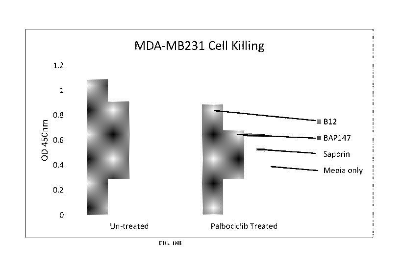
[0087] FIG. 14C shows FACS cell sorting of untreated MDA-MB231 cells that were
B-gal staining
positive.
[0088] FIG. 14D shows FACS cell sorting of MDA-MB231 cells treated with
Palbociclib Isethionate that
were B-gal staining positive.
[0089] FIG. 15A shows FACS sorting of untreated MDA-MB231 cells.
[0090] FIG. 15B shows FACS sorting of MDA-MB231 cells treated with Palbociclib
Isethionate.
[0091] FIG. 16A shows FACS cell sorting of untreated MDA-MB468 cells that were
B-gal staining
negative.
[0092] FIG. 16B shows FACS cell sorting of MDA-MB468 cells treated with
Palbociclib Isethionate that
were B-gal staining negative.
[0093] FIG. 16C shows FACS cell sorting of untreated MDA-MB468 cells that were
B-gal staining
positive.
[0094] FIG. 16D shows FACS cell sorting of MDA-MB468 cells treated with
Palbociclib Isethionate that
were B-gal staining positive.
[0095] FIG. 17A shows FACS sorting of untreated MDA-MB468 cells.
[0096] FIG. 17B shows FACS sorting of MDA-MB468 cells treated with Palbociclib
Isethionate.
[0097] FIG. 18A shows CD73 expression levels in MDA-MB231 and MDA-MB468 cells
before and after
the Palbociclib Isethionate treatment.
[0098] FIG. 18B shows senescent cell killing by an anti-CD73 conditionally
active antibody.
DEFINITIONS
[0099] In order to facilitate understanding of the examples provided herein,
certain frequently occurring
methods and/or terms will be defined herein.
11
CA 03048660 2019-06-26
WO 2018/129007 PCT/US2018/012136
[0100] The definitions of the terms "about," "activity," "agent," "ambiguous
base requirement," "amino
acid," "amplification," "chimeric property," "cognate," "comparison window,"
"conservative amino acid
substitutions," "corresponds to," "degrading effective," "defined sequence
framework," "digestion,"
"directional ligation," "DNA shuffling," "drug" or "drug molecule," "effective
amount," "electrolyte,"
"epitope," "enzyme," "evolution" or "evolving," "fragment," "derivative,"
"analog," "full range of single
amino acid substitutions," "gene," "genetic instability," "heterologous,"
"homologous" or "homeologous,"
"industrial applications," "identical" or "identity," "areas of identity,"
"isolated," "isolated nucleic acid,"
"ligand," "ligation," "linker" or "spacer," "microenvironment," "molecular
property to be evolved,"
"mutations," "naturally-occurring," "normal physiological conditions" or "wild
type operating conditions,"
"nucleic acid molecule," "nucleic acid molecule," "nucleic acid sequence
coding for" or "DNA coding
sequence of" or a "nucleotide sequence encoding," "promotor sequence,"
"nucleic acid encoding an enzyme
(protein)" or "DNA encoding an enzyme (protein)" or "polynucleotide encoding
an enzyme (protein),"
"specific nucleic acid molecule species," "assembling a working nucleic acid
sample into a nucleic acid
library," "nucleic acid library," "nucleic acid construct" or "nucleotide
construct" or "DNA construct,"
"construct," "oligonucleotide" or "oligo," "homologous," "operably linked,"
"operably linked to," "parental
polynucleotide set," "patient" or "subject," "physiological conditions,"
"population," "pro-form," "pre-pro-
form," "pseudorandom," "quasi-repeated units," "random peptide library,"
"random peptide sequence,"
"receptor," "recombinant," "synthetic," "related polynucleotides," "reductive
reassortment," "reference
sequence," "comparison window," "sequence identity," "percentage of sequence
identity," "substantial
identity," "reference sequence," "repetitive index (RI)", "restriction site,"
"selectable polynucleotide,"
"sequence identity," "similarity," "specifically bind," "specific
hybridization," "specific polynucleotide,"
"stringent hybridization conditions," "substantially identical,"
"substantially pure enzyme," "substantially
pure," "treating," "variable segment," "variant," "wild-type," "wild-type
protein" or "wild-type biologic
protein," "parent molecule" or "target protein," "working," "conditionally
active antibody," "antibody-
dependent cell-mediated cytotoxicity" or "ADCC," "cancer" and "cancerous,"
"multispecific antibody," "full
length antibody," "library," "recombinant antibody," and "individual" or
"subject" are the same as in WO
2016/138071.
[0101] The term "antibody", as used herein, refers to intact immunoglobulin
molecules, as well as
fragments of immunoglobulin molecules, such as Fab, Fab', (Fab')2, Fv, and SCA
fragments, that are capable
of binding to an epitope of an antigen. These antibody fragments, which retain
some ability to selectively
bind to an antigen (e.g., a polypeptide antigen) of the antibody from which
they are derived, can be made
using well known methods in the art (see, e.g., Harlow and Lane, supra), and
are described further, as
follows. Antibodies useful in the practice of the claimed invention may be
IgGl, IgG2, IgG3, IgG4, IgM,
IgAl, IgA2, sIgA, IgD or IgE. Antibodies can be used to isolate preparative
quantities of the antigen by
immunoaffinity chromatography. Various other uses of such antibodies are to
diagnose and/or stage disease
(e.g., neoplasia) and for therapeutic application to treat disease, such as
for example: neoplasia, autoimmune
12
CA 03048660 2019-06-26
WO 2018/129007 PCT/US2018/012136
disease, AIDS, cardiovascular disease, infections, and the like. Chimeric,
human-like, humanized or fully
human antibodies are particularly useful for administration to human patients.
[0102] An Fab fragment consists of a monovalent antigen-binding fragment of an
antibody molecule, and
can be produced by digestion of a whole antibody molecule with the enzyme
papain, to yield a fragment
consisting of an intact light chain and a portion of a heavy chain.
[0103] An Fab' fragment of an antibody molecule can be obtained by treating a
whole antibody molecule
with pepsin, followed by reduction, to yield a molecule consisting of an
intact light chain and a portion of a
heavy chain. Two Fab' fragments are obtained per antibody molecule treated in
this manner.
[0104] An (Fab')2 fragment of an antibody can be obtained by treating a whole
antibody molecule with the
enzyme pepsin, without subsequent reduction. A (Fab')2 fragment is a dimer of
two Fab' fragments, held
together by two disulfide bonds.
[0105] An Fy fragment is defined as a genetically engineered fragment
containing the variable region of a
light chain and the variable region of a heavy chain expressed as two chains.
[0106] A single chain antibody ("SCA" or scFv) is a genetically engineered
single chain molecule
containing the variable region of a light chain and the variable region of a
heavy chain, linked by a suitable,
flexible polypeptide liner, and which may include additional amino acid
sequences at the amino- and/or
carboxyl- termini. For example, a single chain antibody may include a tether
segment for linking to the
encoding polynucleotide. A functional single chain antibody generally contains
a sufficient portion of the
variable region of a light chain and a sufficient region of the variable
region of a heavy chain so as to retain
the property of a full-length antibody for binding to a specific target
molecule or epitope.
[0107] The term "antigen" or "Ag" as used herein is defined as a molecule that
is capable of triggering an
immune response. This immune response may involve either antibody production,
or the activation of
specific immunologically-competent cells, or both. A person skilled in the art
will understand that any
macromolecule, including virtually all proteins or peptides, can serve as an
antigen. It is readily apparent
that an antigen can be generated, synthesized or can be derived from a
biological sample. Such a biological
sample can include, but is not limited to a tissue sample, a tumor sample, a
cell or a biological fluid.
[0108] The term "apoptosis", as used herein, refers to a mechanism of cell
death affecting single cells,
marked by shrinkage of the cell, condensation of chromatin, and fragmentation
of the cell into membrane-
bound bodies that are eliminated by phagocytosis. The term "apoptosis" is
often used synonymously with the
term "programmed cell death".
[0109] The term "apoptosis-inducing activity", as used herein, refers to the
intrinsic property of a
compound to selectively invoke apoptosis in a (i) particular cell type and/or
(ii) cell in a particular stage of
development or differentiation, due to internal or external stimuli. A skilled
person is aware of the existence
of in vitro standard assays for determining apoptosis-inducing activity of a
compound in a cell culture, for
example tests that assess levels of cytoplasmic Cytochrome C (marker for
apoptosis) and levels of TUNEL
(marker for apoptosis). Using these standard assays, the skilled person can
easily assess and compare the
apoptosis-inducing activity of different compounds with regard to different
cell type or cells in a different
13
CA 03048660 2019-06-26
WO 2018/129007 PCT/US2018/012136
developmental stage, e.g. senescent vs. non-senescent cells. Other standard
apoptosis assays are an Annexin
V assay and a cleaved caspase-3 staining.
[0110] The term "biosimilar" or "follow-on biologic" is used in a manner that
is consistent with the
working definition promulgated by the U.S. Food and Drug Administration (FDA),
which defines a
biosimilar to be a product that is "highly similar" to a reference product
(despite minor differences in
clinically inactive components). In practice, there can be no clinically
meaningful differences between the
reference product and the biosimilar product in terms of safety, purity, and
potency (Public Health Service
(PHS) Act 262). A biositnilar can also be one that satisfies one or more
guidelines adopted May 30, 2012
by the Committee for Medicinal Products for Human Use (CIIMP) of the European
Medicines Agency and
published by the European Union as "Guideline on similar biological medicinal
products containing
monoclonal antibodies¨ non-clinical and clinical issues" (Document Reference
EMAICHMP/BMWP/403543/2010). For example, a "biosimilar antibody" refers to a
subsequent version of
an innovator's antibody (reference antibody) typically made by a different
company. Differences between a
biosimilar antibody and a reference antibody can include post-translational
modification, e.g. by attaching to
the antibody other biochemical groups such as a phosphate, various lipids and
carbohydrates; by proteolytic
cleavage following translation; by changing the chemical nature of an amino
acid (e.g., formylation); or by
many other mechanisms. Other post-translational modifications can be a
consequence of manufacturing
process operations¨ for example, glycation may occur with exposure of the
product to reducing sugars. In
some cases, storage conditions may be permissive for certain degradation
pathways such as oxidation,
deamidation, or aggregation to occur. As all of these product-related variants
may be included in a
biosimilar antibody.
[0111] The terms "cancer" and "cancerous" refer to or describe the
physiological condition in mammals that
is typically characterized by unregulated cell growth/proliferation. A "tumor"
comprises one or more
cancerous cells. Examples of cancer include, but are not limited to,
carcinoma, lymphoma, blastoma,
sarcoma, and leukemia or lymphoid malignancies. More particular examples of
such cancers include
squamous cell cancer (e.g., epithelial squamous cell cancer), lung cancer
including small cell lung cancer,
non-small cell lung cancer ("NSCLC"), adenocarcinoma of the lung and squamous
carcinoma of the lung,
cancer of the peritoneum, hepatocellular cancer, gastric or stomach cancer
including gastrointestinal cancer,
pancreatic cancer, glioblastoma, cervical cancer, ovarian cancer, liver
cancer, bladder cancer, hepatoma,
breast cancer, colon cancer, rectal cancer, colorectal cancer, endometrial or
uterine carcinoma, salivary gland
carcinoma, kidney or renal cancer, prostate cancer, vulvar cancer, thyroid
cancer, hepatic carcinoma, anal
carcinoma, penile carcinoma, as well as head and neck cancer.
[0112] The term "conditionally active protein" refers to a variant, or mutant,
of a parent protein which is
more or less active under one or more aberrant conditions as compared to the
same activity of a control or
under a normal physiological condition. This conditionally active protein also
exhibits activity in selected
regions of the body and/or exhibits increased or decreased activity under
aberrant, or permissive,
physiological conditions. Normal physiological conditions are those which
would be considered within a
14
CA 03048660 2019-06-26
WO 2018/129007 PCT/US2018/012136
normal range at a location in a subject such as at the site of administration,
or at the tissue or organ at the site
of action, in a subject. An aberrant condition is that which deviates from the
normally acceptable range for
that condition at that location. In one aspect, the conditionally active
protein is virtually inactive at a normal
physiological condition but is active at the aberrant or permissive condition.
For example, in one aspect, an
evolved conditionally active protein is virtually inactive at body
temperature, but is active at lower or higher
temperatures. In another aspect, the conditionally active protein may be
reversibly or irreversibly inactivated
at the normal physiological or control condition. In a further aspect, the
conditionally active protein is a
therapeutic protein. In another aspect, the conditionally active protein is
used as a drug, or therapeutic agent.
In yet another aspect, the conditionally active protein is more or less active
in highly oxygenated blood, such
as, for example, after passage through the lung or in the lower pH
environments found in the kidney. A
conditionally active protein may be a conditionally active biologic protein.
[0113] As used herein, the term "cyclic peptide" refers to a polypeptide chain
whose amino and carboxyl
termini are themselves linked together with a peptide bond that forms a
circular chain (i.e., between the
alpha carboxyl of one residue and the alpha amine of another). For purposes of
this application, cyclic
peptides may also include a linkage other than a peptide bond such as non-
alpha amide linkage, and a
thioether linkage between Trp and Cys residues. The length of the cyclic
peptide may be in the range of
from about 5 to about 500 amino acids, or from about 8 to about 300 amino
acids, or from about 8 to about
200 amino acids, or from about 10 to about 100 amino acids, or from about 10
to about 50 amino acids.
Additionally, amino acids other than naturally-occurring amino acids, for
example 13-alanine, phenyl glycine
and homoarginine, may be included in the cyclic peptides.
[0114] The abbreviation "DRI", as used herein, refers to the D retro inverso
isoform of an L-peptide, in
which the amino acid sequence is reversed in comparison with a fragment or the
full-length of a natural or
wild-type protein, and at least a portion of the amino acid residues in the
DRI peptide are D amino acid
residues instead of the L amino acid residues in the natural or wild-type
protein (FIG. 7). The D retro
inverso peptide can be made by identifying the amino acid sequence of a
fragment or the full-length of a
natural protein, reversing the sequence and synthesizing the D retro reverse
peptide using known methods to
provide a peptide having the reverse of the amino acid sequence of the
fragment or the full-length of a
natural protein and including in the D retro reverse peptide a sufficient
number of D amino acids to provide
the desired function.
[0115] The terms "diseases or conditions where the removal of senescent cells
is beneficial", "diseases or
conditions associated with the presence of senescent cells" and "disorders
where the removal of senescent
cells is beneficial" are used interchangeable, referring to any disease or
condition in a mammalian, for
example a human, subject where removal or clearance or reduced viability of
senescent cells is beneficial to
the subject suffering from said disease or condition. The term encompasses the
situation where senescent
cells are one, or the only, cause of a disease or contribute to the
progression of a disease. The term further
relates to the situation where senescent cells might become, in the future,
the cause of a disease or condition
in said subject. For example, the treatment of a disease or condition where
the removal of senescent cells is
CA 03048660 2019-06-26
WO 2018/129007 PCT/US2018/012136
beneficial, is a disease or condition prevented, preventable or ameliorated by
removing senescent cells. For
example, it is known that chemotherapeutic agents and radiation therapy induce
cellular senescence. It is
advantageous to remove these senescent cells in order to prevent the onset of
diseases or conditions
associated with cellular senescence. The term further encompasses diseases or
conditions where removal of
senescent cells alleviates or reduces symptoms of a disease or condition.
[0116] Removal of senescent cells is beneficial if inter alia the disease or
condition can be healed,
prevented or if the symptoms of the disease or condition or can be reduced or
alleviated. Removal of
senescent cells may be achieved by induction of apoptosis in the senescent
cells. For example, the disease or
condition where the removal of senescent cells is beneficial, is selected from
the group formed by
atherosclerosis, chronic inflammatory diseases such as arthritis or arthrosis,
cancer, osteoarthritis, diabetes,
diabetic ulcers, kyphosis, sclerosis, hepatic insufficiency, cirrhosis,
Hutchinson-Gilford progeria syndrome
(HGPS), laminopathies, osteoporosis, dementia, (cardio)vascular diseases,
obesity, metabolic syndrome,
acute myocardial infarction, emphysema, insulin sensitivity, boutonneuse
fever, sarcopenia,
neurodegenerative diseases such as Alzheimer's , Huntington's or Parkinson's
disease, cataracts, anemia,
hypertension, fibrosis, age-related macular degeneration, COPD, asthma, renal
insufficiency, incontinence,
hearing loss such as deafness, vision loss such as blindness, sleeping
disturbances, pain such as joint pain or
leg pain, imbalance, fear, depression, breathlessness, weight loss, hair loss,
muscle loss, loss of bone density,
frailty and/or reduced fitness. A disease or condition where the removal of
senescent cells is beneficial is a
disease or condition associated with or linked to inflammation, specifically
chronic inflammation, in a
mammalian, for example human, subject, where said inflammation is caused or
mediated by senescent cells.
In some embodiments, said senescent cells causing or mediating said
inflammation are at least partially co-
localized in the same organ, more preferably in the same tissue, as the organ
or tissue, affected by said
disease or condition.
[0117] The term "diseases or conditions associated with the presence of
senescent cells", as used herein,
refers to any disease or condition in a mammalian, for example human, subject
where the presence of
senescent cells, or presence of cellular senescence, in a mammalian, for
example human, subject is linked to
said disease or condition in said subject. In this context, "linked to" can
inter alia refer to the senescent cells
or cellular senescence (i) as the at least partial cause of a disease or
condition, (ii) or as at least a partial
cause of a symptom. In some embodiments, the disease or condition associated
with the presence of
senescent cells, is selected from the group formed by atherosclerosis, chronic
inflammatory diseases such as
arthritis or arthrosis, cancer, osteoarthritis, diabetes, diabetic ulcers,
kyphosis, sclerosis, hepatic
insufficiency, cirrhosis, Hutchinson-Gilford progeria syndrome (HGPS),
laminopathies, osteoporosis,
dementia, (cardio)vascular diseases, obesity, metabolic syndrome, acute
myocardial infarction, emphysema,
insulin sensitivity, boutonneuse fever, sarcopenia, neurodegenerative diseases
such as Alzheimer's,
Huntington's or Parkinson's disease, cataracts, anemia, hypertension,
fibrosis, age-related macular
degeneration, COPD, asthma, renal insufficiency, incontinence, hearing loss
such as deafness, vision loss
such as blindness, sleeping disturbances, pain such as joint pain or leg pain,
imbalance, fear, depression,
16
CA 03048660 2019-06-26
WO 2018/129007 PCT/US2018/012136
breathlessness, weight loss, hair loss, muscle loss, loss of bone density,
frailty and/or reduced fitness. A
specific disease or condition where the removal of senescent cells is
beneficial is a disease or condition
associated with or linked to inflammation, typically chronic inflammation, for
example in a mammalian,
such as human, subject, where said inflammation is caused or mediated by
senescent cells. In some
embodiments, said senescent cells causing or mediating said inflammation is at
least partially co-localized in
the same organ, for example in the same tissue, as the organ or tissue,
affected by said disease or condition.
[0118] The term "extracellular condition of a senescent cell" as used herein
refers to a condition in the
extracellular environment immediately surrounding one or more senescent cells
and which differs from the
same condition surrounding non-senescent cells. The extracellular environment
of a senescent cell can
include, for example, any extracellular matrix or fluid adjacent to the
senescent cell.
[0119] The terms "FOX04 peptide" and "FOX04 protein" as used herein refer to a
protein translated from
a transcript of the forkhead box protein 04 (FOX04) gene. The FOX04 has two
variants (SEQ ID NO:1 and
SEQ ID NO:2). The term "FOX04 DRI peptide" refers to a D retro inverso peptide
that has the reverse
amino acid sequence of at least a fragment of the FOX04 protein and contains
some, for example all D
amino acid residues.
[0120] The term "full length antibody" refers to an antibody which comprises
an antigen-binding variable
region (VI) or VI) as well as a light chain constant domain (CL) and heavy
chain constant domains, CH1,
CH2 and CH3. The constant domains may be native sequence constant domains
(e.g. human native sequence
constant domains) or amino acid sequence variants thereof. Depending on the
amino acid sequence of the
constant domain of their heavy chains, full length antibodies can be assigned
to different "classes". There
are five major classes of full length antibodies: IgA, IgD, IgE, IgG, and IgM,
and several of these may be
further divided into "subclasses" (isotypes), e.g., IgGl, IgG2, IgG3, IgG4,
IgA, and IgA2. The heavy-chain
constant domains that correspond to the different classes of antibodies are
called alpha, delta, epsilon,
gamma, and mu, respectively.
[0121] An "individual" or "subject" is a mammal. Mammals include, but are not
limited to, domesticated
animals (e.g., cows, sheep, cats, dogs, and horses), primates (e.g., humans
and non-human primates such as
monkeys), rabbits, and rodents (e.g., mice and rats).
[0122] The term "library" as used herein refers to a collection of proteins in
a single pool. The library may
be generated using DNA recombinant technology. For example, a collection of
cDNAs or any other protein
coding DNAs may be inserted in an expression vector to generate a protein
library. A collection of cDNAs
or protein coding DNAs may also be inserted into a phage genome to generate a
bacteriophage display
library of wild-type proteins. The collection of cDNAs may be produced from a
selected cell population or a
tissue sample, such as by the methods disclosed by Sambrook et al. (Molecular
Cloning, Cold Spring Harbor
Laboratory Press, 1989). cDNA collections from selected cell types are also
commercially available from
vendors such as Stratagene . The library of wild-type proteins as used herein
is not a collection of
biological samples.
17
CA 03048660 2019-06-26
WO 2018/129007 PCT/US2018/012136
[0123] The term "ligand" as used herein refers to a molecule that is
recognized by a particular receptor and
specifically binds the receptor in one or more binding sites. Examples of
ligands include, but not restricted
to, agonists and antagonists for cell membrane receptors, toxins and venoms,
viral epitopes, hormones,
hormone receptors peptides, enzymes, enzyme substrates, co factors, drugs
(e.g. opiates, steroids, etc.),
lectins, sugars, polynucleotides, nucleic acids, oligosaccharides, proteins,
and monoclonal antibodies.
Typically, a ligand comprises two structural portions: a first portion that is
involved in binding of the ligand
to its receptor and a second portion that is not involved in such binding.
[0124] The term "multispecific antibody" as used herein is an antibody having
binding specificities for at
least two different epitopes. Exemplary multispecific antibodies may bind both
a BBB-R and a brain
antigen. Multispecific antibodies can be prepared as full-length antibodies or
antibody fragments (e.g.
F(ab1)2bispecific antibodies). Engineered antibodies with two, three or more
(e.g. four) functional antigen
binding sites are also contemplated (see, e.g., US 2002/0004587 Al).
[0125] The term "non-naturally occurring amino acid" as used herein refers to
any amino acid that is not
found in nature. Non-natural amino acids include any D-amino acids, amino
acids with side chains that are
not found in nature, and peptidomimetics. Examples of peptidomimetics include,
but are not limited to, b-
peptides, g-peptides, and d-peptides; oligomers having backbones which can
adopt helical or sheet
conformations, such as compounds having backbones utilizing bipyridine
segments, compounds having
backbones utilizing solvophobic interactions, compounds having backbones
utilizing side chain interactions,
compounds having backbones utilizing hydrogen bonding interactions, and
compounds having backbones
utilizing metal coordination. Non-naturally occurring amino acids also include
residues that have side chains
that resist non-specific protein adsorption, which may be designed to enhance
the presentation of the
antimicrobial peptide in biological fluids, and/or polymerizable side chains,
which enable the synthesis of
polymer brushes using the non-natural amino acid residues within the peptides
as monomeric units.
[0126] The term "parent protein" as used herein refers to a polypeptide or
protein that may be evolved to
produce a conditionally active polypeptide or protein using the methods of the
present invention. The parent
protein may be a wild-type protein or a non-naturally occurring protein. For
example, a therapeutic
polypeptide or protein or a mutant or variant polypeptide or protein may be
used as a parent polypeptide or
protein. Parent protein may also be a fragment of another naturally occurring
protein, wild-type protein,
therapeutic protein or mutant protein. Examples of parent proteins include
antibodies, antibody fragments,
enzymes, enzyme fragments cytokines and fragments thereof, hormones and
fragments thereof, ligands and
fragments thereof, receptors and fragments thereof, regulatory proteins and
fragments thereof, and growth
factors and fragments thereof.
[0127] The term "polypeptide" as used herein refers to a polymer in which the
monomers are amino acids
and are joined together through peptide or disulfide bonds. A polypeptide may
be a full-length naturally-
occurring amino acid chain or a fragment, mutant or variant thereof, such as a
selected region of the amino
acid chain that is of interest in a binding interaction. A polypeptide may
also be a synthetic amino acid
chain, or a combination of a naturally-occurring amino acid chain or fragment
thereof and a synthetic amino
18
CA 03048660 2019-06-26
WO 2018/129007 PCT/US2018/012136
acid chain. A fragment refers to an amino acid sequence that is a portion of a
full-length protein, and will be
typically between about 8 and about 500 amino acids in length, preferably
about 8 to about 300 amino acids,
more preferably about 8 to about 200 amino acids, and even more preferably
about 10 to about 50 or 100
amino acids in length. Additionally, amino acids other than naturally-
occurring amino acids, for example 13-
alanine, phenyl glycine and homoarginine, may be included in the polypeptides.
Commonly-encountered
amino acids which are not gene-encoded may also be included in the
polypeptides. The amino acids may be
either the D- or L-optical isomer. The D-isomers are preferred for use in a
specific context, further described
below. In addition, other peptidomimetics are also useful, e.g. in linker
sequences of polypeptides (see
Spatola, 1983, in Chemistry and Biochemistry of Amino Acids. Peptides and
Proteins, Weinstein, ed.,
Marcel Dekker, New York, p. 267). In general, the term "protein" is not
intended to convey any significant
difference from the term "polypeptide" other than to include structures which
comprise two or several
polypeptide chains held together by covalent or non-covalent bonds.
[0128] The term "protein" as used herein refers to a polymer in which the
monomers are amino acids and
are joined together through peptide or disulfide bonds. A protein may be a
full-length naturally-occurring
amino acid chain or a fragment, mutant or variant thereof, such as a selected
region of the amino acid chain
that is of interest in a binding interaction. A protein may be a cyclic
peptide with the amino acid polymer
forming a cyclic structure using the entire or part of the polymer. A protein
may also be a synthetic amino
acid chain, an amino acid chain containing a non-natural amino acid or a
combination of a naturally-
occurring amino acid chain or fragment thereof and a synthetic amino acid
chain. A fragment refers to an
amino acid sequence that is a portion of a full-length protein, and will be
typically between about 8 and
about 500 amino acids in length, preferably about 8 to about 300 amino acids,
more preferably about 8 to
about 200 amino acids, and even more preferably about 10 to about 50 or 100
amino acids in length.
Additionally, amino acids other than naturally-occurring amino acids, for
example 13-alanine, phenyl glycine
and homoarginine, may be included in the polypeptides. Commonly-encountered
amino acids which are not
gene-encoded may also be included in the polypeptides. The amino acids may be
either the D- or L- optical
isomer. The D-isomers are preferred for use in a specific context, further
described below. In addition, other
peptidomimetics are also useful, e.g. in linker sequences of polypeptides (see
Spatola, 1983, in Chemistry
and Biochemistry of Amino Acids. Peptides and Proteins, Weinstein, ed., Marcel
Dekker, New York, p.
267). In general, the term "protein" is not intended to convey any significant
difference from the term
"polypeptide" other than to include structures which comprise two or several
polypeptide chains held
together by covalent or non-covalent bonds.
[0129] The term "receptor" as used herein refers to a molecule that has an
affinity for a given ligand.
Receptors can be naturally occurring or synthetic molecules. Receptors can be
employed in an unaltered
state or as aggregates with other species. Receptors can be attached,
covalently or non-covalently, to a
binding member, either directly or via a specific binding substance. Examples
of receptors include, but are
not limited to, antibodies, including monoclonal antibodies and antisera
reactive with specific antigenic
determinants (such as on viruses, cells, or other materials), cell membrane
receptors, complex carbohydrates
19
CA 03048660 2019-06-26
WO 2018/129007 PCT/US2018/012136
and glycoproteins, enzymes, and hormone receptors. The binding of a ligand to
its receptor indicates a
combination of the ligand and its receptor molecule through specific molecular
recognition to form a
complex, which can be detected by a variety of ligand receptor binding assays
known to a skilled person.
[0130] The term "senescence" or "cellular senescence" as used herein means the
progression from an
actively dividing cell to a metabolically active, non-dividing cell. The term
"senescence" also refers to the
state cells enter after multiple rounds of division and in which state future
cell division is prevented from
occurring even though the cell remains metabolically active.
[0131] The term "senescent cell" as used herein means a cell that is
metabolically active but permanently
withdrawn from the cell cycle (see, e.g., Campisi, Cell, vol. 120, pp.513-522,
2005). Senescent cells do not
replicate and possess one or more of the following additional characteristics
attributed to senescent cells: cell
cycle arrest in the G1 phase; an enlarged, flattened morphology; increased
granularity; staining for 13-
galactosidase activity at pH 6; senescence associated heterochromatic foci;
and characteristic gene
expression that is in part regulated by p16 and p21. Examples of senescent
cells include senescent
preadipocytes, senescent endothelial cells, senescent fibroblasts, senescent
neurons, senescent epithelial
cells, senescent mesenchymal cells, senescent smooth muscle cells, senescent
macrophages, and senescent
chondrocytes.
[0132] The term "senolytic agent" as used herein refers to an agent that
selectively (preferentially or to a
greater degree) destroys, kills, removes, or facilitates selective destruction
of senescent cells. In other words,
the senolytic agent destroys or kills a senescent cell in a biologically,
clinically, and/or statistically
significant manner compared with its capability to destroy or kill a non-
senescent cell. The senolytic agent
may be a small compound or a biological molecule such as proteins,
polynucleotides. A senolytic agent is
used in an amount and for a time sufficient that selectively kills established
senescent cells but is insufficient
to kill (destroy, cause the death of) non-senescent cells in a clinically
significant or biologically significant
number. In certain embodiments, the senolytic agents described herein alter at
least one signaling pathway in
a manner that induces (initiates, stimulates, triggers, activates, promotes)
and results in (i.e., causes, leads to)
death of the senescent cell. The senolytic agent may alter, for example,
either or both of a cell survival
signaling pathway (e.g., Akt pathway) or an inflammatory pathway, for example,
by antagonizing a protein
within the cell survival and/or inflammatory pathway in a senescent cell.
[0133] The term "small molecule" as used herein refers to molecules or ions
that have a molecular weight
of less than 900 a.m.u., or more preferably less than 500 a.m.u. or more
preferably less than 200 a.m.u. or
even more preferably less than 100 a.m.u. In the assays and environments of
the present invention, small
molecules may often be present as a mixture of the molecule and a deprotonated
ion of the molecule,
depending primarily on the pH of the assay or environment.
[0134] The term "target associated with a senescent cell" as used herein means
a molecule, for example a
protein, that is located on the surface of a senescent cell (e.g., a cellular
membrane protein), or present in the
senescent cell, or secreted by the senescent cell into the extracellular
environment of the senescent cell.
CA 03048660 2019-06-26
WO 2018/129007 PCT/US2018/012136
[0135] The term "therapeutic protein" as used herein refers to any protein
and/or polypeptide that can be
administered to a mammal to elicit a biological or medical response of a
tissue, system, animal or human
that is being sought, for instance, by a researcher or clinician. A
therapeutic protein may elicit more than one
biological or medical response. Examples of therapeutic proteins include
antibodies, enzymes, hormones,
cytokines, regulatory proteins, and fragments thereof.
[0136] The term "therapeutically effective amount" as used herein means any
amount which, as compared
to a corresponding subject who has not received such amount, results in, but
is not limited to, healing,
prevention, or amelioration of a disease, disorder, or side effect, or a
decrease in the rate of advancement of
a disease or disorder. The term also includes within its scope amounts
effective to enhance normal
physiological function as well as amounts effective to cause a physiological
function in a patient which
enhances or aids in the therapeutic effect of a second pharmaceutical agent.
[0137] The terms "treat" and "treatment" as used herein refer to medical
management of a disease, disorder,
or condition of a subject (i.e., patient) (see, e.g., Stedman's Medical
Dictionary). In general, an appropriate
dose and treatment regimen provide the senolytic agent in an amount sufficient
to provide therapeutic and/or
prophylactic benefit. Therapeutic benefit for subjects to whom the senolytic
agents described herein are
administered, includes, for example, an improved clinical outcome, wherein the
object is to prevent or slow
or retard (lessen) an undesired physiological change associated with the
disease, or to prevent or slow or
retard (lessen) the expansion or severity of such disease.
[0138] The term "tumor microenvironment" as used herein refers to a
microenvironment in and
surrounding a solid tumor to support the growth and metastasis of the tumor
cells. The tumor
microenvironment includes surrounding blood vessels, immune cells,
fibroblasts, other cells, soluble factors,
signaling molecules, an extracellular matrix, and mechanical cues that can
promote neoplastic
transformation, support tumor growth and invasion, protect the tumor from host
immunity, foster therapeutic
resistance, and provide niches for dormant metastases to thrive. The tumor and
its surrounding
microenvironment are closely related and interact constantly. Tumors can
influence their microenvironment
by releasing extracellular signals, promoting tumor angiogenesis and inducing
peripheral immune tolerance,
while the immune cells in the microenvironment can affect the growth and
evolution of cancerous cells. See
Swarts et al. "Tumor Microenvironment Complexity: Emerging Roles in Cancer
Therapy," Cancer Res, vol.,
72, pages 2473-2480, 2012; Weber et al., "The tumor microenvironment,"
Surgical Oncology, vol. 21, pages
172-177, 2012; Blagosklonny, "Antiangiogenic therapy and tumor progression,"
Cancer Cell, vol. 5, pages
13-17, 2004; Siemann, "Tumor microenvironment," Wiley, 2010; and Bagley, "The
tumor
microenvironment," Springer, 2010.
DETAILED DESCRIPTION
[0139] It must be noted that as used herein and in the appended claims, the
singular forms "a", "an", and
"the" include plural references unless the context clearly dictates otherwise.
Furthermore, the terms "a" (or
21
CA 03048660 2019-06-26
WO 2018/129007 PCT/US2018/012136
"an"), "one or more," and "at least one" can be used interchangeably herein.
The terms "comprising,"
"including," "having," and "constructed from" can also be used
interchangeably.
[0140] Unless otherwise indicated, all numbers expressing quantities of
ingredients, properties such as
molecular weight, percent, ratio, reaction conditions, and so forth used in
the specification and claims are to
be understood as being modified in all instances by the term "about," whether
or not the term "about" is
present. Accordingly, unless indicated to the contrary, the numerical
parameters set forth in the specification
and claims are approximations that may vary depending upon the desired
properties sought to be obtained by
the present disclosure. At the very least, and not as an attempt to limit the
application of the doctrine of
equivalents to the scope of the claims, each numerical parameter should at
least be construed in light of the
number of reported significant digits and by applying ordinary rounding
techniques. Notwithstanding that
the numerical ranges and parameters setting forth the broad scope of the
disclosure are approximations, the
numerical values set forth in the specific examples are reported as precisely
as possible. Any numerical
value, however, inherently contains certain errors necessarily resulting from
the standard deviation found in
their respective testing measurements.
[0141] It is to be understood that each component, compound, substituent or
parameter disclosed herein is
to be interpreted as being disclosed for use alone or in combination with one
or more of each and every other
component, compound, substituent or parameter disclosed herein.
[0142] It is also to be understood that each amount/value or range of
amounts/values for each component,
compound, substituent or parameter disclosed herein is to be interpreted as
also being disclosed in
combination with each amount/value or range of amounts/values disclosed for
any other component(s),
compounds(s), substituent(s) or parameter(s) disclosed herein and that any
combination of amounts/values
or ranges of amounts/values for two or more component(s), compounds(s),
substituent(s) or parameters
disclosed herein are thus also disclosed in combination with each other for
the purposes of this description.
[0143] It is further understood that each range disclosed herein is to be
interpreted as a disclosure of each
specific value within the disclosed range that has the same number of
significant digits. Thus, a range of
from 1-4 is to be interpreted as an express disclosure of the values 1, 2, 3
and 4. It is further understood that
each lower limit of each range disclosed herein is to be interpreted as
disclosed in combination with each
upper limit of each range and each specific value within each range disclosed
herein for the same
component, compounds, substituent or parameter. Thus, this disclosure to be
interpreted as a disclosure of
all ranges derived by combining each lower limit of each range with each upper
limit of each range or with
each specific value within each range, or by combining each upper limit of
each range with each specific
value within each range.
[0144] Furthermore, specific amounts/values of a component, compound,
substituent or parameter
disclosed in the description or an example is to be interpreted as a
disclosure of either a lower or an upper
limit of a range and thus can be combined with any other lower or upper limit
of a range or specific
amount/value for the same component, compound, substituent or parameter
disclosed elsewhere in the
application to form a range for that component, compound, substituent or
parameter.
22
CA 03048660 2019-06-26
WO 2018/129007 PCT/US2018/012136
[0145] The present invention provides a method for producing a conditionally
active protein having activity
on senescent cells from a parent protein that binds to a target associated
with a senescent cell. The method
comprises the steps of
(i) evolving a DNA encoding the parent protein using one or more evolutionary
techniques to create
mutant DNAs;
(ii) expressing the mutant DNAs to obtain mutant proteins;
(iii) subjecting the mutant proteins to an assay under an extracellular
condition of a senescent cell
and an assay under a normal physiologic condition; and
(iv) selecting the conditionally active protein from the mutant proteins that
exhibits at least one of:
(a) a decrease in an activity in the assay under the normal physiological
condition compared
to the same activity of the parent protein in the same assay, and an increase
in the activity in the
assay under the extracellular condition of the senescent cell compared to the
same activity of the
conditionally active protein in the assay under the normal physiological
condition; and
(b) a decrease in the activity in the assay under the normal physiological
condition
compared to the same activity of the parent protein in the same assay, and an
increase in the activity
in the assay under the extracellular condition of the senescent cell compared
to the same activity of
the parent protein in the assay under the extracellular condition of the
senescent cell.
[0146] The parent protein may be an antibody, a ligand, a receptor, or an
enzyme or a fragment of any of
the foregoing. Examples of ligands include cytokines and fragments thereof,
hormones and fragments
thereof, regulatory proteins and fragments thereof, and growth factors and
fragments thereof.
[0147] In the case of an antibody, ligand, or receptor, the parent protein
binds to the target associated with
the senescent cell and the activity may be the binding activity to the target.
For an enzyme, the parent protein
can use at least a portion of the senescent cell as its substrate and the
activity is the enzymatic activity using
at least a portion of the senescent cell as the substrate.
[0148] In some embodiments, the parent protein may be a therapeutic protein or
a biosimilar.
[0149] The target associated with the senescent cell is typically a protein of
a senescent cell. The target is
in some examples a protein on the cellular membrane of the senescent cell. In
some embodiments, the target
is selected from DEP-1, NTAL, EBP50, STX4, VAMP3, ARMCX-3, LANCL1, B2MG, PLD3,
and
VPS26A. These proteins are recognized as biomarkers of senescent cells as
described in WO 2015/181526.
In some embodiments, the target is selected from ITGAV, RAC1, ARHGAP1,
RAPGEF1, CRKL,
NCKAP1, CDC42, CAPNS2, EBP, FGF1, ISG20, KITLG, LPHN1, MAG, MEF2C, OSBPL3,
PFN1,
POU5F1, PPP1CB, pl6INK4a, PRKRA, APC, AXL, BCL2L1, CDKN2C, CLYBL, COPG1, DGKA,
GBA3,
GIT2, IGF1, LCMT2, MADCAM1, MAP3K14, MTHFD2, NAIP, NAPG, NNMT, PARK2, PMS2,
PRPF19, PRTG, RAPGEF1, RET, VIT, WEE1, YAP1, and YWHAE.
[0150] In some embodiments, the target is an Fas protein or a death receptor
(DR). Fas is also sometimes
referred to as a tumor necrosis factor receptor superfamily member 6A
(TNFSF6). This is a membrane
receptor that is easily accessible from outside of senescent cells. DRs are
TNF-related apoptosis-inducing
23
CA 03048660 2019-06-26
WO 2018/129007 PCT/US2018/012136
ligands (TRAILs), see Guicciardi et al., "Life and death by death receptors,"
FASEB J. vol. 23, pp. 1625-
1637, 2009. Examples of DRs include DR4 and DRS.
[0.151] In some embodiments, the target associated with the senescent cell is
selected from MDM2, AKT
(AKT1, AKT2, and AKT3), NOTCH3, DcR2 (TNFRSF10D), and a protein of the BCL-2
anti-apoptotic
protein family. The proteins in this family have BH1-BH4 domains (BCL-2 (i.e.,
the BCL-2 protein member
of the BCL-2 anti-apoptotie protein family), BCL-xL, BCL-w, Al, MCL-1, and BCL-
B); or BI-11, BH2, and
BH3 domains (BAX, BAK, and BOK); or a BH3 domain only (BIK, BAD, BID, BIM,
BMF, HRK, NOXA,
and PUMA) (see, e.g., Cory et al., Nature Reviews Cancer, vol. 2, pp. 647-56,
2002; Cory et al., Cancer
Cell, vol. 8, pp. 5-6, 2005; Adams et al, Oncogene, vol. 26, pp. 1324-1337,
2007). More targets associated
with senescent cells suitable for use in the present invention are described
in Althubiti et al, Cell Death and
Disease, vol. 5, p. e1528, 2014.
[0152] In some embodiments, the target associated with the senescent cell is
selected from a misfolded
form of a protein selected from prion protein (PrP), CD38, Notch-1, CD44,
CD59, Fas ligand, TNF receptor,
and EGF receptor as described in US 2016/0115237. The target may also be
pl6INK4a, or a protein selected
from Tables 1-3 of US 2016/0038576.
[0153] In some embodiments, once the target associated with the senescent cell
is selected, the parent
protein that binds to the target may be selected to be an enzyme that binds to
the selected target and uses at
least a portion of the senescent cell as a substrate, or an antibody, ligand,
or receptor that binds to the target.
Some examples of suitable parent proteins for use in the present invention are
described in WO
2016/138071 in the section "Target Wild-type Proteins."
[0154] The parent protein may be selected from a library, as described in WO
2016/138071. In some
embodiments, the parent protein is selected from the library for example by
use of an assay under a
condition with a pH below 7.0, for example, in a pH range of from 5.0 to below
7.0, or from 5.5 to below
7.0, or from 6.0 to below 7.0, or from 6.2 to 6.8.
[0155] In some other embodiments, the parent protein is selected from the
library for example using a
screening solution that does not contain a small molecule having a pKa between
6 and 7.5, preferably
between 6 and 7, and more preferably between 6.2 and 6.8. Examples of such
small molecules are described
in this application.
[0156] In some embodiments, the parent protein is an antibody. The parent
antibody in some embodiments
has one or more favorable characteristics based upon which it is chosen for
use as the parent antibody. For
example, in certain embodiments, the parent antibody may be selected based on
having a good binding
activity at one or more extracellular conditions of a senescent cell such as
at a pH in the range of 5.0 to less
than 7Ø
[0157] In some embodiments, the parent antibody is selected for its binding
activity to a specific epitope.
Selection based on binding activity to a specific epitope may be combined with
one or more other selection
criteria such as selection for good binding activity at one or more
extracellular conditions of a senescent cell.
24
CA 03048660 2019-06-26
WO 2018/129007 PCT/US2018/012136
[0158] In other embodiments, the parent antibody is selected based on
internalization efficiency. Selection
based on internalization efficiency may be combined with one or more other
selection criteria such as
binding activity to a specific epitope or good binding activity at one or more
extracellular conditions of a
senescent cell.
[0159] In other embodiments, the parent antibody may have similar binding
activity and/or characteristics
under both the normal physiological condition and an extracellular condition
of a senescent cell. In such
embodiments, the parent antibody is selected based on having the most similar
binding activity and/or the
most similar combination of one or more characteristics under both the normal
physiological condition and
the extracellular condition of the senescent cell. For example, if the normal
physiological condition and the
extracellular condition of the senescent cell may be pH 7.4 and pH 6.4
respectively, the antibody that has the
most similar binding activity at pH 7.4 and 6.4, may be selected as the parent
antibody over an antibody
having a less similar binding activity at pH 7.4 and 6.4.
[0160] In some embodiments, the parent protein may be a fragment of a
naturally occurring protein. For
Example, the parent protein may be the catalytic domain of an enzyme, the
binding domain of a ligand or
receptor, or the variable region of an antibody. In some embodiments, the
parent protein may be a peptide of
as few as eight amino acid units or a cyclic peptide.
[0161] After the parent protein is selected, a DNA encoding the parent protein
is evolved using a suitable
evolutionary technique to produce mutant DNAs, which may then be expressed to
produce mutant proteins
for screening to identify a conditionally active protein. Suitable techniques
for evolving the DNA encoding
the parent protein, expressing the mutant DNAs to produce mutant proteins, and
screening the mutant
proteins are described in WO 2016/138071.
[0162] Once selected, the conditionally active protein may be optionally
synthesized in "mimetic" and
"peptidomimetic" forms, as described in WO 2016/138071.
[0163] The selected conditionally active protein may also be produced using a
polypeptide expression cell
production host or an organism. To make the production process more efficient,
the DNA encoding the
conditionally active protein may be subjected to codon optimization for the
cell production host or organism.
Codon optimization has been described previously, such as in, Narum et al.,
"Codon optimization of gene
fragments encoding Plasmodium falciparum merzoite proteins enhances DNA
vaccine protein expression
and immunogenicity in mice," Infect. Immun., vol. 69, pp, 7250-3, 2001, which
describes codon-
optimization in the mouse system; Outchkourov et al., "Optimization of the
expression of Equistatin in
Pichia pastoris, protein expression and purification," Protein Expr. Purif.,
vol. 24, pp. 18-24, 2002, which
describes codon-optimization in the yeast system; Feng et al., "High level
expression and mutagenesis of
recombinant human phosphatidylcholine transfer protein using a synthetic gene:
evidence for a C-terminal
membrane binding domain" Biochemistry, vol. 39, pp. 15399-409, 2000, which
describes codon-
optimization in E. coli; Humphreys et al., "High-level periplasmic expression
in Escherichia coli using a
eukaryotic signal peptide: importance of codon usage at the 5' end of the
coding sequence", Protein Expr.
Purif., vol. 20, pp. 252-64, 2000, which describes how codon usage affects
protein secretion in E. coli.
CA 03048660 2019-06-26
WO 2018/129007 PCT/US2018/012136
[0164] The cell production host may be a mammalian cell production host
selected from one of the group
consisting of CHO, HEK293, IM9, DS-I, THP-I, Hep G2, COS, NIH 3T3, C33a, A549,
A375, SK-MEL-28,
DU 145, PC-3, HCT 116, Mia PACA-2, ACHN, Jurkat, MML-1, Ovcar 3, HT 1080, Panc-
1, U266, 769P,
BT-474, Caco-2, HCC 1954, MDA-MB-468, LnCAP, NRK-49F, and SP2/0 cell lines;
and mouse
splenocytes and rabbit PBMC. The mammalian cell production host is for example
selected from a CHO or
HEK293 cell line. In one specific aspect, the mammalian cell production host
is a CHO-S cell line. In
another embodiment, the mammalian cell production host is a HEK293 cell line.
[0165] In some embodiments, the cell production host is a yeast cell, for
example S. cerevisiae yeast cells
or pichia yeast cells. In some embodiments, the cell production host is a
prokaryotic cell such as E. coli
(Owens, R.J. and Young, R.J., J. Immunol. Meth., vol. 168, p.149, 1994;
Johnson S and Bird RE, Methods
Enzymol., vol. 203, p.88, 1991). The conditionally active protein may also be
produced in plant cells or
plants (Firek et al., Plant Mol. Biol., vol. 23, p.861, 1993).
[0166] The conditionally active protein may be modified through a natural
process or using a chemical
modification technique, as described in WO 2016/138071. The conditionally
active protein may be
synthesized using a solid-phase chemical peptide synthesis method, also as
described in WO 2016/138071.
[0167] The conditionally active protein may be selected using assays under an
extracellular condition of a
senescent cell and/or assays under a normal physiological condition. The
selected conditionally active
protein exhibits at least one of:
(a) a decrease in an activity in the assay under the normal physiological
condition compared to the
same activity of the parent protein in the same assay, and an increase in the
activity in the assay under the
extracellular condition of the senescent cell compared to the same activity of
the conditionally active protein
in the assay under the normal physiological condition; and
(b) a decrease in the activity in the assay under the normal physiological
condition compared to the
same activity of the parent protein in the same assay and an increase in the
activity in the assay under the
extracellular condition of the senescent cell compared to the same activity of
the parent protein in the assay
under the extracellular condition of the senescent cell.
[0168] The condition is the same condition but having a different value of
that condition in the assay under
the extracellular condition of the senescent cell as compared to the assay at
the normal physiological
condition, e.g. the condition may be pH, the value of the pH at the normal
physiological condition may be a
pH of 7.2-7.8, or 7.2-7.6 and the value of the pH at the extracellular
condition of the senescent cell may be
pH 6.0-7.0, or 6.2-6.8.
[0169] The activity may be any activity relevant to treatment of any senescent
cell, such as, for example, a
binding activity of a conditionally active antibody to the target, or a
specific epitope, an internalization
efficiency of the protein, or, for an enzyme, the activity may be, for
example, an enzymatic activity of a
conditionally active enzyme on at least a portion of the senescent cell as a
substrate.
[0170] The extracellular condition of a senescent cell is selected from one or
more of the differences caused
in the extracellular environment immediately adjacent to the senescent cell
that are the result of special
26
CA 03048660 2019-06-26
WO 2018/129007 PCT/US2018/012136
characteristic(s) of senescent cells, as compared to, for example the
characteristics of normal cells. One
group of special characteristics of senescent cells that is useful in the
present invention is the metabolic
activities of senescent cells. For example, senescent cells may exhibit one or
more of the following special
characteristics: (1) growth arrest of senescent cells is essentially permanent
and cannot be reversed by
known physiological stimuli; (2) senescent cells increase in size, sometimes
enlarging more than twofold
relative to the size of their non-senescent counterparts; (3) senescent cells
express a senescence-associated 13-
galactosidase (SAP-gal), which partly reflects the increase in lysosomal mass;
(4) many senescent cells
express pl6INK4a, which is not commonly expressed by quiescent or terminally
differentiated cells. (5)
some senescent cells with persistent DNA damaging response (DDR) signaling
harbor persistent nuclear
foci, termed DNA segments, with chromatin alterations reinforcing senescence
(DNA-SCARS such as
dysfunctional telomeres or telomere dysfunction-induced foci (TIF)) and
contain activated DDR proteins
and are distinguishable from transient damage foci; (6) senescent cells
express and may secrete molecules
associated with senescence, which in certain instances may be observed in the
presence of persistent DDR
signaling; and (7) the nuclei of senescent cells lose structural proteins such
as Lamin B 1 or chromatin-
associated proteins such as histones and HMGB1. See, e.g., Freund et al, Mol.
Biol. Cell, vol. 23, pp. 2066-
75, 2012; Davalos et al, J. Cell Biol., vol. 201, pp. 613-29, 2013; Ivanov et
al, J. Cell Biol.,
DOI:10.1083/jcb.201212110, pp. 1-15, 2013; Funayama et al, J. Cell Biol., vol.
175, pp. 869-80, 2006.
[0171] In some embodiments, the extracellular condition of the senescent cell
is a low pH caused by
increased glycolytic metabolism in the senescent cells (James et al.,
"Senescent human fibroblasts show
increased glycolysis and redox homeostasis with extracellular metabolomes that
overlap with those of
irreparable DNA damage, aging, and disease," J Proteome Res., vol. 14, pp.
1854-71, 2015). Glycolysis
involves breaking down glucose to form two pyruvates and two ATP, where the
pyruvate may be converted
to lactate and excreted, thus lowers the pH in the extracellular environment
of the senescent cells (Wiley and
Campisi, "From Ancient Pathways to Aging Cells-Connecting Metabolism and
Cellular Senescence," Cell
Metab., vol. 23, pp. 1013-21, 2016). This is similar to the tumor
microenvironment where the glycolytic
metabolism in cancer cells lowers the pH in tumor microenvironment. Thus, the
extracellular condition of
the senescent cells is may be an acidic pH in a range of from about 5.5 to
about 7.2, or from about 6.0 to
about 7.0, or from about 6.2 to about 7.0, or from about 6.2 to about 6.8, or
from about 6.4 to about 6.8. The
corresponding normal physiological condition is a normal physiological pH in a
range of from about 7.2 to
about 7.8, preferably from about 7.2 to about 7.6, or more preferably from
about 7.4 to about 7.6.
[0172] In some embodiments, the extracellular condition of the senescent cell
may be a low concentration
of deoxynucleotide, in comparison with a normal physiological concentration of
deoxynucleotide in a
normal cellular environment (Wiley and Campisi, "From Ancient Pathways to
Aging Cells-Connecting
Metabolism and Cellular Senescence," Cell Metab., vol. 23, pp. 1013-21, 2016).
Some senescent cells may
have lost the ability to synthesize deoxynucleotide, thus leading to a lower
concentration of deoxynucleotide
in the extracellular environment of a senescent cell, in comparison with the
extracellular concentration of
deoxynucleotide in the extracellular environment of normal cells. Thus, the
extracellular condition of the
27
CA 03048660 2019-06-26
WO 2018/129007 PCT/US2018/012136
senescent cell may be selected to be a lower concentration of a
deoxynucleotide relative to the normal
physiological concentration of the same deoxynucleotide in the extracellular
environment of a normal cell
and the corresponding normal physiological condition is the concentration of
the same deoxynucleotide in
the extracellular environment of the normal cell.
[0173] hi some embodiments, the extracellular condition of the senescent cell
may be a low concentration
of oxygen, in comparison with a physiological concentration of oxygen in the
extracellular environment of a
normal cell (Wiley and Campisi, "From Ancient Pathways to Aging Cells-
Connecting Metabolism and
Cellular Senescence," Cell Metab., vol. 23, pp. 1013-21, 2016). Senescent
cells have an increased oxygen
consumption compared with non-senescent cells, which may cause a lower
concentration of oxygen to be
present in the extracellular environment of the senescent cells as compared to
the extracellular environment
of normal cells. Thus, the extracellular condition of the senescent cell may
be selected to be a lower
concentration of oxygen relative to the normal physiological concentration of
oxygen in the extracellular
environment of a normal cell and the corresponding normal physiological
condition is the concentration of
oxygen in the extracellular environment of the normal cell.
[0174] hi some embodiments, the extracellular condition of the senescent cell
may be a lower ratio of
NAD+/NADH, than the same ratio in the extracellular environment of a normal
cell (Wiley and Campisi,
"From Ancient Pathways to Aging Cells-Connecting Metabolism and Cellular
Senescence," Cell Metab.,
vol. 23, pp. 1013-21, 2016). Thus, the extracellular condition of the
senescent cell may be selected to be a
lower ratio of NAD+/NADH relative to the normal physiological ratio of
NAD+/NADH in the extracellular
environment of a normal cell and the corresponding normal physiological
condition is the normal ratio of
NAD+/NADH in the extracellular environment of the normal cell.
[0175] hi some embodiments, the extracellular condition of the senescent cell
may be an increased
concentration of a redox homeostasis metabolite selected from hypotaurine,
cysteine sulfinic acid, cysteine-
glutathione disulfide, gamma-glutamylalanine, gamma-glutamylmethionine,
pyridoxate, gamma-
glutamylglutamine, and alanine, in comparison with the normal concentration of
the same redox homeostasis
metabolite in the extracellular environment of a normal growing, confluent or
quiescent cell (James et al.,
"Senescent human fibroblasts show increased glycolysis and redox homeostasis
with extracellular
metabolomes that overlap with those of irreparable DNA damage, aging, and
disease," J Proteome Res., vol.
14, pp. 1854-71, 2015). Thus, the extracellular condition of the senescent
cell may be selected to be an
increased concentration of the redox homeostasis metabolite relative to the
normal physiological
concentration of the same redox homeostasis metabolite in the extracellular
environment of a normal cell
that may be selected from a growing, confluent or quiescent cell and the
corresponding normal physiological
condition is the concentration of the redox homeostasis metabolite in the
extracellular environment of the
normal cell.
[0176] hi some embodiments, the extracellular condition of the senescent cell
may be an increased
concentration of a nucleotide metabolite selected from 3-ureidopropionate,
urate, 7-methylguanine, and
hypoxanthine, in comparison with the concentration of the same nucleotide
metabolite in the extracellular
28
CA 03048660 2019-06-26
WO 2018/129007 PCT/US2018/012136
environment of a normal, growing, confluent or quiescent cell (James et al.,
"Senescent human fibroblasts
show increased glycolysis and redox homeostasis with extracellular metabolomes
that overlap with those of
irreparable DNA damage, aging, and disease," J Proteome Res., vol. 14, pp.
1854-71, 2015). Thus, the
extracellular condition of the senescent cell may be selected to be an
increased concentration of the
nucleotide metabolite relative to the normal physiological concentration of
the same nucleotide metabolite in
the extracellular environment of a normal cell that may be selected from a
growing, confluent or quiescent
cell and the corresponding normal physiological condition is the concentration
of the nucleotide metabolite
in the extracellular environment of the normal cell.
[0177] In some embodiments, the extracellular condition of the senescent cell
may be a decreased
concentration of thymidine in comparison with the concentration of thymidine
in the extracellular
environment of a normal growing, confluent or quiescent cell (James et al.,
"Senescent human fibroblasts
show increased glycolysis and redox homeostasis with extracellular metabolomes
that overlap with those of
irreparable DNA damage, aging, and disease," J Proteome Res., vol. 14, pp.
1854-71, 2015). Thus, the
extracellular condition of the senescent cell may be selected to be a
decreased concentration of thymidine
relative to the normal physiological concentration of thymidine in the
extracellular environment of a normal
cell that may be selected from a growing, confluent or quiescent cell and the
corresponding normal
physiological condition is the concentration of thymidine in the extracellular
environment of the normal cell.
[0178] In some embodiments, the extracellular condition of senescent cells may
be a decreased
concentration of a dipeptide selected from glycylisoleucine, glycylvaline,
glycylleucine, isoleucylglycine,
and valylglycine, in comparison with the concentration of the same dipeptide
in the extracellular
environment of a normal growing, confluent or quiescent cell (James et al.,
"Senescent human fibroblasts
show increased glycolysis and redox homeostasis with extracellular metabolomes
that overlap with those of
irreparable DNA damage, aging, and disease," J Proteome Res., vol. 14, pp.
1854-71, 2015). Thus, the
extracellular condition of the senescent cell may be selected to be a
decreased concentration of a dipeptide
relative to the normal physiological concentration of the same dipeptide in
the extracellular environment of a
normal cell that may be selected from a growing, confluent or quiescent cell
and the corresponding normal
physiological condition is the concentration of the same dipeptide in the
extracellular environment of the
normal cell.
[0179] In some embodiments, the extracellular condition of the senescent cell
may be a decreased
concentration of a fatty acid selected from linoleate, dihomo-linoleate, and
10-heptadecenoate, in
comparison with the concentration of the same fatty acid in the extracellular
environment of a normal
growing, confluent or quiescent cell (James et al., "Senescent human
fibroblasts show increased glycolysis
and redox homeostasis with extracellular metabolomes that overlap with those
of irreparable DNA damage,
aging, and disease," J Proteome Res., vol. 14, pp. 1854-71, 2015). Thus, the
extracellular condition of the
senescent cell may be selected to be a decreased concentration of a fatty acid
selected from linoleate,
dihomo-linoleate, and 10-heptadecenoate, relative to the normal physiological
concentration of the same
fatty acid in the extracellular environment of a normal cell that may be
selected from a growing, confluent or
29
CA 03048660 2019-06-26
WO 2018/129007 PCT/US2018/012136
quiescent cell and the corresponding normal physiological condition is the
concentration of the same fatty
acid in the extracellular environment of the normal cell.
[0180] hi some embodiments, the extracellular condition of the senescent cell
may be an increased
concentration of a phospholipid metabolite selected from 2-hydroxypalmitate, 2-
hydroxystearate, 3-
hydroxydecanoate, 3-hydroxyoctanoate, and glycerophosphorylcholine, in
comparison with the
concentration of the same phospholipid metabolite in the extracellular
environment of normal growing,
confluent or quiescent cell (James et al., "Senescent human fibroblasts show
increased glycolysis and redox
homeostasis with extracellular metabolomes that overlap with those of
irreparable DNA damage, aging, and
disease," J Proteome Res., vol. 14, pp. 1854-71, 2015). Thus, the
extracellular condition of the senescent cell
may be selected to be an increased concentration of a phospholipid metabolite
relative to the normal
physiological concentration of the same phospholipid metabolite in the
extracellular environment of a
normal cell that may be selected from a growing, confluent or quiescent cell
and the corresponding normal
physiological condition is the concentration of the same phospholipid
metabolite in the extracellular
environment of the normal cell.
[0181] hi some embodiments, the extracellular condition of the senescent cell
may be an increased
concentration of an amino acid metabolite selected from alanine, C-
glycosyltryptophan, kynurenine,
dimethylarginine, and orthithine, in comparison with the concentration of the
same amino acid metabolite in
the extracellular environment of a normal growing, confluent or quiescent cell
(James et al., "Senescent
human fibroblasts show increased glycolysis and redox homeostasis with
extracellular metabolomes that
overlap with those of irreparable DNA damage, aging, and disease," J Proteome
Res., vol. 14, pp. 1854-71,
2015). Thus, the extracellular condition of the senescent cell may be selected
to be an increased
concentration of an amino acid metabolite relative to the normal physiological
concentration of the same
amino acid metabolite in the extracellular environment of a normal cell that
may be selected from a growing,
confluent or quiescent cell and the corresponding normal physiological
condition is the concentration of the
same amino acid metabolite in the extracellular environment of the normal
cell.
[0182] hi some embodiments, the extracellular condition of the senescent cell
may be a decreased
concentration of phenylpyruvate in comparison with the concentration of
phenylpyruvate in the extracellular
environment of a normal growing, confluent or quiescent cells (James et al.,
"Senescent human fibroblasts
show increased glycolysis and redox homeostasis with extracellular metabolomes
that overlap with those of
irreparable DNA damage, aging, and disease," J Proteome Res., vol. 14, pp.
1854-71, 2015). Thus, the
extracellular condition of the senescent cell may be selected to be a
decreased concentration of
phenylpyruv ate relative to the normal physiological concentration of
phenylpyruvate in the extracellular
environment of a normal cell and the corresponding normal physiological
condition is the concentration of
phenylpyruvate in the extracellular environment of the normal cell.
[0183] hi some embodiments, the extracellular condition of the senescent cell
may be an increased
concentration of a metabolite selected from fumarate, malonate,
eicosapentaenoate and citrate, in
comparison with the concentration of the same metabolite in the extracellular
environment of a normal
CA 03048660 2019-06-26
WO 2018/129007
PCT/US2018/012136
growing, confluent or quiescent cell (James et al., "Senescent human
fibroblasts show increased glycolysis
and redox homeostasis with extracellular metabolomes that overlap with those
of irreparable DNA damage,
aging, and disease," J Proteome Res., vol. 14, pp. 1854-71, 2015). Thus, the
extracellular condition of the
senescent cell may be selected to be an increased concentration of a
metabolite selected from fumarate,
malonate, eicosapentaenoate and citrate relative to the normal physiological
concentration of the same
metabolite in the extracellular environment of a normal cell and the
corresponding normal physiological
condition is the concentration of the same metabolite in the extracellular
environment of the normal cell.
[0184] In some embodiments, the extracellular condition of the senescent cell
may be an increased ratio of
glycerophosphocholine to phosphocholine, in comparison with the same ratio in
the extracellular
environment of normal non-quiescent cells (Gey and Seeger, "Metabolic changes
during cellular senescence
investigated by proton NMR-spectroscopy," Mech Ageing Dev., vol. 134, pp. 130-
8, 2013). Thus, the
extracellular condition of the senescent cell may be selected to be an
increased ratio of
glycerophosphocholine to phosphocholine relative to the same ratio of
glycerophosphocholine to
phosphocholine in the extracellular environment of a normal non-quiescent cell
and the corresponding
normal physiological condition is the ratio of glycerophosphocholine to
phosphocholine in the extracellular
environment of the normal non-quiescent cell.
[0185] Senescent cells secrete a variety of different proteins, which are
collectively called senescence-
associated secretory phenotype (SASP). These secreted proteins include, for
example, GM-CSF, GROa,
GRC-a,b,y, IGFBP-7, 1L-6, IL-7, IL-8, MCP-1, MCP-2, MIP-1a, MMP -1, MMP-10,
MMP-3,
Arnphiregulin, ENA-78, Eotaxin-3, GCP-2, GITR, HGF, ICAM-1, IGTEP-2, IGFBP-4,
ICiFT3P-5, IGIF13P-6,
IL-1.3, IL-I13, MCP-4, MW, MIP -3a, MMP-12, MMP-13, MMP-14, NAP2, oncostatin
M, osteoprotegerin,
PRA:, RANTES, sgp130, TIMP-2, TRAIL-R3, Acrp30, angiogenin, Pod, bEGF, BLC,
BTC, CTACK, EGE-
R, Fas, FGF-7, G-CSF, GDNF, HCC-4, 1-309, IFN-y, IGFBP-1, IGFBP-3, IL-I RI, IL-
n, IL-15, IL-2R-a,
IL-6 R, 1-TAC, leptin, LW, MMP-2, MSP-a, PA1-1, PAI-2, PDGF-BB, SCF, SDF-1,
sTINIF RI, sTNF RH,
thrombopoietin, TIMP-1, tPA, uPA, uPAR, VEGF, MCP-3, IGF-1, TGF-133 MIP-I -
delta, FGF-7,
PDGF-BB, IL-16, BMP-4, MDC, MCP-4, IL-10, T1MP-1, Fit-3 Ligand, ICAM-1, Axl,
CNTF, INF- y, EGF,
and BMP-6. Additional proteins secreted by senescent cells include TGF-2, and
IC/F-2R, IGFBP-3,
IGFBP-
7, TGF-b, WNT2, CXCR2-binding chemoldnes, INNT16B, SFRP2, SPINK1, ENPP5, EREG,
ANGPTL4,
CSGALN ACT, CC1.26, AREG, ANGPT1, CCK, THBD, CXCL14, NOV, GAL, NPPC, FAM150B,
CST],
MUCL1, NPTX2, TMEM155, EDN1, PSG9, ADAMTS3, CD24, PPBP, CXCL3, CST2, PSG8,
PCOLCE2,
PSG7, TNFSF15, 07orf67, CALCA, FGF18, BMP-2, MATN3, TFP1, SERPINI 1, TNFRSF25,
and IL-
23k hi some embodiments, the extracellular condition of the senescent cell is
either a presence or an
increased concentration of one or more of these secreted proteins as compared
to the concentration of the
same protein in the extracellular environment of a normal cell and the normal
physiological condition the
absence of, or a normal physiological concentration of the same secreted
protein(s) in the extracellular
environment of the normal cell.
31
CA 03048660 2019-06-26
WO 2018/129007 PCT/US2018/012136
[0186] The conditionally active protein of the present invention may be used
as a senolytic agent to kill or
remove senescent cells from a subject. The interaction between the
conditionally active protein and the
senescent cell may inhibit or even kill the senescent cell through inhibiting
a cell survival signaling pathway
and/or an inflammatory pathway that are activated during cellular senescence.
Inhibition of the cell survival
signaling pathway and/or inflammatory pathway can induce (i.e., initiate,
trigger, stimulate or in some
manner remove or inhibit suppression of) a cell death pathway, such as an
apoptotic pathway, in the
senescent cell that will lead to the death of the senescent cell.
[0187] Cell survival signaling pathways that are activated during senescence
include the src kinase
signaling pathway, a PI3K/Akt pathway, a PBK/Akt/inTor pathway, a p38/MAPK
pathway, an ERK/MAPK
pathway, a mTOR pathway, an insulin/IGF-1 signaling pathway, and a TGF-I3
signaling pathway.
Inflammatory pathways that are activated during senescence include a p38/MAPK
signaling pathway, an
ERK/MAPK pathway, an src kinase signaling pathway, and an NF-kB pathway.
[0188] The src kinase signaling pathway is involved in regulation of cell
proliferation, differentiation,
apoptosis, cell adhesion, and stress responses (see, e.g., Wang, Oncogene,
vol. 19, pp. 5643-50, 2000 and
Thomas et at, Annu. Rev. Cell Dev. Biol., vol. 13, pp. 513-609, 1997). The src
kinase signaling pathway is
also involved in inflammatory responses, including macrophage mediated immune
responses (see, e.g.,
Byeon et al, Mediators of Inflammation, vol. 2012, article ID 512926, 2012)
and acute inflammatory
responses (see, e.g., Okutani et al, Am. J. PhysioL Lung Cell MoL Physiol.,
vol. 291, pp. L129-L141, 2006).
Accordingly, a conditionally active protein that alters an src kinase
signaling pathway may alter both a
signaling pathway and an inflammatory pathway.
[0189] Altering a signaling pathway and/or an inflammatory pathway of a cell
may affect a function of one
or more downstream proteins or may affect the interaction of one or more
downstream proteins with other
components of the respective cell signaling or inflammatory pathway. For
example, a conditionally active
protein that alters a src kinase signaling pathway or a PBK/Akt pathway may
alter a function of one or more
downstream proteins in the respective pathway or may affect the interaction of
the one or more downstream
proteins with another component of the respective pathway (see, e.g., Example
1; Figures 2B-2D).
Exemplary proteins that are upregulated in senescent cells include P38/MAPK,
ERKI/2, and PBK
(complex). In certain embodiments, the PBK/Akt pathway, which is a cell
signaling pathway, is activated
during senescence and a conditionally active protein described herein inhibits
the PBK/Akt pathway to
enhance or induce apoptosis in the senescent cells.
[0190] The assay solutions for the assay under the extracellular condition of
senescent cell and the assay
under the normal physiological condition for example include a component
selected from citrate buffers
such as sodium citrate, phosphate buffers, bicarbonate buffers such as the
Krebs buffer, phosphate buffered
saline (PBS) buffer, Hank's buffer, Tris buffer, HEPES buffer, etc. Other
buffers known to a person skilled
in the art to be suitable for the assays may be used.
[0191] The assay solutions of the invention may contain at least one component
selected from inorganic
compounds, ions and organic molecules, for example ones that are commonly
found in a bodily fluid of a
32
CA 03048660 2019-06-26
WO 2018/129007 PCT/US2018/012136
mammal such as a human or animal. These inorganic compounds, ions and organic
molecules are described
in detail in WO 2016/138071.
[0192] The conditionally active protein may interact with one or more of the
inorganic compounds, ions,
and organic molecules. Such interactions between the conditionally active
protein and the component may
be selected from inorganic compounds, ions and organic molecules include
hydrogen bond bonding,
hydrophobic interaction, and Van der Waals interactions.
[0193] In some embodiments, the extracellular condition of the senescent cell
is a lower pH in the range of
from 5.5 to 7.2, or from 6.0 to 7.0, or from 6.2 to 6.8, and the normal
physiological condition is the normal
physiological pH, for example in the range of from 7.2 to 7.8. The assay
solutions for pH as the extracellular
condition may include a component with a pKa between the lower pH of the
extracellular condition and the
normal physiological pH. The pKa is for example up to 0.5, 1, 1.5, 2, 2.5, or
3 units away from the lower pH
of the extracellular condition. This component in some embodiments has a
molecular weight of less than 900
a.m.u. and may for example be selected from histidine, histamine, hydrogenated
adenosine diphosphate,
hydrogenated adenosine triphosphate, citrate, bicarbonate, acetate, lactate,
bisulfide, hydrogen sulfide,
ammonium, dihydrogen phosphate and any combination thereof.
[0194] It has been observed that certain conditionally active proteins contain
an increased number (or
proportion) of charged amino acid residues in comparison to the amino acid
residues of the parent protein
from which the conditionally active proteins are derived. There are three
positively charged amino acid
residues: lysine, arginine and histidine; and two negatively charged amino
acid residues: aspartate and
glutamate. These charged amino acid residues are over-represented in certain
conditionally active proteins in
comparison with the parent protein from which the conditionally active
proteins are derived. As a result, the
conditionally active proteins are more likely to interact with charged species
in the assay solution since the
number of charged amino acid residues in the conditionally active proteins has
increased. This, in turn,
influences the activity of the conditionally active proteins.
[0195] It has also been observed that certain conditionally active proteins
typically have different activities
in the presence of different species in the assay solutions. Species that have
at least two ionization states: an
uncharged or less charged state at one value of a condition such as pH and a
charged or more charged state
at different value of the same condition may alter the activity of the
conditionally active protein. The
charged or more charged state of the species may increase the interaction of
the species with charged amino
acid residues present in the conditionally active proteins. This mechanism may
be employed to enhance the
selectivity and/or pH-dependent activity of the conditionally active proteins.
[0196] The nature of the charge(s) on the conditionally active proteins may be
one factor used to determine
suitable species for influencing the activity of the conditionally active
proteins. In some embodiments, the
conditionally active proteins may have more positively charged amino acid
residues: lysine, arginine and
histidine, in comparison with the parent protein. The conditionally active
proteins can thus be selected to
have the desired level of interaction with a particular species present in the
extracellular environment of
33
CA 03048660 2019-06-26
WO 2018/129007 PCT/US2018/012136
senescent cells where the activity is desired and or to have the desired level
of interaction with a particular
species present in the normal physiological condition where a reduced activity
is desired.
[0197] The location of the charged amino acid residues on the conditionally
active proteins may also have
an influence on the activity. For example, the proximity of charged amino acid
residues to a binding site of
the conditionally active proteins may be used to influence the activity of the
conditionally active proteins.
[0198] In some embodiments, it may be the case that the interaction of the
charged species with the
conditionally active proteins may form salt bridges between different moieties
on the protein, especially the
moieties that are charged or polarized. The formation of salt bridges is known
to stabilize polypeptide
structures (Donald, et al., "Salt Bridges: Geometrically Specific, Designable
Interactions," Proteins, 79(3):
898-915, 2011; Hendsch, et al., "Do salt bridges stabilize proteins? A
continuum electrostatic analysis,"
Protein Science, 3:211-226, 1994). The salt bridges can stabilize or fix the
protein structure which normally
undergoes constant minor structural variation called "breathing" (Parak,
"Proteins in action: the physics of
structural fluctuations and conformational changes," Curr Opin Struct Biol.,
13(5):552-557, 2003). The
protein structural "breathing" is important for protein function and its
binding with its partner because the
structural fluctuation permits the conditionally active protein to efficiently
recognize and bind to its partner
(Karplus, et al., "Molecular dynamics and protein functions," PNAS, vol. 102,
pp. 6679-6685, 2015). By
forming salt bridges, the binding site, especially the binding pocket, on the
conditionally active protein may
be less accessible to its partner, possible because the salt bridges may
directly block the partner from
accessing the binding site. Even with salt bridges remote from the binding
site, the allosteric effect may alter
the conformation of the binding site to inhibit binding. Therefore, after the
salt bridges stabilize (fix) the
structure of the conditionally active protein, the protein may become less
active in binding to its partner,
leading to decreased activity.
[0199] One known example of protein and how its structure is stabilized by
salt bridges is hemoglobin.
Structural and chemical studies have revealed that at least two sets of
chemical groups are responsible for
the salt bridges: the amino termini and the side chains of histidines 0146 and
a122, which have pKa values
near pH 7. In deoxyhemoglobin, the terminal carboxylate group of 0146 forms a
salt bridge with a lysine
residue in the a subunit of the other al3 dimer. This interaction locks the
side chain of histidine 0146 in a
position where it can participate in a salt bridge with negatively charged
aspartate 94 in the same chain,
provided that the imidazole group of the histidine residue is protonated (FIG.
2). At a high pH, the side chain
of histidine 0146 is not protonated and the salt bridges do not form. As the
pH drops, however, the side
chain of histidine 0146 becomes protonated, the salt bridge between histidine
0146 and asp artate 1394 forms,
which stabilizes the quaternary structure of deoxyhemoglobin, leading to a
greater tendency for oxygen to be
released at actively metabolizing tissues (with lower pH). The hemoglobin
shows a pH-dependent binding
activity for oxygen where at a low pH, the binding activity for oxygen is
reduced because of the formation
of salt bridges. On the other hand, at a high pH, the binding activity for
oxygen is increased because of the
absence of salt bridges.
34
CA 03048660 2019-06-26
WO 2018/129007 PCT/US2018/012136
[0200] Similarly, small molecules such as bicarbonate may reduce the binding
activity of the conditionally
active protein to its partner by forming salt bridges in the conditionally
active protein. For example, at a pH
lower than its pKa of 6.4, bicarbonate is protonated and thus not charged. The
uncharged bicarbonate is not
capable of forming salt bridges, thus has little effect on the binding of the
conditionally active protein with
its partner. Hence, the conditionally active protein has high binding activity
with its partner at the low pH.
On the other hand, at a high pH greater than the pKa of bicarbonate,
bicarbonate is ionized by losing the
proton, thus becoming negatively charged. The negatively charged bicarbonate
will form salt bridges
between positively charged moieties or polarized moieties on the conditionally
active protein to stabilize the
structure of the conditionally active protein. This will block or reduce the
binding of the conditionally active
protein with its partner. Hence the conditionally active protein has low
activity at the high pH. The
conditionally active protein thus has a pH-dependent activity at the presence
of bicarbonate with higher
binding activity at low pH than at high pH.
[0201] When a species such as bicarbonate is absent from the assay solutions,
the conditionally active
protein may lose its conditional activity. This is likely due to the lack of
salt bridges on the conditionally
active protein to stabilize (fix) the structure of the protein. Thus, the
partner will have similar access to the
binding site on the conditionally active protein at any pH, producing similar
activity at the first pH and
second pH.
[0202] It is to be understood that, though the salt bridges (ion bonds) are
the strongest and most common
manner for the species to affect the activity of the conditionally active
proteins, other interactions between
such species and the conditionally active proteins may also contribute to
stabilize (fix) the structure of the
conditionally active proteins. The other interactions include hydrogen bonds,
hydrophobic interactions, and
van der Waals interactions.
[0203] In some embodiments, to select a suitable compound or ion as the
species, the conditionally active
protein is compared with the parent protein from which it is evolved to
determine whether the conditionally
active protein has a higher proportion of negatively charged amino acid
residues or positively charged amino
acid residues. A compound with a suitable charge at the normal physiological
pH may then be chosen to
influence the activity of the conditionally active protein. For example, when
the conditionally active protein
has a higher proportion of positively charged amino acid residues than the
parent protein, the suitable
compound should typically be negatively charged at the normal physiological pH
to interact with the
conditionally active protein. On the other hand, when the conditionally active
protein has a higher proportion
of negatively charged amino acid residues than the parent protein, the
suitable small molecule should
typically be positively charged at the normal physiological pH to interact
with the conditionally active
protein.
[0204] Thus, a suitable species may be an inorganic or organic molecule that
transits from an uncharged or
less charged state at the lower pH of extracellular condition of senescent
cells to charged or more charged
state at the normal physiological pH. The species should typically have a pKa
between the lower pH and
normal physiological pH. For example, bicarbonate has pKa at 6.4. Thus, at a
higher pH such as pH 7.4, the
CA 03048660 2019-06-26
WO 2018/129007 PCT/US2018/012136
negatively charged bicarbonate will bind to the charged amino acid residues in
the conditionally active
proteins and reduce the activity. On the other hand, at a lower pH such as pH
6.0-6.2, the less charged
bicarbonate will not bind in the same quantity to the conditionally active
proteins and thus allow a higher
activity of the conditionally active proteins.
[0205] Bisulfide has a pKa 7.05. Thus, at a higher pH such as pH 7.4, the more
negatively charged bisulfide
will bind to the positively charged amino acid residues in the conditionally
active proteins and reduce its
activity. On the other hand, at a lower pH such as pH 6.0-6.8, the less
charged hydrogen sulfide/bisulfide
will not bind at the same level to the conditionally active proteins and thus
allow a higher activity of the
conditionally active proteins.
[0206] Some species are selected from bisulfide, hydrogen sulfide, histidine,
histamine, citrate, bicarbonate,
acetate, and lactate. Each of these small molecules has a pKa between 6.2 and
7Ø Other suitable small
molecules may be found in textbooks using the principles of the present
application, such as CRC Handbook
of Chemistry and Physics, 96th Edition, by CRC press, 2015; Chemical
Properties Handbook, McGraw-Hill
Education, 1998.
[0207] The species for example have a low molecular weight and/or a relatively
small conformation to
ensure maximum access to small pockets on conditionally active protein by
minimizing steric hindrance. For
this reason, small molecules typically have a molecular weight of less than
900 a.m.u., or more preferably
less than 500 a.m.u. or more preferably less than 200 a.m.u. or even more
preferably less than 100 a.m.u.
For example, hydrogen sulfide, bisulfide and bicarbonate all have low
molecular weights and small
structures that provide access to pockets on conditionally active protein.
[0208] The concentration of the species in the assay solutions is for example
at or near the physiological
concentration of the species in a subject. For example, the physiological
concentration of bicarbonate (in
human serum) is in the range of 15 to 30 mM. Thus, the concentration of
bicarbonate in the assay solutions
may be from 10 mM to 40 mM, or from 15 mM to 30 mM, or from 20 mM to 25 mM, or
about 20 mM. The
physiological concentration of bisulfide is also low. The concentration of
bisulfide in the assay solutions
may be from 3 to 500 nM, or from 5 to 200 nM, or from 10 to 100 nM, or from 10
to 50 nM.
[0209] The species may be present in the assay solution for the extracellular
condition of senescent cells
and the assay solution for the normal physiological condition at substantially
the same concentration, e.g.
about 20 M for bicarbonate.
[0210] h) some embodiments, the conditionally active protein is pH-dependent
when two or more different
small molecules are present, for example, a combination of bicarbonate and
histidine. Therefore, these two
or more small molecules are present in the assay solutions.
[0211] The species in the assay solutions may be formed in situ from a
component of the assay solutions or
be directly included in the assay solutions. For example, CO2 from the air may
dissolve in the assay
solutions to provide bicarbonate as the species in the assay solutions. For
another example, sodium
dihydrogen phosphate may be added to the assay solution to provide dihydrogen
phosphate as the species in
the assay solutions.
36
CA 03048660 2019-06-26
WO 2018/129007 PCT/US2018/012136
[0212] When the species is absent, the conditionally active proteins may lose
the pH-dependency. Thus, in
the absence of the species, the conditionally active proteins may have similar
activity between the lower pH
of extracellular condition of senescent cells and the normal physiological pH
in the absence of the species.
This same result can be achieved based on any extracellular condition of a
senescent cell that differs from a
normal physiological condition.
[0213] hi some embodiments, the conditionally active protein shows an
increased activity at the lower pH
of an extracellular condition of senescent cells in comparison with the same
activity at the normal
physiological pH, in the presence of an ancillary protein. The ancillary
protein may be a protein present in
blood or human serum. One suitable protein may be albumin, particularly
mammalian albumin, such as
bovine albumin or human albumin.
[0214] hi one aspect, the ancillary protein such as albumin is present in the
assay solutions used for
screening and selecting the conditionally active protein from the mutant
proteins produced by the evolving
step. In another aspect, the assay solutions with the ancillary protein such
as albumin are also used to test the
activity of the selected conditionally active protein under the same or
different conditions.
[0215] hi some embodiments, the two or more of these inorganic compounds,
ions, and organic molecules
discussed in this application are added at substantially the same
concentrations to both assay solutions for
normal physiological condition and extracellular condition of senescent cells.
For example, both bicarbonate
and histidine are added to both assay solutions.
[0216] hi one embodiment, human serum may be added to both assay solutions for
normal physiological
condition and extracellular condition of senescent cells at substantially the
same concentration. Because the
human serum has a large number of inorganic compounds, ions, organic molecules
(including proteins), the
assay solutions will have multiple and large number of components selected
from inorganic compounds,
ions, organic molecules presented at substantially the same concentrations
between the two assay solutions.
[0217] hi some other embodiments, at least one of the two or more components
is added to the assay
solutions for normal physiological condition and extracellular condition of
senescent cells at different
concentrations. For example, both bicarbonate and histidine are added to the
assay solutions. The
bicarbonate concentration may be different between the assay solutions, while
the histidine may have the
same concentration in both assay solutions.
[0218] hi some embodiments, the assay solutions may be designed for selecting
conditionally active
biological proteins with an activity dependent on two or more conditions. In
one exemplary embodiment, the
conditionally active protein may have activity dependent on both pH and
bicarbonate. The assay solutions
for selecting such a conditionally active protein may be an assay solution for
the normal physiological
condition with pH at 7.2-7.6, bicarbonate at a concentration in the range of
from 25 to 30 mM. The assay
solution for the extracellular condition of senescent cells may be with pH at
6.4-6.8, bicarbonate at a
concentration in the range of from 10 to 20 mM. Optionally the assay solutions
for both normal
physiological condition and extracellular condition of senescent cells may
also comprise an ion to assist the
37
CA 03048660 2019-06-26
WO 2018/129007 PCT/US2018/012136
binding between the mutant proteins and the binding partner, thus to increase
the number of hits for
conditionally active proteins.
[0219] hi some embodiments, certain components of serum may be purposely
minimized or omitted from
the assay solutions. For example, when screening antibodies, components of
serum that bind with or adsorb
antibodies can be minimized in or omitted from the assay solutions. Such bound
antibodies may give false
positives thereby including bound mutant antibodies that are not conditionally
active but rather are merely
bound to a component present in serum under a variety of different conditions.
Thus, careful selection of
assay components to minimize or omit components that can potentially bind with
mutant proteins in the
assay may reduce the number of false positive mutant proteins that may be
inadvertently identified as
positive for conditional activity due to binding to a component in the assay
other than the desired binding
partner. For example, in some embodiments where mutant proteins having a
propensity to bind with
components in human serum are being screened, bovine serum albumin may be used
in the assay solution in
order to reduce or eliminate the possibility of false positives caused by
mutant proteins binding to
components of human serum. Other similar replacements can also be made in
particular cases to achieve the
same goal, which is well appreciated by skilled person in the art.
[0220] hi some embodiments, the evolving step may produce mutant proteins that
may simultaneously have
other desired properties besides the conditionally active characteristics
discussed above. Suitable other
desired properties that may be evolved may include binding affinity,
expression, humanization, etc.
Therefore, the present invention may be employed to produce a conditionally
active protein that also has an
improvement in at least one or more of these other desired properties.
[0221] hi some embodiments, the conditionally active protein may be further
mutated using one of the
mutagenesis techniques disclosed herein in, for example, a second evolving
step, to improve another
property of the conditionally active protein such as binding affinity,
expression, humanization, etc. After the
second evolving step, the mutant proteins may be screened for both the
conditional activity and the
improved property.
[0222] hi some embodiments, after evolving the parent protein to produce
mutant proteins, a first
conditionally active protein is selected, which exhibits at least one of:
(a) a decrease in an activity in the assay under the normal physiological
condition compared to the
same activity of the parent protein in the same assay, and an increase in the
activity in the assay under the
extracellular condition of the senescent cell compared to the same activity of
the conditionally active protein
in the assay under the normal physiological condition; and
(b) a decrease in the activity in the assay under the normal physiological
condition compared to the
same activity of the parent protein in the same assay and an increase in the
activity in the assay under the
extracellular condition of the senescent cell compared to the same activity of
the parent protein in the assay
under the extracellular condition of the senescent cell.
38
CA 03048660 2019-06-26
WO 2018/129007 PCT/US2018/012136
[0223] The selected first conditionally active protein may then be further
subjected to one or more
additional evolving, expressing and selecting steps to select at least a
second conditionally active protein that
also exhibits at least one of:
(a) a decrease in an activity in the assay under the normal physiological
condition compared to the
same activity of the parent protein in the same assay, and an increase in the
activity in the assay under the
extracellular condition of the senescent cell compared to the same activity of
the conditionally active protein
in the assay under the normal physiological condition; and
(b) a decrease in the activity in the assay under the normal physiological
condition compared to the
same activity of the parent protein in the same assay and an increase in the
activity in the assay under the
extracellular condition of the senescent cell compared to the same activity of
the parent protein in the assay
under the extracellular condition of the senescent cell. The second activity
may be the same as the first
activity, in which case it is desirable for the second conditionally active
protein to have a larger ratio
between the activity at the extracellular condition and the activity at the
normal physiological condition, in
comparison with the first conditionally active protein. In some embodiments,
the second activity may be a
different activity than the first activity in which case an activity such as
internalization efficiency or binding
to a specific epitope may be the second activity.
[0224] In certain embodiments, the present invention is aimed at producing
conditionally active proteins
with a ratio of the activity at the extracellular condition of the senescent
cell to the activity at the normal
physiological condition greater than 1.0 (e.g., a high selectivity between the
two conditions). The ratio of
activity, or selectivity, at the extracellular condition of the senescent cell
to the activity at the normal
physiological condition may be at least about 1.3:1, or at least about 2:1, or
at least about 3:1, or at least
about 4:1, or at least about 5:1, or at least about 6:1, or at least about
7:1, or at least about 8:1, or at least
about 9:1, or at least about 10:1, or at least about 11:1, or at least about
12:1, or at least about 13:1, or at
least about 14:1, or at least about 15:1, or at least about 16:1, or at least
about 17:1, or at least about 18:1, or
at least about 19:1, or at least about 20:1, or at least about 30:1, or at
least about 40:1, or at least about 50:1,
or at least about 60:1, or at least about 70:1, or at least about 80:1, or at
least about 90:1, or at least about
100:1.
[0225] In one embodiment, the conditionally active protein is an antibody,
which may have a ratio of the
activity at the extracellular condition of the senescent cell to the activity
at the normal physiological
condition of at least about 5:1, or at least about 6:1, or at least about 7:1,
or at least about 8:1, or at least
about 9:1, or at least about 10:1, or at least about 20:1, or at least about
40:1, or at least about 70:1, or at
least about 100:1.
[0226] In some embodiments, the conditionally active protein is a probody that
comprises an antibody or an
antibody fragment (collectively referred to as an "antibody") conjugated to a
masking moiety (MM) through
a linker (L). The probody is more active in the extracellular environment of
senescent cells in comparison
with the extracellular environment of normal cells. Particularly, in the
extracellular environment of normal
cells, the masking moiety of the probody will mask the activity of the
antibody, which will, as a result, have
39
CA 03048660 2019-06-26
WO 2018/129007 PCT/US2018/012136
a lower binding activity to the target senescent cells. The masking moiety
will be cleaved from the antibody
by a protease present in the extracellular environment of senescent cells. The
antibody is thereby unmasked
and free to bind to the target senescent cells. Therefore, the probody has an
increased binding activity to the
target senescent cells in the extracellular environment of the senescent cells
than the binding activity to the
same target in the extracellular environment of normal cells.
[0227] The antibody fragment that may be included in the probody may include
variable or hypervariable
regions of light and/or heavy chains of an antibody (VL, VH), variable
fragments (Fv), Fab' fragments,
F(ab')2 fragments, Fab fragments, single chain antibodies (scAb), single chain
variable regions (scFv),
complementarity determining regions (CDRs), domain antibodies (dAbs), single
domain heavy chain
immunoglobulins of the BHH or BNAR type and single domain light chain
immunoglobulins.
[0228] The masking moiety functions to reduce the binding activity of the
antibody in the probody to the
target senescent cells, in comparison with the binding activity of the same
antibody without the masking
moiety (e.g. after the masking moiety is cleaved from the probody). The
binding activity of the antibody to
the target senescent cell may be reduced by the masking moiety by at least
50%, at least 55%, at least 60%,
at least 65%, at least 70%, at least 75%, at least 80%, at least 85%, at least
90%, at least 92%, at least 93%,
at least 94%, at least 95%, at least 96%, at least 97%, at least 98%, at least
99% or even 100%. The
reduction in binding activity may be, for example, for a period of at least 2,
4, 6, 8, 12, 28, 24, 30, 36, 48, 60,
72, 84, or 96 hours.
[0229] In one embodiment, the masking moiety (MM) is conjugated through a
linker (L) to one or more
variable regions of the antibody (Ab) to create a barrier between the antibody
and the target senescent cells.
For example, the masking moiety may be conjugated to the N-terminus of the one
or more variable regions.
The masking moiety and the linker form a single chain conjugated to the N-
terminus of the one or more
variable regions. In another example, the masking moiety may be conjugated to
a side chain of an amino
acid of the one or more variable regions in which case the masking moiety and
the linker form a single chain
conjugated to a side chain of an amino acid in the one or more variable
regions. In yet another example, the
masking moiety is conjugated to the C-terminus of the one or more variable
regions when the probody
comprises only a fragment of an antibody (such as only the variable regions).
In some embodiments, the
probody has a structure from the N-terminus to the C-terminus of MM-L-Ab. In
other embodiments, the
probody has a structure from the N-terminus to the C-terminus of Ab-L-MM.
[0230] In some embodiments, the masking moiety may be identified by screening
a library of diverse
peptides for a peptide that binds to one or more of the variable regions of
the antibody (Desnoyers et al.,
"Tumor-specific activation of an EGFR-targeting probody enhances therapeutic
index," Sci Transl Med.,
vol. 5, 207ra144, 2013). The peptide that can specifically bind to the
antibody and block the binding of the
antibody to the target senescent cell when conjugated to the antibody through
the linker is selected as the
masking moiety. The screening may be conducted using known techniques
including, but not limited to,
panning, fluorescence activated cell sorting and magnetic selection with
streptavidin-coated magnetic beads
CA 03048660 2019-06-26
WO 2018/129007 PCT/US2018/012136
(Rice et al., "Bacterial display using circularly permuted outer membrane
protein OmpX yields high affinity
peptide ligands," Protein Sciences, vol. 15, pp. 825-36, 2006).
[0231] hl some embodiments, a random peptide library (e.g., peptides having
from about 2 to about 40
amino acids, or about 5 to about 30 amino acids, or about 8 to about 20 amino
acids, or more than 40 amino
acids) may be used in the screening method to identify a suitable masking
moiety. For example, a masking
moiety with a specific binding affinity for the antibody can be identified
through a screening procedure that
includes providing a library of peptide scaffolds wherein each scaffold is
made up of a transmembrane
protein and a candidate. The library is then contacted with the antibody for
identifying one or more suitable
masking moieties having detectable binding activity to the antibody. Screening
can include one more rounds
of magnetic-activated sorting or fluorescence activated cell sorting.
[0232] Thus, the present invention contemplates that the masking moiety may be
specific for the antibody
in the probody. One masking moiety that works well for a particular antibody
may be less than optimal for
another antibody. Thus, screening of a diverse peptide library using the
antibody in the probody for a
masking moiety best for the antibody may be important for some embodiments of
the present invention.
[0233] hl some embodiments, the masking moiety is screened from a diverse
library of synthetic peptides.
This type of masking moiety may have a certain level of similarity to the
target senescent cell (the natural
binding partner of the antibody). In certain embodiments, the masking moiety
may be modeled after the
natural binding partner of the antibody. For example, the natural binding
partner may be modified by
changing one or more amino acid residues to slightly decrease its binding
activity to the antibody. In other
embodiments, the masking moiety has no more than 5%, no more than 7%, no more
than 10%, no more than
15%, no more than 20%, no more than 25%, no more than 30%, no more than 35%,
no more than 40%, no
more than 45%, no more than 50%, no more than 55%, no more than 60%, no more
than 65%, no more than
70%, no more than 75%, or no more than 80% sequence identity with the natural
binding partner of the
antibody.
[0234] The structural properties of the masking moiety depend on several
factors such as the minimum
amino acid sequence required for interference with antibody binding to the
target senescent cell, the size of
the antibody (full antibody or fragment), the length of the linker, and the
like. In some embodiments, the
masking moiety is coupled to the antibody by covalent bonding. In one example,
the antibody is coupled to
the masking moiety by cysteine-cysteine disulfide bridges between the linker
and the antibody. In another
example, the antibody is coupled to the masking moiety by a peptide bond
between the linker and the
antibody.
[0235] hl some embodiments, the masking moiety may not specifically bind to
the antibody, but rather will
only interfere with the binding of the antibody to the target senescent cell
through one or more non-specific
interactions such as steric hindrance. For example, the masking moiety may be
positioned in the probody
such that the structure of the probody allows the masking moiety to mask the
antibody through charge-based
interaction, thereby holding the masking moiety in place to interfere with
access to the binding site on the
antibody.
41
CA 03048660 2019-06-26
WO 2018/129007 PCT/US2018/012136
[0236] The linker of the probody is positioned between the masking moiety and
the antibody. The linker
comprises a cleavage site (CS) where a protease present in the extracellular
environment of senescent cells
will cleave the linker to release the masking moiety from the probody. The
antibody will then be unmasked
and available to bind to the target senescent cell. The linker may further
comprise one or more flexible
regions (FR) that flank one or both sides of the cleavage site. For example,
the linker may have the structure
of: -FR-CS-FR-, -FR-CS-, -CS-FR-, -FR-FR-CS-, -CS-FR-FR-, -FR-FR-CS-FR-, -FR-
CS-FR-FR-, -FR-FR-
CS-FR-FR-.
[0237] The flexible region provides flexibility to the conformation of the
masking moiety to allow the
masking moiety to reach the binding site of the antibody and interfere with
its binding. The flexible region
consists essentially of small amino acids such as glycine, serine, and alanine
that have small side chains to
provide maximal flexibility. Glycine and glycine-serine polymers are
relatively unstructured, and therefore
may be able to serve as a neutral tether between components. Glycine accesses
significantly more phi-psi
space than even alanine, and is much less restricted than residues with longer
side chains (see Scheraga, Rev.
Computational Chem., pp. 11173-11142, 1992).
[0238] Suitable flexible regions can have different lengths, such as from 1
amino acid to 20 amino acids,
from 2 amino acids to 15 amino acids, from 3 amino acids to 12 amino acids,
from 4 amino acids to 10
amino acids, from 5 amino acids to 9 amino acids, from 6 amino acids to 8
amino acids, or from 7 amino
acids to 8 amino acids, and may be 1, 2, 3, 4, 5, 6, or 7 amino acids in
length.
[0239] Exemplary flexible regions include glycine polymers (G)n, glycine-
serine polymers (including, for
example, (GS)n (SEQ ID NO: 14), (GGS)n (SEQ ID NO: 15), (GSGGS)n (SEQ ID NO:
16), (GSGGS)n
(SEQ ID NO: 17), and (GGGS)n (SEQ ID NO: 18), where n is an integer of at
least one, glycine-alanine
polymers, alanine-serine polymers, and other flexible regions known in the
art. More examples of flexible
regions include GGSG (SEQ ID NO: 19), GGSGG (SEQ ID NO: 20), GSGSG (SEQ ID NO:
21), GSGGG
(SEQ ID NO: 22), GGGSG (SEQ ID NO: 23), and GSSSG (SEQ ID NO: 24),
GSSGGSGGSGGSG (SEQ
ID NO: 25), GSSGGSGGSGG (SEQ ID NO: 26), GSSGGSGGSGGS (SEQ ID NO: 27),
GSSGGSGGSGGSGGGS (SEQ ID NO: 28), GSSGGSGGSG (SEQ ID NO: 29), or GSSGGSGGSGS
(SEQ
ID NO: 30), GSSGT (SEQ ID NO: 31) or GSSG (SEQ ID NO: 32).
[0240] The cleavage site is a substrate for a protease in the extracellular
environment of senescent cells.
The cleavage site is commonly included as a part of the linker. But in some
cases, the cleavage site may be
part of the masking moiety, such that all or a portion of the cleavage site
facilitates masking of the antibody
when the probody is in the inhibited or uncleaved or masked state.
[0241] The cleavage site may be selected based on the protease in the
extracellular environment of
senescent cells. The senescent cells are known to secret proteases into their
extracellular environment, such
as the matrix metalloproteinases (MMPs). Examples of MMP family members
include stromelysin-1 and -2
(MMP-3 and -10, respectively) and collagenase-1 (MMP-1). Other MMPs include
MMP1, MMP2, MMP7,
MMP8, MMP9, MMP13, and MMP14. The natural substrates of these proteases are
also known, which can
assist to design the cleavage site used in the probodies. For example, these
MMPs can cleave MCP-1, -2,
42
CA 03048660 2019-06-26
WO 2018/129007 PCT/US2018/012136
and -4 and IL-8. A variety of other CXCL/CCL family members can also be
cleaved by MMP-9, -2, or -7.
Serine proteases are also present in the extracellular environment of
senescent cells. Members of serine
proteases include urokinase- or tissue-type plasminogen activators (uPA or
tPA, respectively). See Coppe et
al., "The Senescence-Associated Secretory Phenotype: The Dark Side of Tumor
Suppression," Annu Rev
Pathol., vol. 5, pp. 99-118, 2010.
[0242] hi one exemplary embodiment, the cleavage site is a substrate of a
matrix metalloprotease, and thus
is cleavable by the MMP to release the making moiety. In another embodiment,
the cleavage site is a
substrate of a serine uPA, or PSA. In some embodiments, the probody can
comprises more than one
cleavage site, and each can be a substrate of a different protease. Exemplary
cleavage sites that may be
substrates of proteases include: ADAM10, ADAM12, ADAM17, ADAMTS, ADAMTS5,
BACE, Caspase
1-14, Cathepsin A, Cathepsin B, Cathepsin D, Cathepsin E, Cathepsin K,
Cathepsin S, FAP, MT1-MMP,
Granzyme B, Guanidinobenzoatase, Hepsin, Human Neutrophil Elastase, Legumain,
Matriptase 2, Meprin,
MMP1-17, MT-SP1, Neprilysin, N53/4A, Plasmin, PSA, PSMA, TRACE, TMPRSS 3,
TMPRSS 4, and
uPA. Some exemplary cleavage sites are PLGLWA (SEQ ID NO: 33) that can be
cleaved by MMPs
and GPQGIAGQ (SEQ ID NO: 34) that can be cleaved by collagenase. Other
examples of cleavage
sites include YGLLGIAGPPGP (SEQ ID NO: 35), SPGRVVRG (SEQ ID NO: 36), VRG (SEQ
ID NO: 37).
[0243] hi some embodiments, the antibody in the probody is itself
conditionally active. Particularly, the
antibody itself may have a higher binding activity to its target in a
condition in the extracellular environment
of senescent cells in comparison with the same binding activity to the target
at a normal physiological
condition. Such a probody provides a double boost once the probody reaches the
extracellular environment
of the senescent cells by (1) cleaving the masking moiety to free the binding
site of the antibody from the
masking moiety, and (2) the antibody having an increased binding activity to
the target at the condition in
the extracellular environment of senescent cells in comparison with the
binding activity at the normal
physiological condition.
[0244] hi one embodiment, the conditionally active protein is an antibody that
is intended to be conjugated
with another agent. The conditionally active antibody may have a high ratio of
the activity at the
extracellular condition of the senescent cell to the activity at the normal
physiological condition of at least
about 10:1, or at least about 11:1, or at least about 12:1, or at least about
13:1, or at least about 14:1, or at
least about 15:1, or at least about 16:1, or at least about 17:1, or at least
about 18:1, or at least about 19:1, or
at least about 20:1, or at least about 40:1, or at least about 60:1, or at
least about 80:1, or at least about 100:1.
This may be particularly important when the conjugated agent is, for example,
toxic or radioactive, since
such a conjugated agent is desirably concentrated at the disease or treatment
site.
[0245] hi some embodiments, the conjugated agent is a D retro inverso peptide
("DRI peptide"). The DRI
peptide, because of the D amino acids in a reverse sequence, can maintain the
side chain topology of the
amino acids similar to that of the natural protein from which it is derived.
In addition, the DRI peptide is
more resistant to proteolytic degradation, thus tends to have a much longer
half-life than the natural protein
from which it is derived. Furthermore, the DRI peptide has a structure that is
similar to the structure of the
43
CA 03048660 2019-06-26
WO 2018/129007 PCT/US2018/012136
natural protein from which it is derived. Finally, the DRI peptide has a
bioavailability that is comparable
with the natural protein from which it is derived. Thus, the DRI peptide can
be functional replacement for
the natural protein from which it is derived and can compete with the natural
protein from which it is
derived. DRI peptides are thus viewed as promising pharmaceutical agents.
[0246] FOX04 is a molecular pivot that decides whether damaged cells undergo
senescence or apoptosis.
The FOXO protein family, including FOX01, 3, and 4, are negatively regulated
by growth factor signaling,
but can also be activated by oxidative stress (Brunet, A. et al., Science,
vol. 303, pp. 2011-2015 (2004); de
Keizer, P. L. et al., Cancer Res, vol. 70, pp. 8526-8536 (2010); Essers, M. A.
et al., EMBO J., vol. 23, pp.
4802-4812 (2004)). Constitutive foxol¨/¨ mice are embryonic lethal and
foxo3¨/¨ mice show reproductive
deficiencies, but foxo4¨/¨ mice do not harbor a significantly defective
phenotype (Hosaka, T. et al., Proc.
Natl. Acad. Sci. U.S.A, vol. 101, pp. 2975-2980 (2004); Castrillon, D. H. et
al., Science, vol. 301, pp. 215-
218 (2003)). Individual conditional somatic foxo3¨/¨ mice show a slightly
shortened lifespan, whereas
conditional somatic foxol¨/¨ and foxo4¨/¨ do not (Paik, J. H. et al., Cell,
vol. 128, pp. 309-323 (2007)).
Somatic triple foxo1,3,4¨/¨ mice show an increase in lymphoma thus indicating
that in this respect FOXO
proteins are functionally redundant (Id.). Notably however, single somatic
foxo4¨/¨ mice do not show any
shortened lifespan, nor any changes in tumor-free survival. Further, unlike
its counterparts FOX01 and
FOX03, FOX04 mRNA and protein expression rise significantly in response to
senescence-inducing levels
of DNA damage.
[0247] Senescence caused by ionizing radiation (XRAY)-induced DNA damage is
characterized by the
formation of persistent nuclear foci termed DNA-SCARS (or DNA Segments with
Chromatin Alterations
Reinforcing Senescence), which are required for the growth arrest (Rodier, F.
et al., J Cell Sci, vol. 124, pp.
68-81 (2011)). Under these DNA-damaging conditions, a loss of FOX04 expression
using stable short
hairpin-based RNA interference (shRNA) induced apoptosis instead of
senescence. This shows that FOX04
is a pivotal factor in the molecular decision of whether cells senesce or
apoptosis occurs in response to
genotoxic stress.
[0248] The mechanism by which FOX04 restrains apoptosis in favor of senescence
involves its physical
association with the p53 tumor suppressor protein. p53 is well known to
regulate cell fate after DNA damage
(Rodier, F. et al., Nucleic Acids Res, vol. 35, pp. 7475-7484 (2007)) and is a
major component of DNA-
SCARS (Rodier, F. et al., Nat. Cell Biol., vol. 11, pp. 973-979 (2009)). p53
can induce senescence as well as
apoptosis, depending on its post-translational modifications and its
interaction partners (Vousden, K. H. et
al., Nat. Rev. Mol. Cell Biol., vol. 8, pp. 275-283 (2007)). When
phosphorylated on 5er46, p53 strongly
favors apoptosis over cell cycle arrest (Bulavin, D. V. et al., EMBO J., vol.
18, pp. 6845-6854 (1999)).
However, 5er46 is phosphorylated in response to several senescence-inducing
stimuli, including activated
oncogenes (Feng, L. et al., Cell Cycle, vol. 5, pp. 2812-2819 (2006); Bischof,
0. et al., EMBO J., vol. 21, pp.
3358-3369 (2002)). Under DNA damaging conditions, 5er46-phosphorylation of p53
becomes elevated and
that interference with the HIPK2 kinase, which is responsible for 5er46-
phosphorylation (Dauth, I. et al.,
Cancer Res, vol. 67, pp. 2274-2279 (2007)), impairs the apoptotic response
caused by F0X04 depletion.
44
CA 03048660 2019-06-26
WO 2018/129007 PCT/US2018/012136
Thus, FOX04 restrains apoptosis in senescent cells by repressing the apoptotic
function of p53 signaling in
favor of senescence. Inhibition of FOX04, especially its interaction with p53,
will switch senescent cells
into apoptosis.
[0249] Human FOX04 protein has two variants (SEQ ID NOS:1 and 2). In some
embodiments, any
fragment of the FOX04 protein may be used as the basis to design a FOX04 DRI
peptide. In one
embodiment, the FOX04 fragment comprises at least a portion of a functional
domain of the FOX04
protein, such as its DNA binding domain (SEQ ID NO: 3) or p53 interaction
domain (SEQ ID NO:4).
[0250] Any FOX04 DRI peptide that can inhibit the function of FOX04 and/or
interfere with its
interaction with p53 may be used as the conjugate agent to a conditionally
active antibody. Particularly,
three FOX04 DRI peptides are preferred for effectively interfering with the
interaction between FOX04 and
p53: LTLRKEPASE IAQSILEAYS QNGWANRRSG GKRP (SEQ ID NO:5), LTLRKEPASE
IAQSILEAYS QNGWANRRSG GKRPPPRRRQ RRKKRG (SEQ ID NO:6), and SEIAQSILEAYSQNGW
(SEQ ID NO:7). These three FOX04 DRI peptides all consist of D amino acid
residues. At least some of the
D amino acid residues in these FOX04 DRI peptides may be replaced with L amino
acid residues without
significantly diminishing their ability to induce apoptosis in senescent
cells. These FOX04 DRI peptides
interfere with the interaction between FOX04 and p53 thereby inhibiting
FOX04's function of suppressing
p53, which leads to apoptosis in senescent cells.
[0251] FOX04 is itself regulated by other proteins. Referring to FIG. 8, the
members of the FOXO family,
including FOX04, are activated through phosphorylation or methylation by other
proteins: AMPK, JNK,
MST1, CK1, STAT3, p38 through phosphorylation, and PRMT1 through methylation.
The stress-activated
c-Jun N-terminal kinase (INK) and the energy sensing AMP-activated protein
kinase (AMPK), upon
exposure to oxidative and nutrient stress stimuli, phosphorylate and activate
FOX0s. Any protein that
activates FOX04 may be the basis (i.e., the natural or wild-type protein) for
design of a DRI peptide useful
in the invention. In some embodiments, the natural protein is selected from
the group consisting of AMPK,
INK, MST1, CK1, STAT3, p38 and PRMT1.
[0252] Taking the JNK protein as an example. JNK is a c-Jun N-terminal kinase
that can phosphorylate and
activate FOX04. Human INK has an amino acid sequence of SEQ ID NO: 8. A DRI
peptide based on the
JNK protein can modulate ;INK allosterically and selectively by blocking
access to its substrates using a
competitive mechanism (Bonny, C. et al. Diabetes, vol. 50, pp. 77-82 (2001);
Borsello, T. et al. Trends Mol
Med, vol. 10, pp. 239-244, (2004); and Borsello. T. etal. Nat Med, vol. 9, pp.
1180-1186, (2003)). One
exemplary INK DIU peptide is DQSRPVQPFLQ1_,TTPRKP (SEQ ID NO:9).
[0253] Fitrthermore, activators of AMPK, JNK, MST1, CK1, STAT3, p38 and PRMT1
may also be used as
the natural protein for design of the DRI peptide. For example, ASK! is an
apoptosis signal-regulating kinase
1, which activates INK. Human ASK1 has a GenBank accession number No.
NP_005914. The ASK1 protein
may be the natural protein for design of DRI peptide. Such a DRI peptide can
inhibit ASKIE, thus
suppressing the activity of INK, which will lead to inhibition of FOX04.
CA 03048660 2019-06-26
WO 2018/129007 PCT/US2018/012136
[0254] In some embodiments, the natural proteins for design of the DRI
peptides of the invention are
human proteins, such as human FOX04, AMPK, JNK, MST1, CK1, STAT3, p38, PRMT1,
and ASK1. hi
some other embodiments, the natural proteins for design of the DRI peptides of
the invention are
mammalian proteins, such as primate or mouse proteins of FOX04, AMPK, JNK,
MST1, CK1, STAT3,
p38, PRMT1, and ASK1. It is commonly understood that an ortholog protein may
also function in another
species, which means that a DRI peptide designed based on an ortholog may
function in another species. For
example, a DRI peptide designed based on the mouse FOX04 will likely function
on human FOX04, and
thus may be used as a conjugate of the present invention for inducing
apoptosis of senescent cells in humans.
[0255] In one embodiment, a fragment of the natural protein is used to design
the DRI peptides. In another
embodiment, the full length of the natural protein is used to design the DRI
peptide. In these embodiments,
the amino acid sequences of the DRI peptides are the exact reverse of the
amino acid sequences of the
fragments or the full length of the natural proteins of FOX04, AMPK, JNK,
MST1, CK1, STAT3, p38,
PRMT1, and ASK1.
[0256] In some embodiments, the amino acid sequences of the DRI peptides are
not the exact reverse of the
amino acid sequences of the fragments or the full length of the natural
proteins of FOX04, AMPK, JNK,
MST1, CK1, STAT3, p38, PRMT1, and ASK1. In such embodiments, the amino acid
sequences of the DRI
peptides may have at least 51, 52, 53, 54, 55, 56, 57, 58, 59,60 61, 62, 63,
64, 65, 66, 67, 68, 69, 70, 71, 72,
73, 74, 75, 76, 77, 78, 79, 80, 81, 82, 83, 84, 85, 86, 87, 88, 89, 90, 91,
92, 93, 94, 95, 96, 97, 98, 99, or
100% sequence identity to the reversed sequence of the fragment or full length
of the natural protein.
[0257] The DRI peptides may for example be small peptides for enabling entry
of the DRI peptides into the
senescent cells. In some embodiments, the DRI peptides contain 5, 6, 7, 8, 9,
10, 11, 12, 13, 14, 15, 16, 17,
18, 19, 20, 21, 22, 23, 24, 25, 26, 27, 28, 29, 30, 31, 32, 33, 34, 35, 36,
37, 38, 39, 40, 41, 42, 43, 44, 45, 46,
47, 48, 49, 50, 51, 52, 53, 54, 55, 56, 57, 58, 59,60 61, 62, 63, 64, 65, 66,
67, 68, 69, 70, 71, 72, 73, 74, 75,
76, 77, 78, 79, 80, 81, 82, 83, 84, 85, 86, 87, 88, 89, 90, 91, 92, 93, 94,
95, 96, 97, 98, 99, 100, or more
amino acid residues.
[0258] Though the DRI peptides in some embodiments consist of all D amino acid
residues, some
functional DRI peptides may contain a combination of L-amino acid residues and
D amino acid residues. In
some embodiments, the DRI peptides have at most 1, 2, 3, 4, 5, 6, 7, 8, 9, 10,
11, 12, 13, 14, 15, 16, 17, 18,
19, 20, 21, 22, 23, 24, 25, 26, 27, 28, 29, 30, 35, 40, 45, 50, 55, or 60 %
amino acid residues that are L
amino acid residues.
[0259] In some embodiments, the DRI peptides may further comprise one or more
functional domains that
are not part of a natural protein that serves as the basis used for design the
DRI peptides. In one
embodiments, the DRI peptides comprise the sequence "PPRRRORRKKRG" (SEQ ID
NO:10), which
facilitates entry of the DRI peptides into the senescent cells to induce
apoptosis. The skilled person
understands that this functional domain can be replaced by any other protein
domains that facilitate entry of
the DRI peptide into the senescent cells.
46
CA 03048660 2019-06-26
WO 2018/129007 PCT/US2018/012136
[0260] Some other functional domains that may be included in the DRI peptides
include a cell permeable
peptide ("CPP"), such as the primary amphipatic peptide MPG GALFLGELGA
AGSTMGAWSQ
PKKKRKV, SEQ ID NO: 1), Pep-1 (KETWWETWWT EWSQPKKKRKV, SEQ ID NO:12), a
secondary
amphipathic peptide CADY (Ac-GLWRALWRLLRSLWRLLWRA-Cya, SEQ ID NO:13) or octa-
arginine
(R(8)).
[0261] The functional domains in the DRI peptides do not themselves have any
apoptosis-inducing activity,
but may serve to increase the apoptosis-inducing activity of another portion
of the DRI peptides. The
functional domains comprise at least 1, 2, 3, 4, 5, 6, 7, or 10 D amino acid
residues, more preferably all
amino acid residues of the functional domains are D amino acid residues.
[0262] The DRI peptides according to the invention have apoptosis-inducing
activity in senescent cells if
they kill, clear, remove, inactivate or reduce the viability of senescent
cells. In some embodiments, the DRI
peptides can kill, clear, remove, inactivate or reduce the viability of at
least 5, 10, 15, 20, 25, 30, 40, 50, 60,
70, 80, 90 or 95% of the cells in a senescent cell culture.
[0263] hi some embodiments, the DRI peptides selectively exhibit apoptosis-
inducing activity in senescent
cells, and thus have little or no apoptosis-inducing activity in non-senescent
cells. The DRI peptides may
favor apoptosis in senescent cells over apoptosis in non-senescent cells by at
least a ratio of 1.1, 1.2, 1.3, 1.4,
1.5, 1.6, 1.7, 1.8, 1.9, 2.0, 2.5, 3, 4, 5, 6, 7, 8, 9, 10, or higher.
[0264] Using common general knowledge, one skilled in the art can assess via a
standard in vitro test
whether a DRI peptide according to the invention exhibits apoptosis-inducing
activity in senescent cells. For
example, a cell culture of senescent cells can be obtained by subjecting said
cell culture to ionizing radiation
or a chemotherapeutic agent and then mixed with non-senescent cells. Other
ways of providing senescent
cells are (i) continuous passaging until replicative senescence occurs
(=telomere shortening), (ii) via the use
oxidative stressors such as H202 and Rotenone, (iii) chromatin remodelers as
Sodium dibutyrate, or (iv)
expression of hyperactivated oncogenes such as RASG12V or BRAFV600E. The
presence of senescence
cells can be established by testing for SA-B-GAL.
[0265] The second step is to administer to the cell culture a peptide
according to the invention and measure
one or more markers of apoptosis, such as (i) staining for cytoplasmic
cytochrome C or (ii) staining for
TUNEL. Cytochrome C data can be quantified by counting the number of cells
(DAPI can be used to
indicate a cell) in which Cytochrome C has been released from the mitochondria
to the cytosol or (at later
stages) the number of cells that have disappeared completely. This assay can
be done in the presence of a
caspase-inhibitor so that the cells that are about to undergo apoptosis
(indicated by release of Cytochrome C
into the cytosol) are not allowed to actually die as caspases are required for
that. The benefit of this assay is
that it is possible to get a cumulative count on the amount of senescence over
several days (for example 5
days). In TUNEL staining, the percentage of nuclei (DAPI-positive) which stain
positive for TUNEL are
counted. This can easily be performed by eye, but it is also possible to use a
software tool called Cellprofiler
(freeware).
47
CA 03048660 2019-06-26
WO 2018/129007 PCT/US2018/012136
[0266] In some embodiments, the conditionally active protein comprises a
prodrug that is covalently
bonded to a peptide linker, which in turn is conjugated to the conditionally
active protein. A prodrug is a
drug that is conjugated to the peptide linker. Due to the presence of the
covalently bonded peptide linker, the
drug is not in an active form. The peptide linker can be cleaved by a protease
in the extracellular
environment of senescent cells, thus releasing the covalently bonded drug from
the conditionally active
protein in an active form.
[0267] The peptide linker between the drug and the conditionally active
protein may comprise the same
cleavage sites that are used in the probodies described in this application
(e.g., cleavage sites with SEQ ID
NOS: 33-37). The same proteases in the extracellular environment of senescent
cells that can release the
antibody in the probodies will also cleave the peptide linkers to release the
prodrug in an active form from
the conditionally active protein in the extracellular environment of senescent
cells.
[0268] In some embodiment, the peptide linker may be cleaved by the enzyme
legumain. Such a peptide
linker comprises a cleavage site for the legumain. Some exemplary cleavage
sites are: PTN (SEQ ID NO:
38); PNN (SEQ ID NO: 39); PAN (SEQ ID NO: 40); PPN (SEQ ID NO: 41); UN (SEQ ID
NO: 42); TNN
(SEQ ID NO: 43); TAN (SEQ ID NO: 44); TPN (SEQ ID NO: 45); NTN (SEQ ID NO:
46); NNN (SEQ ID
NO: 47); NAN (SEQ ID NO: 48); NPN (SEQ ID NO: 49); ATN (SEQ ID NO: 50); ANN
(SEQ ID NO: 51);
AAN (SEQ ID NO: 52); APN (SEQ ID NO: 53); Trisa, (SEQ ID NO: 54); TTNA (SEQ ID
NO: 55); PTNL
(SEQ 1D NO: 56); PTNA (SEQ ID NO: 57); PNNL (SEQ ID NO: 58); PNNA (SEQ ID NO:
59); TNNL
(SEQ ID NO: 60); TNNA (SEQ ID NO: 61); NK (SEQ ID NO: 62); NL (SEQ ID NO: 63);
NA (SEQ ID
NO: 64); NE (SEQ ID NO: 65); ND (SEQ ID NO: 66); and NN (SEQ ID NO: 67).
[0269] The drug covalently bonded to the peptide linker in the prodrug may be
a cytotoxic drug, a cytostatic
drug or an antiproliferative drug. These drugs are exemplified by:
Alkaloids: Docetaxel, Etoposide, Irinotecan, Paclitaxel, Teniposide,
Topotecan, Vinblastine,
Vincristine, Vindesine.
Alkylating agents: Busulfan, Irnprosulfan, Piposulfan, Benzodepa, Carboquone,
Meturedepa,
Uredepa, Altretamine, triethylenemelamine, Triethylenephosphoramide,
Triethylenethiophosphorarnide, Chlorambucil, Chloranaphazine,
Cyclophosphamide,
Estramustine, Ifosfamide, Mechlorethamine, Mechlorethamine Oxide Hcl,
Melphalan,
Novemebichin, Perfosfamide Phenesterine, Prednimustine, Trofosfamide, Uracil
Mustard,
Carmustine, Chlorozotocin, Fotemustine, Lomustine, Nimustine, Semustine
Ranimustine,
Dacarbazine, Mannomustine, Mitobronitol, Mitolactol, Pipobroman, Temozolomide.
Antibiotics and analogs: Aclacinomycins, Actinomycins, Anthramycin, Azaserine,
Bleomycins,
Cactinomycin, Carubicin, Carzinophilin, Cromomycins, Dactinomycins,
Daunorubicin, 6-
Diazo-5-oxo-L-norleucine, Doxorubicin, Epirubicin, Idarubicin, Menogaril,
Mitomycins,
Mycophenolic Acid, Nogalamycine, Olivomycins, Peplomycin, Pirarubicin,
Plicamycin,
Porfiromycin, Puromycine, Streptonigrin, Streptozocin, Tubercidin, Zinostatin,
Zorubicin.
48
CA 03048660 2019-06-26
WO 2018/129007 PCT/US2018/012136
Antimetabolites: Denopterin, Edatrexate, Methotrexate, Piritrexim,
Pteropterin, Tomudex,
Trimetrexate, Cladridine, Fludarabine, 6-Mercaptopurine, Pentostatine
Thiamiprine,
Thioguanine, Ancitabine, Az.acitidine, 6-Azauridine, Carmofur, Cytarabine,
Doxifluridine,
Emitefur, Floxuridine, Fluorouracil, Gemcitabine, Tegafur;
Platinum complexes: Caroplatin, Cisplatin, Miboplatin, Oxaliplatin;
Others: Aceglatone, Amsacrine, Bisantrene, Defosfatnide, Demecolcine,
Diaziquone, Eflornithine,
Elliptinium Acetate, Etoglucid, Etopside, Fenretinide, Gallium Nitrate,
Hdroxyurea,
Lonidamine, Miltefosine, Mitoguazone, Mitoxantrone, Mopidamol, Nitracrine,
Pentostatin,
Phenamet, Podophillinic acid 2-Ethyl-Hydrazide, Procarbazine, Razoxane,
Sobuzoxane,
Spirogermanium, Teniposide Tenuazonic Acid, Triaziquone, 2,2',2"-
Trichlorotriethylamine,
Urethan.
[0270] The drug covalently bonded to the peptide linker in the prodrug may
also be a chemotherapeutic
drug. Chemotherapeutic drugs may inhibit senescent cells in different ways.
Chemotherapeutic drugs can
damage the DNA template by alkylation, by cross-linking, or by double-strand
cleavage of DNA. Other
chemotherapeutic drugs can block RNA synthesis by intercalation. Some
chemotherapeutic drugs are
spindle poisons, or anti-metabolites that inhibit enzyme activity, or hormonal
and anti-hormonal agents.
Chemotherapeutic drugs may be selected from various groups of agents,
including but not limited to
alkylating agents, antimetabolites, antitumor antibiotics, vinca alkaloids,
epipodophyllotoxins, nitrosoureas,
hormonal and antihormonal agents, and toxins. Some examples are the follows:
Examples of alkylating agents include cyclophosphamide, chlorambucil,
busulfan, melphalan,
thiotepa, ifosphamide, Nitrogen mustard.
Examples of antimetabolites include methotrexate, 5-Fluorouracil, cytosine
arabinoside, 6-
thioguanine, 6-mercaptopurin.
Examples of antitumor antibiotics include doxorubicin, daunorubicin,
idorubicin, nimitoxantron,
dactinomycin, bleomycin, mitomycin, plicamycin.
Examples of vinca alkaloids and epipodophyllotoxins include vincristin,
vinblastin, vindestin,
etoposide, ten iposide.
Examples of nitrosoureas include carrnustin, lomustin, semustin, streptozocin.
Examples of hormonal and antihormonal agents include adrenocorticorticoids,
estrogens,
antiestrogens, progestins, aromatas inhibitors, androgens, antiandrogens.
Examples of random synthetic agents include dacarbazin, hexamethylmelamine,
hydroxyurea,
mitotane, procarbazide, cisplastin, carboplatin.
[0271] In another aspect, the present invention provides a conditionally
active molecule, or conditionally
active medicine (CAM), that is more active under an aberrant condition than
under a normal physiological
condition. The conditionally active molecule is an organic compound and/or a
salt thereof, which is derived
from a parent organic compound that has a molecular weight of less than about
3000 a.m.u. The parent
organic compound can be a therapeutically active compound having molecular
weight ranging from about
49
CA 03048660 2019-06-26
WO 2018/129007 PCT/US2018/012136
100 a.m.u., to about 15(X) a.m.u., or from about 150 a.m.u., to about 1250
a.m.u., or from about 3(X) a.m.u.,
to about 1100 a.m.u., or from about 400 a.m.u., to about 1000 a.m.u.
[0272] The parent organic compound may be selected from the group of agents
consisting of anti-cancer
agents, antibacterial agents, immunomodulating agents, anti-obesity drugs,
antidiabetic drugs, antifungal
agents, anti-viral agents, contraceptives, analgesics, anti-inflammatory
agents (e.g. steroids or non-steroidal
anti-inflammatory drugs (NSAIDs)), antiemetic drugs, vasodilating agents,
vasoconstricting agents, and
cardiovascular agents. Particularly, the parent compound can include, but not
limited to, an anti-cancer agent
such as az.acitidine, bendamustine, bortezomib, cisplatin, carboplatin,
cyclophosphide, carmustine,
daunorubicine, doxorubicin, etoposide, fludarabine, gemcitabine, melphalan,
mitomycin, oxaliplatin,
pemetrexed, pentostatin, streptozocin, thiotepa, topotecan or vinblastine; a
cytoprotective agent such as
amifostine; an anti- bacterial agent such as tigecycline, doxycycline,
chloramphenicol, azhithromycin or
cefazolin; an anti-fungal agent such as caspofungin, micafungin, anidulafungin
or voriconazole; an anti-viral
agent such as acyclovir or ganciclovir; an anti-psychotic drug such as
thiothixene or tnidazolam; an anti-
ulcer agent such as esomeprazole, lansoprazole or pantoprazole; analgesic such
as metamizole,
hydromorphone or remifentanil; anti-inflammatory agent such as hydrocortisone,
methylprednisolone,
indomethacin, ketoprofen or parecoxib; an irmnunomodulating agent such as
methotrexate; an antiemetic
drug such as aprepitant, dolasetron, fosaprepitant, granisetron, ondansetron,
metoclopromide, hycosine or
promethazine; a cardiovascular agent such as atenolol, dobutamine or
epoprostenol; an anesthetic such as
methohexital; and their pharmaceutically acceptable salts, or a combination
thereof.
[0273] In some embodiments, the present invention provides a method for
generating the conditionally
active molecule from the parent organic compound. The method comprises steps
of modifying the parent
organic compound by introducing one or more charged groups to produce modified
organic compounds;
subjecting the modified organic compounds to an assay under a normal
physiological condition and an assay
under an aberrant condition; and selecting the conditionally active molecule
from the modified organic
compounds which exhibits a higher activity under the aberrant condition
compared to under the normal
physiological condition.
[0274] The modification of the parent organic compound may be achieved by
replacing one or more non-
charged and/or partially charged groups on the parent organic compound with
one or more partially charged
or charged groups, or by addition of one or more partially charged or charged
groups. The addition of one or
more partially charged or charged groups to the parent organic compound may be
modified by replacing one
or more atoms, such as hydrogen atoms or neutral groups on the parent organic
compound with one or more
partially charged or charged groups. The partially charged or charged groups
may be positively charged or
negatively charged. Examples of suitable charged groups include but are not
limited to ¨COO", -S03", -PO4",
-P03- -P02-, -B03", -NH2+, -NH3 + and other charged groups. Examples of
suitable partially charged groups
include polar groups or polar side chains.
[0275] In other embodiments, the parent organic compound may be modified by
removing one or more
partially charged or charged groups from the parent organic compound.
CA 03048660 2019-06-26
WO 2018/129007 PCT/US2018/012136
[0276] The produced modified organic compounds are subjected to an assay under
a normal physiological
condition and an assay under an aberrant condition. in some embodiments, the
aberrant condition is a value
of an extracellular condition of a senescent cell such as a pH in the range of
from about 5.0 to less than 7.0,
or from about 5.5 to less than 7.0, or from about 6.0 to less than 7.0, or
from about 6.2 to about 6.8. The
normal physiological condition is a different value of a condition in an
extracellular environment of a
normal cell such as a pH in the range of from about 7.0 to about 7.8, or from
about 7.2 to about 7.8, or from
about 7,2 to about 7.6.
[0277] The activity of the modified organic compound is measured in both
assays. The conditionally active
molecule may be selected from the modified organic compounds which exhibit at
least one of:
(a) a decrease in an activity in the assay under the normal physiological
condition compared to the
same activity of the parent protein in the same assay, and an increase in the
activity in the assay under the
aberrant condition compared to the same activity of the conditionally active
protein in the assay under the
normal physiological condition; and
(b) a decrease in the activity in the assay under the normal physiological
condition compared to the
same activity of the parent protein in the same assay and an increase in the
activity in the assay under the
aberrant condition compared to the same activity of the parent protein in the
assay under the aberrant
condition. The assay solutions used for the assay under aberrant condition and
the assay under normal
physiological condition may also contain the small molecules and/or the
species discussed above.
[0278] The activity measured in both assays under the aberrant condition and
the normal physiological
condition may be the binding activity of the molecule to its target.
[0279] In certain embodiments, the conditionally active molecule has a ratio
of the activity at the aberrant
condition to the activity at the normal physiological condition greater than
1.0 (e.g., a large selectivity
between the two conditions). The ratio of activity may be at least about
1.3:1, or at least about 2:1, or at least
about 3:1, or at least about 4:1, or at least about 5:1, or at least about
6:1, or at least about 7:1, or at least
about 8:1, or at least about 9:1, or at least about 10:1, or at least about
11:1, or at least about 12:1, or at least
about 13:1, or at least about 14:1, or at least about 15:1, or at least about
16:1, or at least about 17:1, or at
least about 18:1, or at least about 19:1, or at least about 20:1, or at least
about 30:1, or at least about 40:1, or
at least about 50:1, or at least about 60:1, or at least about 70:1, or at
least about 80:1, or at least about 90:1,
or at least about 100:1.
[0280] The conditionally active proteins may be further engineered as
described in WO 2016/138071. The
conditionally active protein may be engineered through antibody conjugation,
engineered to produce
multispecific antibodies, engineering to produce a bi-specific conditionally
active antibody against an
immune effector-cell surface antigen, engineered to produce a masked
conditionally active protein, and/or
the Fc region of the antibodies may be engineered, each as described in WO
2016/138071. The
conditionally active protein may also be used for engineering conditionally
active viral particles, as
described in WO 2015/175375.
51
CA 03048660 2019-06-26
WO 2018/129007 PCT/US2018/012136
[0281] T cells are used by the mammalian immune system for combating
substances or cells having foreign
antigens. CAR-T technology uses genetic engineering methods to reprogram
natural circulating T cells by
inserting a chimeric antigen receptor (CAR) into the T cells to produce highly
specific CAR-T cells in which
the CAR directs the engineered CAR-T cells to the target tissue by
specifically binding to an antigen on the
surface of the target tissue. Thus, the CAR-T cells can specifically target
tumor cells, making the CAR-T
cells much more effective than naturally circulating T cells. The CAR-T cells
may be engineered to target
senescent cells.
[0282] The CARs of the invention include at least one antigen specific
targeting region (ASTR), an
extracellular spacer domain (ESD), a transmembrane domain (TM), one or more co-
stimulatory domains
(CSD), and an intracellular signaling domain (ISD), see FIG. 3 and Jensen et
al., "Design and
implementation of adoptive therapy with chimeric antigen receptor-modified T
cells," Immunol Rev., vol.
257, pp. 127-144, 2014. After the ASTR binds specifically to a target antigen,
the ISD activates intracellular
signaling in the CAR-T cells. For example, the ISD can redirect the CAR-T cell
specificity and reactivity
toward a selected target in a non-MHC-restricted manner, exploiting the
antigen-binding properties of
antibodies. The non-MHC-restricted antigen recognition gives the CAR-T cells
the ability to recognize a
senescent cell and initiate antigen processing. In an embodiment, the ESD
and/or CSD are optional. In
another embodiment, the ASTR has a bispecificity, which allows it to
specifically bind with two different
antigens or epitopes. The conditionally active protein of the present
invention may be engineered as the
ASTR or portion thereof, in order to render the CARs more active in an
extracellular environment of a
senescent cell. Such CARs can preferentially deliver the T cells to the
senescent cells thus dramatically
reducing side-effects caused by T cells attacks on normal tissue. This allows
higher doses of T cells to be
used to increase therapeutic efficacy and improves the tolerance of a subject
to the treatment.
[0283] The ASTR may comprise a conditionally active protein, such as antibody,
especially a single-chain
antibody, or a fragment thereof that binds specifically to an antigen on
senescent cells. Some examples of
the proteins suitable for ASTRs include linked cytokines (which leads to
recognition of cells bearing the
cytokine receptor), affibodies, ligand binding domains from naturally
occurring receptors, and soluble
protein/peptide ligands for a receptor on a senescent cell.
[0284] In some embodiments, the CAR of the invention includes at least two
ASTRs which target at least
two different antigens or two epitopes on the same antigen. In one embodiment,
the CAR includes three or
more ASTRs which target at least three or more different antigens or epitopes.
When a plurality of ASTRs is
present in the CAR, the ASTRs may be arranged in tandem and may be separated
by linker peptides (FIG.
3).
[0285] In yet another embodiment, an ASTR includes a diabody. In a diabody,
the scFvs are created with
linker peptides that are too short for the two variable regions to fold
together, driving the scFvs to dimerize.
Still shorter linkers (one or two amino acids) lead to the formation of
trimers, the so-called triabodies or
tribodies. Tetrabodies may also be used in the ASTR.
52
CA 03048660 2019-06-26
WO 2018/129007 PCT/US2018/012136
[0286] Target antigens include surface proteins found on senescent cells such
as the surface proteins
discussed above.
[0287] In some embodiments, the extracellular spacer domain and the
transmembrane domain may be
ubiquitylation-resistant, which can enhance CAR-T cell signaling and thus
augment their activity (Kunii et
la., "Enhanced function of redirected human t cells expressing linker for
activation oft cells that is resistant
to ubiquitylation," Human Gene Therapy, vol. 24, pp. 27-37, 2013). Within this
region, the extracellular
spacer domain is outside of the CAR-T cells, and thus is exposed to different
conditions and can potentially
be made conditionally ubiquitylation-resistant.
[0288] The conditionally active proteins of the present invention may be
included in pharmaceutical
compositions, medical devices, kits, or articles of manufacture for human
pharmaceutical or diagnostic use,
as described in detail in WO 2016/138071.
[0289] The conditionally active proteins and the pharmaceutical composition of
the present invention may
be used to treat senescent cell-associated diseases and disorders, which
include age-related diseases and
disorders, in a subject in need thereof. Examples of senescent cell-associated
conditions, disorders, or
diseases that may be treated by administering the conditionally active protein
or pharmaceutical composition
described herein include, cognitive diseases (e.g., mild cognitive impairment
(MCI), Alzheimer's disease and
other dementias; Huntington's disease); cardiovascular disease (e.g.,
atherosclerosis, cardiac diastolic
dysfunction, aortic aneurysm, angina, arrhythmia, cardiomyopathy, congestive
heart failure, coronary artery
disease, myocardial infarction, endocarditis, hypertension, carotid artery
disease, peripheral vascular
diseases, cardiac stress resistance, cardiac fibrosis); metabolic diseases and
disorders (e.g., obesity, diabetes,
metabolic syndrome); neurological diseases and disorders including
neurodegenerative diseases and
disorders (e.g., Parkinson's disease, motor neuron dysfunction (MND));
cerebrovascular disease;
emphysema; benign prostatic hypertrophy; pulmonary diseases (e.g., idiopathic
pulmonary fibrosis, chronic
obstructive pulmonary disease (COPD), emphysema, obstructive bronchiolitis,
asthma); pulmonary
insufficiency; inflammatory/autoimmune diseases and disorders (e.g.,
osteoarthritis, eczema, psoriasis,
osteoporosis, mucositis, transplantation related diseases and disorders);
ophthalmic diseases or disorders
(e.g., age-related macular degeneration, cataracts, glaucoma, vision loss,
presbyopia); diabetic ulcer;
metastasis; chemotherapeutic side effects, radiotherapy side effects; aging-
related diseases and disorders
(e.g., kyphosis, renal failure or dysfunction, frailty, hair loss, hearing
loss, muscle fatigue, skin conditions,
sarcopenia, and herniated intervertebral disc) and other age-related diseases
that are induced by senescence
(e.g., diseases/disorders resulting from irradiation, chemical exposure,
smoking tobacco, eating a high
fat/high sugar diet, and environmental factors); wound healing; skin nevi; and
fibrotic diseases and disorders
(e.g., cystic fibrosis, renal fibrosis, liver fibrosis, pulmonary fibrosis,
oral submucous fibrosis, cardiac
fibrosis, and pancreatic fibrosis).
[0290] In a more specific embodiment, methods are provided for treating a
senescent cell-associated disease
or disorder by killing or removing senescent cells (i.e., established
senescent cells) associated with the
disease or disorder in a subject who has the disease or disorder by
administering the conditionally active
53
CA 03048660 2019-06-26
WO 2018/129007 PCT/US2018/012136
protein or pharmaceutical composition. In certain exemplary embodiments, the
present invention is used to
treat osteoarthritis; idiopathic pulmonary fibrosis; chronic obstructive
pulmonary disease (COPD); or
atherosclerosis.
[0291] Subjects (i.e., patients, individuals (human or non-human animals)) who
may benefit from use of the
methods described herein that comprise administering the conditionally active
protein or pharmaceutical
composition include those who may also have cancer. The subject treated by
these methods may be
considered to be in partial or complete remission (also called cancer
remission). As discussed in detail
herein, the conditionally active protein or pharmaceutical composition for use
in methods for selective
killing or removal of senescent cells are not intended to be used as a
treatment for cancer, that is, in a
manner that kills or destroys the cancer cells in a statistically significant
manner. Therefore, the methods
disclosed herein do not encompass use of the conditionally active protein or
pharmaceutical composition in a
manner that would be considered a primary therapy for the treatment of a
cancer. Even though the
conditionally active protein, alone or with other chemotherapeutic or
radiotherapy agents, are not used in a
manner that is sufficient to be considered as a primary cancer therapy, the
conditionally active protein or
pharmaceutical composition described herein may be used in a manner (e.g., a
short term course of therapy)
that is useful for inhibiting metastases. In certain embodiments, the subject
to be treated with the
conditionally active protein or pharmaceutical composition does not have a
cancer (i.e., the subject has not
been diagnosed as having a cancer by a person skilled in the medical art).
Cardiovascular Diseases and Disorders
[0292] The senescent cell-associated disease or disorder treated by the
conditionally active protein or
pharmaceutical composition may be a cardiovascular disease. The cardiovascular
disease may be any one or
more of angina, arrhythmia, atherosclerosis, cardiomyopathy, congestive heart
failure, coronary artery
disease, carotid artery disease, endocarditis, heart attack (coronary
thrombosis, myocardial infarction), high
blood pressure/hypertension, aortic aneurysm, brain aneurysm, cardiac
fibrosis, cardiac diastolic
dysfunction, hypercholesterolemia/hyperlipidemia, mitral valve prolapse,
peripheral vascular disease (e.g.,
peripheral artery disease), cardiac stress resistance, and stroke.
[0293] In certain embodiments, methods are provided for treating senescence
cell-associated cardiovascular
disease that is associated with or caused by arteriosclerosis (i.e., hardening
of the arteries). The
cardiovascular disease may be any one or more of atherosclerosis (e.g.,
coronary artery disease (CAD) and
carotid artery disease); angina, congestive heart failure, and peripheral
vascular disease (e.g., peripheral
artery disease (PAD)). The methods for treating a cardiovascular disease that
is associated with or caused by
arteriosclerosis may reduce the likelihood of occurrence of high blood
pressure/hypertension, angina, stroke,
and heart attack (i.e., coronary thrombosis, myocardial infarction (MI)). In
certain embodiments, methods
are provided for stabilizing atherosclerotic plaque(s) in a blood vessel
(e.g., artery) of a subject, thereby
reducing the likelihood of occurrence or delaying the occurrence of a
thrombotic event, such as stroke or MI.
In certain embodiments, these methods comprising administration of a
conditionally active protein reduce
(i.e., cause decrease of) the lipid content of an atherosclerotic plaque in a
blood vessel (e.g., artery) of the
54
CA 03048660 2019-06-26
WO 2018/129007 PCT/US2018/012136
subject and/or increase the fibrous cap thickness (i.e., cause an increase,
enhance or promote thickening of
the fibrous cap).
[0294] hl one embodiment, methods are provided for inhibiting the formation of
atherosclerotic plaques (or
reducing, diminishing, causing decrease in formation of atherosclerotic
plaques) by administering the
conditionally active protein or pharmaceutical composition. In other
embodiments, methods are provided for
reducing (decreasing, diminishing) the amount (i.e., level) of plaque.
Reduction in the amount of plaque in a
blood vessel (e.g., artery) may be determined, for example, by a decrease in
surface area of the plaque, or by
a decrease in the extent or degree (e.g., percent) of occlusion of a blood
vessel (e.g., artery), which can be
determined by angiography or other visualizing methods used in the
cardiovascular art. Also provided herein
are methods for increasing the stability (or improving, promoting, enhancing
stability) of atherosclerotic
plaques that are present in one or more blood vessels (e.g., one or more
arteries) of a subject, which methods
comprise administering to the subject the conditionally active protein or
pharmaceutical composition.
[0295] The effectiveness of the conditionally active protein or pharmaceutical
composition for treating or
preventing (i.e., reducing or decreasing the likelihood of developing or
occurrence of) a cardiovascular
disease (e.g., atherosclerosis) can readily be determined by a person skilled
in the medical and clinical arts.
One or any combination of diagnostic methods, including physical examination,
assessment and monitoring
of clinical symptoms, and performance of analytical tests and methods
described herein and practiced in the
art (e.g., angiography, electrocardiography, stress test, non-stress test),
may be used for monitoring the
health status of the subject. The effects of the treatment by the
conditionally active protein or pharmaceutical
composition can be analyzed using techniques known in the art, such as
comparing symptoms of patients
suffering from or at risk of cardiovascular disease that have received the
treatment with those of patients
without such a treatment or with placebo treatment.
Inflammatory and Autoimmune Diseases and Disorders
[0296] hl certain embodiments, a senescent cell-associated disease or disorder
is an inflammatory disease or
disorder, such as by way of non-limiting example, osteoarthritis, that may be
treated or prevented (i.e.,
likelihood of occurrence is reduced) according to the methods described herein
that comprise administration
of the conditionally active protein or pharmaceutical composition. Other
inflammatory or autoimmune
diseases or disorders include osteoporosis, psoriasis, oral mucositis,
rheumatoid arthritis, inflammatory
bowel disease, eczema, kyphosis, herniated intervertebral disc, and the
pulmonary diseases, COPD and
idiopathic pulmonary fibrosis.
[0297] Unexpectedly, by selectively killing senescent cells, the conditionally
active protein or
pharmaceutical composition reduces the likelihood of occurrence, reduces or
inhibits loss or erosion of
proteoglycan layers in a joint, reduces inflammation in the affected joint,
and promotes (i.e., stimulates,
enhances, induces) production of collagen (e.g., type 2 collagen). Removal of
senescent cells may cause a
reduction in the amount (i.e., level) of inflammatory cytokines, such as IL-6,
produced in joint and
inflammation is reduced. Methods are provided herein for treating
osteoarthritis, by selectively killing or
removing senescent cells possibly located in an osteoarthritic joint of a
subject, and/or inducing collagen
CA 03048660 2019-06-26
WO 2018/129007 PCT/US2018/012136
(such as Type 2 collagen) production in the joint of a subject in need thereof
by administering at least one
conditionally active protein to the subject. The conditionally active protein
also may be used for decreasing
(inhibiting, reducing) production of metalloproteinase 13 (MMP-13), which
degrades collagen in a joint, and
for restoring proteoglycan layer or inhibiting loss and/or degradation of the
proteoglycan layer. Treatment
with the conditionally active protein or pharmaceutical composition may
thereby prevent or reduce
likelihood of occurrence of, inhibit, or decrease erosion, or slow erosion of
the bone. As described in detail
herein, in certain embodiments, the conditionally active protein or
pharmaceutical composition is
administered directly to an osteoarthritic joint (e.g., by intra-articular,
topical, transdermal, intradermal, or
subcutaneous delivery). Treatment with the conditionally active protein or
pharmaceutical composition can
also restore, improve, or inhibit deterioration of strength of a joint. In
addition, the methods comprising
administering the conditionally active protein or pharmaceutical composition
can reduce joint pain and are
therefore useful for pain management of osteoarthritic joints.
[0298] The effectiveness of one or more conditionally active proteins for
treatment or prophylaxis of
osteoarthritis in a subject and monitoring of a subject who receives one or
more senolytic agents can readily
be determined by a person skilled in the medical and clinical arts. One or any
combination of diagnostic
methods, including physical examination (such as determining tenderness,
swelling or redness of the
affected joint), assessment and monitoring of clinical symptoms (such as pain,
stiffness, mobility), and
performance of analytical tests and methods described herein and practiced in
the art (e.g., determining the
level of inflammatory cytokines or chemokines; X-ray images to determine loss
of cartilage as shown by a
narrowing of space between the bones in a joint; magnetic resonance imaging
(MRI), providing detailed
images of bone and soft tissues, including cartilage), may be used for
monitoring the health status of the
subject. The effects of the treatment of one or more senolytic agents can be
analyzed by comparing
symptoms of patients suffering from or at risk of an inflammatory disease or
disorder, such as osteoarthritis,
who have received the treatment with those of patients who have not received
such a treatment or who have
received a placebo treatment.
[0299] In certain embodiments, the conditionally active protein or
pharmaceutical composition may be used
for treating and/or preventing (i.e., decreasing or reducing the likelihood of
occurrence) rheumatoid arthritis
(RA).
[0300] Chronic inflammation may also contribute to other age-related or aging
related diseases and
disorders, such as kyphosis and osteoporosis. Kyphosis has been associated
with cellular senescence. The
capability of a senolytic agent for treating kyphosis may be determined in pre-
clinical animal models used in
the art. By way of example, TTD mice develop kyphosis (see, e.g., de Boer et
al. Science, vol. 296, pp.
1276-1279, 2002); other mice that may be used include BubRl" mice, which are
also known to develop
kyphosis (see, e.g., Baker et al. Nature, vol. 479, pp. 232-36, 2011).
Kyphosis formation is visually
measured over time. The level of senescent cells decreased by treatment with
the senolytic agent can be
determined by detecting the presence of one or more senescent cell associated
markers such as by SA-P-Gal
staining.
56
CA 03048660 2019-06-26
WO 2018/129007 PCT/US2018/012136
[0301] hi still other embodiments, an inflammatory/autoimmune disorder that
may be treated or prevented
(i.e., likelihood of occurrence is reduced) with the conditionally active
protein or pharmaceutical
composition described herein include irritable bowel syndrome (IBS) and
inflammatory bowel diseases,
such as ulcerative colitis and Crohn's disease. Diagnosis and monitoring of
the diseases is performed
according to methods and diagnostic tests routinely practiced in the art,
including blood tests, colonoscopy,
flexible sigmoidoscopy, barium enema, CT scan, MRI, endoscopy, and small
intestine imaging.
[0302] hi other embodiments, the methods described herein may be useful for
treating a subject who has
herniated intervertebral discs. Subjects with these herniated discs exhibit
elevated presence of cell
senescence in the blood and in vessel walls (see e.g., Roberts et al. Eur.
Spine J., 15 Suppl 3: S312-316,
2006). Increased levels of proinflammatory molecules and matrix
metalloproteases are also found in aging
and degenerating discs tissues, suggesting a role for senescence cells (see
e.g., Chang-Qing et al. Ageing
Res. Rev., vol. 6, pp. 247-61, 2007). Animal models may be used to
characterize the effectiveness of a
senolytic agent in treating herniated intervertebral discs; degeneration of
the intervertebral disc is induced in
mice by compression and disc strength evaluated (see e.g., Lotz et al. Spine,
vol. 23, pp. 2493-506, 1998).
[0303] Other inflammatory or autoimmune diseases that may be treated or
prevented (i.e., likelihood of
occurrence is reduced) by using the conditionally active protein or
pharmaceutical composition include
eczema, psoriasis, osteoporosis, and pulmonary diseases (e.g., chronic
obstructive pulmonary disease
(COPD), idiopathic pulmonary fibrosis (IPF), asthma), inflammatory bowel
disease, and mucositis
(including oral mucositis, which in some instances is induced by radiation).
Certain fibrosis or fibrotic
conditions of organs such as renal fibrosis, liver fibrosis, pancreatic
fibrosis, cardiac fibrosis, skin wound
healing, and oral submucous fibrosis may be treated with using the
conditionally active protein or
pharmaceutical composition.
[0304] hi certain embodiments, the senescent cell associated disorder is an
inflammatory disorder of the
skin, such as by way of a non-limiting examples, psoriasis and eczema that may
be treated or prevented (i.e.,
likelihood of occurrence is reduced) according to the methods described herein
that comprise administration
of the conditionally active protein or pharmaceutical composition. The
effectiveness of the conditionally
active protein or pharmaceutical composition for treatment of psoriasis and
eczema and monitoring of a
subject who receives such treatment can be readily determined by a person
skilled in the medical or clinical
arts. One or any combination of diagnostic methods, including physical
examination (such as skin
appearance), assessment of and/or monitoring of clinical symptoms (such as
itching, swelling, and pain), and
performance of analytical tests and methods described herein and practiced in
the art (i.e., determining the
level of pro-inflammatory cytokines).
Pulmonary Diseases and Disorders
[0305] hi one embodiment, methods are provided for treating or preventing
(i.e., reducing the likelihood of
occurrence of) a senescent cell-associated disease or disorder that is a
pulmonary disease or disorder by
killing or removing senescent cells (i.e., established senescent cells)
associated with the disease or disorder
in a subject who has the disease or disorder by administering the
conditionally active protein or
57
CA 03048660 2019-06-26
WO 2018/129007 PCT/US2018/012136
pharmaceutical composition. Senescence associated pulmonary diseases and
disorders include, for example,
idiopathic pulmonary fibrosis (IPF), chronic obstructive pulmonary disease
(COPD), asthma, cystic fibrosis,
bronchiectasis, and emphysema. The involvement of cellular senescence in IPF
is suggested by the
observations that the incidence of the disease increases with age and that
lung tissue in IPF patients is
enriched for SA-P-Gal-positive cells and contains elevated levels of the
senescence marker p21 (see, e.g.,
Minagawa et al, Am. J. Physiol. Lung Cell. Mol. Physiol., vol. 300, pp. L391-
L401, 2011). Short telomeres
are a risk factor common to both IPF and cellular senescence (see, e.g., Alder
et al, Proc. Natl. Acad. Sci.
USA, vol. 105, pp. 13051-56, 2008). Without wishing to be bound by theory, the
contribution of cellular
senescence to IPF is suggested by the report that SASP components of senescent
cells, such as IL-6, IL-8,
and IL-If, promote fibroblast-to-myofibroblast differentiation and epithelial-
mesenchymal transition,
resulting in extensive remodeling of the extracellular matrix of the alveolar
and interstitial spaces (see, e.g.,
Minagawa et al, supra).
[0306] Other pulmonary diseases or disorders that may be treated by using the
conditionally active protein
or pharmaceutical composition include, for example, emphysema, asthma,
bronchiectasis, and cystic fibrosis
(see, e.g., Fischer et al, Am J Physiol Lung Cell Mol Physiol., vol. 304, pp.
L394-400, 2013).
[0307] The methods described herein for treating or preventing (i.e., reducing
the likelihood of occurrence
of) a senescence associate pulmonary disease or disorder may also be used for
treating a subject who is
aging and has loss (or degeneration) of pulmonary function (i.e., declining or
impaired pulmonary function
compared with a younger subject) and/or degeneration of pulmonary tissue. By
administering a senolytic
agent to an aging subject (which includes a middle-aged adult who is
asymptomatic), the decline in
pulmonary function may be decelerated or inhibited by killing and removing
senescent cells from the
respiratory tract. The effects of the treatment by the conditionally active
protein or pharmaceutical
composition can be analyzed using techniques known in the art, such as
comparing symptoms of patients
suffering from or at risk of the pulmonary disease that have received the
treatment with those of patients
without such a treatment or with placebo treatment. In addition, methods and
techniques that evaluate
mechanical functioning of the lung, for example, techniques that measure lung
capacitance, elastance, and
airway hypersensitivity may be performed. To determine lung function and to
monitor lung function
throughout treatment, any one of numerous measurements may be obtained,
expiratory reserve volume
(ERV), forced vital capacity (FVC), forced expiratory volume (FEV) (e.g., FEV
in one second, FEV1),
FEV1/FEV ratio, forced expiratory flow 25% to 75%, and maximum voluntary
ventilation (MVV), peak
expiratory flow (PEF), slow vital capacity (SVC). Total lung volumes include
total lung capacity (TLC),
vital capacity (VC), residual volume (RV), and functional residual capacity
(FRC). Gas exchange across
alveolar capillary membrane can be measured using diffusion capacity for
carbon monoxide (DLCO).
Peripheral capillary oxygen saturation (Sp02) can also be measured.
Neurological Diseases and Disorders
[0308] Senescent cell-associated diseases or disorders treatable by
administering the conditionally active
protein or pharmaceutical composition include neurological diseases or
disorders. Such senescent cell-
58
CA 03048660 2019-06-26
WO 2018/129007 PCT/US2018/012136
associated diseases and disorders include Parkinson's disease, Alzheimer's
disease (and other dementias),
motor neuron dysfunction (MND), mild cognitive impairment (MCI), Huntington's
disease, and diseases and
disorders of the eyes, such as age-related macular degeneration. Other
diseases of the eye that are associated
with increasing age are glaucoma, vision loss, presbyopia, and cataracts.
[0309] Senescence of dopamine-producing neurons is thought to contribute to
the observed cell death in PD
through the production of reactive oxygen species (see, e.g., Cohen et al, J.
Neural Transm. Suppl. 19:89-
103 (1983)); therefore, the conditionally active protein and pharmaceutical
composition described herein are
useful for treatment and prophylaxis of Parkinson's disease.
[0310] Methods for detecting, monitoring or quantifying neurodegenerative
deficiencies and/or locomotor
deficits associated with Parkinson's diseases are known in the art, such as
histological studies, biochemical
studies, and behavioral assessment (see, e.g., U.S. 2012/0005765). Symptoms of
Parkinson's disease are
known in the art and include, but are not limited to, difficulty starting or
finishing voluntary movements,
jerky, stiff movements, muscle atrophy, shaking (tremors), and changes in
heart rate, but normal reflexes,
bradykinesia, and postural instability.
[0311] The effectiveness of the conditionally active protein or pharmaceutical
composition described herein
in a subject who receives one or more senolytic agents can readily be
determined by a person skilled in the
medical and clinical arts. One or any combination of diagnostic methods,
including physical examination,
assessment and monitoring of clinical symptoms, and performance of analytical
tests and methods described
herein, may be used for monitoring the health status of the subject. The
effects of administering the
conditionally active protein or pharmaceutical composition can be analyzed
using techniques known in the
art, such as comparing symptoms of patients suffering from or at risk of
Alzheimer's disease that have
received the treatment with those of patients without such a treatment or with
placebo treatment.
Mild Cognitive Impairment (MCI)
[0312] MCI is a brain-function syndrome involving the onset and evolution of
cognitive impairment
beyond those expected based on age and education of the individual, but which
are not significant enough to
interfere with this individual's daily activities. Administration of the
conditionally active protein may reduce
or inhibit MCI by killing or removing senescent cells. Methods for detecting,
monitoring, quantifying or
assessing neuropathological deficiencies associated with MCI are known in the
art, including astrocyte
morphological analyses, release of acetylcholine, silver staining for
assessing neurodegeneration, and PiB
PET imaging to detect beta amyloid deposits (see, e.g., U.S. 2012/0071468).
Methods for detecting,
monitoring, quantifying or assessing behavioral deficiencies associated with
MCI are also known in the art,
including eight-arm radial maze paradigm, non-matching-to-sample task,
allocentric place determination
task in a water maze, Morris maze test, visuospatial tasks, and delayed
response spatial memory task,
olfactory novelty test (see, id.).
Motor Neuron Dysfunction (MND)
[0313] MND is a group of progressive neurological disorders that destroy motor
neurons, the cells that
control essential voluntary muscle activity such as speaking, walking,
breathing and swallowing. Examples
59
CA 03048660 2019-06-26
WO 2018/129007 PCT/US2018/012136
of MNDs include, but are not limited to Amyotrophic Lateral Sclerosis (ALS),
also known as Lou Gehrig's
Disease, progressive bulbar palsy, pseudobulbar palsy, primary lateral
sclerosis, progressive muscular
atrophy, lower motor neuron disease, and spinal muscular atrophy (SMA) (e.g.,
SMA1 also called Werdnig-
Hoffmann Disease, SMA2, SMA3 also called Kugelberg-Welander Disease, and
Kennedy's disease), post-
polio syndrome, and hereditary spastic paraplegia. Administration of the
conditionally active protein may
reduce or inhibit MNDs by killing or removing senescent cells. Methods for
detecting, monitoring or
quantifying locomotor and/or other deficits associated with Parkinson's
diseases, such as MND, are known
in the art (see, e.g., U.S. 20120005765). Methods for detecting, monitoring,
quantifying or assessing motor
deficits and histopathological deficiencies associated with MND are known in
the art, including
histopathological, biochemical, and electrophysiological studies and motor
activity analysis (see, e.g., Rich
et al., J Neurophysiol, vol. 88, pp. 3293-3304, 2002; Appel et al, Proc. Natl.
Acad. Sci. USA, vol. 88, pp.
647-51, 1991).
Ophthalmic Diseases and Disorders
[0314] In certain embodiments, a senescent cell-associated disease or disorder
is an ocular disease, disorder,
or condition, for example, presbyopia, macular degeneration, or cataracts. In
other certain embodiments, the
senescent cell-associated disease or disorder is glaucoma. Macular
degeneration is a neurodegenerative
disease that causes the loss of photoreceptor cells in the central part of
retina, called the macula. While the
exact causes of age-related macular degeneration are still unknown, the number
of senescent retinal
pigmented epithelial (RPE) cells increases with age. Age and certain genetic
factors and environmental
factors are risk factors for developing ARMD (see, e.g., Lyengar et al, Am. J.
Hum. Genet., vol. 74, pp. 20-
39, 2004; Kenealy et al, Mo/. Vis., vol. 10, pp. 57-61, 2004; Gorin et al,
Mo/. Vis., vol. 5, p. 29, 1999).
Decreased micro RNAs contribute to a senescent cell profile; and DICER!
ablation induces premature
senescence. Diagnosing and monitoring of a subject with macular degeneration
may be accomplished by a
person skilled in the ophthalmic art according to art-accepted periodic eye
examination procedures and
report of symptoms by the subject.
[0315] Age-related changes in the mechanical properties of the anterior lens
capsule and posterior lens
capsule suggest that the mechanical strength of the posterior lens capsule
decreases significantly with age
(see, e.g., Krag et al, Invest. Ophthalmol. Vis. Sci., vol. 44, pp. 691-96,
2003; Krag et al, Invest. Ophthalmol.
Vis. Sci., vol. 38, pp. 357-63, 1997). The laminated structure of the capsule
also changes and may result, at
least in part, from a change in the composition of the tissue.
[0316] Research has suggested that collagen IV influences cellular function
which is inferred from the
positioning of basement membranes underneath epithelial layers, and data
support the role of collagen IV in
tissue stabilization. Posterior capsule opacification (PC0) develops as a
complication in approximately 20-
40% of patients in subsequent years after cataract surgery (see, e.g., Awasthi
et al, Arch Ophthalmol., vol.
127, pp. 555-62, 2009). PCO results from proliferation and activity of
residual lens epithelial cells along the
posterior capsule in a response akin to wound healing. Growth factors, such as
fibroblast growth factor,
transforming growth factor 13, epidermal growth factor, hepatocyte growth
factor, insulin-like growth factor,
CA 03048660 2019-06-26
WO 2018/129007 PCT/US2018/012136
and interleukins IL-1 and IL-6 may also promote epithelial cell migration. As
discussed herein, production
of these factors and cytokines by senescent cells contribute to the SASP. In
contrast, in vitro studies show
that collagen IV promotes adherence of lens epithelial cells (see, e.g.,
Olivero et al, Invest. Ophthalmol. Vis.
Sci., vol. 34, pp. 2825-34, 1993). Adhesion of the collagen IV, fibronectin,
and laminin to the intraocular
lens inhibits cell migration and may reduce the risk of PCO (see, e.g., Raj et
al, Int. J. Biomed. Sci., vol. 3,
pp. 237-50, 2007).
[0317] Without wishing to be bound by any particular theory, selective killing
or removal of senescent cells
by the conditionally active protein described herein may slow or impede
(delay, inhibit, retard) the
disorganization of the type IV collagen network. Removal of senescence cells
and thereby removing the
inflammatory effects of SASP may decrease or inhibit epithelial cell migration
and may also delay
(suppress) the onset of presbyopia or decrease or slow the progressive
severity of the condition (such as slow
the advancement from mild to moderate or moderate to severe). The
conditionally active protein and
pharmaceutical composition described herein may also be useful for post-
cataract surgery to reduce the
likelihood of occurrence of PCO.
[0318] BubRlhypomorphic mice develop posterior subcapsular cataracts
bilaterally early in life, suggesting
that senescence may play a role (see, e.g., Baker et al, Nat. Cell Biol., vol.
10, pp. 825-36, 2008). The
presence and severity of a cataract can be monitored by eye exams using
methods routinely performed by a
person skilled in the ophthalmology art.
[0319] In certain embodiments, at least one conditionally active protein that
selectively kills senescent cells
may be administered to a subject who is at risk of developing presbyopia,
cataracts, or macular degeneration.
Treatment with the conditionally active protein may be initiated when a human
subject is at least 40 years of
age to delay or inhibit onset or development of cataracts, presbyopia, and
macular degeneration. Because
almost all humans develop presbyopia, in certain embodiments, the senolytic
agent may be administered in a
manner as described herein to a human subject after the subject reaches the
age of 40 to delay or inhibit
onset or development of presbyopia.
[0320] In certain embodiments, the senescence associated disease or disorder
is glaucoma. Glaucoma is a
broad term used to describe a group of diseases that causes visual field loss,
often without any other
prevailing symptoms. When the cellular network required for the outflow of
fluid was subjected to SA-P-
Gal staining, a fourfold increase in senescence has been observed in glaucoma
patients (see, e.g., Liton et al,
Exp. Gerontol., vol. 40, pp. 745-748, 2005).
[0321] For monitoring the effect of a therapy on inhibiting progression of
glaucoma, standard automated
perimetry (visual field test) is the most widely used technique. In addition,
several algorithms for
progression detection have been developed (see, e.g., Wesselink et al, Arch
Ophthalmol., vol. 127, pp. 270-
274, 2009, and references therein). Additional methods include gonioscopy
(examines the trabecular
meshwork and the angle where fluid drains out of the eye); imaging technology,
for example scanning laser
tomography (e.g., HRT3), laser polarimetry (e.g., GDX), and ocular coherence
tomography);
ophthalmoscopy; and pachymeter measurements that determine central corneal
thickness.
61
CA 03048660 2019-06-26
WO 2018/129007 PCT/US2018/012136
Metabolic Disease or Disorder
[0322] Senescent cell-associated diseases or disorders treatable by
administering the conditionally active
protein or pharmaceutical composition include metabolic diseases or disorders.
Such senescent cell
associated diseases and disorders include diabetes, metabolic syndrome,
diabetic ulcers, and obesity. The
conditionally active proteins described herein may be used for treating type 2
diabetes, particularly age-,
diet- and obesity-associated type 2 diabetes.
[0323] Involvement of senescent cells in metabolic disease, such as obesity
and type 2 diabetes, has been
suggested as a response to injury or metabolic dysfunction (see, e.g.,
Tchkonia et al, Aging Cell, vol. 9, pp.
667-684, 2010). Fat tissue from obese mice showed induction of the senescence
markers SA-P-Gal, p53, and
p21 (see, e.g., Minamino et al, Nat. Med., vol. 15, pp. 1082-1087, 2009). A
concomitant up-regulation of
pro-inflammatory cytokines, such as tumor necrosis factor-alpha and Cc12/MCP1,
was observed in the same
fat tissue (see, e.g., Minamino et al., supra). Induction of senescent cells
in obesity potentially has clinical
implications because pro-inflammatory SASP components are also suggested to
contribute to type 2 diabetes
(see, e.g., Tchkonia et al, supra). A similar pattern of up-regulation of
senescence markers and SASP
components are associated with diabetes, both in mice and in humans (see,
e.g., Minamino et al, supra).
Accordingly, the methods described herein that comprise administering a
senolytic agent may be useful for
treatment or prophylaxis of type 2 diabetes, as well as obesity and metabolic
syndrome. Without wishing to
be bound by theory, contact of senescent pre-adipocytes with a senolytic agent
thereby killing the senescent
pre-adipocytes may provide clinical and health benefit to a person who has any
one of diabetes, obesity, or
metabolic syndrome.
[0324] A condition or disorder associated with diabetes and senescence is a
diabetic ulcer (i.e., diabetic
wound). An ulcer is a breakdown in the skin, which may extend to involve the
subcutaneous tissue or even
muscle or bone. These lesions occur, particularly, on the lower extremities.
Patients with diabetic venous
ulcer exhibit elevated presence of cellular senescence at sites of chronic
wounds (see, e.g., Stanley et al. J.
Vas. Surg., vol. 33, pp. 1206-1211, 2001). Chronic inflammation is also
observed at sites of chronic wounds,
such as diabetic ulcers (see, e.g., Goren et al. Am. J. Pathol., vol. 168, pp.
65-77), suggesting that the
proinflammatory cytokine phenotype of senescent cells has a role in the
pathology.
[0325] The effectiveness of the conditionally active protein can readily be
determined by a person skilled in
the medical and clinical arts. One or any combination of diagnostic methods,
including physical
examination, assessment and monitoring of clinical symptoms, and performance
of analytical tests and
methods, such as those described herein, may be used for monitoring the health
status of the subject. A
subject who is receiving one or more senolytic agents described herein for
treatment or prophylaxis of
diabetes can be monitored, for example, by assaying glucose and insulin
tolerance, energy expenditure, body
composition, fat tissue, skeletal muscle, and liver inflammation, and/or
lipotoxicity (muscle and liver lipid
by imaging in vivo and muscle, liver, bone marrow, and pancreatic 13-cell
lipid accumulation and
inflammation by histology). Other characteristic features or phenotypes of
type 2 diabetes are known and
62
CA 03048660 2019-06-26
WO 2018/129007 PCT/US2018/012136
can be assayed as described herein and by using other methods and techniques
known and routinely
practiced in the art.
[0326] Subjects who have type 2 diabetes or who are at risk of developing type
2 diabetes may have
metabolic syndrome. Metabolic syndrome in humans is typically associated with
obesity and characterized
by one or more of cardiovascular disease, liver steatosis, hyperlipidemia,
diabetes, and insulin resistance. A
subject with metabolic syndrome may present with a cluster of metabolic
disorders or abnormalities which
may include, for example, one or more of hypertension, type-2 diabetes,
hyperlipidemia, dyslipidemia (e.g.,
hypertriglyceridemia, hypercholesterolemia), insulin resistance, liver
steatosis (steatohepatitis),
hypertension, atherosclerosis, and other metabolic disorders.
Dermatological Disease or Disorder
[0327] Senescent cell-associated diseases or disorders treatable by
administering the conditionally active
protein or pharmaceutical composition described herein include dermatological
diseases or disorders. Such
senescent cell associated diseases and disorders include psoriasis and eczema,
which are also inflammatory
diseases and are discussed in greater detail above. Other dermatological
diseases and disorders that are
associated with senescence include rhytids (wrinkles due to aging); pruritus
(linked to diabetes and aging);
dysesthesia (chemotherapy side effect that is linked to diabetes and multiple
sclerosis); psoriasis (as noted)
and other papulosquamous disorders, for example, erythroderma, lichen planus,
and lichenoid dermatosis;
atopic dermatitis (a form of eczema and associated with inflammation);
eczematous eruptions (often
observed in aging patients and linked to side effects of certain drugs). Other
dermatological diseases and
disorders associated with senescence include eosinophilic dermatosis (linked
to certain kinds of hematologic
cancers); reactive neutrophilic dermatosis (associated with underlying
diseases such as inflammatory bowel
syndrome); pemphigus (an autoimmune disease in which autoantibodies form
against desmoglein);
pemphigoid and other immunobullous dermatosis (autoimmune blistering of skin);
fibrohistiocytic
proliferations of skin, which is linked to aging; and cutaneous lymphomas that
are more common in older
populations. Another dermatological disease that may be treatable according to
the methods described herein
includes cutaneous lupus, which is a symptom of lupus erythematosus. Late
onset lupus may be linked to
decreased (i.e., reduced) function of T-cell and B-cells and cytokines
(immunosenescence) associated with
aging.
Metastasis
[0328] h) a particular embodiment, the conditionally active protein or
pharmaceutical composition can be
used for treatment or prevention of metastasis (i.e., the spreading and
dissemination of cancer or tumor cells)
from one organ or tissue to another organ or tissue in the body. A subject who
has a cancer may benefit from
administration of the conditionally active protein or pharmaceutical
composition for inhibiting metastasis.
Such the conditionally active protein or pharmaceutical composition may
inhibit tumor proliferation.
Metastasis of a cancer occurs when the cancer cells (i.e., tumor cells) spread
beyond the anatomical site of
origin and initial colonization to other areas throughout the body of the
subject. Tumor proliferation may be
determined by tumor size, which can be measured in various ways familiar to a
person skilled in the art,
63
CA 03048660 2019-06-26
WO 2018/129007 PCT/US2018/012136
such as by PET scanning, MRI, CAT scan, biopsy, for example. The effect of the
therapeutic agent on tumor
proliferation may also be evaluated by examining differentiation of the tumor
cells.
[0329] As used herein and in the art, the terms cancer or tumor are clinically
descriptive terms that
encompass diseases typically characterized by cells exhibiting abnormal
cellular proliferation. The term
cancer is generally used to describe a malignant tumor or the disease state
arising from the tumor.
Alternatively, an abnormal growth may be referred to in the art as a neoplasm.
The term tumor, such as in
reference to a tissue, generally refers to any abnormal tissue growth that is
characterized, at least in part, by
excessive and abnormal cellular proliferation. A tumor may be metastatic and
capable of spreading beyond
its anatomical site of origin and initial colonization to other areas
throughout the body of the subject. A
cancer may comprise a solid tumor or may comprise a "liquid" tumor (e.g.,
leukemia and other blood
cancers).
[0330] Cells are induced to senesce by cancer therapies, such as radiation and
certain chemotherapy drugs.
The presence of senescent cells increases secretion of inflammatory molecules
(see description herein of
senescent cells), promotes tumor progression, which may include promoting
tumor growth and increasing
tumor size, promoting metastasis, and altering differentiation. When senescent
cells are destroyed, tumor
progression is significantly inhibited, resulting in tumors of small size and
with little or no observed
metastatic growth (see, e.g., WO 2013/090645). Thus, the conditionally active
protein or pharmaceutical
composition may be administered after the chemotherapy or radiotherapy to kill
or remove these senescent
cells. As discussed herein and understood in the art, establishment of
senescence, such as shown by the
presence of a senescent cell-associated secretory phenotype (SASP), occurs
over several days; therefore,
administering a senolytic agent to kill senescent cells, and thereby reduce
the likelihood of occurrence or
reduce the extent of metastasis, is initiated when senescence has been
established.
[0331] In a certain particular embodiment when chemotherapy or radiotherapy is
administered in a
treatment cycle of at least one day on-therapy (i.e., chemotherapy or
radiotherapy)) followed by at least one
week off-therapy, the conditionally active protein or pharmaceutical
composition is administered on one or
more days during the off-therapy time interval beginning on or after the
second day of the off-therapy time
interval and ending on or before the last day of the off-therapy time
interval. In a more specific embodiment,
when chemotherapy or radiotherapy is administered in a treatment cycle of at
least one day on-therapy (i.e.,
chemotherapy or radiotherapy)) followed by at least one week off-therapy, the
conditionally active protein
or pharmaceutical composition is administered on one day that is the sixth day
of the off-therapy time
interval. In other specific embodiments, when chemotherapy or radiotherapy is
administered in a treatment
cycle of at least one day on-therapy (i.e., chemotherapy or radiotherapy))
followed by at least two weeks off-
therapy, the conditionally active protein or pharmaceutical composition is
administered beginning on the
sixth day of the off-chemo- or radio-therapy time interval and ending at least
one day or at least two days
prior to the first day of a subsequent chemotherapy or radiation therapy
treatment course.
[0332] In another embodiment for treating metastasis, the conditionally active
protein or pharmaceutical
composition may be administered after the treatment regimen of chemotherapy or
radiotherapy has been
64
CA 03048660 2019-06-26
WO 2018/129007 PCT/US2018/012136
completed. In a particular embodiment, the conditionally active protein or
pharmaceutical composition is
administered after the chemotherapy or radiotherapy has been completed on one
or more days within
treatment window (i.e., senolytic agent treatment course) of no longer than 14
days.
[0333] The methods described herein are also useful for inhibiting, retarding
or slowing progression of
metastatic cancer of any one of the types of tumors described in the medical
art. Types of cancers (tumors)
include the following: adrenocortical carcinoma, childhood adrenocortical
carcinoma, aids-related cancers,
anal cancer, appendix cancer, basal cell carcinoma, childhood basal cell
carcinoma, bladder cancer,
childhood bladder cancer, bone cancer, brain tumor, childhood astrocytomas,
childhood brain stem glioma,
childhood central nervous system atypical teratoid/rhabdoid tumor, childhood
central nervous system
embryonal tumors, childhood central nervous system germ cell tumors, childhood
craniopharyngioma brain
tumor, childhood ependymoma brain tumor, breast cancer, childhood bronchial
tumors, carcinoid tumor,
childhood carcinoid tumor, gastrointestinal carcinoid tumor, carcinoma of
unknown primary, childhood
carcinoma of unknown primary, childhood cardiac (heart) tumors, cervical
cancer, childhood cervical
cancer, childhood chordoma , chronic myeloproliferative disorders, colon
cancer, colorectal cancer,
childhood colorectal cancer, extrahepatic bile duct cancer, ductal carcinoma
in situ (DCIS), endometrial
cancer, esophageal cancer, childhood esophageal cancer, childhood
esthesioneuroblastoma, eye cancer,
malignant fibrous histiocytoma of bone, gallbladder cancer, gastric (stomach)
cancer, childhood gastric
(stomach) cancer, gastrointestinal stromal tumors (GIST), childhood
gastrointestinal stromal tumors (GIST),
childhood extracranial germ cell tumor, extragonadal germ cell tumor,
gestational trophoblastic tumor,
glioma, head and neck cancer, childhood head and neck cancer, hepatocellular
(liver) cancer,
hypopharyngeal cancer, kidney cancer, renal cell kidney cancer, Wilms tumor,
childhood kidney tumors,
Langerhans cell histiocytosis, laryngeal cancer, childhood laryngeal cancer,
leukemia, acute lymphoblastic
leukemia (ALL), acute myeloid leukemia (AML), chronic lymphocytic leukemia
(CLL), chronic
myelogenous leukemia (cm1), hairy cell leukemia, lip cancer, liver cancer
(primary), childhood liver cancer
(primary), lobular carcinoma in situ (LCIS), lung cancer, non-small cell lung
cancer, small cell lung cancer,
lymphoma, aids-related lymphoma, Burkitt lymphoma, cutaneous t-cell lymphoma,
Hodgkin lymphoma,
non-Hodgkin lymphoma, primary central nervous system lymphoma (CNS), melanoma,
childhood
melanoma, intraocular (eye) melanoma, Merkel cell carcinoma, malignant
mesothelioma, childhood
malignant mesothelioma, metastatic squamous neck cancer with occult primary,
midline tract carcinoma
involving NUT gene, mouth cancer, childhood multiple endocrine neoplasia
syndromes, mycosis fungoides,
myelodysplasia syndromes, myelodysplasia neoplasms, myeloproliferative
neoplasms, multiple myeloma,
nasal cavity cancer, nasopharyngeal cancer, childhood nasopharyngeal cancer,
neuroblastoma, oral cancer,
childhood oral cancer, oropharyngeal cancer, ovarian cancer, childhood ovarian
cancer, epithelial ovarian
cancer, low malignant potential tumor ovarian cancer, pancreatic cancer,
childhood pancreatic cancer,
pancreatic neuroendocrine tumors (islet cell tumors) , childhood
papillomatosis , paraganglioma, paranasal
sinus cancer, parathyroid cancer, penile cancer, pharyngeal cancer,
pheochromocytoma, pituitary tumor,
plasma cell neoplasm, childhood pleuropulmonary blastoma, prostate cancer,
rectal cancer, renal pelvis
CA 03048660 2019-06-26
WO 2018/129007 PCT/US2018/012136
transitional cell cancer, retinoblastoma, salivary gland cancer, childhood
salivary gland cancer, Ewing
sarcoma family of tumors, Kaposi Sarcoma, osteosarcoma, rhabdomyosarcoma,
childhood
rhabdomyosarcoma, soft tissue sarcoma, uterine sarcoma, Sezary syndrome,
childhood skin cancer,
nonmelanoma skin cancer, small intestine cancer, squamous cell carcinoma,
childhood squamous cell
carcinoma, testicular cancer, childhood testicular cancer, throat cancer,
thymoma and thymic carcinoma,
childhood thymoma and thymic carcinoma, thyroid cancer, childhood thyroid
cancer, ureter transitional cell
cancer, urethral cancer, endometrial uterine cancer, vaginal cancer, vulvar
cancer, Waldenstrom
macroglobulinemia.
Chemotherapy and Radiotherapy Side Effects
[0334] In another embodiment, the senescence cell associated disorder or
condition is a chemotherapeutic
side effect or a radiotherapy side effect. Examples of chemotherapeutic agents
that induce non-cancer cells
to senesce include anthracyclines (such as doxorubicin, daunorubicin); taxols
(e.g., paclitaxel); gemcitabine;
pomalidomide; and lenalidomide. One or more of the senolytic agents
administered as described herein may
be used for treating and/or preventing {i.e., reducing the likelihood of
occurrence of) a chemotherapeutic
side effect or a radiotherapy side effect. Removal or destruction of senescent
cells may ameliorate acute
toxicity, including acute toxicity comprising energy imbalance, of a
chemotherapy or radiotherapy. Acute
toxic side effects include but are not limited to gastrointestinal toxicity
(e.g., nausea, vomiting, constipation,
anorexia, diarrhea), peripheral neuropathy, fatigue, malaise, low physical
activity, hematological toxicity
(e.g., anemia), hepatotoxicity, alopecia (hair loss), pain, infection,
mucositis, fluid retention, dermatological
toxicity (e.g., rashes, dermatitis, hyperpigmentation, urticaria,
photosensitivity, nail changes), mouth (e.g.,
oral mucositis), gum or throat problems, or any toxic side effect caused by a
chemotherapy or radiotherapy.
For example, toxic side effects caused by radiotherapy or chemotherapy (see,
e.g., National Cancer Institute
web site) may be ameliorated by the methods described herein. Accordingly, in
certain embodiments,
methods are provided herein for ameliorating (reducing, inhibiting, or
preventing occurrence (i.e., reducing
the likelihood of occurrence)) acute toxicity or reducing severity of a toxic
side effect (i.e., deleterious side
effect) of a chemotherapy or radiotherapy or both in a subject who receives
the therapy, wherein the method
comprises administering to the subject an agent that selectively kills,
removes, or destroys or facilitates
selective destruction of senescent cells.
[0335] Administration of the conditionally active protein or pharmaceutical
composition for treating or
reducing the likelihood of occurrence, or reducing the severity of a
chemotherapy or radiotherapy side effect
may be accomplished by the same treatment courses described above for
treatment/prevention of metastasis.
As described for treating or preventing (i.e., reducing the likelihood of
occurrence of) metastasis, the
conditionally active protein or pharmaceutical composition is administered
during the off-chemotherapy or
off-radiotherapy time interval or after the chemotherapy or radiotherapy
treatment regimen has been
completed.
[0336] In a more specific embodiment, the acute toxicity is an acute toxicity
comprising energy imbalance
and may comprise one or more of weight loss, endocrine change(s) (e.g.,
hormone imbalance, change in
hormone signaling), and change(s) in body composition. In certain embodiments,
an acute toxicity
66
CA 03048660 2019-06-26
WO 2018/129007 PCT/US2018/012136
comprising energy imbalance relates to decreased or reduced ability of the
subject to be physically active, as
indicated by decreased or diminished expenditure of energy than would be
observed in a subject who did not
receive the medical therapy. By way of non-limiting example, such an acute
toxic effect that comprises
energy imbalance includes low physical activity. In other particular
embodiments, energy imbalance
comprises fatigue or malaise.
[0337] In one embodiment, a chemotherapy side effect to be treated or
prevented (i.e., likelihood of
occurrence is reduced) by the conditionally active protein or pharmaceutical
composition is cardiotoxicity. A
subject who has a cancer that is being treated with an anthracycline (such as
doxorubicin, daunorubicin) may
be treated with one or more senolytic agents described herein that reduce,
ameliorate, or decrease the
cardiotoxicity of the anthracycline. As is well understood in the medical art,
because of the cardiotoxicity
associated with anthracyclines, the maximum lifetime dose that a subject can
receive is limited even if the
cancer is responsive to the drug. Administration of one or more of the
conditionally active proteins may
reduce the cardiotoxicity such that additional amounts of the anthracycline
can be administered to the
subject, resulting in an improved prognosis related to cancer disease. In one
embodiment, the cardiotoxicity
results from administration of an anthracyline, such as doxorubicin.
Doxorubicin is an anthracycline
topoisomerase that is approved for treating patients who have ovarian cancer
after failure of a platinum
based therapy; Kaposi's sarcoma after failure of primary systemic chemotherapy
or intolerance to the
therapy; or multiple myeloma in combination with bortezomib in patients who
have not previously received
bortezomib or who have received at least one prior therapy. Doxorubicin may
cause myocardial damage that
could lead to congestive heart failure if the total lifetime dose to a patient
exceeds 550 mg/m2. Cardiotoxicity
may occur at even lower doses if the patient also receives mediastinal
irradiation or another cardiotoxic
drug. See drug product inserts (e.g., doxil, adriamycin).
[0338] In other embodiments, the conditionally active protein or
pharmaceutical composition described
herein may be used in the methods as provided herein for ameliorating chronic
or long term side effects.
Chronic toxic side effects typically result from multiple exposures to or
administrations of a chemotherapy
or radiotherapy over a longer period of time. Certain toxic effects appear
long after treatment (also called
late toxic effects) and result from damage to an organ or system by the
therapy. Organ dysfunction (e.g.,
neurological, pulmonary, cardiovascular, and endocrine dysfunction) has been
observed in patients who
were treated for cancers during childhood (see, e.g., Hudson et al, JAMA, vol.
309, pp. 2371-81, 2013).
Without wishing to be bound by any particular theory, by destroying senescent
cells, particular normal cells
that have been induced to senescence by chemotherapy or radiotherapy, the
likelihood of occurrence of a
chronic side effect may be reduced, or the severity of a chronic side effect
may be reduced or diminished, or
the time of onset of a chronic side effect may be delayed. Chronic and/or late
toxic side effects that occur in
subjects who received chemotherapy or radiation therapy include by way of non-
limiting example,
cardiomyopathy, congestive heart disease, inflammation, early menopause,
osteoporosis, infertility, impaired
cognitive function, peripheral neuropathy, secondary cancers, cataracts and
other vision problems, hearing
loss, chronic fatigue, reduced lung capacity, and lung disease.
67
CA 03048660 2019-06-26
WO 2018/129007 PCT/US2018/012136
[0339] In addition, by killing or removing senescent cells in a subject who
has a cancer by administering
the conditionally active protein or pharmaceutical composition, the
sensitivity to the chemotherapy or the
radiotherapy may be enhanced in a clinically or statistically significant
manner than if the conditionally
active protein or pharmaceutical composition was not administered. In other
words, development of
chemotherapy or radiotherapy resistance may be inhibited when the
conditionally active protein or
pharmaceutical composition is administered to a subject treated with the
respective chemotherapy or
radiotherapy.
Age-Related Diseases and Disorders
[0340] The conditionally active protein or pharmaceutical composition may also
be useful for treating or
preventing (i.e., reducing the likelihood of occurrence) of an age-related
disease or disorder that occurs as
part of the natural aging process or that occurs when the subject is exposed
to a senescence inducing agent or
factor (e.g., irradiation, chemotherapy, smoking tobacco, high-fat/high sugar
diet, other environmental
factors). An age-related disorder or disease or an age-sensitive trait may be
associated with a senescence-
inducing stimulus. The efficacy of a method of treatment described herein may
be manifested by reducing
the number of symptoms of an age-related disorder or age-sensitive trait
associated with a senescence-
inducing stimulus, decreasing the severity of one or more symptoms, or
delaying the progression of an age-
related disorder or age-sensitive trait associated with a senescence-inducing
stimulus. In other particular
embodiments, preventing an age-related disorder or age-sensitive trait
associated with a senescence-inducing
stimulus refers to preventing (i.e., reducing the likelihood of occurrence) or
delaying onset of an age-related
disorder or age-sensitive trait associated with a senescence-inducing
stimulus, or reoccurrence of one or
more age-related disorder or age-sensitive trait associated with a senescence-
inducing stimulus.
[0341] Age related diseases or conditions include, for example, renal
dysfunction, kyphosis, herniated
intervertebral disc, frailty, hair loss, hearing loss, vision loss (blindness
or impaired vision), muscle fatigue,
skin conditions, skin nevi, diabetes, metabolic syndrome, and sarcopenia.
Vision loss refers to the absence of
vision when a subject previously had vision. Various scales have been
developed to describe the extent of
vision and vision loss based on visual acuity. Age-related diseases and
conditions also include
dermatological conditions, for example without limitation, treating one or
more of the following conditions:
wrinkles, including superficial fine wrinkles; hyperpigmentation; scars;
keloid; dermatitis; psoriasis; eczema
(including seborrheic eczema); rosacea; vitiligo; ichthyosis vulgaris;
dermatomyositis; and actinic keratosis.
[0342] Frailty has been defined as a clinically recognizable state of
increased vulnerability resulting from
aging-associated decline in reserve and function across multiple physiologic
systems that compromise a
subject's ability to cope with every day or acute stressors. In certain
embodiments, aging and diseases and
disorders related to aging may be treated or prevented (i.e., the likelihood
of occurrence of is reduced) by
administering the conditionally active protein or pharmaceutical composition.
The conditionally active
protein or pharmaceutical composition may inhibit senescence of adult stem
cells or inhibit accumulation,
kill, or facilitate removal of adult stem cells that have become senescent.
See, e.g., Park et al, J. Clin. Invest.,
68
CA 03048660 2019-06-26
WO 2018/129007 PCT/US2018/012136
vol. 113, pp. 175-79, 2004 and Sousa-Victor, Nature, vol. 506, pp. 316-21,
2014) describing importance of
preventing senescence in stem cells to maintain regenerative capacity of
tissues.
[0343] The effectiveness of the conditionally active protein or pharmaceutical
composition with respect to
treating a senescent cell-associated disease or disorder described herein can
readily be determined by a
person skilled in the medical and clinical arts. One or any combination of
diagnostic methods appropriate for
the particular disease or disorder, which methods are well known to a person
skilled in the art, including
physical examination, patient self-assessment, assessment and monitoring of
clinical symptoms,
performance of analytical tests and methods, including clinical laboratory
tests, physical tests, and
exploratory surgery, for example, may be used for monitoring the health status
of the subject and the
effectiveness of the senolytic agent. The effects of the methods of treatment
described herein can be
analyzed using techniques known in the art, such as comparing symptoms of
patients suffering from or at
risk of a particular disease or disorder that have received the conditionally
active protein or pharmaceutical
composition with those of patients who were not treated with the conditionally
active protein or
pharmaceutical composition or who received a placebo treatment.
[0344] The effectiveness of the conditionally active protein or pharmaceutical
composition may include
beneficial or desired clinical results that comprise, but are not limited to,
abatement, lessening, or alleviation
of symptoms that result from or are associated with the disease to be treated;
decreased occurrence of
symptoms; improved quality of life; longer disease-free status (i.e.,
decreasing the likelihood or the
propensity that a subject will present symptoms on the basis of which a
diagnosis of a disease is made);
diminishment of extent of disease; stabilized (i.e., not worsening) state of
disease; delay or slowing of
disease progression; amelioration or palliation of the disease state; and
remission (whether partial or total),
whether detectable or undetectable; and/or overall survival. The effectiveness
of the conditionally active
protein or pharmaceutical composition may also mean prolonging survival when
compared to expected
survival if a subject were not receiving the conditionally active protein or
pharmaceutical composition.
[0345] A subject, patient, or individual in need of treatment with the
conditionally active protein or
pharmaceutical composition as described herein may be a human or may be a non-
human primate or other
animal (i.e., veterinary use) who has developed symptoms of a senescence cell-
associated disease or disorder
or who is at risk for developing a senescence cell-associated disease or
disorder. Non-human animals that
may be treated include mammals, for example, non-human primates (e.g., monkey,
chimpanzee, gorilla, and
the like), rodents (e.g., rats, mice, gerbils, hamsters, ferrets, rabbits),
lagomorphs, swine (e.g., pig, miniature
pig), equine, canine, feline, bovine, elephants, bears and other domestic,
farm, and zoo animals.
EXAMPLES
[0346] Examples 1-9 for making conditionally active protein are described in
WO 2016/138071.
69
CA 03048660 2019-06-26
WO 2018/129007 PCT/US2018/012136
[0347] Example 10: Activity of conditionally active antibodies in different
buffers
[0348] The activity of conditionally active antibodies evolved from two
monoclonal antibodies (mAb 048-
01 and mAb 048-02 as parent antibodies) respectively, were measured in two
different buffers (FIG. 4). The
two buffers were phosphate buffer (Condition IV) and Krebs buffer (Condition
I). Six conditionally active
antibodies were evolved from mAb 048-01: CAB Hit 048-01, CAB Hit 048-02, CAB
Hit 048-03, CAB Hit
048-04, CAB Hit 048-05, and CAB Hit 048-06. Three conditionally active
antibodies were evolved from
mAb 048-02: CAB Hit 048-07, CAB Hit 048-08, and CAB Hit 048-09.
[0349] This study showed that the selectivity (the ratio of the activity in
the assay at pH 6.0 to the activity
in the assay at pH/7.4) of the conditionally active antibodies was affected by
the buffer used in the assay.
The conditionally active antibodies evolved from wild-type mAb 048-02 showed a
significantly higher
selectivity in the Krebs buffer than in the phosphate buffer (FIG. 4).
[0350] Example 11: Selectivity of conditionally active antibodies and
bicarbonate
[0351] h) Example 10 higher selectivity of the conditionally active antibodies
was observed in Krebs buffer
(Condition I) than in phosphate buffer (Condition IV). This was directed to
identification of the component
in the Krebs buffer that made the most significant contribution to the higher
selectivity observed in Example
10. The selectivity of one conditionally active antibody was retested in
buffers that were derived from Krebs
buffer with various components subtracted therefrom one at a time (FIG. 5,
left group of bars). When the
complete Krebs buffer was used, the selectivity of the conditionally active
antibody is high with an activity
ratio of pH 6.0/7.4 of about 8. As components A-F were each subtracted from
the Krebs buffer, the
selectivity of the conditionally active antibody was not lost, though the
conditionally active antibody became
less selective when each of components C and D was subtracted. However, when
component G
(bicarbonate) was subtracted from Krebs buffer, the selectivity of the
conditionally active antibody was
completely lost. See FIG. 5. This indicates that bicarbonate is at least
partially responsible for the high
selectivity of the conditionally active antibodies in the Krebs buffer.
[0352] The selectivity of the same conditionally active antibody was then
measured in phosphate buffer
(Condition IV), which does not have bicarbonate and it was observed that he
selectivity of the conditionally
active antibody was completely lost in the phosphate buffer. When bicarbonate
was added to the phosphate
buffer, the selectivity of the conditionally active antibody was restored to
the level observed in the Krebs
buffer. This confirmed that bicarbonate was required for the selectivity of
this conditionally active antibody.
[0353] Example 12: Bicarbonate suppresses binding at pH 7.4
[0354] This example measured the binding activity at pH 7.4 for three
conditionally active antibodies (CAB
Hit A, CAB Hit B, and CAB Hit C) in buffers having different concentrations of
bicarbonate ranging from 0
to the physiological concentration of bicarbonate (about 20 mM, FIG. 6). It
was observed that the binding
activity of all three conditionally active antibodies at pH 7.4 decreased in a
dose-dependent manner as the
concentration of bicarbonate increased from 0 to the physiological
concentration (FIG. 6). On the other
CA 03048660 2019-06-26
WO 2018/129007 PCT/US2018/012136
hand, the binding activity of the wild-type antibody was not affected by the
bicarbonate. This study showed
that the selectivity of the conditionally active antibodies in the presence of
bicarbonate was likely due at
least in part to loss of binding activity for the conditionally active
antibodies at pH 7.4 due to interaction
with the bicarbonate.
[0355] Example 13: Induction of Senescent Cells
[0356] Cell plating: In 6-well plates, cells were seeded as: MDA-MB468 (P10),
MDA-MB231 (Px) at
1.0x10⁵ cells and MCF-7 (Px) 2.0x10⁵ cells for blanks and treatment in 2 mL
culture medium per well. Cells
were cultured overnight.
= MCF-7 is an ERa+ cell line. Palbociclib has anti-proliferative activity
in this cell line arresting cell
growth and inducing senescent cells.
= MDA-MB231 is an ERa- cell line. Palbociclib has anti-proliferative
activity in this cell line
arresting cell growth and inducing senescent cells.
= MDA-MB468 is another ERa- cell line. Palbociclib has no anti-
proliferative effect in this cell line,
and thus does not arrest cell growth and fails to induce senescent cells.
[0357] Preparation of Palbociclib solution: 25 mg Palbociclib Isethionate (PD-
0332991, Selechchem, Cat.
S1579, Batch 4, 25mg) was added to 0.5 mL of H20, producing a solution with a
concentration of 87.15 mM
Palbociclib as a stock solution. 2.3 iL of stock solution was mixed with 198
iL H20, which produced a
Palbociclib solution of 1 mM.
[0358] Induction of senescent cells: add 2 uL of 1 mM Palbociclib solution
into 2 mL culture medium to
yield a final concentration of 1 uM Palbociclib for treatment of the cultured
cells (MCF-7, MDA-MB231,
and MDA-MB468). The cultured cells were treated with this culture medium for 7
days to attempt to induce
senescent cells.
[0359] Detection of senescent cells by FACs (co-staining of B-gal and
antibodies): after 7 days treatment
with Palbociclib, the cells were co-stained with SA-B-gal fluorescent
substrate (C12FDG) and a panel of
antibodies, and Zombie NIR live/dead dye was applied.
1. Wash the cells with PBS twice and detach cells with DetachinTM cell
detachment solution.
2. Stop the DetachinTM reaction with DMEM and count the cells.
3. Stain with 2m1V1 C12FDG (final 33uM), antibodies (5uL to 1 x10^6 cells)
and Zombie NIR
dye (1:1000) in PBS for lhr on ice.
4. Wash the cells with PBS twice, and fix with 4% PFA for 10 mm at room
temperature.
5. Wash with PBS and collect FACs in 100uL PBS.
6. Apply FITC-PE-APC/Cy7.
7. Co-Stain the cells with the following antibodies to detect expression of
the corresponding
antigens:
71
CA 03048660 2019-06-26
WO 2018/129007 PCT/US2018/012136
a) PE anti-human CD54 Clone HCD54, 200 ug/mL, isotype: Ms IgGl. Biolegend,
Catalog
322707, lot B232865, 5u1/106 cells
b) PE anti-human CD73 Clone AD2, isotype: Ms IgGl. Biolegend, Catalog 344004,
lot
B216193, 5u1/106 cells
c) PE anti-human CD261 (DR4, TRAIL-R1) Clone DJR1, 200 ug/mL, isotype: Ms
IgGl.
Biolegend, Catalog 307205, lot B189821, 5u1/106 cells
d) PE anti-human CD95 (Fas) Clone DX2, 100 ug/mL, isotype: Ms IgGl. Biolegend,
Catalog 305607, lot B203942, 5u1/106 cells
e) PE anti-human CD39 Clone Al, 50 ug/mL, isotype: Ms IgGl, Biolegend, Catalog
328208, lot B199643, 5u1/106 cells
f) PE anti-human Nectin4, isotype: Ms IgGl. R&D systems, Catalog FAB2659P, lot
AAA00217031, 5u1/106 cells
g) PE-isotype mouse anti-IgGl, k: Clone MOPC-21, 0.2 mg/mL. Biolegend, Catalog
400112, lot B220359, 5u1/106 cells
[0360] The induced senescent cells were detected by FACS. Stained cells were
washed with PBS and fixed
with 4% paraformaldehyde (PFA) for 10 mm. at room temperature and used for
FACS analysis. SA-B-gal
(Senescence Associated B-Gal) staining using CBA-230 kit from Cell Biolabs was
also performed as a
control.
[0361] The cell lines (MCF-7, MDA-MB231 and MDA-MB468 cells) were observed
under a microscope
after the Palbociclib treatment. Further, the target profile expressed in the
cell lines after the Palbociclib
treatment was also analyzed by staining with corresponding antibodies. The
targets profiled were Target 1
(CD54), Target 2 (CD73), Target 3 (CD261), Target 4 (CD95), Target 5 (CD39),
and Target 6 (Nectin 4).
[0362] MCF-7 cells were responsive to treatment with Palbociclib, which
induced the cells to become
senescent cells (FIGS. 9A-9B). The MCF-7 cells formed clusters which had an
extracellular environment for
the senescent cells (FIG. 9B). FACS analysis clearly showed that the
Palbociclib treated cells (senescent
cells) were different from the untreated cells (non-senescent cells, FIG. 9C).
The target profile of the
Palbociclib treated cells (senescent cells) was found to be different from the
untreated cells (non-senescent
cells, FIG. 9D). Specifically, Targets 1, 2, and 6 were more abundantly
expressed in the senescent cells, with
target 2 having the greatest increase in expression level.
[0363] Similarly, MDA-MB231 cells were also responsive to treatment of
Palbociclib, which induced the
cells to become senescent cells (FIGS. 10A-10B). The MDA-MB231 cells also
formed clusters which had
an extracellular environment (FIG. 10B). FACS analysis clearly showed that the
Palbociclib treated cells
(senescent cells) were different from the untreated cells (non-senescent
cells, FIG. 10C). The target profile
of the Palbociclib treated cells (senescent cells) was found to be different
from the untreated cells (non-
senescent cells, FIG. 10D). Specifically, Targets 1 and 2 exhibited a
significantly higher expression level in
the senescent cells as compared to the untreated, non-senescent cells.
72
CA 03048660 2019-06-26
WO 2018/129007 PCT/US2018/012136
[0364] The control MDA-MB468 cells, were not responsive to treatment with
Palbociclib, and thus this
treatment did not induce the control cells to become senescent cells (FIGS.
11A-11B). The FACS and target
profile analyses did not show any significant differences between the treated
cells and untreated cells.
[0365] Example 14: Palbociclib treatment and Beta-Galactosidase staining of
MDA-MB231 Cells
[0366] MDA-MB231 cells were plated at 1x10⁵ cells/well in a 6-well plate, and
cultured overnight. The
cultured cells were separated into two batches: one batch was treated with 1
iuM of Palbociclib Isethionate
for 7 days and the other batch remained untreated. Both batches were harvested
by detaching the cells from
the wells.
[0367] The harvested cells were stained with beta-galactosidase (B-gal)
substrate (FITC) and a target
antibody (anti-CD73 antibody) and live/dead dye (APC/Cy7) in PBS buffer for 1
hr on ice. The B-gal
staining was performed using Cell Signaling Technologies, Cat#98605 kit. The
stained MDA-MB231 cells
were observed under a microscope. FIG. 12A shows that among the untreated MDA-
MB231 cells there are
few senescent cells since no cell clusters were observed. FIG. 12B shows the
MDA-MB231 cells treated
with Palbociclib. Some of the cells were induced into senescent cells that
formed clusters and an
extracellular environment was also present.
[0368] The stained cells, both untreated and treated, were washed with PBS and
fixed with 4%
paraformaldehyde for 10 mm at room temperature. The fixed cells were used in
FACS cell sorting.
[0369] The untreated cells were mostly B-gal staining negative, though they
were separated by their CD73
activities by FACS sorting (FIG. 14A). B-gal positive cells were present in
significantly smaller numbers,
though they were also separated by their CD73 activities by FACS sorting (FIG.
14C). In contrast, the
Palbociclib treated cells had about the same number of B-gal negative cells
and B-gal positive cells (FIG.
14B and 14D). Likewise, the treated cells, whether B-gal negative or B-gal
positive, were separated by their
CD73 activities by FACS sorting (FIG. 14B and 14D).
[0370] The FACS sorting results for the MDA-MB231 cells are summarized in
FIGS. 15A-15B. FIG. 15A
shows the untreated cells where the number of senescent cells was much smaller
and the CD73 activity of
the cells was at a much lower level, in comparison with the treated cells
(FIG. 15B) that included a larger
number of senescent cells and a higher CD73 activity.
[0371] Example 15: Palbociclib treatment and Beta-Galactosidase staining of
MDA-MB468 Cells
[0372] MDA-MB468 cells were cultured, stained and harvested as described for
the MDA-MD231 cells in
Example 14. The stained MDA-MB468 cells were observed under a microscope. FIG.
13A shows the
untreated MDA-MB468 cells. FIG. 13B shows the MDA-MB468 cells treated with
Palbociclib. No
significant senescent cells (cell clusters) were observed after the treatment.
The untreated and treated cells
appeared to be similar in morphology as observed under microscope.
[0373] The stained cells, both untreated and treated with Palbociclib, were
washed with PBS and fixed with
4% paraformaldehyde for 10 min at room temperature. The fixed cells were used
in FACS cell sorting.
73
CA 03048660 2019-06-26
WO 2018/129007 PCT/US2018/012136
[0374] The untreated cells were mostly B-gal staining negative, though they
were separated by their CD73
activities by FACS sorting (FIG. 16A). The separation was not as clear-cut as
the MDA-MB231 cells in
Example 14. B-gal positive cells were present in a significantly smaller
number, though they were also
separated by their CD73 activities by FACS sorting (FIG. 16C). Similarly, the
treated cells were also mostly
B-gal negative (FIGS. 16B and 16D). Likewise, the treated cells, whether B-gal
negative or B-gal positive,
were separated by their CD73 activities by FACS sorting, though less clear-cut
than for the MDA-MB231
cells in Example 14 (FIG. 14B and 14D).
[0375] The FACS sorting results for the MDA-MB468 cells are summarized in
FIGS. 17A-17B. The
treated and untreated cells had similar numbers of senescent cells and levels
of CD73 activity. These results
indicate that the Palbociclib treatment did not induce a significant number of
senescent cells.
[0376] Example 16: Expression of CD73 in MDA-MB231 and MDA-MB468 Cells
[0377] The CD73 expression levels in MDA-MB231 and MDA-MB468 cells after the
Palbociclib
treatment were measured (FIG. 18A). In MDA-MB231 cells, the Palbociclib
treatment significantly
increased the expression level of CD73 (left two bars in FIG. 18A). The
untreated MDA-MB468 cells had a
lower CD73 expression level than the untreated MDA-MB231 cells (first and
third bars in FIG. 18A).
Further, the Palbociclib treatment did not significantly increase the
expression level of CD73 in the MDA-
MB468 cells (right two bars in FIG. 18A).
[0378] Example 17: Senescent cell killing as measured by a ZAP assay
[0379] ZAP assays were performed according to the protocol recommended by the
manufacturer of the
ZAP assay kit, Advanced Targeting Systems.
[0380] Since Palbociclib treatment was observed to induce senescent cells with
increased CD73 expression
in the MDA-MB231 cells, the cell killing assay (ZAP assay) was performed on
the MDA-MB231 cells.
Briefly, the cells were plated at 4x10³ cells/well in a 96-well plate and
cultured overnight. The cultured cells
were separated into two batches: one batch to be treated with 1 M of
Palbociclib Isethionate for 7 days
another batch that was not treated with Palbociclib Isethionate.
[0381] Both types of MDA-MB231 cells were used in the ZAP assay. Each type of
cells was assayed in
four groups: ZAP assays with BAP147-CD73 (conditionally active anti-CD73
antibody), B12 (isotype
negative control), Saporin (negative control), and media only (negative
control). The ZAP assay was
performed for 72 hrs.
[0382] The cell killing results using the conditionally active anti-CD73
antibody and the negative controls
are presented in FIG. 18B. The OD450nm value of the Y-axis represents the
total number of living cells. The
media had a similar effect on the cells treated with Palbociclib and the
untreated cells, which indicated that
the media had no cell killing activity towards senescent cells. The
conditionally active anti-CD73 antibody
induced a significant reduction in the number of cells for the cells treated
with Palbociclib in comparison
74
CA 03048660 2019-06-26
WO 2018/129007 PCT/US2018/012136
with the untreated cells, which indicated that the conditionally active anti-
CD73 antibody had a significant
cell killing activity on senescent cells.
[0383] B12 appeared to have a small effect on the cells treated with
Palbociclib in comparison with the
untreated cells, which indicated the B12 had a small and less significant cell
killing ability for senescent
cells. Interestingly, Saporin also caused a similar small reduction in the
number of senescent cells as
compared to B12. See FIG. 18B.
[0384] This example demonstrates that conditionally active anti-CD73 antibody
can target the CD73 that
was overexpressed in the senescent cells induced by Palbociclib, thereby
killing a significant number of
these senescent cells.
[0385] All documents mentioned herein are hereby incorporated by reference in
their entirety or
alternatively to provide the disclosure for which they were specifically
relied upon. The applicant(s) do not
intend to dedicate any disclosed embodiments to the public, and to the extent
any disclosed modifications or
alterations may not literally fall within the scope of the claims, they are
considered to be part hereof under
the doctrine of equivalents.
[0386] It is to be understood, however, that even though numerous
characteristics and advantages of the
present invention have been set forth in the foregoing description, together
with details of the structure and
function of the invention, the disclosure is illustrative only, and changes
may be made in detail, especially in
matters of shape, size and arrangement of parts within the principles of the
invention to the full extent
indicated by the broad general meanings of the terms in which the appended
claims are expressed.