Note: Descriptions are shown in the official language in which they were submitted.
CA 03048992 2019-06-28
WO 2018/126266 PCT/US2018/012071
PHAGE-MEDIATED IMMUNOASSAY AND METHODS FOR DETERMINING
SUSCEPTIBILITY OF BACTERIA TO ANTIBIOTIC OR PROBIOTIC AGENTS
CROSS-REFERENCE TO RELATED APPLICATIONS
[0001.1 This application claims the benefit of U.S. Provisional Application
No. 62/440,971, filed
December 30, 2016, incorporated herein by reference in its entirety.
TECHNICAL FIELD
100021 The subject matter described herein relates methods for determining the
susceptibility of
bacteria to test agents and to methods for the determining whether a target
bacterial species is
resistant to one or more antimicrobial agents. Further embodiments are
directed to methods for
screening new test compounds for their antimicrobial or probiotic activity,
including, identifying the
presence of such agents in biological samples, including food and
environmental samples.
BACKGROUND
100031 Since the first practical use of the antibiotic penicillin, many other
antibacterial agents have
been developed, and antibacterial therapy has greatly contributed to the
advancement of modern
medicine and the extension of the average lifespan. However, pathogenic
bacteria have acquired
resistance to a majority of the antibacterial agents, thereby compromising the
overall effectiveness of
antibacterial therapy while also presenting new public health problems. In
particular, methicillin-
resistant Staphylococcus aureus (MRSA), which demonstrates resistance to 13-
lactam antibacterial
agents, is a highly resistant pathogen. It is directly associated with nearly
94,000 new
hospitalizations annually, leading to roughly 19,000 deaths/year in the U.S.
alone (Voss et al.,
International Journal of Antimicrobial Agents, 5:101-106, 1995; McGeer et al.,
LPTP Newsletter,
190:1-4, 1996; CDC MRSA tracking). Partly owing to increased use of
antibiotics in animal
husbandry and hospitals, new strains of multi-drug resistant bacteria are also
emerging at an
alarming rate. For instance, there have been reports of vancomycin
intermediate S. aureus (VISA)
infections in patients being treated with vancomycin for MRSA infections
(Hiramatsu et al., J
Antimicrob Chemother, 40(1), 135-6, 1997; Perichon etal., Antimicrob Agents
(hemother.,
53(11):4580-7, 2009). Indeed, some strains have become resistant to
practically all of the commonly
available agents. A notorious case is the Mu50 strain of MRSA, which is also
resistant to
aminoglycosides, macrolides, tetracycline, chloramphenicol, and lincosamides
(Hiramatsu et al.,
supra). Multi-drug resistant Mycobacterium Tuberculosis, which is resistant to
isoniazid and
rifampicin, has also been identified (Dalton et al., Lancet, 380:1406-17,
2012).
100041 Food-borne bacterial diseases, especially those triggered by drug-
resistant bacteria, also pose
a significant threat to human health. A microbiological study analyzing 150
food samples
1
CA 03048992 2019-06-28
WO 2018/126266
PCT/US2018/012071
comprising vegetable salad, raw egg-surface, raw chicken, unpasteurized milk,
and raw meat for E.
coli revealed that the highest percentages of drug-resistant E. coli isolates
were detected in raw
chicken (23.3%) followed by vegetable salad (20%), raw meat (13.3%), raw egg-
surface (10%) and
unpasteurized milk (6.7%). The overall incidence of drug resistant E coli was
14.7% (Rasheed et
al., Rev Inst Med Trop Sao Paulo, 56(4):341-346, 2014). The study further
highlights the threat
posed by the ability of drug-resistant E. coli to transfer drug resistance
genes to other species, e.g.,
Klebsiella sp.
100051 Increasing scientific evidence points to how bacteria are evolving
defense systems to protect
against five major classes of antibacterial drugs that are presently in use.
These drugs are broadly
categorized as ii-lactains, 0-lactamase inhibitors, cephalosporins,
quinolones, aminoglycosides,
tetracyclines/glycylcyclins and polymyxins. The limitations of each agent,
especially, when used in
singularity, are outlined below.
[000610-lactams are a large class of broad-spectrum drugs that are the main
treatment for gram-
negative infections. The subclasses of ii-lactam drugs range from narrow-
spectrum (penicillin) to
broad-spectrum (carbapenem). Gram-negative bacteria have developed several
pathways to 13-
lactam resistance. Perhaps the most concerning mechanism involves evolution
offt-lactarnases,
enzymes that destroy the (3-lactam antibiotics. Some 13-lactamases destroy
narrow spectrum drugs
(e.g., only active against penicillin) while newer 0-lactamases (e.g,
carbapenemases found in
carbapenem resistant Enterobacteriaceae or CRE) are capable of neutralizing
all 13-lactam
antibiotics.
[00071 ii-lactamase inhibitors are still active against grain-negative
bacteria that have (3-lactamases
with limited activity for destroying 13-lactam antibiotics. Bacteria that are
resistant to extended-
spectrum cephalosporins and carbapenems are usually resistant to these drugs
as well. New 13-
lactamase inhibitor combination drugs in development have the potential to
overcome some, but not
all, of resistance from the most potent 0-lactamases such as those found in
CRE.
[00081 Extended-spectrum cephalosporins have been a cornerstone for treatment
of serious gram-
negative infections for the past 20 years. Resistant gram-negative infections
are spreading into
communities. Resistance often leaves carbapenem as the only effective
antibacterial agent.
[00091 Fluoroquinolones are broad-spectrum antibiotics that are often given
orally, making them
convenient to use in both inpatients and outpatients. However, with increased
use in a patient
population drug-resistant strains rapidly evolve, rendering the drug
ineffective. Increased use is also
associated with an increase in infections caused by resistant, hypervirulent
strains of Clostridium
[0010] Aminoglycosides are often used in combination with (3-lactam drugs for
the treatment of
infections caused by gram-negative bacteria. Despite growing resistance
concerns, these drugs
continue to be an important therapeutic option as a last resort against
serious infections. However,
2
CA 03048992 2019-06-28
WO 2018/126266 PCT/US2018/012071
they are rarely, if ever, used alone by clinicians because of concerns with
resistance and their
prolonged side effects.
100111 Tetracyclines are not a first-line treatment option for serious gram
negative infections;
however, with limited efficacy of other drug classes, they are considered an
option for treating
serious infections. Glycylcyclines (i.e., tigecycline) are often considered
for treatment of multidrug-
resistant gram-negative infections. Tigecycline is a drug that does not
distribute evenly in the body,
so it is often used in combination with other drugs depending upon the site of
infection. Although
relatively uncommon, there have been reported incidences of strains that are
resistant to tigecycline.
[0012] Polymyxins are an older class that fell out of favor because of
toxicity concerns. Now they
are often used as a "last resort" agent for treatment of multi-drug resistant
gram-negative infections.
Because these are generic drugs, there are limited contemporary data on
dosimetry and efficacy.
Additionally, there is some, but limited data regarding the detection of
highly resistant strains.
100131 Given the rapid increase in the number of drug-resistant strains of
bacteria, there is an
immediate need for new and efficient methods for identifying and karyotyping
both clinical and non-
clinical isolates of bacteria, particularly, those belonging to the ESKAPE
group (Enterococcus
faecium, Staphylococcus aureus. Klebsiella pneumoniae, Acinetobacter
baumannii, Pseudomonas
aeruginosa and Enterobacter species). (Boucher etal., Clinical Infectious
Diseases, 48:1-12,
2009). Rapid and accurate pathogen identification is also needed to allow
physicians to react and
respond appropriately to infections, including those that are potentially life
threatening. Currently,
pathogen identification requires culture on solid medium (agar-based plate),
followed by diagnostic
analysis that normally requires additional rounds of replication in culture or
purification of a specific
bacterial product. At best, microbe identification requires multiple days
during which additional
levels of biosafety containment may be required depending on the overall
classification of the
pathogen. Second-generation versions of this biological, growth-based assay
speed the time to
detection of both microbial identification as well as resistance testing by
using radiometric (e.g,
Becton Dickinson's BAC'IECTM) or colorimetric/fluorometric (e.g., Becton
Dickinson's MG1TTm
and Biomerieux's BACT ALERTS) devices to measure metabolic products produced
by growing
bacteria, rather than waiting for the bacterial population to reach a density
sufficient to be seen by
the naked eye. However, these assay systems frequently face contamination
problems, thus
increasing the need for reprocessing and resulting in unnecessary delays
(Tortoli et al., J. Clin.
Microbiol., 40:607-610, 2002).
100141 More recent approaches to speed the biological detection of drug
resistant bacteria have
focused on using bacteriophage to probe the effect an anti-microbial has on an
isolate (Schofield et
al., Bacteriophage, 2(2):105-283, 2012 and WO 08/124119). The phages are used
to infect the
bacteria, hijack the hosts' cellular biosynthetic machines to replicate,
thereby serve as tools for
identifying the presence of particular strains of bacteria in clinical
specimen. A variety of methods
3
CA 03048992 2019-06-28
WO 2018/126266 PCT/US2018/012071
may be employed in the detection of the phage. One method relies on the use of
nucleic acid
amplification (U.S. Patent Pub. No. 2014-0256664 and WO 12/158502). In this
method, drug
susceptibility ofM tuberculosis is screened by analyzing real-time PCR
products of
mycobacteriophage D29 DNA.
[0015] A related method relies on infecting a secondary culture with the phage-
harboring bacteria
and analyzing the growth properties of the secondary culture. This method is
typically used in
identifying drug resistant M tuberculosis. Exemplary commercial kits based on
this indirect
detection method are sold by Biotec, Inc. (Suffolk, UK) under the mark
FASTPLAQUE-
RESPONSETm. (Mole et al.,./Med Microbiol., 56(Pt 10):1334-9, 2007; Albert et
al., J Appl
Microbiol., 103(4):892-9, 2007). The kits are also provided with
mycobacteriophage D29, however,
in contrast to the direct PCR analysis of the D29 DNA, this method attempts to
minimize false
positives by using a virucide to eliminate phages that did not infect the
bacteria. After screening for
infected mycobacteria, the phage-infected M. tuberculosis is combined with a
fast-replicating M.
smegmaiis and the mixture is then plated onto agar dishes. The assay system is
based on the
principle that M smegmatis is efficiently cross-infected by D29 and forms
clear and visible plaques
on M smegmatis bacterial lawns, such that each plaque represents an M.
tuberculosis cell that was
initially infected by D29. Thus, the assay quantitatively measures D29
replication in small pool of
M tuberculosis. Although an accurate and rapid test, this assay is too
complicated and unwieldy for
use in resource-poor settings because the analysis of viral growth by plaque
formation on agar plates
must be performed in a laboratory by a trained technician. Furthermore, the
number of secondary
fast-growing bacteria that are employable for this assay are limited, the
assay cannot be customized
or modified to screen for a large number of target bacterial species.
[0016] Similarly, variations on the original luciferase reporter assay (LRA),
e.g., using engineered
mycobacteriophage TM4, are also limited with regard to sensitivity of
detection. See, Piuri et al.,
PLoS One, 2009; 4(3):e4870, wherein fluorophages (fluoromycobacteriophages)
were able to detect
only 50% ofM tuberculosis cells 16 h post-infection. Also, because this assay
involves detection of
fluorescent or luminescent markers expressed in small samples, the assays are
limited with respect to
types of samples that may be analyzed.
[0017] In summary, current approaches to identify drug resistant bacteria fail
to satisfy today's need
for efficient and effective means for phenotypic analysis of a large variety
of bacteria, including,
mixtures thereof, for e.g., on the basis of the type of resistance they
harbor. There is therefore a
pressing need for assay systems that are useful for screening susceptibility
of particular strains of
bacteria to antibacterial agents. Such assay technology could be effectively
combined with the
diagnosis, treatment and management of many human and veterinary diseases,
such as, cholera,
meningitis, pneumonia, etc. Such systems and assays could also be used in the
screening of
4
CA 03048992 2019-06-28
WO 2018/126266 PCT/US2018/012071
probiotics that can be used to supplement the growth of industrially-useful
microbes, e.g., E. colt, R.
eutropha, S. carnosus, etc.
BRIEF SUMMARY
[0018] It is therefore an object to provide less costly, more efficient, more
specific, faster, more
accessible, and better adaptable processes and apparatuses for selective
microbe (e.g., bacterial)
detection than provided by currently available technology. Accordingly, a
method for deterniining
bacterial resistance to antibiotics and for microbial species identification
is provided. The methods
exploit the intrinsic specificity of bacteriophages to their corresponding
host bacteria. In one
embodiment, a method that provides for identification of a bacterial species
causing an infection and
the simultaneous determination of susceptibility of the identified bacteria to
an antimicrobial or
antibiotic agent is provided.
[0019] In accordance with the foregoing, embodiments provide recombinant
bacteriophages, a
method for constructing and producing such recombinant bacteriophages, and
methods for use of
such recombinant bacteriophages for detecting target bacteria and/or
determining drugs or antibiotics
to which the target bacteria is/are resistant. The compositions and methods
may also be adapted to
screen for new pro-biotic agents that are useful for biosynthesis of enzymes,
hormones, antibodies,
nucleic acids, sugars, and other biomolecules at the laboratory level or on an
industrial scale.
[0020] In accordance with an embodiment, products, kits, and methods that are
capable of detecting
specific types of bacteria, for example, by probing for the presence of a
specific molecule, e.g., a
marker such as protein, in a targeted viable bacterium. Once the drug
resistant strains are identified,
the methods may, for example, be coupled with other techniques for identifying
the molecular basis
for drug resistance mechanism, e.g., genetic mutation, gene duplication,
transformation, antibiotic
degradation, etc. The present utilization of recombinant phages comprising
genes of heterologous
peptide/protein markers, which are detectable by immunoassays achieves the
aforementioned
objectives.
[0021] In one embodiment, a method for identifying a bacterial species in a
sample is provided. The
method comprises culturing or incubating the sample, or an aliquot of the
sample, with a
bacteriophage transformed to express a heterologous protein marker to form a
transformed culture,
and assaying the transformed culture for presence or absence of a heterologous
protein marker.
Presence of the marker indicates presence of the bacteria species. In one
embodiment, the assaying
is performed using a lateral flow immunoassay.
100221 In one embodiment, the bacteriophage is selected from the group
consisting of a lytic
bacteriophage, a lysogenic bacteriophage, and a filamentous bacteriophage.
[0023] In another embodiment, the lytic bacteriophage is selected from the
group consisting of T4,
T7, T3, and MS2.
[0024] In another embodiment, the lysogenic bacteriophage is al phage.
)
CA 03048992 2019-06-28
WO 2018/126266 PCT/US2018/012071
[0025] In another embodiment, the filamentous bacteriophage is selected from
the group consisting
of fl, fd, and M13.
[0026] In another embodiment, the marker is expressible into a nucleic acid or
a protein in the
bacteria.
[0027] In another embodiment, the marker is expressible into a polypeptide
selected from the group
consisting of an antigen, an enzyme, an antibody or a fragment thereof, and an
aptamer, or a
combination thereof.
[0028] In another embodiment, the protein marker comprises a detectable label.
[0029] In another embodiment, the protein marker or the detectable label on
the marker is detected
with an assay selected from a fluorescent assay, a chemiluminescent assay, an
enzyme assay, gel
electrophoresis, an immunoassay, and a ligand-binding assay.
100301 In another embodiment, the detectable label on the protein marker is
detected with a lateral
flow immunoassay.
[0031] In another embodiment, the incubating further comprises incubating in
the presence of an
antimicrobial agent, wherein expression of the heterologous protein marker is
indicative of bacterial
resistance to the antimicrobial agent.
[0032] In another aspect, a method for simultaneous identification of a
bacteria species in a sample
and determination of its susceptibility to an antimicrobial agent is provided.
The method comprises
(a) culturing a sample or an aliquot of a sample with an antimicrobial agent
to generate a primary
culture; (b) culturing the primary culture with a transforming phage specific
to a bacteria species and
which is engineered to express a heterologous marker; and (c) detecting
presence or absence of the
marker, where presence of the marker indicates presence of the bacteria
species in the sample and its
resistance to the antimicrobial agent.
[0033] In another aspect, a method for simultaneous identification of a
bacteria species in a sample
and determination of its susceptibility to an antimicrobial agent is provided.
The method comprises
(a) culturing aliquots of a sample with and without an antimicrobial agent to
generate a set of
primay cultures; (b) culturing portions of the set of primary cultures with
and without a
transforming phage specific to a bacteria species and which is engineered to
express a heterologous
marker, thereby generating a plurality of transformed secondary cultures,
wherein a first transformed
secondary culture comprises transformed bacteria cultured with antimicrobial
agent and a second
transformed secondary culture comprises transformed bacteria not cultured with
the antimicrobial
agent; and (c) detecting presence or absence of the heterologous marker, where
presence of the
marker indicates presence of the bacteria species in the sample and its
resistance to the antimicrobial
agent.
100341 In still another aspect, a method for determining a susceptibility of
bacteria to a test
antimicrobial agent is provided. The method comprises (a) culturing a bacteria
in the presence and
6
CA 03048992 2019-06-28
WO 2018/126266 PCT/US2018/012071
in the absence of an antimicrobial agent to generate primary cultures; (b)
culturing primary cultures
in the presence and in the absence of a transforming phage which is specific
to the bacteria and
which comprises a marker, thereby generating a plurality of transformed
secondary cultures, wherein
a first transformed secondary culture comprises transformed bacteria that have
been treated with the
test antimicrobial agent and a second transformed secondary culture comprises
transformed bacteria
that have not been treated with the antimicrobial agent; and (c) detecting a
level or an activity of the
marker in each of the first and second transformed secondary cultures, thereby
determining the
susceptibility of the bacteria to the antimicrobial agent.
100351 In one embodiment, a method for determining a susceptibility of
bacteria to a test
antimicrobial agent is provided, where the method comprises (a) culturing the
bacteria in the
presence and/or absence of the antimicrobial agent to generate a plurality of
primary cultures; (b)
culturing the primary cultures of (a) in the presence or absence of a
transforming phage which is
specific to the bacteria and which comprises a marker, thereby generating a
plurality of secondary
cultures, wherein a first transformed secondary culture comprises transformed
bacteria that have
been treated with the test antimicrobial agent and a second transformed
secondary culture comprises
transformed bacteria that have not been treated with the antimicrobial agent;
and (c) detecting a level
or activity of the marker in each of the first and second transformed
secondary cultures, thereby
determining the susceptibility of the bacteria to the antimicrobial agent.
Under this embodiment,
steps (a), (b) and (c) can be performed sequentially or non-sequentially. In a
particular embodiment,
steps (a), (b) and (c) are performed sequentially.
100361 In a related embodiment, a method for the determining a susceptibility
of bacteria to a test
antimicrobial agent is provided. The method comprises (a) culturing the
bacteria in the presence
and/or absence of the antimicrobial agent to generate a plurality of primary
cultures; (b) culturing the
primary cultures of (a) in the presence or absence of a transforming phage
which is specific to the
bacteria and which comprises a marker, thereby generating a plurality of
secondary cultures, wherein
a first transformed secondary culture comprises transformed bacteria that have
been treated with the
test antimicrobial agent and a second transformed secondary culture comprises
transformed bacteria
that have not been treated with the antimicrobial agent; and (c) detecting a
level or activity of the
marker in each of the first and second transformed secondary cultures, wherein
a reduction in the
level or activity of the marker in the first transformed secondary culture
compared to the level or
activity of the marker in the second transformed secondary culture (control)
indicates that the
bacteria is susceptible to the test antimicrobial agent.
100371 In another related embodiment, a method for determining a
susceptibility of bacteria to a test
antimicrobial agent is provided. The method comprises (a) culturing the
bacteria in the presence
and/or absence of the antimicrobial agent to generate a plurality of primary
cultures; (b) culturing the
primary cultures of (a) in the presence or absence of a transforming phage
which is specific to the
7
CA 03048992 2019-06-28
WO 2018/126266 PCT/US2018/012071
bacteria and which comprises a marker, thereby generating a plurality of
secondary cultures, wherein
a first transformed secondary culture comprises transfonned bacteria that have
been treated with the
test antimicrobial agent and a second transformed secondary culture comprises
transformed bacteria
that have not been treated with the antimicrobial agent; and (c) detecting a
level or activity of the
marker in each of the first and second transformed secondary cultures, wherein
a uniformity (e.g., no
change) or an increase in the level or activity of the marker in the first
transformed secondary culture
compared to the level or activity of the marker in the second transformed
secondary culture (control)
indicates that the bacteria is not susceptible to or is resistant to the test
antimicrobial agent.
100381 In another embodiment, a method for determining a probiotic effect of a
test agent on
bacteria is provided. The method comprises (a) culturing the bacteria in the
presence and/or absence
of the agent to generate a plurality of primary cultures; (b) culturing the
primary cultures of (a) in the
presence or absence of a transforming phage which is specific to the bacteria
and which comprises a
marker, thereby generating a plurality of secondary cultures, wherein a first
transformed secondary
culture comprises transformed bacteria that have been treated with the test
agent and a second
transformed secondary culture comprises transformed bacteria that have not
been treated with the
test agent; and (c) detecting a level or activity of the marker in each of the
first and second
transformed secondary cultures, wherein an increase in the level or activity
of the marker in the first
transformed secondary culture compared to the level or activity of the marker
in the second
transformed secondary culture (control) indicates that the test agent has a
probiotic effect.
[0039] In another embodiment, a method for the determining a susceptibility of
gram-positive or
gram-negative bacteria to a test antimicrobial agent is provided. The method
comprises (a) culturing
the gram-positive or gram-negative bacteria in the presence and/or absence of
the antimicrobial
agent to generate a plurality of primary cultures; (b) culturing the primary
cultures of (a) in the
presence or absence of a transforming phage which is specific to the gram-
positive or gram-negative
bacteria and which comprises a marker, thereby generating a plurality of
secondary cultures, wherein
a first transformed secondary culture comprises transformed grain-positive or
gram-negative bacteria
that have been treated with the test antimicrobial agent and a second
transformed secondary culture
comprises transformed gram-positive or gram-negative bacteria that have not
been treated with the
antimicrobial agent; and (c) detecting a level or activity of the marker in
each of the first and second
transformed secondary cultures, thereby determining the susceptibility of the
gram-positive or gram-
negative bacteria to the antimicrobial agent.
[0040] In one embodiment, the bacteria is selected from the group consisting
ofAcinetobacter
baumannii, Bacillus anthracis, Bacillus cereus, Bordetella pertussis. Borrelia
burgdotferi, Brucella
aborus. Brucella canis, Brucella melitensis. Brucella suis, Campylobacter
jejuni, Chlamydia
pneumoniae. Chlamydia psittaci, Chlamydia trachomatis, C'lostridium botulinum,
Clostridium
difficile, Clostridium perjkingens, Clostridium tetani, Corynebacterium
diphtheriae, E'ruerobacter
8
CA 03048992 2019-06-28
WO 2018/126266 PCT/US2018/012071
sp., Enterococcus faecalis, vancomycin-resistant Enterococcus faecalis,
Enterococcus faecium,
Escherichia coli, enterotoxigenic Escherichia coli (ETEC), enteropathogenic
Escherichia coli, E
coli 0157:H7, Francisella iularensis, Haemophilus influenzae, Helicobacter
pylori, Klebsiella
pneumoniae, Legionella pneumophila, Leptospira interrogans, Listeria
monocytogenes,
Mycobacterium leprae, Mycobacterium tuberculosis, M,vcoplasma pneumoniae,
Neisseria
gonorrhoeae, Neisseria meningitidis, Proteus, Pseudomonas aeruginosa.
Rickettsia rickettsii,
Salmonella typhi, Salmonella typhimurium, Shigella sonnet, Staphylococcus
aureus, Staphylococcus
epidermis, Staphylococcus saprophyticus, methicillin-resistant Staphylococcus
aureus NRSA),
vancomycin-resistant Staphylococcus aureus (VSA), Streptococcus agalactiae.
Streptococcus
pneumoniae, Streptococcus pyogenes, Treponema pallidum, Vibrio cholerae, and
Yersinia pest/s.
100411 In another embodiment, the bacteria are selected from the group
consisting of E'Nerococcus
sp., Escherichia sp., Staphylococcus sp., Klebsiella sp., Acinetobacter sp.,
Pseudomonas sp. and
Enterobacter sp.
100421 In another embodiment, a method for determining a susceptibility of
bacteria listed in any
one of Tables 1-3 to a test antimicrobial agent is provided, the method
comprising, (a) culturing the
bacteria listed in any one of Tables 1-3 in the presence or absence of the
antimicrobial agent to
generate a plurality of primary cultures; (b) culturing the primary cultures
of (a) in the presence or
absence of a transforming phage listed in any one of Tables 1-3, wherein the
phage is specific to the
bacteria and comprises a sequence for expression of a heterologous marker,
thereby generating a
plurality of secondary cultures, wherein a first transformed secondary culture
comprises transformed
bacteria that have been treated with the test antimicrobial agent and a second
transformed secondary
culture comprises transformed bacteria that have not been treated with the
antimicrobial agent; and
(c) detecting a level or activity of the marker in each of the first and
second transformed secondary
cultures, thereby determining the susceptibility of the bacteria to the
antimicrobial agent. Under this
embodiment, the bacteria may be selected from the group consisting of Bacillus
anthracis, Bacillus
subtilis, Bacillus thuringiensis, Escherichia coli, Lactobacillus delbrueckii,
Lactobacillus plantarum,
Lactococcus lactis, Listeria monocytogenes, Pseudomonas aeruginosa,
Pseudomonas syringae,
Klebsiella, Salmonella, Shigella, and Staphylococcus aureus.
[0043.1 In another embodiment, a method for the determining a susceptibility
of bacteria to a test
antimicrobial agent is provided. The method comprises (a) culturing the
bacteria in the presence
and/or absence of the antimicrobial agent to generate a plurality of primay
cultures; (b) culturing the
primary cultures of (a) in the presence or absence of a transforming
recombinant or engineered
phage which is specific to the bacteria and which comprises a heterologous
marker, thereby
generating a plurality of secondary cultures, wherein a first transformed
secondary culture comprises
transformed bacteria that have been treated with the test antimicrobial agent
and a second
transformed secondary culture comprises transformed bacteria that have not
been treated with the
9
CA 03048992 2019-06-28
WO 2018/126266 PCT/US2018/012071
antimicrobial agent; and (c) detecting a level or activity of the marker in
each of the first and second
transformed secondary cultures, thereby determining the susceptibility of the
bacteria to the
antimicrobial agent.
(00441 In one embodiment, the recombinant or engineered phage may be selected
from the group
consisting of (a) a lytic or productive phage; (b) a temperate or lysogenic
phage; and (c) a
filamentous phage.
100451 In one particular embodiment, the recombinant or engineered phage is a
lytic or productive
phage selected from the group consisting of T4, Ti, T3, and MS2. In a second
particular
embodiment, the recombinant or engineered phage is a temperate or lysogenic X
phage. In a third
particular embodiment, the recombinant or engineered phage is a filamentous
phage is selected from
the group consisting of fl, fd, and Ml 3. The method may be practiced using a
combination of
various phages.
100461 In another embodiment, a method for the determining a susceptibility of
bacteria to a test
antimicrobial agent is provided. The method comprises (a) culturing the
bacteria in the presence
and/or absence of the antimicrobial agent to generate a plurality of primary
cultures; (b) culturing the
primary cultures of (a) in the presence or absence of a transforming phage
which is specific to the
bacteria and which comprises a marker that is expressible into a nucleic acid
or a polypeptide
product in the bacterial cell, thereby generating a plurality of secondary
cultures, wherein a first
transformed secondary culture comprises transformed bacteria that have been
treated with the test
antimicrobial agent and a second transformed secondary culture comprises
transformed bacteria that
have not been treated with the antimicrobial agent; and (c) detecting a level
or activity of the marker
in each of the first and second transformed secondary cultures, thereby
determining the susceptibility
of the bacteria to the antimicrobial agent. In a particular embodiment, the
marker is expressible into
a polypeptide selected from the group consisting of an antigen, an enzyme, an
antibody or a
fragment thereof, and an aptamer, or a combination thereof.
100471 In some embodiments, the expressed polypeptide marker may comprise a
detectable label. In
other embodiments, the polypeptide marker may be detected with an assay
selected from fluorescent
assay, chemiluminescent assay, an enzyme assay, gel electrophoresis, an
immunoassay, a ligand-
binding assay, a chromotrographic assay, spectroscopy, or a combination
thereof. Particularly, the
expressed polypeptide marker is detected with enzyme-linked immunosorbent
assay (ELISA) or a
lateral flow immunoassay. In certain embodiments, the methods may further
comprise validating the
detection results by detecting a secondary marker which is a nucleic acid
selected from the group
consisting of DNA, RNA or a combination thereof In such embodiments wherein
the initial
detection is validated, the secondary nucleic acid marker may be detected with
gel-electrophoresis, a
nucleic acid amplification technique, such as polymerase chain reaction (PCR),
quantitative
polymerase chain reaction (qPCR) or a combination thereof.
CA 03048992 2019-06-28
WO 2018/126266 PCT/US2018/012071
100481 In another embodiment, a method for the determining a susceptibility of
bacteria to a test
antimicrobial agent is provided, comprising, (a) culturing the bacteria in the
presence or absence of
the antimicrobial agent to generate a plurality of primary cultures; (b)
culturing the primary cultures
of (a) in the presence or absence of a transforming phage which is specific to
the bacteria and which
comprises a nucleic acid encoding a heterologous protein which is (1) an
antigen that binds
specifically to an antibody or (2) an enzyme that catalyzes a reaction,
thereby generating a plurality
of secondary cultures, wherein a first transformed secondary culture comprises
transformed bacteria
that have been treated with the test antimicrobial agent and a second
transformed secondary culture
comprises transformed bacteria that have not been treated with the
antimicrobial agent; and (c)
detecting a level or activity of the marker in each of the first and second
transformed secondary
cultures, thereby determining the susceptibility of the bacteria to the
antimicrobial agent. Under this
embodiment, wherein the heterologous protein is (1) an antigen that binds
specifically to an
antibody, the detection step comprises detecting the level of the protein with
an immunoassay. Still
under this embodiment, wherein the heterologous protein is (2) an enzyme that
catalyzes a reaction,
the detection step comprises detecting the activity of the protein with an
enzyme assay.
[0049] In a related embodiment, a method for screening a test agent for anti-
bacterial activity against
a target bacterial specimen is provided, comprising, (a) culturing the target
bacteria in the presence
or absence of the test agent to generate a plurality of primary cultures; (b)
culturing the primary
cultures of (a) in the presence or absence of a transforming phage which is
specific to the bacteria
and which comprises a marker, thereby generating a plurality of secondary
cultures, wherein a first
transformed secondary culture comprises transformed bacteria that have been
treated with the test
agent and a second transformed secondary culture comprises transformed
bacteria that have not been
treated with the test agent; and (c) detecting a level or activity of the
marker in each of the first and
second transformed secondary cultures, wherein a reduction in the level or
activity of the marker in
the first transformed secondary culture compared to the level or activity of
the marker in the second
transformed secondary culture (control) indicates that the test agent has anti-
bacterial activity.
[0050] Another embodiment is a method for determining the presence or absence
of an antibiotic
agent in a food sample, comprising, (a) culturing the food sample in a
plurality of bacterial cultures,
wherein the first culture comprises bacteria that are susceptible to the
antibiotic and a second culture
comprises bacteria that are resistant to the antibiotic, thereby generating a
plurality of primary
cultures; (b) culturing the primary cultures of (a) in the presence or absence
of a transforming phage
which is specific to the bacteria and which comprises a marker, thereby
generating a plurality of
secondary cultures, wherein a first transformed secondary culture comprises
transformed bacteria
that are susceptible to the antibiotic agent and a second transformed
secondary culture comprises
transformed bacteria that are resistant to the antibiotic agent; and (c)
detecting a level or activity of
the marker in each of the first and second transformed secondary cultures,
wherein a reduction in the
11
CA 03048992 2019-06-28
WO 2018/126266 PCT/US2018/012071
level or activity of the marker in the first transformed secondary culture
compared to the level or
activity of the marker in the second transformed secondary culture (control)
indicates that the food
sample comprises the antibiotic agent. Under this embodiment, the susceptible
bacteria and the
resistant bacteria belong to the same strain. Still further, the resistant
bacteria may be a mutant
variant of the susceptible bacteria comprising a recombinant gene that confers
resistance to the
antibacterial agent.
[0051] Another embodiment relates to a method for determining a minimal
inhibitory concentration
(MIC) of an antibacterial agent against a target bacterial specimen,
comprising, (a) culturing the
target bacteria in the absence of presence different concentrations of the
antibacterial agent to
generate a plurality of primary cultures; (b) culturing the primary cultures
of (a) in the presence or
absence of a transforming phage which is specific to the bacteria and which
comprises a marker,
thereby generating a plurality of secondary cultures, wherein an experimental
group comprises
transformed bacteria that have been treated with various concentrations of the
antibacterial agent and
a control group comprises transformed bacteria that have not been treated with
the test agent; and (c)
detecting a level or activity of the marker in each of the experimental and
control groups, wherein
the minimal concentration at which the antibacterial agent is capable of
reducing the level or activity
of the marker compared to a threshold level or activity of the marker in the
control group is
indicative of the MIC. In a related embodiment, methods for determining an
additive, super-
additive, synergistic, or antagonistic activity of two or more antibacterial
agents are provided, the
methods comprising determining the minimal inhibitory concentration (MIC) for
each antibacterial
agent in accordance with the foregoing and determining the inhibitory effect
of a combination
comprising minimal inhibitory concentration of each agent in accordance with
the foregoing;
comparing the inhibitory effect of the combination to that of the singular
agents, thereby determining
the additive, super-additive, synergistic, or antagonistic activity of two or
more antibacterial agents.
[0052] Yet another embodiment relates to a method for the diagnosis and
treatment of a bacterial
disease in a subject in need thereof, comprising, (a) culturing a plurality of
subject samples
comprising bacteria to generate a plurality of primary bacterial cultures; (b)
culturing the primary
cultures of (a) in the presence a plurality of transforming phages, each of
which is specific to a
bacteria and which comprises a nucleic acid encoding a unique polypeptide
marker, thereby
generating a plurality of secondary cultures; (c) detecting the unique
polypeptide marker in the
secondary cultures via immunodetection; (d) correlating the detection of the
marker with the
bacteria; (e) correlating the presence of the bacteria with the bacterial
disease; and (f) optionally
administering, into the subject, an antibiotic agent that is specific to the
detected bacteria, thereby
treating the bacterial disease.
BRIEF DESCRIPTION OF THE DRAWINGS
12
CA 03048992 2019-06-28
WO 2018/126266 PCT/US2018/012071
[0053] The details of one or more embodiments of the invention are set forth
in the accompanying
drawings/tables and the description below. Other features, objects, and
advantages of the invention
will be apparent from the drawings/tables and detailed description, and from
the claims.
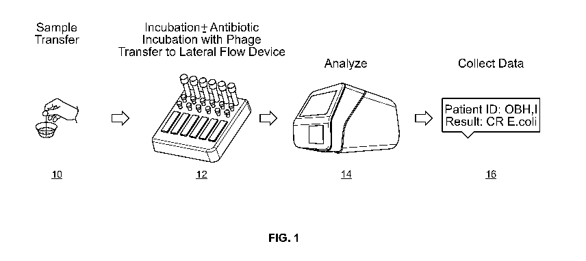
[0054] FIG. 1 shows an exemplary workflow according to one embodiment of a
method described
herein; and
[0055] FIG. 2 shows an exemplary workflow according to another embodiment of a
method
described herein.
DETAILED DESCRIPTION
[0056] Embodiments described herein provide methods and assays for diagnosis
or detection of
bacterial infectious agents and diseases using recombinant bacteriophages. The
methods are suitable
for the detection of bacterial infectious agents and also for determining drug
resistance of such
infectious agents. In addition, the methods are used to provide information
concerning the
susceptibility of the infectious agents to antimicrobial agents.
A. Infectious Bacteria
[0057] Essentially any bacteria can be detected and the methods and
compositions can be used for
determining antibiotic susceptibility of bacteria or for screening a candidate
antibiotic agent that
exerts a desirable (e.g., antimicrobial or cytotoxic) effect on target
bacteria.
[0058] In one embodiment, the bacteria are gram-negative bacteria. Typical
gram-negative bacteria
include proteobacteria such as E. coil, Salmonella, Pseudomonas, and
Helicohacter, and
cyanobacteria. When classified in connection with medicine, they include
Pseudomonas aeruginosa
and Hemophilus influenzae causing the disturbance of the respiratory system,
Escherichia coil and
Proteus mirahilis causing the disturbance of the urinary system, and
Helicobacter pylori and Bacillus
Gaertner causing the disturbance of the alimentary system and micrococci such
as Neisseria
meningitidis, Moraxella catarrhal's, and Neisseria gonorrhea.
[0059] In another embodiment, the bacteria are gram-positive bacteria. By
"gram-positive bacteria"
is meant a bacterium or bacteria that contain(s) teichoic acid (e.g.,
lipoteichoic acid and/or wall
teichoic acid), or a functionally equivalent glycopolymer (e.g., a
rhamnopolysaccharide, teichuronic
acid, arabinogalactan, lipomannan, and lipoarabinomarman) in its cell wall.
Non-limiting examples
of functionally equivalent glycopolymers are described in Weidenmaier et al.,
Nature, 6:276-287,
2008. Additional examples of functionally equivalent glycopolymers are known
in the art. In some
embodiments, a grain positive bacterium is identified using the Gram staining
method (e.g.,
generally including the steps of staining with crystal violet, treating with
an iodine solution,
decolorizing with alcohol, and counterstaining with safranine, wherein a gram
positive bacterium
retains the violet stain). Non-limiting examples of gram positive bacteria are
described herein.
Additional examples of gram-positive bacteria are known in the art. Exemplary
methods for
13
CA 03048992 2019-06-28
WO 2018/126266 PCT/US2018/012071
detecting or identifying gram-positive bacteria are described herein.
Additional methods for
detecting or identifying gram-positive bacteria are known in the art
[0060] The target bacteria include pathogenic bacteria that infect mammalian
hosts (e.g., bovine,
murine, equine, primate, feline, canine, and human hosts). In one embodiment,
the bacteria infect
and/or cause diseases in a human host. Examples of such pathogenic bacteria
include, e.g., members
of a bacterial species such as Bacteroides, Clostridium, Streptococcus,
Staphylococcus,
Pseudomonas, Haemophilus. Legionella, Mycobacterium, Escherichia, Salmonella,
Shigella, Vibrio,
or Listeria. Some clinically relevant examples of pathogenic bacteria that
cause disease in a human
host include, but are not limited to, Bacillus anthracis, Bacillus cereus,
Bordetella pertussis,
Borrelia burgdolferi, Brucella aborus, Brucella cants. Brucella melitensis,
Brucella suis,
Campylobacter jejuni, Chlamydia pneumoniae, Chlamydia psittaci, Chlam.,vdia
trachomatis,
Clostridium botulinum, Clostridium difficik, Clostridium perfringens,
Clostridium tetani,
Corynebacterium diphtheriae, Enterococcus faecalis, vancomycin-resistant
Enterococcus faecalls,
Enterococcus faecium, Escherichia coli, enterotoxigenic Escherichia coil
(ETEC), enteropathogenic
Escherichia coli, E coli 0157:H7, Francisella tularensis, Haemophilus
influenzae, Helicobacter
pylori, Legionella pneumophila, Leptospira interrogans, Listeria
monocytogenes, Mycobacterium
leprae, Mycobacterium tuberculosis, Mycoplasma pneumoniae, Neisseria
gonorrhoeae, Neisseria
meningitidis, Proteus, Pseudomonas aeniginosa, Rickettsia rickettsii,
Salmonella typhi, Salmonella
typhimurium, Shigella sonnei, Staphylococcus aureus, Staphylococcus epidermis,
Staphylococcus
saprophyticus, methicillin-resistant Staphylococcus aureus (MRSA), vancomycin-
resistant
Staphylococcus aureus (YSA), Streptococcus agalactiae, Streptococcus
pneumoniae, Streptococcus
pyogenes, Treponema pallidum, Vibrio cholerae, and Yersinia pestis.
[0061] In another embodiment, the infectious bacteria is selected from the
group consisting of
Clostridium difficile, Carbapenem-Resistant Enterobacteriaceae (CR-Klebsiella
spp; CR-E. coli),
and Neisseria gonorrhoeae. In another embodiment, the infectious bacteria is
selected from the
group consisting of multidrug-resistant Acinetobacter, drug-resistant
Campylobacter, extended
spectrum 13-Lactamase (ESBL)-producing enterobacteriaceae, vancomycin-
resistant enterococcus,
multidrug-resistant pseudomonas aeruginosa, drug-resistant non-typhoidal
Salmonella, drug-resistant
Salmonella enterica serovar Typhi, drug-resistant Shigella, methicillin-
resistant Staphylococcus
aureus (MRSA), drug-resistant Streptococcus pneumoniae, and drug-resistant
Tuberculosis. In
another embodiment, the infectious bacteria is selected from the group
consisting of vancomycin-
resistant Staphylococcus aureus, erythromycin-resistant Group A Streptococcus,
clindamycin-
Resistant Group B Streptococcus.
[0062] In certain embodiments, the infectious agents are natively found in
host subjects. In another
embodiment, the infectious agents are invasive species that are foreign to
host subjects. Preferably,
the hosts are mammals, e.g, a rodent, a human, a livestock animal, a companion
animal, or a non-
14
CA 03048992 2019-06-28
WO 2018/126266 PCT/US2018/012071
domesticated or wild animal. In one embodiment, the subject may be a rodent,
e.g. a mouse, a rat, a
guinea pig, etc. In another embodiment, the subject may be a livestock animal.
Non-limiting
examples of suitable livestock animals may include pigs, cows, horses, goats,
sheep, llamas and
alpacas. In still another embodiment, the subject may be a companion animal.
Non-limiting
examples of companion animals may include pets such as dogs, cats, rabbits,
and birds. In yet
another embodiment, the subject may be a zoological animal. As used herein, a
"zoological animal"
refers to an animal that may be found in a zoo. Such animals may include non-
human primates, large
cats, wolves, and bears. In an exemplary embodiment, the subject is a human.
[0063] The methods may be used to analyze infectious agents contained in a
variety of samples
including, e.g., biological sample, research test samples, environmental
samples (such as water
samples, including water samples selected from natural bodies of water, ponds,
community water
reservoirs, recreational waters, swimming pools, whirlpools, hot tubs, spas,
water parks, naturally
occurring fresh waters, and marine surface waters) and industrial samples
(such as fermenting
inoculums (such as Lactobacteria), chemical reagents, culture media, cleaning
solutions)
[0064.1 Preferably, the sample is a biological sample comprising bodily
fluids, e.g., sputum, tears,
saliva, sweat, mucus, serum, semen, urine, stool, vomit, and blood. The sample
may include e.g,
cerebral spinal fluid (CSF), blood plasma, blood serum, lymph, lung lavage
fluid, pleural fluid, etc.
In some embodiments, the sample may be obtained from the subject using any
known device or
method, e.g., swabs, urethral catheters, aspirators, hypodermic needles, thin
needle biopsies, hollow
needle biopsies, punch biopsies, metabolic cages, and syringes.
100651 In some embodiments, the biological sample is processed for use in the
methods described
herein. As a non-limiting example, a sputum or airway surface fluid (ASF) is
collected in an
appropriate vessel, such as a sterile specimen vial. The sample is solubilized
using, for example,
acetonitrile to a final concentration of about 60%, trifluoroacetic acid to a
final concentration of
about 0.1%, or using N-acetyl cysteine.
100661 In certain embodiments, the biological sample may be manipulated to
culture the bacteria
contained therein. The term "culture" means either the cultured cells, the
culture supernatant, the
mixture thereof, or a culture filtrate if a liquid medium is used; if a solid
medium is used, the term
"culture" means the mixture of the cells and the medium on which they have
grown. For example, if
a liquid medium is used, the marker may be recovered from the culture mixture
by the following
procedures. When the full growth of the bacteria is attained, the culture
mixture is subjected to
treatment with the antibiotic and/or the phage. Such downstream processes may
be intervened by
one or more washing and/or separation steps comprising centrifugation or
filtration, so as to obtain a
crude bacterial preparation that is free from contaminants. The markers may be
detected or analyzed
at the cellular level (e.g., in situ) or after subjecting the cultures to
further processing. For example,
wherein the marker is a protein or a DNA in the cytosol, they may be extracted
by disrupting cells
CA 03048992 2019-06-28
WO 2018/126266 PCT/US2018/012071
using a suitable method such as grinding or ultrasonic treatment. Cells may be
directly subjected to
an ultrasonic treatment in a culture medium so as to disrupt the cells and a
crude enzyme solution
may be obtained by removing any insoluble matter from the treated solution.
100671 If cultivation is performed on a solid medium, the markers may be
analyzed by first
manipulating the culture using the following procedure: water is added to the
solid medium
containing the cultured cells, and any insoluble matter is removed from the
mixture either
immediately or after disrupting the cells by a suitable means such as
ultrasonic treatment. A crude
marker preparation may be isolated from the crude lysate by conventional
purification techniques,
such as organic solvent fractionation, ammonium sulfate fractionation,
dialysis, isoelectric
precipitation and column chromatography, which may be used either
independently or in
combination. The level or activity of the marker may be determined using
conventional methods,
e.g., immunoassays for antigenic protein markers, ligand binding for antibody-
like markers,
enzymatic assays for enzyme-like markers, nucleic acid hybridization and/or
nucleic acid
amplification, etc.
[00681 Depending on the objective, the cell cultures may be analyzed using
routine techniques. For
example, the bacteria may be cultured to logarithmic phase (MSSA USA300 and
MRSA USA300)
and peak logarithmic phage may be detected using conventional techniques,
e.g., spectrophotometry.
Use of logarithmic phase bacteria may be preferable because they are more
likely to be adherent due
to higher expression of adhesins and their peptidoglycan layer is likely to be
less cross-linked and
thick compared to stationary-phase cells and the cells are more metabolically
active allowing for
faster response to damage. However, optimal conditions may vary from strain to
strain. Since
different strains are often encountered in a clinical setting, this
information is important for assessing
the utility of the diagnostic methodology. Although it is contemplated herein
that there will be strain
variability, it is anticipated that the bacteria will behave similarly enough
to permit the use of a
single protocol for testing all the strains. This expectation is based on the
fact that bacterial families
(e.g., staphylococci) are genetically quite similar to each other and thus
have similar cell structures,
which will be the main component in their responsiveness to the particular
phage.
100691 In some embodiments, the methods and compositions are useful for the
determination of
susceptibility of a microbe, e.g., bacteria. As used herein, the term
"susceptibility" refers to the
degree to which a bacterial cell is affected by an antibiotic. That is, the
cell may not be affected at
all, it may have its growth and proliferation slowed or halted without its
being killed or it may be
killed. Susceptibility also refers to the degree a population of a bacterial
species or strain is affected
by an antibiotic. In this case, certain highly susceptible cells of the
population may be very sensitive
and may be killed by very low concentrations of the antibiotic, other less
sensitive cells may have
their growth and proliferation slowed while others may not be affected at all.
16
CA 03048992 2019-06-28
WO 2018/126266 PCT/US2018/012071
[0070] In a related embodiment, the methods and compositions are useful for
identifying resistance
of a microbe, e.g., bacteria, to an antimicrobial agent or an antibiotic. The
term "resistant towards an
antibiotic" herein means that a particular bacterial strain, often a mutant
strain, is not killed, or killed
significantly more slowly compared to the corresponding wild-type strain from
which the strain is
derived. Resistance can also be reflected by altered growth properties of the
mutated and wild-type
strains. For example, a low concentration of the antibiotic in the culture
medium will prevent or
significantly decrease the growth of wild-type strains while the growth of the
mutated strains is not
affected. The phenotype of a resistant strain, e.g., altered growth, cell
division, metabolism, biofilm
production, virulence, etc. may be determined using routine techniques, for
e.g., growing wild-type
and mutant strains under identical conditions to assess a change in the
parameter being measured.
Sensitive strains may be used as reference standards in the assessment of
resistance (positive
control).
100711 In one embodiment, the methods are carried out by culturing a bacterial
sample in presence
of and in the absence of an antibiotic. The culture medium or fermentation
medium may be modified
or adjusted to meet the demands of the respective strains. Descriptions of
culture media for various
microorganisms are present in the "Manual of Methods for General Bacteriology"
of the American
Society for Bacteriology (Washington D.C., USA, 1981). The terms culture
medium and
fermentation medium or medium are interchangeable.
[0072] In its simplest sense, the culture medium contains at least one carbon
source (e.g., glucose)
and at least one nitrogen source (e.g., nitrate), optionally together with a
phosphorus source, e.g.,
phosphoric acid, potassium phosphate or other phosphate salts. Preferably, the
cultured medium is
buffered for bacterial growth. The culture medium may additionally comprise
salts, e.g., chlorides
or sulphates of metals such as, for example, sodium, potassium, magnesium,
calcium and iron, such
as, for example, magnesium sulphate or iron sulphate, which promote growth
and/or metabolic
activity. Finally, essential growth factors such as amino acids, for example
homoserine and
vitamins, for example thiamine, biotin or pantothenic acid, may be added to
the culture media,
depending on necessity. See, US patent No. 9,074,229.
[0073] A starter sample containing the bacteria be added to the culture in the
form of a single batch
or be fed in during the cultivation in a suitable manner, e.g., every 2-4
hours or every 1-3 hours, or
every 1, 2, 3, or 4 hours.
[0074] The pH of the culture can be controlled by employing basic compounds
such as sodium
hydroxide, potassium hydroxide, ammonia or aqueous ammonia, or acidic
compounds such as
phosphoric acid or sulphuric acid in a suitable manner. The pH is generally
adjusted to a value of
from 6.0 to 8.5, preferably 6.5 to 8. To control foaming, it is possible to
employ antifoams such as,
for example, fatty acid polyglycol esters. To maintain the stability of
bacteria, it is possible to add to
the medium suitable selective substances such as, for example, inducers such
as IPTG. The
17
CA 03048992 2019-06-28
WO 2018/126266 PCT/US2018/012071
fermentation is preferably carried out under aerobic conditions. In order to
maintain these
conditions, oxygen or oxygen-containing gas mixtures such as, for example, air
are introduced into
the culture. In batch or fed-batch processes, the cultivation is preferably
continued until an amount
of the desired density of the microbes is reached. Detection is carried out
spectrophotometrically
(absorption, fluorescence). This aim is normally achieved within 2 hours to
160 hours. In
continuous processes, longer cultivation times are possible. The activity of
the microorganisms
results in a concentration (accumulation) of the various markers in the
fermentation medium and/or
in the cells of the microbes.
100751 Examples of suitable fermentation media can be found inter alla in the
U.S. Pat. Nos.
5,770,409; 5,275,940; 5,827,698; 5,756,345; and WO 2007/012078 and WO
2009/043803.
B. Antibiotics
100761 The aforementioned culture media may be supplemented with or without an
antibiotic. As
used herein, the term "antibiotic" or "antimicrobial agent" refers to a
substance that inhibits the
growth of or destroys microorganisms. Preferably, the antibiotic is useful in
curbing the virulence of
an infectious agent and/or treating an infectious disease. Antibiotic also
refers to semi-synthetic
substances wherein a natural form produced by a microorganism, e.g., yeast or
fungus is
subsequently structurally modified.
100771 In another embodiment, the culture media may be supplemented with or
without a probiotic
substance. As used herein, the term "probiotic" refers to a substance that
promotes the growth or
metabolic activity of microorganisms, e.g., a micronutrient, a growth inducer
substance, or a toxin
removing substance.
100781 Preferably, the antibiotic is selected from the group consisting of13-
lactams (including,13-
lactamase inhibitors and cephalosporins), fluoroquinolones, aminoglycosides,
tetracyclines and/or
glycylcyclines and/or polymyxins. Any combination of antimicrobial agents may
also be tested, e.g,
at least one 13-lactam and at least one fluoroquinolone; at least one
aminoglycoside and one
cephalosporin; at least one 13-lactam and one 13-lactarnase inhibitor,
optionally together with an
aminoglycoside, etc.
[00791 As used herein, the term "11-lactam" refers to any antibiotic agent
which contains a p-lactam
ring in its molecular structure. Representative examples include natural and
semi-synthetic
penicillins and penicillin derivatives, clavulanic acid, carbapenems,
cephalosporins, cephamycins
and monobactams. These drugs are metabolized by enzymes broadly referred to as
"13-lactamases."
13-lactamases are organized into four molecular classes (A, B, C and D). Class
A enzymes
preferentially hydrolyze penicillins; class B enzymes include metalloenzymes
that have a broader
substrate profile than the others; class C enzymes are responsible for the
resistance of gram-negative
bacteria to a variety of antibiotics; and class D enzymes are serine
hydrolases, which exhibit a
unique substrate profile.
18
CA 03048992 2019-06-28
WO 2018/126266 PCT/US2018/012071
[0080] Generally, 13-lactams are classified and grouped according to their
core ring structures, where
each group may be divided to different categories. The term "penam" is used to
describe the core
skeleton of a member of a penicillin antibiotic, e.g, ii-lactams containing a
thiazolidine rings.
Penicillins may include narrow spectrum pinicillins, such as benzathine
penicillin, benzylpenicillin
(penicillin G), phenoxymethylpenicillin (penicillin V), procaine penicillin
and oxacillin. Narrow
spectrum penicillinase-resistant penicillins, such as methicillin,
dicloxacillin and flucloxacillin. The
narrow spectrum beta-lactamase-resistant penicillins may include temocillin.
The moderate spectrum
penicillins include for example, amoxicillin and ampicillin. The broad
spectrum penicillins include
the co-amoxiclav (amoxicillin+clavulanic acid). Finally, the penicillin group
also includes the
extended spectrum penicillins, for example, azlocillin, carbenicillin,
ticarcillin, mezlocillin and
piperacillin. Synthetic penicillin derivative includes, for example,
faropenem.
[0081] 13-lactams containing pyrrolidine rings are named carbapenams. The
carbapenems group
includes: biapenem, doripenem, ertapenem, imipenem, meropenem, panipenem and
PZ-601.
[0082] Cephalosporins and cephamycins include cephalexin, cephalothin,
cefazolin, cefaclor,
cefuroxime, cefamandole, cefotetan, cefoxitin, cefotaxime, and cefpodoxime.
Fourth generation
cephalosporins, which are active against Gram positive bacteria, include the
cefepime and
cefpirome. The cephalosporin class may further include: cefadroxil, cefixime,
cefprozil, cephalexin,
cephalothin, cefuroxime, cefamandole, cefepime and cefpirome. Cephamycins
include, for example,
cefoxitin, cefotetan, cefmetazole and flomoxef.
[0083] An example of carbacephems is loracarbef. Monobactams, which are active
against Gram-
negative bacteria include, for example, tigemonam, nocardicin A and tabtoxin.
Synthetic cephems
include, for example, clavulanic acid and oxacephems such as moxalactam and
flomoxef.
[0084] Fluoroquinolones act by inhibiting enzymes that are essential for
bacterial DNA replication.
Representative examples of includes, ciprofloxacin, garenoxacin, gatifloxacin,
gemifloxacin,
levofloxacin, and moxifloxacin.
[0085] Aminoglycosides possess bactericidal activity against most gram-
negative aerobic and
facultative anaerobic bacilli. Representative examples include, for e.g.,
kanamycin, amikacin,
tobramycin, dibekacin, gentamicin, sisomicin, netilmicin, neomycin B, neomycin
C, neomycin E
(paromomycin) and streptomycin, including, synthetic derivatives
clarithromycin and azithromycin.
[0086] Tetracyclines are a subclass of polyketides having an
octahydrotetracene-2-carboxamide
skeleton. They may be naturally-occurring (e.g., tetracycline,
chlortetracycline, oxytetracycline,
demeclocycline) or semi-synthetic (e.g., lymecycline, meclocycline,
methacycline, minocycline,
rolitetracycline). Glycylcyclines (e.g., minocycline/tigecycline) are derived
from tetracyclines.
[0087] Polymyxins are polypeptide antibiotics that are active against gram-
negative bacteria such as
E. coil and P. aeruginosa. Only polymyxin B and polymyxin E (colistin) are
used clinically.
19
CA 03048992 2019-06-28
WO 2018/126266 PCT/US2018/012071
[0088] In practicing the methods the media may be supplemented with one or
more of the
aforementioned antibiotics. The concentration of the antibiotic may range vary
depending upon the
antibiotic and the type of strain tested. Preferably, the dose of the
antibiotic is equal to or greater
than the minimum inhibitory concentration (MIC) of the particular antibiotic
on the particular strain.
Methods for determining MICs are known in the art (see, Andrews et al., J
Antimicroh Chemother.,
48 Suppl 1:5-16, 2001). A representative chart of MICs for 40 or so
antimicrobial agents on four
bacterial strains (E coil, S. aureus, P. aeruginosa, and Enterococcus
faecalis) is shown in Table 3 of
the Report published by European Committee for Antimicrobial Susceptibility
Testing (EUCAST)
entitled "Determination of minimum inhibitory concentrations (MICs) of
antibacterial agents by
broth dilution" (European Society orCliniecil Microbiology and Infectious
Diseases CM!, 9, 1-7,
2003).
[0089] Generally, the concentration of the antibiotic may be increased for
identifying or detecting
resistant strains, e.g., by at least 2-fold, at least 5-fold, at least 10-
fold, at least 20-fold, at least 50-
fold, at least 100-fold, at least 300-fold or even 1000-fold over the baseline
MIC. This is particularly
effective in instances where the target bacteria and the MIC of the antibiotic
on the bacteria are
already known. For instance, for E. coil, the MIC for most antibiotics may
range from about 0.01
mg/L to about 10 mg/L; however, resistant strains may not be susceptible until
the concentration is
increased, e.g, 10-fold (i.e. 1 log fold) - 1000 fold (i.e., 3-log fold) over
the base-line levels. In this
regard, the final antibiotic concentration may be adjusted accordingly.
[0090] Purely for illustrative purposes, the following dosages may be employed
- for testing the
resistance of bacteria to ii-lactams such as amoxicillin, the concentration
may range from about 2
mg/L to about 40 mg/L, particularly from about 5 mg/L to about 20 mg/L. See,
US patent No.
9,347,888. On the other hand, for testing the resistance of bacteria to
cloxacillin, the concentration
may range between about 25 mg/L and about 300 mg/L. For carbapenem, the
concentration may
range between 0.05 and 32 mg/L. This includes a range between about 2 mg/L to
about 32 mg/L for
faropenem and from about 0.05 mg/L to about 2 mg/L for doripenem (see, Woodman
et al., J Med
Microhiol., 19(1):15-23, 1983). For cephalosporins, the concentration may
range between about 1
mg/L to about 20 mg/L, preferably from about 4 mg/L to about 16 mg/L (see,
Waterworth, J Clin
Pathol, 35:1177-1180, 1982).
[0091] More particularly, the antibiotics may be used in a concentration of
any one of 0.1 mg/mL,
0.5 mg/L, 1 mg/mL, 2 mg/mL, 3 mg/mL, 4 mg/mL, 5 mg/mL, 6 mg/mL, 7 mg/mL, 8
mg/mL, 9
mg/mL, 10 mg/mL, 11 mg/mL, 12 mg/mL, 13 mg/mL, 14 mg/mL, 15 mg/mL, 16 mg/mL,
17
mg/mL, 18 mg/mL, 19 mg/mL, 20 ing/mL, 21 mg/mL, 22 mg/mL, 23 mg/mL, 24 mg/mL,
25
mg/mL, 26 mg/mL, 27 mg/mL, 28 mg/mL, 29 mg/mL, 30 mg/mL, 31 mg/mL, 32 mg/mL,
33
mg/mL, 34 mg/mL, 35 mg/mL, 36 mg/mL, 37 mg/mL, 38 mg/mL, 39 mg/mL, 40 mg/mL,
41
mg/mL, 42 mg/mL, 43 mg/mL 44 mg/mL, 45 mg/mL, 50 mg/mL, 60 mg/mL, 70 mg/mL, 80
mg/m,
CA 03048992 2019-06-28
WO 2018/126266 PCT/US2018/012071
90 mg/mL, 100 mg/mL, 150 mg/mL, 200 mg/mL, 250 ing/mL, 300 mg/mL, 400 mg/mL,
500
mg/mL, or more. For example, imipenem and ertapenem may be used in the
concentrations of 50,
30, 20, 15, 10, 5 and 1 mg/mL. The dosages may be adjusted similarly for
combination of
antibiotics, e.g., by first determining MiCs (combined agents) for wild-type
strains and gradually
increase the dosages to identify resistant strain(s).
100921 The bacteria are cultured in presence or absence of the antibiotic for
specified time periods,
e.g., between 2 hours to 160 hours, particularly between 8 hours to 24 hours,
especially between 10
hours to 16 hours. The bacteria may be at their growth phase or stationary
phase prior to contact with
the bacteriophage. The growth phase is a period characterized by cell
doubling, wherein the number
of cells in the culture grows exponentially. The stationary phase results from
both growth of new
bacteria and death of senescent cells, often due to a growth-limiting factor
such as the depletion of
an essential nutrient or accumulation of waste. Preferably, the bacteria are
in growth phase prior to
inoculation with the bacteriophage. Methods for determining growth phases of
bacteria are known in
the art. See, Hall et al., Mol Biol Evol., 31(1):232-8, 2014.
[00931 In one embodiment, the bacteria are treated with the antibiotic prior
to inoculating with the
bacteriophage. The primary culture may be optionally washed, e.g., with a wash
buffer, prior to
inoculation. Depending on the density of the surviving culture, the primary
culture or a wash pellet
thereof (obtained after centrifugation of the primary culture) may be re-grown
in fresh native media
(or antibiotic containing media) that has been inoculated with the
bacteriophage.
100941 In another embodiment, the bacteria are inoculated with the
bacteriophage simultaneously
with treatment with the antibiotic agent. This embodiment may be particularly
suited for non-lytic
phages.
C. Phages
100951 Embodiments of the instant methods utilize host-specific
bacteriophages. As used herein, the
term "bacteriophage" has its conventional meaning as understood in the art,
e.g., a virus that
selectively infects one or more bacteria. Many bacteriophages are specific to
a particular genus or
species or strain of bacteria. The term "bacteriophage" is synonymous with the
term "phage."
Bacteriophages may include, but are not limited to, bacteriophages that belong
to any of the
following virus families: Corticoviridae, Cystoviridae, Inoviridae, Levi
viridae, Microviridae,
Myoviridae. Podoviridae, Siphoviridae. or Tectiviridae. The bacteriophage may
be a lytic
bacteriophage or a lysogenic bacteriophage or a filamentous bacteriophage. A
lytic bacteriophage is
one that follows the lytic pathway through completion of the lytic cycle,
rather than entering the
lysogenic pathway. A lytic bacteriophage undergoes viral replication leading
to lysis of the cell
membrane, destruction of the cell, and release of progeny bacteriophage
particles capable of
infecting other cells. A lysogenic bacteriophage is one capable of entering
the lysogenic pathway, in
which the bacteriophage becomes a dormant, passive part of the cell's genome
through prior to
21
CA 03048992 2019-06-28
WO 2018/126266 PCT/US2018/012071
completion of its lytic cycle. A filamentous bacteriophage contains a circular
single-stranded
deoxyribonucleic acid (ssDNA) genome packaged into long filaments. These
phages do not
reproduce by lysing bacteria; instead, they are secreted into the environment
without killing the host.
[0096] In one embodiment, the phage is a lytic or productive phage (e.g., T4,
17, T3, and MS2). In
another embodiment, the phage is a temperate or lysogenic phage (e.g., ).
phage). In yet another
embodiment, the phage is a filamentous phage (e.g., fl, fd, and M13). A
combination of various
phages may also be employed. Phage display techniques are known in the art,
e.g., U.S. Patent No.
8,685,893; U.S. Patent No. 7,811,973; and U.S. Patent Publication No. 2002-
0044922. Preferably,
the phages are capable of transforming the host bacteria. As used herein, the
term "transformation"
means an introduction of DNA into a host cell such that DNA can be replicated
as an extra-
chromosomal element or by chromosomal integration. That is, transformation
refers to synthetic
alteration of genes by introducing a foreign DNA into the cell. As is
recognized in the art, the DNA
of most bacteria is contained in a single circular molecule, called the
bacterial chromosome and one
or more plasmids.
[0097] The phage is an engineered or a recombinant bacteriophage that serves
as a vector for a gene
that is foreign to the native phage. As used herein, the term "recombinant
phage" or "engineered
phage" is one that contains a nucleic acid sequence that is not naturally
occurring or has a sequence
that is made by an artificial combination of two otherwise separated segments
of sequence. This
artificial combination may be accomplished by chemical synthesis or artificial
manipulation of
isolated segments of nucleic acids, for example, by genetic engineering
techniques or the use DNA
transposition. Similarly, a recombinant protein is one encoded by a
recombinant nucleic acid
molecule. The term recombinant bacteriophage includes bacteriophages that have
been altered
solely by insertion of a nucleic acid, such as by inserting a nucleic acid
encoding a heterologous
protein that serves as a reporter or indicator molecule.
[0098] In certain embodiments, the phages are purified phages. The term
purified does not require
absolute purity; rather, it is intended as a relative term. A purified
molecule is one in which the
molecule is more enriched than it is in its natural environment, such as a
preparation in which the
molecule represents at least 50%, at least 60%, at least 80%, at least 90%, at
least 99% or greater
content of the total content of similar molecules within the sample. For
example, a purified sample
of recombinant phage is one in which the recombinant phage represents at least
50% of all
bacteriophages within the sample.
[0099] A listing of pathogenic bacterial genera and their known host-specific
bacteriophages is
presented in the following paragraphs and preferred types of bacteria-phage
pairs are provided in
Tables 1-3. Synonyms and spelling variants are indicated in parentheses.
Homonyms are repeated as
often as they occur (e.g., D, D, d). Unnamed phages are indicated by "NN"
beside their genus.
22
CA 03048992 2019-06-28
WO 2018/126266 PCT/US2018/012071
[0100] Bacteria of the genus Actinomyces are infected by the following phage:
Av-1, Av-2, Av-3,
BF307, CT!, CT2, CT3, CT4, CT6, CT7, CTS and 1281.
[0101] Bacteria of the genus Aeromonas are infected by the following phage: AA-
1, Aeh2, N, PM1,
TP446, 3, 4, 11, 13, 29, 31, 32, 37, 43, 43-10T, 51, 54, 55R 1, 56, 56RR2, 57,
58, 59.1, 60, 63,
Aehl, F, PM2, 1, 25, 31, 40RR2.8t, (syn=44R), (sy-n=44RR2.81), 65, PM3, PM4,
PM5 and PM6.
[0102] Bacteria of the genus Bacillus are infected by the following phage: A,
aizl, Al-K-I, B,
BCJA1, BC1, BC2, BLL1, BL1, BP142, BSL1, BSL2, BSI, BS3, BS8, BS15, BS18,
BS22, BS26,
BS28, BS31, BS104, BS105, BS106, BTB, BI715V1, C, CK-1, Coll, Corl, CP-53, CS-
1, CS!, D,
D, D, D5, entl, FP8, FP9, PSI, FS2, FS3, FS5, FS8, FS9, G. GH8, GT8, GV-1, GV-
2, GT-4, g3,
g12, g13, g14, g16, g17, g21, g23, g24, g29, H2, ken!, Kum!,
Kyul, J7W-1, LP52,
(syn=LP-52), L7, Mexl, MJ-1, mor2, MP-7, MP10, MP12, MP14, MP15, Neol, No 2,
N5, N6P,
PBC I, PBLA, PBP I, P2, S-a, SF2, SF6, Shal, Sill, SP02, (synAISPP1), SP, STI,
ST1, SU-11, t,
ml, Tb2, Tb5, Tb10, Tb26, Tb51, Tb53, Tb55, Tb77, Tb97, Tb99, Tb560, Tb595,
Td8, Td6, Td15,
Tgl, Tg4, Tg6, Tg7, Tg9, Tg10, Tgll, Tg13, Tg15, Tg21, Tin!, Tin7, Tin8,
Tin13, Tm3, Toe!,
Tog!, toll, TP-1, TP-10vir, TP-15c, TP-16c, TP-17c, TP-19, TP35, TP51, TP-84,
Tt4, Tt6, type A,
type B, type C, type D, type E, T93, VA-9, W, wx23, wx26, Yunl, a, y, pl 1,
9med-2, (pT, 911-4,
(p3T, 975, 9105, (syn=9105), 1A, 1B, 1-97A, 1-97B, 2, 2, 3, 3, 3, 5, 12, 14,
20, 30, 35, 36, 37, 38,
41C, 51, 63, 64, 138D, 1, II, IV, NN-Bacillus (13), ale!, AR!, AR2, AR3, AR7,
AR9, Bace-11,
(syn=11), Bastille, BL!, BL2, BL3, BL4, BLS, BL6, BL8, BL9, BP124, B528, BS80,
Ch, CP-51,
CP-54, D-5, darl, den I, DP-7, ent2, FoS I, FoS2, FS4, FS6, FS7, G, gall,
gamma, GE I, GF-2, (IS!,
GT-1, GT-2, GT-3, GT-4, GT-5, GT-6, GT-7, GV-6, g15, 19, 110, IS1, K. MP9,
MP13, MP21,
MP23, MP24, MP28, MP29, MP30, MP32, MP34, MP36, MP37, MP39, MP40, MP41, MP43,
MP44, MP45, MP47, MP50, NLP-1, No. 1, N17, N19, PBS!, PK1, PMB1, PMB12, PMJ1,
S, SPOI,
SP3, SP5, SP6, SP7, SP8, SP9, SP10, SP-15, 51350, (syn=SP-50), 5P82, SST,
sub!, SW, Tg8, Tg12,
Tg13, Tg14, thul, thu4, thu5, Tin4, Tin23, 'TP-13, TP33, TP50, TSP-1, type V.
type VI, V, Vx, 022,
9e, 9NR2, 925, 963, 1, 1, 2, 2C, 3NT, 4, 5, 6, 7, 8, 9, 10, 12, 12, 17, 18,
19, 21, 138, III, 4 (B.
megaterium), 4 (B. sphaericus), AR13, BPP-10, B532, B5107, B1, B2, GA-1, GP-
10, GV-3, GV-5,
g8, MP20, MP27, MP49, Nf, P135, PP6, SF5, Tg18, TP-1, Versailles, 915, 929, 1-
97, 837/1V, NN-
Bacillus (1), Bat10, BSL10, BSL11, BS6, BS!!, BS16, B523, BS101, BS102, g18,
morl, PBL1,
5N45, thu2, thu3, Tml, Tm2, TP-20, TP21, TP52, type F, type G, type IV, NN-
Bacillus (3), BLE,
(syn=0c), BS2,B54, BSS, BS7, BIO, B12, BS20, BS21, F, W-4, PBA12, AP50, AP50-
04, AP50-
11, AP50-23, AP50-26, AP50-27 and Bam35. The following Bacillus-specific phage
are defective:
DLPI0716, DLP-11946, DPB5, DPB12, DPB21, DPB22, DPB23, GA-2, M, No. 1M, PBLB,
PBSH,
PBSV, PBSW, PBSX, PBSY, PBSZ, phi, SPa, type 1 and Lt.
23
CA 03048992 2019-06-28
WO 2018/126266 PCT/US2018/012071
101031 Bacteria of the genus Bacteriodes are infected by the following phage:
ad12, Baf-44, Baf-
48B, Baf-64, Bf-1, Bf-52, B40-8, F1, 01, (A1, 913r01, 9Br02, 11, 67.1, 67.3,
68.1, NN-Bacteroides
(3), Bf42, Bf71, and BF-41.
101041 Bacteria of the genus Bordetella are infected by the following phage:
134 and NN-Bordetella
(3).
101051 Bacteria of the genus Borrellia are infected by the following phage: NN-
Borrelia (1) and
NN-Borrelia.
101061 Bacteria of the genus Brucella are infected by the following phage:
A422, Bk,
(syn=Berkeley), BM29, FO 1, (syri=F01), (syn=FQ1), D, FP2, (syn=FP2),
(syri=FD2), Fz,
(syn=Fz75/13), (syn =Firenze 75/13), (syn=Fi), F1, (syn::: F I ), Flm,
(syn=F1m), (syn =Fim), F1U,
(syn=F1U), (syn=FiU), F2, (syn=F2), F3, (syn=F3), F4, (syn=F4), F5, (syn=F5),
F6, F7, (syn=F7),
F25, (syn=F25), (syn=f25), F25U, (syn=F25u), (syn=F25U), (syn=F25V), F44,
(syn=F44), F45,
(syn=F45), F48, (syn=F48), T, Im, NI, MC/75, M51, (sy-n=M85), P. (syn=D),
S708, R, Tb, (syri='TB),
(syn=Tbilisi), W, (syn=Wb), (syn=Weybridge), X, 3, 6, 7, 10/1, (syn=10),
(syn=F8), (syn=F8), 12m,
24/11, (syn=24), (syn=F9), (syn=F9), 45/111, (syn=45), 75, 84, 212/V.
(syn=212), (syn=F10),
(syn=F10), 371/XXIX, (syn=371), (syn=F11), (syn=P11) and 513.
101071 Bacteria of the genus Burkholderia are infected by the following phage:
CP75.
101081 Bacteria of the genus Campylobacter are infected by the following
phage: C type,
NTCC12669, NTCC12670, NTCCI2671, NTCC12672, NTCC12673, NTCC12674, NTCC12675,
NTCC12676, NTCC12677, NTCC12678, NTCC12679, NTCC12680, NTCC12681, NTCC12682,
NTCC12683, NTCC12684, 32f, 111c, 191, Vfi-6, (syn=V19), Vfv-3, V2, V3, V8,
VI6, (syti=Vfi-1),
V19, V20(V45), V45, (syn=V-45) and NN-Campylobacter (1).
[OM] Bacteria of the genus Chlamydia are infected by the following phage:
Chpl.
101101 Bacteria of the genus Clostridium are infected by the following phage:
CAKI, CAS, Ca7,
CEO, (syn=1C), CET, Cld I, c-n71, c-203 Tox-, DEP, (syn=1D), (syn=1Dtox+),
HM3, KM1, KT, Ms,
NA!, (syn=Naltox+), PA1350e, Pfo, PL73, PL78, PL81, PI, P50, P5771, P19402,
1Ctox+, 2Ctox-,
2D, (syn=2Dtox+), 3C, (sy-n=3Ctox+), 4C, (syn=4t0x+), 56, III-1, NN-
Clostridium (61),
NB ltox-al, CAI, HMT, HM2, PF1, P-23, P46, Q-05, Q-06, Q-16, Q-21, Q-26, Q-40,
Q-46, S111,
SA02, WA01, WA03, W111, W523, 80, C. CA2, CA3, CPT1, CPT4, cl, c4, c5, HM7,
H11/A1,
H18/A I, H22/S23, H158/A1, K2/A1, K21/S23, ML, NA2tox-, Pf2, Pf3, Pf4, S9/S3,
S41/A1,
S44/S23, a2, 41, 112/S23, 214/S23, 233/A1, 234/S23, 235/S23, II-1, 11-2, 11-3,
NN-Clostridium (12),
CAI, F1, K, S2, 1, 5 and NN-Clostridium (8).
101111 Bacteria of the genus Corynebacterium are infected by the following
phage: CGII
(defective), A, A2, A3, A110, A128, A133, A137, A139, A155, A182, B, BF, B17,
B18, B51, B271,
B275, B276, B277, B279, B282, C, cap!, CC!. CG!, CG2, CG33, CL31, Cog,
(syn=C65), D, E, F,
H, H-1, hql, hq2, 11/H33, 11/H33, J, K, K, (syn=Ktox-), L, L, (syn=Ltox+), M,
MC-I, MC-2, MC-
24
CA 03048992 2019-06-28
WO 2018/126266 PCT/US2018/012071
3, MC-4, MLMa, N, 0, ovl, ov2, ov3, P, P, P. RP6, RS29, S, T, U, UB I, ub2,
UH1, UH3, uh3, uh5,
uh6, 13, (syn=l3tox+),131av64,13vir, y, (syn=ytox-), y19, 8, (syn=8tox+), p,
(syn=ptox-), 99, 9984, a),
1A, 1/1180, 2, 2/1180, 5/1180, 5ad/9717, 7/4465, 8/4465, 8ad/10269, 10/9253,
13/9253, 15/3148,
21/9253, 28, 29, 55, 2747, 2893, 4498 and 5848.
[0112] Bacteria of the genus Enterococcus are infected by the following phage:
DF78, F1, F2, 1, 2,
4, 14, 41, 867, DI, SB24, 2BV, 182, 225, C2, C2F, E3, E62, DS96, 1124, M35,
P3, P9, SB101, S2,
2BII, 5, 182a, 705, 873, 881, 940, 1051, 1057, 21096C, NN-Enterococcus (1),
PEI, PI, F3, F4,
VD13, 1, 200, 235 and 341.
[0113] Bacteria of the genus Erysipelothrix are infected by the following
phage: NN-Erysipelothrix
(1).
[0114] Bacteria of the genus E'scherichia are infected by the following phage:
BW73, B278, D6,
D108, E, El, E24, E41, FI-2, FI-4, FI-5, HI8A, HI8B, i, MM, Mu, (syn=mu),
(syn=Mul), (syn=Mu-
1), (syn=MU-I.), (syn=MuI), (syn=mu), 025, PhI-5, Pk, PSP3, PI, PI D, P2, P4
(defective), Si. W9,
9K13, (pR73 (defective), 91, 92, 97, 992, tv (defective), 7A, 89, 99, 15
(defective), 18, 28-1, 186,
299, NN-Escherichia (2), AB48, CM, C4, C16, DD-V1, (syn=Dd-Vi), (syn=DDVI),
(syn=DDVi),
E4, E7, E28, F11, F13, H, HI, H3, H8, K3, M, N, ND-2, ND-3, ND4, ND-5, ND6, ND-
7, Ox-1,
(syn=0X1), (syn= 11F), Ox-2, (syn=0x2), (syn=0X2), Ox-3, Ox-4, Ox-S,
(syn=0X5), Ox-6,
(syn=66F), (syn=966t), (syn=966t-), 0111, PhI-1, RB42, RB43, RB49, RB69, S.
Sal-1, Sal-2, Sal-3,
Sal-4, Sal-5, Sal-6, TC23, TC45, TuII*-6, (syn=TuII*), TuI1*-24, TuII*46,
TuII*-60, T2,
(syri=gamma), (syn=y), (syri=PC), (syn=P.C.), (syn=T-2), (syn=T2), (syn=P4),
T4, (syri=T-4),
(syn=T4), T6, T35, al, 1, 1A, 3, (syn=Ac3), 3A, 3T+, (syn=3), (syri=M1), 59,
(syn=95), 9266Q,
CF0103, HK620, J, K, K1F, m59, no. A, no. E, no. 3, no. 9, N4, sd, (syn=Sd),
(syn=SD), (syn=Sd),
(syn=sd), (syn=SD), (syn=CD), T3, (syn=T-3), (syn=T3), Ti, (syn=T-7),
(syn=T7), WPK, W31,
9C3888, 9K3, 9K7, 9K12, 9V-1, (1)04-CF, 4005, 4006, (1)07, 91, 91.2, 920, 995,
9263, 91092,
9I, 9II, (syn=9W), c28, 1, 3, 7, 8, 26, 27, 28-2, 29, 30, 31, 32, 38, 39, 42,
933W, NN-Escherichia (1),
Esc-7-11, AC30, CVX-5, CI, DDUP, EC1, EC2, E21, E29, F1, F26S, F275, Hi, 1-
11(022,
(syri=4:10HK97), HK253, HK256, K7, no.
D, PA-2, q, S2, TI., (syn=a), (syn=P28),
(syn=T-1), (syn=T1), T3C, T5, (syn=T-5), (syn=T5), UC-1, w, 4, y2,
(syn=lambda), (synAY),
40326, 9y, (1)06, (1)7, 010, 980, x, (syn=x1), (syn=9), (syn=(px1), 2, 4, 4A,
6, 8A, 102, 150, 168,
174, 3000, AC6, AC?, AC28, AC43, AC50, AC57, AC81, AC95, HK243, K10, ZG/3A, 5,
5A,
21EL, H19-f and 933H.
[0115] Bacteria of the genus Fusobacterium are infected by the following
phage: NN-Fusobacterium
(2), fv83-554/3, fv88-53I/2, 227, fv2377, fv2527 and fv8501.
[0116] Bacteria of the genus Haemophilus are infected by the following phage:
HP1 (Haemopbilus
phage HP!), S2 and N3.
CA 03048992 2019-06-28
WO 2018/126266 PCT/US2018/012071
[0117] Bacteria of the genus Helicobacter are infected by the following phage:
HP! (Helicobacter
pylori phage HP1) and NN-Helicobacter (1).
101181 Bacteria of the genus Klebsiella are infected by the following phage:
AI0-2, K14B, Kl6B,
1(19, (syn=1(19), 1(114, 1(115, K121, 1(128, 1(129, 1(132, IC133, K135,
K110613, K1171B, K1181B,
K1832B, AIO-1, A0-1, A0-2, A0-3, FC3-10, K, KU, (syn=K1I), K12, (syn=KI2),
K13, (syn=K13),
(syn=K170/11), K14, (syn=K14), 1(15, (syn=K15), K16, (spi=K16), K17,
(syn=K17), K18, (syn=K18),
1(119, (syn=K19), IC127, (syn=K127), 1(131, (syn=K131), 1(135, K1171B, II, VI,
IX, CI-1, K14B, 1(18,
K111, 1(112, K113, 1(116,1(117, K118, K120, 1(122, K123, K124, 1(126, K130,
K134, K1106B, K1165B,
K1328B, KLXI, K328, P5046, 11, 380, III, IV, VII, VIII, FC3-11., K12B,
(syn=K12B), K125,
(syn=K125), K142B, (syn=K142), (syn=K142B), K1181B, (syn=K1181), (syn=K1181B),
K1765/1,
(syn=K1765/1), K1842B, (syn=K1832B), K1937B, (syn=K1937B), Li, tp28, 7, 231,
483, 490, 632 and
864/100.
101191 Bacteria of the genus .Leptospira are infected by the following phage:
LEI, LE3, LE4 and
NN-Leptospim (1).
[0120] Bacteria of the genus Listeria are infected by the following phage:
A511, 01761, 4211, 4286,
(syn=B054), A005, A006, A020, A500, A502, A51.1, A118, A620, A640, B01.2,
B021, B024, B025,
B035, B051, B053, B054, B055, B056, B101, B110, B545, B604, B653, C707, D441,
HS047,
H1OG, H8/73, H19, H21, H43, H46, H107, H108, H10, H163/84, H1312, H340, H387,
H391/73,
H684/74, H924A, PSA, U153, 9MLUP5, (syn=P35), 00241, 00611, 02971A, 02971C,
5/476, 5/911,
5/939, 5/11302, 5/11605, 5/1.1704, 184, 575, 633, 699/694, 744, 900, 1.090,
1317, 1444, 1652, 1806,
1807, 1921/959, 1921/11367, 1921/11500, 1921/11566, 1921/12460, 1921/12582,
1967, 2389, 2425,
2671, 2685, 3274, 3550, 3551, 3552, 4276, 4277, 4292, 4477, 5337, 5348/11363,
5348/11646,
5348/12430, 5348/12434, 10072, I1355C, 11711A, 12029, 12981, 13441, 90666,
90816, 93253,
907515, 910716 and NN-Listeria (15).
[0121] Bacteria of the genus Morganella are infected by the following phage:
47.
[0122] Bacteria of the genus Mycobacterium are infected by the following
phage: 13, AG!, AL!,
ATCC 11759, A2, B.C3, BG2, BK1, BK5, butyricum, B-1, B5, B7, B30, B35, Clark,
Cl. C2,
DNAIII, DSP1, D4, D29, GS4E, (synS4E), G57, (synS-7), (syn=GS7), IPa,
lacticola,
Legendre, Leo, L5, (syn(DL-5), MC-1, MC-3, MC-4, minetti, MTPH11. Mx4,
My.F3P/59a, phlei,
(syn=phlei 1), phlei 4, Polonus II, rabinovitschi, smegmatis, TM4, TM9, TM! 0,
TM120, Y7, Y10,
9630, 1B, IF, 1.H, 1/1, 67, 106, 1430, B1, (syn=Bol), B24, D, D29, F-K, F-S,
HP, Polonus I, Roy,
RI, (syn=R1-Myb), (syn=R1), 11, 31, 40, 50, 103a, 103b, 128, 3111-D, 3215-D
and NN-
Mycobacterium (1).
[0123] Bacteria of the genus Neisseria are infected by the following phage:
Group I, group IT and
NP!.
26
CA 03048992 2019-06-28
WO 2018/126266 PCT/US2018/012071
[0124] Bacteria of the genus Nocardia are infected by the following phage: P8,
NJ-L, NS-8, N5 and
NN-Nocardia (I).
[0125] Bacteria of the genus Proteus are infected by the following phage: Pm5,
13vir, 2/44, 4/545,
6/1004, 13/807, 20/826, 57, 67b, 78, 107/69, 121, 9/0, 22/608, 30/680, Pm!,
Pm3, Pm4, Pm6, Pm7,
Pm9, Pm10, Pm!!, Pv2, id,(prn, 7/549, 9B/2, 10A/31, 12/55, 14, 15, 16/789,
17/971, 19A/653,
23/532, 25/909, 26/219, 27/953, 32A/909, 33/971, 34/13, 65, 5006M, 7480b, VI,
13/3a, Clichy 12,
n2600, (px7, 1/1004, 5/742, 9, 12, 14, 22, 24/860, 2600/D52, Pm8 and 24/2514.
[0126] Bacteria of the genus Providencia are infected by the following phage:
PL25, PL26, PL37,
9211/9295, 9213/921 lb, 9248, 7/R49, 74761322, 7478/325, 7479, 7480, 9000/9402
and 9213/9211a.
[0127] Bacteria of the genus Pseudomonas are infected by the following phage:
Pfl, (spi=Pf-1),
Pf2, Pf3, PP7, PRR1, 7s, NN-Pseudomonas (1), Al-!, M-2, B17, B89, CB3, Col 2,
Col 11, Col 18,
Col 21, C154, C163, C167, C2121, E79, F8, ga, gb, H22, K1, M4, N2, Nu, PB-1,
(syn=PB1), pfI6,
PMNI7, PP1, PP8, Psal, PsPI, PsP2, PsP3, PsP4, PsP5, PS3, PS17, PTB80, PX4,
PX7, PY01,
PY02, PY05, PY06, PY09, PY010, PY013, PY014, PY016, PY018, PY019, PY020,
PY029,
PY032, PY033, PY035, PY036, PY037, PY038, PY039, PY041, PY042, PY045, PY047,
PY048, PY064, PY069, PY0103, PIK, SLPI, SL2, S2, UNL-1, wy, Yal, Ya4, Yall,
(pBE,
(pCTX, (pC17, (pKZ, (syn=e*KZ), (p-LT, (I)mu78, (pNZ, (pPLS-1, (pST-1, (p-
2, 1/72, 2/79, 3,
3/DO, 4/237, 5/406, 6C, 6/6660, 7, 7v, 7/184, 8/280, 9/95, 10/502, 11/DE,
12/100, 12S, 16, 21, 24,
25F, 27, 31, 44, 68, 71, 95, 109, 188, 337, 352, 1214, NN-Pseudomonas (23),
A856, B26, CI-1, CI-
2, C5, D, gh-1, F116, HF, H90, K5, K6, K104, K109, KI66, K267, N4, N5, 06N-
25P, PE69, Pf,
PPN25, PPN35, PPN89, PPN91, PP2, PP3, PP4, PP6, PP7, PP8, PP56, PP87, PP114,
PP206, PP207,
PP306, PP651, Psp23 la, Pssy401, Pssy9220, psi, PTB2, PTB20, PTB42, PX1, PX3,
PX10, PX12,
PX14, PY070, PY071, R. SH6, SH133, tf, Ya5, Ya7,
diKf77, (p-MC, 41)mnF82, (pPLS27,
(pPLS743, 9S-1, 1, 2, 2, 3, 4, 5, 6, 7, 7, 8, 9, 10, 11, 12, 12B, 13, 14, 15,
14, 15, 16, 17, 18, 19, 20,
20, 21, 21, 22, 23, 23, 24, 25, 31, 53, 73, 119x, 145, 147, 170, 267, 284,
308, 525, NN-Pseudomonas
(5), at A7, B3, B33, B39, BI-1, C22, D3, D37, D40, D62, D3112, F7, F10, g, gd,
ge, gf, Hw12,
Jb19, KF I, L , OXN-32P, 06N-52P, PCH-1, PCI3-1, PC35-1, PH2, PH51, PH93,
PH132, PMW,
PMI3, PM57, PM6I, PM62, PM63, PM69, PM105, PM113, PM681, PM682, PO4, PP!, PP4,
P135,
PP64, PP65, PP66, PP71, PP86, PP88, PP92, PP401, PP711, PP891, Pssy41, Pssy42,
Pssy403,
Pssy404, Pssy420, Pssy923, PS4, P5-10, Pz, SDI, SL1, 5L3, SL5, SM, (pC5, (pC I
I, (pC11-1, (pC13,
(pCI5, (pM0, 9X, 904, 911, (p240,2, 2F, S. 7m, 11, 13, 13/441, 14, 20, 24, 40,
45, 49, 61, 73, 148,
160, 198, 218, 222, 236, 242, 246, 249, 258, 269, 295, 297, 309, 318, 342,
350, 351, 357-1, 400-1,
NN-Pseudomonas (6), GIO, M6, M6a, Li, PB2, PssyI5, Pssy4210, Pssy4220, PY012,
PY034,
PY049, PY050, PY051, PY052, PY053, PY057, PY059, PY0200, PX2, PX5, SL4, 903,
(p06
and 1214.
101281 Bacteria of the genus Rickettsia are infected by the following phage:
NN-Rickettsia (1).
27
CA 03048992 2019-06-28
WO 2018/126266 PCT/US2018/012071
[0129] Bacteria of the genus Salmonella are infected by the following phage:
b, Beccles, CT, d,
Dundee, f, Fels 2, GI, Gill, GVI, GVIII, k, K, i, j, L, 01., (syn=0-1),
(syri=0-I), (syn=7),
02, 03, P3, P9a, P10, Sab3, Sab5, San15, San17, SI, Taunton, Vii, (syn=Vil),
9, NN-Salmonella (1),
N-1, N-5, N-10, N-17, N-22, 11, 12, 16-19, 20.2, 36, 449C/C178, 966A/C259, a,
B.A.O.R., e, G4,
Gill, L, LP7, M, MG40, N- 18, PSA68, P4, P9c, P22, (syn=P22), (syn=PLT22),
(syn=PLT22),
P22a1, P22-4, P22-7, P22-11, SNT-1, SNT-2, SP6, Villi, ViIV, ViV, ViVI, ViVII,
Worksop, e15,
e34, 1, 37, 1(40), (syn=91[40]), 1,422, 2, 2.5, 3b, 4, 5, 6, 14(18), 8,
14(6,7), 10, 27, 28B, 30, 31, 32,
33, 34, 36, 37, 39, 1412, SNT-3,7-11, 40.3, c, C236, C557, C625, C966N, g, GV,
G5, G173, h, IRA,
Jersey, M78, P22-1, P22-3, P22-12, Sabi, Sab2, Sab2, Sab4, San 1 , San2, San3,
San4, San6, San7,
San8, San9, San13, 5an14, San16, San18, San19, San20, San21, San22, San23,
San24, San25,
San26, SasL1, SasL2, SasL3, SasL4, SasL5, S1BL, Sli, ViII, 91, 1, 2, 3a, 3aI,
1010, NN-Salmonella
(1), N-4, SasL6 and 27.
[0130] Bacteria of the genus Serratia are infected by the following phage:
A2P, PS20, SMB3, SMP,
SMP5, SM2, V40, V56, ç DCP-3, (DCP-6, 3M, 10/1a, 20A, 34CC, 34H, 38T, 345G,
345P, 501B,
SMB2, SMP2, BC, BT, CW2, CW3, CW4, CW5, L1232, L2232, L34, L.228, SLP, SMPA,
V.43, a,
9CW1., CP6-1, (110CP6-2, (1)CP6-5, 3T, 5, 8, 9F, 1.0/1, 20E, 32/6, 34B, 34CT,
34P, 37, 41, 56, 56D,
56P, 60P, 61/6, 74/6, 76/4, 101/8900, 226, 227, 228, 229F, 286, 289, 290F,
512, 764a, 2847/10,
2847/10a, L.359 and SMB1,
[0131] Bacteria of the genus Shigella are infected by the following phage:
Fsa, (syn=a), FSD2d,
(syn=D2d), (syn=W2d), FSD2E, (syn=W2e), fv, F6, f7.8, H-Sh, PE5, P90, SfIT,
Sh, SHIP, SHIV,
(syn=HIV), SHVI, (syn=HVI), SHVIII, (syn=HVIII), SKy66, (syn=gamma 66),
(syn=y66),
(syn=y66b), SKIII, (syn=SIIIb), (syn=III), SKIV, (syn=SIVa), (syn=IV), SKIVa,
(syn=SIVAn),
(syn=IVA), SKVI, (syn=KVI), (sy-n=SVI), (sy-n=VI), SKVIII, (syn=SVIII),
(syn=VIII), SKVIIIA,
(syn=SVIIIA), (syn=VIIIA), STVI, STIX, STXI, STXII, S66, W2, (syn=D2c),
(syn=D20), (pIV1,
3-SO-R, 8368-SO-R, F7, (syn=FS7), (syn=K29), F10, (syn=FS10), (syn=K31), II,
(syn=alfa),
(syn=FSa), (syn=KI8), (syn=a), 12, (syn=a), (syn=K19), 5G35, (syn=G35),
(syn=S0-35/G), 5G55,
(syn=S0-55/G), SG320I, (syn=S0-3201/G), SHIT, (syn=HII.), SHV, (syn=SHV), SHX,
SHX, SKI!,
(syn=1(2), (syn=KII), (syn=SII), (syn=SsII), (syn=II), SKIV, (syn=SIVb),
(syn=SsIV), (syn=IV),
SKIVa, (syn=SIVab), (syn=SsiVa), (syn=1Va), SKV, (syn=K4), (syn=KV), (syn=SV),
(syn=SsV),
(syn=V), SKX, (syn=K9), (syn=10(), (syn=SX), (syn=SsX), (syn=X), STV,
(syn=T35), (syn=35-50-
R), S'TVITT, (syn='T8345), (syn=8345-SO-S-R), WI, (syri=D8), (sy-n=FSD8), W2a,
(syn=D2A),
(syn=FS2a), DD-2, Sf6, FS1, (syn=F1), SF6, (syn=F6), 5G42, (syn=S0-42/G),
5G3203, (syn=S0-
3203/G), SKF12, (syn=SsF12), (syn=F12), (syn=F12), Sill, (syn=1881-SO-R), y66,
(syn=garnma
66a), (syn=Ssy66), 92, B11, DDVIT, (syn=DD7), FSD2b, (syn=W2B), F52, (syn=F2),
(syn=F2),
F54, (syn=F4), (syn=F4), FS5, (syn=F5), (syn=F5), F59, (syn=F9), (syn=F9),
F11, P2-SO-S, 5G36,
(syn=S0-36/G), (syn=G36), 5G3204, (syn=S0-3204/G), 5G3244, (syn=S0-3244/G),
SHI,
28
CA 03048992 2019-06-28
WO 2018/126266 PCT/US2018/012071
(syn=H1), SHVII, (syn=HVII), SHIX, (syn=HIX), SHXI, SHXII, (syn=HXII), SKI,
KI, (syn=SI),
(syn=SsI), SKVII, (syri=KVII), (syri=SVII), (syri=SsVIT), SKTX, (syn=KIX), (sy-
n=SIX),
(syn=SsIX), SICXII, (syn=KXII), (syn=SVII), (syn=SsXII), STI, Sill, STIII,
STIV, STVII, S70,
S206, U2-SO-S, 3210-SO-S, 3859-SO-S, 4020-SO-S, 93, 95, 97, 98, 99, 910, 911,
913, 914, 918,
SHIII, (syn=HIII), SHXI, (syn=HXI) and SXI, (syn=1(XI), (syn=SXI), (syn=SsXI),
(syn=XI).
[0132] Bacteria of the genus Staphylococcus are infected by the following
phage: A, EW, K, Ph5,
Ph9, Ph10, Ph13, Pl, P2, P3, P4, P8, P9, P10, RG, SB-1, (syn=Sb-1), S3K,
Twort, 95K311, 9812,
06, 40, 58, 119, 130, 131, 200, 1623, STC1, (syn=stc I), STC2, (syn=stc2),
44AHJD, 68, AC! AC2,
A6"C", A9"C", b581, CA-1., CA-2, CA-3, CA-4, CA-5, D1.1, L39x35, L54a, M42,
NI, N2, N3, N4,
N5, N7, N8, NIO, N11, N12, N13, N14, N16, Ph6, Ph12, Ph14, UC-18, U4, U15, 51,
S2, S3, S4, S5,
X2, Zl, 9B5-2, 9D, co, ii, (syn=(p11), (syn=P11-M15), 15, 28, 28A, 29, 31,
31B, 37, 42D,
(syn=P42D), 44A, 48, 51, 52, 52A, (syn=P52A), 52B, 53, 55, 69, 71, (syn=P7I),
71A, 72, 75, 76,
77, 79, 80, 80a, 82, 82A, 83A, 84, 85, 86, 88, 88A, 89, 90, 92, 95, 96, 102,
107, 108, 11.1, 129-26,
130, 130A, 155, 157, 157A, 165, 187, 275, 275A, 275B, 356, 456, 459, 471,
471A, 489, 581, 676,
898, 1139, 1154A, 1259, 1314, 1380, 1405, 1563, 2148, 2638A, 2638B, 2638C,
2731, 2792A,
2792B, 2818, 2835, 2848A, 3619, 5841, 12100, AC3, A8, A10, A13, b594n, D, M12,
N9, N15, P52,
P87, 51, S6, Z4, 9RE, 3A, 3B, 3C, 6, 7, 16, 21, 42B, 42C, 42E, 44, 47, 47A,
47C, 51, 54, 54x1, 70,
73, 75, 78, 81, 82, 88, 93, 94, 101, 105, 110, 115, 129/16, 174, 594n,
1363/14, 2460 and NN-
Staphylococcus (1).
[0133] Bacteria of the genus Streptococcus are infected by the following
phage: EJ-1, NN-
Streptococcus (1), a, Cl, FLOThs, H39, Cp-1, Cp-5, Cp-7, Cp-9, Cp-10, AT298,
AS, al 031, al 032,
al0/j5, alO/J9, A25, BT11, b6, CAI, c20-1, c20-2, DP-1, Dp-4, DTI, ET42, el ,
FA101, FEThs,
FK, FICK101, FKLIO, FKP74, FKII, FLO'Ths, FY101, fl, F10, F20140/76, g, GT-
234, HB3,
(syn=HB-3), HB-623, HB-746, M102, 01205, 901205, PST, PO, Pl, P2, P3, P5, P6,
P8, P9, P9,
P12, P13, P14, P49, P50, P51, P52, P53, P54, P55, P56, P57, P58, P59, P64,
P67, P69, P71, P73,
P75, P76, P77, P82, P83, P88, sc, sch, sf, Sfill, (syn=SFil 1), (syn=(pSFi1l),
(syn=diSfi 11),
(syn=(pSfi 11), sfil9, (syn=SFI 19), (syn=(pSFi 19), (syn=(pSfi 19), Sfi21,
(syn=SFi21), (syn=9SFi21),
(syn=(pSfi21), STG, STX, st2, 5T2, ST4, S3, (syn=(p53), s265, 017, 942, 057,
980, 981, 982, 983,
984, 985, 986, 987, 988, 989, 990, 991, 992, 993, 994, 995, 996, 997, 998,
999, 9100, 9101,
9102, 9227, 07201, col, co2, 0o3, 0)4, 0o5, c06, co8, 0o10, I, 6, 9, 10F,
12/12, 14, 17SR, 19S, 24, 50/33,
50/34, 55/14, 55/15, 70/35, 70/36, 71/ST15, 71/45, 71/46, 74F, 79137, 79/38,
80/J4, 8059, 80/5T16,
80/15, 80/47, 80/48, 101, 103/39, 103/40, 121/41, 121/42, 123/43, 123/44,
124/44, 337/5T17 and
NN-Streptococcus (34).
[0134] Bacteria of the genus .Treponema are infected by the following phage:
NN-Treponema (1).
[0135] Bacteria of the genus Vibrio are infected by the following phage:
CTX(I), fs, (syri=s1), fs2,
lvpfs, Vf12, Vf33, VPI(1), VSK, v6, 493, CP-T1, ET25, kappa, K139, LaboI,) XN-
69P, OXN-86,
29
CA 03048992 2019-06-28
WO 2018/126266 PCT/US2018/012071
06N-21P, PB-1, P147, rp-I, SE3, VA-1, (syn=VcA-1), VcA-2, VcA-1, VP1, VP2,
VP4, VP7, VP8,
VP9, VP10, VP17, VP18, VP19, X29, (sy-n=29 d'Herelle), I, (DHAWT-1, (DHAWI-2,
(DHANATI-3,
0:DHAWI-4, 41:0HAWI-5, 41)HAWI-6, 0:DHAWI-7, 41:0HAWI-8, 41:011AW1-9, (DHAWI-
10, (DHC1-1,
(DHC1-2, (DHC1-3, 0:DHC1-4, (DHC2-1, (DHC2-2, (DHC2-3, (DHC2-4, (DHC3-1, (DHC3-
2, (DHC3-3,
(DFID1S-1, (DHD1S-2, (DHD2S-1, (DHD2S-2, (DHD2S-3, (DHD2S-4, (DHD2S-5, (DFIDO-
I, (DHDO-2,
(1*IDO-3, (DHDO-4, (DHDO-5, (DHDO-6, (DKL-33, (DICL-34, (DKL-35, ICL-36,
(DKW1H-2,
(DKWH-3, (DKWH-4, (DMARQ-1, (DMARQ-2, (DMARQ-3, (DMOAT-1, (D0139, (DPEL1A-1,
OPEL IA-2, (DPEL8A-1, (DPEL8A-2, (DPEL8A-3, (DPEL8C-1, (DPEL8C-2, (DPELI3A-1,
OPEL-
13B-I, (1PELI3B3-2, (DPEL13B-3, (DPEL13B-4, (DPEL13B-5, (DPEL13B-6, szDPEL13B-
7,
(DPEL13B-8, (DPEL13B-9, (DPEL13B-10, 9VP143, triVP253, (D16, 9138, 1-11,5, 13,
14, 16, 24, 32,
493, 6214, 7050, 7227, II, (syn=group 11), (syn=92), V, VIII, NN-Vibrio (13),
KVP20, KVP40, nt-
1, 06N-22P, P68, el, e2, e3, e4, e5, FK, G, J, K, nt-6, NI, N2, N3, N4, N5,
06N-34P, OXN-72P,
OXN-85P, OXN-100P, P, Ph-I, PL! 63/10, Q, S. T. 992, 1-9, 37, 51, 57, 70A-8,
72A-4, 72A-10,
110A-4, 333, 4996, I, (syn=group I), III, (syn=group III), VI, (syn=A-
Saratov), VII, IX, X, NN-
Vibrio (6), pAl, 7, 7-8, 70A-2, 71A-6, 72A-5. 72A-8, 108A-10, 109A-6, 109A-8,
110A-1, 110A-5,
110A-7, hv-1, OXN-52P, P13, P38, P53, P65, P108, P111, TP1, VP3, VP6, VPI2,
VP13, 70A-3,
70A-4, 70A-10, 72A-1, 108A-3, 109-B1, 110A-2, 149, (syn=q)149), IV, (syn=group
IV), NN-Vibrio
(22), VP5, VP11, VP15, VP16, al, a2, a3a, 3b, 353B and NN-Vibrio (7).
[0136] Bacteria of the genus Yersinia are infected by the following phage: H,
H-1, H-2, H-3, H-4,
Lucas 110, Lucas 303, Lucas 404, YerA3, YerA7, YerA20, YerA41, 3/M64-76,
5/G394-76, 6/C753-
76, 8/C239-76, 9/F18167, 1701, 1710, PST, 1/F2852-76, D'Herelle, EV, H,
Kotljarova, PTB, R, Y,
YerA41, 9Yer03-12, 3, 4/C1324-76, 7/F783-76, 903, 1/M6176 and Yer2AT.
[0137] In another embodiment, the methods are practiced using a combination of
at least 2, at least
3, at least 4, at least 5, at least 6, at least 7, at least 8, at least 9 or a
greater number of the
aforementioned phages. One skilled in the art recognizes that the efficiency
of transformation may
be manipulated, e.g., enhanced or suppressed, depending on the particular
combination of phages
that are employed.
[0138] In particular, bacteria species (and corresponding, host-specific
bacteriophages) include
Aeromonas hydrophila (PM2), Bacillus anthracis (Gamma), Bacillus subtilus
(SPP1), Bordetella
pertussis (See Pereversev et al. Zh Mikrobiol 5:54-57, 1981), Borrelia
burgdotferi (9BB-I, see
Eggers et al., J Bacteriol 183:4771-4778, 2001), Brucella abortus (TB: 212;
371), Campylobacter
jejuni (pC), Clostridium perfringes ((p3626), Enterococcus faecalis
(tpFC1), Enterococcus
faecium (ENB6), Escherichia coli (Pl; Ti; T3, T4, T5: 17, KHI, 9V10; lambda;
920; mu),
Klebsiella pneumoniae (60; 92), listeria monocytogenes (A511, A118: 243; H387;
2389; 2671;
2685; 4211), Mycobacterium leprae (mycobacteriophage, L5), Mycobacterium
tuberculosis (LG;
DSGA), Pseudomonas aeniginosa (E79, G101; B3; pp. 7), Salmonella anatum (E5),
Salmonella
CA 03048992 2019-06-28
WO 2018/126266 PCT/US2018/012071
bovismorbificans (98), Salmonella choleraesuis (102), Salmonella enteritidis
(L; P22; 102; FO; IRA;
(p8), Salmonella Newington (E34), Salmonella schottmulleri (31; 102; FO; 14),
Salmonella typhi
(163; 175; Vii; ViVI; 8; 23; 25; 46; 175; FO), Serratia marcescens (S24VA),
Shigella dysenieriae
(p8O; P2; 2; 37), Shigella jlexneri (Sf6), Staphylococcus aureus (K; Pl; P14;
UC18; 15; 17; 29; 42D;
47; 52; 53; 79; 80; 81; 83A; 92; Twort, 911), Streptococcus pneumoniae (Dp-1;
Cp-1; HB-3; EJ-1;
MM!; V01), Streptococcus pyogenes (9X240; 1A; 1B; T12; 12/12; 113; 120; 124;
P58; H4489a),
Vibrio cholerae (138; 145; 149; 163), and Yersinia pestis (A1122; R; Y; P1).
Additional information
is provided in U.S. Patent Publication No. 2009-0155768.
[0139] In particular, Tables 1-3 provide representative examples of particular
host-specific phages
and the hosts to which they are specific for, including, the receptors through
which they mediate
their actions. See also Bertozzi et al., FEMS Microbiology Letters, 363, 1-11,
2016.
Table 1. Receptors in the cell wall of Gram-positive bacteria. Host names are
ordered
alphabetically.
Phases Family, Main host Receptor(s)
Siphoviridae Bacillus Membrane surface-anchored protein
anthracis phage receptor (GamR)
SPP1 Siphoviridae Bacillus subtilis Glucosyl residues of
poly(glycerophosphate) on WTA
09 Podoviridae Bacillus subtilis Cell WTA (primary receptor)
Bam35 Tectiviridae Bacillus N-acetyl-muramic acid (MurNAc)
thuringiensis
LL-H Siphoviridae Lactobacillus Glucose moiety of LTA
delbrueckii
BI Siphoviridae Lactobacillus Galactose component of the
wall
J14tntantm
B2 Siphoviridae Lactobacillus Glucose substituents in in
teichoic
dy.antarum acid
5, 13, c2, h, m13, kh, L Siphoviridae Lactococcus Rhamnose moieties in the
cell wall
locus
(1)LC3, TP9Olerm, Siphoviridae Lactococcus Cell wall polysaccharides
TP901-1 locus
p2 Siphoviridae Lactococcus Cell wall saccharides
lactis
A511. Myoviridae Listeria Peptidoglycan (murein)
monocytogenes
A 1 18 Siphoviridae Listeria Glucosaminyl and rhainnosyl
monocytogenes components
A500 Siphoviridae Listeria Glucosaminyl residues
monocytogenes
4)812, 4)K Myoviridae Staphylococcus Anionic backbone of WTA
aureus
52A Siphoviridae Staphylococcus 0-acetyl group from the 6-
position
aureus
W, (1)13, 4)47, +77, Siphoviridae Staphylococcus N-acetylglucosamine
(GleNAc)
4)Sa2m aureus glycoepitope
4)SLT Siphoviridae Staphylococcus Poly(glycerophosphate) moiety
of
aureus LTA
31
CA 03048992 2019-06-28
WO 2018/126266 PCT/US2018/012071
Table 2. Receptors in the cell wall of Gram-negative bacteria. Host names are
ordered
alphabetically.
_Phaees_ ., Family _Main host Recentor(s)
Kr30 Myoviridae Caulobacter Paracrystalline surface (S)
protein
crescentus
... . ...
434 Siphoviridae Escherichia coil Protein lb (OmpC)
BF23 Siphoviridae Escherichia colt Protein BtuB (vitamin
B12
receptor) .
K3 Myoviridae Escherichia colt Protein d or 3A (OmpA)
with LPS
K10 Siphoviridae Escherichia colt Outer membrane protein
LamB
Mel Myoviridae Escherichia coil Protein c (OmpC)
...
Mu G(4-) Myoviridae Escherichia coil Terminal GIca-2GIca I - or
GlcNAca1-2Glcal- of LPS
Mu G(-) Myoviridae Escherichia coil Terminal glucose with a
11,3
glycosidic link
MI Myoviridae Escherichia colt Protein OmpA .
0x2 Myoviridae Escherichia colt Protein OmpA
ST-1 Myoviridae Escherichia colt Terminal Glca-2Glca I - or
GlcNAcal-2GIcal- of LPS
TLS Siphoviridae Escherichia colt
Antibiotic efflux protein To1C .
Tula Myoviridae Escherichia colt Protein Ia (OmpF) with LPS
Tulb Myoviridae Escherichia colt Protein lb (OmpC) with LPS
Tull* Myoviridae Escherichia colt Protein 11* (OmpA) with
LPS
Ti Siphoviridae Escherichia coil Proteins TonA (FluA)
T2 Myoviridae Escherichia coli Protein la (Own with LPS
T3 Podoviridae Escherichia colt Glucosyl-a-1,3-glucose
terminus
of rough LPS
T4 Myoviridae E. coil K-12/E. coil Protein 0-8 (OmpC) with
LPS
B .
T4 Myoviridae E colt B Glucosyl-a-1,3-glucose
terminus
of rough LPS
T5 Siphoviridae Escherichia coli Polymannose sequence in
the 0-
antigen
T6 Myoviridae Escherichia colt Outer membrane protein Tsx
T7 Podoviridae Escherichia colt LPS
U3 Microviridae Escherichia coli Terminal galactose
residue in LPS
A Siphoviridae Escherichia colt
Protein LamB .
OX/ 74 Microviridae Escherichia colt Terminal galactose in
rough LPS
4)80 Siphoviridae Escherichia coil Proteins FhuA and TonB
PM2 Corticoviridae Pseudoalteromonas Sugar moieties on the cell
surface
E79 .Myoviridae Pseudomonas Core polysaccharide of LPS
aeruginosa
JG004 Myoviridae Pseudomonas LPS
aeruginosa
q)CTX Myoviridae Pseudomonas Core polysaccharide of LPS
aeruginosa
9PLS27 Podoviridae Pseudomonas Galactosamine-alanine region
of
aeruginosa LPS .
4)13 Cystovirklae .Pseudomonas Truncated 0-chain of LPS
syringae
ES18 Cvstoviridae Salmonella Protein FIRIA
Gifsy-1, Gifsv-2 Siphoviridae Salmonella Protein OmpC
32
CA 03048992 2019-06-28
WO 2018/126266
PCT/US2018/012071
Phaaes. Family Main host Receptor(s)
SPC35 Siphoviridae Salmonella BtuB (main
receptor) and 012-
antigen (asssiting receptor)
SPNIS, SPN2TCW, Podoviridae Salmonella 0-antigen of LPS
SPN4B, SPN13U
SPN6TCW, Podoviridae Salmonella 0-antigen of LPS
SPN8TCW,
SPN9TCW
SPN7C, SPN9C, Siphoviridae Salmonella Protein BtuB
SPN1OH, SPNI2C
SPN14, SPN17T, Sephoviridae Salmonella Protein BtuB
SPN18
vB SenM-S16 (S16) Abioviridae Salmonella Protein OmpC
Podoviridae Escherichia coli a-1,3-mannosyl linkages
c341 Podoviridae Salmonella 0-acetyl group in
the
marmosyldiamnosy1-0-
acetylgalactose
P22 Podoviridae Salmonella 0-acetylgalactose
à 34 Podoviridae Salmonella 1-13-Gal-Man4R1iad
poly-sacchaiide
Sf6 Podoviridae Shigella Rha 11 1-a-3 Rha III
linkage
33
CA 03048992 2019-06-28
WO 2018/126266 PCT/US2018/012071
'fable 3. Receptors in bacterial complexes other than cell walls. Host names
are ordered
alphabetically.
Phaees Family Main host Receptor(s)
SPN2T, SPN3C, Siphoviridae Salmonella Flagellin protein FliC
SPN8T, SPN9T
SPN I 1T, SPN 13 B. Siphoviridae Salmonella Flagellin protein FliC
SPN 16C
SPN4S, SPN5T, Siphoviridae Salmonella Flagellin proteins FliC or FljB
SPN6T, SPN19
1EPS5 Siphoviridae Salmonella Flagellal molecular ruler
protein
FliK
(I)CbK, (;)Cb13 Siphoviridae Caulobacter Pill portals on the cell pole
crescentus
Fd. Ft; fl, M13 Inoviridae Escherichia colt F pilus followed by TolQRA
complex in membrane
PRD1 Tectiviridae Escherichia colt Mating pair fonnation (Mpf)
complex
4>6 Cystoviridae Pseudomonas Sides of the type IV pilus
MPK7 Podoviridae Pseudomonas Type IV pill (TFP)
aeruginosa
MP22 Siphoviridae Pseudomonas Type IV pili (TFP)
aeruginosa
DMS3 Siphoviridae Pseudomonas Type IV pill (TFP)
aeruginosa
29 Podoviridae Escherichia colt p -D-glucosido-( I -3)-D-
glucoronic
acid bonds in capsule
K1 I Podoviridae Klebsiella -D-glucosyl-(1-3)-13 -D-
glucuronic acid linkages
Vii Afpviridae Salmonella Acetyl groups of the Vi
exopolysaccharide capsule
Viii Siphoviridae Salmonella Acetyl groups of the Vi
exopolysaccharide capsule
Vi III, Vi IV, Vi V, Vi Podoviridae Salmonella Acetyl
groups of the Vi
VI, Vi VII exopolysaccharide capsule
D. Bacteriophage Packaging Sites
[0140] A bacteriophage packaging site is a specific DNA sequence on the phage
genome for genome
packaging into the virion. A plasmid is engineered to contain a phage
packaging site so that plasmid
is packaged into the transducing particles. Host-specific bacteriophages (and
their packaging sites)
include but are not limited to SPP1 (SPP1 pac site), PI (P1 pac site), Ti (Ti
pac site), T7 Cr
concatamer junction), lambda (cos site), mu (mu pac site), P22 (P22 pac site),
(p8 ((p8 pac site), Sf6
(Sf6 pac site), 149 (149 pac site), and A1122 (A1122-concatamer junction). For
most
bacteriophages, the packaging site is termed the pac site. In some cases, the
packaging site is
referred to as a concatamer junction (e.g. 17 concatamer junction). In every
case, the packaging site
is different from the naturally-occurring adjacent sequences in the
bacteriophage genome.
[0141] For some bacteriophages, the packaging site may be unknown. In these
cases, pac sites can
be determined by taking advantage of the property that plasmids containing a
functional
34
CA 03048992 2019-06-28
WO 2018/126266 PCT/US2018/012071
bacteriophage pac site are packaged. For example, the DNA sequences necessary
for packaging of
bacteriophage X were determined by incorporating small restriction fragments
of the X phage
genomic DNA into a plasmid (Hohn et al., PNAS USA 80:7456-7460, 1983).
Following
introduction into an in vivo packaging strain, the efficiency of
packaging/transduction was
quantitatively assessed. Using a similar strategy, the pac sites for a number
of bacteriophages have
been determined: X (Miwa et al., Gene 20:267-279, 1982); Mu (Croenen et al..
Virology 144:520-
522, 1985); filamentous bacteriophages including fl, fd, M13, and Ike (Russel
et al., J Virol.,
63:3284-3295, 1989); P22 (Petri et al., Gene 88:47-55, 1990: Wu et al., MoL
Microbiol 45:1631-
1646, 2002); T7 (Chung et al., .1 Mol Biol 216:927-938, 1990), and T3
(Hashimoto et al., Virology
187:788-795, 1992).
101421 Embodiments of the methods include bacteriophage packaging sequences
and functional
fragments thereof. These nucleic acid embodiments can be for example, at least
9, 10, 11, 12, 13, 14,
15, 16, 17, 18, 19, 20, 21, 22, 23, 24, 25, 26, 27, 28, 29, 30, 31, 32, 33,
34, 35, 36, 37, 38, 39, 40,41,
42, 43, 44,45, 46, 47, 48, 49, 50, 51, 52, 53, 54, 55, 56, 57, 58, 59, 60, 61,
62, 63, 64, 65, 66, 67, 68,
69, 70, 71, 72, 73, 74, 75, 76, 77, 78, 79, 80, 81, 82, 83, 84, 85, 86, 87,
88, 89, 90, 91, 92, 93, 94, 95,
96, 97, 98, 99, 100, 125, 150, 175, 200, 250, 300, 350, 400, 450, 500, 550,
600, 650, 700, 750, 800,
850, and 900 nucleotides in length so long as the nucleotide fragment can
mediate packaging of
plasmid DNA into bacteriophage capsids (as judged by its ability to mediate
packaging and thereby
produce functional transducing particles). The nucleic acids that comprise the
bacteriophage
packaging sites or fragments thereof are incorporated into the plasmids.
E. Marker Gene Constructs
[0143] Gene technology is widely used to monitor cellular gene expression
(Naylor et al., Biochem
Pharm 58:749-757, 1990). Preferably, the marker molecule is gene which encodes
a detectable
product, e.g., a protein or enzyme. Particularly preferably, the marker is
molecule that is not
natively expressed by the phage or the bacteria. For example, the marker may
be a heterologous
eukaryotic protein, a protein of different bacterial species, or a viral
protein.
[0144] In one embodiment, the marker is an antigen, an enzyme, an antibody or
a fragment thereof,
and an aptamer. The term "antigen", as used herein, refers to a molecule that
contains one or more
epitopes (linear, conformational or both) that promotes specific binding to a
binding partner (e.g., an
antibody). The term "antigen" can also refer to antibodies, such as anti-
idiotypc antibodies or
fragments thereof, and to synthetic peptide mimotopes that can mimic an
antigen or antigenic
determinant (epitope). The term "antigen" can also refer to an oligonucleotide
or polynucleotide that
expresses an antigen or antigenic determinant in vivo. As used herein, the
term "epitope" generally
refers to the site on an antigen which is recognized by an antibody or a T-
cell receptor. It may be a
short peptide derived from or as part of a protein antigen. However the term
is also intended to
include peptides with glycopeptides and carbohydrate epitopes. Several
different epitopes may be
CA 03048992 2019-06-28
WO 2018/126266 PCT/US2018/012071
carried by a single antigenic molecule. The term "epitope" also includes
modified sequences of
amino acids or carbohydrates which stimulate responses which recognize the
whole organism.
[0145] In another embodiment, the marker is an antibody. The term "antibody"
includes both whole
antibody molecules and also antigen-binding fragments thereof. Antibodies
include IgG, IgA, IgM,
IgE, IgD as well as antibody variants such as single chain antibodies (scFv).
Suitable antibody
fragments contain an antigen-binding site and thus include but are not limited
to Fv, Fab and F(ab)2
fragments. The antibody can be a polyclonal antibody or a monoclonal antibody.
In a preferred
embodiment, an antibody is a monoclonal antibody. Also included are chimeric
or synthetic
antibodies. In a particularly preferred embodiment, the antibodies are scFv
that bind with specificity
to an antigen of interest. The term "specific binding" refers to level of
binding of the antibody to a
particular target epitope ("signal") over other non-targets ("noise").
Specific detection is achieved
when the signal-to-noise for the detection is at least 0.6-fold, 0.7-fold, 0.8-
fold, 0.9-fold, 1-fold
(100% increase), 1.5-fold, 3-fold, 5-fold, 10-fold, 20-fold, 50-fold, 70-fold,
100-fold, or more.
Means for preparing and characterizing antibodies are well known in the art
(See, e.g., Lane et al.,
"Antibodies: A Laboratory Manual," Cold Spring Harbor Laboratory, 1988).
[0146] In yet another embodiment, the marker is an aptamer. The term "aptamer"
used herein refers
to single-stranded DNA (ssDNA) or RNA having a high specificity and affinity
with respect to a
specific material. Immuno-detection methods are costly and slow due to the
limitations of the
antibody reagents in that they are expensive to manufacture. On the other
hand, since the aptamer is
synthesized using a relatively simple method, and cells, proteins and small
organic materials can be
a target material, new detecting methods using the same can be developed, and
its specificity and
stability are comparable to the developed antibodies. In view of such
advantages. DNA aptamers
may be used for specific detection of the protein markers. It is generally
accepted that an aptamer,
which is specific in its binding to a polypeptide, may be synthesized and/or
identified by in vitro
evolution methods. Means for preparing and characterizing aptamers, including
by in vitro evolution
methods, are well known in the art (e.g. U.S. Patent No. 7,939,313).
[0147] The present methods comprise, in part, on determining the presence (or
absence) or level
(e.g., concentration) or activity (e.g, enzyme activity) of at least one
marker or indicator in a sample.
The term "marker" or "indicator", as it is used herein, refers to a nucleotide
sequence that encodes
for a nucleic acid (e.g., mRNA), peptide or protein that permits determination
or confirmation that
the vector has been transfected or transduced correctly, and that its
sequences are correctly
expressed. The marker may be a nucleotide sequence encoding for a protein or a
gene encoding for
antibiotic resistance, used to select the cells that carry the vector. As used
herein, the term "detecting
the presence of at least one marker" includes determining the presence of each
marker of interest by
using any quantitative or qualitative assay known to one of skill in the art.
In certain instances,
qualitative assays that determine the presence or absence of a particular
trait, variable, genotype,
36
CA 03048992 2019-06-28
WO 2018/126266 PCT/US2018/012071
and/or biochemical or serological substance (e.g., protein or antibody) are
suitable for detecting each
marker of interest. In certain other instances, quantitative assays that
determine the presence or
absence of DNA, RNA, protein, antibody, or activity are suitable for detecting
each marker of
interest. As used herein, the term "detecting the level of at least one
marker" includes determining
the level of each marker of interest by using any direct or indirect
quantitative assay known to one of
skill in the art. In certain instances, quantitative assays that determine,
for example, the relative or
absolute amount of DNA, RNA, protein, antibody, or activity are suitable for
detecting the level of
each marker of interest. One skilled in the art will appreciate that any assay
useful for detecting the
level of a marker is also useful for detecting the presence or absence of the
marker.
[0148] In some embodiments, the marker is a reporter molecule that can signify
its presence, e.g.,
via its luminescent properties or its ability to conduct enzymatic reactions.
In another embodiment,
the marker binds to a reporter molecule to signify the level or activity of
the marker. In the latter
instance, the reporter may be an antibody or a ligand that binds to a marker
protein.
(i) Reporters
[0149] In one embodiment, the reporter molecule is a gene, referred to as a
reporter gene, that
encodes for expression of a detectable protein. Commonly used reporter genes
include
chloramphenicol acetyltransferase (CAD, 13-galactosidase, luciferase, alkaline
phosphatase, and
green fluorescent protein (GFP). In general, reporter genes have the advantage
of low background
activity and sensitive signal detection following gene expression. For
example, the development of
luciferase and GFP as non-invasive markers of gene expression, combined with
ease of detection
using sensitive charge-coupled device (CCD) imaging cameras and fluorescence
microscopy, has
allowed for temporal and spatial information about gene expression even at the
single cell level.
[0150] A review of luciferase genes and their use as reporter genes provides a
list of known
luciferase genes, cDNAs, proteins, and corresponding GENBANK Accession numbers
for Vibrio
harveyi (Accession Nos. M10961 and AAA88685), Vibrio harveyi (Accession Nos.
M10961 and
AAA88686), Vibrio harveyi (Accession Nos. M28815 and AAA27531), Vibrio
fischeri (Accession
Nos. X06758 and CAA29931) Vibrio fischeri (Accession Nos. X06797 and
CAA29932), Vibrio
lischeri (Accession No. AF170104 (including variants thereof)). Photorhabdus
luminescens
(Accession No. M62917), Photinus pyralis (M15077 and AAA29795), Luciola
cruciate (Accession
Nos. M26194 and AAA29135). Vargula hilgendorfii (Accession Nos. E02749,
M25666, and
AAA30332), Aequorea victoria (Accession Nos. M16103, AAA27719, M11394,
AAA27716,
M16104, AAA27717, M16105, AAA27718, L29571, AAA27720, and E02319); Oplophorus
gracilorostris (Accession Nos. AB030246, BAB13776, AB030245 and BAB13775);
Renilla
muelleri (Accession Nos. AY015988 and AAG54094); and Renilla renifirmis
(Accession Nos.
M63501 and AAA29804). See, Greer et al., Luminescence 17:43-74, 2002). Greer
also provides a
large number of constructs and vectors that are useful for imaging (see Table
2, pp 48-52). These
37
CA 03048992 2019-06-28
WO 2018/126266 PCT/US2018/012071
vectors are suitable for expression in Staphylococcus aureus, E. coil and
other bacteria. Among the
known luciferases are the prokaryotic luciferases (Lux), and eukaiyotic
luciferases (Luc, Rue and
their regulatory proteins) both of which are commonly used in imaging of
luciferase expression in
living cells.
[0151] In another embodiment, the reporter molecule comprises a 13-
galactosidase reporter gene
expressed in bacteria (Miller et al., Experiments in Molecular Genetics, Cold
Spring Harbor
Laboratory, Cold Spring Harbor, N.Y.; Sambrook et al., Molecular Cloning, Cold
Spring Harbor
Laboratory, Cold Spring Harbor, N.Y.). 0-galactosidase activity expressed by
bacterial colonies is
detected by blue coloration on medium containing X-Gal (5-bromo-4-chloro-3-
indoly1-13-D-
galactopyranoside). Chloramphenicol acetyltransferase (CAT) is also suitable
for use as a reporter
gene in bacteria. CAT is encoded by a bacterial drug-resistance gene (Kondo et
al., J Bacteriol
88:1266-1276). It inactivates chloramphenicol by acety, lating the drug at one
or both of its two
hydroxyl groups. In a typical CAT assay, cell extracts are incubated in a
reaction mix containing
14C- or 3H-labeled chloramphenicol and n-Butyryl Coenzyme A. CAT transfers the
n-butyryl
moiety of the cofactor to chloramphenicol. The reaction products are extracted
with xylene and the
n-butyryl chloramphenicol partitions mainly into the xylene phase, while
unmodified
chloramphenicol remains predominantly in the aqueous phase. Radiolabeled
chloramphenicol that
partitions into the xylene phase is measured using a scintillation counter.
[0152] Bacterial alkaline phosphatase encoded by phoA of E. coil is
enzymatically active only when
it has been transported across the cellular membrane into the periplasmic
space (Gibson et al., Appl
and Env Microbiol 68:928-932, 2002). This property has been exploited to
engineer PhoA protein as
a molecular sensor of subcellular location. Another bacterial alkaline
phosphatase (PhoZ) derived
from the gram-positive bacterium Enterococcus faecalis (Lee et al., J
Bacteriol 181:5790-5799,
1999) has been developed as a reporter that is highly active in gram-positive
bacteria (Granok et al.,
J Bacteriol 182:1529-1540, 2000; Lee et al., J Bacteriol 181:5790-5799, 1999).
The alkaline
phosphatase activity of PhoZ, like that of PhoA, is dependent on its export
from the cytoplasm. In an
alkaline phosphatase assay, alkaline phosphatase hydrolyzes substrates such as
4-nitrophenyl
phosphate (4NPP) to yield a chromogen (e.g 4-nitrophenol, 4NP).
[0153] Reporter genes allow for simpler manipulation procedures (e.g. reduced
purification or cell
lysis), they are adaptable to large-scale, high throughput screening
measurements, and they are
compatible with bacteria systems. Reporter genes can be either naturally
occurring genes or those
produced by genetic manipulation, such as recombinant DNA technology or
mutagenesis. Reporter
genes are nucleic acid segments that contain a coding region and any
associated expression
sequences such as a promoter, a translation initiation sequence, and
regulatory sequences.
38
CA 03048992 2019-06-28
WO 2018/126266 PCT/US2018/012071
(ii) Bacteria-Specific Promoters
[0154] A reporter gene is typically linked to a promoter sequence that
controls and directs synthesis
of RNA. It will be appreciated by those of ordinary skill in the art that a
promoter sequence may be
selected from a large number of bacterial genes expressed by various bacterial
species. The choice of
promoter is made based on the target bacteria to be detected. For a review of
strategies for achieving
high-level expression of genes in E. coil, see Makrides et al., Microbiol Rev
60:512-538, 1996. An
exemplary promoter sequence effective in E coil is the Ti promoter, but any
promoter or enhancer
that is functional in prokaryotic cells may be used. Useful promoters include,
but are not limited to,
lac promoter (E. coil), tip promoter (E. coli), araBAD promoter (E coil), lac
hybrid promoter, (E.
coil), trc hybrid prormoter (E. coil), PL (X), SP6, and T7.
101551 A promoter sequence is selected on the basis of its ability to achieve
a detectable level of
expression in the target pathogenic bacteria. In a preferred embodiment, the
reporter gene is
preferably coupled to a promoter obtained from the pathogenic bacterial host
to be detected. A
constitutive promoter will express the reporter at a constant rate regardless
of physiological demand
or the concentration of a substrate. Alternatively, it may be advantageous to
use an inducible
promoter to control the timing of reporter gene expression. For inducible
promoters such as the lac
and tip operons, expression is normally repressed and can be induced at a
desired time. In the
absence of lactose, the lac promoter is repressed by lac repressor protein.
Induction can be achieved
by the addition of lactose or IPTG, preventing the binding of repressor to the
lac operator. Similarly,
the lip promoter is negatively regulated by a tryptophan-repressor complex
that binds to the tip
operator. For the tip operon, gene expression can be induced by removing
tryptophan or by adding
13-indoleaciylic acid.
(iii) Bacteria-Specific Origins of Replication
[0156] Origins of replication used in the plasmids may be moderate copy
number, such as colE1 ori
from pBR322 (15-20 copies per cell) or the R6K plasmid (15-20 copies per cell)
or they may be high
copy number, e.g. pUC oris (500-700 copies per cell), pGEM oris (300-400
copies per cell), pTZ
oris (>1000 copies per cell) or pbluescript oris (300-500 copies per cell).
The origins of replication
may be functional in E coil or in any other prokaryotic species such as
Bacillis anthracis or
Yershinia pestis.
[0157] Plasmid replication depends on host enzymes and on plasmid encoded and
plasmid-
controlled cis and trans determinants. For example, some plasmids have
determinants that are
recognized in almost all gram negative bacteria and act correctly in each host
during replication
initiation and regulation. Other plasmids possess this ability only in some
bacteria (Kues et al.,
Microbiol Rev 53:491-516, 1989). Plasmids are replicated by three general
mechanisms, namely
theta type, strand displacement, and rolling circle (reviewed by Del Solar et
al. Microbio and Molec
Biol Rev 62:434-464, 1998).
39
CA 03048992 2019-06-28
WO 2018/126266 PCT/US2018/012071
[0158] For replication by the theta type mechanism, the origin of replication
can be defined as (i) the
minimal cis-acting region that can support autonomous replication of the
plasmid, (ii) the region
where DNA strands are melted to initiate the replication process, or (iii) the
base(s) at which
leading-strand synthesis starts. Replication origins contain sites that are
required for interactions of
plasmid and/or host encoded proteins. Plasmids undergoing theta type
replication also include
pPS 10, RK2 (containing oriV), RP4, R6K (containing oriy), ColE1 and ColE2.
ColE1 is the
prototype of a class of small multicopy plasmids that replicate by a theta-
type mechanism. The
origin of C61EI replication spans a region of about 1 kb (Del Solar et al.
1998).
[0159] Examples of plasmids replicating by the strand displacement mechanism
are the promiscuous
plasmids of the IncQ family, whose prototype is RSF1010. Members of this
family require three
plasmid-encoded proteins for initiation of DNA replication. These proteins
promote initiation at a
complex origin region, and replication proceeds in either direction by a
strand displacement
mechanism. The origin of replication has been defined as the minimal region
able to support
bidirectional replication when the RSF110 replication proteins (RepA, RepB,
and RepC) are
supplied in trans by a second plasmid. The minimal on region includes three
identical 20-bp iterons
plus a 174 bp region that contains a GC-rich stretch (28 bp) and an AT-rich
segment (31 bp) (Del
Solar et al. 1998).
[0160] Replication by the rolling circle (RC) mechanism is unidirectional, and
is considered to be an
asymmetric process because synthesis of the leading strand and synthesis of
the lagging strand are
uncoupled. Studies on the molecular mechanisms underlying RC replication have
been done mainly
with the staphylococcal plasmids pT181, pC221, pUB110, pC194, and with the
streptococcal
plasmid pMV158 and its Amob derivative pLS1. Other plasmids or phages that
undergo RC
replication include but are not limited to 1)5194, fd, 9X174, pE194 and pFX2
(Del Solar et al. 1998).
[0161] Prokaryotes have a circular molecule of chromosomal DNA, typically with
a single origin of
replication. For example, the chromosomal origin of replication of E coil and
other bacteria is
termed oniC. The present methods envision the use of origins of replication
known in the art that
have been identified from species-specific plasmid DNAs (e.g ColE1, RI, pTI
81, and the like
discussed herein above), from bacteriophages (e.g. pX174 and M13) and from
bacterial
chromosomal origins of replication (e.g oriC).
(iv) Antibiotic Resistance Genes
[0162] The plasmid DNA of the transducing particles will optionally have an
antibiotic resistance
gene to facilitate molecular biology cloning of the plasmid and to allow for
selection of bacteria
transformed by plasmid. Antibiotic resistance genes are well known in the art
and include but are not
limited to ampicillin resistance (Ampr), chloramphenicol resistance (Cmr),
tetracycline resistance
(Tetr), kanamycin resistance (Kanr), hygromycin resistance (hyg or hph genes),
and zeomycin
resistance (Zeor). Preferably, the antibiotic resistant gene protects the
bacteria from the antimicrobial
CA 03048992 2019-06-28
WO 2018/126266 PCT/US2018/012071
or cytotoxic effect of a drug other than (or different from) the drug whose
resistance or susceptibility
is being tested. In another embodiment, the antibiotic resistant gene protects
the bacteria from the
antimicrobial or cytotoxic effect of a drug which is the same as the drug
whose resistance or
susceptibility is being tested.
F. Methods of Making Transducing Particles
[0163] The transducing particles or recombinant phages used in the present
methods are obtained by
modifying a naturally-occurring bacteriophage to carry a gene capable of
transforming the target
bacteria to an easily recognizable phenotype, referred to hereinafter as the
reporter gene. The
transducing particle must be capable of specifically introducing the reporter
gene into the target
bacterial host in such a way that the bacterial host can express the gene
function in a detectable
manner. A large number of bacteriophages exist and may be selected for
modification based on the
desired host range and the ability of the bacteriophage to carry and transduce
the gene of interest. In
particular, the bacteriophage must be large enough to accommodate the reporter
gene, the associated
promoter region, the phage packaging site and any other DNA regions which may
be included.
Modified bacteriophages will usually retain the normal host range specificity
of the unmodified
bacteriophage, although some alteration in specificity would be acceptable so
long as it does not
affect the ability to identify the selected target bacteria.
[0164] The bacteriophages to be modified may be temperate or virulent,
preferably being temperate.
Modification of the bacteriophage results in a transducing particle that
remains capable of
introducing the reporter gene into a target bacterial host. The reporter gene
is part of a plasmid or
other self-replicating episomal unit which will be sustained and expressed in
the infected host.
[0165] Transduction of the reporter gene may take place via transient
expression (i.e., expression
from a reporter gene which is not stably inherited by the cell) of the
reporter gene. In such case, the
DNA transduced by the bacteriophage may not survive intact through the entire
test period.
However, transcription of the reporter gene transduced by the phage will be
sufficiently efficient
before the DNA is degraded to ensure that the bacteria has assembled a
functional reporter gene by
the end of the test period. The bacteria can thus be detected by the assay
even if the bacteria degrade
the phage DNA.
[0166] Bacteriophages useful in the present methods may be obtained from
microbiological
repositories, such as the American Type Culture Collection, P.O. Box 1549,
Manassas, Va., 20108,
USA. Virulent bacteriophages are available as bacteria-free lysates, while
lysogenic bacteriophages
are generally available as infected host cells. Wild-type bacteriophage
obtained from any source may
be modified by conventional recombinant DNA techniques in order to introduce a
desired reporter
gene capable of producing the detectable phenotype of interest. Prior to
introduction, the reporter
gene of interest will be combined with a promoter region on a suitable gene
cassette. The gene
cassette may be constructed by conventional recombinant DNA techniques in a
suitable host, such as
41
CA 03048992 2019-06-28
WO 2018/126266 PCT/US2018/012071
E. coll. Both the reporter gene and the promoter region should be chosen to
function in the target
host, and the cassette may optionally include a second reporter gene, such as
antibiotic resistance,
heavy metal resistance, or the like, to facilitate in vitro manipulation.
[0167] The reporter gene (or genes, if not a single gene system) are capable
of expressing a
screenable phenotype in the target bacterial host. As used hereinafter, the
phrase screenable
phenotype is intended to mean a characteristic or trait which allows cells
that express the phenotype
to be distinguished from other cells which do not express the phenotype, even
when all cells are
growing and reproducing normally in a mixed culture. That is, detection of the
characteristic or trait
may be carried out while the infected target cells are present in mixed
population of viable, usually
proliferating non-target bacteria which do not express the phenotype.
Preferably, the screenable
phenotype will comprise a visually detectable trait, i.e., one that can be
directly or indirectly
observed in a mixed population of target and non-target cells. The phenotype
will usually not be
selectable, i.e., one which provides for survival or preferential growth under
particular conditions
(positive selection) or which provides for growth inhibition or killing under
particular conditions.
The method does not require that target bacteria present in the sample be
isolated from or enriched
relative to non-target bacteria which may be present in the sample because the
trait will be
observable when target bacteria comprise only a portion of the viable bacteria
present.
[0168] The reporter gene can encode the screenable phenotype by itself or may
be part of a multiple
gene system encoding the phenotype, where other genes are present in or
separately introduced to
the host to be detected. For example, the transducing particle may carry the
lacZa gene which
requires a complementary lacZfl gene or lacla,M15 deletion in the host for
expression.
[0169] Suitable screenable phenotypes include bioluminescence, fluorescence,
enzyme-catalyzed
color production (e.g., using the enzyme alkaline phosphatase), and the like.
Each of these
phenotypes may be observed by conventional visualization techniques which
provide the chemical
reagents necessary to complete a signal producing reaction. Preferred is the
use of immunodetection,
and more particularly a lateral flow immunoassay for detecting a heterologous
enzyme or protein or
for detecting a molecule that is co-expressed with an enzyme or protein where
the co-expressed
molecule serves as indicator of a functioning expression system.
[0170] For the bacteriophage, it is possible to package the plasmid or the
reporter gene cassette by
attachment of the bacteriophage packaging site in a DNA construct with the
plasmid or cassette. The
packaging site may be obtained from the bacteriophage genome and cloned into
the plasmid carrying
the reporter gene, promoter region, and optional second reporter. The plasmid
may then be
transferred to a suitable bacterial host. The bacterial host will then produce
transducing particles
having the plasmid and/or marker gene cassette packaged within a bacteriophage
coat capable of
inserting the plasmid DNA into bacteria of its host range. The plasmid is
transposed into the desired
bacteriophage by simultaneous infection of a suitable host with both the
plasmid and the
42
CA 03048992 2019-06-28
WO 2018/126266 PCT/US2018/012071
bacteriophage. The host cells are incubated and the transducing particles are
collected. A fraction of
the phage will be carrying the plasmid. The transducing particles can be
separated from the phage by
conventional techniques.
[0171] The host-specific bacteriophage packaging sites are substantially
isolated from sequences
naturally occurring adjacent thereto in the bacteriophage genome. As used
herein, the term
"substantially isolated" with respect to bacteriophage packaging sites, means
they that are not in
their natural environment. That is, the packaging sites are not in a full-
length, bacteriophage
genomic nucleic acid sequence found in nature. The packaging sites may be
isolated from the full
length bacteriophage genomic sequence via experimental techniques, such as use
of restriction
endonuclease enzymes and cloning or amplification by the polymerase chain
reaction. The
packaging sites also may be produced synthetically.
[0172] A bacteriophage packaging site is a nucleic acid fragment devoid, in
whole or part, of
sequences normally associated with it in nature: or a sequence, as it exists
in nature, but having
heterologous sequences in association therewith. It is a fragment
disassociated from the phage
genome.
[0173] As used herein, the phrase "functional equivalents" in the context of
bacteriophage
packaging sites means packaging sites that function the same, qualitatively,
as the wild type
bacteriophage packaging sites. Thus, if an isolated bacteriophage packaging
site directs packaging of
DNA, a DNA fragment would be a functional equivalent if it also directs
packaging of DNA in the
same manner. Quantitative equivalence is not needed for a fragment to be a
functional equivalent
according to the method. Thus bacteriophage packaging sites that have
nucleotide substitutions,
deletions and/or additions can be functional equivalents of an isolated
bacteriophage packaging site.
G. Methods of Using Bacteriophages
[0174] The aforementioned embodiments may be practiced using transducing phage
particles made
up of fully intact phages or variants thereof comprising minimal structural
elements to allow
transduction of the particles into host cells. In some instances it will be
possible to infect a biological
sample and observe the alteration and phenotype directly, although in other
cases it may be preferred
to first prepare a mass culture of the bacteria present in the sample. Methods
for obtaining samples
and (if necessary) preparing mass culture will vary depending on the nature of
the biological sample,
and suitable techniques for preparing various sample types are described in
detail in standard
microbiology and bacteriology texts such as Bergey's Manual of Determinative
Bacteriology (8th
ed.), Buchanan and Gibbons (eds.) Williams & Wilkens Co., Baltimore (1974);
Manual gfMethods
for General Bacteriology, Gerhardt et al. (eds.), Am. Soc. Microbiology, Wash.
(1981); and Manual
of Clinical Microbiology (8th ed.), Patrick, R et al. (eds.), Am. Soc.
Microbiology, Washington
(2003).
43
CA 03048992 2019-06-28
WO 2018/126266 PCT/US2018/012071
[0175] The phage itself may be added to the sample in a variety of forms. It
may be added in a dry
state. The phage may be mixed or suspended into a liquid reagent mixture. It
may be suspended in a
vial to which the sample is added. It also may take any other suitable form.
The phage added to the
sample is sometimes herein referred to as "the parent phage." Once contacted
with bacteriophage,
the sample is referred to as a phage exposed sample.
[0176] The phage exposed sample may be incubated for a predetermined time.
Incubation may be
for a sufficient time to allow production of the phage marker in infected
target bacteria if present in
the exposed sample. The phage exposed sample is in a condition that is
conducive to phage infection
of the target bacteria. This can be accomplished in a variety of ways. For
example, the parent phage
may be mixed into a reagent that, when added to the sample, results in a test
sample conducive to
infection. The sample may be prepared in many different ways to establish
conditions conducive to
phage infection.
[0177] Assuming there were target bacteria in the sample, the test sample will
contain a phage
marker. The parent phage infects the target bacteria by attaching themselves
to cell walls of the
target bacteria and injecting the viral nucleic acid to create infected
bacteria. The recombinant
bacteriophage marker gene is then abundantly expressed in the infected
bacteria. If the bacteria are
metabolically active, e.g., growing or dividing, then each progeny will
contain additional copies of
the marker gene (or be infected by the phage), thus, generating larger
signals. In contrast, if the
bacteria are quiescent or dead, then smaller signals are generated.
[0178] The marker may be analyzed via implementation of a plurality of
processing steps. In one
embodiment, the method involves lysing bacteria. In one embodiment, a
microbial lysozyme is
added to the bacteriophage exposed sample. In one embodiment, lysing involves
adding chloroform
to the bacteriophage exposed sample, treating the bacteriophage exposed sample
with acid, or
otherwise physically processing the bacteriophage exposed sample.
[0179] In contrast to other methods, production of progeny phage, rupturing of
the host, release of
progeny phage into the test sample and subsequent rounds of bacterial
infection are not required.
Moreover, while many prior art methods rely on detecting intact progeny
phages, an embodiment of
the present disclosure involves the detection of an overexpressed marker
protein, which is not
natively expressed by the bacteria or the phage or bacteria-infected host such
as human. In other
embodiments, the product of the marker gene may confer certain phenotype,
e.g., antibiotic
resistance or enhanced growth property, which may be functionally screened.
[0180] In one embodiment, the bacteriophage marker is an indirect indicator of
the presence of
target bacteria in the sample. Where the bacteriophage marker is a component
of parent phage, the
initial concentration of parent phage in the exposed sample may be controlled
such that the
background signal produced is undetectable in the assay. Thus, if no target
bacteria are present in the
sample, no infection occurs, no recombinant bacteriophage marker gene is
expressed, and no new
44
CA 03048992 2019-06-28
WO 2018/126266 PCT/US2018/012071
bacteriophage marker is synthesized. In one embodiment, a negative control is
run using a control
sample that is known to lack the target bacterial type in order to confirm
that the bacteriophage used
does not produce a background signal in the assay. Other aspects of the
disclosure may provide for
use of a negative control to identif' a background signal that is
distinguishable from any signal
arising from an exposed sample comprising target bacteria.
[0181] In certain embodiments, once the biological sample has been prepared
(with or without
growth of a mass culture), it will typically be exposed to transducing
particles under conditions
which promote binding of the particles to the bacteria and injection of the
genetic material, typically
at a temperature which supports rapid growth of the bacteria (e.g., 35 C to
40 C) without agitation
for a time sufficient to allow infection (e.g., 15 minutes to 120 minutes).
Following infection, the
cells are incubated to allow expression of the reporter gene and reporter gene
expression is detected
as described below.
[0182] The methods are applicable for homogenous isolates as well as
heterogeneous bacterial
samples, comprising, e.g., a plurality of species of bacteria. The term
"plurality" means two or more
units, e.g., species of bacteria, although the individual units need not be
structurally and/or
functionally different. In certain embodiments, the samples may be screened to
provide a
homogeneous bacterial population, e.g., using a particular nutritional media
that is adapted to the
particular population.
[0183] In contrast to conventional phage transduction techniques intended to
produce homogeneous
colonies of transduced bacterial cells, the methods do not require that the
transduced bacteria be
isolated in any way. Instead, the screenable phenotype, e.g., a visually
observable trait, conferred by
the reporter gene or a product thereof, can be detected in a non-selected
portion of the biological
sample where viable, usually proliferating, non-target bacteria will be
present. The screening can
occur without selection since there is no need to isolate the transduced
bacteria.
[0184] In some embodiments, the method comprises analyzing a sample for the
presence or absence
of the marker nucleic acid or a product thereof. Suitable methods for the
detection of marker nucleic
acids or products thereof are known in the art, and can and will vary
depending upon the nature of
the sample.
[0185] In some embodiments, methods for determining susceptibility or
resistance of bacteria to an
antibiotic are provided, by carrying out the aforementioned antibiotic
treatment, phage
transformation and detection steps. These steps of antibiotic treatment and
phage transformation
may be carried out in any order or simultaneously. In one embodiment, the
steps of antibiotic
treatment and phage transformation are conducted sequentially. The term
"sequentially" as used
herein means that the steps are carried out in sequence, for example at an
interval or intervals of
minutes, hours, days or weeks. If appropriate the steps may be carried out in
a regular repeating
CA 03048992 2019-06-28
WO 2018/126266 PCT/US2018/012071
cycle. In another embodiment, the antibiotic treatment and phage
transformation steps are carried
out together, followed by the determination steps.
H. Detection Methods
[0186] Methods of detecting a reporter gene or a product thereof may be
indirect or direct. Indirect
detection may comprise separating the reporter gene or a product thereof from
other components in
the sample, or concentrating reporter gene or a product thereof in the sample,
followed by detection
of reporter gene or a product thereof in the purified or concentrated sample.
The reporter gene or
product thereof may be detected in the liberated state (e.g., in the media
containing phages) or in the
bound form (e.g., contained inside bacteria either in the cytosol or
integrated into the genome). In
some instance, the reporter is a molecule that is expressed on the surface of
the bacteria, which
allows detection thereof without the need for lysis. In other embodiments, the
reporter may be a
protein having enzymatic activity, e.g., CAT activity or AP activity, as
described previously. In such
instances, the reporter activity is determined using enzymatic techniques. In
yet another
embodiment, the reporter may be a protein displaying antigenicity to a known
antibody or a binding
partner of a known aptamer or protein.
[0187] In preferred embodiments, the reporter gene or a product thereof is
detected directly by
detecting the presence of the protein product of the gene or a fragment of the
protein (e.g., a peptide
containing antigenic determinants to which an antibody binds specifically
thereto). In this regard, an
epitope binding agent such as an antibody, aptamer, or other molecular ligand
that recognizes the
reporter protein or a fragment thereof may be used to detect reporter protein
or a fragment thereof.
In an exemplary embodiment, an antibody or an antigen-binding fragment thereof
is used to detect
the presence of reporter protein or a fragment thereof In other embodiments,
an antibody may be
used to detect products that are generated from the biosynthetic activity of
the reporter protein, e.g.,
wherein the reporter is a protease that has specific activity against another
protein, the digestion
product of the second protein is detected.
101881 In one embodiment, the reporter protein or a fragment thereof is
detected using mass
spectrometry. In particular, techniques linking a chromatographic step with a
mass spectrometry step
may be used. Generally speaking, the presence of reporter protein or a
fragment thereof may be
determined utilizing liquid chromatography followed by mass spectrometry.
[0189] In some embodiments, the liquid chromatography is high performance
liquid
chromatography (HPLC). Non-limiting examples of HPLC may include partition
chromatography,
normal phase chromatography, displacement chromatography, reverse phase
chromatography, size
exclusion chromatography, ion exchange chromatography, bioaffinity
chromatography, aqueous
normal phase chromatography or ultrafast liquid chromatography. In one
embodiment, the liquid
chromatography may be ultrafast liquid chromatography.
46
CA 03048992 2019-06-28
WO 2018/126266 PCT/US2018/012071
[0190] In some embodiments, the mass spectromety may be constant neutral loss
mass
spectrometry. In other embodiments, the mass spectromety may be tandem mass
spectrometry
(MS/MS). In a different embodiment, the mass spectrometry may be matrix-
assisted laser
desorption/ionization (MALDI). In a specific embodiment, the mass spectrometry
may be
electrospray ionization mass spectrometry (ESI-MS).
[0191] In an exemplary embodiment, the method comprises liquid chromatography
followed by
tandem mass spectrometry/. In a particularly exemplary embodiment, the method
comprises liquid
chromatography followed by tandem mass spectrometry as described in the
examples. In another
exemplary embodiment, the method comprises liquid chromatography followed by
constant neutral
loss mass spectrometry. In a particularly exemplary embodiment, the method
comprises liquid
chromatography followed by constant neutral loss mass spectrometry as
described in the examples.
In still another exemplary embodiment, the method comprises liquid
chromatography followed by
electrospray ionization mass spectrometry (EST-MS).
[0192] In each of the above embodiments, the liquid chromatography followed by
mass
spectrometry may be used to determine the presence of the reporter protein or
a fragment thereof in a
sample, or the liquid chromatography followed by mass spectrometty may be used
to determine the
presence and quantity of the reporter protein or a fragment thereof in a
sample. In preferred
embodiments, the liquid chromatography followed by mass spectrometry may be
used to determine
the presence of the reporter protein or a fragment thereof in a sample.
[0193] In general, an epitope binding agent-based method of assessing the
presence or an amount of
the reporter protein or a fragment thereof comprises contacting a sample
comprising the reporter
protein or a fragment thereof with an epitope binding agent specific for the
reporter protein or a
fragment thereof under conditions effective to allow for formation of a
complex between the epitope
binding agent and the reporter protein or a fragment thereof. Epitope binding
agent-based methods
may occur in solution. or the epitope binding agent or sample may be
immobilized on a solid
surface. Non-limiting examples of suitable surfaces include microtitre plates,
test tubes, beads,
resins, and other polymers.
101941 An epitope binding agent may be attached to the substrate in a wide
variety of ways, as will
be appreciated by those in the art. The epitope binding agent may either be
synthesized first, with
subsequent attachment to the substrate, or may be directly synthesized on the
substrate. The substrate
and the epitope binding agent may be derivatized with chemical functional
groups for subsequent
attachment of the two. For example, the substrate may be derivatized with a
chemical functional
group including, but not limited to, amino groups, carboxyl groups, oxo groups
or thiol groups.
Using these functional groups, the epitope binding agent may be attached
directly using the
functional groups or indirectly using linkers.
47
CA 03048992 2019-06-28
WO 2018/126266 PCT/US2018/012071
[0195] The epitope binding agent may also be attached to the substrate non-
covalently. For example,
a biotinylated epitope binding agent may be prepared, which may bind to
surfaces covalently coated
with streptavidin, resulting in attachment. Alternatively, an epitope binding
agent may be
synthesized on the surface using techniques such as photopolymerization and
photolithography.
Additional methods of attaching epitope binding agents to solid surfaces and
methods of
synthesizing biomolecules on substrates are well known in the art, i.e. VLSIPS
technology from
Affymetrix (e.g., see U.S. Pat. No. 6,566,495, and Rockett et al., Xenobiotica
30(2):155-177, 2000).
[0196] In order to allow a complex to form between the epitope binding agent
and the reporter
protein or a fragment thereof, the individual components are incubated under
effective conditions for
a period of time long enough for the epitope binding agent to bind to any
antigen present. After this
time, the complex may be washed to remove/reduce non-specific binding and the
complex may be
detected by any method well known in the art. Methods of detecting the complex
between the
epitope binding agent and the reporter protein or a fragment thereof are
generally based on the
detection of a label or marker. The term "label", as used herein, refers to
any substance attached to
an epitope binding agent, or other substrate material, in which the substance
is detectable by a
detection method. Non-limiting examples of suitable labels include luminescent
molecules,
chemiluminescent molecules, fluorochromes, fluorescent quenching agents,
colored molecules,
radioisotopes, scintillants, biotin, avidin, streptavidin, protein A, protein
G, antibodies or fragments
thereof, polyhistidine, Ni2+, Flag tags, myc tags, heavy metals, and enzymes
(including alkaline
phosphatase, peroxidase, and luciferase). Such methods are well-known in the
art.
[0197] In some embodiments, the complexes are detected via an immunoassay.
Immunoassays can
be run in a number of different formats. Generally speaking, immunoassays can
be divided into two
categories: competitive immunoassays and non-competitive immunoassays. In a
competitive
immunoassay, an unlabeled analyte in a sample competes with labeled analyte to
bind an epitope
binding agent, such as an antibody. Unbound analyte is washed away and the
bound analyte is
measured. In an alternative embodiment of a competitive immunoassay, an
unlabeled analyte in a
sample displaces labeled epitope binding agent, such as an antibody, from
immobilized analyte. The
amount of displaced antibody is measured as an indication of the amount of
analyte in the sample. In
a non-competitive immunoassay, the epitope binding agent, such as the antibody
is labeled, not the
analyte. Non-competitive immunoassays may use one antibody (e.g. the capture
antibody is labeled)
or more than one antibody (e.g. at least one capture antibody which is
unlabeled and at least one
"capping" or detection antibody which is labeled.) Suitable labels are
described above.
[0198] In some embodiments, the epitope binding agent-based method is an
ELISA. In other
embodiments, the epitope binding agent-based method is a radioimmunoassay. In
still other
embodiments, the epitope binding agent-based method is an immunoblot or
Western blot. In
different embodiments, the epitope binding agent-based method is
immunohistochemistry (IHC). In
48
CA 03048992 2019-06-28
WO 2018/126266 PCT/US2018/012071
alternative embodiments, the epitope binding agent-based method is an array.
In different
embodiments, the epitope binding agent-based method is a lateral flow assay. A
lateral flow assay
may be a device intended to detect the presence (or absence) of a target
analyte in sample.
101991 A second approach for increasing the ability to specifically identify
bacterial hosts involves
the use of immunoadsorption. Immobilized antibodies, including antisera or
monoclonal antibodies,
are utilized to specifically capture bacterial cells based on the identity of
their cell surface epitopes.
The bacteria may then be further detected using the transducing particles, as
described above.
Suitable materials and methods for the immunoadsorption of particular
bacterial species and strains
on solid phase surfaces are described in Enterobacterial Surface Antigens:
Methods for Molecular
Characterization, Korhonen et al. (eds.), Elsevier Science Publishers,
Amsterdam (1986).
102001 In another embodiment, the reporter protein or a fragment thereof may
be detected using
cytometric techniques. Although methods for conducting cytometric measurements
of cultured
bacteria have been reported elsewhere (Martinez et al., Cytometry (1982)
3(2):129-33; Suller et al., J
Antimicrob Chemother (1997) 40(1):77-83; and Roth et al., Appl Environ
Microbiol (1997)
63(0:2421-31), they do not involve detection of phage reporter proteins. The
cytometric detection
methods can be adapted for both Gram-positive and Gram-negative bacteria,
e.g., Escherichia coli
(Martinez, supra), Bacillus cereus (Roth, supra), & aureus (Suller et al., J
Antimicrob Chemother
(1997) 40(1):77-83), Staphylococcus epidermidis (Cohen et al., J ClM Microbiol
(1989) 27(6):1250-
6), Streptococcus pyogenes (Cohen, supra), Klebsiella pneumoniae (Cohen,
supra), Pseudomonas
aeruginosa (Cohen, supra), P. stutzeri (Cohen, supra), Proteus mirabilis
(Cohen, supra), and
Enterobacter spp. (Cohen, supra).
[0201.1 In the aforementioned embodiments, the method makes use of host-
specific recombinant or
engineered phages. For example, a genetically modified (pA1122 capable of
infecting Yersinia pestis
can be used for specific detection of Yersinia pestis. To detect multiple
target bacterial types, one
species of bacteriophage specific to each target bacterial type may be added
to a single test sample,
or individually to divisions thereof.
102021 FIG. 1 shows an exemplary workflow according to one embodiment of a
method described
herein. A sample 10 comprising a bacteria is obtained. As described above, the
sample can be from
a subject, from a food item, from the environment, etc. The sample can be
processed or treated, if
needed or desired. An aliquot of the sample is incubated or cultured 12 in the
presence of an
antibiotic and, optionally, an aliquot of the sample is incubated or cultured
in the absence of the
antibiotic. Simultaneously or sequentially, the sample aliquots are incubated
or cultured 12 with a
recombinant or engineered phage specific for the bacteria in the sample. As
described above, the
engineered phage comprises a heterologous marker. Then the cultures generated
by incubation with
49
CA 03048992 2019-06-28
WO 2018/126266 PCT/US2018/012071
the antibiotic and the engineered phage are analyzed 14 to determine presence
or absence (quantative
or qualitative) of the marker and the result or data 16 is reported.
102031 FIG. 2 shows an exemplary workflow according to another embodiment of a
method
described herein. A sample 20 comprising a bacteria is obtained. The sample
can be processed or
treated, if needed or desired. Containers 22 are prepared that contain a fluid
medium with and
without an antibiotic. Aliquots of sample 24 are placed into each container,
and mixed 26. Then,
each container is incubated for a desired time at a desired temperature, and
in this embodiment, each
container is incubated for 2 hours at 35 C (28). The container can be mixed
again after incubation,
and then the engineered phage is introduced into the container, mixed (34),
and then incubated (36)
to generate a secondary culture. An aliquot of the secondary culture is
deposited onto a lateral flow
immunoassay device (38) which is then analyzed (40) for the presence or
absence (quantative or
qualitative) of the heterologous marker.
102041 The workflow of FIGS. 1 and 2 are exemplary for conducing the methods
and assays
described herein to screen candidate antibiotics for efficacy against a
bacterial sample, a bacterial
strain or a mix of bacterial strains. Generally, the method comprises
contacting a bacterial sample
with a test antibiotic to obtain a primary culture and with a vehicle lacking
the test antibiotic to
obtain a control primary culture; contacting a specific bacteriophage
comprising a nucleic acid
sequence that encodes for expression of a heterologous reporter gene with the
primary culture and
with the control primary culture, to obtain a first secondary culture that
comprises bacteria treated
with the test antibiotic(s) and a second secondary culture that comprises
bacteria not treated with the
test antibiotic(s); and detecting a level or activity of the reporter gene or
a product thereof in the first
and second secondary cultures, wherein a reduction (or absence) in the level
or activity of the
reporter gene or a product thereof in the first secondary culture compared to
the second secondary
culture indicates that the test compound is an antibiotic agent. The methods
are also used for
screening a single test antibiotic against a plurality of bacterial strains.
The methods are also used
for the minimum inhibitory concentration (MIC) of an antibiotic or candidate
antibiotic and/or
screening to determine the efficacy of a clinical antibiotic compound.
102051 As used herein, the term "minimum inhibitory concentration" refers to
the lowest
concentration of an antibiotic that will inhibit the visible growth of a
microorganism. The term also
encompasses the lowest concentration of an antibiotic that effects bacterial
cell death or inhibits cell
wall repair using the methods and assays described herein. In one embodiment,
the methods and
assays described herein permit the determination of a minimum inhibitory
concentration for an
antibiotic or candidate antibiotic against a bacterial strain. In one
embodiment, the minimum
inhibitory concentration of an antibiotic can be determined by measuring a
modulation in the
response of the bacterial cells (e.g., uptake or extrusion of a reporter
stain, change in morphology,
change in metabolism, etc.) in a sample exposed to an antibiotic compared to
the same bacterial cells
CA 03048992 2019-06-28
WO 2018/126266 PCT/US2018/012071
in a sample not exposed to the antibiotic or in a sample exposed to different
concentrations of the
same antibiotic.
102061 The minimum inhibitory concentration is a clinically relevant value
indicating the minimum
effective dose of an antibiotic to be administered to a subject to induce
bacterial cell death and/or
reduce at least one symptom of the bacterial-mediated disease. Clinically, the
minimum inhibitory
concentrations are used not only to determine the amount of antibiotic that a
subject will receive but
also to determine the preferred antibiotic to be used. A minimum inhibitory
concentration can also
be determined for a candidate antibiotic to permit e.g., efficacy
detennination and dosing
information for clinical trials.
102071 The present methods are useful in patient diagnosis as they permit
determination of bacterial
sensitivity to antibiotics and other bactericides. By performing a short
incubation of the bacteria
with an antibiotic or bactericide to be screened prior to exposure to the
transducing particles, the
metabolic activities of the cells will be halted and the alteration of the
phenotype prevented. Such
testing will be useful after the presence of the bacteria is initially
confirmed using the transducing
particles as described above. Antibiotics and bactericides which are
determined to be lethal to the
bacterial infection may then be employed for treatment of the subject. Such
rapid and early detection
of useful antibiotics and bactericides can be invaluable in treating severe
bacterial infections.
1102081 In one embodiment, the diagnostic method may involve contacting a
sample of a subject
suffering from or suspected to be at risk of a bacterial disease with one or
more recombinant phages
as described herein; detecting and optionally quantitating the presence or
absence of the marker
expressed by the phage; correlating the presence of the marker to an
etiological agent of the bacterial
disease (e.g, S. aureus), thereby diagnosing the bacterial disease in the
subject. By "subject" is
meant any member of the phylum Chordata, including, without limitation, humans
and other
primates, including non-human primates such as chimpanzees and other apes and
monkey species;
farm animals such as cattle, sheep, pigs, goats and horses; domestic mammals
such as dogs and cats;
laboratory animals including rodents such as mice, rats and guinea pigs;
birds, including domestic,
wild and game birds such as chickens, turkeys and other gallinaceous birds,
ducks, geese, and the
like. The term covers both adult and newborn individuals.
102091 In certain embodiments, after a positive diagnosis of the bacterial
disease is made, the subject
may be optionally treated, managed, and followed-up in line with standard
clinical procedures. For
instance, a subject may be treated with an effective amount of a
pharmaceutical agent, e.g, an
antibiotic. For purposes of the present methods, an "effective amount" of an
agent will be that
amount which generates a response against the etiological agent, e.g.. S.
aureus, in a subject. In this
regard, a subject suffering from pharyngitis may be treated with penicillin G
benzathine and/or
amoxicillin. If the subject is found to not respond to the treatment, the
etiological agent may be
analyzed for antibiotic resistance using the methods described above. If a
positive identification as to
51
CA 03048992 2019-06-28
WO 2018/126266 PCT/US2018/012071
the resistant strain is made, then the subject may be treated with a second
antibiotic agent or a higher
dosage of the antibiotic agent, or a combination of two or more antibiotic
agents.
[0210] Similarly, the present methods are useful in detecting the presence of
antibiotics, e.g.,
antibiotic residues in animal products. In this approach, an extract of the
material to be analyzed is
added to a culture of a bacterial strain sensitive to the antibiotic in
question, and the mixture is
incubated for a period predetermined to be sufficient to kill the strain if a
given amount of antibiotic
is present. At this point, transducing particles specific to the strain are
added, and the assay as
described herein is performed. If the given amount of antibiotic is present,
the cells of the bacterial
strain will be dead and the read-out will be negative (i.e., lack of
luminescence in a luciferase assay).
If the given amount of antibiotic is not present or lower than MIC, then
bacteria will survive and the
read-out will be positive (i.e., luminescence).
[0211] In a specific embodiment, a means is provided for assaying bacteria
which have been
previously rendered susceptible to transducing particles on a phage-specific
basis. That is, in a first
step, the target bacteria are modified, e.g., by transformation, so that they
contain or express a cell-
specific receptor for the bacteriophage of interest. In a second step, the
modified (or tagged) bacteria
are introduced into, or mixed into, a sample environment in which they are to
be followed. The
sample environment can be any setting where bacteria exist, including outdoors
(e.g., soil, air or
water); on living hosts (e.g, plants, animals, insects); on equipment (e.g.,
manufacturing, processing
or packaging equipment); and in clinical samples. The bacteriophage assay as
described herein is
then conducted using bacteriophage specific for the introduced receptor, and
the presence of the
tagged bacteria can be monitored or quantified.
[0212] The aforementioned embodiments are further described in view of the
following non-limiting
examples.
EXAMPLES
[0213] The structures, materials, compositions, and methods described herein
are intended to be
representative examples of the invention, and it will be understood that the
scope of the invention is
not limited by the scope of the examples. Those skilled in the art will
recognize that the invention
may be practiced with variations on the disclosed structures, materials,
compositions and methods,
and such variations are regarded as within the ambit of the invention.
EXAMPLE 1
CONSTRUCTION OF RECOMBINANT BACTERIOPHAGE FOR EXPRESSION OF HETEROLOGOUS
MARKER
[0214] In this study, a molecule (marker) that is not naturally produced by
target cells or by the
phage vector or by the bacterium-infected host is prepared, followed by
specific detection of the
heterologous molecule (marker) by immunoassay. The marker molecule can be a
peptide or a
protein that is optionally tagged, e.g., with a polyhistidine (His) tag, etc.
52
CA 03048992 2019-06-28
WO 2018/126266 PCT/US2018/012071
[0215] Construction of a recombinant bacteriophage: A recombinant
bacteriophage is constructed by
inserting a DNA sequence encoding for the heterologous marker into a strongly
expressed region of
the phage genome downstream of the nucleic acid sequence encoding the capsid
protein (cps) via
homologous recombination mediated by a recombinant plasmid. A strong promoter,
located
upstream of cps, is selectively activated in the course of the expression of
the bacteriophage genome
following infection, producing many copies of the corresponding mRNA
transcripts. Construction
of the recombinant bacteriophage is accomplished using a fusion product of the
nucleic acid
encoding a reporter, having suitable translation signals (ribosome binding
site, intermediate region,
start codon). A representative method is described in Loessner et al.. App!.
Environ. Microbiol.,
62(4):1133-40, 1996 and U.S. patent No. 5,824,468.
[0216] Electro-transformation of the plasmid vector into an electrocompetent E
coil K1 strain
(ATCC strain 23503): The strain is made electrocompetent by growing to an
optical density (OD) of
0.8 at 37 C in Luria-Bertani (LB) broth, followed by several washes in 15%
glycerol.
Electrotransformation is accomplished with a GENE PULSER available from Bio-
Rad Labs
(Hercules, CA).
[0217] Infection of the transformed E. coil K1 strain with native type
bacteriophage: After infection
of the host bacteria, at least a small number of the native bacteriophage will
undergo homologous
recombination with the portions of the capsid sequence flanking luxAB in the
plasmid, thus
transferring the luxAB to form recombinant phages. The transformed bacteria
are grown to an OD of
0.4 at 37 C in LB-ampicillin media. Bacteriophage K1-5 is added at a
multiplicity of infection
(MOD of approximately 1 bacteriophage per 10 bacteria, and the OD is monitored
until lysis occurs.
The ly,isate is collected by filtering through a 0.45 micron nitrocellulose
membrane (available from
Millipore Corp., Bedford, MA).
[0218] The lysate is plated and plagued, using a serial dilution, onto wild
type E. coil K1 (ATCC
23503) growing on LB solid agar with 50 Lig per mL ampicillin and screened for
recombinant K1-5
bacteriophage by assaying plaques for reporter activity. Confirmation that the
recombinant
bacteriophage has been generated containing the properly-integrated reporter
gene sequence can be
conducted by sequencing the phage genome. Sequencing is accomplished with the
aid of a
commercial sequencer (Commonwealth Biotechnologies, Richmond, VA).
[0219] Finally, the expressed marker is detected by immunoassay.
EXAMPLE 2
CREATION OF RECOMBINANT BACTERIOPHAGES USING DNA TRANSPOSITION
102201 A bacteriophage containing a heterologous reporter nucleic acid is
constructed using the
commercially available EZ::TNTm transposase system (Epicenter Technologies,
Madison, Wis.) as
described in Goryshin et al., I Biol. Chem., 273(13):7367-7374, 1998. Then,
the terminally ME-
53
CA 03048992 2019-06-28
WO 2018/126266 PCT/US2018/012071
bound EZ::TNTm transposome is electrotransfonned into an electrocompetent E.
coli K1 strain
(ATCC strain 23503). The strain is made electrocompetent by growing to an
optical density (OD) of
0.8 at 37 C in LB media, followed by several washes in 15% glycerol.
Electrotransformation is
accomplished using the BIORAD GENE PULSER available from Bio-Rad Laboratories
(Hercules,
CA).
[0221] The transformed E. coil K1 strain is infected with native type K1-5
bacteriophage. The
transformed bacteria are grown to an OD of 0.4 at 37 C in LB-ampicillin
media. Bacteriophage
K1-5 is added at a multiplicity of infection of approximately 1 bacteriophage
per 10 bacteria, and the
OD is monitored until lysis occurs. After infection of the transposome-
electrotransformed host
bacteria, at least a small number of the native K1-5 bacteriophage will
receive the heterologous
reporter gene by random transposition at an innocuous position that does not
affect the plaque-
forming ability of the phage. The lysate is collected by filtering through a
0.45 micron nitrocellulose
membrane (available from Millipore Corp., Bedford, MA).
[0222] The lysate is screened for activity of the product of the reporter gene
using standard methods,
e.g., immunoassay for the reporter protein using an antibody or a fragment
that specifically binds to
the reporter protein. The primary antibody may be detected with a secondary
antibody containing a
detectable moiety.
EXAMPLE 3
SCREENING SAMPLES OBTAINED FROM A SUBJECT USING RECOMBINANT BACIERIOPHAGE
[0223] A subject (e.g., a human patient) exhibiting symptoms of bacterial
infection (for example,
fever, headache, abdominal pain, and nausea) is identified, and the following
samples are collected
from the subject: a 0.01 mL cerebrospinal fluid (CSF) sample, a 1.0 mL sputum
sample, and a 1.0
mL blood sample. Each sample is diluted with 4.0 mL of LB broth, thus
promoting growth of all
bacteria present in the respective sample, and is incubated at 37 C for 4
hours. After incubation,
each sample is distributed by 100 LtL aliquots into 30 wells of a 96-well
plate. Aliquots of the blood
sample are added to wells 1-30, aliquots of the CSF sample are added to wells
31-60, and aliquots of
the sputum sample are added to wells 61-90. Wells 91-93 serve as positive
controls, and wells 94-96
serve as negative controls.
[0224] The following five recombinant bacteriophage are obtained: K1-5::luxAB
bacteriophage,
which infects E. coli K1 bacteria; EBN6::luxAB bacteriophage, which infects
enterococcus bacteria;
Twort:JuxAB bacteriophage, which infects staphylococcus bacteria: Sp6::luxAB
bacteriophage,
which infects Salmonella bacteria; and RZhAuxAB bacteriophage, which infects
streptococcus
bacteria. The phages are obtained from another source or engineered in situ,
using the protocol
described in Example 1.
[0225] Recombinant bacteriophage suspension equivalent to about 3x 10⁸
phages/mL is added to six
individual wells of the groups of 30 wells corresponding to each of the three
samples collected from
54
CA 03048992 2019-06-28
WO 2018/126266 PCT/US2018/012071
the patient. Since the system involves application of the same heterologous
marker across phages of
different specificities, a universal detection system is developed. With minor
adjustments, this
system is adapted for multiplex detection, e.g., wherein a plurality of
samples is processed together
or at the same time. The latter system is especially useful for the
identification of a cohort of
bacterial pathogens that are involved in the pathogenesis of a particular
disease. For instance,
pathogens associated with urinary tract infections (UTI), such as E. colt,
Klebsiella, Enterobacier,
Pseudomonas, Staphylococcus, Proteus, can all be identified and characterized
using the multiplex
array format.
EXAMPLE 4
DETERMINATION OF ANTIMICROBIAL SUSCEPTIBILITY OR RESISTANCE OF A BACTERIAL
SAMPLE
[02261 Biological samples are diluted with 4.0 mL of LB broth and incubated at
37 C for 4 hours to
promote growth of the bacteria contained in the samples. The primary cultures
are aliquoted evenly
into Eppendorf tubes containing LB media supplemented with one of the
following test antibiotic
substances: ampicillin (penicillin class), imipenem (f1-lactam antibiotic),
cefoxitin (cephamycin
class), ciprofloxacin (fluoroquinolone class), kanamycin (aminoglycoside
class) and tetracycline
(tetracycline class). After treatment with the antibiotic, the tubes are
centrifuged, and the bacterial
pellets are washed and re-suspended in LB broth containing phage particles
containing a reporter
gene. The phage-bacteria mixture is cultured under standard conditions to
permit transformation of
the bacteria and generation of secondary cultures. The secondary cultures are
centrifuged and the
bacterial pellets are lysed with a lysis buffer. The amount or activity of the
reporter gene product
(heterologous protein) is assayed using a standard technique, e.g., an ELISA
to assess the amount of
the reporter heterologous protein or an enzymatic assay to assess the activity
of the reporter gene
product (heterologous protein). The experiment may be performed in a singular
or multiplex format.
[0227] The basic outline for the singular assay and the multiplex assay is
presented in Table 4.1
below. Expected outcomes are presented in Table 4.2.
CA 03048992 2019-06-28
WO 2018/126266 PCT/US2018/012071
Table 4.1: Basic experimental set up (multiplex format is in bold)
Ste!) i-cciis Step 2=peptide Step
3=peptide marker
orowth marker expression detection
Detection Sample incubation incubation immunoassay
Bacteria A,
Single
B, ....N 4-media (None) + anti
peptide X/protein X
+media +phage Algene X + anti
peptide X/protein X
+media +
antibiotic +phage Algene X + anti
peptide X/protein X
+media +
antibiotic +phage Algene X + anti
peptide X/protein X
4-anti peptide X/protein
Bacteria A,
Multiplex B A X; anti
peptide Y/protein
, ...
+media ( None) V
+anti peptide X/proteiti
+phage Algene X; X; anti peptide V /protein
+media phage B/gene Y
+anti peptide X/protein
+media + +phage Algene X; X; anti
peptide Y/protein
antibiotic phage B/gene
4-anti peptide X/protein
+media + +phage Algene X; X; anti
peptide Y/protein
antibiotic phage B/gene Y V
Table 4.2: Expected results of the immunoassay (multiplex format is in bold)
Sten 3=Immunoassav Reaction outcome Result
+ anti peptide X/protein X No
signal is observed (negative control) Control
+ anti peptide X/protein X peptide
X/protein X (signal is observed if sample ID test
contains target A)
+ anti peptide X/protein X peptide
X/protein X expressed by resistant strain (signal AST test
is observed if target is drug-resistant);
+ anti peptide X/protein X peptide X/protein X not expressed by
sensitive strain (no negative
signal if target is drug-sensitive)
+anti peptide X/protein X; No
signal is observed (negative control) Control
anti peptide Y/protein Y
+anti peptide X/protein X; Peptide
X/protein X; Peptide Y/protein Y ID test
anti.mtide Y /protein V
+anti peptide X/protein X; Proteins
X & Y expressed by resistant strains (both AST test
anti peptide Y/protein V signals are observed if targets are drug-
resistant);
+anti peptide X/protein X; Proteins
X and Y not expressed by sensitive strains negative
antkpeptide rprotein Y (no signal or partial signal if target is (1rug-
sensitive)
EXAMPLE 5
METHOD FOR ANTIBACTERIAL SUSCEPTIBILITY TESTING
[02281 The basic experimental setup of Example 4 is retained, except that
multiple batches of test
samples are treated with increasing log concentrations of the same antibiotic,
e.g., a fmal
concentration of 0.00 mg/L (control), 0.01 mg/L, 0.03 mg/L., 0.10 mg/L, 0.30
mg/L, 1.0 mg/L., 3.0
mg/L, 1.0 mg/L, 30 mg/L, 1.00 mg/L ampicillin. The minimal inhibitory
concentration (MIC) curve
for ampicillin is ascertained for each individual species of bacteria for each
of the individual test
56
CA 03048992 2019-06-28
WO 2018/126266 PCT/US2018/012071
compounds tested. Species or strains that are related to one another can be
compared, e.g., with
regard to 50% lethal dose (LD50) of the antibiotic and/or MIC values of the
bacteria and also growth
parameters, e.g., effect of the antibiotic on doubling time, etc.
EXAMPLE 6
METHOD FOR DETECTION OF BACILLUS THURINGIENSIS CRY lAB TOXIN
[0229] A lateral flow immunoassay for detection of Cry lAb protein toxin of
Bacillus thuringiensis
(Bt) as a marker protein is prepared using commercially available antibodies
to detect Cry lAb in the
form of purified protein and plasmid encoded protein from lysed E coli cells.
The ability of the
method to directly identify the pathogen and ascertain its antimicrobial
susceptibility (ID/AST) from
urine is assessed and compared to standard protocols provided by guidelines of
Clinical and
Laboratory Standards Institute (CLSI) designed for antimicrobial
susceptibility testing (AST).
102301 A lateral flow immunoassay is prepared using commercial anti-Bt Cry lAb
rabbit polyclonal
antibodies and murine monoclonal antibodies (MyBioSource). Detection of Cry
lAb using the
lateral flow immunoassay is compared to detection of Cry lAb using a
commercially available
EL1SA kit (AMAR Itruntmodiagnostics, catalog number AID007). The lateral flow
immunoassay
has generic capture and detector reagents to allow efficient screening of
marker-specific antibodies.
The capture reagent is composed of anti-fluorescein monoclonal antibodies
immobilized on a
support membrane. The detector reagent is streptavidin-coated europium
nanoparticles
(ThermoFisher). Marker-specific antibodies are labeled with fluorescein and
biotin using
commercial reagents, fluorescein isothiocyanate and biotin-NHS ester, and
tested for pairing in
lateral flow immunoassay format with purified Bt Cry lAb protein. Key
immunoassay parameters -
concentrations of capture and detector reagents, composition of assay buffers
and blockers, sample
volume ¨ are optimized with purified Bt Cry lAb protein (UniProt P0A370) from
MyBioSource.
102311 The ability of the lateral flow immunoassay to detect Cry lAb protein
produced by E. coli
cells harboring Cry lAb protein expression vector (pCrylAb-ET30a) is assessed.
The test system is
calibrated with purified Bt Cry lAb protein (MyBioSource if MB5537737) and the
limit of detection
is computed as concentration of Cry lAb protein corresponding to signal of
[blank sample signal
+3SD] using standard curve with purified protein.
102321 Cry lAb protein expression in E. coli cells, its extracellular release
from lysed cells and the
CFU/mL limits of detection are assessed. The gene encoding Cry lAb is
synthesized (Epoch Life
Sciences) and subcloned into pET30a (Novagen). pET30a is a protein expression
vector under the
transcriptional control of T7 RNA polymerase 1. pCty lAb-ET30 is transformed
into E. coli B and
infected with T7 phage. T7 infection induces Cry lAb expression (by supplying
RNA poly,imerase
1), and after 30-45 minutes post-infection, lyses the cells releasing Cry lAb.
Presence of phage-
induced Cry lAb expression and release from lysed cells is determined using
the lateral flow
immunoassay. Detection capability is assessed using serial dilutions of cells
ranging from 102 to
57
CA 03048992 2019-06-28
WO 2018/126266 PCT/US2018/012071
106 CFU/mL with a constant 108 PFU/mL. As only phage-infected lysed cells
result in Cry lAb in
the supernatant, this strategy of using phage-induced plasmid-borne expression
of CrylAb provides
a useful benchmark for subsequent experiments.
[0233] Other embodiments: The preceding examples can be repeated with similar
success by
substituting the generically or specifically described reactants and/or
operating conditions described
elsewhere in the specification for those used in the preceding examples.
EXAMPLE 7
METHOD FOR DETECTION OF URINARY TRACT INFECTION
[0234] A diagnostic assay for diagnosis of a urinary tract infection that
identifies the bacteria present
is described in this example. The assay was comprised of 0 recombinant phage
for recognition of
target bacterium and for using the target bacterium to express a heterologous
protein marker, and ii)
a lateral flow immunoassay for detection of the heterologous protein marker.
The assay is
contemplated for use with a clinical sample, e.g., urine, that is incubated
with a reagent that contains
an antibiotic and the recombinant phage to form a mixture; after incubation,
the mixture is applied to
the lateral flow immunoassay to determine presence or absence of the
heterologous marker.
Antibiotic-susceptible bacterial strains are inhibited by antibiotic and do
not express the marker and
the lateral flow immunoassay generates no signal (negative result), while
antibiotic resistant
bacterial strains produce marker and will result in a detectable signal
(positive result) on the lateral
flow immunoassay.
[0235] As a model for recombinant phage that express histidine-tagged
biotinylated luciferase as a
heterologous protein marker, recombinant E.coli cells that express constructs
for His-tagged
luciferase with biotin or a biotin-like moiety (streptavidin-binding protein
(SBP) and TwinStrep-
tail) were prepared. These six recombinant cell lines were designated
HM50NanoSBPHis,
HM50NanoHisSBP, HM50HisTwinStrept, HM50TwinStreptHis, HM50HisBiotin,
HM50BiotinHis.
Lysates were prepared from each cell line with Y-PER Plus protein extraction
reagent.
[0236] A lateral flow immunoassay was constructed with the following regions
on a nitrocellulose
substrate in an upstream to downstream position: a reagent pad with the
capture and detector
reagents of, respectively, a murine antibody against His-tag and europium
particles coated with
streptavidin; a test zone composed of a negative control line of goat
antibody, a test line with goat
antibody against murine IgG, and a procedural control line of biotinylated
bovine serum albumin
(BSA); and an absorbent pad.
[0237] Diluted extracts of the cell lysates were placed on lateral flow
immunoassays and signal at
the lines in the test zone was read with an instrument. Detectable signal was
observed from lysates
of cells designated as HM50BiotinHis. HM50HisBiotin and HM50NanoHisSBP. The
cells that
express HM50HisBiotin provided the highest amount of detectable his-luciferase-
biotin signal.
58
CA 03048992 2019-06-28
WO 2018/126266 PCT/US2018/012071
[0238] From the foregoing description, one skilled in the art can easily
ascertain the essential
characteristics of the methods and, without departing from the spirit and
scope thereof, can make
various changes and modifications to adapt it to various usages and
conditions.
[0239] Unless otherwise defined, all technical and scientific terms used
herein have the same
meaning as commonly understood by one of ordinary skill in the art to which
this invention belongs.
Although methods and materials similar or equivalent to those described herein
can be used in the
practice or testing of the present invention, suitable methods and materials
are described in the
foregoing paragraphs. In addition, the materials, methods, and examples are
illustrative only and not
intended to be limiting. In case of conflict, the present specification,
including definitions, will
control.
[02401 All United States patents and published or unpublished United States
patent applications
cited herein are incorporated by reference. All published foreign patents and
patent applications cited
herein are hereby incorporated by reference. All published references,
documents, manuscripts,
scientific literature cited herein are hereby incorporated by reference. All
identifier and accession
numbers pertaining to scientific databases (e.g., NCBI, GENBANK, EBI) that are
hereby
incorporated by reference.
59