Note: Descriptions are shown in the official language in which they were submitted.
CA 03049524 2019-07-05
WO 2018/147858 PCT/US2017/017302
IMAGING SYSTEM FOR COUNTING AND SIZING PARTICLES IN FLUID-FILLED
VESSELS
FIELD OF THE DISCLOSURE
[0001] The present application relates generally to particle detection in
fluid-filled vessels.
BACKGROUND
[0002] In analytical research and clinical diagnostic testing, vessels, or
wells, in plates are used
as test tubes. The fluid contained in these wells may intentionally or
unintentionally contain
particles in a variety of different shapes and sizes. Unintentional particles
can originate from a
number of different sources, such as from the environment, from incorrect
handling or storage of
the fluids, or as a residual from forming, packaging, or filling. The fluid
can also contain
bubbles. As a result, the fluid contained in the vessels is subjected to
quality control procedures
in which particles contained in the fluid need to be characterized.
[0003] Traditional plate-reading systems are incapable of imaging large
volumes of fluid at
once. For example, conventional plate-reading systems may rely on fluorescence
techniques or
optical components that utilize microscope objectives. When microscope
objectives are
implemented, only a thin volume or "slice" of sample fluid can be imaged at
any given time
given the short field of view that is inherent with such systems. Thus, to
analyze an entire
volume of fluid, such systems need to analyze several obtained image slices
for each vessel,
which increases the time required to perform image analyses.
SUMMARY OF THE DISCLOSURE
[0004] Embodiments described herein are directed to a plate-reading system
that improves
upon the traditional indirect measurement techniques described above. In
particular, the system
described herein implements an illumination system including optics configured
to refract source
- 1 -
CA 03049524 2019-07-05
WO 2018/147858 PCT/US2017/017302
light and to direct the refracted source light through a well containing a
fluid. This refracted
source light interacts with particles suspended in the fluid to produce
scattered light, which is
then directed to an imager. The illumination system is configured in such a
manner that the
refracted source light is also diverted away from the imager. In other words,
a substantial
amount of light received by the imager, which is used for particle image
analysis, is the scattered
light. This illumination system prevents the source light from washing out the
scattered light,
providing a large depth of field and allowing accurate imaging of the entire
volume of fluid at
the same time.
[0005] The plate-reading system described herein also agitates momentarily to
facilitate image
analysis. In particular, the imager may capture an image before and after the
plate is agitated. In
doing so, the image analysis discriminates between particles suspended in the
fluid (which move
when the plate is agitated) and other static artifacts (which do not move
after plate agitation). As
a result of this image analysis, the size and number of particles suspended in
the fluid may be
directly measured.
BRIEF DESCRIPTION OF THE DRAWINGS
[0006] The skilled artisan will understand that the figures, described herein,
are included for
purposes of illustration and are not limiting on the present disclosure. The
drawings are not
necessarily to scale, emphasis instead being placed upon illustrating the
principles of the present
disclosure. It is to be understood that, in some instances, various aspects of
the described
implementations may be shown exaggerated or enlarged to facilitate an
understanding of the
described implementations. In the drawings, like reference characters
throughout the various
drawings generally refer to functionally similar and/or structurally similar
components.
- 2 -
CA 03049524 2019-07-05
WO 2018/147858 PCT/US2017/017302
[0007] FIG. 1 illustrates a visual inspection system 100, according to an
embodiment of the
present disclosure;
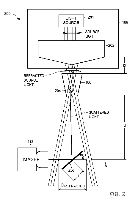
[0008] FIG. 2 is a block diagram 200 illustrating light paths between a light
source and an
imager that are associated with visual inspection system 100, according to an
embodiment of the
present disclosure;
[0009] FIG. 3 is a block diagram example illustrating a control system 300
associated with
visual inspection system 100, according to an embodiment of the present
disclosure;
[0010] FIG. 4 illustrates an example of a method flow 400 for characterizing
particles in a
fluid, according to an embodiment of the present disclosure;
[0011] FIG. 5 illustrates an example of a method flow 500 for characterizing
particles in a
fluid, according to an embodiment of the present disclosure; and
[0012] FIG. 6 illustrates an example plate holder 600 for receiving a plate
containing vessels
having fluid to be imaged.
DETAILED DESCRIPTION
[0013] The various concepts introduced above and discussed in greater detail
below may be
implemented in any of numerous ways, and the described concepts are not
limited to any
particular manner of implementation. Examples of implementations are provided
for illustrative
purposes.
[0014] FIG. 1 illustrates a visual inspection system 100, according to an
embodiment of the
present disclosure. The visual inspection system 100 includes a stage 102 that
is configured to
selectively agitate and to receive a plate 104, which may include one or more
vessels 106. In an
embodiment, the stage 102 includes a plate holder 105 that functions as an
adaptor between the
- 3 -
CA 03049524 2019-07-05
WO 2018/147858 PCT/US2017/017302
stage 102 and the plate 104 to facilitate receipt of the plate 104 for vessel
fluid imaging.
Although stage 102 is shown in FIG. 1 as receiving a single type of plate
holder 105,
embodiments include stage 102 being configured to accept any suitable number
of plate holders
having varying sizes and/or shapes. For example, stage 102 may be configured
having several
nested, adjustable, and/or interchangeable cavities or other suitable formed
parts that accept
different types of plate holders. In this way, visual inspection system 100
may facilitate the
testing of vessels included in plates of different types, sizes, and/or
shapes. The design of plate
holder 105 is further discussed below with reference to FIG. 6.
[0015] In various embodiments, vessels 106 may have one or more transparent
and/or opaque
portions. For example, vessels 106 may be entirely transparent or have
transparent bottoms with
the side walls being opaque. In any event, the visual inspection system 100
further includes an
illumination system 108 that is configured to illuminate the fluid contained
in the one or more
vessels 106 held by the stage 102, one or more imagers 112 that acquire images
of the fluid
contained within the one or more vessels 106 before and after vessel 106 is
agitated, and may
optionally include an optical system 110. The stage 102 and/or the
illumination system 108 may
also be configured to move in one or more axes to accommodate the inspection
of each vessel
106 included in plate 104 and to accommodate the testing of different sizes of
plates and vessels.
[0016] Additional detail regarding the components of the visual inspection
system 100 is
provided below. As an overview, the visual inspection system 100 is configured
to image the
fluid contained in one or more vessels 106 in an iterative manner. For
example, visual inspection
system 100 may be configured to iteratively image each vessel 106 included in
the plate 104 to
identify, for each individual vessel, particles suspended in the fluid for
that particular vessel. To
do so, visual inspection system 100 is configured to move stage 102 and/or
illumination system
- 4 -
CA 03049524 2019-07-05
WO 2018/147858 PCT/US2017/017302
110 to align each vessel 106 with the illumination system 108 for individual
vessel analysis. As
each vessel is tested, one or more images of the vessel are acquired before
and after the stage 102
is agitated in accordance with an agitation profile, as further discussed
below. Thus, in some
embodiments, one or more agitators (not shown) is coupled directly or
indirectly to the stage
102.
[0017] The illumination system 108 also includes one or more optical elements.
As used
herein, the term "optical elements" may apply to a single optical component
individually or a
combination of several optical components. For example, an optical element may
include one or
more single axicons, lenses, beam expanders, mirrors, etc. To provide another
example, an
optical element may include a combination of one or more axicons, lenses, beam
expanders,
mirrors, etc. In any event, illumination system 108 may include an optical
element that is
configured to refract source light into the vessel 106. The refracted source
light interacts with
particles suspended in the vessel's fluid to produce scattered light, which is
received by the
imager and used to acquire the one or more images before and after agitation.
Illumination
system 108 is also configured to refract the source light in such a way that
the refracted source
light is diverted away from the one or more imagers 112, thereby preventing
the source light
from washing out the scattered light and facilitating accurate
characterization of the individual
particles contained in the vessel's fluid. For example, particle
characteristics that may be
determined via the various embodiments described herein include the number
and/or size of
particles, particle morphology, density/buoyancy, etc.
[0018] The visual inspection system 100 analyzes one or more of the images
acquired before
and after the agitation of stage 102 to determine if particles are present in
the fluid contained in
the vessel 106. The one or more images may further be analyzed to count a
number of particles
- 5 -
CA 03049524 2019-07-05
WO 2018/147858 PCT/US2017/017302
present, to size particles, to track particle movement, or to characterize or
classify particles.
Particles may be, for example, dust or other contaminants, or protein
aggregates. In the present
disclosure, particles are discussed; however, it is to be understood that the
concepts of the
present disclosure also apply to bubbles or emulsions.
[0019] The agitation profile includes one or more agitation and non-agitation
periods. During
each agitation and non-agitation period of the agitation profile, a motion is
applied to the vessel
and is discontinued, respectively. For example, the agitation profile may
include a motion in an
agitation period followed by the motion being discontinued for a non-agitation
period. The
discontinuation of a motion may or may not include applying a force to the
stage 102 to
counteract the motion, such as applying a braking force. A braking force may
be, for example, a
friction force. The agitation and the non-agitation period may be the same
time periods or
different time periods depending on the particular fluid being tested. A
motion may be shaking,
vibration, spinning, applying ultrasonic energy, applying acoustic energy,
flipping, another
motion, or any suitable combination thereof. For example, assuming that the
stage 102 occupies
the x-y plane, an agitation motion may be a side-to-side motion of 1
millimeter in the x-axis
followed by an up-and-down motion of 1 millimeter in the y-axis, each of which
may occur for
100 milliseconds followed by a non-agitation period of 100 milliseconds.
[0020] Referring now back to FIG. 1, visual inspection system 100 may be
configured to test
any suitable number of vessels 106 having any suitable volume of fluid
contained therein. Thus,
in embodiments, the plate 104 may be any suitable size and/or shape having any
suitable number
of vessels 106 disposed thereon. For example, plate 104 may be a microplate
including any
suitable number of vessels 106, such as 6, 24, 96, 384, 1536, etc. The vessels
106 may be
arranged in a pattern on the plate 104, such as a 2:3 rectangular matrix, for
example, in the case
- 6 -
CA 03049524 2019-07-05
WO 2018/147858 PCT/US2017/017302
of a standard microplate implementation. In embodiments in which plate 104 is
implemented as
a microplate, vessels 106 may constitute wells on the microplate that contain
a fluid to be
inspected. Each vessel 106 may be configured to receive any suitable volume of
fluid depending
on the size and configuration of plate 104. For example, each vessel 106 may
hold a volume of
fluid in the range of tens of nanoliters up to several milliliters. For
example, when plate 104 is
implemented as a 96-well microplate, each vessel may hold a volume up to 200
microliters.
[0021] Visual inspection system 100 includes an illumination system 108, which
may include
any suitable number and/or type of light sources configured to generate source
light.
Illumination system 108 also includes one or more optical elements configured
to refract the
source light such that the source light is directed through the vessel 106 at
an angle. This is
particularly useful, for example, because the particles suspended in the fluid
may be translucent,
and this ensures that the particles scatter the source light to produce
scattered light for proper
imaging. If the source light is not refracted, and is instead provided in a
manner that is aligned
with the vessel's center axis, such translucent particles would not scatter
the source light to
provide scattered light that can be effectively used for imaging. An example
of the optical
characteristics provided by illumination system 108 are shown in FIG. 2 with
reference to the
example embodiment of visual inspection system 100 shown in FIG. 1. However,
it will be
understood that visual inspection system 100 and/or illumination system 108
may be
implemented in a variety of different configurations and with different
components to facilitate
the embodiments described herein, as further discussed below.
[0022] With continued reference to FIGs. 1 and 2, illumination system 108 may
include one or
more light sources 201, which may include any suitable type of light source
configured to
generate source light having any suitable wavelength or range of wavelengths.
For example,
- 7 -
CA 03049524 2019-07-05
WO 2018/147858 PCT/US2017/017302
light source 201 may be implemented as a light-emitting diode (LED) light
source configured to
produce source light at a single wavelength, a selectable range of
wavelengths, or a wide band of
wavelengths. To provide an illustrative example, the light source 201 may be
implemented as an
LED light source configured to provide source light over a wide range of
wavelengths, with a
particular wavelength or range of wavelengths being selected from within the
wide range of
wavelengths for vessel imaging.
[0023] In embodiments, illumination system 108 also includes one or more
optical elements
configured to refract the source light into the vessel 106 and towards imager
112 in a specific
manner, as shown in FIG. 2 and further discussed below. In particular,
embodiments include
illumination system 108 implementing any suitable number and/or type of
axicons, individual
optical lenses, a train of lenses, etc., configured to refract the source
light such that the refracted
source light enters the vessel under test at an angle with respect to the
vessel's center axis. The
particles suspended in the vessel's fluid then scatter the refracted source
light, which is provided
to imager 112 while the refracted source light is largely (or entirely)
diverted away from the
imager 112.
[0024] In an embodiment, to realize the aforementioned optical
characteristics, illumination
system 108 may include an axicon 202 or other suitable type of optical element
configured to
refract the source light to ensure that the entire volume of fluid contained
in the vessel 106 is
properly illuminated for imaging. For example, axicon 202 may be selected
having a suitable
cone angle to refract the source light such that, for a given distance "D"
from the vessel 106, the
entire volume of fluid contained in the vessel 106 is properly illuminated for
imaging. In an
embodiment, axicon 202 may be implemented as an axicon having a 90 degree apex
angle and a
- 8 -
CA 03049524 2019-07-05
WO 2018/147858 PCT/US2017/017302
1" diameter, such as axicon stock number 83-779, which is manufactured by
Edmund Optics,
Inc. of Barrington, New Jersey at the time of this writing.
[0025] When using axicon 202, it is preferable that light source 201 not be
implemented as a
laser light source or other light source configured to generate coherent
light, as coherent light
passing through an axicon results in the generation of interference patterns
known as Bessel
beams, which are undesirable for imaging vessel 106. In contrast, it is
preferable that the light
source 202 generate incoherent source light when axicon 202 is used as the
optical element.
[0026] Furthermore, a mirror 206 or other suitable optical component may be
strategically
sized and placed with respect to the vessel 106 to direct the scattered light
to imager 112 while
diverting the refracted source light away from the imager 112. For example,
mirror 206 may be
placed in line with the center axis of vessel 106, light source 201, and
axicon 202, as shown from
the side view illustrated in FIG. 2. In an embodiment, mirror 206 is disposed
a distance from the
bottom of vessel 106 to reflect the scattered light to imager 112. Moreover,
mirror 206 may be
sized such that the refracted source light is not reflected by mirror 206, and
is instead diverted
away from the imager 112.
[0027] To provide an illustrative example with reference to FIG. 2, mirror 206
may be a
circular mirror having a radius "r," with its center disposed a distance "d"
from the bottom of
vessel 106. As shown in FIG. 2, axicon 202 refracts the source light through
the vessel 106 to
illuminate the vessel for fluid imaging. For a particular refractive index of
axicon 202 (e.g.,
caused by the axicon's conical angle and its composition) and distance D
between the axicon 202
and the vessel 106, the refracted source light forms a cone of refracted light
having a diameter
0 REFRACTED in the plane "p" that intersects the center of the mirror 206 and
is orthogonal to the
- 9 -
CA 03049524 2019-07-05
WO 2018/147858 PCT/US2017/017302
center axis of vessel 106. Moreover, the scattered light is contained within
this cone of refracted
light. In embodiments, mirror 206 may have a radius r, be placed a distance d
from the bottom
of vessel 106, and be positioned at an angle 0 from the plane p such that 2r
cos 0 < OREERACTED.
Given these parameters, the scattered light is directed towards the imager 112
while the cone of
refracted light is diverted away from the imager 112. For example, as shown in
FIG. 2, the
imager 112 and the vessel 106 may be placed at 90 degrees with respect to one
another, and the
mirror 206 may thus form an angle 0 of 45 degrees with respect to the center
axis of vessel 106.
[0028] Without diverting the retracted source light in this manner, it would
drown out the
scattered light and prevent proper imaging analysis of the fluid contained in
the vessel. An
example of such an arrangement is shown in FIG. 2 with a single particle 204.
However, it will
be understood that the fluid contained in the vessel 106 may include any
number of particles or
bubbles, each scattering the refracted source light, with the scattered light
from each particle
being received by the imager 112.
[0029] Due to the refraction of the source light in this way, illumination
system 108 provides
light with a high degree of intensity to any particles that are suspended in
the fluid contained in
vessel 106. In doing so, imager 112 may implement a smaller aperture on its
main camera lens,
thus achieving a larger depth of field than is possible in conventional well-
plate inspection
systems. In various embodiments, imager 112 uses a telecentric lens that
implements an aperture
size of f/6 or smaller, and preferably between f/8 to f/11 for light source
201 being
approximately 3 Watts. As the aperture size is dependent upon the light source
brightness,
higher power light sources may allow for even smaller aperture sizes than
f/11. Traditional well
plate imagers, on the other hand, typically implement aperture sizes around
f/2 to f/2.8, which
yields a very narrow depth of field.
- 10 -
CA 03049524 2019-07-05
WO 2018/147858 PCT/US2017/017302
[0030] This advantageously allows for all particles present in the fluid to be
in focus and
analyzed at the same time, i.e., by capturing and analyzing images of the
entire contents of vessel
106. This is in contrast to typical microscopy techniques, whereby depth
slices of the vessel's
fluid need to be obtained to properly image larger fluid volumes, as the
imager's main camera
lens aperture needs to be opened to improve visibility. In this way,
illumination system 108
allows the number and size of the particles in the entire volume of fluid
contained in vessel 106
to be characterized via image analysis at the same time.
[0031] In various embodiments, the position of the axicon 202 or other
suitable optical element
in relation to the vessel 106 may be fixed or adjustable. For example, the
axicon 202 may be
mounted within illumination system 108 at a fixed position. Illumination
system 108 may then
be disposed a distance above the stage 102 such that the axicon 202 is offset
a desired distance
"D" from the vessel 106, as shown in FIG. 2, to ensure the proper optical
characteristics for
image analysis depending on the dimensions of the vessel (or plate) being
tested, the
characteristics of the optical element (e.g., axicon 202).
[0032] To provide an illustrative example, if the plate 104 is a 96-well
microplate, then each
vessel 106 may have standard dimensions such as height and diameter.
Furthermore, axicon 202
may be selected having a 90 degree apex angle and a 1" diameter. Thus, the
axicon 202 and the
illumination system 108 may be disposed to provide a distance D of 12 mm
between the axicon
202 and the vessel 106 to ensure the desired optical characteristics as shown
in FIG. 2. Such
embodiments may be particularly useful, for example, when visual inspection
system 100 is
utilized to test a single type of plate having vessels with known,
predetermined dimensions. Of
course, for other types of optical elements and vessel sizes, the distance D
may be greater than or
-11-
CA 03049524 2019-07-05
WO 2018/147858 PCT/US2017/017302
less than 12 mm to ensure the proper optical characteristics, i.e., that the
source light is diverted
from the imager 112 while illuminating the entire volume of fluid in vessel
106.
[0033] However, illumination system 108 may also be configured to test a
variety of different
plate types, which have different vessel sizes. To provide another
illustrative example, plate 104
may include 96-well microplates for one testing configuration and 24-well
microplates for
another. Continuing this example, vessels included in a 24-well microplate
have a larger height
and diameter than vessels included in a 96-well microplate. Therefore, the
optical characteristics
resulting from the axicon 202 being offset a distance D from the vessel 106
may provide
desirable optical characteristics for a 96-well microplate, but not for a 24-
well microplate. There
are several embodiments of visual inspection system 100 to address such
issues.
[0034] For example, embodiments include illumination system 108 being
implemented as one
of several modular components, with each modular component being used for each
different type
of plate being tested. For instance, different modular implementations of
illumination system
108 may include the axicon 202 being disposed at different locations within
the illumination
system 108, resulting in different distances D for different modular
illumination systems 108
disposed the same distance above the stage 102. To provide another example,
different modular
illumination systems 108 may have axicons with different conical angles,
resulting in refracting
the source light at different angles in each case.
[0035] Additionally or alternatively, stage 102 may include one or more
receptacles, fasteners,
etc., positioned to define various preset distances D between the axicon 202
and the vessel 106
for different modular implementations of illumination system 108. In this way,
different
modular implementations of illumination system 108 may be swapped out
depending on the
- 12 -
CA 03049524 2019-07-05
WO 2018/147858 PCT/US2017/017302
particular type of plate being tested to ensure that the desired optical
characteristics are
maintained for different sizes and shapes of vessel 106.
[0036] In other embodiments, illumination system 108 may be a single design
with the distance
D being adjustable. Using the previous example, when testing a 24-well
microplate, the axicon
202 may need to be placed a distance D1 from the vessel 106. However, when
testing a 96-well
microplate, the axicon 202 may need to be placed a different distance D2 from
the vessel 106.
Although two different modular designs of illumination system 108 could
address this issue, it
may be preferable to have a universal adjustable design to facilitate the
testing of different plate
types. Thus, embodiments include illumination system 108 being configured such
that the
distance D is adjustable. In still further embodiments, the stage 102 may be
configured to move
in the x-, y-, and z-axes to allow adjustment of the distance D in addition to
aligning each vessel
106 with the illumination system 108 while being tested.
[0037] In still additional embodiments, other optical components may be
implemented in
conjunction with axicon 202 or another optical element, as the case may be, to
provide another
technique for adjusting the optical properties of illumination system 108. For
example,
illumination system 108 may include one or more beam expanders disposed
between the light
source 201 and the axicon 202, which are configured to vary the or "tune" the
diameter of the
beam illuminating the beak of axicon 202 (i.e., before the source light is
refracted). In other
words, embodiments include varying the diameter of the source light entering
the axicon 202 and
the axicon cone angle to adjust the illumination of the fluid in vessel 106.
Thus, the position
and/or type of such optical components may also be varied among different
modular designs of
illumination system 108, as discussed herein.
- 13 -
CA 03049524 2019-07-05
WO 2018/147858 PCT/US2017/017302
[0038] In any event, to facilitate such adjustments, embodiments include the
stage 102, the
illumination system 108, the axicon 202 and/or other optical components
implemented by
illumination system 108 being movably mounted within visual inspection system
100. For
example, the illumination system 108 and/or the axicon 202 may be mounted to a
linear actuator
or other suitable drive mechanism to allow a desired distance D to be obtained
depending on the
type of plate 104 being tested. To provide another example, the stage 102 may
include one or
more linear actuators or other suitable drive mechanisms to allow displacement
in the z-axis to
adjust the distance D depending on the type of plate 104 being tested. In this
way, the positions
of one or more of the stage 102, the illumination system 108, and/or the
axicon 202 may be
adjusted to ensure that the desired optical characteristics are maintained for
proper imaging when
testing vessels having different sizes and shapes.
[0039] Regardless of the particular implementation of illumination system 108,
embodiments
include the source light 201 being directed downwards into the top of each
vessel 106 to
facilitate imaging the entirety of the fluid contained in the vessel 106 being
tested. Again, to do
so, the light that is scattered from particles suspended in the fluid may be
directed to imager 112
while the refracted source light is diverted away from the imager 112. In some
embodiments,
imager 112 may receive the scattered light via optical system 110, as shown in
FIGs. 1 and 2.
However, optical system 110 is an optional component of visual inspection
system 100, and may
not be present in other embodiments. For example, as shown in FIG. 1, optical
system 110 may
be positioned beneath stage 102 and include one or more mirrors, lenses, etc.
(e.g., mirror 206, as
shown in FIG. 2) configured to reflect the light scattered from the suspended
particles to imager
112. In this way, optical system 110 may facilitate imager 112 being
positioned along a separate
axis than that of illumination system 108, allowing for additional design
flexibility.
- 14 -
CA 03049524 2019-07-05
WO 2018/147858 PCT/US2017/017302
[0040] In other embodiments, illumination system 108 may be configured to
illuminate the
volume of fluid contained in vessel 106 by providing source light from the
side of the vessel 106.
To do so, illumination system 108 may include an additional optical system,
such as optical
waveguides, for example, that direct the light provided by light source 201 to
illuminate the
vessel 106 from the side. This optical system is not shown in FIGs. 1-2 for
purposes of brevity,
but may include any suitable combination of light sources, optical elements,
waveguides, etc.,
configured to provide scattered light to imager 112 while diverting the source
light from the
imager 112. For example, an optical sub with optical waveguides may be
integrated as part of
the plate being tested or as part of the stage 102, and light source 201 may
be disposed within
visual inspection system 100 to provide full illumination of each vessel as it
is tested.
[0041] In still other embodiments, illumination system 108 and imager 112 may
be aligned
along the same axis, thereby eliminating the need for optical system 110. For
example, imager
112 may be mounted beneath stage 102 in place of optical system 110, receiving
the scattered
light directly as it exits the bottom of vessel 106. Of course, such
implementations may be
accompanied by spacing the imager 112 a distance beneath stage 102 to ensure
that the scattered
light is received by the imager 112 while the refracted source light is mostly
(or entirely)
diverted from the imager 112.
[0042] In various embodiments, imager 112 is configured to capture one or more
images
and/or video over one or more consecutive frames. For example, imager 112 may
selectively
capture images and/or video at specific times in response to commands received
from a
controller, which is further discussed below with reference to FIG. 3. The
imager 112 may
capture the images and/or video in a manner that is synchronized with the
agitation of stage 102,
allowing the controller to analyze images at specific times prior to and after
the agitation of stage
- 15 -
CA 03049524 2019-07-05
WO 2018/147858 PCT/US2017/017302
102 (and thus the agitation of fluid contained in vessel 106). When video is
captured,
embodiments include the controller extracting video frames that correspond to
desired time
periods, e.g., prior to and after the agitation of stage 102.
[0043] Imager 112 may include any suitable combination of hardware and/or
software to
facilitate this functionality, such as image sensors, optical stabilizers,
image buffers, frame
buffers, frame grabbers, charge-coupled devices (CCDs), complementary metal
oxide
semiconductor (CMOS) devices, etc. Furthermore, imager 112 may include one or
more
telecentric lenses to provide image magnification of the vessel 106 that is
independent of the
vessel's distance or position in the field of view. Moreover, imager 112 may
communicate with
the controller (as discussed below with reference to FIG. 3), and store
acquired images and/or
video for image analysis to the controller for image analysis. Alternatively,
imager 112 may
store the images and/or video locally in any suitable type of memory, and this
memory may be
accessed by a controller for image analysis. To provide another example,
imager 112 may be
implemented as a "smart camera," with image processing logic built into the
camera using any
suitable techniques, such as field programmable gate array (FPGA)
technologies, for example.
To provide yet another example, imager 112 may be implemented as part of a
plenoptic 3D
camera system.
[0044] Although a single imager 112 is shown in FIGs. 1-2, embodiments of
visual inspection
system 100 may include multiple imagers 112 to acquire images of the vessel
under test from
different locations. Such embodiments may be particularly useful, for example,
to implement
faster parallel imaging, for wide angle versus narrow angle imaging, for small
area versus large
area imaging, for color versus infrared imaging, and so forth. To facilitate
this functionality,
optical system 110 may be configured with optical components (e.g., beam
splitters, optical
- 16 -
CA 03049524 2019-07-05
WO 2018/147858 PCT/US2017/017302
waveguides, etc.) to split the scattered light from the vessel under test such
that each individual
imager 112 receives the same scattered light. Such embodiments may be
particularly useful, for
example, when multiple image analyses need to be performed for the same vessel
under test,
allowing several images to be captured and imaged in parallel.
[0045] FIG. 3 is a block diagram example illustrating a control system 300
associated with
visual inspection system 100, according to an embodiment of the present
disclosure. As further
discussed below, control system 300 may include a controller 302 that is
configured to
communicate with and control various components of visual inspection system
100, such as
illumination system 340, motion actuators/agitators 360, and/or imager 380,
for example.
Furthermore, in an embodiment, control system 300 is configured to facilitate
fully autonomous
or semi-autonomous operation of visual inspection system 100. To do so,
control system 300
may support the automatic analysis of a number of vessels that are included on
a plate to
determine a number and/or size of particles contained in the fluid of each
vessel.
[0046] In an embodiment, illumination system 340 and imager 360 may be an
implementation
of illumination system 108 and imager 112, respectively, as discussed herein
with respect to
FIGs. 1-2. Furthermore, motion actuators/agitators 360 may represent one or
more motors,
servos, actuators (e.g., piezo actuators), etc., associated with one or more
components of visual
inspection system 100. For example, motion actuators/agitators 360 may include
linear actuators
associated described above that may enable adjustments to be made to the
position of the stage
102, the axicon 202, and/or the illumination system 108. To provide another
example, motion
actuators/agitators 360 may include one or more agitators configured to
agitate stage 102.
[0047] Controller 302 may be implemented, for example, as any suitable
combination of
hardware and/or software coupled to or otherwise in communication with
illumination system
- 17 -
CA 03049524 2019-07-05
WO 2018/147858 PCT/US2017/017302
340, motion actuators/agitators 360, and/or imager 380. For example,
controller 302 may be
implemented as device mounted to or integrated as part of stage 102, or
controller 302 may be
located remote from visual inspection system 100. In any event, controller 302
may be coupled
to one or more of illumination system 340, motion actuators/agitators 360,
and/or imager 380 via
wired links, wireless links, or any suitable combination thereof. Therefore,
links 320, 322,
and/or 324 may represent one or more wired and/or wireless links to facilitate
communications
between controller 302 and one or more of illumination system 340, motion
actuators/agitators
360, and/or imager 380. Although three separate links 320, 322, and 324 are
shown in FIG. 3, it
will be understood that controller 302 may communicate with one or more of
illumination
system 340, motion actuators/agitators 360, and/or imager 380 via any suitable
number of links,
including a single shared link.
[0048] To facilitate communication with and control of these components,
controller 302 may
include a processing unit 304, a communication unit 306, and a memory unit
308. Processing
unit 304 may be implemented as any suitable type and/or number of processors,
such as a host
processor of controller 302, for example. To provide additional examples,
processing unit 304
may be implemented as an application specific integrated circuit (ASIC), an
embedded
processor, a central processing unit associated with controller 302, etc.
Processing unit 304 may
be coupled with and/or otherwise configured to communicate, control, operate
in conjunction
with, and/or affect operation of one or more of communication unit 306 and/or
memory unit 308
via one or more wired and/or wireless interconnections, such as any suitable
number of data
and/or address buses, for example. These interconnections are not shown in
FIG. 3 for purposes
of brevity.
- 18 -
CA 03049524 2019-07-05
WO 2018/147858 PCT/US2017/017302
[0049] For example, processing unit 304 may be configured to retrieve,
process, and/or analyze
data stored in memory unit 308, to store data to memory unit 308, to replace
data stored in
memory unit 308, to control various functions associated with illumination
system 340, motion
actuators/agitators 360, and/or imager 380, to analyze images or video frames
captured by
imager 380 and stored in memory unit 308 to identify the number and size of
particles contained
in the fluid of vessel being tested, etc. Additional details associated with
such functions are
further discussed below.
[0050] Communication unit 306 may be configured to support any suitable number
and/or type
of communication protocols to facilitate communications between controller 302
and one or
more of illumination system 340, motion actuators/agitators 360, and/or imager
380.
Communication unit 306 may be configured to facilitate the exchange of any
suitable type of
information between controller 302 and one or more of illumination system 340,
motion
actuators/agitators 360, and/or imager 380 (e.g., via links 320, 322, and/or
324), and may be
implemented as any suitable combination of hardware and/or software to
facilitate such
functionality. For example, communication unit 306 may be implemented with any
number of
wired and/or wireless transceivers, modems, ports, input/output interfaces,
connectors, antennas,
etc.
[0051] In accordance with various embodiments, memory unit 308 may be a
computer-
readable non-transitory storage device that may include any suitable
combination of volatile
(e.g., a random access memory (RAM), or non-volatile memory (e.g., battery-
backed RAM,
FLASH, etc.). Memory unit 308 may be configured to store instructions
executable on
processing unit 304. These instructions may include machine readable
instructions that, when
executed by processing unit 304, cause processing unit 304to perform various
acts as described
- 19 -
CA 03049524 2019-07-05
WO 2018/147858 PCT/US2017/017302
herein. Although the various functions of controller 302 are described herein
in terms of
execution of instructions stored in memory unit 308 via processing unit 304,
it will be
understood that equivalent functions may be realized exclusively via hardware
components (e.g.,
circuit components) or hardware components (e.g., those implemented via
communication unit
306) working in conjunction with processing unit 304 executing instructions
stored in memory
unit 308. Memory unit 308 may also be configured to store any other suitable
data used in
conjunction with visual inspection system 100, such as images or video frames
captured by
imager 380.
[0052] Control module 309 is a region of memory unit 308 configured to store
instructions,
that when executed by processing unit 304, cause processing unit 304 to
perform various acts in
accordance with applicable embodiments as described herein. In an embodiment,
control
module 309 includes instructions that, when executed by processing unit 304,
cause processing
unit 304 to transmit one or more commands to illumination system 340 (e.g.,
via link 320) to
control the state of illumination system 340.
[0053] For example, illumination system 340 may include one or more light
sources, such as
light source 201, for example, as discussed above with reference to FIGs. 1-2.
In some
embodiments, the light source may be on continuously as each vessel is
analyzed, and is not
turned off when the stage 102 moves to test a new vessel. In other
embodiments, the light source
may be turned on and off in a manner that is synchronized to the agitation of
the stage 102 and
the images captured by imager 380, but is not varied with respect to different
agitation profiles or
other parameters such as different fluid volumes, vessel sizes, etc. For
example, the light source
may be turned on during the entire agitation profile and turned off while the
stage 102 moves to
align the next vessel for testing. Embodiments include processing unit 304
executing
- 20 -
CA 03049524 2019-07-05
WO 2018/147858 PCT/US2017/017302
instructions stored in control module 309 to cause the light source included
in illumination
system 340 to turn on and off in such a manner.
[0054] Embodiments also include processing unit 304 executing instructions
stored in control
module 309 such that the light source may be turned on and off in a manner
that is varied with
respect to different agitation profiles or other parameters such as different
fluid volumes, the
viscosity of the fluid, the color of the fluid, etc. In other words, different
agitation profiles may
be stored in memory unit 308, which are executed for a particular plate 104
based upon the size
of the vessel being tested and the characteristics of the fluid contained in
the vessel.
Additionally, image acquisition profiles may be stored in memory unit 308 that
identify the time
periods, during each agitation profile, when images are acquired for each
vessel. For example,
an agitation profile may specify an agitation period and a non-agitation
period for a 96-well
microplate. However, two different 96-well microplates may contain fluids
having different
characteristics (e.g., one fluid being a higher viscosity than another).
Therefore, memory unit
308 may associate two different image acquisition profiles for the same
agitation profile, with
one being applied for one type of fluid being tested and the other image
acquisition profile being
applied for another type of fluid being tested.
[0055] Embodiments also include processing unit 304 executing instructions
stored in control
module 309 to change other parameters associated with the light source
included in illumination
system 340. For example, controller 302 may adjust the intensity of light
output by the light
source, set a wavelength or range of wavelengths used by the light source,
etc.
[0056] Additionally, processing unit 304 may execute instructions stored in
control module
309 to control the state of other components of visual inspection system 100.
For example,
controller 302 may read the current position of stage 102 and transmit one or
more commands
-21 -
CA 03049524 2019-07-05
WO 2018/147858 PCT/US2017/017302
(e.g., via link 322) to a motor or other suitable actuator to move the stage
102 to a new location
so that the next vessel 106 within the plate 104 may be tested. To provide
another example,
controller 302 may transmit one or more commands to a motor or other suitable
actuator to move
the axicon 202 included in the illumination system 340, the illumination
system 304, and/or the
stage 102 to adjust the physical location of various components and/or the
optical characteristics
with such components. For example, a particular modular illumination system
108 may be
selected, a particular optical element may be identified and/or placed, a beam
diameter of the
source light be adjusted, the distance D may be ascertained, etc., as part of
the image acquisition
profile stored in memory unit 308 and associated with a particular tray being
tested, as discussed
above.
[0057] To facilitate the testing of different plate types and/or different
fluid types,
embodiments include controller 302 manually, automatically, or semi-
automatically selecting an
agitation profile and an image acquisition profile for a particular type of
plate and/or fluid being
tested. For example, a user may provide a user input to controller 302 (user
interface not shown)
to select an agitation profile and/or an image acquisition profile when a new
type of plate and/or
fluid needs to be tested. To provide another example, controller 302 may
receive sensor data
measurements from various sensors positioned on stage 102 (not shown) to
identify the type of
tray positioned on stage 102 from weight measurements, plate dimension
measurements, vessel
dimension measurements, etc. Once the plate type is identified (e.g., a 96-
well or a 24-well
microplate), processing unit 306 may then execute instructions stored in
control module 309 to
correlate a stored agitation profile and image acquisition profile to the
identified plate type. In
this way, visual inspection system 100 may automatically adapt the testing
setup to different
plate types as these different types of plates are detected.
- 22 -
CA 03049524 2019-07-05
WO 2018/147858 PCT/US2017/017302
[0058] Furthermore, processing unit 304 may execute instructions stored in
control module 309
to control the operation of imager 380 (e.g., via link 324). That is,
controller 302 may cause the
imager 380 to acquire images in accordance with a particular image acquisition
profile that is
synchronized to the current agitation profile. For example, processing unit
304 may execute
instructions stored in control module 309 to cause imager 380 to capture one
or more images or
video frames prior to an agitation period and after the agitation period
(e.g., during a non-
agitation period). These images may then be stored in memory unit 308 and
analyzed to
determine the size and number of particles suspended in a fluid contained
within the vessel under
test, as further discussed below.
[0059] Image analysis module 311 is a region of memory unit 308 configured to
store
instructions, that when executed by processing unit 304, cause processing unit
304 to perform
various acts in accordance with applicable embodiments as described herein. In
an embodiment,
image analysis module 311 includes instructions that, when executed by
processing unit 304,
cause processing unit 304 to analyze one or more images and/or video frames
acquired by
imager 380 to determine the size and number of particles suspended in a fluid
contained within
the vessel under test.
[0060] In various embodiments, processing unit 304 may execute instructions
stored in image
analysis module 311 to perform image analysis of the images acquired via
imager 380 in
accordance with any suitable techniques, such as frame differencing,
background subtraction,
and/or or minimum intensity ("MinIP") techniques, for example. To provide
another example, in
an embodiment in which imager 380 is implemented as part of a plenoptic camera
system,
processing unit 304 may execute instructions stored in image analysis module
311 to identify the
-23 -
CA 03049524 2019-07-05
WO 2018/147858 PCT/US2017/017302
depth of the objects from a predetermine data set, and use this information to
determine the
amount of particles in an imaged well without necessarily performing
background subtraction.
[0061] For example, imager 380 may capture a first image using the scattered
light that is
received as a result of any particles suspended in the fluid of the vessel
being tested. However,
other artifacts such as dust on the outside of the vessel or scratches may
also cause the source
light provided by the illumination system 340 to be scattered and, in turn,
received by imager
380. Therefore, this first captured image may be a "background" image, which
indicates the
image of the vessel and the position of particles prior to agitation.
Continuing this example,
imager 380 may capture a second "analysis" image after stage 102 has been
agitated, which
shows a new position of the particles within the vessel as a result of their
movement from the
agitation. However, any light that is scattered as a result of artifacts will
not change as a result of
plate agitation. Therefore, processing unit 304 may execute instructions
stored in image analysis
module 311 to subtract the background image from the analysis image to
generate a difference
image, which effectively filters out static images. The difference image may
then be analyzed to
determine the number and size of particles suspended in the fluid.
[0062] To provide another example, a MinIP imaging technique may be
implemented, in which
case imager 380 may capture several images after plate agitation is complete.
In particular, plate
agitation may occur, resulting in movement of particles suspended in the
vessel's fluid, and these
particles may continue to move for some brief period of time after the
agitation has stopped.
Thus, several images may be captured after plate agitation has stopped, and a
minimum intensity
projection may be created using all (or some subset of ) these captured
images. In this way,
although each of the acquired images may contain representations of static
features such as
scratches and dust, these features will not appear in the MinIP assuming that
the particles have
- 24 -
CA 03049524 2019-07-05
WO 2018/147858 PCT/US2017/017302
been sufficiently well agitated and the particles are mobile during image
acquisition. In an
embodiment, the MinIP may then be subtracted from all images in the image set
(i.e., the
acquired images used to create the Min1P) to generate an image stack, with
bright features
corresponding to particles. In this way, the number and/or size of particles
in a vessel's fluid
may then be characterized using the image stack.
[0063] FIG. 4 illustrates an example of a method flow 400 for characterizing
particles in a
fluid, according to an embodiment of the present disclosure. In various
embodiments, one or
more regions of method 400 (or the entire method 400) may be implemented by
any suitable
device. For example, one or more regions of method 400 may be performed by
controller 302,
illumination system 340, and/or imager 380, as shown in FIG. 3. Method 400
represents the
various steps performed during the testing of a single vessel, which may be
repeated for each
vessel within a plate (e.g., plate 104, as shown in FIG. 1).
[0064] Method 400 may begin by generating source light (block 402). This may
include, for
example, controller 302 causing a light source (e.g., light source 201, as
shown in FIG. 2) to turn
on and illuminate a vessel being tested (block 402). This may also include,
for example, the light
source generating light in a default state (e.g., in a continuous manner)
(block 402).
[0065] Method 400 may include refracting the source light through one or more
optical
elements to provide refracted source light (block 404). This may include, for
example, directing
the source light through an axicon to refract the source light, as shown in
FIG. 2 (block 404).
Method 400 may also include directing the refracted source light into the
fluid contained in the
vessel under test (block 404).
[0066] Method 400 may include supplying scattered light to an imager (e.g.,
imager 112, as
shown in FIG. 1) as a result of an interaction between the refracted source
light and particles
- 25 -
CA 03049524 2019-07-05
WO 2018/147858 PCT/US2017/017302
suspended in the fluid (block 406). This may include, for example, the
refracted source light
being directed into the vessel at an angle (i.e., not directly from the top of
the vessel) to cause
particles in the fluid to scatter the refracted source light, which is then
supplied to the imager
(block 406). The refracted source light may also be diverted away from the
imager (block 408).
[0067] Method 400 may include acquiring sequential images using the scattered
light (block
410). This may include, for example, the imager acquiring a background image
before the vessel
under test is agitated and an analysis image after the vessel has been
agitated (block 410). This
may also include, for example, storing the background image and the analysis
image in a
memory (e.g., memory unit 308) (block 410).
[0068] Method 400 may include characterizing particles contained in the fluid
using the
acquired sequential images (block 412). This may include, for example,
generating a difference
image by subtracting the background image (block 410) from the analysis image
(block 410) to
produce a difference image (block 412). Method 400 may include determining a
size and
number of particles contained in the fluid based upon an analysis of this
difference image (block
412).
[0069] FIG. 5 illustrates an example of a method flow 500 for characterizing
particles in a
fluid, according to an embodiment of the present disclosure. In various
embodiments, one or
more regions of method 500 (or the entire method 500) may be implemented by
any suitable
device. For example, one or more regions of method 500 may be performed by
controller 302,
illumination system 340, and/or imager 380, as shown in FIG. 3. Method 500
represents the
various steps performed for iteratively testing several vessels within a plate
(e.g., plate 104, as
shown in FIG. 1).
- 26 -
CA 03049524 2019-07-05
WO 2018/147858 PCT/US2017/017302
[0070] Method 500 may begin by acquiring images of the fluid using scattered
light (block
502). This may include, for example, controller 302 causing an imager (e.g.,
imager 112 as
shown in FIG. 1) to capture an image of the fluid in the vessel being tested
using scattered light
before and after plate agitation, which is received by the imager (block 502).
This may also
include, for example, controller 302 causing an imager (e.g., imager 112 as
shown in FIG. 1) to
capture several images of the fluid in the vessel being after plate agitation
in accordance with
MinIP imaging techniques, which is received by the imager (block 502). Again,
this scattered
light may be the result of refracted source light (e.g., from a light source
201 in conjunction with
axicon 202, as shown in FIG. 2) interacting with particles suspended in the
vessel's fluid,
whereas the refracted source light itself is diverted away from the imager
(block 502). Method
500 may also include storing the acquired images in a suitable memory (e.g.,
memory unit 308,
as shown in FIG. 3) (block 502).
[0071] Method 500 may include counting and sizing the particles using the
acquired images
image (block 504). This may include, for example, performing any suitable type
of image
analysis on the images to identify the overall number of particles suspended
in the fluid and the
size of these particles (block 504). For example, the image analysis may be
performed in
accordance with the imaging techniques described herein with respect to FIG. 3
(block 504).
Method 500 may also include storing the result of this analysis in a suitable
memory (e.g.,
memory unit 308, as shown in FIG. 3) (block 504).
[0072] Once the particles in the vessel's fluid have been sized and counted
(block 504),
method 500 may include determining whether all vessels selected for analysis
have been
analyzed (block 506). This determination may be made, for example, by tracking
the overall
number of vessels that have been analyzed for a given type of plate, which has
a known number
-27 -
CA 03049524 2019-07-05
WO 2018/147858 PCT/US2017/017302
of vessels (e.g., 24 or 96), and determining if this count is less than or
equal to the overall
number of vessels (block 506). This determination may also be made, for
example, by a user
entering any suitable number of vessels for analysis (which may be less than
all vessels in the
plate), tracking the overall number of vessels that have been analyzed, and
determining if this
count is less than or equal to the entered number of vessels being tested
(block 506). In any
event, if additional vessels need to be analyzed, then method 500 continues
(block 508).
However, if the analyzed vessel is the last vessel to be analyzed, then method
500 ends. Upon
ending, method 500 may include storing or outputting a report (e.g., in memory
unit 308, as
shown in FIG. 3) of the analysis of each vessel, which may include the number
and size of
particles contained in the fluid in each vessel. Once this report is
completed, a user may view the
report and/or start the process over with a new plate.
[0073] In the event that additional vessels need to be analyzed, method 500
may include
positioning the next vessel for analysis (block 508). This may include, for
example, a controller
(e.g., controller 302, as shown in FIG. 3) causing one or more actuators
and/or motors to move
the stage (e.g., stage 102, as shown in FIG. 1) in the x- and/or y-axes to
align the next vessel to
be tested with the illumination system (e.g., illumination system 108) (block
508). This may also
include, for example, a controller causing one or more actuators and/or motors
to move the
illumination system (e.g., illumination system 108) in the x- and/or y-axes to
align the next
vessel to be tested with the illumination system (block 508).
[0074] In any event, once the next vessel is positioned (block 508), method
500 may repeat the
process of acquiring images of the fluid contained in this new vessel (block
502). Thus, method
500 is restarted to count and size the particles in the new vessel. In this
way, method 500 may
- 28 -
CA 03049524 2019-07-05
WO 2018/147858 PCT/US2017/017302
iteratively analyze any suitable number of vessels to determine the number and
size of particles
contained in the fluid of each analyzed vessel.
[0075] FIG. 6 illustrates an example plate holder 600 for receiving a plate
containing vessels
having fluid to be imaged. In an embodiment, plate holder 600 is an
implementation of plate
holder 105, as shown and discussed above with reference to FIG. 1. Again, the
plate holder 600
shown in FIG. 6 is an example of one type of plate holder that may be
implemented, and may
include more, less, or alternate components. For example, although plate
holder 600 is shown in
FIG. 6 as including two springs 606.1-606.2 and four spring plungers 610.1-
610.4, embodiments
include plate holder 600 having any suitable number and/or type of springs and
spring plungers.
[0076] In an embodiment, plate holder 600 provides quick and consistent
alignment of vessels
with the imaging components of a visual inspection system. To do so, plate
holder 600 includes
a main housing 602, which forms a cavity 604 that is shaped to accept a plate
having a particular
size and shape (e.g., plate 104). Using visual inspection system 100 as an
example, once the
plate 104 is installed into the plate holder 600, the plate 104 is disposed on
the stage 102 such
that testing of the fluid contained in each vessel 106 can begin, as shown in
FIG. 1.
[0077] To facilitate consistent alignment of plates, the plate holder 600 also
includes springs
606.1-606.2, which are mounted within the housing 602 so as to apply pressure
to sliding spring-
loaded wall 608 as the springs 606.1-606.2 are compressed. To install a plate
into plate holder
600, an operator presses the side of the plate against sliding spring-loaded
wall 608 while loading
the plate, which compresses the springs 606.1-606.2 and allows the plate to be
placed into the
cavity 604.
[0078] Moreover, plate holder 600 includes four spring plungers 610.1-610.4,
which are also
threaded or otherwise mounted to the housing 602. Although not shown in FIG. 6
for purposes
- 29 -
CA 03049524 2019-07-05
WO 2018/147858 PCT/US2017/017302
of brevity, embodiments include each of the spring plungers 610.1-610.4 being
implemented
with a spring-loaded plunger that slightly compresses. This plunger material
may be made of
rubber or other suitable flexible materials to facilitate holding the plate in
place. Furthermore, in
embodiments in which the spring plungers 610.1-610.4 are threaded into the
housing 602, the
spring plungers 610.1-610.4 may be threaded into the housing 602 by an amount
that ensures the
proper fit of an installed plate. Thus, once the plate in installed into the
cavity 604 via
compression of the sliding spring-loaded wall 608, the four threaded spring
plungers 610.1-610.4
function to provide a second snap fit and hold the plate firmly in place
during plate agitation.
When unloading the plate, the operator may then further compress the sliding
spring-loaded wall
608 slightly and lift out the plate.
[0079] Continuing to use visual inspection system 100 as an example, once the
plate 104 is
placed into plate holder 600, each vessel 106 is properly aligned with
illumination system 108,
imager 112, and optional optical system 110 as the stage 102 moves with
respect to these
components. Thus, plate holder 600 ensures proper and consistent imaging of
the fluid contained
in each of vessels 106, with each vessel appearing in the same field of view
during image
analysis. Because visual inspection system 100 may be utilized to test several
plates, plate
holder 600 also ensures uniform alignment and positioning of vessels among
different trays that
are placed into and removed from plate holder 600. In this way, plate holder
600 provides a
convenient solution that allows a user to load and unload plates without
requiring pre-alignment
checks before starting each plate's test run, thereby allowing for tests to be
performed in a more
efficient manner.
[0080] Some of the Figures described herein illustrate example block diagrams
having one or
more functional components. It will be understood that such block diagrams are
for illustrative
- 30 -
CA 03049524 2019-07-05
WO 2018/147858 PCT/US2017/017302
purposes and the devices described and shown may have additional, fewer, or
alternate
components than those illustrated. Additionally, in various embodiments, the
components (as
well as the functionality provided by the respective components) may be
associated with or
otherwise integrated as part of any suitable components. For example, the
controller 302 may be
integrated with the illumination system 340 or the imager 380.
[0081] Embodiments of the disclosure relate to a non-transitory computer-
readable storage
medium having computer code thereon for performing various computer-
implemented
operations. The term "computer-readable storage medium" is used herein to
include any
medium that is capable of storing or encoding a sequence of instructions or
computer codes for
performing the operations, methodologies, and techniques described herein. The
media and
computer code may be those specially designed and constructed for the purposes
of the
embodiments of the disclosure, or they may be of the kind well known and
available to those
having skill in the computer software arts. Examples of computer-readable
storage media
include, but are not limited to: magnetic media such as hard disks, floppy
disks, and magnetic
tape; optical media such as CD-ROMs and holographic devices; magneto-optical
media such as
optical disks; and hardware devices that are specially configured to store and
execute program
code, such as ASICs, programmable logic devices ("PLDs"), and ROM and RAM
devices.
[0082] Examples of computer code include machine code, such as produced by a
compiler, and
files containing higher-level code that are executed by a computer using an
interpreter or a
compiler. For example, an embodiment of the disclosure may be implemented
using Java, C++,
or other object-oriented programming language and development tools.
Additional examples of
computer code include encrypted code and compressed code. Moreover, an
embodiment of the
disclosure may be downloaded as a computer program product, which may be
transferred from a
-31 -
CA 03049524 2019-07-05
WO 2018/147858 PCT/US2017/017302
remote computer (e.g., a server computer) to a requesting computer (e.g., a
client computer or a
different server computer) via a transmission channel. Another embodiment of
the disclosure
may be implemented in hardwired circuitry in place of, or in combination with,
machine-
executable software instructions.
[0083] As used herein, the singular terms "a," "an," and "the" may include
plural referents
unless the context clearly dictates otherwise.
[0084] As used herein, relative terms, such as "above," "below," "up," "left,"
"right," "down,"
"top," "bottom," "vertical," "horizontal," "side," "higher," "lower," "upper,"
"over," "under,"
"inner," "interior," "outer," "exterior," "front," "back," "upwardly,"
"lower," "downwardly,"
"vertical," "vertically," "lateral," "laterally" and the like refer to an
orientation of a set of
components with respect to one another; this orientation is in accordance with
the drawings, but
is not required during manufacturing or use.
[0085] As used herein, the terms "connect," "connected," and "connection"
refer to an
operational coupling or linking. Connected components can be directly or
indirectly coupled to
one another, for example, through another set of components.
[0086] As used herein, the terms "approximately," "substantially,"
"substantial" and "about"
are used to describe and account for small variations. When used in
conjunction with an event or
circumstance, the terms can refer to instances in which the event or
circumstance occurs
precisely as well as instances in which the event or circumstance occurs to a
close
approximation. For example, when used in conjunction with a numerical value,
the terms can
refer to a range of variation less than or equal to 10% of that numerical
value, such as less than
or equal to 5%, less than or equal to 4%, less than or equal to 3%, less
than or equal to 2%,
less than or equal to 1%, less than or equal to 0.5%, less than or equal to
0.1%, or less than
-32-
CA 03049524 2019-07-05
WO 2018/147858 PCT/US2017/017302
or equal to 0.05%. For example, two numerical values can be deemed to be
"substantially" the
same if a difference between the values is less than or equal to 10% of an
average of the values,
such as less than or equal to 5%, less than or equal to 4%, less than or
equal to 3%, less than
or equal to 2%, less than or equal to 1%, less than or equal to 0.5%, less
than or equal to
0.1%, or less than or equal to 0.05%.
[0087] Additionally, amounts, ratios, and other numerical values are sometimes
presented
herein in a range format. It is to be understood that such range format is
used for convenience
and brevity and should be understood flexibly to include numerical values
explicitly specified as
limits of a range, but also to include all individual numerical values or sub-
ranges encompassed
within that range as if each numerical value and sub-range is explicitly
specified.
[0088] While the present disclosure has been described and illustrated with
reference to
specific embodiments thereof, these descriptions and illustrations do not
limit the present
disclosure. It should be understood by those skilled in the art that various
changes may be made
and equivalents may be substituted without departing from the true spirit and
scope of the
present disclosure as defined by the appended claims. The illustrations may
not be necessarily
drawn to scale. There may be distinctions between the artistic renditions in
the present
disclosure and the actual apparatus due to manufacturing processes and
tolerances. There may
be other embodiments of the present disclosure which are not specifically
illustrated. The
specification and drawings are to be regarded as illustrative rather than
restrictive. Modifications
may be made to adapt a particular situation, material, composition of matter,
technique, or
process to the objective, spirit and scope of the present disclosure. All such
modifications are
intended to be within the scope of the claims appended hereto. While the
techniques disclosed
herein have been described with reference to particular operations performed
in a particular
-33 -
CA 03049524 2019-07-05
WO 2018/147858 PCT/US2017/017302
order, it will be understood that these operations may be combined, sub-
divided, or re-ordered to
form an equivalent technique without departing from the teachings of the
present disclosure.
Accordingly, unless specifically indicated herein, the order and grouping of
the operations are
not limitations of the present disclosure.
-34 -