Note: Descriptions are shown in the official language in which they were submitted.
SYSTEMS, DEVICES, AND METHODS FOR CLOSING AN ABDOMINAL WALL DEFECT
CROSS-REFERENCE TO RELATED APPLICATION
[0001] This application claims the benefit and priority of U.S. Provisional
Application Serial No.
62/446,029, filed on January 13, 2017.
TECHNICAL FIELD
[0002] This disclosure relates generally to devices and methods for closure
of a defect in tissue.
More specifically, it relates to methods and devices for performing ventral
hernia repair.
BACKGROUND
[0003] A hernia may occur in a muscle wall where the muscles have weakened
or where a
previous surgery took place. While weakened abdominal muscles can result in a
ventral hernia,
more often ventral hernias are abdominal wall defects that generally occur
following a
breakdown in the closure of a previous abdominal open surgical midline
incision and often
resulting in abdominal tissue pushing through the tear in the abdominal wall
to form a bulge or
hernia sac. 350,000 ¨ 500,000 ventral hernias are repaired annually in the
United States. In
large ventral hernias, the defect may be greater than 10 cm wide and 40 cm or
more in length and
extend below the xiphoid process of the sternum inferiorly to the pubic
symphysis. The defect
may lie under substantial layers of tissue, the skin being the outermost
layer. Beneath the skin,
there may be 5-10 cm of subcutaneous fat, an external fascial layer, the
rectus abdominus
muscle, and another layer of fascial tissue. In ventral hernia repair it may
be desirable to suture
through all of these layers of tissue in order to reappose (close) the defect.
They may be repaired
via conventional "open" methods requiring a large incision, or laparoscopic
procedures requiring
small abdominal incisions.
[0004] Ventral hernias may arise after a patient undergoes abdominal surgery.
For example,
upon completion of an open abdominal surgical procedure, closure of the full
thickness
1
Date Recue/Date Received 2022-06-16
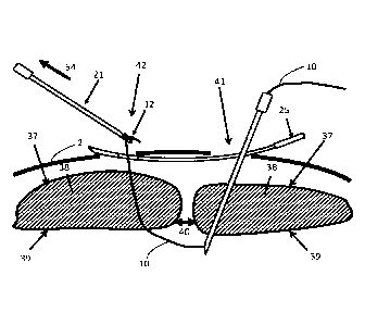
CA 03049660 2019-07-08
WO 2018/132801 PCT/US2018/013764
- 2 -
abdominal wall is performed. Interrupted sutures are placed through the
anterior rectus sheath,
the rectus muscle, and the posterior rectus sheath. These conventional repair
techniques have a
long-term failure rate of 41% - 52%, leading to ventral hernia formation. Poor
tissue strength
coupled with significant tension in the suture lines leads to failure of the
abdominal closure
requiring hernia repair.
[0005]
In conventional laparoscopic repair, multiple trocar ports are inserted to
place a large
patch of prosthetic mesh to cover the defect. This approach causes far less
postoperative pain as
compared to open methods because a large abdominal incision is avoided.
However, the
abdominal defect is generally not fully closed. Instead, the large prosthetic
patch is tacked onto
the inner surface of the abdominal wall to cover the defect. Placement of a
large piece of
artificial material results in a high rate of postoperative complications
including seroma
formation. The fluid loculation of the seroma then increases the potential for
infection of the
laparoscopically placed mesh, necessitating its removal plus antibiotic
therapy. Bowel adhesions
are also a potential complication due to the implantation of a large foreign
body patch.
[0006]
It is desirable to close the abdominal defect using a laparoscopic technique,
either
partially or completely, to significantly decrease the size of the prosthetic
mesh patch needed to
repair a ventral hernia or eliminate the use of a mesh patch entirely at the
discretion of the
surgeon. Current methods use sutures which must be advanced into the body
cavity through
multiple layers of tissue including full-thickness abdominal walls, and as
such, the sutures are
difficult to find and manipulate when inserted into the body cavity. After
looping around the
muscle and both ends of the suture may exit from a single site, and a slip
knot composed of two
half-hitches is tied in the suture, and the suture is tensioned by holding
onto one suture limb
while advancing a laparoscopic knot pusher down the opposite suture limb.
Tension must be
maintained on the slip knot as multiple sutures used in the repair of the
hernia are serially
tensioned to close the large abdominal defect gradually. Tension may be
maintained by applying
a surgical clamp to the base of the knot. However, if the patient is obese, it
may be difficult or
impossible to advance the jaws of a surgical clamp a distance of 5¨ 10 cm down
a tiny skin
puncture to the location of the knot. In the non-obese patient, the presence
of numerous surgical
clamps on the abdominal wall of the patient makes it cumbersome for the
surgeon to manipulate
and function during the procedure. After the array of sutures have been
serially tensioned to
close the abdominal wall defect, a series of at least five square knots must
be tied on top of each
slip knot in each individual suture, and eighty or more knots may need to be
tied to complete the
repair. Each square knot requires tension to be maintained on one limb of the
suture, while a
CA 03049660 2019-07-08
WO 2018/132801 PCT/US2018/013764
- 3 -
laparoscopic knot pusher is advanced along the other suture limb to push the
knot
subcutaneously down to the anterior rectus sheath. This process is tedious, as
fifty or more knots
need to be tied to complete the repair.
[0007] Other methods employ an anchor delivery tool wherein a tissue anchor
lies within the
bore of the needle. As a relatively large diameter needle is required to
deliver an anchor because
the outer diameter of the needle is larger than the diameter of the anchor,
there exists the
potential for an anchor under continuous tension to dilate the tract in the
muscle formed by
needle insertion, leading to pullout of the anchor through the dilated tract.
This scenario may be
observed particularly in the weakened or attenuated tissue encountered in
ventral hernia patients
[0008] A simple laparoscopic technique and instrumentation is desired to
quickly and easily
place multiple interrupted fastening loops through the full abdominal wall and
around a hernia
defect such that the loops may be tensioned without incising, pulling out, or
tearing through the
muscle tissue.
SUMMARY
[0009] The present disclosure is directed to devices and methods for
minimally invasive
closure of a surgical defect such as a ventral hernia using self-locking
straps. These embodiments
may provide for fewer surgical steps, reduced complexity, smaller incision
sites, reduced
pressure on tissue, and easier and faster serial tensioning along the length
of a defect. An
embodiment of a system according to the present disclosure may include a self-
locking strap
having a lock-head, a first needle having a lumen for delivering the self-
locking strap through a
first incision site, a second needle having a hook on its distal end for
engaging the self-locking
strap and pulling the strap from the body through a second incision site, and
a guide having an
aperture near its distal end for passing the second needle therethrough and
retaining the strap
after the second needle is removed from the aperture.
[0010] In other embodiments, the guide may be used for tunneling through
subcutaneous
tissue from the first incision site to a second incision site, and it may have
a hook near its distal
end for engaging with the self-locking strap and pulling the strap back
through the subcutaneous
tissue to the first incision site. Furthermore, the second needle may have a
hook at its distal end,
and it may be capable of passing through the lock-head and engaging with the
distal end of the
self-locking strap and pulling the distal end of the strap through the lock-
head and out of the
body.
CA 03049660 2019-07-08
WO 2018/132801 PCT/US2018/013764
- 4 -
[00111 Embodiments of the system may also include a support tube for
pushing on the self-
locking strap while the strap is being tensioned, a rotational cutter with at
least one distal blade
for severing the self-locking strap adjacent to the lock-head, a linear cutter
having an inner tube,
an outer tube, and a radially flexing blade that depresses into the lumen of
the inner tube when
engaged by the outer tube. The system further may include a tensioner having
at least one
movable lock-head to incrementally tension the self-locking strap; the
tensioner may have a
tension gauge to measure the tension in the strap and an indicator to display
the tension in the
strap. The tension gauge may be a mechanical spring gauge or a force
transducer, and it may be
capable of converting the tension force into an electrical signal and
transmitting the electrical
signal to a display or receiving computer. The system may include a
laparoscopic grasper for
placing the self-locking strap into engagement with the second needle; the
laparoscopic grasper
may have a robotic arm interface that is robotically controlled. The self-
locking strap may
include an attached lock-head capable of receiving a single end of the strap
or detached lock-
head capable of receiving both ends of the strap. The self-locking strap may
have an aperture at
its proximal and distal end or a protuberance at its distal end for engaging
with the second
needle. The strap may also have a plurality of apertures through the strap to
facilitate ingrowth of
tissue. The first needle may have a slot along its length for removing the
self-locking strap, and
the guide may have a robotic interface fixed to its distal end.
[0012] Embodiments of a method for closing a defect may include positioning
a guide
beneath the skin between a first incision and a second incision in the body of
a patient so that the
distal end of the guide resides near the second incision. Inserting a first
needle through the first
incision, inserting a second needle through the second incision and an
aperture in the guide,
placing a distal end of a self-locking strap through the first needle and into
the body cavity.
Next, the surgeon or a robotic aim may engage the second needle with the
distal end of the strap
and pull the second needle through the guide and out of the body leaving the
strap captured by
the aperture in the guide. The strap may be released from the first needle,
and the guide pulled
out of the body through the first incision so that the distal end of the self-
locking strap exits the
first incision with the guide. To lock the strap, the surgeon may place a lock-
head over the distal
end of the self-locking strap and advancing the lock head down the self-
locking strap to tighten
the self-locking strap around a defect.
[0013] In other embodiments, the method may include inserting a first
needle through a first
incision and into a body cavity on a first side of a defect, placing the
distal end of a self-locking
strap through the first needle and into the body cavity, retracting the first
needle from the body,
CA 03049660 2019-07-08
WO 2018/132801 PCT/US2018/013764
- 5 -
and removing the first needle from the strap while leaving the strap inside of
the body. Next, the
surgeon or a robotic arm may place a guide through a second incision site on
the opposite side of
the defect and advance the guide subcutaneously to the first incision site,
engaging the proximal
end of the strap with the guide and pulling the proximal end of the strap
subcutaneously across
the defect to the second incision site. The second needle may be placed
through a lock-head
attached to the proximal end of the strap and advanced into the body cavity to
engage the distal
end of the strap. The second needle may be used to pull the distal end of the
strap out through
muscle tissue and through the lock-head. Finally, the strap may be tightened
to close the defect.
[0014] In other embodiments, initially, pilot needles may be placed through
the skin and
muscle tissue to determine the correct distance lateral to the defect for
inserting surgical
instruments. Furthermore, a laparoscopic grasper, which may be robotically
controlled, may be
used to engage the second needle with the distal end of the strap. The first
needle may include a
hook at its distal end for engaging with the self-locking strap, and the first
needle may have a slot
through which to withdraw the self-locking strap laterally from the needle. A
support tube may
be placed over the self-locking strap such that the surgeon may pull the self-
locking strap while
pushing the support tube to tighten the self-locking strap. Next, the surgeon
may tighten the self-
locking strap with a tensioner that has at least one lock-head to
incrementally tighten the self-
locking strap. The tensioner may have a tension gauge that may be a mechanical
spring gauge or
a force transducer to measure the tension in the strap and to display the
tension force, and the
force signal may be converted into an electrical signal that may be
transmitted to a display or
receiving computer. The excess amount of self-locking strap may be cut inside
of the body
adjacent to the lock-head, hi some embodiments, the strap may include a
plurality of apertures
through the strap to facilitate ingrowth of tissue.
[0015] In another embodiment, a tensioner for tightening a self-locking
strap may include a
shaft, a plunger slidably disposed on the shaft, a stationary lock-head distal
to the plunger and
affixed to the shaft, and a movable lock-head affixed to the plunger and
aligned with the first
stationary shaft so that a strap may pass through both lock-heads. The
tensioner may also include
a frame slidably attached to the shaft and located distal to the stationary
lock-head and a
compression spring compressed between the frame and the stationary lock-head.
An elongate
tube may extend distally from the frame such that the tube abuts against the
lock-head of a self-
locking strap inside of a human body.
[0016] The strap tensioner may also include a force transducer configured
to read the amount
of force on a strap that is held by the stationary lock as the elongate tube
abuts against the lock-
CA 03049660 2019-07-08
WO 2018/132801 PCT/US2018/013764
- 6 -
head of the strap. The tensioner may also include a set of force markings
located on the distal
portion of the shaft configured to read the amount of force on a strap that is
held by the
stationary lock.
[0017] In an embodiment, a self-locking strap having teeth on both sides
may be provided.
The strap may include an elongate body having a distal end, a proximal end, a
top side and a
bottom side, a first set of ramped teeth on the top side, and second set of
ramped teeth on the top
side having a ramp direction in the opposite direction to the first set of
teeth. The bottom side
may have a third set of ramped teeth having a ramp direction in the same
direction as the first set
of teeth and a fourth set of ramped teeth on the bottom side having a ramp
direction in the same
direction as the second set of teeth. The strap may have a detached lock-head
having an aperture
capable of passing the distal end and proximal end simultaneously and opposing
pawls
protruding into the aperture for engaging with the distal end and the proximal
end. The third set
of ramped teeth may be offset longitudinally from the first set of ramped
teeth, and the fourth set
of ramped teeth are offset longitudinally from the second set of ramped teeth
to provide a wider
minimum cross-sectional thickness for the strap. In embodiments, the strap may
have a plurality
of apertures through the strap to facilitate tissue ingrowth and proximal ends
that are tapered
with an aperture therethrough to engage with a surgical instrument.
CA 03049660 2019-07-08
WO 2018/132801 PCT/US2018/013764
- 7 -
[0018] BRil-F DESCRIPTION OF THE DRAWINGS
[0019] The foregoing will be apparent from the following more particular
description of
example embodiments of the invention, as illustrated in the accompanying
drawings in which
like reference characters refer to the same parts throughout the different
views. The drawings
are not necessarily to scale, emphasis instead being placed upon illustrating
embodiments of the
present invention.
[0020] FIG. 1 depicts a system for closing a defect in tissue.
[0021] FIG. 2A-2J illustrate various views of embodiments of self-locking
straps.
[0022] FIGS. 3A-3G illustrate various views of other embodiments of self-
locking straps.
[0023] FIGS. 4A-4E illustrate various views of other embodiments of self-
locking straps.
[0024] FIGS. 5A-5B show various views of other embodiments of self-locking
straps.
[0025] FIGS. 6A-6B show various views of another embodiment of a self-
locking strap.
[0026] FIGS. 7A-7K show various embodiments of a slotted needle that holds
a strap that
may be removed laterally from the lumen of the slotted needle.
[0027] FIGS. 8A-8C show a hook needle that is used to pull a strap from the
body.
[0028] FIGS. 9A-9C show a grasper for grasping a strap within the body.
[0029] FIGS. 10A-10H show various embodiments of a subcutaneous guide for
tunneling
subcutaneously between two incision sites.
[0030] FIGS. 11A-11C show a tubular cutter used to sever the excess length
of the self-
locking strap.
[0031] FIGS. 12A-12B show a rotational cutter used to sever the excess
length of the self-
locking strap.
[0032] FIGS. 13A-13B show an embodiment of a strap tensioning device.
[0033] FIGS. 14A-14E illustrate the operation of a strap tensioning device.
[0034] FIGS. 15A-15E show various embodiments of a releasable strap lock-
head.
[0035] FIG. 16A illustrates another embodiment of a strap tensioning
device.
[0036] FIGS. 17A-17B illustrate the general anatomical layout for the
procedures described
herein.
[0037] FIGS. 18A-18K illustrate an example of a system used to close a
tissue defect.
[0038] FIG. 19A-19K illustrates another embodiment of a system for closing
a defect in
tissue.
CA 03049660 2019-07-08
WO 2018/132801
PCT/US2018/013764
- 8 -
[0039]
FIGS. 20A-20I illustrate another embodiment of a system used to close a
defect.
[0040]
FIGS. 21A-21C illustrate embodiments devices with a surgical robotic
interface.
CA 03049660 2019-07-08
WO 2018/132801 PCT/US2018/013764
- 9 -
[0041] DETAILED DESCRIPTION
[0042] A description of example embodiments of the invention follows.
[0043] Systems and methods for closing a tissue defect are described
herein. While the
present disclosure describes the system and method in the context of hernia
repair, and in
particular ventral hernia repair, the devices and methods presently disclosed
may be used in any
surgical procedure for joining tissue, closing a tissue opening, or fastening
a device to or
between two or more sections of tissue.
[0044] In the patient's midline, the left and right anterior and posterior
rectus sheaths come
together to form a single layer called the linea alba. A ventral hernia defect
may arise as an
opening in this layer. It may also be an opening that extends through the
posterior rectus sheath,
rectus muscle, and anterior rectus sheath; or it may be an opening in the
fascia lateral to the
rectus muscle. While the current disclosure describes systems and methods in
the context of
laparoscopic surgery, the systems and methods may be applied to any other
class of procedure
such as laparotomy, or robotic surgery.
[0045] With reference to FIG. 1, one embodiment of a system 1 for closing a
fascial opening
40 (e.g., ventral hernia) is shown in relation to a layer of skin 1 and a
defect 40 shown in
reference to the anterior rectus sheath 37. The system may be used to deliver
an implant to
constrain tissue, such as a suture or strap through minimally invasive or
laparoscopic surgery.
The system 1 may comprise by way of non-limiting example, a self-locking strap
10, a slotted
needle 15, a hook needle 21, a subcutaneous guide 25, a tubular cutter 30, and
a support tube 45.
An inner tube 17 may reside inside of the slotted needle 15. The tubular
cutter 30 may be
comprised of an outer tube 34 and an inner tube 31 having a cutting blade 33
attached therein.
The aforementioned components are described in further detail in this
disclosure. Other
components and devices, including those disclosed throughout this application,
may be included
in the systems and used in the methods disclosed herein, i.e., the system 1
shown is not
necessarily a complete surgical kit and other devices and methods may be
substituted or added to
the system 1 to form other systems or embodiments that are within the scope of
the invention(s)
disclosed herein. For example, various laparoscopic instruments, such as a
laparoscope with a
camera may also be-be used during the surgery.
[0046] This application discloses methods, systems, and devices that
involve placing a strap
around a defect to close a tissue defect. In general, a strap may be wider
than a suture and have a
flatter shape so as to place less pressure on the tissue and reduce the risk
of cutting into the tissue
CA 03049660 2019-07-08
WO 2018/132801 PCT/US2018/013764
- 10 -
as the tension forces required to approximate muscle tissue may be high. In
this application, the
straps are referred to as "self-locking straps" which means that when the
strap is pulled through
its "lock-head" and released, it tends to stay at the same position without
retracting back through
the lock-head. Self-locking straps use a mechanism such that when the strap is
pulled through
the lock-head, it will not slide back through the lock-head. That is, the
strap may be drawn
through the lock-head in one direction, but a reversal of movement of the
strap within the lock-
head is mechanically restricted so as to effect a locking position in one-way
fashion. There are
many types mechanisms that perform this function such as cable tie mechanisms
(also known as
zip ties), ratchets, cam locks, and ratchet and pawl mechanisms. Some designs
have features on
the strap, such as teeth, that interact with features in the lock-head, such
as a pawl which flexes
or is otherwise spring biased to engage with the features on the strap. The
strap may have holes
or slots into which a spring-loaded pawl feature engages. Other designs may
have a smooth strap
that is gripped by cam-like features, tines, or barbs which grasp the strap
predominantly in one
direction to prevent it from regressing through the lock-head. Some self-
locking straps may have
an integrated lock-head positioned, for example, near an end of the strap
while others may have a
detached head that may be placed on the strap for locking. Such self-locking
straps and
variations thereof are within the scope of this disclosure.
[0047] The use of self-locking straps in ventral hernia procedures
simplifies the procedure
and saves time because the surgeon does not need to tie numerous suture knots
or clamp and re-
clamp partially tightened sutures or worry about the self-locking straps
loosening. The self-
locking functionality of the straps disclosed herein permits the surgeon to
quickly and easily
tension individual straps serially to close a defect incrementally without
tying permanent knots
or releasable knots. This is because the surgeon may pull one or both ends of
the strap through
the lock-head and at any time let go of the strap and it will hold the tension
due to the ratchet
effect of the strap features engaging with the lock-head. Thus, the surgeon
can move on to other
aspects of the surgical procedure, for example, incrementally tightening other
straps along a
defect without having to clamp or knot them to prevent slipping.
100481 For brevity, several self-locking strap embodiments are disclosed in
this application.
However, other types of self-locking straps are within the scope of the
invention(s) described
herein.
[0049] The self-locking strap should be sized such that it has the strength
to approximate and
hold the muscle tissue while being flexible enough for manipulation through
the body and the
instruments. The self-locking straps referred to in this application may be
made of any
CA 03049660 2019-07-08
WO 2018/132801 PCT/US2018/013764
- 11 -
biocompatible material that may be implanted into the body. Candidate
materials include, by
way of nonlimiting example, polymers such as PEEK, polypropylene, or Nylon, or
metals such
as Nitinol or stainless steel.
[0050] FIG. 2A depicts an embodiment of a self-locking strap 10 which may
be made of a
biocompatible material suitable for implanting into a human or mammalian body.
The self-
locking strap 10 may comprise a distal end 11 and a proximal end 13 and, as
mentioned above, it
may be made of a polymeric material such as polypropylene, PEEK, or Nylon, or
for example, a
metallic material such as Nitinol or stainless steel. The diameter of the self-
locking strap 10 may
be of any size that provides enough tensile strength to approximate and hold
tissue together, for
example in ventral hernia repair. In one embodiment, the self-locking strap 10
may be made of
polypropylene, and it may be between .25mm and 2 mm in diameter or, for
example,
approximately 0.35 mm in diameter, and it may be approximately 100 cm in
length or any length
that is appropriate for the size of the patient. hi another embodiment, the
self-locking strap 10
may be made of stainless steel, and it may be approximately 0.10mm to 0.25 mm
in diameter and
approximately 100 cm in length. A protuberance 12 or other grasping feature
may be
permanently formed or attached near the distal end of the self-locking strap
10, and it may be
made of the same material as the self-locking strap 10, or it may be composed
of a different
polymeric or metallic material. The protuberance 12 may be larger than the
strap diameter, or
approximately .5 mm to 0.75 mm in diameter and shaped, for example,
spherically or in any
other shape that allows it to be retained by a hook needle 21 or other grasper
described elsewhere
in this disclosure. In other embodiments, the protuberance 12 may be a loop or
aperture formed
in the distal end 11 of the self-locking strap 10 to facilitate grasping of
the self-locking strap 10.
[0051] The self-locking strap 10 comprises a distal end 11 at one end and a
proximal end 13
at the opposite end. The self-locking strap 10 may have teeth 50 arranged
along the strap to
facilitate locking into a lock-head that may be attached to one end of the
self-locking strap 10.
The teeth 50 may be located on the entirety of the self-locking strap, or on a
portion thereof, or in
some embodiments the entire strap may be smooth. By way of non-limiting
example, in FIG.
2A, an embodiment of the self-locking strap 10 is shown with teeth 50 on the
portion of the strap
towards the proximal end 13. The self-locking strap 10 may be composed of a
single polymeric
or metallic material such that the smooth end (distal end 11) and the toothed
end (proximal end
13) are made as one component, for example via extrusion or injection molding
for polymeric
materials. Each tooth 50 may have a major outer diameter of from 1 mm to 3 mm
or
approximately 2 mm. The teeth 50 may be of any shape that allows them to
engage with the
CA 03049660 2019-07-08
WO 2018/132801 PCT/US2018/013764
- 12 -
lock-head 14 to restrict motion in one-way through the lock-head 14. For
example, in some
embodiments, the teeth 50 may be shaped like an ellipse, square, sphere, cone,
wedge, step,
tooth, or hemisphere. The tel in "diameter" used herein refers to the
major, or largest dimension,
across the tooth 50. The length of the self-locking strap 10 may be
approximately 100 cm or any
length that allows it to extend beyond multiple layers of abdominal tissue
taking into account the
large variation in anatomy due to, for example, variations in adipose tissue.
[0052] The self-locking strap 10 is shown in an example of operation in
FIG. 2B. A sectional
view is shown in FIG. 2C to illustrate how the teeth 50 may interact with the
lock-head 14. The
lock-head 14 may be attached to the proximal end 13 of the self-locking strap
10 and comprises
locking features such as one or more pawls 51 that engage with the teeth 50.
The pawls 51 may
be two dimensional, that is like a beam, or they may comprise a conical shape
(axisymmetrical),
and they may be segmented to allow the pawls 51 to defolin to pass teeth 50
while flexing back
down behind a tooth 50 to lock it from passing back through the lock-head 14.
As shown in FIG.
2C, due to the angled orientation of the pawls 51, the tooth 50, which is of a
triangular or conical
shape in this embodiment is constrained to only pass in the direction of the
arrow 48 as the pawls
51 flex around the tooth 50 and close behind it as the proximal end 13 is
pulled through the lock-
head 14. This operation is one example of a one-way lock and one skilled in
the art will
recognize the examples and embodiments described herein are for illustrative
purposes only and
that various modifications or changes in light thereof will be suggested to
persons skilled in the
art and are to be included within the spirit and purview of this application
and scope of the
appended claims. The pawls 51 of the lock-head 14 may be constructed of a
polymeric material
such as polypropylene, PEEK, or Nylon, or it may be metal, such as stainless
steel. The lock-
head 14 may be integrally formed with the proximal end 13 via, for example,
injection molding,
or it may be a detached element. In further embodiments, the lock-head 14 may
be a single
ratchet pawl such as that used in zip ties or cable ties designed allow
passage of a strap in one
direction while locking it from motion in the opposite direction as described
elsewhere in this
disclosure.
[0053] Alternatively, the flexible members (i.e., beams or pawls) may be
located on the
strap, while the lock-head may have a substantially fixed interface, still
resulting in one-way
locking action. Example embodiments of alternative locking features are shown
in FIGS. 2D-2H.
A toothed strap 60 with a spherical tooth 61 is shown in FIG. 2D, a toothed
strap 62 with a
hemispherical tooth 63 is shown in FIG. 2E, and a toothed strap 64 with a
conical tooth 65 is
shown in FIG. 2F. The strap need not be axisymmetric, as FIGS. 2G and 2H
illustrate flat strap
CA 03049660 2019-07-08
WO 2018/132801 PCT/US2018/013764
- 13 -
embodiments shown in a side view. In FIG. 2G, the toothed strap 66 is flat and
has half-arced
teeth 67, and a toothed strap 68 that is flat and has ramped teeth 69 is shown
in FIG. 2H. It may
be desirable to have apertures through the self-locking strap to facilitate
ingrowth of tissue into
and through the strap so that the strap is more adherent to tissue along its
entire length and it will
be less likely to cut through the abdominal wall and cause dehiscence, and a
ventral hernia. The
apertures may be small holes through the thickness of the strap that are
distributed along the
length. For example, FIG. 21 shows a section of a self-locking strap 232
having ramped teeth
233 and a series of holes 234 perforating through the strap. In other
embodiments, the apertures
may be the actual locking features as shown in FIG. 2J. The section of self-
locking strap 235 has
slots 236 through the strap which may facilitate ingrowth, and they may also
be used for locking
if the lock-head has, for example, mating tabs that protrude into the slots
236 as the tabs are
biased by beam-like flexing or are otherwise spring loaded.
[0054] Now with reference to FIGS. 3A-3D, which illustrate another
embodiment of a self-
locking strap 100 that may have a reduced maximal width because the lock-head
is separate. For
example, FIG. 3A shows the self-locking strap 100 having an elongate body 106
and a
substantially flat cross-sectional profile with proximal teeth 101 and distal
teeth 102 located on a
top side 103 of the self-locking strap 100. The two sets of teeth, the
proximal teeth 101 and distal
teeth 102, are oriented opposite to each other, that is, they are angled to
lock in the opposite
direction when the strap is positioned straight as shown. However, when the
strap is configured
in a loop, the teeth 101 and 102 will be oriented in the same direction. Both
sets of teeth 101 and
102 of the strap may emanate from a central point 105 of the strap 100 which
may be at the
center of the strap 100 or may be offset from the center and the teeth 101 and
102 may be offset
from the central point 105, although in the figures the sets of teeth emanate
from adjacent to the
central point 105, that is, the teeth are arranged symmetrically about the
center of the strap. This
self-locking strap embodiment has a detached lock-head as further described
below. Since the
lock-head may typically be larger in diameter than the underlying strap, the
self-locking strap
100 may fit into a needle with a smaller lumen while the lock-head is
detached.
[0055] FIG. 3B shows a top view of the self-locking strap 100 with proximal
teeth 101 and
distal teeth 102 spaced about the central point 105. Apertures 107 and 108 may
be located at one
or both ends of the self-locking strap and may be of any shape or size that
can be engaged with a
surgical grasper or a hook needle (not shown) within the body as described
below. The width w3
of the strap 100 may be small enough so that the strap 100 can fit into a
small gage needle to
facilitate a minimally invasive surgical procedure. For example, a strap
insertion needle 7 (see
CA 03049660 2019-07-08
WO 2018/132801 PCT/US2018/013764
- 14 -
FIG. 18C) may have an 11 gauge (3 mm) outer diameter and a lumen diameter of
approximately
2.4 mm. Therefore a strap of about 1.7mm to 2.0 mm in width or smaller may be
passed through
the lumen.
[0056] FIG. 3C shows a side view of the self-locking strap 100. As with the
other strap
embodiments disclosed herein, the overall thickness t3 of the strap should be
large enough that
the strap will withstand the tensile force required to approximate the tissue
in a given type of
procedure while remaining flexible enough to form a loop to tighten around the
anatomy. For
reference, in open abdominal closure procedures, a suture with a tensile
strength of 10 lbs is
typically used. The self-locking strap 100 may also have swaged or flattened
ends to facilitate
handling within the body and to provide a flexible section for maneuvering the
self-locking strap
100 within the body and into the lumen of a needle. The minimum thickness of
the strap t3m is
illustrated in the detail view shown in FIG. 3D which shows the central point
105 and the
proximal teeth 101 and distal teeth 102 on either side. This minimum thickness
t3m affects the
overall stiffness and strength of the strap and hence the tooth depth should
be shallow enough so
that the minimum thickness t3m is thick enough to provide enough tensile
strength required for a
given procedure, yet deep enough to engage with a pawl type feature in the
locking head without
slipping under tensile loading. The self-locking strap 100 may be made of a
biocompatible
material such as such as a polymer (e.g., polypropylene, PEEK, or Nylon), or a
metallic material
such as Nitinol or stainless steel.
[0057] As shown in FIG. 3D, the proximal teeth 101 ramp down toward the
proximal section
of the strap, while the distal teeth 102 ramp in the opposite direction, down
toward the distal end
of the strap. An example of the operation is illustrated in FIGS. 3E-3G.
100581 With reference to FIG. 3E, the self-locking strap 100 is shown in a
looped
configuration as it may be wrapped around tissue (not shown) inside the body.
The self-locking
strap 100 is folded such that the bottom side 104 is inside of the loop as it
would be in contact
with the body; at the ends, the bottom side 104 contacts itself while the top
side 103 faces
outward leaving the proximal and distal ends of the strap adjacent. A lock-
head 109, which is
shown in a sectional view to illustrate the function, engages with both ends
of the self-locking
strap 100 as it is advanced in the direction of the arrow 116. As shown in
FIG. 3F, both ends of
the self-locking strap 100 may be pulled through the lock-head 109 in the
direction of the arrow
117 simultaneously. This self-locking mechanism is shown in the detail view of
FIG. 3G. The
lock-head may have a plurality of pawls 110 and 111 that flex open to allow
passage of the self-
locking strap 100, and then close to engage with the proximal teeth 101 and
the distal teeth 102
CA 03049660 2019-07-08
WO 2018/132801 PCT/US2018/013764
- 15 -
respectively to prevent the self-locking strap 100 from moving back through
the lock-head 109,
that is, opposite to the direction of the arrow 117 in FIG. 3F. The pawls 110
and 111 may have
one or more teeth 112 to facilitate gripping the self-locking strap 100, or
the pawls 110 and 111
may have any other surface features that may facilitate gripping with the self-
locking strap 100
such as serrations or surface roughness. Alternatively, the pawls 110 and 111
may be smooth,
relying on the leading edges 114 and 115 of the pawls 110 and 111 respectively
to lock onto
proximal and distal teeth 101 and 102 on the self-locking strap 100 such as in
a typical cable tie.
[0059] Yet another embodiment is shown in FIGS. 4A-4E. A self-locking strap
120 is
shown having an elongate body 126 and a substantially flat cross-sectional
profile with proximal
teeth 121 and distal teeth 122 located on a top side 123 of the self-locking
strap 120. Similar to
the previously disclosed self-locking strap 100, the top side 123 of the self-
locking strap 120 has
two sets of teeth, the proximal teeth 121 at the proximal end 118 and distal
teeth 122 at the distal
end 119, which are oriented opposite to each other about a central point 125,
that is, they are
angled to lock in the opposite direction when the strap is straight as shown.
However, the present
self-locking strap 120 has teeth on both sides of the strap ¨ the top side 123
and the bottom side
124. That is, the bottom side has bottom side proximal teeth 131 and bottom
side distal teeth 132
arranged in opposing fashion like the teeth on the top side 123. This two-
sided tooth arrangement
allows the self-locking strap 120 to lock into a lock-head even if the strap
is twisted as it winds
through the body.
[0060] FIG. 4B is a side view of the self-locking strap 120. The overall
thickness t4 of the
strap should be large enough that the strap will withstand the tensile force
required to
approximate tissue during and after a procedure yet be flexible enough to form
a loop and be
capable of being tightened around the anatomy. The self-locking strap 120 may
have tapered
ends as shown and the thickness t4 may also be swaged or flattened (not shown)
at the ends to
facilitate handling within the body and to provide a flexible section for
maneuvering the self-
locking strap 120 within the body and into the lumen of a needle. The minimum
thickness ti is
illustrated in the detail view shown in FIG. 4C, which is a side view
illustrating the proximal
teeth 121 on the top side 123 and the bottom side proximal teeth 131 on the
bottom side 124. In
the configuration shown, both sides of teeth are aligned, in that the root and
tip of each tooth on
the top side 123 mirrors that on the bottom side 124. As such, the minimum
thickness ti, which
affects the overall stiffness and strength of the strap, is dictated by the
distance tl between the
upper tooth inner edge 133 and lower tooth inner edge 134 as shown by ti.
CA 03049660 2019-07-08
WO 2018/132801 PCT/US2018/013764
- 16 -
[0061] In another embodiment, the teeth may be staggered to increase the
distance tl, thus
providing more cross-sectional area which may increase the strength of the
strap. This
arrangement is illustrated in FIG. 4D which is a view of a section of another
self-locking strap
130 in a detail view analogous to that shown in FIG. 4C, however in this
embodiment the
proximal teeth 141 are offset by a distance o from the bottom side proximal
teeth 142, where o is
less than p, the pitch distance between teeth. Staggering the teeth in this
manner increases the
minimum distance between teeth to t2, providing an effectively thicker strap
where t2 is greater
than tl for a given strap thickness and tooth profile.
[0062] FIG. 4E illustrates the self-locking strap 120 in operation with the
lock-head 109 in a
sectional view to illustrate the one-way lock engagement. Analogous to the
previously described
embodiment, the self-locking strap 120 is shown passing through the lock-head
109. The lock-
head may have a plurality of pawls 110 and 111 that flex open to allow strap
passage, and close
to engage with the proximal teeth 121 and the distal teeth 122 respectively to
prevent the strap
120 from moving back through the lock-head 109. If the distal end 119 of the
strap 120
happened to be rotated (not shown) as the strap transits through the body, the
bottom side distal
teeth 132 would engage with the pawl 111 of the lock-head 109. The pawls 110
and 111 may
have one or more teeth 112 or grip features to facilitate gripping the self-
locking strap 120 or any
other surface features to facilitate holding the self-locking strap 120.
Alternatively, the pawls
110 and 111 may be smooth, relying on their leading edges to lock onto
proximal and distal teeth
101 and 102 on the strap like a standard cable tie.
[0063] In some embodiments, the self-locking strap may have a smooth
surface; FIG. 5A
shows an embodiment of a self-locking strap 150 without teeth inserted through
a lock-head 151
which is shown in a side section view. The self-locking strap 150 may have a
top surface 154
that is relatively smooth and a bottom surface 158 that is relatively smooth,
that is the self-
locking strap 150 may be flat or round in cross-section, and it may lock in
the lock-head 151
even if the strap 150 is twisted since both the bottom surface 158 and the top
surface 154 are
configured to engage with the lock-head 151. That is, if the self-locking
strap 150 is twisted as it
transits through the body, it will still lock as shown in FIG. 5A because both
sides of the strap
engage with the lock-head 151 in the same way. The surface of the self-locking
strap 150 may
not be entirely smooth, as it may have a level of surface texture or roughness
to facilitate
gripping in the lock-head 151. The texture may be small gratings, random
bumps, texture, or a
general grit or surface roughness that can be imparted to the strap, for
example, through injection
molding. The lock-head 151 may have a set of pawls 152 and 153 that
elastically flex to open to
CA 03049660 2019-07-08
WO 2018/132801 PCT/US2018/013764
- 17 -
accept the strap 150, and then tend to close, putting pressure onto the strap
150 and digging into
the top surface 154 and bottom surface 158 so as to prevent the strap 150 from
travelling back
through the lock-head 151, that is, in the direction opposite the arrow 162.
The pawls 152 and
153 may also be referred to as tines, beams, or barbs by one skilled in the
art. In FIG. 5B, the
self-locking strap 150 is shown protruding through another embodiment of a
lock-head 159
having barbs 160 that impinge on the strap 150 to dig into the top surface 154
of the strap 150
such that the strap 150 may only travel in one direction. In embodiments, the
barbs 160 may be
made of a metal such as stainless steel while the body 161 of the lock-head
may be made of a
plastic, rubber, or elastomeric material; the barbs 160 may be insert molded
into the body 161 or
bonded therein or molded as part of the lock-head in one piece.
[0064] Another type of detached lock-head embodiment is a crimp (not
shown). That is, a
malleable lock-head may be slid down the two ends of the strap, similar to the
approach shown
in FIG. 5A, and then crimped in place by a tool such as pliers. The crimp
design does not
inherently create a one-way lock, but the crimp could, for example, be
partially closed until final
tightening, then crimped fully.
[0065] The self-locking strap 150 may be made of a biocompatible material
such as such as a
polymer (e.g., polypropylene, PEEK, or Nylon) or a metallic material such as
stainless steel.
Additionally, since the self-locking strap may remain within the body after
surgery, they may be
made of a bioabsorbable material that is absorbed or disintegrates over time.
[0066] Patient's may feel geometric features such as bulges or protrusions
of an implant that
reside below the surface of the skin, that is, the features be palpable or
cause a sensation when
pressing against the tissue below the skin. A self-locking strap 220 having a
flat top side 237 is
shown in FIG. 6A. The strap 220 may have a lock-head 221 with a slot 223
therethrough for
receiving the distal tip 225, an elongate body 229, a proximal tip 222 with a
proximal aperture
226, a distal aperture 227 and a plurality of teeth 224 arranged along the
length of the body. The
lock-head 221 protrudes on the bottom side 238 and tends to embed into the
fascia and muscle so
that the self-locking strap remains relatively flat on the top side 237, which
may be adjacent to
the skin. That is, the distal tip 225 of the strap may transit through the
lock-head 221 from the
bottom side 238 to the top side 237. As illustrated in a broken view in FIG.
6B, the lock-head
221 may have a pawl 228 to engage with the teeth 224 or any other type of
ratcheting
mechanism that affords one-way movement after the distal tip 225 is pulled
through the lock-
head 221. A needle may be placed through the slot 223 such that the needle can
engage with the
CA 03049660 2019-07-08
WO 2018/132801 PCT/US2018/013764
- 18 -
distal tip 225 to pull the distal tip 225 back through the slot 223 to lock
the strap as is further
described in FIGS. 18A-181.
[0067] Now with reference to FIG. 7A, which shows an embodiment of a
slotted needle 15
comprising a slot 16 that may extend from the heel of the needle to the
proximal end of the
needle where a handle 19 may reside. The slotted needle 15 may have an outer
diameter of
approximately 1.65 mm (16 gauge), the slot 16 may be .25 mm to .75 mm wide or
approximately 0.5 mm wide, and the slotted needle 15 may be approximately 20
cm in length.
Embodiments of the slotted needle 15 allow the needle to be inserted through
the full-thickness
abdominal wall, for example the skin, anterior rectus sheath, the abdominal
wall, and posterior
rectus sheath, without deforming, while providing a conduit for the
advancement of a self-
locking strap into the body. To aid in retaining the strap, the inner lumen of
the slotted needle 15
may contain an inner tube 17 that may have a slot 18 to allow a strap to be
released. For clarity,
this device and other devices disclosed herein may be shown out of proportion
in the drawings to
emphasize details or functionality.
[0068] The slotted needle 15 may be used to deliver a suture or strap into
the body minimally
invasively while allowing the surgeon to withdraw the suture or strap from the
slotted needle 15
through the side of the needle rather than pulling it out through the lumen;
this may lead to a
smaller size of the slotted needle 15, and hence the wound size. A strap with
oversized features
such as a lock-head may be passed through the slotted needle 15 and into the
body, but if the
lock-head doesn't fit through the lumen, the strap may be released through the
side (slot 16) of
the slotted needle 15. This maneuver is illustrated in FIGS. 7B and 7C in the
context of the self-
locking strap 10, but other embodiments of a suture or strap may be
constrained within and
removed from the slotted needle 15 in this manner. As shown in FIG. 7B, the
protuberance 12,
which resides near the distal end 11 of self-locking strap 10, lies in the
lumen near the tip of
slotted needle 15. Once the slotted needle 15 is introduced inside the body
cavity and through
the desired tissue sections, the distal end 11 may be advanced through the
lumen; a tamp or other
driver (not shown) may be used to push the self-locking strap 10 through the
lumen. In FIG. 7C,
the distal end 11 has been advanced out of the slotted needle 15, so that the
protuberance 12 lies
distal to the needle tip. The slotted needle 15 may then be peeled away from
distal end 11, as
shown by peel-away site 20 at the intersection with the slotted needle 15. The
peel-away site 20
may move distally as the self-locking strap 10 is pulled away from the slotted
needle 15
generally in the direction indicated by the arrow 53. Once the distal end 11
has been introduced
CA 03049660 2019-07-08
WO 2018/132801 PCT/US2018/013764
- 19 -
into the abdominal cavity and removed from the slotted needle 15, the slotted
needle 15 may be
retracted from the body leaving at least the distal end 11 in the abdominal
cavity.
[0069] A strap may be retained the lumen of a slotted needle by a variety
of methods. By
way of nonlimiting example, several slotted needle designs are shown in FIGS.
7D-7H, which all
show an end-view of a slotted needle schematically. FIG. 7D illustrates a
slotted needle 70
having a slot 72 with a strap 71 disposed inside the lumen. The diameter dl of
the strap 71 may
be smaller than, equivalent to, or slightly larger than the width of the slot
Swl so that the strap
has a clearance or slight interference fit when passing through the slot 72.
If the strap diameter
dl is smaller than the slot width Swl, it still may be retained because the
strap should align with
the slot 72 along much of the full length of the slot 72 to be removed, and
hence it tends to stay
retained until pulled on transversely to extract the strap 71 incrementally
from the slotted needle
70. As shown in FIG. 7E, a strap 77 with an elongate cross-section is
illustrated, having a
minimum width d2 and a maximum width w2; the strap 77 is shown residing inside
of a slotted
needle 70. The strap 77 will tend to remain captive in the needle 70 even if
the minimum width
d2 is smaller than the slot width swl because the strap 77 will not generally
be aligned with the
slot 72 unless the surgeon twists the strap 77 and needle 70 into alignment in
order to release the
strap.
[0070] One skilled in the art will realize that there are many ways to
retain a strap inside of a
slotted needle while rendering it easily removable when desired. In some
embodiments, the
slotted needle may not be round as shown in FIG. 7F. In this embodiment, the
slotted needle 90
has an oblong shape with a flattened section 92 having a slot 91 therethrough.
The strap 77 may
also have an elongated cross-sectional shape that tends to stay inside the
lumen until its
minimum strap width d2 is aligned with the slot 91 by the surgeon when the
strap 77 is pulled
from the slotted needle 90. In yet another embodiment, FIG. 7G shows a slotted
needle 70
having an inner tube 17 within its lumen. The inner tube 17 may be made of any
flexible
material such as a polymer, for example, polyethylene or Nylon, or an
elastomeric material such
as a rubber, silicone, or a thermoplastic elastomer. The inner tube 17 may
have a slot 18 with a
width Sw3 that may be larger than, equivalent to, or smaller than the diameter
dl of the strap 71
such that it offers some resistance to hold the strap 71 inside of the lumen.
The inner tube 17
may be made of a thin walled membrane that deforms when the strap 71 is peeled
from the
lumen. The slot 18 may be an actual slot through the inner tube 17 that
remains open or slightly
closed until peeled open, or it may be a serrated line that opens with a small
amount of force.
The inner tube 17 may be bonded or otherwise fit into the inner lumen of the
slotted needle 70,
CA 03049660 2019-07-08
WO 2018/132801 PCT/US2018/013764
- 20 -
maximally extending from the heel of the needle point to the proximal end of
the needle, near the
tip, or anywhere therebetween with enough overlap with the slot 72 to retain
the strap 71. In yet
another embodiment, the inner tube 93 may have a circumferential length such
that it has an
overlap 76 that may have a length d4 that may be approximately .5 to 2mm, as
shown in FIG 7H.
The inner tube 93 should have a wall thickness that is thin enough so that it
deforms and opens
under the peeling pressure when the surgeon peels the strap 71 from the
slotted needle 70.
[0071] FIGS. 7I-7K illustrate other embodiments of a slotted needle. In
FIG. 71 a slotted
needle 95 is shown having a stationary handle 98 and a rotating handle 99,
each having a slot - a
stationary slot 84 and a rotating slot 85 respectively. When the slots are
aligned, the strap 71
may pass through the slot 97 in the shaft 96 and through the stationary handle
98 and the rotating
handle 99 to be removed from or inserted into the slotted needle 95. However,
when the
stationary handle 98 and the rotating handle 99 are not aligned, as shown in
FIG 7J, the strap 71
remains captured in the slotted needle 95. One skilled in the art would
recognize that this
operation could also be achieved with a single slotted handle (not shown) that
rotates around the
shaft 96 of the slotted needle 95 to align/misalign the handle slot and the
slot 97 on the shaft 96
as desired to retain or release the strap 71. In yet another embodiment, a
sleeve 113 may be
placed over the slotted needle 96 to cover the slot 97 as shown in FIG. 7K.
The sleeve may be as
long as the slotted needle 96 or smaller in length, and it may have a
serration or gap (not shown)
to facilitate removal or it may be cut and released from the slotted needle 96
by the surgeon in
order to release the strap 71.
[0072] In some embodiments, the system may include a hook needle 21 to
grasp a strap from
within the body cavity. For example, a hook needle 21 can be used as a needle
or a trocar to
directly pierce tissue without the need for an external trocar. As illustrated
in FIG. 8A, the hook
needle 21 may have an open slot 22 leading to a channel 23 near its distal
portion and a beveled
tip 8, so that it is capable of piercing through multiple layers of tissue.
The width of the channel
23 may be .25 mm to 1 mm, or approximately 0.5 mm, but generally of a size
that is slightly
larger than the width of the strap. The outer diameter of the hook needle 21
may be
approximately 1 mm to 3 mm or about 1.65 mm (16 gauge). The dimensions of the
slot 22 and
channel 23 should be selected based on the size of the strap and/or the size
of any grasping
feature on the strap. The hook needle 21 may be approximately 20 cm long, or
any length
appropriate to enter the body cavity for a given patient's anatomy, and it may
include a handle
24 attached to its proximal end. The hook needle 21 shall be sufficiently
strong and stiff that it
can be inserted through multiple layers of tissue in the abdomen, that is, the
full thickness
CA 03049660 2019-07-08
WO 2018/132801 PCT/US2018/013764
- 21 -
abdominal wall which includes the anterior rectus sheath, the rectus abdominus
muscle, and the
posterior rectus sheath to grasp the distal end of a strap to pull it back
through the layers of tissue
and out of the body. As such, the hook needle 21 may have any cross-sectional
shape that
provides the proper stiffness and strength including a round or oval cross-
section which may be
solid or hollow or a flat or rectangular cross-sectional shape.
[0073] FIG. 8B shows the hook needle 21 engaging the self-locking strap 10
proximal to the
protuberance 12 via the open slot 22. The surgeon hooks the self-locking strap
10 with the open
slot 22 and then pulls the tip of the hook needle 21 so that the protuberance
12 seats against the
side of the hook needle 21. As shown in FIG. 8C, the protuberance 12 is hooked
in the channel
23 of hook needle 21, thereby enabling the surgeon to grasp and manipulate the
self-locking
strap 10 via the hook needle 21. The self-locking strap 10 with the
protuberance 12 on the end is
shown here as an example, however, the hook needle 21 may be used to grasp any
suture or strap
or features thereon from within the body, such as a strap with a loop or a
strap having an aperture
for engaging with the hook needle 21 such as the distal aperture 128 of the
aforementioned self-
locking strap 120.
[0074] The open slot 22 on the hook needle 21 may be any passive feature
such as a slot,
hook, or carabiner type latch, or an active grasping mechanism such as a claw,
jaw, or clasp.
Each of which allow the hook needle 21 to grasp a suture or strap that resides
within the body.
For example, a surgical grasper 135 may be used as shown in FIG. 9A. The
grasper 135 may
have a handle 138 with a lever 147 that actuates a movable jaw 137 near the
distal end of a shaft
143. The movable jaw 137 may articulate about a pivot such that it opens to
allow access to the
open slot 140 as shown in FIG. 9B. FIG. 9C shows an analogous partial view of
the distal tip of
another embodiment of a grasper 155 having a sliding jaw 145 that slides in
the direction of the
arrow 148 to expose an open slot 149. The grasper 155 may have a needle tip
136 that is sharp
enough to allow the grasper 155 to penetrate through the multiple layers of
tissue of the full-
thickness abdominal wall to reach the strap within the body for retrieval. One
skilled in the art
will recognize that there are standard graspers having a handle that actuates
a jaw or clamp at a
distal end and may have a ratchet lock 139 (FIG. 9A) to lock the distal
mechanism in place and a
ratchet release 146 to release it. Such devices may be used for this procedure
and are within the
scope of this disclosure.
[0075] Once a self-locking strap has been accessed inside of the body
cavity by the hook
needle 21 or similar instrument and pulled the self-locking strap through the
muscle, the surgeon
needs to manipulate the self-locking strap to tension it and to lock it. The
surgeon may tension it
CA 03049660 2019-07-08
WO 2018/132801 PCT/US2018/013764
- 22 -
outside of the body where it is easier to directly see the self-locking strap
and to manipulate it.
Hence, the surgeon may pull the end of the self-locking strap out of the body
through the skin
incision site on the opposite side of the defect from the hook needle 21 to
join it with the
opposite end of the strap. Without a subcutaneous guide, finding the strap or
a needle below the
surface of the skin is cumbersome because the strap is below the surface of
the skin and out of
sight, as there is generally no camera at the subcutaneous level, so the
surgeon may have to feel
around inside the body to find the strap by trial and error which takes a
considerable amount of
time and effort. It is easier to pull the strap out of the body if the strap
is already captured while
it is in the body. Since the strap is already inside of the hook needle 21, if
the hook needle 21 is
contained by a subcutaneous guide, which may be in place in the body
beforehand, the
subcutaneous guide makes capturing the strap automatic because when the hook
needle 21 is
withdrawn through the subcutaneous guide, the strap is already captured by the
subcutaneous
guide 25. The subcutaneous guide should be rigid enough and able to withstand
axial, buckling,
and bending loads as well as torsion because the subcutaneous tissue that it
must tunnel through
has fibrous interconnections interspersed in the fatty tissue and a
significant amount of force may
be required to perform the blunt dissection across the defect between the
incision sites.
Advancement of the subcutaneous guide using a concomitant back and forth
rotational motion
may be employed.
[00761 FIGS. 10A-10C depict a subcutaneous guide 25 that may have an
elongate body 58, a
handle 57, a distal tip 28 and one or more slots, such as the distal slot 26.
Since the distance
between the incision sites is not generally the same between patients, the
subcutaneous guide 25
may have a series of slots (not shown) along its elongate body 58. The
subcutaneous guide 25 is
used to tunnel between two incisions in the abdomen below the surface of the
skin but above the
muscle and fascia. Therefore, it must be strong enough to dissect layers of
tissue that may
include tough, fibrous fatty tissue without bending or buckling. As such, in
embodiments, to
facilitate dissection, the distal tip 28 may be pointed in shape and may be
beveled or tapered. In
other embodiments, the distal tip 28 may be sharp to facilitate sharp
dissection, or it may be
relatively blunt for blunt dissection. The elongate body 58 may have a tubular
or solid cross-
section that may be shaped as an oval, as shown in FIG. 10B, or any other
cross-sectional shape
that provides the requisite strength to tunnel through the tissue
subcutaneously. The major
diameter may be small such as about 3 mm or approximately lmm to 4mm to
facilitate
tunneling. The slot 26 may be .5mm to 3cm long, for example, or approximately
2cm long and a
width dimension that can accommodate the hook needle 21 or similar instrument
that may be
CA 03049660 2019-07-08
WO 2018/132801 PCT/US2018/013764
- 23 -
introduced through the subcutaneous guide 25. For example, if the hook needle
21 has a diameter
of 1.65mm (16 gauge), then the distal slot 26 should be wider than 1.65mm so
that the hook
needle 21 can be passed through the distal slot 26. The subcutaneous guide 25
may be made of
any biocompatible material suitable for surgical use, such as a metal or
plastic material that is
stiff enough to tunnel through the body, such as stainless steel. The
subcutaneous guide 25 may
be straight or may be shaped in a gradual curve as shown in FIG. 10C. The
curve of the
subcutaneous guide 25 may allow it to enter the skin incision on one side of
the hernia defect,
cross the region of the defect, and locate the skin incision on the opposite
side more easily on
some body types. In other anatomical body shapes, for example if the abdomen
has significant
curvature, a straight subcutaneous guide may be used.
100771 FIGS. 10D-10G illustrate another embodiment of a subcutaneous guide
164 that may
include an elongate body 165, a handle 163, and a distal tip 168 having a slot
166. This
embodiment may have a round cross-sectional shape through most of the elongate
body 165, as
illustrated in the section view FIG. 10E, but may be flared at the distal tip
168 into a wider and
flatter shape to provide a larger target area for the hook needle 21. The body
165 may be tubular
or solid and shall be thick enough to tunnel through the subcutaneous tissue
as described above
for the other embodiment of a subcutaneous guide 25. The cross-sectional shape
of the tip, as
shown in FIG. 10G, is an oval or flattened shape that may be formed by
swaging, crimping, or
otherwise compressing the elongate body 165 to deform it into such a shape.
The shape of the
distal tip 168 may facilitate blunt dissection through tissue, and the flat
and relatively wide
platform of the distal tip 168 provides a large target area when passing the
hook needle 21
through the slot 166. This embodiment may otherwise have the same material,
sharpness, and
geometric attributes described above for the embodiment described in FIGS. 10A-
10D.
[00781 FIG. 10H illustrates yet another of a subcutaneous guide 184 that
may include an
elongate body 185, a handle 183, and a distal tip 188 having a slot 186. This
embodiment may
have the same geometric and structural attributes of the previously described
subcutaneous
guides, but in addition, it may have a gap 187 for access to the slot 186. A
strap or needle can
enter the slot 186 through the gap 187 if the needle or strap is already in
the body when the
subcutaneous guide 184 is tunneled across the subcutaneous tissue.
100791 Once the self-locking strap 10 has been tightened and locked around
tissue in the
body, excess strap material may be removed. In order to limit the amount of
excess strap that
may contact tissue within the body cavity, it is desired to cut the excess
strap near to lock-head
which may be deep inside of the body cavity and difficult to reach. One way to
find the lock-
CA 03049660 2019-07-08
WO 2018/132801
PCT/US2018/013764
- 24 -
head is to follow the excess strap that protrudes outside of the body cavity
down into the body to
the lock-head; this excess strap may be used as a guide to pilot a cutting
device down to the strap
to the desired location for cutting. FIGS. 11A-11C depict an embodiment of a
tubular cutter 30
used to cut the excess remaining length of a self-locking strap after the
hernia defect has been
closed. FIG. 11A shows a perspective view of the tubular cutter 30 that is
comprised of an inner
tube 31 having an opening 32 near its distal end and an outwardly sprung
cutting blade 33 that is
attached to or integral to the inner tube 31 proximal to the opening 32 in the
inner tube 31. An
outer tube 34 lies coaxial to the inner tube 31 and proximal to a cutting
blade 33. FIGS. 11B and
11C schematically illustrate the operation of the tubular cutter 30 via a side
section view. With
reference to FIG. 11B, the surgeon uses the self-locking strap 10 as a guide
to advance the
tubular cutter 30 down over the self-locking strap 10, that is inside of the
lumen of the inner tube
31, to the desired distance where the cutting blade 33 meets the section of
self-locking strap 10.
As shown in FIG. 11C, the surgeon may sever the self-locking strap 10 by
advancing the outer
tube 34 distally over the inner tube 31, as indicated by the arrow 59, such
that the outer tube 34
contacts the cutting blade 33 forcing it to move radially into the lumen of
the inner tube 31 as
indicated by the arrow 86. As the cutting blade 33 moves into the lumen, it
intersects with the
self-locking strap 10, thus cutting it. One skilled in the art would recognize
that there are other
ways of advancing the outer tube 34 to get more force or leverage, such as
having handles
attached. In other embodiments the tubular cutter 30 may have a threaded
interface between the
outer tube 34 and inner tube 31 so that the outer tube 34 may be advanced by
twisting it along
the threads so that it can be advanced over the cutting blade as it is screwed
down the outer tube
34 until it depresses the cutting blade 33. Such designs may provide a larger
mechanical
advantage to actuate the cutting blade 33 than pushing the outer tube 34
directly.
[00801 In
other embodiments, the surgeon may use traditional cutting tools, scissors, or
laparoscopic cutting tools to cut the strap and remove the excess material. In
some embodiments
a rotational cutter may be used. With reference to FIG. 12A, a rotational
cutter 169 is shown
having an elongate body 170 with a length capable of extending through an
incision and into the
body to the desired depth to cut the self-locking strap 100 and an inner
diameter size that will
accommodate passing the strap 100 therethrough. The rotational cutter 169 may
be placed over
the self-locking strap 100 and advanced down along the self-locking strap 100
to the desired
depth where the rotational cutter 169 may be used to sever any excess strap.
In practice, the
surgeon may advance the rotational cutter 169 along the self-locking strap 100
until the surgeon
feels it abut against the lock-head (not shown) of the strap 100 and then the
surgeon may rotate
CA 03049660 2019-07-08
WO 2018/132801 PCT/US2018/013764
- 25 -
the rotational cutter 169 knowing that it has reached the desired depth for
cutting. The elongate
body 170 may have an arrangement of blades in its inner lumen. For example,
the rotational
cutter 169 has a cutting blade 171 mounted to the inner diameter and extending
into the lumen
such that the cutting edge 172 will intersect with the self-locking strap 10
when the body 170 is
rotated, for example in the direction of the arrow 173. In another embodiment
illustrated in FIG.
12B, the rotational cutter 175 has a similar elongate body 176 but has an
arrangement of two
cutting blades 177 and 178 extending into its lumen. The rotational cutter 175
may be passed
down over the self-locking strap 100 such that the strap 10 is between the
cutting edge 180 and
the opposing cutting edge 181. When the elongate body 176 is rotated, for
example in the
direction of the arrow 179, the cutting edges 180 and 181 will intersect with
the self-locking
strap 100, thus severing it. One skilled in the art will recognize that there
are many blade
arrangements that can be provided to cut a strap within the lumen of a tube
upon rotation, all of
which are within the scope of this disclosure. For these embodiments, the
blades may be similar
to razor blades and may be made of steel while the tube may be made of steel
or a polymeric
material. The blades may be joined to the tube by any appropriate metal
joining process such as
a welding process, such as laser welding, or they may be bonded (potted), or
crimped in place by
features on the tube. Alternatively, the blades may be bonded, heat staked, or
insert molded into
a polymeric tube.
[0081] In procedures for tissue approximation wherein a self-locking strap
is used, a strap
tensioning device may be employed to tighten the strap against the lock-head
that will be
positioned in contact with the anterior rectus sheath of the patient. The
tightening device may
have a small diameter (5 mm or less) distal tubular extension that allows it
to be inserted through
the small skin incision and it may extend through a layer of subcutaneous
fatty connective tissue
to rest against the lock-head. For example, a simple version of a tensioning
device is the support
tube 45 (FIGS. 18I-18J) which may be advanced down to the lock-head to allow
the surgeon to
push on the lock-head while pulling the strap in the opposite direction. The
tensioning device
may impart a degree of mechanical advantage to the strap tightening maneuver,
to facilitate the
process of placing multiple straps. Additionally, the tensioning device may
provide a
measurement of the tension applied by the strap to abdominal wall tissue
during the hernia repair
procedure. Excessive tension placed on tissue during abdominal closure,
whether it be
performed by conventional suture or self-locking straps, may result in tissue
tearing and
recurrence of a ventral hernia.
CA 03049660 2019-07-08
WO 2018/132801 PCT/US2018/013764
- 26 -
[0082] An embodiment of a tensioning device 190 is shown in FIG. 13A. The
device 190
may have a series of two locking ratchet heads (lock-heads) that are
translateable from each
other in an axial manner. The stationary lock-head 191 is distal and is
stationary, as it is fixedly
attached to the inner core 195 of the device 190. The movable lock-head 192 is
proximal to the
stationary lock-head 191 and is movable along the inner core 195. The two lock-
heads 191 and
192 are maintained in alignment with each other via their attachment to a
plunger system with
coaxial components that may contain a non-round cross-section or keyway
feature to prevent
relative rotation of the components during actuation. The lock-heads 191 and
192 are shown in a
sectional view in the figures; that is, the inside of the lock-heads is
visible showing a pawl
mechanism in order to clearly convey their orientation and operation. In some
embodiments, the
lock-heads 191 and 192 may have at least one pawl 205 and 210 respectively.
The pawl
mechanism may have teeth, or it may be a single flexible beam that interacts
with the teeth or
other features on the self-locking strap (not shown) such that the pawl flexes
or is otherwise
biased or spring loaded to deflect towards the self-locking strap that passes
through the lock-
head. This type of ratchet mechanism is only one example of a one-way locking
mechanism,
and as noted elsewhere in this disclosure, the locking mechanism may be any
device that permits
one-way motion while locking in the opposite direction via teeth or other
features on the self-
locking strap engaging with pawls or other features in the lock-head, or for
example, the locking
mechanism may be a smooth strap that is gripped by a spring-loaded beam, tine,
wedge, or cam
inside of the lock-head. The lock-heads 191 and 192 may have the same
mechanism as the lock-
head of the self-locking strap.
[0083] The plunger 197 is free to translate along the inner core 195, and
it may be biased
with a return spring 193 such that, in its resting position, the two lock-
heads 191 and 192 are
adjacent to each another as shown in FIG. 13A. An elongate tube 199 lies
distal to the stationary
lock-head 191, in line with the inner channels of both lock-heads 191 and 192;
the elongate tube
199 may be connected to a frame 200 that slides along the inner core 195 of
the device 190. A
gauge spring 194 is situated between the frame 200 and the stationary lock-
head 191 in order to
provide a gauge to measure the tension exerted being pulled on the self-
locking strap. The distal
end of the inner core 195 may have an enlarged section, tabs, or boss that
retains the frame from
sliding off the inner core as it is generally loaded by the gauge spring 194
in the distal direction.
Or, for example, the inner core 195 may have an inner core stopper 217
attached at its distal end
on its outer surface to retain the frame 200 and the elongate tube 199 on the
inner core 195. One
or more transverse cutting blades 201 may be located near the distal tip and
inside of the lumen
CA 03049660 2019-07-08
WO 2018/132801 PCT/US2018/013764
- 27 -
of the elongate tube 199. When the device is rotated against a stationary
indwelling self-locking
strap (not shown) the excess strap may be cleanly severed adjacent to the lock-
head. An end
view of the tensioning device 190 is shown in FIG. 13B wherein the transverse
cutting blades
201 can be seen inside of the lumen of the elongate tube 199 which is offset
from the inner core
195 by the frame 200. In this embodiment, the sliding members have a square
cross-section to
prevent relative rotation between them.
[0084] An example of the operation of the tensioning device 190 is
illustrated in FIGS. 14A-
14E. One end of the self-locking strap 209 is inserted into the distal end of
the elongate tube 199
and advanced through both lock-heads 191 and 192, until teeth or other
features on the strap 209
engage the pawls 205 and 210 on the lock-heads 191 and 192 (FIG. 14A). As
shown in FIG. 14B
as the plunger 197 is pulled in the direction of the arrow 206, toward the
inner core handle 196,
the attached movable lock head 192 pawl 210 move with it, and since the pawl
210 is engaged
with the strap 209, the strap is pulled in the direction of the arrow 206 as
well. The strap 209
slides through the stationary lock-head 191 freely because the lock-heads 191
and 192 are
configured to restrict motion in the direction opposite to the arrow 206. As
illustrated in FIG.
14C, when the plunger 197 is released, the movable lock-head 192 slides freely
back along the
strap 209 to meet the stationary lock-head 191. The stationary lock-head 191
holds the strap 209
in place so that it does not move back out of the elongate tube 199. Repeated
depression of the
plunger 197 pulls the self-locking strap 209 in lengths equal to the length of
excursion of the
plunger 197 to advance the strap 209 incrementally. While the self-locking
strap 209 is loose on
the anatomy, the tensioning device 190 can be held in place while the strap
advances through
automatically due to the action of the two lock-heads 192 and 191. Once the
strap begins to have
tension as it tightens around the anatomy, the tensioning device 190 will be
pulled down into the
body as the surgeon actuates it until the elongate tube 199 contacts the lock-
head of the self-
locking strap 209.
[0085] FIG. 14D schematically shows the self-locking strap 209 encircling
tissue 212 as it is
tensioned. For clarity, the tissue 212 is schematically indicated as a
rectangular cross-section
which may denote any tissue that is being engaged by the strap such as the
rectus abdominus
muscle. The tensioning device 190 abuts against the lock-head 211 of the strap
209 during
tensioning because the tensioning device 190 is eventually pulled down the
strap 209 as it is
incrementally tightened. The surgeon depresses the plunger handle 198 in the
direction opposite
to the arrow 218 to tighten the strap via engagement with the movable lock-
head 192 as
previously described. When the plunger handle 198 is released, the plunger 197
is free to move
CA 03049660 2019-07-08
WO 2018/132801 PCT/US2018/013764
- 28 -
in the direction of the arrow 218, that is the movable lock-head 192 will
release from the strap
209. However, the stationary lock-head 191 will engage with the strap 209 thus
preventing it
from slipping back out of the elongate tube 199 and into the body. The strap
209, pulls on the
stationary lock-head 191 distally and, since the stationary lock-head 191 is
rigidly attached to the
inner core 195, the stationary lock-head 191 is pulled with the strap 209 such
that it compresses
the gauge spring 194. As the gauge spring 194 is compressed, the inner core
195 protrudes from
the frame 200 by an amount that is proportional to the force on the gauge
spring 194 and hence
proportional to the tensile force on the strap 209 because it is the strap 209
that is pulling on the
stationary lock-head 191. This force may be displayed via a force readout 208
on the inner core
195. The force readout may be examined by the surgeon during this process to
avoid application
of excessive tension on abdominal wall tissue during defect closure. Upon
completion of strap
tensioning, the tensioning device may be rotated to sever the strap 209 at its
interface with the
lock-head 211 as shown in FIG. 14E.
[0086] The lock-heads 191 and 192 on the tensioning device 190 may need to
be removed
from the self-locking strap 209 after partial tightening of the self-locking
straps. This may be
necessary during surgeries requiring multiple straps such as in ventral hernia
repair so the
surgeon can gradually reapose the tissue along the defect. Upon placement of
multiple self-
locking straps along an abdominal wall defect, the surgeon may tighten an
individual strap to a
desired degree of tension, as measured by the force readout 208 on the
tensioning device 190.
The tensioning device 190 may then be removed from the strap 209 and applied
to adjacent
straps, to close the ventral hernia defect in stages, without exceeding an
amount of tension that
may cause the strap to cut through abdominal wall tissue during the closure
process. The
stationary lock-head 191 and the movable lock-head 192 on the tensioning
device 190 may
contain a release mechanism as shown in FIGS. 15A-15D. The stationary lock-
head 191 is
shown as an example, however, the same principal may be incorporated into the
movable lock-
head 192 via its release frame 203 and pawl 210. A release frame 202 may pass
through the
window 204 and be attached to or abut against the pawl 205 of the stationary
lock-head 191.
Upon depression of the release frame 202 in the direction of the arrow 213, as
shown in FIG.
15C, the pawl 205 moves inferiorly in the direction of the arrow 206 (FIG.
15D) to release the
strap positioned inside the lock-head 191.
[0087] In order to remove the tensioning device 190 from a strap, the
release frames 202 and
203 on both the stationary lock-head 191 and the movable lock-head 192 and 192
may be
depressed simultaneously. As shown in FIG. 15E, to facilitate depressing both
release frames
CA 03049660 2019-07-08
WO 2018/132801 PCT/US2018/013764
- 29 -
202 and 203 simultaneously, a release frame extension 214 may protrude rigidly
from one
release frame 202 and extend just above the other release frame 203 such that
when the release
frame extension 214 is depressed in the direction of the arrow 215, both
release frames 202 and
203 are depressed, thus releasing the strap (not shown) from both lock-heads
191 and 192.
Alternatively, the release frame extension (not shown) may be rigidly mounted
to the release
frame 202 on the stationary lock-head 191 and extend over the release frame
203 on the movable
lock-head 192.
[0088] FIG. 16A shows another embodiment of a tensioning device 239 having
a lock-head
240 attached to a plunger 245 that slides over an inner core 243. The lock-
head may have any
type of one-way mechanism including a ratchet/pawl mechanism or, for example,
a cam that
locks onto the strap when the inner core handle is pulled. The single lock-
head may have a
ratchet and pawl mechanism, or it may have a cam that engages with the strap
209 when the
plunger 245 is pulled via the plunger handle 246 toward the inner core handle
244. A return
spring 241 tends to return the plunger 245 and its attached lock-head 240 to
release the cam and
slide back toward the frame 248 which is fixedly attached to the inner core
243. The lock-head
211 on the self-locking strap 209 prevents the strap from loosening after each
pull and release by
the plunger 245. An elongate tube 247 slides within the frame 248 and presses
on a gauge spring
242 at the proximal end of the elongate tube 247. As the plunger 245 is pulled
to tension the
strap 209, the inner core 243 and attached frame 248 are pulled against the
lock-head 211 of the
strap 209 and since the elongate tube 247 abuts the lock-head 211, the gauge
spring 242 is
compressed. A force indicator 251 attached to the elongate tube 247 translates
over a force
readout 250 showing a force proportional to the displacement of the gauge
spring 242 and hence
the compression force in the gauge spring 242. This force correlates to the
tension in the strap
209. The strap 209 may be severed by one or more transverse cutting blades 249
located at the
distal end of the elongate tube 247 as previously described.
[0089] Once skilled in the art will recognize that there are many different
types of force
transducer that can be integrated into a device used to tighten the strap and
such devices and
methods are within the scope of the invention(s) disclosed herein. For
example, such load cells
that may be placed on any member that is mechanically strained, such as the
support tube 45, the
elongate tube 247 (or 199), the frame 248 (or 200), or the lock-head 240 (or
192). By way of
nonlimiting example, suitable load cells may include strain gauges or
piezoelectric sensors which
transduce strain into an electrical signal. Or, for example, an
electromagnetic sensor or a linear
displacement sensor such as a potentiometer may be incorporated into the
tensioning device 239
CA 03049660 2019-07-08
WO 2018/132801 PCT/US2018/013764
- 30 -
such that it measures the displacement of the force indicator 251 and
transduces it into an
electrical signal. The electrical signals generated may be used to form a
digital readout on the
device or transmitted by wired or wireless communication (e.g. Bluetooth or
Bluetooth LE) to a
separate device such as a computer or robotic system.
[0090] Various embodiments of a method or technique and instrumentation to
place multiple
interrupted fastening loops on each side of a hernia defect and to maintain
tension in each loop
while allowing serial cinching of each loop to reappose the edges of the
defect will now be
disclosed. The procedures may be performed laparoscopically, via multiple
small incisions and
trocar ports. The order of steps and components described herein is for
illustrative purposes only
and is not intended to limit the scope of the invention(s), as various
alternative combinations or
permutations of the sequence of steps are contemplated. The systems, devices,
and methods
described can be used to repair a hernia defect and in some instances may be
used to repair a
hernia using the components separation method (CSM). For example, a
laparoscopic CSM may
be used when the surgeon desires to shift a fascial opening, which may be
offset from the
midline, toward the abdominal midline. Once the defect is aligned, the systems
and methods
described herein may be used to close the defect.
[0091] In general, the surgeon may create a small midline incision 87 in
the umbilicus, and
inserts a Veress needle to insufflate the abdomen with carbon dioxide gas to
create a working
cavity as shown in FIG. 17A. The Veress needle is removed, and a trocar port
is placed at the
umbilicus for laparoscope insertion. A second trocar port may be placed
lateral to the midline
for laparoscopic instrument insertion. One skilled in the art will recognize
that there are other
surgical and preparation steps for such surgeries. The figures used herein to
illustrate the surgical
methods are similar to FIG. 17B, which is a cross-section through the human
body. The
following figures show skin 2, rectus abdominus muscle 38, and fascial tissue
37 and 39, but for
clarity, the figures do not show other types of tissue such as muscle,
connective tissue, and fat.
However, one skilled in the art will recognize that various tissue layers may
exist between the
skin 2 and the fascial tissues 37 and 39, and the muscle tissue 38. For
simplicity, in the method
drawings, the self-locking straps are shown without details such as locking
features such as teeth.
[0092] FIGS. 18A-18K illustrate an example of a defect closure system
delivering a strap to
the site of a defect and subsequently tensioning the strap in order to close
the opening. Turning
to FIG. 18A, pilot needles 35 may be inserted through skin entry sites 36, and
through the full
thickness abdominal wall which consists of the anterior rectus sheath 37, the
rectus muscle 38,
and the posterior rectus sheath 39 on both sides of a hernia defect 40. The
pilot needles 35 may
CA 03049660 2019-07-08
WO 2018/132801 PCT/US2018/013764
- 31 -
be long (approximately 15 mm or longer depending on the patient's anatomy),
such as
intravenous or spinal needles, and approximately 18 gauge (1.27 mm diameter).
The pilot
needles 35 are used to determine the skin entry sites 36 for subsequent needle
placement in order
to yield a desired tissue margin lateral to the hernia defect 40 (for example
d is approximately 2
cm in this embodiment) for subsequent self-locking strap placement. The tissue
margin may be
directly visualized by an intra-abdominal laparoscope, not shown.
[0093] Based on the entry sites of the pilot needles 35, a first skin
incision 41 and a second
skin incision 42 may be made as shown in FIG. 18B. Skin incisions 41 and 42
may be
approximately 3 mm in length and situated at the pilot needle 35 entry sites.
Next, the
subcutaneous guide 25 is placed which spans from the first skin incision site
41 to a second skin
incision site 42 on the opposite side of the hernia defect 40 as it dissects
and/or tunnels through
the intervening tissue between the incision sites 41 and 42. The subcutaneous
guide 25 is
advanced until the open portion of the slot 26 appears at the second skin
incision site 42. The
distal tip 28 of the subcutaneous guide 25 may protrude through the second
skin incision site 42
as shown or it may reside below the surface of the skin.
[0094] The hook needle 21 may be placed through the distal slot 26 in the
subcutaneous
guide 25 at the second skin incision site 42 as shown in FIG. 18C. The hook
needle 21 may then
be advanced through the anterior rectus sheath 37, the rectus muscle 38, and
the posterior rectus
sheath 39. The slotted needle 15 with the self-locking strap 10 within its
lumen may be placed
through the first skin incision site 41 and adjacent to the subcutaneous
needle guide 25. Once the
slotted needle 15 is in place, the self-locking strap 10 may be advanced
through the slotted
needle 15 into the body cavity. Placing the self-locking strap 10 into the
body cavity via the
slotted needle 15 avoids the use of another device and potentially another
skin entry site to
introduce the strap 10 into the body.
[0095] In order to pull the self-locking strap 10 through the opposing
muscle, it may be
engaged by the hook needle 21. FIG. 18D shows the slotted needle 15 and hook
needle 21 being
manually angled to bring their distal tips close together. The self-locking
strap 10 is then
advanced out of the slotted needle 15, exposing the protuberance 12 at the
distal end 11 allowing
the hook needle 21 to hook the distal end 11 proximal to protuberance 12.
Alternatively, a
laparoscopic grasper (not shown) may be introduced into the body cavity
through another access
site; with the grasper, the surgeon may manually grasp the self-locking strap
10 and attach it to
the hook needle 21. One skilled in the art will recognize that there may be
other features such as
a loop or hole on the end of the self-locking strap 10 that may be grasped by
the hook needle 21,
CA 03049660 2019-07-08
WO 2018/132801 PCT/US2018/013764
- 32 -
or the hook needle 21 may have grasping features such as a snare, or clamp, or
jaws near its
distal tip to hold onto any portion of the self-locking strap 10 regardless of
whether or not the
self-locking strap 10 has grasping features.
[0096] FIG. 18E shows the hook needle 21 being retracted from the patient's
body as
indicated by arrow 54, thereby also pulling the self-locking strap 10 out of
the body at the second
skin incision site 42 because the self-locking strap 10 is captured by the
hook needle 21. Since
the hook needle 21 was placed through the distal slot 26 in the subcutaneous
guide 25, as it exits
through the distal slot 26 the self-locking strap 10 is also pulled through
the slot 26 leaving it
captured by the subcutaneous guide 25. Both ends of the strap are now out of
the body through
the first skin incision site 41 and the second skin incision site 42 as shown
in FIG. 18F. The
slotted needle 15 may be withdrawn from the body, and then the self-locking
strap 10 may be
peeled away from the needle 15 and out of its lumen, as indicated by the arrow
53.
[0097] In order to fasten the self-locking strap 10, both ends of the self-
locking strap 10
should exit from, or at least be accessible from, the same skin incision so
that the strap will
reside entirely inside of the body. This may be accomplished by withdrawing
the subcutaneous
guide 25 from the patient's body through the first skin incision site 41 as
indicated by arrow 55
in FIG. 18G. Since the distal end 11 is positioned through the distal slot 26
in the subcutaneous
guide 25, the distal end 11 is also pulled out of the body through the first
skin incision site 41
leaving the self-locking strap 10 placed through the rectus muscle 38 and
across the hernia defect
40 with both ends of the self-locking strap 10 exiting the body so that the
self-locking strap 10
may be tightened. This configuration is shown in FIG. 18H where traction may
be applied to the
self-locking strap 10, indicated by arrow 56, to pull it through the full
thickness abdominal wall
on both sides of the hernia defect 40 and out of the first skin incision site
41. Since the lock-head
14 protrudes outside of the body near the first skin incision site 41, the
distal end of the self-
locking strap 10 may be placed through the lock-head 14 for one-way
tightening.
[0098] FIG. 181 shows the self-locking strap 10 in the locked configuration
as it is being
tightened, as indicated by the arrow 88. A support tube 45 may be advanced
over the self-locking
strap 10 such that the self-locking strap 10 extends through its lumen and the
distal end 78 of the
support tube 45 may be placed in contact with lock-head 14 to provide counter-
traction during
the tightening process. Thus, the surgeon may pull on the self-locking strap
10 as indicated by
the arrow 57 while pushing on the support tube 45 in the opposite direction.
The support tube 45
may be a tubular device made of any material such that it is strong enough to
withstand the
CA 03049660 2019-07-08
WO 2018/132801 PCT/US2018/013764
- 33 -
tension pulled on the self-locking strap 10, such as a rigid polymeric
material such as
polycarbonate or nylon or a metal, for example, stainless steel.
[0099] FIG. 18J illustrates the self-locking strap 10 being fully tightened
to close the ventral
hernia defect 40 as it is pulled in the direction indicated by arrow 57. The
support tube 45 may
push the lock-head 14 through the first skin incision site 41 and into contact
with anterior rectus
sheath 37 upon closure of the hernia defect to seat the self-locking strap 10
firmly in place. The
excess length of self-locking strap 10 may be removed by any appropriate
method or tool. For
example, and with reference to FIG. 18K, the tubular cutter 30 may be advanced
down the self-
locking strap 10 through first skin incision site 41 to contact the lock-head
14, at which point the
outer tube 34 may be advanced down over the inner tube 31 causing the cutting
blade 33 to flex
toward the self-locking strap 10 thus cutting excess self-locking strap 10. In
other embodiments,
other devices such as the rotational cutter 175 (not shown) may be used to
sever the excess strap.
Furthermore, the tubular cutter 30 or the rotational cutter 175 may also be
used in place of the
support tube 45 to both tighten the strap and sever the excess length. It will
be apparent to one
skilled in the art that there are other alternative methods of cutting excess
strap without departing
from the inventions disclosed herein. Such methods may include snipping the
excess toothed
self-locking strap 10 with a scalpel, scissors, or forceps, by way of non-
limiting example.
[00100] Yet another embodiment of a system and method for approximating full-
thickness
abdominal tissue is illustrated in FIGS. 19A-19K which illustrate an example
of a defect closure
system delivering a strap to a site of a defect and subsequently tensioning
the strap in order to
close the opening. The system 6 incorporates a self-locking strap having a
separate lock-head
allowing the strap to be introduced into the body through a small,
conventional needle.
Following strap placement into the abdominal cavity, the needle may be
withdrawn from the
patient's body without the additional step of peeling the strap out of a
slotted needle. This
method may also be possible with the use of a strap having an attached lock-
head if the size of
the lock head fits into a small needle. The system 6 may comprise by way of
non-limiting
example, a self-locking strap 100, a strap insertion needle 7, a hook needle
21, a subcutaneous
guide 164, a laparoscopic grasper 73, and a support tube 45. Other components
and devices,
including those disclosed throughout this application may be included in the
systems and used in
the methods disclosed herein, i.e. the system 6 shown is not necessarily a
complete surgical kit
and other devices and methods may be substituted or added to the system 6 to
form other
systems or embodiments that are within the scope of the invention(s) disclosed
herein. For
CA 03049660 2019-07-08
WO 2018/132801 PCT/US2018/013764
- 34 -
example, a strap cutting instrument may be included as well as various
laparoscopic instruments,
such as a laparoscope with a camera may also be used during the surgery
[00101] Now with reference to FIG. 19A, pilot needles 35 may be inserted
through skin entry
sites 36, and through the full thickness abdominal wall which consists of the
anterior rectus
sheath 37, the rectus muscle 38, and the posterior rectus sheath 39. The pilot
needles 35 may be
long (approximately 15 mm), such as intravenous or spinal needles, and
approximately 18 gauge
(1.27 mm diameter). The pilot needles 35 are used to determine the skin entry
sites 36 for
subsequent needle placement in order to yield a desired tissue margin lateral
to the hernia defect
40 (for example d = 2 cm in this illustration) for subsequent self-locking
strap 100 placement.
The tissue margin may be directly visualized by an intra-abdominal
laparoscope, not shown.
[00102] Based on the entry sites of the pilot needles 35, a first skin
incision 41 and a second
skin incision 42 may be made as shown in FIG. 19B. Skin incisions 41 and 42
may be
approximately 3 mm in length and situated at the pilot needle 35 entry sites.
Next, the
subcutaneous guide 164 is placed which spans from the first skin incision site
41 to a second skin
incision site 42 on the opposite side of hernia defect 40 as it dissects
and/or tunnels through the
intervening tissue between the incision sites 41 and 42. The subcutaneous
guide 164 is advanced
until the open portion of the slot 166 appears at the second skin incision
site 42. The distal tip
167 of the subcutaneous guide 164 may protrude through the second skin
incision site 42 as
shown or it may reside below the surface of the skin.
[00103] The hook needle 21 may be placed through the distal slot 166 in the
subcutaneous
guide 164 at the second skin incision site 42 as shown in FIG. 19C. The hook
needle 21 may
then be advanced through the anterior rectus sheath 37, the rectus muscle 38,
and the posterior
rectus sheath 39. The strap insertion needle 7 with the self-locking strap 100
within its lumen
may be placed through the first skin incision site 41 and adjacent to the
subcutaneous guide 164.
Once the strap insertion needle 7 is in place, the self-locking strap 100 may
be advanced through
the strap insertion needle 7 into the body cavity. For simplicity, the self-
locking strap 100 is
shown schematically as a line without details such as locking features such as
teeth.
[00104] In order to pull the strap 100 through the opposing muscle, it may be
picked up by the
hook needle 21; if the distal tips of the hook needle 21 and strap insertion
needle 7 are close
together inside the body cavity, they may be manipulated such that the hook
needle 21 engages
with the strap 100 or a distal feature on the strap such as the distal
aperture 108. Alternatively,
as shown in FIG. 19D, a laparoscopic grasper 73 may be introduced into the
body cavity through
another access site; with the grasper 73, the surgeon may manually grasp the
strap 100 or the
CA 03049660 2019-07-08
WO 2018/132801
PCT/US2018/013764
- 35 -
distal aperture 108 and attach it to the hook needle 21. Only the distal end
of the grasper 73 is
shown. One skilled in the art will recognize that there may be other features
such as a loop, hook,
or protuberance on the end of the strap 100 that may be grasped by the hook
needle 21, or the
hook needle 21 may have grasping features such as a snare, or clamp, or jaws
near its distal tip to
hold onto any portion of the strap 100 regardless of whether or not the strap
100 has engagement
features.
[00105] FIG. 19E shows the hook needle 21 being pulled out of the patient's
body as
indicated by arrow 54, thereby pulling the self-locking strap 100 out of the
body at the second
skin incision site 42 as the strap 100 is captured by the hook needle 21.
Since the hook needle 21
was placed through the distal slot 166 in the subcutaneous guide 164, as it
exits through the
distal slot 166 the strap 100 is also pulled through the slot 166 leaving it
captured by the
subcutaneous guide 164. Both ends of the strap are now out of the body, the
distal end of the
strap through the first skin incision site 41 and the proximal end of the
strap through the second
skin incision site 42 as shown in FIG. 19F. The strap insertion needle 7 may
be withdrawn from
the body and withdrawn over the strap 100 leaving the strap exiting through
the first skin
incision site 41 without the strap insertion needle 7.
[00106] In
order to fasten the self-locking strap 10, both ends of the strap 100 should
exit
from, or at least be accessible from, the same skin incision. This may be
accomplished by
withdrawing the subcutaneous guide 164 from the patient's body through the
first skin incision
site 41 as indicated by arrow 215 in FIG. 19G. Since the self-locking strap 10
is positioned
through the distal slot 166 in the subcutaneous guide 164, the self-locking
strap 100 is also
pulled out of the body through the first skin incision site 41 leaving the
self-locking strap 100
placed through the rectus muscle 38 and across the hernia defect 40. With both
ends of the strap
100 exiting the body through the first skin incision site 42 the strap 100 may
be tightened. This is
shown in FIG. 19H where the lock-head 109 has been placed over both ends of
the self-locking
strap 100, as previously described in this disclosure (FIGS. 3 and 4), and
traction may be applied
to both ends of the self-locking strap 100, indicated by arrow 79, to tighten
it through the full
thickness abdominal wall on both sides of the hernia defect 40.
[00107] FIG. 191 shows the self-locking strap 10 in the locked configuration
as it is being
tightened as indicated by the arrow 79. A support tube 45 may be slid over the
self-locking strap
100 such that the strap 100 extends through its lumen and the distal end 78 of
the support tube 45
may be placed in contact with lock-head 109 to provide counter-traction during
the tightening
process. Thus, the surgeon may pull on the strap 100 as indicated by the arrow
79 while pushing
CA 03049660 2019-07-08
WO 2018/132801 PCT/US2018/013764
- 36 -
on the support tube 45 in the opposite direction to tighten the self-locking
strap 100 as shown in
FIG. 19J. The support tube 45 it may be a tubular device made of any material
such that it is
strong enough to withstand the tension pulled on the strap 100; the support
tube 45 may be made,
or a rigid polymeric material such as polycarbonate or a metal, for example,
stainless steel. As
the surgeon may desire to measure the tension force being applied, the support
tube 45 may have
a compression spring along its length that connects to a plunger going through
its inner lumen,
such that when the support tube 45 is pushed, and the strap 100 is pulled, the
plunger extends
showing a force indication. Alternatively, the tensioning device 190
illustrated in FIGS. 13-16
may be used to tighten the strap 100 while providing an indication of the
tension force.
[00108] The excess length of strap 100 may be removed by any appropriate
method or tool.
As previously described herein (FIG. 18K), the tubular cutter 30 may be used.
In other
embodiments, other devices such as the rotational cutter 175 (FIG. 11) may be
used to sever the
excess strap. Furthermore, the tensioning device 190 may be used to sever the
strap 100 as the
device 190 may have transverse cutting blades 201 in its distal end. It will
be apparent to one
skilled in the art that there are other alternative methods of cutting excess
strap without departing
from the inventions disclosed herein. Such methods may include snipping the
excess self-locking
strap 100 with a scalpel, scissors, or forceps, by way of non-limiting
example.
[00109] Yet another embodiment of a system and method for closing a ventral
hernia is
disclosed in FIGS. 20A-20I. The system may comprise by way of non-limiting
example, a self-
locking strap 220, a slotted needle 15, a hook needle 21, a subcutaneous guide
184, a tubular
cutter 30, and a support tube 45. The tubular cutter 30 may have an outer tube
34 and an inner
tube 31 having a cutting blade 33 attached therein. The aforementioned
components are
described in further detail in this disclosure. Other components and devices,
including those
disclosed throughout this application, may be included in the systems and used
in the methods
disclosed herein, i.e., the system shown is not necessarily a complete
surgical kit and other
devices and methods may be substituted or added to the system to form other
systems or
embodiments that are within the scope of the invention(s) disclosed herein.
For example, various
laparoscopic instruments, such as a laparoscope with a camera may also be used
during the
surgery
1001101 Pilot needles 35 may be inserted through skin entry sites 36, and
through the full
thickness abdominal wall which consists of the anterior rectus sheath 37, the
rectus muscle 38,
and the posterior rectus sheath 39 as previously shown and described, for
example in FIG. 18A.
CA 03049660 2019-07-08
WO 2018/132801 PCT/US2018/013764
- 37 -
Based on the entry sites of the pilot needles 35, a first skin incision 41 and
a second skin incision
42 may be made as previously described and shown, for example, in FIG. 18B.
[00111] To demonstrate the surgical procedure clearly, the self-locking strap
220 is shown
schematically as a line drawing leaving out some details such as locking
features such as teeth.
In order to introduce the self-locking strap 220 into the body cavity, a
slotted needle 15 may be
placed through the first incision site 41 and through the full-thickness
abdominal wall as shown
in FIG. 20A to access the body cavity below the posterior rectus sheath 39.
Once the slotted
needle 15 is in place, the self-locking strap 220 may be advanced through the
slotted needle 15
and into the body cavity as shown in FIG. 20B. With the strap introduced into
the body cavity,
the strap may be removed from the slotted needle 15 as shown in FIG. 20D.
Removing the strap
laterally from the slotted needle may allow the slotted needle to be of a
small diameter because
the lock-head 221, which is attached to the self-locking strap 220, does not
need to pass through
the lumen of the slotted needle 15. However, if the lock-head 221 can be made
as small as the
width of the elongate body 229 (FIG. 6A) of the strap 220, then a slot may not
be necessary, as
the lock-head 221 could be pulled directly through the lumen of a conventional
needle.
Alternatively, a conventional needle without a slot may be used to pass the
self-locking strap 220
therethrough if the needle diameter is large enough to accommodate a lock-head
221 which may
be larger than the width of the elongate body 229 of the self-locking strap
220. That is, a needle
with a larger diameter than the slotted needle 15 may be used, subject to the
general desire to
have smaller incisions and smaller needle tracts through the muscle that is
germane to minimally
invasive surgery. For example, if a large needle or trocar of, for example, 10
mm in diameter
were used, the relatively large hole in the fascia would need to be closed via
sutures to prevent a
localized hernia. Furthermore, the large tract in the muscle may cause weaker
tissue where the
strap is placed.
[00112] With reference to FIG. 20D, the subcutaneous guide 184 is placed
spanning from the
second skin incision site 42 to the first skin incision site 41 on the
opposite side of hernia defect
40 as it dissects and/or tunnels through the intervening tissue between the
incision sites 42 and
41. The subcutaneous guide 184 is advanced until the slot 186 appears at the
first skin incision
site 41. The distal tip 188 of the subcutaneous guide 164 may protrude through
the second skin
incision site 42 as shown or it may reside below the surface of the skin so
that the surgeon may
access it to engage the self-locking strap 220. This may be accomplished by,
for example,
placing the proximal tip 222 of the of the self-locking strap 220 through the
gap 187 in the distal
tip 188 of the subcutaneous guide 184 such that the subcutaneous guide 184
hooks into the distal
CA 03049660 2019-07-08
WO 2018/132801 PCT/US2018/013764
- 38 -
aperture 227 of the self-locking strap 220. Once engaged, the subcutaneous
guide 184 may be
pulled back through the second incision site 42 thus extracting the lock-head
221 across the
defect leaving it adjacent to or outside of the second skin incision site 42.
One skilled in the art
will recognize that there may be other features such as a loop, hook, or
protuberance on the end
of the strap 220 that may be grasped by the subcutaneous guide 184, or the
guide 184 may have
grasping features such as a snare, or clamp, or jaws near its distal tip to
hold onto any portion of
the self-locking strap 220 whether or not the strap 220 has engagement
features.
[00113] FIG. 20E shows the subcutaneous guide 184 being pulled out of the
patient's body as
indicated by arrow 230, thereby pulling the self-locking strap 220 out of the
body at the second
skin incision site 42 as the strap 100 is captured by the hook needle 21.
[00114] In order to fasten the self-locking strap 220, both ends of the strap
220 should exit
from, or at least be accessible from, the same skin incision. This may be
accomplished by
placing the hook needle 21 through the slot 223 in the lock-head 221 and
advancing the hook
needle 21 through the full thickness abdominal wall and into the body cavity
where the distal tip
225 of the self-locking strap 220 resides, as shown in FIG. 20F. With the
needle already placed
through the same slot 223 in the lock-head 221 that the distal tip 225 will
pass through, the hook
needle 21 may be withdrawn from the body, thus pulling the self-locking strap
220 through the
full-thickness abdominal wall, and through the lock-head 221 for engagement as
shown in FIG.
20G. As shown in FIG. 20H, the self-locking strap 220 may be tightened and the
lock-head 221
driven down toward the anterior rectus sheath 37.
[00115] A support tube 45 may be advanced over the self-locking strap 220 such
that the strap
220 extends through its lumen and the distal end 78 of the support tube 45 may
be placed in
contact with lock-head 221 to provide counter-traction during the tightening
process. Thus, the
surgeon may pull on the strap 220 as indicated by the arrow 231 while pushing
on the support
tube 45 in the opposite direction to tighten the self-locking strap 220 as
shown in FIG. 201. As
the surgeon may desire to measure the tension force being applied, the support
tube 45 may have
a compression spring along its length that connects to a plunger going through
its inner lumen,
such that when the support tube 45 is pushed and the strap 220 is pulled, the
plunger extends
showing a force measurement. Alternatively, other tensioning devices may be
used, such as the
tensioning device 190 illustrated in FIGS. 12-14 which may be used to tighten
the strap 220
while indicating the tension force.
[00116] The excess length of strap 220 may be removed by any appropriate
method or tool.
As previously described herein (FIG. 18K), the tubular cutter 30 may be used.
In other
CA 03049660 2019-07-08
WO 2018/132801 PCT/US2018/013764
- 39 -
embodiments, other devices such as the rotational cutter 175 (FIG. 12) may be
used to sever the
excess strap. Furthermore, the tensioning device 190 may be used to sever the
strap 220 as the
device 190 may have transverse cutting blades 200 in its distal end. It will
be apparent to one
skilled in the art that there are other alternative methods of cutting excess
strap without departing
from the inventions disclosed herein. Such methods may include snipping the
excess self-locking
strap 220 with a scalpel, scissors, or forceps, by way of non-limiting
example.
1001171 The proposed embodiments of this application are simple, less tedious,
and requires
fewer steps than other minimally invasive techniques. Furthermore, the system
also provides the
high degree of tension required to close a full-thickness abdominal wall
defect. In the present
embodiments, no significant tract is formed whereby the strap may pull back
through the wall.
This reduces the likelihood of failure of the defect closure; the tract
through the rectus muscles
may be as small as the needles used in the procedure. Furthermore, one skilled
in the art would
appreciate that the self-locking strap may be sized large enough in diameter
to hold the required
tension and to resist cutting through tissue as compared to a relatively thin
suture, yet small
enough to fit through a slotted needle 15.
1001181 Systems and methods for closing a fascial opening are described
herein. Multiple
straps may be delivered to the body, and the process may be repeated as many
times as is
necessary in order to close the defect. A large defect may require several
straps which may be
fastened in parallel and incrementally tightened to reappose the defect
gradually so as to reduce
peak forces on the tissue. Part or all of the systems and devices may be re-
used to deliver the
multiple straps, or they may be discarded in whole or in part and a new set
used for each strap.
While the present disclosure describes the system and method in the context of
hernia repair, and
in particular ventral hernia repair, the devices and methods presently
disclosed may be used in
any surgical procedure for joining tissue, closing an opening, or fastening a
device to or between
two or more sections of tissue. Additionally, while the current disclosure
describes a method in
the context of laparoscopic surgery, the method may be applied to any other
class of procedure
such as open surgery or laparotomy. Furthermore, the presently disclosed
systems and methods
may optionally incorporate a hernia mesh similar to those used in typical
hernia repair
procedures or any new mesh systems or methods of application that may arise.
1001191 The technique of laparoscopic ventral hernia repair using the self-
locking straps may
be performed with the assistance of robotic surgical technology. Robotic
assistance may impart
enhanced control to multiple steps in the manual techniques described herein.
For example,
subcutaneous passage of the subcutaneous guide requires exertion of
substantial dissection force
CA 03049660 2019-07-08
WO 2018/132801 PCT/US2018/013764
- 40 -
to drive the guide through the tissue which may be fatty and fibrous; use of a
robotic arm may
allow smooth advancement of the needle guide through tissue, whereas manual
passage in
fibrous tissue may cause jerky movements that may result in errant insertion
of the guide into the
rectus muscle or the peritoneal cavity. As such, the proximal end of the any
of the devices used
in the procedures disclosed herein may contain a connector for attachment to
the distal end of the
robotic arm which may have a standard or custom connection to interface with
the connector.
For example, figure 21A shows a laparoscopic grasper 255 which includes an
elongate shaft 258
supporting an end effector 256 and a robotic arm interface 257 that attaches
to a surgical robotic
arm (not shown). The robotic arm interface 257 may receive and transmit drive
signals and
motion between a robotic arm and the end effector 256 and may interface with
the robotic
system via a quick release mechanism. An articulating wrist joint 259 may
provide two degrees
of freedom of motion between the end effector 256 and the shaft 258, and the
shaft 258 may be
rotatable relative to the robotic arm interface 257 resulting in three
operational degrees of
freedom of operation at the end effector 256. As an example, robotic
assistance may be used
during transfer of the distal end of the self-locking strap to the hook needle
as shown previously
in FIG. 19D. The laparoscopic grasper 255 may be inserted into the abdominal
cavity to grasp
the distal end of the strap, maneuver it towards the hook needle, and
stabilize it as it is secured
by the hook needle.
[00120] Figure 21B shows a subcutaneous guide 265 with a distal end 266 having
a slot 268.
The subcutaneous guide 265 may have a robotic arm interface 267 located at its
proximal end,
and robotic control may be used to manipulate the subcutaneous guide during
subcutaneous
dissection as shown in FIG. 21C. The force, as indicated by the arrow 270, may
be better
controlled by a surgical arm as compared to a manual procedure as higher
forces may be applied
by the robotic system in a more controlled fashion. Robotic assistance also
facilitates intra-
abdominal instrument movements performed under laparoscopic visualization.
[00121] While this invention has been particularly shown and described with
references to
example embodiments thereof, it will be understood by those skilled in the art
that various
changes in fowl and details may be made therein without departing from the
scope of the
invention(s) encompassed by the appended claims. While the above is a complete
description of
the certain embodiments of the invention, various alternatives, modifications,
and equivalents
may be used. The various devices and method steps of the embodiments disclosed
herein may be
combined or substituted with one another, and such alternative embodiments
fall within the
CA 03049660 2019-07-08
WO 2018/132801
PCT/US2018/013764
- 41 -
scope of the claimed invention(s). Therefore, the above description should not
hg,:takti as
limiting in scope of the invention(a) which is defined by the appended claims.