Note: Descriptions are shown in the official language in which they were submitted.
CA 03052147 2019-07-30
WO 2018/144644
PCT/US2018/016307
METHODS AND SYSTEMS FOR LASER OPHTHALMIC SURGERY THAT PROVIDE
FOR IRIS EXPOSURES BELOW A PREDETERMINED EXPOSURE LIMIT
CROSS-REFERENCES TO RELATED APPLICATIONS
[0001] This application claims the benefit under 35 U.S.C. 119(e) of U.S.
Provisional Patent
Application No. 62/452,911, filed January 31, 2017, which is incorporated
herein by reference in
its entirety.
BACKGROUND
[0002] Laser ophthalmic surgery systems are well known and can be used to
make incisions in
various ocular tissues, including the cornea. In laser surgical systems, the
energy that can be
used to make cuts is limited by the amount of light that can be safely
exposure to non-target
tissues. Melanin in the iris is also much more absorptive of ultraviolet
radiation. As a result, use
of shorter wavelength for laser surgery can result in higher absorption, which
can give rise to
localized heating in the iris while incising the cornea. Excessive exposure
can lead to
photokeratitus, or drug laser interaction damage.
[0003] Scanning a laser over the cornea for the purpose of making corneal
incisions can result in
incident light reaching the iris as illustrated in FIG. 22, thus exposing iris
tissue to the emission
the from the light source. While retinal safety limits are well established
and useful for
application to scanning laser systems, the threshold for iris damage from
these systems is not
well described or understood. As such, there is a need for laser ophthalmic
surgical systems
which provide for safe iris exposures even when the focal spot of surgical
light source is focused
on non-iris tissue.
SUMMARY OF THE INVENTION
[0004] According to many embodiments, a system for cataract surgery on an
eye of a patient
comprises: a laser assembly for generating a pulsed laser treatment beam; an
imaging system
configured for imaging an ocular tissue of the patient, the ocular tissue
comprising corneal
tissue; an optical scanning system configured for positioning the focal zone
of the treatment
beam to targeted locations of the ocular tissue, the targeting locations
including a location in the
corneal tissue; and a computer control system operatively coupled to the laser
assembly, the
imaging system, and the optical scanning system. The computer control system
is programmed
1
CA 03052147 2019-07-30
WO 2018/144644
PCT/US2018/016307
to: a) generate an initial treatment scan and an initial iris exposure
corresponding to the initial
treatment scan; b) determine whether the initial iris exposure is less than a
predetermined
exposure limit; c) generate a revised treatment scan comprising one or more
treatment scan
modifying elements when the initial iris exposure is greater than the
predetermined exposure
limit, wherein the one or more treatment scan modifying elements cause the
iris exposure to be
smaller than the predetermined exposure limit; and d) operate the optical
scanning system and
laser assembly to direct a treatment beam in a pattern corresponding to the
revised treatment scan
so as to create a corneal incision while keeping the iris exposure less than
the predetermined
limit.
[0005] According to many embodiments, a system for cataract surgery on an
eye of a patient,
comprises: a laser assembly for generating a pulsed laser treatment beam; an
imaging system
configured for imaging an ocular tissue of the patient, the ocular tissue
comprising corneal
tissue; an optical scanning system configured for positioning the focal zone
of the treatment
beam to targeted locations of the ocular tissue, targeting locations including
a location in the
corneal tissue; and a computer control system operatively coupled to the laser
assembly, the
imaging system, and the optical scanning system. The controller is programmed
to: a) generate
an initial treatment scan and an initial iris exposure and an initial scan
time corresponding to the
initial treatment scan; b) determine a minimum scan time required for the
initial iris exposure to
be below a predetermined exposure limit; c) determine whether the initial scan
time is less than
the minimum scan time; d) generate a revised treatment scan comprising one or
more treatment
scan modifying elements when the initial scan time is less than the minimum
scan, wherein the
one or more treatment scan modifying elements cause a revised scan time to be
longer than the
minimum scan time; and e) operate the optical scanning system and laser
assembly to direct a
treatment beam in a pattern corresponding to the revised treatment scan so as
to create a corneal
incision while keeping the scan time longer than the minimum scan time.
[0006] In many embodiments, including the above embodiments, the one or
more treatment scan
modifying elements is selected from the group consisting of: an extension of
scan paths so that
at least a portion of the respective turnarounds occur beyond an incision
boundary, wherein the
portion of the turnaround extending beyond the boundary is not incisionable
materials; a
reoriented scan axis; an extension of the scan paths so that at least a
portion of the respective
2
CA 03052147 2019-07-30
WO 2018/144644
PCT/US2018/016307
turnarounds are gated and extend beyond the incision boundary; and, an
insertion of a gated rows
with respective active rows in a fixed proportion.
[0007] In many embodiments, the one or more treatment scan modifying
elements comprises an
extension of scan paths so that at least a portion of the respective
turnarounds occur beyond an
incision boundary, and a region corresponding to the portion of the scan paths
extending beyond
the incision boundary is not incisionable by the treatment laser beam. In many
embodiments, the
region beyond incision boundary comprises air, an aqueous medium, or an
ophthalmic humor.
In many embodiments, this scan modifying element is applied to paracentesis
incisions.
[0008] In many embodiments, the extension of the scan paths is
substantially equal or less than a
turnaround distance.
[0009] In many embodiments, the extension of the scan paths is greater than
50% of the
turnaround distance and less than the turnaround distance, or the extension of
the scan paths is
greater than 70% of the turnaround distance and less than the turn the
turnaround distance, or the
extension of the scan paths is greater than 90% of the turnaround distance and
less than the
turnaround distance.
[0010] In many embodiments, the extension of the scan paths is greater than
the turnaround
distance.
[0011] In many embodiments, the one or more treatment scan modifying
elements comprises an
extension of the scan paths so that at least a portion of the respective
turnarounds are gated and
extend beyond the incision boundary. In many embodiments, the extension of the
scan paths is
substantially equal or less than a turnaround distance. In many embodiments,
the extension of
the scan paths beyond the incision boundary is gated at the turnaround and the
extension distance
is greater than 50% of the turnaround distance and less than the turnaround
distance, or the
extension of the scan paths beyond the incision boundary is gated at the
turnaround and the
extension distance is greater than 70% of the turnaround distance and less
than the turn the
turnaround distance, or the extension of the scan paths beyond the boundary is
gated at the
turnaround and the extension distance is greater than 90% of the turnaround
distance and less
than the turnaround distance.
[0012] In many embodiments, the extension of the scan paths beyond the
incision boundary is
gated and the extension distance is greater than the turnaround distance.
3
CA 03052147 2019-07-30
WO 2018/144644
PCT/US2018/016307
[0013] In many embodiments, the one or more treatment scan modifying
elements comprises a
reoriented of a scan axis. In many incisions, the reoriented axis is
reoriented along an axis
corresponding to a largest distance between opposing incision boundaries. In
many
embodiments, this treatment scan modifying element is applied to paracentesis
incisions.
[0014] In many embodiments, the one or more treatment scan modifying
elements comprises an
insertion of gated rows with respective active rows in a fixed proportion. In
many embodiments,
the proportion of gated rows to active rows is substantially one or greater,
or the proportion of
gated rows to active rows is greater than or equal to substantially one and
less or equal to
substantially 10:1. In many embodiments, the proportion of gated rows to
active rows is
substantially 1:1, or substantially 2:1, or substantially 3:1.
[0015] In many embodiments, a laser surgical method for performing a
corneal incision while
maintaining iris exposure below a predetermined exposure limit comprises:
determining an
initial iris exposure based on an initial treatment scan, the treatment scan
corresponding to a
predetermined corneal incision, determining whether the initial iris exposure
is less than the
predetermined exposure limit; generating a revised treatment scan comprising
one or more
treatment scan modifying elements when the initial iris exposure is greater
than the
predetermined exposure limit, and scanning the focal zone of a pulsed laser
beam according to
the revised treatment scan, thereby performing the corneal incision, wherein
the one or more
treatment scan modifying elements causes the iris exposure to be smaller than
the predetermined
exposure limit.
[0016] In many embodiments, a laser surgical method for performing a
corneal incision while
maintaining an iris exposure below a predetermined exposure limit, the method
comprising:
determining an initial iris exposure and an initial scan time based on an
initial treatment scan, the
treatment scan corresponding to a predetermined corneal incision, determining
a minimum scan
time required for the initial iris exposure to be below a predetermined
exposure limit;
determining whether the initial scan time is less than the minimum scan time;
generating a
revised treatment comprising one or more treatment scan modifying elements,
wherein the one or
more treatment scan modifying elements causes a revised scan time to be longer
than the
minimum scan time, and scanning the focal zone of a pulsed laser beam
according to the revised
treatment scan, thereby performing the corneal incision.
4
CA 03052147 2019-07-30
WO 2018/144644
PCT/US2018/016307
[0017] In many embodiments, including the above embodiments, the one or
more treatment scan
modifying elements is selected from the group consisting of: an extension of
scan paths so that
at least a portion of the respective turnarounds occur beyond an incision
boundary; a reoriented
scan axis; an extension of the scan paths so that at least a portion of the
respective turnarounds
are gated and extend beyond the incision boundary; and, an insertion of a
gated rows with
respective active rows in a fixed proportion.
[0018] In many embodiments, the one or more treatment scan modifying
elements comprises an
extension of scan paths so that at least a portion of the respective
turnarounds occur beyond an
incision boundary, and a region corresponding to the portion of the scan paths
extending beyond
the incision boundary is not incisionable by the treatment laser beam. In many
embodiments, the
region beyond incision boundary comprises air, an aqueous medium, or an
ophthalmic humor.
In many embodiments, this scan modifying element is applied to paracentesis
incisions.
[0019] In many embodiments, the extension of the scan paths is
substantially equal or less than a
turnaround distance.
[0020] In many embodiment, the extension of the scan paths is greater than
50% of the
turnaround distance and less than the turnaround distance, or the extension of
the scan paths is
greater than 70% of the turnaround distance and less than the turn the
turnaround distance, or the
extension of the scan paths is greater than 90% of the turnaround distance and
less than the
turnaround distance.
[0021] In many embodiments, the extension of the scan paths is greater than
the turnaround
distance.
[0022] In many embodiments, the one or more treatment scan modifying
elements comprises an
extension of the scan paths so that at least a portion of the respective
turnarounds are gated and
extend beyond the incision boundary. In many embodiments, the extension of the
scan paths is
substantially equal or less than a turnaround distance. In many embodiments,
the extension of
the scan paths beyond the incision boundary is gated at the turnaround and the
extension distance
is greater than 50% of the turnaround distance and less than the turnaround
distance, or the
extension of the scan paths beyond the incision boundary is gated at the
turnaround and the
extension distance is greater than 70% of the turnaround distance and less
than the turn the
turnaround distance, or the extension of the scan paths beyond the boundary is
gated at the
CA 03052147 2019-07-30
WO 2018/144644
PCT/US2018/016307
turnaround and the extension distance is greater than 90% of the turnaround
distance and less
than the turnaround distance.
[0023] In many embodiments, the extension of the scan paths beyond the
incision boundary is
gated and the extension distance is greater than the turnaround distance.
[0024] In many embodiments, the one or more treatment scan modifying
elements comprises a
reoriented of a scan axis. In many incisions, the reoriented axis is
reoriented along an axis
corresponding to a largest distance between opposing incision boundaries. In
many
embodiments, this treatment scan modifying element is applied to paracentesis
incisions.
[0025] In many embodiments, the one or more treatment scan modifying
elements comprises an
insertion of gated rows with respective active rows in a fixed proportion. In
many embodiments,
the proportion of gated rows to active rows is substantially one or greater,
or the proportion of
gated rows to active rows is greater than or equal to substantially one and
less or equal to
substantially 10. In many embodiments, the proportion of gated rows to active
rows is
substantially one, substantially two, or substantially three.
[0026] In many embodiments, a system for cataract surgery on an eye of a
patient comprises: a
laser assembly for generating a pulsed laser treatment beam; an imaging system
configured for
imaging an ocular tissue of the patient, the ocular tissue comprising corneal
tissue; an optical
scanning system configured for positioning the focal zone of the treatment
beam to targeted
locations of the ocular tissue, the targeting locations including a location
in the corneal tissue; a
user interface for receiving input from a user, a graphical user interface for
providing
information to the user; and a computer control system operatively coupled to
the laser assembly,
the imaging system, the optical scanning system, the user interface and the
graphical user
interface. The computer control system is programmed to: a) generate an
initial treatment scan
based on a parameter set received via the user interface, and also generate an
initial iris exposure
corresponding to the initial treatment scan; b) determine whether the initial
iris exposure is less
than a predetermined exposure limit; c) generate a revised treatment scan
based on a revised
parameter set received from the user via the user interface, the revised
parameter set having at
least one different parameter value than the initial parameter set, d)
generate a revised exposure
corresponding to the revised treatment scan; and e) operate the optical
scanning system and laser
assembly to direct a treatment beam in a pattern corresponding to the revised
treatment scan so
as to create a corneal incision if the revised iris exposure is smaller than
the predetermined
6
CA 03052147 2019-07-30
WO 2018/144644
PCT/US2018/016307
exposure limit. The act of operating the optical scanning system and laser
assembly to direct the
treatment beam in a pattern corresponding to the revised treatment scan may
proceed
automatically after acts (a)-(d). Alternatively, one or more additional acts
may be required of a
user of the system prior to operating the optical scanning system as in (d).
For instance, the
system may preferably be configured to provide a message or warning through,
for instance, a
graphical user interface, to a user that a revised treatment scan has been
determined and may be
delivered to the patient. The system may require that a user manually command
delivery of the
treatment scan once an acceptable scan has been determined. The system may
require that the
user press a button, peddle lever or other device to initiate scan. In some
embodiments, it may
be preferable that a user be required to continually depress a button, lever,
peddle or other device
throughout to maintain delivery of the treatment scan from initiation to
completion. In some
embodiments, a user may be required to enter a command via a graphical user
interface in order
to initiate a treatment scan.
[0027] In many embodiments, a system for cataract surgery on an eye of a
patient comprises: a
laser assembly for generating a pulsed laser treatment beam; an imaging system
configured for
imaging an ocular tissue of the patient, the ocular tissue comprising corneal
tissue; an optical
scanning system configured for positioning the focal zone of the treatment
beam to targeted
locations of the ocular tissue, the targeting locations including a location
in the corneal tissue; a
user interface for receiving input from a user, a graphical user interface for
providing
information to the user; and a computer control system operatively coupled to
the laser assembly,
the imaging system, the optical scanning system, the user interface and the
graphical user
interface. The computer controls system is programmed to: a) generate an
initial treatment scan
based on a parameter set received via the user interface, and also generate an
initial iris exposure
corresponding to the initial treatment scan; b) determine whether the initial
iris exposure is less
than a predetermined exposure limit; c) generate one or more revised parameter
sets, each of the
one or more parameter sets having at least one different parameter value, and
generate a revised
treatment scan corresponding to each revised parameter set, wherein a revised
iris exposure
corresponding to each respective revised treatment scan is smaller than the
predetermined
exposure limit; d) cause the one or more revised parameter sets to be provided
to a user via the
graphical user interface, e) receive a selected one of the one or more revised
parameter sets; and
f) operate the optical scanning system and laser assembly to direct a
treatment beam in a pattern
7
CA 03052147 2019-07-30
WO 2018/144644
PCT/US2018/016307
corresponding to the revised treatment scan generated from the selected
parameter set so as to
create a corneal incision. . The act of operating the optical scanning system
and laser assembly to
direct the treatment beam in a pattern corresponding to the revised treatment
scan may proceed
automatically after acts (a)-(e). Alternatively, one or more additional acts
may be required of a
user of the system prior to operating the optical scanning system as in (f).
For instance, the
system may preferably be configured to provide a message or warning through,
for instance, a
graphical user interface, to a user that a revised treatment scan has been
determined and may be
delivered to the patient. The system may require that a user manually command
delivery of the
treatment scan once an acceptable scan has been determined. The system may
require that the
user press a button, peddle lever or other device to initiate scan. In some
embodiments, it may
be preferable that a user be required to continually depress a button, lever,
peddle or other device
throughout to maintain delivery of the treatment scan from initiation to
completion. In some
embodiments, a user may be required to enter a command via a graphical user
interface in order
to initiate a treatment scan.
[0028] In many embodiments, the parameter set includes a plurality of user
adjustable
parameters. In many embodiments, the plurality of useable adjustable
parameters comprises a
horizontal spot spacing, a vertical spot spacing and a pulse energy. In many
embodiments, the at
least one different parameter value comprises one or more of the horizontal
spot spacing, the
vertical spot spacing and the pulse energy.
[0029] In many embodiments, the corneal incision is one or more selected
from the group
consisting of an arcuate incision, a primary cataract incision and a sideport
incision.
[0030] In many embodiments, the corneal incision is an arcuate incision and
the parameter set
defining the arcuate incision comprises a plurality of parameters selected
from the groups
consisting of incision type, axis, optical zone, arc length, centering method,
penetration type,
depth units, uncut anterior, uncut posterior and side cut angle, a horizontal
spot spacing, a
vertical spot spacing and a pulse energy.
[0031] In many embodiments, the corneal incision is a primary cataract
incision, and the
parameter set defining the primary cataract incision comprises a plurality of
parameters selected
from the groups consisting of axis, limbus offset, width, length, uncut
region, depth units, uncut
anterior, uncut posterior, uncut central, length, plane depth, side cut angle,
horizontal spot
spacing, vertical spot spacing and pulse energy.
8
CA 03052147 2019-07-30
WO 2018/144644
PCT/US2018/016307
BRIEF DESCRIPTION OF THE FIGURES
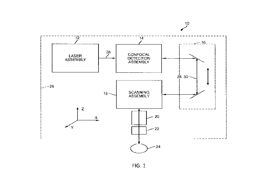
[0032] FIG. 1 is a schematic diagram of a laser surgery system, in
accordance with many
embodiments, in which a patient interface device is coupled to a laser
assembly and a detection
assembly by way of a scanning assembly and shared optics that supports the
scanning assembly.
[0033] FIG. 2 is a schematic diagram of an embodiment of the laser surgery
system of FIG. 1.
[0034] FIG. 3 is a schematic diagram of an embodiment of the laser surgery
system of FIG. 1.
[0035] FIGS. 4A, 4B and 4C illustrate aspects of arcuate incisions of a
cornea that can be formed
by the laser surgery system of FIG. 1, in accordance with many embodiments.
[0036] FIGS. 5A, 5B, 5C, 5D, 5E and 5F illustrate aspects of primary
cataract surgery access
incisions of a cornea that can be formed by the laser surgery system of FIG.
1, in accordance
with many embodiments.
[0037] FIGS. 6A, 6B, 6C, 6D and 6E illustrate aspects of sideport cataract
surgery access
incisions of a cornea that can be formed by the laser surgery system of FIG.
1, in accordance
with many embodiments.
[0038] FIG. 7A is a diagram illustrating certain steps and acts in
connection with an embodiment
of a laser surgical method for performing a corneal incision while maintaining
iris exposure
below a predetermined exposure limit according to one embodiment.
[0039] FIG. 7B is a diagram illustrating certain steps and acts in
connection with an embodiment
of a laser surgical method for performing a corneal incision while maintaining
iris exposure
below a predetermined exposure limit according to one embodiment.
[0040] FIG. 8 is a graphical illustration of a treatment scan that does not
include treatment scan
modifying elements according to the present invention.
[0041] FIG. 9 is a graphical illustration of a treatment scan having an
extension of scan paths in
which the turnarounds occur beyond an incision boundary.
[0042] FIGS. 10A and 10B are graphical illustrations illustrating an
advantage of reorienting an
axis along a longer axis.
[0043] FIGS. 11A and 11B are graphical illustrations of pulse gating.
[0044] FIGS. 12A and 12B are graphical illustration of different
embodiments of pulse gating
turnarounds.
[0045] FIG. 13 is a graphical illustration of pulse gating rows.
9
CA 03052147 2019-07-30
WO 2018/144644
PCT/US2018/016307
[0046] FIG. 14A is a diagram illustrating certain steps and acts in
connection with an
embodiment of a laser surgical method for performing a corneal incision while
maintaining iris
exposure below a predetermined exposure limit according to an embodiment.
[0047] FIG. 14B is a diagram illustrating certain steps and acts in
connection with an
embodiment of a laser surgical method for performing a corneal incision while
maintaining iris
exposure below a predetermined exposure limit according to one embodiment.
[0048] FIG. 15A is a schematic diagram illustrating a Lawn-mower raster
pattern on the iris
surface.
[0049] FIG. 15B is a graphical illustration showing the calculated
temperature on the iris surface
at a single point in time during the scan.
[0050] FIG. 16 is a graphical illustration showing a vertical cross-section
of the temperature
profile of the iris at a single point in time during the scan. The horizontal
layer in the center is the
iris.
[0051] FIG. 17 is a graphical illustration of the temporal profile of the
temperature at different
points of the iris on the center of the scan.
[0052] FIG. 18 is an en face image of an Ex vivo porcine eye showing an MVL
of the iris at
different temperatures and laser power.
[0053] FIG. 19 is an en face image of in vivo exposed rabbit irises, before
(A), immediately after
(B), and 1 h-post exposure (C).
[0054] FIG. 20 is a graphical illustration of an integrating aperture.
[0055] FIG. 21 is a graph of the Time to pass over a square aperture vs.
Threshold Lesion
Exposure (J/cm2).
[0056] FIG. 22 is a schematic diagram illustrating an image of the eye having
an electromagnetic
beam focused on a portion of the cornea of the eye.
DETAILED DESCRIPTION
[0057] In the following description, various embodiments of the present
invention will be
described. For purposes of explanation, specific configurations and details
are set forth in order
to provide a thorough understanding of the embodiments. It will also, however,
be apparent to
one skilled in the art that the present invention can be practiced without the
specific details.
CA 03052147 2019-07-30
WO 2018/144644
PCT/US2018/016307
Furthermore, well-known features may be omitted or simplified in order not to
obscure the
embodiment being described.
System Overview
[0058] Referring now to the drawings in which like numbers reference
similar elements, FIG. 1
schematically illustrates a laser surgery system 10, in accordance with many
embodiments. The
laser surgery system 10 includes a laser assembly 12, a confocal detection
assembly 14, a shared
optics 16, a scanning assembly 18, an objective lens assembly 20, and a
patient interface device
22. The patient interface device 22 is configured to interface with a patient
24. The patient
interface device 22 is supported by the objective lens assembly 20. The
objective lens assembly
20 is supported by the scanning assembly 18. The scanning assembly 18 is
supported by the
shared optics 16. The shared optics 16 has a portion having a fixed position
and orientation
relative to the laser assembly 12 and the confocal detection assembly 14. In
many embodiments,
the patient interface device 22 is configured to interface with an eye of the
patient 24. For
example, the patient interface device 22 can be configured to be vacuum
coupled to an eye of the
patient 24 such as described in co-pending U.S. Provisional Patent Application
serial number:
61,721,693, entitled "Liquid Optical Interface for Laser Eye Surgery System",
filed November 2,
2012. The laser surgery system 10 can further optionally include a base
assembly 26 that can be
fixed in place or repositionable. For example, the base assembly 26 can be
supported by a
support linkage that is configured to allow selective repositioning of the
base assembly 26
relative to a patient and secure the base assembly 26 in a selected fixed
position relative to the
patient. Such a support linkage can be supported in any suitable manner such
as, for example, by
a fixed support base or by a movable cart that can be repositioned to a
suitable location adjacent
to a patient. In many embodiments, the support linkage includes setup joints
with each setup
joint being configured to permit selective articulation of the setup joint and
can be selectively
locked to prevent inadvertent articulation of the setup joint, thereby
securing the base assembly
26 in a selected fixed position relative to the patient when the setup joints
are locked. Laser
surgery system 10, including laser assembly 12, preferably does not include a
pulse picker.
[0059] In many embodiments, the laser assembly 12 is configured to emit an
electromagnetic
radiation beam 28. The beam 28 can include a series of laser pulses of any
suitable energy level,
duration, and repetition rate.
11
CA 03052147 2019-07-30
WO 2018/144644
PCT/US2018/016307
[0060] In many one embodiments, the laser assembly 12 incorporates
femtosecond (FS) laser
technology. By using femtosecond laser technology, a short duration (e.g.,
approximately 10-13
seconds in duration) laser pulse (with energy level in the micro joule range)
can be delivered to a
tightly focused point to disrupt tissue, thereby substantially lowering the
energy level required to
image and/or modify an intraocular target as compared to laser pulses having
longer durations.
In other embodiments, a pulse duration of the laser pulses is generally
between 1 ps and 100 ns.
[0061] The laser assembly 12 can produce laser pulses having a wavelength
suitable to treat
and/or image tissue. For example, the laser assembly 12 can be configured to
emit an
electromagnetic radiation beam 28 such as emitted by any of the laser surgery
systems described
in U.S. Application No. 14/069,042, entitled "Laser Eye Surgery System," filed
October 31,
2013 (issued as U.S. Patent No. 9,445,946); and U.S. Patent Application serial
number
12/987,069, entitled "Method and System For Modifying Eye Tissue and
Intraocular Lenses",
filed January 7, 2011 (published as U.S. 2011/0172649A1). For example, the
laser assembly 12
can produce laser pulses having a wavelength from 1020 nm to 1050 nm. For
example, the laser
assembly 12 can have a diode-pumped solid-state configuration with a 1030 (+/
5) nm center
wavelength. As another example, the laser assembly 12 can produce ultraviolet
light pulses
having a wavelength of between 320 nm and 430 nm, preferably between 320 and
400 nm,
preferably between 320 to 370 nm, and more preferably between 340nm and 360
nm. In many
embodiments, the laser pulses have a wavelength of 355 nm. The 320 nm to 430
nm light
source may be, for instance, a Nd:YAG laser source operating at the 3rd
harmonic wavelength,
355nm.
[0062] When UV wavelengths are used, the tissue modification is preferably
carried out using
chromophore absorption without plasma formation and/or without bubble
formation and an
associated cavitation event. Here, chromophore absorption refers to the
absorption of at least a
portion of the ultraviolet light by one or more chemical species in the target
area. The use of
ultraviolet light significantly reduces the threshold for plasma formation and
associated
formation of cavitation bubbles but also decreases the threshold energy
required for linear
absorption enhanced photodecomposition without the formation of cavitation
bubbles for a few
reasons. First, the focused spot diameter scales linearly with wavelength
which squares the peak
radiant exposure within the focal plane. Second, the linear absorption of the
material itself
allows an even lower threshold for plasma formation or low density
photodecomposition as
12
CA 03052147 2019-07-30
WO 2018/144644
PCT/US2018/016307
initially more laser energy is absorbed in the target structure. Third, the
use of UV laser pulses
in the nanosecond and sub-nanosecond regime enables linear absorption enhanced
photodecomposition and chromophore guided ionization.
[0063] Furthermore, this chromophore guided ionization when using
ultraviolet wavelength
strongly lowers the threshold for ionization in case of plasma formation as
well lowers the
threshold for low density photodecomposition for material modification or
alteration without
cavitation even under very weak absorption. The linear absorption also allows
for the specific
treatment of topical lens structures (e.g. the lens capsule) as the optical
penetration depth of the
laser beam is limited by the linear absorption of the lens. This is especially
true for aged lenses
which absorption in the UV-blue spectral region increases strongly compared to
young lenses.
[0064] The laser pulses preferably have a wavelength 320 nm to 430 nm. For
example, the laser
assembly 12 can include an Nd:YAG laser source operating at the 3rd harmonic
wavelength (355
nm) and producing pulses having 50 picosecond to 15 nanosecond pulse duration.
Depending on
the spot size, typical pulse energies used can be in the nanojoule to micro
joule range. The laser
assembly 12 can also include two or more lasers of any suitable configuration.
[0065] The laser assembly 12 can include control and conditioning
components. For example,
such control components can include components such as a beam attenuator to
control the energy
of the laser pulse and the average power of the pulse train, a fixed aperture
to control the cross-
sectional spatial extent of the beam containing the laser pulses, one or more
power monitors to
monitor the flux and repetition rate of the beam train and therefore the
energy of the laser pulses,
and a shutter to allow/block transmission of the laser pulses. Such
conditioning components can
include an adjustable zoom assembly and a fixed optical relay to transfer the
laser pulses over a
distance while accommodating laser pulse beam positional and/or directional
variability, thereby
providing increased tolerance for component variation.
[0066] In many embodiments, the laser assembly 12 and the confocal
detection assembly 14
have fixed positions relative to the base assembly 26. The beam 28 emitted by
the laser
assembly 12 propagates along a fixed optical path through the confocal
detection assembly 14 to
the shared optics 16. The beam 28 propagates through the shared optics 16
along a variable
optical path 30, which delivers the beam 28 to the scanning assembly 18. In
many embodiments,
the beam 28 emitted by the laser assembly 12 is collimated so that the beam 28
is not impacted
by patient movement induced changes in the length of the optical path between
the laser
13
CA 03052147 2019-07-30
WO 2018/144644
PCT/US2018/016307
assembly 12 and the scanner 16. The scanning assembly 18 is operable to scan
the beam 28
(e.g., via controlled variable deflection of the beam 28) in at least one
dimension. In many
embodiments, the scanning assembly 18 is operable to scan the beam 28 in two
dimensions
transverse to the direction of propagation of the beam 28 and is further
operable to scan the
location of a focal point of the beam 28 in the direction of propagation of
the beam 28. The
scanned beam is emitted from the scanning assembly 18 to propagate through the
objective lens
assembly 20, through the interface device 22, and to the patient 24.
[0067] The shared optics 16 is configured to accommodate a range of
movement of the patient
24 relative to the laser assembly 12 and the confocal detection assembly 14 in
one or more
directions while maintaining alignment of the beam 28 emitted by the scanning
assembly 18 with
the patient 24. For example, in many embodiments, the shared optics 16 is
configured to
accommodate a range movement of the patient 24 in any direction defined by any
combination
of unit orthogonal directions (X, Y, and Z).
[0068] The shared optics 16 supports the scanning assembly 18 and provides
the variable optical
path 30, which changes in response to movement of the patient 24. Because the
patient interface
device 22 is interfaced with the patient 24, movement of the patient 24
results in corresponding
movement of the patient interface device 22, the objective lens assembly 20,
and the scanning
assembly 18. The shared optics 16 can include, for example, any suitable
combination of a
linkage that accommodates relative movement between the scanning assembly 18
and, for
example, the confocal detection assembly 24, and optical components suitably
tied to the linkage
so as to form the variable optical path 30.
[0069] A portion of the electromagnetic radiation beam 28 that is reflected
by eye tissue at the
focal point propagates back to the confocal detection assembly 14.
Specifically, a reflected
portion of the electromagnetic radiation beam 28 travels back through the
patient interface
device 22, back through the objective lens assembly 20, back through (and de-
scanned by) the
scanning assembly 18, back through the shared optics 16 (along the variable
optical path 30), and
to the confocal detection assembly 14. In many embodiments, the reflected
portion of the
electromagnetic radiation beam that travels back to the confocal detection
assembly 14 is
directed to be incident upon a sensor that generates an intensity signal
indicative of intensity of
the incident portion of the electromagnetic radiation beam. The intensity
signal, coupled with
associated scanning of the focal point within the eye, can be processed in
conjunction with the
14
CA 03052147 2019-07-30
WO 2018/144644
PCT/US2018/016307
parameters of the scanning to, for example, image/locate structures of the
eye, such as the
anterior surface of the cornea, the posterior surface of the cornea, the iris,
the anterior surface of
the lens capsule, and the posterior surface of the lens capsule. In many
embodiments, the
amount of the reflected electromagnetic radiation beam that travels to the
confocal detection
assembly 14 is substantially independent of expected variations in the length
of the variable
optical path 30 due to patient movement, thereby enabling the ability to
ignore patient
movements when processing the intensity signal to image/locate structures of
the eye.
[0070] FIG. 2 schematically illustrates details of an embodiment of the
laser surgery system 10.
Specifically, example configurations are schematically illustrated for the
laser assembly 12, the
confocal detection assembly 14, and the scanning assembly 18. As shown in the
illustrated
embodiment, the laser assembly 12 can include an ultrafast (UF) laser 32
(e.g., a femtosecond
laser), alignment mirrors 34, 36, a beam expander 38, a one-half wave plate
40, a polarizer and
beam dump device 42, output pickoffs and monitors 44, and a system-controlled
shutter 46. The
electromagnetic radiation beam 28 output by the laser 32 is deflected by the
alignment mirrors
34, 36. In many embodiments, the alignment mirrors 34, 36 are adjustable in
position and/or
orientation so as to provide the ability to align the beam 28 with the
downstream optical path
through the downstream optical components. Next, the beam 28 passes through
the beam
expander 38, which increases the diameter of the beam 28. Next, the expanded
beam 28 passes
through the one-half wave plate 40 before passing through the polarizer. The
beam exiting the
laser is linearly polarized. The one-half wave plate 40 can rotate this
polarization. The amount
of light passing through the polarizer depends on the angle of the rotation of
the linear
polarization. Therefore, the one-half wave plate 40 with the polarizer acts as
an attenuator of the
beam 28. The light rejected from this attenuation is directed into the beam
dump. Next, the
attenuated beam 28 passes through the output pickoffs and monitors 44 and then
through the
system-controlled shutter 46. By locating the system-controlled shutter 46
downstream of the
output pickoffs and monitors 44, the power of the beam 28 can be checked
before opening the
system-controlled shutter 46.
[0071] As shown in the illustrated embodiment, the confocal detection
assembly 14 can include
a polarization-sensitive device such as a polarized or unpolarized beam
splitter 48, a filter 50, a
focusing lens 51, a pinhole aperture 52, and a detection sensor 54. A one-
quarter wave plate 56
is disposed downstream of the polarized beam splitter 48. The beam 28 as
received from the
CA 03052147 2019-07-30
WO 2018/144644
PCT/US2018/016307
laser assembly 12 is polarized so as to pass through the polarized beam
splitter 48. Next, the
beam 28 passes through the one-quarter wave plate 56, thereby rotating the
polarization axis of
the beam 28. A quarter rotation is a presently preferred rotation amount.
After reflecting from
the focal point in the eye, the returning reflected portion of the beam 28
passes back through the
one-quarter wave plate 56, thereby further rotating the polarization axis of
the returning reflected
portion of the beam 28. Ideally, after passing back through the one-quarter
wave plate 56, the
returning reflected portion of the beam has experienced a total polarization
rotation of 90 degrees
so that the reflected light from the eye is fully reflected by the polarized
beam splitter 48. The
birefringence of the cornea can also be taken into account if, for example,
the imaged structure is
the lens. In such a case, the plate 56 can be adjusted and/or configured so
that the double pass of
the plate 56 as well as the double pass of the cornea sum up to a polarization
rotation of 90
degrees. Because the birefringence of the cornea may be different from patient
to patient, the
configuration/adjustment of the plate 56 can be done dynamically so as to
optimize the signal
returning to the detection sensor 54. Accordingly, the returning reflected
portion of the beam 28
is now polarized to be at least partially reflected by the polarized beam
splitter 48 so as to be
directed through the filter 50, through the lens 51, and to the pinhole
aperture 52. The filter 50
can be configured to block wavelengths other than the wavelengths of interest.
The pinhole
aperture 52 is configured to block any returning reflected portion of the beam
28 reflected from
locations other than the focal point from reaching the detection sensor 54.
Because the amount
of returning reflected portion of the beam 28 that reaches the detection
sensor 54 depends upon
the nature of the tissue at the focal point of the beam 28, the signal
generated by the detection
sensor 54 can be processed in combination with data regarding the associated
locations of the
focal point so as to generate image/location data for structures of the eye.
[0072] In this embodiment, the same laser assembly may be used both for
treatment (i.e.
modification) and imaging of the target tissue. For instance, the target
tissue may be imaged by
raster scanning pulsed laser beam 28 along the target tissue to provide for a
plurality of data
points, each data point having a location and intensity associated with it for
imaging of the target
tissue. In some embodiments, the raster scan is selected to deliver a sparse
pattern in order to
limit the patient's exposure, while still discerning a reasonable map of the
intraocular targets. In
order to image the target tissue, the treatment laser beam (i.e. the laser
beam having the
parameters suitably chosen as described above for the modification of tissue)
is preferably
16
CA 03052147 2019-07-30
WO 2018/144644
PCT/US2018/016307
attenuated to the nanoJoule level for imaging of the structures to be treated.
When used for
imaging, the attenuated laser beam may be referred to as an imaging beam. In
many
embodiments, the treatment beam and the imaging beam may be the same except
that the pulse
energy of the laser source is lower than the treatment beam when the laser
beam is used for
imaging. In many embodiments, the pulse energy of the laser beam when used for
imaging is
preferably from about 0.1 nJ to 10 nJ, preferably less than 2 nJ and more
preferably less than 1.8
nJ. The use of the same laser beam for both treatment and imaging provides for
the most direct
correlation between the position of the focal locations for imaging and
treatment ¨ they are the
same beam. This attenuated probe beam can is preferably used directly in a
back reflectance
measuring configuration, but, alternatively, may be used indirectly in a
fluorescence detection
scheme. Since increases in both backscatter and fluorescence within tissue
structures will be
evident, both approaches have merit.
[0073] In a preferred embodiment, imaging of a first target area to be
modified is performed
sequentially with the modification of the tissue in the first target area
before moving on to a
second, different, target area, i.e. imaging is performed sequentially with
treatment in a
predetermined target area. Thus, for instance imaging of the lens capsule is
preferably followed
by treatment of the lens capsule before imaging is carried out on other either
structures, such as
the cornea or iris. In another embodiment, imaging of a first target area
where a first incision to
be place is performed sequentially with the scanning the treatment beam to
perform the incision
in the first target area before moving on to a second target area for
performing a second incision,
i.e. imaging of the area to be incised is performed sequentially with scanning
the treatment beam
to perform in the predetermined target area.
[0074] In another embodiment, a cataract procedure comprises a capsulotomy
incision, and at
least one of a cataract incision and a limbal relaxing incision. In one
embodiment, imaging of
the target tissue where the capsulotomy is to be performed is followed by
scanning of the
treatment to perform the capsulotomy, and then the treatment beam is scanned
to perform the
capsulotomy. Subsequently, imaging of the target tissue where the at least one
of the cataract
incisions (CI) and the limbal relaxing incision (LRI) is carried out and then
the treatment beam is
scanned to perform the at least one of the LRI and the CI. When an LRI is
selected, this
minimizes the chance for the patient to move between imaging and treatment for
the LRIs which
are the most critical / sensitive to eye movements between image and
treatment.
17
CA 03052147 2019-07-30
WO 2018/144644
PCT/US2018/016307
[0075] As shown in the illustrated embodiment, the scanning assembly 18 can
include a z-scan
device 58 and a xy-scan device 60. The z-scan device 58 is operable to vary a
convergence/divergence angle of the beam 28 and thereby change a location of
the focal point in
the direction of propagation of the beam 28. For example, the z-scan device 58
can include one
or more lenses that are controllably movable in the direction of propagation
of the beam 28 to
vary a convergence/divergence angle of the beam 28. The xy-scan device 60 is
operable to
deflect the beam 28 in two dimensions transverse to the direction of
propagation of the beam 28.
For example, the xy-scan device 60 can include one or more mirrors that are
controllably
deflectable to scan the beam 28 in two dimensions transverse to the direction
of propagation of
the beam 28. Accordingly, the combination of the z-scan device 58 and the xy-
scan device 60
can be operated to controllably scan the focal point in three dimensions, for
example, within the
eye of the patient.
[0076] As shown in the illustrated embodiment, a camera 62 and associated
video illumination
64 can be integrated with the scanning assembly 18. The camera 62 and the beam
28 share a
common optical path through the objective lens assembly 20 to the eye. A video
dichroic 66 is
used to combine/separate the beam 28 with/from the illumination wavelengths
used by the
camera. For example, the beam 28 can have a wavelength of about 355 nm and the
video
illumination 64 can be configured to emit illumination having wavelengths
greater than 450 nm.
Accordingly, the video dichroic 66 can be configured to reflect the 355 nm
wavelength while
transmitting wavelengths greater than 450 nm.
[0077] FIG. 3 schematically illustrates a laser surgery system 300, in
accordance with many
embodiments. The laser surgery system 300 includes the laser assembly 12, the
confocal
detection assembly 14, the shared optics 16, the scanning assembly 18, the
objective lens
assembly 20, the patient interface 22, communication paths 302, control
electronics 304, control
panel/graphical user interface (GUI) 306, and user interface devices 308. The
control electronics
304 includes processor 310, which includes memory 312. The patient interface
22 is configured
to interface with a patient 24. The control electronics 304 is operatively
coupled via the
communication paths 302 with the laser assembly 12, the confocal detection
assembly 14, the
shared optics 16, the scanning assembly 18, the control panel/GUI 306, and the
user interface
devices 308.
18
CA 03052147 2019-07-30
WO 2018/144644
PCT/US2018/016307
[0078] The scanning assembly 18 can include a z-scan device and a xy-scan
device. The laser
surgery system 300 can be configured to focus the electromagnetic radiation
beam 28 to a focal
point that is scanned in three dimensions. The z-scan device can be operable
to vary the location
of the focal point in the direction of propagation of the beam 28. The xy-scan
device can be
operable to scan the location of the focal point in two dimensions transverse
to the direction of
propagation of the beam 28. Accordingly, the combination of the z-scan device
and the xy-scan
device can be operated to controllably scan the focal point of the beam in
three dimensions,
including within a tissue of the patient 24 such as within an eye tissue of
the patient 24. The
scanning assembly 18 is supported by the shared optics 16, which may be
configured to
accommodate patient movement induced movement of the scanning assembly 18
relative to the
laser assembly 12 and the confocal detection assembly 14 in three dimensions.
[0079] The patient interface 22 is coupled to the patient 24 such that the
patient interface 22, the
objective lens assembly 20, and the scanning assembly 18 move in conjunction
with the patient
24. For example, in many embodiments, the patient interface 22 employs a
suction ring that is
vacuum attached to an eye of the patient 24. The suction ring can be coupled
with the patient
interface 22, for example, using vacuum to secure the suction ring to the
patient interface 22.
[0080] The control electronics 304 controls the operation of and/or can
receive input from the
laser assembly 12, the confocal detection assembly 14, the free-floating
assembly 16, the
scanning assembly 18, the patient interface 22, the control panel/GUI 306, and
the user interface
devices 308 via the communication paths 302. The communication paths 302 can
be
implemented in any suitable configuration, including any suitable shared or
dedicated
communication paths between the control electronics 304 and the respective
system components.
[0081] The control electronics 304 can include any suitable components,
such as one or more
processors, one or more field-programmable gate arrays (FPGA), and one or more
memory
storage devices. In many embodiments, the control electronics 304 controls the
control
panel/GUI 306 to provide for pre-procedure planning according to user
specified treatment
parameters as well as to provide user control over the laser eye surgery
procedure.
[0082] The control electronics 304 can include a processor/controller 310
that is used to perform
calculations related to system operation and provide control signals to the
various system
elements. A computer readable medium 312 may be a non-volatile computer
readable medium,
and is coupled to the processor 310 in order to store data used by the
processor and other system
19
CA 03052147 2019-07-30
WO 2018/144644
PCT/US2018/016307
elements, and in many embodiments, to store one or more programs embodying one
or more
steps for carrying out the methods of the present invention. The processor 310
interacts with the
other components of the system as described more fully throughout the present
specification. In
an embodiment, the memory 312 can include a look up table that can be utilized
to control one or
more components of the laser system surgery system 300.
[0083] The processor 310 can be a general purpose microprocessor configured
to execute
instructions and data, such as a Pentium processor manufactured by the Intel
Corporation of
Santa Clara, California. It can also be an Application Specific Integrated
Circuit (ASIC) that
embodies at least part of the instructions for performing the method in
accordance with the
embodiments of the present disclosure in software, firmware and/or hardware.
As an example,
such processors include dedicated circuitry, ASICs, combinatorial logic, other
programmable
processors, combinations thereof, and the like.
[0084] The memory 312 can be local or distributed as appropriate to the
particular application.
Memory 312 can include a number of memories including a main random access
memory
(RAM) for storage of instructions and data during program execution and a read
only memory
(ROM) in which fixed instructions are stored. Thus, the memory 312 provides
persistent (non-
volatile) storage for program and data files, and may include a hard disk
drive, flash memory, a
floppy disk drive along with associated removable media, a Compact Disk Read
Only Memory
(CD-ROM) drive, an optical drive, removable media cartridges, and other like
storage media.
[0085] The user interface devices 308 can include any suitable user input
device suitable to
provide user input to the control electronics 304. For example, the user
interface devices 308 can
include devices such as, for example, a touch-screen display/input device, a
keyboard, a
footswitch, a keypad, a patient interface radio frequency identification
(RFID) reader, an
emergency stop button, and a key switch.
[0086] The laser surgical techniques described herein a pulsed 320nm to
430nm laser to perform
highly precise physical modifications of ocular targets, including tissues
(such as lens, lens
capsule, cornea, etc.) and synthetic intraocular lens implants. This can be
done in two different
operating regimes; with or without cavitation bubble formation. The sub-
cavitation regime can
also be used to modify the refractive index of ocular targets. Although the
wavelengths used in
the present invention are shorter or in the range than those associated with
retinal blue light
toxicity, the absorption of the 320nm to 400nm laser light within the aged
lens further minimizes
CA 03052147 2019-07-30
WO 2018/144644
PCT/US2018/016307
the risk of retinal damage, as this light will be absorbed by the lens volume.
Furthermore, the
risk of damaging the corneal endothelium or other corneal structures is also
minimized. The
threshold pulse energy will be Etb=4:1:0d2/4, where (lois the threshold
radiant exposure and d is
the focal spot diameter. Here, the focal spot diameter, d, is d=AF/Db where is
the wavelength,
F is the focal length of the last focusing element and Db is the beam diameter
of the last lens.
For stable and reproducible operation, pulse energy should exceed the
threshold by at least a
factor of 2; however, the energy level can be adjusted to avoid damage to the
corneal
endothelium.
[0087] The incident light of the laser used for the modification of the eye
tissue generally has a
wavelength of between 320 nm and 430 nm, preferably between 320 and 400 nm,
preferably
between 320 to 370 nm, and more preferably between 340nm and 360 nm. In many
embodiments, the laser light has a wavelength of 355 nm.
[0088] The pulse energy of laser pulses is generally between 0.010 and 5000.
In many
embodiments, the pulse energy will be between 0.1 11.J and 100 0, or more
precisely, between
0.10 and 40 0, or between 0.1 11.J and 10 [t.I.
[0089] A pulse repetition rate of the laser pulses is generally between 500Hz
and 500 kHz. In many
embodiments, the pulse repetition rate is between 1 kHz to 200 kHz, or between
1 KHz to 100
KHz.
[0090] Spot sizes of the laser pulses are generally smaller than 10 p.m. In
many embodiments, the
spot size is preferably smaller than 5 p.m, typically 0.51.tm to 31.tm.
[0091] A pulse duration of the laser pulses is generally between 1ps and
100ns. In many
embodiments, the pulse duration is between 100 ps to 10 ns, or between 100 ps
and 1 ns. In a
preferred embodiment, the pulse duration is between 300 ps and 700 ps,
preferably 400 ps to 700
PS.
[0092] In some embodiments, the beam quality, also referred to as M2 factor,
is between 1 and 1.3.
The M2 factor is a common measure of the beam quality of a laser beam. In
brief, the M2 factor
is defined as the ratio of a beam's actual divergence to the divergence of an
ideal, diffraction
limited, Gaussian TEMoo beam having the same waist size and location as is
described in ISO
Standard 11146.
[0093] A peak power density, obtained by dividing the peak power of the laser
pulse by the focal
spot size, is generally expressed in units of GW/cm2. In general, the peak
power density of the
21
CA 03052147 2019-07-30
WO 2018/144644
PCT/US2018/016307
laser pulses should be sufficiently high to modify the ocular tissue to be
treated. As would be
understood by those ordinarily skilled, the peak power density depends upon a
number of factors,
including the wavelength of the selected laser pulses. In some embodiments, a
peak power
density is generally in the range of 100 GW/cm2 to 800 GW/cm2 will be used to
cut ocular tissue
with 355 nm light.
[0094] The scan range of the laser surgical system is preferably in the range
of 6 to 10 mm.
[0095] In many embodiments for the modification of ocular tissue, spot spacing
between adjacent
laser pulses is typically in the range of about 0.20 p.m to 10 p.m, preferably
0.2 p.m to 6 p.m.
[0096] A numerical aperture should be selected that preferably provides for
the focal spot of the
laser beam to be scanned over a scan range of 6 mm to 10 mm in a direction
lateral to a Z-axis
that is aligned with the laser beam. The NA of the system should be less than
0.6, preferably less
than 0.5 and more preferably in a range of 0.05 to 0.4, typically between 0.1
and 0.3. In some
specific embodiments, the NA is 0.15. For each selected NA, there are suitable
ranges of pulse
energy and beam quality (measured as an M2 value) necessary to achieve a peak
power density in
the range required to cut the ocular tissue. Further considerations when
choosing the NA include
available laser power and pulse rate, and the time needed to make a cut.
Further, in selection of
an appropriate NA, it is preferable to ensure that there is a safe incidental
exposure of the iris,
and other ocular tissues, that are not targeted for cuts.
[0097]
Table 1 and Table 2, below, show typical laser beam parameters in accordance
with many embodiments of the present invention.
22
CA 03052147 2019-07-30
WO 2018/144644 PCT/US2018/016307
[0098] TABLE 1:
wavelength (nm) 355 355 355 355 355 355
energy (uJ) 1 4 2.25 9 0.36 1.44
pulse rate (kHz) 70 100 70 100 70000 100
Pulse length (s) 6.00E-10 6.00E-10 6.00E-10 6.00E-10 6.00E-10
6.00E-10
NA (1/e^2) 0.15 0.15 0.1 0.1 0.25 0.25
MA2 (1/e^2) 1.3 1 1.3 1 1.3 1
spot spacing (um) 1 2 1.5 3 0.6 1.2
theta (rad, 1/eA2) 0.3 0.3 0.2 0.2 0.5 0.5
BP (um, 1/eA2) 0.588 0.452 0.588 0.452 0.587 0.452
SS (um, 1/eA2) 1.95 1.5 2.94 2.26 1.18 0.904
area (mmA2, 1/eA2) 3.01E-06 1.78E-06 6.77E-06 4.01E-06 1.08E-06
6.42E-07
area (cm^2, 1/eA2) 3.01E-08 1.78E-08 6.78E-08 4.01E-08 1.08E-08
6.42E-09
peak energy density 66.4 449 66.4 449 66.34 449
(J/cm^2)
peak power density 1.E+11 7.E+11 1.E+11 7.E+11 1.E+11
7.E+11
(W/cm^2)
peak power density 111 748 111 748 111 748
(GW/cm^2)
ratio to NS 100% 100% 100% 100% 100% 100%
average power (W) 0.07 0.4 0.158 0.9 0.0252 0.144
spots per mmA2 1,000,00 250,000 444,000 111,000 2,778,000
694,000
0
time per pattern mmA2 14.3 2.500 6.35 1.11 39.7 6.94
(s)
average pattern energy 100 100 100 100 100
100
density (J/cm^2)
relative possible iris 353 95.4 192 51.9 758
205
safety limit (8*6TA.75
(J/cm^2))
ratio energy density 0.284 1.05 0.521 1.93 0.132 0.487
delivered/safety
23
CA 03052147 2019-07-30
WO 2018/144644
PCT/US2018/016307
[0099] TABLE 2:
wavelength (nm) 355 355 355 355
energy (uJ) 9 36 0.141 0.562
pulse rate (Hz) 70000 100000 70000 100000
Pulse length (s) 6.00E-10 6.00E-10 6.00E-10 6.00E-10
NA (1/e^2) 0.05 0.05 0.4 0.4
MA2 (1/e^2) 1.3 1 1.3 1
spot spacing ( m) 3 6 0.375 0.75
theta (rad, 1/e^2) 0.1 0.1 0.8 0.8
BP (p.m, 1/eA2) 0.588 0.452 0.0588 0.452
SS (p.m, 1/eA2) 5.88 4.52 0.735 0.565
area (mmA2, 1/eA2) 2.71E-05 1.61E-05 4.24E-07 2.51E-07
area (cm^2, 1/e^2) 2.71E-07 1.61E-07 4.24E-09 2.51E-09
peak energy density (J/cm^2) 66.4 449 66.4 449
peak power density (W/cm^2) 1.E+11 7.E+11 1.E+11 7.E+11
peak power density (GW/cm^2) 111 748 111 748
ratio to NS 100.00% 100.00% 100.00% 100.00%
average power (W) 0.63 3.6 0.00984 0.0563
spots per mmA2 111,000 27,800 7,111,000
1,778,000
time per pattern mmA2 (s) 1.59 0.278 102 17.8
average pattern energy density 100.000 100.000 100.000 100.000
(J/cm^2)
relative possible iris safety limit 67.9 18.4 154 416
(8*6TA.75 (J/cm^2))
ratio energy density delivered/safety 1.47 5.45 0.065 0.241
[00100] In Tables 1 and 2, theta is the divergence half-angle, BP is the
beam parameter product,
SS is the spot size, and the area is the area of the laser spot. Here, the
1/e2 width is equal to the
distance between the two points on the marginal distribution that are 1/e2 =
0.135 times the
maximum value.
[00101] In many embodiments, the laser eye surgery methods and/or laser eye
surgery systems
described herein are used in for maintaining iris exposures below a
predetermined limit while
making corneal incisions during a laser cataract surgery.
[00102] In cataract surgery, a capsulotomy incision, often in the form of a
small round hole is
formed in the anterior side of the lens capsule to provide access to the lens
nucleus.
[00103] In addition, cataract surgery may include three types of cornea
incisions: arcuate
incisions, primary incisions ("primary cataract incisions", or "cataract
incisions") and sideport
incisions. Parameters that may be used to define the capsulotomy include shape
(i.e. circular,
24
CA 03052147 2019-07-30
WO 2018/144644
PCT/US2018/016307
elliptical, rectangular or polygonal) and size. The systems described herein
are designed to
receive these parameters based on user or physician's input and preferably, to
provide a prompt
for their input where not received.
[00104] Primary incisions and sideport incisions may have the same structure.
They are generally
multiplanar structures that create an opening that allow the physician access
into the anterior
chamber. The primaries are used for insertion of the aspiration tool and the
insertion of the IOL.
Sideport incisions may be used for inserting smaller instrumentation into the
anterior chamber.
The location and shape of both the primary incisions and the sideport
incisions are determined by
the user parameters and, optionally, by information from a section scan as
described herein,
where the cornea anterior and posterior surfaces may be modeled by circles.
The anterior and
posterior curvatures of the cornea as measured in the circular fits of the
section scans may
optionally be used to position the cuts. Parameters that may be used to define
the primary
cataract incision or the sideport incision are preferably selected from the
group consisting of
limbus offset, width; side cut angle, plane depth and length. The systems
described herein are
designed to receive these parameters based on user or physician's input and
preferably, to
provide a prompt for their input where not received.
[00105] Arcuate incisions may be used to correct a patient's astigmatism. For
instance, they may
adjust the curvature of the cornea to a more spherical shape by means relaxing
stresses along the
meridian on which they are placed. They are parts of a conical surface that
crosses both the
anterior and posterior surfaces of the cornea. In some embodiments, the
anterior curvature and
posterior curvature of the cornea, as measured in a circular fit to a section
scan, are used to
position an "along-the-cut" scan. The along-the-cut scan lays on the surface
of a cone that
transverses the cornea. The arcuate incision can be located within the along-
the-cut scan.
Parameter that may be used to define the arcuate incision may include the size
of the optical
zone, arc length, uncut anterior portion, uncut posterior portion and side cut
angle. The systems
described herein are designed to receive these parameters based on user or
physician's input and
preferably, to provide a prompt for their input where not received.
[00106] The laser surgery system 10 can be used to form any suitably shaped
arcuate, primary or
sideport incisions.
[00107] FIGS. 4A through 4C illustrate aspects of arcuate incisions of a
cornea that can be formed
by the laser surgery system 10, in accordance with many embodiments. FIG. 4A
shows an en
CA 03052147 2019-07-30
WO 2018/144644
PCT/US2018/016307
face view of arcuate incisions 600, 602 within the optical zone 604 of the
cornea 606 that can be
formed using the system 2. The optical zone 606 is user-adjustable within the
range of 2 mm-11
mm. For asymmetric arcuate incisions, the optical zone 606 is independently
adjustable for each
incision. Arc length 608 is user-adjustable within the range of 10 -120 .
[00108] FIG. 4B shows a cross-sectional view of an arcuate incision 605 in the
cornea 606 that
can be formed using the system 2 and that penetrates the cornea anterior
surface 609 and has an
uncut posterior portion 610. FIG. 4C shows a cross-sectional view of an
arcuate intrastromal
incision 611 in the cornea 606 that can be formed using the system 2. The
arcuate intrastromal
incision 611 has an uncut anterior portion 612 and an uncut posterior portion
610. Side cut angle
614 is user-adjustable within the range of 30 -150 . Uncut posterior and
anterior portions 610,
612 are user-adjustable within the range of 100 Ilm-2501.tm or 20%-50% of the
cornea thickness.
Cornea thickness is measured at the projected intersection of the incision
with the cornea
anterior/posterior measured at 90 to anterior/posterior cornea surface
regardless of what side cut
angle 614 is chosen.
[00109] FIG. 5A shows an en face view of a primary cataract incision 616 in
the cornea 606 that
can be formed using the system 2. The primary cataract incision 616 provides
access to surgical
tools used to, for example, remove the fragmented crystalline lens nucleus and
insert an IOL.
FIG. 5B shows a cross-sectional view of a primary cataract incision 617 of the
cornea 606 that
can be formed using the system 2. Limbus offset 618 is user-adjustable within
the range of 0.0
mm-5.0 mm. Width 620 is user-adjustable within the range 0.2 mm-6.5 mm. Length
622 is
user-adjustable within the range of 0.5 mm-3.0 mm. Side Cut Angle 624 is user-
adjustable
within the range of 30 -150 . Plane depth 626 is user-adjustable within the
range of 125 lm-375
1.tm or 25%-75% of the cornea thickness. Length 622 is defined as the en face
view distance
between the projected incision intersection with the cornea anterior and the
cornea posterior.
FIG. 5C shows a cross-sectional view of a primary cataract incision 627 that
includes an uncut
anterior portion 628. FIG. 5D shows a cross-sectional view of a primary
cataract incision 629
that includes an uncut posterior portion 630. FIG. 5E shows a cross-sectional
view of a primary
cataract incision 631 that includes an uncut central length 632. And FIG. 5F
shows a cross-
sectional view of a primary cataract incision 634 that includes no uncut
portion. Side Cut Angle
636 is user-adjustable within the range of 30 -150 . Uncut central length 632
is user-adjustable
within the range of 25 lm-1000
26
CA 03052147 2019-07-30
WO 2018/144644
PCT/US2018/016307
[00110] FIG. 6A shows an en face view of a sideport cataract incision 638 in
the cornea 606 that
can be formed using the system 2. The sideport cataract incision 638 provides
access for
surgical tools used, for example, to assist in the removal of the fragmented
crystalline lens. FIG.
6B shows a cross-sectional view of a sideport cataract incision 639 of the
cornea 606 that has an
uncut posterior portion 640 and can be formed using the system 2. Limbus
offset 642 is user-
adjustable within the range of 0.0 mm-5.0 mm. Width 644 is user-adjustable
within the range
0.2 mm-6.5 mm. Length 645 is user-adjustable within the range of 0.5 mm-3.0
mm. FIG. 6C
shows a cross-sectional view of a sideport cataract incision 646 that includes
an uncut anterior
portion 648. FIG. 6D shows a cross-sectional view of a sideport cataract
incision 650 that
includes an uncut central length 652. And FIG. 6E shows a cross-sectional view
of a sideport
cataract incision 654 that includes no uncut portion. Side Cut Angle 656, 658,
660 is user-
adjustable within the range of 30 -150 . Uncut central length 652 is user-
adjustable within the
range of 100 Ilm-2501.tm or 20%-50% of the cornea thickness. Cornea thickness
662 is
measured at the projected intersection location of the incision with the
cornea anterior/posterior
measured at 90 to the anterior/posterior cornea surface regardless of what
side cut angle is
chosen.
27
CA 03052147 2019-07-30
WO 2018/144644
PCT/US2018/016307
[00112] Table 3:
User-adjustable
parameters for
arcuate in
Feature Default' Range Increment Step Units
Size
Single,
Incision 'Type N/A Symmetric, N/A 0.5 N/A
Asymmetric
Axis** N/A 0-360 1 2.5 0
Optical Zone** N/A 2-11 0.1 10 mm
Arc Length** N/A 10-120 1 0.5
;
Centering; Method. N/A Pupil Embus, N/A 0.5 N/A
Custom
Penetration Type Anterior Anterior orN/A N/A N/A
Intrastromal
or
Depth Units Percentage Percentage N/A N/A N/A
Absolute
Uncut Anterior*** 20% 20-50% 1 2
100 100-250 1 10 gn
Uncut Posterior 20% 20-50% 1 2 %
100 100-250 1 10 WTI
Side Cut Angie 90 30-150 1 c, 0
Horizontal Spot
4 2-10 1 1 urn
Spacing
Vertical Spot
1-25 1 1 WTI
Spacing
Pulse Energy 2 1-4 0.1 0.1 lir
'Parameters do not have default values; user must select each parameter.
**Independently adjustable parameters for asymmetric incisions.
***Not applicable for anterior penetrating.
28
CA 03052147 2019-07-30
WO 2018/144644
PCT/US2018/016307
[00113] TABLE 4
User-adjustable parameters for
primary cataract incisions.
Step
Feature Default* Range Increment . Units
Size
Axis NIA 0-360 1 --; 0
Limbus Offset N/A 0.0-5.0 0.1 0.1 mm
Width 7.2 0.2-6.5 0 1 0 1 ram
Length 2.2 0.5-3.0 0.1 0.1 mm
Anterior,
Uncut Region Central N/A N/A N/A
Central,
Posterior, None
Depth Units Percentage Percentage or N/A N/A N/A
Absolute
Uncut Anterior/ 20% 20-50% 1% 5% (!io
Uncut Posterior 100 100-250 1 25 Ilfri
Uncut Central Length* 100 25-1000 1 25 p.m
Plane Depth 50% 25-75% 1% 5% %
250 175-375 1 50 pm
Side Cut Angle 120 30-150 1 5 0
Horizontal Spot Spacing 10 2-10 1 c
, p.m
Vertical Spot Spacing 20 1-25 1 c
- ,i1111
Pulse Energy 5 1-4 0.1 05 jr.1
If the uncut central length is longer than the length parameter, then the
uncut central
length will be set as equal to the length parameter.
29
CA 03052147 2019-07-30
WO 2018/144644 PCT/US2018/016307
[00114] TABLE 5
User-adjustable parameters for
sideport cataract incisions.
Unit
Feature Default* Range increment Step
Number of incisions N/A 0-6 1 1 N/A
Axis* NIA (0) 0-360 1 5 0
Liinbus Offset* N/A (1.0) 0.0-50 0.1 0.1 mm
Width* N/A (0.5) 0.2-6.5 0.1 0.1 mm
r,
Uncut Type Central Antetio N/A N/A N/A
Central.
Posterior.
None
Percentage or
Uncut Units Percentage NIA NA N/A
Absolute
Uncut Length 20% 20-50% 1% 2% %
(Anierior, Posterior, 100 100-250 1 10 [tin
Cornea
Side Cut Angle Type Lens Apex Posteiior N/A N/A N/A
Apex,
AC Center,
Lens Apex,
Lens Center,
Custom
Custom Side Cut Angle 90 30-150 1 5
Horizontal Spot 10 2-10 1 5 u,rn
Spacing
Vertical Spot Spacing 20 1-25 5 [tin
Pulse Energy 5 1-4 0.1 0.5 1,0
CA 03052147 2019-07-30
WO 2018/144644 PCT/US2018/016307
[00115] Although many different imaging techniques may be used in different
embodiments, a combination of video/camera imaging and confocal imaging based
on pulsed
laser raster scanning of the tissue to be treated is preferred.
[00116] As illustrated in the embodiment of FIG. 2, video imaging of the
tissue to be
treated, preferably a human eye, can be achieved by a camera 62 and associated
video
illumination 64 integrated with the scanning assembly 18. The camera 62 and
the beam 28
share a common optical path through the objective lens assembly 20 to the eye.
A video
dichroic 66 is used to combine/separate the beam 28 with/from the illumination
wavelengths
used by the camera. In one embodiment, the beam 28 can have a wavelength of
between 320
and 370 nm, preferably about 355 nm, and the video illumination 64 can be
configured to
emit illumination having wavelengths greater than 370 nm, or more than 400 or
more than
450 nm. Accordingly, the video dichroic 66 can be configured to reflect the
beam between
320 and 370 nm while transmitting wavelengths greater than 370 nm, thus
facilitating video
imaging of the eye without interference from beam 28. The location of the
capsulotomy
incision and any corneal incision specified by the physician can be projected
onto the video
image prior to treatment as expected scan locations for each respective
incision.
[00117] In many embodiments, the imaging of the eye 24 further includes
confocally
imaging one or more portions of the tissue, preferably the eye, to be treated.
Any suitable
device, assembly, and/or system, such as described herein, can be used to
confocally image
one or more portions of the eye or other tissue to be imaged. The confocal
imaging methods
used herein generally include using a beam source, preferably a pulsed laser
source, to
generate an electromagnetic radiation beam; propagating the electromagnetic
radiation beam
to a scanner along an optical path to the eye; focusing the electromagnetic
radiation beam to
a focal point at a location within the eye; using the scanner to scan,
preferably raster scan, the
focal point to different locations within the eye; propagating a portion of
the electromagnetic
radiation beam reflected from the focal point location back along the shared
optical path to a
sensor; and generating an intensity signal indicative of the intensity of a
portion of the
electromagnetic radiation beam reflected from the focal point location and
propagated to the
sensor. The method can include modifying polarization of at least one of the
electromagnetic
radiation beam and a portion of the electromagnetic radiation beam reflected
from the focal
point location. The method can include using the polarization-sensitive device
to reflect a
31
CA 03052147 2019-07-30
WO 2018/144644 PCT/US2018/016307
portion of the electromagnetic radiation beam reflected from the focal point
location so as to
be incident upon the sensor.
[00118] Based on the calibration of the system described herein, the focal
point location of
the confocally detected light can be related to the physical location of the
focal point within
the eye, and the location within the eye and the magnitude of the intensity at
each location
can be used to identify boundaries, edges and layers within the eye.
Boundaries, edges and
layers may be located in a confocal image by, for instance, Delaunay
triangulation and
Dijkstra segmentation. These confocal images, including the boundaries, edges
and layers
can then be displayed to a user as a graphical representation of the areas of
the eye to be
treated.
[00119] In many embodiments, the lens capsule, and optionally a portion or
the entire
lens, is imaged using confocal imaging, and preferably, these portions include
the area of the
lens capsule where the capsulotomy will be placed. In general, the parameters
necessary to
define the capsulotomy are input by a user or physician, and a raster scan
with a pulsed laser
beam sweeps through the relevant portion of the lens capsule for imaging the
lens capsule.
Based on the recorded location and magnitude of the confocally reflected
intensity
measurements at each location, the capsule is identified by image recognition,
such as by
Delaunay triangulation and Dijkstra segmentation, and the capsule shape is fit
to the
segmented image. The resulting confocal image of lens may then be shown to the
physician
for use in visualizing the capsulotomy incision.
[00120] In many embodiments, the methods and systems may include confocally
imaging
a cornea by scanning one or more of portions of the cornea where a primary
incision,
sideport incision or arcuate incision is to be placed. In a preferred
embodiment, one sectional
image of the cornea is performed for each selected corneal incision. These
images are
preferably in the form of a section scan. A section scan crosses cornea along
plane and
measures the confocal intensity at every location of a pulsed laser during the
scan.
Preferably, a section scan comprises a raster scan of a pulsed laser beam
along the cornea
including the anterior surface and posterior surface, on a vertical plane
centered at the cornea
incision center and oriented along an incision's meridian. The trajectory goes
from deep to
shallow, inside the eye, crossing the cornea. The posterior and anterior
boundaries of the
32
CA 03052147 2019-07-30
WO 2018/144644 PCT/US2018/016307
cornea may be identified in the image by, for instance, Dijkstra segmentation
of the image,
and the resulting image may be provided to the user.
[00121] If the selected corneal incision is an arcuate incision, an "along-
the-cut" imaging
scan is also preferably performed. An along-the-cut imaging scan may assist a
physician in
choosing the correct location for the arcuate incision in order to maintain an
adequate depth
and avoid posterior penetration. The "along the cut" scan preferably has the
same conical
shape as the arcuate incision and is inclusive of the entire area to be
covered arcuate incision.
The conical sector in the "along the cut" scan is mapped into a rectangular
domain 520
defined by the conical coordinates. The resulting conical image is segmented
and fit.
Optionally, the resulting fits to the anterior and posterior surfaces of the
cornea are used to
construct the arcuates, which can then be overlaid on their sections and
"along the cut" scans.
[00122] In many embodiments, the optical surface of the eye is fit with one
or more with
one or more of a Fourier transform, polynomials, a spherical harmonics, Taylor
polynomials,
a wavelet transform, or Zernike polynomials. The optical tissue surface may
comprise one or
more of the anterior surface of the cornea, the posterior surface of the
cornea, the anterior
surface of the lens capsule, the posterior surface of the lens capsule, an
anterior surface of the
lens cortex, a posterior surface of the lens cortex, an anterior surface of
the lens nucleus, a
posterior surface of the lens nucleus, one or more anterior surfaces of the
lens having a
substantially constant index of refraction, one or more posterior surfaces of
the lens having a
substantially constant index of refraction, the retinal surface, the foveal
surface, a target
tissue surface to correct vision such as a target corneal surface, an anterior
surface of an
intraocular lens, or a posterior surface of an intraocular lens, for example.
[00123] After the relevant portions of the ocular tissue, including the
cornea, have been
imaged, the incisions defined by the physician parameters may be projected
onto the image,
and a treatment scan of the laser light beam is generated. Here, the treatment
scan refers to a
simulated scan pattern, not an actual scan of the laser pulses in the eye
tissue. The treatment
scan preferably consists of a continuous set of x, y, z points arranged in
space that are
designed to carry out the incisions defined by the user. The locations of the
treatment scans
are projected onto at least one of the video and confocal images in order to
define the set of
expected scan locations of the incisions. The treatment scans according to the
invention
generally take the form of a raster scan of the ocular tissue to be incised.
33
CA 03052147 2019-07-30
WO 2018/144644
PCT/US2018/016307
[00124] In many embodiments, the present invention is directed to maintaining
an exposure on
the iris caused by scanning a pulsed laser beam in a pattern according to a
treatment below a
predetermined iris exposure limit.
[00125] The predetermined iris exposure limit is preferably set at a value
below an iris exposure
that will cause damage to the iris tissue. A number of different manners of
establishing exposure
limits are known in the art, and the systems and methods of the present
invention can be suitably
used with any known model for an exposure limit. In many, the predetermined
exposure limit
will be dependent on an exposure time of the iris to the incident radiation.
[00126] In some embodiments, the iris exposure limit (EL) is a percentage of
an Exposure (EMVL,
in J/cm2) causing a Minimal Visible Lesion (MVL) in the iris, which may be
determined
experimentally and/or calculated theoretically. Specifically, the iris
exposure limit, EL, can be
defined by the formula:
EL = c*EmvL, where c < 1.
[00127] The variable c can be chosen based on a desired safety margin below
the EMVL. In some
embodiments, c may be 0.9 or less, 0.8 or less, 0.7 or less, 0.6 or less, 0.50
or less, 0.40 or less,
0.3 or less, 0.2 or less, or 0.10 or less. Preferably, a value of c chosen is
based, at least in part, on
the uncertainty of the data that establishes EMVL. The larger the uncertainty
of EMVL, the smaller
the value of c that is appropriate to use. Preferably, values of c are less
than 0.5.
[00128] In many embodiments, EMVL is an exposure (preferably in J/cm2)
expressed as a function
of an exposure time of an integrating aperture having a specified size. In
many embodiments,
the integrating aperture for the iris is the area of the integrating aperture
specified for the Group
2 anterior chamber limit from IS015004, (0.5 mm diameter) as the most
appropriate aperture for
the iris. In many embodiments, the area of this aperture is converted to a
square aperture of the
same area as the 0.5 mm diameter circular aperture, which is a square of
approximately 0.44 mm
x 0.44 mm (FIG. 20). This square aperture is then used for the minimum visible
lesion threshold
function. The exposure time for an integrating aperture of the square aperture
is determined to
find the threshold exposure from the function. The data over the time region
of interest is
preferably fit by least squares fit of the data at the appropriate time points
(see below, 1.86 s and
19.23 s, rabbit only), which allows the EMVL as a function of time to be
conservatively expressed
as:
EmvL(T) = aTb J/cm 2 (Formula II)
34
CA 03052147 2019-07-30
WO 2018/144644
PCT/US2018/016307
and
EL = c*EmyL(T) = acTb, where c < 1. (Formula III)
[00129] where T is the time for the scanned pattern to pass over selected
integration aperture, and
parameters a and b are the best fit parameter from the data. The parameter, b,
may be 0.75.
[00130] In some embodiments, EMVL as a function of time is expressed as
Formula II.
[00131] EMVL as a function of time is expressed as Formula III.
[00132] Iris exposure limits are typically based on the use of an integration
aperture arranged over
the region to be exposed. In general, these safe exposure limits place an
upper limit on the
amount of power that can be pumped into the integration aperture over a time
period. As a
result, the safety limits are predicated on delivering a permissible amount of
power, W, in a
certain amount of time, T.
[00133] Iris exposures during laser surgery are maintained below a
predetermined exposure limit
according to at least two different strategies: (1) in a first embodiment,
laser surgical methods
and systems safely deliver a predetermined amount of power in a treatment scan
by controlling
the time over which the power is delivered to the ocular tissue; and (2) in a
second embodiment,
laser surgical methods and systems safely deliver a predetermined power by
modifying one or
more incision parameters, thereby limiting the power delivered during the
planned incision.
[00134] Thus, in some embodiments of the present invention, the energy
associated with a
planned treatment scan is maintained below a predetermined exposure level by
extending the
time of the treatment scan by adding one or more treatment scan modifying
elements so as to
cause the iris exposure to be smaller than the predetermined exposure limit.
[00135] A system for cataract surgery on an eye of a patient according to this
embodiment
comprises: a laser assembly for generating a pulsed laser treatment beam; an
imaging system
configured for imaging an ocular tissue of the patient, the ocular tissue
comprising corneal
tissue; an optical scanning system configured for positioning the focal zone
of the treatment
beam to targeted locations of the ocular tissue, the targeting locations
including a location in the
corneal tissue; and a computer control system operatively coupled to the laser
assembly, the
imaging system, and the optical scanning system. The computer control system
is programmed
to: a) generate an initial treatment scan and an initial iris exposure
corresponding to the initial
treatment scan; b) determine whether the initial iris exposure is less than a
predetermined
CA 03052147 2019-07-30
WO 2018/144644
PCT/US2018/016307
exposure limit; c) generate a revised treatment scan comprising one or more
treatment scan
modifying elements when the initial iris exposure is greater than the
predetermined exposure
limit, wherein the one or more treatment scan modifying elements cause the
iris exposure to be
smaller than the predetermined exposure limit; and d) operate the optical
scanning system and
laser assembly to direct a treatment beam in a pattern corresponding to the
revised treatment scan
so as to create a corneal incision. The acts of step (d) may be performed
automatically or may
require additional actions by a user. In some embodiments, the system
automatically delivers the
treatment in some embodiments. In other embodiments, the methods and systems
of the present
invention require some additional act by a user (such as a physician) in order
to initiate delivery
of the treatment beam. For instance, the system may preferably be configured
to provide a
message or warning through, for instance, a graphical user interface, to a
user that a revised
treatment scan has been determined and may be delivered to the patient. The
system may require
that a user manually command delivery of the treatment scan once an acceptable
scan has been
determined. The system may require that the user press a button, peddle, lever
or other device to
initiate scan. In some embodiments, it may be preferable that a user be
required to continually
depress a button, lever, peddle or other device throughout a procedure to
initiate and maintain
delivery of the treatment scan from initiation to completion. In some
embodiments, a user may
be required to enter a command via a graphical user interface in order to
initiate a treatment scan.
[00136] A laser surgical method for performing a corneal incision while
maintaining iris exposure
below a predetermined exposure limit according to this embodiment comprises:
determining an
initial iris exposure based on an initial treatment scan, the treatment scan
corresponding to a
predetermined corneal incision, determining whether the initial iris exposure
is less than the
predetermined exposure limit; generating a revised treatment scan comprising
one or more
treatment scan modifying elements when the initial iris exposure is greater
than the
predetermined exposure limit, and scanning the focal zone of a pulsed laser
beam according to
the revised treatment scan, thereby performing the corneal incision, wherein
the one or more
treatment scan modifying elements causes the iris exposure to be smaller than
the predetermined
exposure limit.
[00137] Certain steps to be carried out in a method or performed by a system
according to this
embodiment are shown in FIG. 7A. These include a step 402 of receiving a
parameter set
defining a corneal incision. The values of the parameter are generally set by
a physician or other
36
CA 03052147 2019-07-30
WO 2018/144644
PCT/US2018/016307
user. In many embodiments, the corneal incisions to be carried out according
to the methods and
systems of the present invention are selected from the group consisting of an
arcuate incision, a
primary cataract incision and sideport incision. The parameters that can be
used to define these
incisions, together with default and permissible values of each incision are
shown in Tables 3, 4,
and Figures 4A-4C, 5A-5F, and 6A-6E respectively and in the related text. The
parameter sets
used to define incisions preferably include the pulse energy of the pulsed
laser beam, the
horizontal spot spacing and the vertical spot spacing.
[00138] A step 404 comprises obtaining image data of an area of the cornea
corresponding to the
incision location. In some embodiments, the image data may be obtained by
optical coherence
tomography imaging or confocal imaging. As indicated above, many embodiments
include
confocally imaging a cornea by scanning one or more of portions of the cornea
where a primary
incision, sideport incision or arcuate incision is to be placed. In a
preferred embodiment, one
sectional image of the cornea is performed for each selected corneal incision.
These images are
preferably in the form of a section scan. A section scan crosses cornea along
plane and measures
the confocal intensity at every location of a pulsed laser during the scan.
Preferably, a section
scan comprises a raster scan of a pulsed laser beam along the cornea including
the anterior
surface and posterior surface, on a vertical plane centered at the cornea
incision center and
oriented along an incision's meridian. The trajectory goes from deep to
shallow, inside the eye,
crossing the cornea.
[00139] A step 406 according this embodiment comprises, projecting the defined
corneal incision
on the image date to generate an Initial Treatment Scan. The Initial Treatment
Scan is a
simulated scan and is not actually delivered to the patient. The initial
treatment scan preferably
comprises of a continuous set of x, y, z points arranged in space that are
designed to carry out the
incisions defined by the user. The Initial Treatment Scan is typically the
scan generated by the
system without consideration of exposure limits to the iris, and therefore
typically is an
optimized scan or default scan pattern generated by the system based solely on
a user parameter
set and without regard to iris safety consideration. The location of the
initial treatment scans
may be projected onto at least one of the video and confocal images in order
to define the set of
expected scan locations of the incisions. The treatment scans according to the
invention
generally take the form of a raster scan of the ocular tissue to be incised.
37
CA 03052147 2019-07-30
WO 2018/144644
PCT/US2018/016307
[00140] A step 408 comprises calculating an initial exposure on the iris
based on the initial
treatment scan. The parameter sets defining the Initial treatment scan
comprise a set of
parameters that fully define the geometry (size and shape) of planned incision
as well as the
pulse energy, the horizontal spot spacing and the vertical spot spacing for
carrying out the
incision. As such, it is possible to calculate the spot density (for instance,
spots/cm2) as well as
the power supplied by the pulses. Since the geometry of the eye in the area of
the planned
incision is known from the imaging scans, the size of the scanned pattern on
the iris can be
computed. As such, one can calculate an exposure of the iris tissue associated
with the initial
treatment scan. The exposure may be calculated by Pulse energy/cm2 as a
function of the time
taken to deliver the energy within a computed integrating aperture on the iris
surface, and the
worst case exposure within all such possible apertures on the iris surface is
evaluated against the
limit.
[00141] A step 410 comprises determining whether the calculated exposure is
less than a
predetermined iris exposure limit. A Step 414 comprises delivering the
treatment beam to carry
out the treatment scan and form the corneal incision. The delivery may be
performed
automatically or may require additional actions by a user. In some
embodiments, if the exposure
on the iris based on the initial treatment scan is lower than the
predetermined limit, the system
automatically delivers the treatment in some embodiments. In other
embodiments, the methods
and systems of the present invention require some additional act by a user
(such as a physician)
in order to initiate delivery of the treatment beam. For instance, the system
may preferably be
configured to provide a message or warning through, for instance, a graphical
user interface, to a
user that a revised treatment scan has been determined and may be delivered to
the patient. The
system may require that a user manually command delivery of the treatment scan
once an
acceptable scan has been determined. The system may require that the user
press a button,
peddle, lever or other device to initiate scan. In some embodiments, it may be
preferable that a
user be required to continually depress a button, lever, peddle or other
device throughout a
procedure to initiate and maintain delivery of the treatment scan from
initiation to completion.
In some embodiments, a user may be required to enter a command via a graphical
user interface
in order to initiate a treatment scan.
[00142] A step 412 comprises generating a simulated revised treatment scan
comprising one or
more treatment scan modifying elements. That is, if the exposure on the iris
caused by the initial
38
CA 03052147 2019-07-30
WO 2018/144644
PCT/US2018/016307
treatment scan is greater than the predetermined exposure limit, the initial
treatment scan is
revised by adding one or more treatment scan modifying elements. The treatment
scan
modifying elements generally extend the period required to scan the ocular
tissue or, in some
instance, extend a period required to scan a specified aperture contained
within the ocular tissue.
In a preferred embodiment the inclusion of the treatment scan modifying
elements to create a
revised treatment scan does not modify the energy of the pulses, the spot
spacing, and/or the spot
density. That is, in many embodiments, the energy of the pulses actually
delivered to the ocular
tissue in the revised treatment scan is 90% or more, or 95% or more or 99% or
more of the
energy of the pulses in the initial treatment scan. In many embodiments, the
spot density of the
pulses actually delivered in the revised treatment scan is 90% or more, or 95%
or more or 99%
or more of the spot density of the pulses in the initial treatment scan. In
many embodiments, the
vertical spot spacing of the pulses actually delivered in the revised
treatment scan is 90% or
more, or 95% or more or 99% or more of the vertical spot spacing of the pulses
in the initial
treatment scan. In many embodiments, the horizontal spot spacing of the pulses
actually
delivered in the revised treatment scan is 90% or more, or 95% or more or 99%
or more of the
horizontal spot spacing of the pulses in the initial treatment scan. Further,
the treatment scan
modifying elements preferably do not change the size or geometry of the
incision or its
placement within the ocular tissue.
[00143] It should be noted that it may not be known precisely how many
treatment scan
modifying elements may be necessary to achieve a revised treatment scan with
an exposure
below the predetermined exposure limit. As such, one or more modified
treatment elements may
be added iteratively until the condition of Step 410 is satisfied. Thus, one
or more initial
treatment scan modifying elements may be included in a revised treatment scan
(Step 410), the
exposure of the revised treatment may be calculated (Step 410) and compared to
the
predetermined exposure limit (410). If the revised treatment scan does not
result in an exposure
below the exposure limit, additional treatment scan modifying elements may be
include in the
revised treatment scan until the condition of Step 408 is satisfied. At step
414, when exposure of
a revised treatment scan is less than the predetermined limit, the system
enables the physician to
deliver the treatment beam to the ocular tissue to carry out the revised
treatment scan, including
any treatment scan modifying elements.
39
CA 03052147 2019-07-30
WO 2018/144644
PCT/US2018/016307
[00144] A second embodiment in which the exposure associated with a planned
treatment scan is
maintained below a predetermined exposure level by extending time of the
treatment scan by
adding one or more treatment scan modifying elements so as to cause the iris
exposure to be
smaller than the predetermined exposure limit is shown in FIG 7B.
[00145] A system for cataract surgery on an eye of a patient according to this
embodiment
comprises: a laser assembly for generating a pulsed laser treatment beam; an
imaging system
configured for imaging an ocular tissue of the patient, the ocular tissue
comprising corneal
tissue; an optical scanning system configured for positioning the focal zone
of the treatment
beam to targeted locations of the ocular tissue, targeting locations including
a location in the
corneal tissue; and a computer control system operatively coupled to the laser
assembly, the
imaging system, and the optical scanning system. The controller is programmed
to: a) generate
an initial treatment scan and an initial iris exposure and an initial scan
time corresponding to the
initial treatment scan; b) determine a minimum scan time required for the
initial iris exposure to
be below a predetermined exposure limit; c) determine whether the initial scan
time is less than
the minimum scan time; d) generate a revised treatment scan comprising one or
more treatment
scan modifying elements when the initial scan time is less than the minimum
scan time, wherein
the one or more treatment scan modifying elements cause a revised scan time to
be longer than
the permissible scan time; and e) operate the optical scanning system and
laser assembly to direct
a treatment beam in a pattern corresponding to the revised treatment scan so
as to create a
corneal incision.
[00146] In this embodiment, a laser surgical method for performing a corneal
incision while
maintaining an iris exposure below a predetermined exposure limit, the method
comprising:
determining an initial iris exposure and an initial scan time based on an
initial treatment scan, the
treatment scan corresponding to a predetermined corneal incision, determining
a minimum scan
time required for the initial iris exposure to be below a predetermined
exposure limit;
determining whether the initial scan time is less than the minimum scan time;
generating a
revised treatment comprising one or more treatment scan modifying elements,
wherein the one or
more treatment scan modifying elements causes a revised scan time to be longer
than the
minimum scan time, and scanning the focal zone of a pulsed laser beam
according to the revised
treatment scan, thereby performing the corneal incision.
CA 03052147 2019-07-30
WO 2018/144644
PCT/US2018/016307
[00147] Steps to be carried out in method to be performed by a system
according to this
embodiment are shown in FIG. 7B. These include a steps 402, 404 and 406, which
are the same
as are describe above with respect to FIG. 7A.
[00148] In the embodiment of FIG. 17B, Step 416 requires determining an
exposure on the iris by
the initial treatment scan and a time, Ti, over which the initial treatment
scan will be carried out
on the ocular tissue. The parameter sets used to derive the initial treatment
scan comprise a set
of parameters that fully define the geometry (size and shape) of planned
incision as well as the
pulse energy, the horizontal spot spacing and the vertical spot spacing for
carrying out the
incision. As such, it is possible to calculate the time which the system will
require to carry out
the initial treatment scan. This can be done with references to the known scan
speed of the
scanning system as well as the total distance traveled in the scan
[00149] A step 418 comprises determining a minimum time, TL, required for the
determined
exposure to be below a predetermined exposure limit. Specifically, since the
energy to be
delivered to the ocular tissue by the initial treatment scan is known, as is
spot density and the
size and geometry of the incision, it is possible to calculate the minimum
time, TL, over which
the power can be safely delivered to the ocular tissue.
[00150] Let
[00151] Ex = the needed exposure to make the incision in: energy/unit area.
(J/cm^2)
[00152] EL = the exposure limit in: energy/unit area = 11*TA0.69 (J/cm^2)
[00153] Ae= the area of the full treatment scan exposure
[00154] Aa=the integrating aperture area
[00155] Te= total exposure time
[00156] Ti = the initial time for the treatment scan
[00157] Tai= the initial time for treatment within the integrating aperture =
Ti*Aa/Ae
[00158] TL = the limit time for the incision
[00159] TaL= the limit time for exposure within the integrating aperture
[00160] Since we want to find the time that gives an exposure limit equal to
the needed exposure:
[00161] EL=Ex
[00162] Then TaL=(Ex/11)^(-0.69)
[00163] TL = TaL*Ae/Aa
41
CA 03052147 2019-07-30
WO 2018/144644
PCT/US2018/016307
[00164] A step 420 comprises determining whether the time Ti of the initial
scan is greater than
the minimum scan time, TL. A Step 414 comprises delivering the treatment beam
to carry out
the treatment scan and form the corneal incision. Thus, if the time Ti of the
initial scan is greater
than minimum time TL, the initial treatment scan is delivered to the ocular
tissue, thereby
forming the corneal incision without any modification to the initial treatment
scan.
[00165] The delivery of the treatment beam may be performed automatically or
may require
additional actions by a user. In some embodiments, the system automatically
delivers the
treatment in some embodiments. In other embodiments, the methods and systems
of the present
invention require some additional act by a user (such as a physician) in order
to initiate delivery
of the treatment beam. For instance, the system may preferably be configured
to provide a
message or warning through, for instance, a graphical user interface, to a
user that a revised
treatment scan has been determined and may be delivered to the patient. The
system may require
that a user manually command delivery of the treatment scan once an acceptable
scan has been
determined. The system may require that the user press a button, peddle, lever
or other device to
initiate scan. In some embodiments, it may be preferable that a user be
required to continually
depress a button, lever, peddle or other device throughout a procedure to
initiate and maintain
delivery of the treatment scan from initiation to completion. In some
embodiments, a user may
be required to enter a command via a graphical user interface in order to
initiate a treatment scan.
[00166] A step 422 comprises generating a revised treatment scan comprising
one or more
treatment scan modifying elements. That is, if the time Ti of the initial scan
is less than the
minimum time TL, the initial treatment scan is revised by adding on or more
treatment scan
modifying elements. The treatment scan modifying elements generally extend the
period
required to scan the ocular tissue or, in some instance, extend a period
required to scan a
specified aperture contained within the ocular tissue. In a preferred
embodiment the inclusion of
the treatment scan modifying elements to create a revised treatment scan does
not modify the
power of the energy pulses, the spot spacing, the spot density, or the number
of pulses per unit
area. That is, in many embodiments, the energy of the pulses actually
delivered to the ocular
tissue in the revised treatment scan is 90% or more, or 95% or more or 99% or
more of the
energy of the pulses in the initial treatment scan. In many embodiments, the
spot density of the
pulses actually delivered in the revised treatment scan is 90% or more, or 95%
or more or 99%
or more of the spot density of the pulses in the initial treatment scan. In
many embodiments, the
42
CA 03052147 2019-07-30
WO 2018/144644
PCT/US2018/016307
vertical spot spacing of the pulses actually delivered in the revised
treatment scan is 90% or
more, or 95% or more or 99% or more of the vertical spot spacing of the pulses
in the initial
treatment scan. In many embodiments, the horizontal spot spacing of the pulses
actually
delivered in the revised treatment scan is 90% or more, or 95% or more or 99%
or more of the
horizontal spot spacing of the pulses in the initial treatment scan. Further,
the treatment scan
modifying elements preferably do not change the size or geometry of the
incision or its
placement within the ocular tissue.
[00167] It should be noted that it in some embodiments, it may not be known
precisely how many
treatment scan modifying elements may be necessary to achieve a revised
treatment scan with a
scam time Ti greater than the minimum scan time, TL. As such, one or more
modified treatment
elements may be added iteratively until the condition of Step 420 is
satisfied. Thus, one or more
initial treatment scan modifying elements may be included in a revised
treatment scan (Step
422), the time of the revised treatment may be calculated (Step 424) and
compared to the
minimum scan time TL (410). If the revised treatment scan does not result in
an exposure below
the exposure limit, additional treatment scan modifying elements may be
include in the revised
treatment scan until the condition of Step 408 is satisfied. At step 414, when
exposure of a
revised treatment scan is less than the predetermined limit, the treatment
beam is delivered to the
ocular tissue to carry out the revised treatment scan, including any treatment
scan modifying
elements.
[00168] In other embodiments, a treatment scan modifying element may have
associated with it a
known scan time so that the addition of a treatment scan modifying element
will have a
predetermined effect on the total scan time. In this way, treatment scan
modifying elements can
be added to an initial treatment scan to achieve a known total scan time, and
one or more
treatment scan modifying elements can be advantageously selected from amongst
the various
treatment scan modifying elements based on their respective known
contributions to the scan
time.
[00169] In the two methods shown in FIGS. 7A and 7B, the evaluation in step
410 and step 420
may be expressed more generally as: Determining whether a calculated
indication of exposure
level of a treatment scan (the initial treatment scan or a revised treatment
scan) satisfies a
predetermined safe exposure condition. In the method of FIG. 7A, the
calculated indication of
exposure level of the treatment scan is the exposure, calculated in step 408,
while the safe
43
CA 03052147 2019-07-30
WO 2018/144644
PCT/US2018/016307
exposure condition is expressed as "the calculated exposure is less than the
predetermined Iris
Exposure Limit" (step 410). In the method of FIG. 7B, the calculated
indication of exposure
level of the treatment scan is the time Ti required to carry out the treatment
scan, calculated in
step 416 for the initial treatment scan and in step 424 for the revised
treatment scan, while the
safe exposure condition is expressed as "the time Ti required to carry out the
treatment scan is
longer than a minimum time TL required for iris exposure to be below a
predetermined exposure
limit" (step 420). In both methods, after generating a revised treatment plan
(step 412 in FIG.
7A and step 422 in FIG. 7B), the method re-calculates the indication of
exposure level of the
revised treatment scan (step 408 in FIG. 7A and step 424 in FIG. 7B), and re-
evaluates the safe
exposure condition for the revised treatment plan (repeat step 410 in FIG. 7A
and step 420 in
FIG. 7B). In both methods, this process is repeated until the safe exposure
condition is satisfied
(Yes in step 410 or 420), before proceeding to step 414.
[00170] In many embodiments, including the above embodiments, the one or more
treatment scan
modifying elements is selected from the group consisting of: an extension of
scan paths so that
at least a portion of the respective turnarounds occur beyond an incision
boundary; a reoriented
scan axis; an extension of the scan paths so that at least a portion of the
respective turnarounds
are gated and also extend beyond the incision boundary; and, an insertion of a
gated rows with
active rows in a fixed proportion. FIGS. 8-13 show different aspects and
embodiments of
treatment scan modifying elements according to the present invention. FIG. 8
is a graphical
illustration of a treatment scan that does not include treatment scan
modifying elements
according to the present invention. FIG. 9 is a graphical illustration of a
treatment scan having
an extension of scan paths in which the turnarounds occur beyond an incision
boundary. FIG.
10A and 10B are graphical illustrations illustrating an advantage of
reorienting an axis along a
longer axis. FIGS. 11A and 11B are graphical illustrations of pulse gating.
FIGS. 12A and 12B
are graphical illustration of different embodiments of pulse gated
turnarounds. FIG. 13 is a
graphical illustration of pulse gated rows.
[00171] FIG. 8 is a graphical illustration of a treatment scan that does not
include treatment scan
modifying elements according to the present invention. As such, it illustrates
certain features of
an initial treatment scan generated by the system without consideration of
exposure limits to the
iris. It may be typically an optimized or default scan pattern generated by
the system based
solely on a user parameter set and without regard to iris safety
consideration. The treatment
44
CA 03052147 2019-07-30
WO 2018/144644
PCT/US2018/016307
scan path 502 is a continuous set of x, y, z points traveled by the focal zone
of the laser beam in
the directions shown by the arrows of the scan path 502. In some embodiments,
the scan path
comprises a plurality of rows 505 with turnarounds 515 at or near boundaries
506, 508.
Although rows 515 are shown as being linear in FIG. 8, rows may be curves. The
plurality of
rows is preferably parallel. Here, the term substantially parallel means an
angle formed by the
intersection of the two rows is preferably less than 20 , or less than 10 , or
less than 5 , or less
than 1 .
[00172] Turnarounds 515 occur as the light beam reaches the end of row 505 and
initiates a
process for reversing the direction of the laser beam along the scan path 504
to scan an adjacent
row. Specifically, the laser beam begins deceleration at location 513 (a
turnaround start location
513) along and reaches a zero velocity location 514 at which its velocity is
zero, before changing
direction and accelerating into the next row. A turnaround distance, d1, is
defined as the distance
from the turnaround start location 513 to zero velocity location 514 in a
direction parallel to the
rows 505.
[00173] Laser pulses 504 are periodically emitted by the pulsed laser system
as the focal zone of
the laser is scanned along scan path 502. The size of the pulses 504 shown in
the drawings are
not to scale and are for illustrative purposes only. The spacing between
adjacent spots is
generally included in the parameter set defining the incision. However, in
raster patterns such as
those of FIG. 8, a higher density of pulses (and closer spot spacing than
intended) can occur at or
near the boundaries 506, 508 due to the deceleration of the laser light beam
in the turnaround,
which results in higher exposures at the incision boundaries 506, 508 where
turnarounds 515
occur. This can result in spot spacing between pulses less than were specified
by a user in the
parameter set defining the incision. Further, the adjacent turnarounds can
cause the occurrence
of regions of very high density laser pulses in a direction perpendicular to
the rows (a first region
between line 512 defined by connecting adjacent turnaround start points and
the boundary 508
and second region on the opposite end of the rows between line 510 and
boundary 506). These
regions of very high exposures, or "hot zones," can cause excessive exposures
or excessive
heating and can significantly degrade incision quality.
[00174] FIG. 9 is a graphical illustration of a treatment scan modifying
element a scan path 502 is
extended such that turnarounds 515 occur beyond an incision boundary. It is
noted that in the
embodiment of FIG. 9, Regions "A" and "B" occurring on either side of the
incision boundaries
CA 03052147 2019-07-30
WO 2018/144644
PCT/US2018/016307
508, 506, respectively, should be non-incisionable material. The portion of
Regions "A" and
"B" into which the turnarounds 515 extend should not be ocular tissue that
might be incised by
the laser beam. More preferably, the focal zone of the laser does not
significantly interact with
material of Regions "A" and "B" as the focal zone is being scanned through
regions. This is
because the focal zone of laser pulses is swept through Regions "A" and "B."
Excessive
exposure of inappropriate incisions could occur if the material were
incisionable or otherwise
significantly interacted with the laser pulses 504. Example of suitable
material for Regions "A"
and "B" include air and water. Examples of incisions in which the embodiment
of FIG. 9 may
be implemented are sideport incisions.
[00175] In FIG. 9, rows 505 are sized so that at least a portion of the
turnarounds 515 of the scan
path 502 extend beyond the incisions boundaries 506, 508. However, the
entirety of the
turnaround need not extend beyond incision boundaries 506, 508. In many
embodiments, rows
505 are sized such that the zero velocity location 514 extends beyond incision
boundary 508 by
an amount greater than 50% of the turnaround distance (0.5d1) and less than
the turnaround
distance (di), or beyond the boundary in an amount greater than 70% of the
turnaround distance
(0.7d1) and less than the turn the turnaround distance (di), or beyond the
boundary in amount
greater than 90% (0.9d1)of the turnaround distance and less than the
turnaround distance (di). In
many embodiments, rows 505 are sized such that the zero velocity location 514
extends beyond
incision boundary 508 by an amount equal to or greater than the turnaround
distance (di). In
many embodiments, rows 505 are sized such that the zero velocity location 514
extends beyond
incision boundary 508 by an amount equal to the turnaround distance (di).
[00176] By scanning the laser beyond incisions boundaries 506, 508, the total
length of time to
complete the scan increases, thus providing more time for the energy supplied
within boundaries
506, 608 to dissipate. Moreover, spot spacing is more uniform within incision
boundaries 506,
508 and hot zones are eliminated.
[00177] FIGS. 10A and 10B are graphical illustrations of a treatment scan
modifying element
having a reoriented scan axis. FIGS. 10A and 10B both show a scan pattern for
scanning an
incision have two sets of opposed boundaries. A first set of opposed
boundaries 506, 508 are
separated by a first and longer distance Li. A second set of opposed
boundaries 522, 524 is
separated by a second and shorter distance L2. In FIG. 10A, rows 505 of scan
pattern 502 are
scanned along the long dimension Li, while in FIG. 10B, rows 505 of scan
pattern 502 are
46
CA 03052147 2019-07-30
WO 2018/144644
PCT/US2018/016307
scanned long the short dimension L2. An exposure aperture 526 is disposed
within the
boundaries of the incision. Since the rows in FIG. 10A are longer and in the
rows in FIG. 10A,
there is a longer period between the time the laser beam exits the aperture
and returns to the
aperture in the scan pattern of FIG. 10A than the analogous time period of
FIG. 10A. This
longer time period provides more time for the aperture area 526 to dissipate
heat between scans
of the laser beam through the aperture. As such, in many embodiments,
particularly in long,
narrow incision, rows 505 of the scan path are preferably oriented along the
longer axis Li.
[00178] In many embodiments, orienting the scan paths along the longer axis
Li, as shown, for
instance in the embodiment of FIG. 10A, may be preferentially adopted, where,
for example, Li
> 1.5L2, Li > 2L2, Li > 3L2, Li > 5L2, or even Li > 5L2. An example of an
incision for which the
treatment scan modifying element may be particularly well suited is the
sideport incision.
[00179] FIGS. 11A and 11B graphically illustrate several aspects of a pulse
gating technique
according to many embodiments of the present invention. In the pulse gating
technique, the laser
source is controlled by the controller so as to be first turned off and then
turned back on one or
more times during a treatment scan of the ocular tissue to be incised. When
the laser source is a
Q-switched ND:YAG laser, the Q-Switch is turned off and on to effectuate the
pulse gating
during the treatment scan. This Off/On sequence during an active scan controls
the placement
and duration of the pulse gate.
[00180] As illustrated in FIG. 11A, as the laser pulses 504 are scanned along
treatment scan path
502, the laser is turned OFF at a time ti and remains OFF until the laser is
turned ON again at a
time t2, thus establishing a total time the laser is off, toff. Although the
laser is OFF during the toff
period, the scanning system remains active during entire entirety of this time
period and
operating to carry out the scan path of the treatment scan. That is, the scan
path traced by the
scanning system continues during this toff period. As such, when the laser is
turned ON at time
t2, the laser pulses are positioned at the same position they otherwise would
have been had the
laser been ON throughout toff.
[00181] This OFF/ON sequence is referred to herein as a "Pulse Gate" or "Pulse
Gating." As
used herein, a treatment scan is "active" when the laser is ON and laser
pulses are being actually
delivered to the ocular tissue laser source and scanning system. And a
treatment scan is "gated"
or "blanked" when the laser is OFF during the treatment scan. Thus, for
instance, in FIG. 11A,
the treatment scan is active before ti and after t2. The treatment scan is
gated (also termed
47
CA 03052147 2019-07-30
WO 2018/144644
PCT/US2018/016307
blanked) during the gated time period, toff. In FIG. 11A, the unfilled circles
represent the
locations of laser pulses that would have been delivered to the ocular tissue
had the laser been
turned ON during the toff period. These can be referred to as "gated pulses"
or "blanked pulses."
[00182] The length of the gated time period can be defined in terms of either
seconds or
milliseconds or, because the scanning system is active during the gated
period, as a number of
gated pulses, n. In some embodiments, the gated time period toff is less than
200 ms, or less than
100 ms, or less than 50 ms or less than 20 ms. It has also been determined
that the first pulse
after the laser is turned on at a time t2 may have a significantly different
energy than other laser
pulses. As such, if the number of pulse gates are sequenced closely together,
the average power
delivered will be larger active pulses may be larger than the set value. Thus,
in some
embodiments, there is a space of 3 active pulses or more, or 5 or more active
pulses, or 10 or
more active pulses between pulse gates.
[00183] FIG. 11B is identical to FIG. 11A except that it represents gated
regions with dashed
lines. This representation is used in FIGS. 12A, 12B, and 13.
[00184] FIGS. 12A and 12B graphically illustrate a gated turnaround treatment
scan element. In
the examples of 12A and 12B, the treatment scan modifying element typically
comprises an
extension of the scan paths so that at least a portion of the respective
turnarounds are gated and
extend beyond the incision boundary
[00185] FIG. 12A is similar to that described above in connection with FIG. 9
except that the
turnarounds in FIG. 9 are active and the turnarounds in FIG. 12A are gated.
Since the
turnarounds in the embodiment of FIG. 12A are gated, the material beyond
incision boundaries
506, 508 in Regions "A" and "B" may be any material, including the same tissue
as is between
boundaries 506, 508. As such, a gated turnaround treatment scan modifying
element may be
used with any number of incisions, especially corneal incisions.
[00186] In the embodiment, rows 505 are sized so that at least a portion of
the turnarounds 530 of
the scan path 502 are gated and extend beyond the incisions boundaries 506,
508. However, the
entirety of the turnaround needs not be gated and extend beyond incision
boundaries 506, 508.
In many embodiments, rows 505 are sized such that the zero velocity location
514 extends
beyond incision boundary 508 by an amount greater than 50% of the turnaround
distance (0.5d1)
and less than the turnaround distance (di) and this portion is gated. Or rows
505 are sized such
that the zero velocity location 514 extends beyond incision boundary 508
beyond the boundary
48
CA 03052147 2019-07-30
WO 2018/144644
PCT/US2018/016307
in an amount greater than 70% of the turnaround distance (0.7d1) and less than
the turn the
turnaround distance (di) and this portion is gated. Or rows 505 are sized such
that the zero
velocity location 514 extends beyond incision boundary 508 beyond the boundary
in amount
greater than 90% (0.9d1) of the turnaround distance and less than the
turnaround distance (di)
and this portion is gated. In many embodiments, rows 505 are sized such that
the zero velocity
location 514 extends beyond incision boundary 508 by an amount equal to the
turnaround
distance (di).
[00187] FIG. 12B shows an embodiment in which rows 505 are sized such that the
zero velocity
location 514 extends beyond incision boundary 508 by an amount greater than
the turnaround
distance (di), and the entire turnaround is gated by an additional extension
amount, dz. Here the
additional amount extension amount dz is added to the turnaround distance in
order to lengthen
the total length of time to complete the treatment scan. In some embodiments,
the total
turnaround distance, dioi> 1.1di, or dioi> 1.5di, or dioi> 2di, or clta > 5di.
[00188] By scanning the laser beyond incisions boundaries 506, 508, the total
length of time to
complete the scan increases, thus providing more time for the energy supplied
within boundaries
506, 608 to dissipate. The gated turnarounds prevent any damage to structures
beyond incision
boundaries 506, 508. And spot spacing is more uniform within incision
boundaries 506, 508 and
hot zones are eliminated.
[00189] FIG. 13 is a graphical illustration of a treatment scan modifying
element comprising
pulse gated rows. The treatment scan modifying element of this embodiment
generally
comprises an insertion of gated rows with respective active rows in a fixed
proportion. In the
preferred embodiment illustrate in FIG. 13, optional gated turnarounds 130 are
shown.
[00190] In the embodiment of FIG. 13, the treatment scan is generated
according the parameter
set defining the incision and gated rows are added to the active rows of the
treatment scan in a
fixed proportion. In the embodiment of FIG. 13 gated rows are added to active
rows in a
substantially 1:1 ratio. In many embodiments the ration of gated rows to
active rows is 1:1, or
2:1 or 3:1. In many embodiments the ratio of gated rows to active rows is
between substantially
1:1 and substantially 10:1. Here, the term "substantially" reflects that the
total number of rows
may be an odd number. Specifically, as shown in FIG. 13, there may be no need
to provide a
gated row after the last active scan in a treatment scan and the laser beam is
exits 516 the scan
location.
49
CA 03052147 2019-07-30
WO 2018/144644
PCT/US2018/016307
[00191] In another embodiment, laser surgical methods and systems safely
deliver a
predetermined power by modifying one or more incision parameters, thereby
limiting the power
delivered during the planned incision.
[00192] In a first embodiment, a user of a laser surgical system is
permitted to revise the incision
parameter so that the iris exposure is below the predetermined iris exposure
limit. According to
this embodiment, a system for cataract surgery on an eye of a patient
comprises: a laser assembly
for generating a pulsed laser treatment beam; an imaging system configured for
imaging an
ocular tissue of the patient, the ocular tissue comprising corneal tissue; an
optical scanning
system configured for positioning the focal zone of the treatment beam to
targeted locations of
the ocular tissue, the targeting locations including a location in the corneal
tissue; a user interface
for receiving input from a user, a graphical user interface for providing
information to the user;
and a computer control system operatively coupled to the laser assembly, the
imaging system,
the optical scanning system, the user interface and the graphical user
interface. The computer
control system is programmed to: a) generate an initial treatment scan based
on a parameter set
received via the user interface, and also generate an initial iris exposure
corresponding to the
initial treatment scan; b) determine whether the initial iris exposure is less
than a predetermined
exposure limit; c) generate a revised treatment scan based on a revised
parameter set received
from the user via the user interface, the revised parameter set having at
least one different
parameter value than the initial parameter set, d) generate a revised exposure
corresponding to
the revised treatment scan; and e) operate the optical scanning system and
laser assembly to
direct a treatment beam in a pattern corresponding to the revised treatment
scan so as to create a
corneal incision if the revised iris exposure is smaller than the
predetermined exposure limit.
[00193] Steps to be carried out in method to be performed by a system
according to this
embodiment are shown in FIG. 14A. These include Steps 702, 704, 706, 708 and
710, which are
analogous to Steps 402, 404, 406, 408 and 410 describe above with respect to
FIG. 7A.
[00194] Thus, for instance, a Step 702 comprises receiving a parameter set
defining a corneal
incision. A Step 704 comprises obtaining image data of an area of the cornea
corresponding to
the to the corneal incision location. A Step 706 comprises projecting the
defined corneal
incisions on the image data to generate an initial treatment scan. A Step 708
comprises
calculating an exposure on the iris based on the initial treatment scan. A
Step 710 comprises
determining whether the calculated exposure is less than a predetermined
exposure limit.
CA 03052147 2019-07-30
WO 2018/144644
PCT/US2018/016307
[00195] A Step 712 comprises delivering the treatment beam to carry out the
treatment scan and
form the corneal incision. Thus, if the exposure on the iris based on the
initial treatment scan is
lower than the predetermined limit, the initial treatment scan is delivered to
the ocular tissue,
thereby forming the corneal incision without any modification to the initial
treatment scan. The
delivery of the treatment beam may be performed automatically or may require
additional
actions by a user. In some embodiments, the system automatically delivers the
treatment. In
other embodiments, the methods and systems of the present invention require
some additional act
by a user (such as a physician) in order to initiate delivery of the treatment
beam. For instance,
the system may preferably be configured to provide a message or warning
through, for instance,
a graphical user interface, to a user that a revised treatment scan has been
determined and may be
delivered to the patient. The system may require that a user manually command
delivery of the
treatment scan once an acceptable scan has been determined. The system may
require that the
user press a button, peddle, lever or other device to initiate scan. In some
embodiments, it may
be preferable that a user be required to continually depress a button, lever,
peddle or other device
throughout a procedure to initiate and maintain delivery of the treatment scan
from initiation to
completion. In some embodiments, a user may be required to enter a command via
a graphical
user interface in order to initiate a treatment scan.
[00196] A Step 714 comprises providing a warning that the calculated exposure
limit exceeds the
predetermined exposure limit. Thus, if the exposure on the iris based on the
initial treatment
scan is higher than the predetermined limit, a warning is provided to a
physician or other user of
the system, thereby informing the user that the iris exposure limit is
exceeded. The warning is
preferably provided to the use via the graphical user interface under the
control of the controller.
The warning preferably provides a means, such as through the graphical user
interface, for
provide a revised parameter set having at least one different parameter value
than the initial
parameter set.
[00197] Step 720 comprising sending a message the user that the treatment scan
will not be
carried out if a revised parameter set is not received. Thus, if no revised
parameter set is
received in response to the warning, the treatment scan is not carried out. In
this way, the
methods and systems of the present embodiment ensure that the iris exposure
limit is not
exceeded by the treatment scan.
51
CA 03052147 2019-07-30
WO 2018/144644
PCT/US2018/016307
[00198] A Step 716 comprises receiving a revised parameter set for the
treatment beam with no
change to incision geometry. A Step 718 comprises receiving a revised
parameter set that
includes changes to incision geometer. As indicated above, a user may provide
a revised
parameter set for the incision to be formed in the ocular tissue. Some
parameters in the revised
parameter set may affect incision geometry and some may not affect incision
geometry. In tables
3, 4, and 5, the parameters that do not affect incision geometry are the
horizontal spot spacing,
the vertical spot spacing and the pulse energy. All of the other parameters
typically affect
incision geometry.
[00199] If, in Step 716, are revised parameter not affected geometry is
received, i.e., the
parameter set contains changes only to one or more horizontal spot spacing,
the vertical spot
spacing and the pulse energy, the exposure on the iris is recalculated at Step
708 and it is
subsequently determined whether the revised exposure exceeds the predetermined
exposure limit
at Step 710.
[00200] In Step 718, if the revised parameter set changes the geometry of an
incisionõ the revised
corneal incision is projected onto the image data to generate a revised
treatment scan at Step 706,
the exposure on the iris is recalculated at Step 708, and it is subsequently
determined whether the
revised exposure exceeds the predetermined exposure limit at Step 710.
[00201] It should be noted that revisions to the parameter set may be
performed iteratively. As
such, one or more parameters may be changed iteratively until the condition of
Step 710 is
satisfied. Thus, one or more parameters values may be revised (Steps 716,
718), the exposure of
the revised treatment may be calculated (Step 706, 708) and compared to the
predetermined
exposure limit (710). If the revised treatment scan does not result in an
exposure below the
exposure limit, additional parameter values may be changed until the condition
of Step 710 is
satisfied. At step 712, when exposure of a revised treatment scan is less than
the predetermined
limit, the treatment beam is delivered to the ocular tissue to carry out the
revised treatment scan,
including any treatment scan modifying elements. The delivery of the treatment
beam may be
performed automatically or may require additional actions by a user. In some
embodiments, the
system automatically delivers the treatment. In other embodiments, the methods
and systems of
the present invention require some additional act by a user (such as a
physician) in order to
initiate delivery of the treatment beam. For instance, the system may
preferably be configured to
provide a message or warning through, for instance, a graphical user
interface, to a user that a
52
CA 03052147 2019-07-30
WO 2018/144644
PCT/US2018/016307
revised treatment scan has been determined and may be delivered to the
patient. The system
may require that a user manually command delivery of the treatment scan once
an acceptable
scan has been determined. The system may require that the user press a button,
peddle, lever or
other device to initiate scan. In some embodiments, it may be preferable that
a user be required
to continually depress a button, lever, peddle or other device throughout a
procedure to initiate
and maintain delivery of the treatment scan from initiation to completion. In
some embodiments,
a user may be required to enter a command via a graphical user interface in
order to initiate a
treatment scan.
[00202] In connection with the above method, a user may not be unsure which
parameter can be
changed in order to reduce the exposure to an exposure below the predetermined
exposure limit.
As such, in some embodiments, the system provides a series of optional scan
patters, each
resulting in an iris exposure below the predetermined iris exposure limit.
[00203] In many embodiments, a system for cataract surgery on an eye of a
patient comprises: a
laser assembly for generating a pulsed laser treatment beam; an imaging system
configured for
imaging an ocular tissue of the patient, the ocular tissue comprising corneal
tissue; an optical
scanning system configured for positioning the focal zone of the treatment
beam to targeted
locations of the ocular tissue, the targeting locations including a location
in the corneal tissue; a
user interface for receiving input from a user, a graphical user interface for
providing
information to the user; and a computer control system operatively coupled to
the laser assembly,
the imaging system, the optical scanning system, the user interface and the
graphical user
interface. The computer controls system is programmed to: a) generate an
initial treatment scan
based on a parameter set received via the user interface, and also generate an
initial iris exposure
corresponding to the initial treatment scan; b) determine whether the initial
iris exposure is less
than a predetermined exposure limit; c) generate one or more revised parameter
sets, each of the
one or more parameter sets having at least one different parameter value, and
generate a revised
treatment scan corresponding to each revised parameter set, wherein a revised
iris exposure
corresponding to each respective revised treatment scan is smaller than the
predetermined
exposure limit; d) cause the one or more revised parameter sets to be provided
to a user via the
graphical user interface, e) receive a selected one of the one or more revised
parameter sets; and
f) operate the optical scanning system and laser assembly to direct a
treatment beam in a pattern
53
CA 03052147 2019-07-30
WO 2018/144644
PCT/US2018/016307
corresponding to the revised treatment scan generated from the selected
parameter set so as to
create a corneal incision.
[00204] Steps to be carried out in method to be performed by a system
according to this
embodiment are shown in FIG. 14B. These include Steps 702, 704, 706, 708, 710,
712 and 714,
which are the same as those of Steps 702, 704, 706, 708, 710, 712 and 714
described above with
respect to FIG. 14A. In the embodiment of FIG. 14B, a Step 716 comprises
providing one or
more revised parameter sets, each resulting in a treatment scan with an iris
exposure below the
predetermined iris exposure limit. A Step 718 comprises determining whether a
selection of a
revised parameter set is received. If a revised parameter set is not received,
Step 720 comprises
sending a message to the user that the treatment scan will not be carried out.
Thus, if no revised
parameter set is received in response to the warning, the treatment scan is
not carried out. In this
way, the methods and systems of the present embodiment ensure that the iris
exposure limit is
not exceeded by the treatment scan. It is noted that in the embodiments of
FIG. 14A and 14B,
the safe exposure condition evaluated in step 710 is similar to that in step
410 of FIG. 7A. In
alternative embodiments, the method may evaluation a safe exposure condition
similar to that in
step 420 of FIG. 7B, i.e., whether the time Ti required to carry out the
treatment scan is longer
than a minimum time TL required for iris exposure to be below a predetermined
exposure limit.
Correspondingly, step 710 in FIGS. 14A and 14B will be replaced by a step
similar to step 416
or step 424 of FIG. 7B to calculate the time Ti required to carry out the
initial or revised
treatment scan. Although flowcharts for such alternative methods are not
provided, those skilled
in the art will be able to implement these alternative methods by referring to
FIGS. 7A and 7B.
Stated more generally, step 710 in FIGS. 14A and 14B is a step of determining
whether a
calculated indication of exposure level of a treatment scan (the initial
treatment scan or a revised
treatment scan) satisfies a predetermined safe exposure condition, where the
calculated
indication of exposure level of the treatment scan is either the exposure or
the time Ti required to
carry out the treatment scan.
Experimental
Methods
[00205] Ex Vivo. Experimental evaluation of minimal visible lesions (MVL) of
the iris was done
on fresh (<25 h post mortem) porcine eyes with removed corneas immersed in
water. A 355 nm
54
CA 03052147 2019-07-30
WO 2018/144644
PCT/US2018/016307
pulsed laser beam (spot size 180 p.m) with variable spot spacing, was scanned
over the iris
surface, creating rectangular lesions of various sizes. Lesions were analyzed
under a surgical
microscope to determine the MVL threshold (FIG. 18 and Table 1).
[00206] In Vivo. Experimental evaluations of iris MVL were conducted using
Dutch-belted
rabbits under an approved animal IACUC protocol. The laser was applied in a 1
mm scanned
pattern. The initial parameter settings were:
Pattern Width (mm) Spot Spacing (iim x itm) Pulse
Energy (jil)
1 1 x 1 0.51
1 2 x 2 0.63
3 1 x 1 1.31
[00207] Immediately after initial exposure, parameter settings were adjusted.
If no MVL were
present energy was increased by 100%. If MVL were present energy was decreased
to halfway
between the current energy and the highest energy where no MVL were present.
(See Table 6,
below).
Table 6. Raw data table from the in vivo exposures for immediate and 1 h MVL
appearance of all parameters tested. MVL 0 = not visible. MVL 1 = visible.
Parameters Rabbit 151 OS OD Rabbit 152 OS OD
Width Spot Energy im. lh Energy im. lh Energy im lh Energy im. lh
(mm) spacing ( J) MVL MVL ( J) MVL MVL (id) MVL MVL (id) MVL MVL
(1111)
3 1 1.31 0 0 1.31 0 0 1.31 0 0 1.31 0
0
3 1 2.62 1 1 2.62 1 1 2.62 1 1 2.62 1
1
3 1 1.96 1 1 1.96 1 1 1.96 1 1 1.96 1
1
3 1 1.64 0 0 1.64 0 0 1.64 0 0 1.64 1
1
3 1 1.80 0 0 1.80 1 1 1.80 1 1 1.47 0
0
1 1 0.51 0 0 0.51 0 0 0.51 0 0 0.51 0
0
1 1 1.03 0 0 1.03 0 0 1.03 0 0 1.03 0
0
1 1 2.06 1 1 2.06 1 1 2.06 1 1 2.06 1
1
1 1 1.54 1 1 1.54 1 1 1.54 1 1 1.54 1
1
1 1 1.29 1 1 1.29 0 0 1.29 1 1 1.29 0
0
1 2 0.63 0 0 0.63 0 0 0.63 0 0 0.63 0
0
1 2 1.27 0 0 1.27 0 0 1.27 0 0 1.27 0
0
1 2 2.54 1 1 2.54 1 1 2.54 1 1 2.54 1
1
1 2 1.90 1 1 1.90 1 1 1.90 1 1 1.90 1
1
1 2 1.58 1 1 1.58 1 1 1.58 0 0 1.58 1
1
Calculated Temperature Profile of Scanning the Iris with Pulsed Laser
[00208] Analytical Point Spread Function (PSF) solution and Finite Element
Mesh (FEM)
methods were used for temperature calculation. Temperature rise was calculated
for a 1 0, 70
CA 03052147 2019-07-30
WO 2018/144644 PCT/US2018/016307
kHz laser beam using a lawn-mower pattern, variable spot spacing (1-3 um) and
distant
boundary conditions at body temperature. The analytical solution was used to
verify the FEM
method. All results were compared to the experimental MVL threshold data (See
Table 7,
below).
Table 7. Ex viva porcine MVL threshold as function of the spot spacing and
width of
the cut. Calculated temperature is 75 C.
Spot spacing (pattern size = lmm x lmm)
1 x 1 pm 1.5 x 1.5 gm 2 x 2 p.m
Lesion No Lesion Lesion No Lesion Lesion
Eye # No Lesion (0) (uJ) OA (1.1) OA
(1.1)
1 1.0 1.1 1.1 1.2 1.1 1.2
2 0.9 1.0 1.0 1.1 1.4 1.5
3 1.1 1.2 1.2 1.3 1.2 1.3
4 0.9 1.0 1.1 1.2 1.2 1.3
MVL ED50 = 1.04 RJ ED50 = 1.148 RJ ED50 = 1.27 RJ
Spot spacing (pattern size = lmm x lmm)
1 x 1 pm 1.5 x 1.5 gm 2 x 2 p.m
MVL ED50 = 1.04 RJ ED50 = 1.84 RJ ED50 = 2.6 RJ
FEM
72.6 C 75.9 C 77.4 C
Temperature
Pattern width (pattern length=lmm, spot spacing =1 mx1p,m)
1 mm 2 mm 3 mm
No Lesion Lesion No Lesion Lesion No Lesion Lesion
Eye # OA (1J) OA (1J) OA (1J)
1 1 1.1 1.7 1.8 2.5 2.6
2 0.9 1.0 1.7 1.8 2.4 2.5
3 1.1 1.2 2.1 2.2 2.7 2.8
4 0.9 1.0 1.7 1.8 2.6 2.7
MVL ED50 = 1.04 RJ ED50 = 1.84 RJ ED50 = 2.6 RJ
Pattern width (pattern length=lmm, spot spacing =1 mx1p,m)
1 mm 2 mm 3 mm
MVL ED50 = 1.04 RJ ED50 = 1.84 RJ ED50 = 2.6 RJ
FEM
72.6 C 76.7 C 75.5 C
Temperature
56
CA 03052147 2019-07-30
WO 2018/144644
PCT/US2018/016307
[00209] Temperature was calculated based on the thermal diffusion equation
with remote fixed-
temperature boundary conditions (Figs. 15A and 15B).
pC _________________ V( kVT) = Qext(x,YZ,t)
a t
p = 1000 kg/m3 mass density
D = 100 pm iris thickness
C = 4781//(kg K) specific heat of tissue
k = 0.597 W,/(m = K) thermal conductivity of tissue
beam,y = 90 pm 1/e beam radius
= 20mm-1 extinction coefficient
/0 = 2.75 MW/m2 irradiance in the center of the beam (70mW)
[00210] Based on these foregoing, the following observations can be made
regarding the
temperature rise in tissue caused by pulsed laser scanning:
1. The temperature of the iris tissue reaches the maximum about 40 p.m under
the iris surface (FIG.
16);
2. The temperature of the iris tissue reaches a steady state in a fraction of
second, and therefore, a
maximum temperature of the iris tissue is independent of cut length;
3. The temperature profile consists of relatively slow temperature changes and
relatively fast
oscillations (FIG. 17);
4. Slow temperature changes correspond to the heat stripe scan in which only
the slow-scanned axis
of the pattern moves, as shown in FIG. 17, upper right. The heat stripe (black
line) is scanned in
one axis (red arrows) within the overall 3D FEM volume mesh. Fast oscillations
were within
15% of the total maximum temperature.
[00211] Using this simplified model, it was found that:
1. A wide cut is cooler than a narrow cut;
2. The temperature of the iris tissue is inversely proportional to the
width of the scan;
3. The maximum temperature of the iris tissue is almost independent of the
spot spacing; and
4. Overall, the calculated temperatures of 75 2 C for all cases were
consistent with the
assumption that the threshold temperature should be the same in all exposure
settings.
[00212] Maximum temperature of the moving stripe depends on width and velocity
of the stripe.
[00213] The velocity of the scanned line for a 2 p.m-spot spacing, 70 kHz
laser is 0.28 mm/s. The
solution of the slowly moving line is close to the stationary line solution
where movement is
slow enough that thermal distribution can follow the movement of the line. As
a result, all
velocities slower than 1 mm/s have the maximum temperature close to the
stationary solution.
57
CA 03052147 2019-07-30
WO 2018/144644
PCT/US2018/016307
For instance, the maximum temperature for 1 p.m spot spacing is 15% greater
than the maximum
for 2 p.m. Thus, temperature has a weak dependence on spot spacing.
[00214] Since the exposure in J/cm 2 is dependent on spot density only, and
the MVL threshold is
a function of time, the threshold must be expressed in terms of the exposure
time of a particular
size of integrating aperture. The integrating aperture was chosen to match the
integrating
aperture specified for the Group 2 anterior chamber limit from IS015004, of
0.5 mm diameter as
the most appropriate aperture for the iris.
[00215] The area of this aperture was then converted to a square aperture of
the same area as the
0.5 mm diameter circular aperture, which is a square of 0.44 mm x 0.44 mm
(FIG. 20) for the
minimum visible lesion threshold function. It is therefore necessary to
compute the exposure
time for an integrating aperture of (0.44 mm) 2 to find the threshold exposure
from the function.
Fitting the data over the time region of interest by least squares fit of the
data at the 1.86 s and
19.23 s (rabbit only) time points allows the MVL as a function of time to be
conservatively
expressed as:
MVL(T) = 22T -69 J/cm 2-
where T is the time for the scanned pattern to pass over a (0.44 mm)2
integration aperture. (See
Table 8)
[00216] Experimental results match the theoretical model for predicting MVL.
Based on these
results, one can calculate parameters of a laser scanning system for various
incision geometries.
58
CA 03052147 2019-07-30
WO 2018/144644
PCT/US2018/016307
Table 8. Combined threshold MVL for porcine and rabbit exposure on the iris.
Combined porcine and rabbit threshold data (J/cm2)
Exposure 1 mm x lmm 1 mm x 1 mm 1 mm x lmm 1 mm x 2 mm 1 mm x 3 mm
parameters x 2 lam x 1.5 lam x 1.0 lam x 1.0 lam x 1.0
lam
0.44 mm cell pass 1.86 2.79 6.56 12.57 19.23
over time (T)
Pig eye 1 29 51 105 175 255
Pig eye 2 36 47 95 175 245
Pig eye 3 31 56 115 215 275
Pig eye 4 31 51 95 175 265
Rabbit OS 151 35 115 190
Rabbit OD 151 35 141 174
Rabbit OS 152 42 115 174
Rabbit OD 152 35 141 157
Average 34 51 115 185 217
Fit (22* T 69) 34 45 82 129 174
Conclusions
[00217] The foregoing experiments support at least the following conclusions:
1. Steady state is achieved in fractions of seconds;
2. Maximum temperature profile is in the center of the cut;
3. Temperature profile consists of slow temperature change and fast
oscillations; and
4. The threshold MVL can be modeled by the function 22T -69 (J/cm2) where T is
the time for the
scanned pattern to pass over a (0.44 mm) 2 integration aperture
[00218] While preferred embodiments of the present invention have been shown
and described
herein, it will be obvious to those skilled in the art that such embodiments
are provided by way
of example only. Numerous variations, changes, and substitutions will now
occur to those skilled
in the art without departing from the invention. It should be understood that
various alternatives
to the embodiments of the invention described herein may be employed in
practicing the
invention. It is intended that the following claims define the scope of the
invention and that
methods and structures within the scope of these claims and their equivalents
be covered thereby.
59