Note: Descriptions are shown in the official language in which they were submitted.
CA 03052197 2019-07-30
WO 2018/145006
PCT/US2018/016869
COMPOUNDS, COMPOSITIONS AND USES THEREOF FOR IMPROVEMENT OF
BONE DISORDERS
CROSS-REFERENCE TO RELATED APPLICATION
[0001] The present application claims priority to and benefit from U.S.
Provisional Patent
Application 62/455,124, filed on February 6, 2017, the disclosure of which is
incorporated herein by
reference in its entirety.
FIELD OF TECHNOLOGY
[0002] The present technology generally relates to compounds, in particular
peptides that may be
used to improve bone disorders. The present technology also generally relates
to uses of such
compounds in methods for preventing and/or treating bone disorders and to
compositions for such
uses.
BACKGROUND INFORMATION
[0003] Insulin-like growth factor binding protein-2 (IGFBP-2) is a 36,000
Dalton protein that is a
member of the IGFBP family. There are six (6) forms of high affinity IGF
binding proteins. In
addition to binding the insulin-like growth factors I and II and acting as
transport proteins, these
proteins have been shown to have some actions that are independent of their
ability to bind to IGFs.
[0004] IGFBP-2 is the second most abundant binding protein in serum. It
circulates in concentrations
in humans that vary between 100-600 ng/ml. Protein concentrations are high
during fetal life and at
birth and fall progressively during childhood and adolescence. There is a
slight rise, an approximately
25% increase that occurs between 60-80 years of age. Serum concentrations of
IGFBP-2 are regulated
by hormones and nutrients. Fasting causes a significant increase in IGFBP-2
and feeding (particularly
feeding protein) restores concentrations to normal. Concentrations are also
suppressed by
administration of insulin or growth hormone, and are increased by insulin-like
growth factor-I. This is
due in part to suppression of growth hormone and insulin, both of which are
suppressed by
administering IGF-I.
[0005] In addition to its role as a carrier protein for Insulin-like growth
factors, IGFBP-2 controls
bone mass and fat metabolism. IGFBP-2 knockout mice (IGFBP-2-/-) have reduced
bone mass and
increased fat mass (DeMambro, Endocrinology, 2008). In contrast,
overexpression of IGFBP-2 in
CA 03052197 2019-07-30
WO 2018/145006
PCT/US2018/016869
mice led to reduced susceptibility to diet-induced obesity and improved
insulin sensitivity
(Wheatcroft, Diabetes, 2007; Hedbacker, Cell Metab, 2010). In vitro, IGFBP-2
directly stimulates
murine and human osteoblast differentiation (Xi, JBMR, 2014) and in contrast
inhibits preadipocyte
differentiation (Wheatcroft, Diabetes, 2007).
[0006] As others IGFBPs, the N-terminal region of IGFBP-2 contains an IGF-I
binding site, whereas
the C-terminal region facilitates IGF-I binding and accounts for the ability
to bind to extracellular
matrix. IGFBP-2 also comprises two heparin binding domains (HBD) that confer
IGF-binding
independent functions. HBD1 is a unique HBD that is located in the linker
region whereas HBD2 is
located in the C-terminal region. While both HBD1 and HBD2 account for the
IGFBP-2 ability to
inhibit adipogenesis (Xi, Endocrinology, 2013), only HBD1 mediates properties
on bone mass
acquisition and osteoblast differentiation (Kawai, JBC, 2011; Xi, JBMR, 2014).
[0007] Prior studies have disclosed peptides including HBD. For example, WO
2005/014635, which
is incorporated herein by reference, discloses Cardiovascular disorder Plasma
Polypeptides (CPPs)
sharing amino acid sequence similarities with HBD1. WO 2005/014635 suggests a
potential
diagnostic function for such CPPs. U.S. Pat. No. 9,220,746, which is also
incorporated herein by
reference, discloses certain HBD1 peptides which conserve the
osteoblastogenesis activity of IGFBP-
2. U.S. Pat. No. 9,220,746, proposes a role for these peptide in the treatment
of bone-related
conditions.
[0008] The size of a peptide influences its efficiency as a therapeutic agent.
Longer peptides are
usually rapidly degraded following administration and their in vivo efficacy
is often weak following
intravenous, subcutaneous or intramuscular bolus administration. In addition,
manufacturing long
peptide is an extensive and expensive process, whether it is manufactured by
solid-phase peptide
synthesis or by recombinant technology. Finally, chronically treating patients
with a long peptide
might represent safety risks for the patients in the form of immunogenicity.
Raising neutralizing
antibodies against a natural peptide is a potential major health risk for the
patients. As such, it is highly
desirable to obtain shorter fragments which retain the activity of the full
length peptide and avoid the
drawbacks of longer peptides.
[0009] In view of the above, it would be highly desirable to identify even
smaller size peptides of
HBD1 that would possess a comparable biological activity to the full-length
HBD1 but that would be
easier and less costly to manufacture.
2
CA 03052197 2019-07-30
WO 2018/145006
PCT/US2018/016869
SUMMARY OF DISCLOSED TECHNOLOGY
[0010] The present technology proposes fragments of IGFBP-2, in particular
fragments of HBD1,
that retain the activity of the full-length HBD1 and that may be useful in
prevention or treatment of
bone disorders.
[0011] According to various aspects, the present technology relates to an
isolated peptide comprising
a fragment of heparin binding domain (HBD) as set forth in SEQ ID NO: 1, said
fragment being 6 to 9
amino acids in length and comprising an amino acid sequence GLEEPK as set
forth in SEQ ID NO: 14
or an analog thereof
[0012] According to various aspects, the present technology relates to an
isolated peptide comprising
a fragment of the heparin binding domain (HBD), wherein the HBD has the amino
acid sequence
KHHLGLEEPKKLR (SEQ ID NO: 1), said fragment being 6 to 9 amino acids in length
and
comprising an amino acid sequence GLEEPK (SEQ ID NO: 14) or an analog thereof
[0013] According to various aspects, the present technology relates to an
isolated peptide consisting
of a fragment of heparin binding domain (HBD) as set forth in SEQ ID NO: 1,
said fragment being 6
to 9 amino acids in length and comprising an amino acid sequence GLEEPK as set
forth in SEQ ID
NO: 14 or an analog thereof.
[0014] According to various aspects, the present technology relates to an
isolated peptide consisting
of a fragment of heparin binding domain (HBD) as set forth in SEQ ID NO: 1,
said fragment being 6
to 10 amino acids in length and comprising an amino acid sequence GLEEPK as
set forth in SEQ ID
NO: 14 or an analog thereof for use in prevention or treatment of a bone
disorder.
[0015] According to various aspects, the present technology relates to the use
of the isolated peptides
as defined herein for prevention and/or treatment of a bone disorder in a
subject in need thereof.
[0016] According to various aspects, the present technology relates to the use
of the isolated peptides
as defined herein in the manufacture of a medicament for prevention and/or
treatment of a bone
disorder in a subject in need thereof.
3
CA 03052197 2019-07-30
WO 2018/145006
PCT/US2018/016869
[0017] According to various aspects, the present technology relates to the use
of the isolated peptides
as defined herein for enhancement of bone formation in a subject in need
thereof.
[0018] According to various aspects, the present technology relates to the use
of the isolated peptides
as defined herein in the manufacture of a medicament for enhancement of bone
formation in a subject
in need thereof.
[0019] According to various aspects, the present technology relates to the use
of the isolated peptides
as defined herein for inhibiting bone resorption in a subject in need thereof.
[0020] According to various aspects, the present technology relates to the use
of the isolated peptides
as defined herein in the manufacture of a medicament for inhibiting bone
resorption in a subject in
need thereof
[0021] According to various aspects, the present technology relates to the use
of the isolated peptides
as defined herein for inducing deposition of bone in a subject in need
thereof.
[0022] According to various aspects, the present technology relates to the use
of the isolated peptides
as defined herein in the manufacture of a medicament for inducing deposition
of bone in a subject in
need thereof
[0023] According to various aspects, the present technology relates to the use
of the isolated peptides
as defined herein for inducing maturation of bone in a subject in need
thereof.
[0024] According to various aspects, the present technology relates to the use
of the isolated peptides
as defined herein in the manufacture of a medicament for inducing maturation
of bone in a subject in
need thereof
[0025] According to various aspects, the present technology relates to the use
of the isolated peptides
as defined herein to stimulate osteoblastogenesis in a subject in need thereof
[0026] According to various aspects, the present technology relates to a
method for prevention and/or
treatment of a bone disorder in a subject in need thereof, the method
comprising administering an
isolated peptide as defined herein to a subject in an amount effective to
prevent or treat the bone
disorder in the subject.
4
CA 03052197 2019-07-30
WO 2018/145006
PCT/US2018/016869
[0027] According to various aspects, the present technology relates to a
method for enhancing bone
formation in a subject in need thereof, the method comprising administering an
isolated peptide as
defined herein to the subject in an amount effective to prevent or enhance
bone formation in the
subject.
[0028] According to various aspects, the present technology relates to a
method for inhibiting bone
resorption in a subject in need thereof, the method comprising administering
an isolated peptide as
defined herein to the subject in an amount effective to inhibit bone
resorption in the subject.
[0029] According to various aspects, the present technology relates to a
method for inducing
deposition of bone in a subject in need thereof, the method comprising
administering an isolated
peptide as defined herein to the subject in an amount effective to induce bone
deposition in the subject.
[0030] According to various aspects, the present technology relates to a
method for inducing
maturation of bone in a subject in need thereof, the method comprising
administering an isolated
peptide as defined herein to the subject in an amount effective to induce
maturation of bone in the
subject.
[0031] According to various aspects, the present technology relates to a
method of expanding stem
cells in vitro or ex vivo, comprising contacting an isolated peptide as
defined herein with stem cells
from a subject, wherein said stem cells are maintained under conditions
whereby they are reintroduced
into the subject.
[0032] According to various aspects, the present technology relates to a
pharmaceutical composition
comprising one or more isolated peptide as defined herein in combination with
a pharmaceutically
acceptable carrier.
[0033] According to various aspects, the present technology relates to the use
of an isolated peptide
as defined herein for inhibition of fat cell differentiation in the subject.
[0034] According to various aspects, the present technology relates to the use
of an isolated peptide
as defined herein in the manufacture of a medicament for inhibition of fat
cell differentiation in a
subject.
[0035] According to various aspects, the present technology relates to the use
of an isolated peptide
as defined herein for modulation of fat mass in a subject.
CA 03052197 2019-07-30
WO 2018/145006
PCT/US2018/016869
[0036] According to various aspects, the present technology relates to the use
of an isolated peptide
as defined herein in the manufacture of a medicament for modulation of fat
mass in a subject.
[0037] Other aspects and features of the present technology will become
apparent to those ordinarily
skilled in the art upon review of the following description of specific
embodiments in conjunction with
the accompanying drawings.
BRIEF DESCRIPTION OF DRAWINGS
[0038] All features of embodiments which are described in this disclosure are
not mutually exclusive
and can be combined with one another. For example, elements of one embodiment
can be utilized in
the other embodiments without further mention. A detailed description of
specific embodiments is
provided herein below with reference to the accompanying drawings in which:
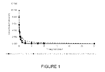
[0039] Figure 1 is a graph showing the pharmacokinetic profile of peptides
according to some
embodiments of the present technology in male Sprague Dawley rats after
intravenous injection of
cyclic HBD1 (3-11), HBD1 (3-11) with C16:0 at N-terminal, HBD1 (3-11) with
C18:0 at N-terminal;
[0040] Figure 2 is a graph showing the pharmacokinetic profile of peptides
according to some
embodiments of the present technology in male Sprague Dawley rats after
subcutaneous injection of
cyclic HBD1 (3 -11), HBD1 (3-11) with C16:0 at N-terminal, HBD1 (3-11) with
C18:0 at N-terminal;
[0041] Figure 3 is a graph showing the pharmacokinetic profile of peptides
according to some
embodiments of the present technology in male Sprague Dawley rats after
subcutaneous injection of
HBD1 (3-11) with C16:0 at N-terminal, HBD1 (3-11) with C14:0 at N-terminal,
HBD1 (3-11) with
C18:0 at N-terminal and HBD1 (3-11) with C20:0 at N-terminal;
[0042] Figure 4 is a graph showing the pharmacokinetic profile of peptides
according to some
embodiments of the present technology after single intravenous (iv) and
subcutaneous (sc) injection in
Gottingen minipigs of HBD1 (3-11) with C18:0 at N-terminal. Individual values
represent the mean of
the values obtained for three different subjects;
[0043] Figures 5A-5C are graphs showing the effect of peptides according to
some embodiments of
the present technology on bone in ovariectomized rat. The graphs show the
percent increase from
OVX vehicle in selected CT parameters from tibial metaphysis after 6-week
HBD1 (3-11) with
C18:0 at N-terminal treatment in OVX Sprague-Dawley rats. Figure 5A shows
BV/TV; Figure 5B
6
CA 03052197 2019-07-30
WO 2018/145006
PCT/US2018/016869
shows Tb.N; Figure 5C shows Conc. D (*: p-value <0.05, **: p-value < 0.01,
***: p-value < 0.001 vs
OVX vehicle); and
[0044] Figures 6A-6B are graphs showing the effect of the peptides according
to some embodiments
of the present technology on the indicated bone biomechanical properties. The
graphs show the
percent increase from OVX vehicle in selected biomechanical parameters from
Lumbar vertebrae
(Figure 6A) and femoral neck (Figure 6B) after a 6-week treatment with HBD1 (3-
11) with C18:0 at
N-terminal in OVX Sprague-Dawley rats (*: p-value <0.05, vs OVX vehicle).
DETAILED DESCRIPTION OF TECHNOLOGY
[0045] This present description of the technology is not intended to be a
detailed catalog of all the
different ways in which the present technology may be implemented, or all the
features that may be
added to the present technology. For example, features illustrated with
respect to one embodiment may
be incorporated into other embodiments, and features illustrated with respect
to a particular
embodiment may be deleted from that embodiment. In addition, numerous
variations and additions to
the various embodiments suggested herein will be apparent to those skilled in
the art in light of the
instant disclosure which does not depart from the present technology. Hence,
the following
specification is intended to illustrate some particular embodiments of the
present technology, and not
to exhaustively specify all permutations, combinations and variations thereof.
Unless defined
otherwise, all technical and scientific terms used herein have the same
meaning as commonly
understood by one of skill in the art to which the present technology belongs.
[0046] The present disclosure stems from the work performed by the present
discoverers on peptide
fragments of IGFBP-2, in particular on peptide fragments of the heparin
binding domain (HBD) of
IGFBP-2, and on their study of how these peptide fragments can be used in
methods of improving
bones, such as in methods of preventing and/or treatment bone disorders.
A. Compounds, peptides, fragments and analogs thereof
[0047] As used herein, the expression and term "heparin binding domain" and
"HBD" refer to the
heparin binding domain of IGFBP-2. The term "HBD1" refers to the heparin
binding domain 1 of
IGFBP-2. HBD1 is intended to refer to a peptide having the amino acid sequence
as set forth in SEQ
ID NO: 1, namely: 1-KHHLGLEEPKKLR-13, wherein "1" refers to amino acid residue
at the 5'-end or
N-Terminal of this HBD1 peptide and "13" refers to amino acid residue at the
3'-end or C-Terminal of
this HBD1 peptide. Accordingly, the amino acids of HBD1 occupy the following
positions:
7
CA 03052197 2019-07-30
WO 2018/145006
PCT/US2018/016869
iK2H3H4L5G6L7E8E9pioKuKi2Li3R
[0048] Well recognized abbreviations in the art will be used to describe amino
acids, including
levorotatory amino acids (L-amino acids or L or L-form) and dextrorotary amino
acids (D-amino acids
or D or D-form), Alanine (Ala or A), Arginine (Arg or R), Asparagine (Asn or
N), Aspartic acid (Asp
or D), Cysteine (Cys or C), Glutamic acid (Glu or E), Glutamine (Gln or Q),
Glycine (Gly or G),
Histidine (His or H), Isoleucine (Ile or I), Leucine (Leu or L), Lysine (Lys
or K), Methionine (Met or
M), Phenylalanine (Phe or F), Proline (Pro or P), Serine (Ser or S), Threonine
(Thr or T), Tryptophan
(Trp or W), Tyrosine (Tyr or Y) and Valine (Val or V). An L-amino acid residue
within the native
peptide sequence may be altered to any one of the 20 L-amino acids commonly
found in proteins or
any one of the corresponding D-amino acids, rare amino acids, such as, but not
limited to, 4-
hydroxyproline or hydroxylysine, or a non-protein amino acid, such as P-
alanine or homoserine.
Unless otherwise indicated, an amino acid named herein refers to the L-form.
[0049] Naturally-occurring variations of the peptides defined herein are those
that may comprise
substitutions, additions or deletions of one or more amino acids which result
due to discrete changes in
the nucleotide sequence of the encoding gene or alleles thereof or which
result due to alternative
splicing of the transcribed RNA. It is understood that these changes do not
substantially affect the
properties, pharmacological and biological characteristics of the peptide
variants.
[0050] The peptides of the present disclosure may be in the form of salts.
Particularly the acidic
functions of the molecule may be replaced by a salt derivative thereof such
as, but not limited to, a
trifluoroacetate salt.
[0051] By "peptide", "polypeptide" or "protein" is meant any chain of amino
acids, regardless of
length or post-translational modification (e.g., glycosylation or
phosphorylation), or chemical
modification, or those containing unnatural or unusual amino acids such as D-
Tyr, ornithine, amino-
adipic acid.
[0052] In some embodiments, the peptide of the present disclosure comprises a
fragment of HBD1. In
some embodiments, the peptide is 10 amino acids in length. In some
embodiments, the peptide is 9
amino acids in length. In some other embodiments, the peptide is 8 amino acids
in length. In some
other embodiments, the peptide is 7 amino acids in length. In some other
embodiments, the peptide is
6 amino acids in length. In some embodiments, the peptide is 5 amino acids in
length.
8
CA 03052197 2019-07-30
WO 2018/145006
PCT/US2018/016869
[0053] As used herein, the term and expression "fragment" or "fragment
thereof' refer to an amino
acid fragment of a peptide such as IGFBP-2 or of the HBD of IGFBP-2 or of the
HBD1 of IGFBP-2.
Fragments of HBD1 are shorter than 13 amino acid residues. Fragments of HBD1
may therefore be
12, 11, 10, 9, 8, 7, 6, 5 or 4 amino acid residues in length. In some
embodiments, the fragment of
HBD1 is 10 amino acids in length. In some embodiments, the fragment of HBD1 is
9 amino acids in
length. In some other embodiments, the fragment of HBD1 is 8 amino acids in
length. In some other
embodiments, the fragment of HBD1 is 7 amino acids in length. In some other
embodiments, the
fragment of HBD1 is 6 amino acids in length. In some other embodiments, the
fragment of HBD1 is 5
amino acids in length. In some other embodiments, the fragment of HBD1 is 4
amino acids in length.
[0054] In one embodiment, the present disclosure provides peptides having the
amino acid sequences
depicted in Table 1. HBD1 (1-13) represents the full-length HBD1. The
remaining peptides presented
in Table 1 are fragments of HBD1 (1-13), wherein amino acid residues at the N-
terminal or at the C-
terminal or at both the N-terminal and the C-terminal are absent.
Table 1: Examples of fragments of HBD1
...............................................................................
...............................................................................
.....
...............................................................................
...............................................................................
............................................................
SEQ ID NO: 1 HBD1 (1-13) 1-KHHLGLEEPKKLR-1/. 13
SEQ ID NO: 2 HBD1 (2-13) 1- HHLGLEEPKKLR-13 12
SEQ ID NO: 3 HBD1 (3-13) 1- HLGLEEPKKLR-13 11
SEQ ID NO: 4 HBD1 (4-13) 1- LGLEEPKKLR-13 10
SEQ ID NO: 5 HBD1 (1-12) 1-KHHLGLEEPKKL -13 12
SEQ ID NO: 6 HBD1 (1-11) 1-KHHLGLEEPKK -13 11
SEQ ID NO: 7 HBD1 (3-10) 1- HLGLEEPK -13 8
SEQ ID NO: 8 HBD1 (3-9) 1- HLGLEEP -13 7
SEQ ID NO: 9 HBD1 (3-12) 1- HLGLEEPKKL -13 10
SEQ ID NO: 10 HBD1 (3-11) 1- HLGLEEPKK -13 9
SEQ ID NO: 11 HBD1 (4-11) 1- LGLEEPKK -13 8
SEQ ID NO: 12 HBD1 (5-11) 1- GLEEPKK -13 7
SEQ ID NO: 13 HBD1 (4-10) 1- LGLEEPK -13 7
SEQ ID NO: 14 HBD1 (5-10) 1- GLEEPK -13 6
SEQ ID NO: 15 HBD1 (4-9) 1- LGLEEP -13 6
SEQ ID NO: 16 HBD1 (2-11) 1- HHLGLEEPKK -13 10
SEQ ID NO: 77 HBD1 (3-11) cyclic 'llel-HLGLEEPKK-13'lle
9
9
CA 03052197 2019-07-30
WO 2018/145006
PCT/US2018/016869
[0055] In some embodiments, the peptides of the present disclosure are
"purified", "isolated" or
"substantially pure". The peptides are "purified", "isolated" or
"substantially pure" when they are
separated from the components that naturally accompany them. Typically, a
compound is substantially
pure when it is at least about 60%, about 65%, about 70%, about 75%, about
80%, about 85%, about
90%, about 91%, about 92%, about 93%, about 94%, about 95%, about 96%, about
97%, about 98%,
or about 99%, by weight, of the total material in a sample. Techniques for
purifying or isolating
peptides are commonly known and used in the art and will be known to persons
skilled in the art.
[0056] In some other embodiments, certain peptides according to the present
disclosure may also be
in cyclized form, such that the N- or C-termini are linked head-to-tail either
directly, or through the
insertion of a linker moiety, such moiety itself generally comprises one or
more amino acid residues as
required to join the backbone in such a manner as to avoid altering the three-
dimensional structure of
the peptide with respect to the non-cyclized form. Such peptide derivatives
may have improved
stability and bioavailability relative to the non-cyclized peptides.
[0057] Methods for cyclizing peptides are well known in the art. Cyclisation
may be accomplished by
disulfide bond formation between two side chain functional groups, amide or
ester bond formation
between one side chain functional group and the backbone a-amino or carboxyl
function, amide or
ester bond formation between two side chain functional groups, or amide bond
formation between the
backbone a-amino and carboxyl functions. These cyclisation reactions have been
traditionally carried
out at high dilution in solution. Cyclisation is commonly accomplished while
the peptide is attached to
the resin. One of the most common ways of synthesizing cyclic peptides on a
solid support is by
attaching the side chain of an amino acid to the resin. Using appropriate
protection strategies, the C-
and N-termini can be selectively deprotected and cyclized on the resin after
chain assembly. This
strategy is widely used, and is compatible with either tert-butyloxycarbonyl
(Boc) or 9-
fluorenylmethoxycarbonyl (Fmoc) protocols. However, it is restricted to
peptides that contain
appropriate side chain functionality to attach to the solid support. A number
of approaches may be
used to achieve efficient synthesis of cyclic peptides. One procedure for
synthesizing cyclic peptides is
based on cyclisation with simultaneous cleavage from the resin. After an
appropriate peptide sequence
is assembled by solid phase synthesis on the resin or a linear sequence is
appended to resin, the
deprotected amino group can react with its anchoring active linkage to produce
protected cyclic
peptides. In general, a final deprotection step is required to yield the
target cyclic peptide. The
procedures for synthesizing cyclic peptides are well known in the art.
CA 03052197 2019-07-30
WO 2018/145006
PCT/US2018/016869
[0058] In other embodiments, the present disclosure provides analogs of the
peptides defined herein.
As used herein, the term "analog" refers to a peptide that has the
physiological activity of the parent
compound thereof, and that includes one or more (e.g., two, three, four, five
or six or more) amino
acids different from the amino acid sequence of a naturally occurring parent
peptide. Such an analog
preferably has at least about 40%, at least about 45%, at least about 50%, at
least about 55%, at least
about 60%, at least about 65%, at least about 70%, at least about 75%, at
least about 80%, at least
about 85%, at least about 90%, or at least about 95% of the physiological
activity of the parent
peptide.
[0059] In some other embodiments, the analogs may be as physiologically active
as the parent (i.e.,
has 100% of physiological activity of the parent peptide) or may be more than
about 100%, more than
about 110%, more than about 120%, more than about 130%, more than about 140%,
more than about
150%, more than about 160%, more than about 170%, more than about 180%, more
than about 190%
or more than about 200% physiologically active than the parent peptide.
[0060] Such different amino acids may be additions, substitutions, deletions,
or combinations thereof,
including addition of non-natural side-chain groups and backbone links.
Modifications of peptides to
produce analogs thereof are known. See, e.g., U.S. Pat. No. 7,323,543; see
also U.S. Pat. Nos.
7,482,171; 7,459,152; and 7,393,919, which are all incorporated herein by
reference. For examples,
analogs of peptides comprising HBD1 or analogs of fragments of HBD1 refer to
either: i) structural
analogs; ii) functional analogs; or iii) structural and functional analogs of
HBD1 which are, inter al/a,
capable of replacing HBD1 in improving bone disorders, such as for example in
preventing and/or
treating bone disorders.
[0061] Analogs of the peptides of the present disclosure that have at least
about 50%, at least about
55%, at least about 60%, at least about 65%, at least about 70%, at least
about 75%, at least about
80%, at least about 85%, at least about 90%, at least about 95%, at least
about 96%, at least about
97%, at least about 98% or at least 99% sequence homology with the amino acid
sequences described
herein over its full length, and sharing at least one of the metabolic effects
or biological activity of
HBD1. A person skilled in the art would readily identify an analog sequence of
HBD1 or an analog
sequence of a fragment of HBD1.
[0062] Analogs of HBD1 or analogs of fragment of HBD1 are, for example,
analogs obtained by
alanine scans or by amino acid substitutions. In some instances, analogs of
HBD1 or analogs of
fragments thereof may comprise a non-naturally encoded amino acid, wherein the
non-naturally
encoding amino acid refers to an amino acid that is not one of the common
amino acids or pyrrolysine
11
CA 03052197 2019-07-30
WO 2018/145006
PCT/US2018/016869
or selenocysteine, or an amino acid that occur by modification (e.g. post-
translational modification) of
naturally encoded amino acid (including, but not limited to, the 20 common
amino acids or
pyrrolysine and selenocysteine) but are not themselves incorporated into a
growing polypeptide chain
by the translation complex. Examples of such non-naturally-occurring amino
acids include, but are not
limited to, N-acetylglucosaminyl-L-serine, N-acetylglucosaminyl-L-threonine
and 0-phosphotyrosine.
[0063] Table 2 presents examples of analogs of HBD1 (3-11) with alanine
substitutions at different
amino acid positions.
Table 2: HBD1 (3-11) fragment with Alanine substitutions at various positions
SJQ1D NO Ammo Ad Seuenee
...............................................................................
...............................................................................
.................
SEQ ID NO: 17 ALGLEEPKK
SEQ ID NO: 18 HAGLEEPKK
SEQ ID NO: 19 HLALEEPKK
SEQ ID NO: 20 HLGAEEPKK
SEQ ID NO: 21 HLGLAEPKK
SEQ ID NO: 22 HLGLEAPKK
SEQ ID NO: 23 HLGLEEAKK
SEQ ID NO: 24 HLGLEEPAK
SEQ ID NO: 25 HLGLEEPKA
[0064] Table 3 presents other examples of analogs of HBD1 fragments comprising
amino acid
substitutions at different amino acid positions of HBD1 (3-11).
Table 3: Analogs of HBD1 (3-11) fragment with amino acid substitutions at
various positions
StQJD 1O Ammo Ad Snc
SEQ ID NO: 26 HLGLERPKK
SEQ ID NO: 27 HLGLEFPKK
SEQ ID NO: 28 HLGLEIPKK
SEQ ID NO: 29 HLGLEPPKK
SEQ ID NO: 30 HLGLESPKK
SEQ ID NO: 31 HLGLEERKK
SEQ ID NO: 32 HLGLEEFKK
SEQ ID NO: 33 HLGLEELKK
12
CA 03052197 2019-07-30
WO 2018/145006
PCT/US2018/016869
SEQ ID NO: 34 HL GLEESKK
SEQ ID NO: 35 HL GLEEDKK
SEQ ID NO: 36 HL GLEEPFK
SEQ ID NO: 37 HL GLEEPPK
SEQ ID NO: 38 HL GLEEPSK
SEQ ID NO: 39 HL GLEEPDK
SEQ ID NO: 40 HL GLEEPKF
SEQ ID NO: 41 HL GLEEPKI
SEQ ID NO: 42 HL GLEEPKP
SEQ ID NO: 43 HL GLEEPKS
SEQ ID NO: 44 HL GLEEPKD
SEQ ID NO: 45 HL GLEEPIK
SEQ ID NO: 46 HL GLEEPVK
SEQ ID NO: 47 HL GLEEPQK
SEQ ID NO: 48 HL GLEEPTK
SEQ ID NO: 49 HL GLEEPEK
SEQ ID NO: 50 HL GLEEPKH
SEQ ID NO: 51 HL GLEEPKR
SEQ ID NO: 52 HL GLEEPKL
SEQ ID NO: 53 HL GLEEPKM
SEQ ID NO: 54 HL GLEEPKW
SEQ ID NO: 55 HL GLEEPKV
SEQ ID NO: 56 HL GLEEPK2
SEQ ID NO: 57 HL GLEEPKN
SEQ ID NO: 58 HL GLEEPKY
SEQ ID NO: 59 HL GLEEPKT
SEQ ID NO: 60 HL GLEEPKE
SEQ ID NO: 61 HL GLEEP SP
SEQ ID NO: 62 HL GLEEPS S
SEQ ID NO: 89 KLGLEEPKK
SEQ ID NO: 90 HVGLEEPKK
SEQ ID NO: 91 HLPLEEPKK
SEQ ID NO: 92 HL GIEEPKK
SEQ ID NO: 93 NL GLEEPKK
SEQ ID NO: 94 HTGLEEPKK
SEQ ID NO: 95 HLKLEEPKK
SEQ ID NO: 96 HL GSEEPKK
13
CA 03052197 2019-07-30
WO 2018/145006
PCT/US2018/016869
SEQ ID NO: 97 HL GLEEPYK
SEQ ID NO: 98 HL GLEEPQK
SEQ ID NO: 99 HL GLEEPNK
SEQ ID NO: 100 HL GLEEP SF
SEQ ID NO: 101 HL GLEEPSV
SEQ ID NO: 102 HL GLEEPLM
SEQ ID NO: 103 HL GLEEPLY
SEQ ID NO: 104 HL GLEEPLN
SEQ ID NO: 105 HL GLEEPI
SEQ ID NO: 106 HL GLEEPFV
SEQ ID NO: 107 HL GLEEPFLQ
SEQ ID NO: 108 HL GLEEPFN
SEQ ID NO: 109 HL GLEEPVM
SEQ ID NO: 110 HL GLEEPVN
SEQ ID NO: 111 HL GLEEPMK
[0065] In some instances, the analogs of HBD1 or fragments thereof may differ
in sequence from
HBD1 by 1, 2, 3, 4, 5, 6, 7, 8 or 9 amino acid substitutions, deletions, or
additions, or combinations
thereof
[0066] In some instances, the amino acid substitution is a conservative amino
acid substitution. As
used herein the expression "conservative amino acid substitution" refers to
substitutions that substitute
a residue with another of like characteristics. Typical conservative amino
acid substitutions include
those among Gly (G), Ala (A), Val (V), Leu (L) and Ile (I); those among Ser
(S), Cys (C), Met (M)
and Thr (T); those among the acidic residues Asp (D) and Glu (E); those among
Asn (N) and Gln (Q);
those among the basic residues His (H), Lys (K) and Arg (R); and those among
the aromatic residues
Phe (F), Try (W) and Tyr (Y).
[0067] In some embodiments, the present technology provides an isolated
peptide having a fragment
of HBD1 as set forth in SEQ ID NO: 1. In some instances, the fragment is
between 6 to 10 amino
acids in length and comprises residues 3 to 10 of HBD1, namely: HLGLEEPK as
set forth in SEQ ID
NO: 7 or an analog thereof. Examples of analogs of a peptide having the amino
acid sequence
HLGLEEPK include, but are not limited to the peptides presented in Table 4.
Table 4: Analogs of HBD1 (3-10) fragment with amino acid substitutions at
various positions
SEQ ID No Ammo Acid Stqutnt
14
CA 03052197 2019-07-30
WO 2018/145006
PCT/US2018/016869
SEQ ID NO: 112 HL GLEEPR
SEQ ID NO: 113 HL GLEEPH
[0068] In some embodiments, the present technology provides an isolated
peptide having a fragment
of HBD1 as set forth in SEQ ID NO: 1. In some instances, the fragment is
between 6 to 10 amino
acids in length and comprises residues 5 to 10 of HBD1, namely: GLEEPK as set
forth in SEQ ID NO:
14 or an analog thereof. In some other embodiments, the fragment is between 6
to 9 amino acids in
length and comprises residues 5 to 10 of HBD1, namely: GLEEPK as set forth in
SEQ ID NO: 14 or
an analog thereof. Examples of analogs of a peptide having the amino acid
sequence GLEEPK
include, but are not limited to the peptides presented in Table 5.
Table 5: Analogs of HBD1 (5-10) fragment with amino acid substitutions at
various positions
SEQ ID NO: 79 GLEEPL
SEQ ID NO: 80 GLEEPR
SEQ ID NO: 81 GLDEPK
SEQ ID NO: 82 GLEDPK
SEQ ID NO: 83 GGEEPK
SEQ ID NO: 84 GVEEPK
SEQ ID NO: 85 GIEEPK
SEQ ID NO: 86 VLEEPK
SEQ ID NO: 87 LLEEPK
SEQ ID NO: 88 ILEEPK
[0069] In some other embodiments, the peptides of the present disclosure may
be modified. As used
herein the term "modified" when used to qualify a peptide, refers to any
changes made to a peptide,
such as changes to the length of the peptide, the amino acid sequence,
chemical structure, co-
translational modification, or post-translational modification of a peptide.
In some instances, the
peptides of the present disclosure comprise one or more amino acid residues
that are modified.
[0070] As used herein, the expression "post-translational modification" refers
to any modification of
a natural or non-natural amino acid that occurs to such an amino acid after it
has been incorporated
into a peptide chain. The term encompasses, by way of example only, co-
translational in vivo
modifications, co-translational in vitro modifications (such as in cell-free
translation system), post-
translational in vivo modifications, and post-translational in vitro
modifications. Examples of post-
translational modifications are, but are not limited to, glycosylation,
pegylation, acetylation, acylation,
CA 03052197 2019-07-30
WO 2018/145006
PCT/US2018/016869
amidation, methylation, carboxylation, phosphorylation, addition of salts,
amides or esters, in
particular C-terminal esters, and N-acyl derivatives of the peptides of the
present disclosure. The types
of post-translational modifications are well known in the art.
[0071] In some embodiments, the peptides of the present disclosure include one
or more
poly(ethylene glycol) (or "PEG") moiety of between about 10,000 and about
40,000 molecular weight
coupled to either the N- or C-terminus of the peptide. "Polyalkylene glycol"
means straight or
branched polyalkylene glycol polymers including, but not limited to,
polyethylene glycol (PEG),
polypropylene glycol (PPG), and polybutylene glycol (PBG), as well as co-
polymers of PEG, PPG and
PBG in any combination, and includes the monoalkylether of the polyalkylene
glycol. Thus, in various
embodiments of the present technology, the polyalkylene glycol in the peptides
of the present
disclosure can be, but is not limited to, polyethylene glycol, polypropylene
glycol, polybutylene
glycol, and any combination thereof. In certain embodiments, the polyalkylene
glycol is polyethylene
glycol or "PEG." The term "PEG subunit" refers to a single polyethylene glycol
unit, i.e.,-
(CH2CH20)-.
[0072] In some embodiments, the polyalkylene glycol (e.g., PEG) can be non-
polydispersed,
monodispersed, substantially monodispersed, purely monodispersed, or
substantially purely
monodispersed. "Monodispersed" is used to describe a mixture of compounds
wherein about 100
percent of the compounds in the mixture have the same molecular weight.
"Substantially
monodispersed" is used to describe a mixture of compounds wherein at least
about 95 percent of the
compounds in the mixture have the same molecular weight. "Purely
monodispersed" is used to
describe a mixture of compounds wherein about 100 percent of the compounds in
the mixture have the
same molecular weight and have the same molecular structure. Thus, a purely
monodispersed mixture
is a monodispersed mixture, but a monodispersed mixture is not necessarily a
purely monodispersed
mixture. "Substantially purely monodispersed" is used to describe a mixture of
compounds wherein at
least about 95 percent of the compounds in the mixture have the same molecular
weight and have the
same molecular structure. Thus, a substantially purely monodispersed mixture
is a substantially
monodispersed mixture, but a substantially monodispersed mixture is not
necessarily a substantially
purely monodispersed mixture. Table 6 presents examples of peptides of the
present disclosure that are
modified by pegylation.
Table 6: PEGylated HBD1 fragments
$gq.W1M;iiii..W#gOkiO4..C.$.0:*001iiiMR!RRRRR!R!RRRM9M
......................................................
...............................................................................
.................................
...............................................................................
...............................................................................
.........
SEQ ID NO: 63 PEG20-C-KHHLGLEEPKKLR
SEQ ID NO: 64 KHHLGLEEPKKLR-C-PEG20
16
CA 03052197 2019-07-30
WO 2018/145006
PCT/US2018/016869
SEQ ID NO: 65 PE G20-C -HHL GLEEPKK
SEQ ID NO: 66 HHLGLEEPKK-C-PE G20
SEQ ID NO: 67 PE G20-C -HL GLEEPKK
[0073] In some other instances, the peptides of the present disclosure include
one or more acyl
group(s) coupled to any amino acid of the peptide. In some instances, the one
or more acyl group(s) is
coupled to the N-terminal or the C-terminal amino acid or to both. In some
instances, acylation of the
peptides of the present disclosure is a fatty acylation by which a fatty acid
is added to one or more
particular amino acid(s) of the peptide. Examples of fatty acylation include
addition of: lauric acid
(C12:0), tridecyclic acid (C13:0), myristic acid (C14:0), pentadecyclic acid
(C15:0), palmitic acid
(C16:0), margaric acid (C17:0), stearic acid (C18:0), nonade cyclic acid
(C19:0), arachidinic acid
(C20:0), heneicosylic acid (C21:0), behenic acid (C22:0), tricosylic acid
(C23:0), or lignoceric acid
(C24:0), or a mixture thereof to one or more amino acid of the peptides of the
present disclosure.
[0074] In some variants, the fatty acid to be added may be unsaturated (e.g.,
monounsaturated or
polyunsaturated). Examples of unsaturated fatty acids include but are not
limited to: i) mono-
unsaturated fatty acid: crotonic acid, myristoleic, palmitoleic acid, sapienic
acid, oleic acid, elaidic
acid, vaccenic acid, gadoleic, eicosenoic acid, erucic acid, nervonic acid;
ii) di-unsaturated fatty acid:
linoleic acid, eicosadienoic acid, docosadienoic acid; iii) tri-unsaturated
fatty acids: linolenic acid,
pinolenic acid, eleostearic acid, mead acid, dihomo-y-linolenic acid,
eicosatrienoic acid; iv) tetra-
unsaturated fatty acid: stearidonic acid, arachidonic acid, eicosatetraenoic
acid, adrenic acid; v)
pentaunsaturated fatty acids: bosseopentaenoic acid, eicosapentaenoic acid,
ozubondo acid, sardine
acid, tetracosanolpentaenoic acid; and vi) hexa-unsaturated fatty acids:
docosahexaenoic acid, and
herring acid.
[0075] In some embodiments, the peptides of the present disclosure may be
coupled to fatty acids that
comprise one or more carboxylic functional groups (-COOH).
[0076] The methods for carrying acylation of peptides are well known in the
art. Table 7 presents
examples of peptides of the present disclosure that are modified by acylation.
Table 7: Acylated HBD1 (2-11) fragments
SEQ ID Os Amine Atd Seuence
SEQ ID NO: 68 C16:0-HHLGLEEPKK
SEQ ID NO: 69 C18:0-HHLGLEEPKK
SEQ ID NO: 70 C20:0-HHLGLEEPKK
17
CA 03052197 2019-07-30
WO 2018/145006
PCT/US2018/016869
SEQ ID NO: 71 C 14: 0 -HLGLEEPKK
SEQ ID NO: 72 C 16: 0 -HLGLEEPKK
SEQ ID NO: 73 C 18: 0 -HLGLEEPKK
SEQ ID NO: 74 C20:0-HLGLEEPKK
SEQ ID NO: 75 C16:0-diacid-HLGLEEPKK
SEQ ID NO: 76 HL GLEEPKK-C 16: 0
SEQ ID NO: 78 C 16: 0 -KHHL GLEEPKKLR
[0077] In some additional embodiments, the peptides of the present disclosure
may be coupled to a
linker or a linker group (e.g., linker moiety). As used herein, the expression
"linker" or "linking
group" includes non-amino acid linking groups such as are known in the art
(see, e.g., U.S. Pat. Nos.
7,468,418; 7,402,652; and 7,351,797, which are all incorporated herein by
reference) or variations
thereof that will be apparent to those skilled in the art.
[0078] In some embodiments, the peptides of the present disclosure may include
more than one
modification (e.g., may include a PEG group and an acyl group).
[0079] In some other embodiments, the peptides of the present disclosure may
be coupled to a
modifying group which is itself modified. For example, the peptides of the
present disclosure may be
coupled to a fatty acid which is itself modified. The modified fatty acid may,
for example, be coupled
to a linker or a linker group and the linker or the linker group may itself be
coupled to another
modifying group such as a PEG group or one or more carboxylic functional
groups (-COOH). Various
combinations of modifications and the methods for achieving them will be
recognized and appreciated
by those skilled in the art.
[0080] Certain aspects of the present technology use polynucleotides. These
polynucleotides include
isolated polynucleotides which encode the HBD1 peptides, fragments and analogs
defined herein.
[0081] As used herein, the term "polynucleotide" refers to a molecule
comprised of a plurality of
deoxyribonucleotides or nucleoside subunits. The linkage between the
nucleoside subunits can be
provided by phosphates, phosphonates, phosphoramidates, phosphorothioates, or
the like, or by
nonphosphate groups as are known in the art, such as peptoid-type linkages
utilized in peptide nucleic
acids (PNAs). The linking groups can be chiral or achiral. The
oligonucleotides or polynucleotides can
range in length from 2 nucleoside subunits to hundreds or thousands of
nucleoside subunits. While
oligonucleotides are preferably 5 to 100 subunits in length, and more
preferably, 5 to 60 subunits in
length, the length of polynucleotides can be much greater (e.g., up to 100).
The polynucleotide may be
18
CA 03052197 2019-07-30
WO 2018/145006
PCT/US2018/016869
any of DNA and RNA. The DNA may be in any form of genomic DNA, a genomic DNA
library,
cDNA derived from a cell or tissue, and synthetic DNA. Moreover, the present
disclosure may, in
certain aspects, use vectors which include bacteriophage, plasmid, cosmid, or
phagemid.
[0082] The polypeptides useful in the present technology may be prepared in
any suitable manner as
known in the art. Such polypeptides include isolated naturally occurring
polypeptides, recombinantly
produced polypeptides, synthetically produced polypeptides, or polypeptides
produced by a
combination of these methods. Means and methods for preparing such
polypeptides are well known in
the art.
B. Therapeutic Actions
[0083] As used herein, the terms "treat," "treating" and "treatment" as used
herein all refer to any
type of treatment that imparts a benefit to a subject afflicted with a
disease, including improvement in
the condition of the patient (e.g., in one or more symptoms), delay in the
progression of the disease, or
the like.
[0084] As used herein, the terms "subjects" and "patient" generally relate to
human subjects and are
used interchangeably. The subjects may be male or female and may be of any
race or ethnicity,
including, but not limited to, Caucasian, African-American, African, Asian,
Hispanic, Indian, etc. The
subjects may be of any age, including newborn, neonate, infant, child,
adolescent, adult, and geriatric.
In some embodiments the subjects are afflicted with a bone disorder. Subjects
may also include animal
subjects, particularly mammalian subjects such as canines, felines, bovines,
caprines, equines, ovines,
porcines, rodents (e.g. rats and mice), lagomorphs, primates (including non-
human primates), etc., for
veterinary medicine or pharmaceutical drug development purposes.
[0085] In some embodiments, the peptides of the present disclosure may be used
for improving
and/or ameliorating a bone disorder (or used in a method for improving and/or
ameliorating a bone
disorder). In some instances, the peptides of the present disclosure may be
used to prevent a bone
disorder, in some other instance the peptides of the present disclosure may be
used to treat a bone
disorder, in some other instances, the peptides of the present disclosure may
be used to both prevent
and treat a bone disorder.
[0086] As used herein, the expression "bone disorder" refers to any of several
diseases that cause
various abnormalities or deformities of one or more bones and/or to bone
cells. Examples of bone
disorders include: osteoporosis, rickets, osteomalacia, osteogenesis
imperfecta, marble bone disease
(osteopetrosis), fibrous dysplasia, postmenopausal osteoporosis, senile
osteoporosis in males and
19
CA 03052197 2019-07-30
WO 2018/145006
PCT/US2018/016869
females, glucocorticoid-induced osteoporosis, immobilization-induced
osteoporosis, weightlessness-
induced osteoporosis, post-transplantation osteoporosis, migratory
osteoporosis, idiopathic
osteoporosis, juvenile osteoporosis, Paget's Disease, chronic
hyperparathyroidism, hyperthyroidism,
rheumatoid arthritis, Gorham-Stout disease, McCune-Albright syndrome,
osteolytic metastases of
various cancers or multiple myeloma.
[0087] As used herein, the expression "bone disorders" also include loss of
bone mass, general bone
fragility, joint degeneration, non-union fractures, orthopedic and dental
problems caused by diabetes,
periimplantitis, poor responses to bone grafts/implants/bone substitute
materials, periodontal diseases,
skeletal aging, broken bones, bone defects, bone transplant, bone grafts, bone
cancer, joint
replacements, joint repair, fusion, facet repair, bone degeneration, dental
implants and repair, bone
marrow deficits and other conditions associated with bone and boney tissue.
Bone defects may be a
gap, deformation and/or a nonunion fracture in a bone. Bone disorders also
include osteopathy in
acromegalic patients, cystic fibrosis-related bone disease, adynamic bone
disease, renal
osteodystrophy associated with chronic kidney disease, bone disease associated
with cystinosis and
bone disease associated with hyperoxaluria.
[0088] Bone degeneration may be due to osteopenia or osteoporosis (e.g. the
patient is afflicted with
geriatric or senile osteoporosis, with post-menopausal osteoporosis, etc.), or
due to dwarfism.
[0089] Joint replacements that may be treated include vertebral, knee, hip,
tarsal, phalangeal, elbow,
ankle and/or other articulating joints or replacements thereof Joint repairs
include, but are not limited
to, vertebral, knee, hip, tarsal, phalangeal, elbow, ankle, and sacroiliac
joint repairs.
[0090] In some embodiments, the peptides of the present disclosure may be used
to enhance bone
formation (i.e., increasing the amount of new bone that is laid down, or used
in a method to enhance
bone formation).
[0091] In some other embodiments, the peptides of the present disclosure may
be used to inhibit bone
resorption (i.e., to reduce the amount of bone that is dissolved) (or used in
a method to inhibit bone
resorption) simultaneously in a subject in need thereof. Non-limiting examples
of subjects for whom
such treatment would be indicated and/or beneficial include women (e.g.,
postmenopausal;
premenopausal) with osteoporosis or low bone mass, men with osteoporosis or
low bone mass,
subjects with a healing fracture, subjects undergoing prolonged
immobilization, subjects who have
been or are immobilized for a prolonged period, subjects likely to undergo or
experience prolonged
immobilization, subjects with estrogen deficiency, etc., as would be known in
the art.
CA 03052197 2019-07-30
WO 2018/145006
PCT/US2018/016869
[0092] In some further embodiments, the peptides of the present disclosure may
be used for inducing
deposition and maturation of bone in a subject in need thereof (e.g., a
subject having a bone disorder)
(or used in a method for inducing deposition and maturation of bone). In some
instances, the peptide
of the present disclosure may be used in combination with a bone resorption
inhibitor.
[0093] In some aspects of these embodiments, the bone disorder is at a
targeted site of the subject.
The targeted site may be an intervertebral space, a facet joint, a site of a
bone fracture, bones of the
mouth, chin and jaw, or an implant site.
[0094] HBD1 was shown to modulate bone mass acquisition and osteoblast
differentiation (Kawai,
JBC, 2011; Xi, JBMR, 2014). In view of this, it is reasonable to infer that
fragments of HBD1 that
retain the physiological activities of HBD1 could also modulate bone mass
acquisition and osteoblast
differentiation. As such, in some embodiments, the peptides of the present
disclosure may be used to
inhibit fat cell differentiation (e.g., inhibiting fat cell precursor
differentiation into mature adipocytes)
in the subject. In some other embodiments, the peptides of the present
disclosure may be used to
modulate fat mass in a subject.
[0095] In some embodiments, the peptides of the present disclosure may be
employed in methods of
in vitro or ex vivo expansion of stem cells, carried out according to
protocols known in the art. Thus,
the present disclosure provides a method of expanding stem cells in vitro or
ex vivo, comprising
contacting the peptides of the present disclosure with stem cells from a
subject, wherein said stem
cells are maintained under conditions whereby they are reintroduced into the
subject. For example in
some ex vivo embodiments, the stem cells are obtained from a subject, e.g., a
human, e.g., from
peripheral blood, umbilical cord blood, or bone marrow, and the stem cells are
contacted with the
compound of this present disclosure outside the body of the subject. Ex vivo
embodiments include
obtaining stem cells from a subject and culturing the cells for a period of
time prior to use (e.g., for
transplantation). In some embodiments, after contact with the peptides of the
present disclosure, the
cells are delivered to a subject, e.g., the same subject from which the cells
were isolated (autologous
donation) or a different subject (non-autologous (e.g., syngeneic or
allogeneic) donation). Non-
limiting examples of a subject for whom these methods would be indicated or
beneficial include a
subject having or who has had chemotherapy, a subject having or who has had
radiation, a subject
having aplastic anemia, a subject having myelodysplasia, and any combination
thereof
[0096] In some embodiments, the uses and methods defined herein comprise
administering to a
subject a therapeutically effective amount of a peptide as defined herein to
achieve the effects
discussed here (e.g., to prevent and/or treat a bone disorder).
21
CA 03052197 2019-07-30
WO 2018/145006
PCT/US2018/016869
[0097] Therapeutically effective dosage of any specific peptide of the present
disclosure will vary
from peptide to peptide, patient to patient, and subject to subject, and will
depend, among other things,
upon the effect or result to be achieved, the condition of the patient and the
route of delivery. In some
embodiments, a dosage is from about 1 lag/kg to about 1 mg/kg. In some other
embodiments, a dosage
is from about 1 mg/kg to about 50 mg/kg.
[0098] In some instances, the dosage is from about 0.001 mg/kg, about 0.05
mg/kg, about 0.1 mg/kg,
about 0.2 mg/kg, about 0.3 mg/kg, about 0.4 mg/kg, about 0.5 mg/kg, about 0.6
mg/kg, about 0.7
mg/kg, about 0.8 mg/kg, about 0.9 mg/kg or about 1.0 mg/kg, up to about 30
mg/kg, or about 40
mg/kg.
[0099] In some other instances, the dosage is about 1 mg/kg, about 2 mg/kg,
about 3 mg/kg, about 4
mg/kg, about 5 mg/kg, about 6 mg/kg, about 7 mg/kg, about 8 mg/kg, about 9
mg/kg, about 10 mg/kg,
about 11 mg/kg, about 12 mg/kg, about 13 mg/kg, about 14 mg/kg, about 15
mg/kg, about 16 mg/kg,
about 17 mg/kg, about 18 mg/kg, about 19 mg/kg, about 20 mg/kg, about 21
mg/kg, about 22 mg/kg,
about 23 mg/kg, about 24 mg/kg, about 25 mg/kg, about 26 mg/kg, about 27
mg/kg, about 28 mg/kg,
about 29 mg/kg, about 30 mg/kg, about 31 mg/kg, about 32 mg/kg, about 33
mg/kg, about 34 mg/kg,
about 35 mg/kg, about 36 mg/kg, about 37 mg/kg, about 38 mg/kg, about 39
mg/kg, about 40 mg/kg,
about 41 mg/kg, about 42 mg/kg, about 43 mg/kg, about 44 mg/kg, about 45
mg/kg, about 46 mg/kg,
about 47 mg/kg, about 48 mg/kg, about 49 mg/kg, or about 50 mg/kg or more may
be used.
[0100] Additional examples of therapeutically effective dosages include:
between about 1 and about
50 mg/kg/96hr; between about 1 and about 50 mg/kg/48hr; between about 1 and
about 50 mg/kg/36hr;
between about 1 and about 50 mg/kg/24hr; between about 1 and about 50
mg/kg/12hr; between about
1 and about 25 mg/kg/96hr; between about 1 and about 25 mg/kg/48hr; between
about 1 and about 25
mg/kg/36hr; between about 1 and about 25 mg/kg/24hr; between about 1 and about
25 mg/kg/12hr;
between about 1 and about 10 mg/kg/96hr; between about 1 and about 10
mg/kg/48hr; between about
1 and about 10 mg/kg/36hr; between about 1 and about 10 mg/kg/24hr; between
about 1 and about
mg/kg/12hr; between about 1 and about 5 mg/kg/96hr; between about 1 and about
5 mg/kg/48hr;
between about 1 and about 5 mg/kg/36hr; between about 1 and about 5
mg/kg/24hr; between about 1
and about 5 mg/kg/12hr; between about 0.001 and about 1 mg/kg/96hr; between
about 0.001 and
about 1 mg/kg/48hr; between about 0.001 and about 1 mg/kg/36hr; between about
0.001 and about 1
mg/kg/24hr; and between about 0.001 and about 1 mg/kg/12hr.
[0101] "Concurrently administering" or "concurrently administer" as used
herein means that the two
or more peptides, compounds or compositions are administered closely enough in
time to produce a
combined effect (that is, concurrently may be simultaneously, or it may be two
or more events
22
CA 03052197 2019-07-30
WO 2018/145006
PCT/US2018/016869
occurring within a short time period before or after each other, e.g.,
sequentially). Simultaneous
concurrent administration may be carried out by, for example, mixing the
compounds prior to
administration, or by administering the compounds at the same point in time
but at different anatomic
sites and/or by using different routes of administration.
C. Pharmaceutical Compositions
[0102] As used herein, the expression "active agent" refers to a peptide as
defined herein.
[0103] The expressions "therapeutically acceptable", "therapeutically
suitable", "pharmaceutically
acceptable" and "pharmaceutically suitable" are used interchangeably herein
and refer to a peptide, a
compound, or a composition that is suitable for administration to a subject to
achieve the effects
described herein, such as the treatment defined herein, without unduly
deleterious side effects in light
of the severity of the disease and necessity of the treatment.
[0104] The peptides described above may be formulated for administration in a
pharmaceutical
carrier in accordance with known techniques. See, e.g., Remington, The Science
And Practice of
Pharmacy (9th Ed. 1995). In the manufacture of a pharmaceutical composition
according to the
present disclosure, the peptide (including the physiologically acceptable
salts thereof) is typically
admixed with, inter alia, an acceptable carrier. The carrier must, of course,
be acceptable in the sense
of being compatible with any other ingredients in the composition and must not
be deleterious to the
patient. The carrier may be a solid or a liquid, or both, and is preferably
formulated with the peptide as
a unit-dose formulation, for example, a tablet, which may contain from about
0.01 or about 0.5% to
about 95% or about 99% by weight of the peptide. One or more active compounds
may be
incorporated in the compositions of the present disclosure, which may be
prepared by any of the well-
known techniques of pharmacy comprising admixing the components, optionally
including one or
more accessory ingredients.
[0105] The composition of the present disclosure include those suitable for
oral, rectal, topical, buccal
(e.g., sub-lingual), vaginal, parenteral (e.g., subcutaneous, intramuscular,
intradermal, or intravenous),
topical (i.e., both skin and mucosal surfaces, including airway surfaces) and
transdermal
administration, although the most suitable route in any given case will depend
on the nature and
severity of the condition being treated and on the nature of the particular
peptide which is being used.
[0106] Compositions suitable for oral administration may be presented in
discrete units, such as
capsules, cachets, lozenges, or tablets, each containing a predetermined
amount of the peptide; as a
powder or granules; as a solution or a suspension in an aqueous or non-aqueous
liquid; or as an oil-in-
water or water-in-oil emulsion. Such compositions may be prepared by any
suitable method of
pharmacy which includes the step of bringing into association the peptide and
a suitable carrier (which
23
CA 03052197 2019-07-30
WO 2018/145006
PCT/US2018/016869
may contain one or more accessory ingredients as noted above). In general, the
compositions of the
present disclosure are prepared by uniformly and intimately admixing the
peptide with a liquid or
finely divided solid carrier, or both, and then, if necessary, shaping the
resulting mixture. For example,
a tablet may be prepared by compressing or molding a powder or granules
containing the peptide,
optionally with one or more accessory ingredients. Compressed tablets may be
prepared by
compressing, in a suitable machine, the compound in a free-flowing form, such
as a powder or
granules optionally mixed with a binder, lubricant, inert diluent, and/or
surface active/dispersing
agent(s). Molded tablets may be made by molding, in a suitable machine, the
powdered compound
moistened with an inert liquid binder.
[0107] Compositions suitable for buccal (sub-lingual) administration include
lozenges comprising the
peptide in a flavoured base, usually sucrose and acacia or tragacanth; and
pastilles comprising the
peptide in an inert base such as gelatin and glycerin or sucrose and acacia.
[0108] Compositions of the present disclosure suitable for parenteral
administration comprise sterile
aqueous and non-aqueous injection solutions of the peptide, which preparations
are preferably isotonic
with the blood of the intended recipient. These preparations may contain anti-
oxidants, buffers,
bacteriostats and solutes which render the composition isotonic with the blood
of the intended
recipient. Aqueous and non-aqueous sterile suspensions may include suspending
agents and thickening
agents. The composition may be presented in unit\ dose or multi-dose
containers, for example sealed
ampoules and vials, and may be stored in a freeze-dried (lyophilized)
condition requiring only the
addition of the sterile liquid carrier, for example, saline or water-for-
injection immediately prior to
use. Extemporaneous injection solutions and suspensions may be prepared from
sterile powders,
granules and tablets of the kind previously described. For example, in one
aspect of the present
disclosure, there is provided an injectable, stable, sterile composition
comprising a peptide as defined
herein, or a salt thereof, in a unit dosage form in a sealed container. The
peptide or salt is provided in
the form of a lyophilizate which is capable of being reconstituted with a
suitable pharmaceutically
acceptable carrier to form a liquid composition suitable for injection thereof
into a subject. The unit
dosage form typically comprises from about 10 mg to about 10 grams of the
peptide or salt. When the
peptide or salt is substantially water-insoluble, a sufficient amount of
emulsifying agent which is
physiologically acceptable may be employed in sufficient quantity to emulsify
the compound or salt in
an aqueous carrier. One such useful emulsifying agent is phosphatidyl choline.
24
CA 03052197 2019-07-30
WO 2018/145006
PCT/US2018/016869
[0109] Compositions suitable for rectal administration are preferably
presented as unit dose
suppositories. These may be prepared by admixing the peptide as defined herein
with one or more
conventional solid carriers, for example, cocoa butter, and then shaping the
resulting mixture.
[0110] Compositions suitable for topical application to the skin preferably
take the form of an
ointment, cream, lotion, paste, gel, spray, aerosol, or oil. Carriers which
may be used include
petroleum jelly, lanoline, polyethylene glycols, alcohols, transdermal
enhancers, and combinations of
two or more thereof
[0111] Compositions suitable for transdermal administration may be presented
as discrete patches
adapted to remain in intimate contact with the epidermis of the recipient for
a prolonged period of
time. Compositions suitable for transdermal administration may also be
delivered by iontophoresis
(see, for example, Pharmaceutical Research 3 (6):318 (1986)) and typically
take the form of an
optionally buffered aqueous solution of the peptide as defined herein.
Suitable compositions comprise
citrate or bis\tris buffer (pH 6) or ethanol/water and contain from 0.1M to
0.2M active ingredient.
[0112] Further, the present disclosure provides liposomal formulations of the
peptide disclosed herein
and salts thereof The technology for forming liposomal suspensions is well
known in the art. When
the peptide as defined herein or salt thereof is an aqueous-soluble salt,
using conventional liposome
technology, the same may be incorporated into lipid vesicles. In such an
instance, due to the water
solubility of the peptide or salt, the peptide or salt will be substantially
entrained within the
hydrophilic center or core of the liposomes. The lipid layer employed may be
of any conventional
composition and may either contain cholesterol or may be cholesterol-free.
When the peptide or salt of
interest is water-insoluble, again employing conventional liposome formation
technology, the salt may
be substantially entrained within the hydrophobic lipid bilayer which forms
the structure of the
liposome. In either instance, the liposomes which are produced may be reduced
in size, as through the
use of standard sonication and homogenization techniques.
[0113] The liposomal formulations containing the active agents disclosed
herein or salts thereof, may
be lyophilized to produce a lyophilizate which may be reconstituted with a
pharmaceutically
acceptable carrier, such as water, to regenerate a liposomal suspension.
[0114] Other pharmaceutical compositions may be prepared from the water-
insoluble active agent
disclosed herein, or salts thereof, such as aqueous base emulsions. In such an
instance, the
composition will contain a sufficient amount of pharmaceutically acceptable
emulsifying agent to
CA 03052197 2019-07-30
WO 2018/145006
PCT/US2018/016869
emulsify the desired amount of the active agent or salt thereof. Particularly
useful emulsifying agents
include phosphatidyl cholines, and lecithin.
[0115] In some embodiments, the peptides of the present disclosure may be
delivered to a subject in
need thereof using a medical device, in particular using orthopedic medical
devices. Examples of
medical devices that may be useful for delivering the peptides of the present
disclosure include, but are
not limited to, sponges (e.g., collagen sponges, gelatin sponges, or the
like), dressing, gauges, stents,
cages (e.g., intervertebral cages, fusion cages, or the like), bone cement,
bone mixers, bone substitutes,
pins, anchors, buttons, prostheses, screws (e.g., facet screws, pedicle screw
systems, bone screws, or
the like), spacers, intramedullary nails, stems (e.g., hip stems or the like),
custom implants, plates (e.g.,
humerous plates, wrist plates, radius plates, cervical plates, lumbar plates
or the like), and trauma
products. In these embodiments, the peptides of the present disclosure may be
incorporated into the
materials used to make the medical device or may be applied onto the materials
used to make the
medical devices or onto the medical device itself.
[0116] In some other embodiments, the peptides of the present disclosure may
be delivered to a
subject in need thereof using a delivery device such as a particle (e.g.,
nanoparticles or microparticles)
or an encapsulation system (e.g., microcapsules, microspheres). In some
instances, the peptides of the
present disclosure may be dispersed throughout the materials forming the
delivery systems, such as for
example, polymeric chains, or may be located into pores or cavities formed
into the delivery system.
In some instances, the release of the peptides from such delivery systems may
be controlled (i.e., slow
release, sustained release or controlled release). Examples of particles and
particles and encapsulation
systems that may be used to deliver the peptides of the present disclosure are
well known in the art.
[0117] In addition to active compound(s), the pharmaceutical compositions may
contain other
additives, such as pH-adjusting additives. In particular, useful pH-adjusting
agents include acids, such
as hydrochloric acid, bases or buffers, such as sodium lactate, sodium
acetate, sodium phosphate,
sodium citrate, sodium borate, or sodium gluconate. Further, the compositions
may contain microbial
preservatives. Useful microbial preservatives include methylparaben,
propylparaben, and benzyl
alcohol. The microbial preservative is typically employed when the formulation
is placed in a vial
designed for multidose use.
[0118] In some embodiments, the present technology provides for kits
comprising one or more
peptides as defined herein together with instructions for use of kit according
to the applications
defined herein.
26
CA 03052197 2019-07-30
WO 2018/145006
PCT/US2018/016869
[0119] Identification of equivalent peptides, compounds, compositions,
methods, uses and kits are
well within the skill of the ordinary practitioner and would require no more
than routine
experimentation, in light of the teachings of the present disclosure. Practice
of the disclosure will be
still more fully understood from the following examples, which are presented
herein for illustration
only and should not be construed as limiting the disclosure in any way.
EXAMPLES
[0120] The examples below are given so as to illustrate the practice of
various embodiments of the
present technology. They are not intended to limit or define the entire scope
of this technology. It
should be appreciated that the technology is not limited to the particular
embodiments described and
illustrated herein but includes all modifications and variations falling
within the scope of the
disclosure as defined in the appended embodiments.
Example 1: Effect of HBD1 fragments on osteocalcin expression during in vitro
osteoblast
differentiation
[0121] Peptides were manufactured according to a standard manufacturing
process in peptide
chemistry by solid phase peptide synthesis (SPPS) using the Fmoc (9-
fluorenylmethyloxycarbonyl)
strategy (Merrifield, R.B. Solid phase peptide synthesis. I. The synthesis of
a tetrapeptide. J. Am.
Chem. Soc. 1963, 85, 2149-2154). Identity of the peptides was verified by LC-
MS. The purity (at
least 95%) and the net peptide content of peptides were determined by RP-HPLC
and elemental
analysis, respectively.
[0122] Each peptide was tested in a biologic assay measuring its ability to
stimulate differentiation of
osteoblast cells over a 18-21 day interval (Table 8) or a 14-15 day interval
(Table 9) as assessed by the
stimulation of osteocalcin protein synthesis. MC-3T3 El clone 4 (CL4)
osteoblast cells were obtained
from ATCC (Manassas, VA, USA). Cells were cultured in a-MEM containing 10%
fetal bovine serum
(FBS; Thermo Fisher Scientific, Pittsburgh, PA, USA). After confluency,
culture medium was
changed to differentiation medium (DM), which contained 10% FBS plus 50 lag/mL
ascorbic acid and
4 mM 13-glycerol phosphate, with or without a test peptide (1 Iag/mL in Table
8 or 1 mon in Table
9). Fresh DM, with or without test peptide, was applied every 72 hours. The
cell monolayers were
lysed in a modified radioimmunoprecipitation assay (RIPA) buffer. Total
cellular protein in the lysates
was determined using BCA (Thermo Fisher Scientific, Rockford, IL, USA). Cell
proteins are
separated on SDS-PAGE gel and transferred on PVDF membrane for analysis.
Osteocalcin detection
27
CA 03052197 2019-07-30
WO 2018/145006
PCT/US2018/016869
was performed using anti-osteocalcin antibody at 1:200 dilution (Santa Cruz
Biotechnology, Inc, Santa
Cruz, CA, USA) or at 1:3000 dilution (NPT Inc., Chapel Hill, NC, USA) and
visualized using
enhanced chemiluminescence (Thermo Fisher Scientific, Rockford, IL, USA).
[0123] In this series of experiments, fragments of HBD1 of various lengths
were tested for their
potency in an in vitro osteoblast differentiation bioassay. The results
presented in Tables 8 and 9 show
that, surprisingly, some fragments as short as 6, 7, 8, 9 or 10 amino acids in
length exhibit potency in
this assay. The deletion of amino acids R, L and the first K at the C-terminus
of the peptide such as:
HBD1 (1-12), HBD1 (1-11), HBD1 (3-11), HBD1 (4-11) and HBD1 (4-10) resulted in
biologically
active peptides. Similarly, deletions of K, H and H at the N-terminus of the
peptide such as: HBD1 (3-
13) and HBD1 (4-13) also resulted in some preserved biological activity. By
combining deletions at
both the N- and C-terminus of the peptide, the shortest active fragment was
HBD1 (5-10), a 6 amino
acid-long peptide as set forth in SEQ ID NO: 14.
Table 8: Ability of the peptides to stimulate differentiation of osteoblast
cells
Control DM 1
HBD1 (1-13) KHHLGLEEPKKLR 3.28 0.72
HBD1 (2-13) HHLGLEEPKKLR 3.45 0.46
HBD1 (3-13) HLGLEEPKKLR 3.04 0.66
HBD1 (4-13) LGLEEPKKLR 3.91 0.88
HBD1 (1-12) KHHLGLEEPKKL 4.05 0.95
HBD1 (1-11) KHHLGLEEPKK 3.91 0.60
HBD1 (3-10) HLGLEEPK 4.09 0.87
HBD1 (3-9) HLGLEEP 2.01 0.30
HBD1 (3-12) HLGLEEPKKL 4.25 0.60
Table 9: Ability of the peptides to stimulate differentiation of osteoblast
cells
Control DM
HBD1 (3-11) HLGLEEPKK 2.51 0.42
HBD1 (4-11) LGLEEPKK 2.93 0.54
HBD1 (5-11) GLEEPKK 2.03 0.68
HBD1 (4-10) LGLEEPK 1.83 0.52
HBD1 (5-10) GLEEPK 1.83 0.53
HBD1 (4-9) LGLEEP 1.29 0.23
HBD1 (2-11) HHLGLEEPKK 3.28 0.36
28
CA 03052197 2019-07-30
WO 2018/145006
PCT/US2018/016869
Example 2: Effects of various doses of HBD1 fragments on osteocalcin
expression during in vitro
osteoblast differentiation
[0124] The HBD1 (3-11) peptide as set forth in SEQ ID NO: 10 was manufactured
according to a
standard manufacturing process in peptide chemistry by solid phase peptide
synthesis (SPPS) using
the Fmoc (9-fluorenylmethyloxycarbonyl) strategy (Merrifield, R.B. Solid phase
peptide synthesis. I.
The synthesis of a tetrapeptide. J. Am. Chem. Soc. 1963, 85, 2149-2154). The
identity of the peptide
was verified by LC-MS. The purity (at least 95%) and the net peptide were
determined by RP-HPLC
and elemental analysis, respectively.
[0125] The peptide was tested at different concentrations in a biologic assay
measuring its ability to
stimulate differentiation of osteoblast cells over a 18 day interval as
assessed by the stimulation of
osteocalcin protein synthesis. MC-3T3 El clone 4 (CL4) osteoblast cells were
obtained from ATCC
(Manassas, VA, USA). Cells were cultured in a-MEM containing 10% fetal bovine
serum (FBS;
Thermo Fisher Scientific, Pittsburgh, PA, USA). After confluency, culture
medium was changed to
differentiation medium (DM), which contained 10% FBS plus 50 itg/mL ascorbic
acid and 4 mM 13-
glycerol phosphate, with or without an ascending dose of the peptide from 0.1
itg/mL to 4 itg/mL.
Fresh DM, with or without the peptide, was applied every 72 hours. The cell
monolayers were lysed in
a modified radioimmunoprecipitation assay (RIPA) buffer. Total cellular
protein in the lysates was
determined using BCA (Thermo Fisher Scientific, Rockford, IL, USA). Cell
proteins are separated on
SDS-PAGE gel and transferred on PVDF membrane for analysis. Osteocalcin
detection was
performed using anti-osteocalcin antibody at 1:200 dilution (Santa Cruz
Biotechnology, Inc, Santa
Cruz, CA, USA) and visualized using enhanced chemiluminescence (Thermo Fisher
Scientific,
Rockford, IL, USA).
[0126] In this experiment, HBD1 (3-11) was tested at several doses in the
osteoblast differentiation
assay. The results of this experiment are summarized in Table 10 below. The
results show that this
peptide improved osteoblast differentiation in a dose-dependent fashion,
exhibiting high potency at the
highest doses tested. This suggests a therapeutic potential for HBD1 (3-11) in
bone disorders, as either
an isolated peptide, an analog thereof, or as a sequence in a larger peptide,
or conjugated to a chemical
moiety, and administered alone or in combination with anabolic or anti-
resorptive agents.
Table 10: Ability of various doses of the HBD1 (3-11) peptide to stimulate
differentiation of osteoblast cells
29
CA 03052197 2019-07-30
WO 2018/145006
PCT/US2018/016869
Control DM 1
HBD1 (3-11) HLGLEEPKK 0.1 0.15
0.25 1.81 0.15
0.5 2.5 0.39
1.0 3.28 0.72
2.0 3.67 0.83
3.0 3.91 0.87
4.0 4.59 1.19
Example 3: Effect of HBD1 fragment analogs with alanine substitutions on
osteocalcin expression
during in vitro osteoblast differentiation
[0127] The peptides were manufactured according to a standard manufacturing
process in peptide
chemistry by solid phase peptide synthesis (SPPS) using the Fmoc (9-
fluorenylmethyloxycarbonyl)
strategy (Merrifield, R.B. Solid phase peptide synthesis. I. The synthesis of
a tetrapeptide. J. Am.
Chem. Soc. 1963, 85, 2149-2154). Identity of peptides was verified by LC-MS.
The purity (at least
95%) and the net peptide content of peptides were determined by RP-HPLC and
elemental analysis,
respectively.
[0128] Each peptide was tested in a biologic assay measuring its ability to
stimulate differentiation of
osteoblast cells over a 15 day interval as assessed by the stimulation of
osteocalcin protein synthesis.
MC-3T3 El clone 4 (CL4) osteoblast cells were obtained from ATCC (Manassas,
VA, USA). Cells
were cultured in a-MEM containing 10% fetal bovine serum (FBS; Thermo Fisher
Scientific,
Pittsburgh, PA, USA). After confluency, culture medium was changed to
differentiation medium
(DM), which contained 10% FBS plus 50 p.g/mL ascorbic acid and 4 mM 13-
glycerol phosphate, with
or without a test peptide (1 p.g/mL). Fresh DM, with or without test peptide,
was applied every 72
hours for 15 days. The cell monolayers were lysed in a modified
radioimmunoprecipitation assay
(RIPA) buffer. Total cellular protein in the lysates was determined using BCA
(Thermo Fisher
Scientific, Rockford, IL, USA). Cell proteins are separated on SDS-PAGE gel
and transferred on
PVDF membrane for analysis. Osteocalcin detection was performed using anti-
osteocalcin antibody at
1:200 dilution (Santa Cruz Biotechnology, Inc, Santa Cruz, CA, USA) and
visualized using enhanced
chemiluminescence (Thermo Fisher Scientific, Rockford, IL, USA).
[0129] In these experiments, the effect of substituting each amino-acid of the
parent HBD1 (3-11)
peptide as set forth in SEQ ID NO: 10 was examined. Each Ala-monosubstituted
peptide were
CA 03052197 2019-07-30
WO 2018/145006
PCT/US2018/016869
synthesized and tested on the in vitro osteoblast differentiation assay.
Results shown in Table 11
indicate that alanine substitutions of HBD1 (3-11) at positions 3, 8, 9, 10 or
11 generated compounds
with residual biological activity. Alanine substitutions on positions 4, 5, 6
or 7 resulted in a decrease in
biological activity, suggesting that the side chains of L(4) G (5), L(6) and
E(7) are important for
biological activity. Altogether, these data show that amino acid substitutions
with natural or non-
natural amino acids can be performed on this 9 amino acid long peptide and may
generate analogs
with preserved or increase biological activity.
Table 11: Ability of HBD1 fragments with Ala-monosubstitutions to stimulate
differentiation of osteoblast cells
HLGLEEPKK 1
17 AL GLEEPKK 0.64 0.08
18 HAGLEEPKK 0.41 0.05
19 HLALEEPKK 0.29 0.04
HLGAEEPKK 0.38 0.05
21 HLGLAEPKK 0.20 0.03
22 HLGLEAPKK 1.00 0.10
23 HLGLEEAKK 1.12 0.11
24 HLGLEEPAK 1.25 0.13
HLGLEEPKA 0.87 0.09
Example 5: Effects of HBD1 fragment analogs with amino-acid substitutions on
osteocalcin
expression during in vitro osteoblast differentiation
[0130] The peptides were manufactured according to a standard manufacturing
process in peptide
chemistry by solid phase peptide synthesis (SPPS) using the Fmoc (9-
fluorenylmethyloxycarbonyl)
strategy (Merrifield, R.B. Solid phase peptide synthesis. I. The synthesis of
a tetrapeptide. J. Am.
Chem. Soc. 1963, 85, 2149-2154). Identity of peptides was verified by LC-MS.
The purity (at least
95%) and the net peptide content of peptides were determined by RP-HPLC and
elemental analysis,
respectively.
[0131] Each peptide was tested in a biologic assay measuring its ability to
stimulate differentiation of
osteoblast cells over a 12 day interval in Table 12, Table 15 and Table 16 or
a 15 day interval in Table
13 and Table 14 as assessed by the stimulation of osteocalcin protein
synthesis. MC-3T3 El clone 4
(CL4) osteoblast cells were obtained from ATCC (Manassas, VA, USA). Cells were
cultured in a-
MEM containing 10% fetal bovine serum (FBS; Thermo Fisher Scientific,
Pittsburgh, PA, USA).
31
CA 03052197 2019-07-30
WO 2018/145006
PCT/US2018/016869
After confluency, culture medium was changed to differentiation medium (DM),
which contained 10%
FBS plus 50 p,g/mL ascorbic acid and 4 mM 13-glycerol phosphate, with or
without a test peptide (1
mon). Fresh DM, with or without test peptide, was applied every 72 hours. The
cell monolayers
were lysed in a modified radioimmunoprecipitation assay (RIPA) buffer. Total
cellular protein in the
lysates was determined using BCA (Thermo Fisher Scientific, Rockford, IL,
USA). Cell proteins are
separated on SDS-PAGE gel and transferred on PVDF membrane for analysis.
Osteocalcin detection
was performed using anti-osteocalcin antibody at 1:3000 dilution (NPT Inc.,
Chapel Hill, NC, USA)
and visualized using enhanced chemiluminescence (Thermo Fisher Scientific,
Rockford, IL, USA).
[0132] In these experiments, the effect of amino-acid substitutions on
positions 8, 9, 10 and 11 of the
parent HBD1 (3-11) peptide was examined. Each of the mono- or poly-substituted
peptides were
synthesized and tested on the in vitro osteoblast differentiation assay.
Results shown in Tables 12, 13
and 14 indicate that conservative or non-conservative substitutions yielded
peptides with preserved or
enhanced biological activity when compared to the parent peptide HBD1 (3-11).
[0133] As an illustrative example, E (acidic amino acid) at position 8 could
be substituted by R,
(basic amino acid), F, I or P (non-polar, hydrophobic amino acids), or S
(polar, uncharged amino
acid), with all peptides mono-substituted at position 8 being biologically
active on the osteoblast
differentiation assay (see Table 12). Similar results were obtained by
performing substitutions of
amino acids 9 (P), 10, (K) and 11(K) of the parent HBD1 (3-11) peptide (Tables
12 and 13).
[0134] Interestingly some substitutions generated peptides with increased
potency when compared to
the parent HBD1 (3-11) peptide. For example, substitution of K at position 11
by I, P or S resulted in
peptides with 2 to 3 fold the potency of the parent HBD1 (3-11) peptide,
substitution of K at position
by Q or Y resulted in peptides with 2 to 3 fold the potency of the parent HBD1
(3-11) peptide.
[0135] Poly-substitutions resulted in biologically active peptides. For
example, the substitution of KK
on positions 10 and 11 generated biologically active peptides (Tables 13 and
14). In particular,
substitution of KK at positions 10 and 11 by FV, FQ, FN or VM resulted in
peptides with about 3 fold
the potency of the parent HBD1 (3-11) peptide (Table 14).
Table 12: Ability of HBD1 fragments with amino-acid substitutions to stimulate
differentiation of osteoblast
cells (12 day interval)
ii!!1$MI1DIINQM11111P01000011**4**01111M111111111111.400011=MIMMTM7111111o011J1
0010.4*vSE1111IMMIMMiii
...............................................................................
..........................................
"
32
CA 03052197 2019-07-30
WO 2018/145006
PCT/US2018/016869
HLGLEEPKK + 1
26 HLGLERPKK + 1.11
27 HLGLEFPKK + 1.58 0.05
28 HLGLEIPKK + 1.42 0.13
29 HLGLEPPKK + 1.95 0.15
30 HLGLESPKK + 1.40 0.32
31 HLGLEERKK 0.62
32 HLGLEEFKK 0.58
33 HLGLEELKK + 1.66
34 HLGLEESKK + 1.06
35 HLGLEEDKK + 1.26
36 HLGLEEPFK + 1.23 0.13
37 HLGLEEPPK + 1.19 0.2
38 HLGLEEPSK ++ 2.01 0.14
39 HLGLEEPDK + 1.31 0.08
40 HLGLEEPKF ++ 2.33 0.03
41 HLGLEEPKI ++ 2.60 0.56
42 HLGLEEPKP ++ 2.67 0.39
43 HLGLEEPKS ++ 2.83 0.09
44 HLGLEEPKD ++ 2.60 0.18
Table 13: Ability of HBD1 fragments with amino-acid substitutions to stimulate
differentiation of osteoblast
cells (15 day interval)
MINIMINUMMEMEMMaaaaanIMEMIMMINI iiiiiiMaaaaaigiM41$1*.S.VIMUaaaaaalUiiii
...............................................................................
...............................................................................
.............................................................
...............................................................................
...............................................................................
............................................................
Control DM 1
10 HLGLEEPKK + 2.80 0.13
45 HLGLEEPIK + 2.27 0.05
46 HLGLEEPVK + 3.83 1.75
47 HLGLEEPQK 1.69 0.20
48 HLGLEEPTK + 4.65 1.44
49 HLGLEEPEK 1.72 0.37
50 HLGLEEPKH + 2.56 1.09
51 HLGLEEPKR + 2.45 0.51
52 HLGLEEPKL + 3.38 0.19
53 HLGLEEPKM + 4.03 0.25
54 HLGLEEPKW 1.54 0.91
33
CA 03052197 2019-07-30
WO 2018/145006
PCT/US2018/016869
55 HLGLEEPKV + 4.05 0.50
56 HLGLEEPK2 + 5.16 0.34
57 HLGLEEPKN + 3.58 1.01
58 HLGLEEPKY + 3.10 0.08
59 HLGLEEPKT + 2.38 0.41
60 HLGLEEPKE + 3.04 0.62
61 HLGLEEPSP + 3.48 1.28
62 HLGLEEPSS + 3.83 0.28
Table 14: Ability of HBD1 fragments with amino-acid substitutions to stimulate
differentiation of osteoblast
cells (15 day interval)
iii$giQiiIRINO;NJ.!golhk.**4ogoggmmMo.goomm mmwwwwmfoRio.goo*mwmwmmiiiiiii
iiiii11111111111111111111111111111111111111112111111111111111111111111111111111
1111111111111111121212121212121111111111111111111111111111111111212121211111111
111111111111111111111111111112121212121111111111A1110.MIA0111111111111221212121
2111111aiii
.:.:.:.:.:.:.:.:.:.:.:.:.:.:.:.:.:.:.:.:..........:...:.......:.:.:.:.:..:.:.:.
:.:.:.:.:.:.:.:.:.:.:..........:.:.:.:.:.:.:.:.:.:.:.:.:.:.:.:.:.:.:.:.:.:.:.:.
:.:.:.:.......:.:.:.:.:..:.:.:.:.:.:..........:.:.:.:.:.:.:.:.:.:.:.:.:.:.:.:..
.....:.:.:.:.:.
:.:.:.:.:.:.:.:..........:.:õõõõõõõõõ.:................:.:.:.:.:.:.:.:.:.:.:.:.
:.:.:.:.:.:.:.:.:.:.:.:.:.:.:.:.,.......õ:õõõõõõõõõõ,..........:.:.:.:.
...............................................................................
.........................................
...............................................................................
...................
Control DM 1
HLGLEEPKK + 2.90 0.86
97 HLGLEEPYK ++ 6.62 1.92
98 HLGLEEPQK + 4.14 1.15
99 HLGLEEPNK 1.46 0.80
100 HLGLEEPSF ++ 6.76 1.32
101 HLGLEEPSV ++ 6.32 1.27
102 HLGLEEPLM + 4.23 0.93
103 HLGLEEPLY + 2.33 0.63
104 HLGLEEPLN - 1.07 0.20
105 HLGLEEPI - 0.64 0.22
106 HLGLEEPFV ++ 6.98 1.3
107 HLGLEEPEQ ++ 7.74 0.73
108 HLGLEEPFN ++ 7.14 2.20
109 HLGLEEPVM ++ 6.54 0.57
110 HLGLEEPVN + 2.49 0.93
111 HLGLEEPMK + 4.79 1.64
[0136] In these experiments, the effect of amino-acid substitutions on
positions 3, 4, 5 and 6 of the
parent HBD1 (3-11) peptide was also examined. Each of the substituted peptides
were synthesized and
tested on the in vitro osteoblast differentiation assay. Results shown in
Table 15 indicate that
conservative or non-conservative substitutions yielded peptides with preserved
or enhanced biological
activity when compared to the parent peptide HBD1 (3-11).
34
CA 03052197 2019-07-30
WO 2018/145006
PCT/US2018/016869
Table 15: Ability of HBD1 fragments with amino-acid substitutions to stimulate
differentiation of osteoblast
cells (12 day interval)
SJQ1D NO .................................. ........ . .
............... ......................... ................. .............
...............................................................................
...................
..................................
....................................................
................................
...............................................................................
...................
...............................................................................
..........................................
...............................................................................
...................
Control DM 1
HLGLEEPKK 3.19 1.00
89 KLGLEEPKK 3.56 1.91
90 HVGLEEPKK 2.40 1.03
91 HLPLEEPKK 3.75 1.60
92 HLGIEEPKK 2.70 1.43
93 NLGLEEPKK 3.21 1.56
94 HTGLEEPKK 3.01 1.06
95 HLKLEEPKK 2.65 0.72
96 HLGSEEPKK 3.15 1.82
[0137] The effect of amino-acid substitutions at position 10 of a truncated
HBD1 (3-11) peptide,
notably HBD1 (3-10), was examined. The substituted peptides were synthesized
and tested on the in
vitro osteoblast differentiation assay. Results shown in Table 16 indicate
that conservative substitution
at position 10 of HBD1 (3-10) yielded peptides with enhanced biological
activity when compared to
the HBD1 (3-11) peptide. In particular, substitution of K at position 10 of
HBD1 (3-10) resulted in
peptides with about up to 3 fold the potency of the parent HBD1 (3-11)
Table 16: Ability of HBD1 fragments with amino-acid substitutions to stimulate
differentiation of osteoblast
cells (12 day interval)
Fold
...............................................................................
..........................................
...............................................................................
...................
...............................................................................
...............................................................................
.............................................................
...............................................................................
...............................................................................
..........................................................
Control DM
10 HLGLEEPKK 2.90 0.86
112 HLGLEEPR ++ 5.84 1.01
113 HLGLEEPH +++ 8.90 1.31
[0138] In conclusion, the substitution of amino acids at positions 3, 4, 5, 6,
8, 9, 10 and 11 of HBD1
(3-11) as well as the truncation of the amino acid at position 11 and the
substitution of the amino acid
at position 10 generated bioactive peptides, some of them being even more
potent that HBD1 (3-11)
itself Mono-substitutions at the N-terminus or mono- or poly-substitutions at
the C-terminus of the
peptide with natural or non-natural amino-acids could therefore be a valid
strategy to design potent
CA 03052197 2019-07-30
WO 2018/145006
PCT/US2018/016869
analogs of HBD1 (3-11), the further design of analogs being performed
according to common art by
scientists skilled in the field of peptide chemistry.
Example 6: Effects of PEGylated HBD1 fragment analogs on osteocalcin
expression during in vitro
osteoblast differentiation
[0139] The peptide backbones were manufactured according to a standard
manufacturing process in
peptide chemistry by solid phase peptide synthesis (SPPS) using the Fmoc (9-
fluorenylmethyloxycarbonyl) strategy (Merrifield, R.B. Solid phase peptide
synthesis. I. The synthesis
of a tetrapeptide. J. Am. Chem. Soc. 1963, 85, 2149-2154). Identity of
peptides was verified by LC-
MS. The purity (at least 95%) and the net peptide content of peptides were
determined by RP-UPLC
and elemental analysis, respectively.
[0140] Peptides were modified with 20 kDa mPEG-maleimide (NOF, Japan) coupled
to a cysteine
residue at either N- or C-terminus. The resulting crude mono-PEGylated peptide
was purified on
cation exchange column. Fractions containing the PEGylated peptides were
pooled and diafiltered
(using a polyethersulfane (PES) filter with a NMWL of 5,000 Da) with 0.9 %
Sodium chloride until
conductivity was stable. The purified PEGylated peptides were analysed by
MALDI-MS and RP-
UPLC in order to determine the identity and the purity (at least 97%) of these
modified peptides. The
lack of unmodified peptides (unPEGylated peptides) was checked by SDS-PAGE.
The PEGylated
peptide concentrations were determined by UV.
[0141] Each peptide was tested in a biologic assay measuring its ability to
stimulate differentiation of
osteoblast cells over a 18-21 day interval as assessed by the stimulation of
osteocalcin protein
synthesis. MC-3T3 El clone 4 (CL4) osteoblast cells were obtained from ATCC
(Manassas, VA,
USA). Cells were cultured in a-MEM containing 10% fetal bovine serum (FBS;
Thermo Fisher
Scientific, Pittsburgh, PA, USA). After confluency, culture medium was changed
to differentiation
medium (DM), which contained 10% FBS plus 50 ps/mL ascorbic acid and 4 mM 13-
glycerol
phosphate, with or without a test peptide (2 p.g/mL). Fresh DM, with or
without test peptide, was
applied every 72 hours. The cell monolayers were lysed in a modified
radioimmunoprecipitation assay
(RIPA) buffer. Total cellular protein in the lysates was determined using BCA
(Thermo Fisher
Scientific, Rockford, IL, USA). Cell proteins are separated on SD S-PAGE gel
and transferred on
PVDF membrane for analysis. Osteocalcin detection was performed using anti-
osteocalcin antibody at
1:200 dilution (Santa Cruz Biotechnology, Inc, Santa Cruz, CA, USA) and
visualized using enhanced
chemiluminescence (Thermo Fisher Scientific, Rockford, IL, USA).
36
CA 03052197 2019-07-30
WO 2018/145006
PCT/US2018/016869
[0142] In these experiments, the effect of the conjugation of HBD1 (3-11),
HBD1 (1-13), or HBD1
(2-11) with a PolyEthyleneGlycol (PEG) chain was examined. Each of the
PEGylated peptides were
synthesized conjugated and tested on the in vitro osteoblast differentiation
assay. As shown in Table
17, PEGylation of the peptides (9, 10 or 13 amino acids in length) with a
PEG20 at the N-terminus
side of the peptide generated compounds with similar activity to that of the
parent unconjugated
peptide HBD1 (1-13). Interestingly, conjugation of PEG20 at the C-terminus of
the 13 amino acid
peptide HBD1 (1-13) or of the 10 amino acid peptide HBD1 (2-11) appeared to
increase the in vitro
biological potency.
Table 17: Ability of PEGylated HBD1 fragments to stimulate differentiation of
osteoblast cells
ii5tQfPAO;imiiiiMoN.W.g*.ogoommiNiNiNiNiNiNiNiN iliTot*ogYimm
ikgHNHNHNNHNHNHNHNHNHNHNHNN MENHNN MNEWSTRICCNOM*SEEMiiiii
1 KHHLGLEEPKKLR + 1
63 PEG20-C-KHHLGLEEPKKLR 1.4 0.3
64 KHHLGLEEPKKLR-C-PEG20 ++ 2.2 0.4
65 PEG20-C-HHLGLEEPKK 1.4 0.1
66 HHLGLEEPKK-C-PE G20 ++ 2.2 0.2
67 PEG20-C-HLGLEEPKK 1.9 0.2
[0143] In conclusion, PEGylation of the peptides of the present disclosure may
be a suitable
conjugation method to improve pharmacokinetic profile while preserving or
enhancing in vitro
biological activity.
Example 7: Effects of Acylated HBD1 fragment analogs on osteocalcin expression
during in vitro
osteoblast differentiation
[0144] The peptides were manufactured according to a standard manufacturing
process in peptide
chemistry by solid phase peptide synthesis (SPPS) using the Fmoc (9-
fluorenylmethyloxycarbonyl)
strategy (Merrifield, R.B. Solid phase peptide synthesis. I. The synthesis of
a tetrapeptide. J. Am.
Chem. Soc. 1963, 85, 2149-2154). Identity of peptides was verified by LC-MS.
The purity (at least
95%) and the net peptide content of peptides were determined by RP-UPLC and
elemental analysis,
respectively.
[0145] Each acylated peptide was tested in a biologic assay measuring its
ability to stimulate
differentiation of osteoblast cells over a 18-21 day interval as assessed by
the stimulation of
37
CA 03052197 2019-07-30
WO 2018/145006
PCT/US2018/016869
osteocalcin protein synthesis. MC-3T3 El clone 4 (CL4) osteoblast cells were
obtained from ATCC
(Manassas, VA, USA). Cells were cultured in a-MEM containing 10% fetal bovine
serum (FBS;
Thermo Fisher Scientific, Pittsburgh, PA, USA). After confluency, culture
medium was changed to
differentiation medium (DM), which contained 10% FBS plus 50 ttg/mL ascorbic
acid and 4 mM 13-
glycerol phosphate, with or without a test peptide (1 jig/L). Fresh DM, with
or without test peptide,
was applied every 72 hours. The cell monolayers were lysed in a modified
radioimmunoprecipitation
assay (RIPA) buffer. Total cellular protein in the lysates was determined
using BCA (Thermo Fisher
Scientific, Rockford, IL, USA). Cell proteins are separated on SDS-PAGE gel
and transferred on
PVDF membrane for analysis. Osteocalcin detection was performed using anti-
osteocalcin antibody at
1:200 dilution (Santa Cruz Biotechnology, Inc, Santa Cruz, CA, USA) and
visualized using enhanced
chemiluminescence (Thermo Fisher Scientific, Rockford, IL, USA).
[0146] In these experiments, the effect of the conjugation of the HBD1 (3-11)
peptide or of the HBD1
(2-11) peptide with an acyl chain of various lengths was examined. Each of the
acylated peptides were
synthesized and tested on the in vitro osteoblast differentiation assay. As
shown in Table 18, acylation
of the peptides (9 or 10 amino acids in length) with 14 to 20 carbon chains
(C14 to C20) at the N-
terminus side of the peptide generated compounds with similar activity to that
of the parent unacylated
peptides (HBD1 (3-11) and HBD1 (2-11)). Acylation at the C-terminus end did
not appear to change
the in vitro biological activity.
Table 18: Ability of Acylated HBD1 fragments to stimulate differentiation of
osteoblast cells
SEQ ID NO Pptt4 Seqwn* F14 inre vs DM SE
Control DM 1
16 HHLGLEEPKK 3.28 0.36
68 C16:0-HHLGLEEPKK 2.52 0.52
69 C18:0-HHLGLEEPKK 2.48 0.27
70 C20:0-HHLGLEEPKK 2.11 0.11
HLGLEEPKK 2.51 0.42
71 C14:0-HLGLEEPKK 1.98 0.22
72 C16:0-HLGLEEPKK 3.21 0.49
73 C18:0-HLGLEEPKK 2.93 0.16
74 C20:0-HLGLEEPKK 5.05 0.95
75 C16:0-diacid-HLGLEEPKK 3.39 0.19
76 HLGLEEPKK-C16:0 3.18 0.31
38
CA 03052197 2019-07-30
WO 2018/145006
PCT/US2018/016869
[0147] In a separate experiment, the effect of cyclization was examined on of
the HBD1 (3-11)
peptide. The cyclic HBD1 (3-11) was synthetized and tested on the in vitro
osteoblast differentiation
assay. Cyclization did not appear to change the in vitro biological activity.
[0148] In conclusion, acylation or cyclization of the peptides of the present
disclosure may be a
suitable method to improve pharmacokinetic profile while preserving the in
vitro biological activity.
Example 8: Effects of various doses of HBD1 fragment analogs on osteocalcin
expression during in
vitro osteoblast differentiation
[0149] Peptides were manufactured according to a standard manufacturing
process in peptide
chemistry by solid phase peptide synthesis (SPPS) using the Fmoc (9-
fluorenylmethyloxycarbonyl)
strategy (Merrifield, R.B. Solid phase peptide synthesis. I. The synthesis of
a tetrapeptide. J. Am.
Chem. Soc. 1963, 85, 2149-2154). Identity of the peptides was verified by LC-
MS. The purity (at
least 95%) and the net peptide content of peptides were determined by RP-HPLC
and elemental
analysis, respectively.
[0150] The peptides were tested at different concentrations in a biologic
assay measuring their ability
to stimulate differentiation of osteoblast cells over a 18 day interval as
assessed by the stimulation of
osteocalcin protein synthesis. MC-3T3 El clone 4 (CL4) osteoblast cells were
obtained from ATCC
(Manassas, VA, USA). Cells were cultured in a-MEM containing 10% fetal bovine
serum (FBS;
Thermo Fisher Scientific, Pittsburgh, PA, USA). After confluency, culture
medium was changed to
differentiation medium (DM), which contained 10% FBS plus 50 p,g/mL ascorbic
acid and 4 mM 13-
glycerol phosphate, with or without an ascending dose of the peptide. Fresh
DM, with or without the
peptide, was applied every 72 hours. The cell monolayers were lysed in a
modified
radioimmunoprecipitation assay (RIPA) buffer. Total cellular protein in the
lysates was determined
using BCA (Thermo Fisher Scientific, Rockford, IL, USA). Cell proteins are
separated on SDS-PAGE
gel and transferred on PVDF membrane for analysis. Osteocalcin detection was
performed using anti-
osteocalcin antibody at 1:200 dilution (Santa Cruz Biotechnology, Inc, Santa
Cruz, CA, USA) and
visualized using enhanced chemiluminescence (Thermo Fisher Scientific,
Rockford, IL, USA). The
results of this experiment are summarized in Tables 19 and 20 below.
39
CA 03052197 2019-07-30
WO 2018/145006
PCT/US2018/016869
[0151] In this experiment an acylated HBD1 (3-11) fragment, a HBD1 (3-11)
fragment with amino
acid substitutions at the C-terminus, as well as truncated HBD1 (3-11)
fragment with an amino acid
substitution at the C-terminus (i.e., HBD1 (3-10) with an amino acid
substitution at the C-terminus), as
identified in Tables 19, 20 and 21, were tested at several doses in the
osteoblast differentiation assay.
The results show that these peptides improved osteoblast differentiation in a
dose-dependent fashion,
exhibiting high potency at the highest doses tested. This suggests a
therapeutic potential for acylated
HBD1 (3-11), for C-terminal analogs of HBD1 (3-11) and for C-terminal analogs
of HBD1 (3-10) in
bone disorders.
Table 19: Ability of various doses of acylated HBD1 (3-11) to stimulate
differentiation of osteoblast cells
Control DM
HLGLEEPKK 1.0 2.65 0.49
73 C18:0-HLGLEEPKK 1.0 3.46 0.47
73 2.0 4.48 0.07
73 3.0 5.66 0.45
Table 20: Ability of various doses of HBD1 (3-11) analogs to stimulate
differentiation of osteoblast cells
7 Control DM 1
10 HLGLEEPKK 0.25 1.24 0.11
10 HLGLEEPKK 0.5 2.43 0.49
10 HLGLEEPKK 1.0 3.32 0.55
108 HLGLEEPFN 0.25 3.7 0.47
108 HLGLEEPFN 0.5 4.80 1.12
108 HLGLEEPFN 1.0 6.20 1.64
Table 21: Ability of various doses of a HBD1 (3-10) analog to stimulate
differentiation of osteoblast cells
.......... ...................................................
................ ....................................
Control DM 1
10 HLGLEEPKK 0.25 1.23 0.16
10 HLGLEEPKK 0.5 2.34 0.45
10 HLGLEEPKK 1.0 3.34 0.45
113 HLGLEEPH 0.25 2.99 0.53
113 HLGLEEPH 0.5 3.83 1.64
113 HLGLEEPH 1.0 4.67 0.46
CA 03052197 2019-07-30
WO 2018/145006
PCT/US2018/016869
Example 9: Stability of HBD1 fragments in human plasma
[0152] The peptides HBD1 (1-13), HBD1 (3-11), cyclic HBD1 (3-11), HBD1 (1-13)
with palmitic
acid at the N-terminus (C16:0), HBD1 (3-11) with palmitic acid at the N-
terminus (C16:0)) and HBD1
(3-11) with stearic acid at the N-terminus (C18:0) were manufactured according
to a standard
manufacturing process in peptide chemistry by solid phase peptide synthesis
(SPPS) using the Fmoc
(9-fluorenylmethyloxycarbonyl) strategy (Merrifield, R.B. Solid phase peptide
synthesis. I. The
synthesis of a tetrapeptide. J. Am. Chem. Soc. 1963, 85, 2149-2154). Identity
of peptides was verified
by LC-MS. The purity (at least 95%) and the net peptide content of peptides
were determined by RP-
HPLC and elemental analysis, respectively.
[0153] The peptides were spiked at 1 litg/mL (peptide backbone) into pre-
warmed human K2EDTA
plasma (at 37 C) and incubated for up to 48 hours. Aliquots of plasma were
extracted at specific time
points (0, 24 and 48 hours post spike). Extraction was performed with
acetonitrile:water (75:25, v/v)
for HBD1 (1-13), HBD1 (1-13) with palmitic acid at the N-terminus (C16:0),
HBD1 (3-11) with
palmitic acid at the N-terminus (C16:0)) and HBD1 (3-11) with stearic acid at
the N-terminus (C18:0)
or with acetonitrile:water:formic acid (75:25:0.1, v/v/v) for HBD1 (3-11) and
cyclic HBD1 (3-11).
The peptides were analyzed using developed LC (C18 reverse phase column) -
positive ion
electrospray MS/MS methods specific to each peptide. The peak area at each
time point was expressed
as a percentage of the value obtained for the t=0 minutes time point. The
results are provided in Table
22.
Table 22: Stability of HBD1 fragments in human plasma
$..t.01.1*N(W.
1 KHHLGLEEPKKLR 29% 18%
78 C16: O-KHHL GLEEPKKLR 58% 52%
HLGLEEPKK 92% 87%
77 Cyclic (HLGLEEPKK) 98% 95%
72 C16: O-HLGLEEPKK 94% 87%
73 C18: O-HLGLEEPKK 89% 91%
[0154] The data provided above show that HBD1 (1-13) was markedly degraded in
human plasma
(29% and 18% remaining parent peptide after 24 and 48 hours incubation at 37
C, respectively). The
N-terminal acylation of HBD1 (1-13) with palmitic acid (SEQ ID NO: 78)
markedly improved the
stability of the parent peptide (52% versus 18% remaining parent peptide after
48 hours incubation at
37 C). This result indicates that acylation at the N-terminus protects this
peptide against peptidase
41
CA 03052197 2019-07-30
WO 2018/145006
PCT/US2018/016869
degradation in human plasma. Surprisingly, a shorter 9 amino acids peptide
HBD1 (3-11) was very
stable in human plasma with minor degradation detected after 24 and 48 hours
incubation at 37 C
(92% and 87% remaining parent peptide, respectively). Cyclic and N-terminal
acylated HBD1 (3-11)
(with palmitic or stearic acid) (SEQ ID NOs: 77, 72 and 73 respectively) had a
plasma stability profile
similar to that of the corresponding unconjugated linear peptide HBD1 (3-11).
[0155] As previously mentioned herein, it is known by people skilled in the
art of developing
peptides that the degradation of peptides in human plasma is a major issue
limiting their use as
therapeutic agents. Degradation in human plasma markedly decreases the
therapeutic exposure, thus
efficacy. In this context, the unexpected observation that the 9 amino acid-
long sequence HBD1 (3-11)
is both stable in human plasma and biologically active (see results from
Example 1) is a relevant
improvement versus the previously described 13 amino acid-long peptide HBD1 (1-
13).
[0156] In addition, both cyclisation and N-terminus acylation of the 13 and
the 9 amino acid peptides
allowed to yield new compounds that were both biologically active and stable
in human plasma.
Example 10: Pharmacokinetic of HBD1 fragments in male Sprague Dawley rats
after intravenous
and subcutaneous injection
[0157] HBD1 (1-13), HBD1 (3-11), cyclic HBD1 (3-11), HBD1 (3-11) with palmitic
acid at the N-
terminus (C16:0) and HBD1 (3-11) with stearic acid at the N-terminus (C18:0)
were manufactured
according to a standard manufacturing process in peptide chemistry by solid
phase peptide synthesis
(SPPS) using the Fmoc (9-fluorenylmethyloxycarbonyl) strategy (Merrifield,
R.B. Solid phase peptide
synthesis. I. The synthesis of a tetrapeptide. J. Am. Chem. Soc. 1963, 85,
2149-2154). Identity of
peptides was verified by LC-MS. The purity (at least 95%) and the net peptide
content of peptides
were determined by RP-HPLC and elemental analysis, respectively.
[0158] The peptides were reconstituted in saline (0.9% NaCl). Three male
Sprague Dawley rats were
used per group. Intravenous doses were administered into a lateral tail vein
at the dose of 1 jtmol net
peptide/kg. Subcutaneous doses were administered into the right flank of each
animal, also at the dose
of 1 jtmol net peptide/kg. Following dosing, serial whole blood samples (ca.
0.25 mL) were collected
from a lateral tail vein into K2EDTA treated containers. Following each blood
sample collection,
samples were placed into a cooling block at 4 C. Samples were collected prior
to dosing then at 2, 5,
15 and 30 minutes then 1, 2, 4, 6 and 8 and 24 hours post dose for intravenous
injection and at 15 and
30 minutes then 1, 2, 4, 6, 8 and 24 hours post dose for subcutaneous
injection.
42
CA 03052197 2019-07-30
WO 2018/145006
PCT/US2018/016869
[0159] Blood samples were centrifuged at 10000 x g for 2 minutes at 4 C and
resultant plasma
aspirated off into clean fully, labelled tubes. Plasma samples were snap
frozen following aspiration
then stored at -80 C. Peptides were extracted and analyzed as described in
Example 9. The limits of
quantification of these methods were 0.001 nmol/mL for cyclic HBD1 (3-11) and
HBD1 (3-11) and
0.003 nmol/mL for HBD1 (1-13). All values below these limits of quantification
were considered as
zero.
[0160] Animals dosed with HBD1 (1-13) and HBD1 (3-11) showed no quantifiable
exposure after
administration by either dose route, therefore no pharmacokinetic parameters
could be calculated for
these two peptides. Interestingly, as shown in Figures 1 and 2, cyclic HBD1 (3-
11) administration
resulted in detectable plasma levels for up to 8 hours and 4 hours after
intravenous and subcutaneous
administration respectively and all HBD1 (3-11) with palmitic acid at the N-
terminus (C16:0) and
HBD1 (3-11) with stearic acid at the N-terminus (C18:0) dosed animals showed
exposure up to 8
hours by either dose routes. Figures 1 and 2 illustrate graphs showing the
pharmacokinetic profiles of
cyclic HBD1 (3-11), HBD1 (3-11) with palmitic acid at the N-terminus (C16:0)
and HBD1 with
stearic acid at the N-terminus (C18:0) after single intravenous and
subcutaneous injection in Sprague
Dawley rats respectively. Individual values represent the mean of the values
obtained for three
different rats. The pharmacokinetic parameters for cyclic HBD1 (3-11), HBD1 (3-
11) with palmitic
acid at the N-terminus (C16:0) and HBD1 (3-11) with stearic acid at the N-
terminus (C18:0) after
intravenous (iv) and subcutaneous (sc) injection are summarized in Table 23.
Table 23: Pharmacokinetic parameters of HBD1 fragments in male Sprague Dawley
rats
MENNHNHNHNHN 4.4i.W.004Ø0*.41*
iililiall1111111111111111111111111111111111111111111111111111111111111111111111
1111111111111111111111111111111111111111111111111111111111111111111111111111111
11111111111111111111111111111111111111111111111
iiiiii......60.............1...iiii..........1.11(...e........11.....61Ø...).
...111111111111111111111111111111111(...C......it0)...1111111111111111111111111
1111111111111111111111111111111111111111111111111111111111121
...............................................................................
............................ ....................... ............
............... ............
................................................
...............................................................................
............................
....................................................
.................................................................
...................................................
...............................................................................
...............................................................................
...............
iv
Sc iv Sc iv Sc
Co. (lmol/mL) 5.72 1.06 7.89 0.41 10.24 0.54
T. 0-0 0.033 0.25 0.033 1.000 0.033 4.000
AUC04 (h*nmol/mL) 2.09 0.99 4.46 1.57 9.28 4.81
Tlast (h) 8 4 8 8 8 8
F(%) 48 35 52
Tbst correspond to the last time point with detectable peptide
[0161] The data provided above shows that cyclic HBD1 (3-11) rapidly diffuse
from the
subcutaneous site of injection to the blood. Interestingly, HBD1 (3-11) with
stearic acid at the N-
43
CA 03052197 2019-07-30
WO 2018/145006
PCT/US2018/016869
terminus (C18:0) showed better intravenous and subcutaneous exposure and
bioavailability in rats than
HBD1 (3-11) with palmitic acid at the N-terminus (C16:0) and cyclic HBD1 (3-
11) (9.28 vs 4.46 and
2.09 h*nmol/mL for iv injection, 4.81 vs 1.57 or 0.99 h*nmol/mL for sc
injection, and 52 vs 35 and
48% respectively).
[0162] In conclusion, these results show that cyclisation or acylation of the
9 amino acid-long peptide
yielded peptides (i.e., cyclic HBD1 (3-11) or HBD1 (3-11) with palmitic acid
at the N-terminus
(C16:0) and HBD1 (3-11) with stearic acid at the N-terminus (C18:0)) with
improved pharmacokinetic
properties when compared to the linear peptide HBD1 (3-11). The cyclisation or
the acylation of the
peptides of the present disclosure may allow to improve their therapeutic
potential.
Example 11: Pharmacokinetic of acylated HBD-1 fragment analogs in male Sprague
Dawley rats
after subcutaneous injection
[0163] HBD1 (1-13) with palmitic acid at the N-terminus (C16:0), HBD1 (3-11)
with palmitic acid at
the N-terminus (C16:0), HBD1 (3-11) with myristic acid at the N-terminus
(14:0), HBD1 (3-11) with
stearic acid at the N-terminus (C18:0), HBD1 (3-11) with arachidic acid at the
N-terminus (C20:0),
HBD1 (3-11) with palmitic diacid at the N-terminus (C16:0-diacid), HBD1 (3-11)
with palmitic acid
at the C-terminus (C16:0) were manufactured according to a standard
manufacturing process in
peptide chemistry by solid phase peptide synthesis (SPPS) using the Fmoc (9-
fluorenylmethyloxycarbonyl) strategy (Merrifield, R.B. Solid phase peptide
synthesis. I. The synthesis
of a tetrapeptide. J. Am. Chem. Soc. 1963, 85, 2149-2154). Identity of
peptides was verified by LC-
MS. The purity (at least 95%) and the net peptide content of peptides were
determined by RP-HPLC
and elemental analysis, respectively.
[0164] The peptides were reconstituted in saline (0.9% NaCl). Three male
Sprague Dawley rats
(approximately 6 weeks of age) were used per group. Subcutaneous doses were
administered into the
right flank of each animal at the target dose level of 1 jtmol net peptide/kg.
Following dosing, serial
whole blood samples (ca. 0.3 mL) were collected from retro-orbital sinus into
K2EDTA treated
containers. Following each blood sample collection, samples were placed into a
cooling block at 4 C.
Samples were collected prior to dosing then at 15 and 30 minutes then 1, 2, 4,
6, 8 and 24 hours post
dose for subcutaneous injection. Blood samples were centrifuged at 3500 x rpm
for 10 minutes at 4 C
and resultant plasma aspirated off into clean fully, labelled tubes. Plasma
samples were snap frozen
following aspiration then stored at -80 C. The peptides were extracted with
acetonitrile:water (75:25,
v/v) and analyzed using developed LC-MS/MS methods as described in Example 9.
The limits of
44
CA 03052197 2019-07-30
WO 2018/145006
PCT/US2018/016869
detection of these methods are 0.003 nmol/mL for HBD1 (1-13) with palmitic
acid at the N-terminus
(C16:0), 0.0008 nmol/mL for HBD1 (3-11) with palmitic acid at the N-terminus
(C16:0), 0.0070
nmol/mL for HBD1 (3-11) with stearic acid at the N-terminus (C18:0), and
0.0015 nmol/mL for
HBD1 (3-11) with myristic acid at the N-terminus (C14:0), HBD1 (3-11) with
arachidic acid at the N-
terminus (C20:0), HBD1 (3-11) with palmitic diacid at the N-terminus (C16:0-
diacid), HBD1 (3-11)
with palmitic acid at the C-terminus (C16:0). All values below these limits of
quantification were
considered as zero. The pharmacokinetic parameters for each peptide after
subcutaneous injection are
summarized in Table 24. PK profile of some of these peptides are shown in
Figure 3.
[0165] Interestingly, as shown in Table 24, peptide administration resulted in
significant plasma
levels for up to 8 hours after subcutaneous administration whatever the
acylated peptide tested.
Table 24: Pharmacolcinetic parameters of acylated HBD-1 analogs in male
Sprague Dawley rats
...............................................................................
...............................................................................
..........................................................
...............................................................................
...............................................................................
..........................................................
iliii11111111111111111111222111111111111111111111111111111111111111111111111111
1111111111111111111111111111111111111111111111111111111111111111111111111111111
1111111111111111(Ø....W......Ø...#0[.......t.....)...1111111111111111100111
11111111111111111111111111111111(...4.........4M.............O.....10...W......
t.....)...111111111111111111111111111111111111Migili=
78 C 16: 0 -KHHL GLEEPKKLR 0.01 6.00 0.01 8
71 C 14: 0 -HL GLEEPKK 0.24 1.00 1.11 6
72 C 16: 0 -HL GLEEPKK 0.35 1.00 1.34 8
73 C 18: 0 -HL GLEEPKK 0.54 4.00 4.81 8
74 C20:0-HLGLEEPKK 0.25 2.00 2.58 8
75 C 16: 0-diacid-HL GLEEPKK 0.58 0.25 1.14 8
76 HL GLEEPKK- C 16: 0 0.50 0.25 1.09 8
Tbst correspond to the last time point with detectable peptide
[0166] The data from Table 24 and Figure 3 show that higher peptide exposure
was obtained with
HBD1 (3-11) with stearic acid at the N-terminus (C18:0). The use of longer
acyl chain conjugated to
the peptide increases the exposure of the corresponding acylated peptide due
most likely to a stronger
interaction with serum proteins (e.g., albumin). But interestingly, HBD1 (3-
11) with arachidic acid at
the N-terminus (C20:0) has a lower exposure than HBD1 (3-11) with stearic acid
at the N-terminus
(C18:0) possibly due to a lower bioavailability of this peptide after
subcutaneous injection.
[0167] In conclusion, HBD1 (3-11) with stearic acid at the N-terminus (C18:0)
is biologically active,
stable in human plasma and has a bioavailability and exposure in rat that are
enhanced vs the
unacylated HBD1 peptide and other acylated peptides. Thus HBD1 (3-11) with
stearic acid at the N-
terminus (C18:0) could be a potential drug candidate in human as a bone
anabolic drug with a suitable
regimen of administration, such as for example a once-a-day subcutaneous
administration.
CA 03052197 2019-07-30
WO 2018/145006
PCT/US2018/016869
Example 12: Pharmacokinetic of HBD1 fragments in GOttingen minipigs after
intravenous and
subcutaneous injection
[0168] HBD1 (3-11) with stearic acid at the N-terminus (C18:0) was
manufactured according to a
standard manufacturing process in peptide chemistry by solid phase peptide
synthesis (SPPS) using
the Fmoc (9-fluorenylmethyloxycarbonyl) strategy (Merrifield, R.B. Solid phase
peptide synthesis. I.
The synthesis of a tetrapeptide. J. Am. Chem. Soc. 1963, 85, 2149-2154).
Identity of the peptide was
verified by LC-MS. The purity (at least 95%) and the net peptide content was
determined by RP-
HPLC and elemental analysis, respectively.
[0169] The peptide was reconstituted in saline (0.9% NaCl). Three Gottingen
minipigs
(approximately 8-10 months of age) were used per group. Intravenous doses were
administered into an
ear vein at the dose of 0.5 jtmol net peptide/kg. Subcutaneous doses were
administered into the right
flank of each animal, also at the dose of 0.5 jtmol net peptide/kg. Following
dosing, serial whole blood
samples (ca. 0.25 mL) were collected from a jugular vein into K2EDTA treated
containers. Following
each blood sample collection, samples were placed into a cooling block at 4 C.
Samples were
collected prior to dosing then at 5 and 30 minutes then 1, 2, 4, 8, 25, 48, 72
and 96 hours post dose for
intravenous injection and at 15 and 30 minutes then 1, 2, 4, 8, 25, 48, 72 and
96 hours post dose for
subcutaneous injection.
[0170] Blood samples were centrifuged at 10000 x g for 2 minutes at 4 C and
resultant plasma
aspirated off into clean fully, labelled tubes. Plasma samples were snap
frozen following aspiration
then stored at -80 C. Peptide extraction and analysis were performed as
described in Example 9. The
limit of quantification of this method is 0.0015 nmol/mL. All values below
this limit of quantification
are considered as zero. The results are presented in Figure 4.
[0171] Following 0.5 jtmol/kg administration of the peptide to Gottingen
minipigs, maximum
concentrations (C) with means of 6.27 and 0.29 nmol/mL, times post dose for
maximum
concentration (Tinax) with medians of 0.083 and 4 hours and mean total
exposure (AUC04) values of
3.752 and 3.072 h*nmol/mL were observed after intravenous and subcutaneous
injection respectively.
The mean calculated subcutaneous bioavailability relative to i.v. was 83.1%.
[0172] Interestingly all HBD1 (3-11) with stearic acid at the N-terminus
(C18:0) dosed Gottingen
minipigs showed exposure up to 25 hours after subcutaneous administration,
longer exposure than in
46
CA 03052197 2019-07-30
WO 2018/145006
PCT/US2018/016869
rats (refer to Example 9). This result strongly suggests that subcutaneous
daily dosing of HBD1 (3-11)
with stearic acid at the N-terminus (C18:0) in human should be sufficient for
at least a full day
exposure of the product.
Example 13: HBD1 fragment induces bone formation in ovariectomized rat
[0173] HBD1 (3-11) with stearic acid at the N-terminus (C18:0) was
manufactured according to a
standard manufacturing process in peptide chemistry by solid phase peptide
synthesis (SPPS) using
the Fmoc (9-fluorenylmethyloxycarbonyl) strategy (Merrifield, R.B. Solid phase
peptide synthesis. I.
The synthesis of a tetrapeptide. J. Am. Chem. Soc. 1963, 85, 2149-2154).
Identity of the peptide was
verified by LC-MS. The purity (at least 95%) and the net peptide content was
determined by RP-
HPLC and elemental analysis, respectively.
[0174] Effects on bone of three doses of HBD1 (3-11) with stearic acid at the
N-terminus (C18:0)
analog were tested in vivo in ovariectomized (OVX) rats, a recognized model
for human osteoporosis,
in particular postmenopausal osteoporosis. Each group included ten female
Sprague-Dawley rats that
were four months of age at the beginning of the in-life phase of the study.
Treatment started six weeks
after OVX surgery and lasted for six additional weeks. Groups were randomized
before surgery
according to body weight and tibial metaphysis Bone Mineral Density (BMD) as
measured by
peripheral quantitative computed tomography (pQCT) from Norland Stratec XCT
Research SA
equipment (Norland Stratec Medizintechnik, Birkenfeld, Germany).
[0175] The peptide was reconstituted in saline solution (0.9% NaCl) and
administered subcutaneously
twice a day at the doses of 0.8 mg/kg (low), 1.6 mg/kg (med) or 3.2 mg/kg
(high) in a volume of 1
mL/kg. The control group was administered twice a day with saline by s.c.
administration. Rats were
weighed once a week and the volume of dosing solution administered was
adjusted accordingly.
[0176] High-resolution micro-computed tomography (p.CT) measurements using
SkyScan 1072 or
1172 High Resolution Scanner (Bruker microCT, Kontich, Belgium) were performed
ex vivo in right
proximal tibia at the end of the treatment period for measuring bone volume,
bone cross-sectional
dimensions and bone microarchitecture. Figures 5A, 5B and 5C show the percent
increase from OVX
animals HBD1 (3-11) with stearic acid at the N-terminus (C18:0) groups for
following selected
parameters: Bone Volume Fraction (BV/TV; %), Trabecular number (Tb.N; mm-1)
and Connectivity
Density (Conn.D; mm-3).
47
CA 03052197 2019-07-30
WO 2018/145006
PCT/US2018/016869
[0177] Bone biomechanical properties were determined by compression test using
Instron 3343
biomechanical testing system (Instron, Norwood, MA, USA). The biomechanical
tests were performed
ex vivo in a lumbar vertebrae (compression test) and in femoral neck
(cantilever bending test) at the
end of the treatment period. Figure 6A shows the percent increase from OVX
animals in group treated
with high dose of HBD1 (3-11) with stearic acid at the N-terminus (C18:0) in
compression test in
lumbar vertebral body for following selected parameters: Maximal load (N),
Energy absorption at
maximal load (mJ) and Stress at maximal load (MPa). Figure 6B illustrates the
percent increase from
OVX animals in group treated with high dose of HBD1 (3-11) with stearic acid
at the N-terminus
(C18:0) in cantilever bending test in femoral neck for following selected
parameters: Load (N) and
Energy (mJ) at maximal load point; Load (N) and Energy (mJ) at break point.
[0178] The results show that HBD1 (3-11) with stearic acid at the N-terminus
(C18:0) has anabolic
effect on bone in ovariectomized rat. In particular, it increased percent bone
volume fraction (BV/TV),
trabecular number (Tb.N) and connectivity density (Conn.D) at tibial
metaphysis in a dose-dependent
manner as presented in Figures 5A, 5B and 5C. In addition, 3.2 mg/kg BID dose
of HBD1 (3-11) with
stearic acid at the N-terminus (C18:0) increased bone strength as indicated by
improved biomechanical
properties (in particular maximal load, energy absorption at maximal load and
stress at maximal load)
of lumar vertebrae and of femoral neck (in particular load at break point) as
presented in Figures 6A
and 6B.
[0179] In conclusion, HBD1 (3-11) with stearic acid at the N-terminus (C18:0)
and the peptides of
the present disclosure may induce bone formation. This may imply a therapeutic
potential in a number
of bone-related disorders, including osteoporosis, osteogenesis imperfecta,
and other disorders
associated with impaired bone metabolism.
[0180] It is understood that the data reported in the present specification
are only given to illustrate
the present disclosure and may not be regarded as constituting a limitation
thereof.
[0181] While the present disclosure has been described in connection with
specific embodiments
thereof, it will be understood that it is capable of further modifications and
this application is intended
to cover any variations, uses, or adaptations of the present disclosure
following, in general, the
principles of the present disclosure and including such departures from the
present disclosure as come
within known or customary practice within the art to which the present
disclosure pertains and as may
be applied to the essential features hereinbefore set forth, and as follows in
the scope of the appended
claims.
48
CA 03052197 2019-07-30
WO 2018/145006
PCT/US2018/016869
[0182] All published documents mentioned in the present specification are
herein incorporated by
reference.
49
CA 03052197 2019-07-30
WO 2018/145006
PCT/US2018/016869
BIBLIOGRAPHY:
WO 2005/014635;
U.S. Pat. No. 9,220,746;
Xi G. et al. The Heparin-Binding Domains of IGFBP-2 Mediate Its Inhibitory
Effect on Preadipocyte
Differentiation and Fat Development in Male Mice. Endocrinology, 154(11):4146-
4157 (2013).
Poster 0268 by Xi et al. presented at the Annual Meeting of the American
Society for Bone and
Mineral Research (ASBMR) in Atlanta on September 16-19, 2016. A unique peptide
containing the
heparin binding domain of IGFBP-2 increases bone mass in ovariectomized (OVX)
rats.
Wheatcroft SB, Kearney MT, Shah AM, Ezzat VA, Miell JR, Modo M, Williams SC,
Cawthorn WP,
Medina-Gomez G, Vidal-Puig A, Sethi JK, Crossey PA. IGF-binding protein-2
protects against the
development of obesity and insulin resistance. Diabetes. 2007;56(2): 285-294.
DeMambro VE, Clemmons DR, Horton LG, et al. Gender-specific changes in bone
turnover and
skeletal architecture in igfbp-2-null mice. Endocrinology. 2008;149(5):2051-
2061.
Hedbacker K, Birsoy K, Wysocki RW, et al. Antidiabetic effects of IGFBP2, a
leptin-regulated gene.
Cell Metab. 2010;11(1) : 11-22.
Xi, G. et al. (2014) IGFBP-2 directly stimulates osteoblast differentiation.
J. Bone Miner. Res. 20,
2427-2438
Kawai M, Breggia AC, DeMambro VE, et al. The heparin binding domain of IGFBP-2
has insulin-like
growth factor binding-independent biologic activity in the growing skeleton. J
Biol Chem. 2011;
286(16): 14670-80.