Note: Descriptions are shown in the official language in which they were submitted.
CA 03054662 2019-08-23
WO 2018/165528 PCT/US2018/021706
METHODS AND COMPOSITIONS FOR TREATING CANCERS USING ANTISENSE
CROSS-REFERENCE TO RELATED APPLICATIONS
[0001] This application claims priority to U.S. Provisional Patent Application
Nos. 62/469,003
filed on March 9, 2017, and 62/629,972, filed on February 13, 2018, each
entitled "Methods and
Compositions for Treating Cancers Using Antisense," the disclosure of each of
which is hereby
incorporated by reference in its entirety.
FIELD OF THE INVENTION
[0002] The present disclosure relates to compositions and methods for treating
cancers using
antisense nucleic acids directed against Insulin-like Growth Factor-1 Receptor
(IGF-1R). The
present disclosure also relates to compositions and methods for treating
cancers by treating
subjects with at least one implantable irradiated biodiffusion chamber (see
U.S. Patent No.
6,541,036 and PCT/U52016/026970, which are incorporated herein by reference in
their entireties)
comprising tumor cells and an antisense nucleic acid directed against IGF-1R.
DESCRIPTION OF THE TEXT FILE SUBMITTED ELECTRONICALLY
[0003] The contents of the text file submitted electronically herewith are
incorporated by reference
in their entirety: a computer readable format copy of the Sequence Listing
(filename:
IMVX 005 02W0 SeqList.txt, date recorded March 8, 2018, file size 12
kilobytes).
BACKGROUND
[0004] Despite advances in cancer therapy, the prognosis for malignant glioma,
particularly
glioblastoma multiforme, and many other cancers remains poor. Modifications of
standard
treatments such as, for example, chemotherapy, external beam radiation, and
brachytherapy
provide only small increments of improvement in both progression-free survival
and overall
survival. Immunotherapy trials, although promising in theory, have not
addressed the challenges
created by solid tumors. For the treatment of glioma, the National Cancer
Institute estimates an
annual incidence of around 28,000 cases annually which increases to over
50,000 if patients with
1
CA 03054662 2019-08-23
WO 2018/165528 PCT/US2018/021706
recurrent gliomas are included. Therefore, there is a need in the art to
obtain new and improved
treatments for cancers, and cancers of the brain in particular.
SUMMARY OF THE INVENTION
[0005] The present disclosure demonstrates that an antisense
oligodeoxynucleotide (AS-ODN)
targeting the insulin-like growth factor receptor-1 (IGF-1R) effectively
stimulates a response in a
subject that treats cancer when used in the therapeutic approaches described
herein. In particular
aspects, methods are effective for treating cancer in a patient as part of an
autologous cancer cell
vaccine alone or, optionally, along with systemic administration. In preferred
approaches, the
methods disclosed herein provide effective cancer therapy as a monotherapy;
i.e. in the absence of
chemotherapy and in the absence of radiation therapy.
[0006] In embodiments, the present disclosure provides a biodiffusion chamber
for implantation
into a subject suffering from a tumor, the biodiffusion chamber comprising
irradiated tumor cells
and irradiated insulin-like growth factor receptor-1 antisense
oligodeoxynucleotide (IGF-1R AS
ODN). In embodiments, the tumor cells are removed from a resection site of the
subject.
[0007] In embodiments, the present disclosure provides a diffusion chamber
comprising irradiated
IGF-1R AS ODN and irradiated, adhesion-enriched, morselized tumor cells;
wherein the
biodiffusion chamber comprises a membrane that is impermeable to the cells and
permeable to the
IGF-1R AS ODN.
[0008] In embodiments, the tumor cells are removed from the resection site
using an endoscopic
device. In further embodiments, the tumor cells are removed from the resection
site using a tissue
morselator. In other embodiments, the tissue morselator comprises a high-speed
reciprocating
inner cannula within a stationary outer cannula. The outer cannula may
comprise a side aperture,
and further wherein the tumor cells are drawn into the side aperture by
electronically controlled
variable suction. In embodiments, the tissue morselator does not produce heat
at the resection site.
In still further embodiments, the tumor cells are enriched for nestin
expression before they are
placed into the biodiffusion chamber. In some embodiments, implantation of the
chamber inhibits
regrowth of the tumor in the subject. In some embodiments, implantation of the
chamber inhibits
regrowth of the tumor for at least 3 months, at least 6 months, at least 12
months, or at least 36
months.
[0009] In additional embodiments, the present disclosure provides a method for
preparing a
biodiffusion chamber for implantation into a subject suffering from a tumor,
the method
2
CA 03054662 2019-08-23
WO 2018/165528 PCT/US2018/021706
comprising placing tumor cells into the biodiffusion chamber in the presence
of an IGF-1R AS
ODN, and irradiating the biodiffusion chamber, wherein the tumor cells are
removed from a
resection site in the subject using a tissue morselator that does not produce
heat at the resection
site. Typically, multiple chambers are used. For example, about 10 chambers,
or about 20
chambers. Advantageously, an optimal anti-tumor response is obtained when the
number of cells
in the chamber is about 750,000 to about 1,250,000; for example about
1,000,000 per chamber
where 20 chambers are implanted.
[0010] In some embodiments, the tissue morselator is an endoscopic device. In
further
embodiments, the tissue morselator comprises a high-speed reciprocating inner
cannula within a
stationary outer cannula. In additional embodiments, the outer cannula
comprises a side aperture,
and the tumor cells are drawn into the side aperture by electronically
controlled variable suction.
[0011] In embodiments, the present disclosure provides a method of treating a
subject suffering
from a tumor, the method comprising implanting one or more biodiffusion
chambers into the
subject, wherein the one or more biodiffusion chambers comprise irradiated
tumor cells, and
irradiated insulin-like growth factor receptor-1 antisense
oligodeoxynucleotide (IGF-1R AS
ODN), wherein the tumor cells are removed from a resection site in the subject
using a tissue
morselator that does not produce heat at the resection site.
BRIEF DESCRIPTION OF THE DRAWINGS
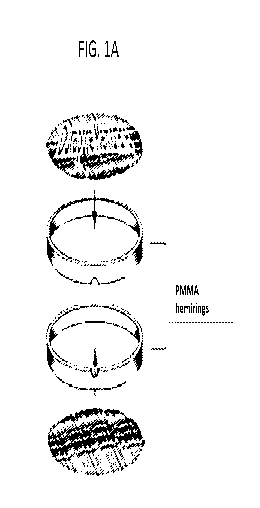
[0012] FIGS. la-lg depict a representative biodiffusion chamber. FIG la.
component parts;
FIG lb. assembled chamber; FIG lc. PMMA port plug to seal the chamber; FIG ld.
photomicrograph of polyvinylidine fluoride Durapore membrane; FIG le. overhead
and lateral
view of the actual chamber; FIG lf. and FIG lg. H & E stained paraffin
sections of Durapore
membranes after explantation; FIG 1 f. explanted phosphate buffered saline
control chamber
from human trial 14379-101; FIG lg. explanted vaccine chamber from human trial
14379-101.
[0013] FIGS. 2a-2c depict survival metrics of subjects in Phase I trial (IND
14379-101,
NCT01550523). FIG. 2a. Overall survival of patients in trial; FIG. 2b.
protocol survival with
two survival cohorts. Nine patients died of disease progression while one died
of intracerebral
hemorrhage and two of sepsis. Overall protocol survival was 48.2 weeks and 9.2
weeks,
respectively for longer (N = 4) and shorter (N = 8) survival cohorts (log-rank
= .014). FIG. 2c.
Excluding one profoundly lymphopenic outlier and three non-disease-related
deaths linear
3
CA 03054662 2019-08-23
WO 2018/165528 PCT/US2018/021706
regression revealed high correlation between protocol survival and lymphocyte
count at enrollment
(R2 = .8, p = .0028).
[0014] FIGS. 3a-3d shows radiographic responses with associated physiologic
measurements.
FIG. 3a. Examples of patient imaging from short survival cohort. Patient TJ11:
A-D; Patient TJ10:
E-H. A, E: pre-operative T 1 -gadolinium-enhanced axial images; G: Ti-
gadolinium-enhanced
coronal image; C: pre-operative axial FLAIR image. B, D, F, H: respective 3
month post-operative
images. FIG. 3b. Examples of patient imaging from longer survival cohort.
Patient TJ06: A-D;
Patient TJ09: E-H. A, E: pre-operative Ti-gadolinium-enhanced axial images; C,
F: pre-operative
axial FLAIR images. B, D, F, H: respective 3 month post-operative images. FIG.
3c. Relationship
between relative cerebral blood volume in tumor v. apparent diffusion
coefficient in longer
survival cohort; there is a high correlation between the ADC and rCBV (R2 =
.96, p = .0005). FIG.
3d. Relationship between relative cerebral blood volume in tumor v. apparent
diffusion coefficient
in short survival cohort.
[0015] FIGS. 4a-4c depict an examination of the explanted chambers by survival
cohorts. FIG.
4a. Explanted chambers were structurally intact with no viable cells. Outer
surfaces of membranes
from both C-p and C-v chambers were coated with CD15+and CD163+ cells, with
dramatically
increased numbers on C-v membranes; FIG. 4b. analysis of factors in chambers
between survival
cohorts revealed significant chamber elevations of VEGF, PDGF-a, IL-11, CCL5,
MCP-3 and
MIP-id in the longer cohort while a number of soluble cancer markers were
significantly elevated
in the short cohort including NSE, osteonectin, and YKL40. Mixture
discriminant analysis
independently identified these cohort differences; FIG. 4c. for both cohorts,
two chemokines
associated with glioma macrophage recruitment were significantly lower in C-v
than other
measurable sources. Both Periostin and CCL2 levels were significantly lower
than serum or SN
(tumor cell supernatant) values, suggesting elimination of cells producing
these chemokines in the
chambers.
[0016] FIGS. 5a-5e depict post-vaccination levels of PBMCs and cytokines.
Serial measurements
of immune effector cell shifts and cytokine/ chemokine shifts after
vaccination in the post-
treatment period; longer survival cohort, (patients TJ03, TJ14, TJ06, TJ09);
example of short
survival cohort, patient TJ13 (for all other short survival cohorts, see Fig.
6). Rows: FIG. 5a. serial
PBMC counts after vaccination; FIG. 5b. serial assessments of PBMC
subpopulation percentages
after vaccination, FIG. Sc. serial levels of CCL21 and CXCL12; FIG. 5d.
relationship of absolute
4
CA 03054662 2019-08-23
WO 2018/165528 PCT/US2018/021706
CD14+CD16- macrophage counts with MCP-1 (CCL2); note correlation with
macrophage levels
in FIG. 5b. and CCL2 spike post-operatively. CC12 levels remained
significantly higher in the
short survival cohort (see Fig. 6); FIG. 5e. scaled comparisons of putative TH-
1 cytokine
responses after vaccinations (TNF-a x 2; CXCL9 x 350; CXCL10 x 80).
Significant correlations
were noted as follows: TNF-a spikes were highly correlated with CCL2 spikes
for both cohorts
(R2 = .99, p = .003). There was a significant immediate perioperative decrease
in CD14+16- cells
(p = .008) not seen in the short cohort (p = .78). For the longer cohort only,
there was a significant
correlation between CD4 and CXCL12 (R2 = .62, p < .0001). Also, a high
correlation was noted
between total monocyte count and CD14+16- monocyte levels (Fig. 5B and 5D, R2
= .8, p <
.0001) and inverse relationships between circulating T cell and monocyte
numbers (R2 = .66, p <
.0001) were noted in the longer survival cohort (FIG. 5) without significant
differences in the
short survival cohort (see FIG. 6).
[0017] FIGS. 6a-6e depicts post-vaccination levels of PBMC and cytokines in
short cohort
patients , (patients TJ01, TJ01, TJ07, TJ08, TJ10, TJ11, TJ12);. Rows: FIG.
6a. serial PBMC
counts after vaccination; FIG. 6b. serial assessments of PBMC subpopulation
percentages after
vaccination (T-cell; B-cell; monocyte); FIG. 6c. serial levels of CCL21 and
CXCL12; FIG. 6d.
relationship of absolute CD14+CD16- macrophage counts with MCP-1 (CCL2). CC12
levels
remained significantly higher in the short survival cohort compared to long
survival cohort. FIG.
6e. scaled comparisons of putative TH-1 cytokine responses after vaccinations
(TNF-a x 2;
CXCL9 x 350; CXCL10 x 80). IFN-g is also shown.
[0018] FIGS. 7a-7h depict the loss of specific, tumor-promoting monocyte cell
populations after
vaccination. Substantial tumor regression was observed over a 3 month period.
FIG. 7a.
Monophasic trend for TME IGF-1R+ cells (ordinal scale); in matched pairs cases
from initial
diagnosis to vaccination (N=5) no significant difference; matched pairs from
vaccine to autopsy
(N=4) reveals significant decrease in IGF-1R+ cells (p = .003). FIG. 7b. IGF-
1R positive cells in
two patients with evaluable paraffin sections from initial diagnosis through
vaccine and autopsy
(patients TJ06 and TJ10). FIG. 7c. Biphasic trend for TME CD163 M2 macrophages
with
significant increase from diagnosis to recurrence (Aperio five 400x fields per
phase of treatment
per patient; left plot, matched pairs *p < .0001, N=6) followed by significant
loss from recurrence
to autopsy after vaccination (right plot, matched pairs *p < .0001, N=4). FIG.
7d. CD163+ cells
in same two patients with evaluable paraffin sections from initial diagnosis
through vaccine and
CA 03054662 2019-08-23
WO 2018/165528 PCT/US2018/021706
autopsy (patients TJ06 and TH 0); increase in CD163 at vaccine v. recurrence
(matched pairs, p =
.052) followed by significant decrease in TME CD163 M2 macrophages at autopsy
v. vaccine
(matched pairs, *p = .001). FIG. 7e. Significant correlation in the short
survival cohort between
peripheral CD163 monocytes and CD163 TAM levels documented at surgery (R2 =
.80, p = .02).
FIG. 7f. Non-significant correlation between peripheral and TAM CD163 cells in
the longer
cohort. FIG. 7g. Fluorescence immunohistochemistry photomicrographs from
paraffin sections.
A, C: Patient TJ10 at Second surgical resection prior to vaccination and B, D:
at autopsy; E-H:
autopsy specimens obtained from glioblastoma patients undergoing re-resection
after standard of
care; I, J: untreated, incidentally found post-mortem glioblastoma. FIG. 7h.
Time course for
treatment response in TJ06 from initial diagnosis through autopsy. Biphasic
occurrence of CD163
cells in the TME with increase after standard treatment and decrease after
vaccination through
autopsy. Loss of CD163 TAMs is associated with increases in both rCBV and ADC
values in the
tumor. Serum nitrate levels spike after each vaccination and are associated
with concomitant
rCBV/ADC increases.
[0019] FIGS. 8a-8d depict differentiation of immature monocytes by cytokines
or serum from
study subject. FIG. 8a. Upregulation of IGF-1R after polarization of monocytes
with M2
cytokines. M1 macrophages do not upregulate the IGF-1R, ***p = .0004. FIG. 8b.
Differences
in monocyte subset distribution after treatment with IGF-1R AS ODN according
to
macrophage polarization protocol described in materials and methods. Flow
cytometry reveals
that IGF-1R AS ODN selectively targets the removal of M2 macrophages. FIG. 8c.
Protocol
patient serum differentiates immature monocytes into a CD163+ phenotype that
co-expresses IGF-
1R and PD-Li. IGF-1R AS ODN knocks down this macrophages population in a dose-
dependent
fashion over a 100-fold concentration range. All values are mean fluorescence
intensity. Duplicate
measurements for each patient serum co-incubation, comparison of means. ***p <
.0001, **p =
.0001, *p = .0002, ===p = .0003,==p = .0009, *p = .009, ''p = .0018, 'p =
.026. FIG.
8d. Summary of means in FIG. 8c.
[0020] FIGS. 9a-9d show that compared to standard of care in the first interim
analysis, there
were significant improvements in both progression-free survival and overall
survival. FIG. 9a.
Progression-free survival (PFS) of entire study cohort compared to standard of
care (SOC); dotted
black lines are 95% confidence interval; FIG. 9b. Overall survival (OS). In
both cases SOC falls
6
CA 03054662 2019-08-23
WO 2018/165528 PCT/US2018/021706
below the lower 95% CI reflecting a significant improvement; FIG. 9c. PFS by
survival cohort at
interim analysis; FIG. 9d. OS by survival cohort at interim analysis.
[0021] FIGS. 10a-10d depict a summary of the Phase lb study and a comparison
of interferon-
gamma levels to the prior trial and between cohorts within the trial. FIG.
10a. Trend of increasing
IFN-y after vaccination in the newly diagnosed vaccine cohort (p = .06); FIG.
10b. Significant
increase in median IFN-y in the newly diagnosed vaccine cohort (p = .02); FIG.
10c. Significant
increases in IFN-y levels in 20 chamber cohorts ***p < .0001, **p <.006, *p <
.02; FIG. 10d. Rate
of diffusion of labeled IGF-1R AS ODN from the biodiffusion chamber over time.
[0022] FIGS. 11a-11j depict the effect of fully formulated biodiffusion
chamber (both irradiation
and exogenously added AS ODN) on pro-inflammatory cytokine production in a
naïve mouse
model. Luminex analysis of explanted mouse chamber contents at 24 hour post
implantation filled
with GL261 cells alone; partial formulation with either addition of 2 tg IGF-
1R AS ODN,
irradiation of GL261 cells with 5 Gy of X-irradiation; or the fully formulated
autologous vaccine
(GL261, 2 tg IGF-1R AS ODN, and 5 Gy of gamma-irradiation). FIG. ha. G-CSF
GL261-AS-
irr v. GL261-irr p < .0117; FIG. 11b. IL-la, GL261-AS-irr v. GL261-irr p <
.008; FIG. 11c. IL-
lb, GL261-AS-irr v. GL261-irr p < .0067; FIG. 11d. IL-2, GL261-AS-irr v. GL261-
irr p < .0002;
FIG. lie. IL-9, GL261-AS-irr v. GL261-irr p <.0413; FIG. llf. IL-10, GL261-AS-
irr v. GL261-
irr p < .0001; FIG. 11g. IL-12(p40), GL261-AS-irr v. GL261-irr p < .001; FIG.
11h. IL-13,
GL261-AS-irr v. GL261-irr p < .0065; FIG. iii. IL-15, GL261-AS-irr v. GL261-
irr p < .0013;
FIG. 11j. M- CSF, GL261-AS-irr v. PBS p = .007. Others tested but not shown:
IL-6, GL261-
AS-irr v. GL261-irr p <.0836; GM-CSF, GL261-AS-irr v. GL261-irr p <.0854; lix,
GL261-AS-
irr v. GL261-irr p < .0001; kc, GL261-AS-irr v.GL261-irr p < .0112; TNF-a,
GL261-AS-irr v.
GL261-irr p <.0082; VEGF, GL261-AS-irr v. GL261-irr p <.0004; lif, GL261-AS-
irr v. GL261-
irr p < .0140; IL-7, GL261-AS-irr v. GL261-irr p < .0038; IL-12(p70) GL261-AS-
irr v. GL261-
irr p <.0120; IFN-y, GL261-AS-irr v. GL261-irr p <.0290.
[0023] FIG. 12 shows titration curves for dendritic cell (DC) activation of
peripheral blood
mononuclear cells (PBMC) from two normal subjects (dark and light) by IGF-1R
AS ODN;
NOBEL antisense 750 tg v. NOBEL sense 75 pg, 7.5 tg, DWA antisense 750 pg, 7.5
pg,
control,***p < .0009; NOBEL antisense 75 tg v. NOBEL sense, DWA antisense 7.5
pg.
[0024] FIGS. 13a-13b depict in vitro T cell response from contents of fully
formulated chamber
utilizing T cells derived from vaccinated mice. FIG. 13a. Pro-inflammatory T
cell response with
7
CA 03054662 2019-08-23
WO 2018/165528 PCT/US2018/021706
DCs primed with antigen retrieved from chambers; **p < .01 for full
formulation in vitro v. no
antigen; *p < .03 for full formulation v. exosomes; FIG. 13b. Pro-inflammatory
T cell response
with DCs pulsed with antigen retrieved from chambers; **p < .005 for full
formulation v. no
antigen; *p < .007 for full formulation v. exosomes.
[0025] FIGS. 14a-14d are schematic representations of biphasic response to
NOBEL antisense
dose titration.
[0026] FIGS. 15a-15b depict M2 polarization of allogenic monocytes from three
normal subjects
with overnight incubation from serum derived from six different glioma
patients. Controls were
not incubated with serum. 1000-fold dilution curve revealed a decrease of M2
macrophages co-
expressing FIG 15a. PDL-1 and FIG 15b. CD163 from 100 pg of NOBEL antisense to
a
significant knockdown at 1 pig. Line in each graph is the grand mean. 1 i.tg
of NOBEL v.
untreated ***p< .0002.
[0027] FIG. 16 depicts comparison of explanted vaccine chamber cytokine levels
v. explanted
PBS control chamber. ***p, .005; ***p < .01; ***p< .02; **p < .03; *p < .05.
[0028] FIG. 17 is a dose-response curve, showing the biphasic response of
systemic IGF-1R AS-
ODN on inhibition of flank glioma tumor growth. 106 GL261 cells were implanted
into the flanks
of C57BL/6 mice and 20 days later, prior to the period when an elevation in
circulating CD163
positive cells is typically observed, the mice were injected intraperitoneally
with a single 0.75 mg
(squares) or 0.075 mg (triangles) dose of NOBEL IGF-1R AS-ODN. The mice were
then followed
for tumor development. Unvaccinated mice (circles) were used as a control.
[0029] FIG. 18 is flow cytometry data showing that systemic IGF-1R AS-ODN
treatment
inhibited the accumulation of circulating M2 monocytes. The data is expressed
as a histogram of
cell numbers expressing CD163 (right hand peak) where one line labelled
"vehicle" represents
tumor-implanted mice treated with PBS (vehicle) and the other line labelled
"AS-ODN" represents
implanted mice treated with NOBEL IGF-1R AS-ODN. These lines are annotated in
the Figure.
[0030] FIG. 19 shows tumor incidence in mice implanted in the flank with
glioma cells, treated
with NOBEL IGF-1R AS-ODN or PBS (vehicle). Tumor incidence between the treated
and
untreated groups was significantly different (* = p <0.05).
[0031] FIG. 20 shows tumor incidence in Tbet deficient mice implanted in the
flank with glioma
cells, and treated with or without NOBEL IGF-1R AS-ODN. Tumor incidence
between the groups
was significantly different (* = p < 0.05).
8
CA 03054662 2019-08-23
WO 2018/165528 PCT/US2018/021706
[0032] FIGS. 21a, 21b, and 21c show pro-inflammatory cytokine levels (pg/ml)
in patient serum
after vaccination, pooled from serial blood draws over time (days 14-42 post-
surgery). A
significant dose-dependent increase in pro-inflammatory cytokines was observed
in patient serum.
FIGS. 21d, 21e, and 21f depict the relationship (polynomial best fit) between
wet weight yield of
tumor tissue and cytokine yield by subject. A wet weight yield of tissue of 3
grams produced the
highest cytokine yield when, after processing, the cells were distributed
among 20 chambers.
[0033] FIG. 22 is a schematic of a representative fully formulated
biodiffusion chamber. When
the chamber is implanted into a patient, antisense molecules and tumor
antigens diffuse through
the porous membranes of the chamber, leading to a tumor-specific immune
response, decreased
M2 polarization, and reduction in the number of M2+ cells.
[0034] FIG. 23 is a schematic of a representative immunization method. If a
patient does not
adequately respond to a first round of vaccination, the procedure is
optionally repeated as many
times as necessary, sometimes in combination with other treatments.
[0035] FIGS. 24a and 25b are Kaplan-Meier curves illustrating median
progression-free survival
(P-FS) and median overall survival (OS) in the intention-to-treat group
(N=30), respectively, in
human patients having brain tumors. (Interim analysis is shown in Fig. 9
above.) The "vaccinated"
population is treated with 20 chambers implanted and each chamber containing 2
g NOBEL. The
"SoC" population is represented using historical data (N=76). The data shows
substantially
increased survival both overall and without progression of the cancer.
[0036] FIGS. 25a and 25b are Kaplan-Meier curves illustrating progression-free
survival and
overall survival comparing the same gender and median age in the vaccinated
and Standard of
Care (SoC) groups respectively. The data shows substantially increased
survival both overall and
without progression of the cancer.
[0037] FIGS. 26a and 26b are Kaplan-Meier curves illustrating progression-free
survival and
overall survival when excluding the 5 patients who withdrew from treatment and
who died from
other causes.
[0038] FIGS. 27a and 27b are Kaplan-Meier curves illustrating progression-free
survival and
overall survival when excluding the 9 patients who did not complete the
standard of care (SOC)
protocol.
[0039] FIGS. 28a, 28b, 28c, 28d. illustrate IFN-y responses induced based on
cell yield in the
high vaccine cohort. The data show that the optimum IFN-y release, based on
cell number in the
9
CA 03054662 2019-08-23
WO 2018/165528 PCT/US2018/021706
chamber. For FIGS 28a and 28b cell yield is shown in millions of cell. IFN-y
are shown as mean
fluorescent intensity (MFI). These data are from the 20-chamber cohort with
each chamber
containing 2i_ig of NOBEL. Data is presented as a polynomial fit (cubic). FIGS
28c and 20db is
an extract of the data in FIGs 28a and b showing the substantially linear
relationship between cell
number yield versus mean and peak IFNy response, respectively, up to 20
million cells. The data
are presented here as pg/ml.
[0040] FIGS. 29a, 29b, and 29c illustrate IFN-y T cell response relative to
chamber formulation
regarding IGF-1R antisense pre-incubation prior to encapsulation. FIG. 29a
shows the protocol
for assessing T-cell response to tumor antigen. For FIG. 29b Antigen was
prepared following the
in-vivo clinical chamber paradigm. Approximately 1 million ex-vivo GL261 tumor
cells were
injected into chambers alone or with indicated antisense concentrations and
incubated overnight
in the chamber which was placed in PBS). The following day, chamber content
was extracted and
used to pulse naive dendritic cells. Chamber content which was not treated
overnight with
antisense was added to the dendritic cells with the indicated amounts of
NOBEL. Dendritic cells
were also left naive for control. Following an overnight pulse with antigen,
dendritic cells were
collected and incubated overnight with T cells from immunized animals in a
cell culture plate
coated with an ELIPSPOT detection antibody for the cytokine IFNy. After
overnight incubation,
the coated plate was processed and developed to enumerate the number of IFNy
producing T-cells
which responded to each respective antigen. The data in FIG. 29b shows that
tumor antigens were
detected in materials recovered from chambers containing GL261 cells plus
antisense but not
materials from chambers cultured with cells alone, even if antisense was added
to the material
when the dendritic cells (DC) were pulsed. The data illustrates that antisense
in chambers with
the glioma cells is required to produce immunostimulatory tumor antigen. For
Fig. 29c, GL261
cells were plated in petri dishes and treated overnight with 4 mg NOBEL per 1
million cells or
were left untreated. The cells were then collected and placed into chambers at
1 million cells and
21.ig NOBEL per chamber. The chambers were then incubated overnight in PBS and
the content
was extracted the following day. Dendritic cells were then pulsed with the
chamber content and
IFNy secretion was measured as described above. The data illustrates that
overnight treatment of
GL261 cells with antisense enhances the amount of antigen produced by these
cells as detected by
an increase in the numbers of tumor-immune T cells producing IFN y.
CA 03054662 2019-08-23
WO 2018/165528 PCT/US2018/021706
[0041] FIGS 30a, 30b, 30c, and 30d illustrate the impact levels of expression
of Nestin on efficacy
in a mouse model. FIG. 30a shows that high level of Nestin is associated with
improved survival
following IGF-1R antisense treatment. Mice were implanted in the flank with
chambers containing
GL261 cells that expressed high or low levels of the nestin protein as well as
4mg antisense. A
control group received high nestin expressing cells alone with no antisense
added. The chambers
were left in the flank for 24 hours. The immune response was then allowed to
develop for several
weeks and the mice were challenged intra-cranially on day 35 post-chamber
implantation. The
immunized mice as well as non-immune controls were monitored for survival
after challenge.
FIG. 30b shows that a high level of Nestin is associated with better clinical
disease score. The
data shows scored morbidity associated with brain tumor progression in
orthotopic model after
vaccination with fully formulated chamber by treatment cohort. FIG. 30c and
FIG. 30d show
increased production of antibody against GL261 cells associated with high
levels of nestin
expression. FIG. 30c shows day 28 post chamber/ pre-intra-cranial.
implantation cell ELISA assay
data performed with sera from experimental mice was tested for antibody
reactivity to GL261 cell;
isolated sera from whole blood taken from the mice. The sera was tested for
whole IgG reactivity
to GL261 cells with an ELISA assay. FIG. 30d shows cell ELISA data from day 35
post intra-
cranial challenge/71 days post-chamber explanation, using sera from
experimental mice, tested for
antibody reactivity to GL261 cells.
DETAILED DESCRIPTION
Definitions
[0042] All terms not defined herein have their common art-recognized meanings.
[0043] As used herein, terms such as "a," "an," and "the" include singular and
plural referents
unless the context clearly demands otherwise.
[0044] As used herein, the term "about" when preceding a numerical value
indicates the value
plus or minus a range of 10%. For example, "about 100" encompasses 90 and 110.
[0045] As used herein, the term "autologous" means cells or tissues obtained
from the same
individual.
[0046] As used herein, the term "autologous cancer cell vaccine" refers to a
therapeutic produced
in part by isolating tumor cells from an individual and processing these tumor
cells ex vivo. The
11
CA 03054662 2019-08-23
WO 2018/165528 PCT/US2018/021706
cells are then re-administered to the individual from whom the tumor cells
were isolated. In
embodiments, an autologous cancer cell vaccine may comprise additional
components in addition
to the tumor cells, such as a buffer and/or antisense nucleic acids. In
embodiments, "autologous
cancer cell vaccine" may refer to a biodiffusion chamber containing the tumor
cells and one or
more additional components. In certain aspects, the "autologous cancer cell
vaccine" may be a
"fully formulated chamber" also referred to herein as "fully formulated
biodiffusion chamber."
[0047] As used herein, the term "fully formulated chamber" or "fully
formulated biodiffusion
chamber" is a biodiffusion chamber that includes autologous tumor cells and
other cells included
in the tumor microenvironment (TME) that may or may not be treated prior to
encapsulation in the
chamber with a first amount of an IGF-1R AS ODN. The cells are encapsulated
with exogenous
addition of a second amount, for example at least 2 i.tg, of IGF-1R AS ODN and
the chamber is
then irradiated with 5 Gy of gamma-irradiation.
[0048] As used herein, the term "small molecules" includes nucleic acids,
peptides, proteins, and
other chemicals (such as, for example, cytokines and growth hormones produced
by cells), but
does not include cells, exosomes, or microvesicles.
[0049] The term "targeting IGF-1R expression" or "targets IGF-1R expression"
as used herein
refers to administering an antisense nucleic acid that has a sequence designed
to bind to the IGF-
1R.
[0050] As used herein, the term "systemic administration" refers to achieving
delivery of a
substance throughout the body of a subject. Typical systemic routes of
administration include
parenteral administration, transdermal administration, intraperitoneal
administration, intravenous
administration, subcutaneous administration, and intramuscular administration.
[0051] Other administration routes include oral administration, nasal
administration topical
administration, intraocular administration, buccal administration, sublingual
administration,
vaginal administration, intraheptic, intracardiac, intrapancreatic, by
inhalation, and via an
implanted pump.
Antisense Molecules
[0052] Antisense molecules are nucleic acids that work by binding to a
targeted complimentary
sequence of mRNA by Watson and Crick base-pairing rules. The translation of
target mRNA is
inhibited by an active and/or passive mechanism when hybridization occurs
between the
12
CA 03054662 2019-08-23
WO 2018/165528 PCT/US2018/021706
complementary helices.
In the passive mechanism, hybridization between the mRNA and
exogenous nucleotide sequence leads to duplex formation that prevents the
ribosomal complex
from reading the message.
In the active mechanism, hybridization promotes the binding of
RnaseH, which destroys the RNA but leaves the antisense intact to hybridize
with another
complementary mRNA target. Either or both mechanisms inhibit translation of a
protein
contributing to or sustaining a malignant phenotype. As therapeutic agents,
antisense molecules
are far more selective and as a result, more effective and less toxic than
conventional drugs.
[0053] The methods and compositions disclosed herein involve the use of
antisense molecules for
treating cancer. Typically, the antisense molecule is an antisense
oligodeoxynucleotide (AS-
ODN). In some embodiments, the antisense molecule comprises a modified
phosphate backbone.
In certain aspects, the phosphate backbone modification renders the antisense
more resistant to
nuclease degradation. In certain embodiments, the modification is a locked
antisense. In other
embodiments, the modification is a phosphorothioate linkage. In certain
aspects, the antisense
contains one or more phosphorothioate linkages. In certain embodiments, the
phosphorothioate
linkages stabilize the antisense molecule by conferring nuclease resistance,
thereby increasing its
half-life. In some embodiments, the antisense may be partially
phosphorothioate-linked. For
example, up to about 1%, up to about 3%, up to about 5%, up to about 10%, up
to about 20%, up
to about 30%, up to about 40%, up to about 50% up to about 60%, up to about
70%, up to about
80%, up to about 90%, up to about 95%, or up to about 99% of the antisense may
be
phosphorothioate-linked. In some embodiments, the antisense is fully
phosphorothioate-linked.
In other embodiments, phosphorothioate linkages may alternate with
phosphodiester linkages. In
certain embodiments, the antisense has at least one terminal phosphorothioate
monophosphate.
[0054] In some embodiments, the antisense molecule comprises one or more CpG
motifs. In other
embodiments, the antisense molecule does not comprise a CpG motif. In certain
aspects, the one
or more CpG motifs are methylated. In other aspects, the one or more CpG
motifs are
unmethylated. In certain embodiments, the one or more unmethylated CpG motifs
elicit an innate
immune response when the antisense molecule is administered to a subject. In
some aspects, the
innate immune response is mediated by binding of the unmethylated CpG-
containing antisense
molecule to Toll like Receptors (TLR).
[0055] In certain embodiments, the antisense molecule comprises at least one
terminal
modification or "cap". The cap may be a 5' and/or a 3' -cap structure. The
terms "cap" or "end-
13
CA 03054662 2019-08-23
WO 2018/165528 PCT/US2018/021706
cap" include chemical modifications at either terminus of the oligonucleotide
(with respect to
terminal ribonucleotides), and including modifications at the linkage between
the last two
nucleotides on the 5' end and the last two nucleotides on the 3' end. The cap
structure may increase
resistance of the anti sense molecule to exonucleases without compromising
molecular interactions
with the target sequence or cellular machinery. Such modifications may be
selected on the basis
of their increased potency in vitro or in vivo. The cap can be present at the
5' -terminus (5' -cap) or
at the 3' -terminus (3' -cap) or can be present on both ends. In certain
embodiments, the 5'- and/or
3' -cap is independently selected from phosphorothioate monophosphate, abasic
residue (moiety),
phosphorothioate linkage, 4' -thio nucleotide, carbocyclic nucleotide,
phosphorodithioate linkage,
inverted nucleotide or inverted abasic moiety (2'-3' or 3'-3'),
phosphorodithioate monophosphate,
and methylphosphonate moiety. The phosphorothioate or phosphorodithioate
linkage(s), when
part of a cap structure, are generally positioned between the two terminal
nucleotides on the 5' end
and the two terminal nucleotides on the 3' end.
[0056] In preferred embodiments, the antisense molecule targets the expression
of Insulin like
Growth Factor 1 Receptor (IGF-1R). IGF-1R is a tyrosine kinase cell surface
receptor that shares
70% homology with the insulin receptor. When activated by its ligands (IGF-I,
IGF-II and
insulin), it regulates broad cellular functions including proliferation,
transformation and cell
survival. The IGF-1R is not an absolute requirement for normal growth, but it
is essential for
growth in anchorage-independent conditions that may occur in malignant
tissues. A review of the
role of IGF-1R in tumors is provided in Baserga et al., Vitamins and Hormones,
53:65-98 (1997),
which is incorporated herein by reference in its entirety.
[0057] In certain embodiments, the antisense molecule is an oligonucleotide
directed against DNA
or RNA of a growth factor or growth factor receptor, such as, for example, IGF-
1R.
[0058] In certain embodiments, the antisense is a deoxynucleotide directed
against IGF-1R (IGF-
1R AS ODN). The full length coding sequence of IGF-1R is provided as SEQ ID
NO:19 (see, for
example, PCT/U52016/26970, which is incorporated herein by reference in its
entirety).
[0059] In certain embodiments, the antisense molecule comprises nucleotide
sequences
complementary to the IGF-1R signal sequence, comprising either RNA or DNA. The
signal
sequence of IGF-1R is a 30 amino acid sequence. In other embodiments, the
antisense molecule
comprises nucleotide sequences complementary to portions of the IGF-1R signal
sequence,
comprising either RNA or DNA. In some embodiments, the antisense molecule
comprises
14
CA 03054662 2019-08-23
WO 2018/165528 PCT/US2018/021706
nucleotide sequences complementary to codons 1-309 of IGF-1R, comprising
either RNA or DNA.
In other embodiments, the antisense molecule comprises nucleotide sequences
complementary to
portions of codons 1-309 of IGF-1R, comprising either RNA or DNA.
[0060] In certain embodiments, the IGF-1R AS ODN is at least about 5
nucleotides, at least about
nucleotides, at least about 15 nucleotides, at least about 20 nucleotides, at
least about 25
nucleotides, at least about 30 nucleotides, at least about 35 nucleotides, at
least about 40
nucleotides, at least about 45 nucleotides, or at least about 50 nucleotides
in length. In some
embodiments, the IGF-1R AS ODN is from about 15 nucleotides to about 22
nucleotides in length.
In certain aspects, the IGF-1R AS ODN is about 18 nucleotides in length.
[0061] In certain embodiments, the IGF-1R AS ODN forms a secondary structure
at 18 C, but
does not form a secondary structure at about 37 C. In other embodiments, the
IGF-1R AS ODN
does not form a secondary structure at about 18 C or at about 37 C. In yet
other embodiments, the
IGF-1R AS ODN does not form a secondary structure at any temperature. In other
embodiments,
the IGF-1R AS ODN does not form a secondary structure at 37 C. In particular
embodiments, the
secondary structure is a hairpin loop structure.
[0062] In some aspects, the IGF-1R AS ODN comprises the nucleotide sequence of
SEQ ID NO:1,
or a fragment thereof In certain embodiments, the IGF-1R AS ODN may have at
least about 70%,
at least about 75%, at least about 80%, at least about 85%, at least about
90%, at least about 95%,
at least about 96%, at least about 98%, or 100% identity to SEQ ID NO: 1, or a
fragment thereof.
In some embodiments, the IGF-1R AS ODN comprises one or more phosphorothioate
linkages.
[0063] In certain aspects, the IGF-1R AS ODN consists of SEQ ID NO: 1. NOBEL
is an 18-mer
oligodeoxynucleotide with a phosphorothioate backbone and a sequence
complimentary to codons
2 through 7 in the IGF-1R gene. As such, NOBEL is an antisense oligonucleotide
directed against
IGF-1R (IGF-1R AS ODN). The NOBEL sequence, derived as the complimentary
sequence of
the IGF-1R gene at the 5' end, is:
5' -TCCTCCGGAGCCAGACTT- 3'.
[0064] NOBEL has a stable shelf life and is resistant to nuclease degradation
due to its
phosphorothioate backbone. Administration of NOBEL can be provided in any of
the standard
methods associated with introduction of oligodeoxynucleotides known to one of
ordinary skill
in the art. Advantageously, the AS ODNs disclosed herein, including NOBEL, may
be
administered with little/no toxicity. Even levels of about 2g/kg (scaled)
based on mice tests (40 i.tg
CA 03054662 2019-08-23
WO 2018/165528 PCT/US2018/021706
in the tail vain) did not reveal toxicity issues. NOBEL can be manufactured
according to ordinary
procedures known to one of ordinary skill in the art.
[0065] The anti sense molecule, for example the NOBEL sequence of SEQ ID
NO: 1, may
also comprise one or more p-ethoxy backbone modifications as disclosed in U.S.
Patent No.
9,744,187, which is incorporated by reference herein in its entirety. In some
embodiments, the
nucleic acid backbone of the antisense molecule comprises at least one p-
ethoxy backbone linkage.
For example, up to about 1%, up to about 3%, up to about 5%, up to about 10%,
up to about 20%,
up to about 30%, up to about 40%, up to about 50% up to about 60%, up to about
70%, up to about
80%, up to about 90%, up to about 95%, or up to about 99% of the antisense
molecule may be p-
ethoxy-linked. The remainder of the linkages may be phosphodiester linkages or
phosphorothioate
linkages or a combination thereof. In a preferred embodiment 50% to 80% of the
phosphate
backbone linkages in each oligonucleotide are p-ethoxy backbone linkages,
wherein 20% to 50%
of the phosphate backbone linkages in each oligonucleotide are phosphodiester
backbone linkages.
[0066] Various IGF-1R antisense sequences are bioactive in some or all of the
multi-modality
effects of the NOBEL sequence. The 18-mer NOBEL sequence has both IGF-1R
receptor
downregulation activity as well as TLR agonist activity, and further
experimentation in mice
suggests that both activities are necessary for in vivo anti-tumor immune
activity. While the AS
ODN molecule has anti-tumor activity, the complimentary sense sequence does
not, despite also
having a CpG motif.
In certain embodiments, the sequence of the antisense is selected from the
group consisting of SEQ
ID NOS 1-14, as shown in Table 1. In some embodiments, the antisense has 90%
sequence identity
to one or more of SEQ ID NOS 1-14. In some embodiments, the antisense has 80%
sequence
identity to one or more of SEQ ID NOS 1-14. In some embodiments, the antisense
has 70%
sequence identity to one or more of SEQ ID NOS 1-14.
TABLE 1: Additional downstream sequences for IGF-1R AS ODN Formulation
Sequences with ACGA Motif Corresponds to IGF-1R SEQ ID NO:
Codons
5' -TCCTCCGGAGCCAGACTT-3' 2-7 1
5'-TTCTCCACTCGTCGGCC-3' 26-32 2
5' -ACAGGCCGTGTCGTTGTC-3 ' 242-248 3
5' -GCACTCGCCGTCGTGGAT-3 ' 297-303 4
16
CA 03054662 2019-08-23
WO 2018/165528 PCT/US2018/021706
Sequences with ACGA Motif Corresponds to IGF-1R SEQ ID NO:
Codons
5' -CGGATATGGTCGTTCTCC-3 ' 589-595 5
5'- TCTCAGCCTCGTGGTTGC-3' 806-812 6
5' -TTGCGGCCTCGTTCACTG-3 ' 1,033-1,039 7
5'-AAGCTTCGTTGAGAAACT-3' 1,042-1,048 8
5' -GGACTTGCTCGTTGGACA-3 ' 1,215-1,221 9
5' -GGCTGTCTCTCGTCGAAG-3 ' 1,339-1,345 10
5' -CAGATTTCTCCACTCGTCGG-3 ' 27-34 11
5' -CCGGAGCCAGACTTCAT-3 ' 1-6 12
5'-CTGCTCCTCCTCTAGGATGA-3' 407-413 13
5'-CCCTCCTCCGGAGCC-3' 4-8 14
[0067] In certain embodiments, the IGF-1R AS ODN comprises the nucleotide
sequence of any
one of SEQ ID NOs:1-14, or fragments thereof In certain embodiments, the IGF-
1R AS ODN
may have at least about 70%, at least about 75%, at least about 80%, at least
about 85%, at least
about 90%, at least about 95%, at least about 96%, at least about 98%, or 100%
identity to any one
of SEQ ID NOs: 1-14, or fragments thereof
[0068] In some embodiments, the antisense molecule downregulates the
expression of genes
downstream of IGF-1R pathway in a cell. In certain aspects, the downstream
gene is hexokinase
(Hex II). In some embodiments, the antisense molecule downregulates the
expression of
housekeeping genes in the cell. In some aspects, the housekeeping gene is L13.
[0069] In certain aspects, the IGF-1R AS ODN is chemically synthesized. In
certain
embodiments, the IGF-1R AS ODN is manufactured by solid phase organic
synthesis. In some
aspects, the synthesis of the IGF-1R AS ODN is carried out in a synthesizer
equipped with a closed
chemical column reactor using flow-through technology. In some embodiments,
each synthesis
cycle sequence on the solid support consists of multiple steps, which are
carried out sequentially
until the full-length IGF-1R AS ODN is obtained. In certain embodiments, the
IGF-1R AS ODN
is stored in a liquid form. In other embodiments, the IGF-1R AS ODN is
lyophilized prior to storing.
In some embodiments, the lyophilized IGF-1R AS ODN is dissolved in water prior
to use. In other
embodiments, the lyophilized IGF-1R AS ODN is dissolved in an organic solvent
prior to use. In
yet other embodiment, the lyophilized IGF-1R AS ODN is formulated into a
pharmaceutical
17
CA 03054662 2019-08-23
WO 2018/165528 PCT/US2018/021706
composition. In some aspects the pharmaceutical composition is a liquid
pharmaceutical
composition. In other aspects, the pharmaceutical composition is a solid
pharmaceutical
composition. Additional antisense nucleic acids are also described in U.S.
Publication No.
2017/0056430, which is incorporated herein by reference in its entirety.
Autologous Cancer Cell Vaccine
Introduction
[0070] Immunotherapy is currently used to target hematologic malignancies with
one common
cellular antigen. Unfortunately, solid tumors are far more complex,
representing epigenetic
progression of genetic changes to a malignant state with an unidentifiable
number of tumor-
specific targets. Even more challenging, within a WHO diagnostic cancer group
there exists
marked variations in tumor phenotypes. An autologous cell vaccine would
encompass all such
variations and all such targets and represent an ideal subject-specific
immunotherapy for solid
tumor cancers. An autologous cancer cell vaccine however, cannot be derived
from primary
cell cultures because serial passages alter the tumor phenotype thus
diminishing the array of
tumor-specific antigens. This would also require impossible lot-release
qualification at each
passage. The present disclosure eliminates these concerns by plating freshly
resected,
morselized tumor cells and reimplanting them within 24 hours as a depot
antigen, as shown in
FIG. 22. In certain aspects, the excellent results achieved herein are
obtained by ensuring that
an appropriate number of cells are present in the chamber(s), among other
specifics described
herein.
[0071] Previous studies have designed autologous cell vaccine through the use
of antigen
presenting cells, instead of autologous tumor cells. In this paradigm, a
subject's monocytes are
collected from a pre-treatment plasma leukopheresis and differentiated into
autologous dendritic
cells (DC) ex vivo. The dendritic cells are then presented with the subject's
tumor crude lysate
inducing DC activation/maturation, and at a later time point, the matured
dendritic cells, now
cross-primed with tumor antigens are injected in the subject as a DC vaccine.
Ex vivo
differentiation, however, is missing a number of key stimulatory components
only occurring in
vivo. In addition, differentiation of DCs from hematopoietic precursors
requires extensive in
vitro manipulations with labor-intensive cell processing in expensive
facilities. The present
disclosure obviates these concerns by providing an endogenous DC maturation
process and an
18
CA 03054662 2019-08-23
WO 2018/165528 PCT/US2018/021706
immunomodulatory and immunostimulatory antisense oligodeoxynucleotide (AS-ODN)
that
promotes the development of an appropriate immune response. More specifically,
the present
disclosure provides a biodiffusion chamber comprising dispersed tumor cells
derived from the
patient and irradiated antisense molecules, which is implanted into the
patient for therapeutically
effective time. Without being bound by any theory, it is thought that the
combination of irradiated
tumor cells, antisense, and biodiffusion chamber act in concert to simulate
the local immune
response, and enhance the response by reducing or eliminating M2 cells,
preventing dampening
of the immune system.
[0072] Thus, the present disclosure shows that an irradiated, implantable
biodiffusion chamber
comprising freshly resected tumor cells and IGF-1R AS ODN safely serves as an
effective,
subject-specific autologous cell vaccine for cancer immunotherapy. As such,
the use of the
claimed implantable biodiffusion chamber to mount an immune response that
selectively targets
tumor cells in a subject provides a new and significant approach for the
treatment of cancer,
especially GBM.
Biodiffusion chamber
[0073] A representative diffusion chamber comprises a chamber barrel having
two ends, a first
end and a second end. In embodiments, the biodiffusion chamber is a small ring
capped on either
side by a porous, cell-impermeable membrane, such as the Duropore membrane
manufactured by
Millipore Corporation. Optionally, one of the ends may be closed off as part
of the chamber body
leaving only one end open to be sealed using the porous membrane. The
membranes can be made
of plastic, teflon, polyester, or any inert material which is strong, flexible
and able to withstand
chemical treatments. The chamber can be made of any substance, such as and not
limited to plastic,
teflon, lucite, titanium, Plexiglass or any inert material which is non-toxic
to and well tolerated by
humans. In addition, the chambers should be able to survive sterilization. In
some aspects, the
diffusion chambers are sterilized with ethylene oxide prior to use. Other
suitable chambers are
described in U.S. Prov. No. 62/621,295, filed January 24, 2018, U.S. Patent
No. 6,541,036,
PCT/U516/26970, and U.S. Patent No. 5,714,170, which are each incorporated
herein by reference
in their entirety.
[0074] In certain embodiments, the membrane allows passage of small molecules
but does not
allow passage of cells (i.e., the cells cannot leave or enter the chamber). In
some aspects, the
diameter of the pores of the membrane allows nucleic acids and other chemicals
(such as, for
19
CA 03054662 2019-08-23
WO 2018/165528 PCT/US2018/021706
example, cytokines produced by cells) to diffuse out of the chamber, does not
allow passage of
cells between the chamber and the subject in which it is implanted. The
biodiffusion chambers
useful in the present disclosure include any chamber which does not allow
passage of cells between
the chamber and the subject in which it is implanted, provided however, that
the chamber permits
interchange and passage of factors between the chamber and the subject. Thus,
in certain aspects,
the pore size has a cut-off that prevent passage of materials that are greater
than 100 m3 in volume
into and out of the chamber. In some embodiments, the pores of the membrane
have a diameter
of about 0.25 [tm or smaller. For example, the pores may have a diameter of
about 0.1 [tm (see
Fig. 1). In particular aspects, the pores range in diameter from 0.1 [tm to
0.25 [tm. See also, Lange,
et al., J. Immunol., 1994, 153, 205-211 and Lanza, et al., Transplantation,
1994, 57, 1371-1375,
each of which is incorporated herein by reference in their entireties. This
pore diameter prevents
the passage of cells in or out of the chamber. In certain embodiments,
diffusion chambers are
constructed from 14 mm Lucite rings with 0.1 [tm pore-sized hydrophilic
Durapore membranes
(Millipore, Bedford, Mass.).
[0075] In certain embodiments, a biodiffusion chamber comprises a membrane
that allows the
IGF-1R AS ODN to diffuse out of the chamber. In some embodiments, about 50% of
the IGF-1R
AS ODN diffuses out of the chamber in about 12 hours, about 60% of the IGF-1R
AS ODN
diffuses out of the chamber in about 24 hours, about 80% of the IGF-1R AS ODN
diffuses out of
the chamber in about 48 hours, and/or about 100% of the IGF-1R AS ODN diffuses
out of the
chamber in about 50 hours.
[0076] In an exemplary approach, to assemble the biodiffusion chamber, a first
porous membrane
is attached to one side of a first diffusion chamber, using glue and pressure
to create a tight seal.
A second porous membrane is similarly attached to a second diffusion chamber
ring. The
membranes can be secured in position with rubber gaskets which may also
provide a tighter seal.
The diffusion chamber rings are left overnight (minimum 8 hours) to dry. Then,
the first diffusion
chamber ring and the second diffusion chamber ring are attached to one another
using glue and
left overnight (minimum 8 hours) to dry. In a preferred embodiment, the first
chamber ring and
second chamber ring joining process comprises using 2 dichloroethane as a
solvent to facilitate
adhesion between the two rings. See, for example, Fig. 22 showing two porous
membranes. In
an alternative approach, the chamber may have only one side that contains a
porous membrane.
CA 03054662 2019-08-23
WO 2018/165528 PCT/US2018/021706
[0077] On the barrel portion of the chamber, one or more openings (e.g. ports)
are provided which
can be covered by a cap which is accessed from outside of the subject's body
once the chamber is
implanted, thus allowing the diffusion chamber to be refilled. The openings
allow for multiple
and sequential sampling of the contents, without contamination and without
harming the subject,
therefore significantly reducing the number of implantation procedures
performed on the subject.
Before implantation into the patient, the one or more openings may be sealed
with bone wax, a
port plug or cap made from, for example, PMNIA. The cap can be a screw-on type
of self-sealing
rubber and fitted to the opening. In some configurations, the diffusion
chamber may contain two
or more injection openings or ports. Sampling of the chamber contents can be
performed by
accessing the opening by removing the cap on the outside of the subject's body
and inserting an
ordinary needle and syringe. In some embodiments, the chamber may further
include a removal
device. Such a device facilitates removal of the chamber from the patient.
[0078] In embodiments, the chamber serves as an antigen depot designed so that
tumor antigens
diffuse out of the chamber for the purpose of promoting a therapeutic host
immune response.
Exogenous IGF-1R AS ODN and ex vivo irradiation promote a pro-inflammatory
response. This
formulation is associated with clinical and radiographic improvements,
prolonged survival on
protocol, and represents a novel autologous cell vaccine that includes an
exogenous active
pharmaceutical ingredient (API) and radiation that we interpret as inducing or
enhancing tumor
immunity effect. Furthermore the addition of low concentration of the IGF-1R
AS ODN is critical
to a pro-inflammatory response (Fig. 12).
[0079] In certain embodiments the disclosure provides a biodiffusion chamber
for implantation
into a subject suffering from cancer comprising: (a) tumor cells; and (b) an
effective amount of an
antisense molecule. In other embodiments is provided a method for treating
cancer in a subject
comprising: (a) obtaining a biodiffusion chamber comprising tumor cells and an
effective amount
of an antisense nucleic acid; (b) irradiating the biodiffusion chamber and
contents; and (c)
implanting the irradiated biodiffusion chamber into the subject for a
therapeutically effective time.
[0080] In certain embodiments, the IGF-1R AS ODN is present in the
biodiffusion chamber in an
amount ranging from about 0.5 i.tg to about 10 pg. In certain aspects, the IGF-
1R AS ODN is
present in an amount ranging from about 1 i.tg to about 5 i.tg per chamber, or
from about 2 i.tg to 4
i.tg per chamber. In specific aspects, the IGF-1R AS ODN is present in an
amount of about 2 i.tg
per chamber. In specific aspects, the IGF-1R AS ODN is present in an amount of
about 4 i.tg per
21
CA 03054662 2019-08-23
WO 2018/165528 PCT/US2018/021706
chamber. Without being bound by theory it is thought that these levels promote
an enhanced Thl
response in a subject, while avoiding an M2 immunostimulatory response in the
subject.
[0081] In certain embodiments, the tumor cells are not treated with an IGF-1R
AS ODN prior to
encapsulation in the chamber. Typically, however, the tumor cells are treated
with an IGF-1R AS
ODN prior to encapsulation in the chamber. The time for treating the cells pre-
encapsulation may
vary. For example, the tumor cells may be treated ex vivo with an IGF-1R AS
ODN immediately
before encapsulation, for up to about 4 hours, for up to about 6 hours, for up
to about 8 hours, for
up to about 12 hours or for up to about 18 hours. Typically, the tumor tissue
may be treated ex
vivo for about 12 hours to about 18 hours pre-encapsulation. Conveniently, the
cells may be
encapsulated after a pre-treatment lasting up to overnight. Without being
bound by theory, it is
thought that the pre-encapsulation treatment plays a desirable role in
stimulating production of
tumor antigen.
[0082] The amount of IGF-1R AS ODN used for the pre-encapsulation treatment
may be in a
range of about 1 mg to 8 mg per million cells; for example, about 2 mg to
about 6 mg per million
cells, about 3 mg to about 5 mg per million cells. Typically the amount of IGF-
1R AS ODN used
for treatment prior to encapsulation is about 4 mg per million cells.
[0083] In some embodiments, the IGF-1R AS ODN for ex vivo treatment of the
tumor cells is
used at a concentration ranging from about at least 2 mg/ml to at least about
5 mg/ml. In certain
aspects, the IGF-1R AS ODN is used at a concentration of at least 4 mg/ml. In
specific
embodiments, the IGF-1R AS ODN is used at a concentration of 4 mg/ml.
[0084] In certain embodiments, the IGF-1R AS ODN used to treat tumor cells ex
vivo and the
IGF-1R AS ODN present in the chamber are the same. In other embodiments, the
IGF-1R AS
ODN used to treat tumor cells ex vivo and the IGF-1R AS ODN present in the
chamber are
different. In certain embodiments, the IGF-1R AS ODN used to treat tumor cells
ex vivo is at least
about 5 nucleotides, at least about 10 nucleotides, at least about 15
nucleotides, at least about 20
nucleotides, at least about 25 nucleotides, at least about 30 nucleotides, at
least about 35
nucleotides, at least about 40 nucleotides, at least about 45 nucleotides, or
at least about 50
nucleotides in length. In some embodiments, the IGF-1R AS ODN used to treat
tumor cells ex
vivo is from about 15 nucleotides to about 22 nucleotides in length. In
certain aspects, the IGF-
1R AS ODN used to treat tumor cells is about 18 nucleotides in length.
22
CA 03054662 2019-08-23
WO 2018/165528 PCT/US2018/021706
[0085] In certain embodiments, the IGF-1R AS ODN used to treat tumor cells ex
vivo forms a
secondary structure at 18 C, but does not form a secondary structure at about
37 C. In other
embodiments, the IGF-1R AS ODN used to treat tumor cells does not form a
secondary structure
at about 18 C or at about 37 C. In yet other embodiments, the IGF-1R AS ODN
used to treat
tumor cells ex vivo does not form a secondary structure at any temperature. In
other embodiments,
the IGF-1R AS ODN used to treat tumor cells does not form a secondary
structure at 37 C. In
particular embodiments, the secondary structure is a hairpin loop structure.
[0086] In some aspects, the IGF-1R AS ODN used to treat tumor cells comprises
the nucleotide
sequence of SEQ ID NO:1, or a fragment thereof. In certain embodiments, the
IGF-1R AS ODN
used to treat tumor cells may have at least about 70%, at least about 75%, at
least about 80%, at
least about 85%, at least about 90%, at least about 95%, at least about 96%,
at least about 98%, or
100% identity to SEQ ID NO: 1, or a fragment thereof. In certain aspects, the
IGF-1R AS ODN
used to treat tumor cells is SEQ ID NO: 1.
[0087] After the tumor cells are treated with the AS-ODN for a period of time,
the AS-ODN is
removed and fresh AS-ODN is added to the chamber, which is then irradiated
prior to implantation
into a subject. In certain aspects, the biodiffusion chamber is treated with
gamma irradiation at an
amount of about 1 Gy, about 2 Gy, about 4 Gy, about 5 Gy, about 6 Gy, about 10
Gy, or up to
about 15 Gy. In certain aspects, the dose of radiation is not more than about
5 Gy. In other aspects,
the dose of radiation is at least about 5 Gy. In some aspects, the dose of
radiation is 5 Gy. In
certain embodiments, the biodiffusion chamber may be irradiated at least once,
at least twice, at
least three times, at least four times, or at least five times. In some
embodiments, the chamber is
irradiated less than about 24 hours prior to implantation into a subject. In
other embodiments,
chamber is irradiated about 24 hours prior to implantation into the subject.
In yet other
embodiments, the chamber is irradiated at least about 24 hours prior to
implantation into the
subject. In still other embodiments, the chamber is irradiated not more than
about 48 hours prior
to implantation into the subject. In yet other embodiments, the chamber is
irradiated at least about
48 hours prior to implantation into the subject.
[0088] While the tumor cells are typically killed prior to implantation; for
example by radiation,
the cells need not be killed and indeed it may be advantageous to maintain the
cells in an alive
state to promote release of antigen. Thus, in certain embodiments, the cells
may not be irradiated
23
CA 03054662 2019-08-23
WO 2018/165528 PCT/US2018/021706
prior to implantation. For safety purposes, however, it is desirable to
prevent release of live tumor
cells into the subject.
[0089] Tumor cells can be placed in a diffusion chamber in varying numbers. In
certain
embodiments, about 1 x 10⁴ to about 5 x 10⁶ tumor cells are placed in each
diffusion chamber. In
other embodiments, about 1 x 10⁵ to about 1.5 x 10⁶ tumor cells are placed in
the diffusion
chamber. In yet other embodiments, about 5 X 10⁵ to 1 x 10⁶ tumor cells are
placed in the chamber.
with a subject can be used. We have discovered that the number of tumor cells
can impact the
subjects' anti-tumor response and that an appropriate range should be selected
to increase the
chance to obtain the desired results. FIG. 28 shows data from patients
implanted with 20 chambers
and shows cell yield (millions of cells) corresponding to immune response. The
anti-tumor
immune response is optimal in a range of about 750,000 to about 1,250,000
cells in a chamber,
with a peak at about 1 million cells/chamber. Multiple chamber containing
irradiated tumor cells
are administered and to maintain the optimal immune the response the number of
cells/chamber is
preferably maintained within the range. Preferably, the tumor cells are intact
and not autolyzed or
otherwise damaged as described herein.
[0090] In certain embodiments, it may be preferable to maintain the ratio of
cells to AS ODN in a
chamber. Thus, in certain aspects a chambers may contain about 2 g of AS ODN
and between
750,000 and 1,250,000 cells; for example 1,000,000 cells. The ratio of cells
to AS ODN may thus
be in a range from about 3.75 x 10⁵ to about 6.25 x 10⁵ per g AS ODN; for
example, about 5.0 x
105 cells per g. Thus, in a typical patient receiving 20 chambers the total
dose of AS ODN is
about 40 g.
[0091] Typically, administration will be in a chamber as described herein;
however, in certain
aspects, the irradiated cells and IGF-1R AS ODN may be co-administered to the
subject without
being contained physically together in the chamber or another container. In
certain methods using
this approach, the irradiated cells IGF-1R AS ODN thus disperse, diffuse, or
are metabolized in
the body limited by the physiology of the subject. Thus, in certain aspects,
e.g. the tumors cells
for use may be prepared as described herein for the chamber and administered
with the IGF-1R
AS ODN but the administration may be not contained within a physical
container. Such
administration is typically intramuscular.
Tumor Tissue Preparation for Chamber
24
CA 03054662 2019-08-23
WO 2018/165528 PCT/US2018/021706
[0092] Tumor cells for use in the autologous vaccination are surgically
removed from the subject.
In embodiments, the tumor cells are removed from the patient using a tissue
morselator. The
extraction device preferably combines a high-speed reciprocating inner cannula
within a stationary
outer cannula and electronically controlled variable suction. The outer
cannula has a diameter of
1.1 mm, 1.9 mm, 2.5 mm, or 3.0 mm, and a length of 10 cm, 13 cm, or 25 cm. The
instrument
also relies on a side-mouth cutting and aspiration aperture located 0.6 mm
from the blunt desiccator
end. The combination of gentle forward pressure of the aperture into the
tissue to be removed and
suction draws the desired tissue into the side aperture, allowing for
controlled and precise tissue
resection through the reciprocal cutting action of the inner cannula. A key
feature is the absence
of a rotation blade; this avoids drawing unintended tissue into the aperture.
An example of a
suitable device is the Myriad tissue aspirator (NICO Corporation
Indianapolis, IN), a
minimally invasive surgical system which may be used for the removal of soft
tissues with direct,
microscopic, or endoscopic visualization. The shaved tissue is suctioned,
gathered in to a
collection chamber, and is collected in a sterile tissue trap. During
collection of the tissue in the
sterile tissue trap, blood is removed from the preparation. Preferably, the
sterile trap contains a
collection dish at the bottom of the trap and a stem that provides access to
the trap. The trap
structure may also contain an inner ladle-shaped structure that is removable
from the trap to
facilitate tissue removal from the trap.
[0093] Preferably, the morselator generates no heat at the resection site or
along its shaft, and
requires no ultrasonic energy for tissue removal. Thus, in particular
embodiments, the tumor tissue
is morselized tumor tissue (i.e. tumor shaved tissue obtained by side-mouth
cutting in the absence
of heat, and optionally in the absence of ultrasonic treatment).
Advantageously, the aspirator-
extract and morselized tissue has higher viability than tissue removed by
other methods. It is
believed that the extraction process maintains higher tumor cell viability in
part due to restricting
exposure of the tumor cells to high temperatures during removal. For example,
the methods herein
do not expose tumor cells to above 25 C during removal. Thus, the cells are
not exposed to
temperatures above body temperature, i.e., about 37 C.
[0094] The amount of tumor tissue obtained from the subject may vary.
Preferably, the amount
is at least 1, at least 2, at least 3 grams or at least 4 grams of wet tumor
tissue is obtained from the
patient. The tissue is removed from the sterile tissue trap and disaggregated
by pipetting with a
sterile pipette to break up large tissue fragments. The disaggregated cell
suspension is then placed
CA 03054662 2019-08-23
WO 2018/165528 PCT/US2018/021706
onto sterile tissue culture plates in serum-containing media, and incubated in
a tissue culture
incubator. This plating step serves to enrich the desired functional cells by
adherence, and also
helps to remove debris from the preparation. Thus, the tumor cells used in
treatments described
herein preferably consist essentially of, or consist of, adherent cells from
the tumor tissue.
[0095] After a predetermined incubation time (e.g., 6, 12, 24, or 48 hours),
the cells are removed
from the plates. The cells may be removed by scraping, by chemical methods
(e.g. EDTA) or by
enzymatic treatment (e.g. trypsin). The cells are placed into one or more
diffusion chambers. In
some embodiments, the cells are split between 2, 3, 4, 5, 6, 7, 8, 9, 10, 11,
12, 13, 14, 15, 16, 17,
18, 19, 20, 21, 22, 23, 24, 25, 26, 27, 28, 29, 30 or more diffusion chambers.
Often, 20 chambers
are used. In some embodiments, each diffusion chamber contains an equal number
of cells. In
some embodiments, a first diffusion chamber contains more cells than a second
chamber.
[0096] In some embodiments, the cells are sorted before being placed in the
chamber. In some
embodiments, the cells are enriched by selecting for one or more cellular
markers before being
placed in the chamber. The selection may be performed, for example, using
beads or by cell sorting
techniques known to those of skill in the art. In some embodiments, the cells
placed into the
chamber are enriched for one or more markers.
[0097] In some embodiments, implantation of the biodiffusion chamber for a
therapeutically
effective time reduces or eliminates return of the cancer in the subject. In
certain aspects,
implantation of the biodiffusion chamber causes a reduction of tumor volume
associated with the
cancer in the subject. In yet other embodiments, implantation of the
biodiffusion chamber for a
therapeutically effective time induces elimination of the tumor in the
subject. In some
embodiments, implantation of the chamber inhibits regrowth of the tumor for at
least 3 months, at
least 6 months, at least 12 months, at least 36 month, or indefinitely.
[0098] The biodiffusion chamber can be implanted in a subject in the following
non-limiting ways:
subcutaneously, intraperitoneally, and intracranially. In certain embodiments,
the diffusion
chamber(s) is implanted into an acceptor site of the body having good
lymphatic drainage and/or
vascular supply such as the rectus sheath. In other embodiments, a refillable
chamber can be
employed such that the diffusion chamber can be re-used for treatments and
emptied following
treatments. In certain aspects, a plurality of diffusion chambers, preferably
between 5 and 20, can
be used in a single subject.
26
CA 03054662 2019-08-23
WO 2018/165528 PCT/US2018/021706
[0099] In certain embodiments, at least about 1, at least about 2, at least
about 3, at least about 4,
at least about 5, at least about 10, at least about 15, at least about 20, at
least about 25, at least
about 30, at least about 35, at least about 40, at least about 45, or at least
about 50 chambers are
implanted into the subject. In some embodiments, 10-20 chambers are implanted
into the subject.
Preferably, about 20 chambers are implanted into the subject. In certain
embodiments, the tumor
cells are divided equally among each chamber.
[00100] Typically, the chamber is removed after period of time. For
example, the chamber
may be implanted in the subject for about 24 hours, about 48 hours, about 72
hours, or about 96
hours. Implantation for about 48 hours is associated with beneficial
therapeutic outcomes.
Accordingly, the preferred time of implantation is about 48 hours. In certain
embodiments, the
vaccination procedure is performed one time per patient. In other embodiments,
the vaccination
procedure is performed multiple times per patient. In embodiments, the
vaccination procedure is
performed two times, three times, four times, five times, six times, seven
times, or eight times in
a single patient. In embodiments, the vaccination is repeated every 7, 14, or
28 days, or every 1,
3, or 6 months for a given period of time. In further embodiments, the
vaccination procedure is
repeated periodically until the patient is free of cancer.
[00101] Without being bound by theory, it is thought that implantation of
the biodiffusion
chamber causes elimination or reduction of M2 cells at or near the
implantation site such that an
immune response against tumor antigens diffusing out from the chamber is
achieved. In certain
aspects, elimination or reduction of M2 cells at the implantation site leads
to enhanced presentation
of autologous tumor antigens by antigen-presenting cells (APC) to CD4 T cells
leading to
production of interferon-gamma (IFNy) and the induction of type 1 tumor
immunity. In certain
aspects, the production of IFNy by tumor antigen-specific CD4 T cells and the
anti-M2 effects of
IGF-1R AS ODN drive type 1 anti-tumor immunity and the loss of anti-
inflammatory M2 cells
from the circulation and tumor microenvironment indirectly interfering with
tumor growth. In
some aspects, the production of IFNy by tumor antigen-specific CD4 T cells and
the anti-M2
effects of IGF-1R AS ODN unleashes effector-mediated damage to the tumor cells
and tumor
microenvironment (M2 cells) and initiates the longer process of programming
memory T cells
recognizing tumor antigens. In certain embodiments, the anti-tumor adaptive
immune response
sustains continued tumor regression.
27
CA 03054662 2019-08-23
WO 2018/165528 PCT/US2018/021706
[00102] Optionally, the cells introduced into the chamber may be enriched
for certain cell
types. Nestin a, cytoskeleton-associated class VI intermediate filament (IF)
protein, has
traditionally been noted for its importance as a neural stem cell marker. We
have discovered that
in certain brain tumor samples, cells positive for nestin (nestin+ cells) are
enriched compared to
benign tissue, and that this associated corresponds to improved therapeutic
response. Thus, in
certain aspects, a subject's tumor can be biopsied to assess the degree of
nestin expression, and
therefore, in certain aspects, the chamber cells are enriched Nestin-positive
("+") cells compared
to benign tissue. Without being bound by theory, it is thought that nestin
provides a marker
associated with antigens suitable useful in producing an anti-tumor immune
response.
Accordingly, the cells implanted into the chamber may be enriched for nestin+
cells compared to
the tumor cell population as a whole when extracted from the subject. Figure
30 illustrates the
enhance immune response obtained when the tumor sample used to stimulate a
response is
enriched with Nestin.
Systemic Administration
[0100] As an alternative to, or supplement to, implantation of the chambers,
IGF-1R AS ODN
may be administered systemically. Thus, in embodiments, the IGF-1R AS ODN is
provided in a
pharmaceutical composition for systemic administration. In addition to the IGF-
1R AS ODN, the
pharmaceutical composition may comprise, for example, saline (0.9% sodium
chloride). The
composition may comprise phospholipids. In some aspects, the phospholipids are
uncharged or
have a neutral charge at physiologic pH. In some aspects, the phospholipids
are neutral
phospholipids. In certain aspects, the neutral phospholipids are
phosphatidylcholines. In certain
aspects, the neutral phospholipids are dioleoylphosphatidyl choline (DOPC). In
some aspects, the
phospholipids are essentially free of cholesterol.
[0101] In some aspects, the phospholipids and oligonucleotides are present at
a molar ratio of from
about 5:1 to about 100:1, or any ratio derivable therein. In various aspects,
the phospholipids and
oligonucleotides are present at a molar ratio of about 5:1, 10:1, 15:1, 20:1,
25:1, 30:1, 35:1, 40:1,
45:1, 50:1, 55:1, 60:1, 65:1, 70:1, 75:1, 80:1, 85:1, 90:1, 95:1, or 100:1. In
some aspects, the
oligonucleotides and phospholipids form an oligonucleotide-lipid complex, such
as, for example,
a liposome complex. In some aspects, at least 75%, 76%, 77%, 78%, 79%, 80%,
81%, 82%, 83%,
84%, 85%, 86%, 87%, 88%, 89%, 90%, 91%, 92%, 93%, 94%, 95%, 96%, 97%, 98%, or
99% of
the liposomes are less than 5 microns in diameter. In various aspects, the
composition further
28
CA 03054662 2019-08-23
WO 2018/165528 PCT/US2018/021706
comprises at least one surfactant, such as, for example, polysorbate 20. In
some aspects, at least
about 5% of the total liposomal antisense drug product consists of surfactant
and at least about
90% of the liposomes are less than 5 microns in diameter. In some aspects, at
least about 15% of
the total liposomal antisense drug product consists of surfactant and at least
about 90% of the
liposomes are less than 3 microns in diameter. In some aspects, the population
of oligonucleotides
are incorporated in the population of liposomes.
[0102] In some aspects the pharmaceutical composition is a liquid
pharmaceutical composition.
In other aspects, the pharmaceutical composition is a solid pharmaceutical
composition.
[0103] Dosages for systemic administration of the antisense in human subjects
may be about 0.025
g/kg, about 0.05 g/kg, about 0.1 g/kg, about 0.15 g/kg, or about 0.2 g/kg. In
certain embodiments,
the dosage for systemic administration may be from 0.025 g/kg to 0.2 g/kg. In
some embodiments,
the dosage is about 0.2 g/kg. In other embodiments, the dosage is from 0.004
g/kg to 0.01 g/kg.
In other embodiments, the dosage is less than 0.01 g/kg. In further
embodiments, the dosage is
not between 0.01 g/kg to 0.2 g/kg. In certain aspects, the antisense is
supplied as a lyophilized
powder and re-suspended prior to administration. When resuspended the
concentration of the
antisense may be about 50 mg/ml, about 100 mg/ml, about 200 mg/ml, about 500
mg/ml, about
1000 mg/ml, or a range between those amounts.
[0104] In certain embodiments, the AS ODN may be administered systemically pre-
operatively;
for example prior to surgery to reduce tumor burden. For example, the AS ODN
may be
administered up to 24 hours, up to 36 hours, up to 48 hours or up to 72 hours
before surgery. In
particular aspects, the pharmaceutical composition may be administered about
48 to about 72 hours
before surgery. Typically, in such circumstances, the administration is by
intravenous bolus.
Combination Therapies
[0105] Historically, cancer therapy has involved treating subjects with
radiation, with
chemotherapy, or both. Such approaches have well-documented challenges.
Advantageously,
however, the chamber implantation methods disclosed herein may be used to
treat a subject having
cancer as a monotherapy. Thus it is preferable that the methods disclosed
herein do not include
chemotherapy or radiation therapy. Notwithstanding the excellent effect
achieved by monotherapy
approaches herein, however, it may be beneficial under certain circumstances
to combine the
chamber methods with other therapies; for example, radiation therapy. In
certain embodiments,
the radiation therapy includes, but is not limited to, internal source
radiation therapy, external beam
29
CA 03054662 2019-08-23
WO 2018/165528 PCT/US2018/021706
radiation therapy, and systemic radioisotope radiation therapy. In certain
aspects, the radiation
therapy is external beam radiation therapy. In some embodiments, the external
beam radiation
therapy includes, but is not limited to, gamma radiation therapy, X-ray
therapy, intensity
modulated radiation therapy (BART), and image-guided radiation therapy (IGRT).
In certain
embodiments, the external beam radiation therapy is gamma radiation therapy.
Radiation may be
administered before chamber implantation or after implantation; for example,
as a salvage therapy.
Typically, such salvage therapy approaches are not implemented until the
cancer is determined to
have returned.
[0106] Thus, in certain combination approaches, both the chamber methods, and
the systemic
methods and compositions, described herein may be used in the same subject,
alone or in
combination with radiation or chemotherapy. In the combination approaches
described herein, the
chamber implantation is preferably used as a first-line therapy. Using the
chamber implantation
first is desirable because the subject's immune system can be inhibited by
other therapies, reducing
the therapeutic benefit of the chamber implantation.
[0107] Optionally, systemic administration may be performed prior to chamber
implantation.
Such an approach can be used to enhance the subjects immune system, as a
priming approach. The
priming approach may be especially advantageous where prior therapy has
resulted in the subject
having a compromised immune system.
[0108] When systemic administration is used in combination, the AS ODN may be
systemically
administered at least 2 weeks, at least 1 week, at least 3 days, or at least 1
day prior to treatment
of the patient using an autologous cancer cell vaccine. In other embodiments,
the AS ODN may
be systemically administered at least 1 day, at least 3 days, at least 1 week,
or at least 2 weeks
following treatment of the patient using an autologous cancer cell vaccine;
i.e. the chamber.
[0109] Optionally, the subject may be revaccinated with chambers using the
methods described
here subsequent to the first vaccination. A second or further additional
vaccination may use tumor
cells taken from the subj ect during the tissue removal and stored.
Optionally, the second or further
additional vaccination may use fresh tumor tissue removed from the subject and
treated as
described herein. Any tumor remaining in the subject may express the same
antigens and thus act
as a depot, providing for re-stimulation. However, recurring tumors may
develop new antigens
and thus provide additional options to stimulate an anti-tumor response. A
subsequent vaccination
CA 03054662 2019-08-23
WO 2018/165528 PCT/US2018/021706
may be after the first treatment is complete and the tumor has recurred or if
the subject has not
responded to the first treatment.
Subjects for Treatment with the IGF-1R AS ODN
[0110] Suitable subjects are animal with cancer; typically, the subject is a
human. While brain
cancers, such as glioblastoma, benefit particularly from this methods
disclosed herein, the methods
apply to cancer generally. Accordingly, the disclosure provides methods of
treating cancers,
including those selected from the group consisting of: glioma, astrocytoma,
hepatocarcinoma,
breast cancer, head and neck squamous cell cancer, lung cancer, renal cell
carcinoma,
hepatocellular carcinoma, gall bladder cancer, classical Hodgkin's lymphoma,
esophageal cancer,
uterine cancer, rectal cancer, thyroid cancer, melanoma, colorectal cancer,
prostate cancer, ovarian
cancer, and pancreatic cancer. In specific embodiments, the cancer is a
glioma. In certain aspects,
the glioma is recurrent malignant glioma. In some embodiments, the cancer is
an astrocytoma. In
certain embodiments, the subject who is a candidate for treatment is suffering
from WHO grade
II, WHO grade III, or WHO grade IV tumor. In some aspects, the tumor is an
astrocytoma. In
certain embodiments, the tumor is selected from grade II astrocytoma, AIII
(IDH1 R132H mutant
grade III astrocytoma), AIII-G (IDH1 wild-type grade III with characteristics
of glioblastoma
multiforme astrocytoma), or grade IV astrocytoma.
[0111] Grade IV astrocytoma is the highest grade glioma and is synonymous with
glioblastoma
(GBM). With a yearly incidence of 3 or 4 per 100,000 GBM is the most common
malignant
primary brain tumor in adults. Standard of care therapy--typically a
combination of radiotherapy
and chemotherapy using Temozolomide¨does not work well and the outcome of GBM
patients
remains poor with a median life expectancy of 15-17 months. Advantageously,
the methods here
may be used to treat newly diagnosed brain cancers and may also be used to
treat recurrent
glioblastoma; for example, in patients previously treated with standard of
care therapy. Thus, in
certain aspects, the subject may be a newly diagnosed GBM subject or a
recurrent GBM subject.
The subject is preferably one who has not been previously treated with any
therapeutic approaches
that are immunosuppressive. In particular aspects, eligible subjects are over
18 years of age and
have a Karnofsky score of 60 or above. Optionally, the subjects do not have
bihemispheric disease
and/or do not have an autoimmune disease.
31
CA 03054662 2019-08-23
WO 2018/165528 PCT/US2018/021706
[0112] Optionally, a subject who is a candidate for treatment may be
identified by performing a
tumor biopsy on the subject. In some embodiments, tumors from the subject are
assayed for the
presence of monocytes. In certain aspects, the monocytes include, but are not
limited to, CD1 lb+,
CD14+, CD15+, CD23+, CD64+, CD68+, CD163+, CD204+, or CD206+ monocytes. The
presence of monocytes in the tumors may be assayed using immunohistochemistry.
In certain
embodiments, a subject who is a candidate for treatment shows CD163+ M2 cells
greater than
about 10%, about 15%, about 20%, about 25%, about 30%, about 35%, about 40%,
about 45%, or
about 50% of the subjects total peripheral blood mononuclear cells (PBMCs). In
certain aspects,
the subject shows CD163+ M2 cells greater than about 20% of the subject's
total PBMCs.
[0113] In yet other embodiments, a subject who is a candidate for treatment is
identified by the
presence of one or more cytokines in the serum of the subject. These cytokines
include, without
limitation, CXCL5, CXCL6, and CXCL7, IL6, IL7, IL8, IL10, IL11, IFN-y, and HSP-
70.
[0114] In yet other embodiments, a subject who is a candidate for treatment is
identified by the
presence of one or more growth factors in the serum of the subject. These
growth factors include,
without limitation, FGF-2, G-CSF, GM-CSF, and M-CSF.
[0115] In some embodiments, a subject who is a candidate for treatment with
the biodiffusion
chamber is identified by measuring the levels of a specific set of cytokines.
In some embodiments,
the subject has elevated levels of these cytokines in comparison to a healthy
subject. As used
herein, the term "healthy subject" refers to a subject not suffering from
cancer or any other disease
and not in need of treatment with the biodiffusion chamber.
[0116] In particular embodiments, the cytokines may be added to the chamber to
augment the anti-
tumor immune response. For example, the cytokines added to the chamber may be
selected from
the group consisting of CCL19, CCL20, CCL21, and CXCL12, and combinations
thereof.
[0117] In certain embodiments, the circulating CD14+ monocytes have an
elevated level of
CD163 in comparison to a healthy subject. In some aspects, the levels of CD163
on the circulating
CD14+ monocytes are elevated by at least about 2 fold, at least about 3 fold,
at least about 4 fold,
at least about 5 fold, at least about 10 fold, at least about 20 fold, at
least about 30 fold, at least
about 40 fold, at least about 50 fold, at least about 60 fold, at least about
70 fold, at least about 80
fold, at least about 90 fold, or at least about 100 fold in comparison to a
healthy subject. In
particular embodiments, the levels of CD163 on the circulating CD14+ monocytes
are elevated by
about 2 fold in comparison to a healthy subject.
32
CA 03054662 2019-08-23
WO 2018/165528 PCT/US2018/021706
[0118] In other embodiments, a subject who is a candidate for treatment has
serum that polarizes
undifferentiated monocytes towards M2 cells. In certain aspects, incubation of
the subject's sera
with undifferentiated monocytes induces the expression of one or more cell
surface markers on the
monocytes including, but not limited to, CD1 lb, CD14, CD15, CD23, CD64, CD68,
CD163,
CD204, and/or CD206. In other aspects, incubation of the subject's sera with
undifferentiated
monocytes elevates the expression of one or more cell surface markers on the
monocytes in
comparison to monocytes not incubated with the subject's sera. In certain
aspects, the cell surface
markers include, but are not limited to, CD1 lb, CD14, CD15, CD23, CD64, CD68,
CD163,
CD204, and/or CD206. In some aspects, the levels of one or more surface
markers are elevated
by at least about 1.3 fold, at least about 1.5 fold, at least about 1.8 fold,
at least about 2 fold, at
least about 3 fold, at least about 4 fold, at least about 5 fold, at least
about 10 fold, at least about
20 fold, at least about 30 fold, at least about 40 fold, at least about 50
fold, at least about 60 fold,
at least about 70 fold, at least about 80 fold, at least about 90 fold, or at
least about 100 fold in
comparison to undifferentiated monocytes not incubated with the subject's
sera. In particular
embodiments, the levels of one or more surface markers are elevated by about 2
fold in comparison
to undifferentiated monocytes not incubated with the subject's sera. Monocytes
polarized by a
subject's sera may be measured using FACS.
Target Cells
[0119] Without being bound by theory it is thought that the AS ODN reduces the
subjects M2 cells
and/or inhibits polarization of cells into M2 cells by downregulating IGF-1R
expression. In some
embodiments, IGF-1R expression in M2 cells is downregulated by at least about
1%, at least about
2%, at least about 5%, at least about 10%, at least about 20%, at least about
30%, at least about
40%, at least about 50%, at least about 60%, at least about 70%, at least
about 80%, at least about
90%, or at least about 95% in comparison to cells not treated with the
antisense. IGF-1R
expression in M2 cells may be measured by quantitative RT-PCR.
[0120] In some embodiments, IGF-1R expression in M2 cells remains
downregulated in the
subject for at least about 1 day, at least about 2 days, at least about 3
days, at least about 4 days, at
least about 5 days, at least about 6 days, at least about 7 days, at least
about 8 days, at least about
9 days, at least about 10 days, at least about 11 days, at least about 12
days, at least about 13 days,
at least about 14 days, at least about 3 weeks, at least about 4 weeks, at
least about 5 weeks, or at
least about 6 weeks after receiving one dose of the antisense.
33
CA 03054662 2019-08-23
WO 2018/165528 PCT/US2018/021706
[0121] In some aspects, the downregulation of expression of IGF-1R in M2 cells
causes a selective
reduction of M2 cells in a subject in comparison to cells not expressing IGF-
1R. In certain
embodiments, M2 cells in a subject are reduced by at least about 2%, at least
about 5%, at least
about 10%, at least about 20%, at least about 30%, at least about 40%, at
least about 50%, at least
about 60%, at least about 70%, at least about 80%, at least about 90%, or at
least about 95% in
comparison to a subject not treated with the antisense. In other embodiments,
the M2 cell
population is eliminated. For example, after implantation of the biodiffusion
chamber, the M2 cell
population may be about 1%, about 2%, about 5%, or about 10% of the population
before
implantation of the biodiffusion chamber. M2 cells in a subject may be
measured using FACS. In
certain aspects, after treatment the M2 cells are eliminated; i.e.,
undetectable by FACS. In other
aspects, the decrease in M2 cells may be measured using a proxy assay; for
example, serum from
the subject may be obtained before and after treatment to assess its ability
to polarize M2 cells.
Following treatment with methods disclosed herein, the ability of the serum to
polarize M2 cells
is reduced by about 80% to about 100%, about 20% to about 60%, or about 10% to
about 50%.
[0122] In some embodiments, targeting the expression of IGF-1R in M2 cells
causes the M2 cells
to undergo cell death. In certain embodiments, the cell death is necrosis. In
other embodiments,
the cell death is apoptosis. Apoptosis, for purposes of this disclosure, is
defined as programmed
cell death and includes, but is not limited to, regression of primary and
metastatic tumors.
Apoptosis is a programmed cell death which is a widespread phenomenon that
plays a crucial role
in the myriad of physiological and pathological processes. Necrosis, in
contrast, is an accidental
cell death which is the cell's response to a variety of harmful conditions and
toxic substances. In
yet other embodiments, targeting the expression of IGF-1R in M2 cells causes
the M2 cells to
undergo cell cycle arrest.
KITS
[0123] Preparation of a completed chamber requires multiple components and
multiple steps. In
another aspects of the disclosure kits containing components for practicing
the methods disclosed
herein are provided. In certain aspects, the kits comprise the chamber body,
which may be present
in one portion or in two halves. Items to seal the chamber may also be
included including one or
more membranes, glues and solvents (e.g., an alcohol, or 2 dichloroethane).
Optionally, the
membrane may by sonically welded onto the chamber to create a seal. The kits
include the
34
CA 03054662 2019-08-23
WO 2018/165528 PCT/US2018/021706
antisense ODN. Optionally, the ODN may be divided into two portions. A first
portion to treat the
cells after surgical removal from the subject, and a second portion to combine
with the cells when
introduced into the subject. Other optional kit items include media for
culturing the cells, and
antibiotics for preventing bacterial growth in the media.
[0124] Optionally, chambers in the kit may be pre-connected (e.g by suture) to
each other using
an eyelet or other device attached to the chamber and adapted to receive the
connecting material.
Advantageously, by pre-connecting multiple chambers, the desired number of
chambers may be
readily introduced and removed by the surgeon.
EXAMPLES
Example 1
Vaccination with Autologous Tumor Cells and IGF-1R AS ODN in Patients with
Recurrent
Glioblastoma
Criteria and Study Objective
[0125] Twelve subjects were enrolled for treatment after failure from standard
therapy. Each
patient met the following criteria: age > 18, a Karnofsky performance score of
60 or better, and no
co-morbidities that would preclude elective surgical re-resection. The
subjects were treated by 24
hour implantation in the rectus sheath of ten biodiffusion chambers containing
irradiated
autologous tumor cells and IGF-1R AS ODN with the objective of stimulating
tumor immunity.
Patients were monitored for safety, clinical and radiographic as well as
immune responses. Study
objectives included assessment of safety and radiographic responses as well as
exploratory
objectives looking at immune function and response.
Table 1 Summary of Patients enrolled
Time Origin al Lymphocyte IDH-
between Chambers lymphocyte count at 1&IDH-2
Previous
Subject Age l(PS surgeries (No.) count enrollment
mutation/
treatment
(weeks) (cells/mm2) (cells/mm2) MGMT
methylation
TJ01 S RT + -/ NA
39 70 177 10 N/A 400 '
TMZ, Bev
TJ02
57 80 90 9 N/A 1570 S' RT + ..
-/methylated
TMZ
CA 03054662 2019-08-23
WO 2018/165528 PCT/US2018/021706
TJ03 S RT + -/NA
75 70 32 7 700 300'
TMZ
TJ06/R S RT + -/NA
66 80 54 8 2000 1300 '
TMZ
TJ07 S, RT +
+/methylated
43 80 215 10 500 430 TMZ, Bev;
RTOG
0525
TJ08 S RT + - /TNS
55 80 52 8 1000 500 '
TMZ
TJ09 S, RT +
57 80 61 7 1400 300 TMZ,
/unmethylate
RTOG
0929
TJ10 S RT + -
/methylated
47 60 376 7 N/A 1800 '
TMZ, Bev
TJ11 S RT + - /TNS
39 70 32 11* 2400 200 '
TMZ
TJ12 S, RT + - /TNS
60 80 74 7 1100 600 TMZ,
Panobinost
at
TJ13 S RT + -
/methylated
64 80 182 11 N/A 2100 '
TMZ
TJ14/R S, RT +
77 90 30 9/11 1800 1100 TMZ
unmethylate
'Compassionate retreatment; *Protocol amendment to include control chamber
filled with
phosphate buffered saline; S: surgery; RT: radiation therapy; TMZ:
temozolamide
chemotherapy; Bev: bevacizumab chemotherapy; IDH-1: isocitrate dehydrogenase-
1; NA:
not available; TNS: tissue not sufficient
Experimental Protocol
[0126] Tumor tissue was surgically removed from patients using a tissue
aspirator (NICO
Myriad ) and placed into sterile tissue traps. The sterile tissue traps were
transferred to a
designated BSL-2 facility, where the tumor tissue was processed and placed
into biodiffusion
chambers. The biodiffusion chambers were irradiated prior to implantation.
[0127] The day following surgery to remove tumor tissue, ten irradiated
biodiffusion chambers
were implanted into the rectus sheath of the subjects. After 24 hours, they
were removed.
[0128] The biodiffusion chambers contained autologous tumor cells removed at
surgery. Prior to
being added to the biodiffusion chambers, the cells were pretreated overnight
(approx. 12-18
hours) with a first amount (4 mg/ml) of an 18-mer IGF-1R AS ODN with the
sequence 5'-
36
CA 03054662 2019-08-23
WO 2018/165528 PCT/US2018/021706
TCCTCCGGAGCCAGACTT- 3' (NOBEL). Based on data showing that the AS ODN has
immunomodulatory properties, a second amount (2 g) of exogenous NOBEL anti
sense was added
to the chambers (C-v), and the chambers were subsequently irradiated. Ten
chambers were
implanted in each patient. An eleventh control chamber containing PBS (C-p)
was also
implanted.
Radiological Assessments
[0129] Serial imaging assessments were performed on Philips 1.5T and 3T Mill
scanners and a GE
1.5T Mill scanner. Routine anatomic MRI features were rated by two
neuroradiologists in all 12
patients. Physiologic MRI techniques of dynamic susceptibility weighted (DSC)
MR perfusion
and 15-direction diffusion tensor imaging (DTI) were also utilized. MR
perfusion and DTI post
processing was performed on Nordic Ice workstation (v.2.3.14). rCBV was
calculated in relation
to contralateral normal white matter. Averaged diffusion coefficient (mean
diffusivity) was
calculated from the DTI data.
Immunological Assessments
[0130] Plasma leukopheresis was performed one week before surgery for baseline
assessment of
immune function. Blood was obtained post-operatively on days 7, 14, 28, 42,
56, and every 3
months after vaccination. Sera and cell fractions were separated by
centrifugation and cells were
treated with red blood cell lysis buffer. White blood cells were either
quantified by flow cytometry
or stored in DMSO at -80 C. Serum samples were also stored at -80 C. Flow
cytometry was
performed using an EasyCyte 8HT (Millipore) and fluorescently-conjugated mAb
specific for
human CD4, CD8, CD1 lb, CD14, CD16, CD20, CD45, CD56, CD80, CD83, and CD86
(all from
BD Biosciences), and CD163 (R&D Systems). Post-collection analysis was
performed with
FlowJo software (Tree Star Inc, Ashland, OR). Serum cytokine factors were
quantified using
Luminex bead arrays (human cytokine/chemokine panels I, II, and III from
Millipore) and
HCMBMAG/ MILLIPLEX Mag Cancer multiplex assay (emdmillipore.com). This
included
6 serum markers for glioma related to stem cell function including DKK-1, NSE,
Osteonectin,
Periostin, YKL-40, and TWEAK. Serum nitrate levels were assayed according to
the Greiss
method (Green L.C., et al., 1982, Anal Biochem 126:131-8). T cell stimulation
was performed
with phorbol 12-myristate, 13 acetate (PMA) and ionomycin as previously
described
(Verbrugge, I., et al., 2012, Cancer Res, 72:3163-74).
37
CA 03054662 2019-08-23
WO 2018/165528 PCT/US2018/021706
[0131] Cytokine/chemokine levels in tumor cell supernatant (SN) and explanted
chamber contents
were analyzed by Luminex kits as designated above. Membranes from paired
vaccine and control
chambers were embedded in paraffin for standard immunohistopathologic
examination.
[0132] Tumor tissue sections were assessed by immunohistochemistry for GFAP
(glial fibrillary
acidic protein), IGF-1R, CD163, CD14, VWF (Von Willebrand Factor), CD4, and
CD8 or
fluorescence immunohistochemistry adapting the method described in Emoto, K.,
et al., 2005,
Histochem Cytochem 53:1311-21). Immunopositive cells were counted
quantitatively with Aperio
or qualitatively by an experienced neuropathologist (LCK) using an ordinal
scale from 0 (no
staining) to 6 (strong diffuse staining) with staining intensity rated as low,
moderate and strong
and staining patterns described as focal or diffuse. Post-mortem autopsy was
limited to
examination of the brain and findings were compared to archival paraffin
blocks of previously
treated or untreated glioblastomas diagnosed at autopsy. Both canonical in
vitro polarization of
naïve monocytes or mixing experiments involving naive monocytes co-incubated
with serum
derived from trial subjects at enrollment were performed as previously
described (Harshyne, LA,
et al., 2015, Neuro Oncol 18(2):206-15; Solinas, G., et al., 2010, J Immunol
185:642-52).
Statistical Analysis
[0133] The level of statistical significance between quantitative measures in
different samples was
determined by a two-tailed unpaired t-test or matched pairs t-test with p <
0.05. Survival analysis
was performed by Kaplan-Meier analysis and significance established by log
rank comparisons.
All statistical analysis including mixture discriminant analysis was performed
with JMP v. 11
software (SAS, North Carolina).
Safety Assessment and Clinical Course
[0134] Only one severe adverse event (SAE) was related to the protocol
(femoral vein thrombosis
after leukopheresis). Nine patients succumbed to tumor progression while three
patients died from
other causes. Five autopsies were performed.
[0135] Median overall survival from initial diagnosis was 91.4 weeks (Fig. 2a)
which compared
favorably to other recurrent glioma immunotherapy trials. Two significantly
different protocol
survival cohorts of 48.2 and 10 weeks were identified as longer and shorter
survival cohorts,
respectively (Fig. 2b). Excluding one outlier (Patient TJ03), we documented a
significant
correlation between protocol survival and degree of lymphopenia at enrollment
(Fig. 2c).
Comparison of CBC values at initial diagnosis and at protocol enrollment
indicated that the mean
38
CA 03054662 2019-08-23
WO 2018/165528 PCT/US2018/021706
lymphocyte count had dropped significantly (65%) after standard therapy (N=8,
p = .012, paired
t-test).
Radiographic Responses
[0136] Routine MRI features were assessed and rated by two neuroradiologists
(K.S.T. and
A.E.F.). In the longer cohort, diminished size of enhancement and FLAIR
envelope at the primary
tumor site were observed, along with slower progression. Examples of anatomic
responses in both
cohorts is shown in Figs. 3a and 3b. Physiologic MRI measurements augmented
these anatomic
observations. Sequential DSC MR perfusion was performed in 7 patients,
including 3 longer-term
survivors (Patients TJ03, TJ06, and TJ09) who had a paradoxical increase in
relative Cerebral
Blood Volume (rCBV) while improving clinically; however, this effect was
transient and there
was a more sustained decrease in rCBV. Sequential 15 directions DTI data
included two long-
term survivors (Patients TJ03 and TJ06) who showed apparent diffusion co-
efficient (ADC) values
increasing in the affected hemisphere, reflecting loss of tumor cellularity
associated with disease
regression. We noted a high correlation between the paradoxical rCBV response
and increasing
ADC not seen in the short cohort (Fig. 3c and 3d). Corresponding levels of
serum nitrate in the
longer cohort reflected the likelihood that an inflammatory response had been
initiated (data not
shown).
Examination of explanted chambers v. pen-operative serum by survival cohort
[0137] Explanted chambers were structurally intact with no viable cells. Outer
surfaces of
membranes from both C-p and C-v chambers were coated with CD15+ and CD163+
cells, but
with dramatically increased numbers on C-v membranes (Fig. 4a).
[0138] For the entire study cohort, analysis of the chamber soluble contents
revealed significant
elevations of a number of growth factors and cytokines/chemokines over matched
perioperative
serum levels, many of which are well-documented in the glioma tumor
microenvironment (TME).
Thirty two of 78 cytokines/chemokines tested were significantly elevated over
serum and matched
pairs analysis revealed significant elevations of cytokines as noted in Table
2 below.
TABLE 2: Chamber Values by Survival Cohort
Cytokines/chemokines present in explanted chambers
cytokine longer short P value Fold
VEGF 4382 1207 .001 3.63
39
CA 03054662 2019-08-23
WO 2018/165528
PCT/US2018/021706
PDGF-AA 272 90 .02 3.02
IL-11 1096 181 .002 6.06
CCL7/MCP-3 3293 1581 .001 2.08
CCL5/ 1484 169 .003 8.78
RANTES
CCL22/MDC 385 1847 .02 0.208
1-309 4.47 11.5 .02 0.389
MW-id 311 182 .002 1.71
Chambers: Matched pairs (chambers v. serum)
cytokine longer P value short P value
VEGF 4028/81 .02 1178/80 .24
PDGF-AA 236/1803 .06 80/1431 .0011
IL-11 958/54 .11 181/25 .047
CCL7/MCP-3 3276/29 .005 1581/18 .0004
CCL5/ 1194/5571 .004 175/6201 .001
RANTES
CCL22/MDC 412/366 .8 1847/348 .09
MW-id 294/523 .02 182/473 .0095
Cancer markers present in explanted chambers
marker longer short P value Fold
DKK1 629 1389 .02 0.452
NSE 7304 11712 .03 0.624
osteonectin 1022 1729 .02 0.591
periostin 286 224 .10 1.27
TRAPS 1421 2441 .007 0.582
OPG 481 1409 .002 0.341
YKL40 8615 12889 .13 0.668
TWEAK 190 171 .87 1.11
Chambers: Matched pairs (chambers v. serum)
Marker Longer P value short P value
DKK1 214/539 .16 457/1389 .01
NSE 6263/2182 .30 11723/1902 .01
CA 03054662 2019-08-23
WO 2018/165528 PCT/US2018/021706
osteonectin 886/953 .70 1729/956 .04
periostin 249/532 .03 224/421 .004
TRAPS 998/1915 .13 2442/1940 .15
OPG 419/205 .48 1409/168 .01
YKL40 7300/12559 .19 12191/5574 .03
TWEAK 142/310 .15 324/217 .53
[0139] These elevations were interpreted as either cytokines/chemokines
produced by the
encapsulated tumor cells or factors produced by the local innate immune
response that had diffused
into the chambers.
[0140] Analysis of factors in chambers between survival cohorts revealed
significant chamber
elevations of VEGF, PDGF-a, IL-11, CCL5, MCP-3, and MW-id in the longer cohort
while a
number of soluble cancer markers were significantly elevated in the short
cohort including NSE,
osteonectin, and YKL40. Mixture discriminant analysis independently identified
these cohort
differences (Fig. 4b).
[0141] For both cohorts, both Periostin and CCL2 levels were significantly
lower in the chambers
(C-v) than serum or SN values, suggesting elimination of cells producing these
chemokines in the
chambers (Fig. 4c).
Serum cytokines/chemokines and PBMC after vaccination by survival cohort
[0142] Levels of 24 of the 78 cytokines/chemokines assessed were significantly
higher in serum
from the longer cohort compared to the short cohort, as shown in Table 3
below.
TABLE 3: Serum Values by Cohort
Cytokine/chemokine serum values (long cohort v. short cohort)
cytokine long short P value
CCL21 279 148 .0007
CTACK 1313 1009 .01
Flt-3L 28 10.6 .02
Fractalkine 102 73 .004
1-309 10.2 6.7 .02
IL-1RA 59 36 .001
IL-10 15 5 .003
IL-12-p40 41 3 .001
41
CA 03054662 2019-08-23
WO 2018/165528 PCT/US2018/021706
IL-13 8.6 3.1 .005
IL-15 11.4 4.6 .0005
IL-la 41 3.6 .003
IL-lb 6.04 2.46 .003
IL-2 7.18 2.87 .006
IL-3 5.06 3.13 .007
IL-5 2.8 1.35 .0001
IL-9 5.61 3.07 .005
MCP-3 28 17 .002
MIP-3b 826 304 .005
MIP- lb 33 24 .01
SCF 11.8 3.6 .007
CXCL12 516 3.6 .008
TGF-a 2.91 1.93 .005
TNF-a 10.8 5.9 .0001
TPO 110 55 .02
[0143] A spike in serum CCL2 occurred after surgery but was absent at re-
operation in two
patients. CCL2 levels remained significantly higher throughout the post-
operative period in the
short cohort. These post-operative spikes were highly correlated with TNF-a
spikes (Figs. 5 and
6).
[0144] Actual CD4 and CD8 T cell counts as well as dendritic cell (DC) counts
were significantly
higher in the longer cohort and perioperative CD14+16- counts were
significantly lower compared
to the short cohort. There was a significant correlation between CD4 and DC
cells and between
CD4 and CXCL12 only in the longer cohort. Day 14 PBMC from the longer survival
subjects
manifested significantly higher Th-1 cytokine production including IFNy after
stimulation with
PMA and ionomycin than the short cohort (data not shown). Coordinated changes
between
circulating levels of T cells, monocytes, and pro-inflammatory
chemokines/cytokines after
vaccination were seen in three of four subjects. The highest correlation was
noted between total
monocyte count and CD14+16- monocyte levels (Fig. 5b and 5d). An inverse
relationship
between circulating T cell and monocyte numbers was also noted in the longer
cohort (Fig. 5)
without significant differences in the short cohort (Fig 6). Predictable and
reciprocal
42
CA 03054662 2019-08-23
WO 2018/165528 PCT/US2018/021706
relationships between immunosuppressive and pro-inflammatory cell populations
as well as
monocyte-chemokine relationships suggested more immune fitness in the longer
cohort.
Examination of paraffin sections
[0145] Paraffin sections from surgical interventions through autopsies were
available for analysis
in four cases allowing us to look at the post-vaccination TME. We compared
trial autopsies to
autopsies from re-operated and untreated GBM patients (Fig. 7). Immunostains
revealed a
significant decrease in IGF-1R positive cells after vaccination in matched
pairs that was
corroborated by fluorescence immunohistochemistry (Fig. 7a and 7g).
Qualitative comparisons to
either recurrent or untreated glioma autopsies revealed abundant CD163 TAMs
and IGF-1R+ cells
in both, diminishing any concern of cell loss as autopsy artifact (Fig. 7g).
[0146] CD163 TAMs peaked at recurrence in matched comparisons to both initial
surgery and
autopsy (Fig. 7c and 7g). Patients TJ06 and TJ10 supported these trends with
evaluable samples
through all phases of treatment (Fig 7b and 7d). In the case of TJ06, CD163
cells dropped after the
second vaccination and persisted through autopsy. This decrease correlated
inversely with rCBV
and ADC values as well as serum nitrate levels all of which increased after
each vaccine (see Fig.
7f).
[0147] Exploring an association with peripheral monocytes, a strong
correlation was noted
between peripheral CD163+ monocytes and CD163 TAMs (Fig. 7e) in the short
cohort not seen
in the longer cohort (Fig 7f).
[0148] We did not see the emergence of T cell populations in the TME after
vaccination in either
cohort.
Coincubation of subject serum with undifferentiated monocytes
[0149] To explore the genesis of the circulating CD163+ monocytes in the
patients we first
polarized naïve monocytes with canonical M1 and M2 cytokines IFN-y and IL-4,
respectively.
We observed upregulation of IGF-1R with M2 polarization only (Fig. 8a). The M2
polarized
CD163+ population was selectively knocked down when incubated with IGF-1R AS
ODN (Fig.
8b).
[0150] Subsequently, we coincubated naïve monocytes with serum obtained from
all study
subjects and documented the emergence of CD163+ cells that co-expressed both
IGF-1R and PDL-
1 (Fig 8c). When treated with IGF-1R AS ODN, cells expressing IGF-1R, PD-Li
and CD163
43
CA 03054662 2019-08-23
WO 2018/165528 PCT/US2018/021706
were significantly knocked down in a parallel and dose-dependent manner (Fig.
8c) summarized
in Fig. 8d.
Discussion
[0151] The revised autologous cell/chamber-based glioblastoma multiforme (GBM)
vaccination
trial did not raise any significant safety concerns.
[0152] We identified two significantly different survival cohorts with
different responses to this
vaccine paradigm. See Figs. 5 and 6. Longer cohort subjects typically
exhibited elevated levels of
tumor-specific antibody isotypes and cytokines/chemokines commonly associated
with Thl
immunity including IgG1 , IgG3, IL12, CXCL10, CXCL12, CCL7, CCL19, and CCL21
following
surgery and vaccination. Elevated levels of these cytokines/chemokines were
not seen in the short
cohort. Accordingly, levels of cytokines/chemokines commonly associated with
Thl immunity
(e.g., IgGl, IgG3, IL12, CXCL10, CXCL12, CCL7, CCL19, CCL21) may be assessed
following
surgery/vaccination to predict survival and to inform further treatment
strategies. Of interest,
CCL21 and CXCL12 synergize with CpG adjuvants and enhance the migratory and T
cell
stimulatory capacity of DCs in a vaccination paradigm. Also, noted elevations
of GM-CSF, IL-6,
Flt-3L and SCF in the longer cohort could enhance DC proliferation, and may
have contributed to
the significant 76% increase in pDCs after vaccination. The significant
elevations of CD4 cells as
well as correlations between CD4 cells, pDC, and the cytokine CXCL12 also
suggests the
successful induction of T cell proliferation facilitated by CXCL12 during
immune synapse.
[0153] Patients in the short survival cohort were typically subjected to a
longer course of treatment
prior to vaccination, leading to lymphophenia. Accordingly, vaccination is
most effective when
administered to patients with normal lymphocyte levels, i.e. non-lymphopenic
patients. The
treatment-induced lymphopenia and the lower CD4:CD8 ratio could also be
ascribed to
temozolamide. (Standard of care included conformal radiation with concomitant
temozolamide
followed by maintenance temozolamide initiated at 6 weeks post-surgery). A
consequence of
longer overall survival would include chronic exposure to tumor antigens and
ongoing glioma
inhibitory signals leading to T cell exhaustion. Similarly,
monocytes/macrophages had an
apparent lack of responsiveness with only modest fluctuations after
vaccination but a distinct
correlation between peripheral CD14+16- cells and TAMs. TAMs have been
associated with
CCL2 production and this correlation could reflect a closed loop amplification
promoting tumor
growth. Supporting this, elevated serum CCL2 levels found in the short cohort
have been
44
CA 03054662 2019-08-23
WO 2018/165528 PCT/US2018/021706
associated with the mesenchymal gene expression profile and a poor prognosis
in glioma
patients.
[0154] The explanted chambers provided a unique snapshot of the encapsulated
TME and its
commerce with the initial immune response. Cytokine elevations in the longer
cohort chambers
collectively indicated that the vaccinations induced a Thl response, and serum
from this cohort
contained tumor-specific antibody isotypes associated with Thl immunity.
Mixture discriminant
analysis established associations with IFN-y, TNF-a, and IL12 production.
[0155] In contrast, the short cohort chambers had greater elevations of cancer
markers reflecting
the emergence of glioma stem cell (GSC)-associated resistance after standard
therapy. One
conspicuous exception was periostin levels that were dramatically lower in all
chambers compared
to paired serum values. Tumor-promoting cell populations in glioma include
TAMs and GSC, the
former supporting the latter in the same perivascular niche (Zhou, W., et al.,
2015, Nat Cell Biol
17:170-82) and both representing strategic targets for treatment. M2
macrophages recruited by
GSC-secreted periostin play a critical role in tumor growth and their
elimination would have
therapeutic advantage. The reduction in periostin levels in chambers
containing treated tumor cells
suggests that GSCs secreting this factor themselves are a target for IGF-1R AS
ODN.
[0156] Notably, despite pre-existing immunosuppression (due to prior treatment
according to the
standard of care) we documented radiographic and clinical improvements
supported by a pro-
inflammatory response after vaccination in 4 of 12 patients. These patients
also had a significant
survival advantage on protocol. Exploring this survival difference further we
noted a higher level
of immune fitness in the longer cohort.
[0157] IGF-1R reduction after vaccination was associated with longer protocol
survival in some
subjects. Without being bound to any particular theory, it is possible that
the IGF-1R+ cell
populations are knocked down as a consequence of type 1 immune mechanisms
promoted in these
individuals by the vaccination paradigm.
[0158] As we have shown in vitro, IGF-1R AS ODN inactivates the CD163+ cells
contained in the
vaccine preparation, thereby eliminating their immunomodulatory factors and
promoting type 1
immunity. Moreover, any IGF-1R AS ODN that diffuses out of the vaccine chamber
has a similar
effect on M2 macrophages that it reaches. This represents a novel platform in
which cells
expressing a variety of tumor-promoting ligands and factors, including PDL-1,
other
CA 03054662 2019-08-23
WO 2018/165528 PCT/US2018/021706
immunomodulatory factors, angiogenic factors, nutrient support, and tumor
invasiveness, are
targeted.
[0159] Differences in the radiographic observations between the longer and
short survival patient
cohorts provide further support for the concept that the vaccination paradigm
has an impact on the
broader glioma TME. Higher rCBV values are typically associated with tumor
progression, and
MR perfusion had only transient increases in the longer cohort, a finding not
previously described.
ADC measurements differentiated tumor progression (lower values) from what we
interpreted as
cell loss (higher values). Since this vaccination paradigm is associated with
loss of both IGF-1R
cell and CD163+ TAM populations, it is possible that rising ADC values are a
reflection of this.
[0160] In summary, we have established the safety profile of an improved
combination glioma
vaccine product and have documented alterations in immune parameters
associated with clinical
and radiographic improvements. With the promise of knocking down specific
monocyte cell
populations that promote tumor growth (e.g. CD163+ cells that co-express IGF-
1R), this paradigm
offers a treatment scheme that does not result in immune compromise.
Summary of Results
[0161] There were no Grade 3 toxicities related to protocol treatment and
overall median survival
from initial diagnosis was 91.4 weeks (Fig. 2a). Two protocol survival cohorts
with median
survivals of 48.2 ("long") and 10 weeks ("short") were identified (Fig. 2b).
Longer survival
subjects had imaging findings including transient elevations in cerebral blood
volume (rCBV) and
sustained elevations of apparent diffusion coefficient (ADC) values
interpreted as transient
hyperemia and cell loss. Vaccine therapy resulted in the sustained loss of
tumor-promoting
CD163+ M2 and IGF-1R+ cell populations from the tumor microenvironment (TME).
In vitro
experiments were performed to explore the origin of CD163+ T cells, and these
experiments
confirmed that subjects' serum differentiated immature monocytes into CD163+
cells with
upregulation of both IGF-1R and PDL-1. Subsequent incubation with IGF-1R AS
ODN resulted
in a dose-dependent knock down of this M2 population which has implications
for the
immunogenicity of the encapsulated TME (tumor microenvironment) treated with
IGF-1R AS-
ODN in the vaccine chamber. The vaccine paradigm was well-tolerated with a
favorable median
survival.
46
CA 03054662 2019-08-23
WO 2018/165528 PCT/US2018/021706
Example 2
Vaccination of Newly Diagnosed Subjects with Glioblastoma
[0162] We demonstrated biological effectiveness of the vaccine protocol
involving an autologous
cell vaccine delivered as part of a formulated combination product involving
implanted
biodiffusion chambers in patients with recurrent malignant gliomas who had
failed standard
treatment,
[0163] Example 2 describes responses to administering the vaccine to newly
diagnosed glioma
patients, including implanting 20 chambers for 24 or 48 hours and 10 chambers
for 24 or 48 hours.
In each case, 2 j_tg of NOBEL was added into the chamber prior to irradiation
in each case. When
compared to standard of care in the first interim analysis, there were
significant improvements in
both progression-free survival and overall survival (Fig. 9). This was most
notably due to the
performance of the higher dose cohorts after vaccination. We first noted
significantly higher peak
and mean interferon-gamma levels after vaccination in the newly diagnosed
patients compared to
patient treated at recurrence. In the trial enrolling newly diagnosed glioma
patients, we noted
striking and significant increases in IFN-y with each vaccine dose escalation
when measuring
aggregate serum measurements. The higher interferon-gamma levels with longer
implantation
correlated roughly with the rate at which the antisense diffuses out of the
biodiffusion chamber.
[0164] These data, summarized in Fig. 10, illustrate that the autologous
chamber vaccine induces
anti-tumor responses in newly diagnosed glioblastoma patients. We further
noted that increased
IFNy levels may represent patient responses to tumor antigens and, if so, be a
predictor of anti-
tumor immunity and improved outcomes. Finally, the more robust response
obtained in newly
diagnosed GBM patients versus recurrent patients, illustrates the impact of
the subject's immune
system and supports vaccination of patients as a first-line therapy.
Example 3
Fully Formulated Chambers Have Greater Adjuvanticity
[0165] The fully formulated chamber includes the autologous tumor cells and
other cells included
in the tumor microenvironment (TME) treated 6 hours prior to implantation with
4 mg/ml of IGF-
1R AS ODN. The treated TME is then encapsulated with exogenous addition of at
least 2 pg of
IGF-1R AS ODN and the chamber is then irradiated with 5 Gy of gamma-
irradiation.
47
CA 03054662 2019-08-23
WO 2018/165528 PCT/US2018/021706
[0166] We increased the number of chambers, meaning that the dose of IGF-1R AS
ODN received
by each patient increased compared to previous studies. For example, twice the
number of
chambers implanted resulted in twice the amount of the antisense implanted and
capable of
diffusing out of the chamber, meaning the dose of AS ODN was about 40m, split
between 20
chambers.
[0167] The anti sense sequence, particularly its palindromic CpG motif, and
the direct mixture with
glioma cells in situ effectively initiate anti-tumor immunity. Notably, the
sense sequence with the
same palindromic CpG motif, is ineffective in the vaccine paradigm.
Additionally, the antisense
sequence must be directly admixed with the tumor inoculum in order to see a
satisfactory response.
The dose of the IGF-1R AS ODN that will inhibit M2 monocyte polarization is at
least an order
of magnitude lower than the dose necessary to down-regulate expression of IGF-
1R.
[0168] In preclinical animal modeling we assessed the efficacy of various
antigen preparations in
restimulating therapeutic IFN-y-producing CD4 T cells from C57 B6 mice that
had rejected
syngeneic GL261 glioma cells implanted in their cerebral cortex after
vaccination. CD4 T cells
were isolated from the spleens of these animals using conventional approaches
and added to bone
marrow-derived dendritic cells from antigen naive mice that had been incubated
with various
GL261 antigen preparations. Antigens recovered from the soluble fraction of a
fully formulated
vaccine chamber including autologous tumor cells, exogenous antisense, and
irradiation elicited
significantly greater numbers of IFN-y-producing CD4 T cells than incomplete
formulations.
[0169] Analysis of chambers containing different GL261 preparations implanted
into the flanks
of C57BL/6 mice for 24 hours also provides evidence that the fully formulated
chamber is most
immunogenic. While IGF-1R AS ODN and irradiation each alone cause elevations
of cytokines
above a PBS control, 16 of 32 cytokines were significantly elevated over all
other variables,
including irradiation alone, when combined with IGF-1R AS ODN. Among these, at
least 11
cytokines are associated with an inflammatory response, including IL-10, IL-6
and TNF-a which
are commonly produced by a radiation-induced pro-inflammatory cytokine
network.
[0170] A proinflammatory response to the fully formulated chambers was
validated in our second
Phase 1 human trial for patients with recurrent glioblastoma. We noted two
distinctly different
survival cohorts after vaccination and established associations between immune
fitness, a
proinflammatory response after vaccination, and longer survival (unpublished
observations). In
particular we noted an elevated CD4:CD8 ratio after vaccination in the longer
cohort that we
48
CA 03054662 2019-08-23
WO 2018/165528 PCT/US2018/021706
interpreted as local TLR9 DC activation directing CD4+ cells toward a Thl
phenotype perhaps
augmented further by the irradiation of tumor cells in the chamber.
Example 4
Fully Formulated Chamber in Naïve Mice
[0171] In naïve C57B6 mice, implantation of a fully formulated vaccine chamber
was significantly
more effective at eliciting an initial immune response than partially
formulated chambers. Mice
were vaccinated in the flank with one chamber for 24 hours. Chamber contents
varied from no
contents (PBS), partially formulated chambers (GL261 glioma cells alone, GL261
with AS ODN,
or GL261 and 5Gy of irradiation), and fully formulated chambers (GL261, AS
ODN, and
irradiation).
[0172] As shown in FIG. 11, there was a greater production of pro-inflammatory
cytokines in mice
implanted with a fully formulated vaccine chamber compared to mice implanted
with partially
formulated vaccine chambers (i.e. vaccine chambers containing tumor cells but
no antisense
molecules).
Example 5
Dose-dependent Dendritic Cell Activation in Normal Samples by IGF-1R AS ODN
[0173] PBMC from two normal donor sources were used to assess dose-dependent
DC activation
by NOBEL antisense as well as the sequence used previously (DWA, 18-mer two
codons upstream
from the NOBEL sequence and described in Andrews et al. (2001) "Results of a
pilot study
involving the use of an antisense oligodeoxynucleotidedirected against the
insulin-like growth
factor type I receptor in malignant astrocytomas." J Clin Oncol 19:2189-2200).
[0174] PBMC were incubated overnight with the antisense sequences, along with
the sense
sequence to the NOBEL antisense, then analyzed by flow cytometry, gating for a
CD123+, CD68+
activated DC population. As shown in FIG. 12, the NOBEL antisense yielded a
dose-dependent
DC activation that was significantly different from unstimulated controls or
NOBEL sense
sequence, and more effective than the DWA sequence. These data illustrate
that, even compared
to other IGF-1 AS, the NOBEL sequence is especially effective.
49
CA 03054662 2019-08-23
WO 2018/165528 PCT/US2018/021706
Example 6
In Vitro T Cell Response from Contents of Fully Formulated Chamber Utilizing T
Cells
Derived from Vaccinated Mice
[0175] We hypothesized that, given the small pore size of the diffusion
chamber (100nm) that
exosomes were the likely source of tumor antigen diffusing through the chamber
membrane during
implantation. C57B6 mice vaccinated with a flank injection that included GL261
glioma cells and
IGF-1R AS ODN were fully protected against a subsequent brain intra-
parenchymal tumor
challenge. We assessed vaccinated, tumor therapeutic T cell immunoreactivity
derived from these
mice to contents of the fully formulated chamber with Elispot assays for IFNy
using the following
antigen sources: 1/ Centrifuged supernatants from chambers loaded with GL261
cells and IGF-1R
AS ODN irradiated and implanted in the mouse flank for 24 hours; 2/
Centrifuged supernatants
from similarly prepared chambers incubated in isotonic PBS medium overnight at
37 C; 3/
Exosomes prepared from GL261 cells. These antigen preparations were added to
dendritic cells
from tumor antigen naive mice and then added to CD4 T cells isolated from the
spleens of GL261
immune mice or incubated overnight prior to addition to the T cells to allow
antigen processing
and presentation. Following 24 hour coculture of the T cells antigen and
dendritic cells the number
of IFNy-producing CD4 T cells was quantified in an Elispot assay. Chamber
contents were
compared to GL261 exosomes at various dilutions. Elispot results revealed a
robust IFN-y
response only with chamber contents retrieved from 24 hour PBS incubation
assayed with antigen
presentation. Neither implanted chambers nor control Elispot assays in which
dendritic cells were
included without preincubation yielded significant differences from exosomes.
These data reveal
that antigens derived from the TME are not exosomal in nature, are most
abundantly produced in
irradiated chambers containing the tumor cells and IGF-1R AS ODN, that they
are expended
during implantation, and that they require antigen presentation by DCs.
Results are summarized in
Fig. 13.
Example 7
Biphasic Dose Response to M2 MonocyteAVIacrophage Polarization
[0176] To determine the optimal dose of NOBEL IGF-1R AS-ODN to inhibit M2
polarization in
vivo, C57BL/6 mice were injected in the flank with 106 GL261 cells. 20 days
later, the mice were
CA 03054662 2019-08-23
WO 2018/165528 PCT/US2018/021706
given a single 0.75 or 0.075 mg dose of NOBEL IGF-1R AS-ODN intraperitoneally.
The mice
were then followed for tumor development.
[0177] The dose titrations of the NOBEL antisense on M2 generation in vivo
yielded a paradoxical
biphasic response. While doses at either extreme of a dose-seeking titration
resulted in M2
monocyte knockdown, intermediate doses actually stimulated M2 monocyte
generation. In US
2017/0056430, it was shown that a single dose of 4 mg is highly effective in
similar experiments.
In the instant experiment, a single dose of 0.075 mg was highly effective,
whereas an intermediate
dose of 0.75 mg was unexpectedly less effective. (FIG. 17). Without being
limited, held or bound
to any particular theory or mechanism of action, we hypothesize that the
biphasic effect may be a
consequence of the immunostimulatory attributes of the NOBEL sequence.
[0178] The effective dose for inhibition of monocyte polarization by AS ODN is
considerably
lower than the dose necessary to downregulate IGF-1R translation according to
Watson-Crick
base-pairing rules. Notably, in vitro doses equivalent to the 0.075 mg dose
per mouse have no
effect on cells that are already expressing IGF-1R. In vitro titration
experiments with human
monocytes reveal a substantial difference in the capacity of IGF-1R AS-ODN
treatment to prevent
polarization as opposed to impact the phenotype or function of polarized M2
monocytes.
[0179] As shown in Fig. 14, the lowest dose achieves the same efficacy as the
highest dose
suggesting a complex dynamic between the NOBEL antisense and M2 generation.
Based on the
monophasic response to DC activation, the ideal chamber dose would be the
point of maximal DC
activation.
Example 8
Dose Response Curve for Inhibition of Monocyte Polarization by NOBEL
[0180] We performed a NOBEL antisense titration to levels in the aggregate
range of
concentrations that feasibly would diffuse locally out of the implanted
chambers.
[0181] As shown in the Fig. 15, allogeneic naïve monocytes from three normal
PBMC collections
were incubated overnight with six different sera obtained from patients with
glioblastoma, in the
presence or absence of different concentrations of IGF-1R specific AS-ODN
(NOBEL). Each
colored dot represents serum from an individual glioblastoma patient.
Expression of markers
including CD163 was assessed by flow cytometry. CD163 expression levels are
presented as the
mean fluorescence index of cells stained with fluorescent conjugated CD163
antibodies.
51
CA 03054662 2019-08-23
WO 2018/165528 PCT/US2018/021706
[0182] Each patient's sera caused differentiation of MO monocytes into M2
CD163 phenotype
with upregulation of both IGF-1R and PDL-1. MO cells cultured without patient
sera (ctrl)
maintained very low levels of CD163 while overnight incubation in sera
strongly induced
expression of this M2 marker (untreated). The addition of IGF-1R specific AS-
ODN to the culture
media inhibited MO-M2 polarization as indicated by the elevated expression of
CD163 in a dose-
dependent manner. We noted a downward trend starting at 100 pg and reaching a
significant level
of inhibition at 1 pg. These data confirm that excess antisense diffusing out
of the chamber can
facilitate the initiation of a Thl response in the initial stages of innate
immunity.
Example 9
Prevention of the appearance of anti-inflammatory M2 monocytes in mice
implanted with
CL261 glioma cells
[0183] C57BL/6 mice implanted with GL261 glioma cells develop tumors in
parallel with elevated
numbers of circulating CD163 expressing M2 monocytes. We hypothesized that the
glioma cells
produce factors that cause monocyte recruitment and polarization to M2. These
cells then infiltrate
tumor tissues where their products promote tumor progression. Systemic
treatment with IGF-1R
AS-ODN may prevent the appearance of M2 cells and thereby inhibit tumor
formation.
[0184] C57BL/6 mice were implanted in the flank with 106 GL261 cells and given
a single dose
of 4 mg NOBEL IGF-1R AS-ODN intraperitoneally or intravenously 20 days later.
14 days later
peripheral blood was obtained from the animals and circulating monocytes
assessed by flow
cytometry for the expression of CD163. FIG. 18 shows a histogram of cell
numbers expressing
CD163 (right hand peak) where "vehicle" labelled line represents implanted
mice treated with PBS
vehicle and the "AS-ODN" labelled line implanted mice treated with the AS-ODN.
The data
shows that CD163+ cells significantly decline. The appearance of cells
expressing CD204 or
CD206 was similarly inhibited (data not shown). Peripheral blood from normal,
non-implanted
mice did not contain cells with high levels of CD163, CD204, or CD206 (data
not shown).
Example 10
Systemic IGF-1R AS-ODN treatment of mice implanted in the flank with glioma
cells
prevents the development of tumors.
[0185] C57BL/6 mice were implanted in the flank with 106 GL261 cells and given
a single 4 mg
dose of NOBEL IGF-1R AS-ODN intraperitoneally or intravenously 20 days later,
prior to the
52
CA 03054662 2019-08-23
WO 2018/165528 PCT/US2018/021706
appearance of circulating CD163-positive monocytes. Another group of C57BL/6
mice were
injected with PBS as a control. Both groups of mice were then followed for
tumor development.
As shown in Fig. 19, tumor incidence between the treated and untreated groups
was significantly
different (* = p <0.05) with the NOBEL-treated mice much more like to remain
tumor-free.
Example 11
Systemic IGF-1R AS-ODN inhibition of flank glioma tumor growth is independent
of anti-
tumor immunity.
[0186] Tbet is a T-cell associated transcription factor, and Tbet deficient
mice lack the ability to
mount anti-glioma immunity. To test whether IGF-1R AS-ODN inhibition of flank
glioma tumor
growth is independent of anti-tumor immunity, Tbet deficient mice on a C57BL/6
background
were implanted in the flank with 106 GL261 cells and given a single 4 mg dose
of NOBEL IGF-
1R AS-ODN intraperitoneally or intravenously 20 days later. The mice were then
followed for
tumor development.
[0187] As shown in Fig. 20, tumor incidence between mice treated with PBS and
mice treated
with NOBEL IGF-1R AS-ODN was significantly different (* = p <0.05) despite the
inability of
Tbet deficient mice to mount therapeutic anti-glioma immunity.
Example 12
Targeting Nestin+ Stem cells in the chamber with NOBEL
[0188] We have shown that Nestin+ stem cells can be knocked down in a dose-
dependent manner
by the NOBEL anti sense in vitro and further that these cells are eliminated
from the TME after the
autologous cell vaccine (trial 14379-101, unpublished observations). As stem
cells that are part of
the glioma tumor microenvironment (TME), selectively knocking them out has
clear therapeutic
benefit. With a morphology that supports an embryonic radial glial cell, these
cells by their design
and long processes could serve as scaffolding allowing for deployment of
glioma cells throughout
the brain. Removing them along with CD163 TAMs could reverse the invasive
nature of these
tumors as well as tumor growth itself. As a targetable cell in the chamber,
antigens from these cells
could be very immunogenic and tumor-specific since they would be embryonic in
origin. Nestin
is primarily expressed in neural progenitor/ stem cells and is located in the
cytoplasm as a type VI
intermediate filament. It has also been identified as a surface protein and a
biomarker for glioma
stem cells. It would therefore be possible to bead-select and enrich this
population thereby
53
CA 03054662 2019-08-23
WO 2018/165528 PCT/US2018/021706
increasing the proinflammatory titer of the chamber. See Jin et al., "Cell
surface Nestin is a
biomarker for glioma stem cells," Biochem Biophys Res Commun. 2013 Apr
19;433(4):496-501
Example 13
The Impact of Irradiation on the Chamber Formulation
[0189] During preparation of the fully formulated chamber, autologous tumor
cells (i.e. freshly
resected tumor tissue) are plated in serum-free culture, optionally treated
with a first amount of an
IGF-1R AS ODN, and later treated with ex vivo irradiation (Fig. if and 1g). A
second amount of
IGF-1R AS ODN is added to the chamber before irradiation.
[0190] Since the autologous vaccination includes irradiation of the
combination product prior to
implantation at a site remote from the tumor and our data support an immune
response with tumor
regression, these data support a novel abscopal effect. Typically, abscopal
effects are attributed to
activation of anti-tumor immunity after in situ radiation of a targeted tumor,
which leads to tumor
regression at sites distant from the radiation. In this particular
formulation, the addition of
exogenous antisense with a CpG motif to the chamber, and subsequent treatment
with gamma-
irradiation has been shown to up-regulate genes engaged in the activation,
proliferation, and
survival of memory T-cells. Such a formulation also prevents the activation of
genes involved in
the generation of Tregs and the induction of immune tolerance. Additionally,
down-regulation of
the IGF-1R radiosensitizes cells which are overexpressing this surface
receptor. Coincubation with
IGF-1R AS ODN also promotes apoptosis of targeted tumor cells (only in vivo)
and tumor-
associated M2 macrophages. Irradiation with 5 Gy leads to death of all
encapsulated cells and
causes the release of endogenous danger signals known as danger/ damage-
associated molecular
patterns (or DAMPS) that augment the presentation of tumor antigens released
from dying tumor
cells.
Example 14
The Explanted Chamber as a Means of Identifying Pro-inflammatory Agents for
Future
Chamber Formulations
[0191] The explanted biodiffusion chamber, retrieved after its application as
a depot antigen
device, also serves as repository documenting the initial immune response
corroborated in a
preclinical mouse model and in a human trial. Characterization of the chamber
contents, with an
appropriate PBS (dummy) chamber control, provides insight into both the host
immune response
54
CA 03054662 2019-08-23
WO 2018/165528 PCT/US2018/021706
(in-diffusion of cytokines/chemokines above the dummy control) as well as
production of
cytokines/chemokines/DAMPS by cells within the chamber (undetectable in the
dummy
chamber). Consistent presence of an array of cytokines informs exogenous
additions of these
cytokines to future formulations. As examples, CCL21 and CXCL, are both
elevated in the vaccine
chambers over PBS chambers, synergize with CpG adjuvants and enhance the
migratory and T
cell stimulatory capacity of DCs in a vaccination paradigm. See Figs. 16 and
17. The exogenous
addition of these cytokines to the chamber formulation could enhance the
initial Th-1 response.
Example 15
Optimum Ratio of Cells to IGF-1R AS ODN in Chamber Leads to Higher Cytokine
Values
[0192] Patients were vaccinated with 20 chambers each containing irradiated
tumor cells and AS
NOBEL ODN (2 g) for 48 hours. In each case, patients also received mandatory
Thomas
Jefferson University Hospital (TJUH) Standard of Care (SOC) therapy. Patients
were followed to
determine progression-free survival (P-FS) (i.e., those patients both alive
and showing no
development of cancer or remission) and overall survival (OS) at certain time-
points. Figs 21a-c
illustrate responses at certain time-points. Figs. 24-27 illustrate patient
outcomes and compares
those patients treated ("vaccinated") vs. the historic standard of care
("SOC"). To determine the
optimum ratio of cells to IGF-1R AS ODN in the chambers, we measured pro-
inflammatory
cytokine levels in patient serum after vaccination and compared these cytokine
levels to cell
number of tumor tissue removed from each patient.
[0193] Initially, as shown in FIGS. 21a-c, a significant dose-dependent
increase in pro-
inflammatory cytokines was observed in patient serum. Overall levels of IFN-y
were elevated
quite significantly for the highest dose cohort. The levels of IL12 and TNFa
were also elevated in
this cohort.
[0194] Each of the three cytokine values from days 14-42 for each patient were
pooled and plotted
against IFNy, IL12 and TNFa mean values. Two polynomial plots with similar
degree fits of 4 and
revealed peak pro-inflammatory cytokine values (Figs. 21d-f).
[0195] Figs 24a and 24b show Kaplan-Meier curves illustrating progression-free
survival and
overall survival in the intention to treat group as a whole. In vaccinated
patients, over about 35%
were alive and were progression-free at 20 months. In contrast, less than 10%
of SoC-treated
patients showed progression-free survival at 20 months. Overall survival was
similarly much
CA 03054662 2019-08-23
WO 2018/165528 PCT/US2018/021706
improved with about 40% of patients surviving beyond 25 months, whereas SOC-
treatment shows
around 5% survival at that time-point. Fig. 24b.
[0196] Figs. 25a and 25b shows survival data for patients with a median age of
61.5 years and
matched such that the female/male numbers are 12/18 in both groups. Again, the
data illustrate
the significantly improved survival at various timepoints.
[0197] During the trial, some patients withdrew from protocol and others died
from unrelated
causes. Figs. 26a and 26b illustrate survival data absent data from patients
from those withdrawn
patients or where deaths were from other causes. Again, the vaccinated
patients perform
significantly better. Certain patients were unable to complete the standard of
care. Data excluding
those patients is shown in Figs. 27a and 27b. These data confirm that the
vaccination approach is
effective when standard of care protocol is not followed.
[0198] FIGS. 28a and 28b illustrate the dosing effect of the cell number on
patient response. IFN-
y levels correspond to subject response. Higher IFN-y levels are associated
with better patient
immune response and hence anti-tumor response. Here, we optimized that
response by
determining the correct titration of cells. The peak response is around the 20
mark, i.e., 20 million
cells, divided among 20 chambers. Thus, peak response is around 1 million
cells/chamber while
an excellent response is obtained with around 15 to 25 million cells, each
divided and implanted
in 20 chambers; i.e. a range of 750,000 cells to 1,250,000 cells per chamber.
These data
demonstrate the efficacy of the optimized vaccination protocol.
Example 16
Enhanced Anti-tumor Response mediated by Vaccination with Cell Population
Enriched for
Nestin Expression
[0199] The production of antigens by IGF-1R-treated glioma cells in chambers
was tested ex-vivo
using glioma-immune T cells isolated from C57BL/6 mice immunized using the
chamber
paradigm and challenged intra-cranially with congenic GL261 cells to detect
the presence of
antigen. Mice to serve as donors of immune T cells were immunized as follows:
Fully formulated
chambers filled with GL261 cells and antisense were implanted for 24 hours in
the flank. Chambers
with only cells and no antisense were also implanted as controls for anti
sense activity. Mice were
bled throughout the experiment and sera was tested for antibody reactivity to
GL261 cells (Figure
30c, 30d). At 35 days post-chamber implantation, the mice were challenged
intra-cranially with
56
CA 03054662 2019-08-23
WO 2018/165528 PCT/US2018/021706
GL261 cells stereotactically. Survival and clinical signs of disease for the
separate groups of mice
were monitored for at least 40 days post- challenge. Survival and clinical
disease score are shown
in Figure 30a and 30b, respectively.
[0200] CD4+ T cells were isolated from the spleens of immunized mice using
magnetic beads.
Naive dendritic cells (DC), which were used to present the antigens to immune
CD4 T cells, were
isolated from the bone marrow of autologous, non-immune C57BL/6 mice. The DC
were pulsed
by overnight culture with GL261 antigens recovered from GL261 cells cultured
in chambers
overnight under different conditions, thereby mirroring what is happening with
respect to antigen
production when similar chambers are implanted in a subject. The chambers
contained either
GL261 cells alone or GL261 with 3 different doses of antisense in phosphate
buffered saline (PBS).
Antisense at the different doses was added to antigen preparations from G261
cells cultured
without antisense to determine whether the antisense content or the effect of
antisense in the
chambers is responsible for optimal antigen production. IFNy production,
believed to be the key
measure of anti-tumor cell immunity, was used to assess the stimulatory
effects of the various
antigen preparations on T cell activation, with the specific number of
responding cells quantified
by the ELISPOT assay, as depicted in Fig. 29a.
[0201] To stimulate production of the antigen, we followed the in-vivo
clinical chamber paradigm.
Approximately 1 million ex-vivo GL261 tumor cells were injected into chambers
alone or with
indicated antisense concentrations and incubated overnight in the chamber
which was placed in
PBS). The following day, chamber content was extracted and used to pulse naive
dendritic cells.
Chamber content which was not treated overnight with antisense was added to
the dendritic cells
with the indicated amounts of NOBEL. Dendritic cells were also left naive for
control. Following
an overnight pulse with antigen, dendritic cells were collected and incubated
overnight with T cells
from immunized animals in a cell culture plate coated with an ELIPSPOT
detection antibody for
the cytokine IFNy. After overnight incubation, the coated plate was processed
and developed to
enumerate the number of IFNy-producing T-cells which responded to each
respective antigen.
[0202] As shown in Figure 29b, tumor antigens were detected in materials
recovered from
chambers containing GL261 cells plus antisense but not materials from chambers
cultured with
cells alone, even if antisense was added to the material when the DC were
pulsed. This shows that
the presence of antisense in chambers with the glioma cells is required to
produce
immunostimulatory tumor antigen.
57
CA 03054662 2019-08-23
WO 2018/165528 PCT/US2018/021706
[0203] To test the impact of overnight treatment with antisense, we also
incubated cells overnight
with 4 mg of antisense prior to addition of the cells to chambers. GL261 cells
were plated in petri
dishes and treated overnight with 4 mg NOBEL per 1 million cells or were left
untreated. The cells
were then collected and placed into chambers at 1 million cells and 21.tg
NOBEL per chamber. The
chambers were then incubated overnight in PBS and the content was extracted
the following day.
Dendritic cells were then pulsed with the chamber content and IFNy secretion
was measured as
described above.
[0204] As shown in Figure 29c overnight treatment of GL261 cells with
antisense enhances the
amount of antigen produced by these cells as detected by an increase in the
numbers of tumor-
immune T cells producing IFNy when DC were pulsed with GL261 cells treated
with 4 mg
antisense overnight.
[0205] To determine if the glioma tumor cell subset that expresses nestin is
associated with
enhanced immunogenicity, mice were immunized with chambers with or without IMV-
001
(NOBEL) antisense containing GL261 cells grown under conditions that resulted
in higher versus
lower levels of the protein nestin. Long-term protection against the
subsequent intracranial
implantation of GL261 glioma cells (Figure 30a, 30b) as well as the production
of GL261 antibody
(Figure 30c, 30d) by the mice were assessed.
[0206] Chambers containing GL261 cells with high levels of nestin and
antisense induced
considerably better immune protection than chambers with similar cells without
antisense or
chambers with low-nestin GL261, regardless of whether or not antisense was
included.
Chambered GL261 cells expressing high levels of nestin were also superior at
inducing GL261-
specific antibody production in the mice than those containing low nestin
levels. However, with
respect to antibody production, the inclusion of antisense had minimal impact.
INCORPORATION BY REFERENCE
[0207] All patents and publications referenced herein are hereby incorporated
by reference in their
entireties.
58