Note: Descriptions are shown in the official language in which they were submitted.
CA 03055341 2019-09-04
WO 2018/161159 PCT/CA2018/050262
JUGULAR VENOUS PRESSURE MEASUREMENT DEVICES
Cross-Reference to Relation Application
[0001] This application claims the benefit of priority of United States
Provisional
Patent Application No. 62/468,108, which was filed on March 7, 2017 and is
hereby
incorporated herein by reference in its entirety.
Technical Field
[0002] The present disclosure relates to devices for measuring the
jugular
venous pressure of a patient.
Background
[0003] Congestive heart failure (CHF) is a common and devastating health
problem that affects upwards of 23 million individuals worldwide. Beyond
incapacitating
symptoms of shortness of breath and fatigue, long-term prognosis of CHF
patients is
extremely poor with only 50% and 10% of affected patients being alive at 5 and
10
years, respectively. Proper medical management of CHF is critical for
improving
symptoms and prolonging life and relies heavily on the physical examination.
The
primary goal of the physical exam among CHF patients is to evaluate for signs
of
volume overload, as excessive intravascular volume results in fluid backing up
in the
lungs causing shortness of breath and strain on the heart. Although there are
multiple
features that facilitate evaluation of volume status, the most informative is
the jugular
venous pressure (JVP). Assessment of the JVP involves attempting to visualize
the
height of a column of blood in a neck vein (internal jugular vein) just below
the skin.
Typically, the patient is placed in a semi-recumbent position, in the range of
30 -60 to
the horizontal, with the head rotated away from the side being examined (10 -
30
rotation). The clinician then examines the patient's neck to determine the
height of the
venous column demarked by the highest biphasic pulsation of the skin (as
opposed to
uniphasic pulsation of the adjacent carotid artery). Unfortunately, clinical
assessment of
the JVP is notoriously inaccurate and challenging to measure. This is a major
clinical
issue as optimal management of heart failure patients depends upon accurate
- 1 -
CA 03055341 2019-09-04
WO 2018/161159 PCT/CA2018/050262
assessment of the JVP. Inaccurate measurement may mislead clinical management
decisions and result in adverse clinical outcomes.
[0004] Two major difficulties associated with measuring the JVP that can
result in
inaccurate measurements are: 1) Failing to correctly identify the height of
the venous
column of fluid along the neck, and 2) Ascertaining the height of the venous
column
relative to the sternal angle (a palpable landmark located along the chest at
the level of
the second ribs). The JVP is reported as height of the column of blood in the
internal
jugular vein, in centimeters, above the sternal angle with this value serving
to guide
subsequent medical therapy. An elevated JVP will generally trigger clinicians
to diurese
(remove fluid from) a patient in order to reduce volume overload, while a
normal or low
JVP reduces the likelihood that the patient is in active heart failure. A
major challenge
in ascertaining the correct height of the JVP relative to the sternal angle
relates to the
distance between the venous column in the neck and the sternal angle.
Clinicians
routinely make a visual estimation of the height, which is invariably error
prone. More
objective measurement of the JVP and standard training in medical school
involves
placing a ruler perpendicular to the horizontal plane and extending another
straight
edge from the ruler to the height of the venous column on the neck. This
technique is
cumbersome and difficult to perform. This is further compounded by clinicians
rarely
ever carrying two long rulers in their pocket during routine clinical rounds.
As a result,
this method is rarely ever performed in routine clinical practice.
[0005] Various devices have been proposed to facilitate measurement of
the
JVP, including (Patent US20100094141) and (Patent US20080294070). Neither of
these techniques address the cumbersome features of the double ruler method,
as both
still involve extending a straight edge from a ruler aligned at the sternal
angle.
[0006] The inventors have determined a need for improved devices for
measuring the JVP.
Summary
[0007] One aspect provides a device for measuring jugular venous pressure
of a
patient. The device comprises a body defining a longitudinal enclosure and
having a
window along a length of the longitudinal enclosure to allow light to exit the
longitudinal
- 2 -
CA 03055341 2019-09-04
WO 2018/161159 PCT/CA2018/050262
enclosure. A beam generator comprises a moveable portion mounted within the
longitudinal enclosure. The beam generator is configured to generate a sheet
of light
along a plane perpendicular to a longitudinal direction and at an adjustable
position
along the longitudinal direction, and direct the sheet of light out the
window. The device
has an adjustment mechanism for adjusting the position of the moveable portion
of the
beam generator relative to the body along the longitudinal direction, and, a
readout
indicating the position of the sheet of light along the longitudinal
direction. Some
aspects also provide a level and/or a secondary light source integrated into
the device.
[0008] Further aspects and details of example embodiments are set forth
below.
Drawings
[0009] The following figures set forth embodiments in which like
reference
numerals denote like parts. Embodiments are illustrated by way of example and
not by
way of limitation in the accompanying figures.
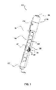
[0010] Figure 1 shows an example device for measuring JVP according to
one
embodiment of the present disclosure.
[0011] Figure 2 is a longitudinal sectional view of the device of Figure
1.
[0012] Figure 2A shows a longitudinal sectional view of a portion of a
device with
a different beam generator and lens configuration according to another
embodiment of
the present disclosure.
[0013] Figure 2B shows a longitudinal sectional view of a device for
measuring
JVP with an internal support rod according to another embodiment.
[0014] Figure 2C shows a longitudinal sectional view of a portion of a
device with
the beam generator and lens configuration of Figure 2A and the internal
support rod of
Figure 2B.
[0015] Figure 3 is a lateral sectional view of the device of Figure 1.
[0016] Figure 3A is a lateral sectional view of the device of Figure 2B.
[0017] Figure 4 shows the device of Figure 1 projecting a light beam.
[0018] Figure 5 shows a testing apparatus for the device of Figure 1.
[0019] Figure 5A shows the device of Figure 1 with an adjusted scale
applied
thereto according to another embodiment of the present disclosure.
- 3 -
CA 03055341 2019-09-04
WO 2018/161159 PCT/CA2018/050262
Detailed Description
[0020] The following describes an example embodiment of a device for
measuring the JVP. The device has an elongated body which is oriented
vertically
when in use, and contains a beam generator that transmits a horizontal beam of
light
perpendicular to the vertical axis from an adjustable position along the body
of the
device. The horizontal beam of light passes through a lens to produce a sheet
of light
oriented along a substantially horizontal plane.
[0021] The vertical height of the horizontal sheet of light may be
adjusted through
adjustment of the height of a moveable portion of the beam generator within
the device
body. As discussed below, in some embodiments, the beam generator comprises a
fixed light source and a moveable reflector, and in other embodiments the beam
generator comprises a moveable light source. Also, in some embodiments the
moveable portion of the beam generator comprises a lens, and in other
embodiments a
lens may be fixed and incorporated into a window on the device body.
[0022] The bottom edge of the device is designed to sit comfortably on
the
sternal angle of a patient inclined at a position approximately 45 (range: 30
-60 ) from
the vertical, with the device oriented vertically. The beam is then directed
towards the
side of the patient's neck (typically right) where the height of the jugular
venous column
can be visualized. The level of the horizontal sheet of light can then be
adjusted to the
height of the venous column by vertically adjusting the height of the moveable
portion of
the beam generator by means of an adjustment mechanism. When the beam is
manually aligned with the height of the jugular venous column, the clinician
simply reads
the height (e.g. in cm) from a readout on the device. Manual vertical
alignment may be
assisted by detent stops or other tactile features. In some embodiments, the
adjustment mechanism provides detent stops every 0.5cm.
[0023] In the illustrated example, a button spirit level is provided at
the top of the
device body to enable the clinician to position the device vertically such
that the beam is
projected in a horizontal plane. In the illustrated example, the height of the
horizontal
sheet of light is adjusted using an adjustment mechanism in the form of a
slider
mechanism, and the readout comprises a scale next to the slider, as described
further
- 4 -
CA 03055341 2019-09-04
WO 2018/161159 PCT/CA2018/050262
below. In other embodiments, the adjustment mechanism may comprise a different
type of slider mechanism, a thumb wheel mechanism (e.g., a rack and pinion), a
twisting or screw-type mechanism (e.g., twisting the base of the body to
adjust the
height of the sheet of light), another suitable mechanism.
[0024] The example device described below is ergonomically shaped and
designed for use with either one or both hands. The device also includes a
second light
source in the form of a broad spectrum light emitting diode (LED) (e.g. a
"white" LED)
integrated into the bottom of the device body to serve as a pen-light for a
variety of
other clinical assessments. In other embodiments the device may also include a
pocket
clip which may incorporate a switch for the LED.
[0025] For simplicity and clarity of illustration, reference numerals may
be
repeated among the figures to indicate corresponding or analogous elements.
Numerous details are set forth to provide an understanding of the examples
described
herein. The examples may be practiced without these details. In other
instances, well-
known methods, procedures, and components are not described in detail to avoid
obscuring the examples described. The description is not to be considered as
limited to
the scope of the examples described herein.
[0026] Figures 1, 2, 3 and 4 show an example device 100 for measuring
JVP.
The device 100 comprises an elongated body 101 that defines a longitudinal
enclosure
102. The body 101 has a level 103 thereon for ensuring that the body 101 is
vertical
when measuring JVP as discussed below. In the illustrated example, the level
103 is on
the top of the body 101. A beam switch 104, secondary light switch 105, and
pocket
clip 106 are also provided on an upper portion of the body 101 in the
illustrated
example. The beam switch 104 is operable to activate a beam generator 110 as
discussed below. The secondary light switch 105 is operable to activate a
secondary
light (e.g. an LED) 130 at a bottom end 109 of the body 101. The switches 104
and 105
may, for example, comprise momentary switches or toggle on/off switches. The
location and configuration of the switches 104 and 105 may differ in other
embodiments.
In some embodiments the beam switch 104 and/or the secondary light switch 105
may,
for example, be incorporated into the button spirit level 103 or the pocket
clip 106, into
the slider 122, or into a lower portion of the body 101.
- 5 -
CA 03055341 2019-09-04
WO 2018/161159 PCT/CA2018/050262
[0027] The beam generator 110 comprises a moveable portion adjustably
mounted within the enclosure 102. The beam generator 110 is configured to
generate a
sheet of light 115 within a plane perpendicular to the longitudinal axis of
device 100, as
described further below, such that when device 100 is vertical, the sheet of
light 115 is
horizontal. The position of the moveable portion of the beam generator 110
within the
enclosure can be adjusted by an adjustment mechanism 120. A window 107 is
provided in the body along the length of the enclosure 102 to allow light to
exit the body
101. A readout such as a scale 108 is provided on the body 101 for indicating
the
position of the beam generator 110 within the enclosure 102. In some
embodiments,
the scale 108 may be printed on the body after calibration of the device, or a
corrected
scale 108A may be adhered to the body 101, to compensate for any errors and
accurately reflect the height of the sheet of light 115 at a distance of 15cm
away from
the device 100, as discussed below with reference to Figures 5 and 5A.
[0028] In the illustrated example, as best seen in Figure 2 the beam
generator
110 comprises a light source in the form of a laser 111 mounted in an upper
portion of
the body 101 above the enclosure 102. The moveable portion of the beam
generator
110 comprises an optical assembly comprising a reflector 113 (e.g. a prism or
mirror)
and a lens 114, which are mounted on a platform 112 slidably mounted within
the
enclosure 102. A battery 119 is provided in the upper portion of the body 101
for
powering the laser 111. In other embodiments, a laser or other light source
could be
mounted in a lower portion of the body 101 below the enclosure 102. In other
embodiments, the lens 114 may be omitted, and the window 107 may comprise a
lens
to spread the light to generate the sheet 115. Other embodiments may have a
beam
generator 110A wherein the moveable portion comprises a laser or other light
source
116 and lens 117 mounted on a slidable platform 118, as shown in Figure 2A. In
other
embodiments, the moveable portion of the beam generator may comprise a light
source
mounted on a slidable platform with the window 107 functioning as a lens.
[0029] In the illustrated example, the adjustment mechanism 120 comprises
a
slider 122 connected to the platform 112 through a slot 121 in the body 101.
The slot
121 is sealed with a flexible elastomer seal 123 configured to keep dust and
contaminants out of the enclosure 102 while allowing movement of the slider
122. The
- 6 -
CA 03055341 2019-09-04
WO 2018/161159 PCT/CA2018/050262
slider 122 has an indicator mark 124 thereon adjacent to the scale 108. The
slot 121
may have detent stops positioned periodically along its length, for example
every 0.5
cm. The scale 108 and adjustment mechanism 120 are configured such that the
indicator mark 124 is adjacent to a marking on the scale 108 indicating the
height of the
sheet of light 115 above the bottom end 109 of the body 101.
[0030] In some embodiments, the platform 112/118 is held in place by
frictional
bearing support from the edges of the body 101 around the slot 121. In other
embodiments, one or more additional elements may provide support for the
platform
112/118. For example, Figures 2B and 3A show an embodiment wherein a ring 112A
attached to platform 112 slides along a supporting rod 112B extending
longitudinally
within the enclosure 102. The platform 112 could be coupled to the supporting
rod
112B in other ways in other embodiments. For example, in some embodiments the
platform 112 has an aperture therethrough sized to receive the supporting rod
112B
such that the platform 112 can slide up and down the rod 112B. In some
embodiments
the platform 112 has a clip formed therein (e.g., a small 'c integrated into
its shape) and
configured to engage the supporting rod 112B. As shown in Figure 2C, the
platform
118 of Figure 2A could also be supported by a supporting rod 112B.
[0031] In operation, a clinician places the bottom 109 of the body 101 on
a
patient's sternal angle, and adjusts the position of the device to ensure the
body 101 is
vertical, as indicated by the level 103. The clinician then adjusts the height
of the sheet
of light 115 until it is aligned with the column of blood in the patient's
vein, and reads the
height from the scale 108.
[0032] Figure 5 shows a testing apparatus 200 for testing the device 100.
Apparatus 200 comprises a base 201, with a laser sight panel 202 comprising a
perpendicular portion 203 and an angled portion 204 having gauge markings 205
thereon extending upwardly from the base 201. A sleeve 206 also extends
upwardly
from the base 201, and holds the device 100 perpendicularly to the base 201
such that
the slider 122 is accessible and the scale 108 is visible. The perpendicular
portion 203
and angled portion 204 are positioned at a predetermined distance to the
sleeve 206
corresponding to a typical horizontal distance from the device to a patient's
neck in a
clinical setting (e.g. about 15cm). A user can test the device 100 by
inserting it on the
- 7 -
CA 03055341 2019-09-04
WO 2018/161159 PCT/CA2018/050262
sleeve 206 and activating the beam generator 110 to generate the sheet of
light 115,
then compare the height of the sheet of light 115 as measured by the gauge
markings
204 with the height as indicated by the scale 108 on the device to ensure the
heights
match.
[0033] In some embodiments, the scale 108 may be printed on the body 101,
or
may be on a sticker or the like applied to the body 101, after calibration of
the device
100 (for example by testing utilizing apparatus 200 or other testing
apparatus) to
account for any height mismatch. In some embodiments, a corrected scale 108A
may
be adhered to the body after testing, as shown in Figure 5A.
[0034] The testing apparatus 200 is also useful for indicating any pitch
or yaw
angular errors in the orientation of the sheet of light 115. If the sheet of
light 115 is not
perpendicular to the device axis and 'pitching up or down, this will result in
a laser
image line that is not parallel to the gauge markings 205 on the angled
portion 204.
Yaw angular errors are illustrated on the perpendicular portion 203 in a
similar manner.
If the sheet of light 115 is tipped (yaw) it will no longer be parallel on the
surface of
perpendicular portion 204 when compared to the markings 205. In some
embodiments,
the testing apparatus 200 also includes a mechanism for automatically
activating the
beam generator 110 when the device 100 is in the sleeve 206 (for example a
physical
feature attached to the sleeve 206 and positioned to contact the beam switch
104).
[0035] In some embodiments, the device 100 may be configured to interact
with,
or be incorporated into, other medical devices. For example, in some
embodiments the
device 100 includes a transducer or other type of sensor that generates a JVP
signal
based on the detected height, and a transmitter configured to send the JVP
signal to
another device such as an ultrasound or dialysis machine. In some embodiments,
the
device 100 transmits the detected height data to an ultrasound or dialysis
machine via
Bluetooth TM or other wireless transmission, or via wired transmission. In
some
embodiments, an ultrasound machine may be used to image the internal jugular
vein
(e.g. in long axis and/or transverse) and precisely determine the top of the
column of
fluid therein, which may be delineated on the patient's skin (either by the
clinician
visually identifying a feature on the skin at that height, or by applying a
marking with, for
example, a pen or marker). The device 100 may then be used as described above
to
- 8 -
CA 03055341 2019-09-04
WO 2018/161159 PCT/CA2018/050262
determine the JVP height. In some embodiments, the device 100 may be
incorporated
into an ultrasound probe such that a single device can be used to image the
internal
jugular vein and determine the JVP height.
[0036] It will be appreciated that numerous specific details are set
forth in order to
provide a thorough understanding of the exemplary embodiments described
herein.
However, it will be understood by those of ordinary skill in the art that the
embodiments
described herein may be practiced without these specific details. In other
instances,
well-known methods, procedures and components have not been described in
detail so
as not to obscure the embodiments described herein. Furthermore, this
description is
not to be considered as limiting the scope of the embodiments described herein
in any
way, but rather as merely describing implementation of the various example
embodiments described herein.
[0037] The description provides many example embodiments of the inventive
subject matter. Although each embodiment represents a single combination of
inventive
elements, the inventive subject matter is considered to include all possible
combinations
of the disclosed elements. Thus if one embodiment comprises elements A, B, and
C,
and a second embodiment comprises elements B and D, then the inventive subject
matter is also considered to include other remaining combinations of A, B, C,
or D, even
if not explicitly disclosed.
[0038] Although the embodiments have been described in detail, it should
be
understood that various changes, substitutions and alterations can be made
herein.
Moreover, the scope of the present application is not intended to be limited
to the
particular embodiments of the process, machine, manufacture, composition of
matter,
means, methods and steps described in the specification.
[0039] The present disclosure may be embodied in other specific forms
without
departing from its spirit or essential characteristics. The described
embodiments are to
be considered in all respects only as illustrative and not restrictive.
- 9 -