Note: Descriptions are shown in the official language in which they were submitted.
CA 03055819 2019-09-06
WO 2018/165390
PCT/US2018/021486
ELECTRICAL IMPEDANCE SENSING DENTAL DRILL SYSTEM CONFIGURED
TO DETECT CANCELLOUS-CORTICAL BONE AND BONE-SOFT TISSUE
BOUNDARIES
CLAIM TO PRIORITY
[0001] The present application claims priority to U. S. Provisional
Patent
Application No. 62/475,724 filed 23 March 2017. The present application also
claims
priority in U. S. Provisional Patent Application No. 62/468,490 filed 8 March
2017. The
entire contents of both provisional applications cited in this paragraph are
incorporated herein
by reference.
GOVERNMENT INTEREST
[0002] This invention was made with government support under grant no. 1
R41
DE024938-01 awarded by the National Institutes of Health. The government has
certain
rights in the invention.
BACKGROUND
[0003] Bone typically has two significantly different forms, cortical
bone and
cancellous bone. Cortical bone is typically found at surfaces of bone
including in joints, as
well as major portions of the shaft of long bones, and other areas that may be
under high
stress. Cortical, or compact, bone lines the outer surfaces of all bone and is
denser and more
structured in nature than cancellous bone. It is organized into tightly-packed
osteons, each
consisting of a Haversian canal (approximately 50 microns in diameter) at the
center
surrounded by concentric rings of matrix. Cancellous bone has a spongy
structure, forming a
mesh network that supports and conveys loads to and from cortical bone
Cancellous bone,
also referred to as trabecular or spongy bone, is found on the inside of long
bones and the jaw
(maxilla and mandible) bones and primarily provides light-weight, more
flexible, structural
support than cortical bone. It is composed of trabeculae ordered into a
honeycomb-like
structure and pores within cancellous bone are often filled with marrow and
blood vessels.
[0004] Physical and biological properties of cortical and cancellous
bone differ
because of the differences in bone structure. In particular, because of the
greatly different
porosity of these bone types, penetration and adhesion of adhesives, the
degree to which a
1
CA 03055819 2019-09-06
WO 2018/165390
PCT/US2018/021486
screw or nail will hold in the bone, and growth rates of bone into porous
implanted objects
differ between cortical and cancellous bone.
[0005] Bone remodels throughout life. Where cortical bone lies over
cancellous
bone, thickness of the cortical bone varies with genetics, childhood
nutritional and exercise
history, age and health of a patient, as well as past medical history
including fractures,
periodontal disease, tooth extractions, muscle usage and weight born on the
bone, and other
factors. Surgeons must expect variation in bone structure between patients. In
the mandible
and maxilla, specifically, clinicians characterize the bone in dental implant
sites according to
the Lekholm and Zarb classification to determine the chance of implant
success. There are
four types, ranging from homogenous cortical bone, to a combination of
cortical and
cancellous bone, to almost entirely low density cancellous bone. Which
classification
depends on where the implant site is located (i.e. in the anterior region vs.
premolar vs.
molar) and patient characteristics.
[0006] Bone, and particularly bones of the head including the mandible
and
maxilla, may be penetrated by nerves and arteries, typically through foramen,
or openings,
through the bone. These nerves and arteries are critical structures as injury
to them has
potential to cause loss of sensation in parts of the mouth or face, or to
cause necrotic
degradation of parts of the bone. For example, the inferior alveolar nerve
(IAN) penetrates
through the mandible.
[0007] When performing surgery, including oral surgery, it is desirable
for the
surgeon to be aware of the type and dimensions of bone and surrounding
structures, including
critical structures, in which he is working. The surgeon may need to modify
surgical
techniques, such as the depth and trajectory of drilling, according to the
dimensions, type, and
thickness of layers of bone the surgeon is working with, to remain in bone to
avoid
penetrating adjacent structures like sinuses such as the maxillary sinus and
nerves such as. the
IAN.
[0008] One common dental surgery procedure is placement of an anchor
implant
to which abutments or dentures may be attached. This procedure requires
drilling bone to
form an initial osteotomy, or cavity within the bone, into which the implant
is placed.
[0009] When performing an initial osteotomy, a surgeon may drill through
a first
layer of cortical bone before reaching cancellous bone, must drill deeply
enough into the
bone to give the implant good bonding surfaces, yet ensure the drill does not
penetrate a thin
distal layer of cortical bone to prevent such surgical complications as
infections or
2
CA 03055819 2019-09-06
WO 2018/165390
PCT/US2018/021486
neurosensory disturbances that result from drilling through the maxilla into a
maxillary sinus
cavity or into a nerve or blood vessel.
SUMMARY
[0010] A dental drill system with electrical-impedance-spectroscopy
sensing
configured to indicate whether a bit of the drill system is in adjacent
cortical or cancellous
bone, is approaching a cancellous/cortical bone interface, or is approaching a
bone/soft tissue
interface includes a dental drill having in its handset a cannulated bit, the
cannulated bit
having an insulating coating covering the entire surface except for a portion
of distal surface
of the cutting edge; a cannula bearing electrically coupled to an uninsulated
interior of a
cannula of the cannulated bit, an electrical impedance spectroscopy sensing
(EIS)
measurement and calculation unit configured to measure impedance between the
cannula
bearing and a ground plate or return electrode, and a processing system
configured to
distinguish changes in electrical properties indicating an approaching
cancellous/cortical
bone interface or a change as the bit of the drill system approaches an
interface between
cancellous and cortical bone, or a bone-soft tissue interface.
[0011] A method of detecting approach of a bit to cortical bone or soft
tissue
while drilling bone with the bit includes providing an insulating coating
extending from near
a cutting end of the bit to a handset end of the bit, contacting the bit with
a cannular bearing,
driving a voltage-limited current between the bit and a ground plate at at
least one alternating
current frequency; measuring voltage and phase between bit and ground plate;
determining
impedance from measured voltage and phase; and generating an alarm when the
impedance
changes, indicating an interface between bone and soft tissue or between
cancellous and
cortical bone.
BRIEF DESCRIPTION OF THE DRAWINGS
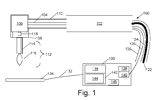
[0012] Fig. 1 is a block diagram of the drilling system with electrical
impedance
spectroscopic sensing.
[0013] Fig. 2 is a sketch of a drill bit of the current drilling system.
[0014] Fig. 3 is a photograph showing an embodiment of a drill having a
bit with
cannular bearing attached.
[0015] Fig. 4 is an illustration of electrical resistance and reactance
of cancellous
and cortical bone samples measured with a prototype integrated on a Nobel
Biocare Drill
with a 2mm twist bit.
[0016] Fig. 5 is a photograph of a DLC-coated drill bit having a bare
cutting end.
3
CA 03055819 2019-09-06
WO 2018/165390
PCT/US2018/021486
[0017] Fig. 6 illustrates contrast of normalized mean resistance and
reactance of
cancellous and cortical bone measured with a prototype integrated on a
standard Nobel
Biocare Drill with a drill bit in ex vivo bone.
[0018] Fig. 7 illustrates contrast of normalized mean resistance and
reactance of
cancellous and cortical bone measured with a prototype integrated on a
standard Nobel
Biocare Drill with a drill bit in fresh, in situ, bone.
[0019] Fig. 8 is a flowchart of a method of detecting approach of a
drill bit to
cortical bone during surgical procedures.
DETAILED DESCRIPTION OF THE EMBODIMENTS
[0020] The vastly different cellular constituents of cortical and
cancellous bone
provide a spectrum of electrical charge carrying and charge storage
capabilities, which are
represented by electrical conductivity (G) and permittivity (6), respectively
(G and c are
inversely related to resistance and reactance). When recording these
electrical properties over
a broad range of frequencies (100's of Hz to 10's of MHz), as is done in
Electrical Impedance
Spectroscopy (EIS), cortical and cancellous bone have been reported to differ
significantly.
Studies have investigated electrical impedance measurements in pedicle screw
insertion into
vertebrae and have shown that electrical property differences between
cancellous and cortical
bone can be used to guide surgeons through vertebral bone.
[0021] We describe herein an EIS device integrated with a drill
configured for
drilling holes in bone, such as may be required for a variety of surgical
procedures in
dentistry and some non-dental surgeries. The drill is particularly configured
for measuring
bioimpedance spectra in vivo during the initial osteotomy of dental implant
procedures. The
drill is particularly adapted for measuring the electrical impedance spectra
of bony structures
in vivo as the drill is advanced into the structure. This EIS drill provides
real-time feedback
to the clinician, as either an auditory or visual signal, allowing the
clinician to stop drilling
before perforation of the cortical layer occurs (enabling immediate clinical
intervention if
necessary). In a particular embodiment, the drill is a dental drill.
[0022] The EIS-sensing drill system 100 is illustrated in Fig. 1. A
dental drill
handset 102 contains a high-speed motor and drive shaft 104 leading to a right-
angle bevel
gear unit 106, the bevel gear unit and a housing 110 of the driveshaft 104
being insulated
with an insulating coating 108. Coupled to the bevel gear unit is a drilling
bit 112 having an
insulated portion 114 and a bare, cutting, portion 116. Bare cutting portion
116 is a portion
of a ball-like burr in some embodiments and a tip of a twist-drill bit in
other embodiments;
4
CA 03055819 2019-09-06
WO 2018/165390
PCT/US2018/021486
the insulated portion extends from the cutting portion to a handset end of the
bit that is
mechanically coupled to into a dental drill handset. Within bevel gear unit
106 is a cannular
bearing 118 electrically coupled to bit 112. Handset 102 has an umbilical
tubular housing
120 holding a tube 122 for irrigation fluid, an electrical drive wire for the
motor of handset
102, and an electrical wire adapted for coupling the cannular bearing 118 to
an electrical
impedance spectroscopy (EIS) measurement and calculation unit 130, EIS
measurement and
calculation unit 130 also couples through another wire 132 to a second
electrode plate 134.
Within EIS measurement and calculation unit 130 are an EIS stimulus unit 136
capable of
operation at 100, 1000, 10000, and 100000 Hz under direction of processor 138
and an EIS
impedance measurement unit 140. In alternative embodiments, EIS impedance
measurement
and calculation unit 130 is capable of operation at two or more frequencies in
the 100 Hz to 1
MHz range. Processor 138 has a memory 142 with EIS measurement firmware and
classifier
firmware 144, the classifier firmware adapted to use EIS measurements to
determine whether
bit 112 is drilling in cancellous or cortical bone and to announce which bone
type bit 112 is in
using indicator 146.
[0023] A twist-drill embodiment is illustrated in more detail in Fig. 2.
A twist-
drill bit 160 has a bare or uninsulated end 162 with cutting edges that may
contact and drill
holes in bone. Bit 160 also has an electrically insulated portion 164 bearing
a coating of
diamond-like carbon (DLC), a coating that is both very hard such that it wears
little while
holes are being drilled in bone, and electrically of high resistivity, the
coating of DLC
extends throughout the remainder of the exterior of bit 160 to the drill end
of bit 160,
including portions that engage bevel gear of drill head 170, and including
portions over flutes
171. Bit 160 also has an uninsulated axial hole 172 extending from drill end
of bit into, but
not all the way through, the bit.
[0024] Within the axial hole 172 and in electrical contact with the
uninsulated
surface of bit 160 in that hole is an uninsulated end portion 174 of cannular
bearing 166.
Cannular bearing 166 extends from bit's 160 end through insulation 176 to
electronics EIS
measurement and calculation unit 130 (Fig. 1). Drive shaft 178 and bevel gear
180 rotate to
drive bevel gear 168 of drill head 170 to rotate bit 160 to drill the holes
into bone.
[0025] Fig. 1 and 2 are schematics, Fig. 3 is a photograph showing an
embodiment of an experimental drill 202 having a bit 204 with cannular bearing
206 and
insulated lead 208 attached, and Fig. 4 is a photograph showing a pair of
uninstalled cannular
bearings.210. In an embodiment, cannular bearings 210, 206, are formed of
stainless steel.
CA 03055819 2019-09-06
WO 2018/165390
PCT/US2018/021486
[0026] In various embodiments the uninsulated end portion 174 of bit
160, or
uninsulated ball portion of bit 116, is from one to three millimeters in
length.
Operation of the EIS Drill System:
[0027] The EIS measurement and calculation unit, drill, and bit with
cannular
bearing together form the EIS drilling system. Positioning the bearing 206
within the drill
bit's cannula does not decrease the surgical working space and still allows
for irrigation
through the intra-cannular channel or around the exterior surface of the drill
bit. The cannular
bearing connects to a lead that interfaces with the impedance analyzer.
Similarly, the return
electrode 134 (fig. 1) connects to another lead that interfaces with the
impedance analyzer. A
voltage-limited, alternating current (AC) current is applied between the two
electrode
elements at several frequencies and the voltage and phase induced between them
is recorded.
From these measurements, impedance is calculated as the ratio of voltage to
current.
[0028] Impedance (Z) is calculated as the ratio of the measured voltage
to the
injected current; we regard the impedance as a complex quantity, consisting of
a real resistive
component (R) and an imaginary reactive component (X), according to the
equation, Z = R +
jX. The electronics box computes an R and X measurement at each frequency
being tested.
From those we compute impedance, conductivity, resistivity, and the like.
[0029] We have shown in previous experiments in ex vivo and in situ pig
femurs
that cortical bone has a higher resistivity and impedance than cancellous
bone. The ratio of
cortical-to-cancellous resistivity ranged from 1.28 ¨ 1.48 in ex vivo bone and
from 2.82 ¨
2.94 in fresh in situ bone. As a result, we expect that, as the drill bit
moves through
cancellous bone towards a cortical interface we will see an increase in
impedance/resistivity
as we approach that interface.
[0030] In an embodiment, the EIS measurement and calculation unit is
configured
to provide a visual and / or aural alarm when the drill bit approaches
cortical bone.
[0031] Clinical use of this device involves using the drill to create
the initial
osteotomy (hole in the bone) marked for implant insertion. Electrical
properties, specifically
the resistance and reactance of the bone, are recorded over a single or
multiple frequencies as
the drill is advanced into the bone. These measurements will be input into a
real-time
classification unit used to sense an approaching tissue transition (i.e. the
cancellous-cortical
interface). A visual or auditory signal that increases in repetition rate,
based on the changing
impedance, will be used as clinician feedback.
6
CA 03055819 2019-09-06
WO 2018/165390
PCT/US2018/021486
[0032] We have collected a significant dataset of ex vivo and in situ
electrical
properties of cortical and cancellous bone and have shown significant
impedance contrast
between the two bone types.
[0033] In the ex vivo experiment, we positioned standard cannulated
drill bits
three millimeters deep into 10 samples each of cortical and cancellous bone
freshly harvested
from swine and recorded impedance from 100 Hz-1 MHz at 41 frequencies. We
demonstrated that there are significant R and X differences (p < 0.05) between
the two bone
types with contrasts in resistance of 41%, 37%, 29%, and 32% at 0.1kHz, lkHz,
10kHz, and
100 kHz, respectively. These trends, recorded with our prototype, are similar
to those
reported previously for cancellous and cortical bone.
[0034] In the in situ experiment, we used a custom DLC-coated drill bit
to record
impedances from 40 samples each of cortical and cancellous bone in the femurs
of pigs 30
minutes after euthanasia. We demonstrated that there are significant R and X
differences (p <
0.001) between the tissue types, with a maximum resistance contrast of ¨300%
at 100 kHz
and a maximum reactance contrast of ¨250% at 1 kHz.
[0035] The electrical impedance sensing is responsive not just to the
tissue type
the tip is in, but to tissue types near the tip. The system can therefore
watch for impedance
changes as the drill penetrates bone and generate an alarm when the impedance
changes
indicate the tip is approaching a cancellous-cortical bone interface, or when
the tip is
approaching a bone-soft tissue interface; bone-soft tissue interfaces include
interfaces
between bone and blood vessels, nerves, sinus lining, muscles, and other non-
ossified tissues.
Features
[0036] Features of this dental drill system with electrical-impedance-
spectroscopy
sensing include:
1) a coated dental drill bit as the sensing or driving electrode,
2) a Diamond-Like-Carbon (DLC) coating to insulate all but the distal few
millimeters of the
drill bit,
3) an intra-cannular bearing to interface the drill bit with the impedance-
sensing module,
4) collecting impedance measurements at multiple frequencies for this
particular surgical drill
application, and
5) extending the interface detection feature beyond pure threshold detection.
[0037] In addition, by interfacing our system to the dental implant
drill through
the cannular space, we do not need to augment the drill in any way nor do we
decrease the
7
CA 03055819 2019-09-06
WO 2018/165390
PCT/US2018/021486
working volume available to the surgeon. Irrigation is still possible despite
the presence of
the bearing, allowing for surgeons to continue using cannulated drill bits as
they were
intended.
[0038] DLC coatings are designed to have extremely high hardness (4000-
9000
HV), high resistivity (up to 106 a-cm), and are bio-compatible. By applying
this insulating
coating to the majority of the drill bit and leaving only the distal 1-3 mm
exposed for sensing,
we provide more robust and repeatable impedance measurements that are not
dependent on
drill bit depth into the material. While some prior art includes provisions
for an insulating
material applied to the drilling device, they do not specify the type of
insulating material, nor
do they leave an area on the distal end exposed for sensing.
[0039] Collecting impedance measurements at multiple frequencies,
instead of a
single frequency, has the potential for better classification between
cancellous and cortical
bone. The increased number of measurements will allow us to explore additional
features that
can be used to contrast the two bone types. Most prior art is based on
threshold detection at a
single frequency to alert clinicians of an approaching tissue interface. We
use multiple
features and algorithms to find an optimal combination to use for interface
detection.
[0040] In an embodiment, the method of detecting approach of a bit to
cortical
bone while drilling bone with the bit includes providing 302 (Fig. 8) an
insulating coating
extending from near a cutting end of the bit to a handset end of the bit, and
contacting 304 the
bit with a cannular bearing. Then the EIS measurement and calculation unit
drives 306 a
voltage-limited current between the bit and a ground plate at at least one
alternating current
frequency and measures voltage and phase, then determines 308 impedance from
measurements of voltage and phase between bit and ground plate; and generates
310 an alarm
when the impedance changes indicating approach to cancellous-cortical bone or
bone-soft
tissue interfaces.
[0041] In an alternative embodiment, a contact portion of the handset
end of the
bit is bare of the DLC insulating coating, and the handset is modified to
provide electrical
contact from the EIS measurement and calculation device to that bare portion
of the handset
end of the bit while isolating the remainder of the drill handset from the EIS
measurement
and calculation device.
Combinations of Features
[0042] A dental drill system designated A with electrical-impedance
sensing
(EIS) configured to indicate whether a bit of the drill system is approaching
a cancellous-
cortical bone interface or a bone-soft tissue interface includes a dental
drill having in its
8
CA 03055819 2019-09-06
WO 2018/165390
PCT/US2018/021486
handset a cannulated bit, the cannulated bit having an insulating coating
extending from near
a cutting end of the bit to a handset end of the bit a cannula bearing
electrically coupled to an
uninsulated interior of a cannula of the cannulated bit, an EIS measurement
and calculation
unit configured to measure impedance between the cannula bearing and a ground
plate, and a
processing system configured to distinguish when the bit of the drill system
approaching a
cancellous-cortical bone or bone-soft tissue interface..
[0043] A dental drill system designated AA including the dental drill
system
designated A wherein the electrically insulated portion of the drilling bit is
insulated with a
diamond-like carbon (DLC) coating.
[0044] A dental drill system designated AB including the dental drill
system
designated A or AA wherein EIS measurement and calculation unit provides a
voltage-
limited current at each of a plurality of frequencies and measures a resulting
voltage and
phase.
[0045] A dental drill system designated AC including the dental drill
system
designated A, AA, or AB wherein the EIS measurement and calculation unit is
configured to
provide a visual and / or aural alarm when the drill bit approaches cortical
bone.
[0046] A dental drill system designated AD including the dental drill
system
designated A, AA, AB, or AC wherein the EIS measurement and calculation unit
is
configured to measure impedance at at least two frequencies in the range 100
to 100000
Hertz.
[0047] A method designated B of detecting approach of a bit to cortical
bone, or
approach of a bit to bone-soft tissue interface, while drilling bone with the
bit includes
providing an insulating coating extending from near a cutting end of the bit
to a handset end
of the bit, contacting the bit with a cannular bearing; driving a voltage-
limited current
between the bit and a ground plate at least one alternating current frequency;
measuring
voltage and phase between bit and ground plate; and determining impedance from
measured
voltage and phase; and generating an alarm when the impedance changes
indicating approach
to cancellous-cortical bone interfaces or bone-soft tissue interfaces.
[0048] A method designated BA including the method designated B wherein
the
voltage-limited current is driven at multiple frequencies between 100 and
100000 Hertz.
Conclusion
[0049] Changes may be made in the above methods and systems without
departing from the scope hereof. It should thus be noted that the matter
contained in the
9
CA 03055819 2019-09-06
WO 2018/165390
PCT/US2018/021486
above description or shown in the accompanying drawings should be interpreted
as
illustrative and not in a limiting sense. The following claims are intended to
cover all generic
and specific features described herein, as well as all statements of the scope
of the present
method and system, which, as a matter of language, might be said to fall
therebetween.