Note: Descriptions are shown in the official language in which they were submitted.
CA 03057211 2019-08-28
WO 2018/160673
PCT/US2018/020187
PM21 PARTICLES TO IMPROVE BONE MARROW HOMING OF NK CELLS
This application claims the benefit of U.S. Provisional Application No.
62/464,747, filed on
February 28, 2017, and which is incorporated herein by reference in its
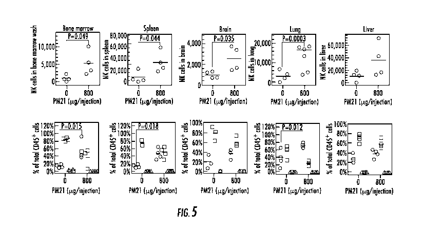
entirety.
I. BACKGROUND
Adoptive natural killer (NK) cell therapy is a promising novel intervention
for oncology including for bone
marrow malignancies. Therefore, the efficiency for trafficking of the NK cells
to be used adoptively is of high
importance. What are needed are methods that can efficiently traffic NK cells
to the bone marrow.
SUMMARY
1. Disclosed are methods and compositions related to trafficking NK cells to
the bone
marrow comprising contacting NK cells with PM21 particles and/or FC21 feeder
cells. In one
aspect, the methods can further comprise stimulating the NK cells with IL-2,
IL-12, IL-18,
and/or IL-18.
2. Also disclosed are methods of treating a bone marrow malignancy or bone
marrow
born malignancy and/or treating a viral infection (such as a bone marrow
associated viral
infection including a bone marrow tropic viral infection or viral infection
that adversely effects
the bone marrow) comprising contacting NK cells with PM21 particles and/or
FC21 feeder cells
and adoptively transferring NK cells to the subject. In some aspects, the
contact of the PM21
particles and/or FC21 feeder cells with the NK cells can occur prior to
transfer of the NK cells to
a patient. In another aspect, the contact of the PM21 particles and/or FC21
feeder cells with the
NK cells can occur in the patient.
III. BRIEF DESCRIPTION OF THE DRAWINGS
3. The accompanying drawings, which are incorporated in and constitute a
part of this
specification, illustrate several embodiments and together with the
description illustrate the
disclosed compositions and methods.
4. Figure 1 shows that PM-21 particles expand cytotoxic NK cells efficiently
and
selectively. Peripheral blood mononuclear cells (PBMCs) were isolated from
leukocyte source
and seeded at 0.1 x 10⁶ NK cells/mL in SCGM supplemented with 10% FBS, 2 mM
Glutamax
and 50 U/mL IL-2. PBMCs were stimulated with PM15 (o, black) or PM21 (0, blue)
particles at
200 pg/mL for 27 days, the cell content was tested every 2-3 days and shown
are relative fold of
NK cell expansion (A) and the percentage of suspension cells (B). PM21-
particles (825 188
fold, N=13, 4 donors) (blue) are more efficient for NK cell expansion compared
to PM15-
particles (425 71, N=35, 9 donors) (black) based on cumulative analysis of
day 14 data for NK
cell expansion (C). PBMCs isolated from three AML patients in remission were
cultured for 14
¨ 1 ¨
CA 03057211 2019-08-28
WO 2018/160673 PCT/US2018/020187
days with PM21-particles (200 ug/mL), seeded at 0.5 x 10⁶ NK cells/mL in SCGM
with 10%
FBS, 2 mM Glutamax, 50 U/mL IL-2. Shown are fold of NK cell expansion from the
primary
PBMCs (D) and lymphocyte content (E) (CD56+CD3- NK cells (.,red), CD56-CD3+ T
cells (N,
blue) and CD56+CD3+ NKT cells (1, black)). PBMCs from patient F021 were
cultured for 16
days as previously above and autologous cytotoxicity toward AML tumors from
the same
patient was analyzed (F). Expanded PM21-NK cells labeled with TFL4, and were
co-incubated
(2 hours) at indicated E:T ratios with AML cells from the same patient during
active disease,
and analyzed by flow cytometry. The amount of spontaneous dead target cells
was determined
using a "Target Alone" control. Each data point was determined in duplicate.
5. Figure 2 shows that Pre-activation of unselected PBMCs with PM21-particles
induces in vivo NK cell expansion. NSG mice were injected i.p. with 2 x 10⁶
cells of either un-
activated PBMCs (A and B) or PBMCs pre-activated ex vivo with PM21-particles
and 100 U/mL
IL-2 for two days (PM21-PBMCs) (C and D). Mice in all groups received 1,000 U
of IL-2, i.p.,
thrice weekly. Groups of mice were also injected with 400 ug of PM21-
particles, i.p., twice
weekly (B and D). Peripheral blood was drawn by sequential cheek bleeds and
analyzed by flow
cytometry for hCD45+ human lymphocytes twice weekly starting on day 6. NK, T
and B cell
amounts were determined based on staining for hCD3, hCD56, hCD19. The left
plots in each
experimental group shows concentration of hNK cells per IA of PB. The right
plots shows the
percentage of hNK cells (0, red) and T cells (7, black) as fraction of total
hCD45+ cells.
6. Figure 3 shows that Proliferation analysis evidences in vivo NK cell
expansion from
PM21-PBMCs. PBMCs freshly thawed or pre-activated with PM21-particles and 100
U/mL IL-
2 for two days (PM21-PBMCs) were labeled with Cell Trace (CT) Violet. 2 x10⁶
of un-activated
PBMCs (A and B) or PM21-PBMCs (C and D) were injected i.p. to NSG mice. Mice
in all
groups received 1,000 U IL-2, i.p., thrice weekly. Two of the groups of mice
were also injected
with 400 ug of PM21-particles, i.p., twice weekly (B and D). Two mice from
each group were
euthanized on day 6 and the peritoneal wash was analyzed by flow cytometry for
CT Violet
fluorescence of hCD45+, hCD3-, hCD56+ NK cells. Histograms of the CT Violet
fluorescence
was analyzed through curve fitting using the Proliferation analysis suite
within FlowLogic.
7. Figure 4 shows that In vivo application of PM21 allows increase of
NK cells in
peripheral blood. PBMCs were pre-activated ex vivo with PM21-particles and 100
U/mL IL-2
for 2 days. PM21-PBMCs in the amount containing 0.2 x 10⁶ of viable NK cells
were injected
i.p. to NSG mice. Mice in all groups received 1,000 U of IL-2, i.p., thrice
weekly. Mice were
also injected with 0 (A), 400 (B), 800 (C), 1,600 ug (D) of PM21-particles,
i.p., twice weekly.
Peripheral blood was analyzed by flow cytometry for hCD45+ lymphocytes twice
weekly
¨ 2 ¨
CA 03057211 2019-08-28
WO 2018/160673 PCT/US2018/020187
starting on day 5 and hNK, hT and hB cell amounts were determined based on
staining for
hCD3, hCD56, hCD19. The left plots in each experimental group shows
concentration of hNK
cells per [IL of PB. The right plots shows the percentage of hNK cells (0,
red) and T cells (7,
black) as fraction of total hCD45+ cells. Analysis of PB samples from day 12
after initial
injection i.p. of PM21-PBMCs shows a dose dependent increase of PB hNK cells
with respect to
in vivo PM21-particle dose (E left) while no significant dose dependent
increase in total CD3+ T
cells was observed (E right).
8. Figure 5 shows that In vivo expanded NK cells biodistribute to key
physiological
sites and the NK cell biodistribution is increased with in vivo application of
PM21-particles. NK
cells (0.2 x 10⁶ cells) as part of PM21-PBMCs, pre-activated ex vivo with PM21-
particles and
100 U/mL IL-2 for 2 days, were injected i.p. to NSG mice. Mice in all groups
received 1,000 U
of IL-2, i.p., thrice weekly. Mice were also injected with 0 or 800 lig of
PM21-particles, i.p.,
twice weekly. Mice were sacrificed 16 days after initial injection i.p. of
PM21-PBMCs. On day
of euthanasia bone marrow (femur), spleen, lung, brain and liver were
collected, organs were
perfused while femur was washed to recover cells. Cells were stained with
antibodies against
hCD3, hCD45, hCD56, hCD19 for flow cytometry analysis. Data for bone marrow,
spleen,
brain, lung and liver (left to right) are shown with the amount of
hCD45+11CD56+11CD3- NK
cells (top plots for each organ) and percentages for hCD45+11CD56+11CD3- NK
cells (0, red),
hCD45+11CD3+ T cells (o, blue) and hCD45+, hCD56-hCD3- other lymphocytes (A,
black) are
shown (bottom plots for each organ). The thick bar for each represents the
mean.
9. Figure 6 shows that In vivo NK cells expansions from different donor
sources are
consistent. The consistency of PM21-particle stimulated in vivo NK cell
expansion was tested
using three different PBMCs obtained from healthy donors. The PBMCs were pre-
activated ex
vivo for 2 days with PM21-particles and 100 U/mL IL-2 for 2 days (PM21-PBMCs)
and were
injected i.p. to NSG mice. Mice in all groups received 1,000 U of IL-2, i.p.,
thrice weekly.
Peripheral blood was analyzed by flow cytometry for hCD45+ lymphocytes twice
weekly
starting on day 5 and hNK, hT and hB cell amounts were determined based on
staining for
hCD3, hCD56, hCD19. Both the concentration of hNK cells in blood 12 days after
i.p. PBMC
injection (A) and the amount of NK cells collected in a wash of the abdominal
cavity 14 days
after i.p. PBMC injection (C) were similar between the different groups
injected with different
NK cell sources (p=0.84 for PB andp=0.69). The corresponding cell content of
hNK cells (o,
red), hT cells (o, blue) and other hCD45+ cells (A, black) were also
consistent between the
groups injected with different PBMC sources in the peripheral blood (B) and in
the abdomen
(D). The thick bar for each represents the mean.
¨ 3 ¨
CA 03057211 2019-08-28
WO 2018/160673 PCT/US2018/020187
10. Figure 7 shows HECA-452 staining of NK cells treated with soluble
cytokines at 0,
1, 7, and 10 days of stimulation.
11. Figure 8 shows HECA-452 staining of NK cells cultured with K562 feeder
cells
following 10, 12, and 14 days of stimulation.
12. Figure 9 shows a HECA-452 staining comparison of NK cells stimulated under
various conditions following 10 days of stimulation.
13. Figure 10 shows HECA-452 staining on NK cells cultured under various
conditions
following 10 days of stimulation.
14. Figure 11 shows HECA-452 staining of NK cells after three days of rest
following 10
days of stimulation.
15. Figure 12 shows HECA-452 staining of pre- and post-freeze thaw following
14 days
expansion of PM21 particles.
16. Figure 13 shows that STAT3 is involved in IL-21 mediated modulation of
FUT7
gene expression in human NK cells. (A) ChIP-seq was performed on IL-21
stimulated naive and
expanded human NK cells with antibodies against STAT3. The arrow indicates
transcription
directionality. Scales are constant for the gene and islands; (B) RNA-seq
reveals the differential
regulation of FUT7 gene expression in response to IL-21 stimulation; (C) IL-21
enhances
STAT3 binding to FUT7 gene in expanded NK cells, and (D) IL-21 upregulates
FUT7 gene
expression in expanded NK cells.
IV. DETAILED DESCRIPTION
17. Before the present compounds, compositions, articles, devices, and/or
methods are
disclosed and described, it is to be understood that they are not limited to
specific synthetic
methods or specific recombinant biotechnology methods unless otherwise
specified, or to
particular reagents unless otherwise specified, as such may, of course, vary.
It is also to be
understood that the terminology used herein is for the purpose of describing
particular
embodiments only and is not intended to be limiting.
A. Definitions
18. As used in the specification and the appended claims, the singular forms
"a," "an"
and "the" include plural referents unless the context clearly dictates
otherwise. Thus, for
example, reference to "a pharmaceutical carrier" includes mixtures of two or
more such carriers,
and the like.
19. Ranges can be expressed herein as from "about" one particular value,
and/or to
"about" another particular value. When such a range is expressed, another
embodiment includes
from the one particular value and/or to the other particular value. Similarly,
when values are
¨ 4 ¨
CA 03057211 2019-08-28
WO 2018/160673 PCT/US2018/020187
expressed as approximations, by use of the antecedent "about," it will be
understood that the
particular value forms another embodiment. It will be further understood that
the endpoints of
each of the ranges are significant both in relation to the other endpoint, and
independently of the
other endpoint. It is also understood that there are a number of values
disclosed herein, and that
each value is also herein disclosed as "about" that particular value in
addition to the value itself
For example, if the value "10" is disclosed, then "about 10" is also
disclosed. It is also
understood that when a value is disclosed that "less than or equal to" the
value, "greater than or
equal to the value" and possible ranges between values are also disclosed, as
appropriately
understood by the skilled artisan. For example, if the value "10" is disclosed
the "less than or
equal to 10"as well as "greater than or equal to 10" is also disclosed. It is
also understood that
the throughout the application, data is provided in a number of different
formats, and that this
data, represents endpoints and starting points, and ranges for any combination
of the data points.
For example, if a particular data point "10" and a particular data point 15
are disclosed, it is
understood that greater than, greater than or equal to, less than, less than
or equal to, and equal to
10 and 15 are considered disclosed as well as between 10 and 15. It is also
understood that each
unit between two particular units are also disclosed. For example, if 10 and
15 are disclosed,
then 11, 12, 13, and 14 are also disclosed.
20. In this specification and in the claims which follow, reference will be
made to a
number of terms which shall be defined to have the following meanings:
21. "Optional" or "optionally" means that the subsequently described event or
circumstance may or may not occur, and that the description includes instances
where said event
or circumstance occurs and instances where it does not.
B. Methods of using the compositions
22. Adoptive natural killer (NK) cell therapy is a promising novel
intervention for
oncology including for bone marrow malignancies and bone marrow born
malignancies; and
including veterinary applications for such. In another aspect, adoptive NK
cells can be
therapeutically used for treatment of marrow resident viruses, including for
example.
parvoviruses that cause aplastic anemia. Therefore, the efficiency for
trafficking of the NK cells
to be used adoptively is of high importance. In particular, how to drive
transferred cells into the
bone marrow where they can be effective as a treatment is of critical
importance.
23. Among the determinants of marrow homing are ligands for selectin binding.
For
binding L-Selectin, a key determinant is the fucosylation state of sialyl
Lewis x (sLex)
carbohydrate chain attached to P-selectin glycoprotein ligand-1 (PSGL-1). In
one aspect, it is
disclosed herein contemplated that stimulation of NK cells with PM21 particles
and/or FC21
¨ 5 ¨
CA 03057211 2019-08-28
WO 2018/160673 PCT/US2018/020187
feeder cells prepared from K562 cells transformed to express engineered
membrane bound form
of IL-21 and/or 41bbl (K562.mb21.41bbl) induces efficient specific expansion
of NK cells and
induces full fucosylation of sLex.
24. Accordingly in one aspect, disclosed herein are methods related to
trafficking NK
cells to the bone marrow and methods of treating a bone marrow malignancy or
bone marrow
born malignancy comprising contacting NK cells with PM21 particles and/or FC21
feeder cells
and adoptively transferring the NK cells to a patient in need thereof In one
aspect, the methods
can further comprise stimulating the NK cells with IL-2, IL-12, IL-15 IL-18,
IL-21 either ex
vivo or in vivo (in the patient).
25. In some aspects, the contact of the PM21 particles and/or FC21 feeder
cells with the
NK cells can occur prior to transfer of the NK cells to a patient. In another
aspect, the contact of
the PM21 particles and/or FC21 feeder cells with the NK cells can occur in the
patient.
26. In one aspect, it is understood and herein contemplated that the efficacy
of NK cell
immunotherapy is dependent on the dose of NK cells administered to the patient
or reached after
infusion through in vivo expansion. Currently available techniques are limited
by their inability
to achieve the level of NK cell expansion required to achieve a therapeutic
effect in a patient.
The lack of a simpler clinical expansion protocol is a major barrier to the
progress and wide
dissemination of NK cell-based immunotherapy. Current ex vivo expansion
protocols use a
combination of high dose cytokines with activating ligands expressed on
leukemia-derived
feeder/stimulator cell lines, posing a significant disadvantage for transfer
to clinical settings in
most centers and are not amenable for direct in vivo expansion. The use of
particle technology,
including exosomes, described herein eliminates the need for stimulator cells,
thus simplifying
the methodology and allowing ex vivo expansion for adoptive therapy or applied
in vivo for
selective in vivo expansion. Accordingly, and in one aspect, disclosed herein
are methods for
treating bone marrow malignancies and bone marrow born malignancies through
the adoptive
transfer of NK cells and/or methods trafficking NK cells to the bone marrow
comprising
contacting NK cells with one or more vesicles comprising an NK cell effector
agent. In one
aspect, also disclosed are methods of treating bone marrow malignancies, bone
marrow born
malignancies, and or viral infections (including viruses with a bone marrow
tropism) further
comprising preactivating or activating in vivo NK cells by contacting at least
one NK cell with
at least one or more stimulatory cytokines. Thus, in one aspect, NK cells
expanded ex vivo by
PM21 particles and/or FC21 feeder cells, or with direct in vivo stimulation
with PM21 or FC21
feeder cells can be used to treat marrow resident viral conditions or
syndromes (e.g. parvovirus)
that cause aplastic anemia.
- 6 -
CA 03057211 2019-08-28
WO 2018/160673 PCT/US2018/020187
27. The disclosed methods accomplish preactivation or activation of NK cells
by
contacting at least on NK cell with at least one or more stimulatory cytokines
(for example IL-2,
IL-12, IL-15, IL-21 and/or IL-18). Thus, in one aspect, disclosed herein are
methods of treating
bone marrow malignancies and bone marrow born malignancies through the
adoptive transfer of
NK cells and/or methods trafficking NK cells to the bone marrow comprising
preactivating NK
cells by contacting one or more NK cells with one or more stimulatory
cytokines is selected
from the group comprising IL-2, IL-12, IL-21, IL-15, and/or IL-18, or any
combination thereof,
including contacting one or more NK cells with 2 or 3 stimulatory cytokines.
For example,
specifically disclosed herein are methods wherein the preactivation or
activation step comprises
contact NK cells with IL-2; IL-12; I1-15, IL-18, IL-12 and IL-15; IL-12 and IL-
18; IL-15 and IL-
18; or IL-12, IL-15, and IL-18. In one aspect, the disclosed methods of
treating bone marrow
malignancies and bone marrow born malignancies through the adoptive transfer
of NK cells
and/or methods trafficking NK cells to the bone marrow can further comprise
contacting the NK
cell with one or more cytokines selected from the group consisting of 4-1BBL,
IL-2, IL-21,
MICA/B, ULBP2, ICAM-1, 2B4, BCM1/SLAMF2, CD155, CD112, CCR7, DAP12, Notch
ligands and/or DAP10 in soluble form or in the form PM21 particles or FC21
feeder cells.
28. It is understood and herein contemplated that the duration of
preactivation or
activation (i.e., the duration of contact between the NK cells and the
stimulatory cytokines (e.g.,
IL-2, IL-12, IL-15, IL-21 and/or IL-18) in soluble form or in the form PM21
particles or FC21
feeder cells can be for any length of time necessary to achieve the desired
preactivation or
activation of NK cells. For example, the contact can be as little as 1 minute
or as much as 7
days (for example, culturing the NK cells in the presence of IL-2, IL-12, IL-
15, IL-21 and/or IL-
18 for 7 days). In one aspect, disclosed herein are methods of treating bone
marrow
malignancies and bone marrow born malignancies through the adoptive transfer
of NK cells
and/or methods trafficking NK cells to the bone marrow comprising
preactivating or activating
NK cells by contacting one or more NK cells with IL-2, IL-12, IL-15, and /or
IL-18 for 5, 6, 7,
8, 9, 10, 11, 12, 13, 14, 15, 16, 17, 18, 19, 20, 21, 22, 23, 24, 36, or 48
hours. It is understood
and herein contemplated that the half-life of a cytokine in culture may be
less than the desired
contact time. Accordingly, disclosed herein are methods wherein one or more NK
cells are
contacted with IL-2, IL-12, IL-15, and/or IL-18 every 1, 2, 3, 4, 5, 6, 7, 8,
9, 10, 11, or 12 hours
within a contact period (for example, every 1, 2, 3, 4, 5, 6, 7, 8, 9, 10, 11,
or 12 in a 24 hour
contact period).
29. Through the use of plasma membrane (PM) particles, exosomes (EX), or
feeder cells
(FC) comprising one or more NK cell effector agents (i.e., stimulatory
peptides, cytokines,
- 7 -
CA 03057211 2019-08-28
WO 2018/160673 PCT/US2018/020187
and/or adhesion molecules) to contact and activate and/or expand NK cells many
hurdles
associated with cytokine toxicity are overcome. Examples of NK cell activating
agents and
stimulatory peptides include, but are not limited to, 41BBL, IL-2, IL-12, IL-
21, IL-18, MICA,
LFA-1, 2B4, BCM/SLAMF2, CCR7, Notch ligands and/or other homing inducing
signaling
molecules. Examples of cytokines include, but are not limited to, IL-2, IL-12,
IL-21, and IL-18.
Examples of adhesion molecules include, but are not limited to LFA-1, MICA,
BCM/SLAMF2.
For example, a plasma membrane (PM) particle, Feeder cells (FC), or exosomes
(EX) prepared
from feeder cells expressing membrane bound IL-21 (FC21 cells, PM21 particles,
and EX21
exosomes, respectively). The membrane bound IL-21 expressing FC21 cells, PM21
particles,
and EX21 exosomes can further comprise additional one or more activating
agents, stimulatory
peptides, cytokines, and/or adhesion molecules including, but not limited to
41BBL, IL-2, IL-12,
IL-15, IL-18, MICA, LFA-1, 2B4, BCM/SLAMF2, CCR7 (for example, PM21 particle,
EX21
exosome, or FC cell expressing 41BBL and membrane bound interleukin-21).
Accordingly, in
one aspect, disclosed herein are methods of treating bone marrow malignancies
and bone
marrow born malignancies through the adoptive transfer of NK cells and/or
methods trafficking
NK cells to the bone marrow comprising by contacting said cells with at least
one vesicle
comprising an NK cell effector agent; wherein the NK cell effector agent
comprising vesicle is
any combination of one or more of PM21 particle, EX21 particle, and/or FC21
feeder cells. For
example, disclosed herein are methods of treating bone marrow malignancies and
bone marrow
born malignancies through the adoptive transfer of NK cells and/or methods
trafficking NK cells
to the bone marrow comprising, amongst other steps contacting said cells with
at least one
vesicle comprising an NK cell effector agent wherein the NK cell effector
agent comprising
vesicle comprises PM21 particles; EX21 exosomes; FC21 feeder cells; PM21
particles and
EX21 exosomes; PM21 particles and FC21 feeder cells; EX21 exosomes and FC21
feeder cells;
or PM21 particles, EX21 exosomes, and FC21 feeder cells.
30. In some aspects, effector agents of the PM21 particles, EX21 exosomes, or
FC21
feeder cells comprise one or more stimulatory peptides coupled to a membrane-
inserting peptide
(for example, Fc, GPI, trans-membrane T-cell receptor, or pHLIP). A membrane-
inserting
peptide may be a molecule that promotes insertion into a membrane. Membrane-
inserting
.. peptides may comprise segments of CD4 or an IgG with affinity for a lipid
bilayer. In addition,
alternative membrane-inserting peptides may comprise human Fc, GPI, trans-
membrane T-cell
receptor, or pHLIP. The membrane self-inserting peptide may be any peptide
known to insert
into a cell membrane. Depending on the use of the membrane self-inserting
peptide conjugate,
certain membrane self-inserting peptides can be better choices than others.
One of skill in the art
¨8--
CA 03057211 2019-08-28
WO 2018/160673 PCT/US2018/020187
would understand what membrane self-inserting peptide is ideal under different
circumstances.
For example, for in vivo use, pHLIP membrane self-inserting peptide may be
suitable. pHLIP
membrane self-inserting peptides insert into the membrane only under
conditions of low pH.
Therefore, pHLIP conjugates will not insert into cell membranes under normal
physiological
conditions. However, upon injection into a tumor environment, the pHLIP
conjugate can insert
into the cell membrane of tumor cells because the tumor environment is more
acidic than normal
physiological conditions. This insertion into the tumor environment allows for
activation of NK
cells in the area of the tumor. Using pHLIP thus prevents unwanted insertion
into random cell
membranes.
31. Membrane-inserting peptides may be coupled to one or more stimulatory
peptides in
a variety of ways and techniques for coupling peptides are well known in the
art. A membrane-
inserting peptide coupled to a stimulatory peptide can also be referred to as
a membrane-
inserting peptide conjugate. In some aspects, the one or more stimulatory
peptides coupled to a
membrane-inserting peptide may comprise a fusion protein encoded by
recombinant DNA and
such fusion-proteins may be produced in bacterial cells. In certain
embodiments, fusion proteins
may consist of one or more stimulatory peptides conjugated or coupled to a
lipophilic molecule
such as a hydrophobic peptide, GPI, or human Fc for anchoring into liposomes
or cellular
membranes. cDNA vectors for these fusion proteins may be ligated into an
expression plasmid,
which allows expression in bacterial (E. coli), insect, or mammalian cells. In
certain
embodiments, cDNA vectors may be FLAG- or HIS-tagged. Bacterial cells may be
transfected
using standard CaCltransfection methods, such as that described in Sambrook et
al., Molecular
Cloning: A Laboratory Manual.2nd ed. Cold Spring Harbor Laboratory Press
(1989). Bacterial
cells may also be cultured in LB media and cells can be harvested and lysed
using a French
Press. Proteins of interest can be purified from lysates by affinity
chromatography. Palmitate-
conjugated protein A and purified Fc fusion proteins can be conjugated as
described in the
literature by mixing 1:2 (w/w) at 4 degrees C. The conjugates may then be
directly injected
intratumorally or may be incorporated into liposomes.
32. Types of coupling and methods for coupling are known to those skilled in
the art. As
used herein, term "couple" refers to the membrane self-inserting peptide being
conjugated,
connected, or otherwise linked to another molecular entity such as a peptide
or protein. For
example, membrane-inserting peptides coupled to stimulatory peptides can be
fusion proteins
wherein the membrane-inserting peptide is coupled to another protein via a
disulfide bond.
Coupling or conjugating may mean that there is a chemical linkage between the
membrane self-
inserting peptide and the NK cell effector agent.
- 9 -
CA 03057211 2019-08-28
WO 2018/160673 PCT/US2018/020187
33. In some aspects, one or more stimulatory peptides may be coupled to
membrane self-
inserting peptides or GPI anchors for in situ self-assembly. For example, 41-
BBL and IL-21 may
be coupled to a pHLIP peptide which inserts itself into cellular membranes
under acidic
conditions, thereby allowing the anchoring of the stimulatory ligands into
cells in the proximity
of tumor. The stimulatory peptides 41BBL, IL-2, IL-12, IL-21, BCM/SLAMF2, CCR7
and/or
other homing receptors may be produced in bacterial cells or purchased from
commercially
available sources and cDNA vectors for these proteins may optionally be
ligated into pTriEX
expression plasmid which allows expression in bacterial (E. coli), insect, or
mammalian cells.
The cDNA vector may code for expression of FLAG- or HIS- tag. Bacterial cells
can be
transfected using standard CaCltransfection methods and may be cultured on LB
media. Cells
can be harvested and lysed using a French press and proteins of interest may
then be purified
from lysates by affinity chromatography.
34. In some embodiments, pHLIP may be prepared by solid-phase peptide
synthesis
using 9-fluorenylmethyloxycarbonyl chemistry and the product may be purified
on a C18
column by reverse-phase chromatography. pHLIP may then be conjugated to
stimulatory human
protein ligands by incubating with a crosslinker, such as benzophenone-4-
iodoacetamide. After
several washes, the conjugated pHLIP protein may be resuspended in media
(saline, for
example) and injected intratumorally or intravenously. Based on evidence from
prior literature
and presented in experimental results, interaction of NK cells with
stimulatory ligands such as
IL-21 and 41-BBL on the surface of such modified tumor cells may stimulate in
situ NK cell
expansion and trigger their cytotoxic response toward a tumor. This type of
stimulatory
approach can be used for treatments of solid tumors such as ovarian cancer
where NK
stimulatory ligands that insert in situ into tumor cells under acidic pH can
be injected into
intraperitoneal space of patients with low dose IL-2 alone or together with NK
cells. There is
strong evidence that cytotoxic lymphocytes that express high levels of FCylII
R (CD16) such as
NK cells are crucial for the efficacy of cancer therapy with therapeutic
antibodies. Thus, this
approach can also be used in combination with therapeutic antibodies.
35. It is understood and herein contemplated that the duration of contact
between the NK
cells and the NK cell effector agent comprising vesicle (i.e., PM21 particles,
EX21 exosomes,
and/or FC21 feeder cells) can be for any length of time necessary to achieve
the desired
expansion of memory NK cells. For example, the contact can be as little as 1
minute or as much
as 60 days (for example, culturing the NK cells in the presence of PM21
particles, EX21
exosomes, and/or FC21 feeder cells for 7 days). In one aspect, the contact
between the NK cells
and the NK cell effector agent comprising vesicle can be between about 6 days
and about 60
¨ 10 ¨
CA 03057211 2019-08-28
WO 2018/160673 PCT/US2018/020187
day, more preferably the contact can be between about 6 days and about 40
days. Also disclosed
herein are methods of methods of treating bone marrow malignancies and bone
marrow born
malignancies through the adoptive transfer of NK cells and/or methods
trafficking NK cells to
the bone marrow comprising contacting NK cells with PM21 particles, EX21
exosomes, and/or
FC21 feeder cells for 5, 6, 7, 8, 9, 10, 11, 12, 13, 14, 15, 16, 17, 18, 19,
20, 21, 22, 23, 24, 25,
26, 27, 28, 29, 30, 31, 32, 33, 34, 35, 36, 37, 38, 39, 40, 41, 42, 43, 44,
45, 46, 47, 48, 49, 50, 51,
52, 53, 54, 55, 56, 57, 58, 59, 60, 61, 62, 63, 64, 65, 66, 67, 68, 69, 70,
71, or 72 days.. It is
understood and herein contemplated that in some instances, multiple contact of
the NK cells
with PM21 particles, EX21 exosomes, and/or FC21 feeder cells may be desired
and can be
employed. For example, the NK cells can be contacted with the PM21 particles,
EX21
exosomes, and/or FC21 feeder cells once every 1, 2, 3, 4, 5, 6, 7, 8, 9, 10,
11, 12, 18, 24hrs, 2,3,
4, 5, 6,7, 8,9,0,10, 11, 12, 13, 14,15, 16, 17, 18, 19, 20, or 21 days.
Accordingly, in one aspect,
disclosed herein are methods of methods of treating bone marrow malignancies
and bone
marrow born malignancies through the adoptive transfer of NK cells and/or
methods trafficking
NK cells to the bone marrow NK cells comprising contacting the NK cells with
PM21 particles,
EX21 exosomes, and/or FC21 feeder cells more than one time, wherein the
contact occurs every
1,2, 3,4, 5, 6, 7, 8, 9, 10, 11, 12, 18, 24hrs, 2, 3, 4, 5, 6,7, 8,9,0,10, 11,
12, 13, 14,15, 16, 17, 18,
19, 20, or 21 days.
36. In one aspect, the plasma membrane particles, feeder cells, or exosomes
can be
purified from feeder cells that stimulate NK cell. NK cell stimulating feeder
cells for use in the
claimed invention, for use in making the plasma membrane particles or
exosomes, disclosed
herein can be either irradiated autologous or allogeneic peripheral blood
mononuclear cells
(PBMCs) or nonirradiated autologous or PBMCs, RPMI8866, HFWT, K562, SKOV3, or
EBV-
LCL cells including autologous or allogeneic peripheral blood mononuclear
cells (PBMCs) or
nonirradiated autologous or PBMCs, RPMI8866, HFWT, K562, SKOV3, or EBV-LCL
cells
transfected with membrane bound IL-21 and 41BBL. In some aspects, the NK cell
feeder cells
can be K562 cells transfected with membrane bound IL-21 and 41BBL.
37. The disclosed compositions can be used to treat any disease where
uncontrolled
cellular proliferation occurs such as cancers and, in particular, malignancies
affecting or
localizing in the bone marrow. A non-limiting list of different types of
cancers is as follows:
lymphomas (Hodgkins and non-Hodgkins), leukemias, carcinomas, carcinomas of
solid tissues,
squamous cell carcinomas, adenocarcinomas, sarcomas, gliomas, high grade
gliomas, blastomas,
neuroblastomas, plasmacytomas, histiocytomas, melanomas, adenomas, hypoxic
tumors,
myelomas, AIDS-related lymphomas or sarcomas, metastatic cancers, or cancers
in general.
- 11 -
CA 03057211 2019-08-28
WO 2018/160673 PCT/US2018/020187
38. A representative but non-limiting list of cancers that the disclosed
compositions can
be used to treat is the following: lymphoma, B cell lymphoma, T cell lymphoma,
mycosis
fungoides, Hodgkin's Disease, myeloid leukemia, bladder cancer, brain cancer,
nervous system
cancer, head and neck cancer, squamous cell carcinoma of head and neck, kidney
cancer, lung
cancers such as small cell lung cancer and non-small cell lung cancer,
neuroblastoma/glioblastoma, ovarian cancer, pancreatic cancer, prostate
cancer, skin cancer,
liver cancer, melanoma, squamous cell carcinomas of the mouth, throat, larynx,
and lung, colon
cancer, cervical cancer, cervical carcinoma, breast cancer, and epithelial
cancer, renal cancer,
genitourinary cancer, pulmonary cancer, esophageal carcinoma, head and neck
carcinoma, large
bowel cancer, hematopoietic cancers; testicular cancer; colon and rectal
cancers, prostatic
cancer, or pancreatic cancer.
39. The disclosed compositions can also be used to treat viral diseases
associated with
the bone marrow. As used herein, a viral disease is associated with the bone
marrow refers to a
viral diseasein which the marrow harbors viruses (i.e., the virus infects
(including chronic, acute,
latent, and persistent infections) or otherwise has tropism for the bone
marrow) or the bone
marrow is adversely affected by the viruses, such as viruses that causes
aplastic anemia, such as,
for example parvovirus (in some cases, the disease or condition that adversely
effects the bone
marrow is a viral infection of the bone marrow). Thus, in one aspect,
disclosed herein are
methods of treating a viral infection in a subject, wherein the viral
infection is associated with
the bone marrow (for example, adversely affects the bone marrow) comprising
contacting NK
cells with PM21 particles and/or FC21 feeder cells and adoptively transferring
NK cells to the
subject with the viral infection. In one aspect, the virus can cause aplastic
anemia and/or be a
bone marrow tropic infection or viral infection that establishes a latent or
chronic infection in the
bone marrow. For example, the virus can include, but are not limited to
dengue, hepatitis virus
(including, but not limited to, Hepatitis A virus, Hepatitis B virus,
Hepatitis C virus, Hepatitis D
virus, Hepatitis E virus, and Hepatitis G virus), Epstein-Barr virus (also
known as Human
Herpes virus 4), cytomegalovirus (also known as Human Herpes virus 5),
parvovirus (including
but not limited to parvovirus B19), Lymphocytic choriomeningitis virus (LCMV),
Human
Immunodeficiency Virus (HIV), and respiratory syncytial virus (RSV).
C. Compositions
40. Disclosed are the components to be used to prepare the disclosed
compositions as
well as the compositions themselves to be used within the methods disclosed
herein. These and
other materials are disclosed herein, and it is understood that when
combinations, subsets,
interactions, groups, etc. of these materials are disclosed that while
specific reference of each
¨ 12 ¨
CA 03057211 2019-08-28
WO 2018/160673 PCT/US2018/020187
various individual and collective combinations and permutation of these
compounds may not be
explicitly disclosed, each is specifically contemplated and described herein.
For example, if a
particular PM21 particle or FC21 feeder cell is disclosed and discussed and a
number of
modifications that can be made to a number of molecules, specifically
contemplated is each and
every combination and permutation of the modifications that are possible
unless specifically
indicated to the contrary. Thus, if a class of molecules A, B, and C are
disclosed as well as a
class of molecules D, E, and F and an example of a combination molecule, A-D
is disclosed,
then even if each is not individually recited each is individually and
collectively contemplated
meaning combinations, A-E, A-F, B-D, B-E, B-F, C-D, C-E, and C-F are
considered disclosed.
Likewise, any subset or combination of these is also disclosed. Thus, for
example, the sub-
group of A-E, B-F, and C-E would be considered disclosed. This concept applies
to all aspects
of this application including, but not limited to, steps in methods of making
and using the
disclosed compositions. Thus, if there are a variety of additional steps that
can be performed it
is understood that each of these additional steps can be performed with any
specific embodiment
or combination of embodiments of the disclosed methods.
41. The disclosed methods of treating bone marrow malignancies and bone marrow
born
malignancies through the adoptive transfer of NK cells and/or methods
trafficking NK cells to
the bone marrow utilize one or more cytokines (for example, IL-12, IL-15,
and/or IL-18) in
combination with a vesicle comprising an NK cell effector agent, such as, for
example, PM21
particles, FC21 feeder cells, and/or EX21 exosomes. It is understood and
herein contemplated
that it would be advantageous to provide the components utilized in the
disclosed methods in a
package that would readily allow a person to perform the disclosed methods.
Thus, in one aspect, disclosed herein are kits for treating bone marrow
malignancies and
bone marrow born malignancies with NK cells comprising one or more cytokines
(for example,
IL-2, IL-12, IL-15 and/or IL-18) and one or more vesicles comprising an NK
cell effector agent.
In one aspect, the vesicle can be PM21 particles, EX21 exosomes, and/or FC21
feeder cells. For
example, the disclosed kits can comprise IL-12 and PM21 particles; IL-15 and
PM21 particles;
IL-18 and PM21 particles; IL-12 and EX21 exosomes, IL-15 and EX21 exosomes; IL-
18 and
EX21 exosomes; IL-12 and FC21 feeder cells; IL-15 and FC21 feeder cells; IL-18
and FC21
feeder cells; IL-12, IL15, and PM21 particles; IL-12, IL-18, and PM21
particles; IL-15, IL-18,
and PM21 particles; IL-12, IL-15, IL-18, and PM21 particles; IL-12, IL15, and
EX21 exosomes;
IL-12, IL-18, and EX21 exosomes; IL-15, IL-18, and EX21 exosomes; IL-12, IL-
15, IL-18, and
EX21 exosomes; IL-12, IL15, and FC21 feeder cells; IL-12, IL-18, and FC21
feeder cells; IL-
15, IL-18, and FC21 feeder cells; IL-12, IL-15, IL-18, and FC21 feeder cells;
IL-12, EX21
- 13 -
CA 03057211 2019-08-28
WO 2018/160673 PCT/US2018/020187
exosomes, and PM21 particles; IL-15, EX21 exosomes, and PM21 particles; IL-18,
EX21
exosomes, and PM21 particles; IL-12, FC21 feeder cells, and PM21 particles; IL-
15, FC21
feeder cells, and PM21 particles; IL-18, FC21 feeder cells, and PM21
particles; IL-12, FC21
feeder cells, and EX21 exosomes; IL-15, FC21 feeder cells, and EX21 exosomes;
IL-18, FC21
feeder cells, and EX21 exosomes; IL-12, FC21 feeder cells, PM21 particles, and
EX21
exosomes; IL-15, FC21 feeder cells, PM21 particles, and EX21 exosomes; IL-18,
FC21 feeder
cells, PM21 particles, and EX21 exosomes; IL-12, IL15, EX21 exosomes, and PM21
particles;
IL-12, IL-18, EX21 exosomes, and PM21 particles; IL-15, IL-18, EX21 exosomes,
and PM21
particles; IL-12, IL-15, IL-18, EX21 exosomes, and PM21 particles; IL-12,
IL15, FC21 feeder
cells, and PM21 particles; IL-12, IL-18, FC21 feeder cells, and PM21
particles; IL-15, IL-18,
FC21 feeder cells, and PM21 particles; IL-12, IL-15, IL-18, FC21 feeder cells,
and PM21
particles; IL-12, IL15, EX21 exosomes, and FC21 feeder cells; IL-12, IL-18,
EX21 exosomes,
and FC21 feeder cells; IL-15, IL-18, EX21 exosomes, and FC21 feeder cells; IL-
12, IL-15, IL-
18, EX21 exosomes, and FC21 feeder cells; IL-12, EX21 exosomes, FC21 feeder
cells, and
PM21 particles; IL-15, EX21 exosomes, FC21 feeder cells, and PM21 particles;
IL-18, EX21
exosomes, FC21 feeder cells, and PM21 particles; IL-12, IL15, EX21 exosomes,
FC21 feeder
cells, and PM21 particles; IL-12, IL-18, EX21 exosomes, FC21 feeder cells, and
PM21 particles;
IL-15, IL-18, EX21 exosomes, FC21 feeder cells, and PM21 particles; or IL-12,
IL-15, IL-18,
EX21 exosomes, FC21 feeder cells, and PM21 particles.
42. It is understood and herein contemplated that the NK cell effector agents
comprised
in the vesicles (e.g., PM21 particles, EX21 exosomes, and/or FC21 feeder
cells) can be selected
from the group of NK cell effector agents consisting of 4-1BBL, IL-2, IL-21,
MICA/B, ULBP2,
ICAM-1, 2B4, BCM1/SLAMF2, CD155, CD112, CCR7, DAP12, Notch ligands and DAP10.
43. It is understood and herein contemplated that the disclosed kits or
devices can
comprise cytokines in addition to IL-12, IL-15, and/or IL-18. Accordingly, in
one aspect are
kits for methods of treating bone marrow malignancies and bone marrow born
malignancies
through the adoptive transfer of NK cells and/or methods trafficking NK cells
to the bone
marrow further comprising 4-1BBL, IL-2, IL-12, IL-18, IL-21, MICA/B, ULBP2,
ICAM-1,
2B4, BCM1/SLAMF2, CD155, CD112, CCR7, DAP12, and DAP10.
44. In one aspect, it is contemplated herein that the disclosed kits or
devices can be used
with NK cells obtained from a donor source including NK cells obtained from an
unselected
population of peripheral blood mononuclear cells. In some instances the donor
source for the
NK cells being used in the disclosed kits for treating bone marrow
malignancies and bone
marrow born malignancies can also be the recipient for the NK cells.
Accordingly, the NK cells
- 14 -
CA 03057211 2019-08-28
WO 2018/160673
PCT/US2018/020187
can be from an autologous source. In other instances the donor source for the
NK cells can be a
haploidentical or allogeneic donor source.
45. It is further contemplated herein that there are instances where it would
be beneficial
to provide NK cells in the kit or device. Accordingly in one aspect, disclosed
herein are kits for
treating a bone marrow malignancy further comprising NK cells or an NK cell
line.
1. Pharmaceutical carriers/Delivery of pharmaceutical products
46. As described above, the compositions can also be administered in vivo in a
pharmaceutically acceptable carrier. By "pharmaceutically acceptable" is meant
a material that
is not biologically or otherwise undesirable, i.e., the material may be
administered to a subject,
along with the nucleic acid or vector, without causing any undesirable
biological effects or
interacting in a deleterious manner with any of the other components of the
pharmaceutical
composition in which it is contained. The carrier would naturally be selected
to minimize any
degradation of the active ingredient and to minimize any adverse side effects
in the subject, as
would be well known to one of skill in the art.
47. The compositions may be administered orally, parenterally (e.g.,
intravenously), by
intramuscular injection, by intraperitoneal injection, transdermally,
extracorporeally, topically or
the like, including topical intranasal administration or administration by
inhalant. As used
herein, "topical intranasal administration" means delivery of the compositions
into the nose and
nasal passages through one or both of the nares and can comprise delivery by a
spraying
mechanism or droplet mechanism, or through aerosolization of the nucleic acid
or vector.
Administration of the compositions by inhalant can be through the nose or
mouth via delivery by
a spraying or droplet mechanism. Delivery can also be directly to any area of
the respiratory
system (e.g., lungs) via intubation. The exact amount of the compositions
required will vary
from subject to subject, depending on the species, age, weight and general
condition of the
subject, the severity of the allergic disorder being treated, the particular
nucleic acid or vector
used, its mode of administration and the like. Thus, it is not possible to
specify an exact amount
for every composition. However, an appropriate amount can be determined by one
of ordinary
skill in the art using only routine experimentation given the teachings
herein.
48. Parenteral administration of the composition, if used, is generally
characterized by
injection. Injectables can be prepared in conventional forms, either as liquid
solutions or
suspensions, solid forms suitable for solution of suspension in liquid prior
to injection, or as
emulsions. A more recently revised approach for parenteral administration
involves use of a
slow release or sustained release system such that a constant dosage is
maintained. See, e.g.,
U.S. Patent No. 3,610,795, which is incorporated by reference herein.
¨ 15 ¨
CA 03057211 2019-08-28
WO 2018/160673 PCT/US2018/020187
49. The materials may be in solution, suspension (for example, incorporated
into
microparticles, liposomes, or cells). These may be targeted to a particular
cell type via
antibodies, receptors, or receptor ligands. The following references are
examples of the use of
this technology to target specific proteins to tumor tissue (S enter, et al.,
Bioconjugate Chem.,
2:447-451, (1991); Bagshawe, K.D., Br. I Cancer, 60:275-281, (1989); Bagshawe,
et al., Br.
Cancer, 58:700-703, (1988); Senter, et al., Bioconjugate Chem., 4:3-9, (1993);
Battelli, et al.,
Cancer Immunol. Immunother ., 35:421-425, (1992); Pietersz and McKenzie,
Immunolog.
Reviews, 129:57-80, (1992); and Roffler, et al., Biochem. Pharmacol, 42:2062-
2065, (1991)).
Vehicles such as "stealth" and other antibody conjugated liposomes (including
lipid mediated
drug targeting to colonic carcinoma), receptor mediated targeting of DNA
through cell specific
ligands, lymphocyte directed tumor targeting, and highly specific therapeutic
retroviral targeting
of murine glioma cells in vivo. The following references are examples of the
use of this
technology to target specific proteins to tumor tissue (Hughes et al., Cancer
Research, 49:6214-
6220, (1989); and Litzinger and Huang, Biochimica et Biophysica Acta, 1104:179-
187, (1992)).
In general, receptors are involved in pathways of endocytosis, either
constitutive or ligand
induced. These receptors cluster in clathrin-coated pits, enter the cell via
clathrin-coated
vesicles, pass through an acidified endosome in which the receptors are
sorted, and then either
recycle to the cell surface, become stored intracellularly, or are degraded in
lysosomes. The
internalization pathways serve a variety of functions, such as nutrient
uptake, removal of
activated proteins, clearance of macromolecules, opportunistic entry of
viruses and toxins,
dissociation and degradation of ligand, and receptor-level regulation. Many
receptors follow
more than one intracellular pathway, depending on the cell type, receptor
concentration, type of
ligand, ligand valency, and ligand concentration. Molecular and cellular
mechanisms of
receptor-mediated endocytosis has been reviewed (Brown and Greene, DNA and
Cell Biology
10:6, 399-409 (1991)).
a) Pharmaceutically Acceptable Carriers
50. The compositions, including antibodies, can be used therapeutically in
combination
with a pharmaceutically acceptable carrier.
51. Suitable carriers and their formulations are described in Remington: The
Science and
Practice of Pharmacy (19th ed.) ed. A.R. Gennaro, Mack Publishing Company,
Easton, PA
1995. Typically, an appropriate amount of a pharmaceutically-acceptable salt
is used in the
formulation to render the formulation isotonic. Examples of the
pharmaceutically-acceptable
carrier include, but are not limited to, saline, Ringer's solution and
dextrose solution. The pH of
the solution is preferably from about 5 to about 8, and more preferably from
about 7 to about
¨ 16 ¨
CA 03057211 2019-08-28
WO 2018/160673 PCT/US2018/020187
7.5. Further carriers include sustained release preparations such as
semipermeable matrices of
solid hydrophobic polymers containing the antibody, which matrices are in the
form of shaped
articles, e.g., films, liposomes or microparticles. It will be apparent to
those persons skilled in
the art that certain carriers may be more preferable depending upon, for
instance, the route of
administration and concentration of composition being administered.
52. Pharmaceutical carriers are known to those skilled in the art. These most
typically
would be standard carriers for administration of drugs to humans, including
solutions such as
sterile water, saline, and buffered solutions at physiological pH. The
compositions can be
administered intramuscularly or subcutaneously. Other compounds will be
administered
according to standard procedures used by those skilled in the art.
53. Pharmaceutical compositions may include carriers, thickeners, diluents,
buffers,
preservatives, surface active agents and the like in addition to the molecule
of choice.
Pharmaceutical compositions may also include one or more active ingredients
such as antimicrobial
agents, antiinflammatory agents, anesthetics, and the like.
54. The pharmaceutical composition may be administered in a number of ways
depending
on whether local or systemic treatment is desired, and on the area to be
treated. Administration
may be topically (including ophthalmically, vaginally, rectally,
intranasally), orally, by inhalation,
or parenterally, for example by intravenous drip, subcutaneous,
intraperitoneal or intramuscular
injection. The disclosed antibodies can be administered intravenously,
intraperitoneally,
intramuscularly, subcutaneously, intracavity, or transdermally.
55. Preparations for parenteral administration include sterile aqueous or non-
aqueous
solutions, suspensions, and emulsions. Examples of non-aqueous solvents are
propylene glycol,
polyethylene glycol, vegetable oils such as olive oil, and injectable organic
esters such as ethyl
oleate. Aqueous carriers include water, alcoholic/aqueous solutions, emulsions
or suspensions,
including saline and buffered media. Parenteral vehicles include sodium
chloride solution,
Ringer's dextrose, dextrose and sodium chloride, lactated Ringer's, or fixed
oils. Intravenous
vehicles include fluid and nutrient replenishers, electrolyte replenishers
(such as those based on
Ringer's dextrose), and the like. Preservatives and other additives may also
be present such as,
for example, antimicrobials, anti-oxidants, chelating agents, and inert gases
and the like.
56. Formulations for topical administration may include ointments, lotions,
creams, gels,
drops, suppositories, sprays, liquids and powders. Conventional pharmaceutical
carriers, aqueous,
powder or oily bases, thickeners and the like may be necessary or desirable.
- 17 -
CA 03057211 2019-08-28
WO 2018/160673 PCT/US2018/020187
57. Compositions for oral administration include powders or granules,
suspensions or
solutions in water or non-aqueous media, capsules, sachets, or tablets.
Thickeners, flavorings,
diluents, emulsifiers, dispersing aids or binders may be desirable..
58. Some of the compositions may potentially be administered as a
pharmaceutically
acceptable acid- or base- addition salt, formed by reaction with inorganic
acids such as
hydrochloric acid, hydrobromic acid, perchloric acid, nitric acid, thiocyanic
acid, sulfuric acid,
and phosphoric acid, and organic acids such as formic acid, acetic acid,
propionic acid, glycolic
acid, lactic acid, pyruvic acid, oxalic acid, malonic acid, succinic acid,
maleic acid, and fumaric
acid, or by reaction with an inorganic base such as sodium hydroxide, ammonium
hydroxide,
potassium hydroxide, and organic bases such as mono-, di-, trialkyl and aryl
amines and
substituted ethanolamines.
b) Therapeutic Uses
59. Effective dosages and schedules for administering the compositions may be
determined empirically, and making such determinations is within the skill in
the art. The
dosage ranges for the administration of the compositions are those large
enough to produce the
desired effect in which the symptoms of the disorder are effected. The dosage
should not be so
large as to cause adverse side effects, such as unwanted cross-reactions,
anaphylactic reactions,
and the like. Generally, the dosage will vary with the age, condition, sex and
extent of the
disease in the patient, route of administration, or whether other drugs are
included in the
regimen, and can be determined by one of skill in the art. The dosage can be
adjusted by the
individual physician in the event of any counterindications. Dosage can vary,
and can be
administered in one or more dose administrations daily, for one or several
days. Guidance can
be found in the literature for appropriate dosages for given classes of
pharmaceutical products.
For example, guidance in selecting appropriate doses for antibodies can be
found in the literature
on therapeutic uses of antibodies, e.g., Handbook of Monoclonal Antibodies,
Ferrone et al., eds.,
Noges Publications, Park Ridge, N.J., (1985) ch. 22 and pp. 303-357; Smith et
al., Antibodies in
Human Diagnosis and Therapy, Haber et al., eds., Raven Press, New York (1977)
pp. 365-389.
A typical daily dosage of the antibody used alone might range from about 1
[tg/kg to up to 100
mg/kg of body weight or more per day, depending on the factors mentioned
above.
D. Examples
60. The following examples are put forth so as to provide those of ordinary
skill in the art
with a complete disclosure and description of how the compounds, compositions,
articles,
devices and/or methods claimed herein are made and evaluated, and are intended
to be purely
exemplary and are not intended to limit the disclosure. Efforts have been made
to ensure
¨ 18 ¨
CA 03057211 2019-08-28
WO 2018/160673 PCT/US2018/020187
accuracy with respect to numbers (e.g., amounts, temperature, etc.), but some
errors and
deviations should be accounted for. Unless indicated otherwise, parts are
parts by weight,
temperature is in C or is at ambient temperature, and pressure is at or near
atmospheric.
1. Example 1: PM21-particles stimulate in vivo NK cell expansion
61. Natural killer (NK) cells are a component of the innate immune system,
identified by
being CD56+CD3-, and can naturally recognize and lyse cells that are virally
compromised or are
malignant. Cell therapy with NK cells is promising as a cancer treatment and
multiple clinical
trials have been conducted and are currently underway for treatment of various
cancers (AML,
lymphomas, breast, ovarian, neuroblastoma, non-small cell lung carcinomas).
For effective anti-
cancer therapy with NK cells, three general aspects must be considered: 1) a
large enough dose
of NK cells must be delivered; 2) NK cells must be highly cytotoxic; and 3) NK
cells must
reach, possibly localize at the site of disease, persist and specifically
target tumor cells.
62. For clinical efficacy in an AML setting, Miller and co-workers have
recommended
attaining a dose that would provide at least 100 NK cells per [IL of
peripheral blood (PB) at two
weeks post infusion. In some examples where treatment with adoptive NK cell
therapy was
efficacious, over 1,000 NK cells per [IL of PB were observed. These
observations highlight the
importance of proficient NK cell expansion methods for delivery of a
sufficient dose for overall
treatment efficacy.
63. Currently, there are broadly three different clinically used strategies
for NK cell
.. expansion for adoptive cell therapy. First, in vivo expansion with
cytokines such as IL-15 and
IL-2, combined with host lymphodepletion/irradiation, can stimulate in vivo
expansion from the
relatively low amount of injected donor NK cells. Second, ex vivo methods with
cytokines,
mainly using IL-2 and IL-15, can activate NK cells, although expansion is
relatively low and
variable. Also, NK cells activated ex vivo with cytokines undergo cytokine
withdrawal after
.. infusion and the NK cells undergo apoptosis. Third, feeder cell methods for
ex vivo NK cell
expansion use co-cultures with other cells that are stimulatory. Feeder cell
methods for NK cell
stimulation include Epstein-Barr virus-LCLs, or engineered tumor cells. Co-
culture with K562
CML cells expressing membrane bound IL-15 (mb15) and 4-1BB ligand (41BBL)
(K562-mb15-
41BBL) are able to expand NK cells several hundred fold in about two weeks,
but the NK cells
expanded by this method experience senescence. In addition, NK cells activated
with IL-15 lose
surface CD16 by proteolytic activity of ADAM17. Rather K562 cells expressing
mb21, instead
of mb15, significantly improves NK cell expansion while avoiding telomere
shortening and
consequent NK cell senescence. Expansion of NK cells with the K562-mb21-41BBL
is very
¨ 19 ¨
CA 03057211 2019-08-28
WO 2018/160673 PCT/US2018/020187
efficient and a mean 48,000-fold expansion with >85% enrichment is typically
achieved in three
weeks. All of these methods are actively being investigated in clinical
trials.
64. While NK cell expansion methods have improved, there are still
disadvantages and
challenges. High, toxic dose of IL-2 is required regardless of expansion
method for survival of
the infused NK cells, although the persistence of the NK cells has been
limited. While ex vivo
methods with feeder cells have been effective for expansion to generate large
amounts of NK
cells, concerns have been raised that long term ex vivo culturing of NK cells
causes loss of
ability to home to the site of disease such as bone marrow. Thus, there has
been a debate about
the overall benefits of in vivo vs. ex vivo expansion. An optimal NK cell
expansion procedure
would be a method which has the proliferation capability of an ex vivo feeder
cell based method,
but could be performed either ex vivo or in vivo.
65. A novel PM21 particle based method for rapid and selective expansion of
cytotoxic
NK cells starting with PB mononuclear cells (PBMCs). The particles
corresponding to closed
plasma membrane vesicles were prepared from plasma membrane of K562-mb15-41BBL
cells
(PM15-particles) and allowed selective NK cell expansion of 250-fold in 14
days and 1,265-fold
after 17 days, which is comparable to the expansion efficiency using K562-mb15-
41 BBL feeder
cells in co-culture. PM15-particle activated NK cells, similar to feeder cell
expanded NK cells,
were highly cytotoxic towards CML and AML cells ex vivo. Importantly, these
particles offer
many advantages over the feeder cell methods. First, they can be prepared in
advance, tested and
stored for more than a year, and can be used as an "off-the-shelf reagent"
without being
constrained to a single GMP facility, which greatly simplifies the clinical
logistics of adoptive
NK cell therapy. Second, use of the PM-particles, instead of feeder cells to
stimulate NK cells,
eliminates steps needed for safety measures when using tumor-derived feeder
cell such as feeder
cell irradiation and testing their presence and proliferation in the final
product. Third, tumor-
derived feeder cells cannot be injected as an adjuvant therapy whereas the PM-
particles can be
injectable to stimulate in vivo expansion of NK cells. The advantages offered
by the PM-particle
based method for NK cell expansion allows for significant clinical benefits.
66. Here in this work, the efficacy of PM-particles prepared from K562-mb21-
41BBL
was tested for in vivo expansion of adoptively transferred NK cells, pre-
activated with a
relatively short and simple procedure that can be easily implemented in a
clinical setting. The
method overcomes the shortcomings of previous studies with i.v. infusion of
adoptive NK cells
that only allowed very minimal in vivo NK cell expansion and limited
persistence. For the
current study, efficacy is shown for PM21-particle stimulated ex vivo and in
vivo expansion of
NK cells from unselected PBMCs injected into the peritoneal cavity, which is
intended to serve
¨ 20 ¨
CA 03057211 2019-08-28
WO 2018/160673 PCT/US2018/020187
as an in situ site for incubation and stimulation by PM21-particles. This
method is expected to
be useful for the in vivo expansion of NK cells at therapeutically relevant
amounts and presents
means to make NK cell-mediated immunotherapy more widely accessible to
patients.
a) MATERIALS AND METHODS
(1) Human samples
67. Primary leukemia blasts were obtained from patients, who signed an IRB-
approved
informed consent, during active disease and comparable PB was collected from
these patients
during remission. Leukocyte source (One Blood, Orlando, FL) or fresh blood
collected from
healthy volunteers who signed and IRB approved informed consent were used as
healthy
samples. PBMCs were isolated using Ficoll-Paque (GE Healthcare, Pittsburgh,
PA). All samples
were de-identified and viably cryopreserved.
(2) Reagents and cell lines
68. K562 cell line was obtained from ATCC (Manassas, VA). Annexin-V FITC kit
for
cytotoxicity assays and Enumeration Flow-Count beads purchased from Beckman
Coulter
(Miami, FL). The following dye conjugated antibodies were used for
phenotyping: CD16-FITC,
NKG2A-PE, NKp46-PE, CD3-APC (Beckman Coulter); CD4-APC-Cy7, CD8-PE, CD56-
BV421, CD94-APC (BD Biosciences); CD3-Alexa488, NKG2D-APC, CD62L-PE-Cy7, CD45-
eFluor450, CD45-APC (eBiosciences); CD56-PE, KIR2D-APC (Miltenyi); NKG2C-PE
NKp44-
APC, TRAIL-PE (R&D Systems).
(3) Preparation and characterization of plasma membrane
particles
69. PM-particles were prepared from K562-mb21-41BBL cells. Cells were grown in
RPMI-1640 media supplemented with 5% fetal bovine serum. Cells were harvested
by
centrifugation (1,000 x g, 10 minutes), washed with DPBS containing 2 mM EDTA.
Cells were
re-suspended in lysis buffer containing 50 mM HEPES, pH 7.4, 150 mM NaCl, 2 mM
MgCl2
and AEBSF, Aprotinin, Leupeptin and Pepstatin A. Cells were disrupted by
nitrogen cavitation
at 300 psi for 30 minutes at 4 C (Parr Instruments, Moline, IL). Cell lysate
was centrifuged
(1,000 x g, 10 minutes) and the supernatant was then centrifuged (100,000 x g)
to pellet the
crude cell membranes. The crude membranes were further purified by sucrose
gradient
.. centrifugation and the fraction that corresponds to closed plasma membrane
vesicles was
collected. All procedures were performed using aseptic techniques and
sterility of the product
was tested in culture. PM-particle preparations were quantified by protein
concentration by BCA
assay and specified as ug of membrane protein/mL. Presence of IL-21 and 41BBL
on PM-
particles was confirmed by ELISA and Western Blot.
¨ 21 ¨
CA 03057211 2019-08-28
WO 2018/160673 PCT/US2018/020187
(4) Ex vivo NK cell expansion from PBMCs
70. NK cells from PBMCs were expanded using PM21-particles. Briefly, PBMCs
were
seeded at 0.1 x 10⁶ NK cells/mL in SCGM (CellGenix, Portsmouth, NH)
supplemented with
10% FBS, 2 mM Glutamax, 100 U/mL IL-2 (Peprotech, Rocky Hill, NJ) and 200
ug/mL PM21-
particles. Media with supplements was replaced routinely every 2-3 days after
day 5.
(5) Autologous patient NK cell cytotoxicity assays
71. Cytotoxicity assays of patient derived NK cells against autologous AML
tumor cells
was assayed with Annexin V (BD Bioscience). NK cells expanded for 16 days (NK
cell content
>90%) were stained with TFL4 dye. Target tumor cells were co-cultured at 0.5 x
106 CD34+
cells/mL with NK cells at E:T ratios of 1:1, 2:1, 5:1, and 10:1 for 2 hours in
37 C, 5% CO2
atmosphere. The cells were then centrifuged and resuspended in Annexin V
labelling buffer
containing Annexin V-FITC, anti-CD34-PE, and anti-CD56-PC7 and incubated for
15 minutes
at 4 C. The labeled cells were diluted to 250 uL and analyzed by flow
cytometry on an Accuri
instrument (BD Bioscience).
(6) In vivo expansion of NK cells in NSG mice
72. PBMCs, either freshly thawed or pre-activated for two days with 200 ug/mL
PM21
and 100 U/mL IL-2, were washed twice and resuspended in phenol red-free RPMI
media. 1 x
105NK cells in a whole PBMC cell suspension were injected i.p. into NSG (NOD-
scid IL-
2Rgammanu11) mice. PM21-particles (amounts specified in figure legends, twice
weekly) and IL-
2 (1,000 U, thrice weekly) were also injected i.p. and PB was collected by
cheek bleeds or
cardiac puncture. Organs were collected at necropsy and were perfused to
obtain single cell
suspensions for analysis.
b) RESULTS
(1) Ex vivo and in vivo expansion of NK cells derived from
healthy donors and leukemia patients.
73. Since K562 cells engineered to express mbIL21, have been reported to have
better
efficiency for NK cell expansion without senescence, PM-particles were
prepared from K562-
mb21-41BBL cells, denoted PM21-particles. The PM21-particles were
characterized for size
distribution and the consistency of mbIL21 content (Figure 51), and tested for
their NK cell
expansion capabilities.
74. PBMCs were cultured with PM21-particles (200 ug/mL) for 28 days. NK cells
stimulated with PM21-particles expanded and the content of NK cells reached
>90% by day 14
in PM21-particle stimulated NK cell cultures c (Figure lAB). Cumulative
analysis of NK cell
expansions, at day 14 1 of culture, showed that PM21-particles (mean 825 fold
expansion,
¨ 22 ¨
CA 03057211 2019-08-28
WO 2018/160673 PCT/US2018/020187
range 163-2,216, n=13) are significantly (p=0.021) more effective as compared
to PM15-
particles (mean 424 fold, range 290-570, n=30) (Figure 1C). Furthermore, NK
cells stimulated
with PM21-particles expanded exponentially during the period of 28 days
reaching over 100,000
fold expansion, in contrast to the NK cell expansion with PM15-particles which
stalled by day
22 of culture due to senescence. Thus, PM21-particles have improved NK cell
expansion
proficiency over the PM15-particles and the NK cell expansion with the PM21-
particles was
comparable to that reported with K562-mb21-41 BBL feeder cells from which the
PM21-
particles were derived. PM21-expanded NK cells were also cytotoxic against
leukemia cell lines
(Figure S2).
75. The NK cell expansion capabilities of PM21-particles were further tested
with
PBMCs from leukemia patients in remission. PM21-particles induced NK cell
expansion
relatively efficiently from all three patient derived samples in 14 days of
culture (113 7 fold for
F021, 810 81 fold for M038, and 352 86 fold for M050, Figure 1D). The
expansion was
specific for NK cells where the percentage of NK cells respective to total
hCD45+ cells rose
.. preferentially (Figure 1E). For sample F021, cytotoxicity of expanded NK
cells was tested in an
autologous setting against tumor blasts obtained from the same patient during
active disease
(Figure 1F). At a relatively low effector to target ratio (E:T) of 1:1, 78 3%
of tumor cells were
apoptotic. Thus this method can be used in an autologous transplant setting.
76. An unprecedented capability of the PM-particles is as an injectable to
spur in vivo
expansion. To test if PM21-particles stimulate in vivo NK cell expansion and
to determine if ex
vivo pre-activation is required, NSG mice were injected i.p. with 0.1 x 10⁶ NK
cells as part of
either untreated PBMCs or PM21-particle pre-activated PBMCs (PM21-PBMCs). Mice
injected
with un-activated PBMCs had low amounts of human NK (hNK) cells in PB and only
hT cells
increased as a percentage of total hCD45+ cells over 15 days post injection
(Figure 2AB). In
significant contrast, PB of mice injected with PM21-PBMCs were found to have
elevated
amounts of hNK cells that peaked 12 days post i.p. injection (Figure 2CD). The
NK cell content
enriched to 53 8 % of hCD45+ cells. In the same experiment, the efficacy was
tested for in vivo
i.p. application of PM21-particles to promote better in vivo NK cell
expansion. For mice injected
with regular PBMCs, additional in vivo PM21-particles did not stimulate hNK
cell expansion.
However, applying PM21-particles in vivo to mice grafted with PM21-PBMCs had
an effect
where hNK cell amounts were higher compared to the mice with PM21-PBMCs that
did not
receive in vivo PM21-particles (Figure 2D).
77. To provide evidence that the PM21-particles induce in vivo NK cell
proliferation,
analysis was performed with CTViolet labeled hNK cells expanding in vivo at 6
days post i.p.
¨ 23 ¨
CA 03057211 2019-08-28
WO 2018/160673 PCT/US2018/020187
inoculation. The cells from mice injected with un-activated PBMCs showed none
or very little
decrease in the CTViolet fluorescence, indicating that there was none or very
few cell divisions
of NK cells (Figure 3 AB). The hNK cells from mice injected with PM21-PBMCs
showed
significant diminishment of the CTViolet fluorescence intensities (Figure 3
CD). Fitting of the
fluorescence intensities showed that the intensity decrease correlates with
the major population,
dividing 7 cell divisions in vivo within 6 days. For the hNK cells obtained
from mice that
received i.p. injections of PM21-particles, one more division can be observed.
This additional
doubling with administration of the in vivo PM21-particles correlates with the
higher NK cell
amounts observed in PB with in vivo PM21-particles.
78. To further verify if in vivo PM21-particles enhance in vivo NK cell
expansion, a dose
dependence of in vivo PM21-particles was studied (Figure 4). A dose dependent
increase in hNK
cells in PB was observed from 0 to 800 pg of PM21-particles per injection
(Figure 4E). At a
dose of 800 pg (corresponding to about 100 ng of mbIL21), 470 40 hNK cells per
pL of PB was
observed at 12 days after i.p. injection of the PM21-PBMCs. This NK cell
concentration in PB
was 5 fold higher than the concentration that is generally thought to be
therapeutically
efficacious in an AML setting. The dose dependent effect for in vivo expansion
was specific for
hNK cells where T cell amounts did not increase significantly (Figure 4E). At
a higher amount
of 1,600 pg per injection, PB hNK cell amounts diminished, similar to the
effect observed ex
vivo where ¨200-400 pg/mL is optimal for PM21-particles or PM15-particles and
higher
amounts attenuated NK cell expansion.
79. The observation of significant amounts of hNK cells in PB shows that hNK
cells
expanding in the i.p. injected PM21-PBMCs can migrate out from the abdominal
cavity to the
PB. To verify that the adoptively transferred hNK cells can migrate to
potential sites of disease,
hNK cells in various organs were quantified (Figure 5). Human NK cells were
found in every
organ inspected, and higher amounts of hNK cells were found in organs from
mice treated in
vivo with 800 pg of PM21-particles, all significantly (p<0.05) except in
livers. Furthermore, the
organs from the mice treated with 800 pg of PM21-particles had higher
percentage of hNK cells
as a fraction of total hCD45+ cells.
80. The mice based studies described here showed that the procedure combining
ex vivo
short pre-activation with PM21-particles and in vivo administration of PM21-
particles induces
significant in vivo NK cell expansion, potentially in the therapeutically
relevant range. To show
consistency, necessary for clinical use, the procedure was applied to
leukocyte sources from
three different donors (different from those used in other experiments)
(Figure 6). The average
amount of hNK cells in both PB and abdominal wash was relatively consistent
between
¨ 24 ¨
CA 03057211 2019-08-28
WO 2018/160673 PCT/US2018/020187
leukocyte sources. The percentage of hNK, hT cells and other hCD45+ cells were
also very
consistent for mice within the group injected with the PM21-PBMCs from a
particular leukocyte
source (n=3) and also between leukocyte sources L8 and L10.
(2) Phenotype of NK cells expanded with PM21-particles.
81. The anti-tumor cytolytic activity of NK cells are determined by the
balance of stimuli
from activating and inhibitory signals. Here, a detailed comparative
inspection was performed
for the PM21-particle stimulated NK cells 1) expanded ex vivo with PM21 for 12
days, 2)
expanded in vivo and isolated from PB, and 3) expanded in vivo and isolated
from the abdominal
wash (AW). These comparisons are made using cells from a single donor in all
of the settings
and performed in parallel (Figure S3).
82. Presence of CD16, the Fcy receptor, on NK cells is required for effective
antibody
dependent cytotoxicity (ADCC). Nearly all NK cells from in vivo expansion show
expression of
CD16 (97% and 87% for PB and AW, respectively). CD94 is a surface receptor
that forms
heterodimeric complexes with NKG2C or NKG2A. About half of the NK cells
expanded ex vivo
have CD94 expression. For NK cells expanded in vivo, cells from the AW (64 9%)
have higher
expression than NK cells from PB (38 13%). Receptors of the NKG2 family both
bind to CD94,
inclusive of NKG2C as an activating receptor and NKG2A as an inhibitory
receptor. The ex vivo
expanded NK cells had relatively low expression of NKG2C, but NK cells from
the AW were
higher (53 8%) and higher yet for NK cells from PB (61 2%). The fraction of NK
cells that
express NKG2A were higher in the AW (82 8%) than PB (67 12%) and those from ex
vivo
expansion (74%). NKG2D is another important activating receptor found on NK
cells and its
expression was found on 61 6% of AW NK cells, 26 3% from PB and about 75% of
NK cells
expanded ex vivo. The expression of CD62L, known to be correlated with marrow
homing, was
higher for NK cells in PB (63 10%) and lower for that in the AW (39 14), which
is consistent
with the expression being higher on cells that were mobilized. NKp44 and NKp46
are members
of the natural cytotoxicity receptor family and play a role in NK cell
mediated cytolysis. NKp46
was expressed on NK cells from both PB (76 9%) and AW (89 5%). NKp44 was
relatively not
well expressed in these NK cells from all the sources. On the other hand,
NKp46 was well
expressed from both PB (89 5) and AW (76 9). TRAIL is a ligand on NK cells
that induces
apoptosis of targets via the death receptor pathway. TRAIL was expressed on 36
6% of NK
cells from AW, 20 4% of PB and 26% of ex vivo expanded cells. KIR2D is the
killer
immunoglobulin-like receptor (KIR) 2D subtype and a minority (about 1/3) of
the NK cells from
in vivo or ex vivo expressed KIR2D. The proportion of CD8 and CD4 T cells were
analyzed and
found that CD8 T cells were more abundant than CD4 T cells from the in vivo
samples. The
¨ 25 ¨
CA 03057211 2019-08-28
WO 2018/160673 PCT/US2018/020187
presence of NK suppressive Treg cells was also probed and very few (<0.1% of
all CD3+ cells)
were observed in in vivo samples.
c) DISCUSSION
(1) PM21-particles facilitate ex vivo and in vivo NK cell
expansion to therapeutically relevant amounts.
83. Adoptive NK cell therapy holds high promise as a cancer therapy for
initial treatment
and remission maintenance of various tumors. A requirement for therapeutic use
of NK cells is a
method for rapid and selective NK cell expansion that is safe, simple, and
overall therapeutically
effective. Several cytokine and feeder cell based methods are currently being
clinically
.. investigated and the methodology using K562-mb21-41BBL cell line is among
the most
effective for ex vivo NK cell expansion. While feeder cell methods are
effective for providing a
high initial dose and can allow for multiple dosing, the ability of the ex
vivo expanded NK cells
for homing to the bone marrow, important for leukemia treatment, can be
affected and the in
vivo persistence of the infused NK cells may not be optimal. The combined ex
and in vivo
PM21-particle based NK cell expansion method described here can significantly
enhance the
efficacy of NK cell adoptive therapy.
84. Importantly, PM21-particles can be used for in vivo stimulation to promote
in vivo
expansion and persistence. The methodology developed here used a short 2 day
ex vivo pre-
activation, followed by in vivo administration of PM21-particles. In vivo
application of the
PM21-particles induces higher in vivo NK cell expansion, dose dependent on the
in vivo applied
PM21-particles. With the current optimized procedure, an average 360-fold in
vivo increase of
PB NK cells was observed between days 5 to 12 after i.p. injection of PM21-
PBMCs, and
perhaps greater fold of expansion in the intraperitoneal cavity. For
comparison, it was shown in
a recent study that following iv. infusion of 1-2 x 10⁶ NK cells only about 5
to 17 NK cells per
4 of blood were observable on day 14 after infusion. In contrast, in this
study using PM21-
particles stimulation, it has been observed that >400 NK cells/4 of blood on
day 12 after i.p.
infusion of 2.0 x 10⁶ PM21-PBMCs (11%, i.e. 0.2 x 10⁶ NK cells). Also, the
former study used 5
pg (-50,000 U) per injection (thrice weekly) of either IL-2 or IL-15, whereas
a relatively low
dose of IL-2 (1,000 U/injection, thrice weekly) was used in the study. In a
different study, 30 x
.. 106 NK cells, preferentially expanded ex vivo with K562-mb15-41BBL feeder
cells, were
injected iv. followed by tracking the injected human lymphocytes using anti-
CD45 antibody
(not by a combination of anti-CD56 and anti-CD3). With their method, high dose
of i.p. injected
IL-2 (25,000 U/daily) was required for lymphocyte persistence, with the NK
cell concentrations
not determined but rather implied. In comparison to these previous methods,
the magnitude of
¨ 26 ¨
CA 03057211 2019-08-28
WO 2018/160673 PCT/US2018/020187
PM21-particle stimulated in vivo NK cell expansion is unprecedented and is a
unique capability
of the PM21-particles
85. Here, the route of delivery of the PM21-PBMCs to NSG mice was by i.p.
injection,
similar to previous pre-clinical studies. In comparison to these previous
studies, the PM21-
particle based method is advantageous in several aspects. First, combined ex
vivo pre-activation
and in vivo stimulation with PM21-particles enables the use of a much smaller
amount of
unselected PBMCs compared to cytokine activation of isolated NK cells, which
requires
collection of a large amount of lymphocytes by apheresis followed by extensive
laboratory
processing for NK cell enrichment. Second, the PM21-particle based method only
requires a
short 2 day pre-activation, instead of two week culture based expansion, that
can allow for better
preservation of physiologically relevant functionality. Third, the current
method allows for far
greater in vivo expansion compared to previous methods that do not allow
expansion or in vivo
persistence without the use of high dose IL-2, which has been associated with
clinical toxicity.
For intraperitoneal tumors, the advantages of the currently described method
can significantly
enhance the overall anti-tumor effect. In the absence of intraperitoneal
tumors, the
intraperitoneal cavity can provide a hospitable environment by confining the
PM21-particle to
this volume to foster good in vivo expansion, clearly shown by proliferation
analysis with
CTViolet, and then the NK cells can migrate out at significant amounts to the
PB and organs.
NK cells were not only observed in PB, but were found in organs and also were
more abundant
.. with in vivo application of PM21-particles. The NK cell amounts measured in
bone marrow are
comparable to those in a study using NK cells generated from CD34+ umbilical
cord blood stem
cells, indicating that these NK cells are competent for marrow homing.
86. Phenotyping of NK cells expanded in parallel ex vivo or in vivo (Figure
S3) indicated
that the resulting cells were similar, irrespective of the approach.
Interesting differences were
.. observed with respect to expansion of NKG2A- and NKG2C+ subpopulations that
were mostly
observed with NK cells expanded in vivo but not in ex vivo settings. NKG2C+ NK
cell
populations have been observed during viral reactivation, associated with
"memory-like"
response and were recently shown to be dependent on monocytes for production
of IL-12.
Presence of NKG2C+ NK cells in patients with CMV reactivation after stem cell
transplantation
for AML was also associated with better outcomes and less relapse. Also, the
existence of
significant population of NKG2A- NK cells that are resistant to HLA-E induced
inhibition can
be important in treatment of multiple myeloma patients where cells
downregulate HLA class I
but express HLA-E to evade NK cells response. Approaches aimed at
downregulation of
NKG2A have been proposed as a means to improve NK cell cytotoxicity and thus
their
¨ 27 ¨
CA 03057211 2019-08-28
WO 2018/160673 PCT/US2018/020187
therapeutic potential. Since ex vivo expanded cells were mostly NKG2A+,
shortening the time of
ex vivo culture with subsequent in vivo expansion can provide additional
benefit in generation of
NK cells with greater phenotypic diversity and potentially good cytotoxicity
against targets.
(2) Potential clinical utility of PM21-particles.
87. The capabilities of PM21-particles for NK cell expansion can allow wider
use of
adoptive NK cell therapy for cancer treatment and potentially for other
maladies as well. The
PM21-particles can easily be substituted for the feeder cells currently used
in clinical trials to
ease logistics and mitigate risks. For regulatory jurisdictions where the use
of tumor derived
feeder cells are prohibited or approval is difficult to obtain, the PM21-
particles are a ready
solution for ex vivo expansion and activation. For use of PM21-particles for
ex vivo expansion in
an allogeneic setting, T cell depletion can be performed prior to ex vivo NK
cell expansion.
Current clinical trials of NK cells grown with K562-mbIL21 utilize T cell
depletion prior to NK
cell expansion to eliminate allogeneic T cells that may cause GvHD. Moreover,
in vivo
administration of the PM21-particles can further expand the NK cells in vivo,
an unprecedented
capability, and possibly diminish T cell expansion to mitigate GvHD. For
treatment of peritoneal
cancer and other intraperitoneal tumors such as in persistent ovarian
epithelial cancer or
desmoplastic small-round-cell tumor, this NK cell expansion method can be
clinically translated.
Anti-tumor efficacy experiments for elimination of intraperitoneal tumor are
currently
underway. Usage of PM21-PBMC and PM21-particles for autologous treatment is
possible and
methodologies for incorporating T cell depletion are being explored for
application in an
allogeneic setting.
88. Importantly, the NK cells expanded by this method biodistribute out from
the
abdominal cavity to peripheral blood and multiple organs that are potential
sites of various other
cancers. While the i.p. route of injection is unconventional for treatment of
hematological
malignancies, delivery of NK cells by this i.p. path results in PB
concentration of NK cells that
is relevant for AML treatment.
89. The particle based approach for NK cell specific signaling can be a
platform to
include other signaling molecules or even a vehicle for packaged delivery of
agents for further
targeted stimulation of NK cells to enhance homing, anti-tumor cytotoxicity
and persistence.
The PM21-particles can be highly complementary with all the innovative NK cell
specific
immunotherapy methods (check point inhibitors, CARs, bispecific engagers
(BiKE), DT fused
IL-2 for Treg depletion, etc.) being developed and with the beneficial effects
being compounded
upon in vivo expansion of NK cells with PM21-stimulation. Even as a pre-
clinical utility, the
currently described method can allow an unprecedented method to study such
combination
¨ 28 ¨
CA 03057211 2019-08-28
WO 2018/160673 PCT/US2018/020187
methods. While of course there are murine models, there are no other methods
to study human
NK cells that can be present in vivo for a significant duration.
90. To summarize, this procedure with PM21-particles allows in vivo
preferential NK
cell expansion at levels typically only achieved with ex vivo expansion with
feeder cells, but
without the need of cell culture with feeder cells or high cytokine doses that
are toxic.
Furthermore, PM21-PBMCs with in vivo delivery of PM21-particles can be used in
autologous
settings, to take advantage of beneficial synergistic effect of other immune
cells on NK cell
function and further combined with other strategies such as anti-MR antibodies
or BiKEs to
maximize NK cell cytotoxicity. Thus this method meets the criteria for
generation of NK cells
for potential therapeutic efficacy while being simple, more amenable for
clinical translation, and
can be impactful for treatment of cancer or other maladies.
2. Example 2: Bone Marrow Trafficking of NK cells through stimulation of
fucosylation by PM21 particles
91. PM21-particles prepared from K562 cells transformed to express engineered
membrane bound form of IL-21 and 41bbl (K562.mb21.41bbl) induces efficient
specific
expansion of NK cells. Stimulation provided by PM21-particles or
K562.mb21.41bbl used as
feeder cells (FC21) in co-culture with NK cells induces full fucosylation of
sLex, observed by
HECA-452 mAb binding by flow cytometry.
92. NK cells were expanded by PM21 or FC21 and were stained with HECA452 mAb
and analyzed by flow cytometry. HECA452 specifically recognizes the
fucoyslated form of
PSGL-1, CD44 and other E-selectin ligands. NK cells stimulated with PM21 or
FC21 had
significantly higher MFI by flow cytometry analysis compared to untreated NK
cells, NK cells
treated with soluble cytokines, or NK cells treated feeder cells having only
mbIL21 (but not
41bb1). This highly indicates that stimulation with PM21 particles and/or FC21
feeder cells can
drive the trafficking of NK cells to the bone marrow and that the bone marrow
trafficking of
therapeutic NK cells produced through stimulation with PM21 or FC21 can
improve bone
marrow and improve treatment of bone marrow born malignancies.
93. Figure 7 shows the effect of soluble cytokines on HECA452 staining of NK
cells
following 0, 1, 7, and 10 days of stimulation. The effect of particle or
feeder cell stimulation is
shown in Figure 8 where NK cells stimulated K562 cells or K562 cells with
membrane bound
IL-21 and 41BBL (FC21 cells or PM21 particles) for 10, 12, or 14 days and
HECA452 stained.
NK cells stimulated with K562 derived FC21 feeder cells or PM21 particles
showed marked
activation relative to NK cells stimulated with K562 feeder cells without IL-
21. Figures 9 and
10 shows the effect of 10 days of culture using various conditions on NK
cells. NK cells were
¨ 29 ¨
CA 03057211 2019-08-28
WO 2018/160673 PCT/US2018/020187
cultured for 10 days with various conditions as shown. NK cells were probed
with FITC
conjugated HECA 452 mAb that detects fucosylated form of SLex. Cells from
culture were
stained with HECA452-FITC and analyzed by flow cytometry by gating on CD56+CD3-
.
(CSTX2 = CytoSen's K562.mb21.41bbl; FC=feeder cells; PM21=plasma membrane
particles
prepared from CSTX2).
94. To see the effect that a rest period would have on stimulated NK cells
(Figure 11),
NK cells were cultured for 10 days with CSTX2-PM21-particles (top) and then
"rested" in
culture for 3 days after removing PM21-particles (bottom). NK cells were
probed with FITC
conjugated HECA 452 mAb that detects fucosylated form of SLex. Cells from
culture were
stained with HECA452-FITC and analyzed by flow cytometry by gating on CD56+CD3-
.
(CSTX2 = CytoSen's K562.mb21.41bbl; FC=feeder cells; PM21=plasma membrane
particles
prepared from CSTX2).
95. The NK cells having been stimulated with PM21 particles are stable and
survived a
freeze thaw process with no discernible effect on the cells (Figure 12). NK
cells were isolated
from PBMCs (top), then cultured for 10 days with CSTX2-PM21-particles
(middle), and then
cryopreserved and thawed (bottom). NK cells were probed with FITC conjugated
HECA 452
mAb that detects fucosylated form of SLex. Cells from culture were stained
with HECA452-
FITC and analyzed by flow cytometry by gating on CD56+CD3-. (CSTX2 = CytoSen's
K562.mb21.41bbl; FC=feeder cells; PM21=plasma membrane particles prepared from
CSTX2).
E. References
Altvater B, Landmeier S, Pscherer S, Temme J, Schweer K, Kailayangiri S, et
al. 2B4 (CD244)
signaling by recombinant antigen-specific chimeric receptors costimulates
natural killer cell
activation to leukemia and neuroblastoma cells. Clin Cancer Res 2009;15:4857-
66.
Bachanova V, Cooley S, Defor TE, Verneris MR, Zhang B, McKenna DH, et al.
Clearance of
acute myeloid leukemia by haploidentical natural killer cells is improved
using IL-2 diphtheria
toxin fusion protein. Blood 2014;123:3855-63.
Berg M, Lundqvist A, McCoy P, Jr., Samsel L, Fan Y, Tawab A, et al. Clinical-
grade ex vivo-
expanded human natural killer cells up-regulate activating receptors and death
receptor ligands
and have enhanced cytolytic activity against tumor cells. Cytotherapy
2009;11:341-55.
Cany J, van der Waart AB, Tordoir M, Franssen GM, Hangalapura BN, de Vries J,
et al. Natural
killer cells generated from cord blood hematopoietic progenitor cells
efficiently target bone
marrow-residing human leukemia cells in NOD/SCID/IL2Rg(null) mice. PLoS One
2013;8:e64384.
Carlsten M, Childs RW. Genetic Manipulation of NK Cells for Cancer
Immunotherapy:
Techniques and Clinical Implications. Front Immunol 2015;6:266.
¨ 30 ¨
CA 03057211 2019-08-28
WO 2018/160673 PCT/US2018/020187
Chang YH, Connolly J, Shimasaki N, Mimura K, Kono K, Campana D. A chimeric
receptor
with NKG2D specificity enhances natural killer cell activation and killing of
tumor cells. Cancer
Res 2013;73:1777-86.
Childs RW, Carlsten M. Therapeutic approaches to enhance natural killer cell
cytotoxicity
against cancer: the force awakens. Nat Rev Drug Discov 2015.
Davis ZB, Cooley SA, Cichocki F, Felices M, Wangen R, Luo X, et al. Adaptive
Natural Killer
Cell and Killer Cell Immunoglobulin-Like Receptor-Expressing T Cell Responses
are Induced
by Cytomegalovirus and Are Associated with Protection against Cytomegalovirus
Reactivation
after Allogeneic Donor Hematopoietic Cell Transplantation. Biol Blood Marrow
Transplant
2015.
Denman CJ, Senyukov VV, Somanchi SS, Phatarpekar PV, Kopp LM, Johnson JL, et
al.
Membrane-bound IL-21 promotes sustained ex vivo proliferation of human natural
killer cells.
PLoS One 2012;7:e30264.
Dijkers PF, Birkenkamp KU, Lam EW, Thomas NS, Lammers JW, Koenderman L, et al.
FKHR-L1 can act as a critical effector of cell death induced by cytokine
withdrawal: protein
kinase B-enhanced cell survival through maintenance of mitochondrial
integrity. J Cell Biol
2002;156:531-42.
Fehniger TA, Cooper MA, Nuovo GJ, Cella M, Facchetti F, Colonna M, et al.
CD56bright
natural killer cells are present in human lymph nodes and are activated by T
cell-derived IL-2: a
potential new link between adaptive and innate immunity. Blood 2003;101:3052-
7.
Foley B, Cooley S, Vemeris MR, Curtsinger J, Luo X, Waller EK, et al. Human
cytomegalovirus (CMV)-induced memory-like NKG2C(+) NK cells are transplantable
and
expand in vivo in response to recipient CMV antigen. J Immunol 2012;189:5082-
8.
Foley B, Cooley S, Vemeris MR, Pitt M, Curtsinger J, Luo X, et al.
Cytomegalovirus
reactivation after allogeneic transplantation promotes a lasting increase in
educated NKG2C+
natural killer cells with potent function. Blood 2012;119:2665-74.
Fujisaki H, Kakuda H, Shimasaki N, Imai C, Ma J, Lockey T, et al. Expansion of
highly
cytotoxic human natural killer cells for cancer cell therapy. Cancer Res
2009;69:4010-7.
Geller MA, Knorr DA, Hermanson DA, Pribyl L, Bendzick L, McCullar V, et al.
Intraperitoneal
delivery of human natural killer cells for treatment of ovarian cancer in a
mouse xenograft
model. Cytotherapy 2013;15:1297-306.
Gleason MK, Ross JA, Warlick ED, Lund TC, Vemeris MR, Wiemik A, et al.
CD16xCD33
bispecific killer cell engager (BiKE) activates NK cells against primary MDS
and MDSC CD33+
targets. Blood 2014;123:3016-26.
Glienke W, Esser R, Priesner C, Suerth JD, Schambach A, Wels WS, et al.
Advantages and
applications of CAR-expressing natural killer cells. Front Pharmacol
2015;6:21.
Klingemann HG. Cellular therapy of cancer with natural killer cells-where do
we stand?
Cytotherapy 2013;15:1185-94.
Knorr DA, Bachanova V, Vemeris MR, Miller JS. Clinical utility of natural
killer cells in cancer
therapy and transplantation. Semin Immunol 2014;26:161-72.
Kohn HE, Thielens A, Marabelle A, Sagiv-Barfi I, Sola C, Chanuc F, et al. Anti-
MR antibody
enhancement of anti-lymphoma activity of natural killer cells as monotherapy
and in
combination with anti-CD20 antibodies. Blood 2014;123:678-86.
¨ 31 ¨
CA 03057211 2019-08-28
WO 2018/160673 PCT/US2018/020187
Leclercq G, Debacker V, de Smedt M, Plum J. Differential effects of
interleukin-15 and
interleukin-2 on differentiation of bipotential T/natural killer progenitor
cells. J Exp Med
1996;184:325-36.
Miller JS, Rooney CM, Curtsinger J, McElmurry R, McCullar V, Verneris MR, et
al. Expansion
and homing of adoptively transferred human natural killer cells in
immunodeficient mice varies
with product preparation and in vivo cytokine administration: implications for
clinical therapy.
Biol Blood Marrow Transplant 2014;20:1252-7.
Miller JS, Soignier Y, Panoskaltsis-Mortari A, McNearney SA, Yun GH, Fautsch
SK, et al.
Successful adoptive transfer and in vivo expansion of human haploidentical NK
cells in patients
with cancer. Blood 2005;105:3051-7.
Miller JS. Should natural killer cells be expanded in vivo or ex vivo to
maximize their
therapeutic potential? Cytotherapy 2009;11:259-60.
Nguyen S, Kuentz M, Vernant JP, Dhedin N, Bones D, Debre P, et al. Involvement
of mature
donor T cells in the NK cell reconstitution after haploidentical hematopoietic
stem-cell
transplantation. Leukemia 2008;22:344-52.
Oyer JL, Igarashi RY, Kulikowski AR, Colosimo DA, Solh MM, Zakari A, et al.
Generation of
highly cytotoxic natural killer cells for treatment of acute myelogenous
leukemia using a feeder-
free, particle-based approach. Biol Blood Marrow Transplant 2015;21:632-9.
Park KU, Jin P, Sabatino M, Feng J, Civini S, Khuu H, et al. Gene expression
analysis of ex vivo
expanded and freshly isolated NK cells from cancer patients. J Immunother
2010;33:945-55.
Phan TG, Long GV, Scolyer RA. Checkpoint inhibitors for cancer immunotherapy.
Multiple
checkpoints on the long road towards cancer immunotherapy. Immunol Cell Biol
2015;93:323-
5.
Rodella L, Zamai L, Rezzani R, Artico M, Peri G, Falconi M, et al. Interleukin
2 and interleukin
15 differentially predispose natural killer cells to apoptosis mediated by
endothelial and tumour
cells. Br J Haematol 2001;115:442-50.
Rolle A, Pollmann J, Ewen EM, Le VT, Halenius A, Hengel H, et al. IL-12-
producing
monocytes and HLA-E control HCMV-driven NKG2C+ NK cell expansion. J Clin
Invest
2014;124:5305-16.
Romagne F, Andre P, Spee P, Zahn S, Anfossi N, Gauthier L, et al. Preclinical
characterization
of 1-7F9, a novel human anti-MR receptor therapeutic antibody that augments
natural killer-
mediated killing of tumor cells. Blood 2009;114:2667-77.
Romee R, Foley B, Lenvik T, Wang Y, Zhang B, Ankarlo D, et al. NK cell CD16
surface
expression and function is regulated by a disintegrin and metalloprotease-17
(ADAM17). Blood
2013;121:3599-608.
Ross ME, Caligiuri MA. Cytokine-induced apoptosis of human natural killer
cells identifies a
novel mechanism to regulate the innate immune response. Blood 1997;89:910-8.
Sarkar S, van Gelder M, Noort W, Xu Y, Rouschop KM, Groen R, et al. Optimal
selection of
natural killer cells to kill myeloma: the role of HLA-E and NKG2A. Cancer
Immunol
Immunother 2015.
Shimasaki N, Fujisaki H, Cho D, Masselli M, Lockey T, Eldridge P, et al. A
clinically adaptable
method to enhance the cytotoxicity of natural killer cells against B-cell
malignancies.
Cytotherapy 2012;14:830-40.
Somanchi SS, Senyukov VV, Denman CJ, Lee DA. Expansion, purification, and
functional
assessment of human peripheral blood NK cells. J Vis Exp 2011.
¨ 32 ¨
CA 03057211 2019-08-28
WO 2018/160673
PCT/US2018/020187
West WH, Tauer KW, Yannelli JR, Marshall GD, Orr DW, Thurman GB, et al.
Constant-
infusion recombinant interleukin-2 in adoptive immunotherapy of advanced
cancer. N Engl J
Med 1987;316:898-905.
Zhu EF, Gai SA, Opel CF, Kwan BH, Surana R, Mihm MC, et al. Synergistic innate
and
adaptive immune response to combination immunotherapy with anti-tumor antigen
antibodies
and extended serum half-life IL-2. Cancer Cell 2015;27:489-501.
¨ 33 ¨