Note: Descriptions are shown in the official language in which they were submitted.
CA 03058451 2019-09-27
WO 2018/183779
PCT/US2018/025299
MULTIPLEX ISOTYPE-SPECIFIC ANTIBODY DETECTION
STATEMENT REGARDING FEDERALLY SPONSORED RESEARCH OR DEVELOPMENT
This invention was made with Government support under contracts
DK108781 and AR067145 awarded by the National Institutes of Health. The
Government has certain rights in the invention.
TECHNICAL FIELD
The present invention pertains generally to the field of immunology and
methods of detecting specific antibody isotypes. In particular, the invention
relates to
multiplex detection of antibodies using antigen-DNA and antibody-binding agent-
DNA conjugates carrying DNA barcodes for identifying and quantitating disease-
relevant antibody isotypes, such as those involved in allergic responses,
autoimmune
diseases, infections, and inflammation.
BACKGROUND
Allergy is a prevalent immune hypersensitivity disease that affects more than
20% of the U.S. population (Gupta et al. (2011) Pediatrics 128:e9-e17,
Akinbami et
al. (2012) NCHS Data Brief 94:1-8). Exposure to allergens can lead to life-
threatening
disorders such as anaphylaxis (Chinthrajah et al. (2016) J. Allergy Clin.
Immunol.
137:984-997) and allergic asthma (Milgrom et al. (1999) N. Engl. J. Med.
341:1966-
1973). Novel anti-allergy therapies directly modify the immunological actors
within
the allergic response (Milgrom et al., supra). For example, recent oral
immunotherapy
trials have shown promise towards inducing tolerance to peanut allergens (Syed
et al.
(2014) J. Allergy Clin. Immunol. 133:500-510, Vickery et al. (2016) J. Allergy
Clin.
Immunol. S0091-6749:30531-0), one of the most common allergies that afflicts
millions of patients worldwide (Gupta et al., supra). These exciting new
therapies all
require cost-effective, sensitive and reliable diagnostics to identify
eligible patients
for therapy and to track their response to treatment. While several
technologies are
currently deployed for this purpose, they consume large amounts of sample and
lack
the cost-effectiveness to meet the clinical needs of allergists and their
patients.
One of the most common allergy tests is the skin-prick test (SPT, Bernstein et
al. (2008) Ann Allergy Asthma Immunol. 100:S1-148). This test measures
allergic
-1-
CA 03058451 2019-09-27
WO 2018/183779
PCT/US2018/025299
responses by subcutaneously injecting allergen extracts and observing the
allergic
lesion that develops. Although the SPT test is inexpensive and can be
performed
quickly, its invasiveness and potential system complications limit widespread
acceptance, especially in pediatric populations (Liccardi et al. (2006) J.
Investig.
Allergol. Clin. Immunol. 16:75-78). Furthermore, results from the SPT
fluctuate
widely, as physicians rely on poorly-standardized parameters for
quantification
(Fatteh et al. (2014) Allergy Asthma Clin. Immunol. 10(1):44).
Molecular tools have shown promise as complementary diagnostic tools with
stronger and more reliable quantitation power (Ferreira et al. (2014) Yonsei
Med. J.
55:839-852). Measurement of total IgE (tIgE) and allergen-specific IgE (sIgE)
reliably
identify, from a simple blood test, the presence of the types of antibodies
(i.e., sIgEs)
that are required for a patient to exhibit an allergic response to a given
allergen
(Ferreira et al., supra). However, several technical challenges limit the
power of the
current generation of IgE molecular tests. The IgE concentration in serum is
very low
(100-500 ng/mL) in comparison to other highly abundant and potentially
interfering
proteins such as IgG (40-50 mg/mL) (Amarasekera et al. (2011) Asia Pac Allergy
1:12-15). The 5-6 orders of magnitude difference in concentration frustrates
the
development of assays to detect the minute amounts of IgE within a sea of
irrelevant
serum proteins.
Further exacerbating the issue, protein impurities from whole-allergen
extracts
often adulterate allergen-specific assays (Ferreira et al., supra). These
impurities
cross-react with non-allergenic antibodies to reduce the specificity of these
tests
(Ferreira et al., supra). Recently work has identified exact allergen proteins
that
correlate strongly with the underlying allergy. Assays that make use of
precise
allergen proteins (as opposed to whole extracts) form the basis of "component-
resolved allergy diagnostics" (Ferreira et al., supra). These tests display
significantly
improved accuracy for IgE-based allergy testing (Ferreira et al., supra).
The ImmunoCAP platform (Phadia, Thermo Fischer) dominates the diagnostic
landscape for component-resolved allergy IgE testing (Chapman et al. (2015)
Curr.
Allergy Asthma Rep. 15:36). A key component of ImmunoCAP is a dense, allergen-
impregnated polymer (Chapman et al., supra). The high allergen loading onto
the
polymer efficiently captures a large portion of allergen-binding antibodies
from the
sample (Chapman et al., supra). This approach renders ImmunoCAP much more
sensitive than the traditional ELISA assay format, which does not employ
polymer-
-2-
CA 03058451 2019-09-27
WO 2018/183779
PCT/US2018/025299
based capture methods. However, the increased allergen consumption increases
the
cost of ImmunoCAP testing ($50 per sample). A 5-component ImmunoCAP test for
peanut allergy can cost as much as $250 and require 200 [iL of plasma. Despite
the
enhanced sensitivity and specificity of high-performance component-resolved
allergy
testing, its relatively high cost and sample consumption signals an
opportunity for
novel diagnostic technologies with improved qualities.
Thus, there remains a need for better methods of diagnostic testing for
antibodies that are sensitive, specific, and cost-effective.
SUMMARY
The present invention is based on the development of sensitive, reliable
diagnostic assays for the detection of antibodies of specific isotypes.
Antigen-DNA
conjugates and antibody-binding agent-DNA conjugates carrying DNA barcodes are
used to detect the presence of specific antibodies. The use of a DNA barcode
allows
antibodies to be identified by nucleic acid-based detection methods, such as
polymerase chain reaction (PCR), isothermal amplification, or microarray
analysis. In
particular, the methods of the invention will allow monitoring of disease-
relevant
antibodies associated with immune disorders, such as allergies, autoimmune
diseases,
infection, or inflammation and thereby enable better disease management.
In one aspect, the invention includes a method of detecting a target antibody
isotype in a sample, the method comprising: a) contacting the sample with i)
an
antibody-binding agent conjugated to a first DNA molecule comprising a first
portion
of a barcode and ii) an antigen conjugated to a second DNA molecule comprising
a
second portion of a barcode, wherein the antigen binds to the target antibody
isotype
in the sample, if present, and the antibody-binding agent specifically binds
to the
target antibody isotype resulting in formation of a complex; b) connecting the
first
DNA molecule to the second DNA molecule in the complex, wherein the first
portion
of the barcode and the second portion of the barcode are joined to form a
complete
barcode; and c) detecting the complete barcode as an indication of the
presence of the
target antibody isotype in the sample.
Connecting the first DNA molecule to the second DNA molecule can be
accomplished in various ways. In one embodiment, the method comprises: a)
contacting the complex with a bridge oligonucleotide, wherein the bridge
oligonucleotide comprises a first portion sufficiently complementary to and
capable of
-3-
CA 03058451 2019-09-27
WO 2018/183779
PCT/US2018/025299
hybridizing with the first DNA molecule, and a second portion sufficiently
complementary to and capable of hybridizing with the second DNA molecule,
wherein the first DNA molecule and the second DNA molecule are in sufficient
proximity to each other in the complex to simultaneously hybridize to the
bridge
oligonucleotide; and b) ligating the first DNA molecule to the second DNA
molecule
in the complex to produce a ligation product comprising the complete barcode.
In
another embodiment, the method comprises hybridization of a nucleotide
sequence in
the first DNA molecule to a complementary nucleotide sequence in the second
DNA
molecule, and using a polymerase to extend the hybridized first and second DNA
molecules to produce a nucleic acid comprising the complete barcode. The
polymerase reaction can be carried out, for example, under isothermal
conditions.
In certain embodiments, the complete barcode is detected using PCR,
isothermal amplification, or microarray analysis. In another embodiment, the
method
further comprises quantitating the amount of the target antibody isotype, for
example,
using quantitative PCR (qPCR).
In certain embodiments, the sample is obtained from a subject having an
immune disorder such as an allergy, an infection, an autoimmune disorder, an
inflammatory disorder. The sample is typically blood, plasma, or serum, but
can be
any sample comprising antibodies.
In certain embodiments, the methods of the invention are used for detecting
anti-human immunodeficiency virus (HIV) antibodies for diagnosing an HIV
infection. For detection of anti-HIV antibodies, the antigen conjugated to the
second
DNA molecule is an HIV antigen. Exemplary HIV antigens include HIV-1 antigens,
HIV-2 antigens, HIV-1/2 antigens, p16, p14, p24, p55, gp120, gp160, gp41, and
gp36.
In certain embodiments, the target antibody analyte is selected from the group
consisting of an immunoglobulin E (IgE), an immunoglobulin M (IgM), an
immunoglobulin G (IgG), an immunoglobulin A (IgA) and an immunoglobulin D
(IgD). In certain embodiments, the antibody is an IgG of a IgGl, IgG2, IgG3,
or
IgG4 subtype.
The antibody-binding agent can be any agent that specifically binds to a
target
antibody isotype. Examples of antibody-binding agents include, without
limitation,
antibodies, antibody fragments, antibody mimetics, and aptamers.
In certain embodiments, the antibody-binding agent comprises an antibody
that specifically binds to the target antibody isotype. The antibody can be,
for
-4-
CA 03058451 2019-09-27
WO 2018/183779
PCT/US2018/025299
example, a monoclonal antibody, a polyclonal antibody, a chimeric antibody, a
nanobody, a recombinant fragment of an antibody, an Fab fragment, an Fab'
fragment,
an F(ab')2 fragment, an F, fragment, or an scF, fragment. In certain
embodiments, the
antibody that specifically binds to the target antibody isotype is selected
from the
group consisting of an anti-IgE antibody, an anti-IgM antibody, an anti-IgG
antibody,
an anti-IgA antibody, and an anti-IgD antibody.
In other embodiments, the antibody-binding agent comprises an aptamer that
specifically binds to the target antibody isotype. For example, a DNA, RNA,
xeno-
nucleic acid (XNA), or peptide aptamer that specifically binds to the target
antibody
isotype may be used.
In yet other embodiments, the antibody-binding agent comprises an antibody
mimetic that specifically binds to the target antibody isotype. Exemplary
antibody
mimetics include affibody molecules, affilins, affimers, affitins,
alphabodies,
anticalins, avimers, darpins, fynomers, and monobodies.
In another embodiment, the method further comprises adding a plurality of
antibody-binding agent-DNA conjugates to the sample, wherein each antibody-
binding agent is conjugated to a DNA molecule comprising a different barcode
sequence and each antibody-binding agent is capable of binding to a different
target
antibody isotype to allow multiplex detection of a plurality of target
antibody isotypes
in the sample. In certain embodiments, the antibody-binding agent-DNA
conjugates
are selected from the group consisting of an anti-IgE secondary antibody-DNA
conjugate for detection of IgE, an anti-IgM secondary antibody-DNA conjugate
for
detection of IgM, an anti-IgG secondary antibody-DNA conjugate for detection
of
IgG, an anti-IgA secondary antibody-DNA conjugate for detection of IgA, and an
anti-IgD secondary antibody DNA conjugate for detection of IgD.
In certain embodiments, the method is used for detecting allergen-specific
antibodies, wherein the method is capable of detecting IgE at concentrations
greater
than or equal to 0.01 ng/mL.
In certain embodiments, the antigen-DNA conjugate comprises an antigen
selected from the group consisting of an allergen, an autoimmune disease
antigen, a
cancer antigen, and a pathogen antigen.
In another embodiment, the invention includes a method of detecting a target
antibody isotype in a sample, the method comprising: a) adding an antibody-
binding
agent conjugated to a first DNA molecule and an antigen conjugated to a second
DNA
-5-
CA 03058451 2019-09-27
WO 2018/183779
PCT/US2018/025299
molecule to the sample, wherein the antigen binds to the target antibody
isotype in the
sample, if present, and the antibody-binding agent specifically binds to the
target
antibody isotype resulting in formation of a complex; b) contacting the
complex with
a bridge oligonucleotide, wherein the bridge oligonucleotide comprises a first
portion
sufficiently complementary to and capable of hybridizing with the first DNA
molecule, and a second portion sufficiently complementary to and capable of
hybridizing with the second DNA molecule, wherein the first DNA molecule and
the
second DNA molecule are in sufficient proximity to each other in the complex
to
simultaneously hybridize to the bridge oligonucleotide; c) ligating the first
DNA
molecule to the second DNA molecule in the complex; and d) detecting the
ligation
product as an indication of the presence of the target antibody isotype in the
sample.
In another embodiment, the invention includes a method of detecting a target
antibody isotype in a sample, the method comprising: a) adding an antibody-
binding
agent conjugated to a first DNA molecule and an antigen conjugated to a second
DNA
molecule to the sample, wherein the antigen binds to the target antibody
isotype in the
sample, if present, and the antibody-binding agent specifically binds to the
target
antibody isotype resulting in formation of a complex, and wherein a portion of
the
first DNA molecule is sufficiently complementary to hybridize with a portion
of the
second DNA molecule; b) extending the hybridized first and second DNA
molecules
with a DNA polymerase to produce an extended DNA product; and d) detecting the
extended DNA product as an indication of the presence of the target antibody
isotype
in the sample.
Exemplary DNA sequences for antigen-DNA conjugates and antibody-binding
agent-DNA conjugates, bridge oligonucleotides, and PCR primers for detection
of the
DNA ligation products are shown in Example 1 and SEQ ID NOS:1-21 of the
Sequence Listing.
In certain embodiments, an antigen-DNA conjugate comprises a DNA
sequence selected from the group consisting of SEQ ID NOS:1, 2, 5, 6, 9, 10,
13, 14,
17, an 18 or a DNA sequence having at least 95% identity to a DNA sequence
selected from the group consisting of SEQ ID NOS:1, 2, 5, 6, 9, 10, 13, 14,
17, an 18.
In certain embodiments, an antibody-binding agent-DNA conjugate comprises
a DNA sequence selected from the group consisting of SEQ ID NOS:1, 2, 5, 6, 9,
10,
13, 14, 17, an 18 or a DNA sequence having at least 95% identity to a DNA
sequence
selected from the group consisting of SEQ ID NOS:1, 2, 5, 6, 9, 10, 13, 14,
17, an 18.
-6-
CA 03058451 2019-09-27
WO 2018/183779
PCT/US2018/025299
In certain embodiments, the bridge oligonucleotide comprises the nucleotide
sequence of SEQ ID NO:21 or a nucleotide sequence having at least 95% identity
to
the sequence of SEQ ID NO:21, wherein the bridge oligonucleotide is capable of
hybridizing to the DNA of the secondary antibody-binding agent-DNA conjugate
and
the DNA of the antigen-DNA conjugate.
In another embodiment, the method is performed with at least one set of
reagents selected from the group consisting of: a) an antigen-DNA conjugate
comprising the DNA sequence of SEQ ID NO:1, an antibody-binding agent-DNA
conjugate comprising the DNA sequence of SEQ ID NO:2, a bridge oligonucleotide
comprising the nucleotide sequence of SEQ ID NO:21, a reverse primer
comprising
the nucleotide sequence of SEQ ID NO:3 and a forward primer comprising the
sequence of SEQ ID NO:4; b) an antigen-DNA conjugate comprising the DNA
sequence of SEQ ID NO:5, an antibody-binding agent-DNA conjugate comprising
the
DNA sequence of SEQ ID NO:6, a bridge oligonucleotide comprising the
nucleotide
sequence of SEQ ID NO:21, a reverse primer comprising the nucleotide sequence
of
SEQ ID NO:7 and a forward primer comprising the sequence of SEQ ID NO:8; c) an
antigen-DNA conjugate comprising the DNA sequence of SEQ ID NO:9, an
antibody-binding agent-DNA conjugate comprising the DNA sequence of SEQ ID
NO:10, a bridge oligonucleotide comprising the nucleotide sequence of SEQ ID
NO:21, a reverse primer comprising the nucleotide sequence of SEQ ID NO:11 and
a
forward primer comprising the sequence of SEQ ID NO:12; d) an antigen-DNA
conjugate comprising the DNA sequence of SEQ ID NO:13, an antibody-binding
agent-DNA conjugate comprising the DNA sequence of SEQ ID NO:14, a bridge
oligonucleotide comprising the nucleotide sequence of SEQ ID NO:21, a reverse
primer comprising the nucleotide sequence of SEQ ID NO:15 and a forward primer
comprising the sequence of SEQ ID NO:16; e) an antigen-DNA conjugate
comprising
the DNA sequence of SEQ ID NO:17, an antibody-binding agent-DNA conjugate
comprising the DNA sequence of SEQ ID NO:18, a bridge oligonucleotide
comprising the nucleotide sequence of SEQ ID NO:21, a reverse primer
comprising
the nucleotide sequence of SEQ ID NO:19 and a forward primer comprising the
sequence of SEQ ID NO:20; f) an antibody-binding agent-DNA conjugate
comprising
the DNA sequence of SEQ ID NO:1, an antigen-DNA conjugate comprising the DNA
sequence of SEQ ID NO:2, a bridge oligonucleotide comprising the nucleotide
sequence of SEQ ID NO:21, a reverse primer comprising the nucleotide sequence
of
-7-
CA 03058451 2019-09-27
WO 2018/183779
PCT/US2018/025299
SEQ ID NO:3 and a forward primer comprising the sequence of SEQ ID NO:4; g) an
antibody-binding agent-DNA conjugate comprising the DNA sequence of SEQ ID
NO:5, an antigen-DNA conjugate comprising the DNA sequence of SEQ ID NO:6, a
bridge oligonucleotide comprising the nucleotide sequence of SEQ ID NO:21, a
reverse primer comprising the nucleotide sequence of SEQ ID NO:7 and a forward
primer comprising the sequence of SEQ ID NO:8; h) an antibody-binding agent-
DNA
conjugate comprising the DNA sequence of SEQ ID NO:9, an antigen-DNA
conjugate comprising the DNA sequence of SEQ ID NO:10, a bridge
oligonucleotide
comprising the nucleotide sequence of SEQ ID NO:21, a reverse primer
comprising
the nucleotide sequence of SEQ ID NO:11 and a forward primer comprising the
sequence of SEQ ID NO:12; i) an antibody-binding agent-DNA conjugate
comprising
the DNA sequence of SEQ ID NO:13, an antigen-DNA conjugate comprising the
DNA sequence of SEQ ID NO:14, a bridge oligonucleotide comprising the
nucleotide
sequence of SEQ ID NO:21, a reverse primer comprising the nucleotide sequence
of
SEQ ID NO:15 and a forward primer comprising the sequence of SEQ ID NO:16; and
j) an antibody-binding agent-DNA conjugate comprising the DNA sequence of SEQ
ID NO:17, an antigen-DNA conjugate comprising the DNA sequence of SEQ ID
NO:18, a bridge oligonucleotide comprising the nucleotide sequence of SEQ ID
NO:21, a reverse primer comprising the nucleotide sequence of SEQ ID NO:19 and
a
forward primer comprising the sequence of SEQ ID NO:20.
In another embodiment, the invention includes a composition for detecting
antibodies in a biological sample comprising at least one set of reagents
selected from
the group consisting of: a) an antigen-DNA conjugate comprising the DNA
sequence
of SEQ ID NO:1, an antibody-binding agent-DNA conjugate comprising the DNA
sequence of SEQ ID NO:2, a bridge oligonucleotide comprising the nucleotide
sequence of SEQ ID NO:21, a reverse primer comprising the nucleotide sequence
of
SEQ ID NO:3 and a forward primer comprising the sequence of SEQ ID NO:4; b) an
antigen-DNA conjugate comprising the DNA sequence of SEQ ID NO:5, an
antibody-binding agent-DNA conjugate comprising the DNA sequence of SEQ ID
NO:6, a bridge oligonucleotide comprising the nucleotide sequence of SEQ ID
NO:21, a reverse primer comprising the nucleotide sequence of SEQ ID NO:7 and
a
forward primer comprising the sequence of SEQ ID NO:8; c) an antigen-DNA
conjugate comprising the DNA sequence of SEQ ID NO:9, an antibody-binding
agent-DNA conjugate comprising the DNA sequence of SEQ ID NO:10, a bridge
-8-
CA 03058451 2019-09-27
WO 2018/183779
PCT/US2018/025299
oligonucleotide comprising the nucleotide sequence of SEQ ID NO:21, a reverse
primer comprising the nucleotide sequence of SEQ ID NO:11 and a forward primer
comprising the sequence of SEQ ID NO:12; d) an antigen-DNA conjugate
comprising
the DNA sequence of SEQ ID NO:13, an antibody-binding agent-DNA conjugate
comprising the DNA sequence of SEQ ID NO:14, a bridge oligonucleotide
comprising the nucleotide sequence of SEQ ID NO:21, a reverse primer
comprising
the nucleotide sequence of SEQ ID NO:15 and a forward primer comprising the
sequence of SEQ ID NO:16; e) an antigen-DNA conjugate comprising the DNA
sequence of SEQ ID NO:17, an antibody-binding agent-DNA conjugate comprising
the DNA sequence of SEQ ID NO:18, a bridge oligonucleotide comprising the
nucleotide sequence of SEQ ID NO:21, a reverse primer comprising the
nucleotide
sequence of SEQ ID NO:19 and a forward primer comprising the sequence of SEQ
ID
NO:20; f) an antibody-binding agent-DNA conjugate comprising the DNA sequence
of SEQ ID NO:1, an antigen-DNA conjugate comprising the DNA sequence of SEQ
ID NO:2, a bridge oligonucleotide comprising the nucleotide sequence of SEQ ID
NO:21, a reverse primer comprising the nucleotide sequence of SEQ ID NO:3 and
a
forward primer comprising the sequence of SEQ ID NO:4; g) an antibody-binding
agent-DNA conjugate comprising the DNA sequence of SEQ ID NO:5, an antigen-
DNA conjugate comprising the DNA sequence of SEQ ID NO:6, a bridge
oligonucleotide comprising the nucleotide sequence of SEQ ID NO:21, a reverse
primer comprising the nucleotide sequence of SEQ ID NO:7 and a forward primer
comprising the sequence of SEQ ID NO:8; h) an antibody-binding agent-DNA
conjugate comprising the DNA sequence of SEQ ID NO:9, an antigen-DNA
conjugate comprising the DNA sequence of SEQ ID NO:10, a bridge
oligonucleotide
comprising the nucleotide sequence of SEQ ID NO:21, a reverse primer
comprising
the nucleotide sequence of SEQ ID NO:11 and a forward primer comprising the
sequence of SEQ ID NO:12; i) an antibody-binding agent-DNA conjugate
comprising
the DNA sequence of SEQ ID NO:13, an antigen-DNA conjugate comprising the
DNA sequence of SEQ ID NO:14, a bridge oligonucleotide comprising the
nucleotide
sequence of SEQ ID NO:21, a reverse primer comprising the nucleotide sequence
of
SEQ ID NO:15 and a forward primer comprising the sequence of SEQ ID NO:16; and
j) an antibody-binding agent-DNA conjugate comprising the DNA sequence of SEQ
ID NO:17, an antigen-DNA conjugate comprising the DNA sequence of SEQ ID
NO:18, a bridge oligonucleotide comprising the nucleotide sequence of SEQ ID
-9-
CA 03058451 2019-09-27
WO 2018/183779
PCT/US2018/025299
NO:21, a reverse primer comprising the nucleotide sequence of SEQ ID NO:19 and
a
forward primer comprising the sequence of SEQ ID NO:20.
In another embodiment, the invention includes a kit for performing isotype-
specific agglutination-polymerase chain reaction (ISAP) comprising at least
one
antigen-DNA conjugate, at least one antibody-binding agent-DNA conjugate
(e.g.,
secondary antibody or aptamer specific for an antibody isotype), at least one
bridge
oligonucleotide, and at least one pair of PCR primers for detecting
antibodies. The kit
may further comprise instructions for performing ISAP to detect antibodies.
The kit
may further comprise a ligase, polymerase, and/or reagents for performing PCR.
ISAP can be combined with other methods of antibody detection, particularly
other methods that utilize DNA barcoding to allow detection of multiple
antibody
isotypes by multiplex PCR. In one multiplex assay format, ISAP is combined
with
proximity ligation assay (PLA) and/or agglutination-polymerase chain reaction
(ADAP).
In certain embodiments, the invention includes a method for detecting allergen
antibodies in a sample, the method comprising performing isotype-specific
agglutination-polymerase chain reaction (ISAP) using at least one allergen-DNA
conjugate in combination with at least one anti-IgE antibody-DNA conjugate to
detect
allergen-specific IgE levels in the sample.
In another embodiment, the invention further comprises performing ISAP with
at least one allergen-DNA conjugate in combination with at least one anti-
immunoglobulin G4 (IgG4) antibody-DNA conjugate to detect allergen-specific
IgG4
levels.
In another embodiment, the invention further comprises performing a
.. proximity ligation assay (PLA) using at least one pair of anti-IgE antibody-
DNA
conjugates to detect total immunoglobulin E (IgE) levels in the sample.
In another embodiment, the invention further comprises performing
agglutination-polymerase chain reaction (ADAP) using at least one pair of
allergen-
DNA conjugates to detect total anti-allergen antibody levels in the sample.
In another embodiment, the invention includes a method for detecting allergen
antibodies in a sample, the method comprising: a) performing a proximity
ligation
assay (PLA) using at least one pair of anti-IgE antibody-DNA conjugates to
detect
total immunoglobulin E (IgE) levels in the sample; b) performing agglutination-
polymerase chain reaction (ADAP) using at least one pair of allergen-DNA
-10-
CA 03058451 2019-09-27
WO 2018/183779
PCT/US2018/025299
conjugates to detect total anti-allergen antibody levels in the sample; and c)
performing isotype-specific agglutination-polymerase chain reaction (ISAP)
using at
least one allergen-DNA conjugate in combination with at least one anti-IgE
antibody-
DNA conjugate to detect allergen-specific IgE levels in the sample. The method
may
further comprise performing ISAP with at least one allergen-DNA conjugate in
combination with at least one anti-immunoglobulin G4 (IgG4) antibody-DNA
conjugate to detect allergen-specific IgG4 levels.
In certain embodiments, ADAP is used to detect the total anti-allergen
antibody levels of IgG, IgM, IgE, IgA, and IgD.
In certain embodiments, total anti-allergen levels of an IgG of subtype IgG1 ,
IgG2, IgG3, or IgG4 subtype, or a combination thereof are detected.
In another embodiment, performing PLA comprises: a) adding said at least
one pair of anti-IgE antibody-DNA conjugates to the sample, wherein said at
least one
pair of anti-IgE antibody-DNA conjugates comprises a first anti-IgE antibody-
DNA
conjugate that binds to an IgE in the sample at a first site and a second anti-
IgE
antibody-DNA conjugate that binds to the same IgE at a second site; b)
contacting the
sample with a PLA bridge oligonucleotide, wherein the PLA bridge
oligonucleotide
comprises: (i) a first portion sufficiently complementary to and capable of
hybridizing with the DNA of the first anti-IgE antibody-DNA conjugate, and
(ii) a
second portion sufficiently complementary to and capable of hybridizing with
the
DNA of the second anti-IgE antibody-DNA conjugate, wherein the DNA of the
first
anti-IgE antibody-DNA conjugate and the DNA of the second anti-IgE antibody-
DNA
conjugate are in sufficient proximity to each other to simultaneously
hybridize to the
PLA bridge oligonucleotide; c) ligating the first anti-IgE antibody-DNA
conjugate
and the second anti-IgE antibody-DNA conjugate to produce a PLA ligation
product;
and d) detecting the PLA ligation product as an indication of the presence of
the IgE
in the sample.
In another embodiment, performing ADAP comprises: a) adding said at least
one pair of allergen-DNA conjugates to the sample, wherein said at least one
pair of
allergen-DNA conjugates comprises a first allergen-DNA conjugate that binds to
an
anti-allergen antibody in the sample at a first site and a second allergen-DNA
conjugate that binds to the same anti-allergen antibody at a second site; b)
contacting
the sample with an ADAP bridge oligonucleotide, wherein the ADAP bridge
oligonucleotide comprises: (i) a first portion sufficiently complementary to
and
-11-
CA 03058451 2019-09-27
WO 2018/183779
PCT/US2018/025299
capable of hybridizing with the DNA of the first allergen-DNA conjugate, and
(ii) a
second portion sufficiently complementary to and capable of hybridizing with
the
DNA of the second allergen-DNA conjugate, wherein the DNA of the first
allergen-
DNA conjugate and the DNA of the second allergen-DNA conjugate are in
sufficient
proximity to each other to simultaneously hybridize to the ADAP bridge
oligonucleotide; c) ligating the first allergen-DNA conjugate and the second
allergen-
DNA conjugate to produce an ADAP ligation product; and d) detecting the ADAP
ligation product as an indication of the presence of the anti-allergen
antibody in the
sample.
In another embodiment, performing ISAP comprises: a) adding at least one
allergen-DNA conjugate in combination with at least one anti-IgE antibody-DNA
conjugate to the sample, wherein the allergen-DNA conjugate binds to the
allergen-
specific IgE in the sample, and the anti-IgE antibody-DNA conjugate binds to
the
same allergen-specific IgE resulting in formation of a first complex; b)
contacting the
first complex with an ISAP bridge oligonucleotide, wherein the ISAP bridge
oligonucleotide comprises: (i) a first portion sufficiently complementary to
and
capable of hybridizing with the DNA of the anti-IgE antibody-DNA conjugate,
and
(ii) a second portion sufficiently complementary to and capable of hybridizing
with
the DNA of the allergen-DNA conjugate, wherein the DNA of the anti-IgE
antibody-
DNA conjugate and the DNA of the allergen-DNA conjugate are in sufficient
proximity to each other in the first complex to simultaneously hybridize to
the ISAP
bridge oligonucleotide; c) ligating the anti-IgE antibody-DNA and the allergen-
DNA
in the first complex to produce a first ISAP ligation product; d) detecting
the first
ISAP ligation product as an indication of the presence of the allergen-
specific IgE in
the sample; e) adding at least one allergen-DNA conjugate in combination with
at
least one anti-IgG4 antibody-DNA conjugate to the sample, wherein the allergen-
DNA conjugate binds to the allergen-specific IgG4 in the sample, and the anti-
IgG4
antibody-DNA conjugate binds to the same allergen-specific IgG4 resulting in
formation of a second complex; f) contacting the second complex with an ISAP
bridge oligonucleotide, wherein the ISAP bridge oligonucleotide comprises: (i)
a first
portion sufficiently complementary to and capable of hybridizing with the DNA
of the
anti-IgG4 antibody-DNA conjugate, and (ii) a second portion sufficiently
complementary to and capable of hybridizing with the DNA of the allergen-DNA
conjugate, wherein the DNA of the anti-IgG4 antibody-DNA conjugate and the DNA
-12-
CA 03058451 2019-09-27
WO 2018/183779
PCT/US2018/025299
of the allergen-DNA conjugate are in sufficient proximity to each other in the
complex to simultaneously hybridize to the ISAP bridge oligonucleotide; g)
ligating
the anti-IgG4 antibody-DNA and the allergen-DNA in the second complex to
produce
a second ISAP ligation product; and h) detecting the second ISAP ligation
product as
an indication of the presence of the allergen-specific IgG4 in the sample.
In certain embodiments, detecting the PLA ligation product, the ADAP
ligation product, the first ISAP ligation product, and the second ISAP
ligation product
is performed using multiplex polymerase chain reaction (PCR), isothermal
amplification, or microarray analysis. The method may further comprise
quantitating
.. the amount of the PLA ligation product, the ADAP ligation product, the
first ISAP
ligation product, and the second ISAP ligation product, for example, by
performing
qPCR.
In another embodiment, PLA is performed with at least one set of reagents
selected from the group consisting of: a) a first anti-IgE antibody-DNA
conjugate
comprising the DNA sequence of SEQ ID NO:1, a second anti-IgE antibody-DNA
conjugate comprising the DNA sequence of SEQ ID NO:2, a bridge oligonucleotide
comprising the nucleotide sequence of SEQ ID NO:21, a reverse primer
comprising
the nucleotide sequence of SEQ ID NO:3 and a forward primer comprising the
sequence of SEQ ID NO:4; b) a first anti-IgE antibody-DNA conjugate comprising
the DNA sequence of SEQ ID NO:5, a second anti-IgE antibody-DNA conjugate
comprising the DNA sequence of SEQ ID NO:6, a bridge oligonucleotide
comprising
the nucleotide sequence of SEQ ID NO:21, a reverse primer comprising the
nucleotide sequence of SEQ ID NO:7 and a forward primer comprising the
sequence
of SEQ ID NO:8; c) a first anti-IgE antibody-DNA conjugate comprising the DNA
sequence of SEQ ID NO:9, a second anti-IgE antibody-DNA conjugate comprising
the DNA sequence of SEQ ID NO:10, a bridge oligonucleotide comprising the
nucleotide sequence of SEQ ID NO:21, a reverse primer comprising the
nucleotide
sequence of SEQ ID NO:11 and a forward primer comprising the sequence of SEQ
ID
NO:12; d) a first anti-IgE antibody-DNA conjugate comprising the DNA sequence
of
SEQ ID NO:13, a second anti-IgE antibody-DNA conjugate comprising the DNA
sequence of SEQ ID NO:14, a bridge oligonucleotide comprising the nucleotide
sequence of SEQ ID NO:21, a reverse primer comprising the nucleotide sequence
of
SEQ ID NO:15 and a forward primer comprising the sequence of SEQ ID NO:16; e)
a
first anti-IgE antibody-DNA conjugate comprising the DNA sequence of SEQ ID
-13-
CA 03058451 2019-09-27
WO 2018/183779
PCT/US2018/025299
NO:17, a second anti-IgE antibody-DNA conjugate comprising the DNA sequence of
SEQ ID NO:18, a bridge oligonucleotide comprising the nucleotide sequence of
SEQ
ID NO:21, a reverse primer comprising the nucleotide sequence of SEQ ID NO:19
and a forward primer comprising the sequence of SEQ ID NO:20; f) a second anti-
IgE
antibody-DNA conjugate comprising the DNA sequence of SEQ ID NO:1, a first
anti-
IgE antibody-DNA conjugate comprising the DNA sequence of SEQ ID NO:2, a
bridge oligonucleotide comprising the nucleotide sequence of SEQ ID NO:21, a
reverse primer comprising the nucleotide sequence of SEQ ID NO:3 and a forward
primer comprising the sequence of SEQ ID NO:4; g) a second anti-IgE antibody-
.. DNA conjugate comprising the DNA sequence of SEQ ID NO:5, a first anti-IgE
antibody-DNA conjugate comprising the DNA sequence of SEQ ID NO:6, a bridge
oligonucleotide comprising the nucleotide sequence of SEQ ID NO:21, a reverse
primer comprising the nucleotide sequence of SEQ ID NO:7 and a forward primer
comprising the sequence of SEQ ID NO:8; h) a second anti-IgE antibody-DNA
conjugate comprising the DNA sequence of SEQ ID NO:9, a first anti-IgE
antibody-
DNA conjugate comprising the DNA sequence of SEQ ID NO:10, a bridge
oligonucleotide comprising the nucleotide sequence of SEQ ID NO:21, a reverse
primer comprising the nucleotide sequence of SEQ ID NO:11 and a forward primer
comprising the sequence of SEQ ID NO:12; i) a second anti-IgE antibody-DNA
conjugate comprising the DNA sequence of SEQ ID NO:13, a first anti-IgE
antibody-
DNA conjugate comprising the DNA sequence of SEQ ID NO:14, a bridge
oligonucleotide comprising the nucleotide sequence of SEQ ID NO:21, a reverse
primer comprising the nucleotide sequence of SEQ ID NO:15 and a forward primer
comprising the sequence of SEQ ID NO:16; and j) a second anti-IgE antibody-DNA
conjugate comprising the DNA sequence of SEQ ID NO:17, a first anti-IgE
antibody-
DNA conjugate comprising the DNA sequence of SEQ ID NO:18, a bridge
oligonucleotide comprising the nucleotide sequence of SEQ ID NO:21, a reverse
primer comprising the nucleotide sequence of SEQ ID NO:19 and a forward primer
comprising the sequence of SEQ ID NO:20.
In another embodiment, ADAP is performed with at least one set of reagents
selected from the group consisting of: a) a first allergen-DNA conjugate
comprising
the DNA sequence of SEQ ID NO:1, a second allergen-DNA conjugate comprising
the DNA sequence of SEQ ID NO:2, a bridge oligonucleotide comprising the
nucleotide sequence of SEQ ID NO:21, a reverse primer comprising the
nucleotide
-14-
CA 03058451 2019-09-27
WO 2018/183779
PCT/US2018/025299
sequence of SEQ ID NO:3 and a forward primer comprising the sequence of SEQ ID
NO:4; b) a first allergen-DNA conjugate comprising the DNA sequence of SEQ ID
NO:5, a second allergen-DNA conjugate comprising the DNA sequence of SEQ ID
NO:6, a bridge oligonucleotide comprising the nucleotide sequence of SEQ ID
NO:21, a reverse primer comprising the nucleotide sequence of SEQ ID NO:7 and
a
forward primer comprising the sequence of SEQ ID NO:8; c) a first allergen-DNA
conjugate comprising the DNA sequence of SEQ ID NO:9, a second allergen-DNA
conjugate comprising the DNA sequence of SEQ ID NO:10, a bridge
oligonucleotide
comprising the nucleotide sequence of SEQ ID NO:21, a reverse primer
comprising
the nucleotide sequence of SEQ ID NO:11 and a forward primer comprising the
sequence of SEQ ID NO:12; d) a first allergen-DNA conjugate comprising the DNA
sequence of SEQ ID NO:13, a second allergen-DNA conjugate comprising the DNA
sequence of SEQ ID NO:14, a bridge oligonucleotide comprising the nucleotide
sequence of SEQ ID NO:21, a reverse primer comprising the nucleotide sequence
of
SEQ ID NO:15 and a forward primer comprising the sequence of SEQ ID NO:16; e)
a
first allergen-DNA conjugate comprising the DNA sequence of SEQ ID NO:17, a
second allergen-DNA conjugate comprising the DNA sequence of SEQ ID NO:18, a
bridge oligonucleotide comprising the nucleotide sequence of SEQ ID NO:21, a
reverse primer comprising the nucleotide sequence of SEQ ID NO:19 and a
forward
.. primer comprising the sequence of SEQ ID NO:20; f) a second allergen-DNA
conjugate comprising the DNA sequence of SEQ ID NO:1, a first allergen-DNA
conjugate comprising the DNA sequence of SEQ ID NO:2, a bridge oligonucleotide
comprising the nucleotide sequence of SEQ ID NO:21, a reverse primer
comprising
the nucleotide sequence of SEQ ID NO:3 and a forward primer comprising the
sequence of SEQ ID NO:4; g) a second allergen-DNA conjugate comprising the DNA
sequence of SEQ ID NO:5, a first allergen-DNA conjugate comprising the DNA
sequence of SEQ ID NO:6, a bridge oligonucleotide comprising the nucleotide
sequence of SEQ ID NO:21, a reverse primer comprising the nucleotide sequence
of
SEQ ID NO:7 and a forward primer comprising the sequence of SEQ ID NO:8; h) a
second allergen-DNA conjugate comprising the DNA sequence of SEQ ID NO:9, a
first allergen-DNA conjugate comprising the DNA sequence of SEQ ID NO:10, a
bridge oligonucleotide comprising the nucleotide sequence of SEQ ID NO:21, a
reverse primer comprising the nucleotide sequence of SEQ ID NO:11 and a
forward
primer comprising the sequence of SEQ ID NO:12; i) a second allergen-DNA
-15-
CA 03058451 2019-09-27
WO 2018/183779
PCT/US2018/025299
conjugate comprising the DNA sequence of SEQ ID NO:13, a first allergen-DNA
conjugate comprising the DNA sequence of SEQ ID NO:14, a bridge
oligonucleotide
comprising the nucleotide sequence of SEQ ID NO:21, a reverse primer
comprising
the nucleotide sequence of SEQ ID NO:15 and a forward primer comprising the
sequence of SEQ ID NO:16; and j) a second allergen-DNA conjugate comprising
the
DNA sequence of SEQ ID NO:17, a first allergen-DNA conjugate comprising the
DNA sequence of SEQ ID NO:18, a bridge oligonucleotide comprising the
nucleotide
sequence of SEQ ID NO:21, a reverse primer comprising the nucleotide sequence
of
SEQ ID NO:19 and a forward primer comprising the sequence of SEQ ID NO:20.
In another embodiment, ISAP is performed with at least one set of reagents
selected from the group consisting of: a) an allergen-DNA conjugate comprising
the
DNA sequence of SEQ ID NO:1, an anti-IgE antibody-DNA conjugate comprising
the DNA sequence of SEQ ID NO:2, a bridge oligonucleotide comprising the
nucleotide sequence of SEQ ID NO:21, a reverse primer comprising the
nucleotide
sequence of SEQ ID NO:3 and a forward primer comprising the sequence of SEQ ID
NO:4; b) an allergen-DNA conjugate comprising the DNA sequence of SEQ ID
NO:5, an anti-IgE antibody-DNA conjugate comprising the DNA sequence of SEQ
ID NO:6, a bridge oligonucleotide comprising the nucleotide sequence of SEQ ID
NO:21, a reverse primer comprising the nucleotide sequence of SEQ ID NO:7 and
a
forward primer comprising the sequence of SEQ ID NO:8; c) an allergen-DNA
conjugate comprising the DNA sequence of SEQ ID NO:9, an anti-IgE antibody-
DNA conjugate comprising the DNA sequence of SEQ ID NO:10, a bridge
oligonucleotide comprising the nucleotide sequence of SEQ ID NO:21, a reverse
primer comprising the nucleotide sequence of SEQ ID NO:11 and a forward primer
comprising the sequence of SEQ ID NO:12; d) an allergen-DNA conjugate
comprising the DNA sequence of SEQ ID NO:13, an anti-IgE antibody-DNA
conjugate comprising the DNA sequence of SEQ ID NO:14, a bridge
oligonucleotide
comprising the nucleotide sequence of SEQ ID NO:21, a reverse primer
comprising
the nucleotide sequence of SEQ ID NO:15 and a forward primer comprising the
sequence of SEQ ID NO:16; e) an allergen-DNA conjugate comprising the DNA
sequence of SEQ ID NO:17, an anti-IgE antibody-DNA conjugate comprising the
DNA sequence of SEQ ID NO:18, a bridge oligonucleotide comprising the
nucleotide
sequence of SEQ ID NO:21, a reverse primer comprising the nucleotide sequence
of
SEQ ID NO:19 and a forward primer comprising the sequence of SEQ ID NO:20; f)
-16-
CA 03058451 2019-09-27
WO 2018/183779
PCT/US2018/025299
an anti-IgE antibody-DNA conjugate comprising the DNA sequence of SEQ ID
NO:1, an allergen-DNA conjugate comprising the DNA sequence of SEQ ID NO:2, a
bridge oligonucleotide comprising the nucleotide sequence of SEQ ID NO:21, a
reverse primer comprising the nucleotide sequence of SEQ ID NO:3 and a forward
primer comprising the sequence of SEQ ID NO:4; g) an anti-IgE antibody-DNA
conjugate comprising the DNA sequence of SEQ ID NO:5, an allergen-DNA
conjugate comprising the DNA sequence of SEQ ID NO:6, a bridge oligonucleotide
comprising the nucleotide sequence of SEQ ID NO:21, a reverse primer
comprising
the nucleotide sequence of SEQ ID NO:7 and a forward primer comprising the
sequence of SEQ ID NO:8; h) an anti-IgE antibody-DNA conjugate comprising the
DNA sequence of SEQ ID NO:9, an allergen-DNA conjugate comprising the DNA
sequence of SEQ ID NO:10, a bridge oligonucleotide comprising the nucleotide
sequence of SEQ ID NO:21, a reverse primer comprising the nucleotide sequence
of
SEQ ID NO:11 and a forward primer comprising the sequence of SEQ ID NO:12; i)
an anti-IgE antibody-DNA conjugate comprising the DNA sequence of SEQ ID
NO:13, an allergen-DNA conjugate comprising the DNA sequence of SEQ ID NO:14,
a bridge oligonucleotide comprising the nucleotide sequence of SEQ ID NO:21, a
reverse primer comprising the nucleotide sequence of SEQ ID NO:15 and a
forward
primer comprising the sequence of SEQ ID NO:16; and j) an anti-IgE antibody-
DNA
conjugate comprising the DNA sequence of SEQ ID NO:17, an allergen-DNA
conjugate comprising the DNA sequence of SEQ ID NO:18, a bridge
oligonucleotide
comprising the nucleotide sequence of SEQ ID NO:21, a reverse primer
comprising
the nucleotide sequence of SEQ ID NO:19 and a forward primer comprising the
sequence of SEQ ID NO:20.
In another embodiment, ISAP is performed with at least one set of reagents
selected from the group consisting of: a) an allergen-DNA conjugate comprising
the
DNA sequence of SEQ ID NO:1, an anti-IgG4 antibody-DNA conjugate comprising
the DNA sequence of SEQ ID NO:2, a bridge oligonucleotide comprising the
nucleotide sequence of SEQ ID NO:21, a reverse primer comprising the
nucleotide
sequence of SEQ ID NO:3 and a forward primer comprising the sequence of SEQ ID
NO:4; b) an allergen-DNA conjugate comprising the DNA sequence of SEQ ID
NO:5, an anti-IgG4 antibody-DNA conjugate comprising the DNA sequence of SEQ
ID NO:6, a bridge oligonucleotide comprising the nucleotide sequence of SEQ ID
NO:21, a reverse primer comprising the nucleotide sequence of SEQ ID NO:7 and
a
-17-
CA 03058451 2019-09-27
WO 2018/183779
PCT/US2018/025299
forward primer comprising the sequence of SEQ ID NO:8; c) an allergen-DNA
conjugate comprising the DNA sequence of SEQ ID NO:9, an anti-IgG4 antibody-
DNA conjugate comprising the DNA sequence of SEQ ID NO:10, a bridge
oligonucleotide comprising the nucleotide sequence of SEQ ID NO:21, a reverse
primer comprising the nucleotide sequence of SEQ ID NO:11 and a forward primer
comprising the sequence of SEQ ID NO:12; d) an allergen-DNA conjugate
comprising the DNA sequence of SEQ ID NO:13, an anti-IgG4 antibody-DNA
conjugate comprising the DNA sequence of SEQ ID NO:14, a bridge
oligonucleotide
comprising the nucleotide sequence of SEQ ID NO:21, a reverse primer
comprising
the nucleotide sequence of SEQ ID NO:15 and a forward primer comprising the
sequence of SEQ ID NO:16; e) an allergen-DNA conjugate comprising the DNA
sequence of SEQ ID NO:17, an anti-IgG4 antibody-DNA conjugate comprising the
DNA sequence of SEQ ID NO:18, a bridge oligonucleotide comprising the
nucleotide
sequence of SEQ ID NO:21, a reverse primer comprising the nucleotide sequence
of
SEQ ID NO:19 and a forward primer comprising the sequence of SEQ ID NO:20; f)
an anti-IgG4 antibody-DNA conjugate comprising the DNA sequence of SEQ ID
NO:1, an allergen-DNA conjugate comprising the DNA sequence of SEQ ID NO:2, a
bridge oligonucleotide comprising the nucleotide sequence of SEQ ID NO:21, a
reverse primer comprising the nucleotide sequence of SEQ ID NO:3 and a forward
primer comprising the sequence of SEQ ID NO:4; g) an anti-IgG4 antibody-DNA
conjugate comprising the DNA sequence of SEQ ID NO:5, an allergen-DNA
conjugate comprising the DNA sequence of SEQ ID NO:6, a bridge oligonucleotide
comprising the nucleotide sequence of SEQ ID NO:21, a reverse primer
comprising
the nucleotide sequence of SEQ ID NO:7 and a forward primer comprising the
sequence of SEQ ID NO:8; h) an anti-IgG4 antibody-DNA conjugate comprising the
DNA sequence of SEQ ID NO:9, an allergen-DNA conjugate comprising the DNA
sequence of SEQ ID NO:10, a bridge oligonucleotide comprising the nucleotide
sequence of SEQ ID NO:21, a reverse primer comprising the nucleotide sequence
of
SEQ ID NO:11 and a forward primer comprising the sequence of SEQ ID NO:12; i)
an anti-IgG4 antibody-DNA conjugate comprising the DNA sequence of SEQ ID
NO:13, an allergen-DNA conjugate comprising the DNA sequence of SEQ ID NO:14,
a bridge oligonucleotide comprising the nucleotide sequence of SEQ ID NO:21, a
reverse primer comprising the nucleotide sequence of SEQ ID NO:15 and a
forward
primer comprising the sequence of SEQ ID NO:16; and j) an anti-IgG4 antibody-
-18-
CA 03058451 2019-09-27
WO 2018/183779
PCT/US2018/025299
DNA conjugate comprising the DNA sequence of SEQ ID NO:17, an allergen-DNA
conjugate comprising the DNA sequence of SEQ ID NO:18, a bridge
oligonucleotide
comprising the nucleotide sequence of SEQ ID NO:21, a reverse primer
comprising
the nucleotide sequence of SEQ ID NO:19 and a forward primer comprising the
sequence of SEQ ID NO:20.
In another embodiment, the invention includes a method for detecting peanut
allergen antibodies in a sample, wherein a) PLA is performed using a pair of
anti-IgE
antibody-DNA conjugates to detect total IgE levels in the sample; b) ADAP is
performed using a pair of Ara hl-DNA conjugates to detect total anti-Ara hl
antibody
levels in the sample, a pair of Ara h2-DNA conjugates to detect total anti-Ara
h2
antibody levels in the sample, and a pair of Ara h3-DNA conjugates to detect
total
anti-Ara h3 antibody levels in the sample; and c) ISAP is performed using an
Ara hl-
DNA conjugate in combination with at least one anti-IgE antibody-DNA conjugate
to
detect Ara hl-specific IgE levels in the sample, an Ara h2-DNA conjugate in
combination with at least one anti-IgE antibody-DNA conjugate to detect Ara h2-
specific IgE levels in the sample, and an Ara h3-DNA conjugate in combination
with
at least one anti-IgE antibody-DNA conjugate to detect Ara h3-specific IgE
levels in
the sample. In another embodiment, performing ISAP further comprises using an
Ara
hl-DNA conjugate in combination with at least one anti-IgG4 antibody-DNA
conjugate to detect Ara hl-specific IgG4 levels in the sample, an Ara h2-DNA
conjugate in combination with at least one anti-IgG4 antibody-DNA conjugate to
detect Ara h2-specific IgG4 levels in the sample, and an Ara h3-DNA conjugate
in
combination with at least one anti-IgG4 antibody-DNA conjugate to detect Ara
h3-
specific IgG4 levels in the sample.
In another embodiment, the method comprises: a) performing PLA with a
first anti-IgE antibody-DNA conjugate comprising the DNA sequence of SEQ ID
NO:1, a second anti-IgE antibody-DNA conjugate comprising the DNA sequence of
SEQ ID NO:2, a bridge oligonucleotide comprising the nucleotide sequence of
SEQ
ID NO:21, a reverse primer comprising the nucleotide sequence of SEQ ID NO:3,
and
a forward primer comprising the sequence of SEQ ID NO:4 to detect the total
IgE
levels in the sample; b) performing ADAP with i) a first Ara hl-DNA conjugate
comprising the DNA sequence of SEQ ID NO:5, a second Ara hl-DNA conjugate
comprising the DNA sequence of SEQ ID NO:6, a bridge oligonucleotide
comprising
the nucleotide sequence of SEQ ID NO:21, a reverse primer comprising the
-19-
CA 03058451 2019-09-27
WO 2018/183779
PCT/US2018/025299
nucleotide sequence of SEQ ID NO:7, and a forward primer comprising the
sequence
of SEQ ID NO:8 to detect the total anti-Ara hl antibody levels in the sample,
ii) a first
Ara h2-DNA conjugate comprising the DNA sequence of SEQ ID NO:9, a second
Ara h2-DNA conjugate comprising the DNA sequence of SEQ ID NO: i0, a bridge
oligonucleotide comprising the nucleotide sequence of SEQ ID NO:21, a reverse
primer comprising the nucleotide sequence of SEQ ID NO: ii, and a forward
primer
comprising the sequence of SEQ ID NO: i2 to detect the total anti-Ara h2
antibody
levels in the sample, and iii) a first Ara h3-DNA conjugate comprising the DNA
sequence of SEQ ID NO: i3, a second Ara h3-DNA conjugate comprising the DNA
sequence of SEQ ID NO: i4, a bridge oligonucleotide comprising the nucleotide
sequence of SEQ ID NO:21, a reverse primer comprising the nucleotide sequence
of
SEQ ID NO:15, and a forward primer comprising the sequence of SEQ ID NO:16 to
detect the total anti-Ara h3 antibody levels in the sample; and c) performing
ISAP
with i) an Ara hl-DNA conjugate comprising the DNA sequence of SEQ ID NO:5, an
anti-IgE antibody-DNA conjugate comprising the DNA sequence of SEQ ID NO:2, a
bridge oligonucleotide comprising the nucleotide sequence of SEQ ID NO:21, a
reverse primer comprising the nucleotide sequence of SEQ ID NO:3, and a
forward
primer comprising the sequence of SEQ ID NO:8 to detect Ara hl-specific IgE
levels
in the sample, ii) an Ara h2-DNA conjugate comprising the DNA sequence of SEQ
.. ID NO:9, an anti-IgE antibody-DNA conjugate comprising the DNA sequence of
SEQ ID NO:2, a bridge oligonucleotide comprising the nucleotide sequence of
SEQ
ID NO:21, a reverse primer comprising the nucleotide sequence of SEQ ID NO:3,
and
a forward primer comprising the sequence of SEQ ID NO: i2 to detect Ara h2-
specific
IgE levels in the sample, iii) an Ara h3-DNA conjugate comprising the DNA
sequence of SEQ ID NO: i3, an anti-IgE antibody-DNA conjugate comprising the
DNA sequence of SEQ ID NO:2, a bridge oligonucleotide comprising the
nucleotide
sequence of SEQ ID NO:21, a reverse primer comprising the nucleotide sequence
of
SEQ ID NO:3, and a forward primer comprising the sequence of SEQ ID NO: i6 to
detect Ara h3-specific IgE levels in the sample, iv) an Ara hl-DNA conjugate
comprising the DNA sequence of SEQ ID NO:5, an anti-IgG4 antibody-DNA
conjugate comprising the DNA sequence of SEQ ID NO:18, a bridge
oligonucleotide
comprising the nucleotide sequence of SEQ ID NO:21, a reverse primer
comprising
the nucleotide sequence of SEQ ID NO: i9, and a forward primer comprising the
sequence of SEQ ID NO:8 to detect Ara hl-specific IgG4 levels in the sample,
v) an
-20-
CA 03058451 2019-09-27
WO 2018/183779
PCT/US2018/025299
Ara h2-DNA conjugate comprising the DNA sequence of SEQ ID NO:9, an anti-IgG4
antibody-DNA conjugate comprising the DNA sequence of SEQ ID NO:18, a bridge
oligonucleotide comprising the nucleotide sequence of SEQ ID NO:21, a reverse
primer comprising the nucleotide sequence of SEQ ID NO:19, and a forward
primer
comprising the sequence of SEQ ID NO:12 to detect Ara h2-specific IgG4 levels
in
the sample, and vi) an Ara h3-DNA conjugate comprising the DNA sequence of SEQ
ID NO:13, an anti-IgG4 antibody-DNA conjugate comprising the DNA sequence of
SEQ ID NO:18, a bridge oligonucleotide comprising the nucleotide sequence of
SEQ
ID NO:21, a reverse primer comprising the nucleotide sequence of SEQ ID NO:19,
and a forward primer comprising the sequence of SEQ ID NO:16 to detect Ara h3-
specific IgG4 levels in the sample.
In another embodiment, the invention includes a composition comprising: a)
reagents for performing PLA comprising: a first anti-IgE antibody-DNA
conjugate
comprising the DNA sequence of SEQ ID NO:1, a second anti-IgE antibody-DNA
conjugate comprising the DNA sequence of SEQ ID NO:2, a bridge oligonucleotide
comprising the nucleotide sequence of SEQ ID NO:21, a reverse primer
comprising
the nucleotide sequence of SEQ ID NO:3, and a forward primer comprising the
sequence of SEQ ID NO:4 for detecting total IgE levels; b) reagents for
performing
ADAP comprising: i) a first Ara hl-DNA conjugate comprising the DNA sequence
of SEQ ID NO:5, a second Ara hl-DNA conjugate comprising the DNA sequence of
SEQ ID NO:6, a bridge oligonucleotide comprising the nucleotide sequence of
SEQ
ID NO:21, a reverse primer comprising the nucleotide sequence of SEQ ID NO:7,
and
a forward primer comprising the sequence of SEQ ID NO:8 for detecting total
anti-
Ara hl antibody levels, ii) a first Ara h2-DNA conjugate comprising the DNA
sequence of SEQ ID NO:9, a second Ara h2-DNA conjugate comprising the DNA
sequence of SEQ ID NO:10, a bridge oligonucleotide comprising the nucleotide
sequence of SEQ ID NO:21, a reverse primer comprising the nucleotide sequence
of
SEQ ID NO: ii, and a forward primer comprising the sequence of SEQ ID NO:12
for
detecting total anti-Ara h2 antibody levels, and iii) a first Ara h3-DNA
conjugate
comprising the DNA sequence of SEQ ID NO:13, a second Ara h3-DNA conjugate
comprising the DNA sequence of SEQ ID NO:14, a bridge oligonucleotide
comprising the nucleotide sequence of SEQ ID NO:21, a reverse primer
comprising
the nucleotide sequence of SEQ ID NO:15, and a forward primer comprising the
sequence of SEQ ID NO:16 for detecting total anti-Ara h3 antibody levels; and
c)
-21-
CA 03058451 2019-09-27
WO 2018/183779
PCT/US2018/025299
reagents for performing ISAP comprising: i) an Ara hl-DNA conjugate comprising
the DNA sequence of SEQ ID NO:5, an anti-IgE antibody-DNA conjugate
comprising the DNA sequence of SEQ ID NO:2, a bridge oligonucleotide
comprising
the nucleotide sequence of SEQ ID NO:21, a reverse primer comprising the
nucleotide sequence of SEQ ID NO:3, and a forward primer comprising the
sequence
of SEQ ID NO:8 for detecting Ara hl-specific IgE levels, ii) an Ara h2-DNA
conjugate comprising the DNA sequence of SEQ ID NO:9, an anti-IgE antibody-
DNA conjugate comprising the DNA sequence of SEQ ID NO:2, a bridge
oligonucleotide comprising the nucleotide sequence of SEQ ID NO:21, a reverse
primer comprising the nucleotide sequence of SEQ ID NO:3, and a forward primer
comprising the sequence of SEQ ID NO: i2 for detecting Ara h2-specific IgE
levels,
iii) an Ara h3-DNA conjugate comprising the DNA sequence of SEQ ID NO: i3, an
anti-IgE antibody-DNA conjugate comprising the DNA sequence of SEQ ID NO:2, a
bridge oligonucleotide comprising the nucleotide sequence of SEQ ID NO:21, a
reverse primer comprising the nucleotide sequence of SEQ ID NO:3, and a
forward
primer comprising the sequence of SEQ ID NO:16 for detecting Ara h3-specific
IgE
levels, iv) an Ara hl-DNA conjugate comprising the DNA sequence of SEQ ID
NO:5,
an anti-IgG4 antibody-DNA conjugate comprising the DNA sequence of SEQ ID
NO:18, a bridge oligonucleotide comprising the nucleotide sequence of SEQ ID
NO:21, a reverse primer comprising the nucleotide sequence of SEQ ID NO: i9,
and a
forward primer comprising the sequence of SEQ ID NO:8 for detecting Ara hl-
specific IgG4 levels, v) an Ara h2-DNA conjugate comprising the DNA sequence
of
SEQ ID NO:9, an anti-IgG4 antibody-DNA conjugate comprising the DNA sequence
of SEQ ID NO:18, a bridge oligonucleotide comprising the nucleotide sequence
of
SEQ ID NO:21, a reverse primer comprising the nucleotide sequence of SEQ ID
NO: i9, and a forward primer comprising the sequence of SEQ ID NO: i2 for
detecting Ara h2-specific IgG4 levels in the sample, and vi) an Ara h3-DNA
conjugate comprising the DNA sequence of SEQ ID NO: i3, an anti-IgG4 antibody-
DNA conjugate comprising the DNA sequence of SEQ ID NO:18, a bridge
oligonucleotide comprising the nucleotide sequence of SEQ ID NO:21, a reverse
primer comprising the nucleotide sequence of SEQ ID NO: i9, and a forward
primer
comprising the sequence of SEQ ID NO: i6 for detecting Ara h3-specific IgG4
levels.
-22-
CA 03058451 2019-09-27
WO 2018/183779
PCT/US2018/025299
In another embodiment, the invention includes a kit comprising a composition
described herein and instructions for detecting allergen antibodies. The kit
may
further comprise a ligase and reagents for performing PCR.
These and other embodiments of the subject invention will readily occur to
those of skill in the art in view of the disclosure herein.
BRIEF DESCRIPTION OF THE FIGURES
FIG. 1 shows a schematic of isotype-specific antibody detection by
agglutination-PCR (ISAP).
FIG. 2 shows that the ISAP method can be used in principle to detect
antibodies of any isotype, including IgE, IgM, IgG, IgA and IgD, and may also
be
used to detect two or more different isotypes in a single assay by adding
corresponding secondary antibody-DNA conjugates into the system. The table (at
right) lists compositions capable of detecting IgG4 and IgE anti-peanut
antigens
antibodies in a single assay.
FIGS. 3A-3C show that the ISAP method can be used to detect IgE antibodies
against peanut antigens Ara hl (FIG. 3A), Ara h2 (FIG. 3B), and Ara h3 (FIG.
3C).
The results correlate well with current gold standard use in the clinic
(ImmunoCAP).
FIG. 4 shows representative silver staining of allergen-DNA and antibody-
DNA conjugates. DNA conjugated allergen or antibodies have a higher mass than
their unconjugated counterparts.
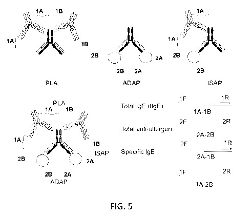
FIGS. 5 shows an overview of PCR-based allergy assays, including a
proximity ligation assay (PLA), which is a PCR-based method for protein
detection,
an antibody detection by agglutination-PCR (ADAP), which detects antigen-
specific
immunoglobulins of all subtypes (IgG, IgM, IgA, IgE, IgD), and isotype-
specific
agglutination-PCR (ISAP), which detects antigen-specific immunoglobulins of a
particular isotype. FIG. 5 (bottom) shows integration of PLA, ADAP and ISAP
into a
single assay. PCR with the indicated primer pairs enables the multiplexed
detection of
allergy markers. The primers 1F/1R detect total IgE, 2F/2R detect total anti-
allergen
antibodies, and 2F/1R or 1F/2R detect allergen-specific IgE.
FIGS. 6A and 6B show that ISAP detects purified antibodies in buffer and
outperforms ELISA. FIG. 6A shows ISAP analysis of a dilution series of
purified
anti-OVA IgE (black square), anti-OVA IgG (dark gray square) and non-specific
IgE
(light gray diamond). FIG. 6B shows ISAP (black circle) and ELISA (gray
square)
-23-
CA 03058451 2019-09-27
WO 2018/183779
PCT/US2018/025299
analysis of a dilution series of purified anti-OVA IgE. The x-axis in both
graphs is the
molar quantity of antibodies. The y-axis represents the delta Ct value in
comparison to
a blank.
FIGS. 7A-7C show that the integrated PCR-based assays detect allergy
markers with minimum cross-talk. In this assay, we detect total IgE (tIgE),
total anti-
allergen and specific IgE (sIgE) multiplexedly. The specificity of the
integrated assay
is assessed by dilution series of (FIG. 7A), anti-OVA IgG, (FIG. 7B) non-
specific
IgE, and (FIG. 7C) anti-OVA IgE respectively.
FIGS. 8A-8E show an integrated PCR-based analysis of serum from
ovalbumin (OVA)-sensitized mice. Serum was collected from OVA-sensitized and
control mice on day 0, 7, 14 and 21. The PCR signal is normalized to day 0 for
each
mouse. FIGS 8A-8C show that the PCR-based analysis detected enhanced
production
of IgE starting on day 7 (FIG. 8A), sIgE on day 14 (FIG. 8B), and total anti-
OVA on
day 7 (FIG. 8C). FIG. 8D shows an ELISA analysis of sIgE on the same set of
serum
samples. Enhanced production is observed on day 14. FIG. 8E shows a
correlation
between PCR-based analysis for total IgE using serum and whole blood samples.
Serum and whole blood sample displayed a correlation coefficient (R) of 0.86.
(*
represents P value smaller than 0.05)
FIGS. 9A and 9B show PCR-based analysis of serum from peanut-sensitized
mice. FIG. 9A shows results from BALB, Rag knockout and Jh knockout mice that
were epicutaneously sensitized with peanut oil. Measured PCR signals are
normalized
to day 0. Increased production of total IgE, anti-Ara hl IgE and total anti-
Ara h3 in
BALB mice were observed after sensitization. FIG. 9B shows peanut-specific IgE
detection by ELISA on the same set of BALB mice serum. No induction of peanut
.. specific IgE is observed.
FIGS. 10A-10E show PCR-based analysis of plasma from peanut-allergic
human patients. Plasma were baseline samples from the POISED immunotherapy
trial
(ClinicalTrials.gov Identifier: NCT02103270). An integrated PCR-based analysis
simultaneously detected 10 allergic features: tIgE, sIgE-Ara-hl, sIgG4-Ara-hl,
total
.. anti-Ara-hl, sIgE-Ara-h2, sIgG4-Ara-h2, total anti-Ara-h2, sIgE-Ara-h3,
sIgG4-Ara-
h3, total anti-Ara-h3 from 1 pL of plasma in a single assay. FIG. 10A-10C show
a
correlation between PCR-based assay and ImmunoCAP. High correlation between
our
PCR-based assay and ImmunoCAP analysis was observed (FIG. 10A (R = 0.64 for
sIgE Ara-hi), FIG. 10B (0.92 for sIgE-Ara-h2); FIG. 10C (0.88 for sIgE Ara-
h3)).
-24-
CA 03058451 2019-09-27
WO 2018/183779
PCT/US2018/025299
FIG. 10D shows a correlation between PCR-based assay and whole-peanut extract
ImmunoCAP. We summed the sIgE signal measured by PCR-based assay for Ara-hl,
Ara-h2 and Ara-h3 as a proxy for reactivity towards whole peanut extract.
Correlation
of the summed ISAP signal versus whole peanut extract ImmunoCAP was high (R =
0.82). FIG. 10E shows a comparison between our PCR-based assay and ImmunoCAP.
(The sample volume and cost are based on all 10 allergic features provided by
our
PCR-based assay).
DETAILED DESCRIPTION OF THE INVENTION
The practice of the present invention will employ, unless otherwise indicated,
conventional methods of immunology, chemistry, biochemistry, molecular biology
and recombinant DNA techniques, within the skill of the art. Such techniques
are
explained fully in the literature. See, e.g., IgE and Anti-IgE Therapy in
Asthma and
Allergic Disease (Lung Biology in Health and Disease, R.B. Fick and P.M.
Jardieu
eds., CRC Press, 2002); Middleton's Allergy: Principles and Practice (N. F.
Adkinson, B.S. Bochner, A.W. Burks, W.W. Busse, S.T. Holgate, R.F. Lemanske,
and R.E. O'Hehir eds., Saunders, 8th edition, 2013); PCR Technology: Current
Innovations (T. Nolan and S.A. Bustin eds., CRC Press, 3rd edition, 2013);
Antibodies
A Laboratory Manual (E.A. Greenfield ed., Cold Spring Harbor Laboratory Press,
2nd
Lab edition, 2013); Handbook of Experimental Immunology,Vols. I-IV (D.M. Weir
and C.C. Blackwell eds., Blackwell Scientific Publications); T.E. Creighton,
Proteins:
Structures and Molecular Properties (W.H. Freeman and Company, 1993); A.L.
Lehninger, Biochemistry (Worth Publishers, Inc., current addition); M.R. Green
and J.
Sambrook Molecular Cloning: A Laboratory Manual (Cold Spring Harbor
Laboratory Press, 4th edition, 2012); Methods In Enzymology (S. Colowick and
N.
Kaplan eds., Academic Press, Inc.).
All publications, patents and patent applications cited herein, whether supra
or
infra, are hereby incorporated by reference in their entireties.
1. DEFINITIONS
In describing the present invention, the following terms will be employed, and
are intended to be defined as indicated below.
It must be noted that, as used in this specification and the appended claims,
the
singular forms "a," "an" and "the" include plural referents unless the content
clearly
-25-
CA 03058451 2019-09-27
WO 2018/183779
PCT/US2018/025299
dictates otherwise. Thus, for example, reference to "an antibody" includes a
mixture
of two or more antibodies, and the like.
The term "about," particularly in reference to a given quantity, is meant to
encompass deviations of plus or minus five percent.
The terms "polynucleotide," "oligonucleotide," "nucleic acid" and "nucleic
acid molecule" are used herein to include a polymeric form of nucleotides of
any
length, either ribonucleotides or deoxyribonucleotides. This term refers only
to the
primary structure of the molecule. Thus, the term includes triple-, double-
and
single-stranded DNA, as well as triple-, double- and single-stranded RNA. It
also
includes modifications, such as by methylation and/or by capping, and
unmodified
forms of the polynucleotide. More particularly, the terms "polynucleotide,"
"oligonucleotide," "nucleic acid" and "nucleic acid molecule" include
polydeoxyribonucleotides (containing 2-deoxy-D-ribose), polyribonucleoti des
(containing D-ribose), any other type of polynucleotide which is an N¨ or C-
glycoside
of a purine or pyrimidine base, and other polymers containing nonnucleotidic
backbones, for example, polyamide (e.g., peptide nucleic acids (PNAs)) and
polymorpholino (commercially available from the Anti-Viral s, Inc., Corvallis,
Oregon, as Neugene) polymers, and other synthetic sequence-specific nucleic
acid
polymers providing that the polymers contain nucleobases in a configuration
which
allows for base pairing and base stacking, such as is found in DNA and RNA.
There
is no intended distinction in length between the terms "polynucleotide,"
"oligonucleotide," "nucleic acid" and "nucleic acid molecule," and these terms
will be
used interchangeably. Thus, these terms include, for example, 3'-deoxy-2',5'-
DNA,
oligodeoxyribonucleotide N3' P5' phosphoramidates, 2'-0-alkyl-substituted RNA,
double- and single-stranded DNA, as well as double- and single-stranded RNA,
DNA:RNA hybrids, and hybrids between PNAs and DNA or RNA, and also include
known types of modifications, for example, labels which are known in the art,
methylation, "caps," substitution of one or more of the naturally occurring
nucleotides
with an analog, internucleotide modifications such as, for example, those with
uncharged linkages (e.g., methyl phosphonates, phosphotriesters,
phosphoramidates,
carbamates, etc.), with negatively charged linkages (e.g., phosphorothioates,
phosphorodithioates, etc.), and with positively charged linkages (e.g.,
aminoalklyphosphoramidates, aminoalkylphosphotriesters), those containing
pendant
moieties, such as, for example, proteins (including nucleases, toxins,
antibodies,
-26-
CA 03058451 2019-09-27
WO 2018/183779
PCT/US2018/025299
signal peptides, poly-L-lysine, etc.), those with intercalators (e.g.,
acridine, psoralen,
etc.), those containing chelators (e.g., metals, radioactive metals, boron,
oxidative
metals, etc.), those containing alkylators, those with modified linkages
(e.g., alpha
anomeric nucleic acids, etc.), as well as unmodified forms of the
polynucleotide or
oligonucleotide.
"Recombinant" as used herein to describe a nucleic acid molecule means a
polynucleotide of genomic, cDNA, viral, semisynthetic, or synthetic origin
which, by
virtue of its origin or manipulation is not associated with all or a portion
of the
polynucleotide with which it is associated in nature. The term "recombinant"
as used
with respect to a protein or polypeptide means a polypeptide produced by
expression
of a recombinant polynucleotide. In general, the gene of interest is cloned
and then
expressed in transformed organisms, as described further below. The host
organism
expresses the foreign gene to produce the protein under expression conditions.
As used herein, a "solid support" refers to a solid surface such as a magnetic
bead, latex bead, microtiter plate well, glass plate, nylon, agarose,
acrylamide, and the
like.
"Substantially purified" generally refers to isolation of a substance
(compound, polynucleotide, protein, polypeptide, peptide composition) such
that the
substance comprises the majority percent of the sample in which it resides.
Typically,
.. in a sample, a substantially purified component comprises 50%, preferably
80%-85%,
more preferably 90-95% of the sample. Techniques for purifying polynucleotides
and
polypeptides of interest are well-known in the art and include, for example,
ion-
exchange chromatography, affinity chromatography and sedimentation according
to
density.
By "isolated" is meant, when referring to a protein, polypeptide or peptide,
that the indicated molecule is separate and discrete from the whole organism
with
which the molecule is found in nature or is present in the substantial absence
of other
biological macro molecules of the same type. The term "isolated" with respect
to a
nucleic acid is a nucleic acid molecule devoid, in whole or part, of sequences
normally associated with it in nature; or a sequence, as it exists in nature,
but having
heterologous sequences in association therewith; or a molecule disassociated
from the
chromosome.
As used herein, the term "target nucleic acid region" or "target nucleic acid"
denotes a nucleic acid molecule with a "target sequence" to be amplified. The
target
-27-
CA 03058451 2019-09-27
WO 2018/183779
PCT/US2018/025299
nucleic acid may be either single-stranded or double-stranded and may include
other
sequences besides the target sequence, which may not be amplified. The term
"target
sequence" refers to the particular nucleotide sequence of the target nucleic
acid which
is to be amplified. The target sequence may include a probe-hybridizing region
contained within the target molecule with which a probe will form a stable
hybrid
under desired conditions. The "target sequence" may also include the
complexing
sequences to which the oligonucleotide primers complex and extended using the
target sequence as a template. Where the target nucleic acid is originally
single-stranded, the term "target sequence" also refers to the sequence
complementary
to the "target sequence" as present in the target nucleic acid. If the "target
nucleic
acid" is originally double-stranded, the term "target sequence" refers to both
the plus
(+) and minus (-) strands (or sense and anti-sense strands).
The term "primer" or "oligonucleotide primer" as used herein, refers to an
oligonucleotide that hybridizes to the template strand of a nucleic acid and
initiates
.. synthesis of a nucleic acid strand complementary to the template strand
when placed
under conditions in which synthesis of a primer extension product is induced,
i.e., in
the presence of nucleotides and a polymerization-inducing agent such as a DNA
or
RNA polymerase and at suitable temperature, pH, metal concentration, and salt
concentration. The primer is preferably single-stranded for maximum efficiency
in
amplification, but may alternatively be double-stranded. If double-stranded,
the
primer can first be treated to separate its strands before being used to
prepare
extension products. This denaturation step is typically effected by heat, but
may
alternatively be carried out using alkali, followed by neutralization. Thus, a
"primer"
is complementary to a template, and complexes by hydrogen bonding or
hybridization
with the template to give a primer/template complex for initiation of
synthesis by a
polymerase, which is extended by the addition of covalently bonded bases
linked at its
3' end complementary to the template in the process of DNA or RNA synthesis.
Typically, nucleic acids are amplified using at least one set of
oligonucleotide primers
comprising at least one forward primer and at least one reverse primer capable
of
hybridizing to regions of a nucleic acid flanking the portion of the nucleic
acid to be
amplified.
The term "amplicon" refers to the amplified nucleic acid product of a PCR
reaction or other nucleic acid amplification process (e.g., isothermal
amplification,
rolling circle amplification, ligase chain reaction (LCR)).
-28-
CA 03058451 2019-09-27
WO 2018/183779
PCT/US2018/025299
As used herein, the term "probe" or "oligonucleotide probe" refers to a
polynucleotide, as defined above, that contains a nucleic acid sequence
complementary to a nucleic acid sequence present in the target nucleic acid
analyte.
The polynucleotide regions of probes may be composed of DNA, and/or RNA,
and/or
synthetic nucleotide analogs. Probes may be labeled in order to detect the
target
sequence. Such a label may be present at the 5' end, at the 3' end, at both
the 5' and
3' ends, and/or internally. The "oligonucleotide probe" may contain at least
one
fluorescer and at least one quencher. Quenching of fluorophore fluorescence
may be
eliminated by exonuclease cleavage of the fluorophore from the oligonucleotide
(e.g.,
TaqMan assay) or by hybridization of the oligonucleotide probe to the nucleic
acid
target sequence (e.g., molecular beacons). Additionally, the oligonucleotide
probe
will typically be derived from a sequence that lies between the sense and the
antisense
primers when used in a nucleic acid amplification assay.
The terms "hybridize" and "hybridization" refer to the formation of complexes
between nucleotide sequences which are sufficiently complementary to form
complexes via Watson-Crick base pairing. Where a primer "hybridizes" with
target
(template), such complexes (or hybrids) are sufficiently stable to serve the
priming
function required by, e.g., the DNA polymerase to initiate DNA synthesis.
It will be appreciated that the hybridizing sequences need not have perfect
complementarity to provide stable hybrids. In many situations, stable hybrids
will
form where fewer than about 10% of the bases are mismatches, ignoring loops of
four
or more nucleotides. Accordingly, as used herein the term "complementary"
refers to
an oligonucleotide that forms a stable duplex with its "complement" under
assay
conditions, generally where there is about 90% or greater homology.
The terms "selectively detects" or "selectively detecting" refer to the
detection
of an antibody isotype using oligonucleotides, e.g., primers and/or probes
that are
capable of detecting a particular DNA barcode, for example, by amplifying
and/or
binding to a DNA barcode of a particular antigen-DNA or antibody-binding agent-
DNA conjugate (e.g., secondary antibody-DNA, antibody mimetic-DNA, or aptamer-
DNA conjugate), or ligation product or extension product thereof, but do not
amplify
and/or bind to other DNA sequences under appropriate hybridization conditions.
The term "antibody" encompasses polyclonal and monoclonal antibody
preparations, as well as preparations including hybrid antibodies, altered
antibodies,
chimeric antibodies and, humanized antibodies, as well as: hybrid (chimeric)
antibody
-29-
CA 03058451 2019-09-27
WO 2018/183779
PCT/US2018/025299
molecules (see, for example, Winter et al. (1991) Nature 349:293-299; and U.S.
Pat.
No. 4,816,567); F(ab1)2 and F(ab) fragments; F, molecules (noncovalent
heterodimers,
see, for example, Inbar et al. (1972) Proc Natl Acad Sci USA 69:2659-2662; and
Ehrlich et al. (1980) Biochem 19:4091-4096); single-chain Fv molecules (sFv)
(see,
e.g., Huston et al. (1988) Proc Natl Acad Sci USA 85:5879-5883); nanobodies or
single-domain antibodies (sdAb) (see, e.g., Wang et al. (2016) Int
Nanomedicine
11:3287 -3303 , Vincke et al. (2012) Methods Mol Blot 911:15-26; dimeric and
trimeric antibody fragment constructs; minibodies (see, e.g., Pack et al.
(1992)
Biochem 31:1579-1584; Cumber et al. (1992) J Immunology 149B:120-126);
humanized antibody molecules (see, e.g., Riechmann et al. (1988) Nature
332:323-
327; Verhoeyan et al. (1988) Science 239:1534-1536; and U.K. Patent
Publication
No. GB 2,276,169, published 21 Sep. 1994); and, any functional fragments
obtained
from such molecules, wherein such fragments retain specific-binding properties
of the
parent antibody molecule.
The term "antigen" as used herein refers to any naturally occurring or
synthetic immunogenic substance. Immunogenic substances include those that are
foreign and those that are naturally occurring within the body of an organism.
As
such, the introduction of a foreign immunogenic substance may induce an
organism to
generate a general or specific immune response to the foreign immunogenic
substance. In other instances, the production of an immunogenic substance
within the
body of an organism may induce the organism to generate a specific or general
autoimmune response to the native immunogenic substance. Antigens, as used
herein,
encompass but are not limited to chemicals, small molecules, biomolecules
(e.g.,
nucleic acids), macromolecules, peptides, polypeptides, cell fragments, cells,
unicellular organisms, multicellular organisms, fragments thereof, and
combinations
thereof. In some instances, antigens may be antigens for which an agent that
binds the
antigen is known, e.g., a polypeptide for which an antibody that binds the
polypeptide
is known. In some instances, antigens may be antigens for which an agent that
binds
the antigen is unknown, e.g., a polypeptide for which an antibody that binds
the
polypeptide is unknown. For example, the use of polypeptides and peptides,
both
naturally occurring and synthetic, as antigens to which antibodies may be
raised has
been described in, e.g., Methods in Molecular Biology: Immunochemical
Protocols.
Ed. Burns, R., Humana Press, 2005, the disclosure of which is incorporated
herein by
reference in its entirety.
-30-
CA 03058451 2019-09-27
WO 2018/183779
PCT/US2018/025299
The phrase "specifically (or selectively) binds" to an antibody or
"specifically
(or selectively) immunoreactive with," when referring to an antigen or
allergen, refers
to a binding reaction that is determinative of the presence of the antigen or
allergen in
a heterogeneous population of proteins and other biologics. Thus, under
designated
immunoassay conditions, the specified antibodies bind to a particular antigen
at least
two times the background and do not substantially bind in a significant amount
to
other antigens present in the sample. Specific binding to an antibody under
such
conditions may require an antibody that is selected for its specificity for a
particular
antigen. For example, polyclonal antibodies raised to an antigen from specific
species
such as rat, mouse, or human can be selected to obtain only those polyclonal
antibodies that are specifically immunoreactive with the antigen and not with
other
proteins, except for polymorphic variants and alleles. This selection may be
achieved
by subtracting out antibodies that cross-react with molecules from other
species. A
variety of immunoassay formats may be used to select antibodies specifically
immunoreactive with a particular antigen. For example, solid-phase ELISA
immunoassays are routinely used to select antibodies specifically
immunoreactive
with a protein (see, e.g., Harlow & Lane. Antibodies, A Laboratory Manual
(1988),
for a description of immunoassay formats and conditions that can be used to
determine specific immunoreactivity). Typically, a specific or selective
reaction will
be at least twice background signal or noise and more typically more than 10
to 100
times background.
As used herein, a "biological sample" refers to a sample of cells, tissue, or
fluid isolated from a subject, including but not limited to, for example,
blood, plasma,
serum, fecal matter, urine, bone marrow, bile, spinal fluid, lymph fluid,
samples of the
skin, external secretions of the skin, respiratory, intestinal, and
genitourinary tracts,
tears, saliva, milk, cells (e.g., epithelial and endothelial cells,
fibroblasts, and
macrophages), muscles, joints, organs (e.g., liver, lung, spleen, thymus,
kidney, brain,
or lymph node), abnormal collections of fluid such as inflammatory transudates
or
exudates, pus, contents of cysts or areas of tissue necrosis, natural or
induced sputum,
fluid obtained by lavage performed for diagnostic or therapeutic purposes,
such as
nasal, pharyngeal or bronchoalveolar lavage, or biopsies and also samples of
in vitro
cell culture constituents including but not limited to conditioned media
resulting from
the growth of cells and tissues in culture medium, e.g., recombinant cells,
and cell
components.
-31-
CA 03058451 2019-09-27
WO 2018/183779
PCT/US2018/025299
As used herein, the terms "label" and "detectable label" refer to a molecule
capable of detection, including, but not limited to, radioactive isotopes,
fluorescers,
chemiluminescers, chromophores, enzymes, enzyme substrates, enzyme cofactors,
enzyme inhibitors, semiconductor nanoparticles, dyes, metal ions, metal sols,
ligands
(e.g., biotin, strepavidin or haptens) and the like. The term "fluorescer"
refers to a
substance or a portion thereof which is capable of exhibiting fluorescence in
the
detectable range. Particular examples of labels which may be used in the
practice of
the invention include, but are not limited to, SYBR green, SYBR gold, a CAL
Fluor
dye such as CAL Fluor Gold 540, CAL Fluor Orange 560, CAL Fluor Red 590, CAL
Fluor Red 610, and CAL Fluor Red 635, a Quasar dye such as Quasar 570, Quasar
670, and Quasar 705, an Alexa Fluor such as Alexa Fluor 350, Alexa Fluor 488,
Alexa Fluor 546, Alexa Fluor 555, Alexa Fluor 594, Alexa Fluor 647,and Alexa
Fluor
784, a cyanine dye such as Cy 3, Cy3.5, Cy5, Cy5.5, and Cy7, fluorescein, 2',
4', 5',
7'-tetrachloro-4-7-dichlorofluorescein (TET), carboxyfluorescein (FAM), 6-
carboxy-
4',5'-dichloro-2',7'-dimethoxyfluorescein (JOE), hexachlorofluorescein (HEX),
rhodamine, carboxy-X-rhodamine (ROX), tetramethyl rhodamine (TAMRA), FITC,
dansyl, umbelliferone, dimethyl acridinium ester (DMAE), Texas red, luminol,
NADPH, horseradish peroxidase (HRP), and a-P-galactosidase.
By "subject" is meant any member of the subphylum chordata, including,
without limitation, humans and other primates, including non-human primates
such as
chimpanzees and other apes and monkey species; farm animals such as cattle,
sheep,
pigs, goats and horses; domestic mammals such as dogs and cats; birds; and
laboratory animals, including rodents such as mice, rats and guinea pigs, and
the like.
The term does not denote a particular age. Thus, both adult and newborn
individuals
are intended to be covered.
2. MODES OF CARRYING OUT THE INVENTION
Before describing the present invention in detail, it is to be understood that
this
invention is not limited to particular formulations or process parameters as
such may,
of course, vary. It is also to be understood that the terminology used herein
is for the
purpose of describing particular embodiments of the invention only, and is not
intended to be limiting.
-32-
CA 03058451 2019-09-27
WO 2018/183779
PCT/US2018/025299
Although a number of methods and materials similar or equivalent to those
described herein can be used in the practice of the present invention, the
preferred
materials and methods are described herein.
The present invention is based on the discovery of reagents and methods for
detection of antibodies of specific isotypes. In particular, antigen-DNA
conjugates
and antibody-binding agent-DNA conjugates carrying DNA barcodes are used to
detect the presence of specific antibodies. The use of a DNA barcode allows
antibodies to be identified by nucleic acid-based detection methods, such as
PCR,
isothermal amplification, or microarray analysis. The methods of the invention
can be
used to detect and/or quantitate multiple antibodies in a single assay. In
addition, the
assays described herein can be readily combined with any other assays for
detection
of antibodies. Multiplex assays can be used, for example, to detect total
levels of
disease-relevant antibodies as well as individual levels of multiple disease-
relevant
antibody isotypes. The methods of the invention will allow monitoring of
disease-
relevant antibodies associated with immune disorders, such as allergies,
autoimmune
diseases, infections, or inflammation and better disease management.
In order to further an understanding of the invention, a more detailed
discussion is provided below regarding the assay methods for detecting
antibodies and
their use in diagnosing and monitoring disease-relevant antibodies.
A. Multiplex Assays for Detecting Antibodies
The methods use antigen-DNA conjugates and antibody-binding agent-DNA
conjugates as well as oligonucleotide reagents (e.g., oligonucleotide primers
and/or
probes) or a combination of reagents capable of detecting one or more antibody
isotypes in a single assay. The assay methods described herein can be used for
detecting antibody isotypes specific for any type of antigen, including, but
not limited
to an allergen, an autoimmune disease antigen, a cancer antigen, or a pathogen
antigen. There are a number of assay designs that can be used to detect
antibody
isotypes, which can be used alone or in combination with each other.
In one embodiment, the invention includes a method of detecting a target
antibody isotype in a sample, the method comprising: a) contacting the sample
with
an antibody-binding agent conjugated to a first DNA molecule comprising a
first
portion of a barcode, and an antigen conjugated to a second DNA molecule
comprising a second portion of a barcode, wherein the antigen binds to the
target
-33-
CA 03058451 2019-09-27
WO 2018/183779
PCT/US2018/025299
antibody isotype in the sample, if present, and the antibody-binding agent
specifically
binds to the target antibody isotype resulting in formation of a complex; b)
connecting
the first DNA molecule to the second DNA molecule in the complex, wherein the
first
portion of the barcode and the second portion of the barcode are joined to
form a
complete barcode; and c) detecting the complete barcode as an indication of
the
presence of the target antibody isotype in the sample.
Connecting the first DNA molecule to the second DNA molecule can be
accomplished in various ways. In one embodiment, the first DNA molecule and
the
second DNA molecule are ligated together using a ligase to produce a ligation
product
comprising the complete barcode. A bridge oligonucleotide may be used to
facilitate
ligation, wherein the bridge oligonucleotide comprises a first portion
sufficiently
complementary to and capable of hybridizing with the first DNA molecule, and a
second portion sufficiently complementary to and capable of hybridizing with
the
second DNA molecule. In the complex comprising the antibody-binding agent
conjugated to the first DNA molecule and the antigen conjugated to the second
DNA
molecule, wherein both conjugates are bound to the target antibody isotype,
the first
DNA molecule and the second DNA molecule are in sufficient proximity to each
other to simultaneously hybridize to the bridge oligonucleotide and undergo
ligation.
The ligation product can be detected and/or quantitated, for example, using
polymerase chain reaction (PCR), isothermal amplification, or microarray
analysis.
In another embodiment, the first DNA molecule and the second DNA
molecule comprise complementary nucleotide sequences, wherein hybridization of
a
nucleotide sequence in the first DNA molecule to a complementary nucleotide
sequence in the second DNA molecule allows a polymerase to extend the
hybridized
first and second DNA molecules to produce a nucleic acid comprising the
complete
barcode. The polymerase reaction can be carried out, for example, under
isothermal
conditions.
The "antibody binding agent" can be any agent that specifically binds to a
target antibody isotype. In some embodiments, the antibody binding agent binds
to a
target antibody isotype with high affinity. Examples of antibody binding
agents
include, without limitation, antibodies, antibody fragments, antibody
mimetics, and
aptamers.
In certain embodiments, the antibody-binding agent comprises an antibody
that specifically binds to the target antibody isotype. Any type of antibody
may be
-34-
CA 03058451 2019-09-27
WO 2018/183779
PCT/US2018/025299
used, including polyclonal and monoclonal antibodies, hybrid antibodies,
altered
antibodies, chimeric antibodies and, humanized antibodies, as well as: hybrid
(chimeric) antibody molecules (see, for example, Winter et al. (1991) Nature
349:293-299; and U.S. Pat. No. 4,816,567); F(ab1)2 and F(ab) fragments; F,
molecules
(noncovalent heterodimers, see, for example, Inbar et al. (1972) Proc Natl
Acad Sci
USA 69:2659-2662; and Ehrlich et al. (1980) Biochem 19:4091-4096); single-
chain
Fv molecules (sFv) (see, e.g., Huston et al. (1988) Proc Natl Acad Sci USA
85:5879-
5883); nanobodies or single-domain antibodies (sdAb) (see, e.g., Wang et al.
(2016)
Int J Nanomedicine 11:3287-3303, Vincke et al. (2012) Methods Mot Biol 911:15-
26;
dimeric and trimeric antibody fragment constructs; minibodies (see, e.g., Pack
et al.
(1992) Biochem 31:1579-1584; Cumber et al. (1992) J Immunology 149B:120-126);
humanized antibody molecules (see, e.g., Riechmann et al. (1988) Nature
332:323-
327; Verhoeyan et al. (1988) Science 239:1534-1536; and U.K. Patent
Publication
No. GB 2,276,169, published 21 Sep. 1994); and, any functional fragments
obtained
from such molecules, wherein such fragments retain specific-binding properties
of the
parent antibody molecule (i.e., specifically binds to a target antibody
isotype).
In other embodiments, the antibody-binding agent comprises an aptamer that
specifically binds to the target antibody isotype. Any type of aptamer may be
used,
including a DNA, RNA, xeno-nucleic acid (XNA), or peptide aptamer that
specifically binds to the target antibody isotype. Such aptamers can be
identified, for
example, by screening a combinatorial library. Nucleic acid aptamers (e.g.,
DNA or
RNA aptamers) that bind selectively to a target antibody isotype can be
produced by
carrying out repeated rounds of in vitro selection or systematic evolution of
ligands by
exponential enrichment (SELEX). Peptide aptamers that bind to a target
antibody
isotype may be isolated from a combinatorial library and improved by directed
mutation or repeated rounds of mutagenesis and selection. For a description of
methods of producing aptamers, see, e.g., Aptamers: Tools for Nanotherapy and
Molecular Imaging (R.N. Veedu ed., Pan Stanford, 2016), Nucleic Acid and
Peptide
Aptamers: Methods and Protocols (Methods in Molecular Biology, G. Mayer ed.,
Humana Press, 2009), Nucleic Acid Aptamers: Selection, Characterization, and
Application (Methods in Molecular Biology, G. Mayer ed., Humana Press, 2016),
Aptamers Selected by Cell-SELEX for Theranostics (W. Tan, X. Fang eds.,
Springer,
2015), Cox et al. (2001) Bioorg. Med. Chem. 9(10):2525-2531; Cox et al. (2002)
Nucleic Acids Res. 30(20): e108, Kenan et al. (1999) Methods Mol Biol. 118:217-
-35-
CA 03058451 2019-09-27
WO 2018/183779
PCT/US2018/025299
231; Platella etal. (2016) Biochim. Biophys. Acta Nov 16 pii: S0304-
4165(16)30447-
0, and Lyu etal. (2016) Theranostics 6(9):1440-1452; herein incorporated by
reference in their entireties.
In yet other embodiment, the antibody-binding agent comprises an antibody
mimetic. Any type of antibody mimetic may be used, including, but not limited
to,
affibody molecules (Nygren (2008) FEBS J. 275 (11):2668-2676), affilins
(Ebersbach
etal. (2007) J. Mol. Biol. 372 (1):172-185), affimers (Johnson etal. (2012)
Anal.
Chem. 84 (15):6553-6560), affitins (Krehenbrink et al. (2008) J. Mol. Biol.
383
(5):1058-1068), alphabodies (Desmet etal. (2014) Nature Communications
5:5237),
anticalins (Skerra (2008) FEBS J. 275 (11):2677-2683), avimers (Silverman et
al.
(2005) Nat. Biotechnol. 23 (12):1556-1561), darpins (Stumpp etal. (2008) Drug
Discov. Today 13 (15-16):695-701), fynomers (Grabulovski et al. (2007) J.
Biol.
Chem. 282 (5):3196-3204), and monobodies (Koide etal. (2007) Methods Mol.
Biol.
352:95-109).
Antigen-DNA and antibody-binding agent-DNA conjugates of the subject
methods include at least one DNA molecule attached to the antigen or antibody-
binding agent, respectively, wherein the DNA molecule comprises a unique
barcode
sequence that identifies a target antigen-specific antibody isotype (i.e., the
complete
barcode produced by joining the DNA sequences of the antigen-DNA and antibody-
binding agent-DNA conjugates). DNA molecules attached to an antigen or
antibody-
binding agent may vary depending, in part, on the detection method employed,
the
method of attachment, the specific antibodies to be detected, etc. Generally,
the length
of the attached DNA molecules will be at least 15 nucleotides, but may range
from 15
nucleotides to 200 nucleotides or more including but not limited to e.g., 20
or more
nucleotides, 25 or more nucleotides, 30 or more nucleotides, 35 or more
nucleotides,
40 or more nucleotides, 45 or more nucleotides, 50 or more nucleotides, 55 or
more
nucleotides, 60 or more nucleotides, 65 or more nucleotides, 70 or more
nucleotides,
75 or more nucleotides, 80 or more nucleotides, 90 or more nucleotides, 95 or
more
nucleotides, 100 or more nucleotides, 15 to 200 nucleotides, 20 to 200
nucleotides, 25
to 200 nucleotides, 30 to 200 nucleotides, 35 to 200 nucleotides, 40 to 200
nucleotides, 45 to 200 nucleotides, 50 to 200 nucleotides, 15 to 100
nucleotides, 20 to
100 nucleotides, 25 to 100 nucleotides, 30 to 100 nucleotides, 35 to 100
nucleotides,
to 100 nucleotides, 45 to 100 nucleotides, 50 to 100 nucleotides, etc.
-36-
CA 03058451 2019-09-27
WO 2018/183779
PCT/US2018/025299
The DNA may be attached to an antigen or antibody-binding agent by any
convenient method, as described in more detail below. The DNA may be attached
to
an antigen or antibody-binding agent at any convenient point along the length
of the
DNA, including at the 3' or 5' termini. In some instances, DNA is attached to
the
antigen or antibody at its 3' end or 5' end. In some instances, both the
antigen and the
antibody-binding agent have DNA molecules attached at their 3' ends. In some
instances, both the antigen and the antibody-binding agent have DNA molecules
attached at their 5' ends.
As used herein, the term "bridging oligonucleotide" or "bridge
oligonucleotide" refers to any oligonucleotide that joins two or more separate
DNA
molecules or two termini of a single DNA molecule by simultaneously
hybridizing
with complementary regions on each DNA molecule or complementary regions of
the
DNA termini. In certain instances, a bridging oligonucleotide joins an antigen-
DNA
conjugate to an antibody-binding agent-DNA conjugate by simultaneously
hybridizing with a first complementary region in the DNA of the antigen-DNA
conjugate and a second complementary region in the DNA of the antibody-binding
agent-DNA conjugate. Bridging oligonucleotides may be partially or completely
single stranded, including partially single stranded and partially double
stranded. The
length of bridging oligonucleotides of the subject disclosure will vary and
may be 10
or more nucleotides and range from 10 to 100 or more nucleotides, including
e.g., 10
to 100 nucleotides, 12 to 100 nucleotides, 14 to 100 nucleotides, 16 to 100
nucleotides, 18 to 100 nucleotides, 20 to 100 nucleotides, 22 to 100
nucleotides, 24 to
100 nucleotides, 26 to 100 nucleotides, 28 to 100 nucleotides, 30 to 100
nucleotides,
10 to 50 nucleotides, 12 to 50 nucleotides, 14 to 50 nucleotides, 16 to 50
nucleotides,
18 to 50 nucleotides, 20 to 50 nucleotides, 22 to 50 nucleotides, 24 to 50
nucleotides,
26 to 50 nucleotides, 28 to 50 nucleotides, 30 to 50 nucleotides, 10 to 40
nucleotides,
12 to 40 nucleotides, 14 to 40 nucleotides, 16 to 40 nucleotides, 18 to 40
nucleotides,
20 to 40 nucleotides, 22 to 40 nucleotides, 24 to 40 nucleotides, 26 to 40
nucleotides,
28 to 40 nucleotides, 30 to 40 nucleotides, 10 to 30 nucleotides, 12 to 30
nucleotides,
14 to 30 nucleotides, 16 to 30 nucleotides, 18 to 30 nucleotides, 20 to 30
nucleotides,
12 or more nucleotides, 13 or more nucleotides, 14 or more nucleotides, 15 or
more
nucleotides, 16 or more nucleotides, 17 or more nucleotides, 18 or more
nucleotides,
19 or more nucleotides, 20 or more nucleotides, 12 nucleotides, 13
nucleotides, 14
nucleotides, 15 nucleotides, 16 nucleotides, 17 nucleotides, 18 nucleotides,
19
-37-
CA 03058451 2019-09-27
WO 2018/183779
PCT/US2018/025299
nucleotides, 20 nucleotides, 21 nucleotides, 22 nucleotides, 23 nucleotides,
24
nucleotides, 25 nucleotides, 26 nucleotides, 27 nucleotides, 28 nucleotides,
29
nucleotides, 30 nucleotides, etc.
Bridging oligonucleotides may "bridge" two or more DNA molecules to form
a complex. In some instances, a bridging polynucleotide may hybridize with two
DNA termini, including termini of the same or different nucleic acids, such
that the
termini are adjacent within the complex, e.g., allowing for the ligation of
the adjacent
termini. In some instances, a bridging polynucleotide may hybridize with two
DNA
termini, including termini of the same or different nucleic acids, such that
the termini
.. are not adjacent in the resulting polynucleotide complex, e.g., are not
adjacent such
that they cannot be directly ligated together. In some instances, e.g., where
two DNA
termini in a complex are not adjacent, a splint polynucleotide may be
hybridized in
the space between the two termini such that the ends of the splint
polynucleotide are
located adjacent to one or more of the termini. The term "splint
polynucleotide" as
used herein refers to a polynucleotide, which may generally be single stranded
or
partially single stranded and partially double stranded, which may be used to
fill one
or more gaps between two DNA termini in a complex, e.g., those complexes
formed
by use of a bridging polynucleotide. In some instances, a splint
polynucleotide may
have complementarity to one or more portions of a bridging oligonucleotide. In
some
instances, DNA termini adjacent to a splint polynucleotide may be ligated to
the splint
polynucleotide.
In some instances, a bridging oligonucleotide of the subject disclosure may
include one or more nucleoside analogs. For example, in some instances, a
bridging
oligonucleotide of the instant disclosure may include one or more
deoxyribouracil
(i.e., deoxyribose uracil, deoxyuridine, etc.) nucleosides/nucleotides. In
certain
instances, a bridging polynucleotide may include 2 or more nucleoside analogs
including but not limited to e.g., 3 or more, 4 or more, 5 or more, 6 or more,
etc. In
some instances, the number of nucleoside analogs as a percentage of the total
bases of
the bridging polynucleotide is 1% or more, including but not limited to e.g.,
2% or
more, 3% or more, 4% or more, 5% or more, 6% or more, 7% or more, 8% or more,
9% or more, 10% or more, 11% or more, 12% or more, 13% or more, 14% or more,
15% or more, 16% or more, 17% or more, 18% or more, 19% or more, 20% or more,
21% or more, 22% or more, 23% or more, 24% or more, 25% or more, 26% or more,
27% or more, 28% or more, 29% or more, 30% or more, etc.
-38-
CA 03058451 2019-09-27
WO 2018/183779
PCT/US2018/025299
Joining of DNA molecules from an antigen-DNA conjugate and an antibody-
binding agent-DNA conjugate produces a complete barcode sequence identifying
the
target antibody isotype, which may be amplified to generate an amplification
product
(i.e., amplicon) that can be detected. Any convenient method of amplification
may be
utilized in generating the amplification product, as described in more detail
below,
and may depend upon the particular complex formed and/or particular
requirements
of the overall detection assay. As the formation of an amplicon is dependent
on a
target antibody in a sample binding to the antigen-DNA conjugate and the
antibody-
binding agent-DNA conjugate binding to the same target antibody, the presence
of an
amplification product is indicative of the presence of target antibodies in
the sample
that are specific for the antigen and also recognized by the antibody-binding
agent
(e.g., a secondary antibody, antibody mimetic, or aptamer specific for a
particular
target antibody isotype).
In some instances, amplification may be performed by polymerase chain
reaction (PCR). In representative PCR amplification reactions, the reaction
mixture
generally includes a template nucleic acid which is combined with one or more
primers that are employed in the primer extension reaction, e.g., the PCR
primers
(such as forward and reverse primers employed in geometric (or exponential)
amplification or a single primer employed in a linear amplification). As such,
in some
instances, the hybridized portions of the above described nucleic acid
complexes may
serve as "primer" for the amplification reaction. For example, in instances
where
linear amplification is employed a single free 3 '-terminus of hybridized
nucleic acid
of an above described nucleic acid complex may serve as a primer for
amplification.
In some instances, one or more additional nucleic acids may be added to serve
as
.. primer in a formed nucleic acid complex. For example, in some instances two
antigen-bound polynucleotides may be joined in a ligation reaction and two
additional
primers may be added to facilitate amplification of the newly ligated nucleic
acid
segment or template. In some instances, a single free 3 '-terminus of
hybridized
nucleic acid of an above described nucleic acid complex may serve as a first
primer
and a second primer may be added to facilitate amplification.
Any oligonucleotide primers with which the template nucleic acid (hereinafter
referred to as template DNA for convenience) is contacted will be of
sufficient length
to provide for hybridization to complementary template DNA under annealing
conditions. The primers will generally be at least 6 bp in length, including
but not
-39-
CA 03058451 2019-09-27
WO 2018/183779
PCT/US2018/025299
limited to e.g., at least 10 bp in length, at least 15 bp in length, at least
16 bp in length,
at least 17 bp in length, at least 18 bp in length, at least 19 bp in length,
at least 20 bp
in length, at least 21 bp in length, at least 22 bp in length, at least 23 bp
in length, at
least 24 bp in length, at least 25 bp in length, at least 26 bp in length, at
least 27 bp in
length, at least 28 bp in length, at least 29 bp in length, at least 30 bp in
length, and
may be as long as 60 bp in length or longer, where the length of the primers
will
generally range from 18 to 50 bp in length, including but not limited to,
e.g., from
about 20 to 35 bp in length. In some instances, the template DNA may be
contacted
with a single primer or a set of two primers (forward and reverse primers),
depending
on whether primer extension, linear or exponential amplification of the
template DNA
is desired. Methods of PCR that may be employed in the subject methods include
but
are not limited to those described in U.S. Pat. Nos.: 4,683,202; 4,683,195;
4,800,159;
4,965,188 and 5,512,462, the disclosures of which are herein incorporated by
reference.
In addition to the above components, a PCR reaction mixture produced in the
subject methods may include a polymerase and deoxyribonucleoside triphosphates
(dNTPs). The desired polymerase activity may be provided by one or more
distinct
polymerase enzymes. In many embodiments, the reaction mixture includes at
least a
Family A polymerase, where representative Family A polymerases of interest
include,
but are not limited to: Thermus aquaticus polymerases, including the naturally
occurring polymerase (Taq) and derivatives and homologues thereof, such as
Klentaq
(as described in Proc. Natl. Acad. Sci USA (1994) 91:2216-2220, the disclosure
of
which is incorporated herein by reference in its entirety); Thermus
thermophilics
polymerases, including the naturally occurring polymerase (Tth) and
derivatives and
homologues thereof, and the like. In certain embodiments where the
amplification
reaction that is carried out is a high fidelity reaction, the reaction mixture
may further
include a polymerase enzyme having 3'-5' exonuclease activity, e.g., as may be
provided by a Family B polymerase, where Family B polymerases of interest
include,
but are not limited to: Thermococcus litoralis DNA polymerase (Vent) (e.g., as
described in Perler et al., Proc. Natl. Acad. Sci. USA (1992) 89:5577, the
disclosure
of which is incorporated herein by reference in its entirety); Pyrococcus
species GB-D
(Deep Vent); Pyrococcus furiosus DNA polymerase (Pfu) (e.g., as described in
Lundberg et al., Gene (1991) 108: 1-6, the disclosure of which is incorporated
herein
by reference in its entirety), Pyrococcus woesei (Pwo) and the like..
Generally, the
-40-
CA 03058451 2019-09-27
WO 2018/183779
PCT/US2018/025299
reaction mixture will include four different types of dNTPs corresponding to
the four
naturally occurring bases are present, i.e. dATP, dTTP, dCTP and dGTP and in
some
instances, may include one or more modified nucleotide dNTPs.
A PCR reaction will generally be carried out by cycling the reaction mixture
between appropriate temperatures for annealing, elongation/extension, and
denaturation for specific times. Such temperature and times will vary and will
depend
on the particular components of the reaction including, e.g., the polymerase
and the
primers as well as the expected length of the resulting PCR product. In some
instances, e.g., where nested or two-step PCR are employed the cycling-
reaction may
be carried out in stages, e.g., cycling according to a first stage having a
particular
cycling program or using particular temperature(s) and subsequently cycling
according to a second stage having a particular cycling program or using
particular
temperature(s).
Multistep PCR processes may or may not include that addition of one or more
reagents following the initiation of amplification. For example, in some
instances,
amplification may be initiated by elongation with the use of a polymerase and,
following an initial phase of the reaction, additional reagent(s) (e.g., one
or more
additional primers, additional enzymes, etc.) may be added to the reaction to
facilitate
a second phase of the reaction. In some instances, amplification may be
initiated with
a first primer or a first set of primers and, following an initial phase of
the reaction,
additional reagent(s) (e.g., one or more additional primers, additional
enzymes, etc.)
may be added to the reaction to facilitate a second phase of the reaction. In
certain
embodiments, the initial phase of amplification may be referred to as
"preamplification".
In some instances, amplification may be carried out under isothermal
conditions, e.g., by means of isothermal amplification. Methods of isothermal
amplification generally make use of enzymatic means of separating DNA strands
to
facilitate amplification at constant temperature, such as, e.g., strand-
displacing
polymerase or a helicase, thus negating the need for thermocycling to denature
DNA.
Any convenient and appropriate means of isothermal amplification may be
employed
in the subject methods including but are not limited to: loop-mediated
isothermal
amplification (LAMP), strand displacement amplification (SDA), helicase-
dependent
amplification (HDA), nicking enzyme amplification reaction (NEAR), and the
like.
LAMP generally utilizes a plurality of primers, e.g., 4-6 primers, which may
-41-
CA 03058451 2019-09-27
WO 2018/183779
PCT/US2018/025299
recognize a plurality of distinct regions, e.g., 6-8 distinct regions, of
target DNA.
Synthesis is generally initiated by a strand-displacing DNA polymerase with
two of
the primers forming loop structures to facilitate subsequent rounds of
amplification.
LAMP is rapid and sensitive. In addition, the magnesium pyrophosphate produced
during the LAMP amplification reaction may, in some instances be visualized
without
the use of specialized equipment, e.g., by eye. SDA generally involves the use
of a
strand-displacing DNA polymerase (e.g., Bst DNA polymerase, Large (Klenow)
Fragment polymerase, Klenow Fragment (3'-5' exo-), and the like) to initiate
at nicks
created by a strand-limited restriction endonuclease or nicking enzyme at a
site
contained in a primer. In SDA, the nicking site is generally regenerated with
each
polymerase displacement step, resulting in exponential amplification. HDA
generally
employs: a helicase which unwinds double-stranded DNA unwinding to separate
strands; primers, e.g., two primers, that may anneal to the unwound DNA; and a
strand-displacing DNA polymerase for extension. NEAR generally involves a
strand-
displacing DNA polymerase that initiates elongation at nicks, e.g., created by
a
nicking enzyme. NEAR is rapid and sensitive, quickly producing many short
nucleic
acids from a target sequence.
In some instances, entire amplification methods may be combined or aspects
of various amplification methods may be recombined to generate a hybrid
amplification method. For example, in some instances, aspects of PCR may be
used,
e.g., to generate the initial template or amplicon or first round or rounds of
amplification, and an isothermal amplification method may be subsequently
employed
for further amplification. In some instances, an isothermal amplification
method or
aspects of an isothermal amplification method may be employed, followed by PCR
for further amplification of the product of the isothermal amplification
reaction. In
some instances, a sample may be preamplified using a first method of
amplification
and may be further processed, including e.g., further amplified or analyzed,
using a
second method of amplification. As a non-limiting example, a sample may be
preamplified by PCR and further analyzed by qPCR.
In some instances, the amplification step and the detection step, described
below, may be combined, with or without the use of a preamplifcation step. In
some
instances, the particular amplification method employed allows for the
qualitative
detection of amplification product, e.g., by visual inspection of the
amplification
reaction with or without a detection reagent. In one embodiment, the ligation
products
-42-
CA 03058451 2019-09-27
WO 2018/183779
PCT/US2018/025299
are amplified by isothermal amplification, e.g., LAMP, and the amplification
generates a visual change in the amplification reaction indicative of
efficient
amplification and thus presence of the antibody isotype in the sample. In some
instances, the amplification and detection steps are combined by monitoring
the
amplification reaction during amplification such as is performed in, e.g.,
real-time
PCR, also referred to herein as quantitative PCR (qPCR).
In some instances, the methods described herein may make use of those
methods, e.g., amplification methods, and components thereof, employed in
proximity
ligation assays (PLA) and proximity elongation assays (PEA) including but not
limited to, e.g., rolling circle amplification (RCA), binding-induced DNA
assembly
(BINDA), nicking enzyme assisted fluorescence signal amplification (NEFSA),
and,
e.g., those described in Janssen et al. (2013) Sensors, 13, 1353-1384, the
disclosure of
which is incorporated herein by reference in its entirety.
The methods of the invention can be adapted to multiplexing. For example, a
plurality of antibody-binding agent-DNA conjugates can be added to a sample,
wherein each antibody-binding agent is conjugated to a DNA molecule comprising
a
different barcode sequence and each antibody-binding agent is capable of
binding to a
different target antibody isotype to allow multiplex detection of a plurality
of target
antibody isotypes in a sample. In certain embodiments, the antibody-binding
agent-
DNA conjugates are selected from the group consisting of an anti-IgE secondary
antibody-DNA conjugate for detection of IgE, an anti-IgM secondary antibody-
DNA
conjugate for detection of IgM, an anti-IgG secondary antibody-DNA conjugate
for
detection of IgG, an anti-IgA secondary antibody-DNA conjugate for detection
of
IgA, and an anti-IgD secondary antibody DNA conjugate for detection of IgD,
wherein the DNA molecules in the conjugates comprise isotype-specific DNA
barcodes that can be amplified and detected simultaneously by using a suitable
combination of primers and/or probes in a multiplex-type assay format.
Exemplary DNA sequences for antigen-DNA conjugates and antibody-binding
agent-DNA conjugates, bridge oligonucleotides, and PCR primers for detection
of the
DNA ligation products are shown in Example 1 and SEQ ID NOS:1-21 of the
Sequence Listing. In certain embodiments, an antigen-DNA conjugate comprises a
DNA sequence selected from the group consisting of SEQ ID NOS:1, 2, 5, 6, 9,
10,
13, 14, 17, an 18 or a DNA sequence having at least 95% identity to a DNA
sequence
selected from the group consisting of SEQ ID NOS:1, 2, 5, 6, 9, 10, 13, 14,
17, an 18.
-43-
CA 03058451 2019-09-27
WO 2018/183779
PCT/US2018/025299
In other embodiments, a secondary antibody-binding agent-DNA conjugate
comprises
a DNA sequence selected from the group consisting of SEQ ID NOS:1, 2, 5, 6, 9,
10,
13, 14, 17, an 18 or a DNA sequence having at least 95% identity to a DNA
sequence
selected from the group consisting of SEQ ID NOS:1, 2, 5, 6, 9, 10, 13, 14,
17, an 18.
In certain embodiments, the bridge oligonucleotide comprises the nucleotide
sequence
of SEQ ID NO:21 or a nucleotide sequence having at least 95% identity to the
sequence of SEQ ID NO:21, wherein the bridge oligonucleotide is capable of
hybridizing to the DNA of the
In another embodiment, the method is performed with at least one set of
reagents selected from the group consisting of: a) an antigen-DNA conjugate
comprising the DNA sequence of SEQ ID NO:1, an antibody-binding agent-DNA
conjugate comprising the DNA sequence of SEQ ID NO:2, a bridge oligonucleotide
comprising the nucleotide sequence of SEQ ID NO:21, a reverse primer
comprising
the nucleotide sequence of SEQ ID NO:3 and a forward primer comprising the
sequence of SEQ ID NO:4; b) an antigen-DNA conjugate comprising the DNA
sequence of SEQ ID NO:5, an antibody-binding agent-DNA conjugate comprising
the
DNA sequence of SEQ ID NO:6, a bridge oligonucleotide comprising the
nucleotide
sequence of SEQ ID NO:21, a reverse primer comprising the nucleotide sequence
of
SEQ ID NO:7 and a forward primer comprising the sequence of SEQ ID NO:8; c) an
antigen-DNA conjugate comprising the DNA sequence of SEQ ID NO:9, an
antibody-binding agent-DNA conjugate comprising the DNA sequence of SEQ ID
NO:10, a bridge oligonucleotide comprising the nucleotide sequence of SEQ ID
NO:21, a reverse primer comprising the nucleotide sequence of SEQ ID NO:11 and
a
forward primer comprising the sequence of SEQ ID NO:12; d) an antigen-DNA
.. conjugate comprising the DNA sequence of SEQ ID NO:13, an antibody-binding
agent-DNA conjugate comprising the DNA sequence of SEQ ID NO:14, a bridge
oligonucleotide comprising the nucleotide sequence of SEQ ID NO:21, a reverse
primer comprising the nucleotide sequence of SEQ ID NO:15 and a forward primer
comprising the sequence of SEQ ID NO:16; e) an antigen-DNA conjugate
comprising
the DNA sequence of SEQ ID NO:17, an antibody-binding agent-DNA conjugate
comprising the DNA sequence of SEQ ID NO:18, a bridge oligonucleotide
comprising the nucleotide sequence of SEQ ID NO:21, a reverse primer
comprising
the nucleotide sequence of SEQ ID NO:19 and a forward primer comprising the
sequence of SEQ ID NO:20; f) an antibody-binding agent-DNA conjugate
comprising
-44-
CA 03058451 2019-09-27
WO 2018/183779
PCT/US2018/025299
the DNA sequence of SEQ ID NO:1, an antigen-DNA conjugate comprising the DNA
sequence of SEQ ID NO:2, a bridge oligonucleotide comprising the nucleotide
sequence of SEQ ID NO:21, a reverse primer comprising the nucleotide sequence
of
SEQ ID NO:3 and a forward primer comprising the sequence of SEQ ID NO:4; g) an
antibody-binding agent-DNA conjugate comprising the DNA sequence of SEQ ID
NO:5, an antigen-DNA conjugate comprising the DNA sequence of SEQ ID NO:6, a
bridge oligonucleotide comprising the nucleotide sequence of SEQ ID NO:21, a
reverse primer comprising the nucleotide sequence of SEQ ID NO:7 and a forward
primer comprising the sequence of SEQ ID NO:8; h) an antibody-binding agent-
DNA
conjugate comprising the DNA sequence of SEQ ID NO:9, an antigen-DNA
conjugate comprising the DNA sequence of SEQ ID NO:10, a bridge
oligonucleotide
comprising the nucleotide sequence of SEQ ID NO:21, a reverse primer
comprising
the nucleotide sequence of SEQ ID NO:11 and a forward primer comprising the
sequence of SEQ ID NO:12; i) an antibody-binding agent-DNA conjugate
comprising
the DNA sequence of SEQ ID NO:13, an antigen-DNA conjugate comprising the
DNA sequence of SEQ ID NO:14, a bridge oligonucleotide comprising the
nucleotide
sequence of SEQ ID NO:21, a reverse primer comprising the nucleotide sequence
of
SEQ ID NO:15 and a forward primer comprising the sequence of SEQ ID NO:16; and
j) an antibody-binding agent-DNA conjugate comprising the DNA sequence of SEQ
ID NO:17, an antigen-DNA conjugate comprising the DNA sequence of SEQ ID
NO:18, a bridge oligonucleotide comprising the nucleotide sequence of SEQ ID
NO:21, a reverse primer comprising the nucleotide sequence of SEQ ID NO:19 and
a
forward primer comprising the sequence of SEQ ID NO:20.
ISAP can be combined with other methods of antibody detection, particularly
other methods that utilize DNA barcoding to allow detection of multiple
antibody
isotypes by multiplex PCR. In one multiplex assay format, ISAP is combined
with
proximity ligation assay (PLA) and/or agglutination-polymerase chain reaction
(ADAP). See, e.g., Gullberg et al. (2005) Proc. Natl. Acad. Sci. U.S.A. 101
(22):8420-9424, Gustafsdottir et al. (2005) Analytical Biochemistry 345 (1):2-
9, for a
description of PLA and Tsai et al. (2016) ACS Central Science. 2 (3): 139-147,
International Patent Application Publication No. W02016168711 Al for a
description
of ADAP; herein incorporated by reference in their entireties.
For example, ISAP can be combined with PLA and/or ADAP for detecting
allergen antibodies in a sample. In one embodiment, the method comprises: a)
-45-
CA 03058451 2019-09-27
WO 2018/183779
PCT/US2018/025299
performing PLA using at least one pair of anti-IgE antibody-DNA conjugates to
detect total IgE levels in the sample; b) performing ADAP using at least one
pair of
allergen-DNA conjugates to detect total anti-allergen antibody levels in the
sample;
and c) performing ISAP using at least one allergen-DNA conjugate in
combination
with at least one anti-IgE antibody-DNA conjugate to detect allergen-specific
IgE
levels in the sample. The method may further comprise performing ISAP with at
least
one allergen-DNA conjugate in combination with at least one anti-
immunoglobulin
G4 (IgG4) antibody-DNA conjugate to detect allergen-specific IgG4 levels. In
certain
embodiments, ADAP is used to detect the total anti-allergen antibody levels of
IgG,
IgM, and IgE.
In performing PLA, the method comprises: a) adding at least one pair of anti-
IgE antibody-DNA conjugates to the sample, wherein at least one pair of anti-
IgE
antibody-DNA conjugates comprises a first anti-IgE antibody-DNA conjugate that
binds to an IgE in the sample at a first site and a second anti-IgE antibody-
DNA
conjugate that binds to the same IgE at a second site; b) contacting the
sample with a
PLA bridge oligonucleotide, wherein the PLA bridge oligonucleotide comprises:
(i) a
first portion sufficiently complementary to and capable of hybridizing with
the DNA
of the first anti-IgE antibody-DNA conjugate, and (ii) a second portion
sufficiently
complementary to and capable of hybridizing with the DNA of the second anti-
IgE
antibody-DNA conjugate, wherein the DNA of the first anti-IgE antibody-DNA
conjugate and the DNA of the second anti-IgE antibody-DNA conjugate are in
sufficient proximity to each other to simultaneously hybridize to the PLA
bridge
oligonucleotide; c) ligating the PLA bridge oligonucleotide to the first anti-
IgE
antibody-DNA conjugate and the second anti-IgE antibody-DNA conjugate to
produce a PLA ligation product; and d) detecting the PLA ligation product as
an
indication of the presence of the IgE in the sample.
In performing the ADAP, the method comprises: a) adding said at least one
pair of allergen-DNA conjugates to the sample, wherein at least one pair of
allergen-
DNA conjugates comprises a first allergen-DNA conjugate that binds to an anti-
allergen antibody in the sample at a first site and a second allergen-DNA
conjugate
that binds to the same anti-allergen antibody at a second site; b) contacting
the sample
with an ADAP bridge oligonucleotide, wherein the ADAP bridge oligonucleotide
comprises: (i) a first portion sufficiently complementary to and capable of
hybridizing with the DNA of the first allergen-DNA conjugate, and (ii) a
second
-46-
CA 03058451 2019-09-27
WO 2018/183779
PCT/US2018/025299
portion sufficiently complementary to and capable of hybridizing with the DNA
of the
second allergen-DNA conjugate, wherein the DNA of the first allergen-DNA
conjugate and the DNA of the second allergen-DNA conjugate are in sufficient
proximity to each other to simultaneously hybridize to the ADAP bridge
oligonucleotide; c) ligating the ADAP bridge oligonucleotide to the first
allergen-
DNA conjugate and the second allergen-DNA conjugate to produce an ADAP
ligation
product; and d) detecting the ADAP ligation product as an indication of the
presence
of the anti-allergen antibody in the sample.
In performing the ISAP, the method comprises: a) adding the at least one
allergen-DNA conjugate in combination with at least one anti-IgE antibody-DNA
conjugate to the sample, wherein the allergen-DNA conjugate binds to the
allergen-
specific IgE in the sample, and the anti-IgE antibody-DNA conjugate binds to
the
same allergen-specific IgE resulting in formation of a first complex; b)
contacting the
first complex with an ISAP bridge oligonucleotide, wherein the ISAP bridge
oligonucleotide comprises: (i) a first portion sufficiently complementary to
and
capable of hybridizing with the DNA of the anti-IgE antibody-DNA conjugate,
and
(ii) a second portion sufficiently complementary to and capable of hybridizing
with
the DNA of the allergen-DNA conjugate, wherein the DNA of the anti-IgE
antibody-
DNA conjugate and the DNA of the allergen-DNA conjugate are in sufficient
proximity to each other in the first complex to simultaneously hybridize to
the ISAP
bridge oligonucleotide; c) ligating the ISAP bridge oligonucleotide to the
anti-IgE
antibody-DNA and the allergen-DNA in the first complex to produce a first ISAP
ligation product; d) detecting the first ISAP ligation product as an
indication of the
presence of the allergen-specific IgE in the sample; e) adding the at least
one allergen-
DNA conjugate in combination with at least one anti-IgG4 antibody-DNA
conjugate
to the sample, wherein the allergen-DNA conjugate binds to the allergen-
specific
IgG4 in the sample, and the anti-IgG4 antibody-DNA conjugate binds to the same
allergen-specific IgG4 resulting in formation of a second complex; f)
contacting the
second complex with an ISAP bridge oligonucleotide, wherein the ISAP bridge
oligonucleotide comprises: (i) a first portion sufficiently complementary to
and
capable of hybridizing with the DNA of the anti-IgG4 antibody-DNA conjugate,
and
(ii) a second portion sufficiently complementary to and capable of hybridizing
with
the DNA of the allergen-DNA conjugate, wherein the DNA of the anti-IgG4
antibody-DNA conjugate and the DNA of the allergen-DNA conjugate are in
-47-
CA 03058451 2019-09-27
WO 2018/183779
PCT/US2018/025299
sufficient proximity to each other in the complex to simultaneously hybridize
to the
ISAP bridge oligonucleotide; g) ligating the ISAP bridge oligonucleotide to
the anti-
IgG4 antibody-DNA and the allergen-DNA in the second complex to produce a
second ISAP ligation product; and h) detecting the second ISAP ligation
product as an
indication of the presence of the allergen-specific IgG4 in the sample.
In certain embodiments, detecting the PLA ligation product, the ADAP
ligation product, the first ISAP ligation product, and the second ISAP
ligation product
comprises using multiplex polymerase chain reaction (PCR), isothermal
amplification, or microarray analysis. The method may further comprise
quantitating
the amount of the PLA ligation product, the ADAP ligation product, the first
ISAP
ligation product, and the second ISAP ligation product, for example, by
performing
qPCR.
In another embodiment, PLA is performed with at least one set of reagents
selected from the group consisting of: a) a first anti-IgE antibody-DNA
conjugate
comprising the DNA sequence of SEQ ID NO:1, a second anti-IgE antibody-DNA
conjugate comprising the DNA sequence of SEQ ID NO:2, a bridge oligonucleotide
comprising the nucleotide sequence of SEQ ID NO:21, a reverse primer
comprising
the nucleotide sequence of SEQ ID NO:3 and a forward primer comprising the
sequence of SEQ ID NO:4; b) a first anti-IgE antibody-DNA conjugate comprising
the DNA sequence of SEQ ID NO:5, a second anti-IgE antibody-DNA conjugate
comprising the DNA sequence of SEQ ID NO:6, a bridge oligonucleotide
comprising
the nucleotide sequence of SEQ ID NO:21, a reverse primer comprising the
nucleotide sequence of SEQ ID NO:7 and a forward primer comprising the
sequence
of SEQ ID NO:8; c) a first anti-IgE antibody-DNA conjugate comprising the DNA
sequence of SEQ ID NO:9, a second anti-IgE antibody-DNA conjugate comprising
the DNA sequence of SEQ ID NO:10, a bridge oligonucleotide comprising the
nucleotide sequence of SEQ ID NO:21, a reverse primer comprising the
nucleotide
sequence of SEQ ID NO:11 and a forward primer comprising the sequence of SEQ
ID
NO:12; d) a first anti-IgE antibody-DNA conjugate comprising the DNA sequence
of
SEQ ID NO:13, a second anti-IgE antibody-DNA conjugate comprising the DNA
sequence of SEQ ID NO:14, a bridge oligonucleotide comprising the nucleotide
sequence of SEQ ID NO:21, a reverse primer comprising the nucleotide sequence
of
SEQ ID NO:15 and a forward primer comprising the sequence of SEQ ID NO:16; e)
a
first anti-IgE antibody-DNA conjugate comprising the DNA sequence of SEQ ID
-48-
CA 03058451 2019-09-27
WO 2018/183779
PCT/US2018/025299
NO:17, a second anti-IgE antibody-DNA conjugate comprising the DNA sequence of
SEQ ID NO:18, a bridge oligonucleotide comprising the nucleotide sequence of
SEQ
ID NO:21, a reverse primer comprising the nucleotide sequence of SEQ ID NO:19
and a forward primer comprising the sequence of SEQ ID NO:20; f) a second anti-
IgE
.. antibody-DNA conjugate comprising the DNA sequence of SEQ ID NO:1, a first
anti-
IgE antibody-DNA conjugate comprising the DNA sequence of SEQ ID NO:2, a
bridge oligonucleotide comprising the nucleotide sequence of SEQ ID NO:21, a
reverse primer comprising the nucleotide sequence of SEQ ID NO:3 and a forward
primer comprising the sequence of SEQ ID NO:4; g) a second anti-IgE antibody-
DNA conjugate comprising the DNA sequence of SEQ ID NO:5, a first anti-IgE
antibody-DNA conjugate comprising the DNA sequence of SEQ ID NO:6, a bridge
oligonucleotide comprising the nucleotide sequence of SEQ ID NO:21, a reverse
primer comprising the nucleotide sequence of SEQ ID NO:7 and a forward primer
comprising the sequence of SEQ ID NO:8; h) a second anti-IgE antibody-DNA
conjugate comprising the DNA sequence of SEQ ID NO:9, a first anti-IgE
antibody-
DNA conjugate comprising the DNA sequence of SEQ ID NO:10, a bridge
oligonucleotide comprising the nucleotide sequence of SEQ ID NO:21, a reverse
primer comprising the nucleotide sequence of SEQ ID NO:11 and a forward primer
comprising the sequence of SEQ ID NO:12; i) a second anti-IgE antibody-DNA
.. conjugate comprising the DNA sequence of SEQ ID NO:13, a first anti-IgE
antibody-
DNA conjugate comprising the DNA sequence of SEQ ID NO:14, a bridge
oligonucleotide comprising the nucleotide sequence of SEQ ID NO:21, a reverse
primer comprising the nucleotide sequence of SEQ ID NO:15 and a forward primer
comprising the sequence of SEQ ID NO:16; and j) a second anti-IgE antibody-DNA
.. conjugate comprising the DNA sequence of SEQ ID NO:17, a first anti-IgE
antibody-
DNA conjugate comprising the DNA sequence of SEQ ID NO:18, a bridge
oligonucleotide comprising the nucleotide sequence of SEQ ID NO:21, a reverse
primer comprising the nucleotide sequence of SEQ ID NO:19 and a forward primer
comprising the sequence of SEQ ID NO:20.
In another embodiment, ADAP is performed with at least one set of reagents
selected from the group consisting of: a) a first allergen-DNA conjugate
comprising
the DNA sequence of SEQ ID NO:1, a second allergen-DNA conjugate comprising
the DNA sequence of SEQ ID NO:2, a bridge oligonucleotide comprising the
nucleotide sequence of SEQ ID NO:21, a reverse primer comprising the
nucleotide
-49-
CA 03058451 2019-09-27
WO 2018/183779
PCT/US2018/025299
sequence of SEQ ID NO:3 and a forward primer comprising the sequence of SEQ ID
NO:4; b) a first allergen-DNA conjugate comprising the DNA sequence of SEQ ID
NO:5, a second allergen-DNA conjugate comprising the DNA sequence of SEQ ID
NO:6, a bridge oligonucleotide comprising the nucleotide sequence of SEQ ID
NO:21, a reverse primer comprising the nucleotide sequence of SEQ ID NO:7 and
a
forward primer comprising the sequence of SEQ ID NO:8; c) a first allergen-DNA
conjugate comprising the DNA sequence of SEQ ID NO:9, a second allergen-DNA
conjugate comprising the DNA sequence of SEQ ID NO:10, a bridge
oligonucleotide
comprising the nucleotide sequence of SEQ ID NO:21, a reverse primer
comprising
the nucleotide sequence of SEQ ID NO:11 and a forward primer comprising the
sequence of SEQ ID NO:12; d) a first allergen-DNA conjugate comprising the DNA
sequence of SEQ ID NO:13, a second allergen-DNA conjugate comprising the DNA
sequence of SEQ ID NO:14, a bridge oligonucleotide comprising the nucleotide
sequence of SEQ ID NO:21, a reverse primer comprising the nucleotide sequence
of
SEQ ID NO:15 and a forward primer comprising the sequence of SEQ ID NO:16; e)
a
first allergen-DNA conjugate comprising the DNA sequence of SEQ ID NO:17, a
second allergen-DNA conjugate comprising the DNA sequence of SEQ ID NO:18, a
bridge oligonucleotide comprising the nucleotide sequence of SEQ ID NO:21, a
reverse primer comprising the nucleotide sequence of SEQ ID NO:19 and a
forward
primer comprising the sequence of SEQ ID NO:20; f) a second allergen-DNA
conjugate comprising the DNA sequence of SEQ ID NO:1, a first allergen-DNA
conjugate comprising the DNA sequence of SEQ ID NO:2, a bridge oligonucleotide
comprising the nucleotide sequence of SEQ ID NO:21, a reverse primer
comprising
the nucleotide sequence of SEQ ID NO:3 and a forward primer comprising the
sequence of SEQ ID NO:4; g) a second allergen-DNA conjugate comprising the DNA
sequence of SEQ ID NO:5, a first allergen-DNA conjugate comprising the DNA
sequence of SEQ ID NO:6, a bridge oligonucleotide comprising the nucleotide
sequence of SEQ ID NO:21, a reverse primer comprising the nucleotide sequence
of
SEQ ID NO:7 and a forward primer comprising the sequence of SEQ ID NO:8; h) a
second allergen-DNA conjugate comprising the DNA sequence of SEQ ID NO:9, a
first allergen-DNA conjugate comprising the DNA sequence of SEQ ID NO:10, a
bridge oligonucleotide comprising the nucleotide sequence of SEQ ID NO:21, a
reverse primer comprising the nucleotide sequence of SEQ ID NO:11 and a
forward
primer comprising the sequence of SEQ ID NO:12; i) a second allergen-DNA
-50-
CA 03058451 2019-09-27
WO 2018/183779
PCT/US2018/025299
conjugate comprising the DNA sequence of SEQ ID NO:13, a first allergen-DNA
conjugate comprising the DNA sequence of SEQ ID NO:14, a bridge
oligonucleotide
comprising the nucleotide sequence of SEQ ID NO:21, a reverse primer
comprising
the nucleotide sequence of SEQ ID NO:15 and a forward primer comprising the
sequence of SEQ ID NO:16; and j) a second allergen-DNA conjugate comprising
the
DNA sequence of SEQ ID NO:17, a first allergen-DNA conjugate comprising the
DNA sequence of SEQ ID NO:18, a bridge oligonucleotide comprising the
nucleotide
sequence of SEQ ID NO:21, a reverse primer comprising the nucleotide sequence
of
SEQ ID NO:19 and a forward primer comprising the sequence of SEQ ID NO:20.
In another embodiment, ISAP is performed with at least one set of reagents
selected from the group consisting of: a) an allergen-DNA conjugate comprising
the
DNA sequence of SEQ ID NO:1, an anti-IgE antibody-DNA conjugate comprising
the DNA sequence of SEQ ID NO:2, a bridge oligonucleotide comprising the
nucleotide sequence of SEQ ID NO:21, a reverse primer comprising the
nucleotide
sequence of SEQ ID NO:3 and a forward primer comprising the sequence of SEQ ID
NO:4; b) an allergen-DNA conjugate comprising the DNA sequence of SEQ ID
NO:5, an anti-IgE antibody-DNA conjugate comprising the DNA sequence of SEQ
ID NO:6, a bridge oligonucleotide comprising the nucleotide sequence of SEQ ID
NO:21, a reverse primer comprising the nucleotide sequence of SEQ ID NO:7 and
a
forward primer comprising the sequence of SEQ ID NO:8; c) an allergen-DNA
conjugate comprising the DNA sequence of SEQ ID NO:9, an anti-IgE antibody-
DNA conjugate comprising the DNA sequence of SEQ ID NO:10, a bridge
oligonucleotide comprising the nucleotide sequence of SEQ ID NO:21, a reverse
primer comprising the nucleotide sequence of SEQ ID NO:11 and a forward primer
comprising the sequence of SEQ ID NO:12; d) an allergen-DNA conjugate
comprising the DNA sequence of SEQ ID NO:13, an anti-IgE antibody-DNA
conjugate comprising the DNA sequence of SEQ ID NO:14, a bridge
oligonucleotide
comprising the nucleotide sequence of SEQ ID NO:21, a reverse primer
comprising
the nucleotide sequence of SEQ ID NO:15 and a forward primer comprising the
sequence of SEQ ID NO:16; e) an allergen-DNA conjugate comprising the DNA
sequence of SEQ ID NO:17, an anti-IgE antibody-DNA conjugate comprising the
DNA sequence of SEQ ID NO:18, a bridge oligonucleotide comprising the
nucleotide
sequence of SEQ ID NO:21, a reverse primer comprising the nucleotide sequence
of
SEQ ID NO:19 and a forward primer comprising the sequence of SEQ ID NO:20; f)
-51-
CA 03058451 2019-09-27
WO 2018/183779
PCT/US2018/025299
an anti-IgE antibody-DNA conjugate comprising the DNA sequence of SEQ ID
NO:1, an allergen-DNA conjugate comprising the DNA sequence of SEQ ID NO:2, a
bridge oligonucleotide comprising the nucleotide sequence of SEQ ID NO:21, a
reverse primer comprising the nucleotide sequence of SEQ ID NO:3 and a forward
primer comprising the sequence of SEQ ID NO:4; g) an anti-IgE antibody-DNA
conjugate comprising the DNA sequence of SEQ ID NO:5, an allergen-DNA
conjugate comprising the DNA sequence of SEQ ID NO:6, a bridge oligonucleotide
comprising the nucleotide sequence of SEQ ID NO:21, a reverse primer
comprising
the nucleotide sequence of SEQ ID NO:7 and a forward primer comprising the
sequence of SEQ ID NO:8; h) an anti-IgE antibody-DNA conjugate comprising the
DNA sequence of SEQ ID NO:9, an allergen-DNA conjugate comprising the DNA
sequence of SEQ ID NO:10, a bridge oligonucleotide comprising the nucleotide
sequence of SEQ ID NO:21, a reverse primer comprising the nucleotide sequence
of
SEQ ID NO:11 and a forward primer comprising the sequence of SEQ ID NO:12; i)
an anti-IgE antibody-DNA conjugate comprising the DNA sequence of SEQ ID
NO:13, an allergen-DNA conjugate comprising the DNA sequence of SEQ ID NO:14,
a bridge oligonucleotide comprising the nucleotide sequence of SEQ ID NO:21, a
reverse primer comprising the nucleotide sequence of SEQ ID NO:15 and a
forward
primer comprising the sequence of SEQ ID NO:16; and j) an anti-IgE antibody-
DNA
conjugate comprising the DNA sequence of SEQ ID NO:17, an allergen-DNA
conjugate comprising the DNA sequence of SEQ ID NO:18, a bridge
oligonucleotide
comprising the nucleotide sequence of SEQ ID NO:21, a reverse primer
comprising
the nucleotide sequence of SEQ ID NO:19 and a forward primer comprising the
sequence of SEQ ID NO:20.
In another embodiment, ISAP is performed with at least one set of reagents
selected from the group consisting of: a) an allergen-DNA conjugate comprising
the
DNA sequence of SEQ ID NO:1, an anti-IgG4 antibody-DNA conjugate comprising
the DNA sequence of SEQ ID NO:2, a bridge oligonucleotide comprising the
nucleotide sequence of SEQ ID NO:21, a reverse primer comprising the
nucleotide
sequence of SEQ ID NO:3 and a forward primer comprising the sequence of SEQ ID
NO:4; b) an allergen-DNA conjugate comprising the DNA sequence of SEQ ID
NO:5, an anti-IgG4 antibody-DNA conjugate comprising the DNA sequence of SEQ
ID NO:6, a bridge oligonucleotide comprising the nucleotide sequence of SEQ ID
NO:21, a reverse primer comprising the nucleotide sequence of SEQ ID NO:7 and
a
-52-
CA 03058451 2019-09-27
WO 2018/183779
PCT/US2018/025299
forward primer comprising the sequence of SEQ ID NO:8; c) an allergen-DNA
conjugate comprising the DNA sequence of SEQ ID NO:9, an anti-IgG4 antibody-
DNA conjugate comprising the DNA sequence of SEQ ID NO:10, a bridge
oligonucleotide comprising the nucleotide sequence of SEQ ID NO:21, a reverse
primer comprising the nucleotide sequence of SEQ ID NO:11 and a forward primer
comprising the sequence of SEQ ID NO:12; d) an allergen-DNA conjugate
comprising the DNA sequence of SEQ ID NO:13, an anti-IgG4 antibody-DNA
conjugate comprising the DNA sequence of SEQ ID NO:14, a bridge
oligonucleotide
comprising the nucleotide sequence of SEQ ID NO:21, a reverse primer
comprising
the nucleotide sequence of SEQ ID NO:15 and a forward primer comprising the
sequence of SEQ ID NO:16; e) an allergen-DNA conjugate comprising the DNA
sequence of SEQ ID NO:17, an anti-IgG4 antibody-DNA conjugate comprising the
DNA sequence of SEQ ID NO:18, a bridge oligonucleotide comprising the
nucleotide
sequence of SEQ ID NO:21, a reverse primer comprising the nucleotide sequence
of
.. SEQ ID NO:19 and a forward primer comprising the sequence of SEQ ID NO:20;
f)
an anti-IgG4 antibody-DNA conjugate comprising the DNA sequence of SEQ ID
NO:1, an allergen-DNA conjugate comprising the DNA sequence of SEQ ID NO:2, a
bridge oligonucleotide comprising the nucleotide sequence of SEQ ID NO:21, a
reverse primer comprising the nucleotide sequence of SEQ ID NO:3 and a forward
primer comprising the sequence of SEQ ID NO:4; g) an anti-IgG4 antibody-DNA
conjugate comprising the DNA sequence of SEQ ID NO:5, an allergen-DNA
conjugate comprising the DNA sequence of SEQ ID NO:6, a bridge oligonucleotide
comprising the nucleotide sequence of SEQ ID NO:21, a reverse primer
comprising
the nucleotide sequence of SEQ ID NO:7 and a forward primer comprising the
sequence of SEQ ID NO:8; h) an anti-IgG4 antibody-DNA conjugate comprising the
DNA sequence of SEQ ID NO:9, an allergen-DNA conjugate comprising the DNA
sequence of SEQ ID NO:10, a bridge oligonucleotide comprising the nucleotide
sequence of SEQ ID NO:21, a reverse primer comprising the nucleotide sequence
of
SEQ ID NO:11 and a forward primer comprising the sequence of SEQ ID NO:12; i)
an anti-IgG4 antibody-DNA conjugate comprising the DNA sequence of SEQ ID
NO:13, an allergen-DNA conjugate comprising the DNA sequence of SEQ ID NO:14,
a bridge oligonucleotide comprising the nucleotide sequence of SEQ ID NO:21, a
reverse primer comprising the nucleotide sequence of SEQ ID NO:15 and a
forward
primer comprising the sequence of SEQ ID NO:16; and j) an anti-IgG4 antibody-
-53-
CA 03058451 2019-09-27
WO 2018/183779
PCT/US2018/025299
DNA conjugate comprising the DNA sequence of SEQ ID NO:17, an allergen-DNA
conjugate comprising the DNA sequence of SEQ ID NO:18, a bridge
oligonucleotide
comprising the nucleotide sequence of SEQ ID NO:21, a reverse primer
comprising
the nucleotide sequence of SEQ ID NO:19 and a forward primer comprising the
sequence of SEQ ID NO:20.
In another embodiment, the invention includes a method for detecting peanut
allergen antibodies in a sample, wherein a) PLA is performed using a pair of
anti-IgE
antibody-DNA conjugates to detect total IgE levels in the sample; b) ADAP is
performed using a pair of Ara hl-DNA conjugates to detect total anti-Ara hl
antibody
.. levels in the sample, a pair of Ara h2-DNA conjugates to detect total anti-
Ara h2
antibody levels in the sample, and a pair of Ara h3-DNA conjugates to detect
total
anti-Ara h3 antibody levels in the sample; and c) ISAP is performed using an
Ara hl-
DNA conjugate in combination with at least one anti-IgE antibody-DNA conjugate
to
detect Ara hl-specific IgE levels in the sample, an Ara h2-DNA conjugate in
combination with at least one anti-IgE antibody-DNA conjugate to detect Ara h2-
specific IgE levels in the sample, and an Ara h3-DNA conjugate in combination
with
at least one anti-IgE antibody-DNA conjugate to detect Ara h3-specific IgE
levels in
the sample. In another embodiment, performing ISAP further comprises using an
Ara
hl-DNA conjugate in combination with at least one anti-IgG4 antibody-DNA
conjugate to detect Ara hl-specific IgG4 levels in the sample, an Ara h2-DNA
conjugate in combination with at least one anti-IgG4 antibody-DNA conjugate to
detect Ara h2-specific IgG4 levels in the sample, and an Ara h3-DNA conjugate
in
combination with at least one anti-IgG4 antibody-DNA conjugate to detect Ara
h3-
specific IgG4 levels in the sample.
In another embodiment, the method comprises: a) performing PLA with a
first anti-IgE antibody-DNA conjugate comprising the DNA sequence of SEQ ID
NO:1, a second anti-IgE antibody-DNA conjugate comprising the DNA sequence of
SEQ ID NO:2, a bridge oligonucleotide comprising the nucleotide sequence of
SEQ
ID NO:21, a reverse primer comprising the nucleotide sequence of SEQ ID NO:3,
and
a forward primer comprising the sequence of SEQ ID NO:4 to detect the total
IgE
levels in the sample; b) performing ADAP with i) a first Ara hl-DNA conjugate
comprising the DNA sequence of SEQ ID NO:5, a second Ara hl-DNA conjugate
comprising the DNA sequence of SEQ ID NO:6, a bridge oligonucleotide
comprising
the nucleotide sequence of SEQ ID NO:21, a reverse primer comprising the
-54-
CA 03058451 2019-09-27
WO 2018/183779
PCT/US2018/025299
nucleotide sequence of SEQ ID NO:7, and a forward primer comprising the
sequence
of SEQ ID NO:8 to detect the total anti-Ara hl antibody levels in the sample,
ii) a first
Ara h2-DNA conjugate comprising the DNA sequence of SEQ ID NO:9, a second
Ara h2-DNA conjugate comprising the DNA sequence of SEQ ID NO: i0, a bridge
oligonucleotide comprising the nucleotide sequence of SEQ ID NO:21, a reverse
primer comprising the nucleotide sequence of SEQ ID NO: ii, and a forward
primer
comprising the sequence of SEQ ID NO: i2 to detect the total anti-Ara h2
antibody
levels in the sample, and iii) a first Ara h3-DNA conjugate comprising the DNA
sequence of SEQ ID NO: i3, a second Ara h3-DNA conjugate comprising the DNA
sequence of SEQ ID NO: i4, a bridge oligonucleotide comprising the nucleotide
sequence of SEQ ID NO:21, a reverse primer comprising the nucleotide sequence
of
SEQ ID NO:15, and a forward primer comprising the sequence of SEQ ID NO:16 to
detect the total anti-Ara h3 antibody levels in the sample; and c) performing
ISAP
with i) an Ara hl-DNA conjugate comprising the DNA sequence of SEQ ID NO:5, an
anti-IgE antibody-DNA conjugate comprising the DNA sequence of SEQ ID NO:2, a
bridge oligonucleotide comprising the nucleotide sequence of SEQ ID NO:21, a
reverse primer comprising the nucleotide sequence of SEQ ID NO:3, and a
forward
primer comprising the sequence of SEQ ID NO:8 to detect Ara hl-specific IgE
levels
in the sample, ii) an Ara h2-DNA conjugate comprising the DNA sequence of SEQ
ID NO:9, an anti-IgE antibody-DNA conjugate comprising the DNA sequence of
SEQ ID NO:2, a bridge oligonucleotide comprising the nucleotide sequence of
SEQ
ID NO:21, a reverse primer comprising the nucleotide sequence of SEQ ID NO:3,
and
a forward primer comprising the sequence of SEQ ID NO: i2 to detect Ara h2-
specific
IgE levels in the sample, iii) an Ara h3-DNA conjugate comprising the DNA
sequence of SEQ ID NO: i3, an anti-IgE antibody-DNA conjugate comprising the
DNA sequence of SEQ ID NO:2, a bridge oligonucleotide comprising the
nucleotide
sequence of SEQ ID NO:21, a reverse primer comprising the nucleotide sequence
of
SEQ ID NO:3, and a forward primer comprising the sequence of SEQ ID NO: i6 to
detect Ara h3-specific IgE levels in the sample, iv) an Ara hl-DNA conjugate
comprising the DNA sequence of SEQ ID NO:5, an anti-IgG4 antibody-DNA
conjugate comprising the DNA sequence of SEQ ID NO:18, a bridge
oligonucleotide
comprising the nucleotide sequence of SEQ ID NO:21, a reverse primer
comprising
the nucleotide sequence of SEQ ID NO: i9, and a forward primer comprising the
sequence of SEQ ID NO:8 to detect Ara hl-specific IgG4 levels in the sample,
v) an
-55-
CA 03058451 2019-09-27
WO 2018/183779
PCT/US2018/025299
Ara h2-DNA conjugate comprising the DNA sequence of SEQ ID NO:9, an anti-IgG4
antibody-DNA conjugate comprising the DNA sequence of SEQ ID NO:18, a bridge
oligonucleotide comprising the nucleotide sequence of SEQ ID NO:21, a reverse
primer comprising the nucleotide sequence of SEQ ID NO:19, and a forward
primer
comprising the sequence of SEQ ID NO:12 to detect Ara h2-specific IgG4 levels
in
the sample, and vi) an Ara h3-DNA conjugate comprising the DNA sequence of SEQ
ID NO:13, an anti-IgG4 antibody-DNA conjugate comprising the DNA sequence of
SEQ ID NO:18, a bridge oligonucleotide comprising the nucleotide sequence of
SEQ
ID NO:21, a reverse primer comprising the nucleotide sequence of SEQ ID NO:19,
and a forward primer comprising the sequence of SEQ ID NO:16 to detect Ara h3-
specific IgG4 levels in the sample.
B. Detection
The presence of the amplification product may be determined, including
qualitatively determined or quantitatively determined, by any convenient
method. In
some instances, the presence of the amplification product may be qualitatively
determined, e.g., through a physical change in the amplification reaction that
is
indicative of efficient amplification of the ligation product.
In some instances, the amplification product is detected and/or the amount of
amplification product is measured by a detection protocol for non-specific
detection
of the amplified nucleic acid or a protocol for specific detection of the
amplified
nucleic acid.
Representative non-specific detection protocols of interest include protocols
that employ signal producing systems that selectively detect double stranded
nucleic
acid products, e.g., via intercalation. Representative detectable molecules
that find use
in such embodiments include fluorescent nucleic acid stains, such as
phenanthridinium dyes, including monomers or homo- or heterodimers thereof,
that
provide enhanced fluorescence when complexed with nucleic acids. Examples of
phenanthridinium dyes include ethidium homodimer, ethidium bromide, propidium
iodide, and other alkyl-substituted phenanthridinium dyes. In another
embodiment, a
nucleic acid stain includes an acridine dye, or a homo- or heterodimer
thereof, such as
acridine orange, acridine homodimer, ethidium-acridine heterodimer, or 9-amino-
6-
chloro-2-methoxyacridine. In yet another embodiment, the nucleic acid stain is
an
indole or imidazole dye, such as Hoechst 33258, Hoechst 33342, Hoechst 34580,
-56-
CA 03058451 2019-09-27
WO 2018/183779
PCT/US2018/025299
DAPI (4',6-diamidino-2-phenylindole) or DIPI (4',6-(diimidazolin-2-y1)-2-
phenylindole). Other permitted nucleic acid stains include, but are not
limited to, 7-
aminoactinomycin D, hydroxystilbamidine, LDS 751, selected psoralens
(furocoumarins), styryl dyes, metal complexes such as ruthenium complexes, and
transition metal complexes (incorporating Tb3+ and Eu3+, for example). In
certain
embodiments, the nucleic acid stain is a cyanine dye or a homo- or heterodimer
of a
cyanine dye that gives an enhanced fluorescence when associated with nucleic
acids.
In some instances, dyes described in U.S. Pat. No. 4,883,867, U.S. Pat. No.
5,582,977,
U.S. Pat. No. 5,321,130, and U.S. Pat. No. 5,410,030, which are incorporated
herein
by reference in their entirety, may be used, including nucleic acid stains
commercially
available under the trademarks TOTO, BOBO, POPO, YOYO, TO-PRO, BO-PRO,
PO-PRO and YO-PRO (Life Technologies, Inc. Grand Island, NY). In some
instances, dyes described in U.S. Pat. No. 5,436,134, U.S. Pat. No. 5,658,751
and
U.S. Pat. No. 5,863,753, which are incorporated herein by reference in their
entirety,
may be used, including nucleic acid stains commercially available under the
trademarks SYBR, SYTO, SYTOX, PICOGREEN, OLIGREEN, and RIBOGREEN
(Life Technologies, Inc. Grand Island, NY). In yet other embodiments, the
nucleic
acid stain is a monomeric, homodimeric or heterodimeric cyanine dye that
incorporates an aza- or polyazabenzazolium heterocycle, such as an
azabenzoxazole,
azabenzimidazole, or azabenzothiazole, that gives enhanced fluorescence when
associated with nucleic acids, including nucleic acid stains commercially
available
under the trademarks SYTO, SYTOX, JOJO, JO-PRO, LOLO, LO-PRO (Life
Technologies, Inc. Grand Island, NY).
In yet other embodiments, a signal producing system that is specific for the
amplification product, as opposed to double stranded molecules in general, may
be
employed to detect the amplification. In these embodiments, the signal
producing
system may include a probe nucleic acid that specifically binds to a sequence
found in
the amplification product, where the probe nucleic acid may be labeled with a
directly
or indirectly detectable label. A directly detectable label is one that can be
directly
detected without the use of additional reagents, while an indirectly
detectable label is
one that is detectable by employing one or more additional reagent, e.g.,
where the
label is a member of a signal producing system made up of two or more
components.
In some embodiments, the label is a directly detectable label, where directly
detectable labels of interest include, but are not limited to: fluorescent
labels,
-57-
CA 03058451 2019-09-27
WO 2018/183779
PCT/US2018/025299
radioisotopic labels, chemiluminescent labels, and the like. In some
embodiments, the
label is a fluorescent label, where the labeling reagent employed in such
embodiments
is a fluorescently tagged nucleotide(s), e.g. fluorescently tagged CTP (such
as Cy3-
CTP, Cy5-CTP) etc. Fluorescent moieties which may be used to tag nucleotides
for
producing labeled probe nucleic acids include, but are not limited to:
fluorescein, the
cyanine dyes, such as Cy3, Cy5, Alexa 555, Bodipy 630/650, and the like. Other
labels, such as those described above, may also be employed.
In those embodiments where the signal producing system is a fluorescent
signal producing system, signal detection typically includes detecting a
change in a
fluorescent signal from the reaction mixture to obtain an assay result. In
other words,
any modulation in the fluorescent signal generated by the reaction mixture is
assessed.
The change may be an increase or decrease in fluorescence, depending on the
nature
of the label employed, and in certain embodiments is an increase in
fluorescence. The
sample may be screened for an increase in fluorescence using any convenient
means,
e.g., a suitable fluorimeter, such as a thermostable cuvette or plate-reader
fluorimeter.
Fluorescence is suitably monitored using a known fluorimeter. The signals from
these
devices, for instance in the form of photo-multiplier voltages, are sent to a
data
processor board and converted into a spectrum associated with each sample
tube.
Multiple reaction vessels, e.g., multiple tubes, multi-well plates, etc., can
be assessed
at the same time.
In some instances, the elongation and/or amplification of a particular DNA
sequence (e.g., from an antigen-DNA conjugate, antibody-DNA conjugate, and a
bridging oligonucleotide) results in the duplication of one or more specific
nucleic
acid sequences resulting in one or more strands containing repeats of the one
or more
specific nucleic acid sequences. Such repetitive sequences may be detected,
e.g.,
through hybridization of a probe nucleic acid specific for the repeated
specific
sequence. In certain instances, a tagged probe nucleic acid, e.g., a
fluorescently tagged
probe nucleic acid, an enzymatically tagged probe nucleic acid, a radiolabel
tagged
probe nucleic acid, etc., specific for the repeated specific sequence may be
utilized to
detect an elongated polynucleotide or amplification product that contains the
repeated
specific sequence. In some instances, hybridization of a tagged probe nucleic
acid to a
repeating sequence of an elongated polynucleotide or amplification product
allows for
the detection of the elongated polynucleotide or amplification product due to
the high
-58-
CA 03058451 2019-09-27
WO 2018/183779
PCT/US2018/025299
number of tagged probe nucleic acids hybridized to the elongated
polynucleotide or
amplification product, which results in a high local concentration of
detectable tag.
For example, in some instances, repeats of one or more sequences of DNA
from an antigen-DNA conjugate or antibody-binding agent-DNA conjugate are
contained in an amplification product or elongation product produced according
to the
methods described herein and the repeats are detected through the use of a
tagged
probe nucleic acid specific for the repeating sequence units. In some
instances, repeats
of one or more sequences of a bridging polynucleotide are contained in an
amplification product or elongation product produced according to the methods
described herein and the repeats are detected through the use of a tagged
probe
nucleic acid specific for the repeating sequence units. In some instances,
repeats of
one or more sequences of a circularizing oligonucleotide are contained in an
amplification product or elongation product produced according to the methods
described herein and the repeats are detected through the use of a tagged
probe
nucleic acid specific for the repeating sequence units.
In certain embodiments, a repeating nucleic acid sequence may be produced
by one or more of the elongation and/or amplification methods described
herein, e.g.,
PCR amplification, isothermal amplification (e.g., RCA), etc., and the
elongation
and/or amplification product may be made detectable through hybridization of
one or
more fluorescently labeled probe nucleic acid to the elongation and/or
amplification
product. Such detectable elongation and/or amplification product may be
identified
through any convenient means for detecting fluorescence, including but not
limited to,
e.g., fluorescent microscopy, flow cytometry, imaging flow cytometry, etc. In
some
instances, identification of a detectable elongation and/or amplification
product may
allow for detection or identification of a molecule, particle, cell, tissue,
organism, etc.,
associated with the antibody of the complex from which the elongation and/or
amplification product was derived. For example, in some instances, fluorescent
probe-
bound elongation and/or amplification product may remain associated with a
cell that
produced the antibody allowing identification of the cell, e.g., by
fluorescent
microscopy, and/or isolation of the cell, e.g., by fluorescent activated cell
sorting
(FACS).
As noted above, in some instances, amplification may be monitored in real
time to provide detection and/or quantitation. Where the detection protocol is
a real-
time protocol, e.g., as employed in qPCR reaction protocols, data may be
collected at
-59-
CA 03058451 2019-09-27
WO 2018/183779
PCT/US2018/025299
frequent intervals, for example once every 10 ms, or more or less frequently
than once
every 10 ms, throughout the reaction. By monitoring the fluorescence of the
reactive
molecule from the sample during each cycle, the progress of the amplification
reaction can be monitored in various ways. For example, the data provided by
melting
peaks can be analyzed, for example by calculating the area under the melting
peaks
and these data plotted against the number of cycles.
The spectra generated in this way can be resolved, for example, using "fits"
of
preselected fluorescent moieties such as dyes, to form peaks representative of
each
signaling moiety (i.e. fluorophore). The areas under the peaks can be
determined
which represents the intensity value for each signal, and if required,
expressed as
quotients of each other. The differential of signal intensities and/or ratios
will allow
changes in labeled probes to be recorded through the reaction or at different
reaction
conditions, such as temperatures. The changes are related to the binding
phenomenon
between the oligonucleotide probe and the target sequence or degradation of
the
oligonucleotide probe bound to the target sequence. The integral of the area
under the
differential peaks will allow intensity values for the label effects to be
calculated.
Screening the mixture for a change in fluorescence provides one or more assay
results, depending on whether the sample is screened once at the end of the
amplification reaction, or multiple times during the reaction, e.g., after
each cycle
(e.g., as is done in real-time PCR monitoring).
According to the methods described herein, the presence of an antibody
isotype may be detected, e.g., as above or below a particular detection
threshold, or
may be measured, e.g., the actual amount or concentration of the antibody
isotype in
the sample may be measured when present above a particular detection
threshold. The
actual detection threshold for a subject antibody isotype detection reaction
will vary
and will depend on, e.g., the antibody isotype to be detected the particular
amplification method employed, the detection method employed, and the like. In
some instances, the detection threshold for the subject detection methods may
range
from 15 ng/ml to 1 pg/ml and may include less than 15 ng/ml, less than 14
ng/ml, less
than 13 ng/ml, less than 12 ng/ml, less than 11 ng/ml, less than 10 ng/ml,
less than 9
ng/ml, less than 8 ng/ml, less than 7 ng/ml, less than 6 ng/ml, less than 5
ng/ml, less
than 4 ng/ml, less than 3 ng/ml, less than 2 ng/ml, less than 1 ng/ml, less
than 500
pg/ml, less than 400 pg/ml, less than 300 pg/ml, less than 200 pg/ml, less
than 100
pg/ml, less than 90 pg/ml, less than 80 pg/ml, less than 70 pg/ml, less than
60 pg/ml,
-60-
CA 03058451 2019-09-27
WO 2018/183779
PCT/US2018/025299
less than 50 pg/ml, less than 40 pg/ml, less than 35 pg/ml, less than 30
pg/ml, less
than 25 pg/ml, less than 20 pg/ml, less than 19 pg/ml, less than 18 pg/ml,
less than 17
pg/ml, less than 16 pg/ml, less than 15 pg/ml, less than 14 pg/ml, less than
13 pg/ml,
less than 12 pg/ml, less than 10 pg/ml, etc. In some instances, the detection
threshold
for a particular detection method described herein may be expressed in the
minimum
moles of an antibody isotype that may be detected in a sample and, such
detection
thresholds may range from 200 attomoles to 100 zeptomoles, including but not
limited to e.g., 200 attomoles, 190 attomoles, 180 attomoles, 170 attomoles,
160
attomoles, 150 attomoles, 140 attomoles, 130 attomoles, 120 attomoles, 110
attomoles, 100 attomoles, 90 attomoles, 80 attomoles, 70 attomoles, 60
attomoles, 50
attomoles, 40 attomoles, 30 attomoles, 20 attomoles, 10 attomoles, 1 attomole,
900
zeptomoles, 800 zeptomoles, 700 zeptomoles, 600 zeptomoles, 500 zeptomoles,
400
zeptomoles, 350 zeptomoles, 300 zeptomoles, 250 zeptomoles, 200 zeptomoles,
190
zeptomoles, 180 zeptomoles, 170 zeptomoles, 160 zeptomoles, 150 zeptomoles,
140
zeptomoles, 130 zeptomoles, 120 zeptomoles, 110 zeptomoles, 100 zeptomoles,
etc.
Following detection, which may or may not include qualitative or quantitative
measurement of the amplification product, the result of the detection may be
assessed
to determine the likelihood that the antibody isotype is present in the
sample. In
making such assessments, in some instances, the subject reaction may be
compared to
.. one or more control reactions or reference values. Control reactions of the
subject
method include positive controls, e.g., a reaction known to contain the
antibody of
interest and/or known to contain a known amount of antigen of interest.
Control
reactions may also include negative controls, e.g., reactions known to not
contain a
critical reagent, e.g., the antigen, the polymerase, a critical
polynucleotide, etc.
Reference values to which results of a detection reaction may be compared
include
but are not limited to a reference measurement from any control reaction
performed
previously, a standard curve gathered from a control reaction, a set of
measured
fluorescent values from positive or negative controls, user-defined reference
values,
manufacturer supplied reference values, etc. In some instances, assessment of
a
subject reaction may include comparison to a scale, e.g., a scale of reference
values,
which can be used to estimate the amount of antibody present in the sample.
-61-
CA 03058451 2019-09-27
WO 2018/183779
PCT/US2018/025299
C. Multiplexing
According to the methods described herein, a sample is readily screened for
the presence of target antibody isotypes. The methods are suitable for
detection of a
single target antibody isotype as well as multiplex analyses, in which two or
more
different target antibody isotypes are assayed in the sample. In these latter
multiplex
situations, the number of different sets of antigen-DNA and antibody-binding
agent-
DNA conjugates that may be employed typically ranges from about 2 to about 20
or
higher, e.g., as up to 100 or higher, 1000 or higher, etc., including but not
limited to
e.g., 2 to 50, 2 to 100, 10 to 100, 50 to 100, 50 to 200, 50 to 300, 50 to
400, 50 to 500,
etc. In one embodiment, a multiplexed assay may make use of various different
antigens conjugated to uniquely tagged DNA molecules such that amplification
of a
particularly uniquely tagged DNA molecule is indicative of the presence of an
antibody specific for a particular antigen. In another embodiment, a
multiplexed assay
may make use of various different antibody-binding agents specific for
different
target antibody isotypes conjugated to uniquely tagged DNA molecules such that
amplification of a particularly uniquely tagged DNA molecule is indicative of
the
presence of an antibody isotype. Accordingly, the subject assays may make use
of
nucleic acid tagging and/or "barcoding" strategies to allow for the detection
and/or
quantification of a plurality of antibody isotypes specific for particular
antigens in a
sample. The number of different antigens and antibody-binding agents uniquely
tagged with nucleic acid barcodes, that may be included in a multiplexed assay
as
described herein may vary and may be limited only by, e.g., the available
length of
the DNA in the antigen-DNA and antibody-binding agent-DNA conjugates for the
barcodes and the physical limits of antigen or antibody concentrations that
may be
present in the sample.
As such, in some instances, a panel of antigen-DNA and antibody-binding
agent-DNA conjugates may be screened in a single reaction and the presence or
quantities of each target antibody isotype of the panel may be assessed. The
detection
methods described above may be utilized in parallel for the detection and
measurement of amplification products in a duplexed assay. In some instances,
in
both multiplexed and non-multiplexed assays, nucleic acid sequencing methods
may
be utilized for detection and/or measurement of amplification products. For
example,
in some instances, quantitative sequencing may be utilized, e.g., in a
multiplexed
assay having produced a plurality of amplification products, to determine the
relative
-62-
CA 03058451 2019-09-27
WO 2018/183779
PCT/US2018/025299
amounts or presence of each amplification product allowing for a highly
sensitive and
highly multiplexed assessment of many different antibody isotypes in a single
sample.
In certain embodiments, a multiplexed assay of the instant disclosure may be
performed in a pooled reaction to form a plurality of amplicons and the formed
amplicons may be subsequently quantified to provide the quantity of the
individual
antibody isotypes of the multiplexed assay. For example, in one embodiment, a
plurality of different antigen-DNA and antibody-binding agent-DNA conjugates
may
be added to a sample containing or suspected to contain one or more target
antibody
isotypes. Thus, upon complex formation and joining of the DNA molecules from
the
antigen-DNA and/or antibody-binding agent-DNA conjugates (optionally, though
the
use of ligation in the presence of a bridging oligonucleotide or polymerase
catalyzed
extension of complementary regions), amplicons are formed comprising a
complete
barcode corresponding to the target antibody isotypes present in the sample.
Accordingly, the relative amounts of each amplicon formed will correspond to
the
relative amounts of each target antibody isotype in the sample. Thus, each
antibody
isotype may be quantified through quantification of the formed amplicons.
Quantification of the formed amplicons may be performed by any convenient
method where the particular method utilized may depend in part on the number
of
different antibody isotypes to be detected, the sensitivity of detection
desired, the
sensitivity of quantification desired, the dynamic range of quantification
desired, etc.
Quantification may be performed in the pooled reaction or the reaction forming
the
amplicons may be aliquoted for quantification. For example, in some instances,
the
amplicons may be formed and quantification may be performed on the pooled
sample,
e.g., through quantitative sequencing of the amplicons. In other instances,
the
amplicons may be formed and quantification may be performed by aliquoting the
sample and individually quantifying each amplicon, e.g., by qPCR using primers
that
hybridize to the amplicon.
In one embodiment of a multiplexed assay, each antigen and/or antibody-
binding agent is conjugated to a DNA molecule that contains a sequence unique
to the
conjugated antigen and/or antibody-binding agent and a universal sequence for
bridging polynucleotides. The unique sequence may be or may include a primer
binding site. The universal sequence may be complementary to a portion of,
including
e.g., half of, a bridging polynucleotide such that upon complex formation
between an
antibody-binding agent-DNA conjugate and an antigen-DNA conjugate, the
attached
-63-
CA 03058451 2019-09-27
WO 2018/183779
PCT/US2018/025299
DNA molecules are brought into such proximity that a bridging oligonucleotide
may
simultaneously bind the universal sequences of the DNA molecules of the
antigen-
DNA and antibody-binding agent-DNA conjugates, allowing ligation of the two
conjugated DNA molecules. The sample, containing a plurality of amplicons
formed
by the ligation reaction may then be aliquoted into individual reactions, each
containing primer sets specific for the primer binding sites of a particular
antigen or
antibody and allowing for qPCR to be performed for the specific amplicon
corresponding to a particular antibody isotype. Accordingly, through
amplification of
each particular amplicon of the pool the amount of each antibody isotype
originally
present in the sample may be determined.
Multiplexed assays of the instant disclosure may be performed using a library
of antigen-DNA and/or antibody-binding agent-DNA conjugates. Such libraries
will
vary depending the number and/or type of antigens and/or antibodies to be
screened.
Accordingly, in some instances, libraries of the instant disclosure may be
categorized
by the type of antigens contained in the library, including e.g., allergen
libraries which
contain various allergen antigens for detection of antibodies produced by a
host
allergic response to an allergen or otherwise serve as a biomarker for a type
of
allergy; pathogen libraries which contain various pathogen antigens for
detection of
antibodies produced by a host response to an infection by the pathogen or
otherwise
serve as a biomarker for an infection; autoimmune libraries which contain
various
self- or auto-antigens for detection of antibodies produced by a subject as
part of an
autoimmune disease or otherwise serve as a biomarker for an autoimmune
disease;
cancer libraries which contain various antigens for detection of antibodies
produced
by a subject in response to the presence of a cancer or tumor or otherwise
serve as a
biomarker for cancer, cytokine libraries which contain various cytokine
antigens for
detection of antibodies produced by the subject as a result of aging or other
neurological disorders, and the like. The number of different antigen-DNA
conjugates
in a library will vary and may range from 10 or less to 1000 or more,
including but
not limited to e.g., 10 to 1000, 20 to 1000, 30 to 1000, 40 to 1000, 50 to
1000, 60 to
1000, 70 to 1000, 80 to 1000, 90 to 1000, 100 to 1000, 100 to 900, 100 to 800,
100 to
700, 100 to 600, 100 to 500, 100 to 400, 100 to 300, 100 to 200, 10 to 900, 10
to 800,
10 to 700, 10 to 600, 10 to 500, 10 to 400, 10 to 300, 10 to 200, 10 to 100,
20 to 100,
30 to 100, 40 to 100, 50 to 100, 60 to 100, 70 to 100, 80 to 100, 90 to 100,
12, 24, 36,
48, 96, 384, etc. The different polynucleotide antigens of a library may be
physically
-64-
CA 03058451 2019-09-27
WO 2018/183779
PCT/US2018/025299
separated, e.g., in separate containers or separate wells of a multi-well
plate, or may
not be physically separated, i.e., may be pooled, in a single solution, in a
single
container, etc.
Additionally, a library may include one or more antibody-binding agent-DNA
conjugates (e.g., secondary antibody, antibody mimetic, or aptamer DNA
conjugates)
capable of binding to antibodies of various isotypes. For example, such a
library may
comprise an anti-IgE secondary antibody-DNA conjugate for detection of IgE, an
anti-IgM secondary antibody-DNA conjugate for detection of IgM, an anti-IgG
secondary antibody-DNA conjugate for detection of IgG, an anti-IgA secondary
.. antibody-DNA conjugate for detection of IgA, and an anti-IgD secondary
antibody
DNA conjugate for detection of IgD.
In some instances, a library of antigen-DNA conjugates and antibody-binding
agent-DNA conjugates may include a corresponding library of primers, e.g.,
primer
pairs, for quantification of antigen-specific antibody isotypes. In one
embodiment, a
pooled library of antigen-DNA and antibody-binding agent-DNA conjugates will
have a corresponding library of primer pairs for specifically amplifying and
quantifying the unique amplicon derived from amplification of the complete
barcode
sequence produced by joining of the DNA molecules from the antigen-DNA and
antibody-binding agent-DNA conjugates (i.e., the barcode produced by
complexation
of an antigen-DNA conjugate with a target antibody specific for the antigen,
which, in
turn, is bound by an antibody-binding agent-DNA conjugate, wherein the DNA
molecules of the antigen-DNA conjugate and the antibody-binding agent-DNA
conjugate in the complex are in sufficient proximity to allow joining by
formation of a
ligation product or hybridization and polymerase-catalyzed extension, wherein
the
barcode corresponds to an antigen-specific antibody isotype). In some
instances, such
a library of primers may contain primer pairs each in individual wells of a
multi-well
plate such that each well is configured for the amplification and
quantification of a
particular amplicon specific for a particular antigen-specific antibody
isotype upon
addition of an aliquot of the ligation reaction to each well. The
quantification of each
amplicon/antigen-specific antibody isotype of the library thus allows for the
determination of the amount of each antigen-specific antibody isotype the
library is
configured to detect that is present in the initial sample. For example, in
some
instances, a library having 12 different pairs of antigen-DNA and antibody-
binding
agent-DNA conjugates will have a corresponding 12-well primer library where
each
-65-
CA 03058451 2019-09-27
WO 2018/183779
PCT/US2018/025299
well contains a primer pair configured to amplify an amplicon specific to one
of the
12 pairs of antigen-DNA and antibody-binding agent-DNA conjugates for
detection
of an antigen-specific antibody isotype. In other instances, a library having
24
different pairs of antigen-DNA and antibody-binding agent-DNA conjugates will
have a corresponding 24-well primer library where each well contains a primer
pair
configured to amplify an amplicon specific to one of the 24 pairs of antigen-
DNA and
antibody-binding agent-DNA conjugates for detection of an antigen-specific
antibody
isotype. In yet other instances, a library having 48 different pairs of
antigen-DNA and
antibody-binding agent-DNA conjugates will have a corresponding 48-well primer
library where each well contains a primer pair configured to amplify an
amplicon
specific to one of the 48 pairs of antigen-DNA and antibody-binding agent-DNA
conjugates for detection of an antigen-specific antibody isotype. In still
other
instances, a library having 96 different pairs of antigen-DNA and antibody-
binding
agent-DNA conjugates will have a corresponding 96-well primer library where
each
well contains a primer pair configured to amplify an amplicon specific to one
of the
96 pairs of antigen-DNA and antibody-binding agent-DNA conjugates for
detection
of an antigen-specific antibody isotype. In other instances, a library having
384
different pairs of antigen-DNA and antibody-binding agent-DNA conjugates will
have a corresponding 384-well primer library where each well contains a primer
pair
configured to amplify an amplicon specific to one of the 384 pairs of antigen-
DNA
and antibody-binding agent-DNA conjugates for detection of an antigen-specific
antibody isotype. In some instances, a library will have more antigen-DNA
conjugates
and/or antibody-binding agent-DNA conjugates than corresponding primer pairs
provided on a multi-well primer pair plate, including e.g., where the primer
library
.. includes multiple plates of primer pairs in order to allow amplification of
all of the
amplicons of the library.
Libraries of the present disclosure may also include one or more additional
reagents for performing all or part of a method as described herein, including
e.g.,
additional reagents for ligation, amplification, detection, etc. In some
instances,
additional reagents may be included in a pooled library. For example, in some
instances, reagents for ligation, e.g., a ligase, may be included within a
pooled library
of antigen-DNA and antibody-binding agent-DNA conjugates. In some instances,
additional reagents may be included in the individual wells of a multi-well
plate. For
example, in some instances, reagents for amplification, e.g., a polymerase,
dNTPs,
-66-
CA 03058451 2019-09-27
WO 2018/183779
PCT/US2018/025299
etc., may be included within the wells of a multi-well plate primer library.
Appropriate buffers, salts, etc. may or may not be included in the libraries
as
described. In some instances, libraries and/or components thereof, e.g., a
primer
library, may be provided in a lyophilized form and may be rehydrated upon use.
D. Applications
The methods and compositions described herein have particular utility in the
detection and/or quantification of antibody isotypes present in a sample. Such
detection may find various applications in a variety of technological fields
including
but not limited to e.g., basic scientific research (e.g., biomedical research,
biochemistry research, immunological research, molecular biology research,
microbiological research, cellular biology research, genetics, and the like),
medical
and/or pharmaceutical research (e.g., drug discovery research, drug design
research,
drug development research, pharmacology, toxicology, medicinal chemistry,
preclinical research, clinical research, personalized or "precision" medicine,
and the
like), medicine, epidemiology, public health, biotechnology, veterinary
science,
veterinary medicine, agriculture, material science, molecular detection,
molecular
diagnostics, and the like.
In some instances, methods described herein find use in detection of an
antibody in a biological sample from a subject. The term "subject" as used
herein
refers to an animal, including humans, livestock, pets, laboratory animals,
bioproduction animals (e.g., animals used to generate a bioproduct, e.g., an
antibody),
and the like. In some instances, a sample is derived from a mammalian subject,
including e.g., mammalian tissue, mammalian cells, mammalian bodily fluid,
mammalian excreted bodily fluids, mammalian semi-solid secretions, and the
like.
Mammals of interest from which such samples may be derived include but are
not limited to e.g., humans, ungulates (e.g., any species or subspecies of
porcine (pig),
bovine (cattle), ovine (sheep) and caprine (goats), equine (horses), camelids
(camels)
or, generally, hooved domestic or farm animals, etc.), rodents (e.g., mice,
rats, gerbils,
hamsters, guinea pigs, and the like), rabbits, cats, dogs, primates, and the
like.
In some instances, samples may be derived from non-human animals
including but not limited to non-human mammals. Non-human mammals from which
samples may be derived include but are not limited to those listed above. Non-
human
animals from which samples may be derived include but are not limited to those
listed
-67-
CA 03058451 2019-09-27
WO 2018/183779
PCT/US2018/025299
above and, in addition, e.g., avians (i.e., birds, such as, e.g., chicken,
duck, etc.),
amphibians (e.g., frogs), fish, etc.
In some instances, the methods described herein are used to detect the
presence and/or measure the amount of an antibody isotype in a sample derived
from
a human in order to make an assessment as to whether the subject has a
particular
condition. In such instances, antibody isotypes derived from the subject will
generally
be monospecific antibody isotypes, e.g., monospecific antibodies, including
e.g.,
monospecific polyclonal antibodies. Monospecific antibodies measured in a
human or
non-human subject may be antibodies that are monospecific for a disease
antigen
where the disease antigen may be endogenous to the host (i.e., a host derived
antigen
or autoantigen) or may be exogenous to the host (i.e., a non-host derived
antigen or
infections pathogen derived antigen).
In some embodiments, the methods described herein are utilized for providing
an assessment, e.g., in the form of a judgment or appraisal of the presence
of, and in
some instances a diagnosis of, a subject's condition, determining a therapy
for a
subject having a condition, monitoring a subject having a condition, etc. In
some
instances, an assessment of a subject's condition using the methods as
described
herein includes generating a written report that includes an artisan's
assessment of the
subject's current state of health i.e., a "diagnosis assessment", of the
subject's
prognosis, i.e., a "prognosis assessment", of possible treatment regimens,
i.e., a
"treatment assessment" and/or of responsiveness to therapy, i.e., a "prognosis
assessment". Thus, a subject method may further include a step of generating
or
outputting a report providing the results of a diagnosis assessment, a
prognosis
assessment, treatment assessment, or a monitoring assessment, and combinations
thereof, which report can be provided in the form of an electronic medium
(e.g., an
electronic display on a computer monitor), or in the form of a tangible medium
(e.g., a
report printed on paper or other tangible medium).
In some instances, assessments as described herein are performed as part of a
treatment regimen, e.g., to assess the effectiveness of treatment or to
determine the
best timing of treatment or to determine whether modulation of treatment is
necessary. For, example, in some instances a pretreatment sample may be
collected
and assessed according to the methods described herein and from the assessment
a
treatment protocol is selected. In other instances, a post-treatment sample is
collected
and compared, according to the assessments described herein, to a pre-
treatment
-68-
CA 03058451 2019-09-27
WO 2018/183779
PCT/US2018/025299
sample in order to evaluate treatment effectiveness. In other instances, one
or more
post treatment assessments are performed to best determine the timing of
further
therapy.
Conditions, including human and non-human animal conditions, for which the
detection methods described herein include but are not limited to those
conditions
involving a subject's immune system and/or immune response. In some instances,
a
subject's condition may be pathogen derived (e.g., an infection) and in other
instances
a subject's condition may be subject derived (e.g., an autoimmune disease or
allergy)
and in some instances the derivation of the condition may be unknown.
In some instances, the instant methods may find use in detecting allergen-
specific antibodies in a subject by detecting the presence of one or more
antibodies to
an allergen in a sample derived from the subject. Allergens include any type
of
antigen that produces an abnormally strong immune response, (i.e. allergic
response)
in a subject having an allergy, wherein the antigen does not normally produce
a strong
immune response (i.e., associated with antigen-induced clinical signs and
symptoms)
in subjects who do not have the allergy. An allergen may trigger
hypersensitivity
mediated in part through an IgE response. In some subjects, hypersensitivity
will be
diminished by the effects of a concomitant sIgG4 response. The immune response
in
subjects with allergic responses may also involve other anti-allergen
antibodies of
various immunoglobulin isotypes (IgG, IgM, IgA, IgD, etc.). An allergic
reaction or
allergen-induced inflammation may be caused by any ingested or inhaled
allergen,
which can induce or 'trigger' a harmful IgE-mediated immune reaction.
Allergenic
source materials may include pollens, animal dander, fungal spores, house dust
mite
fecal particles, arthropod or reptile venoms, foods, latex, and therapeutic
agents such
as drugs, anesthetic agents and therapeutic antibodies or other proteins. In
particular,
food-induced allergic reactions are commonly triggered by peanuts, tree nuts,
eggs,
milk, shellfish, fish, wheat, soy, sesame seeds, mustard, and celery.
Allergens may be associated with triggering and/or exacerbating IgE-mediated
disorders or allergic diseases, allergen-induced inflammation, and asthma,
including
conditions such as, but not limited to, allergic and atopic asthma, atopic
dermatitis and
eczema, allergic rhinitis, allergic conjunctivitis and rhinoconjunctivitis,
allergic
encephalomyelitis, allergic vasculitis, anaphylactic shock, allergies, such
as, but not
limited to, an animal allergy (e.g., cat), a cockroach allergy, a tick
allergy, a dust mite
allergy, an insect sting allergy (e.g. (bee, wasp, and others), a food allergy
(e.g.,
-69-
CA 03058451 2019-09-27
WO 2018/183779
PCT/US2018/025299
strawberries and other fruits and vegetables, peanuts, soy, and other legumes,
walnuts
and other treenuts, shellfish and other seafood, milk and other dairy
products, wheat
and other grains, and eggs), a latex allergy, a medication allergy (e.g.,
penicillin,
carboplatin), mold and fungi allergies (e.g., Alternaria alternata,
Aspergillus and
others), a pollen allergy (e.g., ragweed, Bermuda grass, Russian thistle, oak,
rye, and
others), and a metal allergy.
Infection conditions, as used herein, may vary and include any condition in
which a foreign antigen is present in a host organism including but not
limited to
common infectious diseases, emerging infectious diseases, symptomatic
infections,
asymptomatic infections, and the like. Non-limiting examples of infection
conditions
include but are not limited to those listed here, which are provided with
exemplary
condition-causing pathogens, e.g., Acinetobacter infections (Acinetobacter
baumannii), Actinomycosis (Actinomyces israelii, Actinomyces gerencseriae and
Prop/on/bacterium propionicus), African sleeping sickness (African
trypanosomiasis)
(Trypanosoma brucei), AIDS (Acquired immunodeficiency syndrome) (HIV (Human
immunodeficiency virus)), Amebiasis (Entamoeba histolytica), Anaplasmosis
(Anaplasma genus), Anthrax (Bacillus anthracis), Arcanobacterium haemolyticum
infection (Arcanobacterium haemolyticum), Argentine hemorrhagic fever (Junin
virus), Ascariasis (Ascaris lumbricoides), Aspergillosis (Aspergillus genus),
Astrovirus infection (Astroviridae family), Babesiosis (Babesia genus),
Bacillus
cereus infection (Bacillus cereus), Bacterial pneumonia (multiple bacteria),
Bacterial
vaginosis (BV) (multiple bacteria), Bacteroides infection (Bacteroides genus),
Balantidiasis (Balantidium coli), Baylisascaris infection (Baylisascaris
genus), BK
virus infection (BK virus), Black piedra (Piedraia hortae), Blastocystis
hominis
infection (Blastocystis hominis), Blastomycosis (Blastomyces dermatitidis),
Bolivian
hemorrhagic fever (Machupo virus), Borrelia infection (Borrelia genus),
Botulism
(and Infant botulism) (Clostridium botulinum), Brazilian hemorrhagic fever
(Sabia),
Brucellosis (Brucella genus), Bubonic plague (the bacterial family
Enterobacteriaceae), Burkholderia infection (usually Burkholderia cepacia and
other
Burkholderia species), Buruli ulcer (Mycobacterium ulcerans), Calicivirus
infection
(Norovirus and Sapovirus) (Caliciviridae family), Campylobacteriosis
(Campylobacter genus), Candidiasis (Moniliasis; Thrush) (usually Candida
albicans
and other Candida species), Cat-scratch disease (Bartonella henselae),
Cellulitis
(usually Group A Streptococcus and Staphylococcus), Chagas Disease (American
-70-
CA 03058451 2019-09-27
WO 2018/183779
PCT/US2018/025299
trypanosomiasis) (Trypanosoma cruzi), Chancroid (Haemophilus ducreyi),
Chickenpox (Varicella zoster virus (VZV)), Chikungunya (Alphavirus), Chlamydia
(Chlamydia trachomatis), Chlamydophila pneumoniae infection (Taiwan acute
respiratory agent or TWAR) (Chlamydophila pneumoniae), Cholera (Vibrio
cholerae), Chromoblastomycosis (usually Fonsecaea pedrosoi), Clonorchiasis
(Clonorchis sinensis), Clostridium difficile infection (Clostridium
difficile),
Coccidioidomycosis (Coccidioides immitis and Coccidioides posadasii), Colorado
tick fever (CTF) (Colorado tick fever virus (CTFV)), Common cold (Acute viral
rhinopharyngitis; Acute coryza) (usually rhinoviruses and coronaviruses.),
Creutzfeldt-Jakob disease (CJD) (PRNP), Crimean-Congo hemorrhagic fever (CCHF)
(Crimean-Congo hemorrhagic fever virus), Cryptococcosis (Cryptococcus
neoformans), Cryptosporidiosis (Cryptosporidium genus), Cutaneous larva
migrans
(CLM) (usually Ancylostoma braziliense; multiple other parasites),
Cyclosporiasis
(Cyclospora cayetanensis), Cysticercosis (Taenia sot/urn), Cytomegalovirus
infection
(Cytomegalovirus), Dengue fever (Dengue viruses (DEN-1, DEN-2, DEN-3 and
DEN-4)-Flaviviruses), Desmodesmus infection (Green algae Desmodesmus armatus),
Dientamoebiasis (Dientamoeba fragilis), Diphtheria (Corynebacterium
diphtheriae),
Diphyllobothriasis (Diphyllobothrium), Dracunculiasis (Dracunculus
medinensis),
Ebola hemorrhagic fever (Ebolavirus (EBOV)), Echinococcosis (Echinococcus
genus), Ehrlichiosis (Ehrlichia genus), Enterobiasis (Pinworm infection)
(Enterobius
vermicular/s), Enterococcus infection (Enterococcus genus), Enterovirus
infection
(Enterovirus genus), Epidemic typhus (Rickettsia prow azekii), Erythema
infectiosum
(Fifth disease) (Parvovirus B 19), Exanthem sub/turn (Sixth disease) (Human
herpesvirus 6 (HHV-6) and Human herpesvirus 7 (HHV-7)), Fasciolopsiasis
(Fasciolopsis busk/), Fasciolosis (Fasciola hepatica and Fasciola gigantica),
Fatal
familial insomnia (FFI) (PRNP), Filariasis (Filarioidea superfamily), Food
poisoning
by Clostridium perfringens (Clostridium perfringens), Free-living amebic
infection
(multiple pathogens), Fusobacterium infection (Fusobacterium genus), Gas
gangrene
(Clostridial myonecrosis) (usually Clostridium perfringens; other Clostridium
species), Geotrichosis (Geotrichum candidum), Gerstmann-Straussler-Scheinker
syndrome (GSS) (PRNP), Giardiasis (Giardia intestinal/s), Glanders
(Burkholderia
mallei), Gnathostomiasis (Gnathostoma spinigerum and Gnathostoma hispidum),
Gonorrhea (Neisseria gonorrhoeae), Granuloma inguinale (Donovanosis)
(Klebsiella
granulomatis), Group A streptococcal infection (Streptococcus pyogenes), Group
B
-71-
CA 03058451 2019-09-27
WO 2018/183779
PCT/US2018/025299
streptococcal infection (Streptococcus agalactiae), Haemophilus influenzae
infection
(Haemophilus influenzae), Hand, foot and mouth disease (HFMD) (Enteroviruses,
mainly Coxsackie A virus and Enterovirus 71 (EV71)), Hantavirus Pulmonary
Syndrome (HPS) (Sin Nombre virus), Heartland virus disease (Heartland virus),
Helicobacter pylori infection (Helicobacter pylori), Hemolytic-uremic syndrome
(HUS) (Escherichia coil 0157:H7, 0111 and 0104:H4), Hemorrhagic fever with
renal
syndrome (HFRS) (Bunyaviridae family), Hepatitis A (Hepatitis A Virus),
Hepatitis B
(Hepatitis B Virus), Hepatitis C (Hepatitis C Virus), Hepatitis D (Hepatitis D
Virus),
Hepatitis E (Hepatitis E Virus), Herpes simplex (Herpes simplex virus 1 and 2
(HSV-
1 and HSV-2)), Histoplasmosis (Histoplasma capsulatum), Hookworm infection
(Ancylostoma duodenale and Necator americanus), Human bocavirus infection
(Human bocavirus (HBoV)), Human ewingii ehrlichiosis (Ehrlichia ewingii),
Human
granulocytic anaplasmosis (HGA) (Anaplasma phagocytophilum), Human
metapneumo virus infection (Human metapneumo virus (hMPV)), Human monocytic
ehrlichiosis (Ehrlichia chaffeensis), Human papillomavirus (HPV) infection
(Human
papillomavirus (HPV)), Human parainfluenza virus infection (Human
parainfluenza
viruses (HPIV)), Hymenolepiasis (Hymenolepis nana and Hymenolepis diminuta),
Epstein-Barr Virus Infectious Mononucleosis (Mono) (Epstein-Barr Virus (EBV)),
Influenza (flu) (Orthomyxoviridae family), Isosporiasis (Isospora belli),
Kawasaki
disease (unknown pathogen), Keratitis (multiple pathogens), Kingella kingae
infection
(Kingella kingae), Kuru (PRNP), Lassa fever (Lassa virus), Legionellosis
(Legionnaires' disease) (Legionella pneumophila), Legionellosis (Pontiac
fever)
(Legionella pneumophila), Leishmaniasis (Leishmania genus), Leprosy
(Mycobacterium leprae and Mycobacterium lepromatosis), Leptospirosis
(Leptospira
genus), Listeriosis (Listeria monocytogenes), Lyme disease (Lyme borreliosis)
(usually Borrelia burgdorferi and other Borrelia species), Lymphatic
filariasis
(Elephantiasis) (Wuchereria bancrofti and Brugia malayi), Lymphocytic
choriomeningitis (Lymphocytic choriomeningitis virus (LCMV)), Malaria
(Plasmodium genus), Marburg hemorrhagic fever (MHF) (Marburg virus), Measles
(Measles virus), Middle East respiratory syndrome (MERS) (Middle East
respiratory
syndrome coronavirus), Melioidosis (Whitmore's disease) (Burkholderia
pseudomallei), Meningitis (multiple pathogens), Meningococcal disease
(Neisseria
meningitidis), Metagonimiasis (usually Metagonimus yokagawai),
Microsporidiosis
(Micro sporidia phylum), Molluscum contagiosum (MC) (Molluscum contagiosum
-72-
CA 03058451 2019-09-27
WO 2018/183779
PCT/US2018/025299
virus (MCV)), Monkeypox (Monkeypox virus), Mumps (Mumps virus), Murine
typhus (Endemic typhus) (Rickettsia typhi), Mycoplasma pneumonia (Mycoplasma
pneumoniae), Mycetoma (numerous species of bacteria (Actinomycetoma) and fungi
(Eumycetoma)), Myiasis (parasitic dipterous fly larvae), Neonatal
conjunctivitis
(Ophthalmia neonatorum) (most commonly Chlamydia trachomatis and Neisseria
gonorrhoeae), Nocardiosis (usually Nocardia asteroides and other Nocardia
species),
Onchocerciasis (River blindness) (Onchocerca volvulus), Paracoccidioidomycosis
(South American blastomycosis) (Paracoccidioides brasiliensis), Paragonimiasis
(usually Paragonimus westermani and other Paragonimus species), Pasteurellosis
(Pasteurella genus), Pathogenic enteric diseases (including e.g., those caused
by
pathogenic strains of enteric bacteria (e.g., pathogenic Clostridium
difficile,
pathogenic Salmonella enterica, pathogenic Bacillus cereus, pathogenic
Helicobacter
pylori, pathogenic Campylobacter, etc.), Pediculosis capitis (Head lice)
(Pediculus
humanus capitis), Pediculosis corporis (Body lice) (Pediculus humanus
corporis),
Pediculosis pubis (Pubic lice, Crab lice) (Phthirus pubis), Pelvic
inflammatory disease
(PID) (multiple pathogens), Pertussis (Whooping cough) (Bordetella pertussis),
Plague (Yersinia pestis), Pneumococcal infection (Streptococcus pneumoniae),
Pneumocystis pneumonia (PCP) (Pneumocystis jirovecii), Pneumonia (multiple
pathogens), Poliomyelitis (Poliovirus), Prevotella infection (Prevotella
genus),
Primary amoebic meningoencephalitis (PAM) (usually Naegleria fowleri),
Progressive multifocal leukoencephalopathy (JC virus), Psittacosis
(Chlamydophila
psittaci), Q fever (Coxiella burnetii), Rabies (Rabies virus), Rat-bite fever
(Streptobacillus moniliformis or Spirillum minus), Respiratory syncytial virus
infection (Respiratory syncytial virus (RSV)), Rhinosporidiosis (Rhino
sporidium
seeberi), Rhinovirus infection (Rhinovirus), Rickettsial infection (Rickettsia
genus),
Rickettsialpox (Rickettsia akari), Rift Valley fever (RVF) (Rift Valley fever
virus),
Rocky Mountain spotted fever (RMSF) (Rickettsia rickettsii), Rotavirus
infection
(Rotavirus), Rubella (Rubella virus), Salmonellosis (Salmonella genus), SARS
(Severe Acute Respiratory Syndrome) (SARS coronavirus), Scabies (Sarcoptes
scabiei), Schistosomiasis (Schistosoma genus), Sepsis (multiple pathogens,
including
e.g., Capnocytophaga), Shigellosis (Bacillary dysentery) (Shigella genus),
Shingles
(Herpes zoster) (Varicella zoster virus (VZV)), Smallpox (Variola) (Variola
major or
Variola minor), Sporotrichosis (Sporothrix schenckii), Staphylococcal food
poisoning
(Staphylococcus genus), Staphylococcal infection (Staphylococcus genus),
-73-
CA 03058451 2019-09-27
WO 2018/183779
PCT/US2018/025299
Strongyloidiasis (Strongyloides stercoralis), Subacute sclerosing
panencephalitis
(Measles virus), Syphilis (Treponema pallidum), Taeniasis (Taenia genus),
Tetanus
(Lockjaw) (Clostridium tetani), Tinea barbae (Barber's itch) (usually
Trichophyton
genus), Tinea capitis (Ringworm of the Scalp) (usually Trichophyton
tonsurans),
Tinea corporis (Ringworm of the Body) (usually Trichophyton genus), Tinea
cruris
(Jock itch) (usually Epidermophyton floccosum, Trichophyton rubrum, and
Trichophyton mentagrophytes), Tinea manum (Ringworm of the Hand) (Trichophyton
rubrum), Tinea nigra (usually Hortaea werneckii), Tinea pedis (Athlete's foot)
(usually Trichophyton genus), Tinea unguium (Onychomycosis) (usually
.. Trichophyton genus), Tinea versicolor (Pityriasis versicolor) (Malassezia
genus),
Toxocariasis (Ocular Larva Migrans (OLM)) (Toxocara canis or Toxocara cat/),
Toxocariasis (Visceral Larva Migrans (VLM)) (Toxocara canis or Toxocara cat/),
Trachoma (Chlamydia trachomatis), Trinochccliasis (Toxoplasma gondii),
Trichinlosis (Trichinella spiral/s), Trichomoniasis (Trichomonas vaginal/s),
Trichuriasis (Whipworm infection) (Trichuris trichiura), Tuberculosis (usually
Mycobacterium tuberculosis), Tularemia (Francisella tularensis), Typhoid Fever
(Salmonella enterica subsp. enter/ca, serovar typhi), Ureaplasma urealyticum
infection (Ureaplasma urealyticum), Valley fever (Coccidioides immitis or
Coccidioides posadasii), Venezuelan equine encephalitis (Venezuelan equine
encephalitis virus), Venezuelan hemorrhagic fever (Guanarito virus), Viral
pneumonia
(multiple viruses), West Nile Fever (West Nile virus), White piedra (Tinea
blanca)
(Trichosporon beigelii), Yersinia pseudotuberculosis infection (Yersinia
pseudotuberculosis), Yersiniosis (Yersinia enterocolitica), Yellow fever
(Yellow
fever virus), Zika virus disease (Zika virus), Zygomycosis (Mucorales order
(Mucormycosis) and Entomophthorales order (Entomophthoramycosis)), and the
like.
Generally herein, detection of an infection condition according to the
described
methods includes detecting a host immune response to the infection by
detecting one
or more antigen-specific antibody isotypes, e.g., a host derived antibody to a
pathogen
derived antigen, present in a sample derived from the host.
Accordingly, in some instances, the instant methods may find use in detecting
the presence of a pathogen in a subject derived or other type of sample by
detecting
the presence of one or more antibodies to the pathogen or a component thereof
in the
sample. Pathogens that may be detected according to the instant methods
include but
are not limited to e.g., viral pathogens, bacterial pathogens, fungal
pathogens,
-74-
CA 03058451 2019-09-27
WO 2018/183779
PCT/US2018/025299
protozoa pathogens, and the like. As will be readily understood, the presence
of a
newly discovered pathogen within a sample may be assayed for by isolating an
antigenic component from the pathogen for use as a polynucleotide-conjugated
antigen in according to one or more embodiments of the instant disclosure.
In some instances, the method described herein will detect and/or measure the
presence of an antibody to an HIV antigen including but not limited to e.g.,
HIV-1
antigens, HIV-2 antigens, HIV-1/2 antigens, p16, p14, p24, p55, gp120, gp160,
gp41,
gp36, and the like.
Autoimmune conditions, as used herein, may vary and include any condition
in which a subject's own immune cells attack healthy tissue and/or a subject
develops
an immune response to a subject-derived antigen including but not limited to
symptomatic autoimmune diseases, asymptomatic autoimmune diseases, acute
autoimmune diseases, chronic autoimmune diseases, transplant induced
autoimmune
diseases, and the like. Without being bound by theory, in some instances an
autoimmune disease may be triggered by the presence of a foreign substance but
the
activated immune response may not be specifically directed to the foreign
substance.
Areas of the body generally affected by autoimmune conditions include but are
not
limited to, e.g., blood vessels, connective tissue, endocrine tissues (e.g.,
thyroid
tissues, pancreas tissues, etc.), joint tissues, muscle tissues, hematopoietic
tissues
(e.g., including red blood cells and the like), epithelial tissues (e.g.,
including the skin
and gut). Non-limiting examples of autoimmune conditions and autoimmune-
related
conditions include but are not limited to, e.g., Acute Disseminated
Encephalomyelitis
(ADEM), Acute necrotizing hemorrhagic leukoencephalitis, Addison's disease,
Adrenalitis, Agammaglobulinemia, Alopecia areata, Amyloidosis, Ankylosing
spondylitis, Anti-GBM/Anti-TBM nephritis, Antiphospholipid syndrome (APS),
Autoimmune angioedema, Autoimmune aplastic anemia, Autoimmune dysautonomia,
Autoimmune hepatitis, Autoimmune hyperlipidemia, Autoimmune
immunodeficiency, Autoimmune inner ear disease (AIED), Autoimmune myocarditis,
Autoimmune oophoritis, Autoimmune pancreatitis, Autoimmune retinopathy,
Autoimmune thrombocytopenic purpura (ATP), Autoimmune thyroid disease,
Autoimmune urticaria, Axonal & neuronal neuropathies, Balo disease, Behcet's
disease, Bullous pemphigoid, Cardiomyopathy, Castleman disease, Celiac
disease,
Chagas disease, Chronic fatigue syndrome, Chronic inflammatory demyelinating
polyneuropathy (CIDP), Chronic recurrent multifocal ostomyelitis (CRMO), Churg-
-75-
CA 03058451 2019-09-27
WO 2018/183779
PCT/US2018/025299
Strauss syndrome, Cicatricial pemphigoid/benign mucosal pemphigoid, Crohn's
disease, Cogans syndrome, Cold agglutinin disease, Congenital heart block,
Coxsackie myocarditis, CREST disease, Essential mixed cryoglobulinemia,
Demyelinating neuropathies, Dermatitis herpetiformis, Dermatomyositis, Devic's
disease (neuromyelitis optica), Discoid lupus, Dressier' s syndrome,
Endometriosis,
Eosinophilic esophagitis, Eosinophilic fasciitis, Erythema nodosum,
Experimental
allergic encephalomyelitis, Evans syndrome, Fibromyalgia, Fibrosing
alveolitis, Giant
cell arteritis (temporal arteritis), Giant cell myocarditis,
Glomerulonephritis,
Goodpasture's syndrome, Granulomatosis with Polyangiitis (GPA) (formerly
called
"Wegener's" Granulomatosis), Graves' disease, Guillain-Barre syndrome,
Hashimoto's
encephalitis, Hashimoto's thyroiditis, Hemolytic anemia, Henoch-Schonlein
purpura,
Herpes gestationis, Hypogammaglobulinemia, Idiopathic thrombocytopenic purpura
(ITP), IgA nephropathy, IgG4-related sclerosing disease, Immunoregulatory
lipoproteins, Inclusion body myositis, Interstitial cystitis, Juvenile
arthritis, Juvenile
diabetes (Type 1 diabetes), Juvenile myositis, Kawasaki syndrome, Lambert-
Eaton
syndrome, Leukocytoclastic vasculitis, Lichen planus, Lichen sclerosus,
Ligneous
conjunctivitis, Linear IgA disease (LAD), Lupus (SLE), Lyme disease, chronic,
Meniere's disease, Microscopic polyangiitis, Mixed connective tissue disease
(MCTD), Mooren's ulcer, Mucha-Habermann disease, Multiple sclerosis,
Myasthenia
gravis, Myositis, Narcolepsy, Neuromyelitis optica (Devic's), Neutropenia,
Ocular
cicatricial pemphigoid, Optic neuritis, Palindromic rheumatism, PANDAS
(Pediatric
Autoimmune Neuropsychiatric Disorders Associated with Streptococcus),
Paraneoplastic cerebellar degeneration, Paroxysmal nocturnal hemoglobinuria
(PNH),
Parry Romberg syndrome, Parsonnage-Turner syndrome, Pars planitis (peripheral
uveitis), Pemphigus, Peripheral neuropathy, Perivenous encephalomyelitis,
Pernicious
anemia, POEMS syndrome, Polyarteritis nodosa, Type I, II, & III autoimmune
polyglandular syndromes, Polymyalgia rheumatica, Polymyositis, Postmyocardial
infarction syndrome, Postpericardiotomy syndrome, Progesterone dermatitis,
Primary
biliary cirrhosis, Primary sclerosing cholangitis, Psoriasis, Psoriatic
arthritis,
Idiopathic pulmonary fibrosis, Pyoderma gangrenosum, Pure red cell aplasia,
Raynauds phenomenon, Reactive Arthritis, Reflex sympathetic dystrophy,
Reiter's
syndrome, Relapsing polychondritis, Restless legs syndrome, Retroperitoneal
fibrosis,
Rheumatic fever, Rheumatoid arthritis, Sarcoidosis, Schmidt syndrome,
Scleritis,
Scleroderma, Sjogren's syndrome, Sperm & testicular autoimmunity, Stiff person
-76-
CA 03058451 2019-09-27
WO 2018/183779
PCT/US2018/025299
syndrome, Subacute bacterial endocarditis (SBE), Susac's syndrome, Sympathetic
ophthalmia, Takayasu's arteritis, Temporal arteritis/Giant cell arteritis,
Thrombocytopenic purpura (TTP), Tolosa-Hunt syndrome, Transverse myelitis,
Type
1 diabetes, Ulcerative colitis, Undifferentiated connective tissue disease
(UCTD),
Uveitis, Vasculitis, Vesiculobullous dermatosis, Vitiligo, Wegener's
granulomatosis
(now termed Granulomatosis with Polyangiitis (GPA), and the like. Generally
herein,
detection of an autoimmune condition according to the described methods
includes
detecting a subject autoimmune response by detecting one or more antigen-
specific
antibody isotypes, e.g., a subject derived autoimmune antibody to a subject
derived
antigen, present in a sample derived from the subject. In some instances, a
particular
autoimmune disease may be characterized by the presence of multiple different
autoantibodies of various isotypes and, thus, multiplexed methods for
detection, as
described herein, may be utilized to detect or measure the levels of a panel
of
autoimmune-related autoantibodies.
Methods of the instant disclosure may find use in detecting one or more
clinically relevant autoantibodies including but not limited to e.g., one or
more
autoantibodies generated by a subject in response to a neoplasm including but
not
limited to e.g., where the neoplasm is one or more of prostate cancer, breast
cancer,
lung cancer, colon cancer, stomach cancer, liver cancer and thyroid cancer. In
certain
instances, detection may be performed before the presence of disease symptoms.
In
other instances, detection may be performed following the presence of disease
symptoms including e.g., after the appearance of one or more disease symptoms
but
prior to treatment, during treatment, following treatment, or a combination
thereof
Useful autoantibodies may include cancer autoantibodies (i.e., antibodies that
are indicative of the presence of a cancer). Cancer autoantibodies that may
identify or
predict the presence of a cancer may include but are not limited to e.g.,
prostate
cancer autoantibodies (including but not limited to e.g., those biomarker
autoantibodies that specifically bind the gene product of alpha-methylacyl-CoA
racemase (AMACR), Bromodomain Containing 2 (BRD2), Caldesmon 1 (CALD1),
.. Eukaryotic Translation Initiation Factor 4 Gamma, 1 (EIF4G1), kallikrein-3
(KLK3),
New York Esophageal Squamous Cell Carcinoma 1 (NY-ESO-1), Parkinson Protein 7
(PARK7), PC4 And SFRS 1 Interacting Protein 1 (PSIP1), Ribosomal Protein L13a
(RPL13A), Ribosomal Protein L22 (RPL22), Synovial Sarcoma, X Breakpoint 2
(55X2), (TAR DNA Binding Protein (TARDBP), Transferrin Receptor (TFRC), talin
-77-
CA 03058451 2019-09-27
WO 2018/183779
PCT/US2018/025299
1 (TLN1), X antigen family member D3 (XAGE1B), etc.), breast cancer
autoantibodies (including but not limited to e.g., those biomarker
autoantibodies that
specifically bind the gene product of Alpha2-HS glycoprotein (AHSG), ASB9
ankyrin repeat and SOCS box containing 9 (ASB9), Breast Cancer 1, Early Onset
(BRCA1), Breast Cancer 2, Early Onset (BRCA2),
Carcinoembryonic antigen-related cell adhesion molecules (CEACAM) genes,
Eukaryotic elongation factor-2 kinase (EEF2K), erb-b2 receptor tyrosine kinase
2
(ERBB2), heat-shock protein 60 (HSP60), mucin 1 (MUC1), Myc, NY-ESO-1,
cyclin-dependent kinase inhibitor 2A (p16), PARK7, RELT tumor necrosis factor
receptor, serine active site containing 1 (SERAC1), tumor protein p53 (TP53),
etc.),
lung cancer autoantibodies (including but not limited to e.g., those biomarker
autoantibodies that specifically bind the gene product of annexin Al (ANXA1),
cancer
antigen 1 (CAGE1), CEACAM genes, enolase 1 (EN01), ERBB2, GBU4-5, gastrin
releasing peptide (GRP), MUC1, Myc, NY-ESO-1, phosphoglycolate phosphatase
(PGP), ribosomal protein SA (RPSA), superoxide dismutase 2 (SOD2), TP53,
Triose
phosphate isomerase (TPI), tyrosine 3-monooxygenase/tryptophan 5-monooxygenase
activation protein theta (YWHAQ), etc.), colon cancer autoantibodies
(including but
not limited to e.g., those biomarker autoantibodies that specifically bind the
gene
product of cyclin B1 (CCNB 1), cyclin D1 (CCND1), CEACAM genes, GRP, HSP60,
IMP (inosine 5'-monophosphate) dehydrogenase 1 (IMPDH1), insulin like growth
factor 2 mRNA binding protein 3 (KOC), mucin SAC
(MUC5AC), Myc, nucleobindin 1 (NUCB 1), nucleoporin 62kDa (NUP62),
p16, Fas (TNF receptor superfamily member 6) (TNFRSF6), TP53, etc.), stomach
cancer autoantibodies (including but not limited to e.g., those biomarker
autoantibodies that specifically bind the gene product of CEACAM genes, GRP,
MUC1, TP53, etc.), liver cancer autoantibodies (including but not limited to
e.g.,
those biomarker autoantibodies that specifically bind the gene product of
alpha
fetoprotein (AFP), Apoptosis inducing factor (AIF), angiotensin I converting
enzyme
(DCP), DEAD-box helicase 3, X-linked (DDX3X), EEF2K, glyceraldehyde-3-
phosphate dehydrogenase (GAPDH), thyroid carcinoma, Hurthle cell (HCC),
heterogeneous nuclear ribonucleoprotein A2 (HNRNPA2), HSP70, NUP62,
polycystin 1, transient receptor potential channel interacting (PBP),
peroxiredoxin
(PRDX), SOD2, TP53, TPI, etc.), and the like.
-78-
CA 03058451 2019-09-27
WO 2018/183779
PCT/US2018/025299
Accordingly, in some instances, a subject of the instant disclosure may
include
a subject having cancer, a subject suspected of having cancer and/or a subject
having
been or being treated for cancer including but not limited to e.g., prostate
cancer,
breast cancer, lung cancer, colon cancer, stomach cancer, liver cancer, and
the like.
In some instances, a subject of the instant disclosure may have or be
suspected
of having one or more paraneoplastic syndrome, i.e., a syndrome that is the
consequence of cancer in the body but that is not due to the local presence of
cancer
cells. Paraneoplastic syndromes include but are not limited to e.g., endocrine
paraneoplastic syndromes (including e.g., Cushing syndrome, syndrome of
inappropriate antidiuretic hormone (ADH) secretion (SIADH), hypercalcemia,
hypoglycemia, carcinoid syndrome, polycythemia, hyperaldosteronism, etc.),
neurological paraneoplastic syndromes (including e.g., Lambert-Eaton
myasthenic
syndrome (LEMS), Paraneoplastic cerebellar degeneration, Encephalomyelitis,
Limbic encephalitis, Brainstem encephalitis, Opsoclonus myoclonus ataxia
syndrome,
anti-NMDA receptor encephalitis, Polymyositis, etc.), mucocutaneous
paraneoplastic
syndromes (including e.g., Acanthosis nigricans, Dermatomyositis, Leser-Trelat
sign,
Necrolytic migratory erythema, Sweet's syndrome, Florid cutaneous
papillomatosis,
Pyoderma gangrenosum, Acquired generalized hypertrichosis, etc.),
hematological
paraneoplastic syndromes (including e.g., Granulocytosis, Polycythemia,
Trousseau
sign, Nonbacterial thrombotic endocarditis, Anemia, etc.), Membranous
glomerulonephritis, Tumor-induced osteomalacia, Stauffer syndrome, Neoplastic
fever, and the like. Accordingly, the instant methods may identify the
presence of or
predict the presence of one or more paraneoplastic syndromes or a neoplasm
associated with the paraneoplastic syndrome in a subject e.g., by the
detection of an
autoantibody in the subject.
In some instances, the methods of the instant disclosure include identifying
or
predicting the presence of autoantibodies associated with dermatomyositis,
including
but not limited to e.g., autoantibodies to one or more gene products of MORC
family
CW-type zinc finger 3 (NXP2), tripartite motif containing 33 (TIFly), small
ubiquitin
like modifier activating enzyme (SAE), and the like.
In some instances, the methods of the instant disclosure include identifying
or
predicting the presence of autoantibodies associated with systemic sclerosis,
including
but not limited to e.g., autoantibodies to RNA polymerase III.
-79-
CA 03058451 2019-09-27
WO 2018/183779
PCT/US2018/025299
In some instances, the methods of the instant disclosure include identifying
or
predicting the presence of autoantibodies associated with Lambert-Eaton
myasthenic
syndrome, including but not limited to autoantibodies to one or more gene
products of
voltage-gated calcium channel genes.
In some instances, the methods of the instant disclosure include identifying
or
predicting the presence of autoantibodies associated with Myasthenia gravis,
including but not limited to autoantibodies to one or more gene products of
Titin,
ryanodine receptor, and the like.
In some instances, the methods of the instant disclosure include identifying
or
predicting the presence of autoantibodies associated with Paraneoplastic
pemphigus,
including but not limited to autoantibodies to one or more gene products of
Desmoplakins I, esmoplakins II, envoplakin, plectin, periplakin, and the like.
In some instances, the methods of the instant disclosure include identifying
or
predicting the presence of autoantibodies associated with Paraneoplastic
neurological
disease including but not limited to autoantibodies to one or more gene
products/antigens of Hu (Anti-Neuronal Autoantibody 1 (ANNA1), Yo (Purkinje
cell
cytoplasmic antibody type 1 (PCA-1)), Ri (Anti-Neuronal Autoantibody 2
(ANNA2),
Mal/2 (Paraneoplastic antigen Mal/2 PNMA1/2), CV2 (CV2/CRMP5-Ab),
amphiphysin, SRY (sex determining region Y)-box 1 (S0X1), Zic family member 4
(Zic4), Tr (Delta/Notch-Like Epidermal Growth Factor-Related Receptor (DNER)),
protein kinase C, gamma (PKCy), CARP VII, Ca/ARHGAP26, and the like.
In some instances, subjects of the instant disclosure may include subjects
having or suspected of having or being treated for a neurological disorder,
including
e.g., neurological disorders with an autoimmune component (e.g.,
neuroinflammatory
diseases, inflammatory neuromuscular diseases, etc.) including but not limited
to e.g.,
Myasthenia gravis, multiple sclerosis, and the like. As such, in some
instances, the
methods of the instant disclosure may include identifying or predicting the
presence
of autoantibodies associated with a neurological disorder including but not
limited to
e.g., autoantibodies that bind a component of the voltage-gated potassium
channel
complex (e.g., VGKC, LG11, CASPR2, etc.), autoantibodies that bind a NMDA
receptor (e.g., NR2), autoantibodies that bind an AMPA receptor,
autoantibodies that
bind a GABAA/B receptor, autoantibodies that bind a dipeptidyl-peptidase-like
protein-6 (DPPX), antibodies that bind to IgLON5, autoantibodies that bind to
a
pathogenic component of Myasthenia gravis or autoantibodies generally
associated
-80-
CA 03058451 2019-09-27
WO 2018/183779
PCT/US2018/025299
with Myasthenia gravis (including but not limited to e.g., anti-acetylcholine
receptor
(AChR) antibodies, anti-muscle specific kinase (MuSK) antibodies, anti-
lipoprotein
related protein (LRP)4, antibodies to agrin, antibodies to cortactin, and the
like),
autoantibodies that bind to a pathogenic component of multiple sclerosis or
autoantibodies generally associated with multiple sclerosis (including e.g.,
anti-
aquaporin 4 antibodies, anti-myelin antibodies (anti-MOG, anti-MBP, etc.),
anti-
KIR4.1 antibodies, anti-SPAG16 antibodies, etc.).
In some instances, subjects of the instant disclosure may be subjects having
or
suspected of having, or being treated for biliary cirrhosis. As such, in some
instances,
the methods of the instant disclosure may include identifying or predicting
the
presence of biliary cirrhosis through the detection or measurement of one or
more
biliary cirrhosis associated antibodies including but not limited to e.g.,
anti-M2
mitochondrial antibodies.
In some instances, subjects of the instant disclosure may be subjects having
or
suspected of having, or being treated for autoimmune rheumatic diseases. As
such, in
some instances, the methods of the instant disclosure may include identifying
or
predicting the presence of autoimmune rheumatic diseases through the detection
or
measurement of one or more autoimmune rheumatic diseases associated antibodies
including but not limited to e.g., anti-nuclear antibodies, anti-SSA
autoantibodies
(Anti-Sjogren's-syndrome-related antigen A) anti-Sjogren's syndrome type B
(SSB)
antibodies, anti-Smith antibodies, anti-U1RNP antibody, anti-double stranded
DNA
antibody, anti-phospholipid antibodies, anti-citrullinated protein antibodies,
and the
like.
In some instances, subjects of the instant disclosure may be subjects having
or
suspected of having, or being treated for idiopathic inflammatorymyopathies
(IIMs),
also separately referred to as polymyositis (PM) and dermatomyositis (DM). As
such,
in some instances, the methods of the instant disclosure may include
identifying or
predicting the presence of IIMs through the detection or measurement of one or
more
IIM associated antibodies including but not limited to e.g., anti-Jo-1
antibodies,
aminoacyl-tRNA synthetase autoantibodies (e.g., Jo-1 (histidyl) antibodies, PL-
7
(threonyl) antibodies, PL-12 (alanyl) antibodies, OJ (isoleucyl) antibodies,
EJ (glycyl)
antibodies, KS (asparaginyl) antibodies, Zo (phenylalanyl) antibodies and Ha
(tyrosyl) antibodies, etc.), anti-Mi-2 antibodies, anti-MIDAS antibodies, anti-
NXP2
antibodies, anti-SAE antibodies, and anti-TIFly (p 155/140) antibodies, and
the like.
-81-
CA 03058451 2019-09-27
WO 2018/183779
PCT/US2018/025299
In some instances, subjects of the instant disclosure may be subjects having
or
suspected of having, or being treated for non-inflammatory muscle necrosis. As
such,
in some instances, the methods of the instant disclosure may include
identifying or
predicting the presence of noninflammatory muscle necrosis through the
detection or
measurement of one or more non-inflammatory muscle necrosis associated
antibodies
including but not limited to e.g., anti-3-hydroxy-3-methylglutaryl-coenzyme A
reductase (HMGCR) antibodies.
In some instances, subjects of the instant disclosure may be subjects having
or
suspected of having, or being treated for systemic sclerosis and mixed-
connective
tissue disease. As such, in some instances, the methods of the instant
disclosure may
include identifying or predicting the presence of systemic sclerosis and/or
mixed-
connective tissue disease through the detection or measurement of one or more
systemic sclerosis or mixed-connective tissue disease associated antibodies
including
but not limited to e.g., anti-PM-Scl antibodies, anti-Ku antibodies, and anti-
U1RNP
antibodies, and the like.
In some instances, subjects of the instant disclosure may be subjects having
or
suspected of having, or being treated for a metabolic disease (e.g., diabetes
including
e.g., type 1 diabetes). As such, in some instances, the methods of the instant
disclosure may include identifying or predicting the presence of a metabolic
disease
through the detection or measurement of one or more metabolic disease
associated
antibodies including but not limited to e.g., anti-Glutamic acid decarboxylase
(GAD)
antibodies, anti-tyrosine phosphatase-like molecule antibodies, anti-IA-2
antibodies
and anti-insulin antibodies, e.g., as useful in detecting and/or monitoring
type 1
diabetes.
In some instances, disorders of the instant disclosure and autoantibodies that
may be detected using the methods as described herein for the identification
of a
condition or as part of a prognosis for a condition in a subject include but
are not
limited to e.g., those described in Zaenker & Ziman. Cancer Epidemiol
Biomarkers
Prey. (2013) 22(12):2161-81; Leslie et al. J Clin Invest (2001) 108(10): 1417-
22;
Damoiseaux et al. Autoimmunity Reviews (2015) 14:555-563, the disclosures of
which are incorporated herein by reference in their entirety.
In some instances, the methods described herein may find use in monitoring a
patient following a treatment, e.g., following surgery, including tissue
transplantation,
following cancer therapy, following treatment for an infectious condition,
following
-82-
CA 03058451 2019-09-27
WO 2018/183779
PCT/US2018/025299
treatment for an autoimmune condition, and the like. In some instances, the
methods
described herein may be used to monitor a subject following surgical treatment
for
cancer, e.g., to monitor autoantibody levels associated with the presence or
absence of
cancer following surgical cancer removal. For example, in one embodiment, the
methods described herein may be used to monitor one or more thyroglobulin
autoantibodies following thyroidectomy. In some instances, methods of the
instant
disclosure find use in detecting one or more anti-thryo globulin antibodies,
one or
more anti-Clq antibodies, one or more anti-MPO antibodies, one or more anti-
transglutaminase antibodies, one or more anti-Sm/RNP antibodies, one or more
anti-
GAD65 antibodies, one or more anti-Ro/SSA antibodies, one or more anti-JO-1
antibodies, one or more anti-IA-2 antibodies, one or more anti-La/SSB
antibodies, one
or more anti-PR3 antibodies, one or more anti-Sm B/B' antibodies, one or more
anti-
CENP-A antibodies, one or more anti-Ul-snRNP-C antibodies, one or more anti-
Gliadin antibodies, one or more anti-Histone H3 antibodies, one or more anti-
H2B
antibodies, one or more anti-SmD antibodies, one or more anti-Histone H4
antibodies
or one or more anti-insulin H antibodies. Accordingly, in some instances, an
antigen
of the instant method may be one or more thryoglobulin antigens, one or more
Clq
antigens, one or more MPO antigens, one or more transglutaminase antigens, one
or
more Sm/RNP antigens, one or more GAD65 antigens, one or more Ro/SSA antigens,
one or more J0-1 antigens, one or more IA-2 antigens, one or more La/SSB
antigens,
one or more PR3 antigens, one or more Sm B/B' antigens, one or more CENP-A
antigens, one or more Ul-snRNP-C antigens, one or more Gliadin antigens, one
or
more Histone H3 antigens, one or more H2B antigens, one or more SmD antigens,
one or more Histone H4 antigens or one or more insulin H antigens. In certain
multiplex assays a particular assay may include a combination of multiple
antigens,
individually conjugated to a polynucleotide as described herein, where the
antigens
may be each of or be selected from a thryoglobulin antigen, a Clq antigen, a
MPO
antigen, a transglutaminase antigen, a Sm/RNP antigen, a GAD65 antigen, a
Ro/SSA
antigen, a J0-1 antigen, a IA-2 antigen, a La/SSB antigen, a PR3 antigen, a Sm
B/B'
antigen, a CENP-A antigen, a Ul-snRNP-C antigen, a Gliadin antigen, a Hi stone
H3
antigen, a H2B antigen, a SmD antigen, a Histone H4 antigen and/or an insulin
H
antigen.
In some instances, the subject methods find use in normalizing measured
values for one or more antibody isotypes present in a sample. For example, in
some
-83-
CA 03058451 2019-09-27
WO 2018/183779
PCT/US2018/025299
instances the level of a particular antibody isotype may be normalized
according to
the level of a second antibody present in the sample. In some instances, the
level of an
antibody may be normalized according to the level of one or more
immunoglobulins
present in the sample. In some instances, the level of immunoglobulin in a
sample
may be indicative of an immunoglobulin deficiency.
Any convenient sample may be used in performing the methods as described
herein. In some instances, samples obtained from a subject, e.g., patient
samples, may
include but are not limited to, e.g., tissues samples (e.g., biopsy samples).
Tissue
samples, as used herein, generally refers to samples that contain cells and
other
components and may vary but generally include skin tissue samples, muscle
tissue
samples, tumor tissue samples, blood samples, bone samples, bone marrow
samples,
brain tissue samples, connective tissue samples, and the like. In some
instances, e.g.,
where a tissue sample is solid or semi-solid, a tissue sample may be liquefied
or a
cellular sample may be dissociated and/or homogenized prior to use in the
methods as
described herein. In some instances, such pre-processing is not necessary,
e.g., when
the tissue is a liquid tissue sample, e.g., blood. In some instances, the
methods
described herein may be performed on solid or semi-solid tissue samples
without pre-
processing, e.g., on tissue sections or cytological samples of cells obtained
from a
solid or semi-solid tissue, e.g., as performed in histological or cytological
methods.
Accordingly, in some instances, the subject method may find use in staining,
e.g., for
the identification of an antibody, a histological or cytological sample.
In certain embodiments, the specificity of the antibody detection method of
the
instant disclosure is independent of the presence of anti-polynucleotide
antibodies in
the sample. As such, the described assay may be performed regardless of
whether an
anti-polynucleotide antibody is or is not present in the sample. In some
instances, a
sample of the instant disclosure may be a sample known to contain anti-
polynucleotide antibodies. In some instances, a sample of the instant
disclosure may
be a sample suspected to contain anti-polynucleotide antibodies. The term
"anti-
polynucleotide antibody" as used herein includes those antibodies produced by
a
subject's immune system that specifically bind one or more polynucleotides
including
but not limited to e.g., anti-DNA autoantibodies, anti-double-stranded DNA
(dsDNA)
antibodies, anti-single-stranded DNA (ssDNA) antibodies, etc.
Anti-DNA antibodies have some prevalence in the general population and
certain subjects, including those predisposed to of having autoimmune disease,
have
-84-
CA 03058451 2019-09-27
WO 2018/183779
PCT/US2018/025299
an increased likelihood of displaying anti-DNA antibodies in their blood. In
addition,
certain conditions are or may be correlated with the presence and/or increased
levels
of anti-DNA antibodies. Such conditions include but are not limited to e.g.,
sytemic
lupus erythrumatosus (SLE),
Rheumatological diseases (e.g., Antiphospholipid antibody syndrome,
Rheumatoid arthritis, CREST (calcinosis, Raynaud's disease, esophageal
dysmotility,
sclerodactyly, and telangiectasia), Scleroderma, Vasculitis, Juvenile
rheumatoid
arthritis, Mixed connective tissue disease, etc.), Malignancy diseases (e.g.,
Lymphoma and other cancers), Infectious diseases (e.g., Tuberculosis and other
infections), Endocrine disorders, Hepatitis (e.g., Autoimmune hepatitis,
Chronic
hepatitis B, etc.), Sarcoidosis, Familial Mediterranean fever, Idiopathic
thrombocytopenic purpura, Rheumatic heart disease, Myasthenia Graves' disease,
End
stage renal disease, Ulcerative colitis, Epilepsy, Fibromyalgia,
Osteochondritis,
Osteoarthritis, Evans syndrome, Skin psoriasis, Skin rash, multiple sclerosis,
and the
like. Subjects and conditions associated with the presence or increased levels
of anti-
DNA antibodies include but are not limited to e.g., those described in e.g.,
Isenberg et
al. Rheumatology (Oxford). (2007) 46(7): 1052-6; Attar et al. Saudi Med J.
(2010)
31(7):781-7 and Williamson et al. Proc Natl Acad Sci USA. (2001) 98(4): 1793-
8;
the disclosures of which are incorporated herein by reference in their
entirety.
In some instances, patient samples (e.g., blood, serum, etc.) may contain or
may be more likely to contain or suspected of containing anti-polynucleotide
antibodies, including e.g., those anti-DNA antibody associated conditions
described
above and in the human patient population in general. The inventors of the
instant
disclosure have discovered that, in certain instances, anti-polynucleotide
antibodies
can interfere with agglutination assays by, without being bound by theory,
interfering
with agglutination of desired antigens by target antibodies and/or generating
false-
positive agglutination of polynucleotide-bound antigens with anti-
polynucleotide
antibodies. In some instances, the deleterious effects of anti-polynucleotide
antibodies
on an agglutination assay as described herein may be mitigated by the addition
of
unbound (i.e., free) polynucleotide to the agglutination reaction.
In certain instances, methods of the instant disclosure include the addition
of
free DNA, e.g., free ssDNA) to the agglutination reaction, including where
such free
DNA is added to samples known or expected to contain anti-DNA antibodies and
where free DNA is added prophylactically to sample where the presence of anti-
DNA
-85-
CA 03058451 2019-09-27
WO 2018/183779
PCT/US2018/025299
antibodies is unknown or unexpected. Useful amounts of free DNA in an
agglutination reaction will vary and useful concentrations may range from 0.1
[tM or
less to 1 mM or more, including but not limited to e.g., from 0.1 [tM to 1 mM,
1 [tM
to 1 mM, 2 [tM to 1 mM, 3 [tM to 1 mM, 4 [tM to 1 mM, 5 [tM to 1 mM, 6 [tM to
1
mM, 7 [tM to 1 mM, 8 pIVI to 1 mM, 9 [tM to 1 mM, 10 [tM to 1 mM, 0.1 [tM to
100
[tM, 1 [tM to 100 [tM, 2 [tM to 100 [tM, 3 [tM to 100 [tM, 4 [tM to 100 [tM, 5
[tM to
100 [tM, 6 p,M to 100 pM, 7 p,M to 100 [tM, 8 pM to 100 [tM, 9 p,M to 100 [tM,
10
[tM to 100 [tM, etc. Useful free DNA in agglutination reactions (i.e.,
competitive
DNA or blocking DNA) will generally not have significant homology to the
antigen-
bound polynucleotides or other polynucleotides (e.g., bridge polynucleotides,
splint
polynucleotides, etc.), where "significant homology is considered homology
sufficient
for hybridization under normal reaction conditions. Accordingly, the structure
(e.g.,
length, nucleotide content, sequence, etc.) of such free DNA will vary widely.
In
some instances, the free DNA may range from 50 or less nucleotides to 100 or
more,
including but not limited to e.g., 50 to 100 nucleotides, 50 to 95
nucleotides, 50 to 90
nucleotides, 50 to 85 nucleotides, 50 to 80 nucleotides, 55 to 100
nucleotides, 60 to
100 nucleotides, 60 to 90 nucleotides, 60 to 80 nucleotides, 60 nucleotides,
65
nucleotides, 70 nucleotides, 75 nucleotides, 80 nucleotides, 85 nucleotides,
etc. In
some instances, the G/C content of the free DNA will be 50% or less including
but
not limited to e.g., from 30% to 50%, 35% to 50%, 40% to 50%, 45% to 50%, etc.
In some instances, a sample is a blood sample. Blood samples may be
analyzed as whole blood samples or may be partially or totally fractionated.
In some
instances, a fractionated blood sample my produce a serum sample upon which
the
detection methods described herein may be performed or a plasma sample upon
which
the detection methods described herein may be performed.
In some instances, a sample may be an excreted bodily fluid or semi-solid
such that obtaining the sample is performed non-invasively and/or without any
injury
to the subject.
Excreted bodily fluids and/or semi-solids of interest include but are not
limited
to, e.g., urine, saliva, tears, sweat, pus and stool. In some instances, the
high
sensitivity of the subject methods allows for detection of an antibody isotype
in an
excreted bodily fluid or semi-solid where traditional agglutination methods
and/or
ELISA does not.
-86-
CA 03058451 2019-09-27
WO 2018/183779
PCT/US2018/025299
In some instances, e.g., in biotechnological and/or pharmaceutical
applications, a sample may be assayed for the presence of a particular
antibody
isotype and/or measured (e.g., titered) for the amount of a particular
antibody isotype
as a step in the process of producing a particular antibody isotype and/or
screening the
activity of an agent targeting a particular antibody isotype. In some
instances, samples
in which the methods described herein find use are cellular samples generated
in a
laboratory. Such cellular laboratory samples may be in vitro or in vivo
generated. In
some instances, a cellular sample is an in vitro derived hybridoma and the
subject
antibody is an antibody produced by the hybridoma. In some instances, a
cellular
.. sample is an in vivo derived hybridoma and the subject antibody is an
antibody
produced by the hybridoma. As such, in some instances, the subject methods
described herein, and the multiplexed methods described herein, find use in
screening
hybridomas. Hybridoma screening may be performed for the detection of a
desired
natural or synthetically produced antibody including but not limited to e.g.,
a
monoclonal antibody, a polyclonal antibody, a multi-specific antibody (e.g., a
bi-
specific antibody), and the like.
Methods of hybridoma production and analysis wherein the described methods
find use will be readily apparent to the ordinary skilled artisan and include,
e.g., those
described in Methods in Molecular Biology: Immunochemical Protocols. Ed.
Burns,
R., Humana Press, 2005, the disclosure of which is incorporated herein by
reference
in its entirety.
In some instances, a cell expressing an antibody, e.g., a B-cell, a T-cell, a
hybridoma cell, etc., may be identified as expressing the antibody, e.g., a B-
cell
receptor, a T-cell receptor, an antibody, etc., through detection of an
associated
elongated polynucleotide or amplification product generated according to the
methods
described herein. In some instances, an elongation and/or amplification
product
generated based on aggregation of polynucleotide-bound antigen and antibody
may be
detected using detectable probe nucleic acid, e.g., a fluorescently tagged
probe nucleic
acid, allowing identification of a cell associated with the elongation and/or
amplification product. For example, in some instances, an elongation and/or
amplification product generated based on aggregation of polynucleotide-bound
antigen and antibody may be detected using detectable probe nucleic acid,
e.g., a
fluorescently tagged probe nucleic acid, allowing identification of the cell
that
produced the antibody. In some instances, such identification allows for the
-87-
CA 03058451 2019-09-27
WO 2018/183779
PCT/US2018/025299
quantification of the relative binding of the antibody of the identified cell
to the
antigen (e.g., allowing identification of a cell producing an antibody with
antigen-
antibody binding or with superior antigen-antibody binding). In some
instances, such
identification allows for the sorting of cells (e.g., by FACS) based on their
production
of antibody and/or based on their production of relatively superior antibody,
e.g.,
where multiple different cells are assayed in parallel or in multiplexed
fashion.
In some instances, the method described herein find use in screening a host
animal which has been immunized to generate antibodies. Any convenient host
animal antibody production system may find use in combination with the methods
described herein and may include but is not limited to, e.g., those subject
animals
described above.
As biotechnological and/or pharmaceutical applications encompass the use
and/or production of monospecific and multispecific (e.g., bispecific) antigen
binding
members, the subject methods as described herein may generally be configured
for
the detection of monospecific or multispecific antibodies, e.g., monospecific
or
multispecific antibodies (e.g., bispecific antibodies).
The above described uses are in no way to be considered limiting as the
methods and compositions described herein may have additional utility not
described
herein.
E. Compositions and Kits
The instant disclosure includes compositions, e.g., reagents, kit, and
devices,
useful in practicing the methods described herein. Any of the reagents
described
herein may find use individually in a method or kit for detecting antibodies.
For
example, the instant disclosure provides antigen-DNA conjugates and antibody-
binding agent-DNA conjugates useful in the described assays.
As noted above, the antigen-DNA conjugates and antibody-binding agent-
DNA conjugates may be generated by any convenient method. In some instances,
the
polynucleotide and the antigen or antibody-binding agent may be directly
linked, e.g.,
via a single bond, or indirectly linked e.g., through the use of a suitable
linker, e.g., a
polymer linker, a chemical linker, or one or more linking molecules or
moieties. In
some instances, attachment of the polynucleotide to the antigen or antibody-
binding
agent may be by way of one or more covalent interactions. In some instances,
the
antigen may be functionalized, e.g., by addition or creation of a reactive
functional
-88-
CA 03058451 2019-09-27
WO 2018/183779
PCT/US2018/025299
group, for binding to the polynucleotide. In some instances, the
polynucleotide may
be functionalized, e.g., by addition or creation of a reactive functional
group, for
binding to the antigen. Functionalized antigens, antibody-binding agents,
and/or
polynucleotides may be modified to contain any convenient reactive functional
group
for conjugation. In some instances, the polynucleotide is functionalized to
comprise
one or more functional groups including an amine functional group, e.g., a
terminal
amine functional group, a carboxylic functional group, e.g., a terminal
carboxylic
functional group or a sulfhydryl group, a thiol functional group, e.g., as in
thiolated or
thiol-modified oligonucleotides, and the like.
In instances where a polynucleotide is functionalized with an amine functional
group and/or a carboxylic functional group and/or a sulfhydryl group and a
polypeptide antigen or antibody, the functionalized polynucleotide and the
polypeptide antigen or antibody may be conjugated by any convenient method of
protein conjugation including but not limited to protein crosslinking
including but not
limited to, e.g., glutaraldehyde crosslinking, carbodiimide crosslinking,
succinimide
ester crosslinking, imidoester, crosslinking, maleimide crosslinking,
iodoacetamide
crosslinking, benzidine crosslinking, periodate crosslinking, isothiocyanate
crosslinking, and the like. Such conjugation methods may optionally use a
reactive
sidechain group of an amino acid residue of the polypeptide antigen (e.g., a
reactive
.. side-chain group of a Lys, Cys, Ser, Thr, Tyr, His or Arg amino acid
residue of the
protein, i.e., a polypeptide linking group may be amino-reactive, thiol-
reactive,
hydroxyl-reactive, imidazolyl-reactive or guanidinyl-reactive). In some cases,
a
chemoselective reactive functional group may be utilized that conjugates to a
compatible function group on the polynucleotide. Chemoselective reactive
functional
groups for inclusion in the subject polypeptide antigen include, but are not
limited to:
an azido group, an alkynyl group, a phosphine group, a cysteine residue, a C-
terminal
thioester, aryl azides, maleimides, carbodiimides, N-hydroxysuccinimide (NETS)-
esters, hydrazides, PFP-esters, hydroxymethyl phosphines, psoralens,
imidoesters,
pyridyl disulfides, isocyanates, aminooxy-, aldehyde, keto, chloroacetyl,
bromoacetyl,
and vinyl sulfones. Further exemplary functional groups and crosslinking
methods
and methods of conjugation using such functional groups are described in,
e.g.,
Hermanson, "Bioconjugate Techniques" 2nd Edition, Academic Press, 2008, the
disclosure of which is incorporated herein by reference in its entirety.
-89-
CA 03058451 2019-09-27
WO 2018/183779
PCT/US2018/025299
Depending on the particular functional groups present, whether naturally
occurring or synthetic, on the antigen or antibody-binding agent and the
polynucleotide to be conjugated, in some instances, useful conjugation
reagents may
include but are not limited to e.g., homobifunctional conjugation reagents
(e.g.,
(Bis(2-[Succinimidooxycarbonyloxy]ethyl) sulfone,1,4-Di-(3'-[2'pyridyldithio]-
propionamido) butane, Disuccinimidyl suberate, Disuccinimidyl tartrate,
Sulfodisuccinimidyl tartrate, Dithiobis(succinimidyl propionate), 3,3'-
Dithiobis(sulfosuccinimidyl propionate), Ethylene glycol bis(succinimidyl
succinate),
and the like), heterobifunctional conjugation reagents (e.g., m-
Maleimidobenzoyl-N-
hydroxysuccinimide ester, m-Maleimidobenzoyl-N-hydroxysulfosuccinimide ester,
N-y-Maleimidobutyryloxysuccinimide ester, N-y-
Maleimidobutyryloxysulfosuccinimide ester, N-(8-Maleimidocaproic acid)
hydrazide,
N-(6-Maleimidocaproyloxy) succinimide ester, N-(8-Maleimidocaproyloxy) sulfo
succinimide ester, N-(p-Maleimidophenyl) isocyanate, N-Succinimidy1(4-
iodoacetyl)aminobenzoate, Succinimidyl 4-(N-maleimidomethyl) cyclohexane-l-
carboxylate, Succinimidyl 4-(p-maleimidophenyl) butyrate, N-
Sulfosuccinimidy1(4-
iodoacetyl)aminobenzoate, Sulfosuccinimidyl 4-(N-maleimidomethyl) cyclohexane-
l-
carboxylate, Sulfo succinimidyl 4-(p-maleimidophenyl) butyrate, 1-Ethy1-3-(3-
dimethylaminopropyl) carbodiimide hydrochloride, 1-Ethy1-3-(3-
dimethylaminopropyl) carbodiimide hydrochloride, Maleimide PEG N-
hydroxysuccinimide ester, and the like), photoreactive conjugation reagents
(e.g., p-
Azidobenzoyl Hydrazide, N-5-Azido-2-nitrobenzyloxysuccinimide, p-Azidophenyl
glyoxal monohydrate, N-(44p-Azidosalicylamido]buty1)-3'-(2'-pyridyldithio)
propionamide, Bis(P44-azidosalicylamido]-ethyl) disulfide, N-
.. Hydroxysuccinimidey1-4-azidosalicyclic acid, N-Hydroxysulfosuccinimidy1-4-
azidobenzoate, Sulfosuccinimidyl 2-(7-azido-4-methylcoumarin-3-acetamide)ethyl-
1,3-dithiopropionate, Sulfosuccinimidyl 2-(m-azido-o-nitrobenzamido)-ethyl-1,3
'-
propionate, Sulfosuccinimidyl 6-(4'-azido-2'-nitrophenylamino)hexanoate,
Sulfosuccinimidyl (4-azidophenyl dithio)propionate, Sulfosuccinimidyl-2-(p-
.. azidosalicylamido)ethy1-1,3-dithiopropionate, and the like).
In instances where a polynucleotide is functionalized with a thiol functional
group (e.g., a thiolated oligonucleotide), conjugation to an antigen or
antibody-
binding agent of interest may be achieved through the use of sulfo-
sulfosuccinimidyl
-90-
CA 03058451 2019-09-27
WO 2018/183779
PCT/US2018/025299
4-(N-maleimidomethyl)cyclohexane-l-carboxylate (sulfo-SMCC) conjugation as
described herein.
In instances where a polynucleotide is conjugated to a small molecule, any
convenient method of conjugation may find use in covalently attaching the
polynucleotide to the small molecule depending on various factors including
e.g.,
available reactive groups on the small molecule and the presence or absence of
particular modifications on the polynucleotide. In some instances, amine
functionalized polynucleotide may be conjugated to a desired small molecule
via an
amine-reactive crosslinker including but not limited to e.g., NHS ester
including e.g.,
as described herein.
In some instances, attachment of the polynucleotide to the antigen or antibody-
binding agent utilizes existing functional moieties already present on the
polynucleotide. In some instances, a functional moiety utilized in conjugating
the
polynucleotide to the antigen or antibody is added to a polynucleotide to
generate a
functionalized polynucleotide. Functionalized polynucleotides may be generated
by
modifying one or more nucleotides of the polynucleotide or by adding a
modified
nucleotide to the polynucleotide. Such modified nucleotides may, in some
instances,
be referred to as functionalized nucleotides.
Modified nucleotides may be introduced into the polynucleotide by any
convenient method including but not limited to, e.g., synthetic/chemical
synthesis
(e.g., solid-phase oligonucleotide synthesis, phosphor amidite synthesis,
etc.),
recombinant synthesis, enzymatic incorporation, and the like. Modified
nucleotides
and nucleotide modifications useful in forming an attachment to an antigen of
interest
include but are not limited to, e.g., those useful in Click-Chemistry
functionalization
.. (e.g., azide-functionalized, alkyne-functionalized, dibenzocyclooctyne
(DBCO)
functionalized, etc.), those useful in nucleic acid labeling, those useful in
photocrosslinking, those useful in acrylic phosphor amidite linking, those
useful in
pyrophosphate linking/ligation, and the like, such as, e.g., 3'-Azido-2',3'-
dideoxyadenosine-5'-triphosphate, 5-(3-Azidopropy1)-uridine-5'-triphosphate, 5-
Ethyny1-2'-uridine 5'-triphosphate, 8-Azido-adenosine-5'-triphosphate, N6-(6-
Azido)hexy1-3'-deoxyadenosine-5'-triphosphate, Cytidine-5'-phosphate-3'-(15-
azido-
4,7,10,13-tetraoxa-pentadecanoy1-6-aminohexyl)phosphate, y-(2-Azidoethyl)-
adenosine-5'-triphosphate, y-(6-Azidohexyl)-adenosine-5'-triphosphate, y-[(6-
-91-
CA 03058451 2019-09-27
WO 2018/183779
PCT/US2018/025299
Azidohexyl)-imido]-adenosine-5'-triphosphate, N6-(6-Azido)hexyl-adenosine-5'-
triphosphate, N6-(6-Azido)hexy1-2'deoxy-adenosine-5'-triphosphate, N6-(6-
Azido)hexy1-3'-deoxyadenosine-5'-triphosphate, 3'-Azido-2',3'-dideoxy
thymidine-5'-
triphosphate, 5-(15-Azido-4,7,10,13-tetraoxa-pentadecanoyl-aminoally1)-2'-
deoxyuridine-5'-triphosphate, N6-Propargyl-adenosine-5'-triphosphate,
adenosine-5'-
[y-(propargyNtriphosphate, Adenosine-5'-[y-(propargy1)-imido]triphosphate, 2-
Ethynyl-adenosine-5'-triphosphate, 5-(Octa-1,7-diyny1)-2'-deoxycytidine 5 '-
triphosphate, 5-(Octa-1,7-diyny1)-2'-deoxyuridine 5'-triphosphate, 5-Ethyny1-
2'-
deoxyuridine 5'-triphosphate, 5-Dibenzylcyclooctyne-2'-deoxyuridine 5'-
triphosphate,
2-Aminopurine-2'-deoxyriboside-Triphosphate, 5-Aminoally1-2'-deoxycytidine-5'-
Triphosphate, 5-Aminoally1-2'-deoxyuridine-5'-Triphosphate, 5-Propargylamino-
2'-
deoxycytidine-5'-Triphosphate, 5-Propargylamino-2'-deoxyuridine-5'-
Triphosphate,
5-Iodouridine-5'-Triphosphate, 4-Thiouridine-5'-Triphosphate, 5-Bromouridine-
5'-
Triphosphate, 5'-Acrydite modification, 5'-adenylation modification, and the
like.
In some instances, attachment of a polynucleotide to an antigen or antibody-
binding agent of interest is mediated by one or more functional linkers. A
functional
linker, as used herein, refers to any suitable linker that has one or more
functional
groups for the attachment of one molecule to another. For example, in some
instances
a nucleotide of a polynucleotide of the subject disclosure may be attached to
a
biomolecule linker that comprises a functional group (e.g., an amino
functional group,
a thiol functional group, a hydroxyl functional group, an imidazolyl
functional group,
a guanidinyl functional group, an alkyne functional group, an azide functional
group,
a strained alkyne functional group, etc.). As a non-limiting example, a
nucleotide of a
polynucleotide of the subject disclosure may biotinylated with functional
biotin that
.. comprises a functional group.
In some instances, those modified nucleotides useful in attachment of a
polynucleotide to a desired antigen or antibody-binding agent may include
those
available from commercial suppliers, including but not limited to, e.g.,
Integrated
DNA Technologies, Inc. (Coralville, IA), TriLink BioTechnologies, Inc. (San
Diego,
.. CA), Jena Bioscience GmbH (Jena, Germany), Life Technologies, Inc. (Grand
Island,
NY), New England Biolabs, Inc. (Ipswich, MA), Zymo Research Corporation,
(Irvine, CA), Enzo Life Sciences, Inc. (Farmingdale, NY), and the like.
Generation of the antigen-DNA conjugates and antibody-binding agent-DNA
conjugates of the instant disclosure may take into account the efficiency of
the
-92-
CA 03058451 2019-09-27
WO 2018/183779
PCT/US2018/025299
conjugation reaction, which influences the molar ratio of antigen to
polynucleotide,
e.g., antigen:DNA or antibody-binding agent to polynucleotide, e.g., antibody,
antibody mimetic, or aptamer:DNA molar ratio, following conjugation. The
inventors
of the instant disclosure have discovered that the molar ratio of antigen to
polynucleotide impacts agglutination in the described assay. Without being
bound by
theory, low antigen or antibody to high polynucleotide ratios appear, in some
instances, to inhibit agglutination (e.g., by inhibiting access of the binding
surfaces of
the antigen and the antibody). In many instances, the antigen-to-
polynucleotide or
antibody-to-polynucleotide molar ratio following conjugation will be greater
than 1:5,
including but not limited to e.g., greater than 1:4, greater than 1:3, greater
than 1:2,
etc. In certain instances, the antigen-to-polynucleotide molar ratio following
conjugation will range from 1: 1 to 1:5 including but not limited to e.g., 1:
1 to 1:4, 1:
1 to 1:3, 1: 1 to 1:2, etc. In other instances, the molar ratio of antigen-to-
polynucleotide or antibody-to-polynucleotide following conjugation, for use in
an
assay as described, is essentially 1:1, essentially 1:2, essentially 1:3, and
the like.
The instant disclosure also provides devices related to the subject assays and
detection of the described antibodies. Such devices may include, but are not
limited to
"field-use" devices, e.g., dipstick assay devices, lateral-flow assay devices,
slide-
based devices, and the like, that may allow performing the herein described
agglutination assays with minimal or no laboratory amenities, such as, e.g.,
electricity,
chemical reagents, temperature control, refrigeration, etc. Also included are
devices
for use in the laboratory setting, e.g., those devices utilizing precise
quantification of
the produced amplification product, including, e.g., PCR devices, qPCR
devices,
fluorimeters, scintillation counters, microscopes, plate-readers, nucleic acid
sequencing devices, etc. In some instances, isothermal amplification devices,
such as
those described in Cheng et al. (2012) Sensors 12, 8319-8337, the disclosure
of which
is incorporated herein by reference in its entirety, may be modified for use
as devices
for practicing the methods as described herein.
In yet another aspect, the present disclosure provides kits for practicing the
subject methods, e.g., as described above. The subject kits may include any
combination of the herein described reagents, devices, or compositions useful
in
practicing the methods as described above including but not limited to, e.g.,
one or
more of the described antigen-DNA conjugates, antibody-binding agent-DNA
conjugates, bridging polynucleotides, splint polynucleotides, enzymatic
reagents (e.g.,
-93-
CA 03058451 2019-09-27
WO 2018/183779
PCT/US2018/025299
ligases), primers, and the like. Subject kits may further include one or more
reagent
preparation reagents including but not limited to, e.g., reagents for
functionalizing an
antigen or antibody-binding agent (including e.g., functionalized
polynucleotides for
readily conjugating the polynucleotide to an antigen or antibody-binding agent
of
interest), reagents for functionalizing a polynucleotide (e.g., a
functionalized
nucleotide (i.e., a nucleotide that includes one or more reactive groups),
reagents for
conjugation of a polynucleotide and/or an antigen and/or antibody-binding
agent
(including e.g., one or more conjugation and/or crosslinking reagents or
linkers as
described herein).
In addition, subject kits may further include assay reagents or reagents
useful
in performing an assay of a sample, e.g., a patient sample, to allow for an
assessment,
e.g., of whether one or more antibody isotypes are present in a sample from
the
subject. Such assay reagents may include but are not limited to, e.g.,
detection
reagents, sample preparation reagents, amplification reagents (e.g., PCR
reagents
and/or isothermal amplification reagents and/or qPCR reagents, etc.) and
agglutination reagents (e.g., antigen-DNA conjugates, antibody-binding agent-
DNA
conjugates, and the like), buffers, diluents, etc. Such assay kits may further
include
sample collection components, e.g., sample collection containers and/or sample
collection devices, etc. The above components may be present in separate
containers
or one or more components may be combined into a single container, e.g., a
glass or
plastic vial or tube.
Kits may further include control reagents and samples including but not
limited to, e.g., control samples (e.g., positive control samples, negative
control
samples, etc.) calibration reagents (e.g., fluorescent calibration reagents,
etc.).
In addition to the above components, the subject kits may further include
instructions for practicing the subject methods. These instructions may be
present in
the subject kits in a variety of forms, one or more of which may be present in
the kit.
One form in which these instructions may be present is as printed information
on a
suitable medium or substrate, e.g., a piece or pieces of paper on which the
information
is printed, in the packaging of the kit, in a package insert, etc. Yet another
means
would be a computer readable medium, e.g., diskette, CD, DVD, Blu-ray,
removable
drive (e.g., flash memory device), etc., on which the information has been
recorded.
Yet another means that may be present is a website address which may be used
via the
-94-
CA 03058451 2019-09-27
WO 2018/183779
PCT/US2018/025299
internet to access the information at a removed site. Any convenient means may
be
present in the kits.
3. EXPERIMENTAL
Below are examples of specific embodiments for carrying out the present
invention. The examples are offered for illustrative purposes only, and are
not
intended to limit the scope of the present invention in any way.
Efforts have been made to ensure accuracy with respect to numbers used (e.g.,
amounts, temperatures, etc.), but some experimental error and deviation
should, of
.. course, be allowed for.
Example 1
Isotype-Specific Antibody Detection by Agglutination-PCR (ISAP)
Overview of the ISAP Method
FIG. 1 illustrates the ISAP method for detection of a target antibody. An
antibody-binding agent-DNA conjugate and an antigen-DNA conjugate are
incubated
with a sample comprising the target antibody analyte. The secondary antibody-
DNA
conjugate binds to the target antibody, which, in turn, binds to the antigen-
DNA
conjugate to form a complex in which the secondary antibody-DNA and antigen-
DNA are brought in proximity to each other. Because of their proximity, the
secondary antibody-DNA and antigen-DNA can be ligated in the presence of a
bridge
oligonucleotide. The ligation product can be detected and quantified by qPCR
or any
other DNA detection methodology (e.g., DNA microarray).
The ISAP method can be used in principle to detect antibodies of any isotype
including IgE, IgM, IgG, IgA and IgD. Moreover, the ISAP method can be used to
detect two or more different isotypes in a single assay by adding
corresponding
secondary antibody-DNA conjugates into the system. For example, one or more of
the
following secondary antibody-DNA conjugates may be used: an anti-IgE secondary
antibody-DNA conjugate for detection of IgE, an anti-IgM secondary antibody-
DNA
conjugate for detection of IgM, an anti-IgG secondary antibody-DNA conjugate
for
detection of IgG, an anti-IgA secondary antibody-DNA conjugate for detection
of
IgA, and an anti-IgD secondary antibody DNA conjugate for detection of IgD.
-95-
CA 03058451 2019-09-27
WO 2018/183779
PCT/US2018/025299
Methods
Analysis of plasma samples
Plasma samples from patients with peanut allergy were obtained as part of
their enrollment into an institutional review board-approved clinical trial of
oral
immunotherapy (OTT) in children and adults with peanut allergy (POISED;
ClinicalTrials.gov Identifier: NCT02103270). Peanut allergy was defined as
having a
reaction to a double-blind, placebo-controlled food challenge to peanut (with
reactions elicited with < 500 mg of peanut protein) and a positive skin prick
test
response to peanut (> 5 mm).
OVA sensitization
Sensitization of mice was achieved by administration of ovalbumin (OVA)
with aluminum hydroxide (alum) followed by intranasal challenge with OVA. The
protocol was performed as described (Bernstein et al. (2008) Ann Allergy
Asthma
Immunol. 100:S1-148, herein incorporated by reference), with minor
modifications.
Briefly, mice (n=5/group) were immunized intraperitoneally with 20 g OVA
(Grade
V; Sigma-Aldrich, St Louis, Mo) emulsified in 2.25 mg alum (AlumImuject;
Pierce,
Rockford, Ill) in a total volume of 100 L on days 0 and 14. Control mice
(n=5) were
inoculated similarly, but without OVA. Mice then were challenged intranasally
on
day 21 with a 20 L solution containing 200 g OVA in PBS or PBS alone. Body
temperature changes were measured continuously for 2 hours after challenge.
Successful sensitization is defined by the detection of a marked drop in body
temperature (>3 C) after OVA challenge. Serum samples were collected via
retro-
orbital bleeding on days 0, 7, and 14. On day 21, mice were sacrificed by
inhalation of
CO2 and serum samples were collected from the heart. All serum samples were
stored
at -80 C until used. Whole blood samples were collected alongside serum
samples.
Whole blood samples were diluted 1:1 in 10 mM EDTA and 1X PBS right after
collection to prevent clotting. Whole blood samples were stored at -80 C
until used.
Sensitization with peanut oil
BALB/c wild type mice (n=10), C57BL/6 wild type mice (n=10), BALB/c-Jh
knockout mice (n=5) and BALB/c-RAG knockout mice (n=5) were epicutaneously
sensitized with peanut oil (200 L, Golden Peanut Company, Dawson, Georgia)
for 6
-96-
CA 03058451 2019-09-27
WO 2018/183779
PCT/US2018/025299
weeks. BALB/c and C57BL/6 mice were then challenged intraperitoneally with 5
mg
of peanut protein (extracted from defatted peanut flour as previously
described (Byrd
Mill, Ashland, Virginia). Body temperature was measured as described above to
confirm sensitization. Serum samples were collected on day 0 via retro-orbital
bleeding and on day 45 via heart bleeding after sacrificing the mice by
inhalation of
CO2. Serum samples were stored at -80 C until used.
ELISA analysis
Specific IgE (sIgE) ELISA was performed as described (Bernstein et al.,
supra) Briefly, OVA was deposited on ELISA plates to capture and detect anti-
OVA
IgE from purified IgE samples and serum samples from OVA-sensitized mice. The
quantity of surface-bound IgE was quantified by detecting absorbance after
treatment
with secondary anti-mouse IgE and SA-HRP. For the detection of peanut-specific
IgE, peanut extract was coated on ELISA plates and anti-peanut IgE was
detected as
described above.
ImmunoCAP analysis
ImmunoCAP analysis was performed by Phadia, Thermo Fischer.
DNA sequence design
DNA sequences and primers were provided from Enable Biosciences. The
sequences were optimized to minimize the formation of secondary structure and
primer dimers while maximizing amplification efficiency.
The sequences used in ISAP, including DNA molecules in antibody-DNA or
antigen-DNA conjugates, a bridge oligonucleotide, and PCR primers are shown
below:
1A: 5'-CAGGTAGTAGTACGTCTGTTTCACGATGAGACTGGATGAA-3'
(SEQ ID NO:1)
1B: 5'-TCACGGTAGCATAAGGTGCAAGATAATACTCTCGCAGCAC-3'
(SEQ ID NO:2)
Reverse primer 1: GTGCTGCGAGAGTATTATCT (SEQ ID NO:3)
Forward primer 1: CAGGTAGTAGTACGTCTGTT (SEQ ID NO:4)
-97-
CA 03058451 2019-09-27
WO 2018/183779
PCT/US2018/025299
2A: 5' -GGCCTCCTCCAATTAAAGAATCACGATGAGACTGGATGAA-3'
(SEQ ID NO:5)
2B: 5' -TCACGGTAGCATAAGGTGCAGTACCCAAATAACGGTTCAC-3'
(SEQ ID NO:6)
Reverse primer 2: GTGAACCGTTATTTGGGTAC (SEQ ID NO:7)
Forward primer 2: GGCCTCCTCCAATTAAAGAA (SEQ ID NO:8)
3A: GGATCACTCCAACTAGACTATCACGATGAGACTGGATGAA
(SEQ ID NO:9)
3B: TCACGGTAGCATAAGGTGCAGTTATATCTGCCACTGTCAC
(SEQ ID NO:10)
Reverse primer 3: GTGACAGTGGCAGATATAAC (SEQ ID NO:11)
Forward primer 3: GGATCACTCCAACTAGACTA (SEQ ID NO:12)
4A: AGAGTCCACTTCCCATAATGTCACGATGAGACTGGATGAA
(SEQ ID NO:13)
4B: TCACGGTAGCATAAGGTGCACGGTACTGTCAGCATAGTTC
(SEQ ID NO:14)
Reverse primer 4: GAACTATGCTGACAGTACCG (SEQ ID NO:15)
Forward primer 4: AGAGTCCACTTCCCATAATG (SEQ ID NO:16)
5A: CTACGACTAGGAGATAGATGTCACGATGAGACTGGATGAA
(SEQ ID NO:17)
5B: TCACGGTAGCATAAGGTGCAGTTATGTATAGTACGCTCGC
(SEQ ID NO:18)
Reverse primer 5: GCGAGCGTACTATACATAAC (SEQ ID NO:19)
Forward primer 5: CTACGACTAGGAGATAGATG (SEQ ID NO:20)
Bridge oligo: CTACCGTGATTCATCCAG (SEQ ID NO:21)
FIG. 2 illustrates how these sequences were used to detect IgG4 and IgE anti-
peanut antibodies against Ara-hl, Ara-h2, and Ara-h3 antigens in a single
assay using
ISAP.
-98-
CA 03058451 2019-09-27
WO 2018/183779
PCT/US2018/025299
Synthesis of allergen-DNA and antibody-DNA conjugates
OVA was obtained from Life Technologies (#77120). Ara-hl (#LTN-AH1-1),
Ara-h2 (#RP-AH2-1) and Ara-h3 (#NA-AH3-1) were purchased from Indoor
Technologies. Allergen (OVA, Ara-hl, Ara-h2 and Ara-h3)-DNA conjugates were
synthesized by resuspending recombinant protein Reaction Buffer (1 mg/mL
protein
in 55 mM sodium phosphate, 150 mM sodium chloride, 20 mM EDTA, pH 7.2).
SMCC (succinimidyl 4-(N-maleimidomethyl)cyclohexane-1-carboxylate, Pierce
Biotechnologies) was dissolved in anhydrous DMSO and 5 IAL of a 4 mM solution
was added to 50 IAL of the protein solution and incubated at RT for 2 hours.
Thiolated-DNA (IDT) was resuspended to 100 [tM in Reaction Buffer and 3 IAL
was
added to 50 IAL of Reaction Buffer. To this solution, 4 IAL of a 100 mM
solution of
DTT (Life Technologies) was added to reduce the oxidized thiol-DNA. The
solution
was then incubated at 37 C for 1 hour. 7k MWCO (molecular weight cut-off) gel
microspin columns (Life Technologies) were equilibrated to Reaction Buffer.
The
reduced oligonucleotides were twice desalted by the equilibrated microspin
columns.
Unreacted SMCC was removed from the allergen protein solution by diluting to
500
IAL volume in Reaction Buffer. The thiol-DNA and allergen-SMCC solutions were
then mixed and reacted overnight at 4 C and then purified by 30k MWCO filter
column (Millipore). Concentrations of the conjugates were determined by BCA
assay
(Life Technologies). Conjugation efficiencies were determined by SDS-PAGE and
silver staining as described previously. Representative silver-stains are
provided (FIG.
4). DNA-to-allergen ratios of the conjugates were estimated by UV-VIS
absorption.
Allergen-DNA conjugates were stored at 4 C for short-term usage or aliquoted
for
long-term storage at -80 C.
Antibody-DNA conjugates were synthesized following a similar protocol, but
with minor modifications. Briefly, anti-IgE and anti-IgG4 polyclonal
antibodies were
purchased from Sigma and Thermo Fischer. Instead of 30K MWCO filters, 100K
MWCO filter columns were used to purify the conjugates from unreacted DNA.
Isotype-specific agglutination-PCR (ISAP)
For detecting allergen-specific IgE, 1 fmol of allergen-DNA conjugate and
anti-IgE conjugates were resuspended in 2 IAL of Incubation Buffer C (2% BSA,
0.2%
Triton X-100, 8 mM EDTA in PBS). To this solution, 1 IAL of analyte was added
and
-99-
CA 03058451 2019-09-27
WO 2018/183779
PCT/US2018/025299
then incubated at 37 C for 30 minutes. After incubation, 117 tL of ligation
mix (20
mM Tris, 50 mM KC1, 20 mM MgCl2, 20 mM DTT, 25 pM NAD, 0.025 U/p1 ligase,
bridge oligo 100 nM, 0.001% BSA, pH=7.5) was added, and then incubated for 15
min at 30 C. After this incubation step, 25 tL of the solution was added to
25 tL 2x
PCR Master Mix (Qiagen) with 10 nM primers and then amplified by PCR (95 C
for
min, 60 C for 30s, 95 C for 15s, 13 cycles). The PCR reaction was then
diluted
1:20 in ddH20 and 8.5 tL of the diluted PCR samples were added to 10 tL 2x
qPCR
Master Mix (Life Technologies) with 1.5 tL primers (final concentration 690
nM).
Analysis by qPCR was performed on Bio-Rad CFX96 real-time PCR detection
10 system.
For the detection of allergen-specific IgG4, the procedure was similar with
the
exception that anti-IgG4-DNA conjugates were used in lieu of anti-IgE-DNA
conjugates.
Data analysis
All PCR assays were run alongside a Buffer C-only blank (2% BSA, 0.2%
Triton X-100, 8 mM EDTA in PBS) to correct run-to-run variations. The Ct value
for
each sample was determined by a single-threshold fluorescence value
automatically
chosen by the Bio-Rad software. For each sample, the PCR cycle number with a
fluorescence value corresponding to the threshold value was defined as the
cycle
threshold (Ct) value. ACt is defined as the Ct value of the blank minus the Ct
value of
the samples. The value of ACt is proportional to the initial amplicon
concentration in
the PCR plate well. This amplicon concentration is then also proportional to
the
amount of target antibody.
To determine the detection limit, a non-linear four-parameter logistic fit for
an
antibody dilution series is determined using custom software. The limit of
detection
for PCR-based assay is defined as the average ACt value of the buffer C-only
blank
plus 3 standard deviations of the blank. The value of limit of detection is
calculated
relative to the blank. A similar process was performed for dilution series of
antibodies
measured by ELISA to obtain the corresponding detection limit.
For tests of specimens from mice undergoing OVA or peanut sensitization, we
normalized the PCR-based signal by the signal observed at day 0. We
empirically
observed that such normalization helps to correct for heterogeneity between
mice.
-100-
CA 03058451 2019-09-27
WO 2018/183779
PCT/US2018/025299
For statistical analysis, Mann-Whitney U tests were performed. We
considered a P value smaller than 0.05 to be statistically significant.
Example 2
Ultrasensitive Multiplex Detection of Allergic Responses by PCR
Here we demonstrate a highly sensitive and multiplexable component-resolved
allergy diagnostic with an integrated PCR-based approach. First, we report the
development of isotype-specific agglutination-PCR (ISAP), a new PCR-based
method
for detecting allergen-specific IgE. We then combine ISAP with antibody
detection by
agglutination-PCR (ADAP) (Tsai et al. (2016) ACS Cent Sci 2:139-147) and
proximity ligation assay (PLA) (Fredriksson et al. (2007) Nat Methods 4:327-
329)
into a single platform. This multiplexed assay simultaneously detects four
immunological features: total IgE, specific IgE, specific IgG4 and total anti-
allergen
immunoglobulin (IgG, IgM, IgE, etc.) by encoding their protein levels into
unique
DNA sequences, which are then detected with qPCR.
In a mouse model of peanut allergy, we demonstrate that our PCR-based
approach is significantly more sensitive than ELISA and detects disease-
relevant
allergy markers at levels that are undetectable by ELISA. We also demonstrate
that
our PCR-based approach is cost-effective ($0.5/per sample/per feature) and
consumes
only 1 [IL plasma while correlating well with ImmunoCAP in analyzing a peanut
allergy patient cohort.
RESULTS
Detecting the allergic immunoglobulin response with PCR-based assays.
We integrated three antibody-detecting methods to produce a comprehensive
allergy assay.
First, we created a proximity ligation assay (PLA) to detect total IgE levels
(Fredriksson et al., supra). PLA employs a pair of antibody-DNA conjugates to
detect
target antigens. In this case, we used a pair of polyclonal anti-IgE antibody-
DNA
conjugates to detect IgE. The antibody-DNA conjugates bind to multiple sites
on the
same IgE molecule, positioning single-stranded DNA (ssDNA) on the conjugates
within close proximity. The addition of a short bridging oligonucleotide and
DNA
-101-
CA 03058451 2019-09-27
WO 2018/183779
PCT/US2018/025299
ligase links the two nearby strands into a full-length amplicon. Analysis with
qPCR
determines the amount of reconstituted amplicon, which reflects the abundance
of IgE
molecules in the sample.
Second, we used a previously developed assay (antibody detection by
agglutination-PCR; ADAP) to detect levels of total anti-allergen antibodies.
13
Allergen-binding antibodies agglutinate synthetic allergen-DNA conjugates into
a
large immune complex, positioning the attached DNA strands into close
proximity,
enabling ligation and quantification with qPCR.
Detection of anti-allergen antibodies helps establish a complete view of the
allergic response, where other tests in isolation might provide a misleading
or
incomplete picture. For example, some patients may be negative for sIgE and
sIgG4,
but still have high levels of total anti-allergen antibodies due to the
presence of other
antibody isotypes. These subgroups might display different clinical behavior
than
those who are negative for all isotypes. Thus, the inclusion of total anti-
allergen
antibodies helps classify allergic reactions, especially for patients
undergoing
immunotherapy.
Third, we report the development of a new PCR based assay, termed isotype-
specific agglutination-PCR (ISAP) for the detection of allergen-specific IgE
and
IgG4. ISAP uses an antibody-binding agent-DNA conjugate with an allergen-DNA
conjugate to detect the isotype of antibodies against a particular allergen.
If the anti-
allergen antibody is not of IgE isotype, the anti-IgE-DNA conjugate will not
be
present in the immune complex, precluding ligation and signal generation.
Similarly,
if the antibody is IgE, but does not bind onto the allergen, the allergen-DNA
conjugate will be excluded from the immune complex and yield no signal. Only
if the
antibody is IgE and binds to the target allergen will the two DNA-conjugates
unite on
the same molecule for efficient ligation into a full-length amplicon. We also
extend
this technology to detect sIgG4 by using anti-IgG4-DNA and allergen-DNA
conjugates.
Each allergy feature can then be interrogated by using appropriate primer
pairs
in an integrated PCR-based assay. For example, 1F1R reports total IgE, 2F2R
shows
the levels of total anti-allergen antibodies, while 2F1R quantifies sIgE
levels. This
barcoding approach not only allows deep investigation of the immunoglobulin
response against one allergen, but also permits the surveillance of multiple
allergen
components all at once.
-102-
CA 03058451 2019-09-27
WO 2018/183779
PCT/US2018/025299
Synthesis of allergen-DNA and antibody-DNA conjugates.
The core reagents for our PCR-based assays are high quality allergen-DNA
and antibody-DNA conjugates (Tsai et al., supra).
For allergen-DNA conjugates, we use recombinantly expressed or purified
allergen molecules. The allergen is cross-linked with ssDNA using a small
molecule
cross-linker called sulfo-SMCC, which attaches thiol-functionalized ssDNA to
lysine
residues on the surface of allergens. These conjugates typically bear 2.5
strands of
ssDNA per allergen molecule as determined by gel analysis and UV-VIS
spectrometry. This amount of ssDNA loading is sufficient to generate strong
assay
signal while not blocking antigenic epitopes on the allergens (Tsai et al.,
supra).
The antibody-DNA are synthesized using the same strategy. Specifically, we
use purified polyclonal anti-IgE and anti-IgG4 antibodies in this study. The
resulting
conjugates are also verified by gel and UV-VIS analysis.
Workflow of PCR-based allergic response detection.
The detection of allergy-related antibodies with our integrated PCR-based
approach consists of three steps. (1) First, 1 p.1_, of sample is mixed with 2
p1_, of DNA
conjugate probes and incubated at 37 C for 30 minutes. During this incubation
step,
probes bind to analytes in the sample, bringing ssDNA on probes into close
proximity. (2) Next, a ligation master mix containing DNA ligase and a short
bridge
DNA is added. If probes are appropriately clustered by target analytes, the
bridge
DNA hybridizes to two nearby ssDNA and triggered the ligation by DNA ligase.
Notably, since each ssDNA on probes only has one primer binding site, the
probe
itself cannot be amplified by PCR. Thus, only ligated DNA will have two primer
sites
competent for PCR amplification. (3) The ligated DNA are pre-amplified by PCR
and
then analyzed by real-time qPCR. The pre-amplification step has been shown to
enhance the assay reproducibility by increasing copy numbers of DNA before
qPCR
step (Tsai et al., supra).
In addition, a buffer only negative control is always run alongside. The ACt
value shows the difference between Ct value of the analyte-containing and
buffer-
only samples. Thus, the buffer-only sample serves as a point-of-reference to
correct
for variations between assays (Tsai et al., supra). The ACt value is
proportional to the
amount of target analyte in the samples.
-103-
CA 03058451 2019-09-27
WO 2018/183779
PCT/US2018/025299
In summary, the workflow for our assay only requires serial addition of key
reagents. The assay is performed with readily available real-time qPCR
thermocyclers. We foresee these simple requirements will help promote the
adoption
of the technology in research and clinical labs.
Validation of allergen-specific IgE detection by ISAP
To validate whether isotype-specific agglutination-PCR (ISAP) faithfully
detects antibodies of specific isotypes, we obtained highly purified IgE and
IgG
antibodies against ovalbumin (OVA) protein. We prepared serial dilutions of
both
antibodies in buffer and subject them to ISAP assay using anti-IgE-1B and OVA-
2A
as probes.
As expected, we observed a concentration-dependent signal for the serially
diluted IgE anti-OVA and no signal for IgG anti-OVA. The detection limit for
the IgE
anti-OVA is 12 attomole. Thus, ISAP indeed sensitively and specifically
detects
antibody-antigen binding in an isotype-specific manner.
To further challenge specificity of ISAP, we prepared a dilution series of non-
specific IgE that lacks affinity for OVA. As expected, no signal was observed
from
ISAP assay. This demonstrates ISAP indeed only responds to IgE antibodies
against
the specified allergen, but not random and non-specific IgE molecules.
Importantly, we compare analytical sensitivity of ISAP to ELISA by testing
the same dilution series of IgE anti-OVA antibodies. Improved sensitivity can
reduce
the sample consumption and increases the likelihood of observing even low
levels of
disease-relevant IgE antibodies. These joint features are of special interest
to
researchers using mouse models to study allergy responses, as tail/eye bleeds
yield
very small amounts of serum/plasma. The ISAP assay allows more frequent sample
collections or more types of assays to be run by consuming only 1 [IL sample
per test.
Multiplex detection of allergy responses by integrated PCR-assays
Integration of PLA, ADAP and ISAP would allow simultaneous monitoring
of total IgE, total anti-allergen antibodies, allergen-specific IgE and IgG4
in a single
assay. As a proof-of-concept experiment, we again prepared dilution series of
IgE
anti-OVA, IgG anti-OVA and non-specific IgE antibodies.
In contrast to singleplex assays, the multiplex probe panels included anti-IgE-
1A, anti-IgE-1B, OVA-2A and OVA-2B. The presence of any IgE molecules would
-104-
CA 03058451 2019-09-27
WO 2018/183779
PCT/US2018/025299
trigger the generation of 1A-1B DNA, any anti-OVA antibodies would lead to 2A-
2B
DNA and only IgE anti-OVA would form either 1A-2B or 2A-1B DNA. Thus, we can
simultaneously quantify all three in a single assay by proper selection of
primer pairs.
As expected, 1A-1B only shows concentration-dependent signal when
assaying either IgE anti-OVA or non-specific IgE. 2A-2B only shows
concentration-
dependent signal when assaying either IgE anti-OVA or IgG anti-OVA. 2A-1B only
shows signals when testing IgE anti-OVA.
Although one might have expected 2A-1B and 1A-2B both would be specific
for IgE anti-OVA, we observed that 1A-2B was not as specific as 2A-1B. In
other
words, 1A-2B showed cross-talk when assaying IgG anti-OVA at high
concentrations.
Thus, it is preferable to use 2A-1B to assay allergen-specific IgE antibodies.
These
experiments demonstrate that multiplex detection of diverse allergic
information can
be achieved by proper choice of primer pairs.
Detection of allergic responses in ovalbumin-sensitized mice using 1 pL
sample by PCR-based assays
We next sought to benchmark PCR-based assay performance using OVA-
sensitized mouse allergy model (Nakae et al. (2007) J. Allergy Clin. Immunol.
119:680-686). The OVA-sensitized mouse group was sensitized with two OVA doses
at day 0 and 14. Samples were collected via tail bleeding every 7 days. The
control
group mouse was injected with PBS vehicle and samples were collected as
before.
Importantly, to further investigate the compatibility of sample type for PCR-
based
approach, we collect both whole blood and serum samples for all time points
from
each mouse.
To perform the multiplex PCR-based analysis, we used anti-IgE-1A, anti-
IgE-1B, OVA-2A, OVA-2B as the probe. This panel allowed us to detect tIgE,
sIgE
for OVA and total immunoglobulin against OVA. Briefly, we incubated 1 [IL
samples
with the probes, added the ligation mix, pre-amplified by PCR and then
analyzed by
qPCR.
For serum samples, we observed a significant elevation of total IgE signals
starting at day 7. We also observed anti-OVA sIgE at day 14. Total
immunoglobulin
against OVA was observed at day 7. To further validate the allergy response
observed
by PCR-based approach, we analyzed the same sets of samples with ELISA for
sIgE
against OVA. As expected, sIgE is observed at day 14 by ELISA, which coincided
-105-
CA 03058451 2019-09-27
WO 2018/183779
PCT/US2018/025299
with PCR-based approach. Importantly, the ELISA based assay consumed 20 [iL
serum whereas PCR-based approach only used 1 [iL serum each time.
We also investigated the compatibility of our PCR-based approach with
whole blood samples. Whole blood could be advantageous compared to serum
because it does not require the labor-intensive and time-consuming separation
process
(Ramakrishnan et al. (2008) J. Diabetes Sci. Technol. 2:242-243). Besides, a
whole
blood sample can be transferred from lab-to-lab at room temperature as a dried
blood
spot, which significantly reduces shipping costs (Ramakrishnan et al., supra).
For the whole blood samples, a similar allergy profile to serum samples was
observed. Total IgE shows up at day 7, specific IgE against OVA shows up at
day 14
and total immunoglobulin against OVA was observed at day 7. Importantly, the
signals measured using serum and whole blood samples show a high degree of
correlation (R=0.9).
These experiments demonstrate that PCR-based analysis is compatible with
both serum and whole blood samples. The PCR-based approach can faithfully
reveal
allergy information while consuming much smaller sample volumes than ELISA.
PCR-based detection of multiple peanut component IgE is more sensitive
than ELISA in mouse models
We next sought to demonstrate that our PCR-based approach could reveal
allergic information for multiple peanut components (Ara hl, h2 and h3) in a
single
assay. Both B6 and BALB mice were epicutaneously sensitized with peanut oil
for six
consecutive weeks. Serum samples were collected at day 0 and day 45. The
successful
induction of peanut allergy was verified by anaphylaxis after an intravenous
high-
dose of peanut extract.
In this highly multiplexed PCR-based analysis, we used anti-IgE-1A, anti-
IgE-1B, Ara-h1-2A, Ara-h1-2B, Ara-h2-3A, Ara-h2-3B, Ara-h3-4A and Ara-h3-4B
as probes. We analyzed 1 [iL of mouse serum samples and observed strongly
elevated
signal for total IgE and sIgE against Ara-hl after sensitization. Elevated
signals for
Ara-h2 are observed but do not reach statistical significance. This result
recapitulates
previous results that whole peanut skin sensitization leads to strong
production of
Ara-hl specific IgE in mouse models (Smit et al. (2015) Clin. Transl. Allergy
5:13).
Importantly, when assaying the same sets of mouse serum by ELISA for peanut-
specific IgE, no responses were observed after sensitization. This result
highlights the
-106-
CA 03058451 2019-09-27
WO 2018/183779
PCT/US2018/025299
ability of our PCR-based assay over a standard ELISA to detect very low levels
of
IgE analytes.
Interestingly, when both BALB and B6 mice are subjected to the skin
sensitization, a qualitatively stronger response is observed for BALB mice
than B6
mice. This result is also in line with the current understanding that BALB
mice
generate stronger Th2 responses and thus are more susceptible to IgE induction
(Sahu
et al. (2010) PLoS One 5:e11348).
To further validate that results observed by PCR-based analysis are due to
the induction of specific IgE by peanut sensitization, we epicutaneously
sensitized
both Jh and Rag knockout mice with the same protocol. These two strains of
mice
cannot produce immunoglobulins and thus should not generate sIgE against
peanut
components (Lansford et al. (1998) Int. Immunol. 10:325-332, Chen et al.
(1993) Int.
Immunol. 5:647-656). As expected, when we analyzed serum samples from Jh and
RAG mice, no statistically significant differences were observed after
sensitization.
Here we demonstrated that our PCR-based approach can analyze allergy
information for multiple peanut components in a single assay. The enhanced
sensitivity of PCR-based approach observes relevant signals that are not
detectable by
traditional ELISA. Also, the signal observed is indeed specific because no
signal is
observed in the immune-compromised mouse strains.
Multiplex analysis of peanut allergic responses using 1 ut patient plasma
and correlation to ImmunoCAP.
Finally, we sought to demonstrate that the PCR-based assay could faithfully
capture an immune-response against peanut allergy using clinical patient
samples and
correlate the result to ImmunoCAP.
We obtained 20 baseline patient samples from the POISED clinical trials
(Mukai et al. (2016) J. Allergy Clin. Immunol. S0091-6749:30613-3), which was
designed to investigate the efficacy of peanut oral immunotherapy for
attenuating
allergy responses. Here we used a previously described multiplex panel of anti-
IgE-
DNA, Ara hl-DNA, Ara-h2-DNA and Ara-h3-DNA probe pairs. In addition, we
included anti-IgG4-DNA conjugates to detect sIgG4 against peanut components.
Satisfyingly, our PCR-based approach successfully tracked 10 allergic
parameters from only 1 pL patient plasma. Importantly, the specific IgE signal
for Ara
-107-
CA 03058451 2019-09-27
WO 2018/183779
PCT/US2018/025299
hl, h2 and h3 correlates very well with ImmunoCAP (R=0.82 for Ara hl, 0.92 for
Ara
h2 and 0.84 for Ara h3).
These results demonstrate that the PCR-based approach is at least as effective
as the gold standard ImmunoCAP platform, but with key advantages, including
cost
effectiveness and reduced sample consumption. For instance, the same
information
gathered using ImmunoCAP would require 400 [iL of patient plasma. Our
integrated
PCR-based approach only requires l[iL and detects all these parameters in a
single
assay. In addition, the high amount of antigen required per ImmunoCAP assay
elevates the cost of this format to about $500, whereas the material cost of
our PCR-
based approach is about $5. Future studies are underway to examine the precise
clinical sensitivity and specificity of the PCR-based approach with additional
allergens and patient samples.
DISCUSSION
We report ISAP, a newly developed PCR-based method to detect antigen-
binding antibodies of a specific isotype. We further couple ISAP with other
PCR-
based methods into a single assay. This integrated PCR-based approach detects
total
IgE, specific IgE, specific IgG4 and total anti-allergen antibodies in one
multiplexed
assay.
Our PCR-based approach for allergy detection enjoys several merits. First, it
leverages the high specificity of component-resolved allergy diagnostics by
using
individual allergen protein-DNA conjugate as probes. Second, it is highly
multiplexable via DNA barcoding, allowing simultaneous detection of responses
against multiple allergen components. Third, it exhibits greatly improved
analytical
sensitivity due to the exponential amplification of PCR. Fourth, our PCR-based
approach consumes a minute amount of sample (1 [iL), which aids in testing
pediatric
patients. Fifth, the material cost for PCR-based assay is very low ($0.5/per
sample/per
feature) in comparison to gold-standard allergen assays. Finally, the workflow
for
PCR-based approach is operationally simple and does not require specialized
equipment.
ImmunoCAP assays are accepted as the gold standard for sIgE measurement.
Here we demonstrated that our PCR-based antibody tests correlated well with
ImmunoCAP in analyzing clinical patient samples. Importantly, our PCR-based
approach costs less than $5 per test. A comparable ImmunoCAP assay costs more
-108-
CA 03058451 2019-09-27
WO 2018/183779
PCT/US2018/025299
than $500. Furthermore, the PCR-based approach only uses 1pL of plasma, in
comparison to the 400 [IL required by ImmunoCAP. The significant cost
reduction
may promote widespread adoption of component-resolved allergy diagnostics.
A sister technology to the ImmunoCAP platform is the ISAC technology
(Chapman et al. (2015) Curr. Allergy Asthma Rep. 15:36). In contrast to the
polymer-
based assay used in the traditional ImmunoCAP assay, ISAC uses allergen-
printed
arrays to capture and detect sIgE for large numbers of allergen components.
However,
because less antigen is present for IgE capture, ISAC is less sensitive than
ImmunoCAP. Besides, ISAC is fixed to detect 112 allergens all at once. It has
been
reported that such fixed multiplex format can yield confusing/misleading
information
(Incorvaia et al. (2015) J. Allergy Clin. Immunol. Pract. 3:879-882). Due to
the DNA-
barcode nature, our integrated PCR-based approach enjoys flexible multiplex
power
that ideally can allow detection of 1-96 allergen components depending on the
needs.
However, the PCR-based approach is not without limitations. Serum
samples can contain high levels of IgG antibodies against allergen (sIgG) and
compete with sIgE for probe binding. We experimentally validated that our PCR-
based approach can accommodate up to 1000-10000X excess sIgG. This resilience
to
sIgG interference compares favorably with the clinically-used ISAC format
(Lupinek
et al. (2014) Methods 66:106-119).
In addition, many of the samples analyzed from peanut allergy patients
contained detectable levels of sIgG4. Fortunately, our PCR-based approach
still
accurately detected sIgE against peanut components in these samples. The sIgE
levels
also correlated well with ImmunoCAP. This data suggest that our PCR-based
approach faithfully detects sIgE even in the presence of sIgG.
In the worst-case scenario, our PCR-based approach intrinsically detects total
anti-allergen antibodies, where a false negative IgE test can be identified
should total
anti-allergen or IgG4 show very strong signals.
In conclusion, PCR-based allergy assays feature enhanced sensitivity,
specificity, low sample consumption and assay costs and reliance on standard
qPCR
instruments. Our assay will show particular benefit for pediatric patients due
to the
reduction of sample needed to link these patients to clinical trials and other
emerging
therapies for better disease management. We thus envision ISAP and its allied
PCR-
based approaches will form the foundation for better allergy testing in the
labs and
clinics.
-109-
CA 03058451 2019-09-27
WO 2018/183779
PCT/US2018/025299
Although preferred embodiments of the subject invention have been described
in some detail, it is understood that obvious variations can be made without
departing
from the spirit and the scope of the invention as defined herein.
-110-