Note: Descriptions are shown in the official language in which they were submitted.
CA 03058772 2019-10-01
WO 2019/084475 PCT/US2018/057820
-1-
COMPOSITIONS AND SYSTEMS FOR RENAL FUNCTION DETERMINATION
BACKGROUND OF THE INVENTION
[0001] The field of the disclosure generally relates to methods and
pharmaceutical
compositions comprising pyrazine derivatives to assess the renal function of a
patient in
need thereof.
[0002] Acute renal failure (ARF) is a common ailment in patients admitted to
general medical-surgical hospitals. Approximately half of the patients who
develop ARF
die either directly from ARF or from complications associated with an
underlying medical
condition, while survivors face marked increases in morbidity and prolonged
hospitalization. Early diagnosis is generally believed to be important because
renal failure
is often asymptomatic and typically requires careful tracking of renal
function markers in
the blood. Dynamic monitoring of renal functions of patients is desirable in
order to
minimize the risk of acute renal failure brought about by various clinical,
physiological and
pathological conditions. Such dynamic monitoring tends to be particularly
important in the
case of critically ill or injured patients because a large percentage of these
patients tend to
face risk of multiple organ failure (1\40F) potentially resulting in death.
1\40F is a
sequential failure of the lungs, liver and kidneys and is incited by one or
more of acute lung
injury (ALT), adult respiratory distress syndrome (ARDS), hypermetabolism,
hypotension,
persistent inflammatory focus and sepsis syndrome. The common histological
features of
hypotension and shock leading to MOF generally include tissue necrosis,
vascular
congestion, interstitial and cellular edema, hemorrhage and microthrombi.
These changes
generally affect the lungs, liver, kidneys, intestine, adrenal glands, brain
and pancreas in
descending order of frequency. The transition from early stages of trauma to
clinical MOF
generally corresponds with a particular degree of liver and renal failure as
well as a change
in mortality risk from about 30% up to about 50%.
[0003] Traditionally, renal function of a patient has been determined using
crude
measurements of the patient's urine output and plasma creatinine levels. These
values are
frequently misleading because such values are affected by age, state of
hydration, renal
CA 03058772 2019-10-01
WO 2019/084475 PCT/US2018/057820
-2-
perfusion, muscle mass, dietary intake, and many other clinical and
anthropometric
variables. In addition, a single value obtained several hours after sampling
may be difficult
to correlate with other physiologic events such as blood pressure, cardiac
output, state of
hydration and other specific clinical events (e.g., hemorrhage, bacteremi a,
ventilator
settings and others)
[0004] Chronic Kidney Disease (CKD) is a medical condition characterized in
the
gradual loss of kidney function over time. It includes conditions that damage
the kidneys
and decrease their ability to properly remove waste products from the blood of
an
individual. Complications from CKD include high blood pressure, anemia (low
blood
count), weak bones, poor nutritional health and nerve damage in addition to an
increased
risk of heart disease. According to the National Kidney Foundation,
approximately two-
thirds of all cases of CKD are caused by diabetes or hypertension. In addition
to a family
history of kidney disease, other risk factors include age, ethnicity,
hypertension, and
diabetes. The renal glomerular filtration rate (GFR) is the best test to
determine the level of
kidney function and assess the stage of a patient's CKD.
[0005] The GFR is an important test to determine the level of kidney function
which determines the state of CKD. As shown in Table 1 and Figure 28, the
lower the
GFR, the more serious the CKD. The GFR can be estimated based on a blood test
measuring the blood creatinine level in combination with other factors. More
accurate, and
therefore more useful, methods require the injection of an endogenous
substance into a
patient followed by careful monitoring of urine output over a period of time.
These are
often contrast agents (CA) that can cause renal problems on their own.
Radioisotopes or
iodinated aromatic rings are two common categories of CAs that are used for
GFR
determination.
CA 03058772 2019-10-01
WO 2019/084475 PCT/US2018/057820
-3-
[0006] Table 1.
Stage Description GFR
At increased Increase of risk factors (e.g.,
diabetes, high blood > 90
risk pressure, family history, age, ethnicity)
1 Kidney damage with normal kidney function > 90
2 Kidney damage with mild loss of kidney function 60¨ 89
3a Mild to moderate loss of kidney function 44 ¨ 59
3b Moderate to severe loss of kidney function 30 ¨ 44
4 Severe loss of kidney function 15 ¨ 29
Kidney failure; dialysis required < 15
[0007] Contrast Induced Nephropathy (CIN) is a serious complication connected
to the use of radioisotopes or iodinated CAs. It is thought that CIN is caused
by either renal
vasoconstriction or tubular injury caused by the CA. The definition of CIN
varies from
study to study but the most common definition, based on the symptoms
experienced by the
patient, include an increase in serum creatinine by at least 25% above
baseline occurring
two to five days after exposure to the CA in the absence of other causes of
acute renal
failure. CIN can be fatal for up to 20% of patients hospitalized due to these
complications.
The more severe the CKD, the greater the risk for CIN. Thus, the patients most
in need of
accurate GFR data are most at risk for developing sometimes fatal
complications in getting
that data.
[0008] With regard to conventional renal monitoring procedures, an
approximation of a patient's glomerular filtration rate (GFR) can be made via
a 24 hour
urine collection procedure that (as the name suggests) typically requires
about 24 hours for
urine collection, several more hours for analysis, and a meticulous bedside
collection
technique. Unfortunately, the undesirably late timing and significant duration
of this
conventional procedure can reduce the likelihood of effectively treating the
patient and/or
saving the kidney(s). As a further drawback to this type of procedure,, repeat
data tends to
be equally as cumbersome to obtain as the originally acquired data.
CA 03058772 2019-10-01
WO 2019/084475 PCT/US2018/057820
-4-
[0009] Occasionally, changes in serum creatinine of a patient must be adjusted
based on measurement values such as the patient's urinary electrolytes and
osmolarity as
well as derived calculations such as "renal failure index" and/or "fractional
excretion of
sodium. " Such adjustments of serum creatinine undesirably tend to require
contemporaneous collection of additional samples of serum and/or urine and,
after some
delay, further cal cul ati on s Frequently, dosing of medication is adjusted
for renal function
and thus can be equally as inaccurate, equally delayed, and as difficult to
reassess as the
measurement values and calculations upon which the dosing is based. Finally,
clinical
decisions in the critically ill population are often equally as important in
their timing as
they are in their accuracy.
[0010] It is known that hydrophilic, anionic substances are generally capable
of
being excreted by the kidneys. Renal clearance typically occurs via two
pathways:
glomerular filtration and tubular secretion. Tubular secretion may be
characterized as an
active transport process, and hence, the substances clearing via this pathway
typically
exhibit specific properties with respect to size, charge and lipophilicity.
[0011] Most of the substances that pass through the kidneys are filtered
through
the glomerulus (a small intertwined group of capillaries in the malpighian
body of the
kidney). Examples of exogenous substances capable of clearing the kidney via
glomerular
filtration (hereinafter referred to as "GFR agents") are shown in Figure 1 and
include
creatinine (1), o-iodohippuran (2), and 99mTc-DTPA (3). Examples of exogenous
substances that are capable of undergoing renal clearance via tubular
secretion include
99mTc-MAG3 (4) and other substances known in the art. 99mTc-MAG3 (4) is also
widely
used to assess renal function though gamma scintigraphy as well as through
renal blood
flow measurement. As one drawback to the substances illustrated in Figure 1, o-
iodohippuran (2), 99mTc-DTPA (3) and 99mTc-MAG3 (4) include radioisotopes to
enable
the same to be detected. Even if non-radioactive analogs (e.g., such as an
analog of o-
iodohippuran (2)) or other non-radioactive substances were to be used for
renal function
monitoring, such monitoring would typically require the use of undesirable
ultraviolet
radiation for excitation of those substances.
-5-
[0012] Pyrazine derivatives are known in the art for use in renal monitoring,
including those disclosed in US 8,155,000, US 8,664,392, US 8,697,033, US
8,722,685,
US 8,778,309, US 9,005,581, US 9,114,160, US 9,283,288, US 9,376,399, and US
9,480,687 .
BRIEF DESCRIPTION OF THE INVENTION
[0013] In one aspect, disclosed herein is a compound of Formula I, wherein
each
of
X1 N ,Y2
Y1 )(2
Formula I
X1 and X2 is independently -CO2R1, K 0NRi- 25
CO(AA) or -CONH(PS); each of Y1 and
Y2 is independently selected from the group consisting of -NR1R2 and
i(c H2),õ
¨N µz
`(C H2V =
Z1 is a single bond, -CR NCOR1-, -S-, -SO-, or -SO2-; each of R1 to
R2 are independently selected from the group consisting of H, -CH2(CHOH)aH, -
CH2(CHOH)aCH3, -CH2(CHOH)aCO2H, -(CHCO2H)aCO2H, -(CH2CH20),H, -
(CH2CH20),CH3, -(CH2)aSO3H, -(CH2)aS03-, -(CF12)aSO2H, -(CH2)aS02-,
(CHANHS 0 3H, -(CH2)aNHS03-, -(CH2),NHSO2H, -(CH2)aNHS02-,-(CH2),PO4H3, -
(CH2)aPO4H2-, -(CH2)aPO4H2", -(CH2),P043", -(CH2),P03H2, -(CH2)aP03H-, and -
(CH2)aP032-; AA is a peptide chain comprising one or more amino acids selected
from the
group consisting of natural and unnatural amino acids, linked together by
peptide or amide
bonds and each instance of AA may be the same or different than each other
instance; PS is
a sulfated or non-sulfated polysaccharide chain comprising one or more
monosaccharide
units connected by glycosidic linkages; and 'a' is a number from 1 to 10, 'c'
is a number
from 1 to 100, and each of 'm' and 'n' are independently a number from 1 to 3.
Date Recue/Date Received 2021-03-04
CA 03058772 2019-10-01
WO 2019/084475 PCT/US2018/057820
-6-
[0014] In another aspect, disclosed herein is a system for determining a GFR
in a
patient in need thereof. The system comprises a computing device, a display
device
communicatively coupled to said computing device, a power supply that is
operatively
coupled to said computing device and maintains electrical isolation of the
system from
external power sources, one or more sensor heads operatively coupled to said
computing
device, and at least one tracer agent configured to emit spectral energy when
exposed to
electromagnetic radiation. The computing device is configured to operate and
control said
sensor heads, record one or more measurements sent from said sensor heads, and
calculate
the GFR of said patient based on said measurements The one or more sensor
heads
comprise at least one source of electromagnetic radiation and are configured
to generate
and deliver electromagnetic radiation, detect and measure the spectral energy
emitted by
said tracer agent, and transmit said measurement emitted by said tracer agent
to said
computing device. The tracer agent is configured to be administered to said
patient, and
emit spectral energy that is detectable by said sensor heads when exposed to
electromagnetic radiation.
[0015] In still yet another aspect, disclosed herein is a system for
transdermally
determining a body-size normalized GFR in a patient. The system comprises a
computing
device, a display device communicatively coupled to said computing device, a
power
supply that is operatively coupled to said computing device and maintains
electrical
isolation of the system from external power sources, one or more sensor heads
operatively
coupled to said computing device, and at least one tracer agent configured to
emit spectral
energy when exposed to electromagnetic radiation. The one or more sensor heads
comprise
at least one source of electromagnetic radiation and are configured to
generate and deliver
electromagnetic radiation, detect and measure the spectral energy emitted by
said tracer
agent, and transmit said measurement emitted by said tracer agent to said
computing
device. The tracer agent is configured to be administered to said patient, and
emit spectral
energy that is detectable by said sensor heads when exposed to electromagnetic
radiation.
The computing device is configured to operate and control said sensor heads,
record one or
more measurements sent from said sensor heads, determine a decay parameter
from the
measured spectral energy over a measurement time window, determine a quality
metric
associated with the measured spectral energy over the measurement time window,
use the
CA 03058772 2019-10-01
WO 2019/084475 PCT/US2018/057820
-7-
quality metric to assess whether the decay parameter determination is
sufficiently accurate,
and if not, increase the measurement time window until the quality metric
assessment
indicates sufficient accuracy, convert the decay parameter into a body-size-
corrected or
volume of distribution (Vd) of the tracer agent normalized measurement of GFR
and report
the result on the display device
[0016] In still yet another aspect, disclosed herein is a method for
determining a
glomerular filtration rate (GFR) in a patient in need thereof. The method
comprises
administering to said patient a compound of Formula I, or a pharmaceutically
acceptable
salt thereof, measuring a concentration of the compound of Formula I in said
patient over a
measurement time window, and determining the GFR in said patient, wherein in
the
compound of Formula I, each of )(land X2 is independently
)(1 N -172
Y X2
Formula I
c02R1, c0NR1R2,
CO(AA) or ¨CONH(PS); each of Y1 and Y2 is independently
selected from the group consisting of ¨NR1R2 and
/(c H2),õ
-N µZ 1
(C
Z1 is a single bond, ¨CR1R2 , ¨0¨, ¨NR'¨,
NCOR1¨, ¨S¨, ¨SO¨, or ¨SO2¨; each of R1 to
R2 are independently selected from the group consisting of H, ¨CH2(CHOH)dH, ¨
CH2(CHOH)dCH3, ¨CH2(CHOH)aCO2H, ¨(CHCO2H)aCO2H, ¨(CH2CH20),H, ¨
(CH2CH20),CH3, ¨(CH2),S03H, ¨(CH2),S03-, ¨(CH2)aSO2H, ¨(CH2),S02-, ¨
(CH2)aNHSO3H, ¨(CH2)aNHSO 3-, ¨(CH2)aNHSO2H, ¨(CH2)aNHS02-,¨(CH2)aP041{3,
(CH2),PO4H2-, ¨(CH2)aPO4.H2-, ¨(CH2)aP043-, ¨(CH2)aP03H2, ¨(CH2)aP03H-, and ¨
(CH2)3P032-, AA is a peptide chain comprising one or more amino acids selected
from the
group consisting of natural and unnatural amino acids, linked together by
peptide or amide
bonds and each instance of AA may be the same or different than each other
instance; PS is
CA 03058772 2019-10-01
WO 2019/084475 PCT/US2018/057820
-8-
a sulfated or non-sulfated polysaccharide chain comprising one or more
monosaccharide
units connected by glycosidic linkages; and 'a' is a number from 1 to 10, 'c'
is a number
from 1 to 100, and each of 'm' and 'n' are independently a number from 1 to 3.
[0017] In still yet another aspect, disclosed herein is a method of assessing
renal
function in a patient. The method comprises administering a fluorescent
compound, or a
phamiaceutically acceptable salt thereof, to said patient; exposing said
fluorescent
compound to electromagnetic radiation, thereby causing spectral energy to
emanate from
said fluorescent compound; detecting the spectral energy emanated from said
fluorescent
compound; and assessing renal function of the patient based on the detected
spectral
energy.
BRIEF DESCRIPTION OF THE DRAWINGS
[0018] Figure 1 illustrates several known contrast agents for renal function
monitoring.
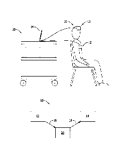
[0019] Figure 2 is an illustration of a system for monitoring the GFR in a
patient.
[0020] Figures 3A, 3B, 3C and 3D are graphs of the clearance of MB-102
illustrating a two-compartment pharmacokinetic model in four different
patients having
different GFR values ranging from 120 mL/min (3A) to 25 mL/min (3D).
[0021] Figure 4 is a graph comparing of the GFR determined using MB-102
compared to Omnipaque*.
[0022] Figure 5 is a bar graph of the percent recovery of MB-102 from the
urine
of human patients after 12 hours.
[0023] Figure 6 is a bar graph of the plasma concentration half-life of MB-102
in
human patients.
[0024] Figure 7 is a graph showing the correlation over time between the
plasma
concentration of MB-102 and the transdermal fluorescence intensity, in a
patient with a
GFR of 117 mL/min/1.73 m2.
CA 03058772 2019-10-01
WO 2019/084475 PCT/US2018/057820
-9-
[0025] Figure 8 is a graph showing the correlation over time between the
plasma
concentration of MB-102 and the trans-cutaneous fluorescence intensity, in a
patient with a
GFR of 61 mL/min/1.73 m2.
[0026] Figure 9 is a graph showing the correlation over time between the
plasma
concentration of MB-102 and the trans-cutaneous fluorescence intensity in a
patient with a
GFR of 23 mL/min/1.73 m2.
[0027] Figure 10 is a graph correlating the transdermally predicted GFR with
the
plasma measured GFR determined using MB-102 and normalized to body surface
area of
the subject (outlier exclusion method 1; hybrid offset method).
[0028] Figure 11 is a graph correlating the transdermally predicted GFR with
the
plasma measured GFR determined using MB-102 and normalized to the volume of
distribution of the tracer agent within the subject (outlier exclusion method
1, hybrid offset
method).
[0029] Figure 12 is a graph correlating the plasma-determined GFR to the trans-
cutaneous fluorescence clearance rate: GFR by Iohexol, Un-Normalized (No
outlier
exclusion; fixed offset fitting method).
[0030] Figure 13 is a graph correlating the plasma-determined GFR to the trans-
cutaneous fluorescence clearance rate: GFR by Iohexol, BSA-Normalized (No
outlier
exclusion; fixed offset fitting method).
[0031] Figure 14 is a graph correlating the plasma-determined GFR to the trans-
cutaneous fluorescence clearance rate: GFR by Iohexol, Vd-Normalized (Method
1) (No
outlier exclusion; fixed offset fitting method).
[0032] Figure 15 is a graph correlating the plasma-determined GFR to the trans-
cutaneous fluorescence clearance rate: GFR by MB-102, Un-normalized (No
outlier
exclusion; fixed offset fitting method).
CA 03058772 2019-10-01
WO 2019/084475 PCT/US2018/057820
-10-
[0033] Figure 16 is a graph correlating the plasma-determined GFR to the trans-
cutaneous fluorescence clearance rate: GFR by MB-102, BSA-Normalized (No
outlier
exclusion; fixed offset fitting method).
[0034] Figure 17 is a graph correlating the plasma-determined GFR to the trans-
cutaneous fluorescence clearance rate: GFR by MB-102, Vd-Normalized, Method 1
(No
outlier exclusion; fixed offset fitting method).
[0035] Figure 18 is a graph correlating the plasma-determined GFR to the trans-
cutaneous fluorescence clearance rate: GFR by MB-102, Vd-Normalized, Method 2
(No
outlier exclusion; fixed offset fitting method).
[0036] Figure 19 is a graph correlating the plasma-determined GFR to the trans-
cutaneous fluorescence clearance rate: Variable Offset Method (No outlier
exclusion; GFR
determination by MB-102 with Vd normalization method 2).
[0037] Figure 20 is a graph correlating the plasma-determined GFR to the trans-
cutaneous fluorescence clearance rate: Hybrid Offset Method (No outlier
exclusion; GFR
determination by MB-102 with Vd normalization method 2).
[0038] Figure 21 is a graph correlating the plasma-determined GFR to the trans-
cutaneous fluorescence clearance rate: Outlier Exclusion Method 1 (Hybrid
offset method;
GFR determination by MB-102 normalized to BSA).
[0039] Figure 22 is a graph correlating the plasma-determined GFR to the trans-
cutaneous fluorescence clearance rate: Outlier Exclusion Method 1 (Hybrid
offset method;
GFR determination by MB-102 with Vd nounalization method 2).
[0040] Figure 23 is a graph correlating the plasma-determined GFR to the trans-
cutaneous fluorescence clearance rate: Outlier Exclusion Method 2 (Hybrid
offset method;
GFR determination by MB-102 normalized to BSA).
[0041] Figure 24 is a graph correlating the plasma-determined GFR to the trans-
cutaneous fluorescence clearance rate: Outlier Exclusion Method 2 (Hybrid
offset method;
GFR determination by MB-102 with Vd normalization method 2).
CA 03058772 2019-10-01
WO 2019/084475 PCT/US2018/057820
-11-
[0042] Figure 25 is a graph summarizing the optimization of the RDTC
transition
for determining the offset method used in fitting the RDTC.
[0043] Figure 26 is a graph summarizing the optimization of the Outlier Error
Threshold for the Fluorescence Decay Rate Constant.
[0044] Figure 27 is a graph summarizing optimization of the Outlier Error
Threshold for plasma-determined GFR.
[0045] Figure 28 is a graphical depiction of the 5 stages of chronic kidney
disease
by GFR.
[0046] Figure 29a is a graph of eGFR vs. plasma PK-determined GFR,
normalized for subject body surface area (nGFR). Superimposed on the graph is
an error
grid, indicating diagnosis accuracy, by number of CKD stages. Measurements
falling
within a grid with only green sides would be correctly diagnosed by eGFR
Measurements
falling within a grid with both green and yellow sides would be incorrectly
diagnosed by
eGFR by one CKD stage Measurements falling within a grid with both yellow and
red
sides would be incorrectly diagnosed by eGFR by two CKD stages.
[0047] Figure 29b is a graph of transdermally determined GFR (tGFR) vs. plasma
PK-determined GFR, normalized for subject body surface area (nGFR).
Superimposed on
the graph is an error grid, indicating diagnosis accuracy, by number of CKD
stages.
Measurements falling within a grid with only green sides would be correctly
diagnosed by
tGFR. Measurements falling within a grid with both green and yellow sides
would be
incorrectly diagnosed by tGFR by one CKD stage. Measurements falling within a
grid with
both yellow and red sides would be incorrectly diagnosed by tGFR by two CKD
stages.
[0048] Figure 29c is a graph of transdermally determined GFR (tGFR) vs. plasma
PK-determined GFR, normalized for the volume of distribution of the tracer
agent within
the subject (nGFR). Superimposed on the graph is an error grid, indicating
diagnosis
accuracy, by number of CKD stages. Measurements falling within a grid with
only green
sides would be correctly diagnosed by tGFR. Measurements falling within a grid
with both
green and yellow sides would be incorrectly diagnosed by tGFR by one CKD
stage.
CA 03058772 2019-10-01
WO 2019/084475 PCMJS2018/057820
-12-
Measurements falling within a grid with both yellow and red sides would be
incorrectly
diagnosed by tGFR by two CKD stages.
[0049] Figure 30 is a graph of transdermally-measured GFR automatically
determined at two body sites in real-time.
DETAILED DESCRIPTION OF THE INVENTION
[0050] All references herein to the "pyrazine", "pyrazine derivative",
"pyrazine
molecule", "pyrazine compound" or "pyrazine analog" apply to all compounds of
Formula
I. Additionally each reference to the pyrazine includes all pharmaceutically
acceptable salts
thereof unless specifically stated otherwise. Salt forms may be charged or
uncharged, and
may be protonated to form the appropriate cation or deprotonated to form the
appropriate
anion. All aspects and embodiments disclosed herein are applicable to
compounds of
Formula I, and specific examples are only illustrative and non-limiting to the
scope of the
disclosure.
[0051] In one aspect, disclosed herein is a pyrazine derivative of Formula I,
or a
pharmaceutically acceptable salt thereof,
N Y2
1LT1N x2
Formula I
wherein each of Xi and X2 is independently ¨CO2R1, ¨CONR1R2, ¨CO(AA) or ¨
CONH(PS); each of Y1 and Y2 is independently selected from the group
consisting of ¨
NR1R2 and
,(c H2),
-N µZ 1
(C H2) r( =
Z1 is a single bond, ¨CR1R2¨, ¨0¨, ¨NR'¨, ¨NCOR1¨, ¨S¨, ¨SO¨, or ¨S02¨; each
of R1 to
R2 are independently selected from the group consisting of H, ¨CH2(CHOH)all, ¨
CH2(CHOH)aCH3, ¨CH2(CHOH)a C 02H, ¨(CHC 02H),C 02H, ¨(CH2CH20)cli, -
CA 03058772 2019-10-01
WO 2019/084475 PCT/US2018/057820
-13-
(CH2CH20),CH3, ¨(CH2),S03H, ¨(CH2),S03-, ¨(CH2)aSO2H, ¨(CH2)aS02-, ¨
(CH2),NHSO3H, ¨(CHANHSO 3 , ¨(CH2)aNHSO2H, ¨(CH2)aNHS02-,¨(CH2)aP04113,
(CH2)aPO4H2-, ¨(CH2)aP041-12", ¨(CH2)aP043", ¨(CH2)aP03H2, ¨(CH7)aP031-1-, and
¨
(CH2)aP032-; AA is a peptide chain comprising one or more amino acids selected
from the
group consisting of natural and unnatural amino acids, linked together by
peptide or amide
bonds and each instance of AA may be the same or different than each other
instance; PS is
a sulfated or non-sulfated polysaccharide chain comprising one or more
monosaccharide
units connected by glycosidic linkages; and 'a' is a number from 0 to 10, 'c'
is a number
from 1 to 100, and each of 'm' and 'n' are independently a number from 1 to 3.
In another
aspect, 'a' is a number from 1 to 10. In still yet another aspect, 'a' is 1,
2, 3, 4, 5, 6, 7, 8, 9
or 10.
[0052] In some aspects, at least one of and X2 is ¨CO(PS) or ¨CO(AA). In yet
another aspect, both X' and X2 are ¨CO(AA).
[0053] (AA) is a peptide chain comprising one or more natural or unnatural
amino
acids linked together by peptide or amide bonds. The peptide chain (AA) may be
a single
amino acid, a homopolypeptide chain or a heteropolypeptide chain, and may be
any
appropriate length. In some embodiments, the natural or unnatural amino acid
is an a-
amino acid In yet another aspect, the a-amino acid is a D-a-amino acid or an L-
a-amino
acid. In a polypeptide chain comprising two or more amino acids, each amino
acid is
selected independently of the other(s) in all aspects, including, but not
limited to, the
structure of the side chain and the stereochemistry. For example, in some
embodiments, the
peptide chain may include 1 to 100 amino acid(s), 1 to 90 amino acid(s), 1 to
80 amino
acid(s), 1 to 70 amino acid(s), 1 to 60 amino acid(s), 1 to 50 amino acid(s),
1 to 40 amino
acid(s), 1 to 30 amino acid(s), 1 to 20 amino acid(s), or even 1 to 10 amino
acid(s). In some
embodiments, the peptide chain may include 1 to 100 a-amino acid(s), 1 to 90 a-
amino
acid(s), 1 to 80 a-amino acid(s), 1 to 70 a-amino acid(s), 1 to 60 a-amino
acid(s), 1 to 50
a-amino acid(s), 1 to 40 a-amino acid(s), 1 to 30 a-amino acid(s), 1 to 20 a-
amino acid(s),
or even 1 to 10 a-amino acid(s). In some embodiments, the amino acid is
selected from the
group consisting of D-alanine, D-arginine D-asparagine, D-aspartic acid, D-
cysteine, D-
glutami c acid, D-glutamine, glycine, D-histidine, D-homoserine, D-isoleucine,
D-leucine,
CA 03058772 2019-10-01
WO 2019/084475 PCT/US2018/057820
-14-
D-lysine, D-methionine, D-phenylalanine, D-proline, D-serine, D-threonine, D-
tryptophan,
D-tyrosine, and D-valine. In some embodiments, the a-amino acids of the
peptide chain
(AA) are selected from the group consisting of arginine, asparagine, aspartic
acid, glutamic
acid, glutamine, histidine, homoserine, lysine, and serine. In some
embodiments, the a-
amino acids of the peptide chain (AA) are selected from the group consisting
of aspartic
acid, glutamic acid, homoserine and serine. In some embodiments, the peptide
chain (AA)
refers to a single amino (e.g., D-aspartic acid or D-serine).
[0054] In some embodiments, (AA) is a single amino acid selected from the
group consisting of the 21 essential amino acids. In other aspects, AA is
selected from the
group consisting of D-arginine, D-asparagine, D-aspartic acid, D-glutamic
acid, D-
glutamine, D-histidine, D-homoserine, D-lysine, and D-serine. Preferably, AA
is D-
aspartic acid, glycine, D-serine, or D-tyrosine. Most preferably, AA is D-
serine.
[0055] In some embodiments, (AA) is a (3-amino acid. Examples of (3-amino
acids
include, but are not limited to, 13-phenylalanine, {3-alanine, 3-amino-3-(3-
bromophenyl)propionic acid, 3 -amin obutan oi c acid, ci s-2-amino-3 -cycl
openten e-1-
carboxyl i c acid, trans-2-amino-3-cyclopentene- 1 -carboxylic acid, 3-
aminoisobutyric acid,
3 -amino-2-phenyl propi onic acid, 3 -amino-4-(4-biphenylyl)butyric acid, ci s-
3-amino-
cyclohexanecarboxylic acid, trans-3 -amino-cy cl ohexanec arb oxyl c acid, 3
amino-
cyclopentanecarboxylic acid, 3 -amino-2-hydroxy-
4-phenylbutyri c acid, 2-
(aminomethyl)phenylacetic acid, 3 -amino-2-methylpropioni c acid, 3 -amino-4-
(2-
naphthyl)butyric acid, 3-amino-5-phenylpentanoic acid, 3-amino-2-p
henylpropionic acid,
4-bromo-13-Phe-OH, 4-chloro-f3-Homophe-OH, 4-chloro-13-Phe-OH, 2-cyano-(3-
Homophe-
OH, 2-cyano-I3-Homophe-OH, 4-cyano-I3-Homophe-OH, 3-cyano-(3-Phe-OH, 4-cyano-
(3-
Phe-OH, 3,4-dimethoxy-(3-Phe-OH, y,y-dipheny[3-Homoala-OH, 4-fluoro-13-Phe-OH,
p-
Gln-OH, 13-Homoala-OH, 13-Homoarg-OH, 13-Homogln-OH, 13-Homoglu-OH, (3-Homohyp-
OH, [3-Homoleu-OH, 13-Homolys-OH, 13-Homomet-OH, 02-homophenylalanine, p-
Homophe-OH, (33-Homopro-OH, p-Homoser-OH, (3-Homothr-OH, (3-Homotrp-OH, p-
Homotrp-OMe, (3-Homotyr-OH, [3-Leu-OH, (3-Leu-OH, f3-Lys(Z)-0H, 3-methoxy-13-
Phe-
OH, 3-methoxy-I3-Phe-OH, 4-methoxy-13-Phe-OH, 4-methy-(3-Homophe-OH, 2-methyl-
(3-
Phe-OH, 3-methyl-(3-Phe-OH, 4-methyl-fl-Phe-OH, 13-Phe-OH, 4-(4-pyridy1)-13-
Homoala-
CA 03058772 2019-10-01
WO 2019/084475 PCT/US2018/057820
OH, 2-(trifluoromethyl)-(3-Homophe-OH, 3 -
(trifluoromethyl)-(3-Hom ophe-OH, 4-
(trifluoromethyl)-13-Homophe-OH, 2-(trifluoromethyl)-(3-Phe-OH, 3-
(trifluoromethyl)-3-
Phe-OH, 4-(trifluoromethyl)-P-Phe-OH, l3-Tyr-OH, Ethyl 3-
(benzylamino)propionate, 13-
Ala-OH, 3 -(amino)-5 -h ex en oi c acid, 3 -(ami n o)-2-m ethyl propi oni c
acid, 3 -(ami n o)-2-
methyl propi oni c acid, 3 -(ami no)-4-(2-n aphthyl)butyri c acid, 3 ,4-
difluoro-P-Hom ophe-OH,
7,7- di ph enyl -(3-Hom oal a-OH, 4-fluoro-p-Hom ophe-OH, (3-G1n -OH, p-Homoal
a-OH, p-
Homoarg-OH, (3-Homogln-OH, p-Homoglu-OH, (3-Homohyp-OH, p-Homoile-OH, [3-
Homoleu-OH, P-Homolys-OH, p-Homomet-OH, p-Homophe-OH, 33-homoproline, [3-
Homothr-OH, p-Homotrp-OH, p-Homotyr-OH, p-Leu-OH, 2-methyl -p-Homophe-OH, 3-
methyl-3-Hom ophe-OH, p-Phe-OH, 4-(3-pyri dy1)-3 -Hom oal a-OH, 3 -(trifl
uoromethyl)-p-
Homophe-OH, P-Glutamic acid, P-Homoalanine, P-Homoglutamic acid, 3-
Homoglutamine, p-Homohydroxyproline, p-Homoisoleucine, P-Homoleucine, p-
Homomethionine, P-Homophenylalanine, P-Homoproline, 13-Homoserine, p-
Homothreonine, P-Homotryptophan, P-Homotyrosine, P-Leucine, P-Phenylalanine,
Pyrrolidine-3-carboxylic acid and 13-Dab-OH.
[0056] (PS) is a sulfated or non-sulfated polysaccharide chain including one
or
more monosaccharide units connected by glycosidic linkages. The polysaccharide
chain
(PS) may be any appropriate length. For instance, in some embodiments, the
polysaccharide chain may include 1 to 100 monosaccharide unit(s), 1 to 90
monosaccharide unit(s), 1 to 80 monosaccharide unit(s), 1 to 70 monosaccharide
unit(s), 1
to 60 monosaccharide unit(s), 1 to 50 monosaccharide unit(s), 1 to 40
monosaccharide
unit(s), 1 to 30 monosaccharide unit(s), 1 to 20 monosaccharide unit(s), or
even 1 to 10
monosaccharide unit(s). In some embodiments, the polysaccharide chain (PS) is
a
homopolysaccharide chain consisting of either pentose or hexose monosaccharide
units. In
other embodiments, the polysaccharide chain (PS) is a heteropolysaccharide
chain
consisting of one or both pentose and hexose monosaccharide units. In some
embodiments,
the monosaccharide units of the polysaccharide chain (PS) are selected from
the group
consisting of glucose, fructose, mannose, xylose and ribose. In some
embodiments, the
polysaccharide chain (PS) refers to a single monosaccharide unit (e.g., either
glucose or
fructose). In yet another aspect, the polysaccharide chain is an amino sugar
where one or
CA 03058772 2019-10-01
WO 2019/084475 PCT/US2018/057820
more of the hydroxy groups on the sugar has been replaced by an amine group.
The
connection to the carbonyl group can be either through the amine or a hydroxy
group.
[0057] In some embodiments, for the pyrazine derivative of Formula I, at least
one of either Y1 or Y2 is
(c 1-12)m
-N µZ 1
(C H2V
where Z1 is a single bond, ¨CR1R2 , ¨0¨, ¨NR'¨, 'wow_ ,
S¨, ¨SO¨, or ¨SO2¨; and
each of R1 to R2 are independently selected from the group consisting of H, ¨
CH2(CHOH),H, ¨CH2(CHOH)aCH3, ¨CH2(CHOH)aCO2H, ¨(CHCO2H)aCO2H, ¨
(CH2CH20),H, ¨(CH2CH20)cCH3, ¨(CH2)aSO3H, ¨(CH2)aS03-, ¨(CH2)aS 02H, ¨
(CH2)aS02-, ¨(CH2)aNHSO3H, ¨(CH2)aNHS03-, ¨(CH2)aNHSO2H, ¨(CH2)aNHS02-,
(CH2)aPO4H3, ¨(CH2)aPO4H2-, ¨(CH2)aPO4H2-, ¨(CH2)aP043-, ¨(CH2)aPO3H2, ¨
(CH2),1303H-, and ¨(CH2)313032-; a, c, m and n are as describe elsewhere
herein.
[0058] In yet another aspect, at least one of Y1 and Y2 is ¨NR1R2, and R1 to
R2 are
as described above. In yet another aspect, both Y1 and Y2 are ¨NR1R2, and R'
to R2 are as
described above. Alternatively, R' and R2 are both independently selected from
the group
consisting of H, ¨CH2(CHOH)aCH3, ¨(CH2),S03H, ¨(CH2)3NHSO3H, and
¨(CH2),1303H2.
In yet another aspect, both R1 and R2 are hydrogen.
[0059] In any aspect of the pyrazine compound, one or more atoms may
alternatively be substituted with an isotopically labelled atom of the same
element. For
example, a hydrogen atom may be isotopically labelled with deuterium or
tritium; a carbon
atom may be isotopically labelled with 13C or 14C; a nitrogen atom may be
isotopically
labelled with 14N or 15N. An isotopic label may be a stable isotope or may be
an unstable
isotope (i.e., radioactive). The pyrazine molecule may contain one or more
isotopic labels.
The isotopic label may be partial or complete. For example, a pyrazine
molecule may be
labeled with 50% deuterium thereby giving the molecule a signature that can be
readily
monitored by mass spectroscopy or other technique As another example, the
pyrazine
-17-
molecule may be labeled with tritium thereby giving the molecule a radioactive
signature
that can be monitored both in vivo and ex vivo using techniques known in the
art.
[0060] Pharmaceutically acceptable salts are known in the art. In any aspect
herein, the pyrazine may be in the form of a pharmaceutically acceptable salt.
By way of
example and not limitation, pharmaceutically acceptable salts include those as
described by
Berge, et al. in J. Pharm. Sc., 66(1), 1 (1977) .
The salt may be cationic or anionic. In some
embodiments, the counter ion for the pharmaceutically acceptable salt is
selected from the
group consisting of acetate, benzenesulfonate, benzoate, besylate,
bicarbonate, bitartrate,
bromide, calcium edetate, camsylate, carbonate, chloride, citrate,
dihydrochloride, edetate,
edisylate, estolate, esylate, fumarate, gluceptate, gluconate, glutamate,
glycollylarsanilate,
hexylresorcinate, hydrabamine, hydrobromide, hydrochloride, hydroxynaphthoate,
iodide,
i sethionate, lactate, lactobionate, m al ate, maleate, mand el ate, me syl
ate, methylb romi de,
methylnitrate, methylsulfate, mucate, napsylate, nitrate, pamoate,
pantothenate, phosphate,
diphosphate, polygalacturonate, salicylate, stearate, subacetate, succinate,
sulfate, tannate,
tartrate, teocl ate, triethiodide, adipate, alginate, aminosalicylate,
anhydromethylenecitrate,
arecoline, aspartate, bisulfate, butylbromide, camphorate, digluconate,
dihydrobromide,
disuccinate, glycerophosphate, j emi sulfate, j
udrofluori de, j udroiodi de,
methylenebis(salicylate), napadi sylate, oxalate,
pectinate, persulfate,
phenylethylbarbarbiturate, picrate, propionate, thiocyanate, tosylate,
undecanoate,
benzathine, chloroprocaine, choline, diethanolamine, ethylenedi amine,
meglumine,
procaine, benethamine, clemizole, diethylamine, piperazine, tromethamine,
aluminum,
calcium, lithium, magnesium, potassium, sodium zinc, barium and bismuth. Any
functional
group in the pyrazine derivative capable of forming a salt may optionally form
one using
methods known in the art. By way of example and not limitation, amine
hydrochloride salts
may be formed by the addition of hydrochloric acid to the pyrazine. Phosphate
salts may be
formed by the addition of a phosphate buffer to the pyrazine. Any acid
functionality
present, such as a sulfonic acid, a carboxylic acid, or a phosphonic acid, may
be
deprotonated with a suitable base and a salt formed. Alternatively, an amine
group may be
protonated with an appropriate acid to form the amine salt. The salt form may
be singly
Date Recue/Date Received 2021-03-04
CA 03058772 2019-10-01
WO 2019/084475 PCT/US2018/057820
-18-
charged, doubly charged or even triply charged, and when more than one counter
ion is
present, each counter ion may be the same or different than each of the
others.
[0061] In yet another aspect, disclosed herein is a method for measuring the
renal
glomerular filtration rate (GFR) in a patient in need thereof. The method
comprises
administering to a patient a pyrazine compound, or a pharmaceutically
acceptable salt
thereof, measuring the transdermal fluorescence in said patient over a period
of time, and
determining the GFR in said patient The period of time used to determine a
single
measurement of GFR is referred to herein as the Measurement Time Window. In
many
situations it will be clinical useful to have a real-time assessment of GFR
over time.
Therefore, in some aspects of the disclosure, multiple sequential assessments
of GFR are
provided. In some aspects, the multiple sequential GFR estimates are provided
after a
single administration of the tracer agent. The total length of time over which
GFR
measurements are provided after a single injection will be referred to herein
as the Single
Injection Reporting Period. In some aspects, there is temporal overlap between
the
Measurement Time Windows. In such cases, the time interval at which GFR is
reported
(the Reporting Time Interval) is not necessarily the same as the Measurement
Time
Window. For example, in one embodiment, adjacent Measurement Time Windows
overlap
by 50%, and the Reporting Time Interval is half of the Measurement Time
Window. In
some aspects, the Measurement Time Windows have variable length. In a
preferred
embodiment, if temporally adjacent Measurement Time Windows are of differing
length,
then the overlap time period is selected to be 50% of the lesser of the two
Measurement
Time Windows. In some aspects, the GFR of a patient is determined using the
system
disclosed elsewhere herein.
[0062] In yet another aspect, the Measurement Time Window is automatically
adjusted according to a metric related to the signal quality (hereafter
referred to as a
Quality Metric). The Quality Metric may be based on estimates of the
fluorescence signal-
to-noise ratio (SNR), signal-to-background ratio (SBR), good-of-fit metrics,
correlation
coefficient, or any combination thereof. In one aspect, a line is fitted to
the log of the
fluorescence intensity vs time over the Measurement Time Window (or
equivalently, a
single exponential is fit to the fluorescence intensify vs. time). The
difference between the
CA 03058772 2019-10-01
WO 2019/084475 PCT/US2018/057820
-19-
fitted line and data ("Fitting Residual") is used to estimate the "Noise". In
one aspect, the
Noise is the root mean square (RMS) of the Fitting Residual. In another
aspect, the Noise is
the median absolute deviation (MAD) of the Fitting Residual. The "Signal" may
be defined
as the amplitude of the single exponential derived from the fit. In another
aspect, the Signal
is chosen as the difference between the fitted fluorescence at the beginning
and end of the
Measurement Time Window. In another aspect, the pre-injection fluorescence
signal level
is used to determine a "Background", and the SBR is computed by dividing the
Background into the Signal level. When using either the SNR or SBR as the
Quality
Metric, a minimum threshold may be defined and only if the Quality Metric
exceeds this
threshold will the fit be considered valid for the purpose of determining GFR.
In another
aspect, the estimated error of the time or rate constant determined by the fit
to fluorescence
vs time is used as the Quality Metric. In this case, the fit may be considered
valid only if
the computed Quality Metric is below a predetermined threshold value. In some
other
aspects, the fitted time or rate constant is defined as the Signal and the
estimated error from
the fit is defined as the Noise, and this version of SNR is used as the
Quality Metric. In
another aspect, a correlation coefficient is used as the Quality Metric. Any
of various
methods known in the art for computing the correlation coefficient may be
employed, such
as Pearson's correlation coefficient, or the concordance correlation
coefficient. In yet
another aspect, a combination of different Quality Metrics are combined into a
single
metric, or the fitted result is only considered valid for the purpose of
determining GFR if
all of the selected Quality Metrics are passed.
[0063] In another aspect, a minimum Measurement Time Window is defined, and
a Quality Metric is used to determine whether to report the GFR, or to extend
the length of
the Measurement Time Window. In one such embodiment, the length of the
Measurement
Time Window is automatically increased until the Quality Metric reaches a
threshold, at
which point the GFR is reported. In another aspect, preliminary fits are used
to the time or
rate constant, or predicted GFR, and are used to set the Measurement Time
Window to a
predetermined length. In one embodiment, the minimum Measurement time is set
to 60
minutes, at which point a fit is performed and a preliminary estimate of GFR
is made. If the
preliminary estimate of GFR is equal to above 75 mL/min/1 73 m2, then the
result is
reported to the user, and the Measurement Time Window is kept at 60 minutes.
However, if
CA 03058772 2019-10-01
WO 2019/084475 PCT/US2018/057820
-20-
the preliminary estimated of GFR is below 75 mL/min/1.73 m2, then the result
is not
reported to the user, and the Measurement Time Window is increased to 120
minutes.
[0064] In another aspect, the remaining Single Injection Reporting Period is
estimated and provided to the user periodically. The basis for estimating the
remaining
Single Injection Reporting Period may be the SNR, SBR, or estimated fitting
error, such as
the methods described above for determining a Quality Metric, but the Quality
Metric used
to determine the Measurement Time Window and the Quality Metric used to
determine the
remaining Single Injection Reporting Period may be the same or different. In
some aspects,
in addition to the Quality Metric, a fitted fluorescence decay time or rate
constant is used to
estimate the remaining Single Injection Reporting Period. In one embodiment,
the fitted
fluorescence decay time constant and the SNR are combined to predict the
remaining
Single Injection Reporting Period. The SNR is scaled to range between minimum
and
maximum values of 0 and 1, and is multiplied with the fluorescence decay time
constant.
The product is then scaled to predict the Single Injection Reporting Period.
The scaling
factor is a calibration factor that is determined through analysis of data
collected previously
on human patients, animals, in vitro studies, simulations, or any combination
thereof
[0065] In yet another aspect, filtering and/or outlier rejection are applied
to the
fluorescence data before fitting within the Measurement Time Window. Examples
of
appropriate filters include: a boxcar average, an infinite response function
filter, a median
filter, a trimmed mean filter. Examples of outlier rejection methods include
all of the above
Quality Metrics described above, but applied to a subset of the Measurement
Tine
Window.
[0066] In some aspects, the Quality Metric is computed from the measured
emission energy of the tracer agent as a function of time over a measurement
time window.
The Quality Metric may be used to determine whether or not to report the
computed GFR.
In other aspects, the Quality Metric is used to decide whether to expand the
measurement
time window. For example, the measurement time window may be automatically
expanded
until the quality metric passes a predetermined threshold, at which time the
GFR is
reported.
CA 03058772 2019-10-01
WO 2019/084475 PCT/US2018/057820
-21-
[0067] GFR normalized to patient body size is determined by fitting the
measured
emission energy of the tracer agent as a function of time over a measurement
time window
to a decay parameter. In some aspects, this decay parameter is the rate
constant (or its
inverse, referred to as a time constant) from a single exponential fit. In
some aspects the
offset of the fitted function is fixed at zero; in other aspects the offset is
a variable term in
the fit; in yet other aspects, whether the offset is fixed or allowed to vary
depends on a
preliminary assessment of the decay parameter. The measurement time window is
chosen
to begin after the tracer agent has equilibrated into the body, during the
period when the
decay of the emission intensity is due to renal clearance of the tracer agent.
The fitted rate
constant is multiplied by a calibration slope to determine the GFR normalized
to patient
body size. The calibration slope is determined through analysis of data
collected previously
on human patients, animals, in vitro studies, simulations or any combination
thereof.
[0068] Because the physical size of a patient can affect the assessment of the
functioning of the kidneys, in some aspects, a body-size metric is used to
normalize the
GFR calculation to further improve the measurement. In some aspects, the body-
size metric
used for normalizing the GFR is body surface area (BSA) In other aspects, the
body-size
metric is the volume of distribution (Yd) of the tracer agent.
[0069] The methods and system disclosed herein also permit the real-time
monitoring of GFR in a patient. Additionally, multiple GFR measurements or
determinations can be done with a single administration of a tracer agent. In
some aspects,
a single GFR measurement is determined after administration of the tracer
agent. In other
aspects, multiple GFR measurements are determined after administration of the
tracer
agent, providing a real-time GFR trend. In some such aspects, an estimate is
provided of
the time remaining during which the remaining concentration of tracer agent
will be
sufficient to continue determining GFR.
[0070] In yet another aspect, disclosed herein is a method for measuring the
renal
glomerular filtration rate (GFR) in a patient in need thereof. The method
comprises
administering to a patient a pyrazine compound, or a pharmaceutically
acceptable salt
thereof, measuring the transdermal fluorescence in said patient over a
Measurement Time
CA 03058772 2019-10-01
WO 2019/084475 PCT/US2018/057820
-22-
Window, and determining the GFR in said patient. In some aspects, the GFR of a
patient is
determined using the system disclosed elsewhere herein.
[0071] In yet another aspect, disclosed herein is a method for determining the
GFR in a patient in need thereof. The method comprises administering to said
patient a
compound of Formula I, or a pharmaceutically acceptable salt thereof, or a
phal __ it aceuti c al I y acceptable formulation thereof, measuring the
concentration of the
compound of Formula I in said patient over a Measurement Time Window, and
determining the GFR in said patient.
[0072] In some aspects and still in reference to the above mentioned method,
measuring the concentration of the pyrazine includes monitoring the
transdermal
fluorescence in the patient. In yet another aspect, measuring the
concentration of the
pyrazine includes taking aliquots of blood from the patient and measuring the
concentration
of the pyrazine by HPLC or other methods as are known it the art. For example,
a pyrazine
may incorporate a radioisotope that can be quantified. In still yet another
aspect, measuring
the concentration of the pyrazine may including collecting the urine of the
patient over a
period of time to determine the rate in which the kidneys eliminate the
compound from the
body of the patient.
[0073] In still yet another aspect and still in reference to the above
mentioned
method, the concentration of the pyrazine in the patient is monitored by
transdermal
fluorescence. This may include contacting a medical device with the skin of
the patient
wherein said medical device is configured to cause a fluorescent reaction in
the compound
of Formula I, and detecting said reaction. The medical device may contact the
skin of the
patient in any suitable location. Specific locations known to be suitable are
the sternum,
lower sternum, pectoralis major, occipital triangle, forehead, chin, upper
hip, and lower
hip. Other locations on a patient may be used as determined by convenience,
medical
device design, and/or medical necessity. In some aspects, this method uses the
system
disclosed elsewhere herein.
CA 03058772 2019-10-01
WO 2019/084475 PCT/US2018/057820
-23-
[0074] In one aspect of the above-mentioned method, a display device is used
to
prompt the user to attach the sensor at one or more particular body sites. In
one such
embodiment, a touch-screen interface is used, and the user is instructed to
touch a rendition
of the body site location at which the sensor was attached, in order to move
to a next step in
the measurement setup process. This has the benefit of discouraging placement
of the
sensor on body sites that are not appropriate or optimal for the GFR
determination.
[0075] In another aspect, the next step is setting the light source output
levels and
the detector gain levels. In one such aspect, the detector gain levels and
light source levels
are both initially set to a low state and then the light source levels are
sequentially
increased until a targeted signal level is achieved. In one embodiment, the
light source is
the excitation source for the fluorescent GFR agent, and the source drive
current is
increased until either a targeted fluorescence signal is achieved or a
predefined maximum
current is reached. In the case that the maximum source current is reached
without attaining
the desired fluorescence signal level, the detector gain is then sequentially
increased until
either the targeted fluorescence signal is achieved, or the maximum detector
gain setting is
reached.
[0076] In some aspects, measurement of the diffuse reflectance of the skin is
made in addition to measurement of fluorescence of the skin and GFR agent. In
such
aspects, the diffuse reflectance signal may be used to determine the optimum
source output
and detector gain levels. In yet further aspects, diffuse reflectance
measurements are made
within the wavelength bands for excitation and emission of the fluorescent GFR
agent. In
such aspects, setting of the LED source levels and detector gains may be
performed by
using the diffuse reflectance instead of the fluorescence signal levels to
guide the settings.
In one such aspect, the target levels or the diffuse reflectance signals are
between 15% and
35% of the signal level at which detector or amplifier saturation effects are
observed. This
provides head-room for signal fluctuations that may be associated with patient
movement
or other physiological variation. The described procedures for optimizing the
light source
output and/or detection gains have the benefit that they provide a means of
compensating
for physiological variations across different patients, or across different
body sites on the
same patient. In one aspect, a primary factor that is compensated is the
melanin content of
CA 03058772 2019-10-01
WO 2019/084475 PCT/US2018/057820
-24-
the skin. Other physiological factors that may require compensation include
blood content,
water content, and scattering within the tissue volume that is optically
interrogated by the
sensor. In another aspect, if the desired signal targets are not attained, the
user is prevented
from proceeding with the measurement. In this manner, the reporting of
inaccurate results
is prevented.
[0077] Once the desired signal levels have been successfully achieved, in
another
aspect, a baseline signal is recorded. In one such aspect, the stability of
the baseline is
assessed, such as by fitting a slope to the signal over time, and the baseline
is not accepted
as valid unless the slope over time is below a pre-determined threshold. In
some aspects, a
display device instructs the user not to proceed with administration of the
tracer agent until
a stable baseline has been achieved. In this manner, measurement is prevented
if the sensor
has not been properly positioned or attached. In addition, the user may be
prevented from
proceeding with a measurement if the tracer agent from a prior injection has
not cleared out
of the body yet to a desired degree.
[0078] Once a stable baseline is acquired, in another aspect of the above-
mentioned method, the tracer agent is injected into the vascular space of the
patient. The
tracer agent administration is automatically detected as a rapid increase in
the transdermal
fluorescence of the patient as measured by the one or more sensors. A
predetermined
threshold for the rate of change, absolute signal change, or relative signal
change may be
employed for this purpose. The automatic agent detection may be reported to
the user on a
display device, such as a touch-screen monitor. In another aspect, once the
tracer agent is
detected, a further threshold is used to determine if sufficient tracer agent
is present to
initiate a GFR measurement. In one such aspect, measurements of fluorescence
(Fmeas)
and diffuse reflectance (DR) are combined in a manner which reduces the
influence of
physiological variation on the combined result (herein referred to as the
Intrinsic
Fluorescence or IF), so that, for example, the influence of skin color on the
measurement is
compensated for. The sufficiency of the tracer agent is then assessed by
comparing the IF
to a pre-determined threshold. In some aspects the IF is determined by using a
formula of
the form:
CA 03058772 2019-10-01
WO 2019/084475 PCT/US2018/057820
-25-
Frneas
IF = Equation (1)
DRkexpRkernpRkem,filtered
ex em em,f iltered
where the subscripts on the DR terms refer to measurements collected within
the tracer
agent excitation (ex) and emission (em) wavelength bands, with both filtered
and un-
filtered detectors, and the superscripts on the DR terms are calibration
coefficients that may
be determined through analysis of data collected previously on human patients,
animals, in
vitro studies, simulations, or any combination thereof. In this manner, if
insufficient tracer
agent has been administered for an accurate GFR assessment, the medical
professional
administering the measurement may be provided the opportunity to administer
additional
tracer agent, or to discontinue the measurement.
[0079] Once the tracer agent has been administered, in another aspect, the
equilibration of the tracer agent into the extracellular space is monitored.
In one aspect, the
Measurement Time Window does not start until it has been determined that
equilibration is
sufficiently complete. A fit to an exponential function may be used to assess
equilibration
progress. For example, the change in fluorescence intensity over time may be
fit to a single
exponential function, and only once the fitted time constant is stable, is
equilibration
deemed to be complete. In one such aspect, a running estimate of when the
first GFR
determination will become available is provided to the user. In another
aspect, the user is
prevented from proceeding to the measurement phase until and unless sufficient
equilibration has been achieved. In one such aspect, the equilibration time is
compared to a
predetermined threshold, and if the equilibration time exceeds the threshold,
the user is
prevented from proceeding with GFR determination. In this manner, if the
sensor is located
in a site that is in poor exchange with the circulatory system, the assessment
of GFR is
prevented.
[0080] In some aspects, the Reporting Time Interval, Measurement Time
Window, and/or Single Injection Reporting Period are based on the specific
medical
assessment being performed and may vary accordingly. For example, for patients
with
chronic kidney failure, a single GFR determination may be sufficient. However,
for
patients with or at risk of acute kidney failure, a real-time assessment or
GFR trend
provides great potential benefit. In some aspects said Reporting Time Interval
will be
CA 03058772 2019-10-01
WO 2019/084475 PCT/US2018/057820
-26-
approximately 15 minutes. In other aspects said Reporting Time Interval will
be
approximately 30 minutes, approximately one hour, approximately two hours,
approximately three hours, approximately five hours, approximately eight
hours,
approximately 10 hours, approximately 12 hours, approximately 18 hours,
approximately
24 hours, approximately 36 hours, approximately 48 hours, approximately 72
hours,
approximately 96 hours, or approximately 168 hours In some aspects the
Reporting Time
Interval will be between 15 minutes and 168 hours. In some aspects the Single
Injection
Reporting Period will be based on the clearance half-life of the pyrazine
compound. Said
clearance half-life can be either previously determined in said patient,
estimated based on
the medical condition of said patient, or determined transdeinially using the
methods
described herein. In some aspects said Single Injection Reporting Period is
one clearance
half-life, two clearance half-lives, three clearance half-lives, four
clearance half-lives, five
clearance half-lives, six clearance half-lives, eight clearance half-lives, or
ten clearance
half-lives. The maximum Single Injection Reporting Period is such that the
pyrazine is no
longer detectable in the blood stream of said patient. "Undetectable" as used
herein means
that the concentration of the pyrazine is no longer detectable by the method
used to make
the determination. In some instances, when the detection level of the
instrument makes this
an extremely long time period (e.g., over one week), "undetectable" means that
the
concentration level has dropped below 0.39% (i.e., eight clearance half-
lives). In yet
another aspect, the Reporting Time Interval is between approximately 1 and 168
hours and
all one hour increments in between.
[0081] Likewise, the Measurement Time Window may vary according to the
specific medical needs of the patient and may vary accordingly. In some
aspects it will be
approximately 15 minutes. In other aspects said Measurement Time Window will
be
approximately 30 minutes, approximately one hour, approximately two hours,
approximately three hours, approximately five hours, approximately eight
hours,
approximately 10 hours, approximately 12 hours, approximately 18 hours,
approximately
24 hours, approximately 36 hours, approximately 48 hours, approximately 72
hours,
approximately 96 hours, or approximately 168 hours. In some aspects the
Measurement
Time Window will be between 15 minutes and 168 hours. There may be one or a
plurality
of Measurement Time Windows during each Single Injection Reporting Period In
some
CA 03058772 2019-10-01
WO 2019/084475 PCT/US2018/057820
-27-
aspects, the Single Injection Reporting Period is divided into multiple
Measurement Time
Windows where each Measurement Time Window is the same. In yet another aspect,
the
Single Injection Reporting Period is divided into multiple Measurement Time
Windows
where each Measurement Time Windows is selected independently of the others
and may
be the same or different than the other Measurement Time Windows
[0082] The methods and system disclosed herein have the benefit of
automatically
adjusting for skin melanin content, such that the GFR determination is
accurate across a
wide range of skin types and levels of pigmentation. The Fitzpatrick scale is
a numerical
classification scheme for human skin color. It is widely recognized as a
useful tool for
dermatological research into human skin pigmentation. Scores range from type I
(very fair
skin with minimal pigmentation) to type VI (deeply pigmented and dark brown).
The
system and methods disclosed herein are suitable for use with all six
categories of skin
pigmentation on the Fitzpatrick scale. Specifically, the systems and methods
disclosed
herein are suitable for use with skin pigmentation of type I, type II, type
III, type IV, type
V and type VI.
[0083] In yet another aspect, the pyrazine is combined with at least one
pharmaceutically acceptable excipient. Said pharmaceutically acceptable
excipients are
selected from the group consisting of solvents, pH adjusting agents, buffering
agents,
antioxidants, tonicity modifying agents, osmotic adjusting agents,
preservatives,
antibacterial agents, stabilizing agents, viscosity adjusting agents,
surfactants and
combinations thereof.
[0084] Pharmaceutically acceptable solvents may be aqueous or non-aqueous
solutions, suspensions, emulsions, or appropriate combinations thereof. Non-
limiting
examples of non-aqueous solvents are propylene glycol, polyethylene glycol,
vegetable oils
such as olive oil, and injectable organic esters such as ethyl oleate.
Examples of aqueous
carriers are water, alcoholic/aqueous solutions, emulsions or suspensions,
including saline
and buffered media
CA 03058772 2019-10-01
WO 2019/084475 PCT/US2018/057820
-28-
[0085] By way of example and not limitation, pharmaceutically acceptable
buffers include acetate, benzoate, carbonate, citrate, dihydrogen phosphate,
gluconate,
glutamate, glycinate, hydrogen phosphate, lactate, phosphate, tartrate, Tris-
HC1, or
combinations thereof having a pH between 4 and 9, preferably between 5 and 8,
most
preferably between 6 and 8, very most preferably between 7.0 and 7.5. In yet
another
aspect, the pH is between 6.7 and 7.7. Other buffers, as are known in the art,
may be
selected based on the specific salt form of the pyrazine derivative prepared
or the specific
medical application. A preferred buffer is phosphate buffered saline at
physiological pH
(approximately 7.2).
[0086] Examples of the tonicity modifying agent are glycerol, sorbitol,
sucrose,
or, preferably, sodium chloride and/or mannitol. Examples of the viscosity
adjusting agent
include bentonite, calcium magnesium silicate and the like. Examples of the
diluent include
ethanol, methanol and the like. Examples of the antimicrobial include
benzalkonium
chloride, benzethonium chloride, ethylparaben, methylparaben and the like.
Examples of
osmotic adjusting agents include aminoethanol, calcium chloride, choline,
dextrose,
diethanolamine, lactated Ringer's solution, meglumine, potassium chloride,
Ringer's
solution, sodium bicarbonate, sodium chloride, sodium lactate, TRIS, or
combinations
thereof. These examples are for illustration only and are not intended to be
exhaustive or
limiting.
[0087] Also disclosed herein is a method of assessing the renal function in a
patient in need thereof, said method comprises administering a compound of
Formula I, or
a pharmaceutically acceptable salt thereof, or a pharmaceutically acceptable
formulation
thereof, to a patient, exposing said patient to electromagnetic radiation
thereby causing
spectral energy to emanate from said compound of Formula I, detecting the
spectral energy
emanated from the compound, and assessing the renal function of the patient
based on the
detected spectral energy.
[0088] In some aspects, the compound of Formula I is not metabolized by the
patient; instead it is entirely eliminated by renal excretion without being
metabolized (e.g.,
no oxidation, glucuronidation or other conjugation). In some aspects, at least
95% of the
CA 03058772 2019-10-01
WO 2019/084475 PCT/US2018/057820
-29-
compound of Formula 1 is not metabolized by the patient prior to renal
excretion. In some
aspects, at least 96% of the compound of Formula I is not metabolized by the
patient prior
to renal excretion. In some aspects, at least 97% of the compound of Formula I
is not
metabolized by the patient prior to renal excretion. In some aspects, at least
98% of the
compound of Formula I is not metabolized by the patient prior to renal
excretion. In some
aspects, at least 99% of the compound of Formula I is not metabolized by the
patient prior
to renal excretion. In some embodiments, said compound is entirely eliminated
by said
patient in less than a predetermined period of time. In some aspects,
assessing the renal
function in a patient may also include determining the GFR in the patient.
[0089] The pyrazine can be administered by any suitable method. The method
will be based on the medical needs of the patient and selected by the medical
professional
administering the pyrazine or conducting the procedure. Examples of
administration
methods include, but are not limited to, transdermal, oral, parenteral,
subcutaneous, enteral
or intravenous administration. Preferably the pyrazine compound will be
administered
using intravenous or transdermal methods. In some embodiments, the pyrazine is
administered via a single bolus intravenous injection. In yet another
embodiment, the
pyrazine is administered by multiple bolus intravenous injections. As used
herein,
transcutaneous and transdermal both refer to administration through the skin
of a patient
and are used interchangeably.
[0090] As used herein, "enteral administration" refers to any method of
administration that delivers a medicament directly or indirectly to the
patient using the
gastrointestinal tract. Examples of enteral administration include, but are
not limited to,
oral, sublingual, buccal and rectal. As used herein, "parenteral
administration" refers to any
method of administration that delivers a medicament directly or indirectly to
the patient by
injection or infusion. Examples or parenteral administration include, but are
not limited to,
intravenous, intraarterial, intradermal, transdermal, subcutaneous and
intramuscular.
[0091] Also disclosed herein is a stable, parenteral composition comprising a
pyrazine derivative of Formula I and a pharmaceutically acceptable buffering
agent. The
composition has a tonicity suitable for administration to a patient via
parenteral
CA 03058772 2019-10-01
WO 2019/084475 PCT/US2018/057820
-30-
administration. The tonicity of the parenteral composition may be adjusted
using a tonicity
adjusting agent as described elsewhere herein. The composition has a pH
suitable for
administration to a patient in need thereof and may be adjusted using a buffer
or other pH
adjusting agent as described elsewhere herein. The composition has an
osmolarity suitable
for administration to a patient in need thereof, and the osmolarity of the
composition may
be adjusted using an osmolarity adjusting agent as described elsewhere herein.
The
composition is packaged in a sealed container and subjected to tenninal
sterilization to
reduce or eliminate the microbiological burden of the formulation. The
composition is
stable against degradation and other adverse chemical reactions, and possesses
a
pharmaceutically-acceptable shelf-life.
[0092] "Stable", as used herein, means remaining in a state or condition that
is
suitable for administration to a patient. Formulations according to the
present disclosure are
found to be stable when maintained at room temperature for at least 12 months,
and are
generally stable at room temperature for 12 to 24 months.
[0093] A "sterile" composition, as used herein, means a composition that has
been brought to a state of sterility and has not been subsequently exposed to
microbiological contamination, i.e. the container holding the sterile
composition has not
been compromised. Sterile compositions are generally prepared by
pharmaceutical
manufacturers in accordance with current Good Manufacturing Practice ("cGMP")
regulations of the U.S. Food and Drug Administration. In some aspects, the
composition is
packaged in a heat sterilized container. The container may be any container
suitable for use
in a medical setting, examples include, but are not limited to, a vial, an
ampule, a bag, a
bottle and a syringe.
[0094] In some embodiments, the composition can take the form of a sterile,
ready-to-use formulation for parenteral administration. This avoids the
inconvenience of
diluting a concentrated parenteral formulation into infusion diluents prior to
infusion or
injection, as well as reducing the risk of microbiological contamination
during aseptic
handling and any potential calculation or dilution error. Alternatively, the
formulation may
be a solid formulation that is diluted prior to administration to the patient.
CA 03058772 2019-10-01
WO 2019/084475 PCT/US2018/057820
-31-
[0095] The aqueous, sterile pharmaceutical composition disclosed herein is
suitable for parenteral administration to a patient in need thereof. For
example, the
composition may be administered in the form of a bolus injection or
intravenous infusion.
Suitable routes for parenteral administration include intravenous,
subcutaneous,
i ntraderm al , intramuscular, intraarti cul ar, and intrathecal . The ready-
to-use formulation
disclosed herein is preferably administered by bolus injection In some
embodiments, the
composition is suitable for transdermal delivery into the epidermis or dermis
of a patient.
Transdermal delivery methods and devices are known in the art and use a
variety of
methods to deliver the pharmaceutical composition to the patient.
[0096] The aqueous, sterile pharmaceutical composition is formulated in
combination with one or more phaimaceutically acceptable excipients as
discussed
elsewhere herein. The aqueous, sterile pharmaceutical composition is
formulated such that
it is suitable for administration to a patient in need thereof. The tonicity,
osmolarity,
viscosity and other parameters may be adjusted using agents and methods as
described
elsewhere herein.
[0097] In yet another aspect, disclosed herein is an aqueous, sterile
pharmaceutical composition for parental administration. The composition
comprises from
about 0.1 to 50 mg/mL of a pyrazine compound of Formula I. It also comprises
from about
0.01 to 2 M buffering agent as disclosed elsewhere herein. It also comprises
from about 0 ¨
500 mg/mL of an osmotic-adjusting agent and from about 0 ¨ 500 mg/mL of a
tonicity-
adjusting agent. The aqueous, sterile phaimaceutical composition may also
optionally
include one or more additional pharmaceutically acceptable excipients.
Examples of
pharmaceutically acceptable excipients may be selected from the group
consisting of
solvents, pH adjusting agents, buffering agents, antioxidants, tonicity
modifying agents,
osmolarity adjusting agents, preservatives, antibacterial agents, stabilizing
agents, viscosity
adjusting agents, surfactants and combinations thereof. Specific examples of
excipients are
disclosed elsewhere herein.
CA 03058772 2019-10-01
WO 2019/084475 PCT/US2018/057820
-32-
[0098] The pyrazine compound used in the aqueous, sterile pharmaceutical
composition is any compound disclosed herein. Specific examples include, but
are not
limited to, all of the compounds prepared in the Examples. One preferred
example is
(2R,2'R)-2,2'-((3,6-diaminopyrazine-2,5-dicarbonyl)bi s(azanediy1))bis (3 -
hy droxy-
propanoi c acid) which is the molecule illustrated in Example 12 (also
identified as MB-102
or 3,6-Diamino-N2,N5-bi5 (D-serine)-pyrazine-2,5 -di carboxamide).
[0099] The pH of the aqueous, sterile pharmaceutical composition is suitable
for
administration to a patient. In some aspects the pH is between 4 and 9,
preferably between
and 8, most preferably between 6 and 8, very most preferably between 7.0 and
7.5. In yet
another aspect, the pH is between 6.7 and 7.7. Still more preferably, the pH
is
approximately 7.2 in phosphate buffered saline.
[0100] In some aspects, the pyrazine is administered to a patient suspected or
known to have at least one medical issue with their kidneys, and the methods
disclosed
herein are used to determine the level of renal impaiiment or deficiency
present in said
patient. In some aspects, said patient has an estimated GFR (eGFR) or
previously
determined GFR of less than 110, less than 90, less than 60, less than 30, or
less than 15.
The eGFR of a patient is determined using standard medical techniques, and
such methods
are known in the art. In some aspects a patient will not have or be suspected
to have
medical issues with their kidneys. The GFR monitoring may be done as part of a
general or
routine health assessment of a patient or as a precautionary assessment.
[0101] As is known in the art, the rate in which a patient eliminates waste
from
their blood stream (i.e., clearance half-life) is dependent on the health and
proper
functioning of their renal system. "Entirely eliminated" as used in this
context means that
the level of they pyrazine in the blood stream has dropped below 0.39% (i.e.,
eight half-
lives). The clearance half-life will depend on the GFR of the patient and
slows greatly as
the functioning of the renal system degrades due to illness, age or other
physiological
factors. In a patient with no known risk factors associated with CKD, having a
normal GFR
and/or a normal eGFR, the Single Injection Reporting Period is 24 hours. In
some aspects,
the Single Injection Reporting Period for a patient with a GFR or eGFR below
110 is 24
CA 03058772 2019-10-01
WO 2019/084475 PCT/US2018/057820
-33 -
hours. For a patient with a GFR or eGFR below 90, the Single Injection
Reporting Period is
24 hours. For a patient with a GFR or eGFR below 60, the Single Injection
Reporting
Period is 48 hours. For a patient with a GFR or eGFR below 30, the Single
Injection
Reporting Period is 48 hours. In some aspects, the Single Injection Reporting
Period for a
patient with a GFR or eGFR below 110 is equal to eight clearance half-lives
For a patient
with a GFR or eGFR below 90, the Single Injection Reporting Period is equal to
eight
clearance half-lives. For a patient with a GFR or eGFR below 60, the Single
Injection
Reporting Period is equal to eight clearance half-lives. For a patient with a
GFR or eGFR
below 30, the Single Injection Reporting Period is equal to eight clearance
half-lives.
[0102] Because an increase of protein concentration in the urine of a patient
may
suggest some manner of kidney impairment or deficiency, the methods disclosed
herein are
suitable for patients whose urinalysis shows an increase in protein levels. In
some aspects,
the patient has an increased level of protein in their urine as determined by
standard
medical tests (e.g., a dipstick test). By way of example and not limitation,
the urinalysis of
a patient may show an increase in albumin, an increase in creatinine, an
increase in blood
urea nitrogen (i.e., the BUN test), or any combination thereof
[0103] Still referring to the above-mentioned method, the pyrazine derivative
is
exposed to electromagnetic radiation such as, but not limited to, visible,
ultraviolet and/or
infrared light. This exposure of the pyrazine to electromagnetic radiation may
occur at any
appropriate time but preferably occurs while the pyrazine derivative is
located inside the
body of the patient. Due to this exposure of the pyrazine to electromagnetic
radiation, the
pyrazine emanates spectral energy (e.g., visible, ultraviolet and/or infrared
light) that may
be detected by appropriate detection equipment. The spectral energy emanated
from the
pyrazine derivative tends to exhibit a wavelength range greater than a
wavelength range
absorbed. By way of example but not limitation, if an embodiment of the
pyrazine
derivative absorbs light of about 440 nm, the pyrazine derivative may emit
light of about
560 nm.
CA 03058772 2019-10-01
WO 2019/084475 PCT/US2018/057820
-34-
[0104] Detection of the pyrazine (or more specifically, the spectral energy
emanating therefrom) may be achieved through optical fluorescence, absorbance
or light
scattering techniques. In some aspects, the spectral energy is fluorescence.
In some
embodiments, detection of the emanated spectral energy may be characterized as
a
collection of the emanated spectral energy and the generation of an electrical
signal
indicative of the collected spectral energy. The mechanism(s) utilized to
detect the spectral
energy from the pyrazine derivative present in the body of a patient may be
designed to
detect only selected wavelengths (or wavelength ranges) and/or may include one
or more
appropriate spectral filters. Various catheters, endoscopes, ear clips, hand
bands, head
bands, surface coils, finger probes and other medical devices may be utilized
to expose the
pyrazine derivative to electromagnetic radiation and/or to detect the spectral
energy
emanating therefrom. The device that exposes the pyrazine to electromagnetic
radiation
and detects the spectral energy emanated therefrom may be the same or
different. That is,
one or two devices may be used. The detection of spectral energy may be
accomplished at
one or more times intermittently or may be substantially continuous.
[0105] Renal function, or GFR, of the patient is determined based on the
detected
spectral energy. This is achieved by using data indicative of the detected
spectral energy
and generating an intensity/time profile indicative of a clearance of the
pyrazine derivative
from the body of the patient. This profile may be correlated to a
physiological or
pathological condition. For example, the patient's clearance profiles and/or
clearance rates
may be compared to known clearance profiles and/or rates to assess the
patient's renal
function and to diagnose the patient's physiological condition. In the case of
analyzing the
presence of the pyrazine derivative in bodily fluids, concentration/time
curves may be
generated and analyzed (preferably in real time) in order to assess renal
function.
Alternatively, the patient's clearance profile can be compared to one or more
previously
measured clearance profiles from the same patient to determine if the kidney
function of
said patient has changed over time. In some aspects, renal function assessment
is done
using the system disclosed elsewhere herein.
CA 03058772 2019-10-01
WO 2019/084475 PCT/US2018/057820
-35-
[0106] Physiological function can be assessed by: (1) comparing differences in
manners in which normal and impaired cells or organs eliminate the pyrazine
derivative
from the bloodstream; (2) measuring a rate of elimination or accumulation of
the pyrazine
in the organs or tissues of a patient; and/or (3) obtaining tomographic images
of organs or
tissues having the pyrazine associated therewith. For example, blood pool
clearance may be
measured non-invasively from surface capillaries such as those in an ear lobe
or a finger, or
it can be measured invasively using an appropriate instrument such as an
endovascular
catheter. Transdermal fluorescence can also be monitored non-invasively on the
body of
said patient. Many locations on the epidermis of a patient may be suitable for
non-
invasively monitoring the transdermal fluorescence. The site on the patient is
preferably
one where vasculature to tissue equilibrium occurs relatively quickly.
Examples of suitable
sites on a patient include, but are not limited to, the sternum, the lower
sternum, pectoralis
major, the occipital triangle, the forehead, the chin, the upper hip, and the
lower hip.
Accumulation of a pyrazine derivative within cells of interest can be assessed
in a similar
fashion.
[0107] A modified pulmonary artery catheter may also be utilized to, inter
alia,
make the desired measurements of spectral energy emanating from the pyrazine
derivative.
The ability for a pulmonary catheter to detect spectral energy emanating from
said pyrazine
is a distinct improvement over current pulmonary artery catheters that measure
only
intravascular pressures, cardiac output and other derived measures of blood
flow.
Traditionally, critically ill patients have been managed using only the above-
listed
parameters, and their treatment has tended to be dependent upon intermittent
blood
sampling and testing for assessment of renal function. These traditional
parameters provide
for discontinuous data and are frequently misleading in many patient
populations.
[0108] Modification of a standard pulmonary artery catheter only requires
making
a fiber optic sensor thereof wavelength-specific. Catheters that incorporate
fiber optic
technology for measuring mixed venous oxygen saturation exist currently. In
one
characterization, a modified pulmonary artery catheter incorporates a
wavelength-specific
optical sensor into a tip of a standard pulmonary artery catheter. This
wavelength-specific
optical sensor is utilized to monitor renal function-specific elimination of a
designed
CA 03058772 2019-10-01
WO 2019/084475 PCT/US2018/057820
-36-
optically detectable chemical entity such as the pyrazine derivatives
disclosed herein. Thus,
real-time renal function can be monitored by the disappearance/clearance of an
optically
detected compound.
[0109] In some aspects, the pyrazine compound is administered to a patient
wherein said patient has been previously diagnosed with at least Stage 1 CKD.
In other
aspects, said patient has been previously diagnosed with Stage 2 CKD, Stage 3
CKD, Stage
4 CKD or Stage 5 CKD. In yet another aspect, the patient has not yet been
diagnosed with
CKD but has one or more risk factors associated with CKD. In yet another
aspect, the
patient has no known risk factors for CKD.
[0110] Administration of the pyrazine compound is done by any suitable method
based on the medical test being performed and the medical needs of the
patient. Suitable
methods are disclosed elsewhere herein. Preferably, the pyrazine is
administered by either
transdermal or intravenous administration.
[0111] Also disclosed herein is a system for determining the GFR or assessing
the
renal function in a patient in need thereof. The system comprises a computing
device, a
display device communicatively coupled to said computing device, a power
supply that is
operatively coupled to said computing device and maintains electrical
isolation of the
system from external power sources, one or more sensor heads operatively
coupled to said
computing device, and at least one tracer agent configured to emit light when
exposed to
electromagnetic radiation. The computing device is configured to operate and
control the
sensor heads, record one or more light measurements sent from said sensor
heads, and
calculate the GFR of said patient based on said light measurements.
[0112] It should be noted that, as used herein, the term "couple" is not
limited to a
direct mechanical, electrical, and/or communication connection between
components, but
may also include an indirect mechanical, electrical, and/or communication
connection
between multiple components. The display device and the one or more sensor
heads may
communicate with the computing device using a wired network connection (e.g.,
Ethernet
or an optical fiber), a wireless communication means, such as radio frequency
(RF), e.g.,
FM radio and/or digital audio broadcasting, an Institute of Electrical and
Electronics
CA 03058772 2019-10-01
WO 2019/084475 PCT/US2018/057820
-37-
Engineers (IEEE ) 802.11 standard (e.g., 802.11(g) or 802.11(n)), the
Worldwide
Interoperability for Microwave Access (vimAxe) standard, a short-range
wireless
communication channel such as BLUETOOTH , a cellular phone technology (e.g.,
the
Global Standard for Mobile communication (GSM)), a satellite communication
link, and/or
any other suitable communication means. IEEE is a registered trademark of the
Institute of
Electrical and Electronics Engineers, Inc., of New York, New York. WIMAX is a
registered trademark of WiMax Forum, of Beaverton, Oregon. BLUETOOTH is a
registered trademark of Bluetooth SIG, Inc. of Kirkland, Washington
[0113] In some aspects, the one or more sensor heads comprise at least one
source
of electromagnetic radiation, generate and deliver electromagnetic radiation
to the skin of
said patient, detect and measure electromagnetic radiation emitted by said
tracer agent, and
transmit said measurement of electromagnetic radiation emitted by said tracer
agent to said
computing device. In a system with more than one sensor head, each sensor head
may be
the same or different and the electromagnetic radiation emitted therefrom may
be the same
or different. In some aspects the sensor heads are configured to attach to the
skin of said
patient. By way of example and not limitation, in a system with two sensor
heads, one
sensor head may emit and monitor one wavelength of electromagnetic radiation
while the
second sensor head may emit and monitor a different wavelength. This would
enable the
data to be compared to increase the accuracy of the GFR determination and the
information
available to the medical professional administering the assessment. In yet
another
nonlimiting example, in a system with two sensor heads, the two sensor heads
are used to
separate the local equilibration kinetics from the terminal phase kinetics.
This enables a
medical professional to determine when equilibration is complete and reduces
artifacts due
to local movement of the sensors.
[0114] In some aspects, the tracer agent is configured to be administered to
said
patient via intravenous or transdermal administration, be eliminated by only
glomerular
filtration in the kidneys of said patient, and emit light that is detectable
by said sensor
heads when exposed to electromagnetic radiation. In some aspects, the tracer
agent is a
pyrazine compound of Formula I as disclosed elsewhere herein. Preferably the
tracer agent
is a compound prepared in one of the Examples. Most preferably, the tracer
agent is
CA 03058772 2019-10-01
WO 2019/084475 PCT/US2018/057820
-38-
(2R,2'R)-2,2'-((3 ,6-diaminopyrazine-2,5-dicarb onyl)bi s(azanediy1))bis (3-
hydroxypropanoic acid) (also called MB-102 or 3,6-diamino-N2,N5-bis(D-serine)-
pyrazine-2,5-dicarboxamide). In some aspects, the tracer agent is the pyrazine
derivative in
a formulation suitable for administration to a patient in need thereof Such
formulations are
described elsewhere herein
[0115] The system for determining the GFR or assessing the renal function in a
patient may be configured to carry out the methods disclosed herein on a
patient in need
thereof. The computing device in the system may be any standard computer
having all of
the capabilities implied therewith, specifically including, but not limited
to, a permanent
memory, a processor capable of complex mathematical calculations, a user
interface for
interacting with the computer, and a display device communicatively coupled to
the
computing device. As such, the permanent memory of the computing device may
store any
infoimation, programs and data necessary to carry out the functions of the
system for
determining the GFR or assessing the renal function in a patient. Such
information,
programs, and data may be standards and/or controls which may be used to
compare
transdermal fluorescence values collected by the one or more sensor heads to
known
values. In some aspects, the computing device may save results from a previous
assessment
or GFR determination in a patient so that results obtained at a later date may
be compared.
This would permit a medical professional to monitor the health of the kidneys
of a patient
over time. In some aspects, the computing device is a laptop computer.
[0116] In various aspects, the computing device includes a processor and/or a
memory device. In various other aspects, the processor is coupled to and one
or more of a
user interface, a display device, and the memory device via a system bus. In
one aspect,
the processor communicates with the user, such as by prompting the user via
the display
device and/or by receiving user inputs via the user interface. The term
"processor" refers
generally to any programmable system including systems and microcontrollers,
reduced
instruction set circuits (RISC), application specific integrated circuits
(ASIC),
programmable logic circuits (PLC), and any other circuit or processor capable
of executing
the functions described herein The above examples are exemplary only, and thus
are not
intended to limit in any way the definition and/or meaning of the term
"processor."
CA 03058772 2019-10-01
WO 2019/084475 PCT/US2018/057820
-39-
[0117] In various aspects, the memory device includes one or more devices that
enable information, such as executable instructions and/or other data, to be
stored and
retrieved. Moreover, the memory device includes one or more computer readable
media,
such as, without limitation, dynamic random access memory (DRAM), static
random
access memory (SRAM), a solid state disk, and/or a hard disk In various
aspects, the
memory device stores, without limitation, application source code, application
object code,
configuration data, additional input events, application states, assertion
statements,
validation results, and/or any other type of data.
[0118] The user interface is configured to receive at least one input from a
user,
such as an operator of the system for determining the GFR or assessing the
renal function
in a patient. In one aspect, the user interface includes a keyboard that
enables the user to
input pertinent information. In various other aspects, the user interface
includes, but is not
limited to, a pointing device, a mouse, a stylus, a touch sensitive panel
(e.g., a touch pad, a
touch screen), a gyroscope, an accelerometer, a position detector, and/or an
audio input
interface (e.g., including a microphone).
[0119] The display device is configured to display information, such as input
events and/or validation results, to the user. The display device may also
include a display
adapter that is coupled to at least one display device. In one aspect, the
display device may
be a visual display device, such as a cathode ray tube (CRT), a liquid crystal
display
(LCD), an organic LED (OLED) display, and/or an "electronic ink" display. In
various
other aspects, the display device includes an audio output device (e.g., an
audio adapter
and/or a speaker) and/or a printer. In some aspects, the display device
includes a touch
screen.
[0120] In various aspects, the computing device generally comprises a
processor.
The processor may be programmed by encoding an operation using one or more
executable
instructions and providing the executable instructions in the memory device.
In one aspect,
the processor is programmed to calculate the time constant for renal decay
over a
predetermined period of time. In one aspect, the transdermal fluorescence data
in a patient
is collected over a predetermined period of time, and a graph is prepared of
time (x-axis)
CA 03058772 2019-10-01
WO 2019/084475 PCMJS2018/057820
-40-
versus fluorescence (y-axis). The rate of decay may be curved or linear and a
time constant
for the rate of decay is calculated. In one aspect, the rate of decay is
linear for a semilog(y)
plot. The time constant is compared to known values thereby determining the
GFR in the
patient. In some aspects, the rate of decay corresponds to standard first
order kinetics. In
yet another aspect, the rate of decay may exhibit a multi-compartment
pharmacokinetic
model. Figures 3A to 3D illustrate two-compartment pharmacokinetics by which
standard
pharmacokinetic software is able to determine the time constant for renal
decay.
[0121] GFR determination is done using linear regression, outlier exclusion,
calculation of the correlation coefficient (R2) and standard error of
calibration and more
fully described in the Examples.
[0122] This written description uses examples to disclose the invention,
including
the best mode, and also to enable any person skilled in the art to practice
the invention,
including making and using any devices or systems and performing any
incorporated
methods. Any aspect or embodiment disclosed herein may be used in combination
with any
other aspect or embodiment as would be understood by a person skilled in the
art. Other
examples are intended to be within the scope of the claims if they have
structural elements
that do not differ from the literal language of the claims, or if they include
equivalent
structural elements with insubstantial differences from the literal languages
of the claims.
[0123] Example 1
[0124] Preparation of 3, 6-di
amino-N2,N2,N5,N5 -tetraki s(2-
methoxy ethyl)pyrazine-2,5-di carb oxami de
0
NH2 OCH3
H3C0 H2N N
0
[0125] A mixture of 3,6-diaminopyrazine-2,5-dicarboxylic acid (200 mg, 1.01
mmol), bis-2-(methoxyethyl)amine (372 ttL, 335.5 mg, 2.52 mmol), 110Bt.H20
(459 mg,
3.00 mmol), and EDC.HC1 (575 mg, 3.00 mmol) were stirred together in DMF (20
mL) for
CA 03058772 2019-10-01
WO 2019/084475 PCT/US2018/057820
-41-
1 h at room temperature. The mixture was concentrated to dryness and the
residue was
partitioned with Et0Ac and water. The layers were separated and the Et0Ac
solution was
washed with saturated NaHCO3 and brine. The solution was dried over anhydrous
Na2SO4,
filtered and concentrated. Purification by radial flash chromatography (SiO2,
10/1 CHC13-
Me0H) afforded 228.7 mg (53% yield) of Example I as an orange foam: 11-1 NMR
(300MHz, CDC13), 8 4.92 (s, 4 H), 3.76 (apparent t, J = 5.4 Hz, 4 H), 3.70
(apparent t, J =
5.6 Hz, 4 H), 3.64 (apparent t, J = 5.4 Hz, 4 H), 3.565 (apparent t, J = 5.4
Hz), 3.67 (s, 6
H), 3.28 (s, 6 H). 13C NMR (75 MHz, CDC13) 8 167.6 (s), 145.6 (s), 131.0 (s),
72.0 (t), 70.8
(t), 59.2 (q), 49.7 (t), 47.1 (t). LCMS (5-95% gradient acetonitrile in 0.1%
TFA over 10
min), single peak retention time = 3.14 min on 30 mm column, (M+H)+ = 429.
UV/vis (100
[iM in PBS) Xabs ¨ 394 nm. Fluorescence (100 nm) Xex = 394 nm Act], = 550 nm.
[0126] Example 2
[0127] 3,6-diamino-N2,N5-bi s(2,3 -dihydroxypropyl)pyrazine-2,5 -dicarboxami
de
0
N
H I OH
OH H2N
0
[0128] Step 1 . Synthesis of 3,6-diamino-N2,N5-bis((2,2-dimethy1-1,3-dioxolan-
4-
yl)m ethyl)pyrazine-2, 5-di c arb oxami de.
0
____________________________ 0 H
H2N N
N
0
[0129] A mixture of 3,6-diaminopyrazine-2,5-dicarboxylic acid (350 mg, 1.77
mmol), racemic (2,2-dimethy1-1,3-dioxolan-4-yl)methanamine (933 lit, 944 mg,
7.20
mmol), HOBt=H20 (812 mg, 5.3 mmol), and EDC=HC1 (1.02 g, 5.32 mmol) were
stirred
together in DMF (20 mL) for 16 h at room temperature. The mixture was
concentrated to
dryness and the residue was partitioned with Et0Ac and water. The layers were
separated
CA 03058772 2019-10-01
WO 2019/084475 PCT/US2018/057820
-42-
and the Et0Ac solution was washed with saturated NaHCO3 and brine. The
solution was
dried over anhydrous Na2SO4, filtered and concentrated to afford 665 mg (88%
yield) of
the bis-amide diastereomeric pair as a yellow solid: 1NMR (300 MHz, CDC13) 8
8.38 (t, J =
5.8 Hz, 2 H), 6.55 (s, 4 H), 4.21 (quintet, J = 5.8 Hz, 2 H), 3.98 (dd, J =
8.4 Hz, 6.3 Hz, 2
H), 3.65 (dd, J = 8.4 Hz, J = 5.8 Hz, 2 H), 3.39 (apparent quartet -
diastereotopic mixture, J
= 5.9 Hz, 4 H), 1.35 (s, 6 H), 1.26 (s, 6 H). 13C NMR (75 MHz, CDC13) 8 165.7
(s), 146.8
(s), 126.8 (s), 109.2 (s), 74.8 (d), 67.2 (t), 42.2, 41.1 (t - diastereotopic
pair), 27.6 (q), 26.1
(q).
[0130] Step 2. The product from Step 1 was dissolved in THF (100 mL) and
treated with 1.0 N HC1 (2 mL). After hydrolysis was complete, the mixture was
treated
with K2CO3 (1 g) and stirred for 1 h and filtered through a plug of C18 with
using
methanol. The filtrate was concentrated to dryness and the residue was
triturated with
Me0H (50 mL). The solids were filtered and discarded and the residue was
treated with
ether (50 mL) The precipitate was collected by filtration and dried at high
vacuum. This
material was purified by radial flash chromatography to afford 221 mg (36%
yield ) of
Example 2 as an orange solid: INMR (300 MHz, DMSO-d6) 8 8.00 (bm, 6 H), 5.39
(bs, 2
H), 4.88 (bs, 2 H), 3.63-3.71 (complex m, 2 H), 3.40 (dd, J= 11.1, 5.10 Hz, 2
H), 3.28 (dd,
J = 11.1, 6.60 Hz, 2 H), 2.92 (dd, J = 12.6, 3.3 Hz, 2 H), 2.65 (dd, J = 12.6,
8.4 Hz, 2 H).
LCMS (5-95% gradient acetonitrile in 0.1% TFA over 10 min), single peak
retention time
= 4.13 min on 30 mm column, (M+H)+ = 345. UV/vis (100 M in H2O) A.
-abs = 432 nm.
Fluorescence Xeõ = 432 nm, kerr, = 558 nm.
[0131] Example 3
[0132] (2S,2'S)-2,2'-((3,6-diaminopyrazine-2,5-dicarbonyl)bis(azanediy1))bis(3-
hydroxypropanoic acid)
HO
0
HOy TIN.rH2 0
H
0 N
H2N N OH
OH
0
CA 03058772 2019-10-01
WO 2019/084475 PCT/US2018/057820
-43-
[0133] Step /. Synthesis of dim ethyl
2,243,6-di aminopyrazine-2,5 -
dicarbonyl)bi s(azanediy1))(2 S,2' S)-bi s(3-(benzyloxy)propanoate).
0
0
H3C0,1<s),-- 11 Ntk..õ,NHH2 a
0 I N
H2N N - OC H3
0
110
[0134] A mixture of sodium 3,6-diaminopyrazine-2,5-dicarboxylate (300 mg, 1.24
mmol), L-Ser(OBn)-0Me.HC1 salt (647 mg, 2.64 mmol), HOBt.H20 (570 mg, 3.72
mmol)
and EDC.HC1 (690 mg, 3.60 mmol) in DMF (25 mL) was treated with TEA (2 mL).
The
resulting mixture was stirred for 16 h and concentrated. Work up as in Example
1 afforded
370 mg (51% yield) of the bisamide as a bright yellow powder: 11\IMR (300
114Hz, CDC13):
8 8.47 (d, J = 8.74 Hz, 2 H), 7.25-7.37 (complex m, 10 H), 5.98 (bs, 4 H),
4.85 (dt, J = 8.7,
3.3 Hz, 2 H), 4.56 (ABq, J = 12.6, Hz, Av = 11.9 Hz, 4 H), 3.99 (one half of
an ABq of d, J
= 8.7, 3.3, Av obscured, 2 H), 3.76-3.80 (one half of an ABq ¨ obscured, 2 H),
3.78 (s, 6
H). NMR (75
MHz, CDC13) 6 170.5 (s), 165.1 (s), 146.8 (s), 138.7 (s) 128.6 (d), 128.1
(d), 127.8 (d), 126.9 (s), 73.5 (t), 69.8 (t), 53.0 (q), 52.9 (q). LCMS (5-95%
gradient
acetonitrile in 0.1% TFA over 10 min), single peak retention time = 4.93 min
on 30 mm
column, (M+H)+ = 581.
CA 03058772 2019-10-01
WO 2019/084475 PCT/US2018/057820
-44-
[0135] Step 2. Synthesis of (2S,2 S)-2,2'43 ,6-diaminopyrazine-2,5-dicarbony1)-
bi s(azanediy1))bi s(3 -(b enzyloxy)propanoi c acid.
o
HO NNNH2
0 N
H2N N - OH
0
0
1:10
[0136] The product from step 1 (370 mg, 0.64 mmol) in THF (10 mL) was treated
with
1.0 N sodium hydroxide (2.5 mL). After stirring at room temperature for 30
min, the
reaction was judged complete by TLC. The pH was adjusted to approximately 2 by
the
addition of 1.0 N HC1 and the resulting solution was extracted (3x) with
Et0Ac. The layers
were combined, dried over sodium sulfate, filtered and concentrated to afford
353 mg
(100% yield) of the diacid as an orange foam: LCMS (5-95% gradient
acetonitrile in 0.1%
TFA over 10 min), retention time = 4.41 min on 30 mm column, (M+H)- = 553.
[0137] Step 3. To the product from step 2 (353 mg, 0.64 mmol) in methanol (20
mL) was
added 5% Pd/C (300 mg) and ammonium formate (600 mg). The resulting reaction
was
heated at reflux for 2 h. The reaction was cooled to room temperature,
filtered through a
plug of celite and concentrated. The residue was recrystallized from methanol-
ether to
provide 191 mg (80% yield) of Example 3 as a yellow foam: 1NMR (300 MHz, DMSO-
d6)
6 8.48 (d, J = 6.9 Hz, 2 H), 6.72 (bs, 4 H), 3.95 (apparent quartet, J = 5.1
Hz, 2 H), 3.60
(apparent ABq of doublets; down-field group centered at 3.71, J = 9.9, 5.1 Hz,
2H; up-field
group centered at 3.48, J = 9.9, 6.3 Hz, 2 H). 13C NMR (75 MHz, CDC13) 6 172.9
(s), 164.9
(s), 147.0 (s), 127.0 (s), 62.9 (d), 55.7 (t). LCMS (5-95% gradient
acetonitrile in 0.1% TFA
over 10 min), single peak retention time = 1.45 min on 30 mm column, (M+H) =
373.
UV/vis (100 iM in PBS) Xabs = 434 nm. Fluorescence Xex = 449 nm, Xerr, = 559
nm.
CA 03058772 2019-10-01
WO 2019/084475 PCT/US2018/057820
-45-
Example 4
[0138] 3, 6-bi s(bis(2-methoxyethyl)amino)-N2,N2,N5,N5-tetraki s(2-
methoxyethyl)
pyrazine-2,5-dicarboxamide bis(TFA) salt
OCH3
OCH3
:NAIN
..00H3 -2 CF3CO2H
N N
0 L
:H3 LI OCH3
OCH3
[0139] Step /. Synthesis of 3,6-dibromopyrazine-2,5-dicarboxylic acid.
0
HO Br
Br ¨N
OH
[0140] 3,6-Diaminopyrazine-2,5-dicarboxylic acid (499 mg, 2.52 mmol) was
dissolved in
48% hydrobromic acid (10 mL) and cooled to 0 C in an ice-salt bath. To this
stirred
mixture was added a solution of sodium nitrite (695 mg, 10.1 mmal) in water
(10 mL)
dropwi se so that the temperature remains below 5 C. The resulting mixture
was stirred for
3 h at 5-15 C, during which time the red mixture became a yellow solution.
The yellow
solution was poured into a solution of cupric bromide (2.23 g, 10.1 immol) in
water (100
mL) and the resulting mixture was stirred at room temperature. After an
additional 3 h, the
aqueous mixture was extracted with Et0Ac (3x). The combined extracts were
dried
(Na2SO4), filtered and concentrated to afford 440 mg (54% yield) 3,6-
dibromopyrazine-
2,5-dicarboxylic acid as a pale yellow solid: 1-3C NMR (75 MHz, CDC13) 6 164.3
(s), 148.8
(s), 134.9 (s). HPLC (5-95% gradient acetonitrile in 0.1% TFA over 10 min),
single peak
retention time = 2.95 min on 250 min column.
CA 03058772 2019-10-01
WO 2019/084475 PCT/US2018/057820
-46-
[0141] Step 2. Synthesis of 3-(Bis(2-methoxyethyl)amino)-6-bromo-N2,N2,N5,N5-
tetraki s(2-methoxy ethyl)pyrazine-2,5 -dicarb oxamid e.
OCH3
H3CO. 0cH3
0
H3CO
Br
0 OC H3
[0142] The product from step 1 (440 mg, 1.36 mmol) was dissolved in DMF (25
mL),
treated with HOBt.H20 (624 mg, 4.08 mmol), and EDC.HC1 (786 mg, 4.10 mmol) and
stirred for 30 min at room temperature. Bis(2-methoxylethyl)amine (620 mL, 559
mg, 4.20
mmol) was added and the resulting mixture was stirred at room temperature for
16 h and
concentrated. The residue was partitioned with water and Et0Ac. The Et0Ac
layer was
separated and the aqueous was extracted again with Et0Ac. The combined organic
layers
were washed with 0.5 N HC1, saturated sodium bicarbonate, and brine. The
organic layer
was dried (Na2SO4), filtered and concentrated to afford 214 mg of 3-(bis(2-
methoxyethyl)amino)-6-bromo-N2,N2,N5,N5-tetrakis(2-methoxyethyl)pyrazine-2,5-
dicarboxamide (26% yield) as a brown oil: LCMS (5-95% gradient acetonitrile in
0.1%
TFA over 10 min), single peak retention time = 3.85 min on 30 mm column, (M+H)-
=
608.
[0143] Step 3. To the product from step 2 (116 mg, 0.19 mmol) was added bis(2-
methoxylethyl)amine (3.0 mL, 2.71 g, 20.3 mmol) and a "spatula tip" of
Pd(PPh3)4. The
resulting mixture was heated to 140 C for 2 h. The reaction was cooled and
concentrated.
The residue was purified by flash chromatography (SiO2, 10/1 CHC13-Me0H). The
resulting material was purified again by reverse phase medium pressure
chromatography
(C18, 10-50% manual gradient acetonitrile in 0.1% TFA) to afford 12 mg (10%
yield) of
Example 4 as an orange-brown film: LCMS (15-95% gradient acetonitrile in 0.1%
TFA
over 10 min), single peak retention time = 3.85 min on 250 mm column, (M+H)- =
661.
UV/vis (100 iM in PBS) X
¨abs = 434 nm. Fluorescence X, = 449 nm, X0m = 559 nm.
CA 03058772 2019-10-01
WO 2019/084475 PCMJS2018/057820
-47-
[0144] Example 5
[0145] 3,6-diamino-N2,N5-bis(2-aminoethyl)pyrazine-2,5-dicarboxamide bis(TFA)
salt
0
H2NN N,. NH2 = 2 CF3CO2H
H I I H
li, N
_A/
H2N Nr. . - N H2
0
[0146] Step I. Synthesis of 3, 6-diamino-N2,N5-bi s[2-(tert-
butoxycarbony1)-
aminoethyl]pyrazine-2,5-dicarboxamide.
0
H
..,0.1.(N.N.J-L-1\1.,,./õN H2 0
H I H
ji
0
H
0
[0147] A mixture of sodium 3,6-diaminopyrazine-2,5-dicarboxylate (500 mg, 2.07
mmol), tert-butyl 2-aminoethylcarbamate (673 mg, 4.20 mmol), HOBt.H20 (836 mg,
5.46
mmol) and EDC.HC1 (1.05 g, 5.48 mmol) in DMF (25 mL) was stirred for 16 h and
concentrated. Work up as in Example 1 afforded 770 mg (76% yield) of the
bisamide as an
orange foam: INMR (300 MHz, DMSO-d6) major conformer, 8 8.44 (t, J = 5.7 Hz, 2
H),
6.90 (t, J = 5.7 Hz, 2 H), 6.48 (bs, 4 H), 2.93-3.16 (complex m, 8 H), 1.37
(s, 9 H), 1.36 (s,
9 H). '3C NMR (75 MHz, DMSO-d6), confoimational isomers 8 165.1 (s), 155.5
(bs),
155.4 (bs), 146.0 (s), 126.2 (s), 77.7 (bs), 77.5 (bs), 45.2 (bt), 44.5 (bt),,
28.2 (q).
[0148] Step 2. To the product from step 1 (770 mg, 1.60 mmol) in methylene
chloride
(100 mL) was added TFA (25 mL) and the reaction was stirred at room
temperature for 2 h.
The mixture was concentrated and the residue taken up into methanol (15 mL).
Ether (200
mL) was added and the orange solid precipitate was isolated by filtration and
dried at high
vacuum to afford 627 mg (77% yield) of Example 5 as an orange powder: INMR
(300
MHz, DMSO-d6) 8 8.70 (t, J = 6 Hz, 2 H), 7.86 (bs, 6 H), 6.50 (bs, 4 H), 3.46-
3.58 (m, 4
H), 3.26-3.40 (m, 4 H). 13C NMR (75 MHz, DMSO-d6) 8 166.4 (s), 146.8 (s),
127.0 (s),
39.4 (t), 37.4 (t). LCMS (5-95% gradient acetonitrile in 0.1% TFA over 10
min), single
CA 03058772 2019-10-01
WO 2019/084475 PCT/US2018/057820
-48-
peak retention time = 362 min on 30 mm column, (M+H) = 283. UV/vis (100 IIM
in
PBS) Xabs = 435 nm. Fluorescence (100 nM) Xeõ = 449 nm, kem = 562 nm.
[0149] Example 6
[0150] 3,6-Diamino-N2,N5-bis (D-aspartate)-pyrazine-2,5-dicarboxamide
OH
0
H 0 N N N H,
- 0
H I H
0 H2N N
OH
0 0
OH
[0151] Step 1. Synthesis of 3,6-Diamino-N2,N5-bis (benzyl D-0-benzyl-
aspartate)-
pyrazine-2,5-dicarboxamide
OBz
05,, N 0
Bz 0 N õ N
0
H) H
0 H2 N N
OBz
0 0
OBz
A mixture of sodium 3,6-diaminopyrazine-2,5-dicarboxylate (600 mg, 2.48 mmol),
Asp(OBn)-0Me-p-TosH salt (2.43 g, 5.00 mmol), HOBt.H20 (919 mg, 6.00 mmol) and
EDC-HC1 (1.14 g, 5.95 mmol) in DMF (50 mL) was treated with TEA (4 mL). The
resulting mixture was stirred over night at room temperature. The reaction
mixture was
concentrated and the residue was partitioned with water and Et0Ac. The Et0Ac
layer was
separated and washed successively with saturated sodium bicarbonate, water and
brine.
The Et0Ac solution was dried (Na2SO4), filtered and concentrated. The residue
was
purified by flash chromatography (Si02, 50/1 CHC13-Me0H to 10/1) to afford
1.15 g of the
bis-amide (58% yield) as a yellow foam: iNMR (500 MHz, CDC13) 8 8.61 (d, J =
8.4 Hz, 2
H), 7.29-7.39 (m, 20 H), 5.85 (bs, 4 H), 5.22 (ABci, J = 10.0 Hz, Av = 17.3
Hz, 4 H), 5.10
CA 03058772 2019-10-01
WO 2019/084475 PCT/US2018/057820
-49-
(ABq, J = 12.2 Hz, Av = 34.3 Hz, 4 H), 506- 5.09 (obs m, 2 H), 3.11 (ABq of d,
J = 17.0,
5.14 Hz, Av = 77.9 Hz, 4 H). 13C NMR (75 MHz, CDC13) 6 170.7 (s)õ 170.7 (s),
165.4 (s),
147.0 (s), 135.7 (s), 135.6 (s), 129.0 (d), 128.9 (d), 128.8 (d), 128.75 (d),
128.7 (d), 126.9
(s), 68. 0 (t), 67.3 (t), 49.1 (d), 37.0 (t). LCMS (50-95% gradient
acetonitrile in 0.1% TFA
over 10 min), single peak retention time = 5.97 min on 250 mm column, (M+H)+ =
789.
[0152] Step 2. To the product from step 1(510 mg, 0.65 mmol) was added THF (20
mL)
and water (10 mL). The stirred mixture was added 10% Pd(C) (500 mg) and
ammonium
formate (1 g). The resulting mixture was heated to 60 C for 2 h and allowed
to cool to
room temperature. The mixture was filtered through celite and concentrated.
The resulting
material was purified again by reverse phase medium pressure chromatography
(C18, 10-
70% manual gradient acetonitrile in 0.1% TFA) to afford 137.8 mg (54% yield)
of
Example 6 as an orange solid: 1NMR (300 MHz, DMSO-d6) 6 8.62 (d, J = 8.4 Hz, 2
H),
6.67 (bs, 4 H), 4.725 (dt, J = 8.4, 5.4 Hz, 2 H), 2.74-2.88 (complex m, 4 H).
"C NMR (75
MHz, DMSO-d6) 6 172.6 (s), 165.2 (s), 147.0 (s), 126.6 (s), 60.8 (t), 49.1
(d). LCMS (5-
95% gradient acetonitrile in 0.1% TFA over 10 min), single peak retention time
= 4.01 min
on 250 mm column, (M+H)+ = 429. UV/vis (100 JIM in PBS) Xabs = 433 nm.
Fluorescence
(100 nM) X,õ = 449 nm, Xem = 558 nm.
[0153] Example 7
[0154] 3,6-Diamino-N2,N5-bis(14-oxo-2,5,8,11-tetraoxa-15-azaheptadecan-17-
yl)pyrazine-2,5-dicarboxamide
0
NH2
I H
0
H2N N
[0155] To a solution of Example 5 (77.4 mg, 0.15 mmol) in DMF (5 mL) was added
TEA (151 mg, 1.49 mmol) and 2,5-dioxopyrrolidin-1-y1 2,5,8,11-
tetraoxatetradecan-14-
oate (113 mg, 0.34 mmol) and the reaction was stirred for 16 h at room
temperature. The
reaction was concentrated and the residue was purified by medium pressure
revered phase
chromatography (LiChroprep RP-18 Lobar (B) 25 x 310 mm - EMD chemicals 40-63
inn,
CA 03058772 2019-10-01
WO 2019/084475 PCT/US2018/057820
-50-
-70 g, 90/10 to 80/20 0.1% TFA-ACN) to afford 37.4 mg (35% yield) of example 7
as an
orange film: 1NMR (300 MHz, DMSO-d6) 6 8.47 (t, J = 5.7 Hz, 2 H), 7.96 (t, J =
5.4 Hz, 2
H), 3.20-3.60 (complex m, 36 H), 3.47 (s, 3 H), 3.46 (s, 3 H), 2.30 (t, J =
6.3 Hz, 4 H). 1/C
NMR (75 MHz, DMSO-d6) 8 170.2 (s), 165.1 (s), 146.0 (s), 126.2 (s), 71.2 (t),
69.7 (t),
69.6 (t), 69.5 (t), 69.4 (t), 66.7 (t), 58.0 (q), 38.2 (t), 36.2 (t). LCIMS (5-
95% gradient
acetonitrile in 0.1% TFA over 10 min), single peak retention time = 4.01 min
on 250 mm
column, (M+H)+ = 719, (M+Na)+ = 741 UV/vis (100 p.M in PBS) ?labs = 437 nm.
Fluorescence (100 nM) Xõ = 437 nm, Xeõ, = 559 nm.
[0156] Example 8
[0157] 3,6-Diamino-N2,N5-bi s(26-oxo-2,5, 8,11,14,17,20,23-octaoxa-27-
azanonacosan-
29-yl)pyrazine-2, 5 -di carb oxami de
xiNFri 2 0
0000 0 I
H2N N N
[0158] To a solution of Example 5 (50.3 mg, 0.10 mmol) in DMF (5 mL) was added
TEA (109 mg, 1.08 mmol) and 2,5-dioxopyrrolidin-1-y1 2,5,8,11,14,17,20,23-
octaoxahexacosan-26-oate (128 mg, 0.25 mmol) and the reaction was stirred for
16 h at
room temperature. The reaction was concentrated and the residue was purified
by medium
pressure revered phase chromatography (LiChroprep RP-18 Lobar (B) 25 x 310 mm -
EMD chemicals 40-63 p.m, -70 g, 90/10 to 80/20 0.1% TFA-ACN) to afford 87.9 mg
(82%
yield) of example 8 as an orange film: 1NMR (300 MHz, DMSO-d6) 6 8.46 (t, J =
5.7 Hz, 2
H), 7.96 (t, J = 5.4 Hz, 2 H), 3.16-3.73 (complex m, 74 H), 2.28-2.32 (m, 2
H). 1/C NMR
(75 MHz, DMSO-d6) - multiple conformations - 6 170.1 (s), 169.9 (s)169.8 (s),
165.1 (s),
146.0 (s), 126.2 (s). 71.2 (t), 69.7 (t), 69.6 (t), 69.5 (t), 66.7 (t), 58.0
(q), 38.2 (t), 36.2 (t).
LCMS (15-95% gradient acetonitrile in 0.1% TFA over 10 min), single peak
retention time
= 5.90 min on 250 mm column, (M+H)+ = 1071, (M+2H)2+ = 536. UV/vis (100 p.M in
PBS) Xabs = 438 nm. Fluorescence (100 nM) Xõ = 438 nm, Xein = 560 nm.
CA 03058772 2019-10-01
WO 2019/084475 PCT/US2018/057820
-51-
[0159] Example 9
[0160] 3,6-Diamino-N2,N5-bi s(38-oxo-2,5, 8,11,14,17,20,23,26,29,32,35-
dodecaoxa-39-
azahentetracontan-41-yl)pyrazine-2, 5 -di carb oxami de
0
N N Ax,N 1;2 0
0 Hh2N ^..tvi
7 0
[0161] To a solution of Example 5 (53.1 mg, 0.10 mmol) in DMF (5 mL) was added
TEA (114 mg, 1.13 mmol) and 2,5-dioxopyrrolidin- 1-y1 2,5,8,11,14,17,20,23,26,
29,32,35-
dodecaoxaoctatriacontan-38-oate (144 mg, 0.21 mmol) in DMF (2.0 naL) and the
resulting
mixture was stirred for 16 h thereafter. The reaction was concentrated and the
residue was
purified by medium pressure revered phase chromatography (LiChroprep RP-18
Lobar (B)
25 x 310 mm ¨ EMD chemicals 40-63 inn, ¨70 g, 90/10 to 80/20 0.1% TFA-ACN) to
afford 87.5 mg (61% yield) of example 9 as an orange film: 1NMR (300 MHz, DMSO-
d6)
8 8.48 (t, J = 5.7 Hz, 2 H), 7.96 (t, J = 5.4 Hz, 2 H), 7.80-7.86 (m, 2 H),
5.94 (bm, 2 H),
3.30-3.60 (complex m, 106 H), 2.26-2.33 (m, 4 H). 13C NMR (75 MHz, DMSO-d6) 8
170.2
(s), 165.1 (s), 146.0 (s), 126.2 (s), 71.2 (t), 69.7 (t), 69.6 (t), 69.5 (t),
66.7 (t), 58.0 (q), 38.2
(t), 36.2 (t). LCMS (15-95% gradient acetonitrile in 0.1% TFA over 10 min),
single peak
retention time = 5.90 min on 250 mm column, (M+2H)2+ = 712. UV/vis (100 M in
PBS)
Xab s = 449 nm. Fluorescence (100 nM) Xex = 449 nm, Xe. = 559 nm.
CA 03058772 2019-10-01
WO 2019/084475 PCT/US2018/057820
-52-
[0162] Example 10
[0163] Bis(2-(PEG-5000)ethyl) 6-(2-(3,6-diamino-5-(2-aminoethylcarbamoyl)
pyrazine-
2-c arb oxami do)ethyl ami no)-6-oxohexane-1,5 -diyl di carb amate
0
H2N.N NH2
0
= =
H I N N
H
H2N N '''N)LiC:f 0 . . 0
0 0
Overall MW ¨ 11,000 HN
- n
0
n 110-114
[0164] A solution of Example 5 (25 mg, 0.049 mmol) in DMF (30 mL) was treated
with
TEA (1 mL) and m-PEG2-NHS (1 g, 0.1 mmol) and the resulting mixture was
stirred for
48 h at room temperature. The mixture was concentrated and the residue was
partially
purified by gel filtration chromatography (G-25 resin, water). The product was
concentrated and further purified by reverse phase medium pressure
chromatography (C18,
10-70% manual gradient acetonitrile in 0.1% TFA) to afford 137.8 mg (54%
yield) of
Example 10 as a tan waxy solid: Maldi MS m/z = 11393.
[0165] Example 11
[0166] (R)-2-(6-(bi s (2-m ethoxy ethyl)ami no)-5-cy ano-3 -morpholi nopyrazi
ne-2-
carb oxami do)succini c acid
H3CON N
Ny-CO2H
OC H3
C 02H
CA 03058772 2019-10-01
WO 2019/084475 PCT/US2018/057820
-53-
[0167] Step /. Synthesis of 2-amino-5-bromo-3,6-dichloropyrazine.
,N NH2
BrN CI
[0168] A solution of 2-amino-6-chloropyrazine (25g, 193.1 mmol) in Me0H (500
mL)
was treated with NIBS (34.3 g, 193.1 mmol), portion-wise, over 1 hour. The
resulting
mixture was stirred for 16 hours thereafter. TLC analysis at this time shows a
small amount
of starting material remaining. Another 1.4 g NIBS added and reaction heated
to 50 C for 2
hours. The mixture was then cooled to 38 C and treated with NCS (25.8 g,
193.1 mmol).
The reaction mixture was heated to 50 C for 16 hours thereafter. The mixture
was then
cooled to room temperature and treated with water (500 mL). The precipitate
was collected
by filtration and dried in a vacuum desiccator to afford 45.4 g (97% yield) of
2-amino-5-
bromo-3,6-dichloropyrazine as a white solid: 13C NMR (75 MHz, CDC13) 6 149.9
(s),
145.6 (s), 129.6 (s), 121.5 (s). LCMS (15-95% gradient acetonitrile in 0.1%
TFA over 10
min), single peak retention time = 4.51 min on 30 mm column, (M+H)+ = 244,
(M+H+ACN)+ = 285.
[0169] Step 2. Synthesis of 5-amino-3,6-dichloropyrazine-2-carbonitrile.
NC N CI
CI N-*N'NH2
[0170] A mixture of CuCN (8.62 g, 96.3 mmol) and NaCN (4.72 g, 96.3 mmol) was
heated under high vacuum to 90 C. The resulting mixture was subjected to
three
Argon/Vacuum cycles and placed under a final positive pressure of Argon. The
mixture
was allowed to cool to room temperature and DMF (150 mL) was added. The
heterogeneous mixture was heated to 130 C for 2.5 hours. To the resulting
homogeneous
mixture of sodium dicyanocuprate was added a solution of the product from step
1 (15.6 g,
64.2 mmol) dissolved in DMF (150 mL), dropwise, over 1 hour. The temperature
was
gradually raised to 150 C and the resulting mixture was stirred at this
temperature for 10
hours thereafter. The reaction was then allowed to cool to room temperature
and poured
into water (1 L). The resulting mixture was extracted with Et0Ac (3x) and the
combined
CA 03058772 2019-10-01
WO 2019/084475 PCT/US2018/057820
-54-
extracts were filtered to remove a flocculent dark solid, washed with brine,
dried (Na2SO4),
filtered again and concentrated. Purification by flash column chromatography
(SiO2, 10/1
hexanes-Et0Ac to 3/1) to afford 6.70 g (55% yield) of the nitrile product as a
tan solid: 13C
NMR (75 MHz, CDC13) 8 153.9 (s), 149.1 (s), 131.7 (s), 115.4 (s), 111.0 (s).
GCMS (Inj.
temperature = 280 C, 1.0 mL/min helium flow rate, temperature program: 100 C
(2 min
hold), ramp to 300 C @ 10 C/min (2 min hold), major peak retention time =
16.556 min,
m/z (El) = 188, 190.
[0171] Step 3. Synthesis of 5-amino-3-(bis(2-methoxyethyl)amino)-6-chloropyra-
zine-2-
carbonitrile.
NC N CI
OCH3
[0172] To the product from step 2 (1.00 g, 5.29 mmol) in ACN (20 mL) was added
bis(2-
methoxyethyl)amine (3.0 mL, 2.71 g, 20.3 mmol) and the reaction mixture was
heated to
70 C for 16 hours thereafter. The reaction was cooled and concentrated. The
residue was
partitioned with Et0Ac and water. The organic layer was separated and the
aqueous was
extracted again with Et0Ac. The combined organic extracts were washed with
brine, dried
(Na2SO4), filtered and concentrated. Purification by flash column
chromatography (5i02,
10/1 hexanes-Et0Ac to 1/1) afforded 950 mg (63% yield) of the desired adduct
as a yellow
solid: 1NMR (300 MHz, CDC13) 8 7.47 (bs, 2 H), 3.77 (t, J = 5.7 Hz, 4 H), 3.52
(t, J = 5.4
Hz, 4 H), 3.25 (s, 6 H). 13C N1VIR (75 MHz, CDC13) 5 154.7 (s), 152.0 (s),
120.9 (s), 119.5
(s), 95.8 (s), 71.0 (t), 59.1 (q), 50.0 (t). LCMS (50-95% gradient
acetonitrile in 0.1% TFA
over 10 min), single peak retention time = 4.91 min on 250 mm column, (114+H)-
= 286,
(M+Na)+ = 308, (M+Na+ACN)+ = 349.
CA 03058772 2019-10-01
WO 2019/084475 PCT/US2018/057820
-55-
[0173] Step 4. Synthesis of 3-(bis(2-methoxyethyl)amino)-5-bromo-6-
chloropyrazine-2-
carbonitrile.
NCNCI
N Br
OCH3
[0174] To the product from step 3 (1.39 g, 4.88 mmol) in 48% hydrobromic acid
(20 mL)
at 0 C (ice-salt bath), was added a solution of sodium nitrite (673 mg, 9.75
mmol) in water
(10 mL) dropwise over 30 min. The resulting mixture was stirred at 0-5 C for
1 h and
poured into a stirred solution of CuBr2 (1.64 g, 7.34 mmol) in water (100 mL).
The
resulting mixture was stirred for 16 h at room temperature thereafter. The
mixture was
extracted with Et0Ac (3x). The combined organic layers were dried (Na2SO4),
filtered and
concentrated. Purification by flash column chromatography (SiO2, 50/1 CHC13-
Me0H)
afforded 1.00 g (58 A yield) of the bromide as an orange-brown solid: INMR
(300 MHz,
CDC13) 6 3.99 (t, J = 5.4 Hz, 4 H), 3.64 (t, J = 5.4 Hz, 4 H), 3.35 (s, 6 H).
13C NMR (75
MHz, CDC13) 6 152.8 (s), 140.8 (s), 133.4 (s), 117.2 (s), 108.3 (s), 70.4 (t),
59.1 (t), 50.5
(q). LCMS (50-95% gradient acetonitrile in 0.1% TFA over 10 min), single peak
retention
time = 4.55 min on 250 mm column, (M+H)+ = 349, 351.
[0175] Step 5. Synthesis of 3 -(b i s(2-methoxy ethyl)amino)-6-chl oro-5-
(furan-2-
yl)pyrazine-2-carb onitril e.
NC N Cl
xrj
H3CON..1\r" 0
/
OCH3
[0176] A mixture of the product from step 4 (1.0 g, 2.87 mmol), 2-furanboronic
acid (643
mg, 5.75 mmol), Cs2CO3 (3.31 g, 10.2 mmol), TFP (35 mol%, 236 mg, 1.02 mmol),
and
Pd2dba3-CHC13 (5 mol%, 10 mol% Pd, 150 mg) was subjected to 3 vacuum/Argon
cycles
and placed under a positive pressure of Argon. Anhydrous dioxane (50 mL) was
added and
CA 03058772 2019-10-01
WO 2019/084475 PCT/US2018/057820
-56-
the reaction mixture was heated to 75 C for 16 h thereafter. The reaction
mixture was
cooled to room temperature, diluted with Et0Ac (100 mL) and filtered through a
medium
frit. Concentration and purification of the residue by flash chromatography
(SiO2, 50/1
CHC13-Me0H) afforded the 757 mg of the furan adduct (78% yield) as a tan
powder:
LCMS (5-95% gradient acetonitrile in 0.1% TFA over 10 min), single peak
retention time
= 6.41 min on 250 mm column, (M+H)+ = 337.
[0177] Step 6. Synthesis of 6-(bis(2-methoxyethyl)amino)-3-chloro-5-
cyanopyrazine-2-
carboxylic acid.
NC N CI
H3C0 NCO2H
OCH3
[0178] To a well stirred mixture of ACN (11 mL), CC14 (7 mL), and water (11
mL) were
added sodium periodate (1.07 g, 5.00 mmol) and Ru02.H20 (13.3 mg, 0.10 mmol),
sequentially. The resulting mixture was stirred vigorously at room temperature
for 30 min
and treated with sodium bicarbonate (2.10 g, 25.0 mmol) followed by water (5
mL).
Vigorous stirring for another 15 minutes was followed by the addition of a
solution of the
product from Step 5 (276 mg, 0.82 mmol) dissolved in ACN (1 mL). The green
mixture
was stirred at room temperature for 5.5 h. The mixture was transferred to a
separatory
funnel and extracted with Et0Ac. The aqueous layer was adjusted to pH-3.5 and
extracted
again with Et0Ac (2x). The combined extracts were washed with 20% sodium
bisulfite and
brine and dried (Na2SO4). Filtration and concentration afforded 140 mg (54%
yield) of
carboxylic acid as a pale yellow solid: LCMS (5-95% gradient acetonitrile in
0.1% TFA
over 10 min), single peak retention time = 5.05 min on 250 mm column, (M+H)+ =
315.
CA 03058772 2019-10-01
WO 2019/084475 PCT/US2018/057820
-57-
[0179] Step 7. Synthesis of (R)-dibenzyl 2-(6-(bis(2-methoxyethyl)amino)-3-
chloro-5-
cyanopyrazine-2-carboxamido)succinate.
NC N CI
0
lr-
H3CO.NI N;C
rj 0 0 411)
OC H3 0
[0180] A mixture of the product from step 6 (140 mg, 045 mmol), EDC-HC1 (128
mg,
0.67 mmol) and HOBt.H20 (102 mg, 0.67 mmol) in anhydrous DNIF (25 mL) was
stirred
together at room temperature for 30 min. To this stirred mixture was added (R)-
dibenzyl
2-aminosuccinate p-Ts0H salt (213 mg, 0.44 mmol) followed by TEA (1 mL). The
resulting mixture was stirred for 16 h thereafter. The reaction mixture was
concentrated and
partitioned with Et0Ac and saturated sodium bicarbonate solution. The Et0Ac
layer was
separated and washed with saturated sodium bicarbonate and brine, dried
(Na2SO4), filtered
and concentrated to afford 240 mg (88% yield) of the pyrazine amide as an
orange foam:
LCMS (15-95% gradient acetonitrile in 0.1% TFA over 10 min), single peak
retention time
= 8.76 min on 250 mm column, (M+H) = 610, (M+Na) = 632.
[0181] Step 8. (R)-dibenzyl 2-(6-(bis(2-methoxyethyl)amino)-5-cyano-3-
morpholinopyrazine-2-carboxamido)succinate.
NC N N
H 3 C NX N-X.r0
0
OCH3 0
0
[0182] To the product from step 7 (240 mg, 0.39 mmol) was added morpholine (5
mL).
The reaction mixture was heated to 70 C for 2 h. The mixture was cooled and
concentrated. The residue was partitioned with Et0Ac and water. The Et0Ac
layer was
CA 03058772 2019-10-01
WO 2019/084475 PCT/US2018/057820
-58-
separated and washed with saturated sodium bicarbonate and brine. The Et0Ac
layer was
dried (Na2SO4), filtered and concentrated. Purification by flash column
chromatography
(SiO2, 3:1 to 1:1 hexanes-Et0Ac) afforded 199 mg (75% yield) of the morpholine
adduct
as an orange foam: LCMS (15-95% gradient acetonitrile in 0.1% TFA over 10
min), single
peak retention time = 8.76 min on 250 mm column, (M+H)+ = 661, (M+Na)+ = 683.
[0183] Step 9. Synthesis of Example 11.
NCN N
õD. c,r.
= 2C F3CO2H
H3CO.,..N..,N/ 0
H NiCO2H
OCH3
CO2H
[0184] The dibenzyl ester (115 mg, 0.17 mmol) in THE (10 mL) was added 1.0 N
sodium
hydroxide (4 mL). The mixture was stirred for lh at room temperature. The pH
was
adjusted to ¨2 with 1.0 N HC1 and the solution was concentrated. Purification
of the
residue by medium pressure reversed phase chromatography (LiChroprep RP-18
Lobar (B)
25 x 310 mm ¨ EMD chemicals 40-63 pm, ¨70 g, 90110 to 50/50 0.1% TFA-ACN)
afforded 32 mg (27 % yield) of example 11 as an orange solid: LCMS (15-95%
gradient
acetonitrile in 0.1% TFA over 10 min), single peak retention time = 4.47 min
on 250 mm
column, (M+H)+ = 481. UV/yis (100 11M in PBS) Xabs = 438 nm. Fluorescence (100
nM
Xex = 449 nm, Xem = 570 nm.
CA 03058772 2019-10-01
WO 2019/084475 PCT/US2018/057820
-59-
[0185] Example 12
[0186] (2R,2'R)-2,2'43,6-diaminopyrazine-2,5 -dicarbonyl)bi s(azanediy1))bis(3
-
hydroxypropanoic acid) ("D-Serine Isomer" or "MB-102")
OH
H 02C N N H2
I X 0 P h
H 2N 11-1r-
H2N N C 02H
0
OH OH
0 0
H2N N OBz H2N N - OH
H 0 I H 0
zB NO - N H2 HO - N N H2
HO 0
HO 0
[0187] Step 1: Formation of dibenzyl 2,2'4(3 ,6-di aminopyrazine-2, 5-di carb
onyl)
bis(azanediy1))(2R,2'R)-bis(3-hydroxypropanoate
[0188] A 500 mL round-bottom flask equipped with a Claisen adapter and an
addition
funnel was charged with D-serine benzyl ester hydrochloride (24.33 g, 105.0
mmol), and
anhydrous DMF (300 mL) was added by cannula. The solution was cooled in an ice-
bath
and stirred for 15 min under N2 atmosphere. DIPEA (19.16 mL, 110.0 mmol) was
added
dropwise via addition funnel over a 30 min period, and after a further 30 min,
the cooling
bath was removed and the diacid (9.91 g, 50.0 mmol) was added in one portion.
The brick-
red suspension was stirred for 30 min and HOBt-fI20 (17.61 g, 115.0 mmol) was
added in
one portion. After 15 min, the reaction flask was cooled in an ice-bath, and
EDC-11C1
(22.05 g, 115.0 mmol) was added in portions over 15 minutes. The resulting
suspension
was slowly allowed to warm to room temperature and stirred overnight (ca. 17
h) under N2-
[0189] The dark solution was concentrated to a syrupy residue under high
vacuum (bath
temp 60 C) that was partitioned between Et0Ac and milli-Q H20 (400 mL each).
The
layers were separated, and the aqueous layer was extracted with Et0Ac (3 x 200
mL). The
combined Et0Ac extracts were successively washed with 0.50 M KHSO4, saturated
NaHCO3, H20, and brine (250 mL each). Removal of the solvent under reduced
pressure
CA 03058772 2019-10-01
WO 2019/084475 PCT/US2018/057820
-60-
gave 23.7 g of an orange solid. The crude product was purified by flash
chromatography
over silica gel using a CHC13:Me0H gradient to give the bisamide (19.6 g, 71%)
as an
orange solid: Rf 0.45 [CHC13:Me0H (9:1, v/v)]. 1H NMR (DMSO-d6) 6 8.56 (d, J =
8.0 Hz,
2 H, exchangeable with D20), 7.40 - 7.33 (m, 10 H), 676 (s, 4 H, exchangeable
with
D20), 5.37 (t, J = 5.5 Hz, 2 H), 5.20 (m, 4 H), 4.66-4.63 (dt, J = 8.0, 4.0
Hz, 2 H), 3.97 -
3.93 (m, 2 H), 3.81 - 3.77 (m, 2 H). 13C NMR (DMSO-d6) 6 170.1, 164.9, 146.4,
135.8,
128.4, 128.0, 127.6, 125.9, 66.2, 61.1, 54.4. RP-LC/MS (ESI) m/z 553.3 (M +
H)+ (tR =
4.44 min, 5-95% B/6 min). Anal. Calcd for C26H28N608: C, 56.52; H, 5.11; N,
15.21.
Found: C, 56.39; H, 5.11; N, 14.99.
[0190] Step 2. Formation of (2R,2R)-2,2'-((3, 6-di ami nopyrazine-
2,5 -di car-
bonyl)bis(azanediy1))bis(3-hydroxypropanoic acid)
[0191] The bisamide (7.74 g, 14.0 mmol) was hydrogenated in the presence of
10% Pd/C
(0.774 g) in Et0H:H20 (560 mL; 3:1, v/v). The reaction mixture was purged with
argon
and stirred under hydrogen atmosphere (slow bubbling) at room temperature for
5.5 h. The
reaction mixture was again purged with Ar, and the catalyst was removed by
filtration over
Celite. The bed was washed with Et0H:H20 (400 mL; 1:1, v/v), and the combined
filtrates
were concentrated in vacuo. The product was dried under high vacuum. The
residue was
triturated with CH3CN to give the D-serine isomer (4.89 g; 94%) as an orange
powder. 1H
NMR (DMSO-d6) 6 8.46 (d, J = 8.3 Hz, 2 H, exchangeable with D20), 6.78 (br s,
4 H,
exchangeable with D20), 4.48 - 4.45 (dt, J = 8.1, 3.9 Hz, 2H), 3.88 (dd, J =
11.1, 3.9 Hz, 2
H), 3.74 (dd, J = 11.1, 3.7 Hz, 2 H). 13C NMR (DMSO-d6) 6 171.6, 164.7, 146.4,
125.9,
61.2, 54.3. RP-LC/MS (ESI) m/z 373.2 (M + H)+ (tR = 2.86 min, 5_95% B/6 min).
Anal.
Calcd for C12H16N60g: C, 38.71; H, 4.33; N, 22.57. Found: C, 38.44; H, 4.51:
N, 22.33.
CA 03058772 2019-10-01
WO 2019/084475 PCMJS2018/057820
-61-
[0192] Example 13
[0193] (2R,2'R)-2,2'43,6-diaminopyrazine-2,5 -dicarbonyl)bi s(azanediy1))
dipropionic
acid ("D-Alanine Isomer")
H O2CN NH2
H 2 N
0
0 0
0
H2N H2N N (rFirOH
0
I H
0
NH2 HO , N NH2
0 0
[0194] Step 1. Formation of diethyl 2,2'43 ,6-di aminopyrazine-2,5 -di carb
onyl)
bi s(azanediy1))(2R,21R)-dipropionate
[0195] Under an inert atmosphere, a flame dried round-bottom flask (100 mL)
equipped
with a magnetic stir bar was charged with 3,6-diaminopyrazine-2,5-dicarboxylic
acid (1.0
g), D-alanine ethyl ester hydrochloride (1.86 g), EDC =HC1 (2.70 g), HOBt=H20
(2.65 g),
and Et3N (2.0 mL) in DMF (anhydrous, 80 mL). Volatiles were removed under
reduced
pressure at 50 C to generate a dark semi-solid. After cooling, acetonitrile (-
100 mL) was
added and solution allowed to stand for about an hour. A red precipitate was
isolated by
centrifugation, washed with Et0Ac and dried. Total weight 1.30 gm of diester
(3.06 mmol,
60.6 % isolated yield). This material (1.3g) was taken forward without further
purification.
[0196] Step 2. Formation of (2R,2'R)-2,2'43 ,6-di aminopyrazine-2,5 -di carb
onyl)
bis(azanediy1)) dipropionic acid
[0197] The diester from Step 1 (1.0g) and LiOH (4 equivs.) in THF/water were
combined
and stirred at ambient temperature for several hours. HPLC indicated complete
hydrolysis.
The pH was made acidic by addition of TFA, and the reaction mixture allowed to
stand
overnight at ambient temperature. The diacid was obtained by purification of
the reaction
mixture by prep RPHPLC. Program: 99:1 A:B for 5 minutes then 5:95 A:B at 27
minutes
CA 03058772 2019-10-01
WO 2019/084475 PCT/US2018/057820
-62-
@ 50 mL/min. Lambda UV 264 nm and fluorescence; Xx= 440 nm, krn= 565 nm.
Fractions
containing desired product were combined and lyophilized (-85 C, 15 mtorr) to
obtain an
orange solid (0.821g, 2.41 mmol, 95.6% isolated yield. M/z 341.13. Proton and
carbon
NMR were consistent with the proposed structure.
[0198] Example 14
[0199] 3,3'4(3,6-diaminopyrazine-2,5-dicarbonyl)bis(azanediy1))dipropionic
acid ("13-
Al anine Isomer")
H 02C X N H2
H2N N C 02H
0
0 0 0 0
QB H 2N NI H2N N
H I 11 N OH
I H
N N H2 H N
NI N H2
N
0 0 0 0
[0200] Step I. Formation of dibenzyl 3,3 '-((3 ,6-di aminopyrazine-2, 5-di
carb onyl)
bis(azanediy1))dipropionate
[0201] Under an inert atmosphere, a flame dried round-bottom flask (100 mL)
was
charged with 3,6-diaminopyrazine-2,5-dicarboxylic acid (0.30 g), benzyl 3-
aminopropanoate p-toluene sulfonate (1.08 g), EDC=HC1 (0.590 g), HOBt=H20
(0.582 g),
and Et3N (1.50 g) in DMF (anhydrous, 40). The reaction mixture was stirred
overnight at
ambient temperature and concentrated in yam) to about 10 mL. The remaining DMF
was
removed by toluene azeotrope. The reaction mixture was partitioned between
Et0Ac (3 x
125 mL) and saturated NaHCO3 (3 x 100 mL) The organic layers were combined and
washed with citric acid (10 % aqueous, 100 mL) and brine (100 mL). The organic
layer
was removed, dried (Na2SO4 anhydrous) and concentrated in vacno to give a
crystalline
solid, 0.58 g. TLC (silica on glass, 1:1 Et0Ac:hexanes) Rf = 0.22. The product
was purified
via flash chromatography over silica gel to give 0.49 g of product. Mass
spectrum (ES+)
521.36 (100 %), 522.42 (30%), 523.34 (approx. 6%). NMR, 1H (DMSO-d6), 400 MHz:
CA 03058772 2019-10-01
WO 2019/084475
PCT/US2018/057820
-63-
2.55 (4H, m), 3.41 (4H, m) 5.01 (4H, s), 6.44 (4H, s), 7.21 (10H, m), 8.41
(2H, m); 13C
(DMSO-d6): 34.18, 35.33, 66.19, 126.74, 128.52, 128.92, 136.56, 146.75,
165.63, 171.90.
[0202] Step 2. Formation of 3,3'-((3,6-diaminopyrazine-2,5-dicarbonyl)bis
(azanediy1))dipropionic acid
[0203] The dibenzyl ester in Step 1 (0.92 g) was combined with Et0H (abs., 75
mL) and
transferred to a Fischer-Porter pressure bottle (6 oz) equipped with inlet and
outlet valves, a
pressure gauge (0-100 psig) and a Teflon coated magnetic stir bar. Water (25
mL) and 10%
Pd on carbon (0.2 g, Degussa/Aldrich wet) were added, and the reaction vessel
sealed.
Following three vacuum / Ar cycles, H2(g) was introduced from a lecture bottle
at 10 psig
to a vigorously stirred solution. After 3.5 hours, the reaction was filtered
through a pad of
celite and the resulting celite/catalyst bed rinsed with about 500 mL 1:1
Et0H:H20 to
obtain a solution that was concentrated in ram). 0.424 g of a solid was
isolated (70.5 %
isolated yield). HPLC/MS gave only a single peak at 9.3 minutes. Mass spectrum
(ES+)
341.32 (100 %), 342.37 (30 %), 344.29 (18 %), 270.30 (62 %). NMR, 111 (DMSO-
d6), 400
MHz: 2.54 (2H, m), 3.42 (2H, m), 6.52 (2H, s), 7.21 (4H, in), 8.38 (211, m),
11.9 (2H, bs);
"C (DMSO-d6): 34.20, 35.33, 126.77, 146.75, 165.55, 173.57.
[0204] Example 15
[0205] 2,2'-((3,6-diaminopyrazine-2,5-dicarbonyl)bis(azanediy1))diacetic acid
("Glycine
Isomer")
HO2C....1\1-,...N
H2N-NICO2H H2N
0
0 0
H2N N NThrCL" H2N ===).i.,OH
0 ,, N
0 I-1 0
N N H2 HO N N H2
0 0
CA 03058772 2019-10-01
WO 2019/084475 PCT/US2018/057820
-64-
[0206] Step 1. Formation of diethyl 2,2'43,6-diaminopyrazine-2,5-dicarbonyl)
bis(azanediy1))diacetate
[0207] A round-bottom flask (300 mL) equipped with a magnetic stir bar was
charged
with 3,6-diaminopyrazine-2,5-dicarboxylic acid (5.0 g), ethyl glycinate
hydrochloride (5.04
g), EDC-HC1 (8.1 g), HOBt.H20 (8.0 g), and DIPEA (5.9 g) in DMF (anhydrous,
200 mL).
A dry argon atmosphere was maintained throughout the course of the reaction.
The
pyrazine was combined with glycinate, and DMF was added with stirring, under
an inert
atmosphere. To this was added base and HOBt After about 15 minutes, EDC was
added
portion-wise over 45 min, and the reaction stirred at ambient temperature
under Ar
overnight. The reaction mixture was concentrated in vacno until a viscous,
semi-solid was
obtained. The semi-solid was treated with toluene (ca. 30 mL) and volatiles
removed in
wenn. After cooling a solid formed. The crude product was dissolved in 500 mL
Et0Ac
and mixed until two layers formed. The solution was washed with brine and
saturated
NaHCO3 and the aqueous layer was removed. The water layer was washed (2x
Et0Ac, 150
mL), and the organic layers combined. The organic layer was washed with
aqueous
NaHSO4, saturated brine, dried over Na2SO4, and concentrated to give an solid.
Isolated
yield: 5.46g. HPLC analysis 96.9 %. M/z 369.2. and 13 C
NMR consistent with
proposed structure. This product was taken forward without further
purification.
[0208] Step 2. Formation of
2,2((3,6-diaminopyrazine-2,5-dicarbonyl)
bis(azanediy1))diacetic acid
[0209] The crude product from Step 1 (700 mg) was dissolved in 40 mL THF with
10 mL
water (DI). LiOH (4.2 equivalents) was added, and the mixture stirred
overnight at ambient
temperature under an inert atmosphere. HPLC analysis indicated complete
conversion to
desired diacid (M/z = 313.3). The reaction mixture was centrifuged (3000 rpm
for 3
minutes), and the supernatant checked by HPLC and discarded. The remaining
solid was
converted to the di-sodium salt by treating with NaOH (6.25 N, 2 equivalents),
and the
resulting solution filtered (0.22 micron). The solution was lyophilized to
give a solid that
was > 95 % pure by HPLC. The di-sodium salt was converted to diacid by adding
slightly
CA 03058772 2019-10-01
WO 2019/084475 PCMJS2018/057820
-65-
more than two equivalents of TFA followed by reverse phase preparative column.
Proton
and carbon NMR were consistent with proposed structure.
[0210] Example 16
[0211] (2S,2'S)-2,2'-((3,6-diaminopyrazine-2,5-dicarbonyl)bis(azanediy1))
dipropionic
acid ("L-Alanine Isomer")
HO2C N NH2
X X
H NO
H2N N CO2H
0
0 0
H2N N (s) H2N N (s) OH
0 0
(s) I H 0 (s) I H
0 N NH2 HO N NH2
0 0
[0212] Step 1. Formation of diethyl 2,2'-((3,6-diaminopyrazine-2,5-
dicarbonyl)bis
(azanediy1))(2S,2'S)-dipropionate
[0213] Under an inert atmosphere, a flame dried round-bottom flask (100 mL)
was
charged with 3,6-diaminopyrazine-2,5-dicarboxylic acid (1.0 g), ethyl L-
alaninate
hydrochloride (1.86 g), EDC-FICI (2.70 g), HOBt-1-120 (2.65 g) in DMF
(anhydrous, 80
mL). Triethylamine was added (1.50 g). After 16 hours at ambient temperature,
the
reaction volatiles were removed in vacuo A semi-solid was isolated. Water was
added (70
mL) and the mixture allowed to stand for about an hour. During this time a
precipitate
formed so the mixture was centrifuged, and a solid isolated that was air dried
overnight.
This material was dissolved in Et0Ac and washed with water, citric acid and
saturated
sodium bicarbonate. The organic layer dried (anhydrous sodium sulfate) and
concentrated
in vacuo to give a solid product (1.38 g). HPLC purity > 95% purity. The crude
product
was used in the next step without further purification.
CA 03058772 2019-10-01
WO 2019/084475 PCT/US2018/057820
-66-
[0214] Step 2. Formation of (2S,2'S)-2,2'43,6-diaminopyrazine-2,5-
dicarbonyl)bis
(azanediyl)) dipropionic acid
[0215] The crude product from Step 1 (1.0 g) was dissolved in TI-IF (30 mL),
and
Li0H-1-170 (4 equiv.) dissolved in water (10 mL) was added at ambient
temperature. After
an hour, the volatiles were removed in vacuo. The product was purified by
preparative
reverse phase HPLC and lyophilized to obtain a solid with > 95% purity of
desired diacid
product. Proton and carbon NMR were consistent with proposed structure.
[0216] Example 17
[0217] 2,2'-((3,6-di aminopyrazin e-2,5 -di carbonyl)bi s(azanediy1))bis(2-m
ethylpropanoic
acid) ("Dimethyl Glycine Isomer")
[0218] Step 1 Formation of diethyl 2,2'-((3,6-diaminopyrazine-2,5-
dicarbonyl)bis
(azanediy1))bis(2-methylpropanoate)
[0219] Under an inert atmosphere, a flame dried round-bottom flask (100 mL)
was
charged with 3,6-diaminopyrazine-2,5-dicarboxylic acid (1.0 g), ethyl gem-
dimethyl 3-
amino propanoic acid hydrochloride (1.86 g), EDC.HC1 (2.70 g), HOBt.H20 (2.65
g) in
DMF (anhydrous, 80 mL). The reaction was initiated by addition of
triethylamine (1.50 g)
and maintained at ambient temperature for 72 hr. Volatiles were removed in
vacuo. A dark
viscous liquid was isolated. After cooling, it taken up in acetonitrile (about
100 mL) and
allowed to stand for about an hour. A precipitate formed that was isolated by
centrifugation
CA 03058772 2019-10-01
WO 2019/084475 PCT/US2018/057820
-67-
and dried to obtain L30 g of the di-ethyl ester (61%) have a purity by RPHPLC
>95%.
This crude product was used directly in the next step.
[0220] Step 2. Formation of 2,2'-((3,6-diaminopyrazine-2,5-dicarbonyl)bis
(azanediy1))bis(2-methylpropanoic acid)
[0221] The crude product from Step 1 (1.0 g) was dissolved in THE:water
(40mL:5 mL).
To this was added LiOH in water (2.5 equivalents in 0.5 mL DI water). Another
two
equivalents of LiOH was added to the reaction mixture and the reaction allowed
to proceed
overnight at ambient temperature. Upon completion the reaction mixture was
acidified with
TFA until pH of about 4 has been reached. The product was isolated by
preparative
RPHPLC. M/z 369.13. Proton and carbon NMR were consistent with proposed
structure.
[0222] Example 18
[0223] 3,6-di amino-N2,N5-bi s((1R,2 S,3R,4R)-1,2,3,4,5-pentahydroxypentyl)
pyrazine-
2,5-dicarboxamide
0
AN NH2 OH N_ H2
HO
HO , OH
OH OH
0
OH OH 0
u OH OH
OH OH H I OH
H2N
0 OH OH
[0224] A round bottom flask (100 mL) equipped with a magnetic stir bar was
charged
with 3,6-diaminopyrazine-2,5-dicarboxylic acid (0.535 g, 2.70 mmol),
(1R,2S,3R,4R)-1-
aminopentane-1,2,3,4,5-pentaol (0.978 g, 5.40 mmol, 2.0 equiv.) and DMF ( 40
mL). To
this was added triethylamine (0.546 g, 0.76 mL, 5.40 mmol, 2.0 equiv.) and
PyBop (3.1 g,
5.94 mmol, 2.2 equiv.). After an hour the reaction was complete by HPLC
analysis and
concentrated in vaczto keeping the temperature below 40 C. The mixture was
taken up in
water (10 mL), passed through a Sephadex G-10 column and fractions containing
a
CA 03058772 2019-10-01
WO 2019/084475 PCT/US2018/057820
-68-
fluorescent product collected and lyophilized to obtain an impure solid. The
target product,
3 ,6-di amino-N2,N5-bi s(( 1 R,2S,3R,4R)-1,2,3,4, 5-
pentahydroxypentyl)pyrazine-2,5-
dicarboxamide, was obtained by preparative C-18 RPHPLC : 160 mg, FIRMS
(theoretical)
M + Na = 547.1970; FIRMS (Observed) M + Na = 547.1969.
[0225] Example 19
[0226] Protocol for Assessing Renal Function
[0227] An example of an in vivo renal monitoring assembly 10 is shown in
Figure 2 and
includes a light source 12 and a data processing system 14. The light source
12 generally
includes or is interconnected with an appropriate device for exposing at least
a portion of a
patient's body to light therefrom. Examples of appropriate devices that may be
interconnected with or be a part of the light source 12 include, but are not
limited to,
catheters, endoscopes, fiber optics, ear clips, hand bands, head bands,
forehead sensors,
surface coils, and finger probes. Indeed, any of a number of devices capable
of emitting
visible and/or near infrared light of the light source may be employed in the
renal
monitoring assembly 10. In one aspect, the light sources are LEDs where one of
the LEDs
emits light near the absorbance maximum of the tracer agent while the second
LED emits
light near the fluorescence emission maximum of the tracer agent. For example,
one LED
emits light at 450 nm while the second LED emits light at 560 nm.
[0228] Still referring to Figure 2, the data processing system 14 of the renal
monitoring
assembly may be any appropriate system capable of detecting spectral energy
and
processing data indicative of the spectral energy. For instance, the data
processing system
14 may include one or more lenses (e.g., to direct and/or focus spectral
energy), one or
more filters (e.g., to fitter out undesired wavelengths of spectral energy), a
photodiode or
photomultiplier (e.g., to collect the spectral energy and convert the same
into electrical
signal indicative of the detected spectral energy), an amplifier (e.g., to
amplify electrical
signal from the photodiode or photomultiplier), and a processing unit (e.g.,
to process the
electrical signal from the photodiode or photomultiplier). The data processing
system 14 is
preferably configured to manipulate collected spectral data and generate an
intensity/time
profile and/or a concentration/time curve indicative of renal clearance of a
pyrazine
CA 03058772 2019-10-01
WO 2019/084475 PCT/US2018/057820
-69-
derivative of the present disclosure from patient 20. Indeed, the data
processing system 14
may be configured to generate appropriate renal function data by comparing
differences in
manners in which normal and impaired cells remove the pyrazine derivative from
the
bloodstream, to determine a rate or an accumulation of the pyrazine derivative
in organs or
tissues of the patient 20, and/or to provide tomographic images of organs or
tissues having
the pyrazine derivative associated therewith.
[0229] By way of example and not limitation, in one aspect the system
comprises two
silicon photomultipliers. The first photomultiplier includes a long pass
filter while the
second photomultiplier is unfiltered. This arrangement permits both the
fluorescence
emission and diffuse reflectance at the excitation and emission wavelengths to
be
measured. In one such embodiment, the fluorescence and diffuse reflectance
measurement
are combined into an Intrinsic Fluorescence measurement that is compensated
for
variations in tissue optical properties. An
example formula for combining the
measurements is provided in Equation 1.
[0230] In one aspect for determining renal function, an effective amount of a
pyrazine
derivative is administered to patients in need thereof (e.g., in the form for
a
pharmaceutically acceptable composition). At least a portion of the body of
the patient 20
is exposed to visible and/or near infrared light from the light source 12 as
indicated by
arrow 16. For instance, the light from the light source 12 may be delivered
via a fiber optic
that is affixed to an ear of the patient 20. The patient may be exposed to the
light from the
light source 12 before or after administration of the pyrazine derivative to
the patient 20. In
some cases, it may be beneficial to generate a background or baseline reading
of light being
emitted from the body of the patient 20 (due to exposure to the light from the
light source
12) before administering the pyrazine derivative to the patient 20. When the
pyrazine
derivative that is in the body of the patient 20 is exposed to the light from
the light source
12, the pyrazine derivative emanates light (indicated by arrow 18) that is
detected/collected
by the data processing system 14. Initially, administration of the pyrazine
derivative to the
patient 20 generally enables an initial spectral signal indicative of the
initial content of the
pyrazine derivative in the patient 20. The spectral signal then tends to decay
as a function
of time as the pyrazine derivative is cleared from the patient 20. This decay
in the spectral
CA 03058772 2019-10-01
WO 2019/084475 PCT/US2018/057820
-70-
signal as a function of time is indicative of the patient's renal function.
Additionally, if the
tracer agent is injected into the vascular space of a patient, the initial
kinetics reflect the
equilibration of the tracer agent into the entire extracellular space of the
patient. In some
aspects, this equilibration is complete in less than 2 hours.
[0231] For example, in a first patient exhibiting healthy/noimal renal
function, the
spectral signal may decay back to a baseline in a time of T. However, a
spectral signal
indicative of a second patient exhibiting deficient renal function may decay
back to a
baseline in a time of T + 4 hours. The extent of renal impairment or
deficiency will affect
the length of time required for the signal to decay back to baseline. A
greater degree of
renal impairment will require a longer period of time. As such, the patient 20
may be
exposed to the light from the light source 12 for any amount of time
appropriate for
providing the desired renal function data. Likewise, the data processing
system 14 may be
allowed to collect/detect spectral energy for any amount of time appropriate
for providing
the desired renal function data.
[0232] Additionally, GFR determination in a patient is not limited to a single
determination based on a single administration of the tracer agent. The time
between
administration of the tracer agent and when it becomes undetectable in the
patient may be
subdivided into multiple smaller segments, and the GFR of' the patient
calculated for each
smaller segment. In some aspects the time segments can overlap. By way of
example and
not limitation, if the entire time period before the tracer agent become
undetectable is 24
hours, then the time period can be divided into four equal segments of six
hours; each new
time segment beginning at the end of the previous segment. In yet another
aspect, the time
segments may overlap. For example, each individual time segment may be four
hours long,
but a new time segment could begin every two hours. This would generate
overlapping
segments throughout the measurement. To more fully illustrate this nonlimiting
example, if
the tracer agent was administered at time equals 0 and became undetectable at
time equals
24 hours, then the following time segments may be generated: 0 ¨ 4 hours, 2 ¨
6 hours, 4 ¨
8 hours, 6 ¨ 10 hours, 8 ¨ 12 hours, 10 ¨ 14 hours, 12 ¨ 16 hours, 14 ¨ 18
hours, 16 ¨ 20
hours, 18 ¨ 22 hours, and 20 ¨ 24 hours. The GFR of the patient can be
calculated in each
time segment individually. This data would them be used to more fully evaluate
the health
CA 03058772 2019-10-01
WO 2019/084475 PCT/US2018/057820
-71-
of the kidneys of a patient. The time segment may be any length that permits
GFR
determination and may be 15 minutes, 30 minutes, 45 minutes, 1 hour, 2 hours,
3 hours, 4
hours, 5 hours, 6 hours, 8 hours, 10 hours or 12 hours. Additionally, the time
segments do
not have to be identical during the measurement The length of each time
segment is
selected individually without regard to any other time segment.
[0233] Pharmacokinetic Study Results
[0234] In a clinical study, 60 human patients were administered MB-102
((2R,2'R)-2,2'-
((3,6-di am in opyrazine-2,5-dicarbony Obi s(azanediy1))bi s(3-hydroxypropanoi
c acid))
prepared in Example 12) intravenously. Blood and urine were collected in
addition to the
methods and techniques disclosed herein. Standard pharmacokinetic data was
collected and
comparison was made between the methods and techniques disclosed herein to
Omnipaque (iohexol), a known contrast agent used for GFR determination.
[0235] Shown in Figures 3A to 3D are data collected from the 60 human patients
tested
with MB-102. Plasma pharmacokinetic data was collected and analyzed using
methods
known in the art and compared to the data measured using the methods and
techniques
disclosed herein.
[0236] Figures 3A to 3D illustrate a two compartment pharmacokinetic model for
the
elimination of MB-102. The model is consistent for patients regardless of
their GFR
values. Figure 3A is for patients having normal kidney function having a
measured GFR
(mGFR) of 120 mL/min. Figure 3B is for patients having a mGFR of 81 mL/min.
Figure
3C is for patients having a mGFR of 28 mL/min. Figure 3D is for patients
having a mGFR
of 25 mL/min. The first compartment in the two compartment model is the
vascular to
tissue equilibrium while the second compartment illustrates renal excretion
only. On
average, the time for equilibration is about one hour, and is subject
dependent.
[0237] Shown in Figure 4 is comparative data for the GFR measured using
Omnipaque
using traditional methods in comparison to MB-102, using the methods disclosed
herein for
patients having an eGFR ranging from about 20 to about 140. The data shows a
correlation
CA 03058772 2019-10-01
WO 2019/084475 PCT/US2018/057820
-72-
coefficient of 0.97. This indicates that the method for determining patient
GFR used herein
provides similar results compared to known methods.
[0238] As part of the clinical 1 study for MB-102, urine was collected from
patients for
12 hours to determine the amount recovered and degree of secondary metabolism.
As
shown in Figure 5, for patients having an eGFR greater than 60, greater than
99% of MB-
102 was recovered unmetabolized after 12 hours. For patients having a mGFR
value below
60, MB--102 a lower percentage was recovered but it was likewise
unmetabolized. For
patients having normal, stage 1 and stage 2 renal function, 12 hour collection
time was
sufficient for greater than 99% recovery of MB-102. In view of the
pharmacokinetic data
and the plasma half-life determination, less than complete collection is
readily understood
for patients having more serious renal function impairment.
[0239] Shown in Figure 6 is the plasma concentration half-life of MB-102 for
patients
having either nounal kidney function, stage 1 renal impairment, stage 2 renal
impairment,
stage 3 renal impairment, or stage 4 renal impairment. Based on this data, it
is clear that in
the urine collection study above, longer than 24 hours will be required to
clear all of MB-
102 from a patient's bloodstream. The normal renal function group has an
average plasma
half-life of two hours, thus the 12 hour collection time is about 6 half-lives
and is enough
time to excrete most of the injected dose. The stage 2 and 3 groups have an
average plasma
half-life of 2.5 hours, resulting in about 5 half-lives of excretion time
which is also enough
to collect most of the injected dose. However, the 4 hour half-life for stage
3 and the 8 hour
half-life for stage 4 does not allow all the injected dose to be collected in
the 12 hour
window for the clinical study.
[0240] In a clinical study, the plasma pharmacokinetics was correlated as a
function of
time (starting 2 hours after tracer agent injection and continuing to the end
of the 12 hr
study period) with the transdermal fluorescence pharmacokinetics measured on
the sternum
of the patients. High correlations between the plasma concentrations and
fluorescence
intensity were observed for patients spanning a wide range of GFR values.
Shown in
Figures 7, 8 and 9 are three individual patients with GFR values between 23
mL/min/1.73
2
m2 (stage 3b renal impairment) to 117 mL/min/1.73 m (normal kidney function).
Thus, the
CA 03058772 2019-10-01
WO 2019/084475 PCT/US2018/057820
-73-
plasma pharmacokinetics are correlated with the transdermal fluorescence for
MB-102, for
patients spanning a wide GFR range.
[0241] Transdemial GFR Determination (Fits to Full Data Set)
[0242] Kinetic analysis was done using LabView, Matlab, WinN onlin and
Microsoft
Excel, version 2010, as follows. The transdermal fluorescence data were fit to
a single
exponential function between 2 hours (relative to the time of injection,
ensuring that the
tracer agent has equilibrated across the extracellular space) and the end of
the available
data (typically about 12 hours) For each subject two fits were performed. (1)
with the
offset fixed at zero, (2) with the offset allowed to vary. The exponential
time constant
determined from the fits is referred to as the renal decay time constant
(RDTC).
[0243] Linear regression, outlier exclusion, and calculation of the
correlation coefficient
(R2) and standard error of calibration (SEC) were performed in Microsoft
Excel, version
2010. The inverse of the RDTC was correlated with GFR using 4 different
methods of GFR
determination:
[0244] Un-normalized ¨ the GFR, as determined from the plasma PK analysis of
both
Iohexol and MB-102.
[0245] BSA-normalized ¨ the height and weight were used to estimate each
subject's
body surface area (BSA), according to method of Mosteller (N Engl J Med, 1987;
317(17)). The GFR determined by plasma PK analysis was divided by the ratio of
the
computed BSA to 1.73 m2 (the BSA for a "standard" sized patient).
[0246] Vd-normalized (Method 1) ¨ the GFR determined by plasma PK analysis was
divided by the ratio of the volume of distribution (Vd) (also determined from
the PK
analysis) to 14,760 mL, the Vd for a "standard" sized patient. The Vd for a
standard-sized
patient was determined by forcing the average nGFR across all Group 1 patients
to be
equal for the Vd and BSA normalization methods. "nGFR" is used here to refer
to
generalized methods (including both BSA and Vd) in which GFR is normalized to
body
size.
CA 03058772 2019-10-01
WO 2019/084475 PCT/US2018/057820
-74-
[0247] Vd-normalized (Method 2) ¨ a single exponential, with offset fixed at
zero, was fit
to the MB-102 plasma concentrations vs. time, between 2 and 12 hours (relative
to the time
of tracer agent injection). The inverse of the fitted time constant was
multiplied by 14,760
mL (the Vd for a "standard" sized patient; see above), resulting in GFR
normalized by the
volume of distribution.
[0248] Correlation coefficients (R2) and standard errors of calibration (SEC)
for plots of
plasma-derived GFR vs transcutaneous renal clearance rate are summarized in
Table 2 and
Figures 12 to 18. In agreement with Rabito's previous findings (J Nucl Med,
1993; 34(2):
199-207), normalization of the GFR by BSA increases R2 and decreases SEC,
confirming
the hypothesis that the rate of renal clearance of MB-102 provides a measure
of the kidney
efficiency that is independent of body size. Further, these results show that
the volume of
distribution (Vd) of the tracer agent is also effective for noimalizing the
GFR to a standard
body size. As can be seen in the in Table 2, the BSA and the second Vd, body
size
normalization methods were equally effective when no outlier exclusion methods
were
applied, and the offset for the RDTC fits was fixed at zero.
[0249] Table 2
GFR SEC
GFR Offset Outlier
2 norm. Absolute
Relative
Compound Method Exclusi R
ons
method (mL/min) ( 70)
lohexol none Fixed at 0 none 0.6494 55 19.0 25.1%
lohcxol BSA Fixed at 0 none 0.7804 55 13.5 19.1%
Iohexol Vd Fixed at 0 none 0.7978 55 14.3 21.0%
MB-102 none Fixed at 0 none 0.6911 55 21.0 24.7%
MB-102 BSA Fixed at 0 none 0.8242 55 13.7 18.0%
MB-102 Vd (1) Fixed at 0 none 0.8016 55 15.8 20.1%
MB-102 Vd (2) Fixed at 0 none 0.8211 55 14.1 19.3%
[0250] RDTC Fitting Offset Method
[0251] The transdermal fluorescence kinetics were fit by two different offset
methods:
(1) offset fixed at zero, and (2) offset allowed to vary. Figures 18 and 19
show the resulting
correlation plots for the fixed and variable offset methods, respectively.
Note that when the
offset is fixed at zero (Figure 18), the data is clustered more tightly in the
low GFR region
CA 03058772 2019-10-01
WO 2019/084475 PCT/US2018/057820
-75-
of the correlation plot, whereas when the offset is allowed to vary (Figure
19), the data
clustering is tighter in the high GFR region.
[0252] This observation points to instability in the baseline fluorescence. In
healthy
subjects with a high rate constant for renal clearance, the fluorescence agent
was cleared
before the end of the 12 hour data collection period. In these subjects it was
observed that
the final plateau level of the fluorescence did not always perfectly match the
initial pre-
injection baseline fluorescence. Allowing the offset to vary in the fits for
these healthy
subjects allowed the fits to compensate for this baseline uncertainty, thereby
improving the
reliability of the extracted clearance rate constant. However, in many
subjects with
compromised kidney function, the agent was not fully cleared within the 12
hour window
in which the measurements were collected. In these subjects, including a
variable offset in
the fits was found to increase the uncertainty in the fitted clearance rate
constant. By fixing
the offset at zero, the reliability of the rate constant improved.
[0253] Based on these observations, a hybrid offset method was developed. In
the hybrid
method, if a fit with the offset fixed indicates that the rate constant is
high (i.e. healthy
kidney function), then the offset is allowed to vary; otherwise (i.e.
compromised kidney
function) the offset is fixed. The optimum transition point was selected to
maximize the R2
and minimize the SEC, as shown in Figure 25. The transition point shown on the
x-axis is
expressed as a time constant (in units of hours), which is the inverse of the
clearance rate
constant. As can be seen in the figure, the optimum time constant was in the
range of 3.5 to
4.5 hours. A correlation plot employing the hybrid offset method (transition
point: 3.5
hours) is shown in Figure 20. A comparison of the fixed, variable, and hybrid
offset
methods, provided in Table 3, shows that the hybrid offset method results in
substantial
increase in R2 and decrease in SEC compared to the other methods.
CA 03058772 2019-10-01
WO 2019/084475 PCT/US2018/057820
-76-
[0254] Table 3
GFR SEC
GFR Offset Outlier
norm. R2 N Absolute
Relative
Compound Method Exclusions
method (mL/min) (%)
MB-102 BSA Fixed at 0 none 0.8242 55 13.7 18.0%
MB-102 BSA Variable none 0.7721 55 15.7 38.0%
MB-102 BSA Hybrid none 0.8278 55 13.6 16.8%
MB-102 Vd (1) Fixed at 0 none 0.8016 55 15.8 20.1%
MB-102 Vd (1) Variable none 0.8554 55 13.5 39.3%
MB-102 Vd (1) Hybrid none 0.9165 55 10.3 15.4%
MB-102 Vd (2) Fixed at 0 none 0.8211 55 14.1 19.3%
MB-102 Vd (2) Variable none 0.8699 55 12.0 38.1%
MB-102 Vd (2) Hybrid none 0.9199 55 9.4 14.5%
[0255] Data Exclusions
[0256] For some of the clinical subjects, the sensor did not remain fully
attached to the
skin over the full 12 hours of the study. Reattachment of the sensor post-
injection often
resulted in a significant shift in the signal level. This could be due to: (1)
inhomogeneity in
the skin auto-fluorescence, (2) inhomogeneity in the interstitial fluid
fraction in the skin, or
(3) changes in coupling efficiency of the light into and out of the skin. To
address this in
the future, the sensor was redesigned to have a smaller footprint and to be
more adherent to
the patient. In order to make use of the clinical data, some data exclusions
were applied.
[0257] Five of the 60 clinical subjects were entirely excluded from the
fluorescence
kinetic analysis: (1) Subject 1 was excluded because the sensor repeatedly
over-heated (in
all subsequent subjects the maximum allowed blue LED power level was reduced
by 60%),
(2) Subjects 8, 37, and 49 were excluded because the sensor came fully off of
the skin and
the original signal level could not be restored by re-attachment, (3) Subject
46 was
excluded because the majority of the data was excluded during the pre-
processing step (the
probable cause was an unintended change in the LED power or detector gain
after tracer
agent injection).
[0258] Different metrics for further excluding subject data were tested,
including:
(1) correlation coefficient between the IF and fit; (2) root mean squared
error (RMSE) of
the IF relative to the fit; (3) the difference between the RDTC determined
from a one
CA 03058772 2019-10-01
WO 2019/084475 PCT/US2018/057820
-77-
exponential and a two exponential fit; (4) the signal-to-noise ratio, computed
as the
amplitude of the fitted exponential term divided by the RMSE; (5) the
coefficient of
variation (CV) of the rate constant, when fitted over multiple shorter time
segments (e.g. 1-
2 hours) within the full 12 hour data set; (6) the estimated error of the rate
constant fitted to
the IF; and (7) the estimated error of the GFR determined from the plasma
data. None of
these metrics includes a priori information about the correlation coefficient
or SEC.
[0259] In one aspect, using data exclusion method (6) above, the estimated
error of the
rate constant fitted to the IF, divided by the rate constant (expressed as a
relative error),
gave the results shown in Figure 26 By excluding subjects for which the
relative error of
the rate constant was greater than 1.4-1.8%, significant improvements in R2
and SEC were
observed. Choosing 1.75% as the cut-off metric, ten of the 55 subjects were
excluded. The
resulting correlation plots are shown in Figures 21 and 22.
[0260] This method was further applied as an exclusion metric to test whether
error in the
plasma-derived GFR determinations are also contributing to the data scatter in
the
correlation plots. Figure 27 shows the optimization of this metric. By
selecting 5% as the
cut-off for acceptability of the GFRs, a slight improvement in R2 and SEC was
observed.
However, this necessitated the exclusion of an additional 10 subjects,
reducing the
remaining subjects to 35. The resulting correlation plots are shown in Figures
23 and 24. A
numerical summary of applying the different data exclusion metrics is provided
in Table 4.
CA 03058772 2019-10-01
WO 2019/084475 PCT/US2018/057820
-78-
Table 4
GFR Outlier SEC
GFR Offset
norm. Exclusion R2 N Absolute Relative
Compound Method
method Methods (mL/min) (%)
MB-102 BSA Hybrid none 0.8278 55 13.6 16.8%
MB-102 BSA Hybrid (1) 0.8906 45 11.0 15.00/a
MB-102 BSA Hybrid (2) 0.8716 35 12.0 14.5%
MB-102 Vd (1) Hybrid none 0.9165 55 10.3 15.4%
MB-102 Vd (1) Hybrid (1) 0.9575 45 7.4 14.1%
MB-102 Vd (l) Hybrid (2) 0.9614 35 7.0 10.2%
MB-102 Vd (2) Hybrid none 0.9199 55 9.4 14.5%
MB-102 Vd (2) Hybrid (1) 0.9575 45 7.0 13.3%
MB-102 Vd (2) Hybrid (2) 0.9597 35 7.7 9.6%
[0261] The calibration slope determined from the above-described methods can
be
applied to generate transcutaneous measurements of body-size-corrected GFR
(herein
referred to as "tGFR"). Figure 10 shows the correlation between the predicted
and plasma
GFR values with BSA normalization. The calibration accuracy can be depicted by
plotting
the tGFR against the "gold standard" determination of body-size-corrected GFR,
derived
from the plasma PK analysis. Figure 10 shows the resulting correlation when
the plasma-
determined GFR values were normalized to patient body surface area (BSA), for
which the
correlation coefficient (R2) is 0.89. Employing the volume of distribution
(Vd) to correct
for body size instead of BSA resulted in a substantially improved correlation
of 0.96
(Figure 11).
[0262] When compared to the most commonly used method for estimating GFR in
clinical practice, eGFR, these tGFR results demonstrate a potential for
substantially
improved accuracy. High accuracy is important in guiding clinical decisions.
Figure 28 and
Table 1 illustrate the 5 stages of chronic kidney disease (CKD). Misdiagnosis
of the CKD
may affect the clinical treatment course. A error grid plot was constructed to
provide
visualization of CKD misdiagnosis of GFR measurements relative to a gold
standard, as
shown in Figures 29a-c. Measurements falling within a box with all green
borders are
correctly classified by CKD stage. Measurements contained within yellow and
green
borders are misdiagnosed by one CKD stage. Measurements contained within red
and
yellow borders are misdiagnosed by two CKD stages. Figure 29a shows the eGFR
error
grid for the same group of subjects as was used in the above-described
analysis. In this
case, 2 of the 60 subjects were misdiagnosed by 2 CKD stages, 16 subjects were
CA 03058772 2019-10-01
WO 2019/084475 PCT/US2018/057820
-79-
misdiagnosed by 1 stage; and 41 subjects were correctly diagnosed. Table 5
provides a
summary of the CKD diagnosis errors as a percent of total measurements.
Figures 29b and
c provide the error grid plots for transdermal GFR (tGFR) determination
Importantly, the
tGFR error grids contain no misdiagnoses by 2 CKD stages. Further, tGFR by the
Vd
normalization method shows a substantial reduction in 1-stage misdiagnoses,
when
compared to eGFR (see Table 5).
[0263] Table 5
% of Measurements
GFR Method by CKD Stage
Diagnosis Error
0 +1 +2
eGFR 70% 27% 3%
tGFR, BSA norm. 71% 29% 0%
tGFR, Vd norm. 84% 16% 0%
[0264] Transdermal GFR Determination (Windowed Fits)
[0265] In the above example, the full available data sets (i.e. following
administration of
the tracer agent and equilibration, 10 hours of fluorescence decay) were used
for
determining the GFR. This may be appropriate for patients with stable kidney
function, but
for patients with or at risk of acute kidney injury, a more rapid and real-
time repeated
assessment of GFR trend is needed. Even for patients with stable kidney
function, waiting
12 hours for the GFR determination may be inconvenient. Tables 6 and 7 show
the results
of varying the Measurement Time Window and Single Injection Reporting Period
for the
same group of subjects as already described above.
[0266] The Measurement Time Windows (column 1 in Tables 6 and 7) were non-
overlapping in this example, so that the Number of Windows (column 2 in Tables
6 and 7)
multiplied by the Measurement Time Windows indicates the total number of hours
after
equilibration over which GFR estimates were made. The best results were
obtained when
the Measurement Time Window was long enough so that the fluorescence intensity
decays
substantially. The time needed to achieve the same fraction of fluorescence
intensity decay
CA 03058772 2019-10-01
WO 2019/084475 PCT/US2018/057820
-80-
will vary according to the health of the kidney. Therefore, the offset was
fixed at zero,
except for the widest Measurement Time Windows on subjects with healthy
kidneys. The
Standard Error of Calibration is summarized for all subjects (column 5 in
Tables 6 and 7),
as well as for subsets of subjects with nGFR below or above 75 mL/min.
[0267] Using BSA normalized GFR as the reference comparison (Table 6), for
subjects
with nGFR above 75, a Measurement Time Window of at least about 1.5 hours was
used in
order to achieve an nGFR calibration accuracy below 15 mL/min. For subjects
with nGFR
below 75, a Measurement Time Window of least about 3 hours was used in order
to
achieve nGFR calibration accuracy below 10 mL/min. Using Vd normalized GFR as
the
reference comparison (Table 7), equivalent nGFR calibration accuracy were
achieved with
Measurement Time Windows of 0.5 hours and 2 hours, respectively. As can be
seen in
Tables 6 and 7, the calibration accuracy targets stated above were maintained
across at
least 2 non-overlapping Measurement Time Windows. However, a significant
increase in
the SEC was typically observed when the predictions were extended across 3 or
4
Measurement Time Windows. By increasing (e.g. doubling or tripling) the
Measurement
Window Time from that used for the first two GFR measurements, at least a
third GFR
measurement provided equivalent accuracy. These results show the utility of
providing an
automatically adjusting Measurement Time Window, not just for accounting for
differing
SNR across different patients, but also to account for the diminishing SNR
over time, as
the tracer agent is progressively cleared by the kidneys.
CA 03058772 2019-10-01
WO 2019/084475
PCT/US2018/057820
-81-
Table 6.
Meas. SEC (mL/min/1.73 m2)
Num. Offset
Window N nGFR nGFR
Windows Method All
(hrs) <75 75
0.5 1 Fixed 44 18.8 19.0 18.2
0.5 2 Fixed 44 20.8 21.0 20.2
0.5 3 Fixed 44 21.0 20.8 21.5
0.5 4 Fixed 44 23.4 20.9 27.5
1 1 Fixed 44 16.0 16.4 15.1
1 2 Fixed 44 17.3 16.0 19.6
1 3 Fixed 44 17.4 16.2 19.5
1 4 Fixed 44 19.5 16.1 24.8
1.5 1 Fixed 45 12.0 11.2 13.3
1.5 2 Fixed 45 12.9 11.8 14.7
1.5 3 Fixed 45 17.4 12.4 24.0
1.5 4 Fixed 45 23.7 12.6 35.9
1.5 5 Fixed 45 28.5 18.3 41.1
1.5 6 Fixed 45 41.0 20.4 63.0
2 1 Fixed 45 11.4 10.3 13.0
2 2 Fixed 45 13.7 11.6 16.8
2 3 Fixed 45 21.1 12.1 31.4
2 4 Fixed 45 28.2 19.2 39.7
3 1 Fixed 45 10.7 9.4 12.7
3 2 Fixed 45 19.2 9.9 29.2
3 3 Fixed 45 29.6 16.5 44.4
4 1 Fixed 45 10.6 9.3 12.5
4 2 Fixed 45 53.3 11.4 88.0
1 Fixed 45 10.6 8.4 13.6
5 2 Fixed 45 19.5 15.6 25.2
5 1 Variable 45 24.9 28.2 17.3
5 1 Hybrid 45 13.9 11.6 17.3
5 2 Hybrid 45 20.2 14.6 27.6
1 Fixed 45 11.6 8.1 16.0
10 1 Variable 45 15.4 14.9 16.3
10 1 Hybrid 45 11.1 6.8 16.3
CA 03058772 2019-10-01
WO 2019/084475 PCT/US2018/057820
-82-
[0268] Table 7.
Window SEC (mL/min/1.476e4 mL)
Num. Offset
N nGFR nGFR
Length W.indows Method All
(hrs) <75 75
0.5 1 Fixed 44 17.4 19.5 11.5
0.5 2 Fixed 44 20.2 22.7 12.8
0.5 3 Fixed 44 20.4 21.9 16.8
0.5 4 Fixed 44 22.1 21.8 22.8
1 1 Fixed 44 14.9 16.7 10.3
1 2 Fixed 44 15.6 15.6 15.4
1 3 Fixed 44 16.8 15.8 18.8
1 4 Fixed 44 19.5 15.7 25.8
1.5 1 Fixed 45 10.4 11.5 8.0
1.5 2 Fixed 45 12.6 12.1 13.6
1.5 3 Fixed 45 17.2 12.6 24.0
1.5 4 Fixed 45 25.4 13.3 39.8
1.5 5 Fixed 45 29.7 18.8 44.1
1.5 6 Fixed 45 41.1 20.7 65.0
2 1 Fixed 45 8.9 9.6 7.5
2 2 Fixed 45 14.0 10.8 18.9
2 3 Fixed 45 22.3 12.5 34.3
2 4 Fixed 45 30.7 20.2 44.9
3 1 Fixed 45 8.9 8.8 9.1
3 2 Fixed 45 19.1 10.6 29.5
3 3 Fixed 45 32.3 17.9 50.0
4 1 Fixed 45 9.5 8.6 11.1
4 2 Fixed 45 53.4 12.4 90.9
1 Fixed 45 9.8 8.0 12.7
5 2 Fixed 45 20.8 16.7 27.3
5 1 Variable 45 23.2 27.3 11.5
5 1 Hybrid 45 11.0 10.8 11.5
5 2 Hybrid 45 19.1 13.8 26.8
1 Fixed 45 12.4 9.0 17.3
10 1 Variable 45 11.6 13.0 7.9
10 1 Hybrid 45 7.0 6.6 7.9
[0269] Real-time Transdermal GFR Measurement
[0270] The methods disclosed herein enable real-time transdermal GFR
determination in
patients. After intravascular injection of the tracer agent into the subj ect,
a waiting period
of two hours was used to allow for equilibration of the tracer agent into the
extravascular
space. After the two hour mark data was accumulated for one more hour before
performing
CA 03058772 2019-10-01
WO 2019/084475 PCT/US2018/057820
-83-
a first fit to the RDTC. The first RDTC fit was performed with the offset term
fixed at zero.
If the RDTC was less than 3.5 hours the fit was repeated, allowing the slope
to vary,
otherwise the original RDTC (with fixed offset) was retained. The estimated
error of the
RDTC was then divided by the RDTC. If the resulting relative error was less
than 1.7%,
the RDTC was converted into BSA-normalized GFR by inverting the RDTC and
multiplying by the slope shown in Figure 23; otherwise the GFR was not
reported. After
the first RDTC fit, the fitting procedure was repeated at approximately 3
second intervals,
with the subsequent fits incorporating all of the data that was in the first
fit as well as all
data that had been subsequently accumulated (at time intervals of about one
second). The
same procedure was applied to the data collected by two sensors: one placed
over the
manubrium of the sternum, and the second placed over the pectoralis major. The
results
generated in real-time during the measurement are displayed in Figure 30. Over
the full
course of the study, the agreement between the tGFR reported by the two
sensors and the
variation in the tGFR reported by each sensor was within 2 mL/min/1.73 m2.
[0271] Stability Testing of MB-102
[0272] Samples of MB-102 were prepared and stored at 25 C and 60% relative
humidity
for 24 months. HPLC evaluation of each sample was performed at various time
points to
assess the stability of the samples. The results are shown in Table 8.
[0273] Table 8.
Months Purity
0 >99%
6 >99%
12 98.9%
18 98.3%
24 97.8%
[0274] When introducing elements of the present disclosure or embodiments
thereof, the
articles "a," "an," "the," and "said" are intended to mean that there are one
or more of the
CA 03058772 2019-10-01
WO 2019/084475 PCT/US2018/057820
-84-
elements. The terms "comprising," including," and "having" are intended to be
inclusive
and mean that there may be additional elements other than the listed elements.
[0275] Although described in connection with an exemplary computing system
environment, embodiments of the invention are operational with numerous other
general
purpose or special purpose computing system environments or configurations.
The
computing system environment is not intended to suggest any limitation as to
the scope of
use or functionality of any aspect of the invention.
[0276] Embodiments of the invention may be described in the general context of
computer-executable instructions, such as program modules, executed by one or
more
computers or other devices. The computer-executable instructions may be
organized into
one or more computer-executable components or modules. Generally, program
modules
include, but are not limited to, routines, programs, objects, components, and
data structures
that perform particular tasks or implement particular abstract data types.
Aspects of the
invention may be implemented with any number and organization of such
components or
modules. For example, aspects of the invention are not limited to the specific
computer-
executable instructions or the specific components or modules illustrated in
the figures and
described herein. Other embodiments of the invention may include different
computer-
executable instructions or components having more or less functionality than
illustrated
and described herein. Aspects of the invention may also be practiced in
distributed
computing environments where tasks are perfouned by remote processing devices
that are
linked through a communications network. In a distributed computing
environment,
program modules may be located in both local and remote computer storage media
including memory storage devices.
[0277] In view of the above, it will be seen that the several advantages of
the
disclosure are achieved and other advantageous results attained. As various
changes could
be made in the above processes and composites without departing from the scope
of the
disclosure, it is intended that all matter contained in the above description
and shown in the
accompanying drawings shall be interpreted as illustrative and not in a
limiting sense.