Note: Descriptions are shown in the official language in which they were submitted.
CA 03058783 2019-10-01
WO 2018/195308 PCT/US2018/028358
STIMULATION OF ANGIOGENESIS BY FIBROBLAST DERIVED EXOSOMES
[0001] The present application claims priority to U.S. Provisional Patent
Application
Serial No. 62/487,143, filed April 19, 2017, which is incorporated by
reference herein in its
entirety.
TECHNICAL FIELD
[0002] Embodiments of the present disclosure include at least the fields of
cell biology,
molecular biology, physiology, and medicine.
BACKGROUND OF THE INVENTION
[0003] Exosomes are nanoparticles (40-100nm) in size that possess highly
defined
homogeneous characteristics [1]. Exosomes are different from microvesicles,
which are released
in a non-specific manner (FIG. 1). Originally, thought to be a by-product of
cell protein turnover
[2], these nanoparticles are becoming appreciated as a critical means of
intracellular
communication in areas ranging from neurotransmission [3], to immune
modulation [2], to
infectious disease [4]. Compared with other secreted vesicles, exosomes have
much better
defined biophysical and biochemical properties, specifically, they have a
diameter of 40-100 nm
(with a density in sucrose of 1.13-1.19 g/ml, and they can be sedimented at
100,000 g [1]. Their
membranes are enriched in cholesterol, sphingomyelin and ceramide, and they
are known to
contain lipid rafts. Exosomes were originally discovered as a means of
exportation of the
transferrin receptor during sheep reticulocyte maturation [5]. In recent
years, an explosion of
interest in exosomes has occurred, with a wide variety of cells being reported
to secrete these
nanoparticles ranging from T cells [6, 7], B cells [8, 9], dendritic cells
[10, 11], tumor cells [12,
13], neurons [14, 15], oligodendrocytes [16], and placental cells [17].
[0004] The present disclosure at least provides solutions to long-felt needs
in the art of
therapy using exosomes.
BRIEF SUMMARY OF THE INVENTION
[0005] Embodiments of the disclosure concern the unexpected finding that
membrane
vesicles, such as exosomes, possess therapeutic properties. Although in some
cases the
therapeutic property can be of any kind, in specific cases the membrane
vesicle comprises
1
CA 03058783 2019-10-01
WO 2018/195308 PCT/US2018/028358
properties for angiogenesis, hematopoiesis, and/or neurogenesis stimulation.
Although the source
of membrane vesicle may be of any source, in specific embodiments the source
of exosome is
from a mammalian cell. In specific embodiments, the membrane vesicle is
derived from one or
more types of fibroblasts. In specific embodiments, membrane vesicles are
utilized to deliver one
or more therapeutic signals from one or more types of cells. In at least
certain cases, the
membrane vesicles allow for leveraging the benefits of a cell therapy without
the drawbacks of
needing to store and deliver actual cells, for example.
[0006] Specific embodiments of the disclosure pertain to the field of
angiogenesis
stimulation, including to the use of conditioned media for stimulation of
angiogenesis, for
example. In specific embodiments, the disclosure encompasses the use of
membrane vesicles
generated from fibroblast-conditioned media for any therapeutic and/or
preventative application,
including at least the stimulation of angiogenesis.
[0007] Examples of embodiments include methods of stimulating angiogenesis in
an
individual, comprising the step of administering to the individual an
effective amount of
fibroblast-derived exosomes or one or more biologically active fractions
thereof An exosome
may be considered to be fibroblast-derived if it is obtained from the culture
of fibroblasts, as an
example. The media of the culture may be specifically manipulated for the
purpose of producing
exosomes having one or more characteristics, for example, the media may
comprise one or more
factors that are mitogenic for fibroblasts.
[0008] In one embodiment, there is a method of stimulating angiogenesis in an
individual, comprising the steps of: a) obtaining one or more fibroblast
cells; b) culturing said
fibroblast cells in a culture under conditions to allow for production of
exosomes into culture
media; c) extracting exosomes from said culture media; and d) administering
said extracted
exosomes or one or more biologically active fractions thereof (that is, able
to stimulate
angiogenesis) into an individual in need of angiogenesis, including
therapeutic angiogenesis. An
individual in need of angiogenesis may be an individual at risk for limb loss,
an individual in
need of prevention of limb loss, an individual with or at risk for
cardiovascular disease or
coronary artery disease, and so forth. The individual may have one or more
underperfused
tissues and/or organs. Tissues and/or organs in need of angiogenesis may be of
any kind,
including muscle, skin, vessels, cartilage, heart, brain, stomach, duodenum,
intestine, pancreas,
spleen, uterus, kidney, liver, and so forth. The individual may have an
ischemic disease (for
2
CA 03058783 2019-10-01
WO 2018/195308 PCT/US2018/028358
example, ischemic heart disease or ischemic brain disease (stroke)), including
one that develops
because of deficient angiogenesis. The individual may require post-stroke
healing. The
individual may have gastrointestinal ulceration, such as a duodenal ulcer. In
cases wherein the
individual has coronary artery disease, the coronary artery disease may not be
amenable to
complete revascularization by medical intervention, such as percutaneous
transluminal coronary
angioplasty and/or coronary artery bypass grafting. The individual may be in
need of wound
healing of any kind. As an example, the individual may have diabetes and, in
some cases, as part
of the diabetes they have insufficient wound healing.
[0009] In specific cases, the fibroblasts are derived from a biopsy. The
fibroblasts may
or may not be from the individual. The fibroblasts may be cultured in a media
allowing for
fibroblast proliferation, for example media that comprises one or more factors
that are mitogenic
for fibroblasts. Examples of factors that are mitogenic for fibroblasts
include one or more factors
selected from the group comprising of: a) FGF-1; b) FGF-2; c) FGF-5; d) EGF;
e) CNTF; f)
KGF-1; g) PDGF; h) platelet rich plasma; i) TGF-alpha; j) HGF-1; and (k) a
combination
thereof.
[0010] In specific embodiments, the fibroblasts are cultured under hypoxia.
The
exosomes may be collected from fibroblasts while said fibroblasts are in a
proliferating state. In
some cases, the exosomes are collected from fibroblasts while the fibroblasts
are cultured in a
media comprising no proliferation-inducing factors or in media that comprise
reduced levels of
the proliferation-inducing growth factors (compared to standard levels). In
particular
embodiments, exosomes are collected from said fibroblasts that have been
cultured in 2-8%
oxygen for at least 1 day, such as for 1-15 days or for 5-10 days, for
example. In specific
embodiments, the cells are passaged for at least 1 passage.
[0011] In particular embodiments, the exosomes are in a preparation, and the
preparation
may comprise less than 5% polyethylene glycol. The exosomes may be purified
using
polyethylene glycol and/or purified using ultrafiltration. The polyethylene
glycol may be added
to the exosomes after purification.
[0012] In particular embodiments, the exosomes express markers selected from a
group
consisting of (a) CD63; (b) CD9; (c) MHC I; (d) CD56; and (e) a combination
thereof The
fibroblasts may be cultured in a media selected from the group consisting of
a) Roswell Park
Memorial Institute (RPMI-1640); b) Dulbecco's Modified Essential Media (DMEM),
c) Eagle's
3
CA 03058783 2019-10-01
WO 2018/195308 PCT/US2018/028358
Modified Essential Media (EMEM), d) Optimem, e) Iscove's Media, and f) a
combination
thereof. In particular embodiments, during extraction and/or following
extraction, the exosomes
are selected for based on one or more of expressed markers by the exosomes,
including those
listed above, as examples.
[0013] In methods including an extracting step, the extracting step may
comprise anion
exchange chromatography under high pressure. The support for the anion
exchange
chromatography may be functionalized with quaternary amines. The support for
the anion
exchange chromatography may be in the form of beads, for example. In specific
cases, the
extracting step comprises gel permeation chromatography, which may occur
before or after the
anion exchange chromatography. In specific cases, the extracting step further
comprises an
enrichment step for the exosomes. Such an enrichment step may comprise one or
more of
centrifugation, clarification, filtration, concentration, and/or
ultrafiltration. In particular cases,
the extracting step further comprises non-specific affinity chromatography.
The extracting step
may further comprise filtration.
[0014] The foregoing has outlined rather broadly the features and technical
advantages of
the present invention in order that the detailed description of the invention
that follows may be
better understood. Additional features and advantages of the invention will be
described
hereinafter which form the subject of the claims of the invention. It should
be appreciated by
those skilled in the art that the conception and specific embodiment disclosed
may be readily
utilized as a basis for modifying or designing other structures for carrying
out the same purposes
of the present invention. It should also be realized by those skilled in the
art that such equivalent
constructions do not depart from the spirit and scope of the invention as set
forth in the appended
claims. The novel features which are believed to be characteristic of the
invention, both as to its
organization and method of operation, together with further objects and
advantages will be better
understood from the following description when considered in connection with
the
accompanying figures. It is to be expressly understood, however, that each of
the figures is
provided for the purpose of illustration and description only and is not
intended as a definition of
the limits of the present invention.
4
CA 03058783 2019-10-01
WO 2018/195308 PCT/US2018/028358
BRIEF DESCRIPTION OF THE DRAWINGS
[0015] For a more complete understanding of the present invention, reference
is now
made to the following descriptions taken in conjunction with the accompanying
drawing, in
which:
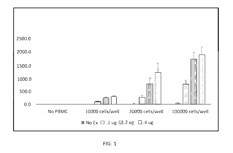
[0016] FIG. 1 shows stimulation of VEGF from PBMCs by fibroblast-derived
exosomes.
[0017] FIG. 2 shows stimulation of HUVEC proliferation.
DETAILED DESCRIPTION OF THE INVENTION
I. Definitions
[0018] As used herein the specification, "a" or "an" may mean one or more. As
used
herein in the claim(s), when used in conjunction with the word "comprising",
the words "a" or
"an" may mean one or more than one. As used herein "another" may mean at least
a second or
more. Still further, the terms "having", "including", "containing" and
"comprising" are
interchangeable and one of skill in the art is cognizant that these terms are
open ended terms.
Some embodiments of the invention may consist of or consist essentially of one
or more
elements, method steps, and/or methods of the invention. It is contemplated
that any method or
composition described herein can be implemented with respect to any other
method or
composition described herein.
[0019] The term "exosome" as used herein refers to small membrane vesicles of
endocytic origin that are secreted by cells in culture, such as fibroblasts.
II. General Embodiments of the Disclosure
[0020] The disclosure encompasses therapy for an individual with a medical
condition or
at risk for a medical condition by providing an effective amount of exosomes
that improve at
least one symptom of the medical condition, for example. In specific
embodiments, the medical
condition is one in which stimulation of angiogenesis would be therapeutic.
Thus, in at least
certain cases the disclosure provides means of stimulating angiogenesis using
exosomes. The
exosomes may be of any source, including those derived from fibroblasts. The
fibroblasts may
or may not be present in tissue cultures.
CA 03058783 2019-10-01
WO 2018/195308 PCT/US2018/028358
[0021] In one embodiment, the disclosure encompasses methods for the
extraction of
exosomes from cultures of fibroblasts and this may also include the
concentration of the
exosomes and administration of the exosomes, including for the purpose of
stimulating
angiogenesis. Without being bound by theory, the exosomes produced by the
tissue culture may
stimulate angiogenesis through directly acting as mitogens for endothelial
cells and/or may
stimulate angiogenesis by inducing production of pro-angiogenic cytokines in
cells of the body,
for example, in cells of the blood. In certain embodiments, such mechanisms
are not involved.
[0022] In one embodiment, fibroblasts are cultured for preserving viability
and
proliferative ability of fibroblasts. The disclosure may be applied both for
individualized
autologous exosome preparations and for exosome preparations obtained from one
or more other
individuals, including established cell lines for experimental or biological
or therapeutic use, for
example. In one embodiment, the disclosure encompasses use of chromatography
separation
methods for preparing membrane vesicles, particularly to separate the membrane
vesicles from
potential biological contaminants, wherein the microvesicles may be exosomes,
and wherein
cells utilized for generating the exosomes are fibroblast cells. Methods of
preparing exosomes
from fibroblasts for a therapeutic use are encompassed in the disclosure.
[0023] As shown herein, membrane vesicles, particularly exosomes, can be
purified that
possess the ability to stimulate angiogenesis. In one embodiment, a strong or
weak anion
exchange may be performed, although in specific embodiments it is a strong
anion exchange. In
addition, in a specific embodiment the chromatography is performed under
pressure. Thus, more
specifically, it may comprise high performance liquid chromatography (HPLC).
[0024] In cases wherein anion exchange chromatography is employed, different
types of
supports may be used to perform the anion exchange chromatography. In
particular
embodiments, these include cellulose, poly(styrene-divinylbenzene), agarose,
dextran,
acrylamide, silica, ethylene glycol-methacrylate co-polymer, or mixtures
thereof, e.g., agarose-
dextran mixtures. In some cases, particular chromatography equipment comprised
of supports,
and particularly the following gels, may be utilized: SOURCE. POROS ,
SEPHAROSE ,
SEPHADEX , TRISACRYL , TSK-GEL SW OR PW , SUPERDEX , TOYOPEARL HW
and SEPHACRYL . Therefore, in a specific embodiment, this disclosure relates
to a method of
preparing membrane vesicles, particularly exosomes, from a biological sample
such as a tissue
culture comprising fibroblasts, comprising at least one step during which the
biological sample is
6
CA 03058783 2019-10-01
WO 2018/195308 PCT/US2018/028358
treated by anion exchange chromatography on a support, including (for example)
one selected
from cellulose, poly(styrene-divinylbenzene), silica, acrylamide, agarose,
dextran, ethylene
glycol-methacrylate co-polymer, alone or in mixtures, and in optional cases
the support is
functionalized.
[0025] In addition, to improve the chromatographic resolution, within the
scope of the
disclosure, it is useful in specific embodiments to use supports in bead form.
In particular
embodiments, these beads have a homogeneous and calibrated diameter, with a
sufficiently high
porosity to enable the penetration of the objects under chromatography (i.e.,
the exosomes). In
this way, given the diameter of exosomes (generally between 50 and 100 nm), to
apply the
teachings of this disclosure, one may use high porosity gels, particularly
between 10 nm and 5
m, such as between approximately 20 nm and approximately 2 m, including
between about
100 nm and about 1 m, for example. For the anion exchange chromatography, the
support used
may be functionalized using a group capable of interacting with an anionic
molecule. Generally,
this group is comprised of an amine that may be ternary or quaternary, which
defines a weak or
strong anion exchanger, respectively. Within the scope of this disclosure, it
is useful to use a
strong anion exchanger. In this way, according to the disclosure, a
chromatography support as
described above, functionalized with quaternary amines, for example, is used.
Therefore,
according to a more specific embodiment of the invention, the anion exchange
chromatography
is performed on a support functionalized with a quaternary amine. In at least
some cases, this
support is selected from poly(styrene-divinylbenzene), acrylamide, agarose,
dextran and silica,
alone or in mixtures, and functionalized with a quaternary amine. Examples of
supports
functionalized with a quaternary amine include the gels SOURCEQ. MONO Q, Q
SEPHAROSE , POROS HQ and POROS QE, FRACTOGEL TMAE type gels and
TOYOPEARL SUPER Q gels.
[0026] In particular embodiments, a support to perform the anion exchange
chromatography comprises poly(styrene-divinylbenzene). An example of this type
of gel that
may be used within the scope of this disclosure is SOURCE Q gel, particularly
SOURCE 15 Q
(Pharmacia). This support offers the advantage of having very large internal
pores, thus offering
low resistance to the circulation of liquid through the gel, while enabling
rapid diffusion of the
exosomes to the functional groups, which are useful parameters for exosomes
given their size.
The biological compounds retained on the column may be eluted in one or more
ways,
particularly using the passage of a saline solution gradient of increasing
concentration, e.g. from
7
CA 03058783 2019-10-01
WO 2018/195308 PCT/US2018/028358
0 to 2 M. A sodium chloride solution may particularly be used, in
concentrations varying from 0
to 2 M, for example. The different fractions purified in this way are detected
by measuring their
optical density (OD) at the column outlet using a continuous spectro-
photometric reading. As an
indication, under the conditions used in the examples, the fractions
comprising the membrane
vesicles were eluted at an ionic strength comprised between approximately 350
and 700 mM,
depending on the type of vesicles.
[0027] Different types of columns may be used to perform this chromatographic
step,
according to requirements and the volumes to be treated. For example,
depending on the
preparations, it is possible to use a column from approximately 100 1 up to
10 ml or greater. In
this way, the supports available have a capacity that may reach 25 mg of
proteins/ml, for
example. For this reason, a 100 1 column has a capacity of approximately 2.5
mg of proteins
which, given the samples in question, allows the treatment of culture
supernatants of
approximately 2 1 (which, after concentration by a factor of 10 to 20, for
example, represent
volumes of 100 to 200 ml per preparation). It is understood that higher
volumes may also be
treated, by increasing the volume of the column, for example. In addition, to
perform at least
certain methods of this disclosure, it is also possible to combine the anion
exchange
chromatography step with a gel permeation chromatography step. In this way,
according to a
specific embodiment of the disclosure, a gel permeation chromatography step is
added to the
anion exchange step, either before or after the anion exchange chromatography
step. In particular
cases, in this embodiment, the permeation chromatography step takes place
after the anion
exchange step. In addition, in a specific variant, the anion exchange
chromatography step is
replaced by the gel permeation chromatography step. The present disclosure
demonstrates that
membrane vesicles may also be purified using gel permeation liquid
chromatography,
particularly when this step is combined with an anion exchange chromatography
or other
treatment steps of the biological sample, as described in detail below.
[0028] To perform the gel permeation chromatography step, a support selected
from
silica, acrylamide, agarose, dextran, ethylene glycol-methacrylate co-polymer
or mixtures
thereof, e.g., agarose-dextran mixtures, may be used. As an illustration, for
gel permeation
chromatography, a support such as SUPERDEX®200HR (Pharmacia), TSK G6000
(TosoHaas) or SEPHACRYL S (Pharmacia) may be used.
8
CA 03058783 2019-10-01
WO 2018/195308 PCT/US2018/028358
[0029] The processes according to the disclosure may be applied to one or more
different
biological samples. In particular, these may comprise a biological fluid from
a subject (bone
marrow, peripheral blood, etc.), a culture supernatant, a cell lysate, a pre-
purified solution or any
other composition comprising membrane vesicles.
[0030] In this respect, in a specific embodiment of the disclosure, the
biological sample
is a culture supernatant of membrane vesicle-producing fibroblast cells.
[0031] In addition, according to a certain embodiment of the invention, the
biological
sample is treated prior to a chromatography step, for example to be enriched
with membrane
vesicles (enrichment stage). In this way, in a specific embodiment, this
disclosure relates to a
method of preparing membrane vesicles from a biological sample, characterized
in that it
comprises at least: a) an enrichment step, to prepare a sample enriched with
membrane vesicles,
and b) a step during which the sample is treated by anion exchange
chromatography and/or gel
permeation chromatography.
[0032] In one embodiment, the biological sample is a culture supernatant
treated so as to
be enriched with membrane vesicles. In particular, the biological sample may
be composed of a
pre-purified solution obtained from a culture supernatant of a population of
membrane vesicle-
producing cells or from a biological fluid, by treatments such as
centrifugation, clarification,
ultrafiltration, nanofiltration and/or affinity chromatography, particularly
with clarification
and/or ultrafiltration and/or affinity chromatography. Therefore, a particular
method of preparing
membrane vesicles according to this disclosure may comprise the following
steps: a) culturing a
population of membrane vesicle (e.g. exosome) producing cells under conditions
enabling the
release of vesicles, b) a step of enrichment of the sample in membrane
vesicles, and c) an anion
exchange chromatography and/or gel permeation chromatography treatment of the
sample.
[0033] As indicated above, the sample (e.g. supernatant) enrichment step may
comprise
one or more of centrifugation, clarification, ultrafiltration, nanofiltration
and/or affinity
chromatography steps on the supernatant. In a first specific embodiment, the
enrichment step
comprises (i) the elimination of cells and/or cell debris (clarification),
possibly followed by (ii) a
concentration and/or affinity chromatography step. In another specific
embodiment, the
enrichment step comprises an affinity chromatography step, optionally preceded
by a step of
elimination of cells and/or cell debris (clarification). A particular
enrichment step according to
this disclosure comprises (i) the elimination of cells and/or cell debris
(clarification), (ii) a
9
CA 03058783 2019-10-01
WO 2018/195308 PCT/US2018/028358
concentration and (iii) an affinity chromatography. The cells and/or cell
debris may be
eliminated by centrifugation of the sample, for example, at a low speed,
preferably below 1000
g, between 100 and 700 g, for example. Preferred centrifugation conditions
during this step are
approximately 300 g or 600 g for a period between 1 and 15 minutes, for
example.
[0034] The cells and/or cell debris may also be eliminated by filtration of
the sample,
possibly combined with the centrifugation described above. The filtration may
particularly be
performed with successive filtrations using filters with a decreasing
porosity. For this purpose,
filters with a porosity above 0.2 m, e.g. between 0.2 and 10 m, may be used.
It is particularly
possible to use a succession of filters with a porosity of 10 m, 1 m, 0.5
p.m followed by 0.22
[0035] A concentration step may also be performed, for example in order to
reduce the
volumes of sample to be treated during the chromatography stages. In this way,
the concentration
may be obtained by centrifugation of the sample at high speeds, e.g. between
10,000 and 100,000
g, to cause the sedimentation of the membrane vesicles. This may comprise a
series of
differential centrifugations, with the last centrifugation performed at
approximately 70,000 g.
The membrane vesicles in the pellet obtained may be taken up with a smaller
volume and in a
suitable buffer for the subsequent steps of the process. The concentration
step may also be
performed by ultrafiltration. In fact, this ultrafiltration allows both to
concentrate the supernatant
and perform an initial purification of the vesicles. According to a certain
embodiment, the
biological sample (e.g., the supernatant) is subjected to an ultrafiltration,
preferably a tangential
ultrafiltration. Tangential ultrafiltration consists of concentrating and
fractionating a solution
between two compartments (filtrate and retentate), separated by membranes of
determined cut-
off thresholds. The separation is carried out by applying a flow in the
retentate compartment and
a transmembrane pressure between this compartment and the filtrate
compartment. Different
systems may be used to perform the ultrafiltration, such as spiral membranes
(Millipore,
Amicon), flat membranes or hollow fibres (Amicon, Millipore, Sartorius, Pall,
GF, Sepracor).
Within the scope of the disclosure, the use of membranes with a cut-off
threshold below 1000
kDa, or which may be between 300 kDa and 1000 kDa, or even which may be
between 300 kDa
and 500 kDa, is advantageous.
[0036] The affinity chromatography step may be performed in various ways,
using
different chromatographic support and material. In particular embodiments, the
chromatography
CA 03058783 2019-10-01
WO 2018/195308 PCT/US2018/028358
is a non-specific affinity chromatography, aimed at retaining (i.e., binding)
certain contaminants
present within the solution, without retaining the objects of interest (i.e.,
the exosomes). It is
therefore a negative selection. In some embodiments, an affinity
chromatography on a dye is
used, allowing the elimination (i.e., the retention) of contaminants such as
proteins and enzymes,
for instance albumin, kinases, deshydrogenases, clotting factors, interferons,
lipoproteins, or also
co-factors, etc. In certain cases, the support used for this chromatography
step is a support as
used for the ion exchange chromatography, functionalized with a dye. As
specific example, the
dye may be selected from Blue SEPHAROSE (Pharmacia), YELLOW 86, GREEN 5 and
BROWN 10 (Sigma). The support is more preferably agarose. It should be
understood that any
other support and/or dye or reactive group allowing the retention (binding) of
contaminants from
the treated biological sample can be used in the instant disclosure.
[0037] In one embodiment, a membrane vesicle preparation process within the
scope of
this disclosure comprises the following steps: a) the culture of a population
of membrane vesicle
(e.g. exosome) producing cells under conditions enabling the release of
vesicles, b) the treatment
of the culture supernatant with at least one ultrafiltration or affinity
chromatography step, to
produce a biological sample enriched with membrane vesicles (e.g. with
exosomes), and c) an
anion exchange chromatography and/or gel permeation chromatography treatment
of the
biological sample. In a particular embodiment, step b) above comprises a
filtration of the culture
supernatant, followed by an ultrafiltration, preferably tangential. In another
embodiment, step b)
above comprises a clarification of the culture supernatant, followed by an
affinity
chromatography on dye, preferably on Blue SEPHAROSE .
[0038] In addition, after step c), the material harvested may, if applicable,
be subjected to
one or more additional treatment and/or filtration stages d), particularly for
sterilization purposes.
For this filtration treatment stage, filters with a diameter less than or
equal to 0.3 are
preferentially used, or even more preferentially, less than or equal to 0.25
m. Such filters have a
diameter of 0.22 m, for example.
[0039] After step d), the material obtained is, for example, distributed into
suitable
devices such as bottles, tubes, bags, syringes, etc., in a suitable storage
medium. The purified
vesicles obtained in this way may be stored cold, frozen or used
extemporaneously. Therefore, a
specific preparation process within the scope of the invention comprises at
least the following
steps: c) an anion exchange chromatography and/or gel permeation
chromatography treatment of
11
CA 03058783 2019-10-01
WO 2018/195308 PCT/US2018/028358
the biological sample, and d) a filtration step, particularly sterilizing
filtration, of the material
harvested after stage c). In a first variant, the process according to the
disclosure comprises: c) an
anion exchange chromatography treatment of the biological sample, and d) a
filtration step,
particularly sterilizing filtration, on the material harvested after step c).
[0040] In another variant, the process according to the disclosure comprises:
c) a gel
permeation chromatography treatment of the biological sample, and d) a
filtration step,
particularly sterilizing filtration, on the material harvested after step c).
According to a third
variant, the process according to the invention comprises: c) an anionic
exchange treatment of
the biological sample followed or preceded by gel permeation chromatography,
and d) a
filtration step, particularly sterilizing filtration, on the material
harvested after step c).
[0041] Further embodiments include a method of optimizing angiogenesis
stimulating
therapeutic factor production from fibroblast cultures through the use of
filters that separate
compositions based on electrical charge, size or ability to elute from an
adsorbent. Numerous
techniques are known in the art for purification of therapeutic factors and
concentration of said
agents. For some particular uses the fibroblast derived compounds will be
sufficient for use as
culture supernatants of said cells in media. Currently media useful for this
purpose include
Roswell Park Memorial Institute (RPMI-1640), Dulbecco's Modified Essential
Media (DMEM),
Eagle's Modified Essential Media (EMEM), Optimem, and Iscove's Media.
[0042] In one embodiment, therapeutic factors for stimulating angiogenesis are
derived
from tissue culture that may comprise exosomes, or may not comprise exosomes
but comprise
factors capable of stimulating angiogenesis. In such as embodiment, culture
conditioned media
may be concentrated by filtering/desalting means known in the art including
use of Amicon
filters with specific molecular weight cut-offs, said cut-offs may select for
molecular weights
higher than 1 kDa to 50 kDa. Supernatant may alternatively be concentrated
using means known
in the art such as solid phase extraction using C18 cartridges (Mini-Spe-ed
C18-14%, S.P.E.
Limited, Concord ON). The cartridges are prepared by washing with methanol
followed by
deionized-distilled water. Up to 100 ml of fibroblast conditioned media
supernatant may be
passed through each of these specific cartridges before elution, it is
understood of one of skill in
the art that larger cartridges may be used. After washing the cartridges
material adsorbed is
eluted with 3 ml methanol, evaporated under a stream of nitrogen, redissolved
in a small volume
of methanol, and stored at 4 C. Before testing the eluate for activity in
vitro, the methanol is
12
CA 03058783 2019-10-01
WO 2018/195308 PCT/US2018/028358
evaporated under nitrogen and replaced by culture medium. The C18 cartridges
are used to
adsorb small hydrophobic molecules from the fibroblast conditioned
supernatant, and allows for
the elimination of salts and other polar contaminants. It may, however be
desired to use other
adsorption means in order to purify certain compounds from the supernatant.
The concentrated
supernatant may be assessed directly for biological activities useful for the
practice of this
invention, or may be further purified. Further purification may be performed
using, for example,
gel filtration using a Bio-Gel P-2 column with a nominal exclusion limit of
1800 Da (Bio-Rad,
Richmond Calif.). The column may be washed and pre-swelled in 20 mM Tris-HC1
buffer, pH
7.2 (Sigma) and degassed by gentle swirling under vacuum. Bio-Gel P-2 material
be packed into
a 1.5x54 cm glass column and equilibrated with 3 column volumes of the same
buffer. Fibroblast
cell supernatant concentrates extracted by C18 cartridge may be dissolved in
0.5 ml of 20 mM
Tris buffer, pH 7.2 and run through the column. Fractions may be collected
from the column and
analyzed for biological activity. Other purification, fractionation, and
identification means are
known to one skilled in the art and include anionic exchange chromatography,
gas
chromatography, high performance liquid chromatography, nuclear magnetic
resonance, and
mass spectrometry. Administration of supernatant active fractions may be
performed locally or
systemically.
[0043] Included in the disclosure are methods of treating an individual for
risk of limb
loss, ischemic heart disease, ischemic brain disease, gastrointestinal ulcer,
and/or one or more
wounds by providing to the individual an effective amount of fibroblast-
derived exosomes or one
or more biologically active fractions thereof. A fraction may be determined to
be biologically
active using routine methods in the art of establishing fractions of a
starting material and testing
for a particular active with each fraction. In specific cases of the
disclosure, the activity to be
tested may be production of one or more particular compounds, such as one or
more factors, for
example VEGF. In additional or other cases the fractions may be tested in an
in vivo model,
such as an in vivo mouse model for limb loss.
EXAMPLES
[0044] The following examples are included to demonstrate preferred
embodiments of
the invention. It should be appreciated by those of skill in the art that the
techniques disclosed in
the examples that follow represent techniques discovered by the inventors to
function well in the
practice of the invention, and thus can be considered to constitute preferred
modes for its
13
CA 03058783 2019-10-01
WO 2018/195308 PCT/US2018/028358
practice. However, those of skill in the art should, in light of the present
disclosure, appreciate
that many changes can be made in the specific embodiments which are disclosed
and still obtain
a like or similar result without departing from the spirit and scope of the
invention.
EXAMPLE 1
FIBROBLAST EXOSOMES STIMULATE VEGF PRODUCTION
[0045] In one embodiment of the invention, exosomes generated from fibroblasts
are
used for "Angiogenesis Therapy". Angiogenesis therapy has been described as a
"biological
bypass", the idea being that through administration of agents capable of
inducing
collateralization, a more natural type of "bypass" can be achieved. Indeed, it
has been observed
that ischemic muscles secrete angiogenic factors in response to hypoxia and
that to some extent
natural angiogenesis does occur in animal models of critical limb ischemia
(CLI) and in humans
(15, 16). Thus by augmenting these natural processes, researchers have
attempted to prevent
amputation. One of the angiogenic factors noted in many ischemic conditions,
including cardiac
ischemia, stroke, and CLI is vascular endothelial growth factor (VEGF) (17-
19). In 1994, limb
salvage and increased angiogenesis was reported in a rabbit CLI model after
single bolus intra-
arterial administration of VEGF-165 (20). Other experiments in the same model
demonstrated
no incidence of calf muscle atrophy and distal limb necrosis, whereas this was
present in 85.7%
of control rabbits, after VEGF administration (21, 22). A variety of studies
have repeated these
findings in other models of CLI (23-25). Unfortunately, this was not
successfully reproduced in
the clinic. Trials using VEGF protein (26), or DNA plasmid, did not show
significant benefit at
reducing leg amputations in a double blind setting (25, 27). In one embodiment
of the invention,
exosomes derived from fibroblasts are utilized to stimulate VEGF production
from cells of the
patient.
[0046] Another approach involved use of the cytokine fibroblast growth factor-
1 (FGF-
1). Given that FGF-1 is considered "upstream" of VEGF, it was believed to
stimulate numerous
angiogenic processes so as to result in creation of more mature vessels (28).
FGF, like VEGF, is
part of the natural tissue response to hypoxia, as demonstrated both in animal
models (29) and
clinical trials (30). The critical role of FGF in endogenous angiogenesis was
conclusively
demonstrated in FGF conditional knockout mice, which displayed inhibited
ability to heal post-
wounding or to form neovascularization (31). Although FGF-1 gene therapy has
clinically been
used in CLI patients with some improvement in ABI and perfusion, results were
mediocre (32).
14
CA 03058783 2019-10-01
WO 2018/195308 PCT/US2018/028358
Attempts at replicating the in vivo angiogenic cascade by combination of
cytokines yielded more
promising results. Cao et al demonstrated synergy between administration of
PDGF-BB and
FGF-2 in terms of increasing blood vessel formation and function in the
femoral artery ligation
model in rats and rabbits (33). Similarly, in cancer angiogenesis, it is known
that several tumor-
derived angiogenic factors synergize for acceleration of neovascularization
(34). Investigators
have attempted to activate upstream mediators of several angiogenic signals
through transfection
of genes encoding transcription factors such as HIF-1 alpha (35). In fact,
this approach has been
demonstrated to be superior to VEGF gene administration in terms of new
capillary sprouting.
In a Phase I dose-escalating trial, transfection of HIF-1 alpha into CLI
patients demonstrated
tolerability with some indication of efficacy (36). In conclusion, while
administration of
angiogenic factors to patients with CLI does induce some benefit in early
trials, data from
randomized trials to date do not support widespread use. The transfection of
upstream
transcription factors such as HIF-1 alpha is a promising approach since it
mimics natural
angiogenesis in that a plurality of growth factors are induced following
transfection (35, 37). In
another embodiment of the invention, the use of fibroblast derived exosomes
for enhancement of
cytokine and other angiogenic therapies is disclosed.
[0047] Exosomes were prepared from the cell culture supernatant of day 4
foreskin
fibroblast cultures by differential centrifugation. Briefly, recovered culture
supernatant was
subjected to three successive centrifugations at 300 g (5 min), 1,200 g (20
min), and 10,000 g (30
min) to eliminate cells and debris, followed by centrifugation for 1 h at
100,000 g. To remove
excess serum proteins, the exosome pellet was washed with a large volume of
PBS, centrifuged
at 100,000 g for 1 h, and finally resuspended in 120 ul of PBS for further
studies. The exosomes
were quantified by a micro Bradford protein assay (Bio-Rad). Each batch was
standardized by
protein content.
[0048] Peripheral blood mononuclear cells (PBMC) were isolated from 5 ml of
blood by
Ficoll density gradient (Sigma-Aldrich). Cells were washed twice in phosphate
buffered saline
(PBS) and plated in round-bottom, 96-well plates (Nunc). In each well, 10,000,
20,000 or
100,000 PBMC where added to a total volume of 200 uL in RPMI media containing
10% fetal
calf serum (Life Technologies). Exosomes were added at concentrations of 0.1
ug/ml, 0.2 ug/ml
and 0.4 ug/ml. Cells were cultured for 48 hours and concentration of VEGF was
analyzed by
ELISA (R&D Systems). Concentrations were expressed as pg/ml in FIG. 1.
CA 03058783 2019-10-01
WO 2018/195308 PCT/US2018/028358
EXAMPLE 2
FIBROBLAST EXOSOMES STIMULATE HUVEC PROLIFERATION
[0049] Fibroblast derived exosomes were obtained as described in Example 1 and
added
to cultures of human umbilical vein endothelial cells (HUVEC). Cells were
incubated for 48
hours and proliferation was assessed by thymidine incorporation assay. In FIG.
2, proliferation
is expressed as counts per minute (CPM).
EXAMPLE 3
PREVENTION OF LIMB LOSS IN CRITICAL LIMB ISCHEMIA MOUSE MODEL
[0050] BALB/c mice were treated by femoral artery ligation and local nerve
injury in a
previously published model (Meng et al., 2007,1 Trans. Med. 5:57).
[0051] Exosomes were prepared from the cell culture supernatant of day 4
foreskin
fibroblast cultures by differential centrifugation. Briefly, recovered culture
supernatant was
subjected to three successive centrifugations at 300 g (5 min), 1,200 g (20
min), and 10,000 g (30
min) to eliminate cells and debris, followed by centrifugation for 1 h at
100,000 g. To remove
excess serum proteins, the exosome pellet was washed with a large volume of
PBS, centrifuged
at 100,000 g for 1 h, and finally resuspended in 120 ul of PBS for further
studies. The exosomes
were quantified by a micro Bradford protein assay (Bio-Rad). Each batch was
standardized by
protein content.
[0052] Mice were administered 5 micrograms of fibroblast exosomes in a volume
of 100
microliters 3 days after femoral artery ligation (treated). Controls where
administered 5
micrograms of fetal calf serum derived exosomes (untreated). Limb loss was
present in all the
untreated mice (7/7), whereas it was observed in only 1/7 treated mice.
Administration of
mesenchymal stem cell exosomes did not result in limb salvage by day 35.
16
CA 03058783 2019-10-01
WO 2018/195308 PCT/US2018/028358
[0053]
4-Control monommom---- mmonommom monomomr--- mmonommom
Limb Loss
Control LimbD LOW
ia Control limb Loss
4 Contra( Limb LO$C
j5 Control ,,=
õõõõõõõõõõõõõõõõõõõõõõõõõõõõõõõõõõõõõõõõõõõõõõõõõõõõõõõõõõõõõõõõõõõõõõõ
umumumun mumumumun unumumum umumumumu
Control ILimb Los.
tomtimb Loss
Treated
Treated
Treated
11 Treated limb Loss
12 Treated:
43 Treated
=mmmmmm
14 Treated
100541 Although the present invention and its advantages have been described
in detail, it
should be understood that various changes, substitutions and alterations can
be made herein
without departing from the spirit and scope of the invention as defined by the
appended claims.
Moreover, the scope of the present application is not intended to be limited
to the particular
embodiments of the process, machine, manufacture, composition of matter,
means, methods and
steps described in the specification. As one of ordinary skill in the art will
readily appreciate
from the disclosure of the present invention, processes, machines,
manufacture, compositions of
matter, means, methods, or steps, presently existing or later to be developed
that perform
substantially the same function or achieve substantially the same result as
the corresponding
embodiments described herein may be utilized according to the present
invention. Accordingly,
the appended claims are intended to include within their scope such processes,
machines,
manufacture, compositions of matter, means, methods, or steps.
17
CA 03058783 2019-10-01
WO 2018/195308 PCT/US2018/028358
REFERENCES
All patents and publications cited herein are hereby incorporated by reference
in their
entirety herein.
1. Thery, C., M. Ostrowski, and E. Segura, Membrane vesicles as conveyors
of
immune responses. Nature reviews. Immunology, 2009. 9(8): p. 581-93.
2. Ludwig, A.K. and B. Giebel, Exosomes: Small vesicles participating in
intercellular communication. The international journal of biochemistry & cell
biology, 2011.
3. Alvarez-Erviti, L., et al., Lysosomal dysfunction increases exosome-
mediated
alpha-synuclein release and transmission. Neurobiology of disease, 2011.
42(3): p. 360-7.
4. Silverman, J.M. and N.E. Reiner, Exosomes and other microvesicles in
infection
biology: organelles with unanticipated phenotypes. Cellular microbiology,
2011. 13(1): p. 1-9.
5. Pan, B.T. and R.M. Johnstone, Fate of the transferrin receptor during
maturation
of sheep reticulocytes in vitro: selective externalization of the receptor.
Cell, 1983. 33(3): p. 967-
78.
6. Alonso, R., et al., Diacylglycerol kinase alpha regulates the formation
and
polarisation of mature multivesicular bodies involved in the secretion of Fas
ligand-containing
exosomes in T lymphocytes. Cell death and differentiation, 2011. 18(7): p.
1161-73.
7. Zhang, H., et al., CD4(+) T cell-released exosomes inhibit CD8(+)
cytotoxic T-
lymphocyte responses and antitumor immunity. Cellular & molecular immunology,
2011. 8(1):
p. 23-30.
8. Mathews, J.A., et al., CD23 Sheddase A disintegrin and metalloproteinase
10
(ADAM10) is also required for CD23 sorting into B cell-derived exosomes. The
Journal of
biological chemistry, 2010. 285(48): p. 37531-41.
9. Buschow, S.I., et al., MEW class II-associated proteins in B-cell
exosomes and
potential functional implications for exosome biogenesis. Immunology and cell
biology, 2010.
88(8): p. 851-6.
18
CA 03058783 2019-10-01
WO 2018/195308 PCT/US2018/028358
10. Hwang, I. and D. Ki, Receptor-mediated T cell absorption of antigen
presenting
cell-derived molecules. Frontiers in bioscience : a journal and virtual
library, 2011. 16: p. 411-
21.
11. Viaud, S., et al., Updated technology to produce highly immunogenic
dendritic
cell-derived exosomes of clinical grade: a critical role of interferon-gamma.
Journal of
immunotherapy, 2011. 34(1): p. 65-75.
12. Clayton, A., et al., Cancer exosomes express CD39 and CD73, which
suppress T
cells through adenosine production. Journal of immunology, 2011. 187(2): p.
676-83.
13. Battke, C., et al., Tumour exosomes inhibit binding of tumour-reactive
antibodies
to tumour cells and reduce ADCC. Cancer immunology, immunotherapy : CII, 2011.
60(5): p.
639-48.
14. Lachenal, G., et al., Release of exosomes from differentiated neurons
and its
regulation by synaptic glutamatergic activity. Molecular and cellular
neurosciences, 2011. 46(2):
p. 409-18.
15. Faure, J., et al., Exosomes are released by cultured cortical neurones.
Molecular
and cellular neurosciences, 2006. 31(4): p. 642-8.
16. Fitzner, D., et al., Selective transfer of exosomes from
oligodendrocytes to
microglia by macropinocytosis. Journal of cell science, 2011. 124(Pt 3): p.
447-58.
17. Mincheva-Nilsson, L. and V. Baranov, The role of placental exosomes in
reproduction. American journal of reproductive immunology, 2010. 63(6): p. 520-
33.
19