Note: Descriptions are shown in the official language in which they were submitted.
CA 03058949 2019-10-02
WO 2018/201156 PCT/US2018/030314
METHOD AND SYSTEM FOR CONCURRENT PHOTOTHERMAL ABLATION AND
INTERSTITIAL PHOTODYNAMIC THERAPY
Statement Regarding Federally Sponsored Research
[0001] This invention was made with government support under CA193610
awarded by
.. the National Institutes of Health. The government has certain rights in the
invention.
Cross-Reference to Related Applications
[0002] This application claims priority to U.S. provisional
Application No. 62/492,171,
filed on April 29, 2017, now pending, the disclosure of which is incorporated
herein by
reference.
.. Field of the Disclosure
[0003] The present disclosure relates to photodynamic therapy.
Background of the Disclosure
[0004] Photodynamic therapy (PDT), in particular interstitial
photodynamic therapy (I-
PDT), offers promising outcomes for patients with refractory locally advanced
cancer. The use
of I-PDT with porfimer sodium (Photofrin0) is approved for palliation in
patients with
esophageal cancer or lung cancer with airway obstruction, who are non-
candidates for surgery or
radiation therapy. In addition, I-PDT with porfimer sodium has been used, in
compassionate care
settings, to treat patients with head and neck squamous cell carcinoma. A
principal clinical goal
has been to shorten treatment times by administering the therapeutic light at
high dose rates (i.e.,
400 mW/cm) that are clinically approved by the FDA for I-PDT with porfimer
sodium.
However, the cure rate for I-PDT with porfimer sodium is limited.
[0005] PDT has also been viewed as beneficial when considering the
relatively minor
nature of adverse effects. Additionally, it has been noted that PDT provides
excellent cosmetic
outcomes. To date, it has been believed that during I-PDT the changes in
tissue temperature do
not affect the response. Physicians and researchers assumed that PDT is
associated with minimal
heating. The clinically approved, and used, light dose rate (400 mW/cm) for
PDT or I-PDT with
porfimer sodium was chosen arbitrarily, about 25 years ago. There was no
systemic study to
evaluate potential tissue heating during I-PDT. Several retrospective clinical
studies suggest that
1
CA 03058949 2019-10-02
WO 2018/201156 PCT/US2018/030314
PDT and I-PDT will result in retention of functional anatomy and other
benefits. While offering
improved palliative outcomes for patients with such advanced diseases, there
remains a need for
further improvement.
Brief Summary of the Disclosure
[0006] In a first aspect, the present disclosure provides a method for
treating a tissue. A
photosensitizer is administered to the tissue. One or more optical fibers are
placed in the tissue.
For example, a portion (such as, for example, an end portion) of the one or
more optical fibers
are inserted in the tissue. The optical fibers may be spaced apart from one
another such that a
light dose may be applied to the tissue. The method includes applying a
treatment light to the
.. tissue by way of the one or more optical fibers. A temperature of the
tissue is measured during
application of the treatment light. The fluence rate (mW/cm2) of the treatment
light is modified
based on the temperature of the tissue. For example, by adjusting the light
dose rate one may
govern the intratumoral fluence rate within the tissue (e.g., intratumoral
fluence rate). For
example, the fluence rate may be modified to be lower if the temperature of
the tissue is higher
than a predetermined threshold. In some embodiments, the fluence rate is
modified to maintain a
tissue temperature between 50 C and 65 C. In some embodiments, the fluence
rate is modified
to maintain a tissue temperature of substantially 60 C. In some embodiments,
the fluence rate is
modified to maintain a tissue temperature between 60 C and 90 C.
[0007] In some embodiments, one or more dosimetry fibers may be placed
in the tissue
and configured to measure light dose (J/cm2). The optical fiber(s) and/or the
dosimetry fiber(s)
may be disposed within one or more light-transmitting catheters (LTCs) placed
in the tissue. For
example, each optical fiber may be disposed in a corresponding LTC. In another
example, each
of a plurality of LTCs may contain an optical fiber and a dosimetry fiber.
[0008] In another aspect, the present disclosure may be embodied as a
system for treating
a tissue. The system includes a light source and an optical fiber operably
coupled to the light
source. The optical fiber is configured to deliver a light dose of treatment
light to the tissue. A
temperature sensor is configured to measure a temperature of the tissue. The
temperature sensor
may be any suitable sensor such as, for example, a thermistor, a thermal
imaging sensor, a fiber
optic, a magnetic resonance thermometer (providing volumetric temperature
data), or the like. In
some embodiments, the temperature sensor is configured to measure temperature
at a plurality of
2
CA 03058949 2019-10-02
WO 2018/201156 PCT/US2018/030314
locations throughout the volume of the tissue. In some embodiments, the
temperature sensor
comprises a plurality of temperature sensitive catheters.
[0009] A controller, such as, for example, a programmable
microprocessor, is in
communication with the temperature sensor and configured to modify a fluence
rate of the
treatment light based on a measured temperature. In some embodiments, the
controller is
configured to modify the fluence rate of the treatment light to maintain a
tissue temperature
between 50 C and 65 C. In some embodiments, the controller is configured to
modify the
fluence rate of the treatment light to maintain a tissue temperature of
substantially 60 C. In some
embodiments, the controller is configured to modify the fluence rate of the
treatment light to
maintain a tissue temperature between 60 C and 90 C.
[0010] The system may further include a dosimetry fiber for measuring
the light dose. A
spectrometer may be operably coupled to the dosimetry fiber. The system may
further include an
LTC, and the optical fiber and/or the dosimetry fiber may be disposed in the
LTC.
[0011] In some embodiments, a second optical fiber is operably coupled
to the light
source and configured to deliver a second light dose of treatment light to the
tissue. The
controller may be configured to modify a fluence rate of the second light dose
based on the
measured temperature. The fluence rate of the second light dose may be
modified based on a
temperature at a second location of the tissue.
Description of the Drawings
[0012] For a fuller understanding of the nature and objects of the
disclosure, reference
should be made to the following detailed description taken in conjunction with
the
accompanying drawings, in which:
Figure 1 depicts a system according to an embodiment of the present
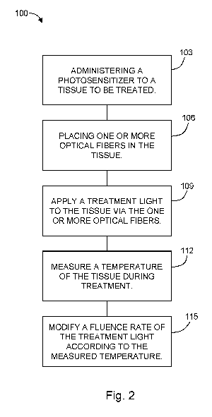
disclosure;
Figure 2 is a chart showing a method according to another embodiment of the
present
disclosure;
Figure 3 shows a mouse being fitted with two optical fibers, each disposed
within a light-
transmitting catheter, for use in interstitial photodynamic therapy (I-PDT);
Figure 4 is a chart showing intratumoral heating results for a light dose of
150 mW/cm,
100 J/cm with and without photosensitizer;
3
CA 03058949 2019-10-02
WO 2018/201156 PCT/US2018/030314
Figure 5 is a chart showing intratumoral heating results for a light dose of
350 mW/cm,
100 J/cm with and without photosensitizer;
Figure 6 is a chart showing tumor size over time in populations of mice, where
a control
population was untreated, a second population was treated with a light dose of
150 mW/cm, 100 J/cm (control vs. lightp = < 0.0001), and a third population
was treated
with I-PDT (control vs. I-PDT p = < 0.0001; light vs. I-PDT p < 0.05);
Figure 7 is a chart showing tumor size over time in populations of mice, where
a control
population was untreated, a second population was treated with a light dose of
350 mW/cm, 100 J/cm (control vs. lightp = < 0.0001), and a third population
was treated
with I-PDT (control vs. I-PDTp = < 0.0001; light vs. I-PDTp = 0.339); and
Figure 8 is a chart showing tumor size over time in populations of mice, where
a control
population was untreated, a second population was treated with a light dose of
100 mW/cm, 540 J/cm (control vs. light p = < 0.0001), and a third population
was treated
with I-PDT concurrent with the same light dose as the second population
(control vs.
lightp = <0.0001; light vs. I-PDT/light p = 0.164).
Detailed Description of the Disclosure
[0013] As mentioned above, a perceived benefit of photodynamic therapy
has been the
lack of significant changes in tissue temperature. However, the present
disclosure
advantageously utilizes increased temperatures induced in the tissue using the
I-PDT techniques
disclosed herein to enhance efficacy as compared to I-PDT without an increased
temperature.
[0014] With reference to Figure 2, the present disclosure may be
embodied as a
method 100 for treating a tissue, for example, treating a tumor, of an
individual using interstitial
photodynamic therapy (I-PDT) (see Figure 2). The method 100 includes
administering 103 a
photosensitizer to the tissue. The photosensitizer may be, for example,
porfimer sodium
(Photofrin0) or any other photosensitizer known for use in I-PDT¨e.g., capable
of generating
reactive oxygen species and radicals when activated by light in the presence
of oxygen. The
photosensitizer may be administered 103 by, for example, intravenous
injection.
[0015] One or more optical fibers are placed 106 into the tissue to be
treated. The optical
fibers may be placed at locations in the tissue according to a predetermined
treatment plan. In
some embodiments, the optical fiber(s) are placed 106 into the tissue by way
of light-
4
CA 03058949 2019-10-02
WO 2018/201156 PCT/US2018/030314
transmitting catheter(s) (LTCs) where each optical fiber is disposed in a
light-transmitting
catheter.
[0016] A treatment light is applied 109 to the tissue by way of the
one or more optical
fibers. The treatment light has a fluence (measured in, for example, joules
per square
centimeter¨Fcm2) and a fluence rate (measured in, for example, milliwatts per
square
centimeter¨mW/cm2).
[0017] The method 100 includes measuring 112 a temperature of the
tissue during
application of the treatment light (i.e., during "treatment"). Measurement 112
of the tissue may
be performed using any technique appropriate. For example, volumetric
measurement may be
accomplished using magnetic-resonance thermometry (MR thermometry or MRT). In
another
example, temperature is measured using a temperature-sensitive catheter. In
embodiments where
LTCs are placed in the tissue, a temperature-sensitive catheter may optionally
be disposed in an
LTC. Other methods for measuring 112 a temperature of a tissue may be used.
[0018] The fluence rate of the treatment light may be modified 115
based on the
temperature of the tissue. The fluence rate of the treatment light within the
tissue (e.g.,
intratumoral fluence rate) may be modified by adjusting the light dose rate.
For example, the
fluence rate may be decreased when the tissue temperature is higher than a
(first) predetermined
threshold. In this way, the temperature of the tissue will decrease.
Similarly, the fluence rate of
the treatment light may be increased if the tissue temperature is lower than a
second
predetermined threshold, which may be the same as or different from the first
predetermined
threshold. By increasing the fluence rate, the tissue temperature will
increase. In exemplary
embodiments, the fluence rate is modified to maintain a tissue temperature of
between 50-65 C,
inclusive. In another exemplary embodiment, the fluence rate is modified to
maintain a tissue
temperature of less than 60 C. In another exemplary embodiment, the fluence
rate is modified to
maintain a tissue temperature of substantially 60 C. By substantially,
embodiments may
maintain the temperature of the tissue within a desired tolerance, for
example, 5 C, 1 C,
0.5 C, 0.2 C, or other tolerance levels between these exemplary values, for
example, in 2 C
increments. In embodiments incorporating thermal ablation, the temperature may
be maintained
at greater than 60 C and less than 100 C (or in some embodiments, less than 90
C) in order to
avoid tissue carbonization.
5
CA 03058949 2019-10-02
WO 2018/201156 PCT/US2018/030314
[0019] With reference to Figure 1, the present disclosure may be
embodied as a
system 10 for treating tissue 90 using I-PDT is provided. The system 10
includes a light
source 12. For example, the light source 12 may be a laser configured to emit
light at a
wavelength for activating a selected photosensitizer. For example, when using
Photofrin, the
emitted light may be 630 nm. An optical fiber 20 is operably coupled to the
light source 12. The
optical fiber 20 is configured to deliver a light dose to the tissue 90. The
optical fiber 20 may
have a cylindrical diffuser, a fiber with a flat-cut end, or other
configuration. Embodiments of
the system 10 may include additional optical fibers, for example, the
embodiment depicted in
Figure 1 includes a second optical fiber 20 for delivering a second light dose
to the tissue 90.
[0020] The system 10 includes a temperature sensor 30 for measuring a
temperature of
the tissue 90. In some embodiments, the temperature sensor 30 may be
configured to measure the
temperature of the tissue 90 at more than one location, for example, more than
one 3-D location
within the tissue 90. In some embodiments, the temperature sensor is a
magnetic-resonance
thermometer. In some embodiments, the temperature sensor comprises one or more
temperature-
sensitive catheters (e.g., a thermistor disposed in a catheter).
[0021] In some embodiments, the system 10 may include a light-
transmitting catheter 22
(e.g., a transparent catheter)(an "LTC"). In such embodiments, the optical
fiber(s) 20 of the
system 10 may be disposed within a corresponding number of LTCs 22. For
example, each
optical fiber 20 may be disposed in a lumen 23 of an LTC 22.
[0022] The system 10 further includes a controller 40 in communication with
the
temperature sensor 30. The controller 40 is configured to modify a fluence
rate of the treatment
light based on the temperature of the tissue 90, as described above. In some
embodiments, the
controller 40 is a programmable microprocessor, programmed to modify the
treatment light
based on a signal received from the temperature sensor. The treatment light
may be modified by
adjusting the intensity of the light source, attenuating the light emitted
from the light source,
independently attenuating the light in each optical fiber, or any other
technique for increasing or
decreasing the fluence rate of the treatment light delivered to the tissue via
the optical fiber(s).
[0023] Some embodiments of the presently-disclosed system may include
a dosimetry
fiber 25 for measuring a light dose. In embodiments wherein the system
includes an LTC 22, the
dosimetry fiber 25 may be disposed in an LTC 22, for example, within a lumen
23 of an LTC 22.
The dosimetry fiber 25 may be operably coupled to a spectrometer 27 for
measuring the light
6
CA 03058949 2019-10-02
WO 2018/201156 PCT/US2018/030314
dose delivered to the tissue. In this way, a desired total dose may be
delivered while also
maintaining the temperature of the tissue in a desired range for effective
photothermal ablation
(or other synergistic effect when combined with I-PDT).
[0024] The present disclosure is further illustrated in the discussion
below, which
includes exemplary embodiments used to test the disclosed technique using
mouse models.
Further Discussion
[0025] The mechanism of action in photodynamic therapy (PDT) involves
the generation
of reactive oxygen species and radicals through light activation of
photosensitizer in the presence
of oxygen¨i.e., an effective photoreaction. Previous studies demonstrated that
oxygen-
conserving light fluence rate (mW/cm2) is required for an effective
photoreaction. In I-PDT,
multiple optical fibers with a cylindrical diffuser or optical fibers with
flat cut end are inserted
into the tumor. During I-PDT, the light dose rate (mW/cm) dictates the
resulted fluence rate in
the tumor. Thus, the light fluence rate depends on the light dose rate
delivered from the optical
fibers.
[0026] In the U.S., Photofrin is the only approved photosensitizer in the
treatment of
obstructing esophageal, non-small cell endobronchial lung cancer and high-
grade dysplasia in
Barrett's esophagus. The FDA-approved light dose rate for I-PDT with Photofrin
is 400 mW/cm
length of the optical fiber cylindrical diffuser, which translates to a
fluence rate of up to
800 mW/cm2. This clinically-approved light dose rate induces significant
thermal ablation that
.. could overwhelm the photoreaction. However, our preclinical data suggest
that this is not the
case, and cure of locally advanced squamous cell carcinoma can be achieved in
a mouse model
treated with I-PDT and thermal ablation. In an ongoing preclinical study it
was found that the
laser light can induce significant heating that can induce immediate tissue
ablation, at T> 60 C,
without impeding the efficacy of the I-PDT.
[0027] The inventors hypothesized that the limited cure rate for I-PDT with
porfimer
sodium is due to the high dose rate that could limit the photodynamic
efficiency by depleting
tumor oxygen levels.
[0028] As further described below, aspects of the presently-disclosed
method and system
were implemented for testing. In particular, in vivo magnetic resonance
thermometry (MRT) was
7
CA 03058949 2019-10-02
WO 2018/201156 PCT/US2018/030314
used to quantify photothermal ablation during I-PDT. Time-to-event and cure
rates were
analyzed with Kaplan Meier curves. Safe and effective light dose rates and
doses with
photothermal ablation were identified.
Animal Model
[0029] All procedures were carried out in accordance with a protocol
approved by the
Institutional Animal Care and Use Committee (IACUC) at Roswell Park Cancer
Institute
(RPCI). Female C3H, 8-12 weeks, mice bearing locally advanced SCCVII tumors
were treated
when the tumors reached a size of 9-10 mm in their largest diameter, and a
volume of 400-500
mm3 calculated from caliper measurements, as is known in the art.
[0030] The animals were randomly assigned to receive light only (no drug)
or I-PDT by
administering 5 mg/kg porfimer sodium 24 hours prior to light delivery. Mice
were considered
cured if there was no palpable tumor any time at or after 60 days post I-PDT.
Interstitial Photodynamic Therapy (I-PDT)
[0031] The laser light (treatment light) was delivered through one to
three 0.98 mm
diameter optical fibers with 20 mm cylindrical light diffuser RD20 (Medlight
SA, Ecublens,
Switzerland). The cylindrical light diffusers were connected to 1.0 Watt laser
diode modules that
emit 630 3 nm light (ML6500, Modulight Inc., Tampere, Finland). During light
delivery, a
custom made template was utilized to guide the placement of the laser fibers
into the tumor, as
shown in Figure 3.
Magnetic Resonance Thermometry
[0032] Magnetic resonance thermometry (MRT) by proton resonance
frequency methods
was carried out in a 4.7 Tesla preclinical scanner using the ParaVision 3Ø2
imaging platform
(Bruker Biospin, Billerica MA) and a 35 mm quadrature transceiver coil.
Figures 4 and 5 show
that there was a temperature increase during the interstitial light delivery,
with and without drug.
These temperatures were averaged over the entire tumor volume.
Results
[0033] Figure 6 shows the percentage of mice with tumors less than
4000 mm3 over a
period of 60 days post treatment. As shown in the chart, a control population
was untreated, a
8
CA 03058949 2019-10-02
WO 2018/201156
PCT/US2018/030314
second population was treated with a light dose of 150 mW/cm, 100 J/cm
(control vs. lightp <
0.0001), and a third population was treated with I-PDT and similar dose
(control vs. I-PDT
p < 0.0001; light vs. I-PDT p = 0.0004). Figure 7 is a similar chart for
populations treated with a
light dose of 350 mW/cm, 100 J/cm (control vs. light p < 0.0001; control vs. I-
PDT p < 0.0001;
light vs. I-PDT p = 0.339). It can be seen that after 60 days, only 10% of
mice remained with
tumors less than 4,000 mm3.
[0034] Figure 8 shows the beneficial results of an embodiment of the
presently-disclosed
techniques using concurrent I-PDT and thermal ablation. It can be seen that
the use of light
without photosensitizer results in a cure rate of 40%. Despite the previous
consideration that a
lack of significant temperature change of the tissue was a benefit of PDT,
Figure 8 shows that
concurrent photothermal ablation and I-PDT resulted in a cure rate of 70%.
[0035] Although claimed subject matter is described in terms of
certain embodiments,
other embodiments, including embodiments that do not provide all of the
benefits and features
set forth herein, are also within the scope of this disclosure. Various
structural, logical, process
step, and electronic changes may be made without departing from the scope of
the disclosure.
9