Note: Descriptions are shown in the official language in which they were submitted.
CA 03060846 2019-10-17
WO 2018/204883 PCT/US2018/031250
IMPLANTABLE DEVICES AND METHODS TO TREAT BENIGN
PROSTATE HYPERPLASIA (BPH) AND ASSOCIATED
LOWER URINARY TRACT SYMPTOMS (LUTS)
BACKGROUND OF THE INVENTION
[0001]The prostate is a walnut-shaped gland that wraps around the urethra
through
which urine is expelled from the bladder and plays a crucial role in the
reproductive
system of men. Although the gland starts out small, it tends to enlarge as a
man ages.
An excessively enlarged prostate results in a disease known as benign
prostatic
hyperplasia (BPH). Benign prostatic hyperplasia (BPH) refers to the abnormal,
but non-
malignant (non-cancerous) growth of the prostate observed very commonly in
aging
men. BPH is a chronic condition and is associated with the development of
urinary
outflow obstruction in the prostatic urethra. It also causes a range of
disorders referred
to collectively as Lower Urinary Tract Symptoms (LUTS), including sexual
dysfunction,
frequent urination, difficulty in voiding urine, urinary retention, urinary
leakage, and
urinary tract and bladder infections that worsen as the abnormal growth in the
prostate
enlarges and progresses.
[0002] BPH presents as an age-related phenomenon in men, typically starting as
early
as 40 years of age. The prostate goes through two main growth periods over
time. The
first occurs in puberty, when the prostate doubles in size. The second phase
begins
around age 25 and continues irregularly thereafter. BPH often begins to
develop during
the second growth phase, and as the prostate enlarges, the gland presses
against and
impinges the urethra. The prevalence of BPH, which has been examined in
several
studies around the world, is approximately 10% for men in their 30s, 20% for
men in
their 40s, reaches 50% to 60% for men in their 60s, and is 80% to 90% for men
in their
70s and 80s. At some time, almost all men will develop some pathological
features
consistent with BPH. As of 2015, over 15 million men in the United States
exhibited
symptoms of BPH.
[0003]Combined with a tendency of the bladder wall to become thicker and
weaker,
BPH patients lose the ability to completely empty the bladder. Urethral
narrowing and
urinary retention cause many of the problems experienced by BPH patients.
-1-
CA 03060846 2019-10-17
WO 2018/204883 PCT/US2018/031250
[0004] Most BPH patients are treated either by medication or surgery to
restore the
ability of urine to pass through the urethra proximate to the prostate gland.
Alpha-
Blockers are the most common drugs prescribed for BPH. They act against the
dynamic
component of urinary outflow obstruction by relaxing smooth muscles in the
bladder
neck, prostate capsule, and prostatic urethra. 5-alpha-reductase inhibitors (5-
AR1s) are
more effective in men with large prostates. They act by reducing the prostate
gland size.
While these drugs provide some relief from BPH, they have unavoidable side
effects
and do not offer a complete solution for many BPH patients. Side effects
include
orthostatic hypotension, dizziness, decreased libido and sexual dysfunction
(e.g.,
erectile dysfunction, ejaculatory dysfunction and retrograde ejaculation).
Other BPH
patients do not experience significant alleviation of symptoms, and many find
the
requirement for daily medication both bothersome and costly.
[0005]Surgical procedures provide BPH relief by removing a significant portion
the
prostate tissue. Several traditional surgical procedures are available, all of
which require
hospitalization and some form of spinal, epidural, or general anesthesia.
Transurethral
resection of the prostate (TURF) is the main surgical treatment for BPH and
remains
the gold standard against which other treatments are compared. Traditional
surgical
techniques differ in the location of the incision made by the surgeon to
access the
prostate and in the method by which prostatic tissue is removed. For example,
some
surgeries use laser energy, heat, or radio frequency to remove tissue from the
prostate.
They include laser enucleation, photoselective vaporization (PVP),
transurethral needle
ablation (TUNA) using radiofrequency energy, transurethral microwave
thermotherapy
(TUMT) and transurethral incision of prostate (TUIP). However, these
traditional surgical
approaches to the treatment of BPH are invasive, non-reversible, and have
significant
drawbacks including the placement of a temporary catheter for a few months,
risk of
infection, loss of sexual function, urinary incontinence, and restenosis--
wherein
recurring hyperplasia of cells in the prostate regrow to cause a recurrence of
the
narrowing of the urethra opening and also a recurrence of the LUTS symptoms
described above.
[0006]Although removing prostatic tissue relieves some BPH symptoms, tissue
removal
by traditional surgical approaches is irreversible and any adverse effects of
the surgery
-2-
CA 03060846 2019-10-17
WO 2018/204883 PCT/US2018/031250
may afflict the patient for life or affect the patients' quality of life.
Moreover, surgical
approaches are associated with the inherent risks from the surgery itself,
risk recurrence
from the regrowth of removed prostatic tissue, and, depending on the extent of
the
disease and the particular surgical approach necessary for an individual
patient, can
require recovery periods as long as 3 to 6 weeks.
[0007]Because of the recognized drawbacks of traditional surgery, less
invasive
therapies have been developed and, depending on the extent of disease, may be
chosen by patients and their physicians as an alternative to lifelong
medication or
surgery. These less-invasive therapies may be suited for those patients not
willing or
medically not fit to have a surgical procedure performed under general
anesthesia.
[0008] Less invasive techniques include transurethral methods that actually
remove
enlarged prostatic tissue, including electrovaporization where an urologist
inserts a
tube-like instrument called a resectoscope through the urethra to reach the
prostate. An
electrode attached to the resectoscope moves along the urethra and adjacent to
the
enlarged prostatic tissue while transmitting an electric current that
vaporizes the
targeted tissue.
[0009]In water-induced thermotherapy, an urologist passes heated water through
a
catheter inserted into the urethra. First, a treatment balloon is placed in
the urethra,
roughly in the middle of the prostate. Then, super-heated water flows through
the
catheter into the treatment balloon, which heats and destroys the surrounding
prostate
tissue.
[0010]In transurethral needle ablation, an urologist inserts a cystoscope
through the
urethra to the prostate and then inserts small needles through the end of the
cystoscope
into the prostate. The needles send radiofrequency energy that heats and
destroys
selected portions of prostate tissue.
[0011]In transurethral microwave thermotherapy, a catheter is inserted down
the
urethra and delivers microwave energy to heat and destroy prostate tissue. The
temperature becomes high enough inside the prostate to destroy enlarged
tissue.
-3-
CA 03060846 2019-10-17
WO 2018/204883 PCT/US2018/031250
[0012]In high-intensity focused ultrasound therapy, an urologist inserts a
special
ultrasound probe into the rectum, near the prostate. Ultrasound energy waves
from the
probe heat and destroy enlarged prostate tissue.
[0013]While these less invasive techniques are generally less traumatic than
traditional
surgery, each destroys prostatic tissue and is irreversible. To avoid
destroying the
prostatic tissue, other therapeutic procedures have been developed that are
designed
to enlarge the diameter of the prostatic urethra without actual removal of
tissue from the
prostate gland.
[0014] In one technique called "Urolift," an urologist inserts the Urolift
device through a
standard rigid cystoscope and determines the areas of the prostate gland that
are
significantly enlarged. Once the desired location has been identified,
urologist deploys
the Urolift implant. The Urololift device inserts a small needle through the
width of the
prostate gland to place an anchor on the far side of the prostate. Then, the
suture is
tightened to forcefully retract prostatic tissue surrounding the urethra and
open the
prostatic urethra. The urologist can place several implants and sutures in
this manner
along the length of the urethra, and the total number of implants and sutures
varies,
depending on the size, shape and length of the obstructive tissue.
[0015]Other procedures rely on an implantable device placed within the
prostatic
urethra that is designed to enlarge the diameter of the urethra. A prostatic
implant
involves a procedure wherein the urologist inserts a small device within the
prostatic
urethra which is narrowed by enlarged prostatic tissue. Once in place, the
implant is
designed to help keep the urethra open, while preventing enlarged prostate
tissue from
total impingement or narrowing of the urethra. Ideally, prostatic implants
eliminate the
need to surgically remove prostatic tissue and are expected to reduce the
risks of
infection, sexual dysfunction, and incontinence, inherent and traditional to
even less-
invasive, surgical approaches. The procedure is also considered reversible
since the
implants may be removed and additional surgical treatments may be performed in
the
future.
[0016] Several different designs for intra-prostatic implants or urethral
stents have been
developed. In one design, a cylindrical tubular mesh is compressed to a
reduced size,
-4-
CA 03060846 2019-10-17
WO 2018/204883 PCT/US2018/031250
inserted through the urethra to the location of the enlarged prostate and
allowed to
expand to increase the diameter of the urethra. While such mesh-style
apparatus does
not destroy prostate tissue, they have a tendency to migrate within the
urethra and into
the urinary bladder. Also, when such implants extend into the bladder--either
by design
or by intra-urethral migration¨the implants can become encrusted with cells
and
mineralization from urine present in the bladder. To avoid the migration
issue, other
implant urethral stent designs are fixed to the walls of the urethra using
different
anchoring features. These designs have the drawback of disrupting the
epithelial layer
of cells on the interior of the urethra, causing injury to the urethral wall
and risking
bleeding, infection, hematuria, abnormal tissue growth, formation of stones or
other
trauma around the point of attachment of the implant to the urethral wall.
Mesh-like
urethral stent designs also have the disadvantage of having a high implant
surface area
relative to the prostatic tissue area over which they apply their expansion or
retraction
force. Higher implant mass and higher implant surface area are desirable to
provide
sufficient retraction forces to push the hyperplastic lobes outward and expand
the lumen
of the prostatic urethra. Too high a retraction force may cause significant
pain to the
patient and damage the urethral wall. Higher implant surface area also
increases the
probability for encrustation and stone formation on the surface of the implant
over time,
thereby causing either urethral narrowing or structural degradation of the
implant. It is
therefore desirable to design an optimal implant with sufficient "retraction
force" or "radial
force" or "expansion force" to push out the hyperplastic tissue of the
prostate and
increase the lumen of the prostate and provide LUTS relief using minimal
implant
surface area and/or implant mass. The present invention describes implant
designs
with low surface area ratio relative to the prostatic tissue area that they
treat to minimize
encrustation and stone formation, while providing effective expansion force to
open the
lumen of the prostatic urethra.
[0017] Other implant designs rely on an expandable structure that rests in the
three
grooves formed between two lateral and the medial lobes of the prostate. The
design
and manufacturing strategy for an intra-urethral prostatic implant, together
with its
deployment strategy, accompanying deployment system, and ability to retrieve
the
implant are particularly important because a number of necessary, and
potentially
conflicting, design criteria must be met. An ideal implant design facilitates
deployment
-5-
CA 03060846 2019-10-17
WO 2018/204883 PCT/US2018/031250
in an office-based procedure that does not involve the drawbacks and potential
complications of traditional surgical techniques and does not require
hospitalization or
general anesthesia. The implant should be easy to deliver and to retrieve
using
conventional companion or ancillary devices such that practicing urologists
are familiar
with the apparatus necessary to deliver the device. The design should be
compatible
with companion urology devices used to diagnose BPH and image the urethra,
bladder
and other anatomical and physiological features of the urinary system.
[0018]Additionally, the design of the implant must account for the unique
physiology of
the prostate gland. The prostate is made up of two larger lateral lobes and a
medial lobe
that are joined together along the length of the urethra and that surround the
urethra on
all sides. Particularly as the prostate tissue expands in a hyperplastic
condition, grooves
are formed along the length of the boundary between the lateral lobes of the
prostate or
between either lateral lobe and the medial lobe. The design of an implant
should result
in exertion of force directly on the lobes of the prostate tissue immediately
proximate to
the urethra and retract prostatic tissue along a length thereof to restore the
patency of
the urethral passageway. Preferably the device is spaced away from the grooves
formed
along the length of the contact between adjacent lobes of the prostate and
does not
migrate during the implantation period, while preserving normal urological and
sexual
function. The implant should also be designed to be placed between the bladder
neck
opening and the external urinary sphincter, without causing undue trauma to
the urethra,
bladder neck and the external sphincter. And more preferable, the device must
be
placed between the bladder neck and the verumontanum to prevent irritation of
the
bladder neck and obstruction of the ejaculatory ducts, respectively.
[0019]All of the implants described above including the Urolift implants are
placed using
rigid metallic sheaths and rigid endoscopes that have a large diameter (22F
and above
or 7mm) used in urological procedures. Inserting the rigid sheaths and
endoscopes
(or cystoscopes) through the penis into the prostatic urethra could be very
painful.
General or local anesthesia is required to place these implants in the
prostatic urethra.
Therefore, there is a need to design flexible systems that are compatible with
flexible
sheaths and flexible endoscopes used in interventional urological procedures.
In
addition, there is a need to reduce the diameter (or profile) of the implant
and delivery
-6-
CA 03060846 2019-10-17
WO 2018/204883 PCT/US2018/031250
systems so that the procedures may be done in an office setting using flexible
cystoscopes, without the need for anesthesia. Also, the delivery and
deployment of the
implants described above, may be challenging since they could obstruct direct
visualization of the urethra during device placement. As such, there is an
additional
need to design the implants and delivery systems that allow for direct
visualization
during advancement of the delivery system and placement of the implant in the
prostatic
urethra.
[0020]It is also desirable to have features on the implant and delivery system
to
reposition the implant in the event that it is misdeployed. Features to hold
the device
and reposition the devices, using traditional graspers or other ancillary
devices to
retrieve stones during urological procedures, in conjunction with imaging
using an
endoscope or cystoscope are needed.
[0021 ] Finally, it is desirable for the implant to be retrievable at the
discretion of the
urologist, patient symptoms after treatment, and patient condition after
relief of BPH
symptoms. So, the design of the implant must facilitate simple and atraumatic
removal
in an outpatient environment, in the physician's office without the need for
hospitalization. In some cases, the implant may be retrieved after a pre-
specified
implantation period and replaced by a fresh, new implant to treat BPH.
[0022] BRIEF SUMMARY OF THE INVENTION
[0023] The invention is devices and methods of treatment and device
manufacturing to
provide an implant and delivery system for the treatment of urinary outflow
obstruction
symptoms and lower urinary tract symptoms associated with or caused by or
secondary
to benign prostatic hyperplasia. The implant is designed to satisfy several
performance
and operational criteria to overcome challenges in the treatment of BPH. The
implant is
adaptable for the range of potential prostate sizes, lengths and tissue
morphologies that
may be encountered in the adult male population. The implant is designed to
resist
migration due to urethra flow dynamics and movement once it is placed at the
target
site. The implant is also configured to permit placement and recovery using
minimally
invasive procedures using a flexible endoscope under local anesthesia (or
topical
anesthesia or no anesthesia). The implant is designed with minimal mass and
surface
-7-
CA 03060846 2019-10-17
WO 2018/204883 PCT/US2018/031250
area to prevent encrustation, while providing sufficient retraction force to
push open the
narrowing of the prostatic urethra. The implants are sized and shaped to be
delivered
and retrieved in a compressed configuration through traditional diagnostic
imaging and
delivery systems, such as traditional flexible cystoscopes used for urological
procedures
and that are used here to permit the delivery, visualization, deployment, and
retrieval of
the implant.
[0024]The implant performance criteria include expansion with sufficient force
to
engage and or retract tissue at the lobes of the prostate, and depending on
the specific
physiology of a patient, engage and displace the lobes of the prostate,
thereby
increasing the diameter of the urethra for urinary flow. The design of the
device should
reduce the potential for migration and must be configured so that it does not
extend
beyond the external urinary sphincter and bladder neck. Although the implant
may be
susceptible of being placed permanently for the life of the patient, it is
also desirable for
the implant to have structural features to facilitate retrieval with minimal
or no tissue
damage, if additional treatments, such as replacement with a new implant, a
different
device, or surgery, are needed.
[0025] Methods for deployment and retrieval of the implant through a
cystoscope under
direct visualization, include retrieval and removal within one month to many
years after
implantation. The overall configuration of the device facilitates atraumatic
removal
through a catheter or a sheath into which the implant is contained by
collapsing the
implant to a reduced diameter and confining the implant at the distal end of a
catheter,
sheath, cystoscope or endoscope channel for atraumatic removal. The structural
profile
of the implant and delivery system design minimizes bleeding, swelling, spasm,
or injury
to the urethra during placement, while restoring urinary function, and
eliminating the
future risk of pain, sexual dysfunction, or urinary dysfunction. The design of
the delivery
system includes visible marking to allow the user to place the implant at a
precise
location relative to anatomical landmarks within the urethra. Such visible
markings
include marker bands, notches, color identification, graduated edges,
diametrical
changes on the delivery system. The design and placement of the device does
not
interfere with urinary function (prevents incontinence and facilitates
urination upon
activation of the external sphincter). The design and placement method also
minimizes
-8-
CA 03060846 2019-10-17
WO 2018/204883 PCT/US2018/031250
the potential for migration of the implant along the urethra and towards the
bladder or
towards the penis.
[0026] The implant exerts an expansion or tissue retraction force greater than
0.5N, or
preferably greater than 2N, and most preferably between 5 and 30N along a
substantial
portion of the length of the implant, counteracting the compression forces
directed
radially and constricting the lumen along the urethra by the enlargements of
prostatic
tissue. Because the prostate has three lobes and is asymmetric, the implant
preferably
has 2 or 4 or more tissue-engaging regions such that the tissue contacting
regions are
not disposed within the three grooves formed by adjacent lateral and medial
lobes of
the prostate. If the design has 3 tissue engaging regions, the design is
preferable
asymmetric relative to the prostate physiology such that the implant is not
disposed in
the interlobular grooves. Instead, the tissue-engaging regions of the implant
directly
engage each of the three lobes of the prostate along the length for retracting
the
enlarged tissue to relieve and expand the fluid communication capacity or
lumen of the
urethra. Visual markings, such as marker bands, notches, coloration, etching,
surface
finish variations may be placed on the expander to facilitate visualization
and accurate
placement or deployment of the implant in the urethra.
[0027] The implant fits within a delivery system having an outer diameter (OD)
less than
18 French (1-6 millimeters) and is compatible with the working channel of
rigid
cystoscope or a flexible cystoscope that may have a diameter of 7 French (1.5-
3
millimeters). The delivery system is able to advance with minimum resistance
through
the working instrument channel of the endoscope or cystoscope. In addition,
the delivery
system also incorporates sufficient free lumen to allow sufficient saline
irrigation for
sailing flow or fluid flow, typically with a minimum flow rate of 0.25 mL per
second for
direct visualization of the urethra during implant advancement and placement.
The
delivery system has a working port to connect to the irrigation source. In a
preferred
embodiment, the implant is confined in a collapsed configuration at the distal
end of a
delivery catheter having a soft tip for atraumatic deployment of the implant.
The delivery
system is capable of being traversed by a guidewire having a soft tip at the
most distal
end and by a mandrel or pusher ending just proximal of the implant.
-9-
CA 03060846 2019-10-17
WO 2018/204883 PCT/US2018/031250
[0028]In another embodiment, imaging elements are integrated into the delivery
system. The imaging elements are compatible with existing video display
systems made
by Olympus, Stryker and Karl-Storz. The overall system profile is less than
26F (9
millimeters), or more preferably between 17-12F (6 millimeters) or smaller, to
further
minimize the pain during delivery and placement of the implant. Moreover, the
integrated delivery system incorporating the implant and imaging elements may
be a
single-use or disposable medical device as compared to embodiments that are
inserted
through flexible and rigid cystoscopes that are resterilized and reusable.
[0029]The methods of the invention include methods of treatment of benign
prostatic
hyperplasia by implantation, and optionally subsequent retrieval, of any of
the implant
designs disclosed herein. All of the embodiments of the implant are designed
to be
maintained in a compressed configuration at the distal end of a delivery
system. In one
embodiment of a method for deployment, the implant is partially deployed, for
example
by transforming or partially relaxing from a completely collapsed to a
partially expanded
configuration, followed by additional manipulation of the delivery system to
position the
implant within the prostatic urethra, followed by completing the deployment
step by
causing the implant to assume the fully expanded configuration. Partial
deployment may
be achieved by preloading the cystoscope and implant into a sheath, with the
implant
adjacent to the distal tip of the cystoscope. The preloaded assembly of the
sheath,
cystoscope and implant are advanced through the urethra and once the desired
position
is reached, the implant is placed in position by pushing the implant
proximally from the
distal end of the cystoscope.
[0030]The method for implantation includes optionally performing a diagnostic
cystoscopy to determine the length of the prostatic urethra from the
verumontanum to
the bladder neck, followed by determining the diameter of the urethra and
selection of
an appropriately sized implant based, at least in part, on the diameter of the
selected
implant of the invention, which may be measured by the diameter of opposing
tissue-
engaging regions of the implant in the expanded configuration. Diagnostic
measurements of urethra length may also be obtained using abdominal ultrasound
or
trans-rectal ultrasound imaging methods. Measurement of urethra length from
the
bladder neck to the external sphincter may also be used to determine the
appropriate
-10-
CA 03060846 2019-10-17
WO 2018/204883 PCT/US2018/031250
implant size. In one deployment method, the clinician selects an implant
having a pre--
designated size that is maintained in a collapsed configuration at the distal
end of the
delivery system. The appropriately sized implant contained within the delivery
system is
introduced into the working channel of the cystoscope. The distal end of the
delivery
system is advanced, preferably under direct visualization, so that the distal
end of the
delivery system is proximal to the verumontanum for deployment. The staged
deployment also includes a partial deployment of the implant in stages, such
as by
selected, partial withdrawal of the outer sheath of a delivery system to an
intermediate
position, preferably followed by verification of the size and position and
orientation of
the implant at the target site within the prostatic urethra. Further
retraction of the outer
sheath completes the deployment in a multi-step process that avoids
inadvertent or
misplaced deployment of the implant, which can be irreversible and require
removal of
the implant and the delivery system assembly. To improve implant deployment
accuracy, it is also conceivable to engage the implant to the delivery system
after the
implant has expanded within the prostatic urethra. The delivery system would
still be
connected to the implant allowing the user to position the implant via the
delivery
system. Once the user is satisfied with the implant position, a release
mechanism as
described below may be triggered by the user to completely release the implant
from
the delivery system.
[0031]A modified version of the delivery system of the invention includes a
delivery
catheter having a braided reinforced sheath having a soft tip and designed to
be
traversed by a flexible tether wire having a fixture at the distal end thereof
for preventing
implant migration when deploying and converting the implant from the collapsed
to the
expanded configuration. Similarly, a dedicated catheter can be used for
retrieval of the
implant from within the prostatic urethra. Under such circumstances, retrieval
is
advantageously achieved by a tether wire having a specially designed distal
tip that
projects from the distal end of a retrieval catheter. A region of the
retrieval tether wire
has a shape memory property such that the tether loops back on itself to make
an open-
loop having a width smaller than the cross-section of the diameter of implant.
Retrieval
is achieved by extending the distal end of the tether wire through an open
structure of
in the solid body of the implant, forming a loop with the distal end of the
tether wire
around the implant, and using the tether wire to withdraw the implant back
into the
-11-
CA 03060846 2019-10-17
WO 2018/204883 PCT/US2018/031250
retrieval catheter and collapsing the implant to a reduced diameter for
withdrawal from
the prostate.
[0032] The structure on the implant itself that is engaged by the distal end
of the tether
wire can be a fixture dedicated for retrieval of the implant or can simply be
any solid
section of the implant, including the arms, that can be grasped by the tether
wire.
Specially designed retrieval catheters can also perform the function of the
retrieval wire,
are known and can be substituted at the selection of the clinician. This can
be
accomplished by a snare, collar, or other mechanical expedient that is used to
pull the
implant within the distal end of the removal sheath, collapsing the three-
dimensional
structure to fit in the distal end.
[0033] Finally, because the integrated device and delivery system are achieved
with
common surgical instruments, specifically with standard cystoscopes used with
other
urologic procedures, the implant can be placed and retrieved by an urologist
without
specialized equipment and under local anesthesia in an office environment and
on an
outpatient basis.
[0034] The methods of the invention include placement of the devices described
herein
within the urethra proximate to the prostate and below the bladder neck,
including at
specified distances between the bladder neck opening and external urinary
sphincter.
The methods include orienting the distal tip of a delivery system within the
prostate and
incrementally deploying the implant from a compressed to an expanded
configuration
such that deployment of the implant may be interrupted between expansion of
the
implant from the compressed to the expanded configuration in order to reorient
or
relocate the implant along the length of the urethra within the prostate. The
methods
also include orienting the device such that the contact regions of the implant
engage a
portion of the prostate away from the 3 apexes formed by the adjoining lobes
of the
prostate and to engage prostate tissue at a point spaced away from each apex.
[0035]Accordingly, the method includes visualization of the prostate lobes and
respective apices during implantation and orientation of the implant using the
delivery
system to specifically engage portions of prostate tissue by the device to
place the
implant into the desired configuration. The ability to incrementally deploy
the implant via
-12-
CA 03060846 2019-10-17
WO 2018/204883 PCT/US2018/031250
manipulation of the delivery system allows precise placement and orientation
of the
implant relative to all of the physiological structures along the length of
the urethra within
the transition (or T)-zone of the prostate and preferably distal to the
bladder neck without
obstructing the verumontanum. The method also includes the deployment of a
plurality
of implants selected and sized for the physiological condition of a particular
BPH patient,
including, the selective deployment of dissimilar embodiments of the invention
as
described herein and in the accompanying Figures.
[0036]The methods include placement or removal of the implant device under
local
anesthesia, topical anesthesia or no anesthesia, using both flexible and rigid
cystoscopes using the delivery systems described herein, together with
visualization
and accompanied by irrigation as described below. The methods also include
atraumatic
removal of the device without injury to the urethra and, optionally, placement
of a
replacement implant.
[0037] BRIEF DESCRIPTION OF THE DRAWINGS
[0038] Figure 1. Figure 1 is a cross-section of the male anatomy comprising
the lower
portion of the bladder, and the prostatic urethra in a physiological
configuration typical
of a patient suffering from BPH. Figure 1 shows the placement of one
embodiment of
the implant from the present invention disposed in the prostatic urethra and
engaging
prostatic tissue on either side thereof between the bladder neck opening and
the
verumontanum.
[0039] Figures 2A-2C: Figures 2A-2C are perspective views, respectively, of an
embodiment of the invention having a terminal hub at one end and a plurality
of
extensions or "arms" extending from the terminal hub to deploy a tissue-
engaging region
of the implant. Each tissue-engaging region originates at the hub, proceeds
from the
hub through a transitional region, and terminates at an atraumatic end. In the
embodiment of Figure 2A the arms are substantially linear across their length,
while in
the embodiment of Figure 2C the arms are comprised of two curves that form
linear
tissue-engaging regions at the distal end of each arm. The structures that
engage the
prostatic tissue have been variously referred to as "legs,", "limbs",
"extensions," and
"arms" among other terms. For consistency in this specification, the term
"arms" is used
-13-
CA 03060846 2019-10-17
WO 2018/204883 PCT/US2018/031250
throughout. Figure 2B is a hub showing detail of the structure thereof,
including an
optional internal space comprising a housing as well as integral transitional
structures
that connect to the hub to the arms.
[0040] Figures 3A-3D are embodiments of the invention having both of a
proximal and
a distal hub at each end thereof and tissue-engaging arm regions extending
away from
and connecting each of the proximal and distal hub. Figure 3A illustrates arms
that are
curvilinear across substantially in entire length thereof between the hubs.
Figure 3A
features an attachment structure both proximal and distal to each hub for ease
of
deployment, and particularly, for retrieval. Figure 3B-3D are embodiments
having a
substantially linear length of the tissue-engaging portion centrally located
along the
length of the arms can connecting the proximal hub and the distal hub. Figure
3D shows
a spiral configuration.
[0041] Figure 4 is an alternate embodiment of the invention illustrated in
Figure 2 having
a pair of hubs, where one hub is that a terminal and (either proximal or
distal) and the
second hub is at an intermediate point of the overall length of the device.
The tissue
engaging regions extend in the same direction and the inferior portion of the
terminal
hub is connected to the superior portion of the second intermediate hub by a
shaft
therebetween.
[0042] Figure 5 is a double embodiment of the design of Figures 3A-3D having
two sets
of tissue-engaging arms interconnected between either of a proximal hub or a
distal hub
and an intermediate portion.
[0043] Figures 6A-6F are embodiments of the present invention wherein the
entire
implant is formed from a continuous or unitary wire, ribbon, sheet or tube
structure. The
embodiments of Figures 6B-6F have tissue-engaging regions with a substantially
linear
segment formed along a length thereof and connected by arcs at opposing ends
of the
implant. The embodiment of 6A shows an additional retrieval element connecting
regions near 63c and 63d to facilitate grasping and removal of the device,
when
necessary.
[0044] Figure 7A-7D are starting materials for the fabrication of different
embodiments
of the invention wherein material is selectively removed from a length of the
construct
-14-
CA 03060846 2019-10-17
WO 2018/204883 PCT/US2018/031250
at a traversing radial distance of the body of the construct and wherein the
material is
removed from the construct along a length that is circular in cross-section.
[0045] Figure 8A-8D are starting materials for the fabrication of different
embodiments
of the invention wherein material is selectively removed from a length of the
construct
at a traversing radial distance of the body of the construct and wherein the
material is
removed from the construct along a length that is rectangular in cross-
section.
[0046] Figure 9 illustrates the proximal end of the delivery system of the
invention
including components A, B.1, B.2, and C in combination and with a conventional
cystoscope showing placement of an embodiment of a system for deployment of
the
implant of the invention from a distal end of the delivery system.
[0047] Figure 10 shows the distal end of a conventional cystoscope of the
invention and
the distal end of the delivery system sheath, protruding through the
instrument or
working channel, from the distal end of the cystoscope. The distal end of the
delivery
system is shown containing an implant of the invention, still maintained
within the sheath
in a compressed or constrained configuration and at the distal end of the
sheath prior to
deployment of the implant.
[0048] Figures 11A-C illustrate a progression of the deployment of an implant
of the
present invention using the delivery system of the invention at various stages
of the
placement of the implant and pursuant to a method of the present invention.
Figure 11A
shows the implant of the invention in a compressed shape at a distal end of
the delivery
system. Figure 11B shows an initial stage of the deployment of the implant of
the
invention wherein the implant is in a partially expanded configuration. Figure
110 shows
a complete expansion of the implant from the compressed configuration and
deployed
at a target site in the expanded configuration outside the delivery sheath.
[0049] DETAILED DESCRIPTION OF THE INVENTION
[0050] Definitions: The terms "therapeutically effective displacement" or
"therapeutically
effective retraction" or "therapeutically effective expansion", are used
interchangeably
herein and refer to an amount of displacement of prostatic tissue proximate to
a
restricted area of a urethra sufficient to increase the urethral lumen and
treat, ameliorate,
-15-
CA 03060846 2019-10-17
WO 2018/204883 PCT/US2018/031250
or prevent the symptoms of benign prostatic hyperplasia (BPH) or comorbid
diseases
or conditions, including lower urinary tract symptoms (LUTS), wherein the
displacement
of prostatic tissues exhibits a detectable therapeutic, prophylactic, or
inhibitory effect.
The effect can be detected by, for example, an improvement in clinical
condition, or
reduction in symptoms or absence of co-morbidities. Examples of clinical
measures
include a decrease in the international prostate symptom score (IPSS),
reduction in
post-void residual (PVR) volume of urine in the bladder after relief or
increase in the
maximum urinary flow rate (Qmax) or improvement in quality of life (QoL),
improvement
in sexual health (sexual health inventory for men or SHIM score) after
treatment. The
precise distance or volume of the displacement of prostatic tissue will depend
upon the
subject's body weight, size, and health; the nature and extent of the enlarged
or
diseased prostatic condition and the size of the implant selected for
placement in the
patient.
[0051]As used herein, a patient "in need of treatment for BPH" is a patient
who would
benefit from a reduction in the presence of or resulting symptoms of enlarged
prostatic
tissue caused by a non-malignant enlarging of the prostate gland and related
disorders,
including LUTS, urinary outflow obstruction symptoms and luminal narrowing of
the
prostatic urethra. As used herein, the terms "implant" or "expander" or
"device" refer to
the prosthetic device that is implanted within the prostatic urethra to
relieve LUTS
associated or caused by BPH.
[0052]As used herein, the terms "tissue engaging" with regard to an arms or
extension
of the structure of the implant refers to a length of the physical structure
of the implant
that engages prostatic tissue along the main portion of the lobes of the organ
compressing on the urethra and restraints the tissue from further impingement
on the
patency of the urethra. "Tissue retracting" refers to the ability of the
structure of the
implant to exert the requisite force to displace tissue away from the
compressed or
narrowed urethra. The requisite force could be supplied by the inherent
structure of the
implant or by the expansion of the implant from the compressed to the expanded
configuration, particularly where the implant is fabricated from a shape-
memory or
super-elastic material having a predetermined expanded configuration designed
to
engage the hyperplasic prostate tissue and exert the requisite force. The
length of a
-16-
CA 03060846 2019-10-17
WO 2018/204883 PCT/US2018/031250
tissue-engaging or tissue-retracting structural feature in contact within
these definitions
is spaced away from the intra-lobular grooves that run along the length of the
prostate
surrounding the urethra and requires contact with a length of tissue along the
length of
the two lateral or lateral and medial lobes.
[0053]With respect to orientation of the various structures and anatomical
references
described herein, the term "proximal" and "distal" are relative to the
perspective of the
medical professional, such as an urologist, who is manipulating the delivery
system of
the invention to deploy the implants described herein. Accordingly, those
features of the
delivery system held by the hand of the urologist are at the "proximal" end
and the
assembled system and the implant, initially in its compressed configuration,
is located
at the "distal" end of the delivery system.
[0054] Each of the embodiments of the invention described below is comprised
of an
implant having a plurality of tissue-engaging structures to exert a force
against enlarged
prostatic tissue proximate to the urethra. As described below, the number of
the plurality
of tissue-engaging structures can be 2,4, or greater than 4 tissue-engaging
extensions.
The use of 3 extensions is avoided when the three extensions are oriented to
each fit
within the intralobular grooves of the prostate. Accordingly, any plurality of
tissue
engaging structures is a possibility as long as the structure is oriented
asymmetrically
to ensure that the implant is oriented outside the 3 intralobular grooves
formed by the
length of tissue contact between the 2 lateral and one medial lobes.
Embodiments using
three tissue-engaging structures may be used to treat anatomies when the
urethral
anatomy consists of bilateral lobes and the third lobe is not involved with
urethral
narrowing.
[0055]The implants of the invention may be fabricated from shape memory
materials,
alloys, spring materials, and super elastic materials including Nitinol
(nickel-titanium
alloy), Nitinol-based alloys, cobalt chromium alloys, spring steels, and
spring stainless
steels. Other known shape memory materials include poly-ether-ether-ketone
(PEEK),
and shape memory and bio-absorbable polymers and metals (polylactic acid,
polyglycolic acid and their copolymers; magnesium alloys). The above materials
may be
coated with thin film coatings to prevent encrustation, corrosion and stone
formation.
Coatings may include ceramic materials like alumina, silicon carbide, silicon
nitride and
-17-
CA 03060846 2019-10-17
WO 2018/204883 PCT/US2018/031250
zirconia and other ceramic coatings that are inert to urine and prevent
encrustation,
stone formation and to prevent the deterioration of the material forming the
implant in
the chemical or urine environment. Coatings may also be polymers such as
polytetrafluoroethylene (PTFE), Parylene, silver and other antimicrobial
coatings,
silicone derivatives, and other similar materials recognized by those of
ordinary skill in
the art.
[0056] The implant may also include therapeutic coatings adhered to the
surface of the
implant for controlled drug release following implantation in the prostatic
urethra in the
manner known for drug-eluting implants to reduce hyperplasia and tissue
proliferation.
The coatings contain pharmaceutically active anti-inflammatory drugs and anti-
proliferative agents including sirolimus, novolimus, everolimus, biolimus,
zotarolimus,
paclitaxel and others that are used to prevent restenosis.
[0057] Implants of the invention may also be coated with drugs to treat BPH
symptoms.
Such embodiments have the advantage of using high locally high tissue doses in
the
diseased prostatic regions of the urethra for greater effectiveness to relax
smooth
muscle cells, reduce tissue proliferation and size of the prostate without
incurring the
side effects from drugs circulating in other parts of the body. Potential drug
candidates
include alpha-adrenergic blockers like, alfuzosin, doxazosin, tamsulosin,
terazosin and
silodosin. Other drug candidates include 5-alpha-reductase inhibitors like,
dutasteride
and finasteride, and anticholinergic agents. Other drug candidates are anti-
cholinergic
agents like, oxybutynin, fesoterodine, darifenacin, tolterodine tartrate,
tolterodine,
solifenacin. A combination of drugs may also be coated on the surface,
including alpha
blocker + 5-alpha-reductase inhibitor or alpha blocker + anticholinergic
agents. In
addition, anti-infective agents or antimicrobial agents or antibiotics like
fluoroquinolones
(e.g., ciprofloxacin) macrolides, tetracyclines, and trimethoprim.
[0058] Typically, the drugs are mixed with solvents and polymers into solution
and spray
coated on the outer surface of the implant to achieve the desired drug release
characteristics. The manufacturing processes are similar to those used for
drug eluting
stents used to treat coronary artery disease. Often, the coating may be on the
abluminal
side to ensure more effective drug release and deposition into the urethral
tissue of the
prostatic urethra and minimize washout during urine outflow. The drugs may
also be
-18-
CA 03060846 2019-10-17
WO 2018/204883 PCT/US2018/031250
deposited in micro-reservoirs or micro-depots on the outer surface of the
implant to load
the drug and covered by a polymeric coating to controllably elute drug into
the urethral
tissue. Typical polymers used to load the drugs are polylactic acid (PLA),
poly-L-lactic
acid (PLLA) polyglycolic acid (PGA), and their copolymers; polyurethanes;
poly(methyl
methacrylate) (PMMA) or poly(n-butyl methacrylate) (PBMA), and their
combinations
thereof. Other polymers and solvents may be used by those skilled in the art
to load
sufficient drug and maintain coating integrity with the implant surface.
Multiple layers of
coatings may be used to achieve the desired drug loading and controlled
release
characteristics.
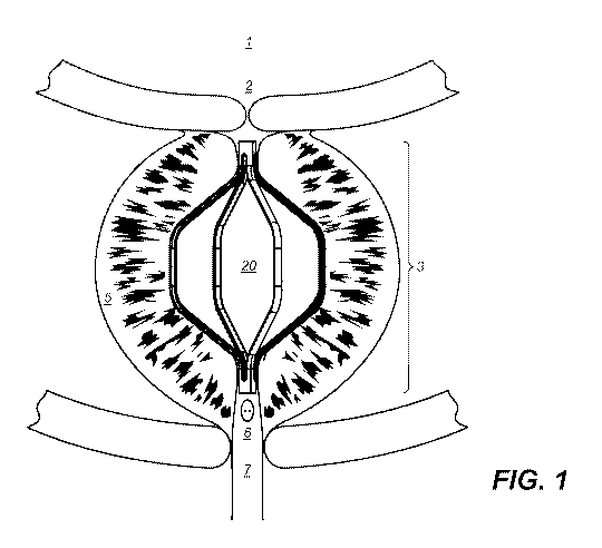
[0059] Referring to Figure 1, a cross-section of the male anatomy shows the
prostate
gland surrounding the urethra. The urethra, under normal conditions, provides
fluid
communication from urine stored in the bladder to be expelled from the body
under
voluntary muscular control of the external urethral sphincter. Normal or
"true" prostate
tissue surrounds the urethra and, in the absence of disease, does not impinge
on the
patency of the urethra. In patients suffering from benign prostatic
hyperplasia (BPH),
the urethra is narrowed by hyperplasic tissue, i.e. prostate tissue that
exhibits excess
growth towards the urethra. This excess of non-cancerous cellular growth leads
to the
symptoms of BPH described above, including, lower urinary tract symptoms
(LUTS) and
urinary outflow obstruction, and urinary incontinence. In Figure 1, an
embodiment of the
implant 20 of the invention is shown engaging prostate tissue 5 along a length
of the
implant 20 to restore the patency of the urethra and to permit unimpeded urine
flow from
the bladder 1. The selective placement of the implant 20 at a target site,
between
bladder neck opening 2 and verumontanum 6, as shown is an important part of
the
invention because the implant 20 does not puncture or incise the surrounding
tissue.
The implant 20 is designed to remain in place within the urethra. The implant
20 does
not extend into the urinary bladder 1, where the structural material of the
implant 20
could become encrusted or otherwise degraded causing complications and making
retrieval more difficult, and the implant 20 does not interfere with the
voluntary control
of the external urethral sphincter or interfere with sexual functions.
[0060] Referring to the Figures 2A-2B, an embodiment of an implant 20 of the
invention
is shown having linear arms as tissue-engaging elements 13a-13d extending
-19-
CA 03060846 2019-10-17
WO 2018/204883 PCT/US2018/031250
substantially radially away from a terminal or proximal hub 11 such that each
of the arms
12a-12d that form the tissue-engaging region have substantially the same
length.
Although the shape of the hub 11 is generally shown as annular in Figures 2-5,
the hub
may be formed of any shape that provides an attachment point for the arms 12a-
12d as
described below. Because the overall dimensions of the implant 20 adopt a
different
configuration as the device transforms from the compressed or constrained
configuration to the expanded or unconstrained configuration, a region of the
arms 12a-
12d most proximate to the hub 11 may be described as a transitional region 15a-
15d
because this short portion of the arms 12a-12d transition from a substantially
linear
configuration, when the device is in the collapsed configuration to a
curvilinear
configuration when the implant 20 is in the expanded configuration or
partially expanded
configuration even if the length of the tissue engaging portions 13a-13d
remains
substantially linear as in Figure 2A. Each of the transitional regions 15a-15d
forms the
connection between the hub 11 and the remainder of the arm 12a-12d comprised
of the
tissue engaging portions 13a-13d.
[0061 ]Referring to Figure 2B, the structure of the hub 11 and its integrally
formed
features are shown in more detail. The hub 11 has a circumferential solid
region 16 at
the end thereof. A second portion of the hub 11 is circumferentially formed
but may not
be entirely solid about the circumference to allow recesses 18a, 18b to form
the arms
12a-12d such that the hub 11 provides structural support for the implant
device 20. The
hub 11 may have an internal open space comprising a housing 17 that traverses
the
entirety of the body of the hub 11 or may be solid through at least the length
of the hub
from the most terminal and to the apex of the recesses 18a,18b at the point of
attachment of the arms 12a-12d. As described in more detail below with respect
to
Figures 7 and 8, depending on the starting material construct from which the
implant is
fabricated, the hub may be a single unitary structure from which material is
removed, or
the individual structures such as the hub 11 and the arms 12a-12d can be
fabricated
separately and assembled into an integrally formed implant assembly. The hub
element
is integral to the implant and designed to incorporate three functional
features. The hub
provides a connection for the arms so that the arms exert a retraction or
radial force on
the prostatic lobes when deployed and implanted inside the urethra. Secondly,
the hub
and recesses allow the implant to be collapsed or constrained into a low-
profile
-20-
CA 03060846 2019-10-17
WO 2018/204883 PCT/US2018/031250
configuration inside the delivery system without exceeding the stress and
strain limits of
the material, thereby allowing the implant to recover to its unconstrained
shape and
dimensions after deployment. Thirdly, the shape of the hub is designed so that
the
implant can be introduced through the working port of the cystoscope in the
constrained
state and placed within the prostatic urethra. The implant may be pushed
through the
working or instrument channel of a cystoscope using a pushrod or push wire.
Alternatively, the implant may be constrained inside a sheath of a delivery
system, and
the delivery system may be advanced through the working channel of a
cystoscope.
[0062]. Either by assembly, or by manufacturing from a single construct or
material
component, each tissue engaging region 13a-13d that is integrally connected
with the
hub 11 is comprised of at least a portion of the length of the arms 12a-12d
and may be
connected to the hub 11 by the transitional regions 15a-15d. Each individual
arm 12a
may be spaced away from each adjacent individual arm 12b at the point of the
transitional region 15a by a small cutout portion 18a to facilitate expansion
of the implant
20 from the compressed to the expanded configuration. As shown in Figure 2A,
each of
the arms 12a-12d terminates at the end spaced farthest away from the hub 11 in
an
atraumatic end 14.
[0063]In the embodiment of Figure 2A, the arms 12a-12d have substantially
equal
length and are oriented to deploy away from the hub 11 in the expanded
configuration
or unconstrained state or partially-constrained state in a substantially
symmetrical
manner. This design results in an overall orientation wherein the atraumatic
ends 14
are positioned at an approximately equal distance from each other and at an
equal
distance the terminal hub 11 as shown. The tissue engaging portions 13a-13d of
the
arms 12a-12d preferably have a substantially flat or substantially planar
ribbon-shape
and, as a result of the manufacturing methodology described herein, can have
equivalent or dissimilar widths or lengths or cross-sectional shapes and
areas.
Depending on the length of the arms 12a-12d and the configuration of the
tissue-
engaging regions 13a-13d, the implant may be symmetrical along an axis
traversing the
terminal hub 11, resulting in the hub 11 being centrally disposed in the
urethra upon
deployment, or may be designed for non¨symmetric positioning of the hub 11
following
deployment.
-21-
CA 03060846 2019-10-17
WO 2018/204883 PCT/US2018/031250
[0064]In another embodiment, the arms 12a-12d may be unequal in length in the
deployed or undeployed state. The hub 11 may be oriented non-centrally so that
it is
positioned asymmetrically along the axis of the urethra where the terminal hub
is
oriented towards one side of the urethral wall. Such configurations have the
advantage
of limiting obstruction of the urethra after deployment. The atraumatic tips
14 reduce
trauma to the urethral wall and may include rounded tips of the distal most
end of the
tissue engaging regions 13a-13d. Such a configuration is readily achieved by
differentially heat-setting the implant 20 such that the atraumatic tips 14 of
the arms 12a-
12d are weaker than the remaining structure or by laser-cutting the tips to
assume an
atraumatic configuration. Heat setting may also be used to shape the
atraumatic tips 14
such that the end portions are slightly curved inward (not shown) to minimize
contact
with the inner tissue layer of the urethral wall.
[0065]Typically, the implant 20 is made from hollow cylindrical tubes or
hypotubes
ranging in diameter between approximately 1-5 mm and wall thicknesses ranging
between approximately 0.2-2 mm. More specifically, having outer diameters
between
approximately 1.5 and 3.0 mm and wall thickness ranging between approximately
0.2
mm and 1.2 mm. Typical width dimensions of the implant 20 are approximately
0.2-3.0
mm. More specifically, typical width dimensions of the arms are approximately
0.5-1.2
mm. The overall length of the implant 20 varies between approximately 10-100
mm.
Implants are laser cut from small-diameter tubes in the collapsed or
constrained
configuration and shape-set to the desired dimensions. Alternatively, the
implants may
be fabricated from large diameter tubes in the expanded state, using tubes
ranging
between 5-50 mm in diameter, or more preferably 10-30mm in diameter. They may
then
be collapsed to smaller size by crimping the implant to a smaller diameter and
constraining them inside a sheath.
[0066] In other embodiments, the implant 20 may be laser-cut and polished from
a solid
tube to increase the force applied by the implant 20 on the prostatic tissue
obstructing
the urethra. The cross section of such implants is in the form of a quadrant
of a circle,
sextant of a circle or circular sector of a circle as described in Figures 7A-
7D and 8A-
8D. Such cross-sectional geometries provide the largest wall forces for a
given surface
area of the implant 20, and hence are highly desirable to reduce the incidence
of
-22-
CA 03060846 2019-10-17
WO 2018/204883 PCT/US2018/031250
encrustation, tissue growth and reduce the potential for migration of the
implant. Typical
diameters of the starting wire range between 1-5mm and circular sector angles
range
between 20-180 degrees.
Typically, the total surface area of the implant is designed to vary between
10-100% of
the total urethral surface area that is treated by the implant from one end to
the other,
or more preferably between 25-80%. The outer surface area of the implant in
contact
with the urethral wall is designed to vary between 5-50% of the total urethral
surface
area treated by the implant from one end to the other. The outer tissue-
pushing or tissue
engaging surface area, where the retraction forces are applied along the
length of
prostatic urethral lobes is designed to vary between 3-30% of the total
urethral surface
area treated by the implant from one end to the other. Such implant
configurations
provide the optimal retraction forces with minimal surface area to minimize or
prevent
encrustation and stone formation. In addition, the low surface area engaging
and
retracting the prostatic tissue and open the urethral lumen minimizes tissue
growth over
the implant and enables implant retrieval, when needed. Accordingly, the
implant
configurations described in this invention also provide high tissue retraction
pressures
or radial pressures, since the retraction forces are concentrated over small
surface
areas in contact with prostatic tissue, to open the narrowed lumen of the
prostatic
urethra while minimizing injury to the urethral surface. For one of the
implants illustrated
in Figure 3B, the average retraction or radial force was measured to be 10N
with a
tissue-engaging outer contact surface area of 40 mm2, yielding a contact
pressure of
0.25 N/mm2. Contact pressures for other embodiments described in this
invention
range between 0.1 to 4 N/mm2 depending on the features of the implant,
including the
number of arms, arm width, arm thickness, arm cross sectional shape and area,
tissue
engaging lengths, number of hubs. Similar calculations may be made for the
retraction
force or radial force per unit mass of the implant. Implant configurations
described in
this invention provide the most efficient us of mass to distribute the forces
along the
length of the prostatic urethra to open the lumen and provide LUTS relief.
[0067] Referring to Figure 2C, an embodiment of the implant 20 of the
invention is shown
with arms 12a-12d having a second curvilinear transitional regions 27a-27d
along their
length and a portion of the tissue engaging segments 28a-28d being
substantially linear.
-23-
CA 03060846 2019-10-17
WO 2018/204883 PCT/US2018/031250
The linear tissue engaging elements 28a-28d minimize trauma to the urethral
wall from
the tips 24 upon deployment and expansion in the urethra. The implant 20, has
a hollow
or solid terminal hub 21, and optionally an asymmetric notch 29 formed in the
body of
the hub 21. The asymmetric notch assists holding the implant during
deployment,
repositioning and retrieval of the implant using delivery systems and
commercially
available grasper devices used in urological procedures. As with the
embodiment of
Figure 2A, the plurality of tissue-engaging regions 23a-23d extend radially
away from
the terminal hub 21 and are integrally connected with the terminal hub 21 by
transitional
regions 25a-25d, as in the embodiment of Figure 2A. In the configuration of
Figure 20,
the transitional regions 25a-25d may be connected to intermediate sections 26a-
6d
disposed along the length of the arms 12a-12d and the transitional regions 25a-
25d and
tissue engaging regions 28a-28d. In the embodiment of Figure 20, each
intermediate
region 26a-26d transitions into a curve 27a-27d leading to a substantially
linear tissue-
engaging region 28a-28d. The length of the linear portion of the tissue-
engaging region
is preferably at least 1-10mm including the range of 1-8 mm. The tissue-
engaging
portions 28a-28d each terminate in an atraumatic end 24. The general shape of
each
arm 12a-12d is comprised of the portion of the hub 21, the transitional
regions 25a-25d,
the intermediate regions 26a-26d, and the tissue engaging regions 28a-28d. As
shown
in Figures 11A-11C below, the implant 20 transforms from a compressed
configuration
or a constrained configuration inside a delivery system to an expanded
configuration
upon deployment. In the compressed configuration, the transitional regions 25a-
25d,
the intermediate regions 26a-26d, and the tissue engaging extensions 23a-23d
are
substantially co-linear and are constrained into a limited diameter within the
delivery
system (not shown). Upon deployment, the configuration of the implant 20 is
restored to
the overall dimensions of the expanded configuration or partially expanded or
deployed
configuration and assumes and orientation within the prostatic urethra as
shown in
Figure 1.
[0068] Referring to Figures 3A-3D, an embodiment of the implant 30 features
tissue-
engaging regions 38a-38d that do not terminate in an atraumatic end, but
rather are
formed of a continuous plurality of arms 32a-32d that terminate in attachment
to a
second terminal hub 31b. Referring to Figure 3A, the two hubs may be described
as a
first proximal hub 31a and a second distal hub 32b having the plurality of
arms 32a-32d
-24-
CA 03060846 2019-10-17
WO 2018/204883 PCT/US2018/031250
extending between the hubs 31a and 31b to establish an integral connection
therebetween. Furthermore, although these embodiments are shown with four
arms,
any plurality of arms numbering two or greater is within the scope of the
invention. As
described below, for embodiments having three arms, an orientation of the arms
is
preferred such that the overall orientation of the device does not result in
the three arms
being placed within the intra-lobular grooves of the prostate.
[0069]Accordingly, referring to Figure 3A, in this embodiment of the invention
the
plurality of arms 38a-38d extend symmetrically away from and around a linear
axis A-A
such that an angle between each arm and the axis A-A is substantially equal
and such
that the angle of each arm 38a-38d relative to any adjacent arm is also
substantially
equal. The arms 38a-38d extend away from each hub 31a,31b in a similar fashion
to
the embodiment of Figures 2A-2B, except that the arms 38a-38d are continuous
between the hubs 31a,31b and are each comprised of first and second
transitional
regions such as 35a,35a' and first and second intermediate regions 36a,36a',
and
centrally disposed tissue-engaging regions 38a-38d that are disposed formed
between
the hubs 31a, 31b.
[0070] Referring to Figures 3B-3D, the tissue engaging regions 38a-38d may
have a
substantially linear portion having a length of at least 0.5 mm and with a
range of 1mm
to 80 mm or more preferably between 1-20mm. For purposes of definition, the
distance
of the linear tissue engaging regions 38a-38d are defined in the expanded
configuration
prior to engaging prostatic tissue and as defined by the shape-memory,
elastic,
superelastic mechanical properties or spring capabilities of the material from
which the
implant 30 is fabricated. As will be readily appreciated by one of skill in
the art, after
deployment within the urethra of a patient suffering from BPH, the overall
dimensions of
the implant 30 will partially conform to the surrounding tissue and so the
dimensions
after deployment may differ from those described herein.
[0071 ] Importantly, the linear distance separating the hubs 31a, 31b is a
first distance
when the implant 30 is in the collapsed configuration, such as when it is
disposed in the
distal end of the delivery system, as described below. Upon deployment into
the
expanded configuration, the hubs 31a, 31b assume a configuration where the
linear
distance separating the hubs 31a, 31b is a second distance wherein the second
distance
-25-
CA 03060846 2019-10-17
WO 2018/204883 PCT/US2018/031250
is less than the first distance. Typically, the ratio of the first distance to
the second
distance may range between 1-10, or more preferably may range between 1.2-3.
Referring to Figure 3D, while individual pairs of arms 38a-38d may the co-
planar i.e.,
exist in a single plane, the hubs 31a,31b and the plurality of interconnected
arms 38a-
38d may assume a rotational orientation relative to each other so that the
arms 38a-38d
form a spiral configuration while maintaining the relative positioning of the
transitional
regions 35a-35d, the intermediate regions 36a-36d, and the tissue engaging
regions
38a-38d.
[0072] Referring to again to the embodiments of Figures 3A-3D, the overall
configuration
and orientation of the implant 20 may be symmetrical about axis A-A as shown
in Figure
3A, particularly when each of the arms and specifically tissue-engaging
regions 38a-38d
are of equivalent dimensions. In such a configuration, the hubs 31a,31b are
centrally
disposed and are both traversed by axis A-A. As noted below, however,
depending on
the method of fabrication, the proximal and distal hubs 31a,31b may not be co-
linear
with a central axis A-A but may be displaced or offset from the central axis
by purpose
of design. The interior of the structure, specifically the area between the 2
terminal hubs
31a, 31b is hollow to facilitate the flow of urine around the implant 30 once
the patency
of the urethra is restored by the expansion of the tissue-engaging regions 38
a- 38d.
[0073]As with the embodiments of Figures 2A-20, the embodiment of Figures 3A-
3D,
the tissue engaging regions 38a-38d are symmetrically oriented around the
central axis
A-A such that the hubs 31a,31b are centrally positioned. The length of the
tissue
engaging regions 38a-38d of the arms are parallel in the embodiments of
Figures 3A-
3D, such that enlarged prostate tissue is engaged along the linear portion of
their arms
at substantially equivalent points relative to the central axis. In other
embodiments, the
arms may be oriented asymmetrically around the central axis and have
asymmetric
shapes. In addition the hubs may be offset on either ends so that they are not
located
centrally and are positioned closed to a first urethral surface on one end and
a second
diametrically-opposite (180 degrees) urethral surface on the other end.
[0074]The outer surface of each tissue-engaging regions in any of the implants
shown
herein may be further comprised of structures or features that function to
prevent
slippage or movement of the implant along the urethra, into the urinary
bladder or exit
-26-
CA 03060846 2019-10-17
WO 2018/204883 PCT/US2018/031250
through the penis, once the implant is deployed. These structural elements may
be any
of barbs, hooks, surface texturing, or any mechanical expedient that engages
tissue
along the length of the outer surface of the implant along the points of
contact with the
interior lumen of the urethra. This embodiment further prevents the tissue-
contacting
regions from positioning the implant or expander completely within the grooves
of the
intra-prostatic lobes.
[0075]As noted above, the implant or expander device described herein is
retrievable
following deployment in the prostate and implantation for a given period of
time as
recommended by the urologist. The implantation period in the prostatic urethra
may
range from 30 days to a few years. To facilitate retrieval of the implant or
expander at
the desired time, it may be constructed to have an integral retrieval fixture
37a,37b, as
shown in Figure 3A affixed to the terminal end of the hub. The fixture 37a,37b
may have
an opening 39a,39b that are engaged by a retriever or any commercially
retrieval or
grasper device, such as the distal end of a catheter wire during the retrieval
process as
described below. One retrieval fixture may be affixed on one terminal end of
the hub or
two fixtures may be designed on both hubs of the implant. In other
embodiments, the
fixtures 37a and 37b may be simple hooks in the shape of a U to be engaged by
a snare
device to engage the implant for retrieval into a sheath. Other fixtures for
retrieval may
be designed on one or both ends by those skilled in the art to retrieve the
implant.
[0076] In another embodiment of the device, it may be constructed using a
single hub
31b on one end, as shown in Figure 3B, connected by four arms and two hubs
31a1
and 31a2 (not shown), constructed by splitting hub 31a into two parts 31a1 and
31a2
(not shown). Each of the hubs 31a1 and 31a2 are connected by two arms. Such a
construct may be deployed in the prostatic urethra with the hubs 31a1 and 31a2
oriented
towards the bladder neck and the hub 31b oriented towards the external
sphincter. Such
a configuration minimized the obstruction of the urethra and facilitates
passage and
placement of a Foley urinary catheter or imaging cystoscope, when needed. A
number
of different combinations may be used, by those skilled in the art, to make
different
implants with one or more hubs on one or both ends, with the hubs connecting
at least
two or more arms to enable the implant to have sufficient expansion force to
retract the
hyperplastic prostatic tissue and open the lumen of the urethra.
-27-
CA 03060846 2019-10-17
WO 2018/204883 PCT/US2018/031250
[0077] Referring to Figure 4, in addition to placing multiple individual
implants separately
within the urethra to retract prostatic tissue along a greater distance than
is possible with
the single implant (to treat a long prostatic urethra), the implant of the
invention can be
provided in a double-aligned configuration that retracts tissue along an axial
length of
the urethra defined by the length of the pairs of the plurality of tissue-
engaging regions.
In the embodiment of Figure 4, a pair of implants of the design of Figures 2A-
2B are
joined at and intermediate hub 45 to create a joining intermediate axis
connecting
member 47 from which the 2 sets of tissue-engaging members 43a-43d, 49a-49d
extend. Proximal tissue engaging regions 48a -48d and distal tissue engaging
regions
49a-49d extend symmetrically away from the intermediate axis 42 and open the
urethral
obstruction in the prostatic urethra. As in the embodiment of Figure 2B, the
arms have
a sigmoid curve shape and extend to tissue-engaging regions having a linear
portion
43a-43d and that each terminate in an atraumatic tip 14. One or more
intermediate hubs
may be used to construct implants of longer length to treat longer urethras.
[0078] The embodiment in Figure 4 may be fabricated as a single integral
structure
without the need for joining at the intermediate hub 47. It may be made from a
hypotube
of a nickel-titanium (superelastic or shape memory nitinol) material using
laser cutting,
cleaning, shape setting and electropolishing processes, known to those skilled
in the
art. In addition, the intermediate hub 45 may be a hollow tube or a solid
tube.
[0079] In other embodiments the intermediate axis or hub connecting member 47
may
incorporate features that make the implant less rigid and conform to the
anatomy. For
example, the connecting member 50 may consist of one or more straight, angled,
slanted, sinusoidal, spiral, or curved connector elements 47 that provide
structure and
flexibility to the implant and can be comprised of a shaft having one or more
elongated
connecting members of substantially circular, rectangular or square cross-
section.
[0080] Referring to Figure 5, a double-ended configuration of the embodiment
of Figures
3A-3E achieves the same advantages of the embodiment of Figure 4 with the
ability to
treat a long urethra with a narrowed lumen due to BPH. An intermediate hub 52
joins 2
sets of tissue engaging regions 53a-53d, 54a-54d having a linear portions 44a-
44d and
having a minimum length of 1mm, between 3 mm and 5 mm, between 5mm and lOmm
in all integral values therein respectively. As in the embodiment of Figures
4, the implant
-28-
CA 03060846 2019-10-17
WO 2018/204883 PCT/US2018/031250
has two separate tissue-engaging regions with similar or pre-selected and
different
diameters and has both a proximal hub 55, and a distal hub 56 for atraumatic
insertion
and delivery to retract prostatic tissue at two selected regions and similarly
atraumatic
retrieval by any of the methods as described herein for the other embodiments.
[0081 ] As described above, one or more intermediate hubs 52 may be used to
construct
implants of different lengths. In addition, the expanded diameters of the
proximal arms
may be different from the expanded diameters of the distal arms although the
constrained diameter of the implant in the delivery system is the same along
the length
of the implant. The embodiment in Figure 5 may be made as unitary structure
without
the need for joining at the intermediate hub 52 as described above by laser
cutting the
desired pattern from a nitinol hypotube and shape setting to the desired
dimensions.
Additionally, each of the structural elements on the implant (hubs, arms,
transitional
regions, diameters, lengths, retrieval fixture) may be a combination of
symmetrical and
asymmetrical features to optimize the implant to facilitate retraction of the
prostatic
tissue upon deployment in the urethra and facilitate retrieval of the implant,
when
desired.
[0082] Referring to Figure 6A-6F, an embodiment of the implant 60 of the
invention is
formed from a continuous wire, ribbon or tube. The continuous wire implant 60,
as
shown in Figure 6A, is in an expanded condition and features tissue engaging
regions
62a-d formed upon expansion of the continuous wire implant 60 following
deployment.
When constrained within the outer sheath 108 (see Figure 11 and 12A) during
delivery
and deployment, the continuous wire implant 60 is tightly compressed within
the inner
diameter of the outer sheath 108 and in this configuration the implant 60 has
four sharp
turns 63a-d, that revert to form a series of curves 63a-63d in the expanded
configuration,
to retract prostatic tissue along the length of the tissue engaging regions
62a-d. The
curves 63a-d act as interconnecting segments for the tissue engaging regions
62a-d.
The arc of the four curves 63a-d is predetermined such that the implant 60
assumes a
predetermined shape in accord with the shape memory properties of the material
from
which the implant 60 is fabricated. The length of the tissue engaging regions
62a-d have
an individual length generally consistent with the retrievable implants of
Figures 2-5, and
as described above with the ability to treat urethra of sufficient length, by
engaging and
-29-
CA 03060846 2019-10-17
WO 2018/204883 PCT/US2018/031250
exerting direct radial forces on lobes of the prostate. The embodiment of 6A
shows an
additional retrieval element connecting regions near 63c and 63d, to
facilitate grasping
and removal of the device, when necessary.
[0083] Figure 6B is an alternative embodiment of the continuous wire or ribbon
implant
60 in the expanded configuration having tissue engaging regions formed by the
intersection of a first tissue engaging region 64a, a second tissue engaging
region 64c,
a third tissue engaging region 64c and a fourth tissue-engaging region 64d. In
the
unexpanded or constrained state inside a delivery system, the tissue-engaging
regions
64a-d are substantially linear along an axis defined by the lumen of the
delivery catheter.
Upon deployment inside the urethra, the implant 60 takes the shape shown in
Figure
6B, in a three-dimensional U-shape configuration, with a first pair of tissue
engaging
regions 64a,64b engaging one side of enlarged tissue in the prostate lumen and
a
second pair of tissue engaging regions 64c,64d engaging an opposite side of
the
enlarged tissue to apply retraction forces on the urethral wall of the
prostate. As in the
embodiment of Figure 6A, in the collapsed configuration the implant 60 has
four sharp
turns 65a-d in the confined configuration that transform, by the shape memory
or
superelastic or spring properties of the material from which the implant 60 is
formed,
into smooth curves upon expansion. The embodiment of Figure 6B is formed such
that
tissue engaging regions 64a and 64b extend at one end of the implant extend
away from
the turns 65c at the other end of the implant by the close engagement of
intermediate
terms 65b, 65d that are centrally disposed along the length of the implant 60.
[0084] Figure 60 is an alternative embodiment of the continuous wire or ribbon
implant
60 in the expanded configuration having tissue engaging regions 66a-66d that
are
atraumatic. The additional turns 68a-68b form additional tissue engaging
regions 69a-
69d. It will be appreciated by one of skill in the art that the configurations
that utilize the
continuous wire embodiment are less dependent on precise placement within the
prostatic urethra and do to varying orientations upon deployment, and due to
differing
regions in the physiology of any individual patient, the tissue-engaging
portions 66a-
66d, 69a-69d may be a subset of all of the regions defined as tissue-engaging
portions.
The implant 60 configuration is modified to engage more prostatic tissue and
reduce the
stress concentration by forming a small loop at the end that forms tissue-
engaging
-30-
CA 03060846 2019-10-17
WO 2018/204883 PCT/US2018/031250
regions 69a-69d. As described above, the tissue-engaging regions 66a-66d, 69a-
69d
are substantially linear along an axis in the constrained or unexpanded state
inside the
delivery system. Upon deployment inside the urethra, the implant 60 takes the
shape
shown in Figure 60, in a three-dimensional saddle-like or W-shaped shape
configuration.
[0085] Figure 6D and Figure 6E are continuous wire embodiments of Figures 6A
and
6B, having a sine wave form and configured to have arched proximal and distal
end
regions. A retrieval feature, as shown in Fig 6A, may additionally be
incorporated to
facilitate repositioning of the implant during deployment or removal of the
implant after
a given implantation period, as desired.
[0086] Referring to Figure 6E-6F, an embodiment of the implant 70 of the
invention is
formed from a continuous wire having a sine wave form and configured to have
arched
proximal and distal end regions. In this embodiment, the tissue engaging
regions 71a-d
are formed by a substantially straight length of the continuous wire. The
proximal and
distal ends of the continuous wire are comprised of the arched end regions
72a, 72b,
and 72c, 72d, respectively. The embodiment of Figure 6E has the same shape and
configuration as Figure 6D but is comprised of six tissue engaging regions 71a-
f and
having three arched end regions at each of the proximal and distal end572a,
72c, 72e,
and 72b, 72d, 72f, respectively. The material from which the implant 70 is
constructed
is characterized as a flat wire or "ribbon" width of the surface facing both
the prostatic
tissue and the interior space of the implant 70 has a width that is equal to
or greater
than its depth. Preferable dimensions include, but are not limited to, a
thickness in the
range of 0.0055-0.055 inches, and more preferably approximately between 0.011-
0.025
inches and a width range of between 0.01-0.18 inches with a preferable width
range of
approximately between 0.02-0.14 inches. These devices are placed such that the
axis
A-A traverses the length of the urethra, although the designation of the
proximal or distal
end is arbitrary, an orientation such that the proximal and distal ends
respectively, are
located along the linear path of the urethra is necessary.
[0087] The embodiment of Figure 6F is a stacked or paired configuration of the
embodiment of Figure 6D, having eight total tissue engaging regions 71a-d, 74
a-d and
which are joined by interconnecting links 73a, 73b. The proximal and distal
ends are
-31-
CA 03060846 2019-10-17
WO 2018/204883 PCT/US2018/031250
formed by the most distal pair of arched regions 72a,b, most proximal end is
formed by
a most proximal pair of arched regions 72c,d, although as noted above, the
distinction
is arbitrary because this embodiment is symmetrical across a horizontal axis
of the
device.
[0088] Figures 7A-7D illustrate the starting materials and fabrication
processes for any
of the embodiments of Figures 2-6. The starting material may be a solid tube
or wire, as
shown in Figures 7A and 70, or may be a hollow tube, as shown in Figures 7B
and 7D.
Fabrication of different embodiments of the invention selectively removes
material along
the length of a construct by traversing a radial distance from the outer
surface of the
body of the construct. The radial distance may traverse the entire diameter or
thickness
of the tube or only a partially towards the center of the circular wire or
tube. Selective or
removal of material along the length of the construct creates the individual
arms of the
implant. The pattern from which material is removed from the construct
dictates the
shape, cross section, number and orientation of the arms. Removing material
from less
than the entire linear dimension along the length of the construct retains the
integrity of
the terminal hub without the need for joining. In the case of a single
terminal hub, such
as in the embodiment of Figures 2A-2B, material is removed from an
intermediate point
proximate to a single terminal hub and along the entire length of the
remainder of the
construct. By retaining a terminal hub at both ends of the construct, the
embodiment of
Figures 3A-3B is manufactured wherein the material is removed selectively from
the
construct along a length of the wire or tube in a given pattern to form the
desired implant
shape and dimensions. Laser cutting or electrodischarge machining (EDM), shape
setting and electropolishing are commonly used manufacturing processes used to
fabricate the implant.
[0089] Figures 8A-8D are the starting wire or tube materials for the
fabrication of the
implant as described above for Figures 7A-7D but having a rectangular cross-
section.
In the embodiments of Figures 8A-D, the material is removed from the construct
to
create a traversing radius such that the arms separate from each other and are
capable
of radial expansion. Accordingly, a cut, or series of cuts, that create a
traversing radius
is defined as a cut or number of cuts that create one or more radii or one
complete
diameter such that the arms may expand away to form tissue-engaging regions
when
-32-
CA 03060846 2019-10-17
WO 2018/204883 PCT/US2018/031250
the arms expand. If a single cut is made to create two extending arms, the
total depth
of the cuts must traverse the entire thickness of the rectangular cross
section of the
construct. Referring to Figure 80, the cuts may be made as, for example for a
solid
construct, for one complete thickness by combining segments a and a'. Three
arms
would be created by executing cuts at segment a, segment b', and d. Four arms
would
be created by cuts at segment a, segment a', segment c, and segment c'. Each
of the
above described configurations would yield an implant where the tissue
engaging
regions deploy symmetrically or asymmetrically depending on the number of cuts
and
the desired shape that the implants are heat-set to in the manufacturing
process to
achieve the desired functional characteristics.
[0090]Similarly, a three-armed implant could be created by radially traversing
cuts at
segment a, segment a', and segment c to create an asymmetric implant. The cut
must
be deep enough to form the arms when all of the cuts are complete. Referring
to Figure
7B, the cuts need not constitute an entire radius of the width of the
construct, but only
that width of the solid portion needed to create the expanding arms.
Accordingly, cutting
the segments a and a' are less than the distance of an entire outer diameter
of the
construct but are still adequate to create the expanding arms.
[0091] Referring to Figure 9, individual and assembled components A, B.1, B.2
and C
of a delivery system 100 of the invention are shown in an operative
relationship with a
conventional cystoscope 111 (shown in Figure 10) for placement of an
embodiment of
the implant of the invention from a distal end of the delivery system 100.
Specifically,
Figure 9 at component A is a proximal assembly of the delivery system 100
showing a
continuous guide wire or an implant holding wire 101 entering the open
proximal port
109 of lumen 106 and proximate to an irrigation port 104 that is in turn
proximate to an
intermediate coupling 105 that creates a seal to form an intact fluid
connection between
the irrigation port 104 and the delivery system 102 through a Luer lock 103
and having
a configuration for a stopper (not shown) to seal the irrigation port 104.
[0092]Figure 10 is a distal end of the outer sheath 108 of the integrated
delivery
assembly 100 showing the distal end of a conventional cystoscope 111 and the
distal
end of the delivery sheath 108 protruding from the distal end of the
cystoscope 111 and
traversing the working channel 112 of the cystoscope 111 and containing an
implant 10,
-33-
CA 03060846 2019-10-17
WO 2018/204883 PCT/US2018/031250
still maintained within the compressed configuration and at the distal end of
the sheath
108 prior to manual, incremental retraction of the sheath 108 for deployment.
The distal
end of the continuous guidewire 101 has a pre-dispositioned shape or fixture
or tether
mechanism 110 to enable controlled and accurate deployment of the expander
implant
at the target site. This feature on the guidewire or implant holding wire also
prevents the
implant from deploying prematurely from the distal end of the assembly formed
of the
outer sheath 108 the implant 10 and the guidewire 101 when the implant 10 is
in the
compressed configuration. In one embodiment of the guidewire feature, the
controlled
deployment of the implant 10 is enabled by creating an undulating portion 101a
on the
guidewire 101 (not shown in the figure). In Figure 10 the guidewire 101
traverses both
the proximal and distal hub 31a, 31b. It will readily be appreciated that any
of the
implants of the invention described herein can be affixed to the distal end of
the delivery
system 100 in a collapsed configuration. If the implant contains one or more
hub fixtures,
for example the hub 11, 21, as illustrated in Figures 2A-B and the proximal
and distal
hub 31a,31b of Figures 3A-E, the guidewire 101 preferably traverses the hub
for
orientation and placement of the implant by the urologist. In other
embodiments, the
guidewire may be engaged to the implant via tether loop. Following deployment
of the
implant, the guidewire may be removed to disengage the guidewire from the
implant.
[0093] Figures 11A-C illustrate the incremental, step-wise deployment of the
implant 10
as described above. Accordingly, the combination of the deployment system and
the
implant progress through 3 basic stages: 1) a first stage (Figure 11A) wherein
the
implant 10 is maintained in a compressed configuration at the distal end of
the delivery
system 110 and maintained in a configuration by the structure of the outer
sheath 108
(Figure 11A), 2) a second stage wherein the user begins to push the mandrel
forward
pushing the implant forward. The outer sheath 108 at this point is only
partially
withdrawn from about the axial length of the implant 10 (Figure 1113), and 3)
a third stage
wherein the mandrel pushes the implant completely out of the outer sheath
(Figure 110).
This allows complete expansion of the implant 10 to the expanded configuration
while
the implant 10 is still engaged by the guidewire 101, thereby allowing further
manipulation of the location and orientation of the implant 110 within the
urethra.
Following or contemporaneous with release of the implant from the delivery
system 100,
the implant 10 expands from the collapsed configuration to the expanded
configuration
-34-
CA 03060846 2019-10-17
WO 2018/204883 PCT/US2018/031250
followed by withdrawal of the guidewire 101 and final deployment of the
implant as
represented by Figure 1. Other deployment mechanisms and embodiments may be
conceived and used by those skilled in the art to deploy the implant in the
prostatic
urethra by retracting the outer sheath 108.
[0094]The implant deployment mechanism of the delivery system of invention may
have
any mechanical expedient (not shown) that allows the physician to actuate a
handle in
rotary fashion to retract the outer sheath to deploy the implant from the
distal end of the
delivery system. The handle is adapted to be grasped by hand and rotates
around a
shaft. Rotation of the handle around the shaft by drawing the handle toward
the user
engages a gear mechanism, having a fixture attached to the outer sheath.
[0095]The delivery system 100 may be fixedly attached to a deployment
mechanism
such that rotation of the handle causes retraction of the sheath 108 along the
length of
the hypotube or pusher rod 119. The rotation provides both a first position
wherein the
implant 10 is fully contained, in the collapsed configuration, within the
distal end of the
outer sheath 108 and is removably attached to the guidewire 101. Actuation of
the
handle can be performed in an incremental fashion such that the implant is
deployed in
stages as described in connection with Figures 11A-11C above. In a preferred
embodiment, the handle has partial stops at incremental deployment steps for
the
implant 10. A first incremental stop is a deployment of the implant 110 to an
initial stage
wherein expansion from the collapsed configuration is begun. A second
incremental
stop provides deployment of the implant 110 to an intermediate phase wherein
the
positioning and orientation of the implant can be visually verified. Rotating
the handle to
the final position completely deploys the implant 110 from the distal end of
the outer
sheath 108 under visualization.
[0096] In use, pursuant to a method of the invention, the delivery system 100
has an
overall outer dimension (OD) less than 7 French and is introduced via the
working
channel 112 of a cystoscope 111, typically having an outer diameter of 17
French. The
urologist visualizes the prostatic urethra using the light source 112 and lens
113
integrated into the cystoscope 111 and typically measures the length of the
prostatic
urethra and evaluates the extent of narrowing of the urethra caused by the BPH
condition. From this visualization, the urologist selects the appropriate
implant size, and
-35-
CA 03060846 2019-10-17
WO 2018/204883 PCT/US2018/031250
selects from a pre- assembled implant package containing the delivery system
100 with
the correctly sized implant 10 already disposed in the collapsed configuration
therein.
While the distal tip of the cystoscope 111 is located inside the patient's
bladder, a saline
source is attached to the irrigation port 104 and irrigation is commenced.
Under direct
visualization, the assembly of the cystoscope 111 and the delivery system 100
is
oriented so that the distal end of the cystoscope 100 is placed just proximal
to the
targeted area at or away from the verumontanum.
[0097]The outer sheath 108 is advanced to a position proximal to the bladder
neck, and
after confirming direct visualization that the implant 110 is proximate to the
target portion
of the urethra impinged or narrowed by prostatic tissue, the delivery sheath
108 is
pushed forward causing the implant 110 to achieve an initial, partially
expanded
configuration. The forward push of the mandrel is interrupted to verify that
the implant is
well-positioned and is located in the appropriate target site. After
verification, the outer
sheath 108 is further withdrawn causing the implant 110 to reach a fully
expanded
configuration at an intermediate step of the implant 110 deployment, similar
to the
overall configuration illustrated in Figure 110. Once placement, targeting,
and
orientation are verified by the urologist, the guidewire 101 is withdrawn,
followed by
withdrawal of the outer sheath 108 and the delivery system 100.
[0098]As noted above, the design of the delivery system 100 and the several
embodiments of the implant 110, permits an incremental and well -controlled
deployment of the implant 110 so that the implant 110 does not "spring open"
or "spring
forward" prematurely and deploy at in an unsuitable configuration or location
away from
the target site. By selected and incremental retraction of the outer sheath
108 from an
initial position where the implant 110 is partially deployed, to an
intermediate position
where the implant 110 has completely reached the expanded configuration but is
still
tethered to the guidewire 101, preferably followed by verification of the size
of the
implant in the placement within the prostatic urethra. Removal of the
guidewire 101 in
the outer sheath 108 completes a multi-step deployment process. The guidewire
avoids
inadvertent, premature deployment of the implant 110 or misplacement of the
implant
10, which can be irreversible and require removal of the implant 110, and
repeat
treatment by deploying a new implant using a new deployment or delivery system
100.
-36-
CA 03060846 2019-10-17
WO 2018/204883 PCT/US2018/031250
[0099]The methods of the invention include deploying an implant into the
urethra
wherein the implant having two or four or a greater number of tissue engaging
regions
to retract at least two discrete regions of enlarged prostatic tissue at the
surface of tissue
of the lobe in a patient in need thereof. A radial force is exerted at at
least the two
discrete regions and each region is each contacted along the interior wall of
the urethra.
In some embodiments, the force is exerted on the tissue along an axis
perpendicular to
an axis that runs the linear length of the urethra and which traverses the
central axis of
the implant of the invention. The methods include a procedure to remove the
implant of
the invention, upon further diagnosis of BPH or LUTS in a patient, and which
is based
on the design of the invention. In the removal process, the most proximal
portion of the
implant is accessed by a wire or suture extending from an opening at the
distal end of a
retrieval tube and the implant is engaged proximate to the hub and drawn into
the
retrieval tube, thereby reversing the deployment process and returning the
implant from
the expanded to the confined configuration. When the implant is placed such
that the
hub is more distal than the tissue engaging portions of the implant based on
the initial
clinical judgment and deployment by the physician, the implant may be pushed
distally
into the bladder and re-oriented such that the hub can be engaged in the
implant drawn
into the distal end of the retrieval tube.
[0100] A method to alleviate clinical symptoms of benign prostatic hyperplasia
is
performed by placing the implant, apart from and optionally proximal of the
ejaculatory
ducts, by-advancing a deployment catheter having a proximal end and a distal
end
through a working channel of a standard urology cystoscope to position the
distal end of
the catheter containing the implant at a point between the bladder neck and
the external
sphincter. Once the distal end of the delivery system reaches the target site,
the implant
is deployed whereupon it expands from a compressed configuration to an
expanded
configuration to engage hyperplasic prostate tissue. During the deployment,
proximal
and distal hubs of the implant, which are in a substantially linear
configuration when the
implant is maintained in the collapsed configuration and while the catheter is
advanced
through the working channel. In this configuration, the arms are maintained in
a
substantially parallel condition being relatively aligned with one another
within the
confines of the inner diameter of the delivery system catheter.
-37-
CA 03060846 2019-10-17
WO 2018/204883 PCT/US2018/031250
[0101] During deployment, the expansion from the initial confined
configuration to the
expanded configuration features characteristic changes in the orientation of
the
structures of the implant. In all of the embodiments, the arms of the implants
that are
comprised of tissue-engaging regions expand to engage the enlarged prostatic
tissue.
Portions of the implant may assume a different shape transforming from
substantially
linear to curvilinear or a sigmoid form depending on the design and
orientation of the
implant upon deployment.
[0102] in the embodiments describing above having a proximal and a distal hub
interconnected by a plurality of arms, the linear distance between the
proximal and the
distal hubs is changed from a first position in the collapsed configuration to
a second
position in the expanded configuration where the distance between the hubs is
reduced
in the second position. In embodiments where a hub is connected to first and
second
transitional region of each of the arms converts, the transitional regions
transform from
a substantially linear to a curvilinear form, wherein the first transitional
region is distal to
the proximate hub and connected thereto and the second transitional region is
proximal
to the distal hub and connected thereto. A tissue-engaging segment of each arm
that is
preferably centrally disposed in the length of the implant expands away from a
central
axis of the retrievable implant to engage enlarged prostate tissue along at
least a portion
of the length of the central tissue-engaging segment. In some embodiments, the
length
of the tissue-engaging region that engages the prostate tissue is
substantially linear.
[0103] The expansion from the collapsed to the expanded configuration produces
an
integral connection between a solid circumferential region of each of the
proximal and
distal hubs and the central tissue-engaging segment of the plurality of arms.
During
deployment, the tissue engaging portions are preferably oriented such that the
plurality
of 4 arms do not engage inter-lobular grooves of the prostate.
[0104] The methods of the invention also include a separate procedure for
retrieving
the implant through the working channel of the cystoscope. The step of
retrieving the
implant can be performed by engaging any portion of the implant that permits
the implant
to be drawn into the distal end of a retrieval system where the implant
reverts from an
expanded configuration to a collapsed configuration. The implant may be
engaged at
any point on the structure of the implant or by engaging a fixture on the
proximal hub
-38-
CA 03060846 2019-10-17
WO 2018/204883 PCT/US2018/031250
and retracting the implant into the distal end of a retrieval catheter.
Engaging in the
implant may be achieved by using a retrieval wire that has a specially
configured distal
end that loops back on itself for secure engagement of the implant.
[0105] The invention also includes the configuration wherein catheter for
delivery of a
retrievable implant in a collapsed configuration is delivered through the
working channel
of a flexible cystoscope and placed in an expanded configuration in a
prostatic urethra
narrowed by hyperplasia where the combination is an outer sheath for
constraining the
retrievable implant at a distal end, a pusher or push rod sized to traverse
the length of
an inner lumen of the catheter and having a fixture at the distal end thereof
to engage
the proximal portion of the retrievable implant and to advance the retrievable
implant
distally relative to the catheter to deploy the implant, and a delivery wire
to assist
accurate placement of the retrievable implant. Preferably, the outer diameter
of the
delivery catheter is less than 9 French and optionally includes: a fluid
communication
lumen, a camera, and scope, or visualization apparatus for direct imaging
during
deployment of the implant. The device can include radiographic, fluoroscopic,
or other
imaging markers to assist in positioning of the delivery system or the
implant.
[0106] The invention also includes unique advantages in the structure and
performance of the implant that is derived from the selection of the starting
materials and
the fabrication processes described herein. In some embodiments, the implant
is made
from a unitary body of shape-memory or super-elastic material by the selective
removal
of material along a selected length of the elongated and unitary body and
traversing a
diameter thereof, wherein the selected length is less than the total length of
the
elongated and unitary body used to fabricate the implant such that the
resulting structure
may be either symmetric or asymmetric about a central axis considered as an
imaginary
line down the length of the implant. Although the embodiments fabricated from
a single
tube can be considered integrally connected and unitary, because they are
formed from
a continuous piece of material, individual structures of the implant can be
welded
together to yield any configuration. Where material is removed from the tube,
essentially
any configuration can be created by known micro-machining techniques with the
only
constraint being that enough material must be removed from a length along the
linear
length of the tube from which the implant is fabricated such that enough
material is
-39-
CA 03060846 2019-10-17
WO 2018/204883 PCT/US2018/031250
removed to form the arms. This parameter can be described as the need to
traverse a
diameter of the construct from which the implant is manufactured such that the
cuts must
are collectively deep enough to at least cut through the solid portion of the
tube in the
middle of a cross section of the tube so that the arms can move away from each
other.
If the tube is solid, such a distance is the entire diameter collectively to
yield a "quadrant,"
but if hollow then through to the hollow portion to form an "arc."
[0107] The methods are driven by the physiology of the individual patient and
are at
the discretion of the urologist, although the procedures generally include the
steps of
advancing a deployment catheter having a proximal end and a distal end through
a
working channel of a urology cystoscope to position the distal end of the
catheter at a
point between the bladder neck and the external sphincter. Once in the proper
position
and orientation, the urologist deploys the implant from the distal end of the
catheter to
expand the implant from a collapsed to an expanded configuration. Deployment
of the
implant causes the tissue-engaging regions of the implant to engage enlarged
prostate
tissue along a length of a plurality of elongate arms of the implant. Some of
the
embodiments have the configuration where the tissue-engaging regions are
disposed
on the arms that connect the two hubs and in this configuration a length of
the arms
forms an elongated structure such that a series of substantially linear
arrangement arms
go from a configuration being constrained by the catheter in the collapsed
configuration
to an expanded configuration wherein each of the arms is integrally connected
at both
ends to the hubs.
[0108] Because of the design of the two-hub embodiment of the implant, the
expansion of the plurality of arms during deployment causes the proximal hub
and the
distal hub to move linearly toward each other along an axis connecting the
hubs while
the plurality of arms expand to engage the enlarged prostate tissue. The
result is that
the distance between the hubs in the expanded configuration is less than the
distance
in the collapsed configuration.
[0109] The arms can be described as having transitional regions that are
integrally
formed with each hub such that a first transitional region is integrally
formed proximate
to one hub and a second transitional region is integrally formed proximate to
the second
hub with the tissue-engaging regions of the plurality of arms disposed
therebetween.
-40-
CA 03060846 2019-10-17
WO 2018/204883 PCT/US2018/031250
These first and second transitional regions convert from a substantially
linear form to a
curvilinear form when the implant converts from the collapsed to the expanded
configuration.
[0110] The implant is designed so that the radial forces exerted by the
implant are
applied directly to the prostate lobes rather than an orientation wherein the
tissue
engaging regions are confined to the intralobular grooves. For this reason,
the method
of the invention includes taking advantage of the design of the implant to
position the
device for deployment such that at least 2 of the plurality of arms do not
engage inter-
lobular grooves of the patient's prostate.
[0111] As noted above, important part of the design features of the present
implant is
the ability to retrieve the implant when the clinical discretion of the
urologist so indicates.
Typically, the retrieval process includes the step of retrieving the implant
through the
working channel of the cystoscope. Specifically, the urologist captures the
implant by
engaging either the body of the implant or a dedicated structure of the
implant and
inserting the implant into the distal end of a retrieval catheter, usually by
drawing the
implant into the catheter, whereby the implant reverts from the expanded to
the collapsed
configuration. The implant is designed so that the retrieval process can
involve engaging
the body of the implant at a dedicated fixture or simply by a grasper that
engages a
cylindrical portion of a hub that can be specifically modified to form a loop
at the most
proximal or most distal portion of the implant or both. Depending on the
physiology of
the individual patient, the retrieval may also occur by advancing the implant
into the
bladder while it is in the expanded or partially expanded configuration
followed by
drawing the implant into the distal end of the retrieval catheter for removal.
[0112] While the retrieval method is preferably comprised of engaging the
implant at
the target site at which the implant was originally deployed and withdrawing
the implant
directly approximately through the urethra, the retrieval can be achieved by
advancing
implant distally into the bladder prior to removal.
[0113] The Examples disclosed above are merely intended to illustrate the
various
utilities of this invention. It is understood that numerous modifications,
variations and
combinations of functional elements and features of the present invention are
possible
-41-
CA 03060846 2019-10-17
WO 2018/204883 PCT/US2018/031250
in light of the above teachings and, therefore, within the scope of the
appended claims,
and the present invention may be practiced otherwise than as particularly
disclosed.
[0114] All patents and publications are herein incorporated for reference to
the same
extent as if each individual publication was specifically and individually
indicated to be
incorporated by reference. It should be understood that although the present
invention
has been specifically disclosed by preferred embodiments and optional
features,
modification and variation of the concepts herein disclosed may be resorted by
those
skilled in the art, and that such modifications and variations are considered
to be within
the scope of this invention.
-42-