Note: Descriptions are shown in the official language in which they were submitted.
CA 03061050 2019-10-21
WO 2018/204405 PCT/US2018/030516
TITLE OF THE INVENTION
STABLE FORMULATIONS OF ANTI-TIGIT ANTIBODIES ALONE AND IN
COMBINATION WITH PROGRAMMED DEATH RECEPTOR 1 (PD-1) ANTIBODIES AND
METHODS OF USE THEREOF
FIELD OF THE INVENTION
The invention relates to formulations of therapeutic antibodies and their use
in treating
various disorders. In one aspect, the invention relates to formulations
comprising antibodies or
antigen binding fragments thereof that bind to T cell immunoreceptor with Ig
and ITIM domains
(TIGIT). In another aspect, such formulation further comprises an anti-human
programmed
death receptor 1 (PD-1) antibody or antigen binding fragment thereof Also
provided are
methods of treating various cancers and chronic infections with the
formulations of the
invention.
CROSS-REFERENCE TO RELATED APPLICATIONS
This application claims the benefit of U.S.S.N 62/500,278, filed May 2, 2017,
the
contents of which are hereby incorporated by reference in their entirety.
REFERENCE TO SEQUENCE LISTING SUBMITTED ELECTRONICALLY
The sequence listing of the present application is submitted electronically
via EFS-Web
as an ASCII formatted sequence listing with a file name "24453W0PCT-SEQTXT-
01MAY2018.TXT", creation date of May 1, 2018, and a size of 227Kb. This
sequence listing
submitted via EFS-Web is part of the specification and is herein incorporated
by reference in its
entirety.
BACKGROUND OF THE INVENTION
Antibody drugs for use in humans may differ somewhat in the amino acid
sequence of
their constant domains, or in their framework sequences within the variable
domains, but they
typically differ most dramatically in the CDR sequences. Even antibodies
binding to the same
protein, the same polypeptide, or even potentially the same epitope may
comprise entirely
different CDR sequences. Therapeutic antibodies for use in human beings can
also be obtained
from human germline antibody sequence or from non-human (e.g. rodent) germline
antibody
sequences, such as in humanized antibodies, leading to yet further diversity
in potential CDR
sequences. These sequence differences result in different stabilities in
solution and different
responsiveness to solution parameters. In addition, small changes in the
arrangement of amino
acids or changes in one or a few amino acid residues can result in
dramatically different antibody
stability and susceptibility to sequence-specific degradation pathways. As a
consequence, it is
not possible at present to predict the solution conditions necessary to
optimize antibody
- 1 -
CA 03061050 2019-10-21
WO 2018/204405 PCT/US2018/030516
stability. Each antibody must be studied individually to determine the optimum
solution
formulation. Bhambhani et al. (2012) J. Pharm. Sci. 101:1120.
Antibodies are also relatively high molecular weight proteins (-150,000 Da),
for example
as compared with other therapeutic proteins such as hormones and cytokines. As
a consequence,
it is frequently necessary to dose with relatively high weight amounts of
antibody drugs to
achieve the desired molar concentrations of drug. In addition, it is often
desirable to administer
antibody drugs subcutaneously, as this enables self-administration. Self-
administration avoids
the time and expense associated with visits to a medical facility for
administration, e.g.,
intravenously. Subcutaneous delivery is limited by the volume of solution that
can be practically
delivered at an injection site in a single injection, which is generally about
1 to 1.5 ml.
Subcutaneous self-administration is typically accomplished using a pre-filled
syringe or
autoinjector filled with a liquid solution formulation of the drug, rather
than a lyophilized form,
to avoid the need for the patient to re-suspend the drug prior to injection.
Antibody drugs must
be stable during storage to ensure efficacy and consistent dosing, so it is
critical that whatever
formulation is chosen supports desirable properties, such as high
concentration, clarity and
acceptable viscosity, and that also maintains these properties and drug
efficacy over an
acceptably long shelf-life under typical storage conditions.
TIGIT (T cell immunoreceptor with Ig and ITIM domains) is an immunomodulatory
receptor expressed primarily on activated T cells and NK cells. TIGIT is also
known as VSIG9;
VSTM3; and WUCAM. Its structure shows one extracellular immunoglobulin domain,
a type 1
transmembrane region and two ITIM motifs. TIGIT forms part of a co-stimulatory
network that
consists of positive (CD226) and negative (TIGIT) immunomodulatory receptors
on T cells, and
ligands expressed on APCs (CD155 and CD112).
An important feature in the structure of TIGIT is the presence of an
immunoreceptor
tyrosine-based inhibition motif (ITIM) in its cytoplasmic tail domain. As with
PD-1 and TIGIT,
the ITIM domain in the cytoplasmic region of TIGIT is predicted to recruit
tyrosine
phosphatases, such as SHP-1 and SHP-2, and subsequent de-phosphorylation of
tyrosine
residues with in the immunoreceptor tyrosine-base activation motifs (ITAM) on
T cell receptor
(TCR) subunits. Hence, ligation of TIGIT by receptor-ligands CD155 and CD112
expressed by
tumor cells or TAMS may contribute to the suppression of TCR-signaling and T
cell activation,
which is essential for mounting effective anti-tumor immunity. Thus, an
antagonist antibody
specific for TIGIT could inhibit the CD155 and CD112 induced suppression of T
cell responses
and enhance anti-tumor immunity.
The need exists for stable formulations of anti-TIGIT antibodies for
pharmaceutical use,
e.g., for treating various cancers and infectious diseases, as well as for
stable formulations of
anti-TIGIT antibodies co-formulated with anti-human PD-1 antibodies.
Preferably, such
formulations will exhibit a long shelf-life, be stable when stored and
transported, and will
preferably exhibit stability over months to years under conditions typical for
storage of drugs for
- 2 -
CA 03061050 2019-10-21
WO 2018/204405 PCT/US2018/030516
self-administration, i.e. at refrigerator temperature in a syringe, resulting
in a long shelf-life for
the corresponding drug product.
SUMMARY OF THE INVENTION
In one aspect, the invention includes a formulation of an anti-TIGIT antibody,
or antigen
binding fragment thereof, comprising (i) an anti-TIGIT antibody, or antigen
binding fragment
thereof (ii) a buffer, (iii) a non-reducing sugar; (iv) a non-ionic
surfactant; and (v) an
antioxidant. In an embodiment, the formulation further comprises an anti-PD-1
antibody, e.g.,
pembrolizumab or nivolumab. In another embodiment, the formulation comprises a
chelator.
In an embodiment of the invention, the formulation comprises (i) about 10
mg/ml to
about 200 mg/ml of an anti-TIGIT antibody, or antigen binding fragment thereof
(ii) about 5
mM to about 20 mM buffer; (iii) about 6% to about 8% weight / volume (w/v) non-
reducing
sugar; (iv) about 0.01 % to about 0.10% (w/v) non-ionic surfactant; and (v)
about 1 mM to
about 20 mM anti-oxidant. In an embodiment, the formulation further comprises
an anti-PD-1
.. antibody, e.g., pembrolizumab or nivolumab. In another embodiment, the
formulation further
comprises a chelator. In one embodiment, the formulation has a pH between 4.5
¨ 6.5. In
particular embodiments, the pH of the formulation is from about pH 5.5 to
about pH 6.2. In a
further embodiment, the pH of the formulation is from about pH 5.6 to about pH
6Ø In another
embodiment, the pH of the formulation is about 5.7. In another embodiment, the
pH of the
formulation is about 5.8. In another embodiment, the pH of the formulation is
about 5.9. In
another embodiment, the pH of the formulation is about 6Ø In another
embodiment, the pH of
the formulation is about 6.1. In another embodiment, the pH of the formulation
is about 6.2.
In one embodiment of the formulation, the buffer is L-histidine buffer or
sodium acetate,
the non-reducing sugar is sucrose, the non-ionic surfactant is polysorbate 80,
and the anti-
oxidant is methionine, or a pharmaceutically acceptable salt thereof In one
embodiment, the
anti-oxidant is L-methionine. In another embodiment, the anti-oxidant is a
pharmaceutically
acceptable salt of L-methionine, such as, for example, methionine HC1.
In another embodiment, the formulation comprises (i) about 10 mg/ml to about
200
mg/ml of an anti-TIGIT antibody, or antigen binding fragment thereof (ii)
about 5 mM to about
20 mM of L-histidine buffer or about 5 mM to about 20 mM of sodium acetate
buffer; (iii) about
6% to about 8% w/v sucrose; (iv) about 0.01 % to about 0.10% (w/v) polysorbate
80; and (v)
about 1 mM to about 20 mM L-methionine. In another embodiment, the formulation
further
comprises an anti-PD-1 antibody, e.g., pembrolizumab or nivolumab. In an
embodiment, the
formulation further comprises a chelator. In one embodiment, the chelator is
present in an
amount of about 1 uM to about 50 M. In one embodiment, the chelator is DTPA.
In another
embodiment, the chelator is EDTA. In one embodiment, the buffer is L-histidine
buffer. In one
embodiment, the formulation comprises about 8m1V1 to about 12 mM of L-
histidine buffer. In
another embodiment, the formulation comprises about 5 mM to about 10 mM of L-
methionine.
- 3 -
CA 03061050 2019-10-21
WO 2018/204405 PCT/US2018/030516
In a further embodiment, the formulation comprises polysorbate 80 at a weight
ratio of
approximately 0.02% (w/v). In one embodiment, the anti-TIGIT formulation
comprises sucrose
at a weight ratio of about 7% (w/v). In any of these embodiments, the
methionine is L-
methionine.
In embodiments of the formulation, the concentration of the anti-TIGIT
antibody or
antigen binding fragment thereof is from about 10 mg/ml to about 100 mg/ml. In
another
embodiment, the concentration of the anti-TIGIT antibody or antigen binding
fragment thereof is
about 10 mg/ml, 12.5 mg/ml, 15 mg/ml, 20 mg/ml, 25 mg/ml, 50 mg/ml, 75 mg/ml
or 100
mg/ml. In one embodiment, the concentration of the anti-TIGIT antibody or
antigen binding
fragment thereof is about 20 mg/ml. In one embodiment, the concentration of
the anti-TIGIT
antibody or antigen binding fragment thereof is about 25 mg/ml. In one
embodiment, the
concentration of the anti-TIGIT antibody or antigen binding fragment thereof
is about 50 mg/ml.
In one embodiment, the concentration of the anti-TIGIT antibody or antigen
binding fragment
thereof is about 75 mg/ml. In one embodiment, the concentration of the anti-
TIGIT antibody or
antigen binding fragment thereof is about 100 mg/ml.
In one aspect, provided is a formulation comprising about 20 mg/ml of an anti-
TIGIT
antibody or antigen binding fragment thereof, 10 mM L-histidine buffer, about
7% w/v sucrose,
about 0.02% w/v polysorbate 80, and about 10 mM L-methionine.
In one aspect, provided is a formulation comprising about 25 mg/ml of an anti-
TIGIT
antibody or antigen binding fragment thereof, 10 mM L-histidine buffer, about
7% w/v sucrose,
about 0.02% w/v polysorbate 80, and about 10 mM L-methionine.
In one aspect, provided is a formulation comprising about 50 mg/ml of an anti-
TIGIT
antibody or antigen binding fragment thereof, 10 mM L-histidine buffer, about
7% w/v sucrose,
about 0.02% w/v polysorbate 80, and about 10 mM L-methionine.
In one aspect, provided is a formulation comprising about 75 mg/ml of an anti-
TIGIT
antibody or antigen binding fragment thereof, 10 mM L-histidine buffer, about
7% w/v sucrose,
about 0.02% w/v polysorbate 80, and about 10 mM L-methionine.
In one aspect, provided is a formulation comprising about 100 mg/ml of an anti-
TIGIT
antibody or antigen binding fragment thereof, 10 mM L-histidine buffer, about
7% w/v sucrose,
about 0.02% w/v polysorbate 80, and about 10 mM L-methionine.
In one aspect of any of the above formulation, the formulation has a pH of
about 5.4 to
about 6.2. In another aspect, the formulation has a pH of about 5.5 ¨ 6.2. In
another
embodiment, the formulation has a pH of about 5.8 ¨ 6.1. In another
embodiment, the pH is
about 5.8. In one embodiment, the pH is 5.9. In another embodiment the pH is
6Ø In a further
embodiment the pH is 6.1.
In one aspect of any of the above formulations, the formulation comprises an
anti-PD1
antibody or antigen binding fragment thereof In one embodiment, the anti-PD1
antibody is
pembrolizumab. In another aspect, the anti-PD1 antibody is nivolumab.
- 4 -
CA 03061050 2019-10-21
WO 2018/204405 PCT/US2018/030516
In another aspect, the formulation may further comprise a chelator. In one
embodiment,
the chelator is DTPA. In one embodiment, the chelator is EDTA. In one aspect,
the chelator is
present in an amount from about l[tM to about 50 [1.M. In one embodiment, the
formulation
comprises about 5 [tM of the chelator. In one embodiment, the formulation
comprises about 10
[tM of the chelator. In one embodiment, the formulation comprises about 15 [tM
of the chelator.
In one embodiment, the formulation comprises about 20 [tM of the chelator. In
one embodiment,
the formulation comprises about 25 [tM of the chelator. In one embodiment, the
formulation
comprises about 30 [tM of the chelator. In one embodiment, the formulation
comprises about 35
[tM of the chelator. In one embodiment, the formulation comprises about 40 [tM
of the chelator.
In one embodiment, the formulation comprises about 45 [tM of the chelator. In
one
embodiment, the formulation comprises about 50 [tM of the chelator. In one
embodiment, the
chelating agent is DTPA, which is present at any of the amounts stated above.
In another
embodiment, the chelating agent is EDTA which is present at any of the amounts
stated above.
In one embodiment, the formulation is contained in a glass vial. In another
embodiment,
the formulation is contained in an injection device. In another embodiment,
the formulation is a
liquid formulation. In one aspect, the formulation is froze to at least below -
70 C. In another
embodiment, the formulation is a reconstituted solution from a lyophilized
formulation.
In certain embodiments, the formulation is stable at refrigerated temperature
(2-8 C) for
at least 3 months, preferably 6 months, and more preferably 1 year, and even
more preferably up
.. to through 2 years. In one embodiment of the formulation, after 12 months
at 5 C the %
monomer of the anti-TIGIT antibody is > 90% as determined by size exclusion
chromatography.
In another embodiment of the formulation, after 12 months at 5 C the %
monomer of the anti-
TIGIT antibody is > 95% as determined by size exclusion chromatography. In
another
embodiment of the formulation, after 12 months at 5 C the % heavy chain and
light chain of the
anti-TIGIT antibody is? 90% as determined by reduced CE-SDS. In another
embodiment of the
formulation, after 12 months at 5 C the % heavy chain and light chain of the
anti-TIGIT
antibody is? 95% as determined by reduced CE-SDS. In another embodiment of the
formulation, after 12 months at 5 C the % intact IgG of the anti-TIGIT
antibody is? 90% as
determined by non-reduced CE-SDS. In another embodiment of the formulation,
after 12
months at 5 C the % intact IgG of the anti-TIGIT antibody is > 95% as
determined by non-
reduced CE-SDS.
In one aspect of any of the formulations described above, the formulation
comprises an
anti-TIGIT antibody or antigen-binding fragment thereof comprising three light
chain CDRs and
three heavy chain CDRs, wherein the light chain CDRs comprise CDRL1 of SEQ ID
NO: 111 or
variant thereof, CDRL2 of SEQ ID NO: 112 or variant thereof, CDRL3 of SEQ ID
NO: 113 or
variant thereof and the heavy chain CDRs comprise CDRH1 of SEQ ID NO: 108 or
variant
thereof, CDRH2 of SEQ ID NO: 154 or variant thereof, and CDHR3 of SEQ ID NO:
110 or
variant thereof In one aspect of any of the formulations described above, the
formulation
- 5 -
CA 03061050 2019-10-21
WO 2018/204405 PCT/US2018/030516
comprises an anti-TIGIT antibody or antigen-binding fragment thereof
comprising three light
chain CDRs and three heavy chain CDRs, wherein the light chain CDRs comprise
CDRL1 of
SEQ ID NO: 111, CDRL2 of SEQ ID NO: 112, CDRL3 of SEQ ID NO: 113 and the heavy
chain
CDRs comprise CDRH1 of SEQ ID NO: 108, CDRH2 of SEQ ID NO: 154, and CDHR3 of
SEQ
ID NO: 110. In another aspect, the formulation comprises an anti-TIGIT
antibody or antigen
binding fragment thereof comprising a heavy chain variable region comprising
SEQ ID NO: 148
or variant thereof and alight chain variable region comprising SEQ ID NO: 152
or variant
thereof In another aspect, the formulation comprises an anti-TIGIT antibody or
antigen binding
fragment thereof comprising a heavy chain variable region comprising SEQ ID
NO: 148 and a
light chain variable region comprising SEQ ID NO: 152. In one aspect, the anti-
TIGIT antibody
or antigen binding fragment thereof further comprises a human heavy chain IgG1
constant
domain comprising the amino acid sequence of SEQ ID NO:291 or variant thereof
and a human
kappa light chain constant domain comprising the amino acid sequence of SEQ ID
NO:293 or
variant thereof In one aspect, the anti-TIGIT antibody or antigen binding
fragment thereof
further comprises a human heavy chain IgG1 constant domain comprising the
amino acid
sequence of SEQ ID NO:291 and a human kappa light chain constant domain
comprising the
amino acid sequence of SEQ ID NO:293. In another aspect, the anti-TIGIT
antibody or antigen
binding fragment thereof further comprises a human heavy chain IgG4 constant
domain
comprising the amino acid sequence of SEQ ID NO:292 and a human kappa light
chain constant
domain comprising the amino acid sequence of SEQ ID NO:293. In another aspect,
the anti-
TIGIT antibody or antigen binding fragment thereof further comprises a human
heavy chain
IgG4 constant domain comprising the amino acid sequence of SEQ ID NO:292 or
variant thereof
and a human kappa light chain constant domain comprising the amino acid
sequence of SEQ ID
NO:293 or variant thereof
In one aspect, the invention provides a co-formulation of an anti-TIGIT
antibody, or
antigen binding fragment thereof and an anti-human PD-1 antibody, or antigen
binding fragment
thereof, comprising (i) an anti-TIGIT antibody, or antigen binding fragment
thereof; (ii) an anti-
human PD-1 antibody, or antigen binding fragment thereof, (ii) a buffer, (iii)
a non-reducing
sugar; (iv) a non-ionic surfactant; and (v) an antioxidant. In an embodiment,
the co-formulation
further comprises a chelator. In one embodiment the chelator is EDTA. In
another embodiment,
the chelator is DTPA. In one embodiment of the co-formulation, the ratio of
the anti-human PD-
1 antibody to the anti-TIGIT antibody is 1:2. In one embodiment of the co-
formulation, the ratio
of the anti-human PD-1 antibody to the anti-TIGIT antibody is 1:1. In one
embodiment of the
co-formulation, the ratio of the anti-human PD-1 antibody to the anti-TIGIT
antibody is 2:1.
In an embodiment of the invention, the co-formulation comprises (i) about 1
mg/ml to
about 200 mg/ml of an anti-TIGIT antibody, or antigen binding fragment
thereof; (ii) about 1
mg/ml to about 200 mg/ml of an anti-human PD-1 antibody (iii) about 5 mM to
about 20 mM
buffer; (iv) about 6% to about 8% weight / volume (w/v) non-reducing sugar;
(v) about 0.01 %
- 6 -
CA 03061050 2019-10-21
WO 2018/204405 PCT/US2018/030516
to about 0.10% (w/v) non-ionic surfactant; and (vi) about 1 mM to about 20 mM
anti-oxidant.
In an embodiment, the co-formulation further comprises a chelator. In one
embodiment, the
chelator is present in an amount of about 1 [tM to about 50 M. In one
embodiment, the chelator
is DTPA. In another embodiment, the chelator is EDTA. In one embodiment of the
co-
formulation, the ratio of the anti-human PD-1 antibody to the anti-TIGIT
antibody is 1:2. In one
embodiment of the co-formulation, the ratio of the anti-human PD-1 antibody to
the anti-TIGIT
antibody is 1:1. In one embodiment of the co-formulation, the ratio of the
anti-human PD-1
antibody to the anti-TIGIT antibody is 2:1. In one embodiment, the co-
formulation has a pH
between 4.5 ¨ 6.5. In particular embodiments, the pH of the formulation is
from about pH 5.5 to
about pH 6.2. In a further embodiment, the pH of the formulation is from about
pH 5.8-6Ø
In one embodiment of the co-formulation, the buffer is L-histidine buffer or
sodium
acetate buffer, the non-reducing sugar is sucrose, the non-ionic surfactant is
polysorbate 80, and
the anti-oxidant is L-methionine. In another embodiment, the co-formulation
comprises (i)
about 1 mg/ml to about 100 mg/ml of an anti-TIGIT antibody, or antigen binding
fragment
thereof; (ii) about 1 mg/ml to about 100 mg/ml of an anti-human PD-1 antibody
or antigen
binding fragment thereof; (iii) about 5 mM to about 20 mM of L-histidine or
about 5 mM to
about 20 mM of sodium acetate buffer; (iv) about 6% to about 8% w/v sucrose;
(v) about 0.01
% to about 0.10% (w/v) polysorbate 80; and (vi) about 1 mM to about 20 mM L-
methionine. In
an embodiment, the co-formulation optionally comprises a chelator. In one
embodiment, the
chelator is present in an amount of about 1 [tM to about 50 M. In one
embodiment, the chelator
is DTPA. In another embodiment, the chelator is EDTA. In one embodiment of the
co-
formulation, the buffer is L-histidine buffer. In one embodiment, the co-
formulation comprises
about 8mM to about 12 mM of L-histidine buffer. In another embodiment, the co-
formulation
comprises about 5 mM to about 10 mM of L-methionine. In a further embodiment,
the co-
formulation comprises polysorbate 80 at a weight ratio of approximately 0.02%
w/v. In one
embodiment, co-formulation comprises sucrose at a weight ratio of about 7%
(w/v).
In embodiments of the co-formulation, the concentration of the anti-TIGIT
antibody or
antigen binding fragment thereof is from about 1 mg/ml to about 100 mg/ml. In
embodiments of
the co-formulation, the concentration of the anti-TIGIT antibody or antigen
binding fragment
thereof is from about 10 mg/ml to about 100 mg/ml. In another embodiment, the
concentration
of the anti-TIGIT antibody or antigen binding fragment thereof is about 10
mg/ml. In another
embodiment, the concentration of the anti-TIGIT antibody or antigen binding
fragment thereof is
about 12.5 mg/ml. In another embodiment, the concentration of the anti-TIGIT
antibody or
antigen binding fragment thereof is about 20 mg/ml. In another embodiment, the
concentration
of the anti-TIGIT antibody or antigen binding fragment thereof is about 25
mg/ml. In another
embodiment, the concentration of the anti-TIGIT antibody or antigen binding
fragment thereof is
about 50 mg/ml. In another embodiment, the concentration of the anti-TIGIT
antibody or
- 7 -
CA 03061050 2019-10-21
WO 2018/204405 PCT/US2018/030516
antigen binding fragment thereof is about 75 mg/ml. In another embodiment, the
concentration
of the anti-TIGIT antibody or antigen binding fragment thereof is about or 100
mg/ml.
In some embodiments of the co-formulation, the concentration of the anti-human
PD-1
antibody is from about 1 mg/ml to about 100 mg/ml. In one embodiments of the
co-formulation,
the concentration of the anti-human PD-1 antibody is from about 10 mg/ml to
about 100 mg/ml.
In another embodiment, the concentration of the anti-human PD-1 antibody is 20
mg/ml. In
another embodiment, the concentration of the anti-human PD-1 antibody is 25
mg/ml.
In one embodiment, the co-formulation comprises about 20 mg/ml of the anti-PD1
antibody, about 20 mg/ml of the anti-TIGIT antibody, 10 mM L-histidine buffer,
about 7% w/v
sucrose, about 0.02% w/v polysorbate 80, and about 10 mM L-methionine.
In one embodiment, the co-formulation comprises about 25 mg/ml of the anti-PD1
antibody, about 25 mg/ml of the anti-TIGIT antibody, 10 mM L-histidine buffer,
about 7% w/v
sucrose, about 0.02% w/v polysorbate 80, and about 10 mM L-methionine.
In one embodiment, the co-formulation comprises about 50 mg/ml of the anti-PD1
antibody, about 50 mg/ml of the anti-TIGIT antibody, 10 mM L-histidine buffer,
about 7% w/v
sucrose, about 0.02% w/v polysorbate 80, and about 10 mM L-methionine.
In one aspect of any of the formulations described above, the formulation
comprises an
anti-TIGIT antibody or antigen-binding fragment thereof comprising three light
chain CDRs and
three heavy chain CDRs, wherein the light chain CDRs comprise CDRL1 of SEQ ID
NO: 111 or
variant thereof, CDRL2 of SEQ ID NO: 112 or variant thereof, CDRL3 of SEQ ID
NO: 113 or
variant thereof and the heavy chain CDRs comprise CDRH1 of SEQ ID NO: 108 or
variant
thereof, CDRH2 of SEQ ID NO: 154 or variant thereof, and CDHR3 of SEQ ID NO:
110 or
variant thereof In one aspect of any of the formulations described above, the
formulation
comprises an anti-TIGIT antibody or antigen-binding fragment thereof
comprising three light
chain CDRs and three heavy chain CDRs, wherein the light chain CDRs comprise
CDRL1 of
SEQ ID NO: 111, CDRL2 of SEQ ID NO: 112, CDRL3 of SEQ ID NO: 113 and the heavy
chain
CDRs comprise CDRH1 of SEQ ID NO: 108, CDRH2 of SEQ ID NO: 154, and CDHR3 of
SEQ
ID NO: 110. In another aspect, the formulation comprises an anti-TIGIT
antibody or antigen
binding fragment thereof comprising a heavy chain variable region comprising
SEQ ID NO: 148
or variant thereof and aught chain variable region comprising SEQ ID NO: 152
or variant
thereof In another aspect, the formulation comprises an anti-TIGIT antibody or
antigen binding
fragment thereof comprising a heavy chain variable region comprising SEQ ID
NO: 148 and a
light chain variable region comprising SEQ ID NO: 152. In one aspect, the anti-
TIGIT antibody
or antigen binding fragment thereof further comprises a human heavy chain IgG1
constant
domain comprising the amino acid sequence of SEQ ID NO:291 or variant thereof
and a human
kappa light chain constant domain comprising the amino acid sequence of SEQ ID
NO:293 or
variant thereof In one aspect, the anti-TIGIT antibody or antigen binding
fragment thereof
further comprises a human heavy chain IgG1 constant domain comprising the
amino acid
- 8 -
CA 03061050 2019-10-21
WO 2018/204405 PCT/US2018/030516
sequence of SEQ ID NO:291 and a human kappa light chain constant domain
comprising the
amino acid sequence of SEQ ID NO:293. In another aspect, the anti-TIGIT
antibody or antigen
binding fragment thereof further comprises a human heavy chain IgG4 constant
domain
comprising the amino acid sequence of SEQ ID NO:292 or variant thereof and a
human kappa
light chain constant domain comprising the amino acid sequence of SEQ ID
NO:293 or variant
thereof In another aspect, the anti-TIGIT antibody or antigen binding fragment
thereof further
comprises a human heavy chain IgG4 constant domain comprising the amino acid
sequence of
SEQ ID NO:292 and a human kappa light chain constant domain comprising the
amino acid
sequence of SEQ ID NO:293.
In one aspect of any of the formulations described above, the anti-human PD-1
antibody
or antigen binding fragment thereof comprises three light chain CDRs and three
heavy chain
CDRs, wherein the light chain CDRs comprise CDRL1 of SEQ ID NO: 1 or variant
thereof,
CDRL2 of SEQ ID NO:2 or variant thereof, CDRL3 of SEQ ID NO:3 or variant
thereof and the
heavy chain CDRs comprise CDRH1 of SEQ ID NO: 6 or variant thereof, CDRH2 of
SEQ ID
NO: 7 or variant thereof, and CDHR3 of SEQ ID NO: 8 or variant thereof In one
aspect of any
of the formulations described above, the anti-human PD-1 antibody or antigen
binding fragment
thereof comprises three light chain CDRs and three heavy chain CDRs, wherein
the light chain
CDRs comprise CDRL1 of SEQ ID NO: 1, CDRL2 of SEQ ID NO:2, CDRL3 of SEQ ID
NO:3
and the heavy chain CDRs comprise CDRH1 of SEQ ID NO: 6, CDRH2 of SEQ ID NO:
7, and
CDHR3 of SEQ ID NO: 8. In another aspect, the formulations comprise an anti-
human PD1
antibody or antigen binding fragment thereof comprising a light chain variable
region
comprising SEQ ID NO: 4 or variant thereof and a heavy chain variable region
comprising SEQ
ID NO: 9 or variant thereof In another aspect, the formulations comprise an
anti-human PD1
antibody or antigen binding fragment thereof comprising a light chain variable
region
comprising SEQ ID NO: 4 and a heavy chain variable region comprising SEQ ID
NO: 9. In
another aspect, the formulations comprise an anti-human PD1 antibody or
antigen binding
fragment thereof comprising a light chain comprising SEQ ID NO: 5 and a heavy
chain
comprising SEQ ID NO: 10. In another aspect, the formulations comprise an anti-
human PD1
antibody or antigen binding fragment thereof comprising a light chain
comprising SEQ ID NO: 5
or variant thereof and a heavy chain comprising SEQ ID NO: 10 or variant
thereof In one aspect
of any of the formulations described above, the anti-human PD-1 antibody or
antigen binding
fragment thereof is pembrolizumab. In another aspect, the anti-human PD-1
antibody or antigen
binding fragment thereof is nivolumab.
In one aspect of any of the co-formulations described above, the formulation
comprises
(i) an anti-TIGIT antibody or antigen-binding fragment thereof comprising
three light chain
CDRs and three heavy chain CDRs, wherein the light chain CDRs comprise CDRL1
of SEQ ID
NO: 111, CDRL2 of SEQ ID NO:112, CDRL3 of SEQ ID NO:113 and the heavy chain
CDRs
comprise CDRH1 of SEQ ID NO: 108, CDRH2 of SEQ ID NO: 154, and CDHR3 of SEQ ID
- 9 -
CA 03061050 2019-10-21
WO 2018/204405
PCT/US2018/030516
NO: 110 and (ii) an anti-human PD-1 antibody or antigen binding fragment
thereof comprising
three light chain CDRs and three heavy chain CDRs, wherein the light chain
CDRs comprise
CDRL1 of SEQ ID NO: 1, CDRL2 of SEQ ID NO:2, CDRL3 of SEQ ID NO:3 and the
heavy
chain CDRs comprise CDRH1 of SEQ ID NO: 6, CDRH2 of SEQ ID NO: 7, and CDHR3 of
SEQ ID NO: 8.
In one aspect of any of the above co-formulations, the formulation comprises
(i) an anti-
TIGIT antibody or antigen binding fragment thereof comprising a heavy chain
variable region
comprising SEQ ID NO: 148 and a light chain variable region comprising SEQ ID
NO: 152 and
(ii) an anti-human PD1 antibody or antigen binding fragment thereof comprising
a light chain
variable region comprising SEQ ID NO: 4 and a heavy chain variable region
comprising SEQ ID
NO: 9.
In another aspect of any of the above co-formulations, the formulation
comprises (i) an
anti-TIGIT antibody or antigen binding fragment thereof comprising a heavy
chain variable
region comprising SEQ ID NO: 148 and further comprising a human heavy chain
IgG1 constant
domain comprising the amino acid sequence of SEQ ID NO:291 and a light chain
variable
region comprising SEQ ID NO: 152 and further comprising a human kappa light
chain constant
domain comprising the amino acid sequence of SEQ ID NO:293 and (ii) an anti-
human PD1
antibody or antigen binding fragment thereof comprising a light chain
comprising SEQ ID NO: 5
and a heavy chain comprising SEQ ID NO: 10.
In another aspect of any of the above co-formulations, the formulation
comprises (i) an
anti-TIGIT antibody or antigen binding fragment thereof comprising a heavy
chain variable
region comprising SEQ ID NO: 148 and further comprising a human heavy chain
IgG1 constant
domain comprising the amino acid sequence of SEQ ID NO:292 and a light chain
variable
region comprising SEQ ID NO: 152 and further comprising a human kappa light
chain constant
domain comprising the amino acid sequence of SEQ ID NO:293 and (ii) an anti-
human PD1
antibody or antigen binding fragment thereof comprising a light chain
comprising SEQ ID NO: 5
and a heavy chain comprising SEQ ID NO: 10.
In one embodiment of any of the formulations described above, the formulation
is
contained in a glass vial. In another embodiment, the formulation is contained
in an injection
device. In another embodiment, the formulation is a liquid formulation. In one
aspect, the
formulation is frozen to at least below -70 C. In another embodiment, the
formulation is a
reconstituted solution from a lyophilized formulation.
The present invention provides a method of treating chronic infection or
cancer in a
mammalian subject (e.g. a human) in need thereof comprising: administering an
effective
amount of the anti-TIGIT formulation or the co-formulation set forth herein.
BRIEF DESCRIPTION OF THE DRAWINGS
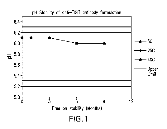
FIGURE 1 shows the pH stability of the formulations over 9 months at various
storage
- 10 -
CA 03061050 2019-10-21
WO 2018/204405 PCT/US2018/030516
conditions.
FIGURE 2 shows the polysorbate 80 concentration stability of the formulations
over 9
months at various storage conditions.
FIGURE 3 shows the potency by ELISA stability data for the formulations over 9
months
at various storage conditions.
FIGURE 4 shows the monomer (%) by UP-SEC stability data for the formulations
over 9
months at various storage conditions.
FIGURE 5 shows the high molecular weight (HMW) species (%) by UP-SEC stability
data for the formulations over 9 months at various storage conditions.
FIGURE 6 shows the low molecular weight (LMW) species (%) by UP-SEC stability
data for the formulations over 9 months at various storage conditions.
FIGURE 7 shows the purity heavy chain + light chain (%) by CE-SDS Reducing
stability
data for the formulations over 9 months at various storage conditions.
FIGURE 8 shows the purity intact IgG (%) by CE-SDS Non-reducing stability data
for
the formulations over 9 months at various storage conditions.
DETAILED DESCRIPTION OF THE INVENTION
In one aspect, the invention provides formulations comprising anti-TIGIT
antibodies and
antigen-binding fragments thereof comprising methionine. Also provided are co-
formulations of
an anti-TIGIT antibody or antigen binding fragment thereof and an anti-human
PD-1 antibody or
antigen binding fragment thereof comprising methionine. In each case, the
formulation and co-
formulation optionally comprises a chelating agent.
I. Definitions and Abbreviations
As used throughout the specification and appended claims, the following
abbreviations
apply:
API active pharmaceutical ingredient
CDR complementarity determining region in the
immunoglobulin
variable regions, defined using the Kabat numbering system,
unless otherwise indicated
CHO Chinese hamster ovary
CI confidence interval
DTPA diethylenetriaminepentaacetic acid
EC50 concentration resulting in 50% efficacy or binding
ELISA enzyme-linked immunosorbant assay
FFPE formalin-fixed, paraffin-embedded
FR framework region
HRP horseradish peroxidase
HNSCC head and neck squamous cell carcinoma
- 11 -
CA 03061050 2019-10-21
WO 2018/204405
PCT/US2018/030516
IC50 concentration resulting in 50% inhibition
IgG immunoglobulin G
IHC immunohistochemistry or immunohistochemical
mAb monoclonal antibody
MES 2-(N-morpholino)ethanesulfonic acid
NCBI National Center for Biotechnology Information
NSCLC non-small cell lung cancer
PCR polymerase chain reaction
PD-1 programmed death 1 (a.k.a. programmed cell death-1
and
programmed death receptor 1)
PD-Li programmed cell death 1 ligand 1
PD-L2 programmed cell death 1 ligand 2
PS80 polysorbate 80
TNBC triple negative breast cancer
VH immunoglobulin heavy chain variable region
VK immunoglobulin kappa light chain variable region
VL immunoglobulin light chain variable region
v/v volume per volume
WFI water for injection
w/v weight per volume
So that the invention may be more readily understood, certain technical and
scientific
terms are specifically defined below. Unless specifically defined elsewhere in
this document, all
other technical and scientific terms used herein have the meaning commonly
understood by one
of ordinary skill in the art to which this invention belongs.
As used throughout the specification and in the appended claims, the singular
forms "a,"
"an," and "the" include the plural reference unless the context clearly
dictates otherwise.
Reference to "or" indicates either or both possibilities unless the context
clearly dictates
one of the indicated possibilities. In some cases, "and/or" was employed to
highlight either or
both possibilities.
"Treat" or "treating" a cancer as used herein means to administer a
formulation of the
invention to a subject having an immune condition or cancerous condition, or
diagnosed with a
cancer or pathogenic infection (e.g. viral, bacterial, fungal), to achieve at
least one positive
therapeutic effect, such as for example, reduced number of cancer cells,
reduced tumor size,
reduced rate of cancer cell infiltration into peripheral organs, or reduced
rate of tumor metastasis
or tumor growth. "Treatment" may include one or more of the following:
inducing/increasing an
antitumor immune response, stimulating an immune response to a pathogen,
toxin, and/or self-
antigen, stimulating an immune response to a viral infection, decreasing the
number of one or
- 12 -
CA 03061050 2019-10-21
WO 2018/204405 PCT/US2018/030516
more tumor markers, inhibiting the growth or survival of tumor cells,
eliminating or reducing the
size of one or more cancerous lesions or tumors, decreasing the level of one
or more tumor
markers, ameliorating, reducing the severity or duration of cancer, prolonging
the survival of a
patient relative to the expected survival in a similar untreated patient.
"Immune condition" or "immune disorder" encompasses, e.g., pathological
inflammation, an inflammatory disorder, and an autoimmune disorder or disease.
"Immune
condition" also refers to infections, persistent infections, and proliferative
conditions, such as
cancer, tumors, and angiogenesis, including infections, tumors, and cancers
that resist
eradication by the immune system. "Cancerous condition" includes, e.g.,
cancer, cancer cells,
tumors, angiogenesis, and precancerous conditions such as dysplasia.
Positive therapeutic effects in cancer can be measured in a number of ways
(See, W. A.
Weber, I Nucl. Med. 50:1S-10S (2009)). For example, with respect to tumor
growth inhibition,
according to NCI standards, a T/C 42% is the minimum level of anti-tumor
activity. A T/C <
10% is considered a high anti-tumor activity level, with T/C (%) = Median
tumor volume of the
treated/Median tumor volume of the control x 100. In some embodiments, the
treatment
achieved by administration of a formulation of the invention is any of
progression free survival
(PFS), disease free survival (DFS) or overall survival (OS). PFS, also
referred to as "Time to
Tumor Progression" indicates the length of time during and after treatment
that the cancer does
not grow, and includes the amount of time patients have experienced a complete
response or a
partial response, as well as the amount of time patients have experienced
stable disease. DFS
refers to the length of time during and after treatment that the patient
remains free of disease. OS
refers to a prolongation in life expectancy as compared to naive or untreated
individuals or
patients. While an embodiment of the formulations, treatment methods, and uses
of the present
invention may not be effective in achieving a positive therapeutic effect in
every patient, it
should do so in a statistically significant number of subjects as determined
by any statistical test
known in the art such as the Student's t-test, the chi2-test, the U-test
according to Mann and
Whitney, the Kruskal-Wallis test (H-test), Jonckheere-Terpstra-test and the
Wilcoxon-test.
The term "patient" (alternatively referred to as "subject" or "individual"
herein) refers to
a mammal (e.g., rat, mouse, dog, cat, rabbit) capable of being treated with
the formulations of the
invention, most preferably a human. In some embodiments, the patient is an
adult patient. In
other embodiments, the patient is a pediatric patient.
The term "antibody" refers to any form of antibody that exhibits the desired
biological
activity. Thus, it is used in the broadest sense and specifically covers, but
is not limited to,
monoclonal antibodies (including full length monoclonal antibodies),
polyclonal antibodies,
humanized, fully human antibodies, and chimeric antibodies. "Parental
antibodies" are
antibodies obtained by exposure of an immune system to an antigen prior to
modification of the
antibodies for an intended use, such as humanization of an antibody for use as
a human
therapeutic antibody.
- 13 -
CA 03061050 2019-10-21
WO 2018/204405 PCT/US2018/030516
In general, the basic antibody structural unit comprises a tetramer. Each
tetramer
includes two identical pairs of polypeptide chains, each pair having one
"light" (about 25 kDa)
and one "heavy" chain (about 50-70 kDa). The amino-terminal portion of each
chain includes a
variable region of about 100 to 110 or more amino acids primarily responsible
for antigen
recognition. The variable regions of each light/heavy chain pair form the
antibody binding site.
Thus, in general, an intact antibody has two binding sites. The carboxy-
terminal portion of the
heavy chain may define a constant region primarily responsible for effector
function. Typically,
human light chains are classified as kappa and lambda light chains.
Furthermore, human heavy
chains are typically classified as mu, delta, gamma, alpha, or epsilon, and
define the antibody's
isotype as IgM, IgD, IgG, IgA, and IgE, respectively. Within light and heavy
chains, the
variable and constant regions are joined by a "J" region of about 12 or more
amino acids, with
the heavy chain also including a "D" region of about 10 more amino acids. See
generally,
Fundamental Immunology Ch. 7 (Paul, W., ed., 2nd ed. Raven Press, N.Y. (1989).
Typically, the variable domains of both the heavy and light chains comprise
three
hypervariable regions, also called complementarity determining regions (CDRs),
which are
located within relatively conserved framework regions (FR). The CDRs are
usually aligned by
the framework regions, enabling binding to a specific epitope. In general,
from N-terminal to C-
terminal, both light and heavy chains variable domains comprise FR1, CDR1, FR2
, CDR2, FR3,
CDR3 and FR4. The assignment of amino acids to each domain is, generally, in
accordance with
the definitions of Sequences of Proteins of Immunological Interest, Kabat,
etal.; National
Institutes of Health, Bethesda, Md. ; 5th ed.; NIH Publ. No. 91-3242 (1991);
Kabat (1978) Adv.
Prot. Chem. 32:1-75; Kabat, etal., (1977)1 Biol. Chem. 252:6609-6616; Chothia,
etal., (1987)
J Mol. Biol. 196:901-917 or Chothia, etal., (1989) Nature 342:878-883.
An antibody that "specifically binds to" a specified target protein is an
antibody
that exhibits preferential binding to that target as compared to other
proteins, but this specificity
does not require absolute binding specificity. An antibody is considered
"specific" for its
intended target if its binding is determinative of the presence of the target
protein in a sample,
e.g. without producing undesired results such as false positives. Antibodies,
or binding
fragments thereof, useful in the present invention will bind to the target
protein with an affinity
that is at least two fold greater, preferably at least ten times greater, more
preferably at least 20-
times greater, and most preferably at least 100-times greater than the
affinity with non-target
proteins. As used herein, an antibody is said to bind specifically to a
polypeptide comprising a
given amino acid sequence, e.g. the amino acid sequence of a mature human
TIGIT or human
PD-1, if it binds to polypeptides comprising that sequence but does not bind
to proteins lacking
that sequence.
"Chimeric antibody" refers to an antibody in which a portion of the heavy
and/or light
chain is identical with or homologous to corresponding sequences in an
antibody derived from a
particular species (e.g., human) or belonging to a particular antibody class
or subclass, while the
- 14 -
CA 03061050 2019-10-21
WO 2018/204405 PCT/US2018/030516
remainder of the chain(s) is identical with or homologous to corresponding
sequences in an
antibody derived from another species (e.g., mouse) or belonging to another
antibody class or
subclass, as well as fragments of such antibodies, so long as they exhibit the
desired biological
activity.
"Co-formulated" or "co-formulation" or "coformulation" or "coformulated" as
used
herein refers to at least two different antibodies or antigen binding
fragments thereof which are
formulated together and stored as a combined product in a single vial or
vessel (for example an
injection device) rather than being formulated and stored individually and
then mixed before
administration or separately administered. In one embodiment, the co-
formulation contains two
different antibodies or antigen binding fragments thereof
The term "pharmaceutically effective amount" or "effective amount" means an
amount
whereby sufficient therapeutic composition or formulation is introduced to a
patient to treat a
diseased or condition. One skilled in the art recognizes that this level may
vary according the
patient's characteristics such as age, weight, etc.
The term "about", when modifying the quantity (e.g., mM, or M) of a substance
or
composition, the percentage (v/v or w/v) of a formulation component, the pH of
a
solution/formulation, or the value of a parameter characterizing a step in a
method, or the like
refers to variation in the numerical quantity that can occur, for example,
through typical
measuring, handling and sampling procedures involved in the preparation,
characterization
and/or use of the substance or composition; through instrumental error in
these procedures;
through differences in the manufacture, source, or purity of the ingredients
employed to make or
use the compositions or carry out the procedures; and the like. In certain
embodiments, "about"
can mean a variation of 0.1%, 0.5%, 1%, 2%, 3%, 4%, 5%, or 10%.
As used herein, "x% (w/v)" is equivalent to x g/100 ml (for example 5% w/v
equals 50
mg/ml).
Formulations of the present invention include antibodies and fragments thereof
that are
biologically active when reconstituted or in liquid form.
The terms "cancer", "cancerous", or "malignant" refer to or describe the
physiological
condition in mammals that is typically characterized by unregulated cell
growth. Examples of
cancer include but are not limited to, carcinoma, lymphoma, leukemia,
blastoma, and sarcoma.
More particular examples of such cancers include squamous cell carcinoma,
myeloma, small-
cell lung cancer, non-small cell lung cancer, glioma, Hodgkin's lymphoma, non-
Hodgkin's
lymphoma, gastrointestinal (tract) cancer, renal cancer, ovarian cancer, liver
cancer,
lymphoblastic leukemia, lymphocytic leukemia, colorectal cancer, endometrial
cancer, kidney
cancer, prostate cancer, thyroid cancer, melanoma, chondrosarcoma,
neuroblastoma, pancreatic
cancer, glioblastoma multiforme, cervical cancer, brain cancer, stomach
cancer, bladder cancer,
hepatoma, breast cancer, colon carcinoma, and head and neck cancer.
- 15 -
CA 03061050 2019-10-21
WO 2018/204405 PCT/US2018/030516
"Chothia" means an antibody numbering system described in Al-Lazikani et al.,
JMB
273:927-948 (1997).
"Kabat" as used herein means an immunoglobulin alignment and numbering system
pioneered by Elvin A. Kabat ((1991) Sequences of Proteins of Immunological
Interest, 5th Ed.
Public Health Service, National Institutes of Health, Bethesda, Md.).
A "growth inhibitory agent" when used herein refers to a compound or
composition
which inhibits growth of a cell, especially cancer cell over expressing any of
the genes identified
herein, either in vitro or in vivo. Thus, the growth inhibitory agent is one
which significantly
reduces the percentage of cells over expressing such genes in S phase.
Examples of growth
inhibitory agents include agents that block cell cycle progression (at a place
other than S phase),
such as agents that induce G1 arrest and M-phase arrest. Classical M-phase
blockers include the
vincas (vincristine and vinblastine) taxanes, and topo II inhibitors such as
doxorubicin,
epirubicin, daunorubicin, and etoposide. Those agents that arrest G1 also
spill over into S-phase
arrest, for example, DNA alkylating agents such as dacarbazine,
mechlorethamine, and cisplatin.
Further information can be found in The Molecular Basis of Cancer, Mendelsohn
and Israel,
eds., Chapter 1, entitled "Cell cycle regulation, oncogens, and antineoplastic
drugs" by
Murakami et al. (WB Saunders: Philadelphia, 1995).
The terms "TIGIT binding fragment," "antigen binding fragment thereof,",
"binding
fragment thereof' or "fragment thereof' encompass a fragment or a derivative
of an antibody
that still substantially retains its biological activity of binding to antigen
(human TIGIT) and
inhibiting its activity (e.g., blocking the binding of human TIGIT to its
native ligands).
Therefore, the term "antibody fragment" or TIGIT binding fragment refers to a
portion of a full
length antibody, generally the antigen binding or variable region thereof
Examples of TIGIT
antibody fragments include Fab, Fab', F(ab1)2, and Fv fragments. Typically, a
binding fragment
or derivative retains at least 10% of its TIGIT inhibitory activity. In some
embodiments, a
binding fragment or derivative retains at least 25%, 50%, 60%, 70%, 80%, 90%,
95%, 99% or
100% (or more) of its TIGIT inhibitory activity, although any binding fragment
with sufficient
affinity to exert the desired biological effect will be useful. In some
embodiments, an antigen
binding fragment binds to its antigen with an affinity that is at least two
fold greater, preferably
at least ten times greater, more preferably at least 20-times greater, and
most preferably at least
100-times greater than the affinity with unrelated antigens. In one embodiment
the antibody has
an affinity that is greater than about 109 liters/mol, as determined, e.g., by
Scatchard analysis.
Munsen et al. (1980) Analyt Biochem. 107:220-239. It is also intended that a
TIGIT binding
fragment can include variants having conservative amino acid substitutions
that do not
substantially alter its biologic activity.
The terms "PD-1 binding fragment," "antigen binding fragment thereof,"
"binding
fragment thereof' or "fragment thereof' encompass a fragment or a derivative
of an antibody
that still substantially retains its biological activity of binding to antigen
(human PD-1) and
- 16 -
CA 03061050 2019-10-21
WO 2018/204405 PCT/US2018/030516
inhibiting its activity (e.g., blocking the binding of PD-1 to PDL1 and PDL2).
Therefore, the
term "antibody fragment" or PD-1 binding fragment refers to a portion of a
full length antibody,
generally the antigen binding or variable region thereof Examples of antibody
fragments
include Fab, Fab', F(ab1)2, and FAT fragments. Typically, a binding fragment
or derivative retains
at least 10% of its PD-1 inhibitory activity. In some embodiments, a binding
fragment or
derivative retains at least 25%, 50%, 60%, 70%, 80%, 90%, 95%, 99% or 100% (or
more) of its
PD-1 inhibitory activity, although any binding fragment with sufficient
affinity to exert the
desired biological effect will be useful. In some embodiments, an antigen
binding fragment
binds to its antigen with an affinity that is at least two fold greater,
preferably at least ten times
greater, more preferably at least 20-times greater, and most preferably at
least 100-times greater
than the affinity with unrelated antigens. In one embodiment the antibody has
an affinity that is
greater than about 109 liters/mol, as determined, e.g., by Scatchard analysis.
Munsen etal.
(1980) Analyt Biochem. 107:220-239. It is also intended that a PD-1 binding
fragment can
include variants having conservative amino acid substitutions that do not
substantially alter its
biologic activity.
"Human antibody" refers to an antibody that comprises human immunoglobulin
protein
sequences only. A human antibody may contain murine carbohydrate chains if
produced in a
mouse, in a mouse cell, or in a hybridoma derived from a mouse cell.
Similarly, "mouse
antibody" or "rat antibody" refer to an antibody that comprises only mouse or
rat
immunoglobulin sequences, respectively.
"Humanized antibody" refers to forms of antibodies that contain sequences from
non-
human (e.g., murine) antibodies as well as human antibodies. Such antibodies
contain minimal
sequence derived from non-human immunoglobulin. In general, the humanized
antibody will
comprise substantially all of at least one, and typically two, variable
domains, in which all or
substantially all of the hypervariable loops correspond to those of a non-
human immunoglobulin
and all or substantially all of the FR regions are those of a human
immunoglobulin sequence.
The humanized antibody optionally also will comprise at least a portion of an
immunoglobulin
constant region (Fc), typically that of a human immunoglobulin. The humanized
forms of rodent
antibodies will generally comprise the same CDR sequences of the parental
rodent antibodies,
although certain amino acid substitutions may be included to increase
affinity, increase stability
of the humanized antibody, or for other reasons.
The antibodies of the present invention also include antibodies with modified
(or
blocked) Fc regions to provide altered effector functions. See, e.g., U.S.
Pat. No. 5,624,821;
W02003/086310; W02005/120571; W02006/0057702; Presta (2006) Adv. Drug Delivery
Rev.
58:640-656. Such modification can be used to enhance or suppress various
reactions of the
immune system, with possible beneficial effects in diagnosis and therapy.
Alterations of the Fc
region include amino acid changes (substitutions, deletions and insertions),
glycosylation or
deglycosylation, and adding multiple Fc. Changes to the Fc can also alter the
half-life of
- 17-
CA 03061050 2019-10-21
WO 2018/204405 PCT/US2018/030516
antibodies in therapeutic antibodies, and a longer half-life would result in
less frequent dosing,
with the concomitant increased convenience and decreased use of material. See
Presta (2005)1
Allergy Clin. Immunol.116:731 at 734-35.
"Fully human antibody" refers to an antibody that comprises human
immunoglobulin
protein sequences only. A fully human antibody may contain murine carbohydrate
chains if
produced in a mouse, in a mouse cell, or in a hybridoma derived from a mouse
cell. Similarly,
"mouse antibody" refers to an antibody which comprises mouse immunoglobulin
sequences
only. A fully human antibody may be generated in a human being, in a
transgenic animal having
human immunoglobulin germline sequences, by phage display or other molecular
biological
methods.
"Hypervariable region" refers to the amino acid residues of an antibody that
are
responsible for antigen-binding. The hypervariable region comprises amino acid
residues from a
"complementarity determining region" or "CDR" (e.g. residues 24-34 (CDRL1), 50-
56 (CDRL2)
and 89-97 (CDRL3) in the light chain variable domain and residues 31-35
(CDRH1), 50-65
(CDRH2) and 95-102 (CDRH3) in the heavy chain variable domain as measured by
the Kabat
numbering system (Kabat et al. (1991) Sequences of Proteins of Immunological
Interest, 5th Ed.
Public Health Service, National Institutes of Health, Bethesda, Md.) and/or
those residues from a
"hypervariable loop" (i.e. residues 26-32 (L1), 50-52 (L2) and 91-96 (L3) in
the light chain
variable domain and 26-32 (H1), 53-55 (H2) and 96-101 (H3) in the heavy chain
variable
domain (Chothia and Lesk (1987)1 Mol. Biol. 196: 901-917). As used herein, the
term
"framework" or "FR" residues refers to those variable domain residues other
than the
hypervariable region residues defined herein as CDR residues. CDR and FR
residues are
determined according to the standard sequence definition of Kabat. Kabat et
al. (1987)
Sequences of Proteins of Immunological Interest, National Institutes of
Health, Bethesda Md.
"Conservatively modified variants" or "conservative substitution" refers to
substitutions
of amino acids are known to those of skill in this art and may be made
generally without altering
the biological activity of the resulting molecule, even in essential regions
of the polypeptide.
Such exemplary substitutions are preferably made in accordance with those set
forth in Table 1
as follows:
Table 1. Exemplary Conservative Amino Acid Substitutions
Original Conservative
residue substitution
Ala (A) Gly; Ser
Arg (R) Lys, His
Asn (N) Gln; His
Asp (D) Glu; Asn
Cys (C) Ser; Ala
Gln (Q) Asn
Glu (E) Asp; Gln
Gly (G) Ala
- 18 -
CA 03061050 2019-10-21
WO 2018/204405 PCT/US2018/030516
Original Conservative
residue substitution
His (H) Asn; Gln
Ile (I) Leu; Val
Leu (L) Ile; Val
Lys (K) Arg; His
Met (M) Leu; Ile; Tyr
Phe (F) Tyr; Met; Leu
Pro (P) Ala
Ser (S) Thr
Thr (T) Ser
Trp (W) Tyr; Phe
Tyr (Y) Trp; Phe
Val (V) Ile; Leu
In addition, those of skill in this art recognize that, in general, single
amino acid
substitutions in non-essential regions of a polypeptide do not substantially
alter biological
activity. See, e.g., Watson etal. (1987)Molecular Biology of the Gene, The
Benjamin/Cummings Pub. Co., p. 224 (4th Edition).
The phrase "consists essentially of," or variations such as "consist
essentially of' or
"consisting essentially of," as used throughout the specification and claims,
indicate the
inclusion of any recited elements or group of elements, and the optional
inclusion of other
elements, of similar or different nature than the recited elements, that do
not materially change
the basic or novel properties of the specified dosage regimen, method, or
composition. As a
non-limiting example, a binding compound that consists essentially of a
recited amino acid
sequence may also include one or more amino acids, including substitutions of
one or more
amino acid residues, that do not materially affect the properties of the
binding compound.
"Comprising" or variations such as "comprise", "comprises" or "comprised of"
are used
throughout the specification and claims in an inclusive sense, i.e., to
specify the presence of the
stated features but not to preclude the presence or addition of further
features that may materially
enhance the operation or utility of any of the embodiments of the invention,
unless the context
requires otherwise due to express language or necessary implication.
"Isolated antibody" and "isolated antibody fragment" refers to the
purification status and
in such context means the named molecule is substantially free of other
biological molecules
such as nucleic acids, proteins, lipids, carbohydrates, or other material such
as cellular debris and
growth media. Generally, the term "isolated" is not intended to refer to a
complete absence of
such material or to an absence of water, buffers, or salts, unless they are
present in amounts that
substantially interfere with experimental or therapeutic use of the binding
compound as
described herein.
"Monoclonal antibody" or "mAb" or "Mab", as used herein, refers to a
population of
substantially homogeneous antibodies, i.e., the antibody molecules comprising
the population
- 19 -
CA 03061050 2019-10-21
WO 2018/204405 PCT/US2018/030516
are identical in amino acid sequence except for possible naturally occurring
mutations that may
be present in minor amounts. In contrast, conventional (polyclonal) antibody
preparations
typically include a multitude of different antibodies having different amino
acid sequences in
their variable domains, particularly their CDRs, which are often specific for
different epitopes.
The modifier "monoclonal" indicates the character of the antibody as being
obtained from a
substantially homogeneous population of antibodies, and is not to be construed
as requiring
production of the antibody by any particular method. For example, the
monoclonal antibodies to
be used in accordance with the present invention may be made by the hybridoma
method first
described by Kohler etal. (1975) Nature 256: 495, or may be made by
recombinant DNA
methods (see, e.g., U.S. Pat. No. 4,816,567). The "monoclonal antibodies" may
also be isolated
from phage antibody libraries using the techniques described in Clackson etal.
(1991) Nature
352: 624-628 and Marks etal. (1991) Mol. Biol. 222: 581-597, for example. See
also Presta
(2005) Allergy Clin. Immunol. 116:731.
"Tumor" as it applies to a subject diagnosed with, or suspected of having, a
cancer refers
to a malignant or potentially malignant neoplasm or tissue mass of any size,
and includes
primary tumors and secondary neoplasms. A solid tumor is an abnormal growth or
mass of tissue
that usually does not contain cysts or liquid areas. Different types of solid
tumors are named for
the type of cells that form them. Examples of solid tumors are sarcomas,
carcinomas, and
lymphomas. Leukemias (cancers of the blood) generally do not form solid tumors
(National
Cancer Institute, Dictionary of Cancer Terms).
The term "tumor size" refers to the total size of the tumor which can be
measured as the
length and width of a tumor. Tumor size may be determined by a variety of
methods known in
the art, such as, e.g. by measuring the dimensions of tumor(s) upon removal
from the subject,
e.g., using calipers, or while in the body using imaging techniques, e.g.,
bone scan, ultrasound,
CT or MRI scans.
"Variable regions" or "V region" as used herein means the segment of IgG
chains which
is variable in sequence between different antibodies. It extends to Kabat
residue 109 in the light
chain and 113 in the heavy chain.
The term "buffer" encompasses those agents which maintain the solution pH of
the
formulations of the invention in an acceptable range, or, for lyophilized
formulations of the
invention, provide an acceptable solution pH prior to lyophilization.
The terms "lyophilization," "lyophilized," and "freeze-dried" refer to a
process by which
the material to be dried is first frozen and then the ice or frozen solvent is
removed by
sublimation in a vacuum environment. An excipient may be included in pre-
lyophilized
formulations to enhance stability of the lyophilized product upon storage.
The term "pharmaceutical formulation" refers to preparations which are in such
form as
to permit the active ingredients to be effective, and which contains no
additional components
- 20 -
CA 03061050 2019-10-21
WO 2018/204405
PCT/US2018/030516
which are toxic to the subjects to which the formulation would be
administered. The term
"formulation" and "pharmaceutical formulation" are used interchangeably
throughout.
"Pharmaceutically acceptable" refers to excipients (vehicles, additives) and
compositions
that can reasonably be administered to a subject to provide an effective dose
of the active
ingredient employed and that are "generally regarded as safe" e.g., that are
physiologically
tolerable and do not typically produce an allergic or similar untoward
reaction, such as gastric
upset and the like, when administered to a human. In another embodiment, this
term refers to
molecular entities and compositions approved by a regulatory agency of the
federal or a state
government or listed in the U.S. Pharmacopeia or another generally recognized
pharmacopeia for
use in animals, and more particularly in humans.
A "reconstituted" formulation is one that has been prepared by dissolving a
lyophilized
protein formulation in a diluent such that the protein is dispersed in the
reconstituted
formulation. The reconstituted formulation is suitable for administration,
e.g. parenteral
administration), and may optionally be suitable for subcutaneous
administration.
"Reconstitution time" is the time that is required to rehydrate a lyophilized
formulation
with a solution to a particle-free clarified solution.
A "stable" formulation is one in which the protein therein essentially retains
its physical
stability and/or chemical stability and/or biological activity upon storage.
Various analytical
techniques for measuring protein stability are available in the art and are
reviewed in Peptide and
Protein Drug Delivery, 247-301, Vincent Lee Ed., Marcel Dekker, Inc., New
York, N.Y., Pubs.
(1991) and Jones, A. Adv. Drug Delivery Rev. 10:29-90 (1993). Stability can be
measured at a
selected temperature for a selected time period. For example, in one
embodiment, a stable
formulation is a formulation with no significant changes observed at a
refrigerated temperature
(2-8 C) for at least 12 months. In another embodiment, a stable formulation
is a formulation
with no significant changes observed at a refrigerated temperature (2-8 C)
for at least 18
months. In another embodiment, stable formulation is a formulation with no
significant changes
observed at room temperature (23-27 C) for at least 3 months. In another
embodiment, stable
formulation is a formulation with no significant changes observed at room
temperature (23-
27 C) for at least 6 months. In another embodiment, stable formulation is a
formulation with no
significant changes observed at room temperature (23-27 C) for at least 12
months. In another
embodiment, stable formulation is a formulation with no significant changes
observed at room
temperature (23-27 C) for at least 18 months. The criteria for stability for
an antibody
formulation are as follows. Typically, no more than 10%, preferably 5%, of
antibody monomer
is degraded as measured by SEC-HPLC. Typically, the formulation is colorless,
or clear to
slightly opalescent by visual analysis. Typically, the concentration, pH and
osmolality of the
formulation have no more than +/-10% change. Potency is typically within 60-
140%,
preferably 80-120% of the control or reference. Typically, no more than 10%,
preferably 5% of
clipping of the antibody is observed, i.e., % low molecular weight species as
determined, for
-21 -
CA 03061050 2019-10-21
WO 2018/204405
PCT/US2018/030516
example, by HP-SEC. Typically, no more than 10%, preferably no more than 5% of
aggregation
of the antibody is observed, i.e. % high molecular weight species as
determined, for example, by
HP-SEC.
An antibody "retains its physical stability" in a pharmaceutical formulation
if it shows no
significant increase of aggregation, precipitation and/or denaturation upon
visual examination of
color and/or clarity, or as measured by UV light scattering, size exclusion
chromatography
(SEC) and dynamic light scattering. The changes of protein conformation can be
evaluated by
fluorescence spectroscopy, which determines the protein tertiary structure,
and by FTIR
spectroscopy, which determines the protein secondary structure.
An antibody "retains its chemical stability" in a pharmaceutical formulation,
if it shows
no significant chemical alteration. Chemical stability can be assessed by
detecting and
quantifying chemically altered forms of the protein. Degradation processes
that often alter the
protein chemical structure include hydrolysis or clipping (evaluated by
methods such as size
exclusion chromatography and SDS-PAGE), oxidation (evaluated by methods such
as by peptide
mapping in conjunction with mass spectroscopy or MALDI/TOF/MS), deamidation
(evaluated
by methods such as ion-exchange chromatography, capillary isoelectric
focusing, peptide
mapping, isoaspartic acid measurement), and isomerization (evaluated by
measuring the
isoaspartic acid content, peptide mapping, etc.).
An antibody "retains its biological activity" in a pharmaceutical formulation,
if the
biological activity of the antibody at a given time is within a predetermined
range of the
biological activity exhibited at the time the pharmaceutical formulation was
prepared. The
biological activity of an antibody can be determined, for example, by an
antigen binding assay.
The term "isotonic" means that the formulation of interest has essentially the
same
osmotic pressure as human blood. Isotonic formulations will generally have an
osmotic pressure
from about 270-328 mOsm. Slightly hypotonic pressure is 250-269 and slightly
hypertonic
pressure is 328-350 mOsm. Osmotic pressure can be measured, for example, using
a vapor
pressure or ice-freezing type osmometer.
Formulations and Co-formulations of the invention.
In one aspect, the invention provides biological formulations comprising anti-
TIGIT
antibodies or antigen binding fragments thereof which specifically bind to
human TIGIT as the
active pharmaceutical ingredient. Inclusion of methionine in such formulations
reduces the
oxidation of the methionine residues in the Fc region of the anti-TIGIT
antibody and, in the
example of an anti-TIGIT antibody comprising a CDRH3 of SEQ ID NO: 110, the
tryoptophan.
Such formulations may further comprise a chelator, such as, DTPA, which can
further reduce
oxidation.
- 22 -
CA 03061050 2019-10-21
WO 2018/204405 PCT/US2018/030516
In one aspect, the invention also provides a co-formulation of an anti-TIGIT
antibody
with an anti-PD-1 antibody. The major degradation pathways of pembrolizumab
included
oxidation of methionine 105 (Met105) in the heavy chain CDR3 (e.g., M105 of
SEQ ID NO: 10)
upon peroxide stress and oxidation of Met105 and Fc methionine residues when
exposed to light.
Pembrolizumab maintained its bioactivity under most stress conditions for the
degradation levels
tested. However, reduction in affinity to PD-1 was observed for peroxide
stressed samples by
Surface Plasmon Resonance (SPR). An exposed methionine residue or a methionine
residue in
the CDR of an antibody has the potential of impacting the biological activity
of the antibody
through oxidation. The addition of methionine is able to reduce oxidation of
Metl 05 within the
pembrolizumab heavy chain CDR.
Anti-PD-1 Antibodies and Antigen-Binding Fragments Thereof
In one aspect, the invention provides stable biological formulations
comprising anti-
TIGIT antibodies or antigen binding fragments thereof, co-formulated with an
anti-human PD-1
antibodies or antigen binding fragments thereof which specifically bind to
human PD-1 (e.g. a
human or humanized anti-PD-1 antibody) as the active pharmaceutical ingredient
(PD-1 API), as
well as methods for using the formulations of the invention. Any anti-PD-1
antibody or antigen
binding fragment thereof can be used in the co-formulations and methods of the
invention. In
particular embodiments, the PD-1 API is an anti-PD-1 antibody, which is
selected from
pembrolizumab and nivolumab. In specific embodiments, the anti-PD-1 antibody
is
pembrolizumab. In alternative embodiments, the anti-PD-1 antibody is
nivolumab. Table 2
provides amino acid sequences for exemplary anti-human PD-1 antibodies
pembrolizumab and
nivolumab. Alternative PD-1 antibodies and antigen-binding fragments that are
useful in the co-
formulations and methods of the invention are shown in Table 3.
In some embodiments, an anti-human PD-1 antibody or antigen binding fragment
thereof
for use in the co-formulations of the invention comprises three light chain
CDRs of CDRL1,
CDRL2 and CDRL3 and/or three heavy chain CDRs of CDRH1, CDRH2 and CDRH3.
In one embodiment of the invention, CDRL1 is SEQ ID NO:1 or a variant of SEQ
ID
NO:1, CDRL2 is SEQ ID NO:2 or a variant of SEQ ID NO:2, and CDRL3 is SEQ ID
NO:3 or a
variant of SEQ ID NO:3.
In one embodiment, CDRH1 is SEQ ID NO:6 or a variant of SEQ ID NO:6, CDRH2 is
SEQ ID NO: 7 or a variant of SEQ ID NO:7, and CDRH3 is SEQ ID NO:8 or a
variant of SEQ
ID NO:8.
In one embodiment, the three light chain CDRs are SEQ ID NO:1, SEQ ID NO:2,
and
SEQ ID NO:3 and the three heavy chain CDRs are SEQ ID NO:6, SEQ ID NO:7 and
SEQ ID
NO:8.
- 23 -
CA 03061050 2019-10-21
WO 2018/204405
PCT/US2018/030516
In an alternative embodiment of the invention, CDRL1 is SEQ ID NO:11 or a
variant of
SEQ ID NO:11, CDRL2 is SEQ ID NO:12 or a variant of SEQ ID NO:12, and CDRL3 is
SEQ
ID NO:13 or a variant of SEQ ID NO:13.
In one embodiment, CDRH1 is SEQ ID NO:16 or a variant of SEQ ID NO:16, CDRH2
is
SEQ ID NO:17 or a variant of SEQ ID NO:17, and CDRH3 is SEQ ID NO:18 or a
variant of
SEQ ID NO:18.
In one embodiment, the three light chain CDRs are SEQ ID NO:1, SEQ ID NO:2,
and
SEQ ID NO:3 and the three heavy chain CDRs are SEQ ID NO:6, SEQ ID NO:7 and
SEQ ID
NO:8.
In an alternative embodiment, the three light chain CDRs are SEQ ID NO:11, SEQ
ID
NO:12, and SEQ ID NO:13 and the three heavy chain CDRs are SEQ ID NO:16, SEQ
ID NO:17
and SEQ ID NO:18.
In a further embodiment of the invention, CDRL1 is SEQ ID NO:21 or a variant
of SEQ
ID NO:21, CDRL2 is SEQ ID NO:22 or a variant of SEQ ID NO:22, and CDRL3 is SEQ
ID
NO:23 or a variant of SEQ ID NO:23.
In yet another embodiment, CDRH1 is SEQ ID NO:24 or a variant of SEQ ID NO:24,
CDRH2 is SEQ ID NO: 25 or a variant of SEQ ID NO:25, and CDRH3 is SEQ ID NO:26
or a
variant of SEQ ID NO:26.
In another embodiment, the three light chain CDRs are SEQ ID NO:21, SEQ ID
NO:22,
and SEQ ID NO:23 and the three heavy chain CDRs are SEQ ID NO:24, SEQ ID NO:25
and
SEQ ID NO:26.
Some anti-human PD-1 antibody and antigen binding fragments of the invention
comprise a light chain variable region and a heavy chain variable region. In
some embodiments,
the light chain variable region comprises SEQ ID NO:4 or a variant of SEQ ID
NO:4, and the
heavy chain variable region comprises SEQ ID NO:9 or a variant of SEQ ID NO:9.
In further
embodiments, the light chain variable region comprises SEQ ID NO:14 or a
variant of SEQ ID
NO:14, and the heavy chain variable region comprises SEQ ID NO:19 or a variant
of SEQ ID
NO:19. In further embodiments, the heavy chain variable region comprises SEQ
ID NO:27 or a
variant of SEQ ID NO:27 and the light chain variable region comprises SEQ ID
NO:28 or a
variant of SEQ ID NO:28, SEQ ID NO:29 or a variant of SEQ ID NO:29, or SEQ ID
NO:30 or a
variant of SEQ ID NO:30. In such embodiments, a variant light chain or heavy
chain variable
region sequence is identical to the reference sequence except having one, two,
three, four or five
amino acid substitutions. In some embodiments, the substitutions are in the
framework region
(i.e., outside of the CDRs). In some embodiments, one, two, three, four or
five of the amino
acid substitutions are conservative substitutions.
In one embodiment of the co-formulations of the invention, the anti-human PD-1
antibody or antigen binding fragment comprises a light chain variable region
comprising or
consisting of SEQ ID NO:4 and a heavy chain variable region comprising or
consisting SEQ ID
- 24 -
CA 03061050 2019-10-21
WO 2018/204405 PCT/US2018/030516
NO:9. In a further embodiment, the anti-human PD-1 antibody or antigen binding
fragment
comprises a light chain variable region comprising or consisting of SEQ ID NO:
i4 and a heavy
chain variable region comprising or consisting of SEQ ID NO: i9. In one
embodiment of the
formulations of the invention, the anti-human PD-1 antibody or antigen binding
fragment
comprises a light chain variable region comprising or consisting of SEQ ID
NO:28 and a heavy
chain variable region comprising or consisting SEQ ID NO:27. In a further
embodiment, the
anti-human PD-1 antibody or antigen binding fragment comprises a light chain
variable region
comprising or consisting of SEQ ID NO:29 and a heavy chain variable region
comprising or
consisting SEQ ID NO:27. In another embodiment, the antibody or antigen
binding fragment
comprises a light chain variable region comprising or consisting of SEQ ID
NO:30 and a heavy
chain variable region comprising or consisting SEQ ID NO:27.
In another embodiment, the co-formulations of the invention comprise an anti-
human
PD-1 antibody or antigen binding protein that has a VL domain and/or a VH
domain with at least
95%, 90%, 85%, 80%, 75% or 50% sequence homology to one of the VL domains or
VH domains
described above, and exhibits specific binding to PD-1. In another embodiment,
the anti-human
PD-1 antibody or antigen binding protein of the co-formulations of the
invention comprises V.
and VH domains having up to 1, 2, 3, 4, or 5 or more amino acid substitutions,
and exhibits
specific binding to PD-1.
In any of the embodiments above, the PD-1 API may be a full-length anti-PD-1
antibody
or an antigen binding fragment thereof that specifically binds human PD-1. In
certain
embodiments, the PD-1 API is a full-length anti-PD-1 antibody selected from
any class of
immunoglobulins, including IgM, IgG, IgD, IgA, and IgE. Preferably, the
antibody is an IgG
antibody. Any isotype of IgG can be used, including IgGi, IgG2, IgG3, and
IgG4. Different
constant domains may be appended to the Vi. and VH regions provided herein.
For example, if a
particular intended use of an antibody (or fragment) of the present invention
were to call for
altered effector functions, a heavy chain constant domain other than IgG1 may
be used.
Although IgG1 antibodies provide for long half-life and for effector
functions, such as
complement activation and antibody-dependent cellular cytotoxicity, such
activities may not be
desirable for all uses of the antibody. In such instances an IgG4 constant
domain, for example,
may be used.
In embodiments of the invention, the PD-1 API is an anti-PD-1 antibody
comprising a
light chain comprising or consisting of a sequence of amino acid residues as
set forth in SEQ ID
NO:5 and a heavy chain comprising or consisting of a sequence of amino acid
residues as set
forth in SEQ ID NO: i0. In alternative embodiments, the PD-1 API is an anti-PD-
1 antibody
comprising a light chain comprising or consisting of a sequence of amino acid
residues as set
forth in SEQ ID NO:15 and a heavy chain comprising or consisting of a sequence
of amino acid
residues as set forth in SEQ ID NO:20. In further embodiments, the PD-1 API is
an anti-PD-1
antibody comprising a light chain comprising or consisting of a sequence of
amino acid residues
- 25 -
CA 03061050 2019-10-21
WO 2018/204405 PCT/US2018/030516
as set forth in SEQ ID NO:32 and a heavy chain comprising or consisting of a
sequence of amino
acid residues as set forth in SEQ ID NO:31. In additional embodiments, the PD-
1 API is an anti-
PD-1 antibody comprising a light chain comprising or consisting of a sequence
of amino acid
residues as set forth in SEQ ID NO:33 and a heavy chain comprising or
consisting of a sequence
of amino acid residues as set forth in SEQ ID NO:31. In yet additional
embodiments, the PD-1
API is an anti-PD-1 antibody comprising a light chain comprising or consisting
of a sequence of
amino acid residues as set forth in SEQ ID NO:34 and a heavy chain comprising
or consisting of
a sequence of amino acid residues as set forth in SEQ ID NO:31. In some co-
formulations of the
invention, the PD-1 API is pembrolizumab or a pembrolizumab biosimilar. In
some co-
formulations of the invention, the PD-1 API is nivolumab or a nivolumab
biosimilar.
Ordinarily, amino acid sequence variants of the anti-PD-1 antibodies and
antigen binding
fragments of the invention and the anti-TIGIT antibodies and antigen binding
fragments will
have an amino acid sequence having at least 75% amino acid sequence identity
with the amino
acid sequence of a reference antibody or antigen binding fragment (e.g. heavy
chain, light chain,
VH, VL, or humanized sequence), more preferably at least 80%, more preferably
at least 85%,
more preferably at least 90%, and most preferably at least 95, 98, or 99%.
Identity or homology
with respect to a sequence is defined herein as the percentage of amino acid
residues in the
candidate sequence that are identical with the anti-PD-1 residues, after
aligning the sequences
and introducing gaps, if necessary, to achieve the maximum percent sequence
identity, and not
considering any conservative substitutions as part of the sequence identity.
None of N-terminal,
C-terminal, or internal extensions, deletions, or insertions into the antibody
sequence shall be
construed as affecting sequence identity or homology.
Sequence identity refers to the degree to which the amino acids of two
polypeptides are
the same at equivalent positions when the two sequences are optimally aligned.
Sequence
identity can be determined using a BLAST algorithm wherein the parameters of
the algorithm
are selected to give the largest match between the respective sequences over
the entire length of
the respective reference sequences. The following references relate to BLAST
algorithms often
used for sequence analysis: BLAST ALGORITHMS: Altschul, S.F., etal., (1990) J.
Mol. Biol.
215:403-410; Gish, W., etal., (1993) Nature Genet. 3:266-272; Madden, T.L.,
etal., (1996)
Meth. Enzymol. 266:131-141; Altschul, S.F., etal., (1997) Nucleic Acids Res.
25:3389-3402;
Zhang, J., etal., (1997) Genome Res. 7:649-656; Wootton, J.C., etal., (1993)
Comput. Chem.
17:149-163; Hancock, J.M. etal., (1994) Comput. Appl. Biosci. 10:67-70;
ALIGNMENT
SCORING SYSTEMS: Dayhoff, M.O., etal., "A model of evolutionary change in
proteins." in
Atlas of Protein Sequence and Structure, (1978) vol. 5, suppl. 3. M.O. Dayhoff
(ed.), pp. 345-
352, Natl. Biomed. Res. Found., Washington, DC; Schwartz, R.M., et
al.,"Matrices for detecting
distant relationships." in Atlas of Protein Sequence and Structure, (1978)
vol. 5, suppl. 3." M.O.
Dayhoff (ed.), pp. 353-358, Natl. Biomed. Res. Found., Washington, DC;
Altschul, S.F., (1991)
J. Mol. Biol. 219:555-565; States, D.J., etal., (1991) Methods 3:66-70;
Henikoff, S., etal.,
- 26 -
CA 03061050 2019-10-21
WO 2018/204405 PCT/US2018/030516
(1992) Proc. Natl. Acad. Sci. USA 89:10915-10919; Altschul, S.F., etal..
(1993)1 Mol. Evol.
36:290-300; ALIGNMENT STATISTICS: Karlin, S., etal.. (1990) Proc. Natl. Acad.
Sci. USA
87:2264-2268; Karlin, S., etal.. (1993) Proc. Natl. Acad. Sci. USA 90:5873-
5877; Dembo, A.,
etal.. (1994) Ann. Prob. 22:2022-2039; and Altschul, S.F. "Evaluating the
statistical
significance of multiple distinct local alignments." in Theoretical and
Computational Methods in
Genome Research (S. Suhai, ed.), (1997) pp. 1-14, Plenum, New York.
Likewise, either class of light chain can be used in the compositions and
methods herein.
Specifically, kappa, lambda, or variants thereof are useful in the present
compositions and
methods.
Table 2. Exemplary PD-1 Antibody Sequences
Antibody Amino Acid Sequence SEQ ID
Feature NO.
Pembrolizumab Light Chain
CDR1 RASKGVSTSGYSYLH 1
CDR2 LASYLES 2
CDR3 QHSRDLPLT 3
Variable EIVLTQSPATLSLSPGERATLSCRASKGVSTSGYSYLHWY 4
Region QQKPGQAPRLLIYLASYLESGVPARFSGSGSGTDFTLTISS
LEPEDFAVYYCQHSRDLPLTFGGGTKVEIK
Light Chain EIVLTQSPATLSLSPGERATLSCRASKGVSTSGYSYLHWY 5
QQKPGQAPRLLIYLASYLESGVPARFSGSGSGTDFTLTISS
LEPEDFAVYYCQHSRDLPLTFGGGTKVEIKRTVAAPSVFI
FPPSDEQLKSGTASVVCLLNNFYPREAKVQWKVDNALQS
GNSQESVTEQDSKDSTYSLSSTLTLSKADYEKHKVYACE
VTHQGLSSPVTKSFNRGEC
Pembrolizumab Heavy Chain
CDR1 NYYMY 6
CDR2 GINPSNGGTNFNEKFKN 7
CDR3 RDYRFDMGFDY 8
Variable QVQLVQSGVEVKKPGASVKVSCKASGYTFTNYYMYWV 9
Region RQAPGQGLEWMGCrINPSNGGTNFNEKFKNRVTLTTDS ST
TTAYMELKSLQFDDTAVYYCARRDYRFDMGFDYWGQG
TTVTVS S
Heavy QVQLVQSGVEVKKPGASVKVSCKASGYTFTNYYMYWV 10
Chain RQAPGQGLEWMGCrINPSNGGTNFNEKFKNRVTLTTDS ST
TTAYMELKSLQFDDTAVYYCARRDYRFDMGFDYWGQG
TTVTVS SASTKGPSVFPLAPCSRSTSESTAALGCLVKDYFP
EPVTVSWNSGALTS GVHTFPAVLQS SGLYSLS SVVTVPS S
SLGTKTYTCNVDHKPSNTKVDKRVESKYGPPCPPCPAPE
FLGGPSVFLFPPKPKDTLMISRTPEVTCVVVDVSQEDPEV
QFNWYVDGVEVHNAKTKPREEQFNSTYRVVSVLTVLHQ
DWLNGKEYKC KV SNKGLP S SIEKTISKAKGQPREPQVYT
LP P S QEEMTKNQV S LTC LVKGFYP S DIAVEWESNGQPEN
NYKTTPPVLDSDGSFFLYSRLTVDKSRWQEGNVFSCSVM
HEALHNHYTQKSLSLSLGK
Nivolumab Light Chain
- 27 -
CA 03061050 2019-10-21
WO 2018/204405
PCT/US2018/030516
Antibody Amino Acid Sequence SEQ ID
Feature NO.
CDR1 RASQSVSSYLA 11
CDR2 DASNRAT 12
CDR3 QQSSNWPRT 13
Variable EIVLTQSPATLSLSPGERATLSCRASQSVSSYLAWYQQKP 14
Region GQAPRLLIYDASNRATGIPARFSGSGSGTDFTLTISSLEPE
DFAVYYCQQSSNWPRTFGQGTKVEIK
Light Chain EIVLTQSPATLSLSPGERATLSCRASQSVSSYLAWYQQKP 15
GQAPRLLIYDASNRATGIPARFSGSGSGTDFTLTISSLEPE
DFAVYYCQQSSNVVPRTFGQGTKVEIKRTVAAPSVFIFPPS
DEQLKSGTASVVCLLNNFYPREAKVQWKVDNALQSGNS
QESVTEQDSKDSTYSLSSTLTLSKADYEKHKVYACEVTH
QGLSSPVTKSFNRGEC
Nivolumab Heavy Chain
CDR1 NSGMH 16
CDR2 VIWYDGSKRYYADSVKG 17
CDR3 NDDY 18
Variable QVQLVESGGGVVQPGRSLRLDCKASGITFSNSGMHWVR 19
Region QAPGKGLEWVAVIWYDGSKRYYADSVKGRFTISRDNSK
NTLFLQMNSLRAEDTAVYYCATNDDYWGQGTLVTVSS
Heavy QVQLVESGGGVVQPGRSLRLDCKASGITFSNSGMHWVR 20
Chain QAPGKGLEWVAVIWYDGSKRYYADSVKGRFTISRDNSK
NTLFLQMNSLRAEDTAVYYCATNDDYWGQGTLVTVSSA
STKGPSVFPLAPCSRSTSESTAALGCLVKDYFPEPVTVSW
NSGALTSGVHTFPAVLQSSGLYSLSSVVTVPSSSLGTKTY
TCNVDHKPSNTKVDKRVESKYGPPCPPCPAPEFLGGPSVF
LFPPKPKDTLMISRTPEVTCVVVDVSQEDPEVQFNWYVD
GVEVHNAKTKPREEQFNSTYRVVSVLTVLHQDWLNGKE
YKCKVSNKGLPSSIEKTISKAKGQPREPQVYTLPPSQEEM
TKNQVSLTCLVKGFYPSDIAVEWESNGQPENNYKTTPPV
LDSDGSFFLYSRLTVDKSRWQEGNVFSCSVMHEALHNH
YTQKSLSLSLGK
Table 3. Additional PD-1 Antibodies and Antigen Binding Fragments Useful in
the Co-
Formulations, Methods and Uses of the Invention.
A. Antibodies and antigen binding fragments comprising light and heavy chain
CDRs of hPD-1.08A in W02008/156712
CDRL1 SEQ ID NO:21
CDRL2 SEQ ID NO:22
CDRL3 SEQ ID NO:23
CDRH1 SEQ ID NO:24
CDRH2 SEQ ID NO:25
CDRH3 SEQ ID NO:26
C. Antibodies and antigen binding fragments comprising the mature h109A heavy
chain variable region and one of the mature KO9A light chain variable regions
in
WO 2008/156712
- 28 -
CA 03061050 2019-10-21
WO 2018/204405 PCT/US2018/030516
Heavy chain VR SEQ ID NO:27
SEQ ID NO:28 or SEQ ID NO:29 or SEQ ID NO:30
Light chain VR
D. Antibodies and antigen binding fragments comprising the mature 409 heavy
chain and one of the mature KO9A light chains in WO 2008/156712
Heavy chain SEQ ID NO:31
Light chain SEQ ID NO:32 or SEQ ID NO:33 or SEQ ID NO:34
In some embodiments of the co-formulation of the invention, the PD-1 API (i.e.
the anti-
PD-1 antibody or antigen binding fragment thereof) is present in a
concentration of from about
25 mg/mL to about 100 mg/mL. In alternative embodiments, the API is present in
a
concentration of about 10 mg/mL, about 25 mg/mL, about 50 mg/mL, about 75
mg/mL, or about
100 mg/mL.
Anti-TIGIT Antibodies and Antigen-Binding Fragment Thereof
In one aspect, the invention provides biological formulations comprising anti-
TIGIT
antibodies or antigen binding fragments thereof which specifically bind to
human TIGIT (e.g. a
.. human or humanized anti-TIGIT antibody) as the active pharmaceutical
ingredient (TIGIT API),
as well as methods for using the formulations of the invention.
In another aspect, the invention also provides biological co-formulations
comprising (i)
anti-TIGIT antibody or antigen binding fragment thereof which specifically
bind to human
TIGIT (e.g. a human or humanized anti-TIGIT antibody) and (ii) an anti-human
PD-1 antibody
or antigen binding fragment thereof which specifically binds to human PD-1.
Any anti-TIGIT
antibody or antigen binding fragment thereof can be used in the formulation,
including the co-
formulation, and methods of the invention. Exemplary anti-TIGIT antibody
sequences are set
forth below in Tables 4 and 5.
TABLE 4 Exemplary anti-TIGIT antibodies
Description SEQ ID SEQUENCE
NO:
14A6 H - CDR1 35 SDYWG
14A6 H - CDR2 36 FITYSGSTSYNPSLKS
14A6 H - CDR3 37 MPSFITLASLSTWEGYFDF
- 29 -
CA 03061050 2019-10-21
WO 2018/204405
PCT/US2018/030516
14A6 L - CDR1 38 KASQSIHKNLA
14A6 L - CDR2 39 YANSLQT
14A6 L - CDR3 40 QQYYSGWT
14A6 PARENTAL VH 41 EVQLQESGPGLVKPSQSLSLTCSVTGSSIA
SDYWGWIRKFPGNKMEWMGFITYSGSTS
YNPSLKSRISITRDTSKNQFFLQLHSVTTD
DTATYS CARMPSFITLASLSTWEGYFDFW
GPGTMVTVSS
14A6 PARENTAL VL 42 DIQMTQ SP SLL SASVGDRVTLNCKAS Q SI
HKNLAWYQQKLGEAPKFLIYYANSLQTG
IP S RF S GS GS GTDFTLTI S GLQPEDVATYF
CQQYYS GWTFGGGTKVELK
Hul4A6VH. 1 43 EVQLQESGPGLVKPSETLSLTCTVS GGSIS
SDYWGWIRQPPGKGLEWIGFITYSGSTSY
NPSLKSRVTISVDTSKNQFSLKLSSVTAA
DTAVYYCARMPSFITLASLSTWEGYFDF
WGQGTMVTVSS
Hul4A6VH. la 44 EVQLQESGPGLVKPSETLSLTCTVS GGSIS
SDYWGWIRQPPGKGLEWIGFITYSGSTSY
NP SLKSRVTISRDTSKNQFSLKLS SVTAAD
TAVYYCARMPSFITLASLSTWEGYFDFW
GQGTMVTVSS
Hul4A6VH. lb 45 EVQLQESGPGLVKPSETLSLTCTVS GGSIS
SDYWGWIRQPPGKGLEWIGFITYSGSTSY
NP SLKSRITISRDTSKNQFSLKLS SVTAAD
TAVYYCARMPSFITLASLSTWEGYFDFW
GQGTMVTVSS
Hul4A6VH. lc 46 EVQLQESGPGLVKPSETLSLTCTVS GS SIS
SDYWGWIRQPPGKGLEWMGFITYS GSTS
YNPSLKSRITISRDTSKNQF SLKLS SVTAA
DTAVYYCARMPSFITLASLSTWEGYFDF
- 30 -
CA 03061050 2019-10-21
WO 2018/204405
PCT/US2018/030516
WGQGTMVTVSS
Hul4A6VH.1d 47 EVQLQESGPGLVKPSETLSLTCTVSGGSIS
S DYWGWIRQPPGKGLEWIGFITYS GS TSY
NPSLKSRVTISRDTSKNQFSLKLHSVTAA
DTAVYYCARMPSFITLASLSTWEGYFDF
WGQGTMVTVSS
Hul4A6VH.1e 48 EVQLQESGPGLVKPSETLSLTCTVSGGSIS
S DYWGWIRQPPGKGLEWIGFITYS GS TSY
NPSLKSRITISRDTSKNQFSLKLHSVTAAD
TAVYYCARMPSFITLASLSTWEGYFDFW
GQGTMVTVSS
Hul4A6VH.lf 49 EVQLQESGPGLVKPSETLSLTCTVS GS SIS
S DYWGWIRQPPGKGLEWIGFITYS GS TSY
NPSLKSRITISRDTSKNQFSLKLHSVTAAD
TAVYYCARMPSFITLASLSTWEGYFDFW
GQGTMVTVSS
Hul4A6VH. lg 50 EVQLQESGPGLVKPSETLSLTCTVS GS SIS
SDYWGWIRQPPGKGLEWMGFITYSGSTS
YNPSLKSRITISVDTSKNQFSLKLHSVTAA
DTAVYYCARMPSFITLASLSTWEGYFDF
WGQGTMVTVSS
Hul4A6VH.2 51 EVQLQESGPGLVKPSETLSLTCAVSGYSIS
S DYWGWIRQPPGKGLEWIGFITYS GS TSY
NPSLKSRVTISVDTSKNQFSLKLSSVTAA
DTAVYYCARMPSFITLASLSTWEGYFDF
WGQGTMVTVSS
Hul4A6VH.2a 52 EVQLQESGPGLVKPSETLSLTCAVSGYSIS
S DYWGWIRQPPGKGLEWIGFITYS GS TSY
NPSLKSRVTISRDTSKNQFSLKLSSVTAAD
TAVYYCARMPSFITLASLSTWEGYFDFW
-31-
CA 03061050 2019-10-21
WO 2018/204405
PCT/US2018/030516
GQGTMVTVSS
Hul4A6VH.2b 53 EVQLQESGPGLVKPSETLSLTCAVSGYSIS
SDYWGWIRQPPGKGLEWIGFITYSGSTSY
NPSLKSRITISRDTSKNQFSLKLSSVTAAD
TAVYYCARMPSFITLASLSTWEGYFDFW
GQGTMVTVSS
Hul4A6VH.2c 54 EVQLQESGPGLVKPSETLSLTCAVSGSSIS
SDYWGWIRQPPGKGLEWMGFITYSGSTS
YNPSLKSRITISRDTSKNQFSLKLSSVTAA
DTAVYYCARMPSFITLASLSTWEGYFDF
WGQGTMVTVSS
Hul4A6VH.2d 55 EVQLQESGPGLVKPSETLSLTCAVSGYSIS
SDYWGWIRQPPGKGLEWIGFITYSGSTSY
NPSLKSRVTISRDTSKNQFSLKLHSVTAA
DTAVYYCARMPSFITLASLSTWEGYFDF
WGQGTMVTVSS
Hul4A6VH.2e 56 EVQLQESGPGLVKPSETLSLTCAVSGYSIS
SDYWGWIRQPPGKGLEWIGFITYSGSTSY
NPSLKSRITISRDTSKNQFSLKLHSVTAAD
TAVYYCARMPSFITLASLSTWEGYFDFW
GQGTMVTVSS
Hul4A6VH.2f 57 EVQLQESGPGLVKPSETLSLTCAVSGSSIS
SDYWGWIRQPPGKGLEWIGFITYSGSTSY
NPSLKSRITISRDTSKNQFSLKLHSVTAAD
TAVYYCARMPSFITLASLSTWEGYFDFW
GQGTMVTVSS
Hul4A6VH.2e 58 EVQLQESGPGLVKPSETLSLTCAVSGSSIS
SDYWGWIRQPPGKGLEWMGFITYSGSTS
YNPSLKSRITISRDTSKNQFSLKLHSVTAA
DTAVYYCARMPSFITLASLSTWEGYFDF
- 32 -
CA 03061050 2019-10-21
WO 2018/204405
PCT/US2018/030516
WGQGTMVTVSS
Hul 4A6Vk. 1 59 DIQMTQ SP SSLSASVGDRVTITCKASQSIH
KNLAWYQQKPGKAPKLLIYYANSLQTGV
PSRFSGSGSGTDFTLTIS SLQPEDFATYYC
QQYYSGWTFGGGTKVEIK
Hul 4A6Vk. 1 a 60 DIQMTQ SP SSLSASVGDRVTITCKASQSIH
KNLAWYQQKPGKAPKFLIYYANSLQTGV
PSRFSGSGSGTDFTLTIS SLQPEDFATYYC
QQYYSGWTFGGGTKVEIK
Hul 4A6Vk. lb 61 DIQMTQ SP SSLSASVGDRVTITCKASQSIH
KNLAWYQQKPGKAPKFLIYYANSLQTGI
PSRFSGSGSGTDFTLTIS SLQPEDFATYYC
QQYYSGWTFGGGTKVEIK
Hul 4A6Vk.2 62 DIQMTQ SP SSLSASVGDRVTITCKASQSIH
KNLAWYQQKPGKVPKLLIYYANSLQTGV
PSRFSGSGSGTDFTLTIS SLQPEDVATYYC
QQYYSGWTFGGGTKVEIK
Hul 4A6Vk.2a 63 DIQMTQ SP SSLSASVGDRVTITCKASQSIH
KNLAWYQQKPGKVPKFLIYYANSLQTGV
PSRFSGSGSGTDFTLTIS SLQPEDVATYYC
QQYYSGWTFGGGTKVEIK
Hul 4A6Vk.2b 64 DIQMTQ SP SSLSASVGDRVTITCKASQSIH
KNLAWYQQKPGKVPKFLIYYANSLQTGI
PSRFSGSGSGTDFTLTIS SLQPEDVATYYC
QQYYSGWTFGGGTKVEIK
16AHA tigit 14a6 humanize 65 EVQLQESGPGLVKP SETL SLTCTVS GS SIA
d VH1 S DYWGWIRQPPGKGLEWIGFITYS GS TSY
NPSLKSRVTISVDTSKNQFSLKLSSVTAA
- 33 -
CA 03061050 2019-10-21
WO 2018/204405
PCT/US2018/030516
DTAVYYCARMPSFITLASLSTWEGYFDF
WGQGTMVTVSSAS
LB155.14A6.G2.A8 VH1
18AHA tigit 14a6 humanize 66 EVQLQESGPGLVKPSETLSLTCTVSGSSIA
d VH2 SDYWGWIRQPPGKGLEWIGFITYSGSTSY
NPSLKSRVTISRDTSKNQFSLKLSSVTAAD
TAVYYCARMPSFITLASLSTWEGYFDFW
LB155.14A6.G2.A8 VH2 GQGTMVTVSS
20AHA tigit 14a6 humanize 67 EVQLQESGPGLVKPSETLSLTCTVSGSSIA
d VH3 SDYWGWIRKPPGKGLEWIGFITYSGSTSY
NPSLKSRVTISRDTSKNQFSLKLSSVTAAD
TAVYYCARMPSFITLASLSTWEGYFDFW
LB155.14A6.G2.A8 VH3 GQGTMVTVSS
21AHA tigit 14a6 humanize 68 EVQLQESGPGLVKPSETLSLTCTVSGSSIA
d VH4 SDYWGWIRQPPGKKLEWIGFITYSGSTSY
NPSLKSRVTISRDTSKNQFSLKLSSVTAAD
LB155.14A6.G2.A8 VH4
TAVYYCARMPSFITLASLSTWEGYFDFW
GQGTMVTVSS
19AHA tigit 14a6 humanize 69 EVQLQESGPGLVKPSETLSLTCTVSGSSIA
d VH5 SDYWGWIRQPPGKGMEWIGFITYSGSTS
YNPSLKSRVTISRDTSKNQFSLKLSSVTAA
DTAVYYCARMPSFITLASLSTWEGYFDF
LB155.14A6.G2.A8 VHS WGQGTMVTVSS
22AHA tigit 14a6 humanize 70 EVQLQESGPGLVKPSETLSLTCTVSGSSIA
d VH6 SDYWGWIRKPPGKKMEWIGFITYSGSTS
YNPSLKSRVTISRDTSKNQFSLKLSSVTAA
DTAVYYCARMPSFITLASLSTWEGYFDF
LB155.14A6.G2.A8 VH6 WGQGTMVTVSS
23AHA tigit 14a6 humanize 71 EVQLQESGPGLVKPSETLSLTCTVSGSSIA
SDYWGWIRQFPGKGLEWIGFITYSGSTSY
- 34 -
CA 03061050 2019-10-21
WO 2018/204405
PCT/US2018/030516
d VH7 NPSLKSRVTISRDTSKNQFSLKLSSVTADD
TAVYYCARMPSFITLASLSTWEGYFDFW
GQGTMVTVSS
LB155.14A6. G2. A8 VH7
24AHA tigit 14a6 humanize 72 EVQLQESGPGLVKPSETLSLTCTVS GS SIA
d VH8 SDYWGWIRKPPGKKMEWIGFITYSGSTS
YNPSLKSRVTISVDTSKNQFSLKLSSVTA
ADTAVYYCARMPSFITLASLSTWEGYFDF
LB155.14A6. G2. A8 VH8 WGQGTMVTVSS
25AHA tigit 14a6 humanize 73 EVQLQESGPGLVKPSETLSLTCSVTGSSIA
d VH9 SDYWGWIRQPPGKGLEWIGFITYS GS TSY
NPSLKSRVTISRDTSKNQFSLKLSSVTAAD
TAVYYCARMPSFITLASLSTWEGYFDFW
LB155.14A6. G2. A8 VH9 GQGTMVTVSS
26AHA tigit 14a6 humanize 74 EVQLQQSGAGLLKPSETLSLTCSVTGSSIA
d VH10 SDYWGWIRQPPGKGLEWIGFITYS GS TSY
NPSLKSRVTISVDTSKNQFSLKLSSVTAA
DTAVYYCARMPSFITLASLSTWEGYFDF
LB155.14A6.G2.A8 VH10 WGQGTMVTVSS
27AHA tigit 14a6 humanize 75 EVQLQESGPGLVKPPGTLSLTCSVTGSSIA
d VH11 SDYWGWVRQPPGKGLEWIGFITYSGSTS
YNPSLKSRVTISVDTSKNQFSLKLSSVTA
ADTAVYYCARMPSFITLASLSTWEGYFDF
LB155.14A6.G2.A8 VH11 WGQGTMVTVSS
09AHA tigit 14a6 humanize 76 DIQMTQSPSSLSASVGDRVTITCKASQSIH
d VL1 KNLAWYQQKPGKAPKLLIYYANSLQTGV
PSRFSGSGSGTDFTLTISSLQPEDFATYYC
QQYYSGWTFGGGTKVEIK
LB155.14A6.G2.A8 VL1
11AHA tigit 14a6 humanize 77 DIQMTQSPSSLSASVGDRVTITCKASQSIH
- 35 -
CA 03061050 2019-10-21
WO 2018/204405
PCT/US2018/030516
d VL2 KNLAWYQQKPGKAPKFLIYYANSLQTGV
PSRFSGSGSGTDFTLTIS SLQPEDFATYYC
LB155.14A6.G2.A8 VL2
QQYYSGWTFGGGTKVEIK
12AHA tigit 14a6 humanize 78 DIQMTQ SP S SLS AS VGDRVTITCKAS QSIH
d VL3 KNLAWYQQKPGKAPKLLIYYANSLQTGV
PSRFSGSGSGTDFTLTIS SLQPEDFATYFC
QQYYSGWTFGGGTKVEIK
LB155.14A6.G2.A8 VL3
13AHA tigit 14a6 humanize 79 DIQMTQ SP S SLS AS VGDRVTITCKAS QSIH
d VL4 KNLAWYQQKPGKAPKFLIYYANSLQTGV
PSRFSGSGSGTDFTLTIS SLQPEDFATYFC
QQYYSGWTFGGGTKVEIK
LB155.14A6.G2.A8 VL4
15AHA tigit 14a6 humanize 80 DIQMTQ SP S SLS AS VGDRVTITCKAS QSIH
d VL5 KNLAWYQQKPGKAPKLLIYYANSLQTGI
PSRFSGSGSGTDFTLTIS SLQPEDFATYYC
QQYYSGWTFGGGTKVEIK
LB155.14A6.G2.A8 VL5
28H5 H - CDR1 81 GYSITSDYAWN
28H5 H - CDR2 82 YISNSGSASYNPSLKS
28H5 H - CDR3 83 LIYYDYGGAMNF
28H5 L - CDR1 84 KASQGVSTTVA
28H5 L - CDR2 85 SASYRYT
28H5 L - CDR3 86 QHYYSTPWT
28H5 PARENTAL VH 87 DVQLQESGPGLVKPSQSLSLTCTVTGYSI
TSDYAWNWIRQFPGNKLEWMGYISNSGS
ASYNPSLKSRISITRDTSKNQFFLQLNSVT
TEDTATYYCATLIYYDYGGAMNFWGQG
TSVTVSS
- 36 -
CA 03061050 2019-10-21
WO 2018/204405
PCT/US2018/030516
28H5 PARENTAL VL 88 DIVMTQSHKFMSTSVGDRVSITCKASQG
VSTTVAWYQQKPGQSPKLLIYSASYRYT
GVPDRFTGSGSGTDFTFTISSVQSEDLAV
YYCQHYYSTPWTFGGGTKLEIK
14H6 L - CDR2 variant 89 YASNLQT
14H6 L - CDR2 variant 90 YASSLQT
14H6 L - CDR2 variant 91 YASTLQT
14H6 L - CDR2 variant 92 YATTLQT
14H6 L - CDR2 variant 93 YASYLQT
14H6 L - CDR2 variant 94 YANQLQT
14H6 L - CDR2 variant 95 YAGSLQT
14H6 L - CDR2 variant 96 YASQLQT
14H6 L - CDR2 variant 97 YADSLQT
14H6 L - CDR3 variant 98 QQYYSGFT
14H6 L - CDR3 variant 99 QQYYSGYT
14H6 L - CDR3 variant 100 QQYYSGIT
14H6 L - CDR3 variant 101 QQYYSGVT
14H6 L - CDR3 variant 102 QQYYSGLT
14H6 H - CDR3 variant 103 MPSFITLASLSTFEGYFDF
14H6 H - CDR3 variant 104 MPSFITLASLSTYEGYFDF
14H6 H - CDR3 variant 105 MPSFITLASLSTIEGYFDF
14H6 H - CDR3 variant 106 MPSFITLASLSTVEGYFDF
14H6 H - CDR3 variant 107 MPSFITLASLSTLEGYFDF
3106 H -CDR1 108 SYVMH
3106 H -CDR2 109 YIDPYNDGAKYNEKFKG
3106 H -CDR3 110 GGPYGWYFDV
-37-
CA 03061050 2019-10-21
WO 2018/204405
PCT/US2018/030516
3106 L - CDRI 111 RASEHIYSYLS
3106 L ¨ CDR2 112 NAKTLAE
3106 L ¨ CDR3 113 QHHFGSPLT
3106 PARENTAL VH (with 114 EVQLQQSGPELVKPGSSVKMSCKASGYT
CDRs underlined) FSSYVMHWVKQKPGQGLEWIGYIDPYND
GAKYNEKFKGKATLTSDKSSSTAYMELS
SLTSEDSAVYYCARGGPYGWYFDVWGA
GTTVTVSS
3106 PARENTAL VL (with 115 DIQMTQSPASLSASVGETVTITCRASEHIY
CDRs underlined) SYLSWYQQKQGKSPQLLVYNAKTLAEG
VPSRFSGSGSGTQFSLKINSLQPEDFGTYY
CQHHFGSPLTFGAGTTLELK
3106 H ¨ CDR2 VARIANT 116 YIDPYNrGAKYNEKFG
(D56R)
3106 H ¨ CDR2 VARIANT 117 YIDPYN1GAKYNEKG
(D56L)
F
3106 H ¨ CDR2 VARIANT 118 YIDPYNkGAKYNEKFG
(D56K)
3106 H ¨ CDR2 VARIANT 119 YIDPYNfGAKYNEKFG
(D56F)
3106 H ¨ CDR2 VARIANT 120 YIDPYNsGAKYNEKFG
(D56S)
3106 H ¨ CDR2 VARIANT 121 YIDPYNyGAKYNEKFG
(D56Y)
3106 H ¨ CDR2 VARIANT 122 YIDPYNvGAKYNEKFG
- 38 -
CA 03061050 2019-10-21
WO 2018/204405
PCT/US2018/030516
(D56V)
3106 H ¨ CDR2 VARIANT 123 YIDPYNDrAKYNEKFKG
(G5 7R)
3106 H ¨ CDR2 VARIANT 124 YIDPYNDnAKYNEKFKG
(G57N)
3106 H ¨ CDR2 VARIANT 125 YIDPYNDqAKYNEKFKG
(G57Q)
3106 H ¨ CDR2 VARIANT 126 YIDPYNDeAKYNEKFKG
(G57E)
3106 H ¨ CDR2 VARIANT 127 YIDPYND1AKYNEKFKG
(G57L)
3106 H ¨ CDR2 VARIANT 128 YIDPYNDkAKYNEKFKG
(G5 7K)
3106 H ¨ CDR2 VARIANT 129 YIDPYNDsAKYNEKFKG
(G57S)
3106 H ¨ CDR2 VARIANT 130 YIDPYNDyAKYNEKFKG
(G57Y)
3106 H ¨ CDR2 VARIANT 131 YIDPYNDvAKYNEKFKG
(G57V)
3106 L ¨ CDR2 variant 132 AAKTLAE
(N50A)
3106 L ¨ CDR2 variant 133 YAKTLAE
(N50Y)
3106 L ¨ CDR2 variant 134 WAKTLAE
(N5OW)
3106 L ¨ CDR2 variant 135 SAKTLAE
(N50S)
- 39 -
CA 03061050 2019-10-21
WO 2018/204405
PCT/US2018/030516
3106 L ¨ CDR2 variant 136 TAKTLAE
(N5 OT)
3106 L ¨ CDR2 variant (N500 137 IAKTLAE
3106 L ¨ CDR2 variant 138 VAKTLAE
(N50V)
3106 L ¨ CDR2 variant 139 NNKTLAE
(A51N)
3106 L ¨ CDR2 variant (A510 140 NIKTLAE
3106 L ¨ CDR2 variant 141 NLLTLAE
(A51L)
3106 L ¨ CDR2 variant 142 NTKTLAE
(A51T)
3106 L ¨ CDR2 variant 143 NVKTLAE
(A51V)
3106 HUMZ VH1 144 EVQLVQSGAEVKKPGASVKVSCKASGYT
F S SYVMHWVRQAPGQRLEWIGYIDPYND
(with CDRs underlined) GAKYSQKFQGRVTLTRDTSASTAYMELS
SLRSEDTAVYYCARGGPYGWYFDVWGQ
GTTVTVS S
3106 HUMZ VH2 145 EVQLVQSGAEVKKPGASVKVSCKASGYT
F S SYVMHWVRQAPGQRLEWIGYIDPYND
(with CDRs underlined) GAKYSQKFQGRVTLTSDKSASTAYMELS
SLRSEDTAVYYCARGGPYGWYFDVWGQ
GTTVTVS S
- 40 -
CA 03061050 2019-10-21
WO 2018/204405
PCT/US2018/030516
3106 HUMZ VH3 146 EVQLVQSGAEVKKPGASVKVSCKASGYT
F S SYVMHWVRQAPGQGLEWIGYIDPYND
(with CDRs underlined) GAKYAQKFQGRVTLTRDTSTSTVYMELS
SLRSEDTAVYYCARGGPYGWYFDVWGQ
GTTVTVS S
3106 HUMZ VH4 147 EVQLVQSGAEVKKPGASVKVSCKASGYT
F S SYVMHWVRQAPGQGLEWIGYIDPYND
(with CDRs underlined) GAKYAQKFQGRVTLTSDKSTSTVYMELS
SLRSEDTAVYYCARGGPYGWYFDVWGQ
GTTVTVS S
3106 HUMZ VH5 148 EV QLVQ S GAEVKKP GS SVKVSCKASGYT
F S SYVMHWVRQAPGQGLEWIGYIDPYND
(with CDRs underlined) GAKYAQKFQGRVTLTSDKSTSTAYMELS
SLRSEDTAVYYCARGGPYGWYFDVWGQ
GTTVTVS S
3106 HUMZ VH6 149 EVQLVQSGAEVKKPGASVKVSCKASGYT
F S SYVMHWVRQAPGQGLEWIGYIDPYND
(with CDRs underlined) GAKYAQKFQGRVTLTSDKSISTAYMELS
RLRSDDTVVYYCARGGPYGWYFDVWGQ
GTTVTVS S
3106 Humz Ll 150 DIQMTQ SP S SLSASVGDRVTITCRASEHIY
SYLSWYQQKPGKAPKLLIYNAKTLAEGV
(with CDRs underlined) P SRF S GS GS GTDFTLTIS SLQPEDFATYYC
QHHFGSPLTFGQGTRLEIK
3106 Humz L2 151 DIQMTQ SP S SLSASVGDRVTITCRASEHIY
SYLSWYQQKPGKAPKLLIYNAKTLAEGV
(with CDRs underlined) P SRF S GS GS GTQFTLTIS SLQPEDFATYYC
QHHFGSPLTFGQGTRLEIK
3106 Humz L3 (with CDRs 152 DIQMTQ SP S SLSASVGDRVTITCRASEHIY
underlined) SYLSWYQQKPGKVPKLLIYNAKTLAEGV
P SRF S GS GS GTDFTLTIS SLQPEDVATYYC
QHHFGSPLTFGQGTRLEIK
3106 Humz L4 (with CDRs 153 DIQMTQ SP S SLSASVGDRVTITCRASEHIY
underlined) SYLSWYQQKPGKVPKLLIYNAKTLAEGV
P SRF S GS GS GTQFTLTIS SLQPEDVATYYC
QHHFGSPLTFGQGTRLEIK
- 41 -
CA 03061050 2019-10-21
WO 2018/204405
PCT/US2018/030516
3106 H ¨ CDR2 variant 154 YIDPYNDGAKYAQKFQG
3106 H ¨ CDR2 variant 155 YIDPYNDGAKYSQKFQG
18G10 ¨ VH sequence 156 QVQLMESGPGLVQPSQTLSLTCTVSGFPL
TSYTVHWVRQPPGKGLEWIGAIWSSGST
DYNSALKSRLNINRDSSKSQVFLKMNSLQ
TEDTAIYFCTKSGWAFFDYWGQGVMVT
VSS
18G10 ¨ VL sequence 157 DIQMTQSPSLLSASVGDRVTLNCIASQNIY
KSLAWYQLKLGEAPKLLIYNANSLQAGIP
SRFSGSGSGTDFALTISGLQPEDVATYFCQ
QYSGGYTFGAGTKLELK
11A1 1 ¨ VH sequence 158 EVQLVESGGDLVQPGRSLKISCVASGFTF
SDYYMAWVRLAPQKGLEWVASISYEGS
RTHYGDSVRGRFIISRDNPKNILYLQMNS
LGSEDTATYFCARHTGTLDWLVYWGQG
TLVIVSS
11A1 1 ¨ VL sequence 159 NIVMAQSPKSMSISAGDRVTMNCKASQN
VDNNIAWYQQKPGQSPKLLIFYASNRYS
GVPDRFTGGGYGTDFTLTIKSVQAEDAAF
YYCQRIYNFPTFGSGTKLEIK
14A6 H - CDR3 160 MPSFITLASLSTXEGYFDF
CONSENSUS X= W, F. Y. I. V. L
14A6 L - CDR2 161 YAX1X2LQT
CONSENSUS X1= N, S. T. G. D
X2= S. N. S. T. Y. Q
14A6 L - CDR3 162 QQYYSGXT
CONSENSUS X= W. F. Y. I. V. L
14A6 VH 163 EVQLQX1SGX2GLX3KPX4X5X6LSLTCX7V
X8GX30SIX3iSDYWGWX9RXioXiiPGX12)(13
PARENTAL
X14EWX15GFITYSGSTSYNPSLKSRX16X17I
X18X19DTSKNQFX20LX21LX22SVTX23X24DT
- 42 -
CA 03061050 2019-10-21
WO 2018/204405
PCT/US2018/030516
CONSENSUS AX25Y
X26CARMPSFITLASLSTX27EGYFDFWGX32
GTX28X29TVSS
Xi=EorQ
X2= P or A
X3= V or L
X4= S or P
X5= Q or E or G
X6= S or T
X7= S or T or A
X8= T or S
X9= I or V
X10= K or Q
XII= F or P
X12= N or K
X13= K or G
X14= M or L
X15= M or I
X16= I or V
X17= S or T
X18= T or S
X19= R or V
X20= F or S
X21= Q or K
X22= H or S
X23= T or A
X24= D or A
- 43 -
CA 03061050 2019-10-21
WO 2018/204405
PCT/US2018/030516
X25= T or V
X26= S or Y,
X27= W, F, Y, I, V or L
X28= M, V, L, A, R, N, P, Q, E, G, I, H, K, F,
S, T, W or Y
X29 = V, T or L
X30= S or G or Y
X31= A or S
X31= P or Q
14A6 VH HUMANIZED 164 EVQLQX1SGX2GLX3KPX4X5TLSLTCX6VX
7GX8SIX9SDYWGWX10RXiiXi2PGKX13X14E
CONSENSUS WX15GFITYSGSTSYNPSLKSRX16TISX17D
TSKNQFSLKLX18SVTAX19DTAVYYCARM
PSFITLASLSTX20EGYFDFWGQGTX21X22T
VSS
Xi=EorQ
X2= P or A
X3 = V or L
X4 = S or P
X5= E or G
X6 = T or A or S
X7 = S or T
X8 = G or S or Y
X9 = S or A
X10= I or V
Xii=Q or K
X12= P or F
X13 = G or K
- 44 -
CA 03061050 2019-10-21
WO 2018/204405
PCT/US2018/030516
X14 = L or M
X15 = I or M
X16 = V or I
X17 = V or R
X18 = S or H
X19 = A or D
X20 = W, F, Y, I, V, L
X21 = M, V, L, A, R, N, P, Q, E, G, I, H, K, F,
S, T, W or Y
X22 = V, T or L
14A6 VL 165 DIQMTQSPSX1LSASVGDRVTX2X3CKASO
SIHKNLAWYQQKX4GX5X15PKX6LIYYAX7
PARENTAL X8LQTGX9PSRFSGSGSGTDFTLTISX10LQP
CONSENSUS EDX11ATYX12CQQYYSGX13TFGGGTKVE
Xi4K
Xi =L or S
X2 = L or I
X3 =N or T
X4 = L or P
X5 = E or K
X6 = F or L
X7= N, S, T, G or D
X8 =5, N, T, Y or Q
X9 = I or V
Xio = G or S
Xii = V or F
X12 = F or Y
X13 = W, F, Y, I, V or L
- 45 -
CA 03061050 2019-10-21
WO 2018/204405
PCT/US2018/030516
X14 = L or I
X15 = A or V
14A6 VL HUMANIZED 166 DIQMTQSPSSLSASVGDRVTITCKASQSIH
KNLAWYQQKPGKX6PKX1LIVYAX,X3LQ
CONSENSUS TGX4PSRFSGSGSGTDFTLTISSLQPEDX7A
TYYCQQYYSGX5TFGGGTKVEIK
Xi = L or F
X2 = N, S. T, G or D
X3 = S. N. T. Y or Q
X4 = V or I
X5 = W. F. Y. I. V or L
X6= A or V
X7 = F or V
3106 H -CDR2 167 YIDPYNX1X2AKYX3X4KFX5G
CONSENSUS Xi= D, R, L, K, F, S, Y or V
X2= G, R, N. Q. E, L K, S. Y or V
X3= N, A or S
X4= E or Q
X5= K or Q
3106 L ¨ CDR2 168 X1X2KTLAE
CONSENSUS Xi= N, A, V, W, S, T, R, H G, I or V
X2= A, N, I, L, T or V
3106 VH 169 EVQLX1QSGX2EX3X4KPGX5SVKX6SCKAS
GYTFSSYVMHWVX7QX8PGQX9LEWIGYI
PARENTAL
DPYN
CONSENSUS
XioXi lAKYX12X13KFX14GX15X16TLTX17DX1
8SX19STX20YMELSX21LX22SX23DX24X25VY
YCARGGPYGX26YFDVWGX27GTTVTVSS
Xi= Q or V
- 46 -
CA 03061050 2019-10-21
WO 2018/204405
PCT/US2018/030516
X2= P or A
X3 =V or L
X4= V or K
X5= S or A
X6= M or V
X7= K or R
X8= K or A
X9= G or R
X10= D, R, L, K, F, S, Y or V
XII= G, R, N, Q, E, L K, S, Y or V
X12= N, A or S
X13= E or Q
X14 = K or Q
X15= R or K
X16= A or V
X17 = S or R
X18 = K or T
X19 = 5, I, A or T
X20 = A or V
X21 = R or S
X22 ¨ T or R
X23 ¨ D or E
X24 ¨ S or T
X25 ¨ A or V
X26 = W, A, D, E, F, G, I, K, N, Q, R, S, T, V
or Y
X27 ¨ A or Q
- 47 -
CA 03061050 2019-10-21
WO 2018/204405
PCT/US2018/030516
3106 VH 170 EVQLVQSGAEVKKPGX1SVKVSCKASGY
TFSSYVMHWVRQAPGQX2LEWIG
HUMANIZED
YIDPYNX3X4AKYX5X5KFX7GRVTLTX8DX
CONSENSUS 9SX10STXIIYMELSX12LRSX13DT
X14VYYCARGGPYGX15YFDVWGQGTTVT
VSS
X1= A or S
X2= R or G
X3= D, R. L. K. F. S, Y or V
X4= G, R, N, Q, E, L K, 5, Y or V
X5= N, A or S
X6= E or Q
X7= K or Q
X8= R or S
X9= T or K
Xio= A. T or I
XII= A or V
Xi2= S or R
X13= E or D
X14= A or V
X15 = W, A, D, E, F. G, I, K, N, Q, R, S, T, V
or Y
3106 VL 171 DIQMTQSPX1SLSASVGX2X3VTITCRASEH
IYSYLSWYQQKX4GKX5PX6LLX7YX8X9KT
PARENTAL LAEGVPSRFSGSGSGTX10FX11LX12IX13SL
CONSENSUS QPEDX14X15TYYCQHHFGSPLTFGX16GTX1
7LEXi8K
X1= A or S
X2= E or D
X3 = T or R
X4= Q or P
- 48 -
CA 03061050 2019-10-21
WO 2018/204405
PCT/US2018/030516
X5= S, A or V
X6= Q or K
X7= V or I
X8= N. A, Y. W. S. T, I or V
X9= A. N. I, L, T or V
X10= Q or D
XII= S or T
X12= K or T
X13= N or S
X14 = F or V
X15= G or A
X16= A or Q
X17 = T or R
X18 = L or I
3106 L ¨ VL 172 DIQMTQSPSSLSASVGDRVTITCRASEHIY
SYLSWYQQKPGKX1PKLLIY
HUMANIZED X2X3KTLAEGVPSRFSGSGSGTX4FTLTISS
CONSENSUS LQPEDX5ATYYCQHHFGSPLTFGQGTRLE
IK
X1= A or V
X2= N, A, Y, W, S, T, I or V
X3 = A, N, I, L, T or V
X4 = D or Q
X5= F or V
3106 H -CDR3 173 GGPYGXYFDV
CONSENSUS X15 = W, A, D, E, F, G, I. K. N, Q, R, S, T,
V
or Y
3106 H -CDR3 174 GGPYGAYFDV
- 49 -
CA 03061050 2019-10-21
WO 2018/204405 PCT/US2018/030516
VARIANT
3106 H -CDR3 175 GGPYGDYFDV
VARIANT
3106 H -CDR3 176 GGPYGEYFDV
VARIANT
3106 H -CDR3 177 GGPYGFYFDV
VARIANT
3106 H -CDR3 178 GGPYGGYFDV
VARIANT
3106 H -CDR3 179 GGPYGIYFDV
VARIANT
3106 H -CDR3 180 GGPYGKYFDV
VARIANT
3106 H -CDR3 181 GGPYGNYFDV
VARIANT
3106 H -CDR3 182 GGPYGQYFDV
VARIANT 183 GGPYGRYFDV
3106 H -CDR3 184 GGPYGSYFDV
VARIANT 185 GGPYGTYFDV
3106 H -CDR3 186 GGPYGVYFDV
VARIANT 187 GGPYGYYFDV
In some embodiments, an anti-TIGIT antibody or antigen binding fragment
thereof
comprises three light chain CDRs of CDRL1, CDRL2, and CDRL3 and/or three heavy
chain
CDRs of CDRH1, CDRH2, and CDRH3.
In one embodiment, the anti- TIGIT antibody or antigen binding fragment
thereof
comprises a CDRH1 comprising SEQ ID NO:35, a CDRH2 comprising SEQ ID NO:36, a
CDRH3 comprising any of SEQ ID NOs:37, 103, 104, 105, 106, 107, or 160, a
CDRL1
comprising SEQ ID NO:38, a CDRL2 comprising any of SEQ ID NOs:39, 89, 90, 91,
92, 93, 94,
- 50 -
CA 03061050 2019-10-21
WO 2018/204405 PCT/US2018/030516
95, 96, 97, or 69, and a CDRL3 comprising any of SEQ ID NOs:40, 98, 99, 100,
101, 102, or
162.
In another embodiment, the anti-TIGIT antibody or antigen binding fragment
thereof
comprises a CDRH1 comprising SEQ ID NO:81, a CDRH2 comprising SEQ ID NO:82, a
CDRH3 comprising SEQ ID NO:83, a CDRL1 comprising SEQ ID NO:84, a CDRL2
comprising SEQ ID NO:85, and a CDRL3 comprising SEQ ID NO:86.
In another embodiment, the anti-TIGIT antibody or antigen binding fragment
thereof
comprises a CDRH1 comprising SEQ ID NO:108, a CDRH2 comprising any of SEQ ID
NOs:109, 116, 117, 118, 119, 120, 121, 122, 123, 124, 125, 126, 127, 128, 129,
130, 131, 154,
155 or 167, a CDRH3 comprising any of SEQ ID NOs:110, 173, 174, 175, 176, 177,
178, 179,
180, 181, 182, 183, 184, 185, 186 or 187, a CDRL1 comprising SEQ ID NO:111, a
CDRL2
comprising any of SEQ ID NOs:112, 132, 133, 134, 135, 136, 137, 138, 139, 140,
141, 142 or
168, and a CDRL3 comprising the amino acid sequence of SEQ ID NO:113.
In one embodiment, the anti-TIGIT antibody or antigen binding fragment thereof
comprises a CDRH1 comprising SEQ ID NO:35, a CDRH2 comprising of SEQ ID NO:36,
a
CDRH3 comprising SEQ ID NO:37, a CDRL1 comprising SEQ ID NO:38, a CDRL2
comprising SEQ ID NO:39, and a CDRL3 comprising the amino acid sequence of SEQ
ID
NO:40.
In one embodiment, the anti-TIGIT antibody or antigen binding fragment thereof
comprises CDRH1 comprising SEQ ID NO:108, a CDRH2 comprising any one of SEQ ID
NO:109, 154 or 145, a CDRH3 comprising SEQ ID NO:110, a CDRL1 comprising SEQ
ID
NO:111, a CDRL2 comprising SEQ ID NO:112, and a CDRL3 comprising the amino
acid
sequence of SEQ ID NO:113.
In another embodiment, the anti-TIGIT antibody or antigen binding fragment
thereof
comprises a CDRH1 comprising the amino acid sequence of SEQ ID NO:108, a CDRH2
comprising SEQ ID NO: 154, a CDRH3 comprising SEQ ID NO:110, a CDRL1
comprising
SEQ ID NO:111, a CDRL2 comprising SEQ ID NO:112, and a CDRL3 comprising SEQ ID
NO:113.
In one embodiment, the anti-TIGIT antibody or antigen binding fragment thereof
comprises a variable heavy chain region and a variable light chain variable
region. In one
embodiment, the anti-TIGIT antibody or antigen binding fragment thereof
comprises a variable
heavy chain region comprising the SEQ ID NO:41 and a variable light chain
region comprising
SEQ ID NO:42.
In one embodiment, the anti-TIGIT antibody or antigen binding fragment thereof
comprises a variable heavy chain region comprising SEQ ID NO:87 and a variable
light chain
region comprising SEQ ID NO:88.
-51 -
CA 03061050 2019-10-21
WO 2018/204405 PCT/US2018/030516
In one embodiment, the anti-TIGIT antibody or antigen binding fragment thereof
comprises a variable heavy chain region comprising SEQ ID NO: 114 and a
variable light chain
region comprising SEQ ID NO:115.
In one embodiment, the anti-TIGIT antibody or antigen binding fragment thereof
comprises a variable heavy chain region comprising any of SEQ ID NOs: 43-58,
65-75 and 87
and a variable light chain region comprising of any one of SEQ ID NOs: 59-64,
76-80 and 88.
In one embodiment, the anti-TIGIT antibody or antigen binding fragment thereof
comprises a variable heavy chain region comprising any of SEQ ID NOs: 144-149
and a variable
light chain region comprising any of SEQ ID NOs: 150-153.
In one embodiment, the anti-TIGIT antibody or antigen binding fragment thereof
comprises a variable heavy chain region comprising a variable heavy chain
region comprising
SEQ ID NO:148 and a variable light chain region comprising SEQ ID NO:152.
In one embodiment, the anti-TIGIT antibody or antigen binding fragment thereof
comprises a variable heavy chain region comprising SEQ ID NO:147 and a
variable light chain
region comprising SEQ ID NO:150.
In one embodiment, the anti-TIGIT antibody or antigen binding fragment thereof
comprises a variable heavy chain region comprising SEQ ID NO:148 and a
variable light chain
region comprising SEQ ID NO:153.
In one embodiment, the anti-TIGIT antibody or antigen binding fragment thereof
comprises a variable heavy chain region comprising SEQ ID NO:163 and a
variable light chain
region comprising SEQ ID NO:165.
In one embodiment, the anti-TIGIT antibody or antigen binding fragment thereof
comprises a variable heavy chain region comprising SEQ ID NO:169 and a
variable light chain
region comprising SEQ ID NO:171.
In one embodiment, the anti-TIGIT antibody or antigen binding fragment thereof
comprises a variable heavy chain region comprising SEQ ID NO:164 and a
variable light chain
region comprising SEQ ID NO:166.
In one embodiment, the anti-TIGIT antibody or antigen binding fragment thereof
comprises a variable heavy chain region comprising SEQ ID NO:170 and a
variable light chain
region comprising SEQ ID NO:172.
TABLE 5: Exemplary sequences of anti-TIGIT antibodies
Description SEQ ID SEQUENCE
NO
14D7 H - 188 GAWMD
CDR1
14D7 H - 189 EIRTKVNNHATNYGESVKG
CDR2
- 52 -
CA 03061050 2019-10-21
WO 2018/204405 PCT/US2018/030516
14D7 H - 190 ALYDGFYFDY
CDR3
14D7 L - 191 SASSSVSSGYLY
CDR1
14D7 L - 192 GTSTLAS
CDR2
14D7 L - 193 HQWSSFPYT
CDR3
14D7 VH 194 EVKLEESGGGLVQPGGSMKLSCVASGFTFSGAWMDWVRQSP
EKGLEWVAEIRTKVNNHATNYGESVKGRFTISRDDSKSSVYLQ
PARENTAL MNNLRAEDSGIYYCRGALYDGFYFDYWGQGTTLTVSS
14D7 VL 195 QIVLTQSPAIMSASPGEKVNLTCSASSSVSSGYLYWYQQKPGSS
PARENTAL PKLWIYGTSTLASGVPARFSGSGSGTSYSLTISNMEAEDAASYF
CHQWSSFPYTFGGGTKLEMK
Hul4D7 VH 196 EVQLVESGGGLVQPGGSLKLSCAASGFTFSGAXiX2DWVRQAP
humanized GKGLEWVAEIRTKVNNHATNYGESVKGRFTISRDX3SKX4X5V
consensus YLQX6X7X8LRAEDX9AVYYCRGALYX10X11FYFDYWGQGTLVT
sequence VSS
X1= W, A, R, N, D, Q, E, G, H, I, L, K, F. P. S, T, Y, V
X2= M, V, L, I, G, A, S, T
X3= D. A. R. N. Q. E. G. H. I. L. K. F. S. T. Y. V
X4= S, N
X=T. S
X6= M. L
X7= N. S
X8= S, N
X9= T, S
X10= D, A. R, N. Q. E. G, H. I. L, K. F. P, S. T, W. Y. V
X11= G, A, R, N, D, Q, E, H, I, L, K, F, P, S, T, W, Y, V
Hul4D7 VH1 197 EVQLVESGGGLVQPGGSLKLSCAASGFTFSGAWMDWVRQAP
(Humanized GKGLEWVAEIRTKVNNHATNYGESVKGRFTISRDDSKSTVYL
VH chain) QMNSLRAEDTAVYYCRGALYDGFYFDYWGQGTLVTVSS
Hul4D7 VH2 198 EVQLVESGGGLVQPGGSLKLSCAASGFTFSGAWMDWVRQAP
(Humanized GKGLEWVAEIRTKVNNHATNYGESVKGRFTISRDDSKSSVYL
VH chain) QMNSLRAEDTAVYYCRGALYDGFYFDYWGQGTLVTVSS
Hul4D7 VH3 199 EVQLVESGGGLVQPGGSLKLSCAASGFTFSGAWMDWVRQAP
(Humanized GKGLEWVAEIRTKVNNHATNYGESVKGRFTISRDDSKNTVYL
- 53 -
CA 03061050 2019-10-21
WO 2018/204405 PCT/US2018/030516
VH chain) QMNSLRAEDTAVYYCRGALYDGFYFDYWGQGTLVTVSS
Hul4D7 VL 200 EIVLTQSPATLSLSPGERAXiLSCSASSSVSSGYLYWYQQKPGQ
humanized APX7LX2IYGTSTLASGX8PARFSGSGSGTDYTLTISSX3EPEDX4A
consensus VYYCHQX5SSFPYTFGQGTKLEX6K
Sequence X1= T, S
X2= W, A, R, N, D, Q, E, G, H, I, L, K, F. P. S, T, Y, V
X3= L. V. I
X4= F. V. L. I. T
X5= W, A, R, N, D, Q, E, G, H, I, L, K, F, P. S, T, Y, V
X6= I, L
X7= K, R
X8= V, I
Hul4D7V Li 201 EIVLTQSPATLSLSPGERATLSCSASSSVSSGYLYWYQQKPGQA
(Humanized PKLWIYGTSTLASGVPARFSGSGSGTDYTLTISSLEPEDFAVYY
VL chain) CHQWSSFPYTFGQGTKLEIK
Hul4D7V L2 202 EIVLTQSPATLSLSPGERATLSCSASSSVSSGYLYWYQQKPGQA
(Humanized PRLWIYGTSTLASGVPARFSGSGSGTDYTLTISSLEPEDFAVYY
VL chain) CHQWSSFPYTFGQGTKLEIK
Hul4D7V L3 203 EIVLTQSPATLSLSPGERATLSCSASSSVSSGYLYWYQQKPGQA
(Humanized PRLWIYGTSTLASGIPARFSGSGSGTDYTLTISSLEPEDFAVYYC
VL chain) HQWSSFPYTFGQGTKLEIK
26B10 H - 204 EFTMH
CDR1
26B10 H - 205 GLKPDNGGISYNQKFKG
CDR2
26B10 H - 206 GAYYRYDADY
CDR3
26B10 L - 207 KASQDVKTAVA
CDR1
26B10 L - 208 SASYRNT
CDR2
26B10 L - 209 QQHYSTPFT
CDR3
26B10 VH 210 EVQLQQSGPELVKPGASVKISCKTSGYTFTEFTMHWVKQSHG
PARENTAL KSLEWIGGLKPDNGGISYNQKFKGRATLAVDKSSNTAYMELR
SLTSEDSAVYYCARGAYYRYDADYWGQGTTLTVSS
26B10 VL 211 DIVLTQSHKFMSTSVGDRVSITCKASQDVKTAVAWYQQKSGQ
PARENTAL SPKLLIYSASYRNTGVPDRFTGSGSGTDFTFTIDSVQAEDLAVY
- 54 -
CA 03061050 2019-10-21
WO 2018/204405 PCT/US2018/030516
FCQQHYSTPFTFGTGTKLELK
26B10 VH 212 EVQLVQSGAEVKKPGASVKISCKX1SGYTFTEFTX2HWVX3QAP
HUMANIZED GKGLEWIGGLKPDX4X5GISYNQKFKGRATLTVDX6STX7TAYX8
CONSESUS EL S SLRSEDX0AVYYCARGAYYRYX10XiiDYWGQ GTLVTV S S
Sequence Xi= T, V
X2= M, V, L, I, G, A, S, T
X3= K. R
X4= N, A, R, D, Q, E, G, H, I, L, K, F. P. S, T, W, Y, V
X5= G, A, R, N, D, Q, E, H, I, L, K, F. P. S, T, W, Y, V
X6= k, t, d, S
X7= N, S
X8= M, V, L, I, G, A, S, T
X9= T, S
X10= D, A, R, N, Q, E, G, H, I, L, K, F, P. S, T, W, Y, V
X11= A, R, N, D, Q, E, G, H, I, L, K, F, P. S, T, W, Y, V. M
Hu26B10 VH1 213 EVQLVQSGAEVKKPGASVKISCKTSGYTFTEFTMHWVKQAPG
(Humanized KGLEWIGGLKPDNGGISYNQKFKGRATLTVDKSTNTAYMELS
VH chain) SLRSEDTAVYYCARGAYYRYDADYWGQGTLVTVSS
Hu26B10 VH2 214 EV QLV Q SGAEVKKPGASVKIS CKTSGYTFTEFTMHWVRQAP G
(Humanized KGLEWIGGLKPDNGGISYNQKFKGRATLTVDKSTSTAYMELS S
VH chain) LRSEDTAVYYCARGAYYRYDADYWGQGTLVTVSS
Hu26B10 VH3 215 EVQLVQSGAEVKKPGASVKISCKVSGYTFTEFTMHWVRQAPG
(Humanized KGLEWIGGLKPDNGGI SYNQKFKGRATLTVDT S TS TAYMELS S
VH chain) LRSEDTAVYYCARGAYYRYDADYWGQGTLVTVSS
26B10 VL 216 DIQLTQSPSSLSASVGDRVTITCKASQDVKTAVAWYQQKPGKA
HUMANIZED PKWYSASYRX1X2GVP
CONSESUS X3RFSGSGSGTDFTX4TISSLQPEDFATYYCQQHYSTPFTFGQGT
sequence KLEIK
X1= N, Q, D, E
X2= T, S, A
X3= D, S
X4= F, L
Hu26B10 VL1 217 DIQLTQSPSSLSASVGDRVTITCKASQDVKTAVAWYQQKPGKA
(Humanized PKLLIYSASYRNTGVPDRFSGSGSGTDFTFTISSLQPEDFATYYC
VL chain) QQHYSTPFTFGQGTKLEIK
Hu26B10 VL2 218 DIQLTQSPSSLSASVGDRVTITCKASQDVKTAVAWYQQKPGKA
- 55 -
CA 03061050 2019-10-21
WO 2018/204405 PCT/US2018/030516
(Humanized PKLLIYSASYRNTGVPSRFS GS GS GTDFTFTIS SLQPEDFATYYC
VL chain) QQHYSTPFTFGQGTKLEIK
Hu26B10 VL3 219 DIQLTQSPSSLSASVGDRVTITCKASQDVKTAVAWYQQKPGKA
(Humanized PKLLIYSASYRNTGVP SRF S GS GS GTDFTLTI S SLQPEDFATYYC
VL chain) QQHYSTPFTFGQGTKLEIK
14D7 H ¨ 220 ALYEGFYFDY
CDR3 (D104E)
14D7 H ¨ 221 ALYDAFYFDY
CDR3
(G1 05A)
14D7 H ¨ 222 ALYDSFYFDY
CDR3 (G105S)
Hu 1 4D7 VH1 223 EVQLVESGGGLVQPGGSLKLS CAASGFTFSGAWMDWVRQAP
(Humanized GKGLEWVAEIRTKVNNHATNYGESVKGRFTISRDDSKSTVYL
VH chain) QMNSLRAEDTAVYYCRGALYEGFYFDYWGQGTLVTVS S
(D104E)
Hu 1 4D7 VH1 224 EVQLVESGGGLVQPGGSLKLS CAASGFTFSGAWMDWVRQAP
(Humanized GKGLEWVAEIRTKVNNHATNYGESVKGRFTISRDDSKSTVYL
VH chain) QMNSLRAEDTAVYYCRGALYDAFYFDYWGQGTLVTVS S
(G1 05A)
Hu 1 4D7 VH1 225 EVQLVESGGGLVQPGGSLKLS CAASGFTFSGAWMDWVRQAP
(Humanized GKGLEWVAEIRTKVNNHATNYGESVKGRFTISRDDSKSTVYL
VH chain) QMNSLRAEDTAVYYCRGALYDSFYFDYWGQGTLVTVS S
(G105S)
Hu 1 4D7 VH2 226 EVQLVESGGGLVQPGGSLKLS CAASGFTFSGAWMDWVRQAP
(Humanized GKGLEWVAEIRTKVNNHATNYGES VKGRFTI S RDD S KS SVYL
VH chain) QMNSLRAEDTAVYYCRGALYDEFYFDYVVGQGTLVTVS S
(D104E)
Hu 1 4D7 VH2 227 EVQLVESGGGLVQPGGSLKLS CAASGFTFSGAWMDWVRQAP
(Humanized GKGLEWVAEIRTKVNNHATNYGES VKGRFTI S RDD S KS SVYL
VH chain) QMNSLRAEDTAVYYCRGALYDAFYFDYWGQGTLVTVS S
(G1 05A)
Hu 1 4D7 VH2 228 EVQLVESGGGLVQPGGSLKLS CAASGFTFSGAWMDWVRQAP
(Humanized GKGLEWVAEIRTKVNNHATNYGES VKGRFTI S RDD S KS SVYL
VH chain) QMNSLRAEDTAVYYCRGALYDSFYFDYWGQGTLVTVS S
(G105S)
Hu 1 4D7 VH3 229 EVQLVESGGGLVQPGGSLKLS CAASGFTFSGAWMDWVRQAP
(Humanized GKGLEWVAEIRTKVNNHATNYGESVKGRFTISRDDSKNTVYL
- 56 -
CA 03061050 2019-10-21
WO 2018/204405 PCT/US2018/030516
VH chain) QMNSLRAEDTAVYYCRGALYEGFYFDYWGQGTLVTVSS
(D104E)
Hui 4D7 VH3 230 EVQLVESGGGLVQPGGSLKLSCAASGFTFSGAWMDWVRQAP
(Humanized GKGLEWVAEIRTKVNNHATNYGESVKGRFTISRDDSKNTVYL
VH chain) QMNSLRAEDTAVYYCRGALYDAFYFDYWGQGTLVTVSS
(G1 05A)
Hui 4D7 VH3 231 EVQLVESGGGLVQPGGSLKLSCAASGFTFSGAWMDWVRQAP
(Humanized GKGLEWVAEIRTKVNNHATNYGESVKGRFTISRDDSKNTVYL
VH chain) QMNSLRAEDTAVYYCRGALYDSFYFDYWGQGTLVTVSS
(G105S)
14D7 L - 232 HQASSFPYT
CDR3
(W92A)
14D7 L - 233 HQDSSFPYT
CDR3(W92D)
14D7 L - 234 HQESSFPYT
CDR3(W92E)
14D7 L - 235 HQFSSFPYT
CDR3(W92F)
14D7 L - 236 HQGSSFPYT
CDR3(W92G)
14D7 L - 237 HQHSSFPYT
CDR3
(W92H)
Hul4D7V Li 238 EIVLTQSPATLSLSPGERATLSCSASSSVSSGYLYWYQQKPGQA
(Humanized PKLWIYGTSTLASGVPARFSGSGSGTDYTLTISSLEPEDFAVYY
VL chain) CHQASSFPYTFGQGTKLEIK
(W92A)
Hul4D7V Li 239 EIVLTQSPATLSLSPGERATLSCSASSSVSSGYLYWYQQKPGQA
(Humanized PKLWIYGTSTLASGVPARFSGSGSGTDYTLTISSLEPEDFAVYY
VL chain) CHQDSSFPYTFGQGTKLEIK
(W92D)
Hul4D7V Li 240 EIVLTQSPATLSLSPGERATLSCSASSSVSSGYLYWYQQKPGQA
(Humanized PKLWIYGTSTLASGVPARFSGSGSGTDYTLTISSLEPEDFAVYY
VL chain) CHQESSFPYTFGQGTKLEIK
(W92E)
Hul4D7V Li 241 EIVLTQSPATLSLSPGERATLSCSASSSVSSGYLYWYQQKPGQA
(Humanized PKLWIYGTSTLASGVPARFSGSGSGTDYTLTISSLEPEDFAVYY
- 57-
CA 03061050 2019-10-21
WO 2018/204405 PCT/US2018/030516
VL chain) CHQFSSFPYTFGQGTKLEIK
(W92F)
Hul4D7V Li 242 EIVLTQSPATLSLSPGERATLSCSASSSVSSGYLYWYQQKPGQA
(Humanized PKLWIYGTSTLASGVPARFSGSGSGTDYTLTISSLEPEDFAVYY
VL chain) CHQGSSFPYTFGQGTKLEIK
(W92G)
Hul4D7V Li 243 EIVLTQSPATLSLSPGERATLSCSASSSVSSGYLYWYQQKPGQA
(Humanized PKLWIYGTSTLASGVPARFSGSGSGTDYTLTISSLEPEDFAVYY
VL chain) CHQHSSFPYTFGQGTKLEIK
(W92H)
Hul4D7V L2 244 EIVLTQSPATLSLSPGERATLSCSASSSVSSGYLYWYQQKPGQA
(Humanized PRLWIYGTSTLASGVPARFSGSGSGTDYTLTISSLEPEDFAVYY
VL chain) CHQASSFPYTFGQGTKLEIK
(W92A)
Hul4D7V L2 245 EIVLTQSPATLSLSPGERATLSCSASSSVSSGYLYWYQQKPGQA
(Humanized PRLWIYGTSTLASGVPARFSGSGSGTDYTLTISSLEPEDFAVYY
VL chain) CHQDSSFPYTFGQGTKLEIK
(W92D)
Hul4D7V L2 246 EIVLTQSPATLSLSPGERATLSCSASSSVSSGYLYWYQQKPGQA
(Humanized PRLWIYGTSTLASGVPARFSGSGSGTDYTLTISSLEPEDFAVYY
VL chain) CHQESSFPYTFGQGTKLEIK
(W92E)
Hul4D7V L2 247 EIVLTQSPATLSLSPGERATLSCSASSSVSSGYLYWYQQKPGQA
(Humanized PRLWIYGTSTLASGVPARFSGSGSGTDYTLTISSLEPEDFAVYY
VL chain) CHQFSSFPYTFGQGTKLEIK
(W92F)
Hul4D7V L2 248 EIVLTQSPATLSLSPGERATLSCSASSSVSSGYLYWYQQKPGQA
(Humanized PRLWIYGTSTLASGVPARFSGSGSGTDYTLTISSLEPEDFAVYY
VL chain) CHQGSSFPYTFGQGTKLEIK
(W92G)
Hul4D7V L2 249 EIVLTQSPATLSLSPGERATLSCSASSSVSSGYLYWYQQKPGQA
(Humanized PRLWIYGTSTLASGVPARFSGSGSGTDYTLTISSLEPEDFAVYY
VL chain) CHQHSSFPYTFGQGTKLEIK
(W92H)
Hul4D7V L3 250 EIVLTQSPATLSLSPGERATLSCSASSSVSSGYLYWYQQKPGQA
(Humanized PRLWIYGTSTLASGIPARFSGSGSGTDYTLTISSLEPEDFAVYYC
VL chain) HQASSFPYTFGQGTKLEIK
(W92A)
- 58 -
CA 03061050 2019-10-21
WO 2018/204405 PCT/US2018/030516
Hul4D7V L3 251 EIVLTQSPATLSLSPGERATLSCSASSSVSSGYLYWYQQKPGQA
(Humanized PRLWIYGTSTLASGIPARFSGSGSGTDYTLTISSLEPEDFAVYYC
VL chain) HQDSSFPYTFGQGTKLEIK
(W92D)
Hul4D7V L3 252 EIVLTQSPATLSLSPGERATLSCSASSSVSSGYLYWYQQKPGQA
(Humanized PRLWIYGTSTLASGIPARFSGSGSGTDYTLTISSLEPEDFAVYYC
VL chain) HQESSFPYTFGQGTKLEIK
(W92E)
Hul4D7V L3 253 EIVLTQSPATLSLSPGERATLSCSASSSVSSGYLYWYQQKPGQA
(Humanized PRLWIYGTSTLASGIPARFSGSGSGTDYTLTISSLEPEDFAVYYC
VL chain) HQFSSFPYTFGQGTKLEIK
(W92H)
Hul4D7V L3 254 EIVLTQSPATLSLSPGERATLSCSASSSVSSGYLYWYQQKPGQA
(Humanized PRLWIYGTSTLASGIPARFSGSGSGTDYTLTISSLEPEDFAVYYC
VL chain) HQGSSFPYTFGQGTKLEIK
(W92G)
Hul4D7V L3 255 EIVLTQSPATLSLSPGERATLSCSASSSVSSGYLYWYQQKPGQA
(Humanized PRLWIYGTSTLASGIPARFSGSGSGTDYTLTISSLEPEDFAVYYC
VL chain) HQHSSFPYTFGQGTKLEIK
(W92H)
26B10 H - 256 GLKPDQGGISYNQKFKG
CDR2 (N55Q)
26B10 H - 257 GLKPDDGGISYNQKFKG
CDR2(N55D)
26B10 H - 258 GLKPDNAGISYNQKFKG
CDR2(N56A)
26B10 H - 259 GLKPDTGGISYNQKFKG
CDR2(N55T)
26B10 H - 260 GLKPDSGGISYNQKFKG
CDR2(N55S)
26B10 H - 261 GLKPDGGGISYNQKFKG
CDR2(N55G)
26B10 H - 262 GLKPDNSGISYNQKFKG
CDR2 (G56S)
26B10 H - 263 GLKPDNTGISYNQKFKG
CDR2(G56T)
Hu26B10 VH1 264 EVQLVQSGAEVKKPGASVKISCKTSGYTFTEFTMHWVKQAPG
(Humanized KGLEWIGGLKPDOGGISYNQKFKGRATLTVDKSTNTAYMELS
- 59 -
CA 03061050 2019-10-21
WO 2018/204405 PCT/US2018/030516
VH SLRSEDTAVYYCARGAYYRYDADYWGQGTLVTVS S
chain)(N55Q)
Hu26B10 VH1 265 EV QLV Q S GAEVKKP GASVKI S CKT S GYTFTEFTMHWVKQAP G
(Humanized KGLEWIGGLKPDDGCrISYNQKFKGRATLTVDKSTNTAYMELS
VH SLRSEDTAVYYCARGAYYRYDADYWGQGTLVTVS S
chain)(N55D)
Hu26B10 VH1 266 EV QLV Q S GAEVKKP GASVKI S CKT S GYTFTEFTMHWVKQAP G
(Humanized KGLEWIGGLKPDNAGISYNQKFKGRATLTVDKSTNTAYMELS
VH SLRSEDTAVYYCARGAYYRYDADYWGQGTLVTVS S
chain)(G56A)
Hu26B10 VH1 267 EV QLV Q S GAEVKKP GASVKI S CKT S GYTFTEFTMHWVKQAP G
(Humanized KGLEWIGGLKPDTGGISYNQKFKGRATLTVDKSTNTAYMEL S
VH SLRSEDTAVYYCARGAYYRYDADYWGQGTLVTVS S
chain)(N55T)
Hu26B10 VH1 268 EV QLV Q S GAEVKKP GASVKI S CKT S GYTFTEFTMHWVKQAP G
(Humanized KGLEWIGGLKPDSGGISYNQKFKGRATLTVDKSTNTAYMELS S
VH LRSEDTAVYYCARGAYYRYDADYWGQGTLVTVS S
chain)(N55S)
Hu26B10 VH1 269 EV QLV Q S GAEVKKP GASVKI S CKT S GYTFTEFTMHWVKQAP G
(Humanized KGLEWIGGLKPDGGGISYNQKFKGRATLTVDKSTNTAYMEL S
VH SLRSEDTAVYYCARGAYYRYDADYWGQGTLVTVS S
chain)(N55G)
Hu26B10 VH1 270 EV QLV Q S GAEVKKP GASVKI S CKT S GYTFTEFTMHWVKQAP G
(Humanized KGLEWI GGLKP DNS GI SYNQKFKGRATLTVDKS TNTAYMEL S S
VH LRSEDTAVYYCARGAYYRYDADYWGQGTLVTVS S
chain)(G56S)
Hu26B10 VH1 271 EV QLV Q S GAEVKKP GASVKI S CKT S GYTFTEFTMHWVKQAP G
(Humanized KGLEWI GGLKP DNT GI SYNQKFKGRATLTVDKS TNTAYMEL S
VH SLRSEDTAVYYCARGAYYRYDADYWGQGTLVTVS S
chain)(G56T)
Hu26B10 VH2 272 EV QLV Q S GAEVKKP GASVKI S CKT S GYTF TEFTMHWVRQAP G
(Humanized KGLEWIGGLKPDQGGISYNQKFKGRATLTVDKSTSTAYMEL S S
VH LRSEDTAVYYCARGAYYRYDADYWGQGTLVTVS S
chain)(N55Q)
Hu26B10 VH2 273 EV QLV Q S GAEVKKP GASVKI S CKT S GYTF TEFTMHWVRQAP G
(Humanized KGLEWIGGLKPDD GCrI SYNQKF KGRATLTVDKS T S TAYMEL S S
VH LRSEDTAVYYCARGAYYRYDADYWGQGTLVTVS S
chain)(N55D)
- 60 -
CA 03061050 2019-10-21
WO 2018/204405 PCT/US2018/030516
Hu26B10 VH1 274 EV QLV Q S GAEVKKP GASVKI S CKT S GYTF TEFTMHWVRQAP G
(Humanized KGLEWIGGLKPDNAGISYNQKFKGRATLTVDKSTSTAYMEL S S
VH LRSEDTAVYYCARGAYYRYDADYWGQGTLVTVS S
chain)(G56A)
Hu26B10 VH2 275 EV QLV Q S GAEVKKP GASVKI S CKT S GYTF TEFTMHWVRQAP G
(Humanized KGLEWIGGLKPDTGGISYNQKFKGRATLTVDKSTSTAYMEL S S
VH LRSEDTAVYYCARGAYYRYDADYWGQGTLVTVS S
chain)(N55T)
Hu26B10 VH2 276 EV QLV Q S GAEVKKP GASVKI S CKT S GYTF TEFTMHWVRQAP G
(Humanized KGLEWI GGLKP D S GGI SYNQKFKGRATLTVDKS TS TAYMEL S S
VH LRSEDTAVYYCARGAYYRYDADYWGQGTLVTVS S
chain)(N55 S)
Hu26B10 VH2 277 EV QLV Q S GAEVKKP GASVKI S CKT S GYTF TEFTMHWVRQAP G
(Humanized KGLEWIGGLKPDGGGISYNQKFKGRATLTVDKSTSTAYMEL S S
VH LRSEDTAVYYCARGAYYRYDADYWGQGTLVTVS S
chain)(N55G)
Hu26B10 VH2 278 EV QLV Q S GAEVKKP GASVKI S CKT S GYTF TEFTMHWVRQAP G
(Humanized KGLEWI GGLKP DNS GI SYNQKFKGRATLTVDKS TS TAYMEL S S
VH LRSEDTAVYYCARGAYYRYDADYWGQGTLVTVS S
chain)(G56S)
Hu26B10 VH2 279 EV QLV Q S GAEVKKP GASVKI S CKT S GYTF TEFTMHWVRQAP G
(Humanized KGLEWI GGLKP DNT GI SYNQKFKGRATLTVDKS TS TAYMEL S S
VH LRSEDTAVYYCARGAYYRYDADYWGQGTLVTVS S
chain)(G56T)
Hu26B10 VH3 280 EV QLV Q S GAEVKKP GASVKI S CKV S GYTFTEFTMHWVRQAP G
(Humanized KGLEWIGGLKPDQGGISYNQKFKGRATLTVDTSTSTAYMELS S
VH LRSEDTAVYYCARGAYYRYDADYWGQGTLVTVS S
chain)(N55Q)
Hu26B10 VH3 281 EV QLV Q S GAEVKKP GASVKI S CKV S GYTFTEFTMHWVRQAP G
(Humanized KGLEWIGGLKP DD GCrI SYNQKFKGRATLTVDTS TS TAYMEL S S
VH LRSEDTAVYYCARGAYYRYDADYWGQGTLVTVS S
chain)(N55D)
Hu26B10 VH3 282 EV QLV Q S GAEVKKP GASVKI S CKV S GYTFTEFTMHWVRQAP G
(Humanized KGLEWI GGLKP DNAGI SYNQKFKGRATLTVDTS TS TAYMEL S S
VH LRSEDTAVYYCARGAYYRYDADYWGQGTLVTVS S
chain)(G56A)
Hu26B10 VH3 283 EV QLV Q S GAEVKKP GASVKI S CKV S GYTFTEFTMHWVRQAP G
(Humanized KGLEWI GGLKP D TGGI SYNQKFKGRATLTVDTS TS TAYMEL S S
- 61 -
CA 03061050 2019-10-21
WO 2018/204405 PCT/US2018/030516
VH LRSEDTAVYYCARGAYYRYDADYWGQGTLVTVSS
chain)(N55T)
Hu26B10 VH3 284 EVQLVQSGAEVKKPGASVKISCKVSGYTFTEFTMHWVRQAPG
(Humanized KGLEWIGGLKPDSGGISYNQKFKGRATLTVDTSTSTAYMELSS
VH LRSEDTAVYYCARGAYYRYDADYWGQGTLVTVSS
chain)(N55S)
Hu26B10 VH3 285 EVQLVQSGAEVKKPGASVKISCKVSGYTFTEFTMHWVRQAPG
(Humanized KGLEWIGGLKPDGGGISYNQKFKGRATLTVDTSTSTAYMELSS
VH LRSEDTAVYYCARGAYYRYDADYWGQGTLVTVSS
chain)(N55G)
Hu26B10 VH3 286 EVQLVQSGAEVKKPGASVKISCKVSGYTFTEFTMHWVRQAPG
(Humanized KGLEWIGGLKPDNSGISYNQKFKGRATLTVDTSTSTAYMELSS
VH LRSEDTAVYYCARGAYYRYDADYWGQGTLVTVSS
chain)(G56S)
Hu26B10 VH3 287 EVQLVQSGAEVKKPGASVKISCKVSGYTFTEFTMHWVRQAPG
(Humanized KGLEWIGGLKPDNTGISYNQKFKGRATLTVDTSTSTAYMELSS
VH LRSEDTAVYYCARGAYYRYDADYWGQGTLVTVSS
chain)(G56T)
hTIGIT 288 SSTTAQVNWEQQDQL
epitope (24-41)
hTIGIT 289 IYHTYPDGT
epitope (85-93)
hTIGIT 290 GRIFL
epitope (96-
100)
In one embodiment, the anti-TIGIT antibody or antigen binding fragment
comprises a
CDRH1 comprising SEQ ID NO:188, a CDRH2 comprising SEQ ID NO:189, a CDRH3
comprising any of SEQ ID NOs:190, 220, 221, or 222, a CDRL1 comprising SEQ ID
NO:191,
a CDRL2 comprising SEQ ID NO:192, and a CDRL3 comprising any of SEQ ID
NOs:193,
232, 233, 234, 235, 236, or 237.
In one embodiment, the anti-TIGIT antibody or antigen binding fragment thereof
comprises a CDRH1 comprising SEQ ID NO:204, a CDRH2 comprising any of SEQ ID
NOs:
205, 256, 257, 258, 259, 260, 261, 262, or 263, a CDRH3 comprising SEQ ID
NO:206, a
CDRL1 comprising SEQ ID NO:207, a CDRL2 comprising SEQ ID NO:208, and a CDRL3
comprising SEQ ID NO:209.
In one embodiment, the anti-TIGIT antibody or antigen binding fragment thereof
comprises a variable heavy chain region and a variable light chain variable
region. In one
- 62 -
CA 03061050 2019-10-21
WO 2018/204405
PCT/US2018/030516
embodiment, the anti-TIGIT antibody or antigen binding fragment thereof
comprises a variable
heavy chain region comprising SEQ ID NO:194 and a variable light chain region
comprising
SEQ ID NO:195.
In one embodiment, the anti-TIGIT antibody or antigen binding fragment thereof
comprises a variable heavy chain region comprising SEQ ID NO:196 and a
variable light chain
region comprising SEQ ID NO:200.
In one embodiment, the anti-TIGIT antibody or antigen binding fragment thereof
comprises a variable heavy chain region comprising SEQ ID NO: 210 and a
variable light chain
region comprising SEQ ID NO:211.
In one embodiment, the anti-TIGIT antibody or antigen binding fragment thereof
comprises a variable heavy chain region comprising SEQ ID NO: 212 and a
variable light chain
region comprising SEQ ID NO:216.
In one embodiment, the anti-TIGIT antibody or antigen binding fragment thereof
comprises a variable heavy chain region comprising any of SEQ ID NOs: 197,
198, 199, 223,
224, 225, 226, 227, 228, 229, 230, and 231 and a variable light chain region
comprising any of
SEQ ID NOs: 201, 202, 203, 238, 239, 240, 241, 242, 243, 244, 245, 246, 247,
248, 249, 250,
251, 252, 253, 254, and 255.
In one embodiment, the anti-TIGIT antibody or antigen binding fragment thereof
comprises a variable heavy chain region comprising any of SEQ ID NOs: 213,
214, 215, 264,
265, 266, 267, 268, 269, 270, 271, 272, 273, 274, 275, 276, 277, 278, 279,
280, 281, 282, 283,
284, 285, and 286 and a variable light chain region comprising any of SEQ ID
NOs: 217, 218,
and 219.
Additional anti-TIGIT antibodies which may be used in the formulations
described
herein include those disclosed, for example, in PCT International Application
No. WO
2016/106302; WO 2016/011264; and WO 2009/126688.
TABLE 6: Exemplary Heavy Chain Sequences
Heavy chain 291
AS TKGP SVFPLAP S S KS TS GGTAAL GC LVKDYFPEPVTV SWN S
GALTS GVHTFPAVLQS S GLYSLS SVVTVPS S SLGTQTYICNVNH
constant KP SNTKVDKKVEPKSCDKTHTCPPCPAPELLGGPSVFLFPPKPK
domain- DTLMISRTPEVTCVVVDVSHEDPEVKFNWYVDGVEVHNAKTK
IgG1 PREEQYNSTYRVVSVLTVLHQDWLNGKEYKCKVSNKALPAPI
EKTISKAKGQPREPQVYTLPPSRDELTKNQVSLTCLVKGFYPSD
IAVEWESNGQPENNYKTTPPVLDSDGSFFLYSKLTVDKSRWQQ
GNVFSC SVMHEALHNHYTQKSLSLSPGK
Heavy chain 292 TKGPSVFPLAPCSRSTSESTAALGCLVKDYFPEPVTVSWNSGAL
constant TS GVHTFPAVLQ S SGLYSLS SVVTVPS S SLGTKTYTCNVDHKPS
domain- NTKVDKRVESKYGPPCPPCPAPEFLGGPSVFLFPPKPKDTLMIS
IgG4 RTPEVTCVVVDVSQEDPEVQFNWYVDGVEVHNAKTKPREEQF
5228P NS TYRVV SVLTVLHQDWLNGKEYKCKV SNKGLP S S IEKTI S KA
- 63 -
CA 03061050 2019-10-21
WO 2018/204405
PCT/US2018/030516
KGQPREPQVYTLPPSQEEMTKNQVSLTCLVKGFYPSDIAVEWE
SNGQPENNYKTTPPVLDSDGSFFLYSRLTVDKSRWQEGNVFSC
SVMHEALHNHYTQKSLSLSLGK
Kappa light 293
VAAPSVFIFPPSDEQLKSGTASVVCLLNNFYPREAKVQWKVDN
chain constant
ALQSGNSQESVTEQDSKDSTYSLSSTLTLSKADYEKHKVYACE
domain VTHQGLSSPVTKSFNRGEC
In any of the above mentioned embodiments, the anti-TIGIT antibody or antigen
binding
fragment thereof is an antibody comprising any of the variable heavy chains
described above and
any human heavy chain constant domain. In one embodiment, the antibody or
antigen binding
fragment thereof of the invention is of the IgG isotype, and comprises a human
IgGl, IgG2,
IgG3 or IgG4 human heavy chain constant domain. In one embodiment, the
antibody or antigen
binding fragment thereof of the invention comprises a human heavy chain IgG1
constant domain
(SEQ ID NO: 291) or a variant thereof, wherein the variant comprises up to 20
modified amino
acid substitutions. In one embodiment, the antibody or antigen binding
fragment thereof of the
invention is an antibody comprising a human heavy chain IgG1 constant domain
comprising the
amino acid sequence of SEQ ID NO: 291. In one embodiment, the antibody or
antigen binding
fragment thereof of the invention comprises a human heavy chain IgG1 constant
domain wherein
the IgG1 constant domain is afucosylated. In one embodiment, the antibody or
antigen binding
fragment thereof of the invention comprises a human heavy chain IgG4 constant
domain or a
variant thereof, wherein the variant comprises up to 20 modified amino acid
substitutions. In
another embodiment, the antibody or antigen binding fragment thereof of the
invention
comprises a human heavy chain IgG4 constant domain, wherein the amino acid at
position 228
(using EU numbering scheme) has been substituted from Ser to Pro. In one
embodiment, the
antibody or antigen binding fragment thereof of the invention comprises a
human heavy chain
IgG4 constant domain comprising the amino acid sequence of SEQ ID NO: 292.
In any of the above mentioned embodiments, the anti-TIGIT antibody or antigen
binding
fragment thereof can comprise any of the variable light chains described above
and human light
chain constant domain. In one embodiment, the antibody or antigen binding
fragment thereof of
the invention comprises a human kappa light chain constant domain or a variant
thereof, wherein
the variant comprises up to 20 modified amino acid substitutions. In another
embodiment, the
antibody or antigen binding fragment thereof of the invention comprises a
human lambda light
chain constant domain or a variant thereof, wherein the variant comprises up
to 20 modified
amino acid substitutions. In one embodiment, the antibody or antigen binding
fragment thereof
of the invention comprises a human kappa light chain constant domain
comprising the amino
acid sequence of SEQ ID NO: 293.
Formulations
- 64 -
CA 03061050 2019-10-21
WO 2018/204405 PCT/US2018/030516
The formulations of the invention minimize the formation of antibody
aggregates (high
molecular weight species) and particulates, high and low molecular weight
species, minimize
oxidation of methionine residues, and insure that the antibody retains
biological activity over
time.
In one aspect, the present invention includes various formulations of an anti-
TIGIT
antibody, or antigen binding fragment thereof For example, the present
invention includes
formulations comprising (i) an anti-TIGIT antibody or antigen binding fragment
thereof, (ii) a
buffer (e.g., L-histidine or acetate), (iii) a non-reducing sugar (e.g.,
sucrose); (iv) a non-ionic
surfactant (e.g., polysorbate 80); and (v) an antioxidant (e.g., L-
methionine). In one embodiment,
the formulation further comprises an anti-PD1 antibody. In one embodiment, the
formulation
further comprises a chelator. In one embodiment, the chelator is present in an
amount of about 1
uM to about 50 M. In one embodiment, the chelator is
diethylenetriaminepentaacetic acid
(DTPA). In another embodiment, the chelator is EDTA.
In another aspect, the present invention also includes various co-formulations
of an anti-
TIGIT antibody, or antigen binding fragment thereof and an anti-human PD-1
antibody, or
antigen binding fragment thereof In one embodiment the formulation comprises
(i) an anti-
TIGIT antibody, or antigen binding fragment thereof, (ii) an anti-human PD-1
antibody or
antigen binding fragment thereof, (iii) a buffer (e.g., L-histidine or
acetate), (iv) a non-reducing
sugar (e.g., sucrose), (v) a non-ionic surfactant (e.g., polysorbate 80), and
(vi) an antioxidant
(e.g., L-methionine). In one embodiment, the formulation further comprises a
chelator In one
embodiment, the chelator is present in an amount of about 1 uM to about 50 M.
In one
embodiment, the chelator is diethylenetriaminepentaacetic acid (DTPA). In
another
embodiment, the chelator is EDTA.
Pharmaceutical formulations of the present invention may include buffers.
Buffers that are useful in the pharmaceutical formulations and methods of the
invention
include succinate (sodium or potassium), L-histidine, phosphate (sodium or
potassium), Tris (tris
(hydroxymethyl) aminomethane), diethanolamine, citrate (sodium), acetate
(sodium) and the
like. In an embodiment of the invention, buffer is present in the formulation
at a concentration
of about 1-20 mM (1, 2, 3, 4, 5, 6, 7, 8, 9, 10, 11, 12, 13, 14, 15, 16, 17,
18, 19 and 20 mM). In
specific embodiments of the invention, the buffer is histidine, e.g., L-
histidine buffer.
The buffer of this invention has a pH in the range from about 4.5 to about
6.5; about 5.0 ¨
6.2; about 5.5 ¨ 6.0; and preferably has a pH of about 5.8. In arriving at the
exemplary
formulation, histidine, and acetate buffers in the pH range of 5.0-6.0 were
explored for
suitability. When a range of pH values is recited, such as "a pH between pH
5.5 and 6.0," the
range is intended to be inclusive of the recited values. For example, a range
from about 5.0 to
about 6.0 includes 5.0, Si, 5.2, 5.3, 5.4, 5.5, 5.6, 5.7, 5.8, 5.9, and 6Ø
Unless otherwise
indicated, the pH refers to the pH after reconstitution of the lyophilized
formulations of the
present invention. pH is typically measured at 25 C using standard glass bulb
pH meter. As
- 65 -
CA 03061050 2019-10-21
WO 2018/204405 PCT/US2018/030516
used herein, a solution comprising "histidine buffer at pH X" refers to a
solution at pH X and
comprising the histidine buffer, i.e. the pH is intended to refer to the pH of
the solution.
In an embodiment of the invention, the anti-TIGIT formulation and the co-
formulation of
anti-TIGIT and anti-human PD-1 comprises a non-reducing sugar. As used herein,
"non-
reducing sugar" is a sugar not capable of acting as a reducing agent because
it does not contain
or cannot be converted to contain a free aldehyde group or a free ketone
group. Examples of
non-reducing sugars include but are not limited to dissacharrides such as
sucrose and trehalose.
In an embodiment of the invention, the non-reducing sugar is present in an
amount of from about
1-10% (w/v) (1, 2, 3, 4, 5, 6, 7, 8, 9 or 10%). In another embodiment, the non-
reducing sugar is
present in an amount from about 6% to about 8% (w/v) (6, 7, or 8%). In a
further embodiment,
the non-reducing sugar is present in an amount of about 6% (w/v). In a further
embodiment, the
non-reducing sugar is present in an amount of about 7% (w/v). In a further
embodiment, the
non-reducing sugar is present in an amount of about 8% (w/v). In one
embodiment, the non-
reducing sugar sucrose, trehalose, or raffinose. In another embodiment, the
non-reducing sugar
is sucrose. In a further embodiment, the sucrose is present at 6-8% w/v. In
one embodiment, the
sucrose is present at about 6% (w/v). In one embodiment, the sucrose is
present at about 7%
(w/v). In one embodiment, the sucrose is present at about 8% (w/v).
The formulations of the invention also comprise a surfactant. As used herein,
a
surfactant is a surface active agent that is amphipathic in nature.
Surfactants may be added to
the formulations herein to provide stability, reduce and/or prevent
aggregation or to prevent
and/or inhibit protein damage during processing conditions such as
purification, filtration,
freeze-drying, transportation, storage, and delivery. In the present
invention, a surfactant may be
useful for providing additional stability to the active ingredient(s).
Non-ionic surfactants that may be useful in the formulations, including the co-
formulation, of the invention includes, but are not limited to,
polyoxyethylene sorbitan fatty acid
esters (Polysorbates, sold under the trade name Tween0 (Uniquema Americas LLC,
Wilmington, DE)) including Polysorbate-20 (polyoxyethylene sorbitan
monolaurate),
Polysorbate-40 (polyoxyethylene sorbitan monopalmitate), Polysorbate-60
(polyoxyethylene
sorbitan monostearate), and Polysorbate-80 (polyoxyethylene sorbitan
monooleate);
polyoxyethylene alkyl ethers such as Brij 58 (Uniquema Americas LLC,
Wilmington, DE) and
Brij 35; poloxamers (e.g., poloxamer 188); Triton X-100 (Union Carbide
Corp., Houston,
TX) and Triton X-114; NP40; Span 20, Span 40, Span 60, Span 65, Span 80 and
Span 85;
copolymers of ethylene and propylene glycol (e.g., the pluronic0 series of
nonionic surfactants
such as pluronic0 F68, pluronic0 10R5, pluronic0 F108, pluronic0 F127,
pluronic0 F38,
pluronic0 L44, pluronic0 L62 (BASF Corp., Ludwigshafen, Germany); and sodium
dodecyl
sulfate (SDS). In one embodiment, the non-ionic surfactant is polysorbate 80
or polysorbate 20.
In one embodiment, the non-ionic surfactant is polysorbate 20. In another
embodiment, the non-
ionic surfactant is polysorbate 80.
- 66 -
CA 03061050 2019-10-21
WO 2018/204405 PCT/US2018/030516
The amount of non-ionic surfactant to be included in the formulations of the
invention is
an amount sufficient to perform the desired function, i.e. a minimal amount
necessary to stabilize
the active pharmaceutical ingredient (i.e. the anti-TIGIT antibody or antigen
binding fragment
thereof, or both the anti-TIGIT antibody or antigen binding fragment thereof
and the anti-human
PD-1 antibody or antigen binding fragment thereof) in the formulation. All
percentages for the
non-ionic surfactant are listed as w/v %. Typically, the surfactant is present
in a concentration of
from about 0.008% to about 0.1% w/v. In some embodiments of this aspect of the
invention, the
surfactant is present in the formulation in an amount from about 0.01% to
about 0.1%; from
about 0.01% to about 0.09%; from about 0.01% to about 0.08%; from about 0.01%
to about
0.07%; from about 0.01% to about 0.06%; from about 0.01% to about 0.05%; from
about 0.01%
to about 0.04%; from about 0.01% to about 0.03%, from about 0.01% to about
0.02%, from
about 0.015% to about 0.04%; from about 0.015% to about 0.03%, from about
0.015% to about
0.02%, from about 0.02% to about 0.04%, from about 0.02% to about 0.035%, or
from about
0.02% to about 0.03%. In specific embodiments, the surfactant is present in an
amount of about
0.02%. In alternative embodiments, the surfactant is present in an amount of
about 0.01%, about
0.015%, about 0.025%, about 0.03%, about 0.035%, or about 0.04%.
In exemplary embodiments of the invention, the surfactant is a nonionic
surfactant
selected from the group consisting of: Polysorbate 20 and Polysorbate 80. In
preferred
embodiments, the surfactant is Polysorbate 80.
In specific embodiments, the formulations, including the co-formulations, of
the
invention comprise about 0.01% to about 0.04% w/v polysorbate 80. In further
embodiments,
the formulations described herein comprise polysorbate 80 in an amount of
about 0.008% w/v,
about 0.01% w/v. In one embodiment, the amount of polysorbate 80 is about
0.015 w/v%. In
another embodiment, the amount of polysorbate 80 is about 0.02% w/v. In a
further
embodiment, the amount of polysorbate 80 is about 0.025% w/v. In another
embodiment, the
amount of polysorbate 80 is about 0.03% w/v. In a further embodiment, the
amount of
polysorbate 80 is about 0.035% w/v. In another embodiment, the amount of
polysorbate 80 is
about 0.04% w/v. In a further embodiment, the amount of polysorbate 80 is
about 0.045% w/v.
In particular embodiments, the formulations of the invention comprise about
0.02% w/v
polysorbate 80.
The formulations, including the co-formulations, of the present invention also
comprise
methionine, or pharmaceutically acceptable salt thereof as an anti-oxidant. In
one embodiment,
the methionine is L-methionine. In another embodiment, the methionine is a
pharmaceutically
acceptable salt of L-methionine, such as, for example, methionine HC1. In an
embodiment of the
invention, methionine is present in the formulation at a concentration of
about 1-20 mM (1, 2, 3,
4, 5, 6, 7, 8, 9, 10, 11, 12, 13, 14, 15, 16, 17, 18, 19 and 20 mM). In
another embodiment, the
methionine is present from about 5mM to about 10 mM (5, 6, 7, 8, 9 and 10 mM).
In another
embodiment, the methionine is present at about 10mM.
- 67 -
CA 03061050 2019-10-21
WO 2018/204405 PCT/US2018/030516
The formulations, including the co-formulations, of the present invention may
also
further comprise a chelating agent. In an embodiment of the invention,
chelating agent is
present in the formulation at a concentration of about 5-30 uM (e.g., 5, 10,
15, 20, 25, or 30
uM). In one embodiment, the chelating agent is DTPA. In another embodiment,
the chelating
agen is EDTA.
Lyophilized Pharmaceutical Compositions
Lyophilized formulations of therapeutic proteins provide several advantages.
Lyophilized formulations in general offer better chemical stability than
solution formulations,
and thus increased half-life. A lyophilized formulation may also be
reconstituted at different
concentrations depending on clinical factors, such as route of administration
or dosing. For
example, a lyophilized formulation may be reconstituted at a high
concentration (i.e. in a small
volume) if necessary for subcutaneous administration, or at a lower
concentration if administered
intravenously. High concentrations may also be necessary if high dosing is
required for a
particular subject, particularly if administered subcutaneously where
injection volume must be
minimized. One such lyophilized antibody formulation is disclosed at U.S. Pat.
No. 6,267,958,
which is hereby incorporated by reference in its entirety. Lyophilized
formulations of another
therapeutic protein are disclosed at U.S. Pat. No. 7,247,707, which is hereby
incorporated by
reference in its entirety.
Typically, the lyophilized formulation is prepared in anticipation of
reconstitution at high
concentration of drug product (DP, in an exemplary embodiment humanized anti-
PD-1 antibody
pembrolizumab, or antigen binding fragment thereof), i.e. in anticipation of
reconstitution in a
low volume of water. Subsequent dilution with water or isotonic buffer can
then readily be used
to dilute the DP to a lower concentration. Typically, excipients are included
in a lyophilized
.. formulation of the present invention at levels that will result in a
roughly isotonic formulation
when reconstituted at high DP concentration, e.g. for subcutaneous
administration.
Reconstitution in a larger volume of water to give a lower DP concentration
will necessarily
reduce the tonicity of the reconstituted solution, but such reduction may be
of little significance
in non-subcutaneous, e.g. intravenous, administration. If isotonicity is
desired at lower DP
concentration, the lyophilized powder may be reconstituted in the standard low
volume of water
and then further diluted with isotonic diluent, such as 0.9% sodium chloride.
The lyophilized formulations of the present invention are formed by
lyophilization
(freeze-drying) of a pre-lyophilization solution. Freeze-drying is
accomplished by freezing the
formulation and subsequently subliming water at a temperature suitable for
primary drying.
Under this condition, the product temperature is below the eutectic point or
the collapse
temperature of the formulation. Typically, the shelf temperature for the
primary drying will
range from about -30 to 25 C (provided the product remains frozen during
primary drying) at a
suitable pressure, ranging typically from about 50 to 250 mTorr. The
formulation, size and type
- 68 -
CA 03061050 2019-10-21
WO 2018/204405
PCT/US2018/030516
of the container holding the sample (e.g., glass vial) and the volume of
liquid will dictate the
time required for drying, which can range from a few hours to several days
(e.g. 40-60 hrs). A
secondary drying stage may be carried out at about 0-40 C, depending primarily
on the type and
size of container and the type of protein employed. The secondary drying time
is dictated by the
desired residual moisture level in the product and typically takes at least
about 5 hours.
Typically, the moisture content of a lyophilized formulation is less than
about 5%, and
preferably less than about 3%. The pressure may be the same as that employed
during the
primary drying step. Freeze-drying conditions can be varied depending on the
formulation and
vial size.
In some instances, it may be desirable to lyophilize the protein formulation
in the
container in which reconstitution of the protein is to be carried out in order
to avoid a transfer
step. The container in this instance may, for example, be a 3, 5, 10, 20, 50
or 100 cc vial.
The lyophilized formulations of the present invention are reconstituted prior
to
administration. The protein may be reconstituted at a concentration of about
10, 15, 20, 25, 30,
40, 50, 60, 75, 80, 90 or 100 mg/mL or higher concentrations such as 150mg/mL,
200 mg/mL,
250 mg/mL, or 300 mg/mL up to about 500 mg/mL. In one embodiment, the protein
concentration after reconstitution is about 10-300 mg/ml. In one embodiment,
the protein
concentration after reconstitution is about 20-250 mg/ml. In one embodiment,
the protein
concentration after reconstitution is about 150-250 mg/ml. In one embodiment,
the protein
concentration after reconstitution is about 180-220 mg/ml. In one embodiment,
the protein
concentration after reconstitution is about 50-150 mg/ml. In one embodiment,
the protein
concentration after reconstitution is about 100 mg/ml. In one embodiment, the
protein
concentration after reconstitution is about 75 mg/ml. In one embodiment, the
protein
concentration after reconstitution is about 50 mg/ml. In one embodiment, the
protein
concentration after reconstitution is about 25 mg/ml. High protein
concentrations are
particularly useful where subcutaneous delivery of the reconstituted
formulation is intended.
However, for other routes of administration, such as intravenous
administration, lower
concentrations of the protein may be desired (e.g. from about 5-50 mg/mL).
Reconstitution generally takes place at a temperature of about 25 C to ensure
complete
hydration, although other temperatures may be employed as desired. The time
required for
reconstitution will depend, e.g., on the type of diluent, amount of
excipient(s) and protein.
Exemplary diluents include sterile water, bacteriostatic water for injection
(BWFI), a pH
buffered solution (e.g. phosphate-buffered saline), sterile saline solution,
Ringer's solution or
dextrose solution.
Liquid Pharmaceutical Compositions
A liquid antibody formulation can be made by taking the drug substance (e.g.,
anti-
humanized PD-1) which is in liquid form (e.g., pembrolizumab in an aqueous
pharmaceutical
- 69 -
CA 03061050 2019-10-21
WO 2018/204405 PCT/US2018/030516
formulation) and buffer exchanging it into the desired buffer as the last step
of the purification
process. There is no lyophilization step in this embodiment. The drug
substance in the final
buffer is concentrated to a desired concentration. Excipients such as sucrose
and polysorbate 80
are added to the drug substance and it is diluted using the appropriate buffer
to final protein
concentration. The final formulated drug substance is filtered using 0.22 um
filters and filled into
a final container (e.g. glass vials).
Methods of Use
The invention also relates to a method of treating cancer in a subject, the
method
comprising administering an effective amount of any of the formulations of the
invention; i.e.,
any formulation described herein, to the subject. In some specific embodiments
of this method,
the formulation is administered to the subject via intravenous administration.
In other
embodiments, the formulation is administered to the subject by subcutaneous
administration. In
one embodiment, the invention comprises a method of treating cancer in a human
patient
comprising administering any formulation of the invention to the patient.
In any of the methods of the invention, the cancer can be selected from the
group
consisting of: melanoma, lung cancer, head and neck cancer, bladder cancer,
breast cancer,
gastrointestinal cancer, multiple myeloma, hepatocellular cancer, lymphoma,
renal cancer,
mesothelioma, ovarian cancer, esophageal cancer, anal cancer, biliary tract
cancer, colorectal
cancer, cervical cancer, thyroid cancer, salivary cancer, prostate cancer
(e.g. hormone refractory
prostate adenocarcinoma), pancreatic cancer, colon cancer, esophageal cancer,
liver cancer,
thyroid cancer, glioblastoma, glioma, and other neoplastic malignancies.
In some embodiments the lung cancer in non-small cell lung cancer.
In alternate embodiments, the lung cancer is small-cell lung cancer.
In some embodiments, the lymphoma is Hodgkin lymphoma.
In other embodiments, the lymphoma is non-Hodgkin lymphoma. In particular
embodiments, the lymphoma is mediastinal large B-cell lymphoma.
In some embodiments, the breast cancer is triple negative breast cancer.
In further embodiments, the breast cancer is ER+/HER2- breast cancer.
In some embodiments, the bladder cancer is urothelial cancer.
In some embodiments, the head and neck cancer is nasopharyngeal cancer. In
some
embodiments, the cancer is thyroid cancer. In other embodiments, the cancer is
salivary cancer.
In other embodiments, the cancer is squamous cell carcinoma of the head and
neck.
In one embodiment, the invention comprises a method of treating metastatic non-
small
cell lung cancer (NSCLC) in a human patient comprising administering a
formulation of the
invention to the patient. In specific embodiments, the patient has a tumor
with high PD-Li
expression [(Tumor Proportion Score (TPS) >50%)] and was not previously
treated with
platinum-containing chemotherapy. In other embodiments, the patient has a
tumor with PD-Li
- 70 -
CA 03061050 2019-10-21
WO 2018/204405 PCT/US2018/030516
expression (TPS >1%) and was previously treated with platinum-containing
chemotherapy. In
still other embodiments, the patient has a tumor with PD-Li expression (TPS
>1%) and was not
previously treated with platinum-containing chemotherapy. In specific
embodiments, the patient
had disease progression on or after receiving platinum-containing
chemotherapy. In certain
embodiments, the PD-Li TPS is determined by an FDA-approved test. In certain
embodiments,
the patient's tumor has no EGFR or ALK genomic aberrations. In certain
embodiments, the
patient's tumor has an EGFR or ALK genomic aberration and had disease
progression on or after
receiving treatment for the EGFR or ALK aberration(s) prior to receiving the
anti-PD-1
antibody, or antigen binding fragment thereof
In some embodiments, the cancer is metastatic colorectal cancer with high
levels of
microsatellite instability (MSI-H).
In some embodiments, the cancer is metastatic colorectal cancer with high
levels of
microsatellite instability (MSI-H).
In some embodiments, the cancer is a solid tumor with a high level of
microsatellite
instability (MSI-H).
In some embodiments, the cancer is a solid tumor with a high mutational
burden.
In some embodiments, the cancer is selected from the group consisting of:
melanoma,
non-small cell lung cancer, relapsed or refractory classical Hodgkin lymphoma,
head and neck
squamous cell carcinoma, urothelial cancer, esophageal cancer, gastric cancer,
and
hepatocellular cancer.
In other embodiments of the above treatment methods, the cancer is a Heme
malignancy.
In certain embodiments, the Heme malignancy is acute lymphoblastic leukemia
(ALL), acute
myeloid leukemia (AML), chronic lymphocytic leukemia (CLL), chronic myeloid
leukemia
(CML), diffuse large B-cell lymphoma (DLBCL), EBV-positive DLBCL, primary
mediastinal
large B-cell lymphoma, T-cell/histiocyte-rich large B-cell lymphoma,
follicular lymphoma,
Hodgkin's lymphoma (HL), mantle cell lymphoma (MCL), multiple myeloma (MM),
myeloid
cell leukemia-1 protein (Mcl-1), myelodysplastic syndrome (MDS), non-Hodgkin
lymphoma
(NHL), or small lymphocytic lymphoma (SLL).
Malignancies that demonstrate improved disease-free and overall survival in
relation to
the presence of tumor-infiltrating lymphocytes in biopsy or surgical material,
e.g. melanoma,
colorectal, liver, kidney, stomach/esophageal, breast, pancreas, and ovarian
cancer are
encompassed in the methods and treatments described herein. Such cancer
subtypes are known
to be susceptible to immune control by T lymphocytes. Additionally, included
are refractory or
recurrent malignancies whose growth may be inhibited using the antibodies
described herein.
Additional cancers that can benefit from treatment with the formulations
described
herein include those associated with persistent infection with viruses such as
human
immunodeficiency viruses, hepatitis viruses class A, B and C, Epstein Barr
virus, human
- 71 -
CA 03061050 2019-10-21
WO 2018/204405 PCT/US2018/030516
papilloma viruses that are known to be causally related to for instance
Kaposi's sarcoma, liver
cancer, nasopharyngeal cancer, lymphoma, cervical, vulval, anal, penile and
oral cancers.
The formulations can also be used to prevent or treat infections and
infectious disease.
Thus, the invention provides a method for treating chronic infection in a
mammalian subject
comprising administering an effective amount of a formulation of the invention
to the subject. In
some specific embodiments of this method, the formulation is administered to
the subject via
intravenous administration. In other embodiments, the formulation is
administered to the subject
by subcutaneous administration.
These agents can be used alone, or in combination with vaccines, to stimulate
the
immune response to pathogens, toxins, and self-antigens. The antibodies or
antigen-binding
fragment thereof can be used to stimulate immune response to viruses
infectious to humans,
including but not limited to: human immunodeficiency viruses, hepatitis
viruses class A, B and
C, Epstein Barr virus, human cytomegalovirus, human papilloma viruses, and
herpes viruses.
Antagonist anti-PD-1 antibodies or antibody fragments can be used to stimulate
immune
response to infection with bacterial or fungal parasites, and other pathogens.
Viral infections
with hepatitis B and C and HIV are among those considered to be chronic viral
infections.
The formulations of the invention may be administered to a patient in
combination with
one or more "additional therapeutic agents". The additional therapeutic agent
may be a
biotherapeutic agent (including but not limited to antibodies to VEGF, EGFR,
Her2/neu, VEGF
receptors, other growth factor receptors, CD20, CD40, CD-40L, OX-40, 4-1BB,
and ICOS), an
immunogenic agent (for example, attenuated cancerous cells, tumor antigens,
antigen presenting
cells such as dendritic cells pulsed with tumor derived antigen or nucleic
acids, immune
stimulating cytokines (for example, IL-2, IFNa2, GM-CSF), and cells
transfected with genes
encoding immune stimulating cytokines such as but not limited to GM-CSF).
As noted above, in some embodiments of the methods of the invention, the
method
further comprises administering an additional therapeutic agent. In particular
embodiments, the
additional therapeutic agent is an anti-LAG3 antibody or antigen binding
fragment thereof, an
anti-GITR antibody, or antigen binding fragment thereof, an anti-CTL4
antibody, or antigen
binding fragment thereof, an anti-CD27 antibody or antigen binding fragment
thereof In one
embodiment, the additional therapeutic agent is a Newcastle disease viral
vector expressing IL-
12. In a further embodiment, the additional therapeutic agent is dinaciclib.
In still further
embodiments, the additional therapeutic agent is a STING agonist.
Suitable routes of administration may, for example, include parenteral
delivery, including
intramuscular, subcutaneous, as well as intrathecal, direct intraventricular,
intravenous,
intraperitoneal. Drugs can be administered in a variety of conventional ways,
such as
intraperitoneal, parenteral, intraarterial or intravenous injection. Modes of
administration in
which the volume of solution must be limited (e.g. subcutaneous
administration) require a
lyophilized formulation to enable reconstitution at high concentration.
- 72 -
CA 03061050 2019-10-21
WO 2018/204405 PCT/US2018/030516
Selecting a dosage of the additional therapeutic agent depends on several
factors,
including the serum or tissue turnover rate of the entity, the level of
symptoms, the
immunogenicity of the entity, and the accessibility of the target cells,
tissue or organ in the
individual being treated. The dosage of the additional therapeutic agent
should be an amount that
provides an acceptable level of side effects. Accordingly, the dose amount and
dosing frequency
of each additional therapeutic agent (e.g. biotherapeutic or chemotherapeutic
agent) will depend
in part on the particular therapeutic agent, the severity of the cancer being
treated, and patient
characteristics. Guidance in selecting appropriate doses of antibodies,
cytokines, and small
molecules are available. See, e.g., Wawrzynczak (1996) Antibody Therapy, Bios
Scientific Pub.
Ltd, Oxfordshire, UK; Kresina (ed.) (1991)Monoclonal Antibodies, Cytokines and
Arthritis,
Marcel Dekker, New York, NY; Bach (ed.) (1993) Monoclonal Antibodies and
Peptide Therapy
in Autoimmune Diseases, Marcel Dekker, New York, NY; Baert et al. (2003) New
Engl. I Med
348:601-608; Milgrom etal. (1999) New Engl. I Med. 341:1966-1973; Slamon etal.
(2001)
New Engl. I Med 344:783-792; Beniaminovitz etal. (2000) New Engl. I Med
342:613-619;
Ghosh et al. (2003) New Engl. I Med. 348:24-32; Lipsky etal. (2000) New Engl.
I Med
343:1594-1602; Physicians' Desk Reference 2003 (Physicians' Desk Reference,
57th Ed);
Medical Economics Company; ISBN: 1563634457; 57th edition (November 2002).
Determination of the appropriate dosage regimen may be made by the clinician,
e.g., using
parameters or factors known or suspected in the art to affect treatment or
predicted to affect
treatment, and will depend, for example, the patient's clinical history (e.g.,
previous therapy), the
type and stage of the cancer to be treated and biomarkers of response to one
or more of the
therapeutic agents in the combination therapy.
Various literature references are available to facilitate selection of
pharmaceutically
acceptable carriers or excipients for the additional therapeutic agent. See,
e.g., Remington's
Pharmaceutical Sciences and US. Pharmacopeia: National Formulary, Mack
Publishing
Company, Easton, PA (1984); Hardman etal. (2001) Goodman and Gilman 's The
Pharmacological Basis of Therapeutics, McGraw-Hill, New York, NY; Gennaro
(2000)
Remington: The Science and Practice of Pharmacy, Lippincott, Williams, and
Wilkins, New
York, NY; Avis etal. (eds.) (1993) Pharmaceutical Dosage Forms: Parenteral
Medications,
Marcel Dekker, NY; Lieberman, etal. (eds.) (1990) Pharmaceutical Dosage Forms:
Tablets,
Marcel Dekker, NY; Lieberman etal. (eds.) (1990) Pharmaceutical Dosage Forms:
Disperse
Systems, Marcel Dekker, NY; Weiner and Kotkoskie (2000) Excipient Toxicity and
Safety,
Marcel Dekker, Inc., New York, NY.
A pharmaceutical antibody formulation can be administered by continuous
infusion, or
by doses at intervals of, e.g., one day, 1-7 times per week, one week, two
weeks, three weeks,
monthly, bimonthly, etc. A preferred dose protocol is one involving the
maximal dose or dose
frequency that avoids significant undesirable side effects. A total weekly
dose is generally at
least 0.05 pg/kg, 0.2 pg/kg, 0.5 pg/kg, 1 pg/kg, 10 pg/kg, 100 pg/kg, 0.2
mg/kg, 1.0 mg/kg, 2.0
- 73 -
CA 03061050 2019-10-21
WO 2018/204405 PCT/US2018/030516
mg/kg, 10 mg/kg, 25 mg/kg, 50 mg/kg body weight or more. See, e.g., Yang etal.
(2003) New
Engl. 1 Med. 349:427-434; Herold etal. (2002) New Engl. 1 Med. 346:1692-1698;
Liu etal.
(1999) J Neurol. Neurosurg. Psych. 67:451-456; Portielji etal. (20003) Cancer
Immunol.
Immunother. 52:133-144. The desired dose of a small molecule therapeutic,
e.g., a peptide
mimetic, natural product, or organic chemical, is about the same as for an
antibody or
polypeptide, on a moles/kg basis.
Embodiments of the invention also include one or more of the biological
formulations
described herein (i) for use in, (ii) for use as a medicament or composition
for, or (iii) for use in
the preparation of a medicament for: (a) therapy (e.g., of the human body);
(b) medicine; (c)
induction of or increasing of an antitumor immune response (d) decreasing the
number of one or
more tumor markers in a patient; (e) halting or delaying the growth of a tumor
or a blood cancer;
(f) halting or delaying the progression of PD-1-related disease or an anti-
TIGIT related disease;
(g) halting or delaying the progression cancer; (h) stabilization of PD-1-
related disease or an
anti-TIGIT disease; (i) inhibiting the growth or survival of tumor cells; (j)
eliminating or
reducing the size of one or more cancerous lesions or tumors; (k) reduction of
the progression,
onset or severity of PD-1-related disease or an anti-TIGIT disease; (1)
reducing the severity or
duration of the clinical symptoms of PD-1-related or anti-TIGIT related
disease such as cancer
(m) prolonging the survival of a patient relative to the expected survival in
a similar untreated
patient n) inducing complete or partial remission of a cancerous condition or
other PD-1 related
or anti-TIGIT related disease, o) treatment of cancer, or p) treatment of
chronic infections.
GENERAL METHODS
Standard methods in molecular biology are described Sambrook, Fritsch and
Maniatis
(1982 & 1989 2nd Edition, 2001 3rd Edition) Molecular Cloning, A Laboratory
Manual, Cold
Spring Harbor Laboratory Press, Cold Spring Harbor, NY; Sambrook and Russell
(2001)
Molecular Cloning, 3'd ed., Cold Spring Harbor Laboratory Press, Cold Spring
Harbor, NY; Wu
(1993) Recombinant DNA, Vol. 217, Academic Press, San Diego, CA). Standard
methods also
appear in Ausbel, etal. (2001) Current Protocols in Molecular Biology, Vols.1-
4, John Wiley
and Sons, Inc. New York, NY, which describes cloning in bacterial cells and
DNA mutagenesis
(Vol. 1), cloning in mammalian cells and yeast (Vol. 2), glycoconjugates and
protein expression
(Vol. 3), and bioinformatics (Vol. 4).
Methods for protein purification including immunoprecipitation,
chromatography,
electrophoresis, centrifugation, and crystallization are described (Coligan,
et al. (2000) Current
Protocols in Protein Science, Vol. 1, John Wiley and Sons, Inc., New York).
Chemical analysis,
chemical modification, post-translational modification, production of fusion
proteins,
glycosylation of proteins are described (see, e.g., Coligan, et al. (2000)
Current Protocols in
Protein Science, Vol. 2, John Wiley and Sons, Inc., New York; Ausubel, etal.
(2001) Current
Protocols in Molecular Biology, Vol. 3, John Wiley and Sons, Inc., NY, NY, pp.
16Ø5-
16.22.17; Sigma-Aldrich, Co. (2001) Products for Life Science Research, St.
Louis, MO; pp. 45-
- 74 -
CA 03061050 2019-10-21
WO 2018/204405 PCT/US2018/030516
89; Amersham Pharmacia Biotech (2001) BioDirectory, Piscataway, N.J., pp. 384-
391).
Production, purification, and fragmentation of polyclonal and monoclonal
antibodies are
described (Coligan, etal. (2001) Current Protocols in Immunology, Vol. 1, John
Wiley and Sons,
Inc., New York; Harlow and Lane (1999) Using Antibodies, Cold Spring Harbor
Laboratory
Press, Cold Spring Harbor, NY; Harlow and Lane, supra). Standard techniques
for
characterizing ligand/receptor interactions are available (see, e.g., Coligan,
etal. (2001) Current
Protocols in Immunology, Vol. 4, John Wiley, Inc., New York).
Monoclonal, polyclonal, and humanized antibodies can be prepared (see, e.g.,
Sheperd
and Dean (eds.) (2000)Monoclonal Antibodies, Oxford Univ. Press, New York, NY;
Kontermann and Dubel (eds.) (2001) Antibody Engineering, Springer-Verlag, New
York;
Harlow and Lane (1988) Antibodies A Laboratory Manual, Cold Spring Harbor
Laboratory
Press, Cold Spring Harbor, NY, pp. 139-243; Carpenter, etal. (2000) J Immunol.
165:6205; He,
etal. (1998) J Immunol. 160:1029; Tang etal. (1999) J. Biol. Chem. 274:27371-
27378; Baca et
al. (1997)1 Biol. Chem. 272:10678-10684; Chothia et al. (1989) Nature 342:877-
883; Foote
.. and Winter (1992) J Mol. Biol. 224:487-499; U.S. Pat. No. 6,329,511).
An alternative to humanization is to use human antibody libraries displayed on
phage or
human antibody libraries in transgenic mice (Vaughan etal. (1996) Nature
Biotechnol. 14:309-
314; Barbas (1995) Nature Medicine 1:837-839; Mendez etal. (1997) Nature
Genetics 15:146-
156; Hoogenboom and Chames (2000) Immunol. Today 21:371-377; Barbas etal.
(2001) Phage
Display: A Laboratory Manual, Cold Spring Harbor Laboratory Press, Cold Spring
Harbor, New
York; Kay etal. (1996) Phage Display of Peptides and Proteins: A Laboratory
Manual,
Academic Press, San Diego, CA; de Bruin etal. (1999) Nature Biotechnol. 17:397-
399).
Purification of antigen is not necessary for the generation of antibodies.
Animals can be
immunized with cells bearing the antigen of interest. Splenocytes can then be
isolated from the
immunized animals, and the splenocytes can fused with a myeloma cell line to
produce a
hybridoma (see, e.g., Meyaard etal. (1997) Immunity 7:283-290; Wright etal.
(2000) Immunity
13:233-242; Preston etal., supra; Kaithamana etal. (1999) J Immunol. 163:5157-
5164).
Antibodies can be conjugated, e.g., to small drug molecules, enzymes,
liposomes,
polyethylene glycol (PEG). Antibodies are useful for therapeutic, diagnostic,
kit or other
purposes, and include antibodies coupled, e.g., to dyes, radioisotopes,
enzymes, or metals, e.g.,
colloidal gold (see, e.g., Le Doussal etal. (1991) J Immunol. 146:169-175;
Gibellini etal.
(1998) J Immunol. 160:3891-3898; Hsing and Bishop (1999) J Immunol. 162:2804-
2811;
Everts etal. (2002) J Immunol. 168:883-889).
Methods for flow cytometry, including fluorescence activated cell sorting
(FACS), are
available (see, e.g., Owens, et al. (1994) Flow Cytometry Principles for
Clinical Laboratory
Practice, John Wiley and Sons, Hoboken, NJ; Givan (2001) Flow Cytometry, 2'd
ed.; Wiley-
Liss, Hoboken, NJ; Shapiro (2003) Practical Flow Cytometry, John Wiley and
Sons, Hoboken,
NJ). Fluorescent reagents suitable for modifying nucleic acids, including
nucleic acid primers
- 75 -
CA 03061050 2019-10-21
WO 2018/204405 PCT/US2018/030516
and probes, polypeptides, and antibodies, for use, e.g., as diagnostic
reagents, are available
(Molecular Probesy (2003) Catalogue, Molecular Probes, Inc., Eugene, OR; Sigma-
Aldrich
(2003) Catalogue, St. Louis, MO).
Standard methods of histology of the immune system are described (see, e.g.,
Muller-
Harmelink (ed.) (1986) Human Thymus: Histopathology and Pathology, Springer
Verlag, New
York, NY; Hiatt, et al. (2000) Color Atlas of Histology, Lippincott, Williams,
and Wilkins,
Phila, PA; Louis, et al. (2002) Basic Histology: Text and Atlas, McGraw-Hill,
New York, NY).
Software packages and databases for determining, e.g., antigenic fragments,
leader
sequences, protein folding, functional domains, glycosylation sites, and
sequence alignments, are
available (see, e.g., GenBank, Vector NTIO Suite (Informax, Inc, Bethesda,
MD); GCG
Wisconsin Package (Accelrys, Inc., San Diego, CA); DeCypher0 (TimeLogic Corp.,
Crystal
Bay, Nevada); Menne, et al. (2000) Bioinformatics 16: 741-742; Menne, et al.
(2000)
Bioinformatics Applications Note 16:741-742; Wren, et al. (2002) Comput
Methods Programs
Biomed. 68:177-181; von Heijne (1983) Eur. I Biochem. 133:17-21; von Heijne
(1986) Nucleic
Acids Res. 14:4683-4690).
Analytical Methods
Analytical methods suitable for evaluating the product stability include size
exclusion
chromatography (SEC), dynamic light scattering test (DLS), differential
scanning calorimetery
.. (DSC), iso-asp quantification, potency, UV at 340 nm, UV spectroscopy, and
FTIR. SEC (J.
Pharm. Scien., 83:1645-1650, (1994); Pharm. Res., 11:485 (1994); J. Pharm.
Bio. Anal., 15:1928
(1997); J. Pharm. Bio. Anal., 14:1133-1140 (1986)) measures percent monomer in
the product
and gives information of the amount of soluble aggregates. DSC (Pharm. Res.,
15:200 (1998);
Pharm. Res., 9:109 (1982)) gives information of protein denaturation
temperature and glass
transition temperature. DLS (American Lab., November (1991)) measures mean
diffusion
coefficient, and gives information of the amount of soluble and insoluble
aggregates. UV at 340
nm measures scattered light intensity at 340 nm and gives information about
the amounts of
soluble and insoluble aggregates. UV spectroscopy measures absorbance at 278
nm and gives
information of protein concentration. FTIR (Eur. J. Pharm. Biopharm., 45:231
(1998); Pharm.
Res., 12:1250 (1995); J. Pharm. Scien., 85:1290 (1996); J. Pharm. Scien.,
87:1069 (1998))
measures IR spectrum in the amide one region, and gives information of protein
secondary
structure.
The iso-asp content in the samples is measured using the Isoquant Isoaspartate
Detection
System (Promega). The kit uses the enzyme Protein Isoaspartyl
Methyltransferase (PIMT) to
specifically detect the presence of isoaspartic acid residues in a target
protein. PIMT catalyzes
the transfer of a methyl group from S-adenosyl-L-methionine to isoaspartic
acid at the .alpha.-
carboxyl position, generating S-adenosyl-L-homocysteine (SAH) in the process.
This is a
- 76 -
CA 03061050 2019-10-21
WO 2018/204405 PCT/US2018/030516
relatively small molecule, and can usually be isolated and quantitated by
reverse phase HPLC
using the SAH HPLC standards provided in the kit.
The potency or bioidentity of an antibody can be measured by its ability to
bind to its
antigen. The specific binding of an antibody to its antigen can be quantitated
by any method
known to those skilled in the art, for example, an immunoassay, such as ELISA
(enzyme-linked
immunosorbant assay).
All publications mentioned herein are incorporated by reference for the
purpose of
describing and disclosing methodologies and materials that might be used in
connection with the
present invention.
Having described different embodiments of the invention herein with reference
to the
accompanying drawings, it is to be understood that the invention is not
limited to those precise
embodiments, and that various changes and modifications may be effected
therein by one skilled
in the art without departing from the scope or spirit of the invention as
defined in the appended
claims.
EXAMPLES
EXAMPLE 1
Anti-TIGIT Formulation Buffer Screening
High throughput formulation development study was performed for three anti-
TIGIT
antibodies, each anti-TIGIT antibody having the following CDRs: HCDR1 of SEQ
ID NO: 108,
HCDR2 of SEQ ID NO: 154, HCDR3 of SEQ ID NO: 110, LCDR1 of SEQ ID NO: 111,
LCDR2 of SEQ ID NO:112, and LCDR3 of SEQ ID NO: 113, to evaluate (1)
biophysical/biochemical liabilities; (2) pre-formulation (pH, salt and buffer)
conditions and (3)
compatibility with platform formulation across. The samples were analyzed by
UVNis
spectrophotometry for turbidity (A350) as surrogate of larger aggregates, size
exclusion
chromatography (UP-SEC) to detect the formation of high molecular weight
species, capillary
isoelectric focusing (cIEF) to measure the effect of stress on distribution of
charges at the surface
of the molecule, reducing sodium dodecyl sulfate capillary electrophoresis (CE-
SDS) to detect
proteolytic cleavage of the heavy or light chains, sub-visible particle
analysis to detect sub-
visible aggregates.
The high throughput formulation screen comprised of 1 mg/mL anti-TIGIT
antibody
formulated in a selection of three buffer species: Acetate buffer with pH
values ranging from 5.0
to 6.2, Citrate buffer with pH values ranging from 5.6 to 6.8 and L-histidine
buffer with pH
values ranging from 5.0 to 6.8. Thus, pH values ranging from 5.0 to 6.8 and
ionic strength from
0-150 mM NaCl was examined. The samples were stressed at 50 C for 10 days and
analyzed for
thermal stability using Differential Scanning Fluorescence (DSF), colloidal
stability using Size-
exclusion chromatography (UP-SEC), aggregation propensity using Guava (a flow-
cytometry
- 77 -
CA 03061050 2019-10-21
WO 2018/204405 PCT/US2018/030516
based sub-visible characterization assay), turbidity (A350 measurements),
charge variants profile
(cIEF), and fragmentation profile using Caliper.
Based on the results obtained from the study, the formulation, which imparted
the
maximum stability to the protein was 10 mM L-Histidine in the pH range of 5.6
to 6.2. 10 mM
L-Histidine in the pH range of 5.6 to 6.2 showed minimal aggregation monitored
by UP-SEC
with ASEC Main ranging between -1.63 to -1.85 % (compared to SEC Main ranging
from -2.0 to
-5.0 % in other buffer and pH conditions) (data not shown). cIEF profile
showed decrease of
relative peak area of the main and basic species and increase of the relative
peak area of the
acidic variants was noted for samples after 10 days at 50 C (cIEF Main ranging
between -9.0 to -
11.3 %) (data not shown). A decrease of basic variants and increase of acidic
variants upon
exposure to elevated temperature is a common occurrence for mAbs. Addition of
salt reduced
the stability of the protein across studied compositions.
EXAMPLE 2
Anti-TIGIT Formulation pH Ranging Study
In this study, an anti-TIGIT antibody having the following CDRs: HCDR1 of SEQ
ID
NO: 108, HCDR2 of SEQ ID NO: 154, HCDR3 of SEQ ID NO: 110, LCDR1 of SEQ ID NO:
111, LCDR2 of SEQ ID NO:112, and LCDR3 of SEQ ID NO: 113, was tested at 50
mg/mL
concentration in 10 mM L-histidine buffer. 7% (w/v) Sucrose was added to the
formulation to
increase the bulk stability (as stabilizer and non-ionic tonicity modifier) of
the molecule. The
anti-TIGIT antibody was formulated in 10 mM L-Histidine buffer, 7% Sucrose at
pH 5.5, pH 6.0
and pH 6.5. Stability of such formulations was evaluated as follows:
(1)
The stability of the molecule was monitored under accelerated thermal and
storage stability conditions (5 C, 25 C and 40 C for up to 6 months),
protected from
light..
(2) Stability
studies for freeze-thaw stress and agitation stress were also
conducted.
(3)
Agitation study was conducted in formulations containing varying
concentrations of polysorbate 80 (PS-80) to assess the concentration of PS-80
in the
formulation.
(4) Light stress
study was conducted to evaluate the requirement of L-
methionine in the formulations.
Materials and Methods
Thermal stability study (3 months)
50 mg/mL anti-TIGIT antibody was formulated in 10 mM L-Histidine buffer, 7%
Sucrose, 0.2 mg/mL polysorbate 80 at pH 5.5, pH 6.0 or pH 6.5. The resulting
formulations were
sterile filtered and filled in 2R vials, stoppered with chlorobutyl stoppers
and capped with
- 78 -
CA 03061050 2019-10-21
WO 2018/204405 PCT/US2018/030516
aluminum caps with seals. The stability study was staged at 5 C (ambient
humidity), 25 C (60%
relative humidity) and 40 C/ (75% relative humidity). The samples were
analyzed using UP-
SEC, HP-IEX, cIEF (select samples), MFI, CE-SDS (non-reduced "NR" and reduced
"R"),
reduced peptide mapping (MFI and reduced peptide mapping was performed at
select time
points).
A 1 month thermal stability study was set up for 25 mg/ml anti-TIGIT antibody
formulated in 10mM L-histidine buffer, 7% sucrose, 0.2 mg/ml PS-80, pH 6Ø
The resulting
formulation was sterile filtered and filled in 2R vials, stoppered with
chlorobutyl stoppers and
capped with aluminum seals. The stability study was staged at 5 C (ambient
humidity), 25 C
(60% relative humidity) and 40 C/ (75% relative humidity) for one month. The
samples were
analyzed using UP-SEC, cIEF, and CE-SDS (NR and R).
Agitation stability study
50 mg/mL anti-TIGIT antibody was formulated in 10 mM L-Histidine buffer, 7%
Sucrose, pH 6.0 with various concentration of polysorbate 80 (0, 0.1, and 0.2
mg/mL). The
resulting formulations were sterile filtered and filled in 2R vials (1.2 mL
fill volume), stoppered
with chlorobutyl stoppers and capped with aluminum caps with seals. The
samples were agitated
in a horizontal position at 300 RPM for up to 7 days at 18-22 C. The samples
were analyzed
using UP-SEC, MFI, CE-SDS (NR and R).
Freeze-thaw stability
50 mg/mL anti-TIGIT antibody was formulated in 10 mM L-Histidine buffer, 7%
Sucrose, 0.2 mg/mL polysorbate 80, pH 6Ø The resulting formulation was
sterile filtered and
filled in 2R vials, stoppered with chlorobutyl stoppers and capped with
aluminum caps with
seals. The samples were subjected to 5 freeze-thaw cycle at -80 C to 18-22 C
(at least 24 hours
at frozen conditions and at room temperature until thawed completely). The
samples were
analyzed using UP-SEC, MFI, CE-SDS (NR and R).
Light stress stability study
Early development studies indicated the presence of an exposed tryptophan
residue as
well as some methionines that were liable for oxidation under light stress.
Studies were set up
under ICH light stress conditions visible light (CWF, 0.1x ICH, 0.2x ICH, 0.5x
ICH, lx ICH) in
formulations with and without L-methionine (Formulation 1: 10 mM L-Histidine
buffer, 7%
Sucrose, 0.2 mg/mL polysorbate 80, pH 6.0 and Formulation 2: 10 mM L-Histidine
buffer, 10
mM L-methionine, 7% Sucrose, 0.2 mg/mL polysorbate 80, pH 6.0). The samples
were analyzed
using UP-SEC, cIEF, CE-SDS (NR and R) and reduced peptide mapping.
Results
Thermal Stability Results
- 79 -
CA 03061050 2019-10-21
WO 2018/204405 PCT/US2018/030516
50 mg/ml formulation: For all the tested stability indicating assays, no
significant
changes were observed at 5 C at all the pH values after 3 months (data not
shown). At 25 C and
40 C, pH 5.5 and pH 6.0 showed similar stability and these conditions were
more stable than pH
6.5 (data not shown). The degradation rates at pH 6.5 for UP-SEC, CE-SDS (NR)
and cIEF
assays at 25 C and 40 C were relatively higher than that seen at pH 5.5 and pH
6.0 and also
when compared to the benchmark molecule. Hence, a pH range of 5.5 to 6.0 (pI
of 8.7) was
considered suitable. No oxidation, deamidation or isomerization was observed
for the anti-
TIGIT antibody after 3 months at all temperatures and pH values.
25 mg/ml formulation: Degradation was observed at 25 C and 40 C for all the
tested
assays. Degradation rates were found to be similar to 50 mg/ml conditions (see
above).
Agitation stability study
Formulations which did not contain polysorbate 80 showed visible particles at
the end of
7 days. Subvisible particle analysis showed that particles 10 um or greater
were significantly
reduced in the formulation containing 0.2 mg/mL polysorbate 80 concentration.
Significant
differences between samples were not seen using the other assays.
Freeze-thaw stability
No changes were seen in the stability of the molecule after 5 cycles of freeze-
thaw in all
of the tested assays.
Light stress stability study
Degradation was observed when samples of both formulations were exposed to
light
stress at or above 0.5x ICH when tested by UP-SEC, cIEF, CE-SDS (NR and R).
Conditions
setup below 0.5x ICH did not show significant degradation for both
formulations. Reduced
peptide mapping data under light stress conditions 0.5x and above showed
oxidation of
tryptophan and methionine residues. 10 mM L-methionine in the formulation
reduced the levels
of oxidation of the methionine residues but did not impact the oxidation
levels of tryptophan.
Conclusion
Based on the foregoing, 10 mM L-Histidine buffer, 10 mM L-methionine, 7%
Sucrose, 0.2 mg/mL polysorbate 80 pH 5.5-6.0 is considered adequate to impart
stability to
support shelf life under refrigerated conditions.
EXAMPLE 3
Additional pH studies
An anti-TIGIT antibody having the following CDRs: HCDR1 of SEQ ID NO:
108, HCDR2 of SEQ ID NO: 154, HCDR3 of SEQ ID NO: 110, LCDR1 of SEQ ID NO:
111,
LCDR2 of SEQ ID NO:112, and LCDR3 of SEQ ID NO: 113 and on an IgG1 backbone
was
formulated in six 10 mM histidine buffers with different pH (ranging from 5.0
to 6.5). The
thermal stability in different formulations was studied at 2-8 C, 25 C and 40
Cover 8 weeks.
- 80 -
CA 03061050 2019-10-21
WO 2018/204405
PCT/US2018/030516
Formulation Anti- Polysorbate
TIGIT Sucrose 80 (% w/v)
antibody Buffer pH (% w/v)
1 50 mg/M1 10 mM L-Histidine pH=5.0 7% 0.02%
2 50 mg/mL 10 mM L-Histidine pH=5.3 7% 0.02%
3 50 mg/mL 10 mM L-Histidine pH=5.6 7% 0.02%
4 50 mg/mL 10 mM L-Histidine pH=5.9 7% 0.02%
50 mg/mL 10 mM L-Histidine pH=6.2 7% 0.02%
6 50 mg/mL 10 mM L-Histidine pH=6.5 7% 0.02%
Histidine buffers of different pH (5.0-6.5) were prepared by titrating 10 mM L-
histidine
buffer into 10 mM L-histidine-HC1 buffer. The anti-TIGIT antibody was buffer
exchanged into
six different histidine buffers with different pH through four to five rounds
of ultrafiltration
5 using the centrifuge device under the condition of 4 C and 4500 rpm-5000
rpm (105-260 min in
each round). After buffer exchange, the specific amount of sucrose and
polysorbate 80 stock
solution (1%, w/w) was added to solutions of different pH to reach the target
amount and
appropriate amount of corresponding histidine buffer was added as well to
adjust the antibody
concentration to around 50 mg/ml.
The formulations were then aseptically filtered with 0.22-um membrane filter.
3 mL of
each sample was aseptically filled into 6-mL glass vials for the TO, 4 week
(4W) and 8 week
(8W) thermal stability study. 1 mL of each sample was aseptically filled into
6-mL glass vials
for the two week (2W) thermal stability study. The filled vials were stoppered
and crimp-over-
sealed immediately after the filling. All the above steps were performed in
bio-safety hood.
Those vials were put into covered boxes and stored in different temperature
conditions for
thermal stability study.
Results and Discussion
The appearance of all samples remained the same within four weeks at all
conditions.
However, after 8 weeks, samples at 2-8 C and 25 C showed slightly yellowish
and
samples at 40 C showed deeper yellowish. All samples were slightly opalescent
and
free of visible particles during the study period. A considerable change of
protein concentration
of all samples was not seen during study.
The colloidal stability of the samples were assessed by size exclusion
chromatography
(SEC) for purity in which the percentage of monomer, the percentages of high
molecular weight
species (HMW), and late eluting peaks (LMW species). The analysis was
performed using an
Agilent 1260 Infinity system with the TSKGel G3000SWXL size exclusion
chromatography
column (300 x 7.8 mm, 5 um). The mobile phase was 50 mM PB, 300 mM NaC1, pH
7.0 0.2
- 81 -
CA 03061050 2019-10-21
WO 2018/204405 PCT/US2018/030516
and the flow rate was set at 1.0 mL/min. Samples were diluted to 10 mg/mL for
injection and
detected at 280 nm with a UV detector.
The UPSEC data is set forth in the table below:
2-8 C 25 C 40 C
pH TO 2W 4W 8W 2W 4W 8W 2W 4W 8W
97.7 97.5 97.4 97.5 97.7 97.4 97 97.2 96 94.5
5.3 97.7 97.5 97.7 97.4 97.6 97.2 96.8 96.5 96.2 94.6
Main Peak
5.6 97.6 97.4 97.6 97.3 97.1 97.1 96.8 97 96.1 94.8
5.9 97.5 97.3 97.5 97.3 97.3 97 96.5 96.2 96 94.7
6.2 97.3 97.2 97.5 97.1 97.1 96.8 96.5 96.4 95.7 94.3
6.5 97.3 97.1 97.4 97.1 96.9 96.6 96.2 95.8 95.5 94.6
5 2.3 2.5 2.6 2.5 2.3 2.4 2.8 2.4 2.8 3.4
5.3 2.3 2.5 2.3 2.6 2.4 2.5 2.9 2.8 2.9 3.7
% HMW 5.6 2.4 2.6 2.4 2.7 2.7 2.7 3 2.8 3.2
3.9
5.9 2.5 2.7 2.5 2.7 2.7 2.8 3.2 3.3
3.3 4.1
6.2 2.7 2.8 2.5 2.9 2.9 3 3.3 3.3
3.6 4.6
6.5 2.7 2.9 2.6 2.9 3.1 3.2 3.5 3.7
3.9 4.3
5 N.D. N.D. N.D. N.D. N.D. 0.2 0.3 0.4 1.3 2.1
5.3 N.D. N.D. N.D. N.D. N.D. 0.2 0.2 0.6 0.8 1.7
LMW 5.6 N.D. N.D. N.D. N.D. 0.2 0.2 0.2 0.2 0.7 1.3
5.9 N.D. N.D. N.D. N.D. N.D. 0.2 0.3 0.5 0.7 1.2
6.2 N.D. N.D. N.D. N.D. N.D. 0.2 0.2 0.3 0.7 1.1
6.5 N.D. N.D. N.D. N.D. N.D. 0.2 0.2 0.5 0.7 1.1
5
As shown in the table above, the SEC main peak% was stable at 2-8 C in all
samples,
however, at 25 C and 40 C, significant main peak % decrease was observed. The
rate for main
peak% decrease was faster in samples at 40 C than that at 25 C. At 40 C for
eight weeks, the
HMW% was larger in pH 6.2 and 6.5 samples, while the LMW% was larger in pH 5.0
and 5.3
samples.
To evaluate the chemical stability of the formulations, capillary isoelectric
focusing
(cIEF) was performed to evaluate the chemical stability and to monitor the
change in the charge
variant profile over time. In brief, 20 pL (2.0 mg/mL) of reference standard
or the sample was
mixed with 0.5 pt of pI 5.85 marker, 0.5 pt of pI 9.77 marker, 1 pL of
Pharmalyte 3-10, 0.5 pL
of Pharmalyte 5-8, 0.5 pL of Pharmalyte 8-10.5,35 pL of 1% methyl cellulose,
37.54, of 8 M
urea. Purified water was added to make up a final volume of 100 pL. The
mixture was then
analyzed with iCE-3 capillary isoelectric focusing analyzer equipped with a
fluorocarbon-coated
whole-column detection capillary. The focusing was carried out by two steps:
(1) 1.5 kV for 1
- 82 -
CA 03061050 2019-10-21
WO 2018/204405 PCT/US2018/030516
min, and (2) 3 kV for 8 min. During the experiment, the auto-sampler tray was
maintained at 5
C.
The cIEF data to evaluate the levels of Acidic Variants, % Main Peak and %
Basic
Variants is in the Table below.
2-8 C 25 C 40 C
pH TO 2W 4W 8W 2W 4W 8W 2W 4W 8W
5 62.2 63 60 62.6 62.8 57.6 58.4 50.2 37.5
29.1
5.3 62.2 63 59.9 62.4
62.8 57.7 58.6 50.6 39.1 30.2
Main Peak 5.6 62.3 62.8 59.9 62.8 62.6 58.1
58.7 51.9 41.4 31
5.9 62 62.9 59.7 62.5 62.8 58.1 58.4 51.7 41.2 32.4
6.2 62.1 62.8 59.6 62.2 62.5 57.5 58.6 51.7 41.8 31.8
6.5 62.2 62.9 59.4 62.5 61.9 56.1 57.4 51.1 40.4 33.2
5 31.7 30.3 32.7 30.2 30.5 33.5 33.9 39.7 49.2 58.2
5.3 31.8 30.6 32.3 30.6 30.5 34 34.1 40.3 49.5 59.4
5.6 31.4 30.8 32.6 30.3 30.8 34.2 34.3 39.8 48.9 59.5
Acidic %
5.9 31.9 30.7 33.1 30.6 30.9 34.3 34.8 40.7 48.8 59.1
6.2 32.1 30.8 33.5 31.2 31.3 34.5 34.6 41 49 59.7
6.5 31.5 31 33.3 31 31.9 35.5
35.8 41.5 50.3 58.5
5 6 6.7 7.3 7.2 6.8 8.9 7.7 10 13.3 12.6
5.3 6 6.4 7.7 7 6.7 8.3 7.3
9.1 11.3 10.3
5.6 6.2 6.5 7.5 6.8 6.5 7.6 7.2 8.2 9.6 9.5
Basic%
5.9 6.1 6.4 7.3 6.9 6.3 7.5 6.7 7.6 9.9 8.4
6.2 5.9 6.4 6.9 6.6 6.3 8 6.8 7.2 9.3 8.6
6.5 6.3 6.2 7.2 6.4 6.2 8.4 6.8 7.3 9.4 8.3
As seen in the table above, at 2-8 C, the cIEF main peak%, acid peak% and
basic peak% were relatively stable and comparable in all samples.
At 2-8 C, the cIEF main basic peak% was also increased; the increase in pH 5.0
and 5.3
buffer samples was greater than those in other samples. At 25 C, the main
peak%, acid peak%
and basic peak% was stable within the first two weeks but slightly changed
after four weeks
where the main peak% decreased while the acid peak% increased
correspondingly). The change
rate in different formulation was comparable. At 40 C, significant decrease in
main peak% and
noteworthy increase in acid peak% were found in all formulations even after
two weeks but the
change extent was similar in each formulation. The basic peak% was also
increased; the increase
in pH 5.0 and 5.3 buffer samples was greater than those in other samples.
To evaluate the purity of the formulations, non-reduced Caliper analysis was
carried out.
In brief, the purchased sample buffer was mixed with 10% Sodium Dodecyl
Sulfate (SDS)
solution at a volume ratio of 20 to 1, and 100 mM N-ethylmaleimide solution
was added to the
- 83 -
CA 03061050 2019-10-21
WO 2018/204405 PCT/US2018/030516
mixed solution at a volume ratio of 0.7 to 20 (referred to the sample
denaturing solution). The
standard or sample was diluted to 1 mg/mL first, and 2 pL of diluted standard
or sample was
mixed with 7 pL of sample denaturing solution. The mixture was incubated at 70
C for 10 min.
35 pL of purified water was added to the incubated solution, and 42 pL of the
mixed solution
was transferred to a 96-well plate for analyzing. The sample plate was
analyzed with the
LabChip GX II HT, using the HT Antibody Analysis 200 assay.
The non-reduced Caliper analysis data to evaluate the % purity is shown in the
Table
below:
2-8 C 25 C 40 C
pH TO 2W 4W 8W 2W 4W 8W 2W 4W 8W
Caliper Non-Reduced
5 96 95.2 94.6 94.9 94.9 93.6 94.1 91.4 87.8 84.4
5.3 96 95 94.7 94.9 94.8 93.8 94.1 92.7 89.5 86.4
Purity 5.6 96.1 95 94.6 94.9 94.8 93.9 94.1 93 90.7 88.5
% 5.9 96.3 94.9 94.5 94.9 94.8 93.9 94.2 93.4 91.1 89.3
6.2 96.2 94.9 94.5 95 94.8 93.7 94.5 93.3 91.3 88.7
6.5 96 95.1 94.4 94.9 94.7 93.4 94.1 93.2 90.7 89.3
As shown in the Table above - at 2-8 C for eight weeks, the Caliper Non-
reduced purity
in each formulation was relatively stable. At 25 C for eight weeks, the purity
in each formulation
was decreased slightly. At 40 C, the purity declined significantly, especially
when samples were
in pH 5.0 and 5.3 buffer, the decrease was much faster than that of others.
The molecular size
was stable during the study (data not shown).
To further evaluate the purity of the formulations, reduced Caliper analysis
was also
carried out. In brief, the purchased sample buffer was mixed with 10% SDS
solution at a volume
ratio of 20 to 1, and 1M dithiothreitol solution was added to the mixed
solution at a volume ratio
of 0.7 to 20 (referred to the sample denaturing solution). The standard or
sample was diluted to 1
mg/mL first, and 2 pt of diluted standard or sample was mixed with 7 pL of
sample denaturing
solution. The mixture was incubated at 70 C for 10 min. 35 pL of purified
water was added to
the incubated solution, and 42 pL of the mixed solution was transferred to a
96-well plate for
analyzing. The sample plate was analyzed with the LabChip GX II HT, using the
HT Antibody
Analysis 200 assay.
The reduced Caliper analysis data to evaluate the % purity is shown in the
Table below:
2-8 C 25 C 40 C
pH TO 2W 4W 8W 2W 4W 8W 2W 4W 8W
Purity % 5 99.4 99.4 99.3 99.2 99.3 99.1 98.9 98 97.1 91.3
(Caliper_R) 5.3 99.4 99.4 99.2 99.2 99.3 99 99 98.5 97.7 94.9
- 84 -
CA 03061050 2019-10-21
WO 2018/204405 PCT/US2018/030516
5.6 , 99.4 99.4 99.3 99.1 99.3 99.2 98.8 98.7 98.2 96.8
5.9 , 99.4 99.4 99.2 99.1 99.2 99.2 98.8 99.1 98.6 96.8
6.2 99.3 99.3 99.3 99.2 99.3 99.2 98.9 98.9 98.7 96.9
6.5 99.3 99.4 99.2 99.1 99.3 99.2 98.8 98.9 98.5 97.5
As seen above, at 2-8 C and 25 C for eight weeks, the Caliper Reduced purity
in each
formulation was relatively stable. At 40 C, an apparent decline in Caliper _R
purity was found in
all formulations. The purity of samples in pH 5.0 buffer had the greatest
decrease, followed by
samples in pH 5.3 buffer. The decrease rate in pH 5.6, 5.9 and 6.2 buffers
were comparable but
slower. The purity of samples in pH 6.5 buffer had the slowest decrease. The
purity decline was
likely due to the decrease in the heavy chain (HC)% while the light chain
(LC)% was stable in
the study. The antibody light chain and heavy chain size were stable in all
samples over 8W.
EXAMPLE 4
Anti-TIGIT Formulation without Methionine
An anti-TIGIT antibody having the following CDRs: HCDR1 of SEQ ID NO: 108,
HCDR2 of SEQ ID NO: 154, HCDR3 of SEQ ID NO: 110, LCDR1 of SEQ ID NO: 111,
LCDR2 of SEQ ID NO:112, and LCDR3 of SEQ ID NO: 113, was tested at 50 mg/mL
concentration in 10 mM L-Histidine buffer, 7% Sucrose, 0.2 mg/mL PS-80 with pH
ranging
from 5.0 to 6.5. The stability of the molecule was monitored under accelerated
thermal and
storage stability conditions, protected from light. In addition to thermal
stability, freeze-thaw
stability, agitation stability, light stress stability studies were also
conducted. Stability was
tested including UP-SEC, cIEF, CE-SDS, MFI and reduced peptide mapping.
Results
Thermal stability study (8 weeks)
For all the tested stability indicating assays, the anti-TIGIT antibody was
stable at 5 C
for all the tested liabilities. The degradation rates as observed using UP-
SEC, Caliper CE-SDS,
cIEF, MFI were higher at 40 C than at 25 C. At 25 C and 40 C, the following
results were
notable:
UP-SEC: A decline in % monomer was observed for all pH values from 5.0 to 6.5.
At the
lower pH values (5.0 and 5.3), the main peak decline was primarily due to
increase in % low
molecular weight (LMW) species whereas the % monomer decline at pH 6.5 was
mainly due to
the increase in % high molecular weight (HMW) species. The % monomer decline
was highest
at pH 6.5 after 8 weeks of accelerated stability. (data not shown)
cIEF: cIEF main peak decline was observed for all pH values from 5.0 to 6.5 at
25 C and
C. At the higher pH values (6.3-6.5), the main peak decline was mainly due to
the increase in
- 85 -
CA 03061050 2019-10-21
WO 2018/204405
PCT/US2018/030516
acidic variants whereas at pH 5.0 and 5.3, the main peak decline was due to
the increase in both
acidic as well as basic variants. (data not shown)
CE-SDS (Caliper): The non-reduced CE-SDS main peak decline was mainly observed
at
40 C due to the presence of fragmented species. Formulations at lower pH
values (5.0 and 5.3)
showed a relatively higher rate of fragmentation than the rest of the pH
values.
Reduced CE-SDS (Caliper) analysis revealed that at 5 C and 25 C for eight
weeks, the
purity in each formulation was relatively stable. At 40 C, an apparent decline
in purity was
found in all formulations. The purity of samples in pH 5.0 buffer had the
greatest decrease,
followed by samples in pH 5.3 buffer.
MFI: Sub-visible particle increases were observed for all formulations at 40
C. pH 6.3
and 6.5 had the highest increases in sub-visible particles relative to the
rest of the buffers.
Reduced peptide mapping: Among the identified liabilities, only M254 showed a
relative
increase in oxidation after 8 weeks at 40 C relative to the initial samples.
Conclusion: 10 mM L-Histidine buffer, 7% Sucrose, 0.2 mg/mL PS-80, pH 5.6-6.3
adequately supported storage stability of the anti-TIGIT antibody over 8
weeks.
Agitation stability study
No changes in soluble aggregates, charged variants, fragmentation or
subvisible particles
were observed when the 50 mg/mL anti-TIGIT formulation (in 10 mM L-Histidine
buffer, 7%
Sucrose, pH 5.8 and either 0, 0.1, 0.2 and 0.3 mg/mL PS-80) was mildly
agitated for up to 7 days
100 RPM at 18-22 C.
Freeze-thaw stability
No changes in soluble aggregates, charged variants, fragmentation or
subvisible particles
were observed when 50 mg/mL of the anti-TIGIT antibody (in 10 mM L-Histidine
buffer, 7%
Sucrose, 0.2 mg/mL PS-80, pH 5.8) upon 5 cycles of freeze/thaw (frozen at -80
C for 2 hours
and thawed at room temperature for 1 hour).
Light stress stability study
The 50 mg/ml formulation was subjected to 48 hours under visible light stress
(5000 lx).
Under these conditions, there was minimal change in soluble aggregates,
charged variants,
subvisible particles, pH, concentration, fragmentation and oxidation under
¨0.2x ICH conditions
(12H light exposure). Under ¨1x (48H light exposure) conditions, there was an
increase in
soluble aggregates, acidic variants, fragmentation, subvisible particles and
methionine oxidation.
Conclusion
Based on these studies, 10 mM L-Histidine buffer, 7% Sucrose, 0.2 mg/mL PS-80
pH
5.3-6.3 was able to support the stability of the anti-TIGIT antibody.
Methionine oxidation was
- 86 -
CA 03061050 2019-10-21
WO 2018/204405 PCT/US2018/030516
observed upon exposure to severe light stress. As noted in Example 2, the
addition of 10 mM L-
methionine reduces oxidation of the methionine residues.
EXAMPLE 5
Polysorbate 80 Screening
An anti-TIGIT antibody having the following CDRs: HCDR1 of SEQ ID NO: 108,
HCDR2 of SEQ ID NO: 154, HCDR3 of SEQ ID NO: 110, LCDR1 of SEQ ID NO: 111,
LCDR2 of SEQ ID NO:112, and LCDR3 of SEQ ID NO: 113, was formulated into four
(4)
formulations of pH 5.8, 10 mM L-histidine buffer, and with different PS-80
concentrations (as
shown below). The protein stability in different formulations was studied in
condition of with or
without agitation over 7-day period at 20 C.
anti-
TIGIT Sucrose Polysorbate
Formulation antibody Buffer pH (% w/v) 80 (mg/ml)
1 50 mg/mL 10 mM L-Histidine pH=5.8 7% 0 mg/mL
2 50 mg/mL 10 mM L-Histidine pH=5.8 7% 0.1 mg/mL
3 50 mg/mL 10 mM L-Histidine pH=5.8 7% 0.2 mg/mL
4 50 mg/mL 10 mM L-Histidine pH=5.8 7% 0.3mg/mL
The formulations were formulated 10 mM L-histidine buffer at pH 5.8 using a
lab-scale
TFF buffer exchange system. The formulated proteins with different polysorbate
80 content were
then aseptically filtered with 0.22-p.m membrane filter. 2 mL of each sample
was then aseptically
filled into 6-mL glass vials. The filled vials were stoppered and crimp-over-
sealed immediately
after filling. Samples were divided into agitation group and non-agitation
group. In the agitation
group, those vials were transferred to covered boxes and then put in the
thermostat shaker and
agitated at 100 rpm, 20 C for up to 7 days. In the non-agitation group, those
vials were
transferred to covered boxes and put in the thermostat shaker but the shaker
was kept still at
20 C for up to 7 days.
The antibody stability in the different formulations with or without agitation
was studied
after 3 and 7 days.
The UPSEC data to evaluate the levels of High Molecular Weight Species (HMW or
aggregates), % monomer and LMW (Low Molecular Weight species) is in the Table
below:
3 days 7 days
Non-
Non-
Formulatio agitatio 100
agitation
TO 100 r m r m
Main 1 97.1 97.2 97.2 97.1 97.2
- 87 -
CA 03061050 2019-10-21
WO 2018/204405 PCT/US2018/030516
Peak % 2 97 97.1 97.1 97 97
3 97 97.1 97.1 96.9 96.9
4 97.1 97.1 97.1 96.9 96.9
High 1 2.9 2.8 2.8 2.9 2.8
Molecula 2 3 2.9 2.9 3 3
r Weight 3
3 2.9 2.9 3.1 3.1
(HMW)
% 4 2.9 2.9 2.9 3.1 3.1
Low 1 N.D. N.D. N.D. N.D. N.D.
Molecula 2 N.D. N.D. N.D. N.D. N.D.
r Weight 3 N.D. N.D. N.D. N.D. N.D.
(HMW)
% 4 N.D. N.D. N.D. N.D. N.D.
As can be seen the polysorbate 80 content did not generate the significant
impact on the
SEC purity in the condition of with or without agitation. The polysorbate 80
content did not
significantly impact the pI, the percentage of main peak, acid peak, and basic
peak in cIEF assay
in the condition of with or without agitation up to 7 days.
Non- Non-
1 61.5 61.4 61.4 62.4 62.3
,
2 61.5 61.5 61.9 61.9 62.1
Main 3 61.2 60.8 60.5 61.5 61.6
Peak % 4 61.9 61.4 61.5 62 61.8
1 30.9 31.1 30.5 31.1 30.6
2 31.2 31.6 30.5 31.3 31.1
3 31.5 32.2 32.3 31.5 31.6
Acid % 4 31.1 31.8 31.4 31.4 31.5
1 7.6 7.5 8.1 6.5 7.1
2 7.3 6.9 7.6 6.8 6.7
3 7.3 7 7.2 6.9 6.8
,
Basic % 4 7 6.7 7 6.7 6.7
Polysorbate 80 content had no significant impact on the Caliper Non-reduced
purity with
or without agitation up to 7 days as shown in the Table below.
3 days 7 days
- 88 -
CA 03061050 2019-10-21
WO 2018/204405 PCT/US2018/030516
Formulatio Non- 100 Non- 100
n TO agitation rpm agitation rpm
Caliper Non Reduced
1 94.9 94.8 95 94.4 94.5
Purity 2 94.9 94.8 94.6 94.7 94.4
% 3 94.7 94.8 94.8 94.3 94.4
4 94.9 94.8 94.6 94.3 94.3
Reduced Caliper analysis was also carried out. PS-80 content had no
significant impact on
Caliper Reduced purity with or without agitation in 7 days.
3 days 7 days
Formulatio Non- 100 Non- 100
n TO agitation rpm agitation rpm
Caliper Non Reduced
1 99.2 99.2 99.2 99.2 99.2
Purity 2 99.2 99.3 99.2 99.2 99.2
% 3 99.1 99 99.2 99.2 99.2
4 99.2 99.1 99.2 99.2 99.1
In order to measure the subvisible particles, around 1500 pL of each sample
was taken
out from the glass vial container and tested by Micro-Flow Imaging (MFI)
according to user's
manual. The particle concentration in different size ranges including 1-2 p.m,
2- 5 p.m, 5-10 p.m,
10-25 p.m and >25 p.m were reported (see below). Polysorbate 80 content had no
significant
impact on particle concentration with or without agitation up to 7 days.
3 days 7 days
,
Non- Non-
Formulations TO agitation 100 r m
agitation 100 r m
1 8318 NA 9080 1205 2046
111/111 2 25396 10831 4429 7668 5898
ECD <
3 2 um 8048 2660 15877 2952 4932
,
4 8867 2590 13115 9214 2728
1 1792 4516 2700 140 574
2 iam 2
6068 4215 711 1137 806
ECD <
3 5 m
818 363 3950 927 735
u
4 1321 297 3476 2418 310
5 iam < 1 369 NA 486 27 52
ECD < 2 770 647 79 237 63 63
,
10 ttm . 3 121 63 379 121 94
- 89 -
CA 03061050 2019-10-21
WO 2018/204405 PCT/US2018/030516
4 104 80 253 167 61
ttm 1 78 NA 103 2 22
< 2 135 73 7 59 5
ECD < 3 39 13 66 20 8
25 um 4 14 25 34 16 8
1 0 NA 6 0 2
ECD > 2 7 14 3 21 15
25 um 3 10 10 10 2 2
4 2 6 12 0 12
EXAMPLE 6
Addition of Chelator
This study compared the stability of an anti-TIGIT antibody having the
following CDRs:
5 HCDR1 of SEQ ID NO: 108, HCDR2 of SEQ ID NO: 154, HCDR3 of SEQ ID NO:
110,
LCDR1 of SEQ ID NO: 111, LCDR2 of SEQ ID NO:112, and LCDR3 of SEQ ID NO: 113
in
10 mM L-Histidine buffer (pH=5.8), 0.02% (w/v) polysorbate 80, 10 mM L-
Methionine ("L-
Met"), 7% w/v sucrose in the presence or absence of 20 uM or 50 uM DTPA.
The three formulations were filled into vials and staged on stability at 5 C
(ambient
10 humidity), 25 C ( 60% relative humidity), and 40 C (75% relative
humidity) for eighteen
weeks protected from light.
Anti- L-Histidine
Formulatio TIGIT Buffer (pH Sucrose Polysorbate
antibody 5.8) % (w/v) L-Met 80 %
(w/v) DTPA
1 50 10 mM 0.02%
mg/mL 10 mM 7% 0
2 50 10 mM 0.02% 20 uM
mg/mL 10 mM 7% DTPA
3 50 10 mM 0.02% 50 uM
mg/mL 10 mM 7% DTPA
The colloidal stability of the samples were assessed by size exclusion
chromatography
(SEC) for purity in which the percentage of monomer was determined, as well as
the percentages
of high molecular weight species (HMW) and late eluting peaks (LMW species).
The UPSEC
data to evaluate the levels of % HMW (aggregates), % monomer and %LMW is in
the Table
below:
5 C 25 C 40 C
Form. TO 4W 8W 18W 4W 8W 18W 2W 4W 8W 18W
- 90 -
CA 03061050 2019-10-21
WO 2018/204405 PCT/US2018/030516
%HMW 1 1.54 N/A 1.57 1.57 1.53 1.57 1.64 1.51 1.61 1.72 2.2
2 1.57 1.59 1.59 1.57 1.56 1.57 1.59 1.51 1.56 1.65 1.94
3 1.61 1.61 1.61 1.58 1.56 1.57 1.59 1.53 1.57 1.63 1.91
1 98.2 N/A 98.2 98.2 98.1 98.0 97.8 97.9 97.6 97.2 95.7
Monome 2 98.2 98.1 98.1 98.2 98.1 98.0 97.9 98.0 97.7 97.3 96.1
3 98.1 98.1 98.1 98.2 98.1 98.0 97.9 98.0 97.7 97.4 96.2
%LMW 1 0.30 N/A 0.28 0.22 0.36 0.42 0.54 0.55 0.82 1.11 2.07
2 0.28 0.28 0.27 0.22 0.35 0.40 0.55 0.53 0.77 1.02 1.94
3 0.25 0.27 0.26 0.23 0.35 0.41 0.56 0.51 0.77 1.02 1.92
As shown in the table above, at 5 C, 25 C and 40 C, all three formulations
showed a
trend of increase in %HMW peak and % LMW peak (and a consequent decrease in %
monomer
peak) for up to 18-week time point. At 25 C, both the formulations showed
similar trends, but
smaller changes, as compared to 40 C. At 5 C, no substantial changes were
observed.
Formulation 1 shows a greater increase in %HMW and %LMW as compared to
Formulation 2
(20 uM DTPA) and Formulation 3 (50 uM DTPA). Additionally, Formulation 1
showed a
greater decrease of % monomer as compared to Formulation 2 and 3. Similar
results were seen
with HP-IEX analysis (data not shown).
To evaluate if DTPA can protect the formulations from oxidative stress, the
three
formulations were filled into vials and exposed to light (0.5X ICH and 1X
ICH). As seen in the
table below, Formulation 1 shows a greater increase in % oxidation of M254,
M430 and W104
(the methionines and tryptophan that are susceptible to oxidation) as compared
to Formulation 2
(20 uM DTPA) and Formulation 3 (uM DTPA). Thus, DTPA can further improve the
stability of
the anti-TIGIT antibody formulation.
Formulatio
Formulation 1
Formulation 2 Formulation 3
n 1
Dark 0.5X lx ICH 0.5X 1X 0.5X 1X
control ICH ICH ICH
ICH ICH
LC M4 0.2 0.2 0.2 0.2 0.2 0.2 0.2
HC M34 0.3 0.3 0.3 0.3 0.3 0.3 0.4
HC M81 0.2 0.2 0.2 0.2 0.2 0.2 0.3
HC M254 3.6 16.3 30.2 13.7 27.5 15.9 21.5
HC M430 1 10.9 19.2 9.3 18.3 8.6 16.2
HC W104 0.6 8.8 17.7 7.3 16.7 6.9 12.1
5 C 25 C 40 C
Form. TO 4W 8W 18W 4W 8W 18W 2W 4W 8W 18W
PS 80 1 0.24 0.23 0.23 0.22
0.20 0.18 0.16 0.19 0.17 0.16 0.13
Conc. 2
0.24 0.23 0.23 0.21 0.20 0.18 0.16 0.19 0.16 0.16 0.14
3 0.22 0.22 0.22 0.21 0.19 0.17 0.15 0.18 0.16 0.15 0.13
-91 -
CA 03061050 2019-10-21
WO 2018/204405 PCT/US2018/030516
EXAMPLE 7
Long Term Stability of Anti-TIGIT antibody formulations
This example describes long term stability data for an anti-TIGIT antibody
formulated in
an L-histidine buffer, L-methionine, sucrose, polysorbate 80 and water for
injection as follows:
Concentration Quantity
Ingredients
(mg/mL) (mg/vial)
Anti-TIGIT
Active 50.0 100.0
antibody
L-Histidine 0.465 0.930
L-Histidine
Monohydrochlori 1.47 2.94
de Monohydrate
Inactive
L-Methionine 1.49 2.98
(Excipien
ts) Sucrose 70 140.0
Polysorbate 80 0.20 0.40
Water for NA Quantity sufficient
Injection to 2.0 mL
The solutions were filled in a USP Type 1 glass vial with elastomeric stopper
and
aluminum seal. The vials were then incubated at three different storage
conditions: 5 C
(ambient humidity), 25 C ( 60% relative humidity), and 40 C (75% relative
humidity). Data is
collected at time zero, 1 month, 3 months, 6 month for all storage conditions,
at 9 months (5 C
and 25 C storage conditions), 12 months (5 C and 25 C storage conditions), 18
months (5 C
storage conditions), 24 months (5 C storage conditions) and 36 months (5 C
conditions).
Results
The results demonstrate overall physical and chemical stability of the anti-
TIGIT antibody when
stored at the recommended long term conditions of 5 C for 18 months. There was
no
measurable loss of potency observed and the purity was within specifications
under the
recommended storage condition. The results are set forth in the following
tables:
5 C / ambient Humidity
Attribute Measured Time Point (months)
- 92 -
CA 03061050 2019-10-21
WO 2018/204405
PCT/US2018/030516
Initial 1 3 6 9 12 18
Biological Potency 96 92 100 94 101 92 99
by Binding ELISA
Purity by UPSEC
%
High Molecular 1.33 1.48 1.40 1.63 1.65 1.66 1.77
Weight Species(%)
Monomer (%) 98.7 98.5 98.6 98.3 98.3 98.3 *
Low Molecular < QL < QL < QL < QL < QL < QL < QL
Weight Species (%)
Charge Variants by
HP-IEX %
Acidic Variants 21.46 21.54 22.04 22.51 22.49
22.48 22.92
Total Main 68.8 68.4 67.9 67.3 67.1 67.3 67.1
Basic Variants 9.70 10.11 10.06 10.15 10.38 -- 10.20
-- 9.95
Purity by non- 96.4 96.4 96.3 96.2 96.0 95.9 95.7
reduced CE-SDS %
Purity by Reduced 98.1 98.2 98.0 98.1 98.0 98.1 97.7
CE-SDS %
pH 6.1 6.1 6.1 6.0 6.0 6.0 6.0
Protein 51.7 51.4 51.2 51.0 51.5 50.9 51.5
Concentration
UV A350 0.150 0.142 0.138 0.144 0.146
0.145 0.150
QL = Quantitation Limit (0.4%)
* value is 98.2
25 C / 60% Relative Humidity
Attribute Measured Time Point (months)
Initial 1 3 6 9 12
Biological Potency 96 90 94 94 98 95
by Binding ELISA
Purity by UPSEC
%
High Molecular 1.33 1.57 1.48 1.72 1.77 1.83
Weight Species(%)
Monomer (%) 98.7 98.4 98.3 97.9 97.7 97.4
Low Molecular < QL < QL < QL < QL 0.57 0.77
Weight Species (%)
Charge Variants by
HP-IEX %
Acidic Variants 21.46 23.19 28.74 34.16 39.86 44.04
- 93 -
CA 03061050 2019-10-21
WO 2018/204405
PCT/US2018/030516
Total Main 68.8 65.8 59.7 53.8 47.6 43.5
Basic Variants 9.70 11.06 11.51 12.02 12.55 12.44
Purity by non- 96.4 96.0 95.2 94.4 93.2 92.1
reduced CE-SDS %
Purity by Reduced 98.1 98.0 97.8 97.4 96.7 96.1
CE-SDS %
pH 6.1 6.1 6.1 6.0 6.0 6.0
Protein 51.7 51.1 51.1 50.9 51.5 50.9
Concentration
UV A350 0.150 0.150 0.163 0.179 0.196 0.209
QL = Quantitation Limit (0.4%)
40 C / 75% Relative Humidity
Attribute Measured Time Point (months)
Initial 1 3 6c 9 12
Biological Potency 96 96 99 94 ND ND
by Binding ELISA
Purity by UPSEC
%
High Molecular 1.33 1.64 1.78 2.63 ND ND
Weight Species(%)
Monomer (%) 98.7 97.9 96.9 93.8 ND ND
Low Molecular < QL 0.43 1.28 3.53 ND ND
Weight Species (%)
Charge Variants by
HP-IEX %
Acidic Variants 21.46 38.03 61.62 80.02 ND ND
Total Main 68.8 47.8 25.1 10.0 ND ND
Basic Variants 9.70 14.17 13.29 9.95 ND ND
Purity by non- 96.4 93.6 88.9 79.6 ND ND
reduced CE-SDS %
Purity by Reduced 98.1 96.9 94.2 87.7 ND ND
CE-SDS %
pH 6.1 6.1 6.1 6.0 ND ND
Protein 51.7 52.0 51.2 51.4 ND ND
Concentration
UV A350 0.150 0.188 0.251 0.453 ND ND
QL = Quantitation Limit (0.10%)
Protein Concentration
- 94 -
CA 03061050 2019-10-21
WO 2018/204405 PCT/US2018/030516
Protein concentration stability data for all time points and conditions did
not exhibit any
noteworthy changes as a function of storage time or condition and all results
were within the
acceptance criteria of 45 ¨ 55 mg/ml.
There was no significant change in pH for the 5 C, 25 C, and 40 C conditions.
Figure 1
sets forth the pH data from time point 0 to 9 months.
Polysorbate 80
The polysorbate 80 content at the recommended storage condition of 5 C
slightly
decreased to 0.13 mg/ml at 9 months and 18 months (18 month data not shown) .
A decreasing
trend in polysorbate 80 was observed at 25 C (accelerated) and 40 C
(stressed). At 40 C the
polysorbate 80 concentration decreased to 0.06 mg/ml at 6 months and the 25 C
polysorbate 80
content decreased to 0.07 mg/ml at 9 months. The polysorbate 80 concentration
data for up to 9
months is set forth in Figure 2.
Potency Binding by ELISA
There was no evident trend at any time point or condition in the ELISA results
obtained
The potency data for up to 9 months is set forth in Figure 3.
Purity by UP-SEC
The data for purity by UP-SEC is illustrated below in Figure 4 for % Monomer,
Figure 5
for % High Molecular Weight species, and Figure 6 for % Low Molecular Weight
species, up to
9 months.
At the recommended storage condition of 5 C, there is a slight decrease in the
%
Monomer with a corresponding slight increase in % High Molecular Weight
species over 18
months of stability. The % Low Molecular Weight Species from Initial to 18
months is below
the quantitation limit (< QL) which is equal to 0.4%. At the 25 C condition,
the % Monomer
decreased from the Initial to 12 months with a corresponding increase in %
High Molecular
Weight species. At 9 and 12 months, the % Low Molecular Weight species were
reported above
the QL.
At the stressed condition of 40 C, % Monomer decreased from 98.7% to 93.8 %
with
corresponding increases in High Molecular Weight species from 1.33% to 2.63%
and Low
Molecular Weight species from <QL to 3.53%. This result was not unexpected
given the nature
of the storage condition.
Reduced and Non-Reduced CD-SDS
Figure 7 and 8 show the purity data up to 9 months as determined by Reduced
and Non-
reduced CD-SDS. There were no noteworthy trends in Reduced (% heavy and light
chain) or
Non-reduced (% intact IgG) CE-SDS at the long term storage condition of 5 C
and results were
- 95 -
CA 03061050 2019-10-21
WO 2018/204405 PCT/US2018/030516
within the GMP drug product acceptance criteria of? 90.0%. At the accelerated
25 C condition,
a decreasing trend was observed for the Non-reduced condition. For the Reduced
CE-SDS
condition, a decreasing trend was also observed. For both the Reduced and Non-
Reduced CE-
SDS all results up to the 9 month time point were within the GMP acceptance
criteria. At the
40 C stressed condition, at 6 months, both the Reduced and Non-Reduced CD-SDS
results were
below the? 90.0% acceptance criteria set for the GMP drug product. The Non-
Reduced CE-
SDS result fell out of specification at 3 months with a result of 88.9% and
then further decreased
at 6 months to 79.6%. For the Reduced CE-SDS, the % heavy chain and % light
chain decreased
from 94.2% at 3 months to 87.7% at 6 months. This decrease was not unexpected
at 40 C
considering the nature of the condition.
Charge Variants by HP-IEX
At 5 C (long term storage), there is a slight increase in % Acidic Variants
from the initial
at 21.46% to 9 months at 22.49% with a corresponding slight decrease in the
Total Main from
68.8% to 67.1% at 9 months. The % Basic Variants begin to slightly increase at
9 months with
an increase from 10.15% at 6 months to 10.38% at 9 months. At 25 C
(accelerated), the Total
Main decreased from 68.8% at the Initial time point to 47.6% at 9 months.
Along with a
decrease in the Total Main, a corresponding increase in Acidic Variants was
observed from
21.46% to 39.86% and a slight increase in Basic Variants from 9.70% to 11.51%.
At 40 C
(stressed), there was a considerable decrease in Total Main to 10.1% at 6
months along with a
corresponding considerable increase in Acidic Variants to 80.02% and the Basic
Variants to
9.95%.
Particulate Matter
Particulate matter was measured by mHIAC. Results at the 5 C condition were
well
below the acceptance criteria of < 6000 particles per container for? 10 p.m
and < 600 particles
per container for? 25 p.m from the Initial to 9 months. At 25 C, an increase
in particles >= 10
p.m was reported from 13 particles per container at the Initial time point to
460 particles per
container at 9 months. There was a decrease in particles for the >= 25 p.m
particulates with a
result of 3 particles per container at 9 months. All time points for the 25C
data were within the
acceptance criteria for both the >=10 p.m and >= 25 p.m analysis. The data at
the 40 C
condition showed a drastic increase in particles >=10 p.m with 8258 particles
per container at 9
months. This result was outside the acceptance criteria of <=600 particles per
container. The
result for particles >= 25 p.m increased at the 9 month stability time point
to 124 particles per
container meeting the >= 25 p.m acceptance criteria (<=600 particles per
container).
Turbidity
Turbidity was determined from the spectrophotometric absorbance at 350 nm. At
the
longer term storage condition 5 C, there was no noteworthy change up to the 9
month time point.
- 96 -
CA 03061050 2019-10-21
WO 2018/204405 PCT/US2018/030516
At the 25C condition, there is a slight increase at 3 months with a result of
0.163 AU and it
continues to increase to 9 months with a result of 0.196 AU. At 40C, there is
a more pronounced
increase starting at 1 month with 0.188 AU and then greatly increasing to
0.453 AU at 9 months.
Conclusions
Based on the data, at the 18 month testing date, no major changes or trends
were
observed at the storage condition of 5 C over the course of the stability
studies for pH, protein
concentration, appearance and visible particles (data not shown) and potency
and particulate
matter (data not shown). With the exception of a slight increase in color and
a decrease in the
PS-80 content to 0.13 mg/ml, no noteworthy changes or trends were observed for
any stability
test at 5 C.
Based on the data for long term stability of 5C, the anti-TIGIT formulation
containing L-
histidine buffer, sucrose, polysorbate 80 and L-methionine has an expected
shelf life of 30
months. Formulations further comprising a chelator are expected to reduce the
degradation of
polysorbate 80 which was observed.
EXAMPLE 8
Co-Formulation of an anti-TIGIT antibody and an anti-PD-1 antibody.
Co-formulation of two antibodies into a single formulation in more convenient
for
patients and increases compliance with dosing the two antibodies together. Co-
formulation of
two antibodies into a single formulation in more convenient for patients and
increases
compliance with dosing the two antibodies together. An anti-TIGIT antibody
having the
following CDRs: HCDR1 of SEQ ID NO: 108, HCDR2 of SEQ ID NO: 154, HCDR3 of SEQ
ID NO: 110, LCDR1 of SEQ ID NO: 111, LCDR2 of SEQ ID NO:112, and LCDR3 of SEQ
ID
NO: 113 on an IgG1 backbone was co-formulated with pembrolizumab. Based on the
protein-
protein interactions (shown below), the co-formulation (shown below) were
found to be stable
across pH 5.0-6Ø Hence, the co-formulation (P1T1) at pH 5.0, 5.5 and 6.0
were chosen and
placed on additional thermal stability at 5 C, 25 C and 40 C along with the
two controls (PD1
antibody and an anti-TIGIT antibody).
Coformulations Pembrolizumab/Anti- Pembrolizumab Anti-TIGIT Total
Concentration
anti-TIGIT Ab ratio antibody
(w/w)
P1T1 1:1 20 mg/mL 20 mg/mL 40 mg/mL
P1 (Control) 1:0 20 mg/mL None 20 mg/mL
mg/mL Ti (Control) 0:1 None 20 mg
10 mg/mL
- 97 -
CA 03061050 2019-10-21
WO 2018/204405
PCT/US2018/030516
The formulations were prepared as liquid formulations as follows:
Cryoprotectant
Formulation Buffer pH / Tonicity
Surfactant Antioxidant
modifier
L-Histidine PS-80
P1T1 5, 5.5, 6 Sucrose (7%) (0.02%)
10 mM L-Met
(10 mM)
L-Histidine PS-80
P1 (Control) 5, 5.5, 6 Sucrose (7%) (0.02%)
10 mM L-Met
(10 mM)
L-Histidine PS-80
Ti (Control) 5, 5.5, 6 Sucrose (7%) (0.02%)
10 mM L-Met
(10 mM)
Each formulation was filled at 1 mL into 2R vials. Stability will be measured
by visual
inspection, protein concentration, Microwflow Imaging (MFI) (evaluation of
particulates),
mixed mode size exclusion chromatography (SEC) (evaluation of aggregation, IEX
(evaluation
of charge variants), and UP-SEC (evaluation of aggregation). The thermal
stability protocol is
as follows:
TO 1 month 2 Month 3 Month 5 month Extra
5 C 1 1
1 combo+ 1 combo+ 1 combo+
(ambient combo+ combo+
2 mono 2 mono 2 mono
humidity) 2 mono 2 mono
25 C
(60% 1 combo+ 1 combo+ 1 combo+ 1 combo+ 1 1
combo+ combo+
relative 2 mono 2 mono 2 mono 2 mono
2 mono 2 mono
humidity)
40 C
(75% 1 combo+ 1 combo+ 1 combo+ 1 1
combo+ combo+
relative 2 mono 2 mono 2 mono
2 mono 2 mono
humidity)
Protein-protein interactions, which are indicative of colloidal and thermal
stability of the
different co-formulation was measured. A repulsive protein-protein
interaction, as indicated by a
positive diffusion interaction parameter (KD) value of KD >0 indicates a
stable formulation with
low propensity for aggregation. The Kd for the coformulation was found to have
a positive KD
- 98 -
CA 03061050 2019-10-21
WO 2018/204405 PCT/US2018/030516
value which is indicative of repulsive and stabilizing protein-protein
interaction which would
indicate a lesser propensity to aggregate and a stable coformulation.
Based on the positive diffusion interaction parameter (KD) or KD >0, the
antibodies when
co-formulated, are expected to behave well when co-formulated together,
similar to the single
antibody formulations.
- 99 -