Note: Descriptions are shown in the official language in which they were submitted.
CA 03061440 2019-10-24
WO 2018/197193 PCT/EP2018/059005
Optical scanning arrangement and method
Field of the invention
The present invention relates to a method and to an arrangement for optical
scanning of at
least one object region, in particular comprising a histological tissue
sample.
Prior art
WO 2004/095360 Al discloses a method and apparatus for pre-focus in a linear
array based
slide scanner, wherein focus information is computed prior to scanning
microscope slides
with a line scan camera. Thereby, the slide is moved to the desired
measurement location,
the objective lens is moved through a predefined set of height values and
imaging data are
acquired at each height and the height of the maximum contrast is determined.
Further, a
ribbon-focus procedure is disclosed, wherein imaging data are acquired
continuously, while
the slide and objective lens are in motion.
WO 2010/067256 Al discloses a scanning microscope, wherein a focusing
mechanism
translates the sample relative to the imaging light spot on a vertical axis.
Autofocus optics
generate during an autofocus interval a set of autofocus light spots in the
sample, the
autofocus light spots having different vertical positions. A controller
determines from the
detector signal a vertical position of an object in the sample and causes the
focusing
mechanism to adapt the vertical position of the imaging light spot to the
vertical position of
the object in the sample.
WO 2011/049608 A2 discloses an optical scanning microscope and components for
scanning and collecting focused images of a tissue sample. Thereby, the
focusing system
provides for determining best focus for each snapshot as a snapshot is
captured which may
be referred to as "on-the-fly focusing".
JP 2016 173594 A discloses a scanning microscope, wherein the focus position
of an object
is set on a trajectory that interpolates between measured optimum focus
settings on selected
measurement locations, this procedure being prone to errors and time-consuming
thereby
limiting the system throughput. The whole slide scanner applies a method,
where a tilted
autofocus image sensor images an oblique cross-section of the slide. For
focusing, multiple
1
,
sequential overlapping images which have been taken by the tilted sensor are
compared.
The axial position of the tissue layer can be determined from a polar error
signal resulting
from this differential measurement.
WO 96/01438 Al discloses an autofocus system for scanning microscopy in
particular
applied to fluorescence stained cellular components. The exposure problem of
photobleaching during fluorescence microscopy is avoided by using phase-
contrast
microscopy, implemented through the same optics as fluorescence microscopy.
Image
planes are scanned over the image object and the images are aligned timewise
in a buffer.
The image plane having the best focus is selected in a microscope objective is
automatically
positioned at the selected plane.
WO 2010/055361 Al discloses a laser scanning microscope for scanning multiple
regions
of interest. A laser beam is deflected for moving a focus spot of the focused
laser beam
along a scanning trajectory at an average scanning speed.
It has however been observed that the conventional systems and methods of
scanning
microscopy have deficiencies in particular regarding measurement time.
Thus, there may be a need for a method and for an arrangement for optical
scanning of at
least one object region, in particular a tissue sample, such as a histological
tissue sample,
wherein different focus positions within the object region are accounted for,
wherein sharp
partial images are enabled to be acquired from the object region and wherein
total
acquisition time is lower than is conventionally required.
Summary of the invention
The need is satisfied by the subject-matter of the independent claims. The
dependent claims
specify particular embodiments of the present invention.
According to an embodiment of the present invention it is provided a method
for optical
scanning of at least one object region placed on a transparent specimen
holder. Thereby, the
method comprises: for each sample lateral position of plural predefined sample
lateral
positions performing a focus determination by: performing laser reflection and
using a first
camera taking plural first images to determine a reference distance between
the specimen
holder and an objective lens; performing transmission flash illumination and
using a second
2
CA 3061440 2019-12-11
CA 03061440 2019-10-24
WO 2018/197193 PCT/EP2018/059005
camera taking plural second images to define a focus distance taking into
account the
reference distance; after completing the focus determination, determining a
focus distance
topology across the object region based on the focus distances determined for
all sample
lateral positions; and laterally moving the specimen holder and acquiring
third images while
focusing according to the focus distance topology.
The object region may for example comprise a (cut) tissue sample, such as a
histological
sample of a natural organism. The tissue sample may have been treated for
example for
preserving the tissue sample and/or for staining the tissue sample and/or for
adding and/or
binding particular components or molecules that enable performing light
microscopy or which
may have fluorescence dies attached on it, in order to enable performing
fluorescence
scanning microscopy. The object region may have a lateral extent of for
example between 1
cm x 1 cm and 2 cm x 2 cm and may have a thickness of 1 pm to 10 pm, for
example. During
the optical scanning, when the third images are acquired, the object region
may be
illuminated in transmission by for example visible or ultraviolet or infrared
light and the
traversed visible light may be detected, the traversed visible light
substantially having a same
wavelength (range) as the light used for illumination. Alternatively or
additionally, the object
region may be illuminated in transmission by light or in general
electromagnetic radiation
which excites fluorescence components or molecules comprised in the object
region so that
fluorescence emission radiation is generated which may be detected as the
third images.
The transparent specimen holder may for example comprise a conventional glass
plate or
glass slide such as a rectangular glass slide. The object region may be
covered with for
example a cover glass. The object region may be dry or wet.
Previous to performing the focus determination and also previous to performing
the actual
scanning, the sample lateral positions may have been defined such as to in
particular evenly
cover the object region (or to cover each of plural object regions arranged on
the specimen
holder). For example, 3 to 20, further in particular 3 to 10, in particular
around 5 predefined
sample lateral positions may be used for each object region. The number of
predetermined
sample lateral positions may depend on the lateral size of the object region,
the type of
object region and/or pre-treatment of the object region under consideration.
For an object
region having about a size of 1.5 cm x 1.5 cm, five predefined sample lateral
positions (e.g.
defined by two dimensional coordinates, such as a 2-tuple of numbers) may be
sufficient.
3
CA 03061440 2019-10-24
WO 2018/197193 PCT/EP2018/059005
The focus determination is, for every sample lateral position of the plural
predefined sample
lateral positions, a two-step procedure, wherein the first step applies laser
reflection and
acquiring plural first images using a first camera, while the second step
applies transmission
microscopy, wherein the plural second images are acquired using a second
camera, the
second camera being differently configured, in particular being different,
from the first
camera.
The first camera may in particular be configured to have a lower resolution
and/or a smaller
field of view than the second camera. During the first step of the focus
determination
involving the laser reflection, the object region may be illuminated by
traversing a laser beam
through the objective lens and receiving light, reflected from the object
region and having
traversed the objective lens, by the first camera. During the second step
involving the
transmission flash illumination, the object region may be illuminated from
below and the
transmitted light may traverse the objective lens before being received and
detected by the
second camera.
The determination of the reference distance (each sample lateral position) as
a result of the
first step procedure may be relatively fast and the reference distance may
serve as a starting
point for the second step procedure, in particular as a starting point of the
vertical position of
the objective lens. The second step procedure may change or adjust the
vertical position of
the objective lens in a vertical range substantially corresponding or being
equal to the
(expected maximum) height of the object region. Since the object region may
have a height
typically between 3 pm and 10 pm, also the second step procedure may be
performed in a
relatively short time interval. Thereby, for each sample lateral position, the
focus distance
may be determined in a short time interval.
The first camera may for example have a depth of focus between 0.3 pm and 1
pm, in
particular around 0.5 pm. Thus, the laser beam may be reflected in particular
from an upper
surface of the specimen holder, in a sharp manner only when the vertical
position of the
objective lens is adjusted such that the laser beam is focused on the upper
surface of the
specimen holder, with an error less than the depth of focus of the first
camera. Thereby, an
accurate determination of the vertical position of the (upper surface of the)
specimen holder
may be enabled in the first step of the focus determination. The second step
of the focus
determination (for each sample lateral position) may use the reference
distance as
determined by the first step of the focus determination as a starting value.
4
CA 03061440 2019-10-24
WO 2018/197193 PCT/EP2018/059005
Since the sample lateral positions may be laterally farther away from each
other than the
field of view during the actual scanning for acquiring the third images, also
knowledge of
focus distances of lateral positions in between the sample lateral position
may be required for
appropriate focusing during the actual scanning procedure. For this purpose,
the focus
distance topology is determined which may for example comprise to determine
focus
distances for plural other lateral positions between and around the sample
lateral positions,
for example located on a regular mesh having a spacing to be lower than (or
substantially
equal to) the extent of the field of view during the scanning involving
acquiring the third
images. In particular, at positions in between or intermediate between two
sample lateral
positions, the corresponding focus distances may be determined to be in
between the focus
distances determined for the two sample lateral positions. In particular, a
kind of
interpolation, extrapolation or curve fitting may be applied to determine the
focus distances of
lateral positions between sample lateral positions and around sample lateral
positions. In
particular, it may be taken into account that the object region is expected to
be thinner at the
outer edges than at the center. In particular, the focus distance topology may
comprise one
or more convex sub-regions within the object region. Acquiring the third
images may be
performed using the second camera or an additional third camera. The regions
may be
calculated based on the macro camera image.
The first step and the second step of the focus determination as well as the
scanning for
acquiring the third images may be performed using a scanning microscopy
system. The
recognition of the object region(s) and/or the definition of the sample
lateral positions may in
contrast be performed using another system, including for example a light
table and an
overview camera.
According to an embodiment of the present invention, determining the reference
distance (for
each sample lateral position of the plural predefined sample lateral position)
comprises:
translating the specimen holder such that the sample lateral position is
located in an optical
axis of the objective lens above the object region; traversing the objective
lens by a laser
beam, in particular having wavelength between 600 nm and 900 nm, to illuminate
the object
region at the sample lateral position; varying a distance between the specimen
holder and
the objective lens, meanwhile receiving light returning from the object
region, in particular
comprising reflection form a dichroic filter/reflector, at the first camera
(also referred to as
fokus camera), to acquire plural first images, in particular comprising
between 128 x 128 and
CA 03061440 2019-10-24
WO 2018/197193 PCT/EP2018/059005
32 x 32 pixel each, in particular applying subsampling; analyzing the first
images to detect
the reference distance at which the laser beam is substantially focused at and
reflected from
an upper surface of the specimen holder.
Herein, the specimen holder may be situated on a translation stage allowing to
laterally
translate the specimen holder (relative to the objective lens) in an x-
direction and in an y-
direction being e.g. perpendicular to the x-direction. The translation stage
may comprise a
driving means which may receive control signals from a (central) processor or
in particular
directly from a processor comprised within the first camera. The laser beam
may be directed
towards the object region after having traversed the objective lens. For
varying the distance
between the specimen holder and the objective lens, the specimen holder may
keep its
vertical position and the objective lens may be vertically moved, in
particular by a driving
means which may receive control signals from a processor comprised in the
first camera or
from any other processor of other equipment, such as a computing device. In
particular, the
distance may be continuously varied, such as by moving the objective lens at a
constant
velocity in the vertical direction. The laser beam may continuously (e.g.
uninteruptedly)
illuminate the object region without providing a flash laser beam
illumination. The first images
may be analyzed using image processing software which may in particular run on
a
processor included within the first camera. Thereby, the image processing may
involve to
detect when an edge within the first images is sharpest, the edge being
between a (bright)
region in the first image into which the laser beam is imaged and a (dark)
region into no laser
beam is imaged. The first images may not be required to be transferred to a
computing
device external to the first camera, but may be processed within the first
camera by
appropriately programming a camera processor. Thereby, the focus determination
procedure
may be accelerated. The first camera may also control the driving means for
the translation
stage and/or for vertically moving the objective lens.
According to an embodiment of the present invention varying the distance
(between the
specimen holder and the objective lens) during the first step of the focus
determination
comprises: starting at a largest distance (e.g. 10 cm or 1 cm or 3 mm or 1 mm
above the
object holder) and reducing the distance, wherein in particular between 2000
and 4000 first
images per second are acquired, in particular while the objective lens is
moving. Varying the
distance may further comprise supplying control signals from the first camera
to a driving
means, in particular comprising a piezo drive, adapted to vertically move the
objective lens.
6
CA 03061440 2019-10-24
WO 2018/197193 PCT/EP2018/059005
The largest distance may correspond to a highest vertical position of the
objective lens where
the specimen holder is expected to be focussed.
The step of analyzing the first images may further comprise detecting a first
reflex of the
laser beam at an upper surface of a cover glass covering the object region;
detecting a
second reflex of the laser beam at a lower surface of the cover glass;
detecting a third reflex
of the laser beam at the upper surface of the specimen holder; wherein
analyzing the first
images in particular comprises: using a processor included in the first
camera.
When it is started from the largest distance and when the distance (between
the specimen
holder and the objective lens) is reduced, three events are expected to be
encountered: First,
the laser beam is expected to be reflected at an upper surface of a cover
glass covering the
object region and imaged to one of the first images. When the objective lens
is moved further
down, secondly, a second reflex of the laser beam reflected from a lower
surface of the cover
glass is expected to be comprised in another of the first images. Finally, a
third reflex of the
laser beam reflected at the upper surface of the specimen holder is expected
to be captured
by still another one of the first images. When the first images are analyzed
searching for the
first reflex, the second reflex and also the third reflex, and when all of
these reflexes, i.e. the
first reflex, the second reflex and the third reflex are in fact found within
subsequent of the
first images, the reliability of the detection of the third reflex may be very
high. Thus, also the
corresponding focus distance at which the third reflex is detected reliably
defines the vertical
position of the objective lens, where the laser beam is in fact reflected from
the upper surface
of the specimen holder and imaged in a focused manner.
The step of varying the distance may be stopped, and the receiving the light
by the first
camera and the acquiring of the first images may be stopped, when the
reference distance is
found. Thus, for example, as soon as the first reflex, the second reflex and
the third reflex are
found, the first step of the focus determination may be terminated and the
second step of the
focus determination may be started, in particular without changing the lateral
position, thus
staying at the sample lateral position for which the reference distance has
been determined
using the first step of the focus determination.
During the first step of the focus determination, the reflected laser beam may
be reflected
from a dichroic mirror which may reflect for example infrared radiation but
not visible light.
Other configurations are possible.
7
CA 03061440 2019-10-24
WO 2018/197193 PCT/EP2018/059005
The following embodiments describe details and particular configurations of
the second step
of the focus determination.
According to an embodiment of the present invention, performing flash
illumination and using
the second camera taking the plural second images to define the focus
distance, in particular
within 500 ms, further in particular within 200 ms, comprises: allowing at
least one flash of
illumination light traversing the object region at the sample lateral
position, then traversing
the objective lens and to be incident on the second camera; acquiring the
plural second
images using the second camera, in particular having a field of view between
0.5 mm x 0.5
mm and 1 mm x 1 mm, meanwhile decreasing the distance starting from the
reference
distance by vertically moving the objective lens; determining for each of the
second images a
degree of sharpness, and defining the focus distance as the distance for which
a degree of
sharpness is greatest, in particular within 200 ms, further in particular
within 100 ms.
To perform the flash illumination may comprise operating a light source in a
flash mode
and/or operating a light source in a continuous mode and operating a shutter
placed in
between the light source and the object region. The second camera acquires the
second
images from which finally the focus distance for the particular sample lateral
position is
determined. The flash illumination may be performed from below the object
region such that
the flash of the illumination light passes through the object region and then
passes through
the objective lens to be detected on the second camera. The first camera as
well as the
second camera may both take two-dimensional images. However, the second camera
may
take two-dimensional images having a larger number of pixels than the images
taken by the
first camera. The first camera and the second camera may comprise different
(sizes and/or
types) light sensors, in particular having a different number of light
sensitive cells. The
second camera may also be used for acquiring the third images during the
scanning the
object region. Determining the degree of sharpness may apply one or more
procedures of
image processing. The analysis of the second images may be performed by a
processor
comprised in the second camera or alternatively by an external processor for
example of a
computing device.
According to an embodiment of the present invention, the flash of illumination
light has a
duration between 0.5 ps and 20 ps, in particular 1 ps, wherein the flash
and/or the acquisition
of second images is triggered by a measuring system that measures a vertical
position of the
8
CA 03061440 2019-10-24
WO 2018/197193 PCT/EP2018/059005
objective lens, wherein in particular a vertical interval of the distance
between acquired
subsequent first and/or second images is between 0.25 pm and 2 pm, in
particular 0.5 pm.
When the flash of illumination light has the mentioned duration, the objective
lens may be
continuously moved while acquiring the second images without leading to
blurred second
images. When the measuring system which measures the vertical position of the
objective
lens also triggers the acquisition of the second images and/or the flash of
the illumination
light, the accurate vertical position of the objective lens to which a
particular one of the
second images is associated with may be determined. The vertical interval of
the distance
between acquired subsequent first and/or second images may be selected or
adjusted
depending on a depth of focus of the optical system comprising the respective
camera and
the objective lens, such that the inaccuracy of the focus distance
determination is not larger
or not considerably larger than the depth of focus of the optical system.
According to an embodiment of the present invention, determining for each of
the second
images the degree of sharpness comprises at least one of: searching the second
image
having highest contrast; applying an edge detection algorithms such as
according to Laplace
and/or Sobel; applying difference of Gauss algorithm; applying an image file
compression
technique. The method may in particular further comprise; not illuminating the
object region
with the laser beam while taking the second images, after taking the first
images. Which of
these procedures to determine the degree of sharpness is used may depend on
whether light
scanning microscopy or fluorescence scanning microscopy is applied or may also
depend on
the object region, in particular the staining and/or pre-treatment and other
factors.
According to an embodiment of the present invention, determining the focus
distance
topology comprises: calculating focus distances at mesh positions of a lateral
two
dimensional mesh covering the object region based on the determined focus
distances at the
sample lateral positions, the lateral two-dimensional mesh in particular
having been
determined before taking the first images and second images. The mesh position
may
correspond or be (substantially) equal to the lateral positions for which the
third images are
finally taken during the scanning. Thus, advantageously, the lateral positions
at which the
third images are taken may be acquired in a focused manner.
According to an embodiment of the present invention, determining the focus
distance
topology further comprises: subdividing, based on the focus distances
determined for the
9
CA 03061440 2019-10-24
WO 2018/197193 PCT/EP2018/059005
sample lateral positions, the object region in subregions being essentially
convex; and/or
determining at least one convex envelope comprising sample lateral positions
which focus
distances lie on a convex surface; applying a smoothing operation.
In each subregion, the focus distances may be smallest in the center and may
be greater
towards the boundary of the subregion. Thus, between subregions, the focus
distances may
have relatively greater values than in the center of the subregions. For
example, a tissue
sample may be torn in particular areas, such that in these areas,
substantially no tissue is
present. These areas may be identified as borders between subregions. Thereby,
the
determination of the focus topology may be improved.
The following embodiments describe details of particular steps which may be
applied during
the actual scanning of the object region.
According to an embodiment of the present invention, the scanning includes for
all mesh
positions starting from a start (lateral) scan position: translating, in
particular in meander
shape, the specimen holder laterally such that a current mesh position is in
the optical axis of
the objective lens; meanwhile moving the objective lens to be at the mesh
focus distance
associated with the present mesh position; acquiring one of the third images
by a third
camera (or the second camera), in particular while performing flash
illumination having a
duration between 0.5 ps and 20 ps, in particular 1 ps. The flash illumination
may in particular
be adapted to excite (or not to excite) fluorescence in the object region,
wherein fluorescence
emission radiation emanating from the object region may detected by the third
(or second)
camera.
The third images may be acquired while the specimen holder is translating with
constant
speed and/or while the objective lens is moved towards the corresponding mesh
focus
distance. This may be enabled by the short duration of the flash illumination,
without causing
blurred third images.
According to an embodiment of the present invention, a velocity of the
specimen holder
during the scanning, adjusting the vertical position of the objective lens and
acquiring the
third images is between 5 mm/s and 30 mm/s, in particular between 10 mm/s and
20 mm/s.
Thereby, completing scanning the object region and in particular completing
scanning of
plural object regions on the specimen holder may be achieved in a short time
interval.
CA 03061440 2019-10-24
WO 2018/197193 PCT/EP2018/059005
The following embodiments describe method steps which may be performed prior
to the
focus determination and prior to the scanning itself.
According to an embodiment of the present invention, the method further
comprises before
taking the first and second images: in transmitted illumination mode,
acquiring an optical
overview image, in particular using an overview camera having a focal length
between 5 mm
and 10 mm, having a working distance of between 50 mm and 150 mm, of at least
a portion
of the specimen holder; analyzing the overview image to determine a lateral
localization of
the at least one object region; defining the plural, in particular at least
five, sample lateral
positions for focusing within the determined object region such that they are
distributed
across essentially the whole object region, in particular evenly; in
particular defining, based
on the localization of the object region, mesh positions of a lateral two
dimensional mesh, at
which later the third images are acquired.
The overview camera may have a different configuration than the first camera
and also the
second camera and also the third camera, if present. The overview camera may
be adapted
to acquire a macro image imaging in particular the entire specimen holder on
which plural
object regions are placed. Thus, the overview image may for example image an
area in
reality of between 5 cm x 20 cm and 2 cm x 10 cm, for example. The lateral
localization of
the at least one object region may comprise determination of (coordinates of)
a lateral border
of the object region or determination of (coordinates of) mesh lateral
positions which are
within the object region. The sample lateral positions may be substantially
evenly distributed
(e.g. such that relative lateral distances are similar) across each of the
identified object
regions. The determination of the lateral localization of the object regions
may be achieved
by applying image processing, in particular including object recognition
software
methodologies. The recognition methods applied may depend on the staining, the
pre-
treatment of the tissue samples and also the kind of the tissue samples.
According to an embodiment of the present invention, defining the plural
sample lateral
positions is performed in an overview coordinate system, associated with an
overview
camera mounted above a diffusely illuminated area, in particular a light
table, on which the
specimen holder is placed, wherein the light table in particular comprises at
least two light
source (e.g. LED) stripes laterally attached to an acrylic glass above which a
diffusing
element and a clear acrylic glass is arranged, the specimen holder being
placed onto the
11
CA 03061440 2019-10-24
WO 2018/197193 PCT/EP2018/059005
clear acrylic glass, wherein positions of the overview coordinate system are
transformed into
a microscopy coordinate system for taking the first, the second and the third
images.
The light table may homogeneously illuminate the entire specimen holder from
below.
Thereby, the specimen holder may be arrangeable relative to the overview
camera (and/or
relative to the light table) in a defined lateral position which may enable to
transform the
positions of the overview coordinate system to positions of the microscopy
coordinate
system.
According to an embodiment of the present invention, the first camera and
second camera
are configured for different images format and/or sampling and/or subsampling
and/or
resolution, wherein the configuration of the first camera and the second
camera is kept fixed
during the method. In particular, the second camera may be configured to have
a lower
resolution than the first camera and/or having a smaller field of view than
the first camera.
Thus, determining the reference distance may be achieved in a relatively short
time interval.
It should be understood that features, individually or in any combination,
disclosed for a
method for optical scanning of at least one object region may also be applied,
individually or
in any combination, to an arrangement for optical scanning of at least one
object region
according to embodiments of the present invention and vice versa.
According to an embodiment of the present invention it is provided an
arrangement for
optical scanning of at least one object region placed on a transparent
specimen holder, the
arrangement comprising: a laser generating a laser beam; a first camera, a
second camera,
the laser, the first camera being configured to acquire plural first images of
the laser beam
reflected from each sample lateral position of plural predefined sample
lateral positions, to
determine a reference distance between the specimen holder and an objective
lens; the
second camera being configured to taking, each sample lateral position, plural
second
images upon transmission flash illumination, to define a focus distance; a
processor adapted
to determine a focus distance topology across the object region based on the
focus
distances determined for all sample lateral positions, the arrangement in
particular further
comprising: a drive means for vertically moving the objective lens; and a
translation stage
adapted to laterally move the specimen holder while acquiring third images and
focusing
according to the focus distance topology using the drive means.
12
CA 03061440 2019-10-24
WO 2018/197193 PCT/EP2018/059005
Furthermore, the arrangement may comprise a light table and an overview camera
which
may be configured to implement an embodiment of the method as described above.
Furthermore, the arrangement may comprise a data processing apparatus, such as
a
computing device which may have loaded therein an image processing software.
Furthermore, the computing device or data processing device may comprise a
processor
which may provide control signals to driving means of the objective lens
and/or a translation
stage.
Short description of the drawing
Fig. 1 schematically illustrates an arrangement for optical scanning of at
least one object
region according to an embodiment of the present invention in a cross-
sectional side view;
Fig. 2 schematically illustrates an arrangement for optical scanning according
to an
embodiment of the present invention in a perspective view;
Fig. 3 schematically illustrates a light table which may be comprised in an
arrangement
according to an embodiment of the present invention;
Fig. 4 illustrates an overview image as taken during a method for optical
scanning according
to an embodiment of the present invention;
Fig. 5 illustrates an object region with sample lateral positions as used
within a scanning
method according to an embodiment of the present invention;
Fig. 6 illustrates in an example of a focus distance topology as smoothed
according to an
embodiment of the present invention; and
Fig. 7 illustrates an example of a focus distance topology smoothed and
subdivided
according to an embodiment of the present invention.
Detailed description of embodiments
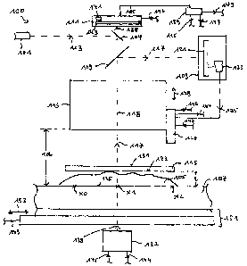
The arrangement 100 schematically illustrated in Fig. 1 in a cross-sectional
side view is
adapted to carry out a method for optical scanning at least one object region
105 placed on a
transparent specimen holder 107 according to an embodiment of the present
invention. The
13
CA 03061440 2019-10-24
WO 2018/197193 PCT/EP2018/059005
arrangement 100 is adapted in particular to perform a first step of a focus
determination and
a second step of a focus determination.
Therefore, the arrangement 100 comprises a laser 101 for generating a laser
beam 103. The
arrangement 100 further comprises a first camera 109 and a second camera 111.
During the
focus determination, for each sample lateral position x0, x1, x2 (per object
region for
example 5 sample lateral positions), the focus determination is carried out
involving two
steps at each sample lateral position. In the first step of the focus
determination, the laser
beam 103 is reflected at reflector 104, traverses the objective lens 113 and
impinges onto a
location (in the optical axis 118 of the objective lens 113) of the object
region 105 after
traversing the objective lens 113 and traversing the cover glass 115. The
laser beam 117
being reflected is reflected by a dichroic filter/reflector 119 and is
incident on the first camera
109 comprising (non illustrated optics and) a spatially resolving light
sensitive device 121 and
further comprising a processor 123 which receives measurement results of the
spatially
resolving light sensitive device 121. The processor 123 of the first camera is
adapted to
supply control signals 125 to a drive means 127 which is adapted to adjust a
vertical position
of the objective lens 113 in the vertical direction 129.
In particular, at the beginning of the first procedure of the focus
determination, the objective
lens 113 is positioned at a largest (or predefined) distance between the
specimen holder 107
and the objective lens 113. Then, the objective lens 113 is moved downwards
towards the
specimen holder 107, the laser beam 103 continuously illuminates a location of
the object
region 105 and the reflected laser light 117 is received by the first camera
which acquires a
plurality of first images. Each of the acquired first images is then analyzed
by the processor
123 comprised in the first camera 109 in order to detect characteristic
features in the first
images. In particular, the processor 123 comprises image processing software
to detect a
first reflex of the laser beam 103 which is caused by a reflection of the
laser beam 103 at an
upper surface 131 of the cover glass 115 covering the object region 105 (which
may
comprise a tissue sample). The objective lens 113 is then lowered further and
further first
images are acquired and one of the first images will comprise an imaged second
reflex of the
laser beam which originated from a reflection at a lower surface 133 of the
cover glass 115.
The objective lens 113 is further lowered until a third reflex of the laser
beam is detected
which third reflex originates from a reflection of the laser beam 103 from the
upper surface
135 of the specimen holder 107.
14
CA 03061440 2019-10-24
WO 2018/197193 PCT/EP2018/059005
In the illustration of Fig. 1, the objective lens 113 is in the vertical
position in which the laser
beam 103 is focused at the upper surface 135 of the specimen holder 107,
wherein the
distance rd between the objective lens 113 and the upper surface 135 of the
specimen
holder 107 represents a reference distance which is a starting position for
the second step of
the focus determination of the current sample lateral position x1.
For the second step of the focus determination the laser 101 may be switched
off and a flash
illumination light source 137 may be operated to generate flash illumination
light 138
traversing from below the object region 105. The flash illumination light
source 137 may for
example comprise a controllable shutter which is placed downstream a
continuously emitting
light source. The main processor 139 which may also be comprised within the
arrangement
100 may control the flash illumination light source 137 and/or may also
control the second
camera 111, and/or the translation stage and/or the driving means 127.
Starting from the
reference distance rd between the objective lens 113 and the specimen holder
107, the
objective lens 113 is then in a stepwise manner or in a continuous manner
having constant
velocity moved upwards, thus the distance between the objective lens 113 and
the specimen
holder 107 is increased while performing transmission flash illumination and
using the
second camera 111 to take plural second images.
The second images may be processed by a processor 141 comprised in the second
camera
111 (which also comprises a spatially resolving light detection device 143) or
by the main
processor 139. In particular, the main processor 139 may provide control
signals 145 to the
flash illumination light source 137, may provide control signals 147 to the
drive means 127
and also may supply control signals 149 to the translation stage 151 on which
the specimen
holder 107 is arranged in order to laterally translate the specimen holder 107
in the lateral
direction 153 (and additional in a lateral direction perpendicular to the
direction 153 and
perpendicular to the vertical direction 129). Further, the processor 139 may
communicate
with the second camera 111 via a control line 155. The processor 139 may also
be employed
during the actual scanning, in order to control the illumination light source
137, the translation
stage 151, the vertical driving means 127 and also the second camera 111.
The plural second images acquired by the second camera 111 are then analyzed
regarding a
degree of sharpness and the second image associated with the highest sharpness
is
determined. The distance between the objective lens 113 and the specimen
holder 107 at
which the second image has highest sharpness is considered to be the focus
distance at
CA 03061440 2019-10-24
WO 2018/197193 PCT/EP2018/059005
which the object region 105 is imaged in a focussed manner at the current
sample lateral
position x1.
In a same manner, focus determinations are performed for all other sample
lateral positions
x0, x2, ... across the object region 105.
Then a focus topology is calculated and the object region is scanned using the
focus
topology for moving the objective lens in the appropriate vertical positions.
Fig. 2 illustrates an arrangement 200 for optical scanning of at least one
object region
according to another embodiment of the present invention in a perspective
view. The
arrangement 200 also comprises a first camera 209 and a second camera 211, an
objective
lens 213, and a translation stage 251 at which the specimen holder 207 is
placed. Further,
the arrangement 200 comprises a not illustrated laser and a processor.
The arrangements 100, 200 increase the degree of automization for processing
and
diagnosing histological samples. Further, they support telepathology. The
arrangements
provide accelerated electronic processing and enable a focusing method with a
convex
envelope.
Fig. 3 schematically illustrates a light table 360 which may be utilized in a
scanning method
according to an embodiment of the present invention and may be comprised in an
arrangement according to an embodiment of the present invention. Above the
light table 360,
an overview camera 361 is placed in order to acquire an overview image of an
entire
specimen holder having placed thereon several object regions. The light table
360 comprises
LED stripes 363 being laterally attached to an acrylic glass 365 above which a
diffusing
element 367 and a clear acrylic glass 369 is arranged. Below the clear
plexiglass 365, a
white sheet 371 is arranged. The light table 360 enables to diffusely
illuminate an object
holder which is placeable on top of the clear plexiglass 369 for acquiring an
overview image
using the overview camera 361. In particular, the light table 360 generates a
homogeneous
illumination light. The overview camera 361 may have a focus length of 6 mm
and may have
a working distance of around 100 mm.
The overview image 473 taken by the overview camera 361 is illustrated in Fig.
4. The
overview image 473 comprises the whole specimen holder 407 having placed
thereon plural
16
CA 03061440 2019-10-24
WO 2018/197193 PCT/EP2018/059005
object regions 405a,b,c,d,e,f. The overview image 473 is analyzed to determine
the
localizations (e.g. center position (x,y) and boundary lines in overview
coordinate system
404) of the several object regions 405a,...,f. For generating the overview
image 473
illustrated in Fig. 4, the light table 360 and the overview camera 361 have
been used,
thereby illuminating the specimen holder from below. Additionally, the
specimen holder may
be illuminated from above to detect data codes or other identification
information identifying
the different object regions 405a,. ..f. The identification information is
then associated with the
respective overview image of the respective object region. The determined
localizations of
the object regions may be described by coordinates covering the respective
object region. At
this stage, a two-dimensional lateral (equidistant) mesh may be defined, which
is adapted to
the used objective lens (20 x objective lens with 2/3" 0.63 x adapter) having
a field of view of
e.g. 0.6 mm x 0.6 mm.
Furthermore, from the overview image 473, plural sample lateral positions 475
are defined in
each object region such as to essentially evenly cover the respective object
region 405f, as is
illustrated in Fig. 5.
Then, the specimen holder 407 is withdrawn from the light table 360 and is
placed onto the
translation stage 151 of the arrangement 100, as is illustrated in Fig. 1. The
translation stage
151 allows translating the specimen holder 107 in two perpendicular
directions. The
coordinates or localizations of the object regions 405a,...,f are transformed
into a coordinate
system of the translation stage 151. Thus, the sample lateral positions 475
are known as
coordinates in the coordinate system of the translation stage 151 and are
labelled in Fig. 1 as
x0, x1, x2.
A focusing procedure for determining a focus distance for each of the sample
lateral
positions consists of two parts.
The first part includes to finding the vertical position of the surface of the
specimen holder.
Thereby, a laser beam, preferably > 800 nm, is used which does not harm or
damage the
object region. The laser beam is traversed through the objective lens 113
towards the
specimen holder 107. The reflection light 117 of the laser 101 is not allowed
to impinge onto
the second camera 111, but is coupled using the dichroic filter 119 towards
the first camera
109 which is provided especially for the focus determination. This first
camera 109 performs
the image processing and also provides the control signals 125 for the
vertical drive means
17
CA 03061440 2019-10-24
WO 2018/197193 PCT/EP2018/059005
127. The first camera 109 is adapted to achieve a processing speed of 3250
images per
second. At the first part of the focus determination, the objective lens 113
starts at the
maximal distance to the specimen holder 107. The objective lens 113 then
approaches in 0.5
pm steps towards the specimen holder 107. At each step, a first image is
acquired by the first
camera 109 and is analyzed with respect to a characteristic laser reflection.
The light point
(originating from reflection of the laser beam) is visible in its full
intensity and degree of
details only within the depth of focus of the objective lens 113. The first
reflection arises at
the entry of the cover glass 115. The second reflection arises at the exit of
the cover glass
115. The third reflection corresponds to the entry into the specimen holder
107. Thereby, a
reference distance is found.
During the second part of the focus determination, the laser 101 is shut off.
At each vertical
position of the objective lens 113 (starting at the reference distance and
moving upwards), a
transmission condenser with a particular flash controller is operated for a
duration of 10 ps.
The flash current and the camera trigger are synchronized by a vertical
measurement system
which is also comprised in the vertical drive means 127. Thus, the vertical
drive and
measurement system 127 provide a control signal 144 to the flash illumination
light source
137 (in particular to a condenser comprised therein) and also supplies a
control signal 146 to
the second camera 111, in order to synchronize them. The second images are
acquired,
while the objective lens 113 is in motion. Starting from the reference
distance rd, at which the
laser beam 103 was focused at the upper surface 135 of the specimen holder
107, the
second camera 111 (also referred to as photo camera) acquired plural second
images such
that two subsequent second images are associated with vertical positions of
the objective
lens being 0.5 pm spaced apart. It is continued, until the objective lens has
moved upwards
corresponding to the thickness t of the tissue, i.e. the object region 105.
The thickness may
for example be between 4 and 6 pm. When using light microscopy, the degree of
sharpness
is used by the Sobel methodology (sum, abs, 3x3).
The Sobel operator, sometimes called the Sobel¨Feldman operator or Sobel
filter, is used in
image processing and computer vision within edge detection algorithms where it
creates an
image emphasising edges. "Isotropic 3x3 Image Gradient Operator" is a discrete
differentiation operator, computing an approximation of the gradient of the
image intensity
function. At each point in the image, the result of the Sobel¨Feldman operator
is either the
corresponding gradient vector or the norm of this vector. The Sobel¨Feldman
operator is
based on convolving the image with a small, separable, and integer-valued
filter in the
18
CA 03061440 2019-10-24
WO 2018/197193 PCT/EP2018/059005
horizontal and vertical directions and is therefore relatively inexpensive in
terms of
computations. The operator uses two 3x3 kernels which are convolved with the
original
image to calculate approximations of the derivatives ¨ one for horizontal
changes, and one
for vertical. If we define A as the source image, and Gx and Gy are two images
which at
each point contain the horizontal and vertical derivative approximations
respectively, the
computations may be as follows:
n n
a, y + j ¨ a)ki , j)
dr.-. =¨=10.
[1 0 ¨I -
G=SA=2 0 -2 f A
1 0 -H _
-
1 2 1
Gv = Sy * A = 0 0 0 * A
L ¨1 ¨2 ¨1
"I" is the Intensity of a pixel at (x,y). All pixel intensities of an image
have to be added.
That is the value of a Sobel-Filtered Image that describes the strength of
edges.
"sum, abs, 3x3" means, all values are added, negative matrix results are not
allowed, matrix
size (kernelsize) is 3x3.
When using fluorescence microscopy, either the difference of Gauss method or a
JPEG
focus method (compression to 100%) is performed.
Difference of Gauss method also applies the formula:
V4 016
= E E Az i - a, y + j ¨ a)k(i, j)
i=1.. 5=1
thus, the same basic formula as used for the Sobel-algorithm. It is also a
convolution.
The non-separated matrix is e.g. a 3x3 matrix, for example a classic Gauss-
bell (3x3 matrix):
1 ¨ 2 ¨ 1
2 ¨ 4 ¨ 2
1 ¨ 2 ¨ 1
An example of a 5x5 matrix, which may be used for problematic images, is:
19
CA 03061440 2019-10-24
WO 2018/197193 PCT/EP2018/059005
1 4 7 4 1
4 16 26 16 4
1
7 26 41 26 7
273
4 16 26 16 4
1 4 7 4 1
The difference of Gauss-method may work as follows:
From an image the added values are calculated with the described formulas, to
obtain a first
value. On the gaussed image, you again calculate the gauss value, to obtain a
second value.
The difference of these two values describes the sharpness in a very solid
way, even for
problematic images, where standards like Sobel-method will fail. Its problem
is that it
consumes a lot of calculation power. Because of that it is only used, if
necessary.
The JPEG Focus method may work as follows:
From an image a jpeg compression is calculated with max quality settings. The
total byte
size of this compressed image describes not only the "size", but also the
sharpness. If two
images with the absolute same content, but one image is less sharp, than the
sharper image
always have the bigger byte size. If the step size of a focus batch is greater
than depth of
field, the JPEG focus method may in particular be applied.
To determine the focus distance on average less than 100 ms are required.
A mesh object is created whose polygons contain the coordinates of the focal
point (xyz).
The mesh object is smoothed and subdivided, in an improved manner to determine
the
topology of the intermediate regions.
Fig. 6 illustrates a smoothed focus topology 600, wherein the focus topology
assigns at each
mesh lateral position (xm, ym) a focus distance fd. The focus topology (xm,
ym, fd) 600 is
CA 03061440 2019-10-24
WO 2018/197193 PCT/EP2018/059005
calculated based on the focus distances determined for the plural sample
lateral positions.
Fig. 7 illustrates a focus topology 700 derived from the focus topology 600
after subdividing
it.
When the focus topology has been determined for the object region 105, the
actual scanning
can be performed. Thereby, the translation stage 151 moves the object region
105 in a
meander manner, while the objective lens 113 is adjusted according to the
determined focus
topology and third images are acquired while moving the translation stage 151
and moving
the objective lens 113 as needed. Thereby, the flash illumination light source
137 may for
example provide flashes of 10 ps while the translation stage 151 moves with 14
mm/s.
Since during the first part of the focus determination the upper surface of
the specimen
holder is detected, no markers are necessary. During the focusing, only the
objective lens
but not the translation stage is moved, thereby reducing the moved mass.
The objective lens may be moved using a piezo drive enabling to achieve an
accuracy in the
nm range. The third images may be stitched together in real-time.
To use two different cameras, i.e. the first camera and the second camera to
perform the
focus determination has several advantages:
A particular camera always has a dead time (depending on the switching,
depending on the
sensor type and the application programming interface of the camera up to one
full second),
if its parameters of the format of the image are changed, for example, binning
or
subsampling. Since the camera for the laser focus needs to be operated at a
smaller
resolution (than the camera used for the scanning), in order to achieve the
high repetition
rate of about 3000 images per second, it would be required, if only one camera
would be
available, to change the format of the camera. Thereby, the method would be
very time-
consuming. In the method according to embodiments of the present invention,
however, the
configurations (in particular regarding format and/or resolution) of the first
camera as well as
the second camera do not need to be changed regarding the resolution (format)
thereby
avoiding dead times.
21
CA 03061440 2019-10-24
WO 2018/197193 PCT/EP2018/059005
Furthermore, when the laser provides an infrared laser beam, damage of the
biological
sample may be reduced. The objective lens may be a 20 objective lens having a
numerical
aperture of 0.8.
The invention is not limited to the described or illustrated embodiments.
22