Note: Descriptions are shown in the official language in which they were submitted.
CA 03061644 2019-10-28
WO 2018/197306 1 PCT/EP2018/059944
VENTRICULAR ASSIST DEVICE AND METHOD
FIELD OF THE INVENTION
The present invention generally relates to the field of medical devices and
surgery
devices. More specifically, the invention relates to a catheter and
corresponding methods of
use of the catheter. The present invention is particularly useful in the
context of minimally
invasive transcatheter and/or percutaneous procedures, such as those described
in PCT
Application No. PCT/EP2015/055578, entitled "PERCUTANEOUS SYSTEM, DEVICES
AND METHODS" filed 17 March 2015 and expressly incorporated herein by
reference in its
entirety.
BACKGROUND
In PCT/EP2015/055578, the Inventor describes an intracorporeal connector for
fluid
communication between a first and a second anatomical compartment, in
particular a
ventricular assist system for allowing blood flow between the left atrium and
the aorta of a
patient. The system is implanted across the roof of the left atrium and the
aortic wall and
generally comprises two main components, namely an anchor or connector element
and a
fluid regulation device such as a pump.
The ventricular assist system is preferably delivered and implanted using a
transcatheter
system as described for example in PCT Application No. PCT/EP2015/055578, or
in
PCT/EP2016/082889 entitled "TRANSCATHETER INSERTION SYSTEM" filed on 29th
December 2016; PCT Application No. PCT/EP2017/050275 entitled "CONNECTOR AND
METHOD FOR COUPLING ANATOMICAL WALLS" filed on 6th January 2017, and US
application Nos. US 15/288642 and US 15/288738 filed on 7th October 2016, all
incorporated
herein by reference.
The connector element comprises a proximal portion, an intermediate portion
and a
distal portion. The proximal portion comprises a plurality of arms which, in a
working
configuration, lie against the wall of the first compartment; the intermediate
portion
comprises a fluid conduit and, in a working configuration, is positioned
across the anatomical
walls; the distal portion comprises a plurality of arms which, in a working
configuration, lie
CA 03061644 2019-10-28
WO 2018/197306 2 PCT/EP2018/059944
against the aortic wall. The intermediate portion is adapted and configured to
keep the two
anatomical walls to remain in contact with each other; while the distal and
proximal arms are
adapted and configured to maintain the structural integrity of the anatomical
walls. This is
particularly important as the connector is adapted and configured to safely
support the fluid
regulation device across the anatomical walls, which will be under pressure
and susceptible to
dislodgment due to e.g. the structure of the fluid regulation device itself,
blood flow created
by the pump and patient movements.
While the above ventricular assist system can be safely implanted and fluid
flow
successfully established, the size and structure of the heart is such that
there has been a need
to adapt the fluid regulation device and consequently the delivery and
implantation methods
and systems. The fluid regulation device would typically comprise a pump
element, a motor
element, and optionally a battery element (as described for example in PCT
Application No.
PCT/EP2016/069159 filed on 11th August 2016) and, if required, means for
recharging said
battery. Space and manipulation within the heart is limited and
miniaturisation can only be
considered insofar as the efficiency of the fluid regulation device is not
negatively affected.
As the size of the device increases, more pressure is exerted on the
anatomical walls, the
integrity of which could become compromised. There is therefore a risk of
heart tissue
trauma, which dangerous and potentially be lethal to the patient.
It is an object of this invention to mitigate problems such as those described
above.
SUMMARY OF THE INVENTION
According to a first aspect, there is provided a method for supporting heart
function of
a patient comprising the step of securing an intracorporeal device across at
least two
anatomical walls of the heart, wherein at least one anatomical wall is an
intra-cardiac wall
and a least one anatomical wall is an extra-cardiac wall.
Thus, the intracorporeal device is safely secured to the heart using a two-
point
anchoring system. The device is stabilised and the pressure exerted by the
device and blood
flow onto the heart structure is shared so that the risk of strain and trauma
to a single wall is
reduced.
CA 03061644 2019-10-28
WO 2018/197306 3 PCT/EP2018/059944
Within the context of the invention, "intracorporeal" means inside the
patient's body
and "extracorporeal" means outside the patient's body. For example, an
intracorporeal device
or component will be located within the patient's body, while an
extracorporeal device or
component will be located outside the patient's body.
Within the context of the invention, "intra-cardiac" means inside the heart
and "extra-
cardiac" means between the inside and the outside of the heart or outside the
heart. For
example, an intra-cardiac wall is an anatomical wall located inside the heart.
Examples of
intra-cardiac walls include, but are not limited to the atrial septum between
the right and left
atria and the interventricular septum between the right and left ventricles.
An extra-cardiac
wall can be an anatomical wall between the inside and the outside of the heart
for example
the wall between the inside of the left atrium, of the right atrium, of the
left ventricle or of the
right ventricle and the outside of the heart and also the aortic wall.
In a preferred embodiment, the intra-cardiac wall is the atrial septum and the
extra-
cardiac wall is the wall of the left atrium, most preferably, the roof of the
left atrium. These
anatomical walls are particularly advantageous when a fluid regulation device
is to be
implanted, which regulates the flow of fluid from the left atrium to the
aorta. For example, in
PCMP2015/055578, the fluid regulation device is inserted through a puncture
through the
atrial septum, then implanted across the roof of the left atrium so that blood
flows through
inlets positioned in the left atrium to outlets positioned in the aorta. The
left atrium and aortic
walls are subjected to tension due to the implantation of the fluid regulation
device itself and
to pressure due to the fluid flow. By providing a second anchoring point, e.g.
across the atrial
septum, the device is stabilised and the anatomical walls are individually
subjected to less
pressure. Thus, the risk of injury, trauma and leak is minimised. The atrial
septum is
preferred in that it is generally rigid and robust enough to secure and
support an
intracorporeal device but flexible enough to buffer for any movement during
and post-
implantation. In addition, the atrial septum is often used as an insertion
path and can be used
as a second anchoring point without the need for further puncture.
Preferably, the method further comprises the step of securing the
intracorporeal device
across at least a third anatomical wall. More preferably, the third anatomical
wall is a wall
adjacent to the extra-cardiac wall. Within the context of the invention,
"adjacent wall" means
a "wall naturally physically close". For example, two adjacent walls may be
the adjacent
CA 03061644 2019-10-28
WO 2018/197306 4 PCT/EP2018/059944
walls of two adjacent anatomical compartments, such as the wall of the left
atrium and the
aortic wall. In a preferred embodiment, the third anatomical wall is the
aortic wall adjacent to
the roof of the left atrium.
The present invention is particularly useful when the flow of fluid is to be
established
between two anatomical compartments separated by at least two anatomical
walls. As
described in PCT/EP2015/055578, the anatomical walls are pushed into contact
with each
other by means of a delivery catheter or outer sheath, punctured and secured
together for
example using a connector. When two anchoring points are provided, the
pressure and
tension is spread and are no longer focussed on and around the connector.
Preferably, the intracorporeal device comprises a proximal portion intended to
be
positioned in a first anatomical compartment, an intermediate portion intended
to be
positioned in a second anatomical compartment, a distal portion intended to be
positioned in
a third anatomical compartment. In a preferred embodiment, the proximal
portion of the
intracorporeal device is intended to be positioned in the right atrium, the
intermediate portion
is intended to be positioned in the left atrium and the distal portion is
intended to be
positioned in the aorta. The intracorporeal device can therefore be secured to
the atrial
septum (between the proximal and the intermediate portion) and to the wall of
the left atrium
and aortic wall (between the intermediate portion and the distal portion).
The position of the various elements of the intracorporeal device may be
adapted and
configured to assist fluid flow between any two compartments as will be
described below in
more details. In a preferred embodiment, the intracorporeal device is a fluid
regulation device
for assisting fluid flow from the second to the third compartment, e.g. from
the left atrium to
the aorta.
Preferably, the intracorporeal device is secured to one or more anatomical
walls by
means of a connector. More preferably, the connector may be a separate
connector or may be
integrally formed or attached to the intracorporeal device.
Preferably, the connector comprises a neck intended to be positioned across
one or
more anatomical wall(s), a first plurality of arms extending from a first end
of the neck, and a
second plurality of arms extending from the second end of the neck. As
described in the
CA 03061644 2019-10-28
WO 2018/197306 5 PCT/EP2018/059944
applicant's previous applications cited herein, the arms are preferably
movable from a
transcatheter delivery configuration (e.g. in line with the neck) to a working
configuration
(e.g. substantially perpendicular to the neck).
In the case of a separate connector, the connector and/or the intracorporeal
device
comprise means for coupling the intracorporeal device to the connector.
Preferably, the connector is integrally formed or coupled to the
intracorporeal device.
In this embodiment, the connector will comprise means for connecting the
intracorporeal
device to the anatomical wall(s). For example, the intracorporeal device may
comprise a
plurality of arms extending from the intracorporeal device. Preferably, the
arms are
preferably movable from a transcatheter delivery configuration (e.g. in line
with the neck) to
a working configuration (e.g. substantially perpendicular to the neck).
In a preferred embodiment, the intracorporeal device comprises a means for
fixing the
intracorporeal device to the connector. Preferably the fixing means comprises
a plurality of
arms extending from the intracorporeal device, preferably the distal end of
the intracorporeal
device. The arms may be moved from delivery configuration (e.g. extending
substantially
longitudinally from the intracorporeal device) to a working configuration
(e.g. extending
away from the longitudinal axis of the intracorporeal device). In the working
configuration,
the fixing arms may cooperate with the connector to anchor the intracorporeal
device to the
anatomical walls. In addition, the fixing arms may serve as additional support
to the wall
tissue, against the fluid flow and/or the intracorporeal device's own weight
and bulk.
Preferably, the intracorporeal device comprises one or more recesses adapted
and
configured to receive one or more anatomical walls. In a preferred embodiment,
the
intracorporeal device comprises an elongated housing or a substantially
cylindrical housing.
The intracorporeal device may comprise a circumferential recess adapted to
receive the
anatomical wall(s) therein, thereby securing the intracorporeal device to the
wall. Preferably,
to facilitate the insertion of the anatomical wall(s) into the recesses, the
recesses have slopped
or curved walls.
CA 03061644 2019-10-28
WO 2018/197306 6 PCT/EP2018/059944
According to a second aspect of the invention, there is provided an
intracorporeal
device for supporting heart function of a patient, wherein said device is
adapted and
configured to be secured across at least two anatomical walls of the heart.
Preferably, the at least one anatomical wall is an intra-cardiac wall and a
least one
anatomical wall is an extra-cardiac wall.
Preferably, the intracorporeal device is adapted and configured to be secured
to one or
more anatomical walls by means of a connector, said connector being integrally
formed or
coupled to the intracorporeal device.
Preferably, the intracorporeal device is adapted and configured to be secured
to one or
more anatomical walls by means of a connector and a fixing means, said
connector being
arranged to be positioned across one or more anatomical walls and said fixing
means being
integrally formed or coupled to the intracorporeal device.
Preferably, the fixing means comprises a plurality of arms extending from the
intracorporeal device, for example the distal end of the intracorporeal
device.
Preferably, the intracorporeal device comprises one or more recesses adapted
and
configured to receive one or more anatomical walls.
Preferably, the intracorporeal device comprises a proximal portion intended to
be
positioned in a first anatomical compartment, an intermediate portion intended
to be
positioned in a second anatomical compartment, a distal portion intended to be
positioned in
a third anatomical compartment.
Preferably, the intracorporeal device comprises a motor located in the
proximal portion.
Preferably, the intracorporeal device comprises one or more fluid inlet ports
in the
intermediate portion. Preferably, the intracorporeal device comprises a pump
in the
intermediate portion.
Preferably, the pump comprises an impeller and a pump housing, wherein the
impeller
is positioned within the pump housing. The impeller is a rotatable element
that accelerates
CA 03061644 2019-10-28
WO 2018/197306 7 PCT/EP2018/059944
fluid outwards from the centre of rotation in a direction parallel to the
impeller's major
(longitudinal) axis, which is generally referred to as an axial flow impeller.
The impeller
rotates about its major axis with respect to the pump housing. The impeller is
surrounded by
the pump housing so that the rotational velocity of the impeller transfers
into pressure when
the outward movement of the fluid is confined by the pump housing.
Preferably, the impeller comprises a tapered profile. The tapered profile
increases from
the proximal portion end to a mid portion of the impeller, wherein the tapered
profile then
decreases towards the distal portion end such that the cross section of the
tapered impeller
approximates an ellipse, wherein the major axis of the ellipse is parallel to
the major axis of
the pump housing. The tapered profile of the impeller has an advantage of
increasing fluid
pressure in the pump housing. Thus fluid, such as blood, spends less time
around parts of the
device that generate heat, such as the motor and various bearings within the
intracorporeal
device. This reduces the probability of the fluid being damaged by heat
generated by the
device, which in turn reduces the probability of the fluid clotting and
blocking the circulatory
system. Thus fluid cools the surface of the motor housing and the internal
pump elements.
Preferably, the intracorporeal device comprises one or more fluid outlet ports
in the
distal portion. One or more fluid inlet ports may be positioned in the
proximal and/or
intermediate portion depending on the compartments the fluid flows from/to. In
a preferred
embodiment, the proximal portion of the intracorporeal device is intended to
be positioned in
the right atrium, the intermediate portion is intended to be positioned in the
left atrium, and
the distal portion is intended to be positioned in the aorta. In a most
preferred embodiment,
the intermediate portion comprises one or more inlet ports and the distal
portion comprises
one or more outlet ports so that fluid can flow from the left atrium to the
aorta.
Preferably, the length of the intermediate portion and the distal portion is
designed such
that the one or more fluid inlet ports in the intermediate portion are
positioned in the left
atrium and the one or more fluid outlet ports in the distal portion are
positioned in the aorta.
Preferably, the intracorporeal device comprises a static diffuser positioned
in the distal
portion of the intracorporeal device. Preferably, the diffuser is positioned
between the
impeller and the one or more fluid outlet ports. Preferably, the diffuser is
coupled to an end of
the impeller via a bearing. Preferably, the diffuser is fixedly attached to
the interior of the
CA 03061644 2019-10-28
WO 2018/197306 8 PCT/EP2018/059944
pump housing such that the diffuser is not able to rotate. Preferably, the
diffuser and bearing
support the impeller and allow the impeller to rotate about its major axis,
whilst the diffuser
remains fixed to the interior of the pump housing. The diffuser has an
advantage of increasing
fluid diffusion from the outlet of the device due to the angle and profile of
the blades of the
diffuser.
Preferably, the intracorporeal device comprises a fixing means that is
arranged to
secure the intracorporeal device to an anatomical wall. Preferably, the fixing
means is at an
end of the distal portion of the intracorporeal device.
Preferably, in use, the connector is positioned through the roof of the left
atrium and the
aortic wall. Preferably, the neck of the connector and/or the distal portion
of the
intracorporeal device form a pericardial seal. Preferably, the neck of the
connector acts as a
docking means to assist in coupling the distal portion of the intracorporeal
device across the
anatomical walls and with respect to the connector.
The fixing means may be a type of anchor/support member with a number of
arms/tissue
support members that can deploy against the wall of the aorta for example.
This may position
and secure the distal portion of the device with respect to the aorta to allow
for efficient fluid
transfer through the device.
Preferably, the intracorporeal device comprises a static diffuser positioned
at an end of
the distal portion. This has the advantage of further increasing fluid
diffusion at an outlet of
the device because the fixed blades aid in fluid diffusion. This also has the
advantage of
facilitating easier deployment of the device because a guide wire and/or
balloon may be
attached to a guide wire holder on the static diffuser to enable accurate
positioning within the
human body.
Preferably, the intracorporeal device comprises a motor coupling element that
is
arranged to couple a drive shaft of the motor to the pump. Preferably, the
motor coupling
element is positioned between the motor and the impeller. Preferably, the
motor coupling
element magnetically couples the drive shaft of the motor to the pump. This
has an advantage
of maintaining a hermetic seal of the motor and drive shaft with respect to
fluid within the
circulatory system, whilst also enabling the motor and drive shaft to be
easily removed from
CA 03061644 2019-10-28
WO 2018/197306 9 PCT/EP2018/059944
the remainder of the intracorporeal device. Thus components of the
intracorporeal device that
are most likely to need removal, modification or replacement, e.g. the motor,
can be detached
and replaced easily whilst maintaining the remainder of the intracorporeal
device in position
within the body.
Preferably, the motor coupling element axially couples the drive shaft of the
motor to
the pump. This has an advantage of simplifying the coupling between the drive
shaft of the
motor and the pump, whilst maintaining a hermetic seal between the motor and
fluid within
the body.
Preferably, the motor coupling element radially couples the drive shaft of the
motor to
the pump. Preferably, a portion of the motor coupling element surrounds and
magnetically
couples to an elongate portion of the motor that houses the motor drive shaft.
This has an
advantage of increasing the torque transfer between the drive shaft and the
pump impeller.
Radial configuration of the coupling magnet forces eliminates additional
bearing loading and
heat generated by friction.
Preferably, the motor, the drive shaft, and a magnetic element at an end of
the drive
shaft are situated in a hermetically sealed housing. This has an advantage of
allowing the
motor to be positioned in the circulatory system. If the elements discussed
above were not
situated in a hermetically sealed housing, fluid within the circulatory system
would damage
these elements and/or the elements may cause contamination to the fluid within
the
circulatory system.
Preferably, a portion of the motor coupling element partially surrounds a
portion of the
hermetically sealed housing containing the magnetic element. This has an
advantage of
facilitating radial magnetic coupling and allowing fluid to flow between the
motor coupling
element and the portion of the hermetically sealed housing.
Preferably, an interstitial space/void is present between the portion of the
motor
coupling element and the portion of the hermetically sealed housing. This has
an advantage
of facilitating fluid flow in the interstitial space to prevent fluid damage.
CA 03061644 2019-10-28
WO 2018/197306 10 PCT/EP2018/059944
Preferably, an interface of the motor coupling element is magnetically fixable
to an
interface of the motor in order to couple the motor drive shaft to the pump,
and wherein a
fluid inlet is defined between said interfaces during coupling. This has an
advantage of
facilitating fluid flow between the interfaces so that fluid can enter the
interstitial space
between the interfaces.
Preferably, a further interface of the motor coupling element couples to an
interface of
the impeller. This has an advantage of translating movement of the motor drive
shaft to the
impeller.
Preferably, the motor coupling element comprises one or more bore portions.
This has
an advantage of allowing fluid that has entered the interstitial spaces to
exit the motor
coupling element. This results in washing of a bearing inside of the motor
coupling element,
without the fluid temperature increasing to a point where the fluid becomes
damaged..
Preferably, the one or more bore portions are bore holes and/or segmented arms
that
couple the interfaces of the motor coupling element to the respective impeller
interfaces.
Preferably, in use, fluid is arranged to flow between the motor interface and
the
interface of the motor coupling element and through the one or more bore
portions towards
the impeller. This has an advantage of reducing heat in the bearing and
preventing the fluid
from being damaged by excess heat.
Preferably, the one or more fluid inlet ports are positioned between the one
or more
bore portions and the impeller in the intermediate portion of the pump
housing. This has an
advantage of allowing fluid exiting from the bore portions to mix with fluid
entering the fluid
inlet ports. Thus any excess heat absorbed by the fluid exiting the bore
portions can be
dissipated efficiently with fluid entering via the fluid inlet ports.
Preferably, the intracorporeal device comprises power and control means.
Preferably,
the control means is coupled to the motor via a tapered portion. The tapered
portion has an
advantage of reducing strain on the connector between the motor and the power
and control
means. Preferably, the tapered portion reduces in dimension as it tapers away
from the
proximal portion of the intracorporeal device.
CA 03061644 2019-10-28
WO 2018/197306 11 PCT/EP2018/059944
Within the context of the invention, the terms "proximal" and "distal" are
used relative
to the medical professional, e.g. the proximal end is the end nearest the
medical professional
and the distal end is the part of the device that is inserted first into the
patient.
Within the context of the invention, transcatheter includes percutaneous,
trans-atrial,
trans-femoral (through the leg), trans-apical (in the chest between the ribs),
and trans-aortic
(in the upper chest). Preferred embodiments are percutaneous systems, devices
and methods.
The invention will be further described with reference to the drawings and
figures, in
which:
Figures 1, 1A and 1B illustrate a method according to the present invention
using a first
intracorporeal device;
Figure 2 illustrates a method according to the present invention using a
second
intracorporeal device;
Figure 3 is a schematic representation of an intracorporeal device
incorporating fixing
means and control means according to the present invention;
Figure 4 is a schematic representation of the intracorporeal device from
figure 3
without fixing means.
Figure 5A is a schematic representation of an outer view of an intracorporeal
device
without fixing means or control means.
Figure 5B is a schematic representation of an internal view of the
intracorporeal device
without fixing means or control means.
Figure 5C is a schematic representation of a static diffuser utilised in the
intracorporeal
device in FIGs 5A and 5B.
Figure 5D is a schematic representation of an alternative impeller.
Figure 6A is a schematic representation of a cross section of an
intracorporeal device
utilising radial coupling between a motor and a motor coupling element.
Figure 6B is a schematic representation of a cross section of an
intracorporeal device
utilising axial coupling between a motor and a motor coupling element.
Figure 7 is a schematic representation of segmented arms from the motor
coupling
element.
Figure 8 is an exploded schematic representation of constituent elements of an
intracorporeal device.
CA 03061644 2019-10-28
WO 2018/197306 12 PCT/EP2018/059944
DETAILED DESCRIPTION
The invention is described by way of examples, which are provided for
illustrative
purposes only. These examples should not be construed as intending to limit
the scope of
protection that is defined in the claims. For example, although various
aspects have been
described with respect to the heart and the circulatory system, this is not
intended to be
limiting, and is merely performed to provide an example of implementation.
Aspects
disclosed herein may be utilised in any medical device implantable within the
human body,
for example in the cardiovascular system, respiratory system, gastric system,
neurological
system, and the like, some examples including implantable pumps and drug
delivery pumps.
As used herein, the term "means" can be equivalently expressed as, or
substituted with, any
of the following terms: device, apparatus, structure, part, sub-part,
assembly, sub-assembly,
machine, mechanism, article, medium, material, appliance, equipment, system,
body or
similar wording.
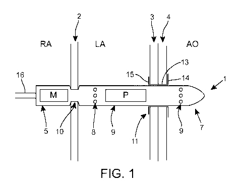
Referring to Figure 1, there is illustrated a method according to the present
invention
for supporting heart function of a patient comprising the step of securing an
intracorporeal
device 1 across at least two anatomical walls of the heart, wherein at least
one anatomical
wall is an intra-cardiac wall and a least one anatomical wall is an extra-
cardiac wall.
In this example, the intracorporeal device 1 is secured across the atrial
septum 2 (an
intra-cardiac anatomical wall), the roof of the left atrium 3 (an extra-
cardiac anatomical wall)
and the aortic wall 4 (i.e. a third anatomical wall). The intracorporeal
device 1 comprises a
proximal portion 5 located in use in the right atrium RA, an intermediary
portion 6 located in
use in the left atrium LA, and a distal portion 7 located in use in the aorta.
A power and
control cable 16 is coupled to an end of the proximal portion 5.
The intracorporeal device 1 is substantially cylindrical or comprises a
substantially
cylindrical housing. A motor M is located in the proximal portion 5 and a pump
P is located
in the intermediate portion 6. The position of the fluid inlet and outlet
ports may be adjusted
so that the fluid inlet ports are formed in the first fluid feeding
compartment and the fluid
outlet ports are formed in the second fluid receiving compartment. In this
example, the fluid
inlet ports 8 are formed in the intermediate portion 6 positioned in the left
atrium LA and the
fluid outlet ports 9 are formed in the distal portion 7 positioned in the
aorta AO.
CA 03061644 2019-10-28
WO 2018/197306 13 PCT/EP2018/059944
In an alternative implementation, the motor 5 may be housed in the
intermediate portion
6. As a result, the proximal portion 5 is no longer required and is no longer
situated in the
right atrium. Thus only a power and control cable would be present in the
right atrium.
In figure 1, the intracorporeal device 1 comprises a circumferential recess 10
between
its proximal and intermediate portions 5,6. The shape and dimensions of the
recess are such
that the atrial septum can be received into the recess 10. The recess 10 may
have sloped or
curved walls as shown in figures 1 A and 1B, respectively, to facilitate the
insertion of the
atrial septum 2 into the recess 10.
Where the intracorporeal device 1 is to be secured to a single anatomical wall
(e.g. the
atrial septum 2) then a recess 10 may be sufficient. However, when the
intracorporeal device
1 is to be secured across two or more anatomical walls (e.g. the wall of the
left atrium and the
aortic wall 4), then a connector 11 may be preferred. The connector 11 shown
in figure 1 is a
separate connector.
Connectors suitable for use in the context of the present invention are
described in
detail in PCT/EP2017/050275, US 15/288642 and US 15/288738. The connector 11
generally
comprises a neck 13 for fluid passage between two anatomical compartments,
positioned in
use across/through the anatomical walls 3,4; a first plurality of arms and/or
blades 15
extending from the distal end of the neck 13 and lying in use against the wall
of the receiving
compartment and a second plurality of arms and/or blades 14 extending from the
proximal
end of the neck 13 and lying in use against the wall of the feeding
compartment. The arms
and/or blades are preferably integrally formed or secured to the distal end of
the neck 13. In
use, the arms and/or blades rest partially or wholly against the anatomical
walls 3, 4. The
neck 13 also supports the intracorporeal device 1 when it is positioned across
the anatomical
walls 3, 4. In use, (part of) the distal portion 7 on the intracorporeal
device 1 is positioned
through the neck 13 of the connector 11, and thus across the anatomical walls
3, 4. For
example, the intracorporeal device may include a recess to receive the neck of
the connector.
The dimensions of the neck 13 and the distal portion 7 of the intracorporeal
device 1 are
arranged such that coupling the distal portion 7 with the neck 13 forms a
pericardial space
seal. Thus the neck 13 facilitates a seal as well as facilitating pump docking
and support of
the intracorporeal device 1.
CA 03061644 2019-10-28
WO 2018/197306 14 PCT/EP2018/059944
Upon removal of the intracorporeal device from connector 11, the connector 11
forms a
seal between the anatomical walls 3, 4 to prevent fluid diffusing between the
two regions
defined by said walls 3, 4.
This specific configuration secures the connector 11 to the anatomical walls
3,4 and
enables the connector 11 to maintain the anatomical walls 3,4 in contact with
each other
while supporting the integrity of the anatomical walls 3,4. Thus the arms
and/or blades act as
tissue supporting members in order to support the integrity of the anatomical
walls 3,4.
The intracorporeal device 1 may be provided with one or more recesses e.g. a
circumferential recess to receive the neck 13 of the connector 11 therein.
Other means for securing the intracorporeal device 1 to the anatomical wall(s)
2,3, 4 are
envisaged, including but not limited to tabs, hooks, arms, cushions, high
friction surfaces,
biologically active covering, and the like.
Referring to figure 2, there is illustrated an alternative method of securing
the
intracorporeal device 1 with respect to the connector 11. Previously,
referring to figure 1, the
distal portion 7 of the intracorporeal device was coupled through/to the neck
13 of the
connector 11, e.g., via frictional force or through the compression of
connector 11 (i.e.
connector 11 is preferably made of an expandable/compressible material).
Alternatively or in
combination, a number of tissue support/fixing members 12 may be coupled to
the distal
portion 7 of the intracorporeal device 1. These tissue support/docking members
12, when
deployed, contact the aortic wall and further support the intracorporeal
device 1 with respect
to the connector 11. Thus a combination of the connector 11 and the tissue
support/docking
members 12 enable enhanced support as well as the ability to easily couple and
de-couple the
distal portion 7 of the intracorporeal device 1 with respect to the connector
11.
Referring to Figure 3, there is illustrated a schematic representation of an
intracorporeal
device 300 with associated fixing means 302 and control means 304. The fixing
means 302 in
this embodiment relates to the tissue support/docking members 12 described in
relation to
figure 2. The fixing means 302 comprise a coupler 330 attached to a proximal
end of the
fixing means 302. A number of pump docking members/support arms 306 are
attached to a
distal (opposite) end of the fixing means 302. Figure 3 illustrates the fixing
means 302 in a
CA 03061644 2019-10-28
WO 2018/197306 15 PCT/EP2018/059944
"deployed" position, wherein the number of pump docking members/support arms
306 are
splayed out substantially perpendicular to the longitudinal axis of the
intracorporeal device
300. These pump docking members/support arms 306, in use, abut a portion of
the
anatomical wall 4 (see figure 1,2), for example the aortic wall, in order to
position and secure
the intracorporeal device 300 between the anatomical walls 3, 4 (see figure
1,2) and inside
the neck 13 of the connector 11.
The number of pump docking members/support arms 306 act as a tissue shield and
pump
protector, since the wall of the aorta is held away from the intracorporeal
device 300 that is
positioned inside the aorta. The number of pump docking members/support arms
306
distribute pressure so that each individual docking member/arm does not damage
the
anatomical wall 4.
If the intracorporeal device 300 needs to be removed from across the
anatomical walls
3, 4, the pump docking members/support arms 306 are re-positioned into a
"delivery"
position, wherein the pump docking members/support arms 306 are arranged
substantially
parallel with the longitudinal axis of the intracorporeal device 300 to enable
said device to be
removed from the neck of the connector 11 (see figure 2). This has an
advantage of allowing
the intracorporeal device 300 to be removed without damaging the anatomical
walls 3, 4,
which are protected by the connector 11. On removal of the intracorporeal
device 300, the
connector 11 seals the space between the anatomical walls, 3, 4 until the
intracorporeal
device 300 is re-inserted.
In a preferred embodiment, the delivery of the intracorporeal device 300 is
via echo
guided trans-septal and/or trans-aortic methods for the specific puncture
sites, wherein echo
planes may be used for all puncture sites. An echo plane is a defined
projection/view where
anatomy and angles are predefined so as to visualise specific regions of
interest in a specific
way. The echo guided methods may be, for example, intra-cardiac, trans-
esophageal, or trans-
thoracic.
The coupler 330 is positioned over a crown connector/coupling member (not
visible) of
the intracorporeal device 300 and abuts an end portion 332 of the
intracorporeal device 300.
CA 03061644 2019-10-28
WO 2018/197306 16 PCT/EP2018/059944
The control means 304 comprise a drive-line 308 that houses cabling to power
and/or
control the intracorporeal device 300. In this example, the control means 304
is coupled to a
proximal portion 310 of the intracorporeal device 300 via a tapered portion
312. The tapered
portion 312 tapers in size away from the proximal portion 310 of the
intracorporeal device
300. The tapered portion 312 has an advantage of reducing strain on the
connector interface
(not shown) that is housed in portion 314 between the proximal portion 310 and
the tapered
portion 312. The connector interface couples the cabling in the control means
304 to the
motor 316. The motor 316, connector interface and control means 304, form a
hermetically
sealed unit, which is arranged to prevent fluid ingress.
A portion of the motor 316 is situated within a rear portion 318 of the pump
housing
320. The rear portion 318 of the pump housing 320 defines a number of washing
holes 322,
also called bore portions, that enable fluid, such as blood, to flow through
the rear portion
318 of the pump housing and between a driving portion (not shown) of the motor
316 and a
motor coupling element 324, which is partially visible in this figure. Fluid
inlets 326 are
arranged in the rear portion 318 of the pump housing 320. Between the fluid
inlets 326 and
the fixing means 302 is an impeller (not shown) situated within a front
portion 328 of the
pump housing 320.
The crown connector/coupling member (not shown) acts as a main outlet for
fluid of
the intracorporeal device 300. A static diffuser 305 (partially visible),
inside of the crown
connector/coupling member, interferes with the flow of fluid to generate a
desired fluid flow
out of the main outlet and into the aorta. The crown connector/coupling member
comprises
one or more fluid outlet ports 303.
In some other embodiments, for example the embodiment of figure 1, the fixing
means
302 may be dispensed with. In these embodiments, the crown connector/coupling
member
may also be dispensed with. Thus the diffuser 305 may be situated within the
distal end of the
front portion 328 of the pump housing 320, rather than inside the crown
connector/coupling
member.
In this example, the distal portion of the intracorporeal device 300 comprises
the fixing
means 302 and crown connector/coupling member (not shown), wherein in use the
distal
portion is situated within the aorta. The intermediate portion of the
intracorporeal device 300
CA 03061644 2019-10-28
WO 2018/197306 17 PCT/EP2018/059944
comprises the pump housing 320 with its associated elements such as the
impeller, and motor
coupling element 324. The proximal portion of the intracorporeal device 300
comprises the
portion of the motor that is not within the pump housing 320, the portion 314,
tapered portion
312, and control means 304.
Referring to figure 4, the intracorporeal device 300 from figure 3 is
illustrated without
fixing means 302 coupled to the intracorporeal device. Thus in this example,
the crown
connector/coupling member 331 can be viewed in more detail. The crown
connector 331
comprises the static diffuser 305 that is coupled to the side walls of the
crown connector 331.
The diffuser comprises static blades 404 and a guide wire holder 402. In use,
fluid flows over
the static blades 404 of the static diffuser 305, wherein the blades are
orientated so to affect
the orientation of fluid as it flows through the crown connector 331 to the
one or more fluid
outlet ports 303. The guide wire holder 402 also allows enhanced guide wire
connectivity
and/or balloon connectivity. For example, a guide wire (not shown) can be
threaded through
guide wire holder 402 to enable the intracorporeal device 300 to be accurately
positioned
within the human body via, for example, a catheter based implantation method.
In some examples, the crown connector 331 may be dispensed with and the static
diffuser 305 may be positioned at a front portion 328 of the pump housing 320.
Referring again to figure 4, the portion 314 is illustrated so that connector
interface 406
can be viewed. Connector interface 406 electrically couples the control means
304 to the
back end of the motor. This enables power and/or control of the motor 316.
Tapered portion
312 reduces strain on the connector interface 406. This is particularly
important in the present
invention because the intracorporeal device 300 needs to maintain flexibility.
This is because
in use the intracorporeal device 300 is implanted within the left and right
atrium of the heart
and the aorta via a catheter based insertion system. As such, the
intracorporeal device needs
to be flexible enough to follow the direction of the arterial system. Portion
314 maintains the
hermetic seal of the motor 316, whilst allowing coupling of the control means
304 to the
motor 316 so that the motor can be implanted within the circulatory system of
the human
body.
Referring to figure 5, there is illustrated a schematic representation of an
intracorporeal device 500. In this example, power and control means are not
illustrated.
CA 03061644 2019-10-28
WO 2018/197306 18 PCT/EP2018/059944
Further, in this example, the intracorporeal device 500 is illustrated without
fixing means 302
or a crown connector/coupling member 331. Thus a diffuser 534 is situated at
the outlet 503
of the intracorporeal device 500 rather than in the crown connector/coupling
member 331, as
illustrated in figures 3 and 4.
Figure 5A illustrates an outer view of the intracorporeal device 500, whilst
figure 5B
illustrates an internal view of the intracorporeal device 500. Figure 5C
illustrates the diffuser
534 and figure 5D illustrates an alternative impeller design.
Intracorporeal device 500 in figure 5A comprises motor 502 and pump housing
504.
A portion of the motor 502 is situated within the pump housing 504. The
intracorporeal
device in figure 5A comprises a proximal portion 506, an intermediate portion
508 and a
distal portion 510, as discussed previously.
The portion of the pump housing 504 that contains the portion of the motor
comprises
a number of bore portions, which may also be washing holes/slits 512. Pump
housing 504
further comprises a number of fluid inlets 514.
Referring to figure 5B, it can be seen that there is an interstitial space 516
between the
motor 502 and a motor coupling element 518. The dotted line represents part of
the motor
502 that extends inside the motor coupling element 518. In this example, this
part relates to a
hermetically sealed motor drive shaft 520. A bearing 522 couples the
hermetically sealed
motor drive shaft 520 to the motor coupling element 518. The hermetically
sealed motor
drive shaft 520 inside the motor coupling element 518 is of a smaller diameter
than the motor
coupling element 518, wherein the motor coupling element 518 is suspended
around the
hermetically sealed motor drive shaft 520 with the assistance of the bearing
522 and a
magnetic field that is generated due to one or more magnetic elements on the
motor drive
shaft 520 and in the motor coupling element 518. In this example, the
hermetically sealed
motor drive shaft 520 inside of the motor coupling element 518 comprises a
magnet or series
of magnets (not shown) of a first polarity. The motor coupling element 518
comprises a
magnet or series of magnets of a second polarity, wherein the first and second
polarities are
different. Thus an interstitial space is maintained between the portion of the
hermetically
sealed motor drive shaft 520 inside the motor coupling element 518 and the
portion of the
motor coupling element 518 that surrounds the hermetically sealed motor
coupling element
518. This can be better understood from figure 6.
CA 03061644 2019-10-28
WO 2018/197306 19 PCT/EP2018/059944
Magnetic coupling between the magnet on the motor drive shaft 520 and the
motor
coupling element 518 has an advantage that movement of the motor drive shaft
520 can be
replicated by the motor coupling element 518 without the motor drive shaft 520
being
exposed to fluid. Again, this feature can be better understood from figure 6.
This allows the
hermetic seal of the motor 502 to be maintained, allowing operation in a
fluidic environment.
A further advantage of the magnetic coupling between the magnet on the motor
drive
shaft 520 and the motor coupling element 518 is that the motor 502, and any
associated
control means (not shown, see 304 from figures 3 and 4), can be disconnected
from the
remainder of the intracorporeal device 500 when the device is situated within
the body. Thus
parts of the intracorporeal device 500 that are more likely to require
removal, replacement, or
modification, such as the motor 502 and cabling, can be removed and replaced,
whilst
keeping the pump housing 504 and associated elements such as the motor
coupling element
518 in position in the body. This has an advantage of reducing movement and re-
positioning
of the pump housing 504, which may be positioned between anatomical walls of a
patient's
heart, such as the left and right atrium and aorta. Routine movement and/or re-
positioning of
the pump housing 504 with respect to the anatomical walls of a patient's heart
may in some
circumstances include risk of damage to said anatomical walls.
In this example, a number of segmented arms 524 surround the bearing 522.
Interfaces of the segmented arms 524 couple the motor coupling element 518 to
an impeller
526. Thus movement of the magnet on the motor drive shaft 520 can be
translated to the
impeller 526 without the drive shaft, or any other direct connection, of the
motor 502 being
coupled directly onto the impeller 526. The segmented regions between the arms
524 enable
fluid to flow from the interstitial space 516 and to join fluid being forced
through the impeller
526, via the segmented arms 524.
In another example, the segmented arms 524 may be replaced by one or more bore
holes in the motor coupling element. The bore holes and segmented arms may
collectively be
referred to as bore portions.
There are several advantages of the arrangement of the motor 502 and the motor
coupling element 518, which will now be discussed. During operation, the
bearing 522 will
generate heat as it supports movement of the motor coupling element 518 with
respect to the
hermetically sealed motor drive shaft 520. Fluid can flow through the
interstitial space 516
between the motor 502 and the motor coupling element 518 to cool the bearing
522. The
CA 03061644 2019-10-28
WO 2018/197306 20 PCT/EP2018/059944
segmented arms 524, and/or bore holes, enable fluid to flow away from the
bearing 522. Thus
fluid can flow through washing holes 512 and into the interstitial space 516,
cool the bearing
522 and mix with fluid being drawn into the impeller 530 via the fluid inlets
514. This
enables cooling of the bearing 522, without the fluid increasing significantly
in temperature
to a point where it can become damaged. Without the segmented arms 524 or bore
holes,
fluid, such as blood, would not be able to easily flow past the bearing 522.
Thus heat
transferring from the bearing 522 to the fluid could cause the fluid to
increase in temperature
and become damaged. A two degree temperature rise in blood can cause blood
damage
and/or clotting. These clots could become dislodged and move around the
circulatory system
causing undesirable blockages.
In another example, the segmented arms 524 may be joined together to form a
continuous arm. In this example, one or more bore holes may be present to
enable fluid flow
out of the motor coupling element 518.
Preferably the bearing 522 is formed from a ceramic material, which has an
advantage
of increased heat and wear tolerance as well as requiring less cooling. In
turn, less heat is
transferred to the fluid and thus reduces localised heating of the fluid
and/or surrounding
tissue.
As discussed above, the impeller 526 is coupled to the motor coupling element
518
via the segmented arms 524. In another example, wherein the segmented arms are
joined
together, the impeller is coupled to the motor coupling element 518 via the
continuous arm.
During operation, the impeller 526 rotates about its axis 528 and draws fluid
into the
pump housing via the fluid inlet 514 and the segmented arms 524 (via the
interstitial space
516). The impeller 526 comprises a body 530 and a number of blades 532. The
blades 532
force fluid past the impeller 526 with respect to the pump housing at a rate
defined by the
rotational speed of the impeller 526.
Preferably, the body 530 of the impeller 526 is tapered, wherein the taper
increases
from the motor coupling element 518 end to a mid region of the impeller,
before reducing
again to an outlet end of the intracorporeal device 500. The tapered body is
thus elliptical in
shape with respect to the longitudinal axis of the impeller. The taper of the
body 530 of the
impeller 526 increases fluid pressure in the pump housing around the impeller
526. This
CA 03061644 2019-10-28
WO 2018/197306 21 PCT/EP2018/059944
results in fluid spending less time around parts of the motor 502 that
generate heat, thereby
reducing blood damage/clotting in and/or around the intracorporeal device 500.
In this example, the diffuser 534 is coupled to an outlet end of the impeller
526 via a
bearing 536. The bearing 536 may be similar to bearing 522. The bearing 536
allows the
impeller 526 to rotate about its axis whilst being supported by the diffuser
534. The diffuser
534 is coupled to the walls of the housing 504 so that it does not rotate. An
end portion 538
of the diffuser 534 is positioned at the outlet 503 of the pump housing 504.
As illustrated in figure 5C, the diffuser 534 comprises a body 539, a number
of blades
535 coupled to the body 539, and a guide wire holder 541. The guide wire
holder 541 allows
a guide wire and/or balloon to be coupled to the intracorporeal device 500.
Preferably, the
diffuser comprises four blades 535. The blades 535 vary in thickness and
orientation with
respect to the body 539 of the diffuser 534. The blades 535 curve away from or
towards the
body 539. The thickness of the blades 535 varies as the blades 535 move away
from the body
539 of the diffuser. The thick/thin profile of the blades 535 coupled with the
angle of the
blades 535, allows optimal diffusion of fluid from the outlet of the
intracorporeal device 500.
Thus the thickness profile and the angle of the blades 535 are optimised to
minimise blood
damage and maximise pressure generation inside the intracorporeal device 500.
A general operation of the intracorporeal device 500 will now be given. The
hermetically sealed motor drive shaft 520 rotates about its longitudinal axis,
resulting in
magnet(s) on the motor drive shaft and the magnet(s) on the motor coupling
element 518 also
rotating with respect to each other, which in turn rotates the impeller 526
about its
longitudinal axis, whilst the diffuser 534 remains in a fixed position. In
use, the proximal
portion 506 is positioned in the right atrium. The intermediate portion 508,
comprising the
washing holes/slits 512 and the fluid inlets 514 are positioned in the left
atrium. The distal
portion comprising the outlet of the intracorporeal device 500 is positioned
in the aorta. Thus
the pump housing 504 is positioned between the wall of the left atrium 3 and
the aortic wall 4
(see figure 1). A connector 11 seals the wall of the left atrium 3 and the
aortic wall 4 around
the pump housing 504, effectively providing a fluid seal. Thus fluid, such as
blood, can only
flow between the wall of the left atrium 3 and the aortic wall 4 via the
intracorporeal device
500 when the device is operating at full capacity. Impeller 530 draws fluid
into pump housing
504 via the fluid inlets 514, the interstitial space 516 and associated
segmented arms 524.
Fluid pressure builds up in the pump housing 514 due to the tapered design of
the body 530
CA 03061644 2019-10-28
WO 2018/197306 22 PCT/EP2018/059944
of the impeller 526. The impeller blades 532 generate an axial fluid flow
through the impeller
526, wherein the diffuser 534 optimally provides a flow/diffusion of fluid to
the outlet of the
pump housing 504 and into the aorta. As discussed previously, the length of
the pump
housing 504 and constituent components are designed such that the one or more
fluid inlet
ports are in the left atrium and the one or more fluid outlet ports are in the
aorta.
In an example, wherein the intracorporeal device 500 is operating at partial
capacity,
for example to provide partial support to a patient's heart, there may be a
partial flow of fluid,
such as blood, through the left ventricle.
Referring to figure 5D, an alternative impeller 550 is illustrated. In this
example, the
alternative impeller, denoted the "mixed flow" impeller 550 is illustrated
coupled to the
motor coupling element 518 via the segmented arms 524. The mixed flow impeller
550
comprises a first set of blades 552 and a second set of blades 554, wherein
the first set of
blades 552 are longer than the second set of blades 554. The distribution of
different shaped
and angled blades gives the mixed flow impeller 550 a partial radial outlet,
as well as an axial
outlet. Thus the mixed flow impeller generates an axial as well as a radial
flow of fluid
towards the outlet of the intracorporeal device 500. This has an advantage of
increasing
efficiency of the intracorporeal device 500 as the mixed flow impeller 550
provides higher
output pressure compared to axial flow impellers. A further advantage of the
mixed flow
impeller 550 is that an intracorporeal device 500 utilising this impeller 550,
as opposed to the
impeller 530, has a reduced overall length because there is no need for the
diffuser 534.
Optionally, a diffuser that similar to diffuser 534 may also optionally be
coupled to
the mixed flow impeller 550.
Figure 6A illustrates a schematic representation of radial coupling between
the motor
and motor coupling element that may be utilised in the intracorporeal device,
and figure 6B
illustrates a schematic representation of axial coupling between the motor and
motor coupling
element that may be utilised in the intracorporeal device. Both figures 6A and
6B include a
diffuser 634 between a main outlet of the intracorporeal device 600, 650 and
the impeller 632.
Thus fixing means 302 and crown connector/coupling member 331 are not
illustrated.
Referring to figure 6A, a cross section of an intracorporeal device 600 is
illustrated.
Motor 602 includes a motor drive shaft 604 that extends into a portion of the
motor that is
partially surrounded by a motor coupling element 606. An end of the shaft 604
includes a
CA 03061644 2019-10-28
WO 2018/197306 23 PCT/EP2018/059944
first set of magnets 608 of a first polarity. The first set of magnets 606 are
housed in a
hermetically sealed unit 610 that encapsulates the magnet 606 and drive shaft.
The motor
coupling element 606 partially surrounds the hermetically sealed unit 610,
wherein a second
set of magnets 615 of an opposing polarity are situated within the motor
coupling element
606. A bearing 612 rotatably couples the motor coupling element 606 to the
hermetically
sealed unit 610. The opposing magnetic fields generated by the first set of
magnets 608 and
the second set of magnets 615 attract each other, and thus pull the motor
coupling element
606 towards the hermetically sealed unit 610. Magnets surround the full
circumference of the
hermetically sealed unit 610 and the motor coupling element 606 such that
there is an equal
magnetic force that prevent any interfaces of the hermetically sealed unit 610
and motor
coupling element 606 from touching, thereby generating an interstitial space
between the
hermetically sealed unit 610 and the motor coupling element 606. In use,
fluid, for example
blood, flows into interstitial space 614 between the motor 602 and the motor
coupling
element 606 and through the interstitial space defined by the hermetically
sealed unit 610 and
the motor coupling element 606 and exits via the gaps between the segmented
arms 616.
Thus the bearing 612 is "washed" with fluid, which prevents the bearing 612
from generating
excessive heat. Due to the flow of fluid from the interstitial space 614 to
the segmented arms
616, the bearing 612 does not generate localised heating or heat the fluid as
it "washes" the
bearing 612.
Additionally, fluid flows 617 into fluid inlets 618 and mixes with fluid 619
exiting
between the segmented arms 616. As discussed previously, the segmented arms
616 may be
replaced by a continuous arm having one or more bore holes to achieve the same
fluid flow
effect.
In this example, the magnet 608 on the drive shaft 604 rotates along the axis
of the
shaft 604, resulting in an associated rotation of the magnet 615 in the motor
coupling element
606. This radial coupling eliminates axial forces in the coupling assembly and
has a higher
rated torque compared to an axially coupled device (discussed in figure 6B).
Furthermore, the
bearing 612 experiences less friction compared to an axially coupled device.
Referring to figure 6B, a cross section of an axially coupled intracorporeal
device 640
is illustrated, comprising a motor housing part 650 and a pump part 651. In
this example, a
first magnet 652 of a first polarity is arranged on an end of motor shaft 654
within the motor
housing part 650. A second magnet 656 of a second different polarity is
positioned opposite
CA 03061644 2019-10-28
WO 2018/197306 24 PCT/EP2018/059944
the first magnet in the pump part 651, wherein movement of the shaft 654 and
thus the first
magnet 652 is replicated by the second magnet 656. This form of coupling is
defined as axial
coupling.
An interstitial space 670 is defined between the motor housing part 650 and
the pump
part 651, similar as discussed with respect to figure 6A. The interstitial
space allows fluid to
flow 671 around a bearing 672 that supports the pump part 651 with respect to
the motor
housing part 650.
Axial coupling is simpler in design than radial coupling illustrated in figure
6A. This
form of indirect coupling maintains a hermetic seal of the motor and allows
torque transfer
between the motor and impeller drive shaft 658. Further, axial coupling is
simpler to
manufacture because the magnetic elements are not as thin as the magnetic
elements needed
for radial coupling.
Referring to figure 7, an example of the segmented arms from figure 6 are
illustrated.
Segmented arms 702 couple the impeller 704 to the motor coupling element 706.
The
regions/gaps between the segmented arms 702 allow fluid to flow past and
"wash" the
bearing (not shown). Thus fluid can flow in the interstitial space between the
hermetically
sealed motor drive shaft (not shown) and the motor coupling element 706. This
has an
advantage of cooling the device and maintaining a flow of fluid to prevent
damage and/or
clotting. The fluid that is output from the gaps in the segmented arms 702
mixes with the
fluid drawn into the pump housing (not shown) during rotation of the impeller.
The combined
fluid flows through the impeller 704 in an axial manner.
Referring to figure 8, an exploded view of an intracorporeal device 800 is
illustrated.
The separate features will be put into context using the described components
in figures 6A
and 6B.
Motor 802 comprises motor shaft 803, wherein the motor shaft 803 is housed
inside
hermetically sealed unit 804 and sealing conduit 806. The hermetically sealed
unit 804
corresponds to the hermetically sealed unit 610 from figure 6A. The
hermetically sealed unit
804 is hollow to enable the motor shaft 803 and magnet (not shown) to rotate
within the
hermetically sealed unit. The sealing conduit 806 houses the hermetically
sealed unit 804 and
part of the motor coupling element 812. In this example, the sealing conduit
806 provides the
CA 03061644 2019-10-28
WO 2018/197306 25 PCT/EP2018/059944
gaps 614 in figure 6A to enable fluid to enter the void between the motor
coupling element
606 and the hermetically sealed unit 610 from figure 6A.
Bearing 808 relates to bearing 612 from figure 6A, and is positioned such that
it
rotatably couples the hermetically sealed unit 804 to the motor coupling
element 812, thereby
enabling rotation of the motor coupling element 812 about the bearing 808
axis. Magnetic
components 810 relate to the second magnet 615 situated within the motor
coupling element
606 from figure 6A. The magnetic components 810 are fixably attached to the
motor
coupling element 812 so that movement of the magnet within hermetically sealed
unit 804 is
translated to the motor coupling element 812. Impeller 814 is coupled to the
motor coupling
element 812 so that movement of the magnet within the hermetically sealed unit
804 is also
translated to the impeller 814. Diffuser 818 is coupled to the impeller 814
via a bearing (not
shown) and to the pump housing 816. The impeller 814 rotates about its
longitudinal axis,
whilst the diffuser 818 remains fixed. Pump housing 816 is positioned around
components as
illustrated with respect to figure 6A.
Optionally, the bearings discussed above, for example bearing 808, may be
hydraulic
bearings, or a combination of ceramic and hydraulic bearings, wherein the base
of the bearing
(motor side) may be ceramic and the top of the bearing (outlet side) may be
hydraulic.
Optionally, the diffuser 818 may be positioned in a crown connector/coupling
member (not shown), such as the crown connector 331 from figure 3
Although the present invention has been described with respect to a left
atrium to aorta
procedure, the system and method can also be applied to other delivery sites
including, but
not limited to, right atrium-aorta, vena cava-pulmonary artery, vena cava-
aorta. Thus, the
present invention can be broadly applied for example as left ventricular
assist devices
(LVAD), right ventricular assist devices (RVAD) or biventricular assist
devices (BiVAD),
for cardiopulmonary support (CPS) or for intra-corporeal membrane oxygenation
(ICM0) or
bubble oxygenation, for the treatment of other organs with pressure issues
(e.g. gastric or
neurological procedures). The present invention is versatile and a wide
variety of
applications can therefore be envisaged.
Thus, from the above description, it can be seen that the present invention
provides a
connector for establishing fluid communication between two anatomical
compartments. The
connector also enables a pump or other medical devices to be securely
implanted across one
CA 03061644 2019-10-28
WO 2018/197306 26 PCT/EP2018/059944
or more anatomical walls. This can be achieved accurately and safely. The
present invention
provides a device which can establish fluid communication with minimal risk of
blood
leakage during the implantation procedure, and whilst providing support to the
anatomical
walls and tissues so as to prevent injury to the patient.