Note: Descriptions are shown in the official language in which they were submitted.
CA 03061791 2019-10-29
FUSION PROTEIN CONTAINING TGF-13 RECEPTOR AND
MEDICINAL *USES THEREOF
FIELD OF THE INVENTION
The present invention relates to the field of tumor immunotherapy drugs. In
particular,
the invention relates to fusion proteins for the treatment of cancer,
involving a fusion protein
comprising a targeting molecule and an immunomodulatory factor (such as TGF-
13R11). More
specifically, the present invention relates to a fusion protein formed by a
targeting molecule
PD-Ll antibody and an immunomodulatory factor (such as TGF-PRII), a
pharmaceutical
composition comprising the same, and its use as an anticancer drug.
BACKGROUND OF THE INVENTION =
In the treatment of cancer, people have recognized the high toxicity caused by
chemotherapy and the negative effects that can lead to generat drug-resistant
cancer cells.
Even if the treatment targeted overexpressed or activated proteins which are
associated with
tumor survival, cancer cells would still rely on mutations to reduce or escape
the dependence
on the pathways which are targeted by targeted therapy, and would also survive
via other
pathways. Tumor immunotherapy received much attention in recent years and is
the focus of
cancer treatment. It is difficult to develop drug resistance, and this is the
outstanding
advantage of this therapy. Based on immunological theory and method, tumor
immunotherapy
mainly enhances the immunogenicity of tumor cells and the sensitivity to
effector cell killing,
stimulates and enhances the anti-tumor immune response, and injects immune
cells and
effector molecules into the host to coordinate with immune system to kill
tumor cell and
inhibit tumor growth.
Programmed death protein 1 (PD-1) is a member of the CD28 superfamily. PD-1 is
expressed on activated T cells, B cells and myeloid cells, which have two
ligands, PD-Li
(programmed death ligand 1) and PD-L2. PD-Li interacts with PD-1 on T cells
and plays an
important role in the negative regulation of immune responses. The expression
of protein
PD-L1 can be detected in many human tumor tissues. Tumor microenvironment can
induce
the expression of PD-Li on tumor cells. The expression of PD-Li is beneficial
to the
occurrence and growth of tumors, and induces apoptosis of anti-tumor T cell.
The
= 1
CA 03061791 2019-10-29
PD-1/PD-L1 pathway inhibitor can block the binding of PD-1 to PD-L1, and block
the
negative regulatory signal, and restore the T cell activity to enhance the
immune response.
Therefore, immunoregulation targeting PD-1/PD-L1 is important for tumor
suppression.
Transforming growth factor-13 (TGF-I3) belongs to the TGF-13 superfamily that
regulates
cell growth and differentiation. TGF-13 transmits signal through a
heterotetrameric receptor
complex which compose of two type I and two type II transmembrane
serine/threonine kinase
receptors.
TGF-13 is a multifunctional cytokine that exerts tumor suppressor or tumor-
promoting
effects in a cell-dependent or background-dependent manner. The tumor
suppressive effect of
TGF-13 signaling is due to the ability to induce expression of multiple genes.
When a mutation
or epigenetic modification occurs during tumor development, cancer cells
gradually tolerate
the inhibition of TGF-13 signaling, which ultimately leads to the development
of tumors.
Studies have found that blocking TGF-I3 signaling pathway can reduce tumor
metastasis.
The truncated Smad2/3 dominant negative mutant was used to inhibit the TGF-I3
signaling
pathway of the breast tumor cell line, and it was found that the metastatic
ability of the tumor
cells was inhibited. Microsatellite instability studies of colon cancer found
that inactive
mutations of TGF-13RII reduced metastasis and increased postoperative
survival. However, in
general, the use of TGF-13 signaling pathway inhibitor alone has a weak effect
in clinical
treatment, which may be related to the high expression of TGF-13 mainly in
tumor cells and
the bioavailability of signaling pathway inhibitors.
Therefore, inhibiting the PD-1/PD-L1 pathway based on blocking and
neutralizing
TGF-13 in tumor microenvironment can restore the T cell activity, enhance the
immune
response, and improve the effect of inhibiting tumor occurrence development. A
PD-Li
antibody is provided by the applicant's prior PCT application
PCT/CN2016/104320.
Up to date, there have been antibody/TGF-13 receptor fusion proteins disclosed
in
W02006074451A2, W02009152610A1,. W02011109789A2, W02013164694A1,
W0201 4164427A1, W02015077540A2, W09309228A1,
W09409815A1,
W02015077540A2, W0201 5118175A2. However, some fusion proteins still have
problems
of instability or low expression. There is still a need to further develop
products with better
performance in both production and clinical practice. The present invention
provides a
technical solution which benefits production and has more stable performance.
2
CA 03061791 2019-10-29
SUMMARY OF THE INVENTION
The present invention provides a fusion protein containing TGF-f3 receptor,
comprising a
targeting moiety and a TGF-I3 receptor moiety, wherein the TGF-13 receptor
moiety is an
N-terminal truncated form of the extracellular domain of TGF-PRII.
In a preferred embodiment of the present invention, wherein the N-terminal
truncated
form of the extracellular domain of TGF-13R11 involves a deletion of 26 or
fewer contiguous
amino acids at the N-terminus of the extracellular domain of TGF-PRII,
preferably a deletion
of 14-26 contiguous amino acids, more preferably a deletion of 14-21
contiguous amino acids,
most preferably a deletion of 14-21 contiguous amino acids; As non-limiting
examples, the
N-terminal truncated form of the extracellular domain of TGF-PRII comprises
the sequence of
SEQ ID NO: 14 or SEQ ID NO: 15.
In a preferred embodiment of the present invention, the sequence of
extracellular domain
of TGF-13RII is shown as SEQ ID NO: 13.
In a preferred embodiment of the present invention, wherein the targeting
moiety is a
cell-specific targeting moiety; preferably, the targeting moiety is a cancer
cell-specific
targeting moiety.
In a preferred embodiment of the present invention, wherein the cancer cell-
specific
targeting moiety is selected from the group consisting of an antibody or
antigen-binding
fragment thereof, a growth factor, a hormone, a peptide, a receptor and a
cytokine.
In a preferred embodiment of the present invention, the antibody or antigen-
binding
fragment thereof is selected from the group consisting of a full length
antibody, a chimeric
antibody, Fab', Fab, F(ab')2, a single domain antibody (DAB), Fv, scFv, a
small antibody, a
bispecific antibody, and a tri-specific antibodies or mixture thereof.
In a preferred embodiment of the present invention, wherein the antibody or
antigen-binding fragment thereof binds to one or more of the following
polypeptides or
proteins selected from the group consisting of HER2, HER3, immune checkpoint
molecule,
CD33, VEGF, VEGFR, VEGFR-2, CD152, TNF, IL-1, IL-5, IL-17, IL-6R, IL-1, IL-2R,
BLYS, PCSK9, EGFR, c-Met, CD2, CD3, cpila, CD19, CD30, CD38, CD20, CD52, CD60,
CD80, CD86, TNF-a, IL-12, IL-17, IL-23, IL-6, IL-113, RSVF, IgE, RANK, BLyS,
a4137,
PD-1, CCR4, SLAMF7, GD2, CD21, CD79b, IL2ORa, CD22, CD79a, CD72, IGF-1R and
RANKL; preferably wherein the antibody or antigen-binding fragment thereof
binds to
immune checkpoint molecule.
.3
CA 030617.91 2019-10-29
In a preferred embodiment of the present invention, wherein the antibody is an
anti-PD-Li antibody; preferably, the anti-PD-Li antibody is selected from the
group
consisting of: MSB0010718C, MEDI4736, BMS-936559 and MPDL3280A; or the
anti-PD-Li antibody comprises one or more CDR(s) selected from the group
consisting of
below or the mutant thereof:
HCDR1: SYWMH SEQ ID NO: 1
HCDR2: RI XIPNSG X2TSYNEKFKN SEQ ID NO: 2
HCDR3: GGS SYDYFDY SEQ ID NO: 3
LCDR1: RAS ES VS IHGTHLMH SEQ ID NO: 4
= LCDR2: AASNLES SEQ ID NO: 5
LCDR3: QQSFEDPLT SEQ ID NO: 6;
wherein Xi is H or G, preferably G; X2 is G or F, preferably F.
In a preferred embodiment of the present invention, wherein the antibody or
antigen-binding fragment thereof is a chimeric antibody or a functional
fragment thereof, a
humanized antibody or a functional fragment thereof, or a human antibody or a
functional
fragment thereof
In a preferred embodiment of the present invention, wherein the humanized
antibody
comprises a heavy chain variable region of SEQ ID NO: 7, preferably comprises
a heavy
chain variable region of SEQ ID NO: 9.
In a preferred embodiment of the present invention, wherein the humanized
antibody
further comprises a heavy chain of SEQ ID NO: 11.
In a preferred embodiment of the present invention, wherein the humanized
antibody
comprises a light chain variable region of SEQ ID NO: 8 or 10 or the mutant
thereof
In a preferred embodiment of the present invention, wherein the humanized
antibody
comprises a light chain of SEQ ID NO: 12.
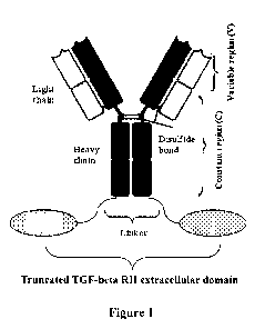
In a preferred embodiment of the present invention, wherein the fusion protein
comprising TGF-(3 receptor is as shown in the general formula (I):
Ab-L- TGF-13R11 ECD (I)
wherein the TGF-13R11 ECD is a truncated form of the extracellular domain of
TGF-I3RII;
Ab is an antibody;
L is a linker.
In a preferred embodiment of the present invention, wherein the linker is
(G4S),,G,
wherein x is 3-6, preferably is 4-5.
4
CA 03061791 2019-10-29
The present invention further provides a pharmaceutical composition,
comprising a
therapeutically effective amount of a fusion 'protein containing TGF-13
receptor as described
above, and one or more pharmaceutically acceptable carrier(s), diluent(s) or
excipient(s).
The present invention further provides DNA molecule encoding the fusion
protein
comprising TGF-13 receptor as described above.
The present invention further provides an expression vector, comprising the
DNA
molecule as described above. .
The present invention further provides a host cell transformed with the
expression vector
as described above, wherein the host cell is selected from the group
consisting of a bacterial,
yeast, and mammalian cell; preferably a mammalian cell.
The present invention further provides a use of the fusion protein containing
TGF-13
receptor as described above or the pharmaceutical composition thereof for the
preparation of a
medicament for the treatment of tumors; preferably for preparation of a
medicament for
treating a PD-Li-mediated tumor; more preferably a cancer expressing PD-Li.
The present invention further provides a method for treating or preventing a
tumor
comprising administering to a patient in need thereof a therapeutically
effective amount of the
fusion protein containing TGF-P receptor as described above.
The present invention further provides, a truncated extracellular domain of
TGF-PRII,
wherein the truncated extracellular domain of TGF-13RII invovles a deletion of
26 or fewer
contiguous amino acids at the N-terminus of SEQ ID NO: 13, preferably a
deletion of 14-26
contiguous amino acids at the N-terminus, more preferably a deletion of 14-21
contiguous
amino acids at the N-terminus; the non-limiting examples of the truncated
extracellular
domain of TGF-PRII comprises sequence shown as SEQ ID NO: 14 or SEQ ID NO: 15.
The present invention further provides a pharmaceutical composition,
comprising a
therapeutically effective amount of a truncated extracellular domain of TGF-
PRII of the
present invention, and one or more pharmaceutically acceptable carrier(s),
diluent(s) or
excipient(s).
The present invention further provides a use of the truncated extracellular
domain of
TGF-PRII of the present invention or a pharmaceutical composition thereof for
the
preparation of a medicament for the treatment or inhibition of diseases or
disorders associated
with cancer cell proliferation or metastasis.
The present invention further provides a method for treating or preventing a
tumor
comprising administering to a patient in need thereof a therapeutically
effective amount of the
CA 03061791 2019-10-29
truncated extracellular domain of TGF-I3RII of the present invention or the
pharmaceutical
composition thereof.
The tumor or cancer described in the present disclosure is selected from the
group
consisting of colorectal, breast, ovary, pancreas, stomach, prostate, kidney,
cervix, myeloma,
lymphoma, leukemia, thyroid, endometrium; uterus, bladder, neuroendocrine,
head and neck,
liver, nasopharynx, testis, small cell lung cancer, non-small cell lung
cancer, melanoma, basal
cell skin cancer, squamous cell skin cancer, dermatofibrosarcoma protuberans,
Meck Cell
carcinoma, glioblastoma, glioma, sarcoma, mesothelioma, and myelodysplastic
syndrome.
DRAWING DESCRIPTION
Figure 1: Schematic diagram of the structure of the fusion protein.
Figure 2: Results showing the binding of fusion protein to human TGF-131 in
vitro.
Figure 3: Results showing the binding of fusion protein to human TGF-131 in
vitro.
Figure 4: Results showing the binding of fusion protein to human PD-Li in
vitro.
Figure 5: Result showing the detection of PD-1/PD-Li pathway blocking by
fusion
protein in vitro.
Figure 6: Fusion protein inhibits TGFI3-induced pSMAD3 reporter activity in a
dose-dependent manner.
Figure 7: All fusion protein samples enhance the secretion of the cytokine IFN-
7 by
activated T lymphocytes.
Figure 8: Effect of fusion protein on tumor weight of tumor-bearing mice.
DETAILED DESCRIPTION OF THE INVENTION
TERMS
For the invention to be more readily understood, certain technical and
scientific terms are
specifically defined below. Unless specifically defined elsewhere herein, all
other technical
and scientific terms used herein have the meaning commonly understood by one
of ordinary
skills in the art to which this invention pertains.
As used herein, the single-letter code and the three-letter code for amino
acids are as
described in J. Biol. Chem, 243, (1968) p3558.
6
CA 03061791 2019-10-29
As used herein, "antibody" refers to immunoglobulin, a four-peptide chain
structure
formed by two identical heavy chains and to identical light chains connected
by inter-chain
disulfide bond. Different immunoglobulin heavy chain constant regions exhibit
different
amino acid compositions and sequences, hence present different antigenicity.
Accordingly,
immunoglobulins can be divided into five categories, also referred as
immunoglobulin
isotypes, namely IgM, IgD, IgG, IgA and IgE; the corresponding heavy chains
thereof are la
chain, 6 chain, y chain, a chain, e chain, respectively. According to amino
acid composition of
hinge region and the number and location of heavy chain disulfide bonds,
immunoglobulins
can be divided into different sub-categories, for example, IgG can be divided
into IgG1 , IgG2,
IgG3, and IgG4. Light chain can be divided into K or X chain, based on
different constant
region. Each category of Ig among these five categories involves ic or X
chain.
In the present invention, the antibody light chain mentioned herein further
comprises a
light chain constant region, which comprises a human or murine K, X chain or a
variant
thereof
In the present invention, the antibody heavy chain mentioned herein further
comprises a
heavy chain constant region, which comprises human or murine IgG1 , IgG2,
IgG3, IgG4 or a
variant thereof.
At the N-terminal of the antibody heavy and light chains, about 110 amino
acids vary
largely, which is known as variable region (Fv region); the amino acid
sequence at the
C-terminus is relatively stable, which is known as constant region. Variable
region comprises
three hypervariable regions (HVR) and four FR regions (FR) with relatively
conserved
sequence. Three hypervariable regions determine the specificity of the
antibody, also known
as complementarity determining region (CDR). Each light chain variable region
(LCVR) and
each heavy chain variable region (HCVR) is composed of three CDR regions and
four FR
regions, arranged from the amino terminal to the carboxyl terminal: FR1, CDR1,
FR2, CDR2,
FR3, CDR3, and FR4. Three light chain CDR regions refer to LCDR1, LCDR2, and
LCDR3;
three heavy chain CDR regions refer to HCDR1, HCDR2 and HCDR3. The number and
7
CA 03061791 2019-10-29
=
location of CDR region amino acid residues in LCVR and HCVR regions of the
antibody or
antigen binding fragment herein comply with known Kabat numbering criteria
(LCDR1-3,
HCDR2-3), or comply with kabat and chothia numbering criteria ( HCDR1).
The antibody of the present invention comprises full-length antibody selected
from the
group consisting of murine antibody, chimeric antibody and humanized antibody,
preferably is
humanized antibody.
The term "murine antibody" in the present invention refers to anti-human PD-Li
monoclonal antibody prepared according to the knowledge and skills in the
field. During the
preparation, test subject was injected with PD-Li antigen, and then hybridoma
expressing
antibody which possesses desired sequence' or functional characteristics was
isolated. In a
preferred embodiment of the present invention, the murine PD-Li antibody or
antigen binding
fragment thereof, further comprises light chain constant region of murine K,
X. chain or a
variant thereof, or further comprises heavy chain constant region of murine
IgG1 , IgG2, IgG3
or a variant thereof.
The term "chimeric antibody", is an antibody which is formed by fusing the
variable
region of a murine antibody with the constant region of human antibody, so as
to alleviate the
murine antibody-induced immune response. To establish a chimeric antibody, a
hybridoma
secreting specific murine monoclonal antibody is first established, variable
region genes are
then cloned from murine hybridoma cells, and then constant region genes of
human antibody
are cloned as desired, the murine variable region genes are ligated with human
constant region
genes to form a chimeric gene which can be inserted into a human vector, and
finally the
chimeric antibody molecule is expressed in a eukaryotic or prokaryotic
industrial system. In a
preferred embodiment of the present invention, the light chain of the PD-Li
chimeric
antibody further comprises the light chain constant regions derived from human
lc, X chain or
a variant thereof. The heavy chain of PD-Li chimeric antibody further
comprises the heavy
chain constant region(s) derived from human IgGI, IgG2, IgG3, IgG4 or a
variant thereof.
The constant region(s) of human antibody can be selected from heavy chain
constant region(s)
derived from human IgGl, IgG2, IgG3, IgG4 or a variant thereof, preferably
comprises heavy
chain constant region derived from human IgG2 or IgG4, or IgG4 without ADCC
(antibody-dependent cell-mediated cytotoxicity) after amino acid mutation.
The term "humanized antibody", also known as CDR-grafted antibody, refers to
an
8
CA 03061791 2019-10-29
antibody generated by murine CDR sequences grafted into human antibody
variable region
framework, i.e. antibody generated from different types of sequences of human
germline
antibody framework. Humanized antibody conquers the disadvantageously strong
anti-antibody response induced by chimeric antibody which carries a large
number of murine
components. Such framework sequences can be obtained from public DNA database
covering
germline antibody gene sequences or published references. For example,
germline DNA
sequences of human heavy and light chain variable region genes can be found in
"VBase"
human germline sequence database (available on web
www.mrccpe.com.ac.uk/vbase), as well
as found in Kabat, EA, et al.1991 Sequences of Proteins of Immunological
Interest, 5th Ed. To
avoid the decrease in activity caused by reduction of immunogenicity, the
variable region
framework of the human antibody is subjected to minimum back-mutation to
maintain the
activity. The humanized antibody of the present invention also comprises a
humanized
antibody which is further obtained by phage display for the purpose of CDR
affinity
maturation.
The terms "human antibody" and "antibody from human" are used interchangeably
to
mean that one or more variable and constant regions are derived from a human
immunoglobulin sequence. In a preferred embodiment, all of the variable and
constant regions
are derived from human immunoglobulin sequences, i.e., " antibodies fully
derived from
human" or "fully human antibodies." These antibodies can be obtained in a
variety of ways,
including by phage display technology; isolation of B cells from human PBMC,
spleen or
lymph nodes; construction of a native single-stranded phage human antibody
library; or by
immunization of transgenic mice that express human antibody light and heavy
chains; and
screening thus obtained antibodies. .
As used herein, "antigen-binding fragment" or "functional fragment" refers to
Fab
fragment, Fab' fragment, F(ab')2 fragment with antigen-binding activity, as
well as Fv
fragment scFv fragment binding with human PD-Ll. Fv fragment is the minimum
antibody
fragment which involves all antigen-binding sites, Fv fragment comprises a
heavy chain
variable region and a light chain variable region, but without a constant
region. Generally, Fv
antibody further comprises a polypeptide linker between the VH and VL domains
to form a
structure required for antigen binding. Also, different linkers can be used to
connect the
variable regions of two antibodies to form a polypeptide, named single chain
antibody or
9
CA 03061791 2019-10-29
single chain Fv (scFv). As used herein, the term "binding with PD-Li" means
the ability to
interact with human PD-Li. As used herein, the term "antigen-binding site" of
the present
invention refers to discontinuous, three-dimensional sites on the antigen,
recognized by the
antibody or the antigen-binding fragment of the present invention.
As used herein, the term "ADCG", namely antibody-dependent cell-mediated
cytotoxicity, refers to the cells expressing Fc receptors that directly kill
the target cells coated
by an antibody by recognizing the Fc segment of the antibody. ADCC effector
function of the
antibody can be reduced or eliminated by modifying the Fc segment in IgG. The
modification
refers to mutations on the antibody heavy chain constant region, such as
mutations selected
from N297A, L234A, L235A in IgGl; IgG2/4 chimera; or F234A/L235A mutations in
IgG4.
"Mutation" in the "mutant sequence" of the present invention includes, but is
not limited
to "back mutation", "conservative modification" or "conservative replacement
or
substitution". "Conservative modification" or "conservative replacement or
substitution" in
the present disclosure refers to substitutions of amino acids in a protein
with other amino
acids having similar characteristics (e.g. charge,
side-chain size,
hydrophobicity/hydrophilicity, backbone conformation and rigidity, etc.), such
that the
changes can frequently be made without altering the biological activity of the
protein. Those
of skilled in the art recognize that, in general, single amino acid
substitution in non-essential
region of a polypeptide does not substantially alter biological activity (see,
e.g., Watson et al.
(1987) Molecular Biology of the Gene, The Benjamin/Cummings Pub. Co., p. 224
(4th Ed)).
In addition, substitutions of structurally or functionally similar amino acids
are less likely to
disrupt biological activity.
The "mutant sequence" as used in the present invention means that the
nucleotide
sequence and the amino acid sequence of the present invention are subjected to
substitution,
insertion or deletion, thus the obtained nucleotide sequence and amino acid
sequence share
varying percentage identity with the nucleotide sequence and the amino acid
sequence of the
present invention.
As used herein, "identity" indicates the degree of similarity between two
nucleic acids or
two amino acid sequences. The sequence identity in the present invention is at
least 85%, 90%
or 95%, preferably at least 95%. Representative examples include, but are not
limited to, 85%,
86%, 87%, 88%, 89%, 90%, 91%, 92%, 93%, 94%, 95%, 96%, 97%, 98%, 99%, 100%.
The
CA 03061791 2019-10-29
comparison of sequences and determination of percent identity between two
sequences can be
accomplished using the default settings of the BLASTN/BLASTP algorithm
available on the
National Center for Biotechnology Institute's .website.
The "PD-Li antibody or antigen-binding protein thereof' of the present
invention could
include any of the anti-PD-Li antibodies or antigen-binding fragments thereof
described in
the art. The anti-PD-Li antibody may be a PD-Li antibody which is commercially
available
or has been disclosed in the literature, including, but not limited to, PD-Li
antibody
BMS-936559, MPDL3280A, MEDI4736, MSB0010718C (see US2014341917,
US20130034559, US8779108) and the like. The antibody may be a monoclonal
antibody, a
chimeric antibody, a humanized antibody, or a human antibody. The antibody
fragment
includes Fab fragment, Fab' fragment, F(ab')2 fragment having antigen-binding
activity, and
Fv fragment and scFv fragment which bind to the antigen.
As an exemplary PD-Li antibody preparation process of the present invention,
see
PCT/CN2016/104320, the PD-Li antibody comprises CDRs of heavy chain variable
regions
as described below:
HCDR1: SYWMH SEQ ID NO: 1
HCDR2: RI XIPNSG X2TSYNEKFKN SEQ ID NO: 2
HCDR3: GGSSYDYFDY SEQ ID NO: 3.
In an alternative embodiment, Xi is selected from H or G; and X2 is selected
from G or F.
In another embodiment, an exemplary PD-Li antibody of the invention further
comprises CDRs sequence of a light chain variable region as described below:
LCDR1: RASESVSIHGTHLMH SEQ ID NO: 4
LCDR2: AASNLES SEQ ID NO: 5
LCDR3: QQSFEDPLT SEQ ID NO: 6.
In another embodiment, the above CDR regions are humanized by CDR grafting,
and the
FR of humanized light chain templates are IGKV7-3*01 and hjk2.1, the FR of
humanized
heavy chain templates are IGHV1-46*01 and hjh6.1, and the humanized variable
region
sequences are as follows:
humanized heavy chain variable region: =
11
CA 03061791 2019-10-29
QVQLVQSGAEVKKPGASVKVSCKASGYTFTSYWMHWVRQAPGQGLEWMGRIXIP
NSGNITSYNEKFKNRV7'ATIRDTSTSTVYMELSSLRSEDTAVYYCARGGSSYDYFDY
WGQGTTV7'VSS
SEQ ID NO: 7
humanized light chain variable region:
DIVLTQSPASLAVSPGQRA T/TCRASESYSIHOTHLMEIWYQQKPGQPPKLLIYAASN
LESGVPARFSGSGSGTDFTLTINPVEANDTANYYCOQSFEDPLIFGQGTKLEIK
SEQ ID NO: 8
NOTE: The order is FR1-CDRI-FR2-CDR2-FR3-CDR3-FR4, italic portion represents
FR sequence, and the underlined portion represents CDR sequence.
In another embodiment, back mutation design on the humanized antibody of the
present
invention was performed, see the table as follows:
Table 1
VL VH
VL. 1 grafted VH.1 grafted
VL. 1A Y91 F VH, 1 A T74K
VL.1B Y91 F,G72E VH.1B T74K, R72V, M48I, M7OL
VL. 1 C Y91 F,G72E, 122S VH.1C T74K, R72V, M48I, M7OL, R38Q
VH.10 T74K, R72V, M48I, M7OL, R38Q, L83F
VH.1 E T74K, R72V, M48I, M7OL, R380,L83F, V68A,
V79A
Note: For example, Y91F indicates a back-mutation from Y to F at position 91
according
to Kabat numbering system.
"Grafted" indicates that the murine antibody CDR was implanted into human
germline
FR sequences.
New humanized antibody can be obtained by various combination of mutations in
heavy
chain and light chain shown in the above table.
In another aspect of the invention, an embodiment for construction of a
humanized clone
is provided, as follows:
Primers were designed, and VHNK gene fragments of each humanized antibody were
constructed by PCR and then inserted into the expression vector pHr (with
signal peptide and
constant region gene (CH1-Fc/CL) fragment) to perform homologous
recombination, in order
to construct a full-length antibody expression vector: VH-CH1-Fc-pHr/VK-CL-
pHr.
1. Primer Design:
The online software DNAWorks (v3.2.2) (http://helixweb.nih.gov/dnaworks/) was
used
12
CA 03061791 2019-10-29
to design multiple primers for synthesis of VH/VK containing gene fragments
required for
recombination: 5'-30bp signal peptide + VH/VK + 30bp CH1/CL-3
2. Fragment splicing:
According to operation instructions for Primer STAR GXL DNA polymerase from
TaKaRa Company, using the primers designed above, VH/VK containing gene
fragments
required for recombination was obtained by two-step PCR amplification.
3. Construction of expression vector pHr (with signal peptide and constant
region gene
(CH1-FC/CL) fragment) and enzymatic digestion:
The expression vector pHr (with signal peptide and constant region gene (CH1-
FC/CL)
fragment) was designed and constructed by using some special restriction
endonuclease, such
as BsmBI which recognizes the distinctive feature between the sequence and
restriction site.
BsmBI digested the vector, and then the digested fragments were extracted by
using gel and
stored for use.
4. Recombinant construction of expression vector VH-CH1-Fc-pHrNK-CL-pHr
VH/VK containing gene fragments required for recombination and expression
vector
pHr (with signal peptide and constant region gene (CH1-FC/CL) fragment) that
has been
digested with BsmBI were added into DH5H competent cells at a ratio of 3:1,
incubated at 0
C on ice for 30min, heat-shocked at 42 C for 90s, combined with 5 volumes of
LB medium,
incubated at 37 C for 45min, plated on LB-Amp plate, and cultured at 37 C
overnight.
Single clone was picked for sequencing and a clone of interest was obtained.
5. The plasmid was constructed according to the design of the present example,
then the
purified protein, and the affinity of the obtained protein was measured by the
detection
described in Example SPR.
6. Finally, the affinity of the humanized back-mutation mutant or hybridoma
antibodies
to human PD-Li-his was measured by BIACORE, the obtained humanized back-
mutation
sites and sequence combination through screening are as follows:
heavy chain variable region:
QVQLVQSGAEVKKPGASVKVSCKASGY7'FTSYWMH WVRQAPGQGLE WMGRI,QPN
SGETSYNEKFKNR VTMTRDTSTSTVYMELSSLRSEDTAVYYCARGG S SY D YFD Y WG
QGTTVTVS'S
SEQ ID NO: 9
wherein CDR2 is a sequence in which Xi of SEQ ID NO: 7 is G and X2 is F.
13
CA 03061791 2019-10-29
light chain variable region:
DIVLTQSPASLAVSPGQRATITCRASESVS1HGTHLMHWYQQKPGQPPKLLIYAASN
LESGVPARF'SGSGSGTDFTLTINPVEAEDTANYYMOSFEDPLTFGQGTKLEIK
SEQ ID NO : 10
NOTE: The order is FR1-CDR1-FR2-CDR2-FR3-CDR3-FR4, italic portion represents
FR sequence, and the underlined portion represents CDR sequence.
In another aspect of the present invention, an embodiment for constructing and
expressing an anti-PD-Li human IgG4 type antibody is provided, and further
provided is a
PD-Li antibody used for fusion protein construction. The PD-Li antibody can
also be used as
a control molecule in the test examples of the present invention.
Since PD-Li is also expressed in activated T cells, therefore the use of wild-
type IgG1
constant regions can cause Fc-mediated effects such as ADCC and CDC, which
could result
in the reduction of activated T cells. The present invention selects mutated
IgG4 to obtain
antibodies without ADCC and CDC. The clone obtained by affinity maturation was
converted
into IgG4 type, and the core hinge region of IgG4 contained 5228P mutation,
and F234A and
L23 5A mutations were further introduced (mAbs 4:3, 310-318; May/June 2012).
At the same
time, in order to avoid breakage occurred at the C-terminus of the antibody
heavy chain when
the linker peptide (which is used to link the extracellular domain of TGF-
13RII) was
introduced, the last amino acid K of the PD-Li antibody heavy chain was
further mutated to A,
so as to increase the stability of the fusion protein. The PD-Li antibody
sequence of the
present invention used for fusion protein construction is as follows:
PD-Li antibody heavy chian: IgG4(AA). (S228P)
OVOLVOSGAEVKKPGASVKVSCKASGYTFTSYWMHWVRQAPGOGLEWMGRI
GPNSGFTSYNEKFKNRVTMTRDTSTSTVYMELS SLR SEDT AVYYCA R GGSSYD
YF DYW GQGTTVTV S S ASTKGPSVF PLAPC SRSTSE STAALG CLVKDYFPEPVTV
SWN SGALTSGVHTFPAVLQSSGLYSLSSV VTVPSSSLGTKTYTCNVDHICPSNTK
VDKRVESKY GPPCPPCPAPEAAGG PS VFLFPPKPKDTLMISRTPEVTCV V VD VS
QEDPEVQFNW YVDGVEVHNAKTKPREEQFNSTYRVVSVLTVLHQDWLNGKE
YKCKVSNKGLPSSIEKTISKAKGQPREPQVYTLPPSQEEMTKNQVSLTCLVKGF
YPSD1A VE W ESNG QPENNYKTTPP VLDSDG SFFLYSRLTVDKSRWQEGN VFSCS
VMHEALHNH YTQKSLSLSLG A
SEQ 1D NO : 11
PD-L1 antibody light chian:
14
CA 03061791 2019-10-29
DIVLTOSPASLAVSPGORAT1TCRASESVSIHGTHLMHWYOOKPGOPPKWYA
ASNLESGVPARFSGSGSGTDFILTINPVEAEDTANYYCOOSFEDPLTFGOGTKLE
IKRT VAAPSVF1F PPSDEQLKS GTAS VVCLLNNFYPREAKVQWKVDNALQSGN S
QESVTEQDSKDSTYSLSSTLTLSKADYEKTIKVYACEVTHQGLSSPVTKSFNRGE
SEQ ID NO: 12
NOTE: The underlined portion is a variable region sequence of the antibody
heavy or
light chain, or the encoding nucleotide sequence thereof; The remaining
portion is antibody
constant region sequence and the encoding nucleotide sequence thereof.
As used herein, a fusion protein described in the present invention is a
protein product
obtained by co-expressing two genes via DNA recombination technology. Methods
for
producing and purifying antibodies and antigen-binding fragments are well
known in the art
and can be found, for example, in Antibodies, A Laboratory Manual, Cold Spring
Harbor,
chapters 5-8 and 15. For example, mice can be immunized with human PD-Li or
fragments
thereof, and the resulting antibodies can then be renatured, purified, and
sequenced for amino
acid sequences by using conventional methods well known in the art. Antigen-
binding
fragments can also be prepared by conventional methods. The antibody or
antigen binding
fragments of the present invention are engineered to graft CDRs derived from
non-human
antibody into one or more human FRs. By aligning against the database of IMGT
human
antibody variable region germline using 'MOE software, human framework
germline
sequences can be obtained from ImMunoGeneTics (IMGT) website
http://imgt.cines.fr, or
from The Immunoglobulin Facts Book, 2001, ISBN 012441351.
The engineered antibodies or antigen binding fragments of the present
invention may be
prepared and purified using known methods. For example, cDNA sequences
encoding a heavy
chain and a light chain may be cloned and engineered into a GS expression
vector. The
engineered immunoglobulin expression vector may then be stably transfected in
CHO cells.
As a more recommended method known in the art, mammalian expression system
will result
in glycosylation of antibody, typically at highly conserved N-terminal sites
in the Fe region.
Stable clones may be obtained by expression of an antibody specifically
binding to human
PD-Li. Positive clones may be expanded in serum-free culture medium for
antibody
production in bioreactors. Culture medium, into which the antibody has been
secreted, may be
purified by conventional techniques. For example, the medium may be loaded
onto a Protein
CA 03061791 2019-10-29
A or G Sepharose FF column that has been equilibrated with a compatible
buffer. The column
is washed to remove nonspecific binding components. The bound antibody is
elutedby pH
gradient and antibody fractions are detected by SDS-PAGE, and then collected.
The antibody
may be filtered and concentrated using common techniques. Soluble aggregate
and multimers
may be effectively removed by common techniques, including size exclusion or
ion exchange.
The product may be immediately frozen, for example at -70 C, or may be
lyophilized.
The "immunomodulatory molecule" of the present invention can be used to
attenuate the
immune tolerance of cancer cells. The present invention uses a truncated form
of the
extracellular domain of TGF-13R11 as the immunomodulatory molecule in the
fusion protein.
"TGF-f3 receptor II (TGF-13R11)" binds ligands TGF-P1 and 3 with high
affinity. The TGF-f3
RH/ TGF-13 complex recruits TGF-f3 RI to form a signal transduction complex
(Won et al,
Cancer Res. 1999; 59: 1273-7). The extracellular domain of TGF-PRII is a 136
amino acid
residue peptide from the N-terminus of TGF-PRII extracellular, an exemplary
example of
which is shown in SEQ ID NO: 13. Other variants of about 136 amino acids in
length and
derived from human extracellular domain of TGF-PRII, which capable of binding
to TGF-131
and 3, also belong to the extracellular domain of TGF-PRII of the invention.
The present
invention has found that the structure and function of the N-terminal
contiguous truncated
form of the TGF-PRII extracellular domain is more stable than that of the
untruncated
molecule. A fusion protein comprising the N-terminal untruncated form of TGF-
PRII
extracellular domain (a polypeptide shown as aa.1-136 of SEQ ID NO: 13) is
susceptible to
cleavage. In particular, a truncated form comprising a deletion of at most 26
amino acids at its
N-terminus is more stable, preferably a truncation of 14-26 amino acids, more
preferably a
truncation of 14-21 amino acid at N-terminus with a higher expression level,
most preferably,
a truncation of 19 or 21 contiguous amino acids at N-terminus.
"Administration" and "treatment," as it applies to an animal, human,
experimental
subject, cell, tissue, organ, or biological fluid, refers to contact of an
exogenous
pharmaceutical, therapeutic, diagnostic agent, or composition to the animal,
human, subject,
cell, tissue, organ, or biological fluid. "Administration" and "treatment" can
refer, e.g., to
therapeutic, pharmacokinetic, diagnostic, research and experimental methods.
Treatment of a
cell encompasses contact of a reagent to the cell, as well as contact of a
reagent to a fluid,
where the fluid is in contact with the cell. "Administration" and "treatment"
also mean in vitro
16
CA 03061791 2019-10-29
and ex vivo treatments, e.g., of a cell, by a reagent, diagnostic, binding
compound, or by
another cell. "Treatment," as it applies to a human, veterinary, or research
subject, refers to
therapeutic treatment, prophylactic or preventative measures, to research and
diagnostic
applications.
"Treat" means to administer a therapeutic agent, such as a composition
containing any of
the binding compounds of the present invention, internally or externally to a
patient having
one or more disease symptoms for which the agent has known therapeutic
activity. Typically,
the agent is administered in an amount effective to alleviate one or more
disease symptoms in
the patient or population to be treated, to induce the regression of or
prevent the progression
of such symptom(s) from clinically measurable degree. The amount of a
therapeutic agent that
is effective to alleviate any particular disease symptom (also referred to as
the "therapeutically
effective amount") may vary according to factors such as the disease state,
age, and weight of
the patient, and the ability of the drug to elicit a desired response in the
patient. Whether a
disease symptom has been alleviated can be assessed by any clinical
measurement typically
used by physicians or other skilled healthcare providers to assess the
severity or progression
status of the symptom. Although an embodiment of the present invention (e.g.,
a treatment
method or article of manufacture) may not be effective in alleviating the
target disease
symptom(s) in every patient, it should alleviate the target disease symptom(s)
in a statistically
significant number of patients as determined by any statistical test known in
the art such as
the Student's t-test, the chi-square test, the U-test according to Mann and
Whitney, the
Kruskal-Wallis test (H-test), Jonckheere-Terpstra-test and the Wilcoxon-test.
"Conservative modifications" or "conservative substitution" refers to
substitutions of
amino acids in a protein with other amino acids having similar characteristics
(e.g. charge,
side-chain size, hydrophobicity/hydrophilicity, backbone conformation and
rigidity, etc.), such
that the changes can frequently be made without altering the biological
activity of the protein.
Those of skill in this art recognize that, in general, single amino acid
substitution in
non-essential region of a polypeptide do not substantially alter biological
activity (see, e.g.,
Watson et al. (1987) Molecular Biology of the Gene, The Benjamin/Cummings Pub.
Co., p.
224 (4th Ed.)). In addition, substitutions of structurally or functionally
similar amino acids are
less likely to disrupt biological activity.
"Effective amount" encompasses an amount sufficient to ameliorate or prevent a
symptom or sign of the medical condition. Effective amount also means an
amount sufficient
to allow or facilitate diagnosis. An effective amount for a particular patient
or veterinary
17
CA 03061791 2019-10-29
subject may vary depending on factors such 'as the condition being treated,
the overall health
condition of the patient, the route and dose of administration and the
severity of side effects.
An effective amount can be the maximal dose or dosing protocol that avoids
significant side
effects or toxic effects.
"Exogenous" refers to substances that are produced outside an organism, cell,
or,human
body, depending on the context. "Endogenous" refers to substances that are
produced within a
cell, organism, or human body, depending on the context.
"Homology" refers to sequence similarity between two polynucleotide sequences
or
between two polypeptide sequences. The molecules are deemed as homologous at
one
position, when this position in both of the sequences to be compared is
occupied by the same
base or amino acid monomer subunit, e.g., when a position in each of two DNA
molecules is
occupied by adenine. The percent of homology between two sequences is a
function of the
number of matching or homologous positions shared by two sequences divided by
the number
of all positions to be compared and then multiplied byl 00. For example, in an
optimal
alignment, if 6 of 10 positions in two sequences are matched or homologous,
then the two
sequences share 60% homology. Generally, the comparison is made when two
sequences are
aligned to give maximum percent homology..
"Immune checkpoint molecules" include stimulatory immune checkpoint molecule
and
inhibitory immune checkpoint molecule, and exemplary molecules include CD27,
CD28,
CD40, CD4OL, CD122, 0X40, OX4OL, GITR, ICOS, AZAR, B7-H3, B7-H4, BTLA,
CTLA-4, IDO, KIR (Killer-cell Immunoglobulin-like Receptor), LAG3, PD-1, PD-
L1, PD-L2,
TIM-3, VISTA, etc.
As used herein, the expressions "cell," "cell line," and "cell culture" are
used
interchangeably and all such designations include progeny thereof. Thus, the
words
"transformant" and "transformed cell" include the primary subject cells and
cultures derived
therefrom, regardless of the number of passage. It should be also understood
that all progeny
may not be precisely identical in the aspect of DNA content, due to
intentional or
unintentional mutations. Mutant progeny that have the same function or
biological activity as
that of the originally transformed cells are obtained by screening and shall
be included in the
invention. Where distinct designations are intended, it will be clearly
understood from the
context.
As used herein, "polyrnerase chain reaction" or "PCR" refers to a procedure or
technique
in which small amount of specific segments of nucleic acid, RNA and/or DNA are
amplified
18
CA 03061791 2019-10-29
as those described in, e.g., U.S. Pat. No. 4,683,195. Generally, sequence
information at the
ends of or beyond the region of interest is needed, such that oligonucleotide
primers can be
designed; the sequence of these primers will be identical or similar to the
opposite strand of
the template to be amplified. The 5' terminal 'nucleotides of the two primers
coincide with the
ends of the material to be amplified. PCR can be used to amplify specific RNA
sequences,
specific DNA sequences from total genomic DNA, and cDNA transcribed from total
cellular
RNA, bacteriophage or plasmid sequences, etc. See generally Mullis et al.
(1987) Cold Spring
Harbor Symp. Ouant. Biol. 51:263; Erlich, ed., (1989) PCR TECHNOLOGY (Stockton
Press,
N.Y.). The PCR used in the present invention is considered to be one, but not
the only,
example of polymerase reaction method for amplifying a nucleic acid test
sample. The
method comprises the use of a known nucleic acids as primers and nucleic acid
polymerase to
amplify or generate a specific segment of nucleic acid.
"Optional" or "optionally" means that the event or situation that follows may
occur, but
not necessarily, and the description includes the instances in which the event
or circumstance
does or does not occur. For example, "optionally contains 1-3 antibody heavy
chain variable
region(s)" means the antibody heavy chain variable region with specific
sequence can be
present, but not necessarily.
"Pharmaceutical composition" refers to a mixture comprising one or more
compounds
according to the present invention or physiologically/pharmaceutically
acceptable salt or
prodrug thereof and other chemical components, said chemical components are
such as
physiologically/pharmaceutically acceptable carrier(s) and excipient(s). The
pharmaceutical
composition aims at promoting the administration by an organism, facilitating
the absorption
of the active ingredient and thereby exerting biological effect.
EXAMPLES AND TEST EXAMPLES
=
Hereinafter, the present invention is further described with reference to
examples.
However, the scope of the present invention is not limited thereto.
In the examples of the present invention, where specific conditions are not
described, the
experiments are generally conducted under conventional conditions or under
conditions
proposed by the material or product manufacturers. See Sambrook et al.,
Molecular Cloning,
Laboratory Manual, Cold Spring Harbor Laboratory; Modern Molecular Biology
Methods,
Ausubel et al., Greene Publishing Association, Wiley Interscience, NY. Where
the source of
19
CA 03061791 2019-10-29
the reagents is not specifically indicated, the reagents are commercially
available
conventional reagents.
EXAMPLES
Example 1: Fusion protein PD-L1/TGF-13 trap cloning and expression
The TGF-13RII extracellular domain (full length or truncated form of SEQ ID
NO: 13) is
used as the portion for immunomodulatory 'molecule in the fusion protein, and
the PD-Li
antibody is used as a targeting portion of the fusion protein to form a PD-Li
antibody/
TGF-13R11 extracellular domain fusion protein (PD-L1 /TGF-f3 trap). Studies
have found that
the truncated form of the extracellular domain of TGF-f3RII is relatively
stable, especially the
truncated form involves the deletion of less than 26 amino acids at its N-
terminus, preferably
a deletion of 14-26 amino acids, more preferably a deletion of 14-21
contiguous amino acids,
which exhibits higher expression and stable structure; more preferably a
deletion of 14, 19 or
21 contiguous amino acids. The sequences of the non-limiting examples of the
TGF-f3RII
extracellular domain and its truncated form of the invention are as follows:
Sequence of TGF-13R11 extracellular domain: ECD (1-136)
IPPHVQKSVNNDMIVTDNNGAVKFPQLCKFCDVRFSTCDNQKSCMSNCSIT
SICEKPQEVCVAVWRKNDENITLETVCHOPKLPYHDFILEDAASPKCIMKEKKK
PGETFF MC SC SSDECNDNIIFSEE YNTSNPD
SEQ ID NO: 13
Sequence of truncated TGF-f3RII extracellular domain which involves a deletion
of 19
contiguous amino acids at N-terminus: ECD (20-136)
G AVKFPQL CKFC DVRFSTCDNQK SC MSN C SITS] CEKPQEVCVAVWRKNDE
NITLETVCHDPKLPYHDFILEDAASPKCIMKEKKKPGETFTMCSCSSDECNDNIIF
SEEYNTSNPD
SEQ ID NO: 14
Sequence of truncated TGF-f3RII extracellular domain which involves a deletion
of 21
contiguous amino acids at N-terminus: ECD (22-136):
VKFPQ LC KFCDV RF STCDNQ KSCM SNC SITS IC EK PQEVCVAVWR KNDENIT
LETVCHDPKLPYHDFILEDAASPKCIMKEKKKPGETFFM CSCSSDECNDNIIFSE
EYNTSNPD
SEQ ID NO: 15
Sequence of truncated TGF-13R11 extracellular domain which involves a deletion
of 14
'20
CA 03061791 2019-10-29
contiguous amino acids at N-terminus: ECD (15-136):
VIDNNGAVKFPQLCKFCDVRFSTCDNQKSCMSNCSITSICEKPOEVCVAVW
RKNDENITLETVCHDPKLPYHDFILEDAASPKCIMKEKKKPGETFFMCSCSSDEC
NDNIIFSEEYNTSNPD
SEO ID NO: 16
The heavy chain C-terminal amino acid of the PD-Li antibody of the present
invention
was ligated by linker (G4S).G to the extracellular domain of TGF-13RII with
varing lengths by
homologous recombination technique, and was conventionally expressed by the
293
expression system together with the light chain, and the obtained fusion
proteins are shown in
Table 2:
Table 2: PD-Li antibody/ TGF-pRII extracellular domain fusion protein
the number of
contiguous amino acid
Fusion protein Sequence description
deleted at
N-terminus
=
Fusion protein 1 Ab-(G4S)4G-ECD(1-136) No deletion
Fusion protein 2 Ab-(G4S)3G-ECD(15-136) 14
Fusion protein 3 Ab-(G4S)3G-ECD(15-136, N19A) 14
Fusion protein 4 Ab-(G4S)3G-ECD(20-136) 19
Fusion protein 5 Ab-(G4S)3G-ECD(22-136) 21
Fusion protein 6 Ab-(G4S)3G-ECD(27-136) 26
Fusion protein 7 Ab-(G4S)4G-ECD(15-136) 14
Fusion protein 8 Ab-(G4S)4G-ECD(15-136, N19A) 14
Fusion protein 9 Ab-(G4S)4G-ECD(20-136) 19
Fusion protein 10 Ab-(G4S)4G-ECD(22-136) 21
Fusion protein 11 Ab-(G4S)4G-ECD(27-136) 26
Fusion protein 12 Ab-(G4S)5G-ECD(15-136) 14
Fusion protein 13 Ab-(G4S)5G-ECD(15-136, N19A) 14
Fusion protein 14 Ab-(G4S)5G-ECD(20-136) 19
Fusion protein 15 Ab-(G4S)5G-ECD(22-136) 21
Fusion protein 16 Ab-(G4S)5G-ECD(27-136) 26
Fusion protein 17 Ab-(G4S)6G-ECD(27-136) 26
21
CA 03061791 2019-10-29
Note: Ab represents PD-Li antibody of .the present invention, ECD (n-136) in
Sequence
Description represents the full-length or truncated form of the extracellular
domain of
TGF-f3RII, n represents the starting number of amino acid after experiencing
truncation of
the extracellular domain of TGF-PRII. The structure of the fusion protein of
the present
invention is shown in Figure 1; Ni 9A indicates that the amino acid at
position 19 of the
extracellular domain of TGF-PRII was mutated into A.
The nucleotide sequence encoding the PD-Li antibody, the nucleotide sequence
encoding the extracellular domain of TGF-f3RII, and the nucleotide sequence of
the linker
protein fragment ((G4S)xG) are obtained by conventional technique in the art.
The C-terminal
nucleotide of the PD-Li antibody was ligated through linker protein to the N-
terminal
nucleotide of the extracellular domain of TGF-PRII with different length by
homologous
recombination technique, and then cloned into the Phr-BsmbI vector.
Recombinant
PD-L1/TGF-f3 trap was expressed in 293 cells and purified as described in
Example 2. The
purified protein can be used in the experiments of the following examples.
Example 2: Purification of PD-Ll/TGFAI trap fusion protein
The cell culture medium was centrifuged at high speed, and the supernatant was
collected, and the first step of purification was performed by affinity
chromatography. The
chromatographic medium is Protein A or a derived filler that interacts with
Fc, such as GE's
Mabselect. The equilibration buffer was 1xPBS (137 mmol/L NaCl, 2.7 mmol/L
KC1, 10
mmol/L Na2HPO4, 2 mmol/L KH2PO4, pH 7.4). After equilibrating 5 column
volumes, the
cell supernatant was loaded for binding, and the flow rate was controlled so
that the sample
was allowed to be remained on the column for 1 min. After sample was loaded,
the column
was washed with 1 xPBS (pH 7.4) until the A280 read-out was reduced to
baseline. Then, the
column was washed with 0.1 M glycine (pH 3.0) elution buffer, and the eluted
peak was
collected according to the A280 ultraviolet absorption peak, and the collected
eluted sample
was neutralized with 1 M Tris (pH 8.5).
The neutralized eluted sample was concentrated by ultrafiltration, and then
subjected to
size exclusion chromatography, the buffer was 1xPBS, and the column was
X1(26/60
Superdex 200 (GE). The flow rate was controlled at 4 ml/min, the loading
volume was less
than 5 ml, and the target protein peak was pooled according to A280
ultraviolet absorption.
The purity of the collected protein was greater than 95% as identified by SEC-
HPLC, and was
= 22
CA 03061791 2019-10-29
verified by LC-MS. The verified sample was aliquoted for use. The PD-L1/TGF-13
trap was
obtained.
The performance and benefits of the present invention are verified by
biochemical test
methods as indicated below.
Biological activity evaluation in vitro
Test Examplel: In vitro ELISA detection of PD-Ll/TGF-p trap binding to TGF-P1
The detection process is described as follows:
a. 96-well plates were coated with 100 p1/well of human TGF-131 (8915LC, CST)
at a
concentration of 1 ug/m1 at 4 C overnight.
b. Washing 3 times with 250 pl of 1 xPBST, 250 IA of 5% milk PBSwas added for
blocking at 37 C for 2 hours.
c. Washing 3 times with 250111 of 1 xPBST, gradient dilution of PD-L1/TGF-13
trap,
TGF-13 trap and positive control were added, and incubated for 1 hour at 37
C.
d. Washing 3 times with 250 IA 1 xPBST.
e. 100 ul of Anti-human Fe antibody-HRP (1:4000) was added to each well and
incubated for 40 minutes at 37 C.
f. 100 ul of TMB was added into each well, incubated for 10 minutes at room
temperature, and the reaction was stopped by adding 100 p,1 of 1 M H2SO4.
g. The absorbance at 450 nm was mesured on a microplate reader, and the data
was
analyzed by Graphpad Prism5.
The results of binding of the fusion protein to human TGF-I31 in vitro are
shown in
Figures 2 and 3. The ELISA showed that fusion protein 1 in Table 2 did not
retain the binding
activity to human TGF-131. Mass spectrometry analysis showed that fusion
protein 1 (i.e., the
non-truncated form of extracellular domain of TGF-13RII (1-136)) was unstable,
and it was
easy to break in the heavy chain TGF-I3RII, and positive control has the same
defect. The
fusion proteins comprising the N-terminal truncated form of the extracellular
domain of
TGF13RII, such as fusion proteins 7, 9, 10, 12-15, are specific for binding to
human TGF-131.
Test Example 2: In vitro ELISA detection of PD-Ll/TGF-P trap binding to PD-Li
Antigen used for detection: PD-Li-His =
23
CA 03061791 2019-10-29
FTVTVPKDLYVVEYGSNMTIECKFPVEKQLDLAAL1VYWEMEDKNIIQFVHGEE
DLKVQHSSYRQRARLLKDQLSLGNAALQITDVKLQDAGVYRCMISYGGADYK
RITVKVNAPYNKINQRILVVDPVTSEHELTCQAEGY PKAEV1WTSSDHQVLSGK
TTTTNSKREEKLFNVTSTLRINTTTNEIFYCTFRRLDPEENHTAELVIPELPLAHPP
NEREQKLISEEDLHHHHHH
SEQ ID NO: 17
The detection process is described as follows:
a. 96-well plates were coated with 100 jtl /well of human PD-Li-His (SEQ ID
NO: 17) at
a concentration of 5 p.g/m1 at 4 C overnight.
b. Washing 3 times with 250 p,1 of 1 xPBST, 250 pi of 5% milk PBS was added
for
blocking at 37 C for 2 hours.
c. Washing 3 times with 250p,1 of 1 xPBST, gradient dilution of PD-L1/TGF-r3
trap,
PD-Li antibody as positive control were added, and incubated for 1 hour at 37
C.
d. Washing 3 times with 254,11 xPBST.
e. 1000 of Anti-human Fc antibody-HRP (1:4000) was added into each well and
incubated for 40 minutes at 37 C.
f. 100 1 of TMB as added into each well, incubated for 10 minutes at room
temperature,
and the reaction was stopped by adding 100 IA of 1 M H2504.
g. The absorbance at 450 nm was mesured on a microplate reader, and the data
was
analyzed by Graphpad Prism 5.
The results of binding of the fusion protein of the present invention to human
PD-Li in
vitro are shown in FIG.4. The ELISA showed that all fusion proteins retained
the binding
activity to human PD-Li.
Test Example 3: Blocking detection of PD-1/PD-L1 in vitro
1. Testing purpose:
In order to investigate the blocking effect of PD-Ll/TGF-P trap on PD-1/PD-L1
signaling pathway, cell-based antibody blocking experiment was performed on
cells carrying
human PD-1 and PD-Li receptor molecules which were constructed by Promaga,
respectively.
2. Testing samples
0 PD-L1 antibody: SEQ ID NO: 11, SEQ ID NO: 12;
0 Control 1 (20T-Fc): ECD(20-136)-Fc, a fusion protein comprising truncated
.24
CA 030617.91 2019-10-29
TGF-PRII extracellular domain fragment ECD (20-136) and Fe
Sequence is as follows:
GAVXFPQLCKFCDVRFSTCDNQKSCMSNCSITSICEKNEVCVAVWRKNDENIT
LETVCHDPKLPYHDFILEDAASPKCIMKEKKKPGETFFMCSCSSDECNDNIIFSE
EYNTSNPDAESKYGPPCPPCPAPEAAGGPSVFLFPPKPKDTLMISRTPEVTCVVV
DVSQEDPEVQFNWYVDGVEVHNAKTKPREEQFNSTYRVVSVLTVLHQDWLNG
KEYKCKVSNKGLPSSIEICTISKAKGQPREPQVYTLPPSQEEMTKNQVSLTCLVK
GFYPSDIAVEWESNGQPENNYKTTPPVLDSDGSFFLYSRLTVDKSRWQEGNVFS
CSVMHEALHNHYTQKSLSLSLG
SEQ 1D NO: 18;
0 Control 2 (22T-Fc): ECD(22-136)-Fc, a fusion protein comprising truncated
TGF-I3RII extracellular domain fragment ECD (22-136) and Fe
Sequence is as follows:
VKFPQLCKFCDVRFSTCDNQKSCMSNCSITSICEKPQEVCVAVWRKNDENITLET
VCHDPKLPYHDFILEDAASPKCIMKEKKKPGETFFMCSCSSDECNDNI1FSEEYN
TSNPDAESKYGPPCPPCPAPEAAGGPSVFLFPPKPKDTLMISRTPEVTCVVVDVS
QEDPEVQFNWYVDGVEVHNAKTKPREEQFNSTYRVVSVLTVLHQDWLNGKEY
KCKVSNKGLPSSIEKTISKAKGQPREPQVYTLPPSQEEMTKNQVSLTCLVKGFYP
SDIAVEWESNGQPENNYKTTPPVLDSDGSFFLYSRLTVDKSRWQEGNVFSCSVM
HEALHNHYTQKSLSLSLG
SEQ ID NO: 19:
0 Fusion protein 9, fusion protein 15;
human IgG: blank control, human. immunoglobulin obtained from mixed normal
human serum by purification using a conventional affinity chromatography
method such as
Protein A;
0 Positive control (M7824, prepared by reference patent W02015118175): PD-Li
antibody/TGF-PRII extracellular domain fusion protein;
Light chain amino acid sequence of PD-Li antibody:
QSALTQPASVSGSPGQSITISCTGTSSDVGGYNYVSWYQQHPGKAPKLM1YDVS
NRPSGVSNRFSGSKSGNTASLT1SGLQAEDEADYYCSSYTSSSTRVFGTGTKVTV
LGQPKANPTVTLFPPSSEELQANKATLVCLISDFYPGAVTVAWKADGSPVKAG
VETTKPSKQSNNKYAASSYLSLTPEQWKSHRSYSCQVTHEGSTVEKTVAPTECS
SEQ ID NO:20
H chain amino acid sequence of PD-Li antibody heavy chain/ TGF-13R11
extracellular
domain(1-136):
CA 03061791 2019-10-29
EVQLLESGGGLVQPGGSLRLSCA ASGFI'FSSYIM M WVRQAPGKGLEWVSSI Y PS
GGITFY ADTVKGRFTISRDNSKNTLYLQMN SLRAEDTAVYYCARIKLGTVTTV
DYWGQGTLVTVSSASTKGPSVFPLAPSSKSTSGGTAALGCLVKDYFPEPVTVS
WNSGALTSGVHTFPAVLQSSGLYSLSSVVTVPSSSLGTQTYICNVNHKPSNTKV
DKRVEPKSCDKTHTCPPCPAPELLGGPSVFLFPPKPKDTLMISRTPEVTCVVVDV
SHEDPEVKFNWYVDGVEVHNAKTKPREEQYNSTYRVVSVLTVLHQDWLNGK
EYKCKVSNKALPAPIEKTISKAKGQPREPQVYTLPPSREEMTKNQVSLTCLVKG
FYPSDIAVEWESNOQPENNYKTTPPVLDSDGSFFLYSKLTVDKSRWQQGNVFS
CSVMHEALHNHYTQKSLSLSPGAGGGGS6GGGSGGGGSGGGGSGIPPHVQKS
VNNDMIVTDNNGAVKFPQLCKFCDVRFSTCDNQKSCMSNCSITSICEKPQEVCV
AVWRKNDENITLETVCHDPKLPYHDFILEDAASPKCIMKEKKKPGETFFMCSCS
SDECNDNIIFSEEYNTSNPD
SEQ ID NO:21.
3. Testing process
CHO/PD-L1 cells (CS187108, Promega) were digested and resuspended in F-12
Nutrient
Mixture (Ham) complete medium. The cell density was adjusted to 4x10⁵/mL using
complete
medium according to the cell count results. The cell suspension was ransferred
to the loading
tank, added to the 96-well plate at 100 4/well using a multi-channel pipette,
and incubated at
37 C, 5% CO2 incubator for 20-24 h; The Jurkat/PD-1 (CS187102, Promega) cell
suspension
was prepared the next day, and the cells were resuspended according to the
cell count results
using assay medium, and the cell density was adjusted to 1.25 x10⁶/mL; The
cell culture plates
comprising CHO/PD-L 1 cells were taken out from the incubator, 954 of the
culture solution
was taken out per well using a multi-channel pipette, and the gradient-diluted
fusion protein,
PD-Li antibody and positive coontrol (M7824) were respectively added at 40
4/well. Then
the Jurkat/PD-1 cell suspension was transferred to a loading tank, added to
the cell culture
plate at 40 4/well, and incubated at 37 C 5% CO2 for 5-6 h. During the
incubation with
protein, the BioGloTM Reagent was taken out and allowed to return to room
temperature.
Took out the cell culture plates and placed them at room temperature for 5-10
min. Then 404
BioG1oTM Reagent was added to each well, incubated in a safety cabinet for 5-
10 mm, and
the chemiluminescence signal value was read using a multi-function microplate
reader.
4. Results
As shown in Fig. 5, just like positive control molecule, the fusion protein 9
of the present
invention was able to effectively block the binding of PD-1-expressing Jurkat
cells to
CHO/PD-L 1 cells, and took effect in a dose-dependent manner along with drug
concentration.
Fusion protein 15 has the same blocking ability as that of fusion protein 9.
26
CA 03061791 2019-10-29
=
Test Example 4: Binding affinity and kinetics detection in vitro by Biacore
The affinity of the test molecule to human or murine TGF-f31 or human PD-Li
protein
was determined by Biacore T200 (GE). The experimental procedure is described
as follows:
A certain amount of PD-L1/TGF-f3 trap' was captured with Protein A chip, and
then the
human or murine TGF-I31 (8915LC, CST) or human PD-Li (Sino Biological) was
flowed
through the surface of the chip. The reaction signal was detected in real-time
using Biacore to
obtain the association and dissociation curves. The biochip was then washed
and regenerated
with glycine-hydrochloric acid (pH 1.5, GE). The buffer solution used in the
experiment was
HBS-EP Buffer (GE). The experimental data were fitted to (1:1) Langmuir model
using
BIAevaluation version 4.1 software (GE), and the affinity values were obtained
and as shown
in Table 3.
Table 3: Affinity of fusion proteins of the invention to TGF-f31 or human PD-
Li in virto
Fusion protein* Affinity ka (1/Ms) kd (1/s) KD (M)
sample
Fusion protein 9 Human , 1.73E7 7.28E-4 4.22E-11
Fusion protein 15 TGF-131 2.69E7 6.08E-4 2.26E-11
Fusion protein 9 murine 4.33E7 1.33E-3 3.07E-11
Fusion protein 15 TGF-131 3.57E7 1.22E-3 3.42E-11
Fusion protein 9 human PD-Li 1.97E6 1.24E-4 6.31E-11
Fusion protein 15 2.00E6 1.24E-4 6.10E-11
* Fusion protein form is shown in Table 2.
The fusion protein binding activity is shown in Table 3. The results indicate
that the
fusion proteins 9 and 15 of the present invention have extremely high affinity
to human,
murine TGF-f31 and human PD-Li.
Test Example 5: SMAD3 reporter gene inhibition assay
1. Testing purpose:
In this experiment, the Smad3 binding element (SBE) with luciferase reporter
gene was
expressed in HepG2 cells to study the inhibitory effect of PD-L 1 /TGF-f3 trap
on
TGF-131-induced Smad3 activation, and the activity of PD-L1/TGF-f3 trap in
virto was
evaluated according to IC50 vaule.
2. Test Sample: fusion protein 9, positive control (M7824)
3. Testing process
27
CA 03061791 2019-10-29
HepG2 cells were cultured in MEM complete medium (GE, SH30243.01) containing
10%
FBS and passaged every 3 days. On the first day of the experiment, 25,000
cells per well were
inoculated to 96-well plates (Corning, 3903), and cultured at 37 C, 5% CO2
for 24 hours. On
the next day, the medium in the cell culture plates was discarded, and 100 ng
of 3TP-Lux
plasmid was transfected per well. The cells were further cultured at 37 C, 5%
CO2 for 24
hours. Six hours before the addition of the test sample, the complete medium
in the 96-well
plate was discarded, and 80 [LL of incomplete medium (MEM + 0.5% FBS) was
added to each
well. After 6 hours, 10 1_, of human T6F-I31 (R&D, 240-B-010) solution
prepared in
incomplete medium (final concentration of 2 ng/mL) and 10 1..iL of the test
sample (the final
concentration is 500, 50, 5, 0.5, 0.05, 0.005, 0.0005 and 0 nM) were added,
the human
TGF-f31 solvent was used as a control, and the cells were cultured at 37 C,
5% CO2 for
another 18 h. Then, 100 ti,L of the prepared luciferase substrate ONEGloTM
Luciferase Assay
system (promega, E6110) was added to each, well, and incubated at room
temperature for 10
minutes in darkness, and then the luminescent signal vaule was read using a
Victor3
multi-plate reader (Perkin Elmer). The IC50 value of the test sample was
obtained by
calculating using the data software Graphpad Prism 5Ø
Figure 6 shows that fusion protein 9 inhibits TGFp-induced pSMAD3 activity in
a
dose-dependent manner, and has comparable efficacy and IC50 (concentration
required to
inhibit 50% of maximum activity) to that of positive control M7824. The test
results of the
PD-Ll antibody showed that it had no inhibitory effect (IC50>500 nM).
Test Example 6: In vitro Detection of IFNy secretion by PBMC due to tuberculin
(TB) stimulation
1. Test purpose
To investigate the activation of T lymphocytes by PD-L1/TGF-13 trap, human
peripheral
blood mononuclear cells (PBMC) were collected and purified, and the secretion
of IFNy was
detected after stimulation with tuberculin (TB) for 5 days.
2. Test sample
0 Human IgG; PD-Ll antibody; 0 Fusion protein 9 0' control 1 (20T-Fc):
ECD(20-136)-Fc; PD-L1 antiboy+control 1 (20T-Fc).
3. Test process:
15 ml of purified fresh PBMC, about 3 x 10⁷ cells, and 20 !IL of tuberculin
was added
thereto, and cultured in an incubator for 5 days at 37 C, 5% CO2. On day 6,
the cultured cells
were collected and centrifuged, washed once with PBS and resuspended in fresh
medium with
28
CA 03061791 2019-10-29
the density adjusted to of 1 x 10⁶ cell/ml, 90111 of resuspended cells were
added into the
96-well plate. 10pL/well of different concentrations of antibodies were
separately added to
corresponding wells of the above 96-well cell culture plate, 10 1 PBS was
added in the
control and blank group, respectively. Then, the cell culture plate was
incubated in the
incubator for three days at 37 C, 5% CO2. The cell culture plate was taken
out, and the
supernatant was taken from each well after Centrifugation (4000 rpm, 10 min).
After 10-fold
dilution, the secretion of IFN-y was detected by ELISA (human IFNI, detection
kit,
Xinbosheng, EHC 102g.96), refer to the reagent instructions for specific
operations. As shown
in the figure, PD-L1/TGF-13 trap fusion protein samples were able to enhance
the secretion of
cytokine IFN-y by activated T lymphocytes, and had a drug concentration dose
effect.
= Table 4
EC50 Maximum Minimal Fold
Antibody (nM secretion of secretion of (secretion
)
IFNy (pg/ml) IFNy (pg/ml) of IFNy)
PD-Li antibody 0.05 2684 737 3.6
Fusion protein 9 0.12 3422 638 5.4
Control 1(20T-Fc) >50 , 780 490 1.6
PD-Llantibody
0.054 2879 746
+ control 1 3.9
Human IgG >50 375 298 1.2
Blank control 536 536 1
4. Result
As shown in Figure 7 and Table 4, the fusion protein 9 was able to enhance the
activated
T lymphocyte to secrete cytokine IFN-y in dose-dependent manner, and had a
stronger
activation effect than that of the PD-Li antibody and 20T-FC.
Pharmacokinetic evaluation
Test Example 7:
Three SD rats, female, were purchased from Jiesijie Experimental Animal Co.,
Ltd. and
maintained in 12/12-hour light-dark cycle (the temperature is 24 3 C, the
relative humidity is
50-60%), the rats were free access to water and diet. On the day of the
experiment, SD rats
were injected with fusion protein in the tail vein at a dose of 6 mg/kg and an
injection volume
of 5 ml/kg.
Blood was collected at time point: 15 mm, 7 h (on the first day), 24h (2nd
day), 3rd day,
4th day, 6th day, 8th day, 10th day, and 15th day after administration, 200 pl
blood (equivalent to
29
CA 03061791 2019-10-29
100 ill serum) was taken from the fundus vein of the rat. The blood sample was
placed at
room temperature for 30min to allow agglutination, and then centrifuged at
10000 g for 10
minutes at 4 C. The supernatant were taken and stored at-80 C immediately.
The
concentration of the fusion protein in the serum was measured by ELISA.
The measure process is described as follows:
a. 96-well plates were coated with 100 l/well of human PD-Li-His at a
concentration of
2 g/ml, overnight at 4 C.
b. Washing 4 times with 250 pA of lx.PBST, 250 pi of 5% milk PBS was added for
blocking at 37 C for 3 hours.
c. Washing 4 times with 250 1 of 1xPBST, 100 pl of the gradient-diluted serum
sample
was added, and incubated at 37 C for 1 hour, with fusion protein 9 served as
positive control.
d. Washing 5 times with 250 11xPBST.
e. 100 ill/well of biotinylated anti-human TGF-PRII antibody (R&D) was added,
and
incubated for 1 hour at 37 C.
f. Washing 5 times with 250 pl 1xPBST.
g. 100 p1/well of TMB was added, incubated for 10 minutes at room temperature,
and the
reaction was stopped by adding 100 1 of 1 M H2SO4.
h. The absorbance at 450 nm was measured on a microplate reader, and the data
was
analyzed by Graphpad Prism5.
Table 5: T1/2 of fusion protein in rat
Administration T1/2
Test drug
mode (Mean SD, h)
Fusion
IV (6 mg/kg) 236 10
protein 9
The results of PK analysis indicated that the half-life of the fusion protein
9 of the
present invention in rats was about 236 h (9.8 days), see table 5.
Biological activity evaluation in vivo
Test Example 8: Effect of PD-L1/TGF-11 trap on murine subcutaneous xenograft
of
human breast cancer MDA-MB-231
The murine strain used in this experiment was a NOD/SCID female mouse
(Cavens).
=
CA 03061791 2019-10-29
=
The human peripheral blood mononuclear cells used in the experiment were
extracted from
freshly collected blood, and the extraction method was as follows: The heparin
anticoagulated
venous blood was mixed with PBS containing 2% FBS in the same volume, and
after mixing,
25 ml of the diluted blood was slowly added to a centrifuge tube containing 15
ml of
lymphocyte separation solution, and centrifuged at 1200 g for 10 minutes at
room temperature.
The lymphocyte layer was pipetted to another centrifuge tube, cells were
washed by PBS and
centrifuged at 300g for 8 minutes at room temperature. After repeating once,
the cells were
resuspended in RPMI-1640 medium containing 10% FBS, and the cells were added
to a
6-well plate pre-coated with CD3 antibody (OKT3, 40 ng/ml) at 2x10⁶ cells/well
( 2 ml), and
then placed in a 37 C incubator for 4 days.
Test sample:
blank control: PBS; fusion protein 9-4.8mpk; fusion protein 9-24mpk;0
PD-Ll antibody-4mpk; PD-L1 anibody-20mpk; 0 PD-Ll antibody-4mpk +control 1
(20T-Fc)-2.14mpk; control 1 (20T-Fc)-2.14mpk.
MDA-MB-231 cells were resuspended in serum-free RPMI-1640 medium, and mixed
with an equal volume of matrigel, 100111 (2.3 x10⁶) was inoculated
subcutaneously into the
right flank of NOD/SCID mice. 11 days later, animals bearing oversized or
undersized
tumor were excluded, mice were randomized into groups, with 9 animals in each
group.
5x10⁵ stimulated PBMCs (60 Ill) were injected into the tumor tissues, and the
remaining
PBMCs were further cultivated free of stimulation. One week later, 5 x 10⁶
PBMCs (100 ill)
were intraperitoneally injected into tumor-bearing mice, as the first round of
injection.
Throughout the experimental period, for 2 and a half rounds, a total of 5 PBMC
injections
were provided. On the day of the first intratumoral injection, intraperitoneal
administration
was performed, three times a week for a total of 14 administrations. The
administration
regimen was shown in table 6. The tumor volume and weight were measured twice
a week.
The experimental results were shown in Table 7.
31
CA 03061791 2019-10-29
Table 6: Test grouping and administration
Group Administration Dose
Blank control: PBS 0
Fusion protein 9-4.8mpk 4.8 mg/kg
Fusion protein 9-24mpk 24mg/kg
OPD-L1antibody-4mpk 4mg/kg
PD-L1 antibody -20mpk 20mg/kg
PD-L1 antibody-4mpk+control 1-2.14mpk 4 mg/kg +2.14 mg/kg
control 1-2.14mpk 2.14mg/kg
Table 7: Effect of fusion protein 9 on murine subcutaneous xenograft of MDA-MB-
231
Day 0 Day 25 Day 32 Day 33
P (vs
Group Mean+SEM Mean+SEM Mean SEM
Mean+SEM
%TGI %TGI
PBS)
(V mm3) (V mm3) (V mm3) (TW g) (TW)
Blank control: PBS 62.5+2.9 623.4+43.3 941.1+54.9
0.859+0.063
O Fusion protein
62.6+3.5 414.6+17.1*** 37.24% 618.9+28.7*** 36.68% 0.454+0.025*** 2.06E-05
9-4.8mpk
0 Fusion protein
62.7+3.3 329.8+22.5*** 52.38% 495.3+42.6*** 50.76% 0.367+0.026*** 2.20E-06
9-24mpk
PD-L1
63.1+3.5 454.4+40.8* 30.24% 722.8+65.8* 24.91% 0.592+0.052** 0.0050
antibody-4mpk
= PD-L1 antibody
62.6+3.3 466.4+17.2** 28.01% 741.8+32.9** 22.70% 0.650+0.033** 0.0100
-20mpk
= PD-Li antibody
-4mpk+control
62.6+3.3 447.5+29.6** 31.38% 669.2+45.3** 30.96% 0.566+0.039** 0.0012
1-2.14mpk
control 1-2.14mpk 60.7+3.3 601.5+30.9 3.58% 861.7+34.2
8.83% 0.652+0.041* 0.0178
Note: Day 0: time for the first administration; *p<0.05 **p<0.01 ***p<0.001,
compared
with PBS by Student's t test.
The results were shown in Figure 8, antibody fusion protein 9 (4.8, 24 mg/kg)
can
significantly inhibit the growth of murine subcutaneous xenogyaft of human
breast cancer
MDA-MB-231. There was a dose-dependent.relationship between high and low
doses, and it
was superior to reference drug PD-Li antibody (4, 20 mg/kg), TGF-13RII control
molecule
20T-FC (2.14 mg/kg) and the combination group (PD-Li antibody -4 mg/kg + 20 T-
FC - 2.14
32
CA 03061791 2019-10-29
mg/kg) at equivalent molar dose, respectively. Each dose of fusion protein 9
maintained an
ideal anti-tumor effect since the 14th day after administration; when compared
with PD-Li
antibody-20mpk, fusion protein 9 at high dose has obvious advantage (p<0.05).
On the 25th
days after administration, the anti-tumor effect of each antibody reached
optimum level. The
inhibition rates achieved by the group of fusion protein 9 combined with PDL-1
antibody (at
both high and low dose) were 37.24%, 52.38%, 30.24%, 28.01%, and 31.38%,
respectively.
On the 32' days after administration, the antitumor effect of fusion protein 9
was still very
significant. The %TGI of the low and high dose group was 36.68% and 50.76%,
respectively,
and the tumor volume was statistically different, when compared with the
control group
(p<0.05).
Test Example 9: Physical stability of PD-L1/TGF-11 trap
This test example was used to detect the stability of fusion protein 9 and
fusion protein
15.
DSC (Differential scanning calorimetry) was used to detect the thermal
stability of
different antibodies, and the stability in different buffer systems was
compared. Buffer
systems comprise such as 10mM acetate/135 mM NaC1 (pH 5.5) and 10mM acetate /
9%
trehalose (pH 5.5).
The sample was dissolved in the corresponding buffers, and the concentration
was
controlled at about 50mg/ml. The detection was performed by MicroCal* VP-
Capillary DSC
(Malvern). Prior to test, each sample and blank buffer were degassed for 1 to
2 min using a
vacuum degassing device. Each well of the plate was added with 400 1 sample or
blank buffer
(the loading quantity was 300 1). Finally, two pairs of well-plates were added
with 14%
Decon 90 and ddH20, respectively, and were ready to wash. The sample was
loaded on the
plate, and then the plate was sealed with a plastic cover. Scanning began with
a temperature at
25 C and ended at 100 C, and the scanning rate is 60 C/h. The results are
shown in table 8,
indicating that both fusion protein 9 and fusion protein 15 show good thermal
stability in
these two test systems.
33
CA 03061791 2019-10-29
Table 8
Sample Buffer Tm-onset ( C) TM ( C)
10mM acetate /135mM '
57.99 66.33
Fusion NaC1
protein 9 10mM acetate /9%
58.64 67.83
trehalose
10mM acetate /135mM
57.33 66.17
Fusion NaC1
protein 15 10mM acetate /9%
57.41 67.44
trehalose
The periodic stability at certain concentration was investigated by monitoring
purity via
SEC-HPLC, exemplary conditions, for example, the concentration of the sample
was
controlled at about 50mg/ml. The stability of different antibodies in 10 mM
acetate/135mM
NaC1 (pH5.5) was compared under the conditions such as 5 cycles of freezing
and thawing at
-80 C and stored at 40 C for one month. Xbridge protein BEH SEC 200A (Waters)
HPLC
column was used for detection. The result was shown in table 9 as follows,
these two fusion
protein showed good stability.
.Table 9
fusion protein 9(b.%) fusion protein 15(A%)
40 C 3 .3 9% 1.8%
-80 C freeze-thaw 1.44% 1 .3 9%
Note: A% indicates the rate of change.
Test Example 10: Chemical stability of fusion protein
Deamidation is a common chemical modification which will influence the
stability of
antibody in later stage. Especially, over-deamidation of amino acids in CDR
regions shall be
avoided, or reduce of mutations in these regions is preferred. 1600ps antibody
to be tested
was dissolved in 200 1.11 10mM acetate/135mM NaCl (pH5.5), and placed in 40 C
incubator.
Samples were taken on day 0, 14 and 28 for enzymatic hydrolysis assay. 1001.tg
of each
sample taken at different time points was dissolved in 1000 0.2 M His-HC1, 8 M
Gua-HC1
solution, pH 6.0; 3111 0.1g/mL DTT was added, and then the sample was
incubated in 50 C
34
CA 03061791 2019-10-29
water bath for 1 hour. Then the sample was ultrafiltrated two times with 0.02M
His-HC1 (pH
6.0), and digested overnight at 37 C in water bath by adding 31.1.1_,
0.25mg/mL trypsin,. The
deamidation modification was examined using an Agilent 6530 Q-TOF LC-MS, and
the
results are shown in Table 10 below.
Table 10
Heavy Modification
Sample Day 0 Day 14 Day 28
chain site
Heavy N314 2.38% 2.28% 2.45%
Fusion protein 9
chain N324 0.20% 3.60% 7.88%
Heavy N314 2.87% 2.86% 2.87%
Fusion protein 15
chain N324 0.00% 3.61% 7.93%
Note: N represents the detectable modified asparagine, and the number
represents the
position in the light chain or heavy chain from N-terminus. The percent
content represents the
ratio of deamidation modification detected by LC-MS to the signal of all
peptides at that site.
The results of mass spectrometry showed that the two fusion proteins don't
have obvious
deamidation modification sites, suggesting that the fusion proteins have good
chemical
stability.
=