Note: Descriptions are shown in the official language in which they were submitted.
CA 03061948 2019-10-17
WO 2018/156905 PCT/US2018/019437
IMPLANT FEATURES, IMPLANTS AND METHODS OF DESIGNING AND
MANUFACTURING DEVICES WITH A REDUCED VOLUMETRIC DENSITY
CROSS REFERENCE TO RELATED APPLICATIONS
This application claims the benefit of U.S. Provisional Patent Application No.
62/463,089 filed February 24, 2017, U.S. Provisional Patent Application No.
62/480,383
filed April 1, 2017, U.S. Provisional Patent Application No. 62/480,391 filed
April 1,
2017, and U.S. Provisional Patent Application No. 62/619,260 filed January 19,
2018,
which are hereby incorporated by reference in their entirety.
FIELD OF THE INVENTION
The present invention relates to implant features and the design and
manufacture
of implants with a reduced volumetric density and, in particular, to implant
features and a
method of using an additive process to manufacture implants with a lattice
structure.
BACKGROUND OF THE INVENTION
Medical implants with porous or open cell structures are useful for providing
a
.. scaffold for bone or tissue growth. Existing methods of manufacturing
implants with
porous or open cell structures include the use of additive processes, such as
direct metal
laser sintering (hereinafter "DMLS") and selective laser sintering
(hereinafter "SLS").
DMLS and SLS are similar in that they are capable of producing an object by
using a
power source (a laser) to sinter or melt layers of powdered material. The
layers of
material are generally built on a substantially flat platform or bed
(hereinafter "platform")
and each layer can overhang the previous layer by a certain amount. The first
layer of
material is sintered or attached directly to the platform to provide stability
to the rest of
- 1 -
CA 03061948 2019-10-17
WO 2018/156905 PCT/US2018/019437
the object during the additive process. When the object is complete, the bond
between
the first layer and the platform must be broken.
The use of DMLS, SLS or another additive process (hereinafter "additive
process") allow the manufacture of implants with intricate internal structures
that would
be difficult to replicate using traditional manufacturing methods. Despite the
advantages
of additive processes, as the surface porosity of an object increases or the
volumetric
density of the object's surface decreases, it becomes increasing difficult to
break the bond
between the platform and the first layer without damage after the
manufacturing process
is complete. When an additive process is used to manufacture an implant with a
highly
porous surface or a low volumetric density structure, the surface area of the
implant or
outer layers of the structure attached to the platform are likely to be
damaged during
removal.
Therefore, there is a need for a method of designing and manufacturing
implants
with a reduced volumetric density without damaging or deforming portions of
the surface
or structure.
BRIEF SUMMARY OF THE INVENTION
The present invention provides implant features and a method of designing and
manufacturing implants using an additive process that avoids damage when
removing the
implant from a build surface of an additive process machine. The build surface
of an
additive process machine can be the build platform itself or a support between
the
manufactured device and the build platform. When used herein, a build surface
can refer
to the build platform or any intermediate surface between the build platform
and the
manufactured device. The inventive method involves designing an implant and
build
- 2 -
CA 03061948 2019-10-17
WO 2018/156905 PCT/US2018/019437
orientation with a portion of increased volumetric density in contact with the
build
surface. In some embodiments, the contact area between a device and a build
surface is
reduced to provide easier detachment after the additive process is complete.
The invention disclosed herein includes implant features that can be used, in
some
embodiments, on devices with a volumetric density of less than about 100
percent and
devices with a surface roughness of some value. The implant features include
one or
more protrusions mounted on the forward edge of an implant that can ease the
distraction
of tissue during implantation and reduce the occurrence of damage during a
manufacturing process. In some embodiments, the protrusions have gaps in a non-
axial
direction with respect to the implant to allow axial compression with respect
to the
protrusions. In some embodiments, the protrusions have a circumferential gap
between
them and a body of a device to reduce any impact on the device's elastic
modulus.
BRIEF DESCRIPTION OF THE SEVERAL VIEWS OF THE DRAWINGS
FIG. Al is an isometric view of a single modified rhombic dodecahedron unit
cell
containing a full modified rhombic dodecahedron structure along with radial
struts that
comprise portions of adjacent unit cells.
FIG. A2 is a side view of a single modified rhombic dodecahedron unit cell
showing the
configuration of interconnections when viewed from a lateral direction.
FIG. A3 is a side view of a single modified rhombic dodecahedron unit cell
where the
central void is being measured using the longest dimension method.
FIG. A4 is a side view of a single modified rhombic dodecahedron unit cell
where an
interconnection is being measured using the longest dimension method.
- 3 -
CA 03061948 2019-10-17
WO 2018/156905 PCT/US2018/019437
FIG. A5 is a side view of the central void of a modified rhombic dodecahedron
unit cell
being measured with the largest sphere method.
FIG. A6 is a view from a direction normal to the planar direction of an
interconnection
being measured with the largest sphere method.
FIG. A7 is an isometric view of a single radial dodeca-rhombus unit cell.
FIG. A8 is a side view of a single radial dodeca-rhombus unit cell.
FIG. A9 is an isometric view of an example of a single node and single strut
combination
that could be used in a radial dodeca-rhombus unit cell.
FIG. A10 is a side view of an example of a single node and single strut
combination that
could be used in a radial dodeca-rhombus unit cell.
FIG. All is a side view of a single node and single strut combination
configured for use
in a lattice with an elastic modulus of approximately 3 GPa, viewed from the
corner of
the volume defining the bounds of the combination.
FIG. Al2 is a side view of a single node and single strut combination
configured for use
in a lattice with an elastic modulus of approximately 4 GPa, viewed from the
corner of
the volume defining the bounds of the combination.
FIG. A13 is a side view of a single node and single strut combination
configured for use
in a lattice with an elastic modulus of approximately 10 GPa, viewed from the
corner of
the volume defining the bounds of the combination.
FIG. A14 is a side view of a single node and two adjacent struts viewed from
the corner
of the volume defining the bounds of the combination and the lateral
separation angle.
FIG. A15 is an isometric view of a sub-unit cell comprised of a single node
and four
struts.
- 4 -
CA 03061948 2019-10-17
WO 2018/156905 PCT/US2018/019437
FIG. A16 is an isometric view of two sub-unit cells in a stacked formation
where the
upper sub-unit cell is inverted and fixed to the top of the lower sub-unit
cell.
FIG. A17 is an isometric view of eight sub-unit cells stacked together to form
a single
unit cell.
FIG. 1 is a front view of a first exemplary embodiment of the invention
showing leading-
edge features to aid in distraction without increasing the bulk elastic
modulus.
FIG. 2 is an upper lateral view of a first exemplary embodiment of the
invention showing
the leading-edge features and the configuration of the upper endplate.
FIG. 3 is an upper lateral sectioned view of a first embodiment of the
invention showing
the configuration of the leading-edge, including its substantially horizontal
gap and
circumferential gap.
FIG. 4 is a side sectioned view of a first embodiment of the invention also
showing the
configuration of the leading-edge, including its substantially horizontal gap
and
circumferential gap.
FIG. 5 is a side view of a first embodiment of the invention showing the
configuration of
the lead edge features and endplates.
FIG. 6 is a top sectioned view of a first exemplary embodiment of the
invention showing
the configuration of the circumferential gap behind the nose.
FIG. 6A is a top sectioned view of the lower nose with measurements of the
leading edge
comprising a circular sector.
FIG. 6B is a top sectioned view of an alternative lower nose shape.
FIG. 6C is a top sectioned view of another alternative lower nose shape.
- 5 -
CA 03061948 2019-10-17
WO 2018/156905 PCT/US2018/019437
FIG. 7 is an isometric view of a second exemplary embodiment of the invention
showing
an alternative configuration for an impact rail.
FIG. 8 is a side view of a second exemplary embodiment of the invention
showing an
alternative configuration for the leading-edge features, endplates and impact
rail.
FIG. 9 is a side view of a third exemplary embodiment of the invention showing
an
alternative configuration for the leading-edge features and impact rail.
FIG. 10 is a side view of a fourth exemplary embodiment of the invention
showing
another alternative configuration for the leading-edge features and impact
rail.
FIG. 11 is a side view of a first exemplary embodiment of an implant designed
using the
.. method of the present invention prior to removal from a build surface.
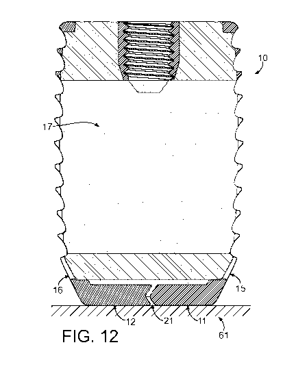
FIG. 12 is a side sectioned view of a first exemplary embodiment of an implant
designed
using the method of the present invention prior to removal from a build
surface.
FIG. 13 is a perspective view of a first exemplary embodiment of an implant
designed
using the method of the present invention in its build orientation.
FIG. 14 is a perspective sectioned view of a first exemplary embodiment of an
PLIF/TLIF implant designed using the method of the present invention in its
build
orientation.
FIG. 15 is a top sectioned view of a first exemplary embodiment of an implant
designed
using the method of the present invention prior to removal from a build
surface.
FIG. 16 is a perspective view of a second exemplary embodiment of an implant
designed
using the method of the present invention after removal from a build surface.
DETAILED DESCRIPTION OF THE INVENTION
- 6 -
CA 03061948 2019-10-17
WO 2018/156905 PCT/US2018/019437
In many situations, it is desirable to use an implant that is capable of bone
attachment or osteointegration over time. It is also desirable in many
situations to use an
implant that is capable of attachment or integration with living tissue.
Examples of
implants where attachment to bone or osteointegration is beneficial include,
but are not
limited to, cervical, lumbar, and thoracic interbody fusion implants,
vertebral body
replacements, osteotomy wedges, dental implants, bone stems, acetabular cups,
cranio-
facial plating, bone replacement and fracture plating. In many applications,
it is also
desirable to stress new bone growth to increase its strength. According to
Wolff s law,
bone will adapt to stresses placed on it so that bone under stress will grow
stronger and
bone that isn't stressed will become weaker.
In some aspects, the systems and methods described herein can be directed
toward
implants that are configured for osteointegration and stimulating adequately
stressed new
bone growth. Many of the exemplary implants of the present invention are
particularly
useful for use in situations where it is desirable to have strong bone
attachment and/or
bone growth throughout the body of an implant. Whether bone growth is desired
only for
attachment or throughout an implant, the present invention incorporates a
unique lattice
structure that can provide mechanical spacing, a scaffold to support new bone
growth and
a modulus of elasticity that allows new bone growth to be loaded with
physiological
forces. As a result, the present invention provides implants that grow
stronger and
healthier bone for more secure attachment and/or for a stronger bone after the
implant
osteointegrates.
The exemplary embodiments of the invention presented can be comprised, in
whole or in part, of a lattice. A lattice, as used herein, refers to a three-
dimensional
- 7 -
CA 03061948 2019-10-17
WO 2018/156905 PCT/US2018/019437
material with one or more interconnected openings that allow a fluid to
communicate
from one location to another location through an opening. A three-dimensional
material
refers to a material that fills a three-dimensional space (i.e. has height,
width and length).
Lattices can be constructed by many means, including repeating various
geometric shapes
or repeating random shapes to accomplish a material with interconnected
openings. An
opening in a lattice is any area within the bounds of the three-dimensional
material that is
devoid of that material. Therefore, within the three-dimensional boundaries of
a lattice,
there is a volume of material and a volume that is devoid of that material.
The material that provides the structure of the lattice is referred to as the
primary
material. The structure of a lattice does not need to provide structural
support for any
purpose, but rather refers to the configuration of the openings and
interconnections that
comprise the lattice. An opening in a lattice may be empty, filled with a
gaseous fluid,
filled with a liquid fluid, filled with a solid or partially filled with a
fluid and/or solid.
Interconnections, with respect to openings, refer to areas devoid of the
primary material
and that link at least two locations together. Interconnections may be
configured to allow
a fluid to pass from one location to another location.
A lattice can be defined by its volumetric density, meaning the ratio between
the
volume of the primary material and the volume of voids presented as a
percentage for a
given three-dimensional material. The volume of voids is the difference
between the
volume of the bounds of the three-dimensional material and the volume of the
primary
material. The volume of voids can comprise of the volume of the openings, the
volume
of the interconnections and/or the volume of another material present. For
example, a
lattice with a 30% volumetric density would be comprised of 30% primary
material by
- 8 -
CA 03061948 2019-10-17
WO 2018/156905 PCT/US2018/019437
volume and 70% voids by volume over a certain volume. A lattice with a 90%
volumetric density would be comprised of 90% primary material by volume and
10%
voids by volume over a certain volume. In three-dimensional materials with a
volumetric
density of less than 50%, the volume of the primary material is less than the
volume of
voids. While the volumetric density refers to the volume of voids, the voids
do not need
to remain void and can be filled, in whole or in part, with a fluid or solid
prior to, during
or after implantation.
Lattices comprised of repeating geometric patterns can be described using the
characteristics of a repeating unit cell. A unit cell in a repeating geometric
lattice is a
three-dimensional shape capable of being repeated to form a lattice. A
repeating unit cell
can refer to multiple identical unit cells that are repeated over a lattice
structure or a
pattern through all or a portion of a lattice structure. Each unit cell is
comprised of a
certain volume of primary material and a certain void volume, or in other
words, a spot
volumetric density. The spot volumetric density may cover as few as a partial
unit cell or
a plurality of unit cells. In many situations, the spot volumetric density
will be consistent
with the material's volumetric density, but there are situations where it
could be desirable
to locally increase or decrease the spot volumetric density.
Unit cells can be constructed in numerous volumetric shapes containing various
types of structures. Unit cells can be bound by a defined volume of space to
constrict the
size of the lattice structure or other type of structure within the unit cell.
In some
embodiments, unit cells can be bound by volumetric shapes, including but not
limited to,
a cubic volume, a cuboid volume, a hexahedron volume or an amorphous volume.
The
unit cell volume of space can be defined based on a number of faces that meet
at corners.
- 9 -
CA 03061948 2019-10-17
WO 2018/156905 PCT/US2018/019437
In examples where the unit cell volume is a cubic, cuboid or hexahedron
volume, the unit
cell volume can have six faces and eight corners, where the corners are
defined by the
location where three faces meet. Unit cells may be interconnected in some or
all areas,
not interconnected in some or all areas, of a uniform size in some or all
areas or of a
nonuniform size in some or all areas. In some embodiments disclosed herein
that use a
repeating geometric pattern, the unit cells can be defined by a number of
struts defining
the edges of the unit cell and joined at nodes about the unit cell. Unit cells
so defined can
share certain struts among more than one unit cell, so that two adjacent unit
cells may
share a common planar wall defined by struts common to both cells. In some
embodiments disclosed herein that use a repeating geometric pattern, the unit
cells can be
defined by a node and a number of struts extending radially from that node.
While the present application uses volumetric density to describe exemplary
embodiments, it is also possible to describe them using other metrics,
including but not
limited to cell size, strut size or stiffness. Cell size may be defined using
multiple
methods, including but not limited to cell diameter, cell width, cell height
and cell
volume. Strut size may be defined using multiple methods, including but not
limited to
strut length and strut diameter.
Repeating geometric patterns are beneficial for use in lattice structures
contained
in implants because they can provide predictable characteristics. Many
repeating
geometric shapes may be used as the unit cell of a lattice, including but are
not limited to,
rhombic dodecahedron, diamond, dodecahedron, square, pentagonal, hexagonal,
octagonal, sctet struts, trunic octa, diagonal struts, other known geometric
structures, and
rounded, reinforced, weakened, or simplified versions of each geometry.
- 10 -
CA 03061948 2019-10-17
WO 2018/156905 PCT/US2018/019437
Lattices may also be included in implants as a structural component or a
nonstructural component. Lattices used in structural applications may be
referred to
herein as structural lattices, load-bearing lattices or stressed lattices. In
some instances,
structural lattices, load-bearing lattices or stressed lattices may be simply
referred to as a
lattice. Repeating geometric shaped unit cells, particularly the rhombic
dodecahedron,
are well suited, in theory, for use in structural lattices because of their
strength to weight
ratio. To increase the actual strength and fatigue resistance of a rhombic
dodecahedron
lattice, the present invention, in some embodiments, includes a modified strut
comprised
of triangular segments, rather than using a strut with a rectangular or
circular cross
section. Some embodiments herein also modify the angles defining the rhombic
faces of
a rhombic dodecahedron to change the lattice's elastic modulus and fatigue
resistance.
The use of triangular segments provides a lattice with highly predictable
printed
properties that approach the theoretical strength values for a rhombic
dodecahedron
lattice.
In structural lattice applications, the strength and elastic modulus of the
lattice can
be approximated by the volumetric density. When the volumetric density
increases, the
strength and the elastic modulus increases. Compared to other porous
structures, the
lattice of the present invention has a higher strength and elastic modulus for
a given
volumetric density because of its ability to use the high strength to weight
benefits of a
.. rhombic dodecahedron, modified rhombic dodecahedron or radial dodeca-
rhombus unit
cell.
When configured to provide support for bone or tissue growth, a lattice may be
referred to as a scaffold. Lattices can be configured to support bone or
tissue growth by
-11-
CA 03061948 2019-10-17
WO 2018/156905 PCT/US2018/019437
controlling the size of the openings and interconnections disposed within the
three-
dimensional material. A scaffold, if used on the surface of an implant, may
provide an
osteointegration surface that allows adjacent bone to attach to the implant. A
scaffold
may also be configured to provide a path that allows bone to grow further than
a mere
surface attachment. Scaffolds intended for surface attachment are referred to
herein as
surface scaffolds. A surface scaffold may be one or more unit cells deep, but
does not
extend throughout the volume of an implant. Scaffolds intended to support in-
growth
beyond mere surface attachment are referred to herein as bulk scaffolds.
Scaffolds may
also be included in implants as a structural component or a nonstructural
component.
Scaffolds used in structural applications may be referred to herein as
structural scaffolds,
load-bearing scaffolds or stressed scaffolds. In some instances, structural
scaffolds, load-
bearing scaffolds or stressed scaffolds may be simply referred to as a
scaffold. In some
instances, the use of the term scaffold may refer to a material configured to
provide
support for bone or tissue growth, where the material is not a lattice.
The scaffolds described herein can be used to promote the attachment or in-
growth of various types of tissue found in living beings. As noted earlier,
some
embodiments of the scaffold are configured to promote bone attachment and in-
growth.
The scaffolds can also be configured to promote attachment of in-growth of
other areas of
tissue, such as fibrous tissue. In some embodiments, the scaffold can be
configured to
promote the attachment or in-growth of multiple types of tissue. Some
embodiments of
the scaffolds are configured to be implanted near or abutting living tissue.
Near living
tissue includes situations where other layers, materials or coatings are
located between a
scaffold and any living tissue.
- 12 -
CA 03061948 2019-10-17
WO 2018/156905 PCT/US2018/019437
In some embodiments, the present invention uses bulk scaffolds with openings
and interconnections that are larger than those known in the art. Osteons can
range in
diameter from about 100 p.m and it is theorized that a bundle of osteons would
provide
the strongest form of new bone growth. Bone is considered fully solid when it
has a
.. diameter of greater than 3 mm so it is theorized that a bundle of osteons
with a diameter
equaling approximately half of that value would provide significant strength
when grown
within a scaffold. It is also theorized that osteons may grow in irregular
shapes so that
the cross-sectional area of an osteon could predict its strength. A
cylindrical osteon
growth with a 3 mm diameter has a cross-sectional area of approximately 7
square mm
and a cylindrical osteon with a 1.5 mm diameter has a cross-sectional area of
1.8 square
mm. It is theorized that an osteon of an irregular shape with a cross-
sectional area of at
least 1.8 square millimeters could provide a significant strength advantage
when grown in
a scaffold.
Most skilled in the art would indicate that pores or openings with a diameter
or
width between 300 p.m to 900 p.m, with a pore side of 600 p.m being ideal,
provide the
best scaffold for bone growth. Instead, some embodiments of the present
invention
include openings and interconnections with a diameter or width on the order of
1.0 to
15.0 times the known range, with the known range being 300 p.m to 900 p.m,
resulting in
openings from 0.07 mm2 up to 145 mm2 cross sectional area for bone growth. In
some
examples, pores or openings with a diameter or width between and including 100
p.m to
300 p.m could be beneficial. Some examples include openings and
interconnections with
a diameter on the order of 1.0 to 5.0 times the known range. It has been at
least theorized
that the use of much larger openings and interconnections than those known in
the art
- 13 -
CA 03061948 2019-10-17
WO 2018/156905 PCT/US2018/019437
will allow full osteons and solid bone tissue to form throughout the bulk
scaffold,
allowing the vascularization of new, loadable bone growth. In some examples,
these
pores may be 3 mm in diameter or approximately 7 mm2 in cross sectional area.
In other
examples, the pores are approximately 1.5 mm in diameter or approximately 1.75
mm2 in
cross sectional area. The use of only the smaller diameter openings and
interconnections
known in the art are theorized to limit the penetration of new bone growth
into a bulk
scaffold because the smaller diameter openings restrict the ability of
vascularization
throughout the bulk scaffold.
A related structure to a lattice is a closed cell material. A closed cell
material is
.. similar to a lattice, in that it has openings contained within the bounds
of a three-
dimensional material, however, closed cell materials generally lack
interconnections
between locations through openings or other pores. A closed cell structure may
be
accomplished using multiple methods, including the filling of certain cells or
through the
use of solid walls between the struts of unit cells. A closed cell structure
can also be
referred to as a cellular structure. It is possible to have a material that is
a lattice in one
portion and a closed cell material in another. It is also possible to have a
closed cell
material that is a lattice with respect to only certain interconnections
between openings or
vice versa. While the focus of the present disclosure is on lattices, the
structures and
methods disclosed herein can be easily adapted for use on closed cell
structures within
the inventive concept.
The lattice used in the present invention can be produced from a range of
materials and processes. When used as a scaffold for bone growth, it is
desirable for the
lattice to be made of a biocompatible material that allows for bone
attachment, either to
- 14 -
CA 03061948 2019-10-17
WO 2018/156905 PCT/US2018/019437
the material directly or through the application of a bioactive surface
treatment. In one
example, the scaffold is comprised of an implantable metal. Implantable metals
include,
but are not limited to, zirconium, stainless steel (316 & 316L), tantalum,
nitinol, cobalt
chromium alloys, titanium and tungsten, and alloys thereof. Scaffolds
comprised of an
implantable metal may be produced using an additive metal fabrication or 3D
printing
process. Appropriate production processes include, but are not limited to,
direct metal
laser sintering, selective laser sintering, selective laser melting, electron
beam melting,
laminated object manufacturing and directed energy deposition.
In another example, the lattice of the present invention is comprised of an
implantable metal with a bioactive coating. Bioactive coatings include, but
are not
limited to, coatings to accelerate bone growth, anti-thrombogenic coatings,
anti-microbial
coatings, hydrophobic or hydrophilic coatings, and hemophobic,
superhemophobic, or
hemophilic coatings. Coatings that accelerate bone growth include, but are not
limited to,
calcium phosphate, hydroxyapatite ("HA"), silicate glass, stem cell
derivatives, bone
.. morphogenic proteins, titanium plasma spray, titanium beads and titanium
mesh. Anti-
thrombogenic coatings include, but are not limited to, low molecular weight
fluoro-
oligomers. Anti-microbial coatings include, but are not limited to, silver,
organosilane
compounds, iodine and silicon-nitride. Superhemophobic coatings include
fluorinated
nanotubes.
In another example, the lattice is made from a titanium alloy with an optional
bioactive coating. In particular, Ti6A14V ELI wrought (American Society for
Testing
and Materials ("ASTM") F136) is a particularly well-suited titanium alloy for
scaffolds.
While Ti6A14V ELI wrought is the industry standard titanium alloy used for
medical
- 15 -
CA 03061948 2019-10-17
WO 2018/156905 PCT/US2018/019437
purposes, other titanium alloys, including but not limited to, unalloyed
titanium (ASTM
F67), Ti6A14V standard grade (ASTM F1472), Ti6A17Nb wrought (ASTM 1295),
Ti5Al2.5Fe wrought (British Standards Association/International Standard
Organization
Part 10), CP and Ti6A14V standard grade powders (ASTM F1580), Ti13Nb13Zr
wrought
(ASTM F1713) , the lower modulus Ti-24Nb-4Zr-85n and Ti 12Mo6Zr2Fe wrought
(ASTM F1813) can be appropriate for various embodiments of the present
invention.
Titanium alloys are an appropriate material for scaffolds because they are
biocompatible and allow for bone attachment. Various surface treatments can be
done to
titanium alloys to increase or decrease the level of bone attachment. Bone
will attach to
even polished titanium, but titanium with a surface texture allows for greater
bone
attachment. Methods of increasing bone attachment to titanium may be produced
through a forging or milling process, sandblasting, acid etching, and the use
of a
bioactive coating. Titanium parts produced with an additive metal fabrication
or 3D
printing process, such as direct metal laser sintering, can be treated with an
acid bath to
reduce surface stress risers, normalize surface topography, and improve
surface oxide
layer, while maintaining surface roughness and porosity to promote bone
attachment.
Additionally, Titanium or other alloys may be treated with heparin, heparin
sulfate (HS), glycosaminoglycans (GAG), chondroitin-4-sulphate (C45),
chondroitin-6-
sulphate (C65), hyaluronan (HY), and other proteoglycans with or without an
aqueous
calcium solution. Such treatment may occur while the material is in its pre-
manufacturing form (often powder) or subsequent to manufacture of the
structure.
While a range of structures, materials, surface treatments and coatings have
been
described, it is believed that a lattice using a repeating modified rhombic
dodecahedron
- 16 -
CA 03061948 2019-10-17
WO 2018/156905 PCT/US2018/019437
(hereinafter "MRDD") unit cell can present a preferable combination of
stiffness,
strength, fatigue resistance, and conditions for bone ingrowth. In some
embodiments, the
repeating MRDD lattice is comprised of titanium or a titanium alloy. A generic
rhombic
dodecahedron (hereinafter "RDD"), by definition, has twelve sides in the shape
of
rhombuses. When repeated in a lattice, an RDD unit cell is comprised of 24
struts that
meet at 14 vertices. The 24 struts define the 12 planar faces of the structure
and disposed
at the center of each planar face is an opening, or interconnection, allowing
communication from inside the unit cell to outside the unit cell.
An example of the MRDD unit cell B 10 used in the present invention is shown
in
FIGS. A1-A5. In FIG. Al is an isometric view of a single MRDD unit cell B 10
containing a full MRDD structure along with radial struts that comprise
portions of
adjacent unit cells. In FIG. A2 is a side view of a single MRDD unit cell B 10
showing
the configuration of interconnections when viewed from a lateral direction. A
top or
bottom view of the MRDD unit cell B 10 would be substantially the same as the
side view
depicted in FIG. A2. The MRDD unit cell B 10 differs in both structural
characteristics
and method of design from generic RDD shapes. A generic RDD is comprised of 12
faces where each face is an identical rhombus with an acute angle of 70.5
degrees and an
obtuse angle of 109.5 degrees. The shape of the rhombus faces in a generic RDD
do not
change if the size of the unit cell or the diameter of the struts are changed
because the
struts are indexed based on their axis and each pass through the center of the
14 nodes or
vertices.
In some embodiments of the MRDD, each node is contained within a fixed
volume that defines its bounds and provides a fixed point in space for the
distal ends of
- 17 -
CA 03061948 2019-10-17
WO 2018/156905 PCT/US2018/019437
the struts. The fixed volume containing the MRDD or a sub-unit cell of the
MRDD can
be various shapes, including but not limited to, a cubic, cuboid, hexahedron
or
amorphous volume. Some examples use a fixed volume with six faces and eight
corners
defined by locations where three faces meet. The orientation of the struts can
be based
on the center of a node face at its proximate end and the nearest corner of
the volume to
that node face on its distal end. Each node is preferably an octahedron, more
specifically
a square bipyramid (i.e. a pyramid and inverted pyramid joined on a horizontal
plane).
Each node, when centrally located in a cuboid volume, more preferably
comprises a
square plane parallel to a face of the cuboid volume, six vertices and is
oriented so that
each of the six vertices are positioned at their closest possible location to
each of the six
faces of the cuboid volume. Centrally located, with regards to the node's
location within
a volume refers to positioning the node at a location substantially
equidistant from
opposing walls of the volume. In some embodiments, the node can have a
volumetric
density of 100 percent and in other embodiments, the node can have a
volumetric density
of less than 100 percent. Each face of the square bipyramid node can be
triangular and
each face can provide a connection point for a strut.
The struts can also be octahedrons, comprising an elongate portion of six
substantially similar elongate faces and two end faces. The elongate faces can
be
isosceles triangles with a first internal angle, angle A, and a second
internal angle, angle
.. B, where angle B is greater than angle A. The end faces can be
substantially similar
isosceles triangles to one another with a first internal angle, angle C, and a
second
internal angle, angle D, where angle D is greater than angle C. Preferably,
angle C is
greater than angle A.
- 18 -
CA 03061948 2019-10-17
WO 2018/156905 PCT/US2018/019437
The strut direction of each strut is a line or vector defining the orientation
of a
strut and it can be orthogonal or non-orthogonal relative to the planar
surface of each
node face. In the MRDD and radial dodeca-rhombus structures disclosed herein,
the strut
direction can be determined using a line extending between the center of the
strut end
faces, the center of mass along the strut or an external edge or face of the
elongate portion
of the strut. When defining a strut direction using a line extending between
the center of
the strut end faces, the line is generally parallel to the bottom face or edge
of the strut.
When defining a strut direction using a line extending along the center of
mass of the
strut, the line can be nonparallel to the bottom face or edge of the strut.
The octahedron
nodes of the MRDD can be scaled to increase or decrease volumetric density by
changing
the origin point and size of the struts. The distal ends of the struts,
however, are locked at
the fixed volume comers formed about each node so that their angle relative to
each node
face changes as the volumetric density changes. Even as the volumetric density
of an
MRDD unit cell changes, the dimensions of the fixed volume formed about each
node
does not change. In FIG. Al, dashed lines are drawn between the comers of the
MRDD
unit cell B10 to show the cube B11 that defines its bounds. In the MRDD unit
cell in
FIG. Al, the height B12, width B13 and depth B14 of the unit cell are
substantially the
same, making the area defined by B11 a cube.
In some embodiments, the strut direction of a strut can intersect the center
of the
node and the corner of the cuboid volume nearest to the node face where the
strut is
fixed. In some embodiments, the strut direction of a strut can intersect just
the corner of
the cuboid volume nearest to the node face where the strut is fixed. In some
embodiments, a reference plane defined by a cuboid or hexahedron face is used
to
- 19 -
CA 03061948 2019-10-17
WO 2018/156905 PCT/US2018/019437
describe the strut direction of a strut. When the strut direction of a strut
is defined based
on a reference plane, it can be between 0 degrees and 90 degrees from the
reference
plane. When the strut direction of a strut is defined based on a reference
plane, it is
preferably eight degrees to 30 degrees from the reference plane.
By indexing the strut orientation to a variable node face on one end and a
fixed
point on its distal end, the resulting MRDD unit cell can allow rhombus shaped
faces
with a smaller acute angle and larger obtuse angle than a generic RDD. The
rhombus
shaped faces of the MRDD can have two substantially similar opposing acute
angles and
two substantially similar opposing obtuse angles. In some embodiments, the
acute angles
are less than 70.5 degrees and the obtuse angles are greater than 109.5
degrees. In some
embodiments, the acute angles are between 0 degrees and 55 degrees and the
obtuse
angles are between 125 degrees and 180 degrees. In some embodiments, the acute
angles
are between 8 degrees and 60 degrees and the obtuse angles are between 120
degrees and
172 degrees. The reduction in the acute angles increases fatigue resistance
for loads
oriented across the obtuse angle corner to far obtuse angle corner. The
reduction in the
acute angles and increase in obtuse angles also orients the struts to increase
the MRDD's
strength in shear and increases the fatigue resistance. By changing the
rhombus corner
angles from a generic RDD, shear loads pass substantially in the axial
direction of some
struts, increasing the shear strength. Changing the rhombus corner angles from
a generic
RDD also reduces overall deflection caused by compressive loads, increasing
the fatigue
strength by resisting deflection under load.
When placed towards the center of a lattice structure, the 12 interconnections
of a
unit cell connect to 12 different adjacent unit cells, providing continuous
paths through
-20-
CA 03061948 2019-10-17
WO 2018/156905 PCT/US2018/019437
the lattice. The size of the central void and interconnections in the MRDD may
be
defined using the longest dimension method as described herein. Using the
longest
dimension method, the central void can be defined by taking a measurement of
the
longest dimension as demonstrated in FIG. A3. In FIG. A3, the longest
dimension is
.. labeled as distance AA. The distance AA can be taken in the vertical or
horizontal
directions (where the directions reference the directions on the page) and
would be
substantially the same in this example. The interconnections may be defined by
their
longest measurement when viewed from a side, top or bottom of a unit cell. In
FIG. A4,
the longest dimension is labeled as distance AB. The distance AB can be taken
in the
vertical or horizontal directions (where the directions reference the
directions on the
page). The view in FIG. A4 is a lateral view, however, in this example the
unit cell will
appear substantially the same when viewed from the top or bottom.
The size of the central void and interconnections can alternatively be defined
by
the largest sphere method as described herein. Using the largest sphere
method, the
central void can be defined by the diameter of the largest sphere that can fit
within the
central void without intersecting the struts. In FIG. A5 is an example of the
largest
sphere method being used to define the size of a central void with a sphere
with a
diameter of BA. The interconnections are generally rhombus shaped and their
size can
alternatively be defined by the size of the length and width of three circles
drawn within
the opening. Drawn within the plane defining a side, a first circle BB1 is
drawn at the
center of the opening so that it is the largest diameter circle that can fit
without
intersecting the struts. A second circle BB2 and third circle BB3 is them
drawn so that
they are tangential to the first circle BB1 and the largest diameter circles
that can fit
-21-
CA 03061948 2019-10-17
WO 2018/156905 PCT/US2018/019437
without intersecting the struts. The diameter of the first circle BB1 is the
width of the
interconnection and the sum of the diameters of all three circles BB1, BB2 &
BB3
represents the length of the interconnection. Using this method of measurement
removes
the acute corners of the rhombus shaped opening from the size determination.
In some
instances, it is beneficial to remove the acute corners of the rhombus shaped
opening
from the calculated size of the interconnections because of the limitations of
additive
manufacturing processes. For example, if an SLS machine has a resolution of 12
p.m
where the accuracy is within 5 p.m, it is possible that the acute corner could
be rounded
by the SLS machine, making it unavailable for bone ingrowth. When designing
lattices
for manufacture on less precise additive process equipment, it can be helpful
to use this
measuring system to better approximate the size of the interconnections.
Using the alternative measuring method, in some examples, the width of the
interconnections is approximately 600 p.m and the length of the
interconnections is
approximately 300 p.m. The use of a 600 p.m length and 300 p.m width provides
an
opening within the known pore sizes for bone growth and provides a surface
area of
roughly 1.8 square millimeters, allowing high strength bone growth to form.
Alternative
embodiments may contain interconnections with a cross sectional area of 1.0 to
15.0
times the cross-sectional area of a pore with a diameter of 300 p.m. Other
embodiments
may contain interconnections with a cross sectional area of 1.0 to 15.0 times
the cross-
sectional area of a pore with a diameter of 900 p.m.
The MRDD unit cell also has the advantage of providing at least two sets of
substantially homogenous pore or opening sizes in a lattice structure. In some
embodiments, a first set of pores have a width of about 200 p.m to 900 p.m and
a second
- 22 -
CA 03061948 2019-10-17
WO 2018/156905 PCT/US2018/019437
set of pores have a width of about 1 to 15 times the width of the first set of
pores. In
some embodiments, a first set of pores can be configured to promote the growth
of
osteoblasts and a second set of pores can be configured to promote the growth
of osteons.
Pores sized to promote osteoblast growth can have a width of between and
including
about 100 p.m to 900 p.m. In some embodiments, pores sized to promote
osteoblast
growth can have a width that exceeds 900 p.m. Pores sized to promote the
growth of
osteons can have a width of between and including about 100 p.m to 13.5 mm. In
some
embodiments, pores sized to promote osteon growth can have a width that
exceeds 13.5
mm.
In some embodiments, it is beneficial to include a number of substantially
homogenous larger pores and a number of substantially homogenous smaller
pores,
where the number of larger pores is selected based on a ratio relative to the
number of
smaller pores. For example, some embodiments have one large pore for every one
to 25
small pores in the lattice structure. Some embodiments preferably have one
large pore
for every eight to 12 smaller pores. In some embodiments, the number of larger
and
smaller pores can be selected based on a percentage of the total number of
pores in a
lattice structure. For example, some embodiments can include larger pores for
four
percent to 50 percent of the total number of pores and smaller pores for 50
percent to 96
percent of the total number of pores. More preferably, some embodiments can
include
larger pores for about eight percent to 13 percent of the total number of
pores and smaller
pores for about 87 percent to 92 percent of the total number of pores. It is
believed that a
lattice constructed with sets of substantially homogenous pores of the
disclosed two sizes
provides a lattice structure that simultaneously promotes osteoblast and
osteon growth.
-23-
CA 03061948 2019-10-17
WO 2018/156905 PCT/US2018/019437
The MRDD unit cell may also be defined by the size of the interconnections
when
viewed from a side, top or bottom of a unit cell. The MRDD unit cell has the
same
appearance when viewed from a side, top or bottom, making the measurement in a
side
view representative of the others. When viewed from the side, as in FIG. A4,
an MRDD
unit cell displays four distinct diamond shaped interconnections with
substantially right
angles. The area of each interconnection is smaller when viewed in the lateral
direction
than from a direction normal to the planar direction of each interconnection,
but the area
when viewed in the lateral direction can represent the area available for bone
to grow in
that direction. In some embodiments, it may be desirable to index the
properties of the
unit cell and lattice based on the area of the interconnections when viewed
from the top,
bottom or lateral directions.
In some embodiments of the lattice structures disclosed herein, the central
void is
larger than the length or width of the interconnections. Because the size of
each
interconnection can be substantially the same in a repeating MRDD structure,
the
resulting lattice can be comprised of openings of at least two discrete sizes.
In some
embodiments, it is preferable for the diameter of the central void to be
approximately two
times the length of the interconnections. In some embodiments, it is
preferable for the
diameter of the central void to be approximately four times the width of the
interconnections.
In some embodiments, the ratio between the diameter of the central void and
the
length or width of the interconnections can be changed to create a structural
lattice of a
particular strength. In these embodiments, there is a correlation where the
ratio between
- 24 -
CA 03061948 2019-10-17
WO 2018/156905 PCT/US2018/019437
the central void diameter and the length or width of the interconnections
increases as the
strength of the structural lattice increases.
It is also believed that a lattice using a repeating radial dodeca-rhombus
(hereinafter "RDDR") unit cell can present a preferable combination of
stiffness,
strength, fatigue resistance, and conditions for bone ingrowth. In some
embodiments, the
repeating RDDR lattice is comprised of titanium or a titanium alloy. In FIG.
A7 is an
isometric view of a single RDDR unit cell B20 containing a full RDDR
structure. In
FIG. A8 is a side view of a single RDDR unit cell B20 showing the
configuration of
interconnections when viewed from a lateral direction. A top or bottom view of
the
RDDR unit cell B20 would be substantially the same as the side view depicted
in FIG.
A8.
As used herein, an RDDR unit cell B20 is a three-dimensional shape comprised
of
a central node with radial struts and mirrored struts thereof forming twelve
rhombus
shaped structures. The node is preferably an octahedron, more specifically a
square
bipyramid (i.e. a pyramid and inverted pyramid joined on a horizontal plane).
Each face
of the node is preferably triangular and fixed to each face is a strut
comprised of six
triangular facets and two end faces. The central axis of each strut can be
orthogonal or
non-orthogonal relative to the planar surface of each node face. The central
axis may
follow the centroid of the strut. The RDDR is also characterized by a central
node with
one strut attached to each face, resulting in a square bipyramid node with
eight struts
attached.
Examples of node and strut combinations are shown in FIGS. A9-A13. In FIG.
A9 is an isometric view of a single node B30 with a single strut B31 attached.
The node
-25-
CA 03061948 2019-10-17
WO 2018/156905 PCT/US2018/019437
B30 is a square bipyramid oriented so that two peaks face the top and bottom
of a volume
B32 defining the bounds of the node B30 and any attached strut(s) B31. The
node B30 is
oriented so that the horizontal corners are positioned at their closest point
to the lateral
sides of the volume B32. The strut B31 extends from a node B30 face to the
corner of
the volumeB32 defining the bounds of the node and attached struts. In FIG. A9,
the
central axis of the strut is 45 degrees above the horizontal plane where the
node's planar
face is 45 degrees above a horizontal plane.
FIG. A9 also details an octahedron strut B31, where dashed lines show hidden
edges of the strut. The strut B31 is an octahedron with an elongate portion of
six
substantially similar elongate faces and two end faces. The elongate faces B3
la, B3 lb,
B31c, B31d, B3 le & B3 if of the strut B31 define the outer surface of the
strut's elongate
and somewhat cylindrical surface. Each of the elongate faces B31a, B31b, B31c,
B31d,
B3le & B3 if are isosceles triangles with a first internal angle, angle A, and
a second
internal angle, angle B, where angle B is greater than angle A. The strut B31
also has
two end faces B3lf & B31g that isosceles triangles that are substantially
similar to one
another, having a first internal angle, angle C, and a second internal angle,
angle D, and
where angle D is greater than angle C. When comparing the internal angles of
the
elongate faces B31a, B31b, B31c, B31d, B3le & B3lf to the end faces B3lf &
B31g,
angle C is greater than angle A.
In FIG. A10 is a side view of the node B30 and strut B31 combination bounded
by volume B32. In the side view, the height of the node B30 compared to the
height of
the cube B32 can be compared easily. In FIGS. All-A13 are side views of node
and
strut combinations viewed from a corner of the volume rather than a wall or
face, and
-26-
CA 03061948 2019-10-17
WO 2018/156905 PCT/US2018/019437
where the combinations have been modified from FIGS. A9-A10 to change the
volumetric density of the resulting unit cell. In FIG. All, the height of the
node B130
has increased relative to the height of the volume B132. Since the distal end
of the strut
B131 is fixed by the location of a corner of the volume B132, the strut B131
must change
.. its angle relative to its attached node face so that it becomes
nonorthogonal. The node
B130 and strut B131 combination, where the angle of the strut B131 from a
horizontal
plane is about 20.6 degrees, would be appropriate for a lattice structure with
an elastic
modulus of approximately 3 GPa.
In FIG. Al2, the height of the node B230 relative to the height of the cube
B232
has been increased over the ratio of FIG. All to create a node B230 and strut
B231
combination that would be appropriate for a lattice structure with an elastic
modulus of
approximately 4 GPa. As the height of the node B230 increases, the angle
between the
strut B231 and a horizontal plane decreases to about 18.8 degrees. As the
height of the
node B230 increases, the size of the node faces also increase so that the size
of the strut
B231 increases. While the distal end of the strut B231 is fixed to the corner
of the
volume B232, the size of the distal end increases to match the increased size
of the node
face to maintain a substantially even strut diameter along its length. As the
node and
strut increase in size, the volumetric density increases, as does the elastic
modulus. In
FIG. A13, the height of the node B330 relative to the height of the volume
B332 has been
increased over the ratio of FIG. A13 to create a node B330 and strut B331
combination
that would be appropriate for a lattice structure with an elastic modulus of
approximately
10 GPa. In this configuration, the angle B333 between the strut B331 and a
horizontal
plane decreases to about 12.4 degrees and the volumetric density increases
over the
-27-
CA 03061948 2019-10-17
WO 2018/156905 PCT/US2018/019437
previous examples. The single node and strut examples can be copied and/or
mirrored to
create unit cells of appropriate sizes and characteristics. For instance, the
angle between
the strut and a horizontal plane could be increased to 25.8 degrees to render
a lattice with
a 12.3 percent volumetric density and an elastic modulus of about 300 MPa.
While a
.. single node and single strut were shown in the examples for clarity,
multiple struts may
be attached to each node to create an appropriate unit cell.
Adjacent struts extending from adjacent node faces on either the upper half or
lower half of the node have an angle from the horizontal plane and a lateral
separation
angle defined by an angle between the strut directions of adjacent struts. In
the MRDD
.. and RDDR structures, adjacent struts have an external edge or face of the
elongate
portion extending closest to the relevant adjacent strut. The lateral
separation angle, as
used herein, generally refers to the angle between an external edge or face of
the elongate
portion of a strut extending closest to the relevant adjacent strut. In some
embodiments, a
lateral separation angle defined by a line extending between the center of the
strut end
faces or a line defined by the center of mass of the struts can be used in
reference to a
similar calculation for an adjacent strut.
The lateral separation angle is the angle between the nearest face or edge of
a strut
to an adjacent strut. The lateral separation angle can be measured as the
smallest angle
between the nearest edge of a strut to the nearest edge of an adjacent strut,
in a plane
containing both strut edges. The lateral separation angle can also be measured
as the
angle between the nearest face of a strut to the nearest face of an adjacent
strut in a plane
normal to the two strut faces. In embodiments without defined strut edges or
strut faces,
the lateral separation angle can be measured as an angle between the nearest
portion of
-28-
CA 03061948 2019-10-17
WO 2018/156905 PCT/US2018/019437
one strut to the nearest portion of an adjacent strut. For a unit cell in a
cubic volume, as
the strut angle from the horizontal plane decreases, the lateral separation
angle
approaches 90 degrees. For a unit cell in a cubic volume, as the strut angle
from the
horizontal plane increases, the lateral separation angle approaches 180
degrees. In some
embodiments, it is preferable to have a lateral separation angle greater than
109.5
degrees. In some embodiments, it is preferable to have a lateral separation
angle of less
than 109.5 degrees. In some embodiments, it is preferable to have a lateral
separation
angle of between and including about 108 degrees to about 156 degrees. In some
embodiments, it is more preferable to have a lateral separation angle of
between and
including 111 degrees to 156 degrees. In some embodiments, it is more
preferable to
have a lateral separation angle of between and including 108 degrees to 120
degrees. In
some embodiments, it is most preferable to have a lateral separation angle of
between and
including about 111 degrees to 120 degrees. In some embodiments, it is more
preferable
to have a lateral separation angle of between and including 128 degrees to 156
degrees.
In FIG. A14 is a side view, viewed from a corner of the cube B432, of a single
node
B430 with two adjacent struts B431 & B434 attached and where the lateral
separation
angle B443 is identified. When measured from the nearest edge of a strut to
the nearest
edge of an adjacent strut, the lateral separation angle B443 is about 116
degrees.
In some embodiments, a unit cell is built up from multiple sub-unit cells
fixed
together. In FIG. A15 is an isometric view of an exemplary sub-unit cell
comprising a
single node and four struts. In FIG. A16 is an isometric view of two sub-unit
cells in a
stacked formation where the upper sub-unit cell is inverted and fixed to the
top of the
-29-
CA 03061948 2019-10-17
WO 2018/156905 PCT/US2018/019437
lower sub-unit cell. In FIG. A17 is an isometric view of eight sub-unit cells
stacked
together to form a single RDDR unit cell.
In FIG. A15, the node B530 is a square bipyramid, oriented so that the two
peaks
face the top and bottom of a cubic volume B532. In some embodiments, the
volume
B532 can be a cuboid volume, a hexahedron volume, an amorphous volume or of a
volume with one or more non-orthogonal sides. The peaks refer to the point
where four
upper faces meet and the point where four lower faces meet. The node B530 is
oriented
so that the horizontal vertices face the lateral sides of the cubic volume
B532. The strut
B531 is fixed to a lower face of the node B530 face on its proximate end and
extends to
the nearest corner of the cubic volume B532 at its distal end. The distal end
of the strut
B531 can remain fixed to the cubic volume B532 even if the node B530 changes
in size
to adjust the sub-unit cell properties.
On the lower face of the node B530 opposite the face which strut B531 is
fixed,
the proximate end of strut B534 is fixed to the node B530. The strut B534
extends to the
nearest corner of cubic volume B532 at its distal end. The strut B535 is fixed
on its
proximate end to an upper node B530 face directed about 90 degrees laterally
from the
node B530 face fixed to strut B531. The strut B535 extends to the nearest
corner of the
cubic volume B532 at its distal end. On the upper face of the node B530
opposite the
face which strut B535 is fixed, the proximate end of strut B536 is fixed to
the node B530.
The strut B536 extends to the nearest corner of the cubic volume B532 at its
distal end.
In some embodiments, the struts B531 & B534-B536 are octahedrons with
triangular faces. The strut face fixed to a node B530 face can be
substantially the same
size and orientation of the node B530 face. The strut face fixed to the
nearest corner of
-30-
CA 03061948 2019-10-17
WO 2018/156905 PCT/US2018/019437
the cube B532 can be substantially the same size as the strut face fixed to
the node B530
and oriented on a substantially parallel plane. The remaining six faces can be
six
substantially similar isosceles triangles with a first internal angle and a
second internal
angle larger than said first internal angle. The six substantially similar
isosceles triangles
can be fixed along their long edges to an adjacent and inverted substantially
similar
isosceles triangle to form a generally cylindrical shape with triangular ends.
When forming a sub-unit cell B540, it can be beneficial to add an eighth node
B538 to each corner of the cube B532 fixed to a strut B531 & B534-B536. When
replicating the sub-unit cell B540, the eighth node B538 attached to each
strut end is
combined with eighth nodes from adjacent sub-unit cells to form nodes located
between
the struts of adjacent sub-unit cells.
In FIG. A16 is a first sub-unit cell B540 fixed to a second sub-unit cell B640
to
form a quarter unit cell B560 used in some embodiments. The second sub-unit
cell B640
comprises a square bipyramid node B630 is a square bipyramid, oriented so that
the two
peaks face the top and bottom of a cubic volume. The node B630 is oriented so
that the
horizontal vertices face the lateral sides of the cubic volume. The strut B635
is fixed to a
lower face of the node B630 face on its proximate end and extends to the
nearest corner
of the cubic volume at its distal end. On the lower face of the node B630
opposite the
face which strut B635 is fixed, the proximate end of strut B636 is fixed to
the node B630.
The strut B636 extends to the nearest corner of cubic volume at its distal
end. The strut
B634 is fixed on its proximate end to an upper node B630 face directed about
90 degrees
laterally from the node B630 face fixed to strut B635. The strut B634 extends
to the
nearest corner of the cubic volume at its distal end. On the upper face of the
node B630
-31-
CA 03061948 2019-10-17
WO 2018/156905 PCT/US2018/019437
opposite the face which strut B634 is fixed, the proximate end of strut B631
is fixed to
the node B630. The strut B631 extends to the nearest corner of the cubic
volume at its
distal end.
The first sub-unit B540 is used as the datum point in the embodiment of FIG.
A16, however, it is appreciated that the second sub-unit cell B640 or another
point could
also be used as the datum point. Once the first sub-unit cell B540 is fixed in
position, it
is replicated so that the second sub-unit cell B640 is substantially similar
to the first. The
second sub-unit cell B640 is rotated about its central axis prior to being
fixed on the top
of the first unit-cell B540. In FIG. A16, the second sub-unit cell B640 is
inverted to
achieve the proper rotation, however, other rotations about the central axis
can achieve
the same result. The first sub-unit cell B540 fixed to the second sub-unit
cell B640 forms
a quarter unit cell B560 that can be replicated and attached laterally to
other quarter unit
cells to form a full unit cell.
Alternatively, a full unit cell can be built up by fixing a first group of
four
substantially similar sub-unit cells together laterally to form a square,
rectangle or
quadrilateral when viewed from above. A second group of four substantially
similar sub-
unit cells rotated about their central axis can be fixed together laterally to
also form a
square, rectangle or quadrilateral when viewed from above. The second group of
sub-
unit cells can be rotated about their central axis prior to being fixed
together laterally or
inverted after being fixed together to achieve the same result. The second
group is then
fixed to the top of the first group to form a full unit cell.
In FIG. A17 is an example of a full unit cell B770 formed by replicating the
sub-
unit cell B540 of FIG. A15. The cube B532 defining the bounds of the sub-unit
cell
-32-
CA 03061948 2019-10-17
WO 2018/156905 PCT/US2018/019437
B540 is identified as well as the node B530 and struts B531 & B534-B536 for
clarity.
The full unit cell B770 of FIG. A17 can be formed using the methods described
above or
using variations within the inventive concept.
Each strut extending from the node, for a given unit cell, can be
substantially the
.. same length and angle from the horizontal plane, extending radially from
the node. At
the end of each strut, the strut is mirrored so that struts extending from
adjacent node
faces form a rhombus shaped opening. Because the struts can be non-orthogonal
to the
node faces, rhombuses of two shapes emerge. In this configuration, a first
group of four
rhombuses extend radially from the node oriented in vertical planes. The acute
angles of
the first group of rhombuses equal twice the strut angle from the horizontal
plane and the
obtuse angles equal 180 less the acute angles. Also in this configuration is a
second
group of eight rhombuses extending radially so that a portion of the second
group of eight
rhombuses fall within the lateral separation angle between adjacent struts
defining the
first group of four rhombuses. The acute angles of the second group of
rhombuses can be
about the same as the lateral separation angle between adjacent struts that
define the first
group of four rhombuses and the obtuse angles equal 180 less the acute angles.
The
characteristics of a scaffold may also be described by its surface area per
volume. For a
1.0 mm x 1.0 mm x 1.0 mm solid cube, its surface area is 6.0 square mm. When a
1.0
cubic mm structure is comprised of a lattice structure rather than a 100
percent
volumetric density material, the surface area per volume can increase
significantly. In
low volumetric density scaffolds, the surface area per volume increases as the
volumetric
density increases. In some embodiments, a scaffold with a volumetric density
of 30.1
percent would have a surface area of 27.4 square mm per cubic mm. In some
-33-
CA 03061948 2019-10-17
WO 2018/156905 PCT/US2018/019437
embodiments, if the volumetric density was decreased to 27.0 percent, the
lattice would
have a surface area of 26.0 square mm per cubic mm and if the volumetric
density were
decreased to 24.0 percent, the lattice would have a surface area of 24.6
square mm per
cubic mm.
The MRDD and RDDR structures disclosed herein also have the advantage of an
especially high modulus of elasticity for a given volumetric density. When
used as a
lattice or scaffold, an implant with an adequate modulus of elasticity and a
low
volumetric density can be achieved. A low volumetric density increases the
volume of
the implant available for bone ingrowth.
In Table 1, below, are a number of example lattice configurations of various
lattice design elastic moduli. An approximate actual elastic modulus was given
for each
example, representing a calculated elastic modulus for that lattice after
going through the
manufacturing process. The lattice structures and implants disclosed herein
can be
designed to a design elastic modulus in some embodiments and to an approximate
actual
elastic modulus in other embodiments. One advantage of the presently disclosed
lattice
structures is that the approximate actual elastic modulus is much closer to
the design
elastic modulus than has been previously achieved. During testing, one
embodiment of a
lattice was designed for a 4.0 GPa design elastic modulus. Under testing, the
lattice had
an actual elastic modulus of 3.1 GPa, achieving an actual elastic modulus
within 77
percent of the design elastic modulus.
For each lattice design elastic modulus, a volumetric density, ratio of design
elastic modulus to volumetric density, surface area in mm2, ratio of surface
area to
volumetric density and ratio of surface area to lattice design elastic modulus
is given.
-34-
CA 03061948 2019-10-17
WO 2018/156905
PCT/US2018/019437
TABLE 1
Table of example lattice structures based on lattice design elastic modulus in
GPa
Ratio of Surface Ratio
of Ratio of
Lattice Approx.
Design Area Surface
Surface Area
Design Actual Volumetric
Elastic (mm2) Area
to to Lattice
Elastic Elastic Density
Modulus to Volumetric Design Elastic
Modulus Modulus (percent)
Volumetric Density Modulus
(GPa) (GPa)
Density
0.3 0.233 18.5 1.6 22.5 121.5 74.9
3 2.33 29.9 10.0 27.5 92.2 9.2
4 3.10 33.4 12.0 28.8 86.4 7.2
3.88 36.4 13.8 29.9 82.2 6.0
6 4.65 38.8 15.5 30.7 79.1 5.1
7 5.43 40.8 17.2 31.3 76.9 4.5
8 6.20 42.1 19.0 31.8 75.4 4.0
9 6.98 43.2 20.8 32.1 74.3 4.0
5
-35-
CA 03061948 2019-10-17
WO 2018/156905 PCT/US2018/019437
In some of the embodiments disclosed herein, the required strut thickness can
be
calculated from the desired modulus of elasticity. Using the following
equation, the strut
thickness required to achieve a particular elastic modulus can be calculated
for some
MRDD and RDDR structures:
Strut Thickness = (-0.0035*(EA2)) + (0.0696* E) + 0.4603
In the above equation, "E" is the modulus of elasticity. The modulus of
elasticity
can be selected to determine the required strut thickness required to achieve
that value or
it can be calculated using a preselected strut thickness. The strut thickness
is expressed in
mm and represents the diameter of the strut. The strut thickness may be
calculated using
a preselected modulus of elasticity or selected to determine the modulus of
elasticity for a
preselected strut thickness.
In some embodiments, the unit cell can be elongated in one or more directions
to
provide a lattice with anisotropic properties. When a unit cell is elongated,
it generally
reduces the elastic modulus in a direction normal to the direction of the
elongation. The
elastic modulus in the direction of the elongation is increased. It is
desirable to elongate
cells in the direction normal to the direction of new bone growth contained
within the
interconnections, openings and central voids (if any). By elongating the cells
in a
direction normal to the desired direction of reduced elastic modulus, the
shear strength in
the direction of the elongation may be increased, providing a desirable set of
qualities
when designing a structural scaffold. Covarying the overall stiffness of the
scaffold may
augment or diminish this effect, allowing variation in one or more directions.
In some embodiments, the sub-unit cells may be designing by controlling the
height of the node relative to the height of the volume that defines the sub-
unit cell.
-36-
CA 03061948 2019-10-17
WO 2018/156905 PCT/US2018/019437
Controlling the height of the node can impact the final characteristics and
appearance of
the lattice structure. In general, increasing the height of the node increases
the strut
thickness, increases the volumetric density, increases the strength and
increases the
elastic modulus of the resulting lattice. When increasing the height of the
node, the width
.. of the node can be held constant in some embodiments or varied in other
embodiments.
In some embodiments, the sub-unit cells may be designing by controlling the
volume of the node relative to the volume that defines the sub-unit cell.
Controlling the
volume of the node can impact the final characteristics and appearance of the
lattice
structure. In general, increasing the volume of the node increases the strut
thickness,
increases the volumetric density, increases the strength and increases the
elastic modulus
of the resulting lattice. When increasing the volume of the node, the width or
height of
the node could be held constant in some embodiments.
In Table 2, below, are a number of example lattice configurations of various
lattice design elastic moduli. An approximate actual elastic modulus was given
for each
example, representing a calculated elastic modulus for that lattice after
going through the
manufacturing process. The lattice structures and implants disclosed herein
can be
designed to a design elastic modulus in some embodiments and to an approximate
actual
elastic modulus in some embodiments. For each lattice design elastic modulus,
a lattice
approximate elastic modulus, a node height, a volumetric density, a node
volume, a ratio
of node height to volumetric density, a ratio of node height to lattice design
elastic
modulus and a ratio of volumetric density to node volume is given.
-37-
CA 03061948 2019-10-17
WO 2018/156905
PCT/US2018/019437
TABLE 2
Table of example lattice structures based on lattice design elastic modulus in
GPa
Ratio of Ratio of
Lattice
Lattice Ratio of Node Vol.
Approx.
Design Node Volumetric Node Node Height to Density to
Actual
Elastic
Elastic Height Density Volume Height Lattice Node
Modulus (mm) (percent) (mm3) to Vol. Design Volume
Modulus
(GPa) Density Elastic
(GPa)
Modulus
0.30 0.23 0.481 18.5 0.0185 2.60 1.60 9.98
3.00 2.33 0.638 29.9 0.0432 2.14 0.21 6.91
4.00 3.10 0.683 33.4 0.0530 2.05 0.17 6.29
5.00 3.88 0.721 36.4 0.0624 1.98 0.14 5.82
6.00 4.65 0.752 38.8 0.0709 1.94 0.13 5.48
7.00 5.43 0.776 40.8 0.0779 1.90 0.11 5.23
8.00 6.20 0.793 42.1 0.0831 1.88 0.10 5.07
9.00 6.98 0.807 43.2 0.0877 1.87 0.09 4.93
-38-
CA 03061948 2019-10-17
WO 2018/156905 PCT/US2018/019437
Some embodiments of the disclosed lattice structures are particularly useful
when
provided within an elastic modulus range between an including 0.375 GPa to 4
GPa.
Some embodiments, more preferably, include a lattice structure with an elastic
modulus
between and including 2.5 GPa to 4 GPa. Some embodiments include a lattice
structure
with a volumetric density between and including five percent to 40 percent.
Some
embodiments, more preferably, include a lattice structure with a volumetric
density
between and including 30 percent to 38 percent.
The lattice structures disclosed herein have particularly robust loading and
fatigue
characteristics for low volumetric density ranges and low elastic moduli
ranges. Some
embodiments of the lattice structures have a shear yield load and a
compressive yield
load between and including 300 to 15000N in static and dynamic loading up to
5,000,000
cycles at 5 Hz. Some embodiments have a compressive shear strength and an
axial load
between and including 300 to 15000N in static and dynamic loading up to
5,000,000
cycles at 5 Hz. Some embodiments have a shear strength and an axial load
between and
including 300 to 15000N in static and dynamic loading up to 5,000,000 cycles
at 5 Hz.
Some embodiments have a torsional yield load up to 15 Nm.
In one example, the inventive lattice structure has a volumetric density of
between
and including 32 percent to 38 percent, an elastic modulus between and
including 2.5
GPa to 4 GPa and a shear strength and an axial load between and including 300
to
15000N in static and dynamic loading up to 5,000,000 cycles at 5 Hz. Some
examples
include a first set of substantially homogeneous openings with a width of
about 200 p.m
to 900 p.m and a second set of substantially homogenous openings with a width
of about
1 to 15 times the width of the first set of openings, where the number of
openings in the
-39-
CA 03061948 2019-10-17
WO 2018/156905 PCT/US2018/019437
second set are provided at a ratio of about 1:8 to 1:12 relative to the number
of openings
in the first set.
The disclosed structures can also have benefits when used in applications
where
osteointegration is not sought or undesirable. By including a growth
inhibiting coating or
skin on a structure, the lattice disclosed herein can be used to provide
structural support
without providing a scaffold for bone growth. This may be desirable when used
in
temporary implants or medical devices that are intended to be removed after a
period of
time.
In some embodiments, the present invention includes an implant comprising a
body roughness with a leading edge roughness that is comparatively smooth to
ease
distraction during implantation. The implants of the present invention may
also include
an optional impact rail feature to accommodate the attachment of a surgical
instrument.
The body surface can have roughness attributable to the application of a
surface treatment
or it can be attributable to the properties of the body material or structure.
The term
"rough" as used herein with regards to a surface characteristic refers to any
surface
irregularity, however small, that deviates from a perfectly smooth surface. In
some
embodiments, the roughness can be quantified by Ra, where Ra is the arithmetic
average
of the absolute profile height deviations from the mean line. In some
embodiments, the
body Ra is greater than zero. In some embodiments, the body Ra is more than 1
nm. In
some embodiments, the body Ra is more than 1 p.m. In some embodiments, the
body
roughness has an Ra value in the nano, micro or macro scale. In some
embodiments, the
body roughness has multiple Ra values that can fall within the nano, micro and
macro
scales. In some embodiments, the body roughness has multiple Ra values that
fall within
-40-
CA 03061948 2019-10-17
WO 2018/156905 PCT/US2018/019437
each of the nano, micro and macro scales. In some embodiments, the body
roughness has
multiple Ra values that fall within the micro and macro scales. In some
embodiments,
the body roughness has multiple Ra values that fall within the nano and macro
scales. In
some embodiments, the body roughness has multiple Ra values that fall within
the nano
.. and micro scales. As used herein, the nano scale tends to refer to a size
measurable in
nanometers or microns. As used herein, the micro scale tends to refer to a
size
measurable in microns. As used herein, the macro scale tends to refer to a
size
measurable in millimeters. In some cases, the surface irregularities can
promote bone
attachment. Surface irregularities can include projections, lumps and
indentations. A
.. rough surface could possess surface irregularities that are visible to the
eye or it could
possess surface irregularities that are only visible using magnification.
Surface
irregularities include any deviation from a substantially flat surface and can
include
irregularities with sharp edges, rounded edges and anything in between. It is
understood
that various other measures of roughness may be used to achieve the devices
and methods
disclosed herein.
The leading edge of the present invention can have a roughness level that is
described as smooth, however it must only be smooth relative to the body
roughness to
provide a benefit. Smooth, unless specified otherwise, merely refers to a
surface having
projections, lumps or indentations of a lower magnitude than that of another
surface.
In some aspects, the present invention is directed towards implants that
possess
body roughness, whether the body roughness is due to a surface treatment
applied to the
implant or whether the body roughness is due to a material property. Body
roughness
-41-
CA 03061948 2019-10-17
WO 2018/156905 PCT/US2018/019437
may be due to the use of a biocompatible lattice structure in an implant,
either on the
surface or extending below the surface.
Body roughness on an implant is beneficial for providing a surface that
promotes
bone attachment and/or to provides a scaffold for bone growth. The body
roughness that
provides these benefits also can cause additional damage during the
implantation process
because it does not easily distract during insertion. Once the access is made,
some bone
or soft tissue may remain (deliberately or otherwise) in the space and the
leading edge of
an implant is used to push the remaining tissue aside as the implant is
positioned. When
an implant has surface roughness, the remaining tissue tends not to move to
the sides of
the implant, causing the procedure to take longer and increasing patient risk.
To ease implantation, the present invention can include an implant with body
roughness and a comparatively smooth leading edge to distract tissue during
implantation
and reduce severity in the event of unintentional tissue contact with the
leading edge of
the device. The exemplary embodiments used in this disclosure are interbody
implants,
but there are many other implants that would benefit from a smooth leading
edge.
Leading edge, as used herein, generally refers to the area of an implant that
is inserted
first into a patient. The leading edge can also refer to another surface of an
implant for
devices that are designed for rotation during implantation. The leading edge
can refer to
any surface of a device that distracts tissue during implantation.
In the first exemplary embodiment of an implant 10, the structure of the
implant is
provided by a body 17. In some embodiments, the body 17 comprises a lattice.
The
body 17 extends between an upper endplate 18 on the upper surface of the
implant and a
lower endplate 19 on the lower surface of the implant. The use of a body
comprising a
- 42 -
CA 03061948 2019-10-17
WO 2018/156905 PCT/US2018/019437
lattice and the use of separate endplates is optional. In some embodiments,
the body
comprises a material with a body roughness Ra of greater than zero. In some
embodiments, the body extends to the upper and lower surfaces of the implant.
The term
endplate, as used herein, refers to an area with a higher volumetric density
than the body
of an implant and placed on an outer surface of the implant. The specific
directional
references used to describe the figures are exemplary and are merely used to
in reference
to the example orientations described herein. Other directional references
could be used,
such as a directional reference based on an implant's orientation after
implantation. For
example, the terms upper and lower can refer to the superior and inferior
directions when
a spinal interbody is implanted in the spine. Front can refer to the end of a
spinal
interbody implant that is generally inserted first and back can refer to the
end of a spinal
interbody implant opposite the front. The back end of the exemplary implant 10
is
characterized with a threaded opening that can accept the threaded portion of
an insertion
tool.
In some embodiments, the implant 10 is a posterior lumbar interbody fusion
(hereinafter "PLIF") implant or a transforaminal lumbar interbody fusion
(hereinafter
"TLIF") implant. That some exemplary embodiments comprise a PLIF or TLIF
implant
does not limit the type of devices capable of design or manufacture using the
implant
features and methods of design and manufacturing disclosed herein. A single
implant can
be referred to as either a PLIF or TLIF in some embodiments because it is
appreciated
that PLIF and TLIF implants are often very similar and sometimes
indistinguishable.
Compared to PLIF implants, TLIF implants may be slightly longer (front to
back) and
may have a curve in a lateral direction. PLIF implants are generally implanted
from a
-43-
CA 03061948 2019-10-17
WO 2018/156905 PCT/US2018/019437
straight posterior approach, where TLIF implants are generally implanted from
an angle
between the posterior direction and a lateral direction. Both PLIF and TLIF
implants
may have lordosis.
In the exemplary implant 10, the leading edge comprises an upper nose 11 and
lower nose 12 that has a leading edge roughness that is less than the body
roughness.
While the upper nose 11 and lower nose 12 do not need to be perfectly smooth
to provide
a benefit during insertion, they should be as smooth as practical and at least
less rough
than the body roughness. Smoother, when used herein, can refer to a surface
that is less
rough than another surface or that has a lower roughness, Ra, than another
surface.
The leading edge of the implant 10 is further comprised of an optional gap 21
that
separates the upper nose 11 from the lower nose 12. The upper and lower
surfaces of the
gap 21 do not need be horizontal, but merely should provide a separation
between the
upper nose 11 and lower nose 12. In the exemplary embodiment, the gap 21 is V-
shaped
when viewed from the side.
The gap 21 provides a level of flexibility between the upper nose 11 and the
lower
nose 12 during insertion and post-operation. When using a body 17 with an
elastic
modulus that allows compression when subjected to normal physiological
stresses, the
gap 21 prevents the leading edge from creating an area of excess rigidity. By
allowing
the upper nose 11 and the lower nose 12 to move relative to one another, there
is a
reduced risk of failure when the body 17 is compressed. The body 17 may be
compressed during implantation if the space for the implant 10 is less than
the height of
the implant 10 or post-operation when a patient moves.
- 44 -
CA 03061948 2019-10-17
WO 2018/156905 PCT/US2018/019437
In FIG. 1 is a front view of the implant 10. In the figures disclosed herein,
the
directions front, side and top are defined based on the orientation of the
implant 10 in
FIG. 1. The body 17 of the implant 10 is disposed behind the leading edge and
can have
a volumetric density of less than 100%. The use of a comparatively smooth
leading edge
is particularly useful in implants with a lattice structure body and a
volumetric density of
less than 85 percent. In some embodiments, the body 17 comprises a lattice
throughout,
however, a solid, nonporous body with a body roughness Ra of greater than zero
could
also be used. Some embodiments have a body with a volumetric density of less
than
about 50 percent. Some embodiments have a body with a volumetric density of
between
and including 32 percent to 38 percent. Some embodiments have a body with a
porous
surface. In some embodiments, the body 17 can have a volumetric density of
about
100% and have a body roughness Ra of greater than zero.
The exemplary embodiment of an implant 10 also includes upper nose extensions
13 & 15 and lower nose extensions 14 & 16 that extend to the upper and lower
surfaces
of the implant 10. In some embodiments, the upper nose extensions 13 & 15 are
fixed on
one end to the upper endplate 18. In some embodiments, the lower nose
extensions 14 &
16 are fixed on one end to the lower endplate 19. When the combined height of
the upper
nose 11 and lower nose 12 is less than the height of the implant 10, the use
of nose
extensions 13, 14, 15 & 16 can ease tissue distraction. The outer areas other
than the
upper nose 11, lower nose 12 and nose extensions 13, 14, 15 & 16 can have a
relatively
rough surface to promote bone attachment and/or bone ingrowth.
In some embodiments, the nose extensions 13-16 are angled in a taper towards
the
leading edge of the device. Angling the nose extensions 13-16 in a taper
towards the
-45-
CA 03061948 2019-10-17
WO 2018/156905 PCT/US2018/019437
leading edge assists in distracting tissue. The nose extensions 13-16 can be
angled in a
taper along one or more planes. The nose extensions 13-16 can taper in only
one plane,
extending from an upper or lower surface of a device down towards a centrally
positioned
leading edge. The nose extensions 13-16 can also tapper in only one plane by
extending
from a lateral surface of a device towards a centrally positioned leading
edge. The nose
extensions 13-16 can taper in more than one plane by extending from an upper
or lower
and lateral surface of a device towards a centrally positioned leading edge.
The nose
extensions 13-16 can be substantially straight in some embodiments and curved
in some
embodiments.
In some embodiments, the angle or curvature of the nose extensions 13-16 can
be
described relative to a line normal to vertical plane that is normal to the
leading edge
(hereinafter the "normal line"). In some embodiments, the nose extensions 13-
16 are
offset by at least 5 degrees from the normal line. In some embodiments, the
nose
extensions 13-16 are offset by at least 20 degrees from the normal line. In
some
embodiments, the nose extensions 13-16 are offset laterally and vertically
from the
normal line. In some embodiments, the nose extensions 13-16 are offset by a
greater
angle laterally than vertically. In some embodiments, the nose extensions 13-
16 are
offset by a greater angle vertically than laterally. In some embodiments, the
nose
extensions 13-16 are offset by about the same angle vertically as laterally.
In FIG. 2 is an upper lateral view of the implant 10 and in FIG. 3 is an upper
lateral sectioned view of the implant 10. The exemplary implant 10 includes an
optional
upper endplate 18, an optional lower endplate 19 and optional nose extensions
13, 14, 15
& 16. Omitting the upper and lower endplates 18 & 19 would maximize the ratio
of
-46-
CA 03061948 2019-10-17
WO 2018/156905 PCT/US2018/019437
rough surface to comparatively smooth surface. The rear portion of the implant
10
further includes an optional tool engagement area 31 and an optional impact
rail feature
32 to distribute force from the tool engagement area 31 to the body 17 or
lower endplate
19. The tool engagement area 31 is further comprised of a threaded portion to
securely
attach a surgical instrument to the implant 10. The tool engagement area 31
and impact
rail feature 32 may be omitted if an attachment point for an instrument is not
needed.
In FIG. 4 is a side sectioned view of the exemplary implant 10. The side
sectioned view shows the gap 21 between the upper nose 11 and lower nose 12 as
well as
the circumferential gap 22 between both upper 11 and lower 12 nose features
and the
body 17. In FIG. 5 is a side view of the exemplary implant 10, showing an
alternate view
of the rough surface provided by the body 17. The gap in exemplary implant 10,
more
clearly visible in FIG. 5, is a v-shaped cut from the lateral direction with
comparable
surface area on either side of the cut. The gap 21 can be configured in other
ways,
including but not limited to, two parallel surfaces, a convex conical surface
and a
corresponding concave conical surface, a v-shaped cut from the front to back
direction.
In some embodiments, the gap 21 may include more complex cut shapes including
curvature(s) in off-plane direction(s), additional linear bend(s) in a
perpendicular
plane(s), or penetrative feature(s) where one post protrudes into or is
enveloped by the
other.
The gap 21 can also be characterized by the length of the gap in the axial
direction
relative to the upper nose feature 11 and lower nose feature 12, which are
elongate in
about the vertical direction. While the nose features 11 & 12 as about
vertical in the
implant 10, they may be optionally angled rearward from a vertical plane by
zero degrees
-47-
CA 03061948 2019-10-17
WO 2018/156905 PCT/US2018/019437
to 90 degrees. In some embodiments, the gap 21 can be about horizontal. In
some
embodiments where the nose features 11 & 12 are about vertical, the gap 21 can
be at any
angle that is not the axial direction of the nose features 11 & 12. The length
of the gap
can range between zero percent to 85 percent of the implant's overall height.
An implant
with a low elastic modulus could require a larger gap than an implant with a
high elastic
modulus to allow the implant to compress without fully compressing the gap. In
some
embodiments, the length of the gap is between and including 1 percent to 25
percent of
the implant's overall height. In some embodiments, the length of the gap is
between and
including 3 percent to 12 percent of the implant's overall height. In some
embodiments,
.. multiple gaps are used with an aggregate total gap distance between zero
and 85 percent
of the implant's overall height.
The leading edge can comprise multiple shapes and configurations other than
the
upper and lower nose 11 & 12 in the exemplary implant 10. In some embodiments,
the
leading edge is elongate in one direction and fixed to the body towards the
ends of the
elongate ends of the leading edge. In some embodiments, the leading edge is
elongate in
one direction and fixed to the body towards the middle of the leading edge. In
some
embodiments, the leading edge comprises multiple segments spaced apart from
one
another and each fixed to the body, directly or indirectly. In some
embodiments, the
leading edge comprises multiple segments nested within one another to provide
a lockout
feature in top to bottom and front to back compression. In some embodiments,
the
leading edge comprises a single area offset from the normal line on one
lateral edge. In
some embodiments, the leading edge comprises a single area offset from the
normal line
on two lateral edges. In some embodiments, the leading edge comprises a single
area
-48-
CA 03061948 2019-10-17
WO 2018/156905 PCT/US2018/019437
offset from the normal line on one upper or lower edge. In some embodiments,
the
leading edge comprises a single area offset from the normal line on one
lateral edge and
one upper or lower edge. In some embodiments, the leading edge comprises a
single area
offset from the normal line on two lateral edges and one upper edge and one
lower edge.
In some embodiments, the leading edge comprises multiple segments offset from
the
normal line on one or more edge.
In FIG. 6 is a top sectioned view of the exemplary implant 10 where the
implant
is sectioned horizontally at its vertical middle. In FIG. 6, the configuration
of the nose
portion in relationship to the body 17 is visible. The lower nose 12 is
visible in this view,
including the circumferential gap 22 between the lower nose 12 and the body
17. The
use of a circumferential gap 22 between the nose 11 & 12 and the body 17
allows the
body 17 to compress independently of the nose 11 & 12. The circumferential gap
22 is
particularly useful when the body 17 is comprised of a lattice with a lower
elastic
modulus than the nose 11 & 12.
In the exemplary implant 10, the implant can be manufactured in a front to
back
orientation, using external supports. When manufactured in a front to back
orientation,
external supports can be used in support areas 71, 72, 73, 74 & 75 identified
in FIG. 4.
Since the opposite side of the implant 10 is substantially the same as the
side visible in
FIG. 4, there is a support area on the opposite side that mirrors support
areas 71, 72 & 73.
.. In some embodiments, the build orientation of the implant 10 can be
selected to minimize
the need for external supports during the manufacturing process. In some
embodiments,
the build orientation of the implant 10 can be selected to eliminate the need
for external
supports during the manufacturing process. In some embodiments, the build
orientation
-49-
CA 03061948 2019-10-17
WO 2018/156905 PCT/US2018/019437
of the implant 10 can be selected to minimize the area of the body in contact
with
external supports during the manufacturing process. In some embodiments, the
build
surface comprises external supports with a volumetric density less than or
equal to the
first volumetric density. In some embodiments, the build surface comprises
external
supports comprising struts with a reduced diameter near their interface with
the implant
at locations such as support areas 71-75. In some embodiments, where the body
comprises a lattice, external supports can connect to the body 17 with struts.
In some
embodiments using struts to connect external supports to the body 17, the
connecting
struts can have a smaller diameter than the struts in the body's lattice
structure near their
10 .. interface with the body 17. The leading edge of the upper nose 11 and
lower nose 12 of
the exemplary implant 10 is rounded and comprises a circular sector centered
on the
lateral center of the implant 10 and facing forward. A closer view of the
lower nose 12 is
included in FIG. 6A where the lower nose 12 has been sectioned horizontally at
a height
of about one third up from the bottom of the implant 10. The circular sector
shape of the
.. lower nose 12 can be described based on a sector angle S and a sector
diameter D. In
some embodiments, the circular sector shape of the lower nose 12 is
substantially similar
to a circular sector shape of the upper nose 11. In some embodiments, a
circular sector
defined by the upper nose 11 or lower nose 12 has a sector angle S between and
including
1 degree to 225 degrees. In some embodiments, a circular sector defined by the
upper
nose 11 or lower nose 12 has a sector angle S between and including 25 degrees
to 180
degrees. In some embodiments, a circular sector defined by the upper nose 11
or lower
nose 12 has a sector angle S between and including 45 degrees to 90 degrees.
- 50 -
CA 03061948 2019-10-17
WO 2018/156905 PCT/US2018/019437
The size of a circular sector may be modified based on the dimensions and
surface
roughness of the remaining portions of the implant. For implants with a
smoother outer
surface, the circular sector of the nose may be reduced. For implants with a
rougher outer
surface, the circular sector of the nose may continue in a tangential or other
direction
from the end of the circular sector. As the length of the implant increases,
additional
bulleting at the nose can be added while maintaining a sufficient amount of
support area.
The diameter of the circular sector may also be modified based on the surface
roughness
of the nose. Implants with a smoother nose surface can use a larger diameter
circular
sector and implants with a rougher nose surface can use a smaller diameter
circular
.. sector. In some embodiments, the circular sector diameter D is about one
third the width
of the implant. In some embodiments, the circular sector diameter D is between
and
including 0.15 to 0.9 times the width of the implant. In some embodiments, the
circular
sector diameter D is between and including 0.2 to 0.5 times the width of the
implant. In
some embodiments, the circular sector diameter D is between and including 0.2
to 0.35
times the width of the implant. In some embodiments, the circular sector
diameter D is
between and including 0.36 to 0.44 times the width of the implant.
While the nose portion of the exemplary embodiment follows a circular sector
when viewed from above, other configurations may be used for the leading edge.
The
leading edge, in some examples, has an angled appearance, a flat appearance or
a pointed
appearance when viewed from above. In FIG. 6B is an example of a lower nose
512 and
implant body 517 where the leading edge has a pointed appearance when viewed
from
above. The view in FIG. 6B is a top sectioned view where the implant has been
sectioned horizontally about one third of the way up from the bottom. In FIG.
6C is an
-51-
CA 03061948 2019-10-17
WO 2018/156905 PCT/US2018/019437
example of a lower nose 612 and implant body 617 where the leading edge has a
flat
appearance when viewed from above. The view in FIG. 6C is a top sectioned view
where
the implant has been sectioned horizontally about one third of the way up from
the
bottom.
To ease distraction, the leading edge of the nose 11 & 12 and the nose
extensions
13-16 can have a volumetric density that is between that of the body 17 and a
value up to
and including 100 percent. The leading edge can have the same volumetric
density as the
nose 11 & 12 and/or the nose extensions 13-16 or a different volumetric
density. When
the body 17, nose 11 & 12 and nose extensions 13-16 comprise the same material
and
same lattice structure, the volumetric density of the leading edge of the nose
11 & 12 and
nose extensions 13-16 can merely be greater than the volumetric density of the
body 17
to provide a benefit. In some examples, the volumetric density of the leading
edge of the
upper nose 11 and lower nose 12 is between and including 60 percent to 100
percent.
The leading edge of the nose extensions 13-16 may have the same volumetric
density as
the upper nose 11 and lower nose 12, a different volumetric density or a
volumetric
density that reduces in a gradient in a direction away from the leading edge
of the upper
nose 11 and lower nose 12. In some embodiments, the volumetric density of the
leading
edge of the upper nose 11 and the lower nose 12 is between and including 32
percent to
100 percent. In some embodiments, the volumetric density of the leading edge
of the
upper nose 11 and the lower nose 12 is between and including 10 percent to 80
percent.
In some embodiments, where the volumetric density of the upper nose 11 and the
lower
nose 12 are below 100 percent, a leading edge of a higher volumetric density
than the
- 52 -
CA 03061948 2019-10-17
WO 2018/156905 PCT/US2018/019437
nose 11 & 12 may be fixed to the front of the nose 11 & 12 to provide a
smoother leading
edge surface.
In some examples, the volumetric density of the upper nose 11 and lower nose
12
is between and including 60 percent to 100 percent. The leading edge of the
nose
extensions 13-16 may have the same volumetric density as the upper nose 11 and
lower
nose 12, a different volumetric density or a volumetric density that reduces
in a gradient
in a direction away from the leading edge. In some embodiments, the volumetric
density
of the upper nose 11 and the lower nose 12 is between and including 32 percent
to 100
percent. In some embodiments, the volumetric density of the upper nose 11 and
the
lower nose 12 is between and including 10 percent to 80 percent.
Also, to ease distraction, the leading edge of the nose 11 & 12 and the nose
extensions 13-16 can have a surface roughness that is less than the body 17
roughness.
The surface roughness of the leading edge of the nose 11 & 12 and nose
extensions 13-16
can merely be less than the body 17 roughness to provide a benefit. In some
embodiments, the leading edge roughness of the upper nose 11 and lower nose 12
is less
than 100 percent of the body 17 roughness. In some embodiments, the leading
edge
roughness of the upper nose 11 and lower nose 12 is between zero percent to 80
percent
of the body roughness. In some embodiments, the leading edge roughness of the
upper
nose 11 and lower nose 12 is between and including 10 percent to 30 percent of
the body
roughness. In some embodiments, the leading edge of the upper nose 11 and
lower nose
12 have a roughness gradient that changes in value across its surface. The
nose
extensions 13-16 may have the same roughness as the upper nose 11 and lower
nose 12, a
different roughness or a roughness that changes in a gradient in a direction
away from the
- 53 -
CA 03061948 2019-10-17
WO 2018/156905 PCT/US2018/019437
upper nose 11 and lower nose 12. In some embodiments, the leading edge
roughness of
the nose extensions 13-16 is less than 100 percent of the body roughness. In
some
embodiments, the leading edge roughness of the nose extensions 13-16 is
between zero
percent to 80 percent of the body roughness. In some embodiments, the leading
edge
roughness of the nose extensions 13-16 is between and including 10 percent to
30 percent
of the body roughness.
In FIG. 7 is an isometric view of a second exemplary embodiment of the
invention as shown on a second implant 110. In FIG. 8 is a side view of the
second
implant 110. The elements in the alternative embodiments which are
substantially the
same as the corresponding elements of the first embodiment described are
identified with
the same numeral. Elements which are similar (but not necessarily identical)
in function
are denoted by the same numeral plus 100. Directional references used in
reference to
the second implant 110 are exemplary and used to describe the example
orientations
disclosed herein.
The second implant 110 is comprises a body 117 with an upper endplate 118
fixed
to the top of the body 117 and a lower endplate 119 fixed to the bottom of the
body 117.
The front of the implant incorporates an upper nose 111 and lower nose 112
that can ease
distraction during implantation. Extending between the upper nose 111 and the
upper
endplate 118 are upper nose extensions 113 & 115. Similar extensions extend
from the
lower nose 112 to the lower endplate 119, however only lower nose extension
116 is
visible in the presented views. Disposed between upper nose 111 and lower nose
112 is a
gap 121 that provides a physical gap between the portions of the implant.
Directly
behind the upper nose 111 and lower nose 112 is a circumferential gap 122
between the
- 54 -
CA 03061948 2019-10-17
WO 2018/156905 PCT/US2018/019437
aforementioned portions and the body 117. The use of a gap 121 and
circumferential gap
122 with the body 117 is an optional feature of the upper and lower nose 111 &
112.
The second implant 110 further comprises an optional tool engagement area 131
and an optional impact rail feature 134. In the second implant 110, the impact
rail feature
134 extends largely in the horizontal direction from the back of the implant
towards the
front, terminating at a location on the side of the body 117. The impact rail
feature 134
can distribute impact from the tool engagement area 131 to the body 117 and
therefore
can be sized or positioned as necessary for the anticipated impact stress. In
the second
implant 110, the impact rail feature 134 is on the exterior side of the body
117, however,
it could be disposed fully within the body 117 or on the edge of the body
facing the
lumen (i.e. the central vertical opening). The use of an impact rail feature
134 that
extends horizontally allows the upper endplate 118 and lower endplate 119 to
move
independently of one another and prevents the impact rail feature 134 from
imparting
excess rigidity to the implant.
In FIG. 9 is a third exemplary embodiment of the invention as shown on a third
implant 210. The specific directional references used to describe the third
implant 210
are exemplary and used to describe the example orientations disclosed herein.
The implant 210 comprises of a body 217 without additional endplates attached
to
the upper or lower surfaces. The upper 223 and lower surfaces 224 are further
characterized by teeth that prevent the expulsion of the implant after
insertion. The front
of the implant 210 incorporates an upper nose 211 and lower nose 212 to ease
distraction
during implantation. Extending between the upper nose 211 and upper surface
223 are
upper nose extensions 215 disposed on each side of the implant 210. While only
upper
- 55 -
CA 03061948 2019-10-17
WO 2018/156905 PCT/US2018/019437
nose extension 215 is shown in the views, the opposite side can have a
substantially
similar upper nose extension. Similar extensions extend from the lower nose
212 to the
lower surface 224. Only lower nose extension 216 is visible in the presented
views,
however the opposite side can have a substantially similar lower nose
extension.
Disposed between upper nose 211 and lower nose 212 is a gap 221 that provides
a
physical gap between the portions of the implant. Directly behind the upper
nose 211 and
lower nose 212 is a circumferential gap 222 between the aforementioned
portions and the
body 217. The use of a gap 221 and circumferential gap 222 with the body 217
is an
optional feature of the upper and lower nose 211 & 212.
The third implant 210 is further comprises an optional tool engagement area
231
and an optional impact rail feature 234. In the third implant 210, the impact
rail feature
234 extends largely in the horizontal direction from the back of the implant
towards the
front, terminating at a location on the side of the body 217. The impact rail
feature 234
can distribute impact from the tool engagement area 231 to the body 217 and
therefore
can be sized or positioned as necessary for the anticipated impact stress. In
the third
implant 210, the impact rail feature 234 is on the exterior side of the body
217, however,
it could be disposed fully within the body 217 or on the edge of the body
facing the
lumen (i.e. the central vertical opening). The use of an impact rail feature
234 that
extends horizontally allows the upper surface and lower surface to move
independently of
one another and prevents the impact rail feature 234 from imparting excess
rigidity to the
implant.
- 56 -
CA 03061948 2019-10-17
WO 2018/156905 PCT/US2018/019437
In FIG. 10 is a fourth exemplary embodiment of the invention as shown on a
fourth implant 310. The specific directional references are exemplary and used
to
describe the example orientations disclosed herein.
The fourth implant 310 comprises of a body 317 without additional endplates
attached to the upper 323 or lower surfaces 324. The upper 323 and lower
surfaces 324
can be substantially flat when viewed from the side, and they can include
surface
roughness attributed to a surface treatment or due to the material properties
of the body
317. The front of the implant 310 incorporates an upper nose 311 and lower
nose 312
with a leading edge that can ease distraction during implantation. Extending
between the
.. upper nose 311 and upper surface 323 are upper nose extensions 315 disposed
on each
side of the implant 310. While only upper nose extension 315 is shown in the
views, the
opposite side can have a substantially similar upper nose extension. Similar
extensions
extend from the lower nose 312 to the lower surface 324. Only the lower nose
extension
316 is visible in the presented views, however the opposite side can have a
substantially
similar lower nose extension. Disposed between upper nose 311 and lower nose
312 is
an optional gap 321 that provides a physical gap between the portions of the
implant.
Directly behind the upper nose 311 and lower nose 312 is a circumferential gap
322
between the aforementioned portions and the body 317. The use of a gap 321 and
circumferential gap 322 with the body 317 is an optional feature of the upper
and lower
nose 311 & 312.
Between the upper nose extension 315 and the upper surface 323 of the implant
310 is an optional upper anti-expulsion feature 341 that prevents the implant
310 from
being displaced once implanted. A similar optional lower anti-expulsion
feature 342 is
-57 -
CA 03061948 2019-10-17
WO 2018/156905 PCT/US2018/019437
disposed between the lower nose extension 316 and the lower surface 324 of the
implant
310. The anti-expulsion features 341 & 342 are shown in FIG. 10 as
asymmetrical V-
shaped grooves oriented in the lateral direction, however, any type of
depression between
the nose extensions 315 & 316 and the upper and lower surfaces 323 & 324 of
the
implant 310 will provide a measure of anti-expulsion benefit. The anti-
expulsion features
341 & 342 may also accomplished through the addition of a rise between the
nose
extensions 315 & 316 and the upper and lower surfaces 323 & 324 of the implant
310. In
some embodiments, the anti-expulsion features 341 & 342 have a roughness Ra
that is
higher than the leading edge roughness or the body roughness.
The fourth implant 310 further comprises an optional tool engagement area 331
and an optional impact rail feature 334. In the fourth implant 310, the impact
rail feature
334 extends largely in the horizontal direction from the back of the implant
towards the
front, terminating at a location on the side of the body 317. The impact rail
feature 334
can distribute impact from the tool engagement area 331 to the body 317 and
therefore
can be sized or positioned as necessary for the anticipated impact stress. In
the fourth
implant 310, the impact rail feature 334 is on the exterior side of the body
317, however,
it could be disposed fully within the body 317 or on the edge of the body
facing the
lumen (i.e. the central vertical opening). The use of an impact rail feature
334 that
extends horizontally allows the upper surface and lower surface to move
independently of
.. one another and prevents the impact rail feature 334 from imparting excess
rigidity to the
implant.
The volumetric density ranges and material properties described for the first
exemplary embodiment shown in the first implant 10 can be similarly applied to
the
- 58 -
CA 03061948 2019-10-17
WO 2018/156905 PCT/US2018/019437
alternative embodiments of the implant 110, 210 & 310. Similarly, the sector
diameter
and sector width ranges stated for the first implant 10 can be applied to the
alternative
embodiments of the implant 110, 210 & 310.
Another aspect of the present invention is a method of designing and
manufacturing implants comprising reduced volumetric density structures. The
method
of designing and manufacturing implants disclosed herein is particularly
useful for
implants comprising a lattice, porous or open cell structure, allowing their
manufacture
without damaging or distorting a portion of the implant that is in contact
with the build
surface during an additive manufacturing process. Implants comprising lattice
structures
can be useful to provide a scaffold for bone or tissue growth in the body, but
when the
first layer of an implant is a lattice structure, damage is highly likely when
separating the
implant from the build surface after the completion of an additive process.
While DMLS
and SLS are the focus of the present invention, other appropriate additive
production
processes include, but are not limited to, selective laser melting, electron
beam melting,
laminated object manufacturing and directed energy deposition.
While the exemplary embodiments focus on the use of a lattice structure that
can
provide a scaffold for bone or tissue growth, the methods disclosed herein can
be applied
to other structures with similar results, including but not limited to, open
cell surfaces,
closed cell surfaces, closed cell structures, porous surfaces and porous
structures. An
open cell surface is a layer of open cell material applied or fixed to the
surface of an
implant. An open cell surface may be one or more cells deep, but does not
extend
throughout the volume of an implant. A closed cell surface is a layer of
closed cell
material applied or fixed to the surface of an implant that does not extend
throughout the
- 59 -
CA 03061948 2019-10-17
WO 2018/156905 PCT/US2018/019437
volume of the implant. A closed cell structure is a volume of closed cell
material that is
similar to an open cell structure or open cell scaffold, but where the cells
are not open to
one another. A closed cell structure may be accomplished by multiple methods,
including the filling of certain cells or through the use of solid walls
between the struts of
unit cells. A closed cell structure can also be referred to as a cellular
structure.
When using an additive process to produce an implant, the use of an open cell
structure at or near the surface often results in damage to the surface,
especially if the
first layer printed is a lattice structure. As used herein, the first layer
refers to the layer of
material sintered or attached directly to the build surface during the first
or first few
passes in an additive process. The first layer must conform to the build
surface and
remain attached during the additive process. If the first layer warps and/or
pulls away
from the build surface prior to the completion of the additive process, the
accuracy of the
object would be highly compromised. The first layer also needs to be
sufficiently strong
to resist damage when the implant is removed from the build surface after the
completion
of the additive process. If the first layer is overly fragile, it can be
damaged when
separated. Therefore, it is necessary to have an adequately robust first layer
and a secure
bond between the first layer and the build surface during the additive
process.
This secure bond between the first layer and the build surface becomes
problematic as the volumetric density of the first layer decreases and area of
the first
.. layer increases. As the volumetric density decreases, at a certain level,
rather than
breaking away cleanly from the build surface after the additive process,
portions of the
first layer can stick and deform or cause fractures internal to the implant as
the implant is
removed. In most circumstances, objects manufactured using an additive process
are
- 60 -
CA 03061948 2019-10-17
WO 2018/156905 PCT/US2018/019437
removed by hand by either pushing the distal end relative to the first layer
to generate a
torque at the union between the first layer and the build surface or by
twisting the object
to provide a torsional force at the union. Using either method to remove an
implant with
a first layer comprising a lattice structure is likely to result in a
deformation of that
surface. Implants may also be removed from a build surface using a scraping
device, but
the shear force could potentially damage a first layer comprising a lattice
structure.
Deformations in the first layer can cause irregularities in the structure near
that surface,
leaving partial or weakened structures at the surface that could break away in
vivo. As
the area of the first layer increases, the amount of force necessary to
separate the implant
from the build surface increases. Therefore, as the area of the first layer
increases, the
amount of torque necessary to separate the first layer from the build surface
increases,
thus increasing the possibility of deformation or damage to the implant. While
a first
layer with a smaller area is desirable to reduce the required removal torque,
the first layer
must still be adequately sized to bond with the build surface for the duration
of the
additive process. In an alternate method, implants may be cut from the print
bed using
electrical discharge machining (EDM) or similar processes, from which the open
cell
scaffold would also benefit from this invention.
Capable of preventing the deformation of a lattice first layer, the present
invention
provides a method of design and manufacture that reduces the occurrence of
damage
when removing lattice structures from the build surface on a machine using an
additive
process. The present invention can include the step of selecting a build
orientation the
step of adding features to the first layer to ease removal from the build
surface and
optimizing the area of the first layer to balance the need for stability
during an additive
-61-
CA 03061948 2019-10-17
WO 2018/156905 PCT/US2018/019437
process and the need for ease of separating the device when the additive
process is
complete.
The build orientation for the implant is selected by taking into account the
limitations of the additive process machine, the mechanical loads the implant
is expected
to experience and optionally by reducing the area of the first layer. Other
factors may
also be considered in selecting a build orientation, including but not limited
to, impact on
the isotropy of the construct (prior to heat-treatment for grain unification)
for which
orientation is typically kept in a direction non-orthogonal to the principle
axis of loading;
minimize risk of delamination between layers (prior to heat-treatment for
grain
unification); avoid overhanging features at angles greater than typically 45
(or according
to the specific limitations of the additive process machine being used); round
and
threaded features perform better when built vertically; concentricity is a
concern when
built vertically; sag will increase further from the test-bed. The build
orientation defines
the direction in which the implant will be manufactured using the additive
process. For
instance, if the build orientation of an implant is bottom to top, the bottom
surface would
be the first layer attached to the build surface and each successive layer
would be
horizontal to the build platform. The build orientation does not necessarily
need to place
the implant in an upright position during the additive process, but the build
orientation
does need to take the orientation of the layers into account. Objects produced
using an
additive process without supplemental treatment, such as hot isostatic
pressing (HIP), are
generally weaker in shear in a direction parallel to the build platform than
in a direction
normal to the build platform. Depending on the final application of the
implant, there
could be strength considerations that dictate a certain build orientation.
Other
- 62 -
CA 03061948 2019-10-17
WO 2018/156905 PCT/US2018/019437
considerations may also need to be considered when selecting a build
orientation,
including the limitations of the machine and whether external supports will be
used.
External supports are required on many DMLS and SLS machines when adding
overhanging successive layers with less than a 45-degree angle from the build
platform.
Taking the strength requirements for the implant and the potential limitation
of
the additive process machine into account, the build orientation can be
selected. The
build orientation selection process may optionally seek to reduce the area of
the first
layer. Reducing the area of the first layer tends to make it easier to break
the bond
between the first layer and the build surface after the additive process is
complete. With
all things being equal, if one build orientation has a first layer with a
smaller area, it
would be more desirable to use the build orientation with a smaller first
layer area than a
build orientation with a larger first layer area. While it is desirable to
reduce the area of
the first layer, the modifications disclosed herein to the first layer would
provide a benefit
even without limiting the area of the first layer.
With a build orientation selected, the implant is then modified to reduce the
occurrence of damage when removed from the build surface. To reduce damage,
the first
layer, defined as the bottom layer when manufactured in the selected build
orientation, is
modified by locally increasing its volumetric density, in whole or in part.
The modified
first layer with an increased volumetric density resists deformation when
removed from
the build surface and increases stability during the additive process over an
equally sized
first layer with a lower volumetric density. The first layer also may
optionally be
modified to reduce its area. Reducing the area of the first layer can further
reduce the
amount of force needed to remove the implant from the build surface. Depending
on the
- 63 -
CA 03061948 2019-10-17
WO 2018/156905 PCT/US2018/019437
build orientation selected, it may be possible to narrow the implant in the
direction of the
first layer to reduce the area of the first layer. In some embodiments, the
first layer may
be reduced by spacing multiple first layer portions apart from one another.
With the build orientation selected and the first layer modified to locally
increase
volumetric density, the implant can be produced using an additive process.
After the
implant is manufactured and removed from the build surface, the first layer
comprised of
material with a higher volumetric density may be left in place or mechanically
removed.
The method of designing and manufacturing implants disclosed herein are
demonstrated on the exemplary implant 10, however, the method disclosed herein
could
.. be applied to other types of implants and implants comprising a different
design. The
build orientation for the implant 10 was selected as front to back so that the
front of 25
the implant 10 faces downward towards the build surface 61, and the back 26
faces
upward. The front to back build orientation in the implant 10 example
minimizes the
area of the first layer, while providing sufficient shear strength and taking
the limitations
of most additive process machines into account. The first layer of the implant
10 when in
the front to back build orientation is the area at the front 25 of the implant
10. In the
implant 10 example, the oblique faces on the front 25 of the implant can be
angled
rearward by at least 45 degrees from the front plane to allow the implant to
be produced
in this orientation without the use of external supports. The oblique faces on
the front 25
of the implant 10 could be angled at less than 45 degrees from the build
platform if the
additive manufacturing machine allows greater overhand between successive
layers or if
external supports are used. If a different implant shape is needed, the use of
external
supports during the additive process could optionally be used.
- 64 -
CA 03061948 2019-10-17
WO 2018/156905 PCT/US2018/019437
By selecting a front to back build orientation, the area of the first layer is
limited
to reduce the chance of deformation when removed from the build surface. The
first
layer in the implant 10 example still provides adequate stability, in part,
because it
extends for nearly the full height of the implant. While the implant 10
includes an
elongate first layer, many other first layer configurations are possible to
provide the
benefits described herein. In some embodiments, the first layer is elongate in
a direction.
In some embodiments, the first layer includes multiple first layer portions
spaced apart
from one another. In some embodiments, the first layer is a single area that
is
substantially rectangular, circular or oval.
In FIGS. 11 & 12 are views of the implant 10 in a front to back build
orientation
after manufacturing and prior to being removed from the build surface 61. In
the front to
back build orientation, the upper nose 11 and lower nose 12 are facing
downward prior to
being removed from the build surface 61. In FIG. 11, the implant 10 is shown
in a side
view and attached to the build surface 61. In FIG. 12, the implant 10 is
sectioned and
shown from the same direction as in FIG. 11. In FIG. 13 is a perspective view
of the
implant 10 in its front to back build orientation, where the upper nose 11 and
lower nose
12 are facing downward. In FIG. 14 is a perspective sectioned view of the
implant 10 in
its build orientation and in FIG. 15 is a top sectioned view of the implant 10
prior to
being removed from the build surface 61.
Because the build orientation of the exemplary implant 10 is a front to back
orientation, the first layer comprises the leading edge of the upper nose 11
and lower nose
12. The volumetric density of the leading edge of the upper nose 11 and lower
nose 12
were modified in the disclosed method so that they have a volumetric density
that is
-65-
CA 03061948 2019-10-17
WO 2018/156905 PCT/US2018/019437
between that of the body 17 and a value up to and including 100%. While the
volumetric
density of the first layer must only be greater than the volumetric density of
the body 17
to provide a benefit, in some embodiments, the volumetric density of the first
layer is
between and including 60 percent to 100 percent. In some embodiments, the
volumetric
density of the first layer is between and including 70 percent to 100 percent.
In some
embodiments, the volumetric density of the first layer is between and
including 90
percent to 100 percent. The entire upper nose 11 and lower nose 12 do not need
to have
the same volumetric density as the first layer to provide a benefit. In some
embodiments,
the first layer has a different volumetric density than the remainder of the
upper nose 11
and lower nose 12. In some embodiments, the volumetric density is reduced in a
gradient
from the first layer in a rearward direction across the upper nose 11 and
lower nose 12.
In FIG. 16 is a perspective view of another exemplary implant 450 designed
using
the method of the present invention after removal from a build surface. The
implant 450
is roughly the shape of a rounded rectangle when viewed from above or
generally disc
shaped, leaving a lateral wall of the implant 450 as the ideal first layer
when
manufactured using an additive process. The lateral walls of the implant 450
have a
significant surface area, making it difficult to manufacture in low volumetric
densities
because most build orientations have a first layer with a significant area. In
some
embodiments, the implant 450 is an anterior lumbar interbody fusion
(hereinafter
"ALIF") implant so that the front of the implant is the posterior side when
implanted in a
patient and the back of the implant is the anterior side.
In FIG. 16, the front of the implant 450 is facing upward so that the front
side 425
and the bottom end 142 are visible. The implant 450 was manufactured in a
front to back
- 66 -
CA 03061948 2019-10-17
WO 2018/156905
PCT/US2018/019437
build orientation so that the first layer was the front side 425 of the
implant. In
accordance with the inventive method disclosed herein, the first layer was
modified to
include localized areas of higher volumetric density to reduce the occurrence
of damage
to the body 417 upon removal from the build surface.
On the front 425 of the implant 450 are a number of areas of higher volumetric
density to aid in the removal of the implant from the build surface after the
additive
process is complete. The implant 450 comprises an upper first layer support
443, a
middle first layer support 444, and two lower first layer supports 445 & 446.
The first
layer supports 443-446 are configured to reduce the occurrence of damage to
the body
417 when removed from a build surface, while minimizing their impact on the
elastic
modulus of the implant 450 when compressed from the top 441 to the bottom 442.
The
first layer supports 443-446 are separated across the front 425 of the implant
450 and
largely not parallel to the top 441 to bottom 442 direction to prevent their
presence from
imparting excess rigidity to the body 417 in the top 441 to bottom 442
direction. The
.. first layer supports 443-446 are also thin walled, largely follow the
orientation of the cells
comprising the body 417 and there are also separations between the first layer
supports
443-446 in the top 441 to bottom 442 direction to allow the top 441 and bottom
442 to
move independently of one another. The configuration of first layer supports
443-446 is
exemplary in nature and can be modified by a person skilled in the art, within
the
inventive concept disclosed herein.
The volumetric density of the first layer supports 443-446 were modified in
the
disclosed method so that they have a volumetric density that is between that
of the body
417 and a value up to and including 100%. While the volumetric density of the
first layer
- 67 -
CA 03061948 2019-10-17
WO 2018/156905 PCT/US2018/019437
supports 443-446 must only be greater than the volumetric density of the body
417 to
provide a benefit, in some embodiments, the volumetric density of the first
layer supports
is between and including 60 percent to 100 percent. In some embodiments, the
volumetric density of the first layer is between and including 70 percent to
100 percent.
In some embodiments, the volumetric density of the first layer is between and
including
90 percent to 100 percent. The entire upper nose 11 and lower nose 12 do not
need to
have the same volumetric density as the first layer to provide a benefit. In
some
embodiments, the first layer has a different volumetric density than the
remainder of the
upper nose 11 and lower nose 12. In some embodiments, the volumetric density
is
reduced in a gradient from the first layer in a rearward direction across the
upper nose 11
and lower nose 12.
What has been described are implants components for use in implant with
surface
roughness or a reduced volumetric density and a method of designing and
manufacturing
implants with a reduced volumetric density. In this disclosure, there are
shown and
described only exemplary embodiments of the implant components and exemplary
embodiments of implants created using the inventive method, but, as
aforementioned, it
is to be understood that the invention is capable of use in various other
combinations and
environments and is capable of changes or modifications within the scope of
the
inventive concept as expressed herein.
-68-