Note: Descriptions are shown in the official language in which they were submitted.
CA 03062191 2019-10-31
WO 2018/226946 PCT/US2018/036447
Apparatus for Improving Magnetic Resonance Imaging
Technical Field
[0001] This patent application claims priority from provisional U.S. patent
application no. 62/516,376, filed June 7, 2017, entitled, "Apparatus for
Improving
Magnetic Resonance Imaging," and naming Xin Zhang, Stephan Anderson,
Guangwu Duan, and Xiaoguang Zhao as inventors [practitioner's file 3273/119],
the
disclosure of which is incorporated herein, in its entirety, by reference.
Technical Field
[0002] The present invention relates to imaging, and more particularly to
magnetic resonance imaging.
Background Art
[0003] Magnetic resonance imaging ("MRI") is a medical imaging technique
that captures an image of a specimen's internal structures without the use of
X-rays.
An MRI machine applies a strong magnetic field and electromagnetic stimulus to
the specimen, causing atoms of the specimen to emit electromagnetic signals in
response. The MRI machine captures the electromagnetic signals emitted by the
specimen and from those captured signals constructs the image.
[0004] A known limitation of MRI machines is the signal-to-noise ratio (SNR)
of the captured signals. Noise is generated by a variety of sources, including
the
circuitry of the MRI machine itself, and corrupts and obscures the signals
emitted by
the specimen. SNR may be improved by either boosting the signal, for example
by
increasing the strength of the static magnetic field, or by reducing the
noise, for
example by improving the MRI machine's signal processing circuitry, or by a
combination of both. Such approaches are less than ideal, however, there are
limits
1
CA 03062191 2019-10-31
WO 2018/226946 PCT/US2018/036447
to the amount of power that can be safely applied to some specimens, such as a
living animal, and noise cannot be completely eliminated.
Summary of Various Embodiments
[0005] In accordance with illustrative embodiments, an apparatus includes a
plurality of resonators, and operates to increase the signal-to-noise ratio of
radiofrequency signals emitted by a patient and captured by an MRI machine.
[0006] In an illustrative embodiment, the apparatus includes an array of
resonators (each resonator is a "unit cell") configured to resonate at the
working
frequency. The array is configured to be disposed within the bore of an MRI
machine, along with a specimen in the bore, when the MRI machine is imaging
the
specimen. In operation, the array increases the signal-to-noise ratio of the
signals
measured by the MRI machine.
[0007] In some embodiments, the apparatus has a resonance frequency
different from the working frequency of the MRI machine. Indeed, in some
embodiments the apparatus has a resonance frequency that can be tuned by
changing the spacing between the resonators of the array.
[0008] In some embodiments, each resonator is a broadside-coupled split-ring
resonator. In other embodiments, each resonator is an open-loop coil, and may
be a
helical coil. In general, each of the resonators is configured to couple with
and
amplify the magnetic field of the signal, but not couple with the electric
field of the
signal.
[0009] An illustrative embodiment provides an apparatus for improving
operation of an MRI machine, the MRI machine characterized by a working
frequency, by improving the signal-to-noise ratio of signals received by the
MRI
machine. The apparatus includes an array of unit cells, the array sized to be
(or
configured to be) disposed within a bore of the MRI machine along with a
specimen
in the bore, when the MRI machine is imaging the specimen. Each unit cell has
a
resonant frequency, and the array has a resonance frequency at or near the
working
2
CA 03062191 2019-10-31
WO 2018/226946 PCT/US2018/036447
frequency of the MRI machine (for example, in some embodiments the array has a
resonance frequency with +/- 5% (inclusive) of the working frequency of the
MRI
machine). The unit cells are configured such that they couple with one another
(e.g.,
magnetically couple with one another), the array producing, in the signals
measured
by the MRI machine, a signal-to-noise ratio of at least 50. In some
embodiments, the
unit cells are low-dielectric constant resonators. In preferred embodiments,
the unit
cells are configured to amplify the magnetic field of the signal, but not
amplify the
electric field of the signal.
[0010] In some embodiments, each unit cell includes a broadside-coupled
split-ring resonator.
[0011] In other embodiments, the unit cells are open-loop coils, and in
preferred embodiments are helical coils. In preferred embodiments, the array
is
configured such that its resonance frequency can be tuned by changing the
spacing
between the unit cells. In preferred embodiments, the unit cell includes a
core, and
an open-loop coil wound around the core. In some such embodiments, the core
has
a relative permittivity of between 80 and 173, and in some embodiments the
core is
made of titanium dioxide.
[0012] In some embodiments, the resonance frequency of the array is
different from the working frequency of the MRI machine.
[0013] In some embodiments, each unit cell includes a coil having two ends,
and each unit cell further includes a capacitor electrically coupled between
the two
ends. In other embodiments, each unit cell includes an inductor electrically
coupled
between the two ends.
[0014] In illustrative embodiments, each unit cell includes a coil having two
ends, and a coupler having a controllable variable impedance coupled between
the
two ends. Such unit cells have a first resonant frequency when the coupler is
in a
first impedance state, and a second resonant frequency when the coupler is in
a
second impedance state. In illustrative embodiments, the coupler is a
semiconductor patch configured to change from the first impedance state to the
3
CA 03062191 2019-10-31
WO 2018/226946 PCT/US2018/036447
second impedance state in response to RF energy transmitted by the MRI
machine,
to shift the resonant frequency of the unit cell away from the working
frequency of
the MRI machine such that the unit cell is effectively non-resonant. In other
embodiments, the coupler is a switch configured to change from the first
impedance
state to the second impedance state in response to a signal from the MRI
machine, to
shift the resonant frequency of the unit cell away from the working frequency
of the
MRI machine.
[0015] In yet another embodiment, a method of magnetic resonance imaging
a specimen includes providing an MRI machine having a bore and a working
frequency, placing the specimen within the bore, and placing, in the bore with
the
specimen, an array of unit cells. The array of unit cell is sized to be
disposed within
a bore of the MRI machine along with a specimen in the bore, when the MRI
machine is imaging the specimen. Each unit cell has a resonant frequency, and
the
array has a resonance frequency at or near the working frequency of the MRI
machine. Then, the method includes operating the MRI machine, in ways known in
the art, to image the specimen.
[0016] In a preferred embodiment, the MRI machine is a 1.5 Tesla MRI
machine having a working frequency of 64 MHz, and the resonance frequency of
the
array is within 5 percent (+/- 5%, inclusive) of 64 MHz. In another preferred
embodiment, the MRI machine is a 3 Tesla MRI machine having a working
frequency of 128 MHz, and the resonance frequency of the array is within 5
percent
(+/- 5%, inclusive) of 128 MHz.
Brief Description of the Drawings
[0017] The foregoing features of embodiments will be more readily
understood by reference to the following detailed description, taken with
reference
to the accompanying drawings, in which:
[0018] Figs. 1A, 1B, 1C and 1D schematically illustrate an embodiment of an
MRI machine;
4
CA 03062191 2019-10-31
WO 2018/226946 PCT/US2018/036447
[0019] Fig. 2A is an MRI image captured without the use of a resonator array;
[0020] Fig. 2B is an MRI image captured with use of an embodiment of a
resonator array;
[0021] Fig. 2C is an MRI image captured with use of another embodiment of a
resonator array;
[0022] Fig. 3A and Fig. 3B schematically illustrate an embodiment of a
resonator array;
[0023] Fig. 3C schematically illustrates an embodiment of a honeycomb
resonator array;
[0024] Fig. 4A is a graph illustrating quality factor of a resonating
structure;
[0025] Fig. 4B graphically illustrates the relationship between the
periodicity
of a resonator array and its frequency response relative to the working
frequency of
an MRI machine;
[0026] Figs. 5A, 5B and 5C schematically illustrate an embodiment of a helical
resonator;
[0027] Fig. 5D and Fig. 5E schematically illustrate operating characteristics
of
an embodiment of an array of helical resonators;
[0028] Fig. 5F schematically illustrates a helical resonator cell having an
additional impedance;
[0029] Fig. 5G and Fig. 5H schematically illustrate an embodiment of a unit
cell with water in a dish to demonstrate the relationship between the unit
cell's
resonant frequency and the permittivity of the volume of the interior of the
unit cell;
[0030] Fig, 51 schematically illustrates the relationship between the unit
cell's
resonant frequency and the permittivity of the volume of the interior of the
unit cell;
[0031] Figs. 6A, 6B, 6C, 6D and 6E schematically illustrate an embodiment of,
and some characteristics of, a broadside-coupled split ring resonator;
[0032] Fig. 7A and Fig. 7B schematically illustrate embodiments of flexible
resonator arrays;
[0033] Figs. 8A, 8B, 8C, 8D, 8E, 8F and 8G schematically illustrate
CA 03062191 2019-10-31
WO 2018/226946 PCT/US2018/036447
embodiments of tunable unit cells;
[0034] Fig. 9 is a flow chart for a method of imaging a specimen.
Detailed Description of Specific Embodiments
[0035] An apparatus having a plurality of resonators increases signal-to-noise
ratio of radiofrequency ("RF") signals emitted by a specimen and captured by
an
MRI machine, and does so without increasing the power transmitted by the MRI
machine. In some embodiments, the apparatus increases the magnetic field
component of radiofrequency energy during both signal transmission of
radiofrequency energy from the MRI machine to the specimen, and transmission
of
radiofrequency signals from the specimen to the MRI machine, while in other
embodiments, the apparatus increases the magnetic field component of
radiofrequency energy only during transmission of radiofrequency signals from
the
specimen to the MRI machine, and not during transmission of radiofrequency
energy from the MRI machine to the specimen. Moreover, the apparatus enhances
specimen safety by substantially avoiding unwanted generation or, or increase
in,
an electric field. Use of the apparatus improves the images generated by the
MRI
machine, and/or reduces the time necessary for the MRI machine to capture the
image.
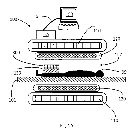
[0036] Fig. 1A schematically illustrates an MRI machine 100 in cross-section,
and shows several well-known features of such machines. A specimen 99 being
scanned by the MRI machine lays on a table 101. Typically, the specimen 99
must
lay as still as possible for the duration of the scan, which can be 30 minutes
or more.
[0037] Main field coils 110 produce a magnetic field around and through the
specimen 99, and body coils 120 subject the specimen 99 to electromagnetic
(e.g.,
radio frequency) stimulus. In response, atoms of the specimen emit
electromagnetic
pulses that may be detected by body coils 120, and/or specimen coils 130.
Specimen
coils 130 may be preferred, because they are closer to the specimen 99, and
produce
6
CA 03062191 2019-10-31
WO 2018/226946 PCT/US2018/036447
signals with greater signal-to-noise ratio ("SNR") than the signals produced
by the
more remote body coils 120. A computer 150 is in data communication with the
MRI machine, such as by communications link 151, and receives and processes
the
signals received by the body coils 120, and/or specimen coils 130, to produce
an
image of internal structures of the specimen. The body coils 120 and specimen
coils
130 are wired to the MRI machine 100. The body coils are in power
communication
and control communication with the MRI machine to receive power and control
signals required to produce the electromagnetic stimulus. Both the body coils
120
and specimen coils 130 are in data communication with the MRI machine 100 to
provide to the MRI machine 100 the signals they detect from the specimen 99.
To
that end, some embodiments of an MRI machine include a controller 140
configured
to provide control signals to the MRI machine, and/or to an array as described
below in connection with control signal 821, and/or to receive signals from
the body
coils 120 and specimen coils 130.
[0038] The quality of the image, and the time needed for the MRI machine
100 to collect a sufficient number of emitted signals to produce the image,
depend in
part on the SNR of the signals received. An increase in the SNR may improve
the
MRI's output and/or reduce the time required to collect signals emitted by the
specimen 99.
[0039] Fig. 1A and Fig. 1B each schematically illustrates an embodiment of a
resonator array 300 for improving the operation of, and results produced by,
an MRI
machine.
[0040] In Fig. 1A, specimen coils 130 are placed between the specimen 99 and
the resonator array 300, and in Fig. 1B, the resonator array 300 is disposed
between
the specimen 99 (in this illustration, a limb or appendage 799 of specimen 99)
and
the specimen coils 130. In some embodiments, the resonator array 300 may be
positioned in the bore 102 of the MRI machine without specimen coils 130, for
example when the MRI machine 100 uses body coils 120 to receive
electromagnetic
pulses emitted by the specimen 99. As used herein, the term "bore" 102 of an
MRI
7
CA 03062191 2019-10-31
WO 2018/226946 PCT/US2018/036447
machine 100 means the place in which the specimen 99 is disposed when being
imaged by the MRI machine 100. For example, in a closed MRI machine 100, the
bore 102 is the interior of the machine's toroid section; in an open MRI
machine 100,
the bore 102 is the space between the machine's top and bottom magnetic areas;
and
in an open upright MRI machine 100, the bore 102 is the space between the
machines left and right magnetic areas.
[0041] Although Figs. 1A and Fig. 1B illustrate the specimen 99 between the
specimen coils 130 and resonator array 300 and the table, that is not a
limitation on
the use of the resonator array 300, which may be placed, with or without
specimen
coils 130, between the specimen 99 and the table 101, as schematically
illustrated in
Fig. 1C and Fig. 1D.
[0042] In contrast to the body coils 120, the resonator array 300 is passive
in
that it does not require or receive power signals, and in some embodiments
does not
require or receive control signals, in order to perform its function. In
illustrative
embodiments, the resonator array 300 (including its unit cells 301) is
separate from,
not part of, body coils 120 or specimen coils 130. Moreover, in illustrative
embodiments, the resonator array 300 (including its unit cells 301) is
physically
separate from the MRI machine 100 and the body coils 120 and specimen coils
130,
and is not wired to MRI machine 100 and the body coils 120 and specimen coils
130.
Also, in contrast to both the body coils 120 and the specimen coils 130, the
resonator
array 300 is not in data communication with the MRI machine 100.
[0043] The inventors have discovered that use of a resonator array 300 as
schematically illustrated in Fig. 1A - Fig. 1D, with or without a specimen
coil 130,
improves the SNR of radiofrequency signals transmitted from the MRI machine
100
to the specimen 99, and improves the SNR of signals emitted by the specimen 99
and received by the MRI machine 100, and can increase the quality of the MRI's
output image, and/or reduce the time required to scan a specimen 99, each of
which
represents an improvement over existing MRI technologies. Due to its unusual
properties, the resonator array 300, and/or its resonators 301, may be thought
of as a
8
CA 03062191 2019-10-31
WO 2018/226946 PCT/US2018/036447
metamaterial. However, that does not require that the resonator array 300,
and/or
its unit cells 301, have a negative index of refraction, negative
permittivity, and/or
negative permeability. In various embodiments, the resonator array 300, and/or
its
unit cells 301, may have a positive index of refraction, positive
permittivity, and/or
positive permeability.
[0044] For example, Fig. 2A shows results of an MRI scan using conventional
MRI technology without a resonator array 300. To produce these results, the
inventors measured the strength of a signal at nine positions (numbered 1-9 in
Fig.
2A) within the bore 102 of a 1.5T MRI machine, and measured the noise at three
positions (numbered 10-11) of the MRI machine. The inventors then calculated
the
average of the noise measurements, and then calculated the SNR of each signal
measurement to the average of the noise measurement. The results are shown
below, and reveal SNRs ranging from 33.2 to 39Ø These results may be
referred-to
as the "baseline" SNRs.
[0045] Signal Strength (Mean)
1 2 3 4
157.2 173.2 178.5 178.1
6 7 8 9
158.5 166.3 172.3 151.3 184.8
[0046] Noise Level (StdDev)
11 12 Avrg
4.4 4.6 5.2 4.7
[0047] SNR
1 2 3 4
33.2 36.6 37.7 37.6
5 6 7 8 9
33.5 35.1 36.4 32.0 39.0
[0048] In contrast to the results shown in Fig. 2A, Fig. 2B and Fig. 2C each
shows results of an MRI scan at the same nine positions using the same 1.5T
MRI
machine with the resonator array 300 having unit cells 301 that are helical
resonators
500 (e.g., Fig. 5A-5C). To produce these results, the inventors measured
signal and
9
CA 03062191 2019-10-31
WO 2018/226946 PCT/US2018/036447
noise in the way described above in connection with Fig. 2A, but obtained
significantly improved SNRs.
[0049] In the embodiment for Fig. 2B, the SNRs were considerably higher
than the baseline SNRs. The results are shown below, and reveal SNRs ranging
from 68.4 to 277.3. Comparing the SNR for location 1 in Fig. 2B to the results
for
location 1 in Fig. 2A shows a large increase in SNR - from a baseline SNR of
33.2 to
an improved SNR of 277.3.
[0050] Signal Strength (Mean)
1 2 3 4
1174 640.4 546.6 481.1
6 7 8 9
193.1 404.5 428.6 267.6 289.7
[0051] Noise Level (StdDev):
11 12 Avrg
4.1 4.7 3.9 4.2
[0052] SNR
1 2 3 4
277.3 151.3 129.1 113.6
5 6 7 8 9
45.6 95.5 101.2 63.2 68.4
[0053] In the embodiment for Fig. 2C employed an array 300 in which the
unit cells 301 had different periodicity (i.e., different spacing relative to
one another)
than the array employed to generate Fig. 2B. That embodiment also produced
SNRs at the same nine positions that are considerably higher than the baseline
SNRs. The results are shown below, and reveal SNRs ranging from 46.2 to 401.5
Comparing the SNR for location 1 in Fig. 2C to the results for location 1 in
Fig. 2A
shows a large increase in SNR - from a baseline SNR of 33.2 to an improved SNR
of
401.5.
[0054] Signal Strength (Mean)
1 2 3 4
1258.0 605.9 498.2 381.7
CA 03062191 2019-10-31
WO 2018/226946 PCT/US2018/036447
6 7 8 9
95.9 363.6 343.1 156.6 144.9
[0055] Noise Level (StdDev):
11 12 Avrg
2.7 3.5 3.2 3.1
[0056] SNR
1 2 3 4
401.5 193.4 159.0 121.8
5 6 7 8 9
30.6 116.0 109.5 50.0 46.2
[0057] In general, a resonator array 300 increases the SNR of signals emitted
by a specimen. For a given MRI machine, relative to the SNR of signals
received by
that MRI machine without use of a resonator array, embodiments of a resonator
array 300 increases the SNR of such signals to at least 45.6, 50, 60, 95, 100,
120, 150,
and/or at least 193.4, or any point between 45 and 401.
[0058] Resonator Array
[0059] An illustrative embodiment of a resonator array 300 is schematically
illustrated in Fig. 3A and Fig. 3B. The array 300 in this embodiment includes
16 unit
cells 301, in a 4x4 array, but other embodiments may use more or fewer unit
cells
301, and may be arranged in different arrangements, such as square, honeycomb
[Fig. 3C], or rectangular for example.
[0060] Each unit cell 301 may also be referred to as a "resonator," because it
is
configured to resonate in response to applied electromagnetic signals, such as
signals applied to a specimen 99 by an MRI machine 100, and/or signals
received by
the unit cell 301 from a specimen 99 in the MRI machine 100. For example, each
unit cell may have an inductance (L) and a capacitance (C), and therefore
resonate as
do LC resonators known in the electrical engineering arts. Each unit cell 301
has a
resonant frequency, and has a Q, as described in connection with Fig. 4A.
11
CA 03062191 2019-10-31
WO 2018/226946 PCT/US2018/036447
[0061] Fig. 4A graphically illustrates quality factor of a resonating device.
A
resonating unit cell may be characterized, in part, by its quality factor,
which may be
referred to as its "Q-factor," or simply as its "Q." A unit cell's Q-factor is
a measure
of its resonance characteristics.
[0062] For example, unit cell 301 may receive an electromagnetic signal
emitted by an atom of a specimen 99 in an MRI machine 100, or from the MRI
machine itself, and that electromagnetic energy may include energy at one or
more
frequencies. The energy will resonate in the unit cell 301, in ways known from
LC
circuits from the art of electrical engineering.
[0063] Ideally, the energy resonates at the resonant frequency fo (401) of the
unit cell, although the unit cell 301 may resonate to some degree at lower
frequencies as well, as illustrated by the curved spectrum in Fig. 4A. The
maximum
energy may be at frequency fo (401), which may be referred-to as the center
frequency, represented by amplitude Al. At other frequencies, the energy is
less
than that at the center frequency 401, as also schematically illustrated in
Fig. 4A. At
some frequency 402 above the center frequency 401 (which may be known as the
upper 3dB frequency), and at another frequency 403 below the center frequency
(which may be known as the lower 3dB frequency), the energy in the resonating
signal will be half of the energy at the center frequency 401. The spectrum
400 in
Fig. 4A shows that some of the energy resonating in the unit cell 301 is above
a noise
floor, indicated at point 405.
[0064] The Q of the unit cell 301 is then defined as the ratio of the center
frequency (fo) divided by difference (Af or delta-f) between the upper 3dB
frequency
and the lower 3dB frequency. In Fig. 4A, the Q is the center frequency 401
divided
by the frequency difference 410 between upper 3dB frequency 402 and lower 3dB
frequency 403. As such, Q is a dimensionless parameter.
[0065] In operation, a unit cell 301 may receive a packet of electromagnetic
energy (e.g., RF energy) from one or more atoms in a specimen 99, the
electromagnetic energy having a frequency at or near the working frequency of
the
12
CA 03062191 2019-10-31
WO 2018/226946 PCT/US2018/036447
MRI machine. For example, in preferred embodiments the electromagnetic energy
having a frequency within +/-5% (inclusive) of the working frequency of the
MRI
machine is defined as being at or near the working frequency of the MRI
machine.
Over time (e.g., during the operation of the MRI machine), each unit cell 301
will
receive many packets of electromagnetic energy, and store the sum of that
energy.
The higher the Q of the unit cell 301, the more efficiently the unit cell 301
stores the
energy it receives.
[0066] In addition, as the unit cell 301 resonates, it amplifies the magnetic
field component of that received electromagnetic energy, and increases the
signal-
to-noise ratio of the received electromagnetic energy. As such, each unit cell
301,
individually, has the ability to resonate, without regard to other unit cells
(if any)
that may be nearby, and has some ability to amplify the magnetic field
component
of received electromagnetic energy.
[0067] The inventors have discovered, however, some limitations on the
usefulness of individual unit cells 301. First, a single unit cell 301 has
limited
capacity to amplify the magnetic field component of received electromagnetic
energy. Second, a unit cell 301 may have a resonant frequency that is not well
matched to the MRI machine 100, in which case its ability to amplify the
magnetic
field component of received electromagnetic energy is less efficient than it
would
otherwise be. Third, it is not possible to change the resonant frequency,
and/or the
Q, of an individual unit cell 301, at least without disassembling and
rebuilding the
unit cell 301.
[0068] The inventors have also discovered, however, that an array 300 of unit
cells 301 has characteristics that are different from a mere aggregation of
the
characteristics of its constituent unit cells 301. In other words, the
resonator array
300 exhibits a synergy.
[0069] For example, an array 300 of unit cells provides a homogenous
amplification of the magnetic field component of received electromagnetic
energy
(see, e.g., Fig. 5D and the text that describes that figure).
13
CA 03062191 2019-10-31
WO 2018/226946 PCT/US2018/036447
[0070] In addition, the resonant frequency of the array 300 may not be the
same as the resonant frequencies, respectively, of its constituent unit cells
301.
Rather, the unit cells 301 couple to one another to produce the resonant
frequency of
the array 300. To that end, in preferred embodiments, the unit cells 301
magnetically couple to one another, and are not wired to one another.
[0071] Moreover, the resonant frequency of the array 300 may be tuned by
adapting the spacing of the unit cells 301 within the array 300.
[0072] In addition, the array 300 is modular, in that unit cells 301 may be
added to an array 300 at the same periodicity (i.e., X-pitch 310 and/or Y-
Pitch 311)
of unit cells 301 already in the array 300, without significantly changing the
resonance characteristics of the array 300. Adding unit cells 301 to an array
300 at
the same periodicity of unit cells 301 already in the array 300 does not
change the
resonance characteristics of the array as much as changing the periodicity of
the unit
cells 301 of the array 300. Adding unit cells in this way may be desirable,
for
example, to increase the size of the array 300 to image a larger specimen 99,
or a
larger portion of a specimen 99.
[0073] Similarly, unit cells 301 already in an array 300 with a given
periodicity may be removed from the array 300 without significantly changing
the
resonance characteristics of the array 300. Removing unit cells 301 from an
array
300 with a given periodicity does not change the resonance characteristics of
the
array as much as changing the periodicity of the unit cells 301 of the array
300.
Removing unit cells may be desirable, for example, to reduce the size of the
array to
fit into the bore 102 of an MRI machine 100, or to image a smaller specimen
99, or a
smaller portion of a specimen 99.
[0074] The resonator array 300 is configured to have a resonance frequency at
or near the working frequency of the MRI machine 100 (i.e., the resonance
frequency
of the array is within +/- 5%, inclusive, of the working frequency of the MRI
machine 100). For example, the working frequency (or "operating frequency") of
a
1.5 Tesla (i.e., 1.5T) MRI machine is approximately 64 MHz (which is a
14
CA 03062191 2019-10-31
WO 2018/226946 PCT/US2018/036447
radiofrequency for purposes of this disclosure), and the working frequency of
a 3
Tesla (i.e., 3T) MRI machine is approximately 128 MHz (which is also a
radiofrequency for purposes of this disclosure).
[0075] The resonance frequency of the resonator array 300 is partially
determined by the periodicity (spacing) of the unit cells 301 of the array
300, and
also by the resonance frequency of the individual unit cells 301. In the
illustrative
resonator array 300 of Fig. 3A and Fig. 3B, the resonators are evenly spaced:
each
unit cell 301 is separated by a dimension, the X-pitch 310, of 37.33 mm in the
X-axis,
and by a dimension, the Y-pitch 311, of 37.33 mm in the Y-axis. In this
configuration, the resonance frequency 463 of the resonator array 300 is
centered at
the working frequency 452 of the MRI machine 100. In general, the difference
between the working frequency 452 of the MRI machine and the resonance
frequency of the resonator array 300 may be specified by the designer or
operator of
the MRI machine. In preferred embodiments, the resonance frequency of the
resonator array 300 is within +/- 5% (inclusive) of the working frequency 452
of the
MRI machine.
[0076] At a larger periodicity (i.e., greater X-pitch 310 and Y-pitch 311),
the
resonance frequency of the resonator array 300 is reduced, and at a lower
periodicity
(i.e., smaller X-pitch 310 and Y-pitch 311), the resonance frequency of the
resonator
array 300 is increased. Fig. 4B graphically illustrates the relationship
between the
periodicity of a resonator array 300 and its frequency response relative to
the
working frequency 452 of an MRI machine. Curve 462 schematically illustrates
the
resonance of an array 300 tuned to the working frequency 452 of the MRI
machine
100, with a resonant frequency at point 463. In contrast, curve 460
schematically
illustrates the resonance of the array 300 tuned to a frequency 450 slightly
below the
working frequency 452 of the MRI machine 100, with its resonant frequency at
point
461, and curve 464 schematically illustrates the resonance of the array 300
tuned to a
frequency 454 slightly higher than the working frequency 452 of the MRI
machine,
with its resonant frequency at point 465.
CA 03062191 2019-10-31
WO 2018/226946 PCT/US2018/036447
[0077] Consequently, the resonance frequency of the resonator array 300 can
be adjusted and established as necessary or desired for a given MRI machine or
application. For example, the inventors have realized that the presence of
soft tissue
near the array 300 may change the permittivity of the area surrounding the
array
300. If such a change of permittivity interferes with or degrades the
operation of the
MRI machine 100 or resonator the resonance frequency of the resonator array
300
may be adjusted by changing the spacing of the unit cells 301 of the resonator
array
300.
[0078] Helical Unit Cell
[0079] An illustrative embodiment 500 of a unit cell 301 in the form of a
helical resonator 500 is schematically illustrated in Fig. 5A, and Fig. 5B,
and Fig. 5C.
The resonator 500 includes a helical conductor 510 around a low-dielectric
core 520.
[0080] The helical conductor 510, which may be copper, is wrapped around
the core 520 so that each successive turn (513) (or "loop") around the core is
separated from its predecessor by a gap 515.
[0081] The unit cell 301 has both inductance (L) and capacitance (C). The
inductance arises from the coiled conductor 510, and the capacitance arises in
the
gap 515 between successive turns 513 of the conductor 510. Consequently, the
resonant frequency of the unit cell 301 is determined, at least in part, by
the number
of turns 513 of the conductor 510 and the dimensions of the gap 515 between
turns
513. A designer may therefore establish the resonant characteristics of the
unit cell
301 to suit a desired application by establishing the inductance and
capacitance
through specification of its properties (e.g., the number of turns 513 and/or
the gap
515) of the coiled conductor 510 and/or the dielectric constant (k) and/or
loss angle
of the core 520. Moreover, the resonant frequency of an array 300 of unit
cells 301
may be tuned by specifying, or adapting, the resonant characteristics of the
unit cells
301 by, for example, increasing or decreasing the number of turns 513 of the
conductor 510, and/or increasing or decreasing the gap 515 between turns 513
of the
conductor 510.
16
CA 03062191 2019-10-31
WO 2018/226946 PCT/US2018/036447
[0082] In some embodiments, the conductor 510 does not overlap itself, but in
other embodiments the conductor 510 may overlap itself as long as there is no
direct
electrical contact between different regions of the conductor 510. For
example, the
conductor 510 may overlap itself if it includes an electrically insulating
coating 512.
[0083] Fig. 5C schematically illustrates a core 520 without the conductor 510.
In some embodiments, the outer surface 523 of the core 520 includes a helical
groove
530 to receive the conductor 510 and define its helical shape.
[0084] The ends 511 of the conductor 510 do not connect to one another, or to
another conductor, or to the conductor 510 of another resonator. Consequently,
the
conductor 510 may be referred to as an open-loop resonator or an open-loop
coil or
an open-loop helical resonator.
[0085] In preferred embodiments, the core 520 has a low dielectric constant
(k) and a low loss angle. For example, the core 520 may be made of materials
such
as polyvinylchloride ("PVC"), which as a dielectric constant of 3 (k=3). As
used
herein, a dielectric constant (relative permittivity) lower than 15 is
considered a
"low-dielectric constant" (or "low relative permittivity") and dielectric
constant
(relative permittivity) greater than or equal to 15 is considered a "high-
dielectric
constant" (or "high relative permittivity").
[0086] The core 520 in some embodiments may, however, have a dielectric
constant of greater than 3, which reduces the size of the unit cell 301 while,
possibly
with adjustments of other properties of the unit cell 301, maintaining the
same
resonant characteristics. For example, the inventors experimented with water,
which has a permittivity of approximately 80 at 20 degrees Celsius, as
schematically
illustrated in Fig. 5G and Fig. 5H and Fig. 51. Unit cell 500 is placed in a
dish 560
encircled by a coupling loop 561 which is coupled to a network analyzer. When
the
dish 560 is filled only with air, the unit cell 500 has a resonant frequency
of 63 MHz,
as shown by point 567 in Fig. 51. However, when the dish contains water such
that
water fills about ten percent (10%) of the core 520 (the surface 566 of the
water at
10%), the unit cell 500 has a resonant frequency of 55 MHz, as shown by point
568 in
17
CA 03062191 2019-10-31
WO 2018/226946 PCT/US2018/036447
Fig. 51. When the dish contains water such that water fills about twenty
percent
(20%) of the core 520 (the surface 566 of the water at 20%), the unit cell 500
has a
resonant frequency of 39 MHz, as shown by point 569 in Fig. 51. Consequently,
it
can be understood that including within a given unit cell 500 a material with
a
permittivity higher than the permittivity of air, the resonant frequency of
the unit
coil 500 is reduced. Conversely, to a produce a unit cell 500 having a given
resonant
frequency, the unit cell 500 can be made smaller (e.g., have fewer turns 513),
relative
to a unit cell 500 having air in its core 520, of the interior 503 of the unit
cell 500 has a
relatively higher relative permittivity, for example between 86 and 173. For
example, some embodiments include a core with a permittivity of between 86 and
173. In some embodiments, the relative permittivity may be even greater than
173.
In some such embodiments include a core 520 made of titanium dioxide.
[0087] Some embodiments omit the core 520, and include a conductor 510
fixed into a helical shape (see, for example, Fig. 5B). In such embodiments,
in air, the
volume within the helical coil 510 has a dielectric constant of air, which is
near one
(k=1).
[0088] The characteristics of a helical resonator 500 may be determined by the
type of MRI machine in which they will be used. In the embodiment of Fig. 5A,
the
core 520 is a hollow cylinder with an outside diameter 522, and an inside
diameter
521, and a height 525. That shape and those dimensions, however, are not
limitations of all embodiments, and other solid or hollow shapes may be used,
including shapes having cross-sections that are square or triangular, to name
but a
few examples. Characteristics of illustrative embodiments of helical
resonators 500
are given below for 1.5T MRI machines and 3T MRI machines.
[0089]
Characteristic 1.5T 3T
Outside diameter 522 3.0 cm 2.0 cm
Height 525 3.2 cm 3.0 cm
18
CA 03062191 2019-10-31
WO 2018/226946 PCT/US2018/036447
Number of turns of conductor 510 25 25
X-pitch 310 3.7 cm 2.3 cm
Y-pitch 311 3.7 cm 2.3 cm
[0090] Operation of Resonator Array
[0091] In operation, the resonator array 300 is placed on or near a specimen
99 in an MRI machine 100, as schematically illustrated for example in Fig. 1A,
Fig.
1B, Fig. 1C and Fig. 1D.
[0092] The resonator array 300 resonates at or near the working frequency 452
of the MRI machine 100, and thereby increases the magnetic field strength of
the RF
signals emitted by the specimen 99. In this way, the SNR of the RF signals is
increased.
[0093] The resonator array 300 increases the magnetic field component of
radiofrequency energy during signal transmission by the MRI machine 100 to the
specimen 99, and reception of radiofrequency energy from the specimen 99 to
the
MRI machine.
[0094] For example, Fig. 5D graphically illustrates the magnetic field
intensity
at various elevations, above the top 302 of the unit cells 301 (e.g., in the Z
axis),
within an embodiment of a resonator array 300 in which the unit cells 301 are
helical
resonators 500. Fig. 5E graphically illustrates the magnetic field enhancement
ratio
at the center point of said array 300 as a function of distance from the
middle 303 of
the unit cells 301, and shows that the enhancement is greatest near the middle
303 of
the unit cells 301, and decreases with distance from the middle 303 of the
unit cells.
It should be noted, from Fig. 5D, that the magnetic field enhancement is
substantially uniform across the resonator array 300. In the helical resonator
500,
magnetic field enhancement arises due to the overlap between the self-resonant
frequency of the helical resonator 500 and the frequency of excitation of the
magnetic field.
19
CA 03062191 2019-10-31
WO 2018/226946 PCT/US2018/036447
[0095] Beneficially, the resonator array 300 also substantially avoids
generation of an electric field, or minimizes an increase in the electrical
field
component of those RF signals. For example, an electric field created at one
end 501
of a resonator 500 very nearly completely cancels an electric field at the
other end
502. Also, in various embodiments increase in the electrical field component
of
those RF signals less than the increase the magnetic field component of those
RF
signals. This is beneficial for specimen safety, since electrical fields may
cause burns
to the specimen, for example. Specifically, the helical resonators 500 are
configured
such that they do not couple with the electric field of the RF signals,
thereby
mitigating amplification by the helical resonators 500, and the array 300, of
the
electric field component of RF signals.
[0096] Fig. 5F schematically illustrates an alternate embodiment of a helical
resonator 500, including an additional fixed reactance 550 electrically
coupled
between the ends 511 of the unit cell's conductor 510. The additional
reactance 550
is in addition to the inductance and/or capacitance of the conductor 510. The
additional reactance 550 may be a capacitance (C), or an inductance (L). In
practice,
the additional reactance 550 interacts with the capacitance or inductance of
the other
structures of the helical resonator 500. For example, because the resonant
frequency
of the helical resonator 500 is dominated by 1/VT, inclusion of an inductor
(L) in
the additional reactance 550 produces a helical conductor 500 with the same
resonant characteristics described above, but with a fewer number of turns 513
or/and a smaller diameter 521 of the helix. Likewise, inclusion of a capacitor
(C) in
the additional reactance 550 produces a helical conductor 500 with the same
resonant characteristics described above, but requires less capacitance from
the
helical conductor 510.
[0097] BC-SRR Unit Cell
[0098] An embodiment of a unit cell 301, in the form of a broadside-coupled
split ring resonator 600 ("BC-SRR"), is schematically illustrated in Fig. 6A.
The BC-
CA 03062191 2019-10-31
WO 2018/226946 PCT/US2018/036447
SRR resonator 600 includes two "C" shaped split-ring resonators 610, 620, each
defining a gap 611 and 621, respectively. The split-ring resonators 610, 620
are
disposed parallel to one another in the X-Y plane of Fig. 6A, and do not
intersect or
physically contact one another. As illustrated in Fig. 6A, the split-ring
resonators
610, 620 are positioned such that their gaps, 611 and 621, are diametrically
opposed
to one another (i.e., 180 degrees from one another). The BC-SRR unit cells
resonate
well even if the gaps 611 and 621 are not 180 degrees from one another, but
this is
the preferred arrangement because the inventors have discovered that this
arrangement produces the lowest electrical field. The top split-ring resonator
610
defines a top surface 601 of the BC-SRR 600, and a bottom surface 602 of the
BC-SRR
600, for reference.
[0099] In the BC-SRR unit cell 600, magnetic field enhancement arises due to
the overlap between the self-resonant frequency of the unit cells 600 and the
frequency of excitation of the magnetic field. The BC-SRR unit cells are
configured
so that excited electric dipoles exhibit cancellation, thereby mitigating
amplification
by the unit cells 301, and the array 300, of the electric field component of
RF signals.
[00100] Figs. 6B-6D schematically illustrate operating characteristics of a
BC-SRR 600 configured for resonance at 64 Mhz.
[00101] .. Fig. 6B schematically illustrates the magnetic field (Bz)
distribution in a cross-section in the X-Z plane, of a single unit cell BC-SRR
600, and
Fig. 6C schematically illustrates that magnetic field distribution in the X-Y
plane 10
millimeters away from the top surface 601 of the BC-SRR 600. Fig. 6D
schematically
illustrates the magnetic field enhancement factor at a point 10 millimeters
away
from the top surface 601 of the BC-SRR 600. In this embodiment, an electric
field
created at one end of the BC-SRR 600 (i.e., the end nearest the top surface
601) very
nearly completely cancels an electric field at the other end (i.e., the end
nearest the
bottom surface 602).
21
CA 03062191 2019-10-31
WO 2018/226946 PCT/US2018/036447
[00102] Fig. 6E schematically illustrates an array 300 of BC-SRR unit
cells 600. In this embodiment, the BC-SRRs are photo-lithographically
fabricated on
a high-permittivity substrate 650.
[00103] Embodiments of resonator arrays 300 may be rigid or flexible.
For example, the array 300 of BC-SRR resonators in Fig. 6E may be rigid, while
the
arrays 300 of Fig. 7A and Fig. 7B are flexible. The BC-SRR array 300 of Fig.
7A has a
flexible substrate 700, and as shown in Fig. 7A may even be wrapped around the
limb 799 of a specimen 99, for example. Similarly, the array 300 of helical
resonators
500 has a flexible substrate 700, and may be contoured to a portion of the
body of a
specimen 99, or even formed into a cone.
[00104] In some applications, it may be desirable to increase the
magnetic field component of radiofrequency energy only during transmission of
radiofrequency signals from the specimen to the MRI machine, and not during
transmission of radiofrequency energy from the MRI machine 100 to the specimen
99. To that end, some embodiments include a tunable array 300 and tunable unit
cells 301.
[00105] Figs. 8A-8G schematically illustrate embodiments of tunable
unit cells 301. An array 300 with tunable unit cells 301 is tunable by tuning
its
constituent unit cells 301.
[00106] Fig. 8A schematically illustrates a tunable unit cell 301. The
tunable unit cell 301 may include, for example, a helical coil 500 as
described above,
or a BC-SRR 600 as described above, along with a coupler 801.
[00107] The coupler 801 has at least two electrical states (or
"impedance" states), including a first state in which the electrical
conductivity of the
coupler 801 is lower than its electrical conductivity in the second state.
Stated
alternately, the electrical impedance of the coupler 801 is higher in the
first state
than it is in the second state. The resonant properties of the unit cell 301
vary
depending on the state of the coupler 801.
22
CA 03062191 2019-10-31
WO 2018/226946 PCT/US2018/036447
[00108] In the embodiment of Fig. 8A, the coupler 801 is electrically
coupled between the two ends 511 of a helical coil (e.g., 500), but may be
coupled to
one or more unit cells in several ways, as described below. In its first
state, the
impedance of the coupler 801 is sufficiently high that the operation of the
unit cell
301 is as described above. In the second state, however, the impedance of the
coupler is lower, creating an electrical connection via a conductive path
between the
two ends 511 of the coil 500. That electrical connection changes the
properties of the
helical coil 500 so that it no longer resonates, or so that its resonant
frequency is
shifted to a frequency away from the working frequency 452 of the MRI machine.
In
general, the difference between the working frequency 452 of the MRI machine
and
the helical coil's resonant frequency, when the coupler 801 is in the second
state,
may be specified by the designer or operator of the MRI machine. For example,
in
preferred embodiments, when the coupler 801 is in the second state, the
resonant
frequency of the helical coil 500 changes such that - if it resonates at all -
its resonant
frequency is at least +/- 15 percent different than the working frequency 452
of the
MRI machine, and/or at least +/- 15 percent different than its resonant
frequency
when the coupler 801 is in the first state. Consequently, changing the state
of the
coupler 801 changes the resonant properties of the unit cell 301. In general,
when the
resonant frequency of a unit cell 300 (in this example, the helical coil 500)
is at least
+/- 15 percent different than the working frequency 452 of the MRI machine,
and/or at least +/- 15 percent different than its resonant frequency when the
coupler 801 is in the first state, the unit cell is said to be "effectively
non-resonant."
[00109] Moreover, in an array 300 of such unit cells 301, changing the
state of the coupler 801 changes the operating properties of the array 300.
For
example, when the coupler 801 is in the first state, each unit cell 301, and
an array
300 of such unit cells 301, operate as described above in connection with
Figs. 3A-
3C, 4A-4B, 5A-5F and 6A-6E. When the coupler 801 is in the second state, the
resonant properties of the array 300 are changed such that amplification of
the
magnetic field produced by the array 300 is reduced. In effect, each unit cell
301,
23
CA 03062191 2019-10-31
WO 2018/226946 PCT/US2018/036447
and the array 300, and can be "turned on" by placing the coupler 801 in the
first
state, and "turned off" by placing the coupler 801 in the second state. A
variety of
couplers 801, unit cell 301 configurations, and array 300 configurations, are
described below. In general, the coupler 801 may be referred to as a non-
linear
material or non-linear device.
[00110] Fig. 8B schematically illustrates an array 300 of BC-SRRs 600.
Each BC-SRR unit cell includes at least one coupler 801, and in some
embodiments
more than one coupler 801. The coupler 801 in Fig. 8B is referred to as a
semiconductor patch 810. The semiconductor patch 810 may be, for example,
doped
silicon that changes its impedance in response to RF energy from the MRI
machine
100, but not in response to the generally much lower amount of RF energy of
signals
from the specimen 99. The semiconductor patch may be said to be nonlinear.
[00111] In illustrative embodiments, the semiconductor material of the
semiconductor patch 810 may be GaAs, InAs, or InSb, to name but a few
examples.
A preferred embodiment uses GaAs as the semiconductor material. Intrinsic
GaAs,
without doping, has a carrier density of 2.1*106 cm-3.
[00112] The properties of the semiconductor are tuned by doping.
Doping is known in the semiconductor arts. In illustrative embodiments, the
GaAs
is doped it to have a carrier density of 3*107 cm-3.
[00113] In illustrative embodiments, a semiconductor patch 810 may be
prepared from a 2 inch or 4 inch wafer (0.5mm thick) of doped semiconductor
(e.g.,
GaAs doped as above). The wafer is diced into patches with 3 mm by 5 mm in
size,
and two electrodes are patterned onto the patch in ways known in the
semiconductor art, with micrometer size gap such as 2*10-6 m.
[00114] As schematically illustrated in Fig. 8A, the semiconductor patch
810 is electrically coupled (e.g., soldered) to unit cell 301. By applying
alternating
magnetic field (e.g., a radiofrequency electromagnetic signal), a strong
electric field
can be induced at the micrometer size gap as high as 400 kV/cm to excite the
impact
ionization at the gap.
24
CA 03062191 2019-10-31
WO 2018/226946 PCT/US2018/036447
[00115] In illustrative embodiments, when the MRI machine 100 is not
applying such an alternating magnetic field (e.g., a radiofrequency
electromagnetic
signal), the conductivity of the semiconductor patch 810 is approximately 1*10-
7
(ohm cm)-1 (in illustrative embodiments, with carrier density up to 107 cm-3).
In
contrast, when the MRI machine 100 applies stimulus as described above, the
conductivity of the doped GaAs of the semiconductor patch 810 increases to
approximately 20 (ohm cm)-1 (in illustrative embodiments, with carrier density
up to
1018 cm-3), resulting in the resonant frequency shift of the unit cell 301
described
herein.
[00116] Taking a doped semiconductor patch 810 as an example, during
transmission of RF energy by the MRI machine 100, the electric field at the
gap of
the BC-SRR 600 or inside the metallic helices 500 is very high, and so the
carrier
density of the doped silicon semiconductor patch 810 is excited to a much
higher
level than in the absence of such RF energy. In this state, the doped silicon
semiconductor patch 810 can be treated as a conductor. Consequently, during
transmission of RF energy by the MRI machine 100, the resonant frequency of
the
unit cells 301 deviates from the frequency of RF energy transmitted by the MRI
machine 100.
[00117] In contrast, during reception by a unit cell 301 of RF signals
from the patent 99 - which occurs when the MRI machine 100 is not transmitting
RF
energy - the above-mentioned electric field strength is much lower, and so the
doped silicon semiconductor patch 810 is not an effective conductor.
Consequently,
the resonant frequency of each unit cell 301 remains aligned with the working
frequency 452 of the MRI machine 100, as the doped silicon semiconductor patch
810 is functioning as an isolator.
[00118] The semiconductor patch 810 is disposed within the first gap
611 of the first SRR 610 in the BC-SRR 600, and changes its state in response
to RF
energy from the MRI machine 100. More specifically, in the absence of RF
energy
from the MRI machine 100, the semiconductor patch 810 is in the first state
(high
CA 03062191 2019-10-31
WO 2018/226946 PCT/US2018/036447
impedance), so the BC-SRR 600 behaves as described above in connection with
Figs.
6A-6E. When the MRI machine transmits RF energy, however, the semiconductor
patch 810 changes its impedance to the second state (low impedance), thus
electrically coupling the opposing ends 612, 612 of the first gap 611, thereby
changing the physical and resonant characteristics of the BC-SRR 600, and
thereby
changing the operating characteristics of the array 300, as described above.
[00119] In some embodiments, each of the SRRs 610, 620 of a BC-SRR
600 includes a semiconductor patch 810 as described above, to even further
change
the characteristics of each unit cell 301 and of the array 300.
[00120] .. Fig. 8C schematically illustrates an array 300 of helical unit
cells
500. In this embodiment, a semiconductor patch 810 is coupled between the
respective ends 511 of adjacent unit cells 301, and preferably is disposed
within the
interior 802 if the helical coil itself - e.g., surrounded by the helical
turns 513. In this
configuration, in the absence of RF energy from the MRI machine 100, the
semiconductor patch 810 is in the first state (high impedance), so the
resonator 500
behaves as described above in connection with Figs. 5A-5F. When the MRI
machine
transmits RF energy, however, the semiconductor patch 810 changes its
impedance
to the second state (low impedance), thus coupling together the adjacent unit
cells
301, and thereby changing the operating characteristics of the array 300, as
described
above.
[00121] .. Fig. 8D and Fig. 8E schematically illustrate an alternate
embodiment of a coupler 801, in which the coupler 801 is a switch 820, and
alternate
embodiments of arrays 300 with such couplers 801. Although the unit cells 301
in
these embodiments respond to the control signal 821 (and therefore may be said
to
be in control communication with the MRI machine 100 or its controller 140),
each of
the arrays 300 may still be considered passive in that it does not require
input of
external energy in order to amplify the magnetic field and increase the SNR of
signals from the specimen 99.
26
CA 03062191 2019-10-31
WO 2018/226946 PCT/US2018/036447
[00122] In Fig. 8D, at least one SRR 610 of each BC-SRR 600 has a switch
820 disposed in its gap 611. A control signal 821 from the MRI machine (e.g.,
from
controller 140) changes the switch 820 between its first state (high
impedance) and
second state (low impedance), thus electrically coupling the opposing ends
612, 612
of the first gap 611. Those two states change the resonant characteristics of
the BC-
SRR 600, and thereby change the operating characteristics of the array 300, as
described above in connection with Fig. 8B. In some embodiments, each of the
SRRs 610, 620 of a BC-SRR 600 includes a switch 820 as described above, to
even
further change the characteristics of each unit cell 301 and of the array 300.
[00123] Fig. 8E schematically illustrates an array 300 of helical unit
cells
500. In this embodiment, a switch 820 is coupled between the respective ends
511 of
adjacent unit cells 301. A control signal 821 from the MRI machine changes the
switch 820 between its first state (high impedance) and second state (low
impedance). Those two states change the resonant characteristics of the
helical cell
500, and thereby change the operating characteristics of the array 300, as
described
above in connection with Fig. 8C.
[00124] Fig. 9 is a flow chart for an embodiment of a method of
magnetic resonant imaging a specimen 99. Step 901 requires providing an MRI
machine 100 having a bore 102 and a working frequency. The MRI machine 100
may be, for example, a 1.5 Tesla MRI machine having a working frequency of 64
MHz or a 3 Tesla MRI machine having a working frequency of 128 MHz.
[00125] Step 902 includes placing the specimen in the bore 102, and step
903 includes placing, in the bore with the specimen, an array 300 of unit
cells 301. It
should be noted that steps 902 and 903 may be performed in any order with
respect
to one another.
[00126] In preferred embodiments, the array 300 is sized to be disposed
within the bore 102 of the MRI machine 100 along with a specimen 99 in the
bore
102, when the MRI machine 100 is imaging the specimen 99. For example, the
array
300 of unit cells 301 may be any of the arrays 300 disclosed above.
27
CA 03062191 2019-10-31
WO 2018/226946 PCT/US2018/036447
[00127] In preferred embodiments, each unit cell 301 of the array 300
has a resonant frequency, and the array 300 has a resonance frequency at or
near the
working frequency of the MRI machine 100.
[00128] At step 904, the method images the specimen 99 with the MRI
machine in ways known in the art.
[00129] In some embodiments, step 904 further includes controlling the
coupler 801 to be in its first state (high-impedance) when the MRI machine is
not
applying electromagnetic (e.g., radio frequency) stimulus to the specimen 99,
and to
be in its second state (low impedance) when the MRI machine is applying such
stimulus to the specimen. For example, if the coupler 801 is a switch 820,
step 904
may include controlling the switch 820 with a control signal 821 from
controller 140,
as described above. As another example, if the coupler 801 is a semiconductor
patch 810, step 904 may include controlling the semiconductor patch 810 to be
in its
first state (high-impedance) by withholding electromagnetic stimulus from the
MRI
machine 100, and controlling the semiconductor patch 810 to be in its second
state
(low-impedance) by applying electromagnetic stimulus from the MRI machine 100.
In such embodiments, the coupler 801 is in a high-impedance state (and so the
unit
cells 301 resonate) when the MRI is not applying electromagnetic stimulus to
the
specimen, and the coupler 801 is in a low-impedance state (and so the unit
cells 301
are effectively non-resonant) when the MRI is applying such electromagnetic
stimulus to the specimen.
[00130] The following is a list of reference numbers used herein.
[00131] 99: Specimen;
[00132] .. 100: MRI machine in cross-section;
[00133] 101: Table;
[00134] .. 102: Bore of MRI machine;
[00135] 110: Main field coils;
[00136] 120: Body coils;
[00137] 130: Specimen coils;
28
CA 03062191 2019-10-31
WO 2018/226946 PCT/US2018/036447
[00138] 140: MRI machine controller;
[00139] 150: Computer;
[00140] 151: Computer communications link;
[00141] 300: Resonator array;
[00142] 301: Unit cell;
[00143] 302: top of unit cell;
[00144] 303: middle of unit cell;
[00145] 310: X-Pitch;
[00146] 311: Y-Pitch;
[00147] 400: Response of a resonator;
[00148] 401: Center frequency;
[00149] 402: Upper 3dB point;
[00150] 403: Lower 3dB point;
[00151] 405: Noise level;
[00152] 410: Frequency delta;
[00153] 450: Frequency below working frequency of MRI machine;
[00154] 452: Working frequency of MRI machine;
[00155] 454: Frequency above working frequency of MRI machine;
[00156] 460: Resonance response of array tuned to frequency below
working frequency of MRI machine;
[00157] 461: Resonant frequency of array tuned to frequency below
working frequency of MRI machine;
[00158] 462: Resonance response of array tuned to working frequency of
MRI machine;
[00159] 463: Resonant frequency of array tuned to working frequency of
MRI machine;
[00160] 464: Resonance response of array tuned to frequency above
working frequency of MRI machine;
29
CA 03062191 2019-10-31
WO 2018/226946 PCT/US2018/036447
[00161] 465: Resonant frequency of array tuned to frequency above
working frequency of MRI machine;
[00162] 500: Helical resonator;
[00163] 501: Top end of resonator;
[00164] 502: Bottom end of resonator;
[00165] 503: Interior of resonator;
[00166] 510: Conductor;
[00167] 511: End of conductor;
[00168] 512: Electrically insulating covering;
[00169] 513: Turn;
[00170] 515: Conductor gap;
[00171] 520: Core;
[00172] 521: Core outside diameter;
[00173] 522: Core inside diameter;
[00174] 523: Outer surface of core;
[00175] 525: Core height;
[00176] 530: Groove;
[00177] 550: Additional reactance;
[00178] 560: Dish;
[00179] 561: Coupling loop;
[00180] 565: Water;
[00181] 566: Surface of water;
[00182] 567: Dry resonant frequency;
[00183] 568: 10% water resonant frequency;
[00184] 569: 20% water resonant frequency;
[00185] 600: BC-SRR resonator;
[00186] 601: Top surface of BC-SRR;
[00187] 602: Bottom surface of BC-SRR;
[00188] 610: First split-ring resonator;
CA 03062191 2019-10-31
WO 2018/226946 PCT/US2018/036447
[00189] 611: First gap;
[00190] 612 -613: Opposing ends of first gap;
[00191] 620: Second split-ring resonator;
[00192] 621: Second gap;
[00193] 650: High-permittivity substrate;
[00194] 700: Flexible substrate;
[00195] 799: Limb of specimen;
[00196] 801: Coupler;
[00197] __ 802: Interior of helical coil;
[00198] 810: Semiconductor patch;
[00199] 820: Switch;
[00200] The embodiments of the inventions described above are
intended to be merely exemplary; numerous variations and modifications will be
apparent to those skilled in the art. All such variations and modifications
are
intended to be within the scope of the present invention as defined in any
appended
claims.
31