Note: Descriptions are shown in the official language in which they were submitted.
Apparatus and Method for Isolating Stem Cells
Field of the Invention
The present invention relates to isolating stem cells, and more specifically,
to
an apparatus and method for isolating stem cells from a sample.
Background Art
Stem cells are biological cells that can differentiate into other types of
cells
.. and can divide to produce more of the same type of stem cells. They are
found in multicellular organisms, including mammals such as humans. In
mammals, adult stem cells can act as a repair system for the body by
replenishing adult tissues, thus giving rise to use of stem cells in medical
therapies such as bone marrow transplantation for the treatment of leukemia
and lymphoma.
Mesenchymal stem cells (MSCs) can differentiate into the cells that make up
bone, cartilage, tendons, and ligaments, as well as muscle, neural and other
progenitor tissues. As such, mesenchymal stem cells have been the main
type of stem cells studied in the treatment of diseases affecting these
tissues. Hematopoietic stem cells (HSCs) can differentiate into cells that
make up blood including red blood cells, white blood cells and platelets. As
such, Hematopoietic stem cells have been the main type of stem cells studied
in the treatment of diseases affecting the blood, such as leukemia.
Stem cell therapy is the use of the harvested stem cells to treat or prevent a
disease or condition. Adult stem cell treatments have been successfully used
to treat leukemia and related bone/blood cancers through bone marrow
transplants. Bone marrow transplant is a form of stem cell therapy that has
CA 3062243 2019-11-21
been used in treating several other conditions including liver cirrhosis,
chronic limb ischemia and end stage heart failure. Adult stem cells are also
used in veterinary medicine to treat tendon and ligament injuries in horses.
In stem cell therapy, healthy and functional stem cells must be harvested
from a donor, who may or may not be the same person as the patient to be
treated. There are several well-known sources of adult stem cells, also called
somatic stem cells, in humans:
(i) Bone marrow, which require extraction by harvesting, that is, drilling
into
bone (typically the femur or iliac crest);
(ii) Adipose tissue (fat cells), which require extraction by liposuction; and
(iii) Blood, which requires extraction through apheresis, wherein blood is
drawn from the donor (similar to a blood donation), centrifuged to separate
components and, after selected components are drawn off, returned to the
donor.
Stem cells can also be taken from umbilical cord blood just after birth. Of
all
stem cell types, autologous harvesting, where the cells are obtained from the
patient's own body, involves the least risk of rejection.
Prior to treatment of the patient, the donor stem cells must be isolated from
the bone marrow, adipose tissue, blood or other tissue which was extracted
from the donor. Current methods for isolating stem cells from extracted
tissue are based on centrifugation. Specifically, any solid-liquid mixture or
liquid that may contain stem cells .is subjected to centrifugation, and the
application of centrifugal force attempts to separate particles according to
their size, shape, density and/or viscosity. Centrifugation is widely used and
often effective in many biotechnology applications. However, it does not
2
CA 3062243 2019-11-21
isolate stem cells efficiently. Instead, centrifugation merely sends any
nucleated cells or heavy cells to the bottom of the centrifugation tube. This
method does not isolate or select stem cells in an efficient manner.
.. Furthermore, devices used for stem cell isolation tend to be relatively
large,
cumbersome and difficult to transport. For example, centrifuge machines are
not easily portable.
It would be beneficial to develop a method, and perhaps an apparatus, by
which stem cells can be isolated from extracted tissue in a cost-effective and
efficient manner.
Furthermore, it would be beneficial to develop an apparatus which is easy to
use, relatively small in size and easily portable.
Summary of the Invention
In one aspect, the present invention comprises an apparatus for isolating
stems cells from extracted mammalian tissue comprising:
a portable hollow casing having fixed dimensions and a sized internal spatial
volume;
a filter housed and contained within said sized internal spatial volume,
wherein said filter captures particles in said extracted mammalian tissue
having a diameter of about 5 to 10 microns or more and allows particles in
said extracted mammalian tissue having a diameter of less than about 5 to
10 microns to pass through;
a first channel to which a container holding said extracted mammalian tissue
can attach, and through which said extracted mammalian tissue is input into
the hollow casing;
3
CA 3062243 2019-11-21
wherein a stem cell collection chamber can attach to said first channel, and
the particles having a diameter of about 5 to 10 microns or more are output
from the hollow casing through said first channel and collected in the stem
cell collection chamber; and
a second channel to which a remnant collection chamber can attach, and
through which the particles having a diameter of less than about 5 to 10
microns are output from the hollow casing and collected in the remnant
collection chamber.
In another aspect, the present invention provides a method for isolating stem
cells from extracted mammalian tissue comprising:
(a) providing a container holding said extracted mammalian tissue;
(b) attaching the container holding said extracted mammalian tissue to an
apparatus for isolating stems cells from extracted mammalian tissue
comprising:
a portable hollow casing having fixed dimensions and a sized internal spatial
volume;
a filter housed and contained within said sized internal spatial volume,
wherein said filter captures particles in said extracted mammalian tissue
having a diameter of about 5 to 10 microns or more and allows particles in
said extracted mammalian tissue having a diameter of less than about 5 to
10 microns to pass through;
a first channel to which the container holding said extracted mammalian
tissue can attach, and through which said extracted mammalian tissue is
input into the hollow casing;
wherein a stem cell collection chamber can attach to said first channel, and
the particles having a diameter of about 5 to 10 microns or more are output
from the hollow casing through said first channel and collected in the stem
cell collection chamber; and
4
CA 3062243 2019-11-21
a second channel to which a remnant collection chamber can attach, and
through which the particles having a diameter of less than about 5 to 10
microns are output from the hollow casing and collected in the remnant
collection chamber;
wherein the hollow casing comprises a top portion and a bottom portion
which are detachable from each other;
wherein the filter is inserted between the top portion and the bottom portion;
(c) causing the extracted mammalian tissue to move from the container
holding said extracted mammalian tissue, through the first channel, into the
hollow casing and into contact with the filter;
(d) allowing particles having a diameter of about 5 to 10 microns or more to
be captured by the filter, move out of the hollow casing through the first
channel and into the stem cell collection chamber; and
(e) allowing particles having a diameter of less than about 5 to 10 microns to
pass through the filter, move out of the hollow casing through the second
channel and into the remnant collection chamber.
Brief Description of the Drawings
Advantages of the invention will become apparent upon reading the following
description of the drawings and description of the invention.
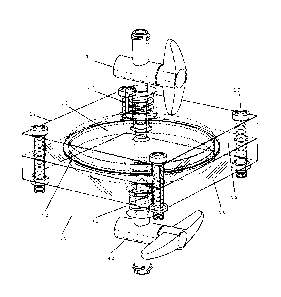
Figure 1 illustrates a perspective view of a preferred embodiment of the
apparatus of the present invention;
Figure 2 illustrates an exploded view of a preferred embodiment of the
apparatus of the present invention;
.. Figure 3 illustrates a cross-sectional view of a preferred embodiment of
the
apparatus of the present invention where the top portion and bottom portion
of the hollow casing are detached from each other;
Figure 4 illustrates a cross-sectional view of a preferred embodiment of the
apparatus of the present invention where a syringe containing a sample of
5
CA 3062243 2019-11-21
blood or blood plasma and/or saline solution is about to be attached to the
second channel;
Figure 5 illustrates a cross-sectional view of a preferred embodiment of the
apparatus of the present invention where a syringe containing a sample of
blood or blood plasma and/or saline solution is attached to the second
channel and the sample of blood or blood plasma and/or saline solution is
being input into the bottom portion;
Figure 6 illustrates a cross-sectional view of a preferred embodiment of the
apparatus of the present invention with the valves at both the first channel
and second channel in a closed position;
Figure 7 illustrates a cross-sectional view of a preferred embodiment of the
apparatus of the present invention where a syringe containing extracted
mammalian tissue is attached to the first channel;
Figure 8 illustrates a cross-sectional view of a preferred embodiment of the
apparatus of the present invention where a syringe containing extracted
mammalian tissue is attached to the first channel and the extracted
mammalian tissue has been input into the hollow casing;
Figure 9 illustrates a cross-sectional view of a preferred embodiment of the
apparatus of the present invention where a syringe for collecting remnants is
attached to the second channel and the remnants have been output from the
bottom portion;
Figure 10 illustrates a cross-sectional view of a preferred embodiment of the
apparatus of the present invention where a syringe for collecting stem cells
is
attached to the first channel and stem cells are being output from the hollow
casing;
Figure 11 illustrates a syringe which can be used with a preferred
embodiment of the apparatus of the present invention;
Figure 12 illustrates a timeline for the generation of homogenous MSC
populations. BM preparations isolated from wild-type female C57BL/6 mice
6
CA 3062243 2019-11-21
were either plated directly or processed once or twice using the apparatus of
the present invention. For this experiment, n=6/group and "P<0.01;
Figure 13 illustrates an assessment of the number of MSC-like colonies at
day 10 post-plating. BM preparations isolated from wild-type female C57BL/6
mice were either plated directly or processed once or twice using the
apparatus of the present invention. The number of generated colonies was
quantified using a contrast phase microscope. For this experiment,
n=6/group and "P<0.01; and
Figure 14 illustrates an assessment of the number of MSC colonies free of
"satellite cells" at day 10 post-plating. BM preparations isolated from wild-
type female C57BL/6 mice were either plated directly or processed once or
twice using the apparatus of the present invention. The number of generated
colonies free of "satellite cells" was quantified using a contrast phase
microscope. For this experiment, n=6/group and "P<0.01.
Detailed Description of the Preferred Embodiments
Tissue containing stems cells are harvested from a donor. Such extracted
tissue may be taken from a source such as, but not limited to, bodily fluid,
fat, bone marrow, umbilical cord and the placenta. In the case of some types
of tissue such as bone marrow, the extracted tissue may be mixed with an
anticoagulant such as heparin or acid citrate dextrose solution (ACD). The
specific method used to harvest the tissue containing stem cells differs
depending on the target location.
In the case of bone marrow, the bone marrow can be aspirated from a donor
using a commercial trochar. The aspiration site, preferably at the posterior
superior iliac crest of the donor, is marked upon visualization with
ultrasound. An anesthetic, such as 2% Lidocaine, may be injected into the
soft tissue and periosteum. An entry point through the donor's skin is created
with an introducer needle, and the bone is drilled through the periosteum
7
CA 3062243 2019-11-21
into the spongy bone. Using the trochar, 1 to 2 cc of bone marrow may be
aspirated per level while slowly withdrawing until approximately 5 to 100 cc
of bone marrow is collected in multiple syringes. Preferably, the syringes
contain an anticoagulant such as heparin to mix with the extracted bone
marrow.
In the case of adipose-derived aspiration, an aspiration site, preferably at
the
suprapubic abdominal of the donor, is marked. An anesthetic, such as 2%
Lidocaine, may injected in the supramuscular space. A tumescent fluid is
prepared, preferably comprising: 500 ml injectable saline, 25 cc plain 2%
Lidocaine, 2 ampoules of epinephrine 1:1000, and 10 cc 8.4% sodium
bicarbonate. Entry points through the donor's skin may be created with 18
gauge needles, and 60 cc of the tumescent fluid is injected slowly in the
abdominal fat space. After preferably waiting 5 to 10 minutes, lipoaspiration
of the fat tissue along with the tumescent fluid is conducted. Much of the
tumescent fluid separates from the fat without any action required. The fat
tissue may be centrifuged for about 4 minutes to separate it from the rest of
the tumescent fluid. The lipoaspirate is preferably emulsified and transfered
to smaller syringes.
The harvested tissue is passed through the apparatus 10 of the present
invention to isolate the stem cells. Isolated stem cells may include but are
not limited to mesenchymal stem cells (MSCs), hematopoietic stem cells
(HSCs), pericytes, fibroblasts, tissue-specific stem cells, embryonic stem
cells, induced pluripotent stem cells and others. As shown in Figures 1 to 10,
the apparatus 10 of the present invention separates the stem cells into a
stem cell collection chamber 100, while the remnants go into a remnant
collection chamber 200.
8
CA 3062243 2019-11-21
Referring to Figures 1, 2 and 10, the apparatus 10 of the present invention
comprises a filter 50 which captures the stem cells based on their size of
generally about 8 to 10 microns (micrometers) or more, such as for example
mesenchymal, hematopoetic, pericytes and fibroblasts, and sends them to a
stem cell collection chamber 100. In one preferred embodiment, the stem
cell collection chamber 100 forms part of a syringe, as shown in Figure 11.
Other cells in the extract pass through the filter 50 and are sent to a
remnant
collection chamber 200, as shown in Figure 9.
The portable hollow casing 12 may comprise one or more of many different
solid materials such as metal, glass or plastic. In one preferred embodiment
which is illustrated in Figures 1 to 3, the hollow casing 12 comprises a top
portion 14 and a bottom portion 16 which are detachable from each other.
When the top portion 14 and the bottom portion 16 are detached from each
other, the filter 50 (or a new or replacement filter) can be inserted between
them.
With reference to Figures 1 to 3, the top portion 14 and the bottom portion
16 can be subsequently attached to each other, with the filter 50 housed and
contained within the hollow casing 12. The top portion 14 and the bottom
portion 16 of the hollow casing 12 may be attached to each other by any
suitable fastening means 18, such as with screws and nuts.
Preferably, the pressure inside the hollow casing 12, more preferably in the
top portion 14 of the hollow casing, is set to about 1 to 5 atm (about 14.7
psi
to 73.5 psi), more preferably about 1.5 atm to 5 atm, even more preferably
about 2 atm to 5 atm, even more preferably about 3 atm to 5 atm.
As best shown in Figure 2, the filter 50 preferably comprises a nylon-based
disc filter, a paper-based filter or a ceramic-based filter, which functions
as a
9
CA 3062243 2019-11-21
semi-permeable barrier. Preferably, the filter operates using surface
filtration, where particles that cannot pass through the pores of the filter
are
caught on or above the filter surface.
The nylon-based disc filter, paper-based filter or ceramic-based filter is
preferably manufactured to have pores that allow particles having a diameter
of less than about 5 to 10 microns to pass through, more preferably less than
about 5 to 8 microns, more preferably less than about 5 to 7 microns, even
more preferably less than about 5 to 6 microns, even more preferably less
than about 5 microns. For example, stem cells found in bone marrow
generally have a diameter of about 8 to 10 microns, and will not pass
through but rather be captured by a filter 50 having a pore size of about 5 to
8 microns.
Preferably, the filter 50 is intended for a single use only. In alternative
embodiments, the filter 50 may be used more than once.
Preferably, a barrier 52 seals and surrounds the filter 50, and separates the
top portion 14 from the bottom portion 16 of the hollow casing 12.
Specifically, the barrier 52 prevents any material from moving from the top
portion 14 to the bottom portion 16, and vice versa, by any means other
than by passing through the filter 50. No material can move around the filter
50 as it will be blocked by the barrier 52. In one preferred embodiment, the
filter 50 is circular in shape and the barrier 52 is an 0-ring which surrounds
the filter 50. Preferably, the filter 50 is sealed using a Teflon-based 0-ring
52. Teflon is a preferred material as it is a biocompatible and hydrophobic.
The first channel 20 is preferably positioned in the top portion 14 of the
hollow casing 12. Referring to Figures 7 and 8, a container 300 holding the
extracted mammalian tissue is preferably attachable and removable from the
first channel 20. In one preferred embodiment, the container 300 holding the
extracted mammalian tissue is the barrel of an extraction syringe. The
CA 3062243 2019-11-21
extraction syringe was preferably used to harvest the mammalian tissue from
the donor. By depressing the plunger or piston 302 of the extraction syringe,
the extracted mammalian tissue is expelled out an opening at the front of the
barrel of the extraction syringe, through the first channel 20 and into the
sized internal spatial volume of the hollow casing 12. Preferably, the
extracted mammalian tissue travels into the hollow casing 12 at a pressure of
about 1 to 5 atm.
Inside the hollow casing 12, the extracted mammalian tissue is subjected to
the filter 50. Preferably, pressure from depressing the plunger or piston 302
of the extraction syringe forces the extracted mammalian tissue to encounter
the filter 50.
Particles having a diameter of about 5 to 10 microns or more are too large to
pass through the filter 50. This includes the stem cells which are to be
isolated. The stem cells are preferably stuck or captured in the top portion
of
the hollow casing 12 above the filter 50.
As illustrated in Figure 10, a stem cell collection chamber 100 is preferably
attachable and removable from the first channel 20. In one preferred
embodiment, the stem cell collection chamber 100 is the barrel of a stem cell
collection syringe (Figure 11). By pulling the plunger or piston 102 of the
stem cell collection syringe, the stem cells captured in the top portion 14 of
the hollow casing 12 are aspirated from the hollow casing 12, through the
first channel 20 and the opening at the front end of the barrel of the stem
cell collection syringe, and into the barrel of the stem cell collection
syringe.
Particles having a diameter of less than about 5 to 10 microns pass through
the filter 50, especially when pressure from depressing the plunger or piston
302 of the extraction syringe is applied. This does not include the stem cells
11
CA 3062243 2019-11-21
which are being isolated. The filtrate containing remnants pass through the
filter 50 and into the bottom portion 16 of the hollow casing 12.
The second channel 30 is preferably positioned in the bottom portion 16 of
the hollow casing 12. A remnant collection chamber 200 is preferably
attachable and removable from the second channel 30. In one preferred
embodiment shown in Figure 9, the remnant collection chamber 200 is the
barrel of a remnant collection syringe. By pulling the plunger or piston 202
of
the remnant collection syringe, the remnants which passed through the filter
50 are aspirated from the hollow casing 12, through the second channel 30
and the opening at the front end of the barrel of the remnant collection
syringe, and into the barrel of the remnant collection syringe.
In an alternative embodiment, the filtrate containing remnants is subjected
is to gravity and allowed to drip out of the hollow casing 12, through the
second channel 30 and into the remnant collection chamber 200.
In one preferred embodiment, a sample of blood or blood plasma is input into
the bottom portion 16 of the hollow casing 12. Preferably, the sample of
blood or blood plasma is input into the bottom 16 of the hollow casing 12
prior to the extracted mammalian tissue being input into the hollow casing
12. More preferably, the sample of blood or blood plasma is taken from the
same subject as the extracted mammalian tissue.
The presence of the sample of blood or blood plasma in the bottom portion
16 of the hollow casing 12 increases the efficiency of the filtration.
Specifically, particles having a diameter of less than about 5 to 10 microns
pass through the filter 50 and into the bottom portion 16 of the hollow casing
12 more efficiently. The sample of blood or blood plasma, or a substance in
the sample of blood or blood plasma, may act as a chemoattractant and a
12
CA 3062243 2019-11-21
chemical gradient forms across the filter 50. Particles having a diameter of
less than about 5 to 10 microns move in the direction from a low to a high
concentration of the chemoattractant, namely across the filter 50 and into
the bottom portion 16 of the hollow casing 12.
In one preferred embodiment, the presence of the sample of blood or blood
plasma increases the efficiency of concentrating non-hematopoetic stem cells
including mesenchymal stem cells, pericytes, fibroblasts, tissue-specific stem
cells, embryonic stem cells and induced pluripotent stem cells in the top
portion 14 above the filter 50. Hematopoetic stem cells tend to migrate
across the filter 50 and into the sample of blood or blood plasma by a natural
phenomenon called chemotaxis. In particular, hematopoetic stem cells are
attracted to cytokines in the sample of blood or blood plasma, such as G-
CSF, SDF-la and SLF, and migrate across the filter 50. This increases the
concentration of non-hematopoetic stem cells, such as Mesenchymal stem
cells, pericytes, fibroblasts, tissue-specific stem cells, embryonic stem
cells,
and induced pluripotent stem cells in the top portion 14, and increases the
concentration of hematopoetic stem cells in the bottom portion 16.
In another preferred embodiment, a saline solution is input into the bottom
portion 16 of the hollow casing 12. Preferably, the saline solution is input
into
the bottom portion 16 of the hollow casing 12 prior to the extracted
mammalian tissue being input into the hollow casing 12.
More preferably, the saline solution is hypertonic, meaning any saline
solution with a concentration of sodium chloride (NaCI) higher than
physiological saline (0.9%). Preferred saline solutions include but are not
limited to 2%, 3%, 5%, 7%, and 23% NaCI solutions.
13
CA 3062243 2019-11-21
The presence of the saline solution in the bottom portion 16 of the hollow
casing 12 increases the efficiency of the filtration. Specifically, particles
having a diameter of less than about 5 to 10 microns pass through the filter
50 and into the bottom portion 16 of the hollow casing 12 more efficiently.
.. The saline solution may cause a high concentration of salt in the bottom
portion 16, and thus causes the particles to move across more efficiently by
osmosis. Specifically, the high concentration of salt promotes the flow of
fluid
and blood from the upper portion, across the filter 50, and to the bottom
portion 16 via osmosis. Particles having a diameter of less than about 5 to 10
microns pass along with the fluid and blood through the filter 50 and into the
bottom portion 16 of the hollow casing 12 more efficiently. While some prior
stem cell isolation methods use saline, none of them use it for the purpose of
increasing the concentration of stem cells in the other chamber.
In another preferred embodiment, both a sample of blood or blood plasma
and a saline solution, preferably as a mixture, are input into the bottom
portion 16 of the hollow casing 12.
The sample of blood or blood plasma and/or the saline solution may be input
into the bottom portion 16 of the hollow casing 12 via the second channel 30.
For example as shown in Figures 4 and 5, a syringe 400 containing the
sample of blood or blood plasma and/or the saline solution may be attached
to the second channel 30. By depressing the plunger or piston 402 of the
syringe 400, the sample of blood or blood plasma and/or the saline solution
is expelled out an opening at the front of the barrel of the syringe 400,
through the second channel 30 and into the bottom portion 16 of the hollow
casing 12. Subsequently, the syringe 400 may be detached from the second
channel 30 and replaced with a remnant collection chamber 300.
14
CA 3062243 2019-11-21
In a further preferred embodiment, the temperature inside the hollow casing
12 is maintained at about the normal human body temperature, more
preferably at about 35 to 37 degrees Celsius. Preferably, the temperature is
maintained by a heating device such as an LED light or a heating coil.
Valves 40 are preferably placed at one or more of the channels 20, 30, as
well as any other opening in the portable hollow casing 12. The valves 40
prevent undesired spillage or flow of any materials to and from the hollow
casing 12. The valves 40 can also work to control the pressure in the hollow
casing 12 to the preferred pressure of about 1 to 5 atm. In Figure 6, the
valves 40 are shown in a closed position.
In another aspect, the present invention provides a method for isolating stem
cells from extracted mammalian tissue. In one preferred embodiment as
shown in Figure 4, the valve 40 at the second channel 30 of the apparatus 10
is opened. As illustrated in Figures 4 and 5, a syringe 400 containing a
sample of blood or blood plasma and/or saline solution is attached to the
second channel 30. By depressing the plunger or piston 402 of the syringe
400, the sample of blood or blood plasma and/or the saline solution is
expelled out an opening at the front of the barrel of the syringe 400, through
the second channel 30 and into the bottom portion 16 of the hollow casing
12. The valve 40 at the second channel 30 is closed in order to prevent
undesired spillage or flow of any materials through the second channel 30.
Subsequently, the syringe 400 is detached from the second channel 30 and
replaced with a remnant collection chamber 300.
Another valve 40 at the first channel 20 of the apparatus 10 is opened.
Referring to Figures 7 and 8, a container 300 holding the extracted
mammalian tissue is preferably the barrel of an extraction syringe and is
attached to the first channel 20. By depressing the plunger or piston 302 of
CA 3062243 2019-11-21
the extraction syringe, the extracted mammalian tissue is expelled out an
opening at the front of the barrel of the extraction syringe, through the
first
channel 20 and into the sized internal spatial volume of the hollow casing 12.
The valve 40 at the first channel 20 can then be closed. Subsequently, the
extraction syringe is detached from the first channel 20 and replaced with a
stem cell collection chamber 100.
Inside the hollow casing 12, the extracted mammalian tissue is subjected to
the filter 50. Particles having a diameter of about 5 to 10 microns or more
are too large to pass through the filter 50. This includes the stem cells
which
are to be isolated. The stem cells are preferably stuck or captured in the top
portion of the hollow casing 12 above the filter 50.
Particles having a diameter of less than about 5 to 10 microns pass through
the filter 50. This does not include the stem cells which are being isolated.
The filtrate containing remnants pass through the filter 50 and into the
bottom portion 16 of the hollow casing 12.
As mentioned above and shown in Figure 9, a remnant collection chamber
200 is attached to the second channel 30. Preferably, the remnant collection
chamber 200 is the barrel of a remnant collection syringe. The valve 40 at
the second channel 30 is opened. By pulling the plunger or piston 202 of the
remnant collection syringe, the remnants which passed through the filter 50
are aspirated from the hollow casing 12, through the second channel 30 and
the opening at the front end of the barrel of the remnant collection syringe,
and into the barrel of the remnant collection syringe. The valve 40 at the
second channel 30 can then be closed.
As mentioned above and shown in Figure 10, the stem cell collection
chamber 100 is attached to the first channel 20. Preferably, the stem cell
16
CA 3062243 2019-11-21
collection chamber 100 is the barrel of a stem cell collection syringe. The
valve 40 at the first channel 20 is opened. By pulling the plunger or piston
102 of the stem cell collection syringe, the stem cells captured in the top
portion 14 of the hollow casing 12 are aspirated from the hollow casing 12,
through the first channel 20 and the opening at the front end of the barrel of
the stem cell collection syringe, and into the barrel of the stem cell
collection
syringe. The valve 40 at the first channel 20 can then be closed.
Once the isolated stem cells are loaded into the stem cell collection syringe
as illustrated in Figure 11, the patient, who may or may not be the same
person as the donor, can be treated. Chlorhexidine swabs may be used to
disinfect the patient's injection site. Under guided imaging such as but not
limited to ultrasound guidance or fluoroscopic guidance, the stem cells are
injected into the appropriate site where treatment is required. For example,
.. about 5 to 6 cc is injected into the knees and 8 to 10 cc injected into the
hips.
The apparatus and method of the present invention provide a unique,
efficient and cost-effective isolation process to select the stem cells
directly.
Furthermore, the apparatus of the present invention is easy to operate,
relatively small in size and easily portable. For example, a single apparatus
can be moved in between and used in multiple treatment rooms or laboratory
rooms of a medical clinic, or easily moved in between and used in multiple
medical clinics.
Experimental Section
Protocol
17
CA 3062243 2019-11-21
Twelve 8-14 week old female C57BL/6 mice were used in the study. For the
unprocessed samples, the femur and tibias of each mouse was flushed in
AMEM media. The 6 Bone Marrow (BM) preparations were then washed twice
by centrifugation prior to subjecting the cell pellets to red blood cell (RBC)
lysis buffer. Following removal of the RBC buffer, the cell suspension was
filtered using a 70 pm cell strainer then plated in 60 mm2 dishes using
complete AMEM media supplemented with 10% FBS and 50 U/mL Penicillin-
Streptomycin. All plates were then incubated at 37 C. The media was
replaced every 96 h until the end of the study.
For processed samples, the BM was collected as described above then
processed using the apparatus of the present invention. Briefly, the whole
marrow from the femur of a female C57BL/6 mouse was flushed in AMEM
supplemented with 10% FBS, plasma lysate and 50 U/mL Penicillin-
Streptomycin. The collected cells were re-suspended in 10 ml media. The
device was then assembled and 1 ml of media was passed through to wet the
membrane before closing the valve at the lower part of the device. Half of
the cell suspension was loaded in a 10 ml syringe and gently injected into the
device. Half way through the process the valve was opened to allow air the
liquid to pass while gently injecting the remaining volume. After all the 10
ml have been injected into the device, the valve was closed. Plasma lysate
was introduced, and the device placed right side up under the hood for 3-5
minutes. Finally, the enriched sample was collected by aspirating the cell
suspension from the top then plated in 60 mm2 dishes using complete AMEM
media supplemented with 10% FBS and plasma lysate and 1%
Penicillin/Streptomycin. All plates were then incubated at 37 C. The media
was replaced every 96 h until the end of the study.
Results
Timeline required to generate a homoaenous MSC population.
18
CA 3062243 2019-11-21
MSCs are normally isolated by flushing the femur and tibis of mice to collect
the BM. The cell suspension is then allowed to settle down in culture plates
for 3-4 days. Using this procedure, murine MSCs normally take ¨3-4 weeks
and 1-2 passages before reaching a completely homogenous population
displaying a CD45-CD44+CD73 CD90+/-CD105+/- phenotype. Without the use
of the method of the present invention, MSCs took about 16 days to reach a
fully homogenous population. A single BM filtration using the apparatus of
the present invention leads to a homogenous MSC population within the
same timeline. Processing the BM sample twice minimized the time for MSC
generation by a week or 30% (Figure 12). Thus, multiple filtrations (>1) can
significantly accelerate the generation of MSCs in vitro.
Number of MSC colonies generated
The time required to generate MSCs depends heavily on the number of MSCs
clones within the collected sample. Thus, we next quantified the number of
MSC colonies by counting the number of foci containing cells with an MSC-
like phenotype. We found that plating unprocessed MSC leads to variable
colony numbers from one BM preparation to the other. In contrast, the
filtration process leads to consistency as all preparations led to 9-10
colonies
(Figure 13).
Filtration using the apparatus of the present invention leads to the
generation of "satellite cell-free" colonies.
Following the plating of BM preparation, colonies of various shapes and sizes
form in the following 3-4 days. These colonies may contain a large number of
monocytes/macrophages or even dendritic cells (e.g. mostly myeloid cells).
These cells adhere in closer proximity to reach the panoply of growth factors
produced by MSCs. To assess whether these "satellite cells" play a role in
impeding or accelerating the rate of MSC generation, a second set of
quantification was conducted on freshly isolated MSCs as described above.
19
CA 3062243 2019-11-21
Interestingly, we observed almost no or little satellite cells in closer
proximity
to MSCs following a single or multiple device filtrations (Figure 14). This
clearly demonstrates that processing the BM preparation using the apparatus
of the present invention removes efficiently most of these "satellite cells"
consistent with the idea of MSC enrichment.
Conclusion
The main objective of the current study was to assess whether the apparatus
of the present invention could be used to accelerate the generation of a
homogenous MSC population. Our data clearly demonstrate that it is indeed
possible as filtering the BM sample at least twice before plating minimizes
the
time required to obtain MSCs. Although processing the BM preparation once
did not improve the time to obtain a homogenous MSC population, it
consistently led to the generation of similar colony numbers that are free of
"satellite cells".
While the present description is susceptible to various modifications and
alternative forms, specific embodiments and implementations are shown by
way of example in the drawings and will be described herein. It should be
.. understood, however, that the description is not intended to be limited to
the
particular forms disclosed. Rather, the description is to cover all
modifications, equivalents and alternatives falling within the spirit and
scope
of the invention as defined in the appended claims. The scope of the claims
should not be limited by the preferred embodiments set forth in the
examples, but should be given the broadest interpretation consistent with the
description as a whole.
CA 3062243 2019-11-21