Note: Descriptions are shown in the official language in which they were submitted.
CA 03062876 2019-11-07
WO 2018/213440 PCT/US2018/032962
DETECTION OF MISFOLDED TAU PROTEIN
CROSS-REFERENCE TO RELATED APPLICATIONS
[0001] This application claims priority from U.S. Provisional Patent
Application No.
62/507,166, filed on May 16, 2017, the entire contents of which are
incorporated herein by
reference.
BACKGROUND
[0002] Tauopathies may include, for example, Alzheimer's disease (AD),
Parkinson's
Disease (PD), Progressive Supranuclear Palsy (PSP), FrontoTemporal Dementia
(FTD),
Corticobasal degeneration (CBD), Mild cognitive impairment (MCI), Argyrophilic
grain disease
(AgD) Traumatic Brain Injury (TBI), Chronic Traumatic Encephalopathy (CTE),
and Dementia
Pugilistica (DP), and the like. Misfolded tau aggregates and fibrils may be
formed and accumulate
via nucleation and growth. The misfolded tau aggregates may induce cellular
dysfunction and
tissue damage, among other effects.
[0003] Real time quaking-induced conversion (RT-QuiC) has been shown to
cause
replication of 3-repeat (3R) tau isoforms from brain homogenate and
cerebrospinal fluid samples
drawn from Pick disease subjects, allowing sensitive detection of this rare
disease and
discrimination from other tauopathies. Surprisingly, however, for more common
tauopathies of
clinical importance that include misfolding of 4R tau, the efficacy of RT-QuiC
was reduced by 3
to 5 orders of magnitude, rendering it ineffective and impractical for
clinical and laboratory use.
Such adverse results were obtained by seeding with brain samples containing
predominant 4-repeat
(4R) tau aggregates from cases of CBD, AgD, and FTDP-17, and PSP, as well as
AD, a 4R + 3R
tauopathy. Some AD and PSP samples gave signals above the detection limit, but
the signals were
outliers and much weaker compared to Pick disease brain samples. Additionally,
the AD and PSP
samples which generated weak responses were not analyzed for contamination.
The RT-QuiC
analyses of 4R or 4R + 3R tauopathies in general do not appear to be
significantly different from
controls using diseased subjects with no immunohistologically detected tau
pathology. Such
controls included diagnoses of senile change (SC), cerebrovascular disease
(CVD), diffuse Lewy
body disease (DLBD), frontotemporal dementia with TDP-43 (FTD-TDP), and
amyotrophic
lateral sclerosis (ALS). In sum, RT-QuiC analyses were shown to be generally
ineffective and
impractical for 4R tauopathies including 4R predominant and 4R + 3R mixed
tauopathies.
[0004] The present application appreciates that detection of misfolded tau
protein, e.g., for
diagnosis of related diseases, may be a challenging endeavor.
SUMMARY
- 1 -
CA 03062876 2019-11-07
WO 2018/213440 PCT/US2018/032962
[0005] In one embodiment, a method is provided for determining a presence
or absence in
a sample of a first misfolded protein aggregate. The method may include
performing a first protein
misfolding cyclic amplification (PMCA) procedure. The first PMCA procedure may
include
forming a first incubation mixture by contacting a first portion of the sample
with a first substrate
protein. The first substrate protein may include 4R tau protein. The first
PMCA procedure may
include conducting an incubation cycle two or more times under conditions
effective to form as
first amplified, misfolded protein aggregate. Each incubation cycle may
include incubating the
first incubation mixture effective to cause misfolding and/or aggregation of
the first substrate
protein in the presence of the first misfolded protein aggregate. Each
incubation cycle may include
disrupting the first incubation mixture effective to form the first amplified,
misfolded protein
aggregate. The first PMCA procedure may include determining the presence or
absence in the
sample of the first misfolded protein aggregate by analyzing the first
incubation mixture for the
presence or absence of the first amplified, misfolded protein aggregate. The
first misfolded protein
aggregate may include the first substrate protein. The first amplified,
misfolded protein aggregate
may include the first substrate protein.
[0006] In another embodiment, a method is provided for determining a
presence or absence
in a subject of a tauopathy corresponding to a first misfolded protein
aggregate. The method may
include providing a sample from the subject. The method may include performing
at least a first
PMCA procedure. The first PMCA procedure may include forming a first
incubation mixture by
contacting a first portion of the sample with a first substrate protein. The
first substrate protein
may include a tau isoform. The first substrate protein may be subject to
pathological misfolding
and/or aggregation in vivo to form the first misfolded protein aggregate. The
first PMCA
procedure may include conducting an incubation cycle two or more times under
conditions
effective to form a first amplified, misfolded protein aggregate. Each
incubation cycle may include
incubating the first incubation mixture effective to cause misfolding and/or
aggregation of the first
substrate protein in the presence of the first misfolded protein aggregate.
Each incubation cycle
may include disrupting the first incubation mixture effective to form the
first amplified, misfolded
protein aggregate. The first PMCA procedure may include determining the
presence or absence
in the sample of the first misfolded protein aggregate by analyzing the first
incubation mixture for
the presence or absence of the first amplified, misfolded protein aggregate.
The first PMCA
procedure may include determining the presence or absence of the tauopathy in
the subject
according the presence or absence of the first misfolded protein aggregate in
the sample. The first
misfolded protein aggregate may include the first substrate protein. The first
amplified, misfolded
- 2 -
CA 03062876 2019-11-07
WO 2018/213440 PCT/US2018/032962
protein aggregate may include the first substrate protein. The method may
provide that the
tauopathy excludes Pick's disease when the first substrate protein consists of
monomeric 3R tau.
[0007] In one embodiment, a method is provided using capturing for
determining a
presence or absence in a sample of a first misfolded protein aggregate. The
method may include
capturing the first misfolded protein aggregate from the sample to form a
captured first misfolded
protein aggregate. The method may include performing at least a first PMCA
procedure. The first
PMCA procedure may include forming a first incubation mixture by contacting
the captured first
misfolded protein aggregate with a molar excess of a first substrate protein.
The first substrate
protein may be subject to pathological misfolding and/or aggregation in vivo
to form the first
misfolded protein aggregate. The molar excess may be greater than an amount of
protein monomer
included in the captured first misfolded protein aggregate. The method may
include conducting
an incubation cycle two or more times effective to form a first amplified,
misfolded protein
aggregate. Each incubation cycle may include incubating the first incubation
mixture effective to
cause misfolding and/or aggregation of the first substrate protein in the
presence of the captured
first misfolded protein aggregate. Each incubation cycle may include
disrupting the first
incubation mixture effective to form the first amplified, misfolded protein
aggregate. The first
PMCA procedure may include determining the presence or absence of the first
misfolded protein
aggregate in the sample by detecting the first amplified, misfolded protein
aggregate. The first
misfolded protein aggregate may include the first substrate protein. The first
amplified, misfolded
protein aggregate may include the first substrate protein.
[0008] In another embodiment, a method is provided for determining a
presence or absence
of a tauopathy in a subject, the tauopathy including Alzheimer's disease (AD).
The method may
include providing the subject. The method may include obtaining a sample from
the subject. The
sample may include one or more of: a bio-fluid, a biomaterial, a homogenized
tissue, and a cell
lysate. The method may include performing at least a first PMCA procedure. The
first PMCA
procedure may include forming a first incubation mixture by contacting the
sample with a first
substrate protein. The first substrate protein may include 4R tau. The first
PMCA procedure may
include conducting an incubation cycle two or more times effective to form a
first amplified,
misfolded protein aggregate. Each incubation cycle may include incubating the
first incubation
mixture effective to cause misfolding and/or aggregation of the first
substrate protein in the
presence of the first misfolded protein aggregate. Each incubation cycle may
include disrupting
the incubation mixture effective to form the first amplified, misfolded
protein aggregate. The first
PMCA procedure may include determining the presence or absence in the sample
of the first
misfolded protein aggregate by detecting in the first incubation mixture the
presence or absence of
- 3 -
CA 03062876 2019-11-07
WO 2018/213440 PCT/US2018/032962
the first amplified, misfolded protein aggregate. The method may include
determining the
presence or absence in the subject of AD according to the presence or absence
of the first misfolded
protein aggregate in the sample.
[0009] In one embodiment, a method is provided for determining a presence
or absence of
a tauopathy in a subject, the tauopathy including Parkinson's disease (PM The
method may
include providing the subject. The method may include obtaining a sample from
the subject. The
sample may include one or more of: a bio-fluid, a biomaterial, a homogenized
tissue, and a cell
lysate. The method may include performing at least a first PMCA procedure. The
first PMCA
procedure may include forming a first incubation mixture by contacting the
sample with a first
substrate protein. The first substrate protein may include 4R tau. The first
PMCA procedure may
include conducting an incubation cycle two or more times effective to form a
first amplified,
misfolded protein aggregate. Each incubation cycle may include incubating the
first incubation
mixture effective to cause misfolding and/or aggregation of the first
substrate protein in the
presence of the first misfolded protein aggregate. Each incubation cycle may
include disrupting
the incubation mixture effective to form the first amplified, misfolded
protein aggregate. The first
PMCA procedure may include determining the presence or absence in the sample
of the first
misfolded protein aggregate by detecting in the first incubation mixture the
presence or absence of
the first amplified, misfolded protein aggregate. The method may include
determining the
presence or absence in the subject of PD according to the presence or absence
of the first misfolded
protein aggregate in the sample.
[0010] In another embodiment, a method is provided for determining a
presence or absence
of a tauopathy in a subject, the tauopathy including Progressive Supranuclear
Palsy (PSP). The
method may include providing the subject. The method may include obtaining a
sample from the
subject. The sample may include one or more of: a bio-fluid, a biomaterial, a
homogenized tissue,
and a cell lysate. The method may include performing at least a first PMCA
procedure. The first
PMCA procedure may include forming a first incubation mixture by contacting
the sample with a
first substrate protein. The first substrate protein may include 4R tau. The
first PMCA procedure
may include conducting an incubation cycle two or more times effective to form
a first amplified,
misfolded protein aggregate. Each incubation cycle may include incubating the
first incubation
mixture effective to cause misfolding and/or aggregation of the first
substrate protein in the
presence of the first misfolded protein aggregate. Each incubation cycle may
include disrupting
the incubation mixture effective to form the first amplified, misfolded
protein aggregate. The first
PMCA procedure may include determining the presence or absence in the sample
of the first
misfolded protein aggregate by detecting in the first incubation mixture the
presence or absence of
- 4 -
CA 03062876 2019-11-07
WO 2018/213440 PCT/US2018/032962
the first amplified, misfolded protein aggregate. The method may include
determining the
presence or absence in the subject of PSP according to the presence or absence
of the first
misfolded protein aggregate in the sample.
[0011] In one embodiment, a method is provided for determining a presence
or absence of
a tauopathy in a subject, the tauopathy including FrontoTemporal Dementia
(FTD). The method
may include providing the subject. The method may include obtaining a sample
from the subject.
The sample may include one or more of: a bio-fluid, a biomaterial, a
homogenized tissue, and a
cell lysate. The method may include performing at least a first PMCA
procedure. The first PMCA
procedure may include forming a first incubation mixture by contacting the
sample with a first
substrate protein. The first substrate protein may include 4R tau. The first
PMCA procedure may
include conducting an incubation cycle two or more times effective to form a
first amplified,
misfolded protein aggregate. Each incubation cycle may include incubating the
first incubation
mixture effective to cause misfolding and/or aggregation of the first
substrate protein in the
presence of the first misfolded protein aggregate. Each incubation cycle may
include disrupting
the incubation mixture effective to form the first amplified, misfolded
protein aggregate. The first
PMCA procedure may include determining the presence or absence in the sample
of the first
misfolded protein aggregate by detecting in the first incubation mixture the
presence or absence of
the first amplified, misfolded protein aggregate. The method may include
determining the
presence or absence in the subject of FTD according to the presence or absence
of the first
misfolded protein aggregate in the sample.
[0012] In another embodiment, a method is provided for determining a
presence or absence
of a tauopathy in a subject, the tauopathy including Corticobasal degeneration
(CBD). The method
may include providing the subject. The method may include obtaining a sample
from the subject.
The sample may include one or more of: a bio-fluid, a biomaterial, a
homogenized tissue, and a
cell lysate. The method may include performing at least a first PMCA
procedure. The first PMCA
procedure may include forming a first incubation mixture by contacting the
sample with a first
substrate protein. The first substrate protein may include 4R tau. The first
PMCA procedure may
include conducting an incubation cycle two or more times effective to form a
first amplified,
misfolded protein aggregate. Each incubation cycle may include incubating the
first incubation
mixture effective to cause misfolding and/or aggregation of the first
substrate protein in the
presence of the first misfolded protein aggregate. Each incubation cycle may
include disrupting
the incubation mixture effective to form the first amplified, misfolded
protein aggregate. The first
PMCA procedure may include determining the presence or absence in the sample
of the first
misfolded protein aggregate by detecting in the first incubation mixture the
presence or absence of
- 5 -
CA 03062876 2019-11-07
WO 2018/213440 PCT/US2018/032962
the first amplified, misfolded protein aggregate. The method may include
determining the
presence or absence in the subject of CBD according to the presence or absence
of the first
misfolded protein aggregate in the sample.
[0013] In one embodiment, a kit is provided for determining a presence or
absence in a
sample of a first misfolded protein aggregate. The kit may include a first
substrate protein that
may include 4R tau. The kit may include an indicator of the first misfolded
protein aggregate.
The first misfolded protein aggregate may include the first substrate protein.
The first misfolded
protein aggregate may correspond to a tauopathy. The kit may include a buffer.
The kit may
include heparin. The kit may include a salt. The kit may include instructions.
The instructions
may direct a user to obtain the sample. The instructions may direct the user
to perform at least a
first PMCA procedure. The first PMCA procedure may include forming a first
incubation mixture
by contacting a first portion of the sample with the first substrate protein,
the indicator of the first
misfolded protein aggregate, the buffer, the heparin, and the salt. The first
incubation mixture may
be formed with a concentration of one or more of: the first substrate protein
of less than about 20
[tM; the heparin of less than about 75 [tM; the salt as NaCl of less than
about 190 mM; and the
indicator of the first misfolded protein aggregate as Thioflavin T of less
than about 9.5 M. The
first PMCA procedure may include conducting an incubation cycle two or more
times effective to
form a first amplified, misfolded protein aggregate. Each incubation cycle may
include incubating
the first incubation mixture effective to cause misfolding and/or aggregation
of the first substrate
protein in the presence of the first misfolded protein aggregate. Each
incubation cycle may include
disrupting the incubation mixture effective to form the first amplified,
misfolded protein aggregate.
The instructions may direct the user to determine the presence or absence in
the sample of the first
misfolded protein aggregate by analyzing the first incubation mixture for the
presence or absence
of the first amplified, misfolded protein aggregate according to the indicator
of the first misfolded
protein aggregate.
BRIEF DESCRIPTION OF THE DRAWINGS
[0014] The accompanying figures, which are incorporated in and constitute a
part of the
specification, illustrate example methods and results, and are used merely to
illustrate example
embodiments.
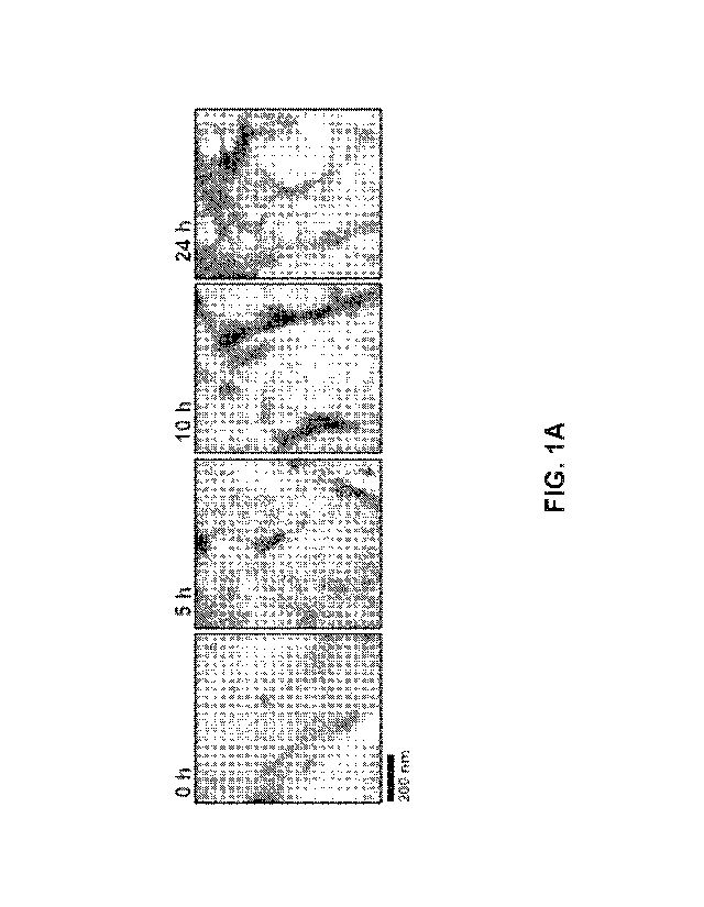
[0015] FIG. 1A shows electron micrographs taken at Oh, 5h, 10h, and 24h of
incubation.
[0016] FIG. 1B is a western blot of soluble oligomeric AP protein
aggregates.
[0017] FIG. 2A is a graph showing non-amplified amyloid formation measured
by ThT
fluorescence as a function of time seeded by various concentrations of
synthetic soluble oligomeric
AP protein of EXAMPLE 1.
- 6 -
CA 03062876 2019-11-07
WO 2018/213440 PCT/US2018/032962
[0018] FIG. 2B is a graph showing amplification cycle-accelerated amyloid
formation
measured by ThT fluorescence as a function of time seeded by various
concentrations of synthetic
soluble oligomeric AP protein of EXAMPLE 1.
[0019] FIG. 3A is a graph of amyloid formation versus time, measured as a
function of
ThT fluorescence labeling, showing the average kinetics of AP aggregation
seeded by CSF from
representative samples from the AD, NND, and NAND groups.
[0020] FIG. 3B is a graph of the lag phase time in h for AP aggregation in
the presence of
samples from the AD, NND, and NAND groups.
[0021] FIG. 3C is a graph showing the extent of amyloid formation obtained
after 180 AP-
PMCA cycles, e.g. 90 h of incubation (P90) in the presence of CSF samples from
AD, NND and
NAND patients.
[0022] FIGS. 4A-D are plots of the true positive rate (sensitivity) as a
function of the false
positive rate (specificity) for different cut-off points using the lag phase
values showed in FIG. 3B
for AD vs NAND (FIG. 4A), AD vs NND (FIG. 4B) and AD vs All control samples
(FIG. 4C).
FIG. 4D estimates the most reliable cut-off point for the different set of
group comparisons.
[0023] FIG. 5, Table 1 shows estimations of the sensitivity, specificity
and predictive
value of the AP-PMCA test, calculated using the lag phase numbers.
[0024] FIG. 6 is a graph of the lag phase time in h for samples obtained
after 300 AP-
PMCA cycles, e.g. 150 h of incubation (P90) in the presence of CSF samples
from AD and control
patients.
[0025] FIG. 7A is a western blot showing results of immunodepletion using
synthetically
prepared AP oligomers spiked into human CSF.
[0026] FIG. 7B is a graph showing the kinetics of AP aggregation seeded by
control and
immunodepleted CSF samples.
[0027] FIG. 7C is a graph showing the kinetics of AP aggregation seeded by
control and
immunodepleted CSF samples, depleted only with the All conformational
antibody.
[0028] FIG. 8A is a schematic representation of an ELISA solid phase method
employed
to capture AP oligomers from complex biological samples.
[0029] FIG. 8B is a schematic representation of a magnetic bead solid phase
method
employed to capture AP oligomers from complex biological samples.
[0030] FIG. 9, Table 2 shows the ability of specific antibodies to capture
the AP
oligomers.
- 7 -
CA 03062876 2019-11-07
WO 2018/213440 PCT/US2018/032962
[0031] FIG. 10 is a graph of amyloid formation versus time showing the
acceleration of
AO aggregation by the presence of different quantities of synthetic oligomers
spiked in human
plasma.
[0032] FIG. 11 is a graph showing time to reach 50% aggregation in an A13-
PMCA assay
in the presence of plasma samples from AD patients and controls.
[0033] FIG. 12 is a western blot showing the results of amplification of AO
aggregation
by cycles of incubation/sonication in the presence of distinct quantities of
synthetic AO oligomers
monitored by Western blot after protease digestion.
[0034] FIG. 13A is a graph of Thioflavin T fluorescence versus time showing
the detection
of aS seeds by PD-PMCA.
[0035] FIG. 13B is a graph of time to reach 50% aggregation plotted as a
function of the
indicated amounts aS seeds.
[0036] FIG. 14 shows detection of aS seeds in CSF samples from human PD
patients by
PD-PMCA, versus controls with Alzheimer's disease (AD) or a non-
neurodegenerative disease
(NND).
[0037] FIG. 15, Table 3 demonstrates the ability of different sequence or
conformational
antibodies to capture aS oligomers.
[0038] FIG. 16A is a schematic representation of an ELISA solid phase
method employed
to capture aS oligomers.
[0039] FIG. 16B is a schematic representation of a magnetic bead solid
phase method
employed to capture aS oligomers.
[0040] FIGS. 17A, 17B, and 17C are a series of graphs that show the results
of
immunoprecipitation/aggregation of a-Synuclein oligomers from human blood
plasma using three
different a-Synuclein antibodies. FIG. 17A shows results with antibody N-19.
FIG. 17B shows
results with antibody 211. FIG. 17C shows results with antibody C-20.
[0041] FIGS. 18A, 18B, and 18C are a series of graphs that show the results
of detection
for aS seeds in CSF samples. FIG. 18A shows results in control samples. FIG.
18B shows results
in PD patients. FIG. 18C shows results in patients with Multiple System
Atrophy (MSA).
[0042] FIG. 19 is a flow chart showing the preparation and purification of
recombinant
full-length 4R tau protein.
[0043] FIG. 20A is a graph of aggregation in % according to ThT
fluorescence for various
initial amounts of tau seeds and a control. The values in FIG. 20A are the
mean of two replicates,
with the error bars indicating standard deviation.
- 8 -
CA 03062876 2019-11-07
WO 2018/213440 PCT/US2018/032962
[0044] FIG. 20B is a graph of T50, the time to 50% aggregation as measured
by ThT
fluorescence versus the log of the amount of oligomeric tau seeds in fmol.
[0045] FIG. 20C is a graph of aggregation followed over time by ThT
fluorescence.
[0046] FIG. 20D is a graph of the relationship between the quantity of tau
oligomers and
the Tau-PMCA signal (time to reach 50% aggregation).
[0047] FIGS. 20E-20L are a series of graphs that display the aggregation
results based on
ThT fluorescence of 8 of the conditions tested, including 4 different time
points (0, 7, 14 and 30
days) with samples subjected to freezing and thawing or not and in the
presence of buffer or CSF.
[0048] FIG. 20E is a graph of aggregation based on ThT fluorescence of a
first seed
preparation at 0 days.
[0049] FIG. 20F is a graph of aggregation based on ThT fluorescence of a
first seed
preparation at 7 days.
[0050] FIG. 20G is a graph of aggregation based on ThT fluorescence of a
first seed
preparation at 14 days.
[0051] FIG. 20H is a graph of aggregation based on ThT fluorescence of a
first seed
preparation at 30 days.
[0052] FIG. 201 is a graph of aggregation based on ThT fluorescence of a
second seed
preparation at 0 days.
[0053] FIG. 20J is a graph of aggregation based on ThT fluorescence of a
second seed
preparation at 7 days.
[0054] FIG. 20K is a graph of aggregation based on ThT fluorescence of a
second seed
preparation at 14 days.
[0055] FIG. 20L is a graph of aggregation based on ThT fluorescence of a
second seed
preparation at 30 days.
[0056] FIG. 20M is a table of T5o values showing reproducibility across 16
different
conditions.
[0057] FIG. 20N is a graph of ThT fluorescence vs time for the tau assay
seeded with 1
pm of tau, Ar340, AB42, His aSyn, Hu aSyn, and a control with no seeds.
[0058] FIG. 21A is a graph showing ThT fluorescence at 447h of incubation
for patients
with AD, patients with MCI, patients with other tauopathies, positive controls
using samples of
healthy CSF spiked with synthetic Tau oligomers (12.5 fmol), negative controls
of samples of
healthy CSF without Tau seeds; and control patients with other neurological
diseases.
- 9 -
CA 03062876 2019-11-07
WO 2018/213440 PCT/US2018/032962
[0059] FIG. 21B shows fluorescence signals for samples from patients with
AD or other
tauopathies for tau-PMCA comparable to that observed in samples containing
recombinant tau
oligomers.
[0060] FIG. 22 is a graph showing aggregation % based on ThT versus time
for patients
affected by AD, FTD (frontotemporal dementia), CBD (corticobasal
degeneration), and PSP
(progressive supranuclear palsy), versus representative CSF samples from a
control.
DETAILED DESCRIPTION
[0061] Methods and kits are provided for the detection or characterization
of misfolded tau
protein in a sample, including for the determination or diagnosis of
tauopathies in a subject from
which the sample is taken. Misfolded aggregates of tau proteins may be formed
and accumulate.
The misfolded aggregates may induce cellular dysfunction and tissue damage
among other effects.
For example, tauopathies may include those that predominantly regard
misfolding of 4R, or
misfolding of mixtures of 4R and 3R: Alzheimer's disease (AD), Parkinson's
Disease (PD),
Progressive Supranuclear Palsy (PSP), FrontoTemporal Dementia (FTD),
Corticobasal
degeneration (CBD), Mild cognitive impairment (MCI), Argyrophilic grain
disease (AgD)
Traumatic Brain Injury (TBI), Chronic Traumatic Encephalopathy (CTE), Dementia
Pugilistica
(DP), and the like.
[0062] In some embodiments, tauopathies herein may exclude Pick's disease.
In some
embodiments, the tauopathies described herein may exclude those that
predominantly regard 3R
tau misfolding, e.g., Pick's disease.
[0063] The methods may include protein misfolding cyclic amplification
(PMCA), which
may provide ultra-sensitive detection of misfolded protein aggregates such as
tau through artificial
acceleration and amplification of the misfolding and aggregation process in
vitro. The basic
concept of PMCA has been previously demonstrated experimentally for prions
(Soto et al, WO
2002/04954; Estrada, et al., U.S. Pat. App. Pub. No. 20080118938, each of
which is entirely
incorporated herein by reference) and for other protein misfolding, such as of
"AP" or "beta
amyloid" in Alzheimer's disease and alpha synuclein in Parkinson's disease
(Soto et al, WO
2016/040907, which is entirely incorporated herein by reference). However,
prior to the filing
date of the present document, no reference has described PCMA for the
amplification and detection
of misfolded tau protein corresponding to any tauopathy that predominantly
regards misfolding of
4R, or that regards misfolding of 3R tau in the presence of 4R tau, or for any
tauopathy other than
Pick's disease. This document discloses specific examples and details which
enable PMCA
technology for the detecting the presence or absence of misfolded tau
aggregates, and, in various
embodiments, one or more additional PMCA procedures for the detection of other
misfolded
- 10 -
CA 03062876 2019-11-07
WO 2018/213440 PCT/US2018/032962
proteins such as misfolded A13 in Alzheimer's disease and alpha synuclein in
Parkinson's disease.
Such one or more additional PMCA procedures may provide discrimination among
the various
tauopathies, for example, to distinguish AD and PD from each other and from
PSP, FTD, CBD,
MCI, AgD, TBI, CTE, DP, and the like.
[0064] As used herein, "A13" or "beta amyloid" refers to a peptide formed
via sequential
cleavage of the amyloid precursor protein (APP). Various A13 isoforms may
include 38-43 amino
acid residues. The A13 protein may be formed when APP is processed by 13-
and/or y-secretases in
any combination. The A13 may be a constituent of amyloid plaques in brains of
individuals
suffering from or suspected of having AD. Various A13 isoforms may include and
are not limited
to Abeta40 and Abeta42. Various A13 peptides may be associated with neuronal
damage associated
with AD.
[0065] As used herein, "aS" or "alpha-synuclein" refers to full-length, 140
amino acid a-
synuclein protein, e.g., "aS-140." Other isoforms or fragments may include "aS-
126," alpha-
synuclein-126, which lacks residues 41-54, e.g., due to loss of exon 3; and
"aS-112" alpha-
synuclein-112, which lacks residue 103-130, e.g., due to loss of exon 5. The
aS may be present in
brains of individuals suffering from PD or suspected of having PD. Various aS
isoforms may
include and are not limited to aS-140, aS-126, and aS-112. Various aS peptides
may be associated
with neuronal damage associated with PD.
[0066] As used herein, "tau" refers to proteins are the product of
alternative splicing from
a single gene, e.g., MAPT (microtubule-associated protein tau) in humans. Tau
proteins include
up to full-length and truncated forms of any of tau's isoforms. Various
isoforms include, but are
not limited to, the six tau isoforms known to exist in human brain tissue,
which correspond to
alternative splicing in exons 2, 3, and 10 of the tau gene. Three isoforms
have three binding
domains and the other three have four binding domains. Misfolded tau may be
present in brains
of individuals suffering from AD or suspected of having AD, or other
tauopathies that, like AD,
regard misfolding in the presence of both 4R and 3R tau isoforms. Misfolded
tau may also be
present in diseases that regard misfolding of primarily 4R tau isoforms, such
as progressive
supranuclear palsy (PSP), tau-dependent frontotemporal dementia (FTD),
corticobasal
degeneration (CBD), mild cognitive impairment (MCI), argyrophilic grain
disease (AgD), and the
like.
[0067] As used herein, a "misfolded protein aggregate" is a protein that
contains in part or
in full a structural conformation of the protein that differs from the
structural conformation that
exists when involved in its typical, non-pathogenic normal function within a
biological system. A
misfolded protein may aggregate. A misfolded protein may localize in a protein
aggregate. A
-11-
CA 03062876 2019-11-07
WO 2018/213440 PCT/US2018/032962
misfolded protein may be a non-functional protein. A misfolded protein may be
a pathogenic
conformer of the protein. Monomeric protein compositions may be provided in
native,
nonpathogenic conformations without the catalytic activity for misfolding,
oligomerization, and
aggregation associated with seeds (a misfolded protein oligomer capable of
catalyzing misfolding
under PMCA conditions). Monomeric protein compositions may be provided in seed-
free form.
[0068] As used herein, "monomeric protein" refers to single protein
molecules. "Soluble,
aggregated misfolded protein" refers to oligomers or aggregations of monomeric
protein that
remain in solution. Examples of soluble, misfolded protein may include any
number of protein
monomers so long as the misfolded protein remains soluble. For example,
soluble, misfolded
protein may include monomers or aggregates of between 2 and about 50 units of
monomeric
protein.
[0069] Monomeric and/or soluble, misfolded protein may aggregate to form
insoluble
aggregates, higher oligomers, and/or tau fibrils. For example, aggregation of
AP or tau protein
may lead to protofibrils, fibrils, and eventually misfolded plaques or tangles
that may be observed
in AD or tauopathy subjects. "Seeds" or "nuclei" refer to misfolded protein or
short fragmented
fibrils, particularly soluble, misfolded protein with catalytic activity for
further misfolding,
oligomerization, and aggregation. Such nucleation-dependent aggregation may be
characterized
by a slow lag phase wherein aggregate nuclei may form, which may then catalyze
rapid formation
of further aggregates and larger oligomers and polymers. The lag phase may be
minimized or
removed by addition of pre-formed nuclei or seeds. Monomeric protein
compositions may be
provided without the catalytic activity for misfolding and aggregation
associated with misfolded
seeds. Monomeric protein compositions may be provided in seed-free form.
[0070] As used herein, "soluble" species may form a solution in biological
fluids under
physiological conditions, whereas "insoluble" species may be present as
precipitates, fibrils,
deposits, tangles, or other non-dissolved forms in such biological fluids
under physiological
conditions. Such biological fluids may include, for example, fluids, or fluids
expressed from one
or more of: amniotic fluid; bile; blood; cerebrospinal fluid; cerumen; skin;
exudate; feces; gastric
fluid; lymph; milk; mucus, e.g. nasal secretions; mucosal membrane, e.g.,
nasal mucosal
membrane; peritoneal fluid; plasma; pleural fluid; pus; saliva; sebum; semen;
sweat; synovial
fluid; tears; urine; and the like. Insoluble species may include, for example,
fibrils of AP, aS, 4R
tau, 3R tau, combinations thereof such as 3R tau + 4R tau, and the like. A
species that dissolves
in a non-biological fluid but not one of the aforementioned biological fluids
under physiological
conditions may be considered insoluble. For example, fibrils of AP, aS, 4R
tau, 3R tau,
combinations thereof such as 3R tau + 4R tau, and the like may be dissolved in
a solution of, e.g.,
- 12 -
CA 03062876 2019-11-07
WO 2018/213440 PCT/US2018/032962
a surfactant such as sodium dodecyl sulfate (SDS) in water, but may still be
insoluble in one or
more of the mentioned biological fluids under physiological conditions.
[0071] In some embodiments, the sample may exclude insoluble species of the
misfolded
proteins such as AP, aS, 4R tau, 3R tau, combinations thereof such as 3R tau +
4R tau and the like
as a precipitate, fibril, deposit, tangle, plaque, or other form that may be
insoluble in one or more
of the described biological fluids under physiological conditions.
[0072] For example, in some embodiments, the sample may exclude tau in
fibril form. The
sample may exclude misfolded tau proteins in insoluble form, e.g., the sample
may exclude the
misfolded tau proteins as precipitates, fibrils, deposits, tangles, plaques,
or other insoluble forms,
e.g., in fibril form. The methods described herein may include preparing the
sample by excluding
the misfolded protein in insoluble form, e.g., by excluding from the sample
the misfolded tau
protein as precipitates, fibrils, deposits, tangles, plaques, or other
insoluble forms, e.g., in fibril
form. The kits described herein may include instructions directing a user to
prepare the sample by
excluding from the sample the misfolded tau protein as precipitates, fibrils,
deposits, tangles,
plaques, or other insoluble forms, e.g., in fibril form. The exclusion of such
insoluble forms of the
described misfolded proteins from the sample may be substantial or complete.
[0073] As used herein, aggregates of misfolded protein refer to non-
covalent associations
of protein including soluble, misfolded protein. Aggregates of misfolded
protein may be "de-
aggregated", or disrupted to break up or release soluble, misfolded protein.
The catalytic activity
of a collection of soluble, misfolded protein seeds may scale, at least in
part with the number of
such seeds in a mixture. Accordingly, disruption of aggregates of misfolded
protein in a mixture
to release misfolded protein seeds may lead to an increase in catalytic
activity for oligomerization
or aggregation of monomeric protein.
[0074] In various embodiments, a method is provided for determining a
presence or
absence in a sample of a first misfolded protein aggregate. The method may
include performing a
first protein misfolding cyclic amplification (PMCA) procedure. The first PMCA
procedure may
include forming a first incubation mixture by contacting a first portion of
the sample with a first
substrate protein. The first substrate protein may include 4R tau protein. The
first PMCA
procedure may include conducting an incubation cycle two or more times under
conditions
effective to form as first amplified, misfolded protein aggregate. Each
incubation cycle may
include incubating the first incubation mixture effective to cause misfolding
and/or aggregation of
the first substrate protein in the presence of the first misfolded protein
aggregate. Each incubation
cycle may include disrupting the first incubation mixture effective to form
the first amplified,
misfolded protein aggregate. The first PMCA procedure may include determining
the presence or
- 13 -
CA 03062876 2019-11-07
WO 2018/213440 PCT/US2018/032962
absence in the sample of the first misfolded protein aggregate by analyzing
the first incubation
mixture for the presence or absence of the first amplified, misfolded protein
aggregate. The first
misfolded protein aggregate may include the first substrate protein. The first
amplified, misfolded
protein aggregate may include the first substrate protein.
[0075] In various embodiments, a method is provided for determining a
presence or
absence in a subject of a tauopathy corresponding to a first misfolded protein
aggregate. The
method may include providing a sample from the subject. The method may include
performing at
least a first PMCA procedure. The first PMCA procedure may include forming a
first incubation
mixture by contacting a first portion of the sample with a first substrate
protein. The first substrate
protein may include a tau isoform. The first substrate protein may be subject
to pathological
misfolding and/or aggregation in vivo to form the first misfolded protein
aggregate. The first
PMCA procedure may include conducting an incubation cycle two or more times
under conditions
effective to form a first amplified, misfolded protein aggregate. Each
incubation cycle may include
incubating the first incubation mixture effective to cause misfolding and/or
aggregation of the first
substrate protein in the presence of the first misfolded protein aggregate.
Each incubation cycle
may include disrupting the first incubation mixture effective to form the
first amplified, misfolded
protein aggregate. The first PMCA procedure may include determining the
presence or absence
in the sample of the first misfolded protein aggregate by analyzing the first
incubation mixture for
the presence or absence of the first amplified, misfolded protein aggregate.
The first PMCA
procedure may include determining the presence or absence of the tauopathy in
the subject
according the presence or absence of the first misfolded protein aggregate in
the sample. The first
misfolded protein aggregate may include the first substrate protein. The first
amplified, misfolded
protein aggregate may include the first substrate protein. The method may
provide that the
tauopathy excludes Pick's disease when the first substrate protein consists of
monomeric 3R tau.
[0076] In various embodiments, a method is provided using capturing for
determining a
presence or absence in a sample of a first misfolded protein aggregate. The
method may include
capturing the first misfolded protein aggregate from the sample to form a
captured first misfolded
protein aggregate. The method may include performing at least a first PMCA
procedure. The first
PMCA procedure may include forming a first incubation mixture by contacting
the captured first
misfolded protein aggregate with a molar excess of a first substrate protein.
The first substrate
protein may be subject to pathological misfolding and/or aggregation in vivo
to form the first
misfolded protein aggregate. The molar excess may be greater than an amount of
protein monomer
included in the captured first misfolded protein aggregate. The method may
include conducting
an incubation cycle two or more times effective to form a first amplified,
misfolded protein
- 14 -
CA 03062876 2019-11-07
WO 2018/213440 PCT/US2018/032962
aggregate. Each incubation cycle may include incubating the first incubation
mixture effective to
cause misfolding and/or aggregation of the first substrate protein in the
presence of the captured
first misfolded protein aggregate. Each incubation cycle may include
disrupting the first
incubation mixture effective to form the first amplified, misfolded protein
aggregate. The first
PMCA procedure may include determining the presence or absence of the first
misfolded protein
aggregate in the sample by detecting the first amplified, misfolded protein
aggregate. The first
misfolded protein aggregate may include the first substrate protein. The first
amplified, misfolded
protein aggregate may include the first substrate protein.
[0077] In various embodiments, a method is provided for determining a
presence or
absence of a tauopathy in a subject, the tauopathy including Alzheimer's
disease (AD). The
method may include providing the subject. The method may include obtaining a
sample from the
subject. The sample may include one or more of: a bio-fluid, a biomaterial, a
homogenized tissue,
and a cell lysate. The method may include performing at least a first PMCA
procedure. The first
PMCA procedure may include forming a first incubation mixture by contacting
the sample with a
first substrate protein. The first substrate protein may include 4R tau. The
first PMCA procedure
may include conducting an incubation cycle two or more times effective to form
a first amplified,
misfolded protein aggregate. Each incubation cycle may include incubating the
first incubation
mixture effective to cause misfolding and/or aggregation of the first
substrate protein in the
presence of the first misfolded protein aggregate. Each incubation cycle may
include disrupting
the incubation mixture effective to form the first amplified, misfolded
protein aggregate. The first
PMCA procedure may include determining the presence or absence in the sample
of the first
misfolded protein aggregate by detecting in the first incubation mixture the
presence or absence of
the first amplified, misfolded protein aggregate. The method may include
determining the
presence or absence in the subject of AD according to the presence or absence
of the first misfolded
protein aggregate in the sample.
[0078] In some embodiments, determining the presence or absence in the
subject of AD
may include distinguishing AD from one or more additional tauopathies by
determining a signature
of AD tau protein aggregate. The signature AD tau protein aggregate may
include one or more of:
one or more AD-specific corresponding PMCA kinetic parameters of: lag phase,
T5o,
amplification rate, and amplification extent; an assay using an antibody
selective for a
conformational epitope of AD tau protein aggregate; an indicator selective for
AD tau protein
aggregate; and a spectrum characteristic of AD tau protein aggregate.
[0079] In various embodiments, a method is provided for determining a
presence or
absence of a tauopathy in a subject, the tauopathy including Parkinson's
disease (PD). The method
- 15 -
CA 03062876 2019-11-07
WO 2018/213440 PCT/US2018/032962
may include providing the subject. The method may include obtaining a sample
from the subject.
The sample may include one or more of: a bio-fluid, a biomaterial, a
homogenized tissue, and a
cell lysate. The method may include performing at least a first PMCA
procedure. The first PMCA
procedure may include forming a first incubation mixture by contacting the
sample with a first
substrate protein. The first substrate protein may include 4R tau. The first
PMCA procedure may
include conducting an incubation cycle two or more times effective to form a
first amplified,
misfolded protein aggregate. Each incubation cycle may include incubating the
first incubation
mixture effective to cause misfolding and/or aggregation of the first
substrate protein in the
presence of the first misfolded protein aggregate. Each incubation cycle may
include disrupting
the incubation mixture effective to form the first amplified, misfolded
protein aggregate. The first
PMCA procedure may include determining the presence or absence in the sample
of the first
misfolded protein aggregate by detecting in the first incubation mixture the
presence or absence of
the first amplified, misfolded protein aggregate. The method may include
determining the
presence or absence in the subject of PD according to the presence or absence
of the first misfolded
protein aggregate in the sample.
[0080] In some embodiments, determining the presence or absence in the
subject of PD
may include distinguishing PD from one or more additional tauopathies by
determining a signature
of PD tau protein aggregate. The signature PD tau protein aggregate may
include one or more of:
one or more PD-specific corresponding PMCA kinetic parameters of: lag phase,
T50, amplification
rate, and amplification extent; an assay using an antibody selective for a
conformational epitope
of PD tau protein aggregate; an indicator selective for PD tau protein
aggregate; and a spectrum
characteristic of PD tau protein aggregate.
[0081] In various embodiments, a method is provided for determining a
presence or
absence of a tauopathy in a subject, the tauopathy including Progressive
Supranuclear Palsy (PSP).
The method may include providing the subject. The method may include obtaining
a sample from
the subject. The sample may include one or more of: a bio-fluid, a
biomaterial, a homogenized
tissue, and a cell lysate. The method may include performing at least a first
PMCA procedure.
The first PMCA procedure may include forming a first incubation mixture by
contacting the
sample with a first substrate protein. The first substrate protein may include
4R tau. The first
PMCA procedure may include conducting an incubation cycle two or more times
effective to form
a first amplified, misfolded protein aggregate. Each incubation cycle may
include incubating the
first incubation mixture effective to cause misfolding and/or aggregation of
the first substrate
protein in the presence of the first misfolded protein aggregate. Each
incubation cycle may include
disrupting the incubation mixture effective to form the first amplified,
misfolded protein aggregate.
- 16 -
CA 03062876 2019-11-07
WO 2018/213440 PCT/US2018/032962
The first PMCA procedure may include determining the presence or absence in
the sample of the
first misfolded protein aggregate by detecting in the first incubation mixture
the presence or
absence of the first amplified, misfolded protein aggregate. The method may
include determining
the presence or absence in the subject of PSP according to the presence or
absence of the first
misfolded protein aggregate in the sample.
[0082] In some embodiments, determining the presence or absence in the
subject of PSP
may include distinguishing PSP from one or more additional tauopathies by
determining a
signature of PSP tau protein aggregate. The signature PSP tau protein
aggregate may include one
or more of: one or more PSP-specific corresponding PMCA kinetic parameters of:
lag phase, T5o,
amplification rate, and amplification extent; an assay using an antibody
selective for a
conformational epitope of PSP tau protein aggregate; an indicator selective
for PSP tau protein
aggregate; and a spectrum characteristic of PSP tau protein aggregate.
[0083] In various embodiments, a method is provided for determining a
presence or
absence of a tauopathy in a subject, the tauopathy including FrontoTemporal
Dementia (FTD).
The method may include providing the subject. The method may include obtaining
a sample from
the subject. The sample may include one or more of: a bio-fluid, a
biomaterial, a homogenized
tissue, and a cell lysate. The method may include performing at least a first
PMCA procedure.
The first PMCA procedure may include forming a first incubation mixture by
contacting the
sample with a first substrate protein. The first substrate protein may include
4R tau. The first
PMCA procedure may include conducting an incubation cycle two or more times
effective to form
a first amplified, misfolded protein aggregate. Each incubation cycle may
include incubating the
first incubation mixture effective to cause misfolding and/or aggregation of
the first substrate
protein in the presence of the first misfolded protein aggregate. Each
incubation cycle may include
disrupting the incubation mixture effective to form the first amplified,
misfolded protein aggregate.
The first PMCA procedure may include determining the presence or absence in
the sample of the
first misfolded protein aggregate by detecting in the first incubation mixture
the presence or
absence of the first amplified, misfolded protein aggregate. The method may
include determining
the presence or absence in the subject of FTD according to the presence or
absence of the first
misfolded protein aggregate in the sample.
[0084] In some embodiments, determining the presence or absence in the
subject of FTD
may include distinguishing FTD from one or more additional tauopathies by
determining a
signature of FTD tau protein aggregate. The signature FTD tau protein
aggregate may include one
or more of: one or more FTD-specific corresponding PMCA kinetic parameters of:
lag phase,
T5o, amplification rate, and amplification extent; an assay using an antibody
selective for a
- 17 -
CA 03062876 2019-11-07
WO 2018/213440 PCT/US2018/032962
conformational epitope of FTD tau protein aggregate; an indicator selective
for FTD tau protein
aggregate; and a spectrum characteristic of FTD tau protein aggregate.
[0085] In various embodiments, a method is provided for determining a
presence or
absence of a tauopathy in a subject, the tauopathy including Corticobasal
degeneration (CBD).
The method may include providing the subject. The method may include obtaining
a sample from
the subject. The sample may include one or more of: a bio-fluid, a
biomaterial, a homogenized
tissue, and a cell lysate. The method may include performing at least a first
PMCA procedure.
The first PMCA procedure may include forming a first incubation mixture by
contacting the
sample with a first substrate protein. The first substrate protein may include
4R tau. The first
PMCA procedure may include conducting an incubation cycle two or more times
effective to form
a first amplified, misfolded protein aggregate. Each incubation cycle may
include incubating the
first incubation mixture effective to cause misfolding and/or aggregation of
the first substrate
protein in the presence of the first misfolded protein aggregate. Each
incubation cycle may include
disrupting the incubation mixture effective to form the first amplified,
misfolded protein aggregate.
The first PMCA procedure may include determining the presence or absence in
the sample of the
first misfolded protein aggregate by detecting in the first incubation mixture
the presence or
absence of the first amplified, misfolded protein aggregate. The method may
include determining
the presence or absence in the subject of CBD according to the presence or
absence of the first
misfolded protein aggregate in the sample.
[0086] In some embodiments, determining the presence or absence in the
subject of CBD
may include distinguishing CBD from one or more additional tauopathies by
determining a
signature of CBD tau protein aggregate. The signature CBD tau protein
aggregate may include
one or more of: one or more CBD-specific corresponding PMCA kinetic parameters
of: lag phase,
T5o, amplification rate, and amplification extent; an assay using an antibody
selective for a
conformational epitope of CBD tau protein aggregate; an indicator selective
for CBD tau protein
aggregate; and a spectrum characteristic of CBD tau protein aggregate.
[0087] In further embodiments, each of the methods described herein above
may
incorporate one or more of the following features. In particular, each feature
described with
reference to any protein substrate, misfolded protein aggregate, amplified
misfolded protein
aggregate, incubation mixture, PMCA procedure, portion of the sample, and the
like, should be
understood to describe, independently selected in various other embodiments,
any other protein
substrate, misfolded protein aggregate, amplified misfolded protein aggregate,
incubation mixture,
PMCA procedure, portion of the sample, and the like. For example, features
described for a "first"
protein substrate may, in some embodiments, also be independently selected to
describe a "second"
- 18-
CA 03062876 2019-11-07
WO 2018/213440 PCT/US2018/032962
protein substrate; features described for a "first" misfolded protein
aggregate may also be
independently selected to describe a "second" misfolded protein aggregate;
features described for
a "first" incubation mixture may also be independently selected to describe a
"second" incubation
mixture; features described for a "first" PMCA procedure may also be
independently selected to
describe a "second" PMCA procedure; and the like. Further, for example,
features described with
reference to "each" protein substrate, misfolded protein aggregate, amplified
misfolded protein
aggregate, incubation mixture, PMCA procedure, portion of the sample, and the
like, should be
understood to describe, independently selected in various other embodiments,
any other
enumerated element, e.g., "first," "second," "third," and the like, as applied
to the protein substrate,
misfolded protein aggregate, amplified misfolded protein aggregate, incubation
mixture, PMCA
procedure, portion of the sample, and the like. For example, a description
with reference to "each
substrate protein" may be independently selected to describe and support
recitations of a "first
substrate protein," a "second substrate protein," a "third substrate protein,"
and the like.
[0088] In several embodiments, features described generally for enumerated
or specified
elements, e.g., "first," "second," "each," and the like, may be independently
selected to be the
same or distinct. For example, in some embodiments, a first substrate protein
may include a 4R
tau and a second substrate protein may include AP; a condition such as a
temperature may be
selected independently for a first and second PMCA procedure, and the like. In
several
embodiments, some features described generally for such first and second
elements may be
selected to be the same, or to overlap, while other features described
generally for such first and
second elements may be independently selected to be distinct. For example, in
some embodiments,
first and second portions of the sample may be the same or combined, and first
and second
incubation mixtures may be the same or combined, while corresponding first and
second PMCA
procedures may be conducted in parallel or in series in the combined
incubation mixture using
different first and second substrate proteins, e.g., 4R tau and AP.
[0089] In several embodiments, one, two, or more instances may be
independently selected
for each protein substrate, misfolded protein aggregate, amplified misfolded
protein aggregate,
incubation mixture, PMCA procedure, portion of the sample, and the like. For
example, various
method embodiments may include a first PMCA procedure using 4R tau as a first
substrate protein,
a second PMCA procedure using AP as a second substrate protein, a third PMCA
procedure using
alpha synuclein as a third substrate protein, a fourth PMCA procedure using 3R
tau as a fourth
substrate protein, and the like. Such multiple PMCA procedures may be
performed for a sample,
e.g., a laboratory sample, or a sample drawn from a subject, such as a subject
having a tauopathy.
Such multiple PMCA procedures may be performed in parallel for each protein
substrate,
- 19 -
CA 03062876 2019-11-07
WO 2018/213440 PCT/US2018/032962
misfolded protein aggregate, amplified misfolded protein aggregate, incubation
mixture, PMCA
procedure, portion of the sample, and the like, for example as follows.
[0090] In various embodiments, the method may include determining the
presence in the
sample of the first misfolded protein aggregate. The method may include
performing at least a
second PMCA procedure to determine the presence or absence in the sample of at
least a second
misfolded protein aggregate. The second PMCA procedure may include forming a
second
incubation mixture by contacting a second portion of the sample with a second
substrate protein.
The second substrate protein may be subject to pathological misfolding and/or
aggregation in vivo
to form the second misfolded protein aggregate. The second PMCA procedure may
include
conducting an incubation cycle two or more times under conditions effective to
form a second
amplified, misfolded protein aggregate. Each incubation cycle may include
incubating the second
incubation mixture effective to cause misfolding and/or aggregation of the
second substrate protein
in the presence of the second misfolded protein aggregate. Each incubation
cycle may include
disrupting the second incubation mixture effective to form the second
amplified, misfolded protein
aggregate. The second PMCA procedure may include determining the presence or
absence in the
sample of the second misfolded protein aggregate by analyzing the second
incubation mixture for
the presence or absence of the second amplified, misfolded protein aggregate.
The second
misfolded protein aggregate may include the second substrate protein. The
second amplified,
misfolded protein aggregate may include the second substrate protein.
[0091] In some embodiments, the method may include determining the presence
in the
sample of the first misfolded protein aggregate and the second misfolded
protein aggregate. The
method may include performing at least a third PMCA procedure to determine the
presence or
absence in the sample of at least a third misfolded protein aggregate. The
third PMCA procedure
may include forming a third incubation mixture by contacting a third portion
of the sample with a
third substrate protein. The third substrate protein may be subject to
pathological misfolding
and/or aggregation in vivo to form the third misfolded protein aggregate. The
third PMCA
procedure may include conducting an incubation cycle two or more times under
conditions
effective to form a third amplified, misfolded protein aggregate. Each
incubation cycle may
include incubating the third incubation mixture effective to cause misfolding
and/or aggregation
of the third substrate protein in the presence of the third misfolded protein
aggregate. Each
incubation cycle may include disrupting the third incubation mixture effective
to form the third
amplified, misfolded protein aggregate. The third PMCA procedure may include
determining the
presence or absence in the sample of the third misfolded protein aggregate by
analyzing the third
incubation mixture for the presence or absence of the third amplified,
misfolded protein aggregate.
- 20 -
CA 03062876 2019-11-07
WO 2018/213440 PCT/US2018/032962
The third misfolded protein aggregate may include the third substrate protein.
The third amplified,
misfolded protein aggregate may include the third substrate protein.
[0092] In several embodiments, the method may include determining the
presence in the
sample of the first misfolded protein aggregate, the second misfolded protein
aggregate, and the
fourth misfolded protein aggregate. The method may include performing at least
a fourth PMCA
procedure to determine the presence or absence in the sample of a fourth
misfolded protein
aggregate. The fourth PMCA procedure may include forming a fourth incubation
mixture by
contacting a fourth portion of the sample with a fourth substrate protein. The
fourth substrate
protein may be subject to pathological misfolding and/or aggregation in vivo
to form the fourth
misfolded protein aggregate. The fourth PMCA procedure may include conducting
an incubation
cycle two or more times under conditions effective to form a fourth amplified,
misfolded protein
aggregate. Each incubation cycle may include incubating the fourth incubation
mixture effective
to cause misfolding and/or aggregation of the fourth substrate protein in the
presence of the fourth
misfolded protein aggregate. Each incubation cycle may include disrupting the
fourth incubation
mixture effective to form the fourth amplified, misfolded protein aggregate.
The fourth PMCA
procedure may include determining the presence or absence in the sample of the
fourth misfolded
protein aggregate by analyzing the fourth incubation mixture for the presence
or absence of the
fourth amplified, misfolded protein aggregate. The fourth misfolded protein
aggregate may
include the fourth substrate protein. The fourth amplified, misfolded protein
aggregate may
include the fourth substrate protein.
[0093] In various embodiments, the first substrate protein may
independently include a tau
isoform, e.g., 3R tau, 4R tau, and the like. In several embodiments, the first
substrate protein may
include 4R tau. The first substrate protein may include 3R tau. The first
substrate protein may
exclude 3R tau, for example, when the sample corresponds to Pick's disease or
is drawn from a
subject having Pick's disease. The first substrate protein may be soluble. The
first substrate
protein may be monomeric. The first substrate protein may be in a native in
vivo conformation,
e.g., folded. The first substrate protein may be distinct from each other
substrate protein.
[0094] In various embodiments, the second substrate protein may
independently include
one of: a tau isoform, e.g., 3R tau, 4R tau, and the like; AP, alpha
synuclein, and the like. The
second substrate protein may include one of: 3R tau, AP, alpha synuclein, and
the like. The second
substrate protein may include 3R tau. The second substrate protein may include
AP. The second
substrate protein may include alpha synuclein. The second substrate protein
may consist
essentially of, or consist of, one of: 3R tau, 4R tau, AP, or alpha synuclein.
The second substrate
protein may be soluble. The second substrate protein may be monomeric. The
second substrate
- 21 -
CA 03062876 2019-11-07
WO 2018/213440 PCT/US2018/032962
protein may be in a native in vivo conformation, e.g., folded. The second
substrate protein may
be distinct from each other substrate protein.
[0095] In various embodiments, the third substrate protein may
independently include one
of: a tau isoform, e.g., 3R tau, 4R tau, and the like; A13, alpha synuclein,
and the like. The third
substrate protein may include one of: 3R tau, A13, alpha synuclein, and the
like. The third substrate
protein may include 3R tau. The third substrate protein may include A13. The
third substrate
protein may include alpha synuclein. The third substrate protein may consist
essentially of, or
consist of, one of: 3R tau, 4R tau, A13, or alpha synuclein. The third
substrate protein may be
soluble. The third substrate protein may be monomeric. The third substrate
protein may be in a
native in vivo conformation, e.g., folded. The third substrate protein may be
distinct from each
other substrate protein.
[0096] In various embodiments, the fourth substrate protein may
independently include
one of: a tau isoform, e.g., 3R tau, 4R tau, and the like; A13, alpha
synuclein, and the like. The
fourth substrate protein may include one of: 3R tau, A13, alpha synuclein, and
the like. The fourth
substrate protein may include 3R tau. The fourth substrate protein may include
A13. The fourth
substrate protein may include alpha synuclein. The fourth substrate protein
may consist essentially
of, or consist of, one of: 3R tau, 4R tau, A13, or alpha synuclein. The fourth
substrate protein may
be soluble. The fourth substrate protein may be monomeric. The fourth
substrate protein may be
in a native in vivo conformation, e.g., folded. The fourth substrate protein
may be distinct from
each other substrate protein.
[0097] In some embodiments the sample may be taken from a subject. The
method may
include determining or diagnosing the presence or absence of a tauopathy in
the subject according
to the presence or absence of the first misfolded protein aggregate in the
sample.
[0098] In several embodiments, the method may include performing at least a
second
PMCA procedure to determine the presence or absence in the sample of second
misfolded protein
aggregate, e.g., a misfolded protein aggregate that includes a second
substrate protein. The second
PMCA procedure may include forming a second incubation mixture by contacting a
second portion
of the sample with a second substrate protein. The second substrate protein
may be subject to
pathological misfolding and/or aggregation in vivo to form the second
misfolded protein
aggregate. The methods may include determining the presence or absence in the
sample of the
second misfolded protein aggregate by analyzing the second incubation mixture
for the presence
or absence of the second amplified, misfolded protein aggregate. The second
misfolded protein
aggregate may include the second substrate protein. The second amplified,
misfolded protein
- 22 -
CA 03062876 2019-11-07
WO 2018/213440 PCT/US2018/032962
aggregate may include the second substrate protein. The second substrate
protein may include one
of: amyloid-beta (A13), alpha synuclein, and 3R tau.
[0099] In some embodiments, the sample may be taken from a subject. The
methods may
include determining or diagnosing the presence or absence of a tauopathy in
the subject according
to the presence or absence of the first misfolded protein aggregate in the
sample. The methods
may include performing at least a second PMCA procedure to determine the
presence or absence
in the sample of a second misfolded protein aggregate. The second PMCA
procedure may include
forming a second incubation mixture by contacting a second portion of the
sample with a second
substrate protein. The second substrate protein may be subject to pathological
misfolding and/or
aggregation in vivo to form the second misfolded protein aggregate. The second
PMCA procedure
may include conducting an incubation cycle two or more times under conditions
effective to form
a second amplified, misfolded protein aggregate. Each incubation cycle may
include incubating
the second incubation mixture effective to cause misfolding and/or aggregation
of the second
substrate protein in the presence of the second misfolded protein aggregate.
Each incubation cycle
may include disrupting the second incubation mixture effective to form the
second amplified,
misfolded protein aggregate. The second PMCA procedure may include determining
the presence
or absence in the sample of the second misfolded protein aggregate by
analyzing the second
incubation mixture for the presence or absence of the second amplified,
misfolded protein
aggregate. The second misfolded protein aggregate may include the second
substrate protein. The
second amplified, misfolded protein aggregate may include the second substrate
protein.
[00100] In various embodiments, the subject may have the tauopathy. The
methods may
include characterizing an identity of the tauopathy in the subject according
to: the presence in the
sample of the first misfolded protein aggregate; and the presence or absence
in the sample of the
second misfolded protein aggregate. The second substrate protein may include
one of: amyloid-
beta (Af3), alpha synuclein, and 3R tau. For example, the presence of
misfolded 4R tau aggregate
as the first misfolded protein aggregate and A13 as the second misfolded
protein aggregate may
indicate the tauopathy in the subject is AD; the presence of misfolded 4R tau
aggregate as the first
misfolded protein aggregate and alpha synuclein as the second misfolded
protein aggregate may
indicate the tauopathy in the subject is PD; the presence of misfolded 4R tau
aggregate as the first
misfolded protein aggregate and the absence of the second misfolded protein
aggregate including
A13, alpha synuclein, or 3R may indicate a 4R tauopathy, such as PSP, CBD,
AGD, and the like.
[00101] In various embodiments, the tauopathy may include a primary
tauopathy or a
secondary tauopathy. The tauopathy may be characterized at least in part by
misfolding and/or
aggregation of 4R tau protein. The tauopathy may be characterized at least in
part by misfolding
- 23 -
CA 03062876 2019-11-07
WO 2018/213440 PCT/US2018/032962
and/or aggregation of 4R tau protein and 3R tau protein. The tauopathy may be
characterized at
least in part by misfolded and/or aggregated 4R tau protein, in a ratio to
misfolded and/or
aggregated 3R tau protein, of one of about: 1:99, 5:95, 10:90, 15:85, 20:80,
25:75, 30:70, 35:65,
40:60, 45:55, 50:50, 55:45, 60:40, 65:35, 70:30, 75:25, 80:20, 85:15, 90:10,
95:5, and 99:1, or a
range between any two of the preceding ratios, for example, between 1:99 and
99:1.
[00102] In several embodiments, the methods may include characterizing an
identity of the
tauopathy by analyzing the first amplified, misfolded protein aggregate or one
or more
corresponding PMCA kinetic parameters thereof for a signature of at least one
of: Alzheimer's
disease (AD), Parkinson's Disease (PD), Progressive Supranuclear Palsy (PSP),
FrontoTemporal
Dementia (FTD), Corticobasal degeneration (CBD), Mild cognitive impairment
(MCI),
Argyrophilic grain disease (AgD) Traumatic Brain Injury (TBI), Chronic
Traumatic
Encephalopathy (CTE), and Dementia Pugilistica (DP). For example,
characterizing the identity
of the tauopathy may include determining the one or more corresponding PMCA
kinetic
parameters, including one or more of: lag phase, T50, amplification rate, and
amplification extent.
Characterizing the identity of the tauopathy may include comparing the one or
more corresponding
PMCA kinetic parameters to one or more corresponding predetermined
corresponding PMCA
kinetic parameters that are characteristic of the identity of the tauopathy to
determine a similarity
or difference effective to characterize the identity of the tauopathy.
[00103] In some embodiments, the methods may include characterizing the
identity of the
tauopathy using an antibody selective for a conformational epitope of a
tauopathy-specific
misfolded tau protein aggregate. The methods may include characterizing the
identity of the
tauopathy using an indicator selective for each tauopathy-specific misfolded
tau protein aggregate.
The indicator selective for each tauopathy-specific misfolded tau protein
aggregate may include a
small molecule, a peptide, or a DNA or RNA aptamer; and the like. The methods
may include
characterizing the identity of the tauopathy using a spectrum characteristic
of each tauopathy-
specific misfolded tau protein aggregate.
[00104] In some embodiments, the methods may include, for example,
characterizing the
identity of the tauopathy by analyzing the proteolytic resistance of each
tauopathy-specific
misfolded tau protein aggregate. For example, each tauopathy-specific
misfolded tau protein
aggregate may be contacted with a proteinase, e.g., proteinase K, trypsin,
chymotrypsin, and the
like, at a proteinase concentration of from 0.1 to 5000 [tg/mL, at various
temperatures from 20 C
to 120 C and for various times, e.g., from 1 min to 4 h. The proteolytic
resistance of each
tauopathy-specific misfolded tau protein aggregate may be characterized and
used to distinguish
the various tauopathy-specific misfolded tau protein aggregates.
- 24 -
CA 03062876 2019-11-07
WO 2018/213440 PCT/US2018/032962
[00105] In several embodiments, the methods may include characterizing the
identity of the
tauopathy by analyzing the stability to denaturation of each tauopathy-
specific misfolded tau
protein aggregate. For example, each tauopathy-specific misfolded tau protein
aggregate may be
treated with guanidinium or urea at a sufficiently elevated temperature to
induce protein
denaturation of each tauopathy-specific misfolded tau protein aggregate. The
concentration of
guanidinium or urea may range from 0.1 M to 8 M. The temperature may range
between 20 C to
120 C. The stability of each tauopathy-specific misfolded tau protein
aggregate may be
characterized and used to distinguish the various tauopathy-specific misfolded
tau protein
aggregates.
[00106] The methods may include sedimentation of each tauopathy-specific
misfolded tau
protein aggregate. The methods may include gel chromatography to characterize
the size of each
tauopathy-specific misfolded tau protein aggregate. The methods may include
circular dichroism
spectroscopy of each tauopathy-specific misfolded tau protein aggregate. The
methods may
include Fourier transform infrared spectroscopy to analyze secondary structure
of each tauopathy-
specific misfolded tau protein aggregate. The methods may include nuclear
magnetic resonance
spectroscopy to analyze structure of each tauopathy-specific misfolded tau
protein aggregate. The
methods may include mass spectrometry, e.g., fragmentation and collision
induced dissociation to
analyze secondary and tertiary structure of each tauopathy-specific misfolded
tau protein
aggregate. The methods may include microscopy, e.g., atomic force microscopy,
cryo-electron
microscopy, and the like to analyze morphology of each tauopathy-specific
misfolded tau protein
aggregate. Each of these methods may be coupled with substitution using atomic
isotopes of
different mass, magnetic properties, and/or isotopic stability to complement
the methods; for
example, nuclear magnetic resonance spectroscopy may be coupled with deuterium
exchange in
each tauopathy-specific misfolded tau protein aggregate to obtain structural
information.
[00107] In various embodiments, the methods are provided such that the
tauopathy
specifically excludes Pick's disease. In various embodiments, the exclusion of
Pick's disease does
not encompass the remainder of Pick's complex of diseases.
[00108] In several embodiments, the methods may include determining or
diagnosing the
presence or absence of a tauopathy in the subject including comparing the
presence or absence of
the first misfolded protein aggregate in the sample to a control sample taken
from a control subject.
The detecting may include detecting an amount of the first misfolded protein
aggregate in the
sample. The sample may be taken from a subject. The methods may include
determining or
diagnosing the presence or absence of a tauopathy in the subject by comparing
the amount of the
first misfolded protein aggregate in the sample to a predetermined threshold
amount. The sample
- 25 -
CA 03062876 2019-11-07
WO 2018/213440 PCT/US2018/032962
may be taken from a subject exhibiting no clinical signs of dementia according
to cognitive testing.
The methods may include determining or diagnosing the presence or absence of a
tauopathy in the
subject according to the presence or absence of the first misfolded protein
aggregate in the sample.
The sample may be taken from a subject exhibiting no cortex plaques or tangles
according to
contrast imaging. The methods may include determining or diagnosing the
presence or absence
of a tauopathy in the subject according to the presence or absence of the
first misfolded protein
aggregate in the sample. The sample may be taken from a subject exhibiting
clinical signs of
dementia according to cognitive testing. The methods may include determining
or diagnosing the
presence or absence of a tauopathy as a contributing factor to the clinical
signs of dementia in the
subject according to the presence or absence of the first misfolded protein
aggregate in the sample.
The sample may be taken from a subject exhibiting no clinical signs of
dementia according to
cognitive testing. The subject may exhibit a predisposition to dementia
according to genetic
testing. The genetic testing may indicate, for example, an increased risk of
tauopathy according
to one or two copies of the ApoE4 allele, variants of the brain derived
neurotrophic factor (BDNF)
gene, such as the va166met allele, in which valine at AA position 66 is
replaced by methionine,
and the like. The methods may include determining or diagnosing the presence
or absence of a
tauopathy in the subject according to the presence or absence of the first
misfolded protein
aggregate in the sample.
[00109] In some embodiments, the methods may include preparing the first
incubation
mixture characterized by at least one concentration of: the first substrate
protein of less than about
20 [tM; heparin of less than about 75 [tM; NaCl of less than about 190 mM; and
Thioflavin T of
less than about 9.5 [1.M.
[00110] In various embodiments, the methods may include preparing the first
incubation
mixture including the first substrate protein at a concentration in [tM of one
or more of about:
0.001, 0.01, 0.1, 0.25, 0.5, 0.75, 1, 1.25, 1,5, 1.75, 2, 2.5, 3, 3.5, 4, 4.5,
5, 5.5, 6, 6.5, 7, 7.5, 8, 8.5,
9, 9.5, 10, 10.5, 11, 11.5, 12, 12.5, 13, 13.5, 14, 14.5, 15, 15.5, 16, 16.5,
17, 17.5, 18, 18.5, 19,
19.5, 20, 25, 50, 70, 100, 150, 200, 250, 500, 750, 1000, 1500, or 2000, or a
range between any
two of the preceding values, for example, between about 0.001 [tM and about
2000 [1.M. The
methods may include preparing the first incubation mixture characterized by
heparin at a
concentration in [tM of one or more of about: 0.001, 0.01, 0.1, 0.25, 0.5,
0.75, 1, 1.25, 1,5, 1.75,
2, 2.5, 3, 3.5, 4, 4.5, 5, 6, 7, 8, 9, 10 11, 12, 12.5, 15, 17.5, 20, 25, 30,
35, 40, 45, 50, 55, 60, 65,
70, and 75, or a range between any two of the preceding values, for example,
between about 0.001
[tM and about 75 [tM. The methods may include preparing the first incubation
mixture including
a buffer composition of one or more of: Tris-HCL, PBS, MES, PIPES, MOPS, BES,
TES, and
- 26 -
CA 03062876 2019-11-07
WO 2018/213440 PCT/US2018/032962
HEPES. The methods may include preparing the first incubation mixture
including the buffer
composition at a total concentration of one or more of about: 1 uM, 10 uM, 100
uM, 250 uM, 500
uM, 750 uM, 1 mM, 10 mM, 100 mM, 250 mM, 500 mM, 750 mM, and 1M, or a range
between
any two of the preceding values, for example, between about 1 uM and about 1
M. The methods
may include preparing the first incubation mixture including a salt
composition at a total
concentration of one or more of: 1 uM, 10 uM, 100 uM, 250 uM, 500 uM, 750 uM,
1 mM, 10
mM, 20 mM, 30 mM, 40 mM, 50 mM, 60 mM, 70 mM, 80 mM, 90 mM, 100 mM, 110 mM,
120
mM, 130 mM, 140 mM, 150 mM, 160 mM, 170 mM, 180 mM, 190 mM, 200 mM, 250 mM,
500
mM, 750 mM, and 1M, or a range between any two of the preceding values, for
example, between
about 1 uM and about 1 M. The salt composition may include one or more of:
NaCl and KC1.
[00111] In various embodiments, the methods may include preparing or
maintaining the
first incubation mixture at a pH of one or more of about: 5, 5.5, 6, 6.4, 6.5,
6.6, 6.7, 6.8, 6.9, 7,
7.1, 7.2, 7.3, 7.4, 7.5, 7.6, 7.7, 7.8, 7.9, 8, 8.1, 8.2, 8.3, 8.4, 8.5, or 9,
or a range between any two
of the preceding values, e.g., from about pH 5 to about pH 9.
[00112] In some embodiments, the methods may include preparing the first
incubation
mixture including an indicator at a total concentration of one or more of: 1
nM, 10 nM, 100 nM,
250 nM, 500 nM, 750 nM, 1 uM, 2 uM, 3 uM, 4 uM, 5 uM, 6 uM, 7 uM, 8 uM, 9 uM,
9.5 uM,
uM, 25 uM, 50 uM, 100 uM, 250 uM, 500 uM, 750 uM, 1 mM, or a range between any
two
of the preceding values, for example, between about 1 nM and about 1 mM.
[00113] In some embodiments of the methods, the incubating may include
heating or
maintaining the first incubation mixture at a temperature in C of one of: 5,
10, 15, 20, 22.5, 25,
27.5, 30, 31, 32, 33, 34, 35, 36, 37, 38, 39, 40, 41, 42, 43, 44, 45, 46, 47,
50, 55, 60, or a range
between any two of the preceding values, for example, between about 5 C and
about 60 C.
[00114] In several embodiments, the methods may include contacting an
indicator of the
first misfolded protein aggregate to the first incubation mixture. The
indicator of the first
misfolded protein aggregate may be characterized by an indicating state in the
presence of the first
misfolded protein aggregate and a non-indicating state in the absence of the
first misfolded protein
aggregate. Determining the presence of the first misfolded protein aggregate
in the sample may
include detecting the indicating state of the indicator of the first misfolded
protein aggregate. The
indicating state of the indicator and the non-indicating state of the
indicator may be characterized
by a difference in fluorescence. Determining the presence of the first
misfolded protein aggregate
in the sample may include detecting the difference in fluorescence. The
methods may include
contacting a molar excess of the indicator of the first misfolded protein
aggregate to the first
incubation mixture. The molar excess may be greater than a total molar amount
of protein
- 27 -
CA 03062876 2019-11-07
WO 2018/213440 PCT/US2018/032962
monomer included in the first substrate protein and the first misfolded
protein aggregate in the first
incubation mixture. The indicator of the first misfolded protein aggregate may
include one or
more of: a thioflavin, e.g., thioflavin T or thioflavin S; Congo Red, m-I-
Stilbene, Chrysamine G,
PIB, BF-227, X-34, TZDM, FDDNP, Me0-X-04, IMPY, NIAD-4, luminescent conjugated
polythiophenes, a fusion with a fluorescent protein such as green fluorescent
protein and yellow
fluorescent protein, derivatives thereof, and the like.
[00115] In various embodiments, the method may include determining an
amount of the
first misfolded protein aggregate in the sample. For example, known amounts of
in vitro, synthetic
misfolded protein aggregate seeds may be added to various portions of a
biological fluid of a
healthy patient, e.g., CSF. Subsequently, PMCA may be performed on the various
portions. In
each of the various portions, a fluorescent indicator of the misfolded protein
aggregate may be
added, and fluorescence may be measured as a function of, e.g., number of PMCA
cycles, to
determine various PMCA kinetics parameters, e.g., number of PMCA cycles to
maximum
fluorescence signal, number of PMCA cycles to 50% of maximum fluorescence
signal, lag phase
in increase of fluorescence signal, rate of increase in fluorescence signal
versus PMCA cycles, and
the like. A calibration curve showing the relationship between the
concentration of synthetic seeds
added and the PMCA kinetic parameters. The kinetic parameters may be measured
for unknown
samples and compared to the calibration curve to determine the expected amount
of seeds present
in a particular sample. Alternatively, the amount of the first misfolded
protein aggregate in the
sample may be determined by a series of known dilutions of the sample, and
PMCA of each serial
dilution to determine whether the first misfolded protein aggregate can be
detected or not in a
particular dilution. The amount of the first misfolded protein aggregate in
the undiluted sample
can be estimated based on the known dilution that results in no detection of
the first misfolded
protein aggregate by PMCA. In another example, the amount of the first
misfolded protein
aggregate in the sample may be determined by a series of known dilutions of
the sample, and
PMCA to determine a detection signal in each serial dilution. The collected
detection signals in
the serial dilutions can be fit, e.g., via least squares analysis, to
determine whether the first
misfolded protein aggregate can be detected or not in a particular dilution.
In another example,
the amount of the first misfolded protein aggregate in the sample may be
determined by known
amounts of antibodies to the first misfolded protein aggregate.
[00116] In some embodiments, the methods may include detecting the amount
of the first
misfolded protein aggregate in the sample at a sensitivity of at least about
one or more of: 70%,
71%, 72%, 73%, 74%, 75%, 76%, 77%, 78%, 79%, 80%, 81%, 82%, 83%, 84%, 85%,
86%, 87%,
88%, 89%, 90%, 91%, 92%, 93%, 94%, 95%, 96%, 97%, 98%, 99%, and 100%, e.g., at
least about
- 28 -
CA 03062876 2019-11-07
WO 2018/213440 PCT/US2018/032962
70%. The methods may include detecting the amount of the first misfolded
protein aggregate in
the sample at less than about one or more of: 625, 62.5, 6.25, 630 pg, 63 pg,
6.3 pg, 630 ng, 63
ng, 6.3 ng, 630 pg, 200 pg, 63 pg, 6.3 pg, 630 fg, 300 fg, 200 fg, 125 fg, 63
fg, 50 fg, 30 fg, 15 fg,
12.5 fg, 10 fg,5 fg, or 2.5 fg, The methods may include detecting the amount
of the first misfolded
protein aggregate in the sample at less than about one or more of: 100 nmol,
10 nmol, 1 nmol, 100
pmol, 10 pmol, 1 pmol, 100 fmol, 10 fmol, 3 fmol, 1 fmol, 100 attomol, 10
attomol, 5 attomol, 2
attomol, 1 attomol, 0.75 attomol, 0.5 attomol, 0.25 attomol, 0.2 attomol, 0.15
attomol, 0.1 attomol,
and 0.05 attomol, e.g., less than about 100 nmol. The methods may include
detecting the amount
of the first misfolded protein aggregate in the sample in a molar ratio to the
first substrate protein
included by the sample. The molar ratio may be less than about one or more of:
1: 100, 1: 10,000,
1: 100,000, and 1: 1,000,000, e.g., less than about 1: 100. The methods may
include determining
the amount of the first misfolded protein aggregate in the sample compared to
a control sample.
[00117] In several embodiments, the methods may include detecting the first
misfolded
protein aggregate in the sample with a specificity of at least about one or
more of: 70%, 71%,
72%, 73%, 74%, 75%, 76%, 77%, 78%, 79%, 80%, 81%, 82%, 83%, 84%, 85%, 86%,
87%, 88%,
89%, 90%, 91%, 92%, 93%, 94%, 95%, 96%, 97%, 98%, 99%, and 100%, e.g. at least
about 70%.
The methods may include detecting the first misfolded protein aggregate
including one or more
of: a Western Blot assay, a dot blot assay, an enzyme-linked immunosorbent
assay (ELISA), a
fluorescent protein/peptide binding assay, a thioflavin binding assay, a Congo
Red binding assay,
a sedimentation assay, electron microscopy, atomic force microscopy, surface
plasmon resonance,
and spectroscopy. The ELISA may include a two-sided sandwich ELISA. The
spectroscopy may
include one or more of: quasi-light scattering spectroscopy, multispectral
ultraviolet spectroscopy,
confocal dual-color fluorescence correlation spectroscopy, Fourier-transform
infrared
spectroscopy, capillary electrophoresis with spectroscopic detection, electron
spin resonance
spectroscopy, nuclear magnetic resonance spectroscopy, and Fluorescence
Resonance Energy
Transfer (FRET) spectroscopy. Detecting the first misfolded protein aggregate
may include
contacting the first incubation mixture with a protease; and detecting the
first misfolded protein
aggregate using anti-misfolded protein antibodies or antibodies specific for a
misfolded tau
aggregate in one or more of: a Western Blot assay, a dot blot assay, and an
ELISA.
[00118] In various embodiments, the methods may include providing the first
substrate
protein in labeled form. The first substrate protein in labeled form may
include one or more of: a
covalently incorporated radioactive amino acid, a covalently incorporated,
isotopically labeled
amino acid, and a covalently incorporated fluorophore. The methods may include
detecting the
- 29 -
CA 03062876 2019-11-07
WO 2018/213440 PCT/US2018/032962
first substrate protein in labeled form as incorporated into the first
amplified, misfolded protein
aggregate.
[00119] In some embodiments, the sample may include one or more of a bio-
fluid, e.g.,
blood, a biomaterial, e.g., cerumen, a homogenized tissue, and a cell lysate.
The sample may
include one or more of: amniotic fluid; bile; blood; cerebrospinal fluid;
cerumen; skin; exudate;
feces; gastric fluid; lymph; milk; mucus; mucosal membrane; peritoneal fluid;
plasma; pleural
fluid; pus; saliva; sebum; semen; sweat; synovial fluid; tears; and urine. The
sample may be
derived from cells or tissue of one or more of: skin, brain, heart, liver,
pancreas, lung, kidney,
gastro-intestine, nerve, mucous membrane, blood cell, gland, and muscle. The
methods may
include obtaining the sample from a subject, such as by drawing a bio-fluid or
biomaterial,
performing a tissue biopsy, and the like. The volume of each portion of the
sample added to a
particular PMCA reaction, e,g., in fluid or homogenized form, may be a volume
in pi of one of
about 5,000, 4,000, 3,000, 2,000, 1000, 900, 800, 750, 700, 650, 600, 550,
500, 450, 400, 350,
300, 250, 200, 150, 125, 100, 90, 80, 70, 60, 50, 40, 30, 25, 20, 15, 10, 5,
or 1, or a range between
any two of the preceding values, e.g., from about 1 [it to about 1000 L. In
some embodiments,
when the sample is CSF, the amount of each portion added to a particular PMCA
reaction may be
a volume in [it of any of the preceding, for example, one of about 80, 70, 60,
50, 40, 30, 25, 20,
15, or 10, or a range between any two of the preceding values, e.g., e.g.,
from about 10 [it to about
80 pi, e.g., about 40 pt. In some embodiments, when the sample is plasma, the
amount of each
portion added to a particular PMCA reaction may be a volume in [it of any of
the preceding, for
example, one of about 750, 700, 650, 600, 550, 500, 450, 400, 350, 300, 250,
or a range between
any two of the preceding values, e.g., e.g., from about 250 [it to about 750
[it, e.g., about 500 pt.
In some embodiments, when the sample is blood, the amount of each portion
added to a particular
PMCA reaction may be a volume in [it of any of the preceding, for example, one
of about 5,000,
4,000, 3,000, 2,000, 1000, 900, 800, 750, 700, 650, 600, 550, 500, 450, 400,
350, 300, 250, or 200,
or a range between any two of the preceding values, e.g., from about 200 [it
to about 1000 pt.
[00120] In several embodiments, the subject may be one of a: human, mouse,
rat, dog, cat,
cattle, horse, deer, elk, sheep, goat, pig, and non-human primate. The subject
may be one or more
of: at risk of a tauopathy, having the tauopathy, and under treatment for the
tauopathy. The
methods may include determining a progression or homeostasis of a tauopathy in
the subject by
comparing the amount of the first misfolded protein aggregate in the sample to
an amount of the
first misfolded protein aggregate in a comparison sample taken from the
subject at a different time
compared to the sample. The subject may be treated with a tauopathy modulating
therapy. The
methods may include comparing the amount of the first misfolded protein
aggregate in the sample
- 30 -
CA 03062876 2019-11-07
WO 2018/213440 PCT/US2018/032962
to an amount of the first misfolded protein aggregate in a comparison sample.
The sample and the
comparison sample may be taken from the subject at different times over a
period of time under
the tauopathy modulating therapy. The methods may include determining the
subject is one of:
responsive to the tauopathy modulating therapy according to a change in the
first misfolded protein
aggregate over the period of time, or non-responsive to the tauopathy
modulating therapy
according to homeostasis of the first misfolded protein aggregate over the
period of time. The
methods may include treating the subject determined to be responsive to the
tauopathy modulating
therapy with the tauopathy modulating therapy. The methods may include
treating the subject
with a tauopathy modulating therapy to inhibit production of the first
substrate protein or to inhibit
aggregation of the first misfolded protein aggregate.
[00121] In some embodiments, the subject may be treated with a protein
misfolding disorder
(PMD) modulating therapy. The method may include comparing the amount of the
each misfolded
protein aggregate in the sample to an amount of the each misfolded protein
aggregate in a
comparison sample. The sample and the comparison sample may be taken from the
subject at
different times over a period of time under the each misfolded protein
aggregate modulating
therapy. The method may include determining or diagnosing the subject is one
of: responsive to
the each misfolded protein aggregate modulating therapy according to a change
in the each
misfolded protein aggregate over the period of time, or non-responsive to the
each misfolded
protein aggregate modulating therapy according to homeostasis of the each
misfolded protein
aggregate over the period of time. The method may include treating the subject
determined to be
responsive to the each misfolded protein aggregate modulating therapy with the
each misfolded
protein aggregate modulating therapy. For AD, for example, the PMD modulating
therapy may
include administration of one or more of: an inhibitor of BACE1 (beta-
secretase 1); an inhibitor
of y-secretase; and a modulator of AP homeostasis, e.g., an immunotherapeutic
modulator of AP
homeostasis. The AP modulating therapy may include administration of one or
more of: E2609;
MK-8931; LY2886721; AZD3293; semagacestat (LY-450139); avagacestat (BMS-
708163);
solanezumab; crenezumab; bapineuzumab; BIIB037; CAD106; 8F5 or 5598 or other
antibodies
raised against AP globulomers, e.g., as described by Barghorn et al, "Globular
amyloid 0-peptidei-
42 oligomer--a homogenous and stable neuropathological protein in Alzheimer's
disease" I
Neurochem. , 2005, 95, 834-847, the entire teachings of which are incorporated
herein by reference;
ACC-001; V950; Affitrope AD02; and the like.
[00122] For PD, for example, the PMD modulating therapy may include active
immunization, such as PDO1A+ or PDO3A+, passive immunization such as PRX002,
and the like.
The PMD modulating therapy may also include treatment with GDNF (Glia cell-
line derived
-31 -
CA 03062876 2019-11-07
WO 2018/213440 PCT/US2018/032962
neurotrophic factor), inosine, Calcium-channel blockers, specifically Cav1.3
channel blockers
such as isradipine, nicotine and nicotine-receptor agonists, GM-CSF,
glutathione, PPAR-gamma
agonists such as pioglitazone, and dopamine receptor agonists, including D2/D3
dopamine
receptor agonists and LRRK2 (leucine-rich repeat kinase 2) inhibitors.
[00123] In several embodiments, the amount of misfolded protein may be
measured in
samples from patients using PMCA. Patients with elevated misfolded protein
measurements may
be treated with disease modifying therapies for a PMD. Patients with normal
misfolded protein
measurements may not be treated. A response of a patient to disease-modifying
therapies may be
followed. For example, misfolded protein levels may be measured in a patient
sample at the
beginning of a therapeutic intervention. Following treatment of the patient
for a clinical
meaningful period of time, another patient sample may be obtained and
misfolded protein levels
may be measured. Patients who show a change in misfolded protein levels
following therapeutic
intervention may be considered to respond to the treatment. Patients who show
unchanged
misfolded protein levels may be considered non-responding. The methods may
include detection
of misfolded protein aggregates in patient samples containing components that
may interfere with
the PMCA reaction.
[00124] In various embodiments, the methods may include selectively
concentrating the
first misfolded protein aggregate in one or more of the sample and the first
incubation mixture.
The selectively concentrating the first misfolded protein aggregate may
include pre-treating the
sample prior to forming the first incubation mixture. The selectively
concentrating the first
misfolded protein aggregate may include pre-treating the first incubation
mixture prior to
incubating the first incubation mixture. The selectively concentrating the
first misfolded protein
aggregate may include contacting one or more antibodies capable of binding the
first misfolded
protein aggregate to form a captured first misfolded protein aggregate. The
one or more antibodies
capable of binding the first misfolded protein aggregate may include one or
more of: an antibody
specific for an amino acid epitope sequence of the first misfolded protein
aggregate, and an
antibody specific for a conformation of the first misfolded protein aggregate.
The antibody
specific for a conformation of the first misfolded protein aggregate may be
selective for a
conformational epitope of a tauopathy-specific misfolded tau aggregate. The
one or more one or
more antibodies capable of binding the first misfolded protein aggregate may
be coupled to a solid
phase. The solid phase may include one or more of a magnetic bead and a
multiwell plate.
[00125] For example, ELISA plates may be coated with the antibodies used to
capture first
misfolded protein aggregate from the patient sample. The antibody-coated ELISA
plates may be
incubated with a patient sample, unbound materials may be washed off, and the
PMCA reaction
- 32 -
CA 03062876 2019-11-07
WO 2018/213440 PCT/US2018/032962
may be performed. Antibodies may also be coupled to beads. The beads may be
incubated with
the patient sample and used to separate first misfolded protein aggregate -
antibody complexes from
the remainder of the patient sample.
[00126] In some embodiments, contacting the sample with the first substrate
protein to form
the first incubation mixture may include contacting a molar excess of the
first substrate protein to
the sample including the captured first misfolded protein aggregate. The molar
excess of the first
substrate protein may be greater than a total molar amount of protein monomer
included in the
captured first misfolded protein aggregate. Incubating the first incubation
mixture may be
effective to cause misfolding and/or aggregation of the first substrate
protein in the presence of the
captured first misfolded protein aggregate to form the first amplified,
misfolded protein aggregate.
[00127] In several embodiments, disrupting the first incubation mixture may
include
physically disrupting and/or thermally disrupting. For example, the disrupting
may include one or
more of: sonication, stirring, shaking, freezing/thawing, laser irradiation,
autoclave incubation,
high pressure, and homogenization. Disrupting the first incubation mixture may
include cyclic
agitation. The cyclic agitation may be conducted for one or more of: between
about 50 rotations
per minute (RPM) and 10,000 RPM, between about 200 RPM and about 2000 RPM, and
at about
500 RPM. Disrupting the first incubation mixture may be conducted in each
incubation cycle for
one or more of: between about 5 seconds and about 10 minutes, between about 30
sec and about
1 minute, between about 45 sec and about 1 minute, and about 1 minute.
Incubating the first
incubation mixture may be independently conducted, in each incubation cycle
for one or more of:
between about 1 minute and about 5 hours, between about 5 minutes and about 5
hours, between
about 10 minutes and about 2 hours, between about 15 minutes and about 1 hour,
and between
about 25 minutes and about 45 minutes. Each incubation cycle may include
independently
incubating and disrupting the first incubation mixture for one or more of:
incubating between
about 1 minute and about 5 hours and disrupting between about 5 seconds and
about 10 minutes;
incubating between about 5 minutes and about 5 hours and disrupting between
about 5 seconds
and about 10 minutes; incubating between about 10 minutes and about 2 hours
and disrupting
between about 30 sec and about 1 minute; incubating between about 15 minutes
and about 1 hour
and disrupting between about 45 sec and about 1 minute; incubating between
about 25 minutes
and about 45 minutes and disrupting between about 45 sec and about 90 seconds;
incubating for
about 29 minutes and for about 1 minute; and incubating about 1 minute and
disrupting about 1
minute. Conducting the incubation cycle may be repeated for one or more of:
between about 2
times and about 1000 times, between about 5 times and about 500 times, between
about 50 times
and about 500 times, and between about 150 times and about 250 times.
- 33 -
CA 03062876 2019-11-07
WO 2018/213440 PCT/US2018/032962
[00128] In various embodiments, contacting the sample with the first
substrate protein to
form the first incubation mixture may be conducted under physiological
conditions. the methods
may include contacting the sample with a molar excess of the first substrate
protein to form the
first incubation mixture. The molar excess may be greater than a total molar
amount of protein
monomer included in the first misfolded protein aggregate in the sample.
[00129] In some embodiments, the methods may include contacting the sample
with a
thioflavin, e.g., thioflavin T or thioflavin S, and a molar excess of the
first substrate protein to form
the first incubation mixture. The molar excess may be greater than an amount
of the first substrate
protein included in the first misfolded protein aggregate in the sample. The
method may include
conducting the incubation cycle two or more times effective to form the first
amplified, misfolded
protein aggregate. Each incubation cycle may include incubating the first
incubation mixture
effective to cause misfolding and/or aggregation of at least the portion of
the first substrate protein
in the presence of the first misfolded protein aggregate. Each incubation
cycle may include
shaking the first incubation mixture effective to form the first amplified,
misfolded protein
aggregate. The methods may include determining the presence of the first
misfolded protein
aggregate in the sample by detecting a fluorescence of the thioflavin
corresponding to the first
misfolded protein aggregate.
[00130] In several embodiments, the first substrate protein may be produced
by one of:
chemical synthesis, recombinant production, and extraction from non-
recombinant biological
samples. The first misfolded protein aggregate may include one or more of a
soluble first
misfolded protein aggregate and an insoluble first misfolded protein
aggregate. The first
amplified, misfolded protein aggregate may include one or more of: a soluble
portion and an
insoluble portion. The first misfolded protein aggregate may be substantially
be a soluble first
misfolded protein aggregate. In some embodiments, the methods may provide that
the sample
excludes tau fibrils. For example, the sample may be filtered or centrifuged
to remove tau fibrils.
[00131] In various embodiments, the second substrate protein may be
distinct from the first
substrate protein. The second substrate protein may include one of: amyloid-
beta (AP), alpha
synuclein, 3R tau, and 4R tau. The first substrate protein may include 4R tau.
[00132] In some embodiments, the methods may include performing at least a
second
PMCA procedure to determine the presence or absence in the sample of a second
misfolded protein
aggregate. The second PMCA procedure may include forming a second incubation
mixture by
contacting a second portion of the sample with a second substrate protein. The
second substrate
protein may be subject to pathological misfolding and/or aggregation in vivo
to form the second
misfolded protein aggregate. The second PMCA procedure may include conducting
an incubation
- 34 -
CA 03062876 2019-11-07
WO 2018/213440 PCT/US2018/032962
cycle two or more times under conditions effective to form a second amplified,
misfolded protein
aggregate. Each incubation cycle may include incubating the second incubation
mixture effective
to cause misfolding and/or aggregation of the second substrate protein in the
presence of the second
misfolded protein aggregate. Each incubation cycle may include disrupting the
second incubation
mixture effective to form the second amplified, misfolded protein aggregate.
The second PMCA
procedure may include determining the presence or absence in the sample of the
second misfolded
protein aggregate by analyzing the second incubation mixture for the presence
or absence of the
second amplified, misfolded protein aggregate. The second misfolded protein
aggregate may
include the second substrate protein. The second amplified, misfolded protein
aggregate may
include the second substrate protein.
[00133] In several embodiments, the tauopathy may be present in the
subject. The methods
may include characterizing an identity of the tauopathy in the subject
according to the presence in
the sample of the first misfolded protein aggregate. The methods may include
characterizing an
identity of the tauopathy in the subject according to the presence or absence
in the sample of the
second misfolded protein aggregate.
[00134] In some embodiments, the methods may provide that that the
tauopathy is not
primarily characterized by misfolding and/or aggregation of 3R tau protein.
For example, the
tauopathy may be characterized at least in part by misfolded and/or aggregated
4R tau protein, in
a ratio to misfolded and/or aggregated 3R tau protein, of one of about: 1:99,
5:95, 10:90, 15:85,
20:80, 25:75, 30:70, 35:65, 40:60, 45:55, 50:50, 55:45, 60:40, 65:35, 70:30,
75:25, 80:20, 85:15,
90:10, 95:5, and 99:1, or a range between any two of the preceding ratios, for
example, between
1:99 and 99:1.
[00135] In various embodiments, the methods may include contacting the
sample with a
thioflavin, e.g., thioflavin S or thioflavin T, and a molar excess of the
first substrate protein to form
the first incubation mixture. The molar excess may be greater than an amount
of protein monomer
included in the first misfolded protein aggregate in the sample. The methods
may include
conducting the incubation cycle two or more times effective to form the first
amplified, misfolded
protein aggregate. Each incubation cycle may include incubating the first
incubation mixture
effective to cause misfolding and/or aggregation of at least the portion of
the first substrate protein
in the presence of the first misfolded protein aggregate. Each incubation
cycle may include
shaking the first incubation mixture effective to form the first amplified,
misfolded protein
aggregate. The methods may include determining the presence or absence of the
first misfolded
protein aggregate in the sample by detecting a fluorescence of the thioflavin
corresponding to the
first misfolded protein aggregate.
- 35 -
CA 03062876 2019-11-07
WO 2018/213440 PCT/US2018/032962
[00136] In some embodiments of the methods, the capturing the first
misfolded protein
aggregate from the sample to form a captured first misfolded protein aggregate
may be conducted
using one or more antibodies specific for the first misfolded protein
aggregate. The one or more
antibodies specific for the first misfolded protein aggregate may include one
or more of: an
antibody specific for an amino acid epitope sequence of the first misfolded
protein aggregate and
an antibody specific for a conformation of the first misfolded protein
aggregate. The antibody
specific for a conformation of the first misfolded protein aggregate may be
selective for a
conformational epitope of a tauopathy-specific first misfolded protein
aggregate. The antibody
specific for the conformation of the first misfolded protein aggregate may
correspond to one of:
Alzheimer's disease (AD), Parkinson's Disease (PD), Progressive Supranuclear
Palsy (PSP),
FrontoTemporal Dementia (FTD), Corticobasal degeneration (CBD), Mild cognitive
impairment
(MCI), Argyrophilic grain disease (AgD) Traumatic Brain Injury (TBD, Chronic
Traumatic
Encephalopathy (CTE), and Dementia Pugilistica (DP). The one or more
antibodies specific for
the first misfolded protein aggregate may be coupled to a solid phase. The
solid phase may include
one or more of a magnetic bead and a multiwell plate. Contacting the sample
with the first
substrate protein to form the first incubation mixture may include contacting
a molar excess of the
first substrate protein to the sample. The molar excess of the first substrate
protein may be greater
than a total molar amount of protein monomer included in the captured first
misfolded protein
aggregate. Incubating the first incubation mixture may be effective to cause
misfolding and/or
aggregation of the first substrate protein in the presence of the captured
first misfolded protein
aggregate to form the first amplified, misfolded protein aggregate. The first
substrate protein may
include 4R tau protein.
[00137] In various embodiments, the methods may include performing at least
a second
PMCA procedure to determine the presence or absence in the sample of a second
misfolded protein
aggregate. The second PMCA procedure may include forming a second incubation
mixture by
contacting a second portion of the sample with a second substrate protein, the
second substrate
protein may be subject to pathological misfolding and/or aggregation. The
second PMCA
procedure may include conducting an incubation cycle two or more times under
conditions
effective to form a second amplified, misfolded protein aggregate. Each
incubation cycle may
include incubating the second incubation mixture effective to cause misfolding
and/or aggregation
of the second substrate protein in the presence of the second misfolded
protein aggregate. Each
incubation cycle may include disrupting the second incubation mixture
effective to form the
second amplified, misfolded protein aggregate The second PMCA procedure may
include
determining the presence or absence in the sample of the second misfolded
protein aggregate by
- 36 -
CA 03062876 2019-11-07
WO 2018/213440 PCT/US2018/032962
analyzing the second incubation mixture for the presence or absence of the
second amplified,
misfolded protein aggregate. The tauopathy may be present in the subject. The
methods may
include characterizing an identity of the tauopathy in the subject according
to: the presence in the
sample of the first misfolded protein aggregate; and the presence or absence
in the sample of the
second misfolded protein aggregate. The second misfolded protein aggregate may
include the
second substrate protein. The second amplified, misfolded protein aggregate
may include the
second substrate protein. The second substrate protein may be distinct from
the first substrate
protein. The second substrate protein may include one of: amyloid-beta (AP),
alpha synuclein,
and 3R tau.
[00138] In some embodiments, the methods may include distinguishing each
tauopathy
from one or more additional tauopathies by analyzing for at least one
signature of one or more
misfolded aggregates each corresponding to one of AP, alpha synuclein, and 3R
tau. Each
signature may correspond to one or more of: an assay using an antibody
selective for a
conformational epitope of any of the one or more misfolded aggregates; an
assay using an antibody
selective for a conformational epitope of any of the one or more misfolded
aggregates; one or more
PMCA kinetic parameters of the one or more misfolded aggregates, including one
or more of: lag
phase, T50, amplification rate, and amplification extent; an indicator
selective for any of the one or
more misfolded aggregates; and a spectrum characteristic of any of the one or
more misfolded
aggregates.
[00139] Further, for example, specific antibodies may be employed for
second misfolded
protein aggregates. For example, for AD, amyloid antibodies may include one or
more of: 6E10,
4G8, 82E1, All, X-40/42, 16ADV; and the like. Such antibodies may be obtained
as follows:
6E10 and 4G8 (Covance, Princeton, NJ); 82E1 (IBL America, Minneapolis, MN);
All
(Invitrogen, Carlsbad, CA); X-40/42 (MyBioSource, Inc., San Diego, CA); and
16ADV (Acumen
Pharmaceuticals, Livermore, CA).
[00140] Further, for PD, for example, the one or more synuclein specific
antibodies may
include PD specific antibodies including one or more of: a/3-synuclein N-19; a-
synuclein C-20-
R; a-synuclein 211; a-synuclein Syn 204; a-synuclein 2B2D1; a-synuclein LB
509; a-synuclein
5PM451; a-synuclein 3G282; a-synuclein 3H2897; a/3-synuclein Syn 202; a/I3-
synuclein 3B6;
a/r3/y-synuclein FL-140; and the like. In some examples, the one or more
specific antibodies may
include one or more of: a/r3-synuclein N-19; a-synuclein C-20-R; a-synuclein
211; a-synuclein
Syn 204; and the like. Such antibodies may be obtained as follows: a/3-
synuclein N-19 (cat. No.
SC-7012, Santa Cruz Biotech, Dallas, TX); a-synuclein C-20-R (SC-7011-R); a-
synuclein 211
(SC-12767); a-synuclein Syn 204 (SC-32280); a-synuclein 2B2D1 (SC-53955); a-
synuclein LB
- 37 -
CA 03062876 2019-11-07
WO 2018/213440 PCT/US2018/032962
509 (SC-58480); a-synuclein SPM451 (SC-52979); a-synuclein 3G282 (SC-69978); a-
synuclein
3H2897 (SC-69977); a/r3-synuclein Syn 202 (SC-32281); a/r3-synuclein 3B6 (SC-
69699); or a/0/y-
synuclein FL-140 (SC-10717).
[00141] In various embodiments, a kit is provided for determining a
presence or absence in
a sample of a first misfolded protein aggregate. The kit may include a first
substrate protein that
may include 4R tau. The kit may include an indicator of the first misfolded
protein aggregate.
The first misfolded protein aggregate may include the first substrate protein.
The first misfolded
protein aggregate may correspond to a tauopathy. The kit may include a buffer.
The kit may
include heparin. The kit may include a salt. The kit may include instructions.
The instructions
may direct a user to obtain the sample. The instructions may direct the user
to perform at least a
first PMCA procedure. The first PMCA procedure may include forming a first
incubation mixture
by contacting a first portion of the sample with the first substrate protein,
the indicator of the first
misfolded protein aggregate, the buffer, the heparin, and the salt. The first
incubation mixture may
be formed with a concentration of one or more of: the first substrate protein
of less than about 20
uM; the heparin of less than about 75 uM; the salt as NaCl of less than about
190 mM; and the
indicator of the first misfolded protein aggregate as Thioflavin T of less
than about 9.5 M. The
first PMCA procedure may include conducting an incubation cycle two or more
times effective to
form a first amplified, misfolded protein aggregate. Each incubation cycle may
include incubating
the first incubation mixture effective to cause misfolding and/or aggregation
of the first substrate
protein in the presence of the first misfolded protein aggregate. Each
incubation cycle may include
disrupting the incubation mixture effective to form the first amplified,
misfolded protein aggregate.
The instructions may direct the user to determine the presence or absence in
the sample of the first
misfolded protein aggregate by analyzing the first incubation mixture for the
presence or absence
of the first amplified, misfolded protein aggregate according to the indicator
of the first misfolded
protein aggregate.
[00142] In several embodiments, the kit may include any element of the
methods described
herein. Moreover, the kit may include instructions directing the user to
conduct any of the steps
of the methods described herein.
[00143] In some embodiments, for example, the instructions may include
directing the user
to obtain the sample from a subject. The sample may include one or more of: a
bio-fluid, a
biomaterial, a homogenized tissue, and a cell lysate. The instructions
directing the user to
determine or diagnose a tauopathy in the subject according to the presence or
absence in the sample
of the first misfolded protein aggregate.
- 38 -
CA 03062876 2019-11-07
WO 2018/213440 PCT/US2018/032962
[00144] In various embodiments, the kit may include a second substrate
protein and an
indicator of a second misfolded protein aggregate. The second misfolded
protein aggregate may
include the second substrate protein. The second substrate protein may be
distinct from the first
substrate protein. The second substrate protein may include one of: amyloid-
beta (AP), alpha
synuclein, 3R tau, and 4R tau. The instructions may direct the user to perform
at least a second
PMCA procedure. The second PMCA procedure may include forming a second
incubation
mixture by contacting a second portion of the sample with the second substrate
protein and the
indicator of the second misfolded protein aggregate. The second PMCA procedure
may include
conducting an incubation cycle two or more times effective to form a second
amplified, misfolded
protein aggregate. Each incubation cycle may include incubating the second
incubation mixture
effective to cause misfolding and/or aggregation of the second substrate
protein in the presence of
the second misfolded protein aggregate. Each incubation cycle may include
disrupting the
incubation mixture effective to form the second amplified, misfolded protein
aggregate. The
second PMCA procedure may include determining the presence or absence in the
sample of the
second misfolded protein aggregate by analyzing the second incubation mixture
for the presence
or absence of the second amplified, misfolded protein aggregate. The
instructions may also direct
the user to characterize the sample for an identity of a tauopathy according
to: the presence in the
sample of the first misfolded protein aggregate; and the presence or absence
in the sample of the
second misfolded protein aggregate.
[00145] In some embodiments, the kit may include a PMCA apparatus. The PMCA
apparatus may include one or more of: a multiwall microtitre plate; a
microfluidic plate; a shaking
apparatus; a spectrometer; and an incubator. The apparatus may be included
either as one or more
of the individual plates or apparatuses, as a combination device, and the
like. For example, a
shaking microplate reader may be used to perform cycles of incubation and
shaking and
automatically measure the ThT fluorescence emission during the course of an
experiment (e.g.,
FLUOstar OPTIMA, BMG LABTECH Inc., Cary, NC).
[00146] The antibody specific for the conformation of the first misfolded
protein aggregate
may correspond to one of: Alzheimer's disease (AD), Parkinson's Disease (PD),
Progressive
Supranuclear Palsy (PSP), FrontoTemporal Dementia (FTD), Corticobasal
degeneration (CBD),
Mild cognitive impairment (MCI), Argyrophilic grain disease (AgD) Traumatic
Brain Injury
(TBI), Chronic Traumatic Encephalopathy (CTE), and Dementia Pugilistica (DP).
The
instructions may include determining, according to a binding assay using the
antibody specific for
the conformation of the first misfolded protein aggregate, the presence or
absence in the subject of
one of AD, PD, PSP, FTD, CBD, MCI, AgD, TBI, CTE, and DP.
- 39 -
CA 03062876 2019-11-07
WO 2018/213440 PCT/US2018/032962
EXAMPLES
EXAMPLE 1: PREPARATION OF SYNTHETIC Al3 OLIGOMERS
[00147] A131-42 was synthesized using solid-phase N-tert-butyloxycarbonyl
chemistry at
the W. Keck Facility at Yale University and purified by reverse-phase HPLC.
The final product
was lyophilized and characterized by amino acid analysis and mass
spectrometry. To prepare
stock solutions free of aggregated, misfolded AP protein, aggregates were
dissolved high pH and
filtration through 30 kDa cut-off filters to remove remaining aggregates. To
prepare different
types of aggregates, solutions of seed-free A131-42 (10 p,M) were incubated
for different times at
25 C in 0.1 M Tris-HC1, pH 7.4 with agitation. This preparation contained a
mixture of AP
monomers as well as fibrils, protofibrils and soluble, misfolded AP protein in
distinct proportions
depending on the incubation time. The degree of aggregation was characterized
by ThT
fluorescence emission, electron microscopy after negative staining, dot blot
studies with the All
conformational antibody and western blot after gel electrophoresis using the
4G8 anti-A13 antibody.
[00148] A mixture of AP oligomers of different sizes were generated during
the process of
fibril formation. Specifically, soluble, misfolded AP protein was prepared by
incubation of
monomeric synthetic A131-42 (10 p,M) at 25 C with stirring. After 5 h of
incubation, an abundance
of soluble, misfolded AP protein, globular in appearance, was observed by
electron microscopy
after negative staining, with only a small amount of protofibrils and fibrils
observed. At 10 h there
are mostly protofibrils and at 24 h, a large amount of long fibrils are
observed. FIG. 1A shows
electron micrographs taken at Oh, 5h, 10h, and 24h of incubation.
[00149] The soluble, misfolded AP protein aggregates tested positive using
Al 1 anti-
oligomer specific antibody according to the method of Kayed, et al. "Common
structure of soluble
amyloid oligomers implies common mechanism of pathogenesis," Science 2003,
300, 486-489.
After further incubation at 10 h and 24 h, protofibrillar and fibrillar
structures were observed. The
size of the aggregates was determined by filtration through filters of defined
pore size and western
blotting after SDS-PAGE separation. Soluble, misfolded AP protein formed by
incubation for 5 h
was retained in filters of 30 kDa cut-off and passed through 1000 kDa cutoff
filters. FIG. 1B is a
western blot of soluble, misfolded AP protein aggregates. Electrophoretic
separation of this
soluble, misfolded AP protein showed that the majority of the material
migrated as ¨170 kDa SDS-
resistant aggregates, with a minor band at 17 kDa.
EXAMPLE 2: A13-PMCA DETECTS SYNTHETIC Al3 OLIGOMERS
[00150] EXAMPLE 2A. Seeding of AP aggregation was studied by incubating a
solution
of seed-free A(31-42 in the presence of Thioflavin T with or without different
quantities of synthetic
- 40 -
CA 03062876 2019-11-07
WO 2018/213440 PCT/US2018/032962
soluble, misfolded AP protein (Control (no AP oligomer); or 3, 80, 300, and
8400 femtomolar in
synthetic soluble, misfolded AP protein ). AP-PMCA general procedure:
Solutions of 2 p.M
aggregate-free A131-42 in 0.1 M Tris-HC1 pH 7.4 (200 pL total volume) were
placed in opaque 96-
wells plates and incubated alone or in the presence of synthetic AP aggregates
(prepared by
incubation over 5h as described in EXAMPLE 1) or 40 pL of CSF aliquots.
Samples were
incubated in the presence of 5 p.M Thioflavin T (ThT) and subjected to cyclic
agitation (1 min at
500 rpm followed by 29 min without shaking) using an Eppendorf thermomixer, at
a constant
temperature of 22 C. At various time points, ThT fluorescence was measured in
the plates at 485
nm after excitation at 435 nm using a plate spectrofluorometer. FIG. 2A is a
graph of amyloid
formation (without cyclic amplification) versus time as measured by Thioflavin
T fluorescence,
using the indicated femtomolar concentration of synthetic soluble, misfolded
AP protein seeds.
The peptide concentration, temperature and pH of the buffer were monitored to
control the extent
of the lag phase and reproducibility among experiments. Under these
conditions, no spontaneous
AP aggregation was detected during the time in which the experiment was
performed (125 h).
Aggregation of monomeric A131-42 protein was observed in the presence of 0.3
to 8.4 fmol of the
synthetic soluble, misfolded AP protein of EXAMPLE 1.
[00151] EXAMPLE 2B: Amplification cycles, combining phases of incubation
and
physical disruption were employed. The same samples as in FIG. 2A were
incubated with cyclic
agitation (1 min stirring at 500 rpm followed by 29 min without shaking).
Aggregation was
measured over time by the thioflavin T (ThT) binding to amyloid fibrils using
a plate
spectrofluorometer (excitation: 435; emission: 485 nm). Graphs show the mean
and standard
error of 3 replicates. The concentration of AP oligomers was estimated
assuming an average
molecular weight of 170 kDa. FIG. 2B is a graph showing amplification cycle-
accelerated
amyloid formation measured by ThT fluorescence as a function of time for
various concentrations
of the synthetic soluble, misfolded AP protein of EXAMPLE 1. Under these
conditions, the
aggregation of monomeric A131-42 protein induced by 8400, 300, 80 and 3 fmol
of the synthetic
soluble, misfolded AP protein was clearly faster and more easily distinguished
from that observed
in the absence of the synthetic soluble, misfolded AP protein. This result
indicates the detection
limit, under these conditions, is 3 fmol of soluble, misfolded AP protein or
less in a given sample.
EXAMPLE 3: A13-PMCA DETECTS MISFOLDED Al3 IN THE CEREBROSPINAL
FLUID OF AD PATIENTS
[00152] Aliquots of CSF were obtained from 50 AD patients, 39 cognitively
normal
individuals affected by non-degenerative neurological diseases (NND), and 37
patients affected
by non-AD neurodegenerative diseases including other forms of dementia (NAND).
Test CSF
- 41 -
CA 03062876 2019-11-07
WO 2018/213440 PCT/US2018/032962
samples were obtained from 50 patients with the diagnosis of probable AD as
defined by the DSM-
IV and the NINCDS-ADRA guidelines (McKhann et al., 1984) and determined using
a variety of
tests, including routine medical examination, neurological evaluation,
neuropsychological
assessment, magnetic resonance imaging and measurements of CSF levels of A131-
42, total Tau
and phospho-Tau. The mean age of AD patients at the time of sample collection
was 71.0 8.1
years (range 49-84). Control CSF samples were obtained from 39 patients
affected by non-
degenerative neurological diseases (NND), including 12 cases of normal
pressure hydrocephalus,
7 patients with peripheral neuropathy, 7 with diverse forms of brain tumor, 2
with ICTUS, 1 with
severe cephalgia, 3 with encephalitis, 1 with hypertension and 6 with unclear
diagnosis. The mean
age at which CSF samples were taken from this group of patients was 64.6
14.7 years (range 31-
83). Control CSF samples were also taken from 37 individuals affected by non-
AD
neurodegenerative diseases (NAND), including 10 cases of fronto-temporal
dementia (5
behavioral and 5 language variants), 6 patients with Parkinson's disease
(including 4 associated
with dementia and 1 with motor neuron disease), 6 with progressive
supranuclear palsy, 6 with
spinocerebellar ataxia (1 associated with dementia), 4 with amyotrophic
lateral sclerosis, 2 with
Huntington's disease, 1 with MELAS, 1 with Lewy body dementia, and 1 with
vascular dementia.
The mean age at sample collection for this group was 63.8 11.1 years (range
41-80). CSF
samples were collected in polypropylene tubes following lumbar puncture at the
L4/L5 or L3/L4
interspace with atraumatic needles after one night fasting. The samples were
centrifuged at 3,000
g for 3 min at 4 C, aliquoted and stored at -80 C until analysis. CSF cell
counts, glucose and
protein concentration were determined. Albumin was measured by rate
nephelometry. To
evaluate the integrity of the blood brain barrier and the intrathecal IgG
production, the albumin
quotient (CSF albumin/serum albumin) X 103 and the IgG index (CSF
albumin/serum
albumin)/(CSF IgG/serum IgG) were calculated. The study was conducted
according to the
provisions of the Helsinki Declaration and was approved by the Ethics
Committee.
[00153] The experiments as well as the initial part of the analysis were
conducted blind.
FIG. 3A is a graph of amyloid formation versus time, measured as a function of
ThT fluorescence
labeling, showing the average kinetics of AP aggregation of 5 representative
samples from the AD,
NND, and NAND groups.
[00154] The results indicate that CSF from AD patients significantly
accelerates AP
aggregation as compared to control CSF (P<0.001). The significance of the
differences in AP
aggregation kinetics in the presence of human CSF samples was analyzed by one-
way ANOVA,
followed by the Tukey's multiple comparison post-test. The level of
significance was set at
- 42 -
CA 03062876 2019-11-07
WO 2018/213440 PCT/US2018/032962
P<0.05. The differences between AD and samples from the other two groups were
highly
significant with P<0.001 (***).
[00155] FIG. 3B is a graph of the lag phase time in h for samples from the
AD, NND, and
NAND groups. To determine the effect of individual samples on AP aggregation,
the lag phase
was estimated, defined as the time to ThT fluorescence larger than 40
arbitrary units after
subtraction of a control buffer sample. This value was selected considering
that it corresponds to
¨5 times the reading of the control buffer sample.
[00156] FIG. 3C is a graph showing the extent of amyloid formation obtained
after 180 AP-
PMCA cycles, e.g. 90 h of incubation (P90). Comparison of the lag phase and
P90 among the
experimental groups reveals a significant difference between AD and control
samples from
individuals with non-degenerative neurological diseases or with non-AD
neurodegenerative
diseases. Further, no correlation was detected between the aggregation
parameters and the age of
the AD patients, which indicates that the levels of the marker corresponds to
aggregated AP protein
in patient CSF, and not patient age.
[00157] FIG. 5, Table 1 shows estimations of the sensitivity, specificity
and predictive
value of the AP-PMCA test, calculated using the lag phase numbers.
[00158] To study reproducibility, an experiment similar to the one shown in
FIGS. 3A-C
was independently done with different samples, reagents and a new batch of AP
peptide as
substrate for AP-PMCA. The extent of amyloid formation obtained after 300 AP-
PMCA cycles,
e.g. 150 h of incubation (P150), was measured in each patient. The control
group includes both
people affected by other neurodegenerative diseases and non-neurologically
sick patients. Data
for each sample represent the average of duplicate tubes. Statistical
differences were analyzed by
student-t test. FIG. 6 is a graph of the lag phase time in h for samples
obtained after 300 AP-
PMCA cycles, e.g. 150 h of incubation (P90).
[00159] During the course of the study an entire set of CSF samples coming
from a fourth
location did not aggregate at all, even after spiking with large
concentrations of synthetic
oligomers. It is expected that reagent contamination during sample collection
interfered with the
assay.
[00160] The differences in aggregation kinetics between different samples
were evaluated
by the estimation of various different kinetic parameters, including the lag
phase, A50, and P90.
Lag phase is defined as the time required to reach a ThT fluorescence higher
than 5 times the
background value of the buffer alone. The A50 corresponds to the time to reach
50% of maximum
aggregation. P90 corresponds to the extent of aggregation (measured as ThT
fluorescence) at 90
h. Sensitivity, specificity and predictive value were determined using this
data, with cutoff
- 43 -
CA 03062876 2019-11-07
WO 2018/213440 PCT/US2018/032962
thresholds determined by Receiver Operating Characteristics (ROC) curve
analysis, using
MedCalc software (MedCalc Software, Ostend, Belgium).
EXAMPLE 4: DETERMINATION OF THRESHOLD VALUES OF MISFOLDED
FOR A13-PMCA DETECTION OF AD IN CSF
[00161] In support of FIG. 5, TABLE 1, sensitivity, specificity and
predictive value were
determined using the lag phase data, with cutoff thresholds determined by
Receiver Operating
Characteristics (ROC) curve analysis, using the MedCalc software (version
12.2.1.0, MedCalc,
Belgium). As shown in FIG. 5, TABLE 1, a 90.0% sensitivity and 84.2%
specificity was
estimated for the control group consisting of age-matched individuals with non-
degenerative
neurological diseases. By contrast, for the clinically more relevant
differentiation of AD from
other neurodegenerative diseases including other forms of dementia, 100%
sensitivity and 94.6%
specificity was estimated. This ability of AP-PMCA to distinguish AD from
other forms of
neurodegenerative diseases is clinically significant. The overall sensitivity
and specificity
considering all control individuals was 90% and 92%, respectively.
[00162] To evaluate the performance of the AP-PMCA test to distinguish AD
patients from
controls, the true positive rate (sensitivity) was plotted as a function of
the false positive rate
(specificity) for different cut-off points. For this analysis the lag phase
values for AD vs NAND
(FIG. 4A), AD vs NND (FIG. 4B) and AD vs All control samples (FIG. 4C) was
used. The
performance of the test, estimated as the area under the curve was 0.996
0.0033, 0.95 0.020
and 0.97 0.011 for the comparison of AD with NAND, NND and all controls,
respectively.
Statistical analysis was done using the MedCalc ROC curve analysis software
(version 12.2.1.0)
and the result indicated that the test can distinguish AD from the controls
with a P<0.0001. To
estimate the most reliable cut-off point for the different set of group
comparisons, sensitivity (blue
line) and specificity (red line) were plotted for each cut-off value (FIG.
4D). The graph shows the
curve and the 95% confidence intervals for the AD vs all control samples
(including NAND and
NND groups). These cut-off values were used to estimate sensitivity,
specificity and predictive
value in FIG. 5, Table 1.
EXAMPLE 5: A13-0LIGOMER IMMUNODEPLETION REMOVES Al3 SEEDS IN
HUMAN CEREBROSPINAL FLUID AND CONFIRMS A13-PMCA DETECTS SOLUBLE
MISFOLDED Al3 PROTEIN IN AD CSF
[00163] Immunodepletion experiments were performed to confirm that AP-PMCA
detects
a seeding activity associated to soluble, misfolded AP protein present in CSF.
The methodology
for efficient immunodepletion of soluble, misfolded AP protein was first
optimized by using
synthetically prepared soluble, misfolded AP protein. Immunodepletion was
performed by
- 44 -
CA 03062876 2019-11-07
WO 2018/213440 PCT/US2018/032962
incubation with dynabeads conjugated with a mixture of antibodies recognizing
specifically the
sequence of AP (4G8) and conformational (A11) antibodies. FIG. 7A is a western
blot showing
results of immunodepletion using synthetically prepared AP oligomers spiked
into human CSF.
Soluble, misfolded AP protein was efficiently removed by this immunodepletion.
[00164] FIGs. 7A and 7B are graphs of amyloid formation versus time as
measured by
Thioflavin T fluorescence, demonstrating that seeding activity in AD CSF is
removed by soluble,
misfolded AP protein immuno-depletion. Samples of AD CSF before or after
immunodepletion
with 4G8 and All antibodies were used to seed AP aggregation in the AP-PMCA
assay.
Immunodepletion was applied to 3 AD CSF. FIG. 7B is a graph showing the
kinetics of control
and immunodepleted CSF samples. FIG. 7B shows that for immunodepleted AD CSF,
the kinetics
of AP aggregation in the AP-PMCA reaction was comparable to that observed in
control CSF
samples, and both were significantly different from the aggregation observed
with AD CSF prior
to immunodepletion. FIG. 7C is a graph showing the kinetics of control and
immunodepleted
CSF samples, depleted only with the All conformational antibody and
aggregation monitored by
AP-PMCA assay. FIG. 7C shows similar results, obtained using AD CSF
immunodepleted using
the Al 1 conformational antibody, which specifically recognizes, misfolded AP.
These results
confirm that AP-PMCA detects soluble, misfolded (3 protein in AD CSF.
EXAMPLE 6: SOLID PHASE IMMUNO CAPTURING
[00165] FIGs. 8A and 8B are schematic representations of two solid phase
methods used to
capture soluble, misfolded AP protein from complex samples such as blood
plasma. Strategy 1
employed ELISA plates pre-coated with specific antibodies bound to a solid
phase on the ELISA
plate. After washing the plates, the AP-PMCA reaction was carried out in the
same plates.
Strategy 2 used magnetic beads as the solid phase coated with specific
antibodies. This approach
provided concentration of the samples.
EXAMPLE 7: SPECIFICITY OF IMMUNO CAPTURING
[00166] FIG. 9 shows Table 2, demonstrating the ability of specific
antibodies to capture
the AP oligomers. The top panel shows a schematic representation of the
epitope recognition site
on the AP protein of the diverse sequence antibodies used in this study. Table
2 in FIG. 9
demonstrates the efficiency of different sequence or conformational antibodies
to capture AP
oligomers. The capacity to capture oligomers was measured by spiking synthetic
AP oligomers in
healthy human blood plasma and detection by AP-PMCA. The symbols indicate that
the detection
limits using the different antibodies were: <12 fmol (+++); between 10-100
fmol (++); >lpmol
(+) and not significantly higher than without capturing reagent (-).
EXAMPLE 8: DETECTION OF Al3 OLIGOMERS SPIKED IN HUMAN PLASMA
- 45 -
CA 03062876 2019-11-07
WO 2018/213440 PCT/US2018/032962
[00167] FIG. 10 is a graph of amyloid formation versus time as measured by
Thioflavin T
fluorescence showing detection of soluble, misfolded A13 protein spiked in
human plasma. ELISA
plates pre-coated with protein G were coated with an anti-conformational
antibody (16ADV from
Acumen). Thereafter, plates were incubated with human blood plasma (100 [1.1)
as such (control)
or spiked with different concentrations of synthetic soluble, misfolded A13
protein. After
incubation, plates were subjected to A13-PMCA (29 min incubation and 30 s
shaking) in the
presence of A1340 monomer (2 [tM) and ThT (5 [tM). Amyloid formation was
measured by
Thioflavin fluorescence. FIG. 10 is representative of several experiments done
with 3 different
antibodies which worked similarly.
EXAMPLE 9: CAPTURING OF SOLUBLE MISFOLDED Al3 FROM AD PATIENT
SAMPLES VS CONTROLS
[00168] FIG. 11 is a graph showing time to reach 50% aggregation in an A13-
PMCA assay
in plasma samples from AD patients and controls. Blood plasma samples from
patients affected
by AD, non-AD neurodegenerative diseases (NAD), and healthy controls were
incubated with
anti-A13 antibody (82E1) coated beads. A13-PMCA was carried out as described
in EXAMPLE 2.
The time needed to reach 50% aggregation was recorded in individual patients
in each group.
Differences were analyzed by one-way ANOVA followed by the Tukey's post-hoc
test. ROC
analysis of this data showed a 82% sensitivity and 100% specificity for
correctly identifying AD
patients from controls.
EXAMPLE 10: SONICATION AND SHAKING ARE EFFECTIVE WITH VARIOUS
DETECTION METHODS
[00169] FIG. 12 is a western blot showing the results of amplification of
A13 aggregation
by using sonication instead of shaking as a mean to fragment aggregates. The
experiment was
done in the presence of distinct quantities of synthetic A13 oligomers.
Samples of 10 ng/m1 of seed-
free monomeric A131-42 were incubated alone (lane 1) or with 300 (lane 2), 30
(lane 3) and 3 (lane
4) fmols of, misfolded A13. Samples were either frozen without amplification
(non-amplified) or
subjected to 96 PMCA cycles (amplified), each including 30 min incubation
followed by 20 sec
sonication. Aggregated A13 was detected by western blot using anti-A13
antibody after treatment
of the samples with proteinase K (PK). In our experiments, it was observed
that detection using
thioflavin T fluorescence was not compatible with sonication, but works very
well with shaking
as a physical disruption method. FIG. 12 shows that using a different
detection method for the
A13 aggregates, in this case Western Blotting, sonication works as well as
shaking.
EXAMPLE 11: PRODUCTION OF MONOMERIC Al3 AS PMCA SUBSTRATE
- 46 -
CA 03062876 2019-11-07
WO 2018/213440 PCT/US2018/032962
[00170] Seed-free monomeric AP was obtained by size exclusion
chromatography. Briefly,
an aliquot of a 1 mg/mL peptide solution prepared in dimethylsulfoxide was
fractionated using a
Superdex 75 column eluted at 0.5 mL/min with 10 mM sodium phosphate at pH 7.5.
Peaks will
be detected by UV absorbance at 220 nm. The peak corresponding to 4-10 kDa
molecular weight
containing monomer/dimmers of AP was collected and concentration determined by
amino acid
analysis. Samples were stored lyophilized at -80 C.
EXAMPLE 12: PRODUCTION AND PURIFICATION OF Al3
[00171] E. coli cells harboring pET28 GroES-Ub-A1342 plasmid were grown in
Luria broth
(LB) at 37 C, and expression was induced with 0.4 mM IPTG. After 4 h, cells
were harvested
and lysed in a lysis buffer (20 mM Tris-C1, pH 8.0, 10 mM NaCl, 0.1 mM PMSF,
0.1 mM EDTA
and 1 mM 0-mercaptoethanol) and centrifuged at 18,000 rpm for 30 min.
Inclusion bodies were
re-suspended in a resuspension buffer (50 mM Tris-C1, pH 8.0, 150 mM NaCl, and
1 mM DTT)
containing 6 M urea. Insoluble protein was removed by centrifugation at 18,000
rpm for 30 min.
The supernatant containing GroES-Ub-A1342 fusion protein will be collected. To
cleave off A1342
from fusion protein, the fusion protein was diluted 2-fold with resuspension
buffer and treated with
recombinant de-ubiquinating enzyme (Usp2cc) 1: 100 enzyme to substrate molar
ratio at 37 C for
2 h. After that, samples was loaded on a C18 column (25 mm x 250 mm, Grace
Vydac, USA).
Ar342 was purified with a solvent system buffer 1 (30 mM ammonium acetate, pH
10, 2%
acetonitrile) and buffer 2 (70% acetonitrile) at a flow rate 10 ml/min using a
20-40% linear
gradient of buffer 2 over 35 min. Purified A1342 was lyophilized and stored at
-80 C, until use.
EXAMPLE 13: DETECTION OF aS SEEDS BY PD-PMCA
[00172] EXAMPLE 13A: Seeding of aS aggregation was studied by incubating a
solution
of seed-free aS in the presence of Thioflavin T with or without different
quantities of synthetic
soluble oligomeric aS protein: Control (no aS oligomer); or 1 ng/mL, 10 ng/mL,
100 ng/mL, and
1 p.g/mL of the synthetic soluble oligomeric aS protein seed. aS-PMCA general
procedure:
Solutions of 100 g/mL aS seed-free aS in PBS, pH 7.4 (200 pL total volume)
were placed in
opaque 96-wells plates and incubated alone or in the presence of the indicated
concentrations of
synthetic aS aggregates or 40 pL of CSF aliquots. Samples were incubated in
the presence of 5
p.M Thioflavin T (ThT) and subjected to cyclic agitation (1 min at 500 rpm
followed by 29 min
without shaking) using an Eppendorf thermomixer, at a constant temperature of
22 C. At various
time points, ThT fluorescence was measured in the plates at 485 nm after
excitation at 435 nm
using a plate spectrofluorometer. FIG. 13A is a graph of Thioflavin T
fluorescence as a function
of time, showing the detection of aS seeds by PD-PMCA, using the indicated
concentration of
synthetic soluble oligomeric aS protein seeds. The peptide concentration,
temperature and pH of
- 47 -
CA 03062876 2019-11-07
WO 2018/213440 PCT/US2018/032962
the buffer were monitored to control the extent of the lag phase and
reproducibility among
experiments. Aggregation of monomeric aS protein was observed in the presence
of 1 ng/mL, 10
ng/mL, 100 ng/mL, and 1 [tg/mL aS of the synthetic soluble oligomeric aS
protein seed.
[00173] EXAMPLE 13B: The time to reach 50% aggregation as a function of
amounts of
aS seeds added was determined using the samples in EXAMPLE 1A. FIG. 13B is a
graph
showing time to reach 50% aggregation plotted as a function of amounts of aS
seeds added.
EXAMPLE 14: aS-PMCA DETECTS OLIGOMERIC aS IN THE CEREBROSPINAL
FLUID OF PD PATIENTS
[00174] Detection of seeding activity in human CSF samples from controls
and PD patients
was performed by PD-PMCA. Purified seed free alpha-synuclein (100 [tg/mL) in
PBS, pH 7.4
was allowed to aggregate at 37 C with shaking at 500 rpm in the presence of
CSF from human
patients with confirmed PD, AD or non-neurodegenerative neurological diseases
(NND). The
extend of aggregation was monitored by Thioflavin fluorescence at 485 nm after
excitation at 435
nm using a plate spectrofluorometer.
[00175] Aliquots of CSF were obtained from PD patients, cognitively normal
individuals
affected by non-degenerative neurological diseases (NND), and patients
affected by Alzheimer's
disease (AD). Test CSF samples were obtained from patients with the diagnosis
of probable PD
as defined by the DSM-IV and determined using a variety of tests, including
routine medical
examination, neurological evaluation, neuropsychological assessment, and
magnetic resonance
imaging. CSF samples were collected in polypropylene tubes following lumbar
puncture at the
L4/L5 or L3/L4 interspace with atraumatic needles after one night fasting. The
samples were
centrifuged at 3,000 g for 3 min at 4 C, aliquoted and stored at -80 C until
analysis. CSF cell
counts, glucose and protein concentration were determined. Albumin was
measured by rate
nephelometry. To evaluate the integrity of the blood brain barrier and the
intrathecal IgG
production, the albumin quotient (CSF albumin/serum albumin) X 103 and the IgG
index (CSF
albumin/serum albumin)/(CSF IgG/serum IgG) were calculated. The study was
conducted
according to the provisions of the Helsinki Declaration and was approved by
the Ethics Committee.
[00176] The experiments as well as the initial part of the analysis were
conducted blind.
FIG. 14 is a graph of aS oligomerization versus time, measured as a function
of ThT fluorescence
labeling, showing the average kinetics of aS aggregation of representative
samples from the PD,
AD, and NND groups.
[00177] The results indicate that CSF from PD patients significantly
accelerates aS
aggregation as compared to control CSF (P<0.001). The significance of the
differences in aS
aggregation kinetics in the presence of human CSF samples was analyzed by one-
way ANOVA,
- 48 -
CA 03062876 2019-11-07
WO 2018/213440 PCT/US2018/032962
followed by the Tukey's multiple comparison post-test. The level of
significance was set at
P<0.05. The differences between PD and samples from the other two groups were
highly
significant with P<0.001 (***).
EXAMPLE 15: SPECIFICITY OF IMMUNO CAPTURING
[00178] FIG. 15 shows Table 3, demonstrating the ability of different
sequence or
conformational antibodies to capture aS oligomers. The capacity to capture
oligomers was
measured by spiking synthetic aS oligomers in healthy human blood plasma and
detection by aS-
PMCA. The first column shows various antibodies tested and corresponding
commercial sources.
The second column lists the epitope recognition site on the aS protein of the
diverse sequence
antibodies used in this study. The third column indicates the observed ability
of specific antibodies
to capture the aS oligomers. The symbols indicate that the detection limits
using the different
antibodies were: <12 fmol (+++); between 10-100 fmol (++); >lpmol (+) and not
significantly
higher than without capturing reagent (-). Alpha/beta-synuclein antibody N-19
(N-terminal
epitope) and alpha-synuclein antibody C-20-R (C-terminal epitope) showed the
best results; and
alpha-synuclein antibody 211 (epitope: amino acids 121-125) showed very good
results; alpha-
synuclein antibody 204 (epitope: fragment 1-130) showed good results; and 16
ADV Mouse IgG1
(conformational epitope) showed no result.
EXAMPLE 16: SOLID PHASE IMMUNO CAPTURING
[00179] FIGs. 16A and 16B are schematic representations of two solid phase
methods used
to capture soluble, misfolded aS protein from complex samples such as blood
plasma. Strategy 1
employed ELISA plates pre-coated with specific antibodies bound to a solid
phase on the ELISA
plate. After washing the plates, the aS-PMCA reaction was carried out in the
same plates. Strategy
2 used magnetic beads as the solid phase coated with specific antibodies. This
approach provided
concentration of the samples.
EXAMPLE 17: aS-PMCA FOR THE DETECTION OF a-SYNUCLEIN OLIGOMERS
SPIKED IN HUMAN BLOOD PLASMA
[00180] Immunoprecipitation of a-Synuclein oligomers from human blood
plasma was
performed by anti-a-Synuclein antibody-coated beads (Dynabeads) and a seeding
aggregation
assay using a-Synuclein monomers as seeding substrate along with thioflavin-T
for detection. The
anti-a-Synuclein coated beads (1x10⁷ beads) were incubated with human blood
plasma (500 L)
with a-Synuclein seeds (+ 0.2 pg Seed) and without a-Synuclein seeds (- Seed).
After
immunoprecipitation, the beads were re-suspended in 20 L, of reaction buffer
(1X PBS), and 10
pL of beads were added to each well of a 96-well plate. The aggregation assay
was performed by
adding a-Synuclein monomers (200 pg/mL) and thioflavin-T (5 M). The increase
in florescence
- 49 -
CA 03062876 2019-11-07
WO 2018/213440 PCT/US2018/032962
was monitored by a fluorimeter using an excitation of 435 nm and emission of
485 nm. FIG. 17A
illustrates immunoprecipitation/aggregation results with N-19 antibody in
blood plasmas with and
without seed. FIG. 17B illustrates immunoprecipitation/aggregation results
with 211 antibody in
blood plasmas with and without seed. FIG. 17C illustrates
immunoprecipitation/aggregation
results with C-20 antibody in blood plasmas with and without seed.
EXAMPLE 18: aS-PMCA DETECTS OLIGOMERIC aS IN THE CEREBROSPINAL
FLUID OF PATIENTS AFFECTED BY PD AND MULTIPLE SYSTEM ATROPHY WITH
HIGH SENSITIVITY AND SPECIFICITY.
[00181] To study the efficiency of aS-PMCA for biochemical diagnosis of PD
and related
a-synucleinopathies, such as multiple system atrophy (MSA), tests were
performed on CSF from
many patients affected by these diseases as well as controls affected by other
diseases. FIGS.
18A, 18B, and 18C show detection of seeding activity in human CSF samples from
controls and
patients affected by PD and MSA, respectively, using aS-PMCA. Purified seed
free alpha-
synuclein (100 pg/mL) in buffer MES, pH 6.0 was allowed to aggregate at 37 C
with shaking at
500 rpm in the presence of CSF from human patients and controls. The extent of
aggregation was
monitored by Thioflavin T fluorescence at 485 nm after excitation at 435 nm
using a plate
spectrofluorometer.
[00182] Test CSF samples were obtained from patients with the diagnosis of
probable PD
and MSA as defined by the DSM-IV and determined using a variety of tests,
including routine
medical examination, neurological evaluation, neuropsychological assessment,
and magnetic
resonance imaging. CSF samples were collected in polypropylene tubes following
lumbar
puncture at the L4/L5 or L3/L4 interspace with atraumatic needles after one
night fasting. The
samples were centrifuged at 3,000 g for 3 min at 4 C, aliquoted and stored at -
80 C until analysis.
CSF cell counts, glucose and protein concentration were determined. Albumin
was measured by
rate nephelometry. The study was conducted according to the provisions of the
Helsinki
Declaration and was approved by the Ethics Committee.
[00183] The experiments as well as the initial part of the analysis were
conducted blind.
FIGS. 18A, 18B, and 18C are graphs of aS aggregation versus time, measured as
a function of
ThT fluorescence labeling, showing the average kinetics of aS aggregation,
respectively, for
controls and two representative samples from the PD and MSA groups.
[00184] The results indicate that CSF from PD patients significantly
accelerates aS
aggregation as compared to control CSF (P<0.001). The significance of the
differences in aS
aggregation kinetics in the presence of human CSF samples was analyzed by one-
way ANOVA,
followed by the Tukey's multiple comparison post-test. The level of
significance was set at
- 50 -
CA 03062876 2019-11-07
WO 2018/213440 PCT/US2018/032962
P<0.05. The differences between PD and samples from the other two groups were
highly
significant with P<0.001 (***).
[00185] The outcome of the overall set of 29 PD or MSA samples and 41
controls was that
26 of the 29 PD or MSA samples were positive, whereas 3 of the 41 control
samples were positive,
which corresponded to a 90% sensitivity and 93% specificity.
EXAMPLE 19: SYNTHESIS OF FULL-LENGTH 4R TAU PROTEIN AND SEEDS
[00186] FIG. 19 is a flow chart showing the preparation and purification of
recombinant
full-length 4R tau protein. A gene for hTau40 was transfected into E. coli and
incubated under
standard conditions to express the hTau40. After a period of growth, the E.
coli cells were pelted,
lysed, heat treated, and precipitated with ammonium sulfate to produce a crude
product. The crude
product was subjected to cation exchange chromatography, dialysis, then
concentrated and the
buffer exchanged. Cut-off filtration at a mass of 100kDa was employed to
further purify the
hTau40. The yield of the purified hTau 40 was 20 mg/L of bacterial culture.
Full-length 4R Tau
seeds were then prepared by incubating hTau 40 monomer with 12.5 p.M heparin
in 10 mM HEPES
pH 7.4, 100 mM NaCl for 3 days at 37 C using cyclic agitation (1 min shaking
at 500 rpm followed
by 29 min without shaking).
EXAMPLE 20A: TAU PMCA
[00187] A Tau-PMCA assay was performed on 96 well plates using 12.5 p.M Tau
monomer,
1.25 p.M heparin, 5 p.M Thioflavin T, using cyclic agitation (1 min shaking at
500 rpm followed
by 29 min without shaking). Seeds were added to the wells in amounts of 12.5
pmol, 1.25 pmol,
125 fmol, 12.5 fmol, and 1.25 fmol. Controls were performed without seeds.
Aggregation was
followed over time by ThT fluorescence using a plate spectrofluorometer
(excitation: 435;
emission: 485). FIG. 20A is a graph of aggregation in % for the various
initial amounts of seeds
and the control. The values in FIG. 20A are the mean of two replicates, with
the error bars
indicating standard deviation. FIG. 20B is a graph of T5o, the time to 50%
aggregation as measured
by ThT fluorescence versus the log of the amount of oligomeric tau seeds in
fmol.
EXAMPLE 20B: TAU PMCA
[00188] For optimization of the tau-PMCA assay, full-length human Tau40
that includes
four imperfect tandem microtubule binding repeats (4R). Tau oligomers were
generated by
incubation of full-length Tau (50 pM) in the presence of heparin (12.5 pM) for
3 days at 37 C.
Seeds were characterized by ability to seed tau aggregation, binding to
thioflavin T, western blot
and electron microscopy. The preformed aggregates were used to nucleate and
induce the
aggregation of Tau. For the assay, seed-free monomeric tau (15 p,M) in 10 mM
HEPES pH 7.4,
100 mM NaCl containing 3 p,M of heparin in the absence or the presence of
different quantities of
-51 -
CA 03062876 2019-11-07
WO 2018/213440 PCT/US2018/032962
synthetic seeds was subjected to cycles of tau-PMCA by incubating at 20 C for
29.5 min followed
by shaking for 30 sec at 500 rpm. Under these conditions, Tau was only
observed to aggregate in
the presence of preformed seeds and the kinetic of aggregation was dependent
on the amount of
seeds added. Importantly, the PMCA signal was observed to be directly
proportional to the amount
of seeds added to the reaction. This assay corresponded to a detection
threshold of 0.125 pg of
oligomeric tau. This detection threshold corresponds to -2 atto-mol, based on
the molecular weight
of the tau monomer, or 0.15 atto-mol, based on the molecular weight of a 12-
mer oligomer as a
proxy for average oligomer size.
EXAMPLE 20C: TAU PMCA
[00189] A further Tau-PMCA assay was performed using full-length Tau seeds
prepared by
incubating Tau monomer with 12.5 1.1.M heparin in 10 mM HEPES pH 7.4, 100 mM
NaCl for 3
days at 37 C with shaking. The assay was performed on 96 well plates using
12.5 1.1.M Tau
monomer, 1.25 1.1.M heparin, and 5 1.1.M Thioflavin T, using cyclic agitation
(1 min shaking at 500
rpm followed by 29 min without shaking). FIG. 20C is a graph of aggregation
followed over time
by ThT fluorescence using a plate spectrofluorometer (excitation: 435;
emission: 485). FIG. 20C
shows the mean and SD of two replicates. FIG. 20D is a graph of the
relationship between the
quantity of tau oligomers and the Tau-PMCA signal (time to reach 50%
aggregation).
[00190] EXAMPLE 20D: TAU PMCA IS REPRODUCIBLE
[00191] A large scale experiment was conducted to evaluate the robustness
and
reproducibility of the tau-PMCA assay to analyze the performance at four
different times (0, 14,
28 and 30 days) with or without freezing/thawing. Two different set of
synthetic seeds and five
different concentrations of synthetic seeds (1250, 125, 12.5, 1.25 and 0.125
pg of seeds) were
employed, spiked either in buffer or control CSF. Each sample was run in
triplicate. The
experiment encompassed several steps, including the large-scale expression and
purification of tau
in quantities needed to perform all experiments, quality control of the
material produced,
generation and characterization of synthetic tau oligomeric seeds and the tau-
PMCA experiments
to investigate assay precision and reproducibility in buffer and in the
biological matrix (CSF). In
total the experiment employed 32 different conditions (2 different seeds x 4
time points x: 2
manners of dilution (freezing or not freezing) x 2 different matrices (buffer
or CSF)). Since all
conditions were tested with five different concentrations of oligomeric seeds
and each was done
in triplicate, the entire experiment involved 480 wells. The protein
concentration, buffer and
PMCA conditions were the same as EXAMPLE 20B. From the 32 conditions tested
only one
gave results that were slightly significantly different from the others,
indicating high precision and
reproducibility. FIGS. 20E-20L are a series of graphs that display the
aggregation results based
- 52 -
CA 03062876 2019-11-07
WO 2018/213440 PCT/US2018/032962
on ThT fluorescence of 8 of the conditions tested, including 4 different time
points (0, 7, 14 and
30 days) with samples subjected to freezing and thawing or not and in the
presence of buffer or
CSF, and two different seed preparations (FIGS. 20E-20H and FIGS. 201-20L).
FIGS. 20E-20L
demonstrate that the results obtained are very similar between the
triplicates, different time points,
and distinct seeds. Data correspond to average standard error of triplicate
samples. Tau substrate
in the absence of seeds was not observed to aggregate under any condition
within the time in which
experiments were done.
[00192] To analyze the reproducibility of the assay, Tso values (time to
reach 50%
aggregation) for the experiments in the presence of 1250 pg of Tau seeds. The
Tso values for the
experiments done at different days, with one of two seed preparations A or B
and using fresh or
frozen seeds did not show any statistically significant difference and an
average Tso of 71.5 1.8
h was obtained. Similar non-significant differences were observed for the
studies done in the
presence of all the other seed concentrations or for the experiments done in
buffer. FIG. 20M is
a table of Tso values showing reproducibility across 16 different conditions.
All values were
analyzed by one way ANOVA, followed by Tukey multiple comparison test.
[00193] EXAMPLE 20E: TAU PMCA IS SPECIFIC
[00194] The tau PMCA assay was investigated for specificity, particularly
for the ability to
detect aggregates composed of other amyloidogenic proteins. AP and aSyn
oligomeric species
were prepared and used to cross-seed monomeric tau. FIG. 20N is a graph of ThT
fluorescence
vs time for the tau assay seeded with 1 pm of tau, Ar340, AB42, His aSyn, Hu
aSyn, and no seeds.
FIG. 20N shows that no significant signal was detectable in the presence of AP
or aSyn seeds and
no signal was detected before about 100 h, even when the concentration of
these particles was
relatively high (equivalent to 2 ng of tau seeds). These AP are aSyn seeds are
very efficient in
inducing aggregation in the respective AP- or aSyn-PMCA assays described in
preceding
EXAMPLES. These results indicate that under the conditions and concentrations
used there is no
cross-seeding between other protein aggregates and that tau-PMCA is specific
for detecting tau
oligomers.
EXAMPLE 21A: TAU PMCA DETECTS TAU IN HUMAN CSF
[00195] Human CSF samples from AD patients (7 cases), 5 other Tauopathies
(1FTD,
2PSP, 2CBD), people affected by mild cognitive impairment (MCI) and controls
affected by other
neurological diseases (7 samples) were analyzed by Tau-PMCA. FIG. 21A is a
graph showing
ThT fluorescence at 447h of incubation, in which most samples have reached the
maximum
fluorescence. Positive controls used samples of healthy CSF spiked with
synthetic Tau oligomers
(12.5 fmol). Negative controls correspond to samples of healthy CSF without
Tau seeds. FIG.
- 53 -
CA 03062876 2019-11-07
WO 2018/213440 PCT/US2018/032962
21A shows that patients with AD, other tauopathies, and MCI showed Tau
aggregation
significantly above the negative control and consistent with the positive
control. 6 of the 7 control
patients with other neurological diseases were consistent with the negative
control. One control
patients in the other neurological disease group showed maximum fluorescence
consistent with a
tauopathy. This may indicate an undiagnosed tauopathy in that patient, or
alternatively,
inadvertent contamination.
EXAMPLE 21B: TAU PMCA DETECTS TAU IN HUMAN CSF
[00196] Human
CSF samples from 11 patients affected by AD, 11 from other tauopathies
(4 PSP, 1 FTD, 5 CBD and 1 CTE) and 7 controls affected from unrelated
neurological disorders
were examined. Positive controls were prepared by spiking CSF with 20 ng of
recombinant tau
oligomers. Negative controls were healthy CSF without tau seeds. FIG. 21B is a
graph of
fluorescence signals for samples from patients with AD or other tauopathies
for tau-PMCA
comparable to that observed in samples containing recombinant tau oligomers.
Consequently,
samples from patients with AD or other tauopathies were able to accelerate tau
aggregation.
Conversely, 6 out of 7 of the controls produced a low signal in tau-PMCA, with
values equivalent
to those observed in CSF without seeds. Despite the small sample size, the
differences between
controls and patients were statistically significant. These positive results
indicate that tau-PMCA
is capable of detecting tau aggregates in CSF of patients. FIG. 21B shows the
maximum ThT
fluorescence, expressed as arbitrary units. Differences were analyzed by one-
way ANOVA
followed by the Tukey's multiple comparison post-test. ** P<0.01.
EXAMPLE 22: TAU PMCA DETECTS TAU IN HUMAN CSF
[00197] The
performance of Tau-PMCA assay was examined in the presence of
representative CSF samples from a control, and patients affected by AD, FTD
(frontotemporal
dementia), CBD (corticobasal degeneration), and PSP (progressive supranuclear
palsy). The Tau-
PMCA assay was performed on 96 well plates using 12.5 [tM Tau monomer, 1.25
[tM heparin, 5
[tM Thioflavin T, using cyclic agitation (1 min shaking at 500 rpm followed by
29 min without
shaking).
Aggregation was followed over time by ThT fluorescence using a plate
spectrofluorometer (excitation: 435; emission: 485). FIG. 22 is a graph
showing aggregation %
based on ThT versus time. The various tauopathies differed in T50,
amplification rate, and
amplification extent. For example, the T50 of PSP and AD samples was about the
same at 150 h,
while the PSP sample appeared to have a shorter lag phase, a lower
amplification rate, and a lower
extent of amplification compared to AD. CBD appeared to have a Tso of about
175 h, with a lower
amplification rate, and a lower extent of amplification compared to both AD
and PSP. FTD
appeared to have a Tso of about 210 h, with a higher amplification rate, and a
lower extent of
- 54 -
CA 03062876 2019-11-07
WO 2018/213440 PCT/US2018/032962
amplification compared to each of AD, PSP, and CBD. The control CSF sample had
a similar Tso
and amplification rate compared to FTD, with a far lower extent of
amplification. The
amplification in the control CSF sample may be due to spontaneous (seed-free)
amplification,
inadvertent contamination, or an undiagnosed tauopathy with somewhat similar
kinetics to FTD.
DISCUSSION
[00198] The present tau-PMCA may provide various advantages. For example,
embodiments of the present invention may detect the tau misfolded protein
directly, by contrast
with known indirect measures such as non-pathogenic biomarkers, measurement of
the total pool
of tau of which only a small fraction forms the synapto-toxic oligomeric
aggregates, or
measurement of variously phosphorylated species of tau. In various
embodiments, the present
invention detects the misfolded tau oligomers that seed tau misfolding and are
believed to
contribute to spreading the damage in the brain during the disease.
[00199] To the extent that the term "includes" or "including" is used in
the specification or
the claims, it is intended to be inclusive in a manner similar to the term
"comprising" as that term
is interpreted when employed as a transitional word in a claim. Furthermore,
to the extent that the
term "or" is employed (e.g., A or B) it is intended to mean "A or B or both."
When the applicants
intend to indicate "only A or B but not both" then the term "only A or B but
not both" will be
employed. Thus, use of the term "or" herein is the inclusive, and not the
exclusive use. See Bryan
A. Garner, A Dictionary of Modern Legal Usage 624 (2d. Ed. 1995). Also, to the
extent that the
terms "in" or "into" are used in the specification or the claims, it is
intended to additionally mean
"on" or "onto." To the extent that the term "selectively" is used in the
specification or the claims,
it is intended to refer to a condition of a component wherein a user of the
apparatus may activate
or deactivate the feature or function of the component as is necessary or
desired in use of the
apparatus. To the extent that the term "operatively connected" is used in the
specification or the
claims, it is intended to mean that the identified components are connected in
a way to perform a
designated function. To the extent that the term "substantially" is used in
the specification or the
claims, it is intended to mean that the identified components have the
relation or qualities indicated
with degree of error as would be acceptable in the subject industry.
[00200] As used in the specification and the claims, the singular forms
"a," "an," and "the"
include the plural unless the singular is expressly specified. For example,
reference to "a
compound" may include a mixture of two or more compounds, as well as a single
compound.
[00201] As used herein, the term "about" in conjunction with a number is
intended to
include 10% of the number. In other words, "about 10" may mean from 9 to 11.
- 55 -
CA 03062876 2019-11-07
WO 2018/213440 PCT/US2018/032962
[00202] As used herein, the terms "optional" and "optionally" mean that the
subsequently
described circumstance may or may not occur, so that the description includes
instances where the
circumstance occurs and instances where it does not.
[00203] In addition, where features or aspects of the disclosure are
described in terms of
Markush groups, those skilled in the art will recognize that the disclosure is
also thereby described
in terms of any individual member or subgroup of members of the Markush group.
As will be
understood by one skilled in the art, for any and all purposes, such as in
terms of providing a
written description, all ranges disclosed herein also encompass any and all
possible sub-ranges and
combinations of sub-ranges thereof Any listed range can be easily recognized
as sufficiently
describing and enabling the same range being broken down into at least equal
halves, thirds,
quarters, fifths, tenths, and the like. As a non-limiting example, each range
discussed herein can
be readily broken down into a lower third, middle third and upper third, and
the like. As will also
be understood by one skilled in the art all language such as "up to," "at
least," "greater than," "less
than," include the number recited and refer to ranges which can be
subsequently broken down into
sub-ranges as discussed above. Finally, as will be understood by one skilled
in the art, a range
includes each individual member. For example, a group having 1-3 cells refers
to groups having
1, 2, or 3 cells. Similarly, a group having 1-5 cells refers to groups having
1, 2, 3, 4, or 5 cells,
and so forth. While various aspects and embodiments have been disclosed
herein, other aspects
and embodiments will be apparent to those skilled in the art.
[00204] As stated above, while the present application has been illustrated
by the description
of embodiments thereof, and while the embodiments have been described in
considerable detail,
it is not the intention of the applicants to restrict or in any way limit the
scope of the appended
claims to such detail. Additional advantages and modifications will readily
appear to those skilled
in the art, having the benefit of the present application. Therefore, the
application, in its broader
aspects, is not limited to the specific details, illustrative examples shown,
or any apparatus referred
to. Departures may be made from such details, examples, and apparatuses
without departing from
the spirit or scope of the general inventive concept.
[00205] The various aspects and embodiments disclosed herein are for
purposes of
illustration and are not intended to be limiting, with the true scope and
spirit being indicated by
the following claims.
- 56 -