Note: Descriptions are shown in the official language in which they were submitted.
CA 03063061 2019-11-08
WO 2017/197253
PCT/US2017/032387
PEPTIDES AND METHODS FOR TREATING
NEURODEGENERATIVE DISORDERS
CROSS REFERENCE TO RELATED APPLICATIONS
This application claims the benefit of U.S. Provisional Application number
62/335,159,
filed May 12, 2016, which is hereby incorporated by reference in its entirety
for all purposes.
BACKGROUND
Alzheimer's disease (AD) is the most common form of dementia, and its risk
accelerates
after age 65. With a rapidly expanding aging population, AD is projected to
become an
overwhelming medical burden to the world.
A definitive pathological hallmark of Alzheimer's disease (AD) is the
progressive
aggregation of P-amyloid (A13) peptides in the brain, a process also known as
P-amyloidosis,
which is often accompanied by neuroinflammation and formation of
neurofibrillary tangles
containing Tau, a microtubule binding protein_'.
Evidence from human genetic studies showed that overproduction of A13 due to
gene
mutations inevitably inflicts cascades of cytotoxic events, ultimately leading
to
neurodegeneration and decay of brain functions. Cerebral accumulation of A13
peptides,
especially in their soluble forms, is therefore recognized as a key culprit in
the development of
AD 'In the brain, A13 peptides mainly derive from sequential cleavage of
neuronal Amyloid
Precursor Protein (APP) by the 0- and y-secretases. However, despite decades
of research,
molecular regulation of the amyloidogenic secretase activities remains poorly
understood,
hindering the design of therapeutics to specifically target the APP
amyloidogenic pathway.
Pharmacological inhibition of the 0- and y-secretase activities, although
effective in
suppressing A13 production, interferes with physiological function of the
secretases on their other
substrates. Such intervention strategies therefore are often innately
associated with untoward side
effects, which have led to several failed clinical trials in the past 2-4. To
date, no therapeutic
regimen is available to prevent the onset of AD or curtail its progression.
Besides A13, Tau is another biomarker that has been intensively studied in AD.
Cognitive
decline in patients sometimes correlates better with Tau pathology than with
A13 burden 5'6.
Overwhelming evidence also substantiated that malfunction of Tau contributes
to synaptic loss
and neuronal deterioration 7.
1
CA 03063061 2019-11-08
WO 2017/197253
PCT/US2017/032387
In addition to AD, many other neurodegenerative diseases also involves Al3 or
Tau
pathologies, and there is no disease modifying therapy available for any of
these debilitating
diseases.
SUMMARY
Disclosed herein are peptides, compositions, and methods to treat and prevent
neurodegenerative diseases that involve P-amyloid pathologies and/or Tau
pathologies, including
but not limited to Alzheimer's disease, Lewy body dementia, frontotemporal
dementia, cerebral
amyloid angiopathy, primary age-related tauopathy, chronic traumatic
encephalopathy,
Parkinson's disease, postencephalitic parkinsonism, Huntington' s disease,
amyolateral sclerosis,
Pick's disease, progressive supranuclear palsy, corticobasal degeneration,
Lytico-Bodig disease,
gang,lioglioma and gang,liocytoma, subacute sclerosing panencephalitis,
Hallervorden- Spatz
disease, and/or Creutzfeldt-Jakob disease.
These peptides, compositions, and methods may also be used to prevent these
neurodegenerative diseases in at-risk subjects, such as people with Down
syndrome and those
who have suffered from brain injuries or cerebral ischemia, as well as the
aging population.
In some embodiments, the disclosed peptides, compositions, and methods disrupt
the
binding between Protein Tyrosine Phosphatase sigma (PTPa) and APP, preventing
f3-
amyloidogenic processing of APP as well as Tau aggregation
In some embodiments, the disclosed compositions and methods restore the
physiological
balance of two classes of PTPa ligands in the brain microenvironment, namely
the chondroitin
sulfates (CS) and heparin or its analog heparan sulfates (HS), and thereby
prevent abnormally
increased 0- amyloidogenic processing of APP.
Unlike the anti-A13 antibodies in current clinical trials that passively clear
P-amyloid, the
therapeutic strategy disclosed herein inhibits the process upstream of P-
amyloid production.
Unlike the 0- and y-secretase inhibitors in current clinical trials, the
therapeutic strategy
disclosed herein inhibits P-amyloid production without affecting other major
substrates of these
secretases. Therefore the strategy disclosed herein may be more effective with
fewer side effects
compared to the most advanced AD drug candidates in clinical trials.
Disclosed herein is a peptide for treating or preventing the aforementioned
neurodegenerative disorders, the peptide comprising a decoy fragment of APP, a
decoy fragment
of PTPG, or a combination thereof In some embodiments, the decoy fragment of
APP is a
peptide comprising at least 5 consecutive amino acids of SEQ ID NO:1. In some
embodiments,
the decoy fragment of APP is a peptide comprising at least 10 consecutive
amino acids of SEQ
2
CA 03063061 2019-11-08
WO 2017/197253
PCT/US2017/032387
ID NO:1 . For example, the decoy fragment of APP can comprise an amino acid
sequence
selected from the group consisting of SEQ ID NO:88, SEQ ID NO:91, SEQ ID
NO:101, SEQ ID
NO:112, SEQ ID NO:139, SEQ ID NO:151, SEQ ID NO:157, SEQ ID NO:251, SEQ ID
NO:897. In some embodiments, the decoy fragment of PTPG is a peptide
comprising at least 4
consecutive amino acids of SEQ ID NO:442. For example, the decoy fragment of
PTPG can
comprises the amino acid sequence SEQ ID NO:655, SEQ ID NO:769, SEQ ID NO:898,
or SEQ
ID NO :899. In some embodiments, the peptide further comprises a blood brain
barrier
penetrating sequence. For example, the blood brain barrier penetrating
sequence comprises
amino acid sequence SEQ ID NO: 880, SEQ ID NO: 883, SEQ ID NO: 888, SEQ ID NO:
894,
SEQ ID NO: 895, SEQ ID NO: 896.
Also disclosed is a method that restores the physiological molecular CS/HS
balance that
may be used to treat and prevent aforementioned neurodegenerative diseases. In
some
embodiments, administering HS, or its analog heparin, or their mimetics
modified to reduce anti-
coagulant effect, with a saccharide chain length of 17, 18, 19, 20, 21, 22,
23, 24 units or longer,
could assist in restoring the CS/HS balance. In some embodiments, the
physiological molecular
CS/HS balance is restored by administering enzymes that digest CS (such as
Chondroitinase
ABC, also known as ChABC) or prevent HS degradation (such as Heparanase
inhibitors PI-88,
OGT 2115, or PG545). Alternatively or in addition, agents that mimic the
HS/heparin effect of
PTPa clustering 8, such as multivalent antibodies, could be administered.
Also disclosed is a method of treating a neurodegenerative disorder in a
subject, the
method comprising administering to the subject an aforementioned composition
or combination
of compositions. In some embodiments, the neurodegenerative disease is
selected from the group
consisting of Alzheimer's Disease, Lewy body dementia, frontotemporal
dementia, cerebral
amyloid angiopathy, primary age-related tauopathy, chronic traumatic
encephalopathy,
Parkinson's disease, postencephalitic parkinsonism, Huntington' s disease,
amyolateral sclerosis,
Pick's disease, progressive supranuclear palsy, corticobasal degeneration,
Lytico-Bodig disease,
gang,lioglioma and gang,liocytoma, subacute sclerosing panencephalitis,
Hallervorden- Spatz
disease, and/or Creutzfeldt-Jakob disease. In some embodiments, subjects are
selected from at-
risk populations, such as the aging population, people with Down syndrome, and
those suffered
from brain injuries or cerebral ischemia, to prevent subsequent onset of
neurodegenerative
diseases.
Also disclosed is a method of screening for candidate compounds that slow,
stop, reverse,
or prevent neurodegeneration. In some embodiments, the method comprises
providing a sample
comprising APP and PTPG in an environment permissive for APP-PTPG binding,
contacting the
3
CA 03063061 2019-11-08
WO 2017/197253
PCT/US2017/032387
sample with a candidate compound, and assaying the sample for APP-PTPG
binding, wherein a
decrease in APP-PTPG binding compared to control values is an indication that
the candidate
agent is effective to slow, stop, reverse, or prevent neurodegeneration. In
some embodiments,
the method comprises contacting/incubating a candidate compound with cell
membrane
preparations extracted from fresh rodent brain homogenates, wherein a decrease
in APP 0-
and/or y-cleavage products is an indication that the candidate agent has the
potential to slow,
stop, reverse, or prevent neurodegeneration.
The details of one or more embodiments of the invention are set forth in the
accompa-
nying drawings and the description below. Other features, objects, and
advantages of the
invention will be apparent from the description and drawings, and from the
claims.
DESCRIPTION OF DRAWINGS
Figures 1A-1I. PTPG is an APP binding partner in the brain. a-f,
Colocalization of
PTPG (a, green) and APP (b, red) in hippocampal CA1 neurons of adult rat is
shown by confocal
imaging Nuclei of CA1 neurons are stained with DAPI (c, blue). d, Merge of
three channels.
Scale bar, 50 um. e, Zoom-in image of the soma layer in d. Arrows, intensive
colocalization of
PTPG and APP in the initial segments of apical dendrites; arrow heads,
punctates of
colocalization in the perinuclear regions. Scale bar, 20 um. f, Zoom-in image
of the very fine
grained punctates in the axonal compartment in d. Arrows points to the
colocalization of PTPG
and APP in axons projecting perpendicular to the focal plane. Scale bar, 10
um. g, Schematic
diagram of PTPG expressed on cell surface as a two-subunit complex. PTPG is
post-
translationally processed into an extracellular domain (ECD) and a
transmembrane- intracellular
domain (ICD). These two subunits associate with each other through noncovalent
bond. Ig-like,
immunoglobulin-like domains; fibronectin III-like domains; D1 and
D2, two
phosphatase domains. h, i, Co-immunoprecipitation (co-IP) of PTPG and APP from
mouse
forebrain lysates. Left panels, expression of PTPG and APP in mouse
forebrains. Right panels, IP
using an antibody specific for the C-terminus (C-term) of APP. Full length APP
(APP FL) is
detected by anti-APP C-term antibody. h, PTPG co-IP with APP from forebrain
lysates of wild
type but not PTPG-deficient mice (Balb/c background), detected by an antibody
against PTPG-
ECD. i, PTPG co-IP with APP from forebrain lysates of wild type but not APP
knockout mice
(B6 background), detected by an antibody against PTPG-ICD. Dotted lines in i
indicate lanes on
the same western blot exposure that were moved adjacent to each other. Images
shown are
representatives of at least three independent experiments using mice between
ages of lmonth to
2 years.
4
CA 03063061 2019-11-08
WO 2017/197253
PCT/US2017/032387
Figures 2A-2C. Molecular complex of PTPG and APP in brains of various rodent
species. a, b, Co-immunoprecipitation using an anti-APP antibody specific for
amino acid
residues 1-16 of mouse A13 (clone M3.2). PTPG and APP binding interaction is
detected in
forebrains of Balb/c (a) and B6 (b) mice. c, PTPG co-immunoprecipitates with
APP from rat
forebrain lysates using an antibody specific for the C-terminus of APP. Images
shown are
representatives of at least three independent experiments using different
animals.
Figures 3A-31. Genetic depletion of PTPG reduces 13-amyloidogenic products of
APP. a, Schematic diagram showing amyloidogenic processing of APP by the 0-
and y-
secretases. Full length APP (APP FL) is cleaved by 13-secretase into soluble N-
terminal (sAPPf3)
and C-terminal (CTF0) fragments. APP CTFf3 can be further processed by y-
secretase into a C-
terminal intracellular domain (AICD) and an A13 peptide. Aggregation of A13 is
a definitive
pathology hallmark of AD. b, PTPG deficiency reduces the level of an APP CTF
at about 15 KD
in mouse forebrain lysates, without affecting the expression of APP FL.
Antibody against the C-
terminus of APP recognizes APP FL and CTFs of both mouse and human origins. c
and d, The
15 KD APP CTF is identified as CTFf3 by immunoprecipitation (IP) followed with
western blot
analysis, using a pair of antibodies as marked in the diagram (a). Antibodies
against amino acids
1-16 of Af3 (anti-Af3 1-16) detect CTFf3 but not CTFa, as the epitope is
absent in CTFa. c, Mouse
endogenous CTFf3 level is reduced in PTPG-deficient mouse brains. 4 repeated
experiments were
quantified by densitometry. d, Human transgenic CTFf3 level is reduced in PTPG-
deficient mouse
brains harboring human APP-SwDI transgene. 6 repeated experiments were
quantified by
densitometry. Within each experiment in both c and d, the value from PTPG
deficient sample
was normalized to that from the sample with wild type PTPG. e and f, PTPG
deficiency reduces
the levels of Af340 (e) and Af342 (f) in TgAPP-SwDI mice as measured by ELISA
assays. n=12
for each group. The mean values from PTPG deficient samples was normalized to
that from the
samples with wild type PTPG. g and h, Af3 deposition in the hippocampus of 10-
month old
TgAPP- SwDI mice. Images shown are representatives of 5 pairs of age- and sex-
matched mice
between 9- to 11-month old. Af3 (green) is detected by immunofluore scent
staining using anti-Af3
antibodies clone 6E10 (g) and clone 4G8 (h). DAPI staining is shown in blue.
PTPG deficiency
significantly decreases Af3 burden in the brains of TgAPP-SwDI mice. h, Upper
panels, the
stratum oriens layer between dorsal subiculum (DS) and CA1 (also shown with
arrows in g);
middle panels, oriens layer between CA1 and CA2; lower panels, the hilus of
dentate gyms (DG,
also shown with arrow heads in g). Left column, control staining without
primary antibody (no
10 Ab). No Af3 signal is detected in non-transgenic mice (data not shown).
Scale bars, 500 um in
5
CA 03063061 2019-11-08
WO 2017/197253
PCT/US2017/032387
g and 100 um in h. i, Genetic depletion of PTPcr suppresses the progression of
Afl pathology in
TgAPP-SwDI mice. ImageJ quantification of Afl immunofluorescent staining (with
6E10) in DG
hilus from 9- and 16-month old TgAPP-SwDI mice. n=3 for each group. Total
integrated density
of Afl in DG hilus was normalized to the area size of the hilus to yield the
average intensity as
show in the bar graph. Mean value of each group was normalized to that of 16
month old
TgAPP-SwDI mice expressing wild type PTPcr. All p values, Student's t test, 2-
tailed. Error bars,
SEM.
Figures 4A-4F. Genetic depletion of PTPa reduces 13-amyloidogenic products of
APP. a and b, Antibody against the C-terminus of APP recognizes full length
(FL) and C-
terminal fragments (CTFs) of both mouse and human APP. PTPcr deficiency does
not affect the
expression level of APP FL (a), but reduces the level of an APP CTF at about
15 KD in mouse
forebrain lysates (b). Images shown are representatives of at least three
independent experiments.
c, Human CTFP in the forebrains of APP-SwInd transgenic mice is identified
using the method
as described in Fig.2d. CTF0 is immunoprecipitated by an antibody against the
C-terminus of
APP and detected by western blot analysis using an antibody against amino
acids 1-16 of human
Afl (6E10), which reacts with CTF0 but not CTFa (regions of antibody epitopes
are shown in
Fig. 2a). d, Densitometry quantification of experiments as shown in panel c
repeated with 5 pairs
of mice. For each experiment, the value from PTPcr deficient sample was
normalized to the value
from the sample with wild type PTPcr. e, Representative images of Afl
immunofluorescent
staining (with 6E10) in the hippocampus of 15-month old TgAPP-SwInd mice.
Arrows point to
Afl deposits. Scale bars, 50 um. f, Afl immunofluorescent staining in the
hippocampus of 15-
month old TgAPP-SwInd mice, as shown in panel e, was quantified using ImageJ.
APP-
SwInd(+)PTPcr(+/+), n=7; APP-SwInd(+)PTPcr(-/-), n=8. The mean value of APP-
SwInd(+)PTPcr(-/-) samples was normalized to that of APP-SwInd(+)PTPcr(+/+)
samples. All
error bars, SEM. All p values, Student's t test, 2-tailed.
Figures 5A-5C. Lower affinity between BACE1 and APP in PTPa-deficient brains.
a, Co-immunoprecipitation experiments show nearly equal BACE1-APP association
in wild type
and PTPcr-deficient mouse brains under mild detergent condition (1% NP40).
However, in
PTPcr-deficient brains, BACE1-APP association detected by co-
immunoprecipitation is more
vulnerable to increased detergent stringency as compared to that in wild type
brains. Panels of
blots show full length APP (APP FL) pulled down with an anti-BACE1 antibody
from mouse
forebrain lysates. NP40, Nonidet P-40, non-ionic detergent. SDS, Sodium
dodecyl sulfate, ionic
6
CA 03063061 2019-11-08
WO 2017/197253
PCT/US2017/032387
detergent. b, Co-immunoprecipitation under buffer condition with 1% NP40 and
0.3% SDS, as
shown in the middle panel of a, were repeated with three pair of mice. Each
experiment was
quantified by densitometry, and the value from PTPG-deficient sample was
calculated as a
percentage of that from the wild type sample (also shown as orange points in
c). Error bar, SEM.
p value, Student's t test, 2-tailed. c, Co-immunoprecipitation experiments
were repeated under
each detergent condition. The percentage values shown in dots are derived
using the same
method as in b. Bars represent means. Increasingly stringent buffer conditions
manifest a lower
BACE1-APP affinity in PTPG-deficient brains. p value and R2, linear
regression.
Figures 6A-6F. PTPa does not generically modulate b- and g- s ecretas es.
Neither
expression levels of the secretases or their activities on other major
substrates are affected by
PTPG depletion. Mouse forebrain lysates with or without PTPG were analyzed by
western blot, a
and b, PTPG deficiency does not change expression level of BACE1 (a) or y-
secretase subunits
(b). Presenilinl and 2 (PS1/2) are the catalytic subunits of y-secretase,
which are processed into
N-terminal and C-terminal fragments (NTF and CTF) in their mature forms.
Nicastrin, Presenilin
Enhancer 2 (PEN2), and APH1 are other essential subunits of y-secretase. c,
PTPG deficiency
does not change the level of Neuregulinl (NGR1) CTFP, the C-terminal cleavage
product by
BACE1. NRG1 FL, full length Neuregulinl. d, The level of Notch cleavage
product by y-
secretase is not affected by PTPG deficiency. TMIC, Notch
transmembrane/intracellular
fragment, which can be cleaved by y-secretase into a C-terminal intracellular
domain NICD
(detected by an antibody against Notch C-terminus in the upper panel, and by
an antibody
specific for y-secretase cleaved NICD in the lower panel). e, Actin loading
control for a and c. f,
Actin loading control for b and d. All images shown are representatives of at
least three
independent experiments. All images shown are representatives of at least
three independent
experiments using different animals.
Figures 7A-7K. PTPG deficiency attenuates reactive astroglios is in APP trans
genic
mice. Expression level of GFAP, a marker of reactive astrocytes, is suppressed
in the brains of
TgAPP- SwDI mice by PTPG depletion. Representative images show GFAP (red) and
DAPI
staining of nuclei (blue) in the brains of 9-month old TgAPP- SwDI mice with
or without PTPG,
along with their non-transgenic wild type fittermate. a-f, Dentate gyms (DG)
of the
hippocampus; scale bars, 1001.tm. g-j, Primary somatosensory cortex; scale
bars, 2001.tm. k,
ImageJ quantification of GFAP level in DG hilus from TgAPP- SwDI mice aged
between 9 to 11
months. APP-SwDI(-)PTPG(+/+), non-transgenic wild type littermates (expressing
PTPG but not
7
CA 03063061 2019-11-08
WO 2017/197253
PCT/US2017/032387
the human APP transgene). Total integrated density of GFAP in DG hilus was
normalized to the
area size of the hilus to yield average intensity as shown in the bar graph.
Mean value of each
group was normalized to that of APP-SwDI(-)PTPG(+/+) mice. APP-SwDI(-
)PTPG(+/+), n=4;
APP-SwDI(+)PTPG(+/+), n=4; APP-SwDI(+)PTPa(-/-), n=6. All p values, Student's
t test, 2-
tailed. Error bars, SEM.
Figures 8A-8G. PTPG deficiency protects APP transgenic mice from synaptic
loss.
Representative images show immunofluorescent staining of presynaptic marker
Synaptophysin
in the mossy fiber terminal zone of CM region. a-f, Synaptophysin, red; DAPI,
blue. Scale bars,
10011m. g, ImageJ quantification of Synaptophysin expression level in CM mossy
fiber terminal
zone from mice aged between 9 to 11 months. Total integrated density of
Synaptophysin in CM
mossy fiber terminal zone was normalized to the area size to yield average
intensity as shown in
the bar graph. Mean value of each group was normalized to that of wild type
APP-SwDI(-
)P TP (+/+) mice. APP- SwDI(- )P TP (+/+), n=4; APP- SwDI(+)P TP (+/+), n=6;
APP -
SwDI(+)PTPa(-/-), n=6. All p values, Student's t test, 2-tailed. Error bars,
SEM.
Figures 9A-91I. PTPG deficiency mitigates Tau pathology in TgAPP-SwDI mice. a,
Schematic diagram depicting distribution pattern of Tau aggregation (green)
detected by
immunofluorescent staining using an anti-Tau antibody (Tau-5) against its
proline-rich region, in
brains of 9 to 11 month-old TgAPP- SwDI transgenic mice. Similar results are
seen with Tau-46,
an antibody recognizing the C-terminus of Tau (Extended Data Fig. 6).
Aggregated Tau is found
most prominently in the molecular layer of piriform and entorhinal cortex, and
occasionally in
hippocampal regions in APP-SwDI(+)PTPG(+/+) mice. b, PTPG deficiency
diminishes Tau
aggregation. Bar graph shows quantification of Tau aggregation in coronal
brain sections from 4
pairs of age- and sex-matched APP-SwDI(+)PTPG(+/+) and APP-SwDI(+)PTPa(-/-)
mice of 9 to
11 month-old. For each pair, the value from APP-SwDI(+)PTPa(-/-) sample is
normalized to the
value from APP-SwDI(+)PTPG(+/+) sample. p value, Student's t test, 2-tailed.
Error bar, SEM.
c, d, Representative images of many areas with Tau aggregation in APP-
SwDI(+)PTPG(+/+)
brains. f, g, Representative images of a few areas with Tau aggregation in age-
matched APP-
SwDI(+)PTPa(-/-) brains. c and f, Hippocampal regions. d-h, Piriform cortex.
e, Staining of a
section adjacent to d, but without primary antibody (no 10 Ab). h, no Tau
aggregates are detected
in aged-matched non-transgenic wild type littermates (expressing PTPG but not
the human APP
transgene). Tau, green; DAPI, blue. Arrows points to Tau aggregates. Scale
bars, 501.tm.
8
CA 03063061 2019-11-08
WO 2017/197253
PCT/US2017/032387
Figures 10A-10E. PTPG deficiency mitigates Tau pathology in TgAPP-SwInd mice.
Tau aggregation (green) is detected by immunofluorescent staining, using an
anti-Tau antibody
(Tau-5, as in Fig 5) in the brains of 15 month-old TgAPP-SwInd transgenic
mice. Similar results
are seen with Tau-46, an antibody recognizing the C-terminus of Tau (Extended
Data Fig. 6).
Aggregated Tau is found most prominently in the molecular layer of the
entorhrinal (a, b) and
piriform cortex (c, d), and occasionally in the hippocampal regions (images
not shown). e, PTPG
deficiency diminishes Tau aggregation as quantified in coronal brain sections
from 15 month-old
APP-SwInd(+)PTPG(+/+) (n=7) and APP-SwInd(+)PTPa(-/-) mice (n=8). The mean
value of
APP-SwInd(+)PTPa(-/-) samples is normalized to that of APP-SwInd(+)PTPG(+/+).
p value,
Student's t test, 2-tailed. Error bars, SEM. Tau, green; DAPI, blue. Arrows
points to Tau
aggregates. Scale bars, 50 um.
Figures 11A-11J. Morphology of Tau aggregates found in APP transgenic brains.
a-
h, Tau aggregation (green) is detected by immunofluorescent staining, using an
anti-Tau
antibody (Tau-5) against the proline-rich domain of Tau (same as in Fig. 5 and
Extended Data
Fig. 5). Tau aggregates in TgAPP-SwDI and TgAPP-SwInd brains show similar
morphologies.
a-f, Many of the Tau aggregates are found in punctate shapes, likely as part
of cell debris, in
areas that are free of nuclei staining. g, h, Occasionally the aggregates are
found in fibrillary
structures, probably in degenerated cells before disassembling. i, An
additional anti-Tau antibody
(Tau-46), which recognizes the C-terminus of Tau, detects Tau aggregation in
the same pattern
as Tau-5. j, Image of staining without primary antibody at the same location
of the Tau
aggregates in the section adjacent to i. Both these antibodies recognize Tau
regardless of its
phosphorylation status. Tau, green; DAPI, blue. All scale bars, 20 um.
Figure 12. Tau expression is not affected by PTPG or human APP trans genes.
Upper
panel, total Tau level in brain homogenates. Lower panel, Actin as loading
control. Tau protein
expression level is not changed by genetic depletion of PTPG or expression of
mutated human
APP transgenes. All mice are older than 1 year, and mice in each pair are age-
and sex matched.
Images shown are representatives of three independent experiments.
Figures 13A-13C. PTPG deficiency rescues behavioral deficits in TgAPP-SwDI
mice.
a, In the Y-maze assay, performance of spatial navigation is scored by the
percentage of
spontaneous alternations among total arm entries. Values are normalized to
that of non-
transgenic wild type APP-SwDI(-)PTPG(+/+) mice within the colony. Compared to
non-
9
CA 03063061 2019-11-08
WO 2017/197253
PCT/US2017/032387
transgenic wild type mice, APP-SwDI(+)PTPcr(+/+) mice show deficit of short-
term spatial
memory, which is rescued by genetic depletion of PTPcr in APP-SwDI(+)PTPcr(-/-
) mice. APP-
SwDI(-)PTPcr(+/+), n=23 (18 females and 5 males); APP-SwDI(+)PTPcr(+/+), n=52
(30 females
and 22 males); APP-SwDI(+)PTPcr(-/-), n=35 (22 females and 13 males). Ages of
all genotype
groups are similarly distributed between 4 and 11 months. b, c, Novel object
test. NO, novel
object. FO, familiar object. Attention to NO is measured by the ratio of NO
exploration to total
object exploration (NO+FO) in terms of exploration time (b) and visiting
frequency (c). Values
are normalized to that of non-transgenic wild type mice. APP-SwDI(+)PTPcr(+/+)
mice showed
decreased interest in NO compared to wild type APP-SwDI(-)PTPcr(+/+) mice. The
deficit is
reversed by PTPcr depletion in APP-SwDI(+)PTPcr(-/-) mice. APP-SwDI(-
)PTPcr(+/+), n=28 (19
females and 9 males); APP-SwDI(+)PTPcr(+/+), n=46 (32 females and 14 males);
APP-
SwDI(+)PTPcr(-/-), n=29 (21 females and 8 males). Ages of all groups are
similarly distributed
between 4 and 11 months. All p values, Student's t test, 2-tailed. Error bars,
SEM.
Figure 14. PTPa deficiency restores short-term spatial memory in TgAPP-SwDI
mice. In the Y-maze assay, performance of spatial navigation is scored by the
percentage of
spontaneous alternations among total arm entries. The raw values shown here
are before
normalization in Fig. 6a. Compared to non-transgenic wild type APP-SwDI(-
)PTPcr(+/+)mice,
APP-SwDI(+)PTPcr(+/+) mice show deficit of short-term spatial memory, which is
rescued by
genetic depletion of PTPcr. APP-SwDI(-)PTPcr(+/+), n=23 (18 females and 5
males); APP-
SwDI(+)PTPcr(+/+), n=52 (30 females and 22 males); APP-SwDI(+)PTPcr(-/-), n=35
(22 females
and 13 males). Ages of all genotype groups are similarly distributed between 4
and 11 months.
All p values, Student's t test, 2-tailed. Error bars, SEM.
Figures 15A-15D. PTPa deficiency enhances novelty exploration by TgAPP-SwDI
mice. NO, novel object. FO, familiar object. a and b, In novel object test, NO
preference is
measured by the ratio between NO and FO exploration, where NO/FO >1 indicates
preference
for NO. c and d, Attention to NO is additionally measured by the
discrimination index,
NO/(NO+FO), the ratio of NO exploration to total object exploration (NO+F0).
The raw values
shown here in c and d are before normalization in Fig. 6b and c. Mice of this
colony show a low
baseline of the NO/(NO+FO) discrimination index, likely inherited from their
parental Balb/c
line. For non-transgenic wild type APP-SwDI(-)PTPcr(+/+) mice, the
discrimination index is
slightly above 0.5 (chance value), similar to what was previously reported for
the Balb/c wild
type mice 27. Thus, a sole measurement of the discrimination index may not
reveal the preference
CA 03063061 2019-11-08
WO 2017/197253
PCT/US2017/032387
for NO as does the NO/FO ratio. Although not as sensitive in measuring object
preference, the
NO/(NO+FO) index is most commonly used as it provides a normalization of the
NO
exploration to total object exploration activity. While each has its own
advantage and
shortcoming, both NO/FO and NONO+FO measurements consistently show that the
expression
of TgAPP- SwDI gene leads to a deficit in attention to the NO, whereas genetic
depletion of
PTPG restores novelty exploration to a level close to that of non-transgenic
wild type mice. a and
c, measurements in terms of exploration time. b and d, measurements in terms
of visiting
frequency. APP-SwDI(-)PTPG(+/+), n=28 (19 females and 9 males); APP-
SwDI(+)PTPG(+/+),
n=46 (32 females and 14 males); APP-SwDI(+)PTPa(-/-), n=29 (21 females and 8
males). Ages
of all groups are similarly distributed between 4 and 11 months. All p values,
Student's t test, 2-
tailed. Error bars, SEM.
Figures 16A-16C. PTPG deficiency improves behavioral performance of TgAPP-
SwInd mice. a, Performance of spatial navigation is scored by the percentage
of spontaneous
alternations among total arm entries in the Y-maze assay. Compared to APP-
SwInd(+)PTPG(+/+) mice, APP-SwInd(+)PTPa(-/-) mice showed improved short-term
spatial
memory. APP-SwInd(+)PTPG(+/+), n=40 (20 females and 20 males); APP-
SwInd(+)PTPa(-/-),
n=18 (9 females and 9 males). Ages of both genotype groups are similarly
distributed between 4
and 11 months. b, c, Novel object test. NO, novel object. FO, familiar object.
NO preference is
measured by the ratio of NO exploration time to total object exploration time
(b) and the ratio of
NO exploration time to FO exploration time (c). PTPG depletion significantly
improves novelty
preference in these transgenic mice. APP-SwInd(+)PTPG(+/+), n=43 (21 females
and 22 males) ;
APP-SwInd(+)PTPa(-/-), n=24 (10 females and 14 males). Ages of both groups are
similarly
distributed between 5 and 15 months. All p values, Student's t test, 2-tailed.
Error bars, SEM.
FIG. 17. CS and HS regulate I3-cleavage of APP in opposite manners. Membrane
preparations from fresh mouse brain homogenates are incubated with C518
(chondroitin sulfate of
18 oligosaccharides) or HS17 (heparan sulfate analog, heparin fragment of 17
oligosaccharides) at
37C for 30 min. Levels of APP 0-cleavage product (CTF0) as detected by
Western blot analysis
are enhanced by C518 treatment but diminished by H517 treatment. FL APP, full
length APP.
Control, no treatment.
FIGS. 18A and 18B. TB! enhances PTPG-APP binding and I3-cleavage of APP. a, Co-
immunoprecipitation of PTPG with APP showed increased PTPG-APP binding in
after TBI in rat. b,
11
CA 03063061 2019-11-08
WO 2017/197253
PCT/US2017/032387
Level of APP 0-cleavage product (CTF0) is enhanced in correlation with
increased PTPG-APP
binding. Similar results are found using in mouse TBI brains.
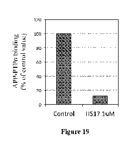
FIG. 19 Heparin fragment of 17 oligosaccharides inhibits APP-PTPG binding.
Recombinant human APP fragment binding to PTPG is detected by kinetic FT ISA
assay. Heparin
fragment of 17 oligosaccharides (heparan sulfate analog) effectively disrupts
APP-PTPG binding
when included in the binding assay. APP fragment used here corresponds to SEQ
ID NO:1, which
is the region between El and E2 domains. PTPG fragment used here includes its
IG1 and IG2
domains.
FIG. 20 Ligand binding site of PTPG IG1 domain interacts with APP. Binding of
human APP fragment (SEQ ID NO :1) with various PTPG fragments is measured by
kinetic FT ISA
assay. APP fragment corresponds to SEQ ID NO:1, which is a region between El
and E2 domains.
PTPG fragments used here include IG1,2 (containing IG1 and IG2 domains),
ALysIG1,2 (containing
IG1 and IG2 domains, with lysine 67, 68, 70,71 mutated to alanine), IG1-FN1
(containing IG1,
IG2, IG3 and FN1 domains), ECD (full extracellular domain of PTPG containing
all 3 IG domains
and 4 FN domains). Value shown are mean SEM, n=3 for each group. ***, p<0.001,
Student t test,
comparison with the IG1,2.
DETAILED DESCRIPTION
Experimental results in Example 1 show that neuronal receptor PTPG mediates
both 13-
amyloid and Tau pathogenesis in two mouse models. In the brain, PTPG binds to
APP. Depletion
of PTPG reduces the affinity between APP and 13-secretase, diminishing APP
proteolytic
products by 13- and y-cleavage without affecting other major substrates of the
secretases,
suggesting a specificity of (3-amyloidogenic regulation. In human APP
transgenic mice during
aging the progression of (3-amyloidosis, Tau aggregation, neuroinflammation,
synaptic loss, as
well as behavioral deficits, all show unambiguous dependency on the expression
of PTPG.
Additionally, the aggregates of endogenous Tau are found in a distribution
pattern similar to that
of early stage neurofibrillary tangles in Alzheimer brains. Together, these
findings unveil a
gatekeeping role of PTPG upstream of the degenerative pathogenesis, indicating
a potential for
this neuronal receptor as a drug target for Alzheimer' s disease.
Experimental results in Example 2 show that two classes of PTPa ligands in the
brain
microenvironment, CS and HS, regulate APP amyloidogenic processing in opposite
manners. CS
increases APP 13-cleavage products, whereas HS decreases APP 13-cleavage
products. Because
12
CA 03063061 2019-11-08
WO 2017/197253 PCT/US2017/032387
CS and HS compete to interact with receptor PTPa yet lead to opposite
signaling and neuronal
responses, the ratio of perineuronal CS and HS is therefore crucial for the
downstream effects of
PTPa and maintaining the health of the brain.
Experimental results in Example 3 further define that the binding between APP
and
PTPa is mediated by a fragment on APP between its El and E2 domain and the IG1
domain of
PTPcy.
The findings that PTPG plays a pivotal role in the development of P-amyloid
and Tau
pathologies indicate that peptides, compositions, and methods disclosed herein
may be suitable
to treat and prevent neurodegenerative diseases that involve P-amyloid
pathologies and/or Tau
pathologies, including but not limited to Alzheimer's disease, Lewy body
dementia,
frontotemp oral dementia, cerebral amyloid angiopathy, primary age-related
tauopathy, chronic
traumatic encephalopathy, Parkinson's disease, postencephalitic parkinsonism,
Huntington's
disease, amyolateral sclerosis, Pick's disease, progressive supranuclear
palsy, corticobasal
degeneration, Lytico-Bodig disease, ganglioglioma and gangliocytoma, subacute
sclerosing
panencephalitis, Hallervorden- Spatz disease, and/or Creutzfeldt-Jakob
disease.
Additionally, these peptides, compositions, and methods may also be used to
prevent
these neurodegenerative diseases in at-risk populations, such as subjects with
Down syndrome
and those suffered from brain injuries or cerebral ischemia, as well as the
aging population.
Definitions
As used in the specification and claims, the singular form "a," "an," and
"the" include
plural references unless the context clearly dictates otherwise. For example,
the term "a cell"
includes a plurality of cells, including mixtures thereof
The terms "about" and "approximately" are defined as being "close to" as
understood by
one of ordinary skill in the art. In one non-limiting embodiment the terms are
defined to be
within 10%. In another non-limiting embodiment, the terms are defined to be
within 5%. In still
another non-limiting embodiment, the terms are defined to be within 1%.
The terms "protein," "peptide," and "polypeptide" are used interchangeably to
refer to a
natural or synthetic molecule comprising two or more amino acids linked by the
carboxyl group
of one amino acid to the alpha amino group of another. The term "protein"
includes amino acids
joined to each other by peptide bonds or modified peptide bonds, e.g., peptide
isosteres, etc., and
can contain modified amino acids other than the 20 gene-encoded amino acids.
The
polypeptides can be modified by either natural processes, such as post-
translational processing,
13
CA 03063061 2019-11-08
WO 2017/197253
PCT/US2017/032387
or by chemical modification techniques which are well known in the art. The
term also includes
peptidomimetics and cyclic peptides.
As used herein, "peptidomimetic" means a mimetic of a peptide which includes
some
alteration of the normal peptide chemistry. Peptidomimetics typically enhance
some property of
the original peptide, such as increase stability, increased efficacy, enhanced
delivery, increased
half life, etc. Methods of making peptidomimetics based upon a known
polypeptide sequence is
described, for example, in U.S. Patent Nos. 5,631,280; 5,612,895; and
5,579,250. Use of
peptidomimetics can involve the incorporation of a non-amino acid residue with
non-amide
linkages at a given position. One embodiment of the present invention is a
peptidomimetic
wherein the compound has a bond, a peptide backbone or an amino acid component
replaced
with a suitable mimic. Some non-limiting examples of unnatural amino acids
which may be
suitable amino acid mimics include 13-alanine, L-a-amino butyric acid, L-y-
amino butyric acid,
L-a-amino isobutyric acid, L-c-amino caproic acid, 7-amino heptanoic acid, L-
aspartic acid, L-
glutamic acid, N- c-Boc-N- a-CBZ-L-lysine, N- c-Boc-N-a-Fmoc-L-lysine, L-
methionine sulfone,
L-norleucine, L-norvaline, N-a-Boc-N-6CBZ-L-ornithine, N-6-Boc-N-a-CBZ-L-
ornithine, Boc-
p-nitro-L-phenylalanine, Boc-hydroxyproline, and Boc-L-thioproline.
A "fusion protein" refers to a polypeptide formed by the joining of two or
more
polypeptides through a peptide bond formed between the amino terminus of one
polypeptide and
the carboxyl terminus of another polypeptide. The fusion protein can be formed
by the chemical
coupling of the constituent polypeptides or it can be expressed as a single
polypeptide from
nucleic acid sequence encoding the single contiguous fusion protein. A single
chain fusion
protein is a fusion protein having a single contiguous polypeptide backbone.
Fusion proteins can
be prepared using conventional techniques in molecular biology to join the two
genes in frame
into a single nucleic acid, and then expressing the nucleic acid in an
appropriate host cell under
conditions in which the fusion protein is produced.
As used herein, protein "binding" is the binding of one protein to another.
The binding
may comprise covalent bonds, protein cross-linking, and/or non-covalent
interactions such as
hydrophobic interactions, ionic interactions, or hydrogen bonds.
The term "protein domain" refers to a portion of a protein, portions of a
protein, or an
entire protein showing structural integrity; this determination may be based
on amino acid
composition of a portion of a protein, portions of a protein, or the entire
protein.
"Amyloid precursor protein" (APP) is an integral membrane protein expressed in
many
tissues and concentrated in the synapses of neurons. It has been implicated as
a regulator of
synapse formation, neural plasticity and iron export. APP is cleaved by beta
secretase and
14
CA 03063061 2019-11-08
WO 2017/197253
PCT/US2017/032387
gamma secretase to yield Aft Amyloid beta (A0) denotes peptides of 36-43 amino
acids that are
involved in Alzheimer's disease as the main component of the amyloid plaques
found in the
brains of Alzheimer patients. AP molecules cleaved from APP can aggregate to
form flexible
soluble oligomers which may exist in various forms. Certain misfolded
oligomers (known as
"seeds") can induce other AP molecules to also take the misfolded oligomeric
foiln, leading to a
chain reaction and buildup of amyloid plaques. The seeds or the resulting
amyloid plaques are
toxic to cells in the brain.
"Protein tyrosine phosphatases" or "receptor protein tyrosine phosphatases"
(PTPs) are a
group of enzymes that remove phosphate groups from phosphorylated tyrosine
residues on
proteins. Protein tyrosine phosphorylation is a common post-translational
modification that can
create novel recognition motifs for protein interactions and cellular
localization, affect protein
stability, and regulate enzyme activity. As a consequence, maintaining an
appropriate level of
protein tyrosine phosphorylation is essential for many cellular functions.
Tyrosine-specific
protein phosphatases catalyze the removal of a phosphate group attached to a
tyrosine residue.
These enzymes are key regulatory components in many signal transduction
pathways (such as
the MAP kinase pathway) that underlie cellular functions such as cell cycle
control/proliferation,
cell death, differentiation, transformation, cell polarity and motility,
synaptic plasticity, etc.
The term "subject" refers to any individual who is the target of
administration or
treatment. The subject can be a vertebrate, for example, a mammal. Thus, the
subject can be a
human or veterinary patient. The term "patient" refers to a subject under the
treatment of a
clinician, e.g., physician. An "at-risk" subject is an individual with a
higher likelihood of
developing a certain disease or condition. An "at-risk" subject may have, for
example, received
a medical diagnosis associated with the certain disease or condition.
"Tau proteins" (or T proteins) are proteins that stabilize microtubules. They
are abundant
in neurons of the central nervous system and are less common elsewhere, but
are also expressed
at very low levels in CNS astrocytes and oligodendrocytes. Neurodegenerative
disorders such as
Alzheimer's disease, Parkinson's disease, and other tauopathies are associated
with tau proteins
that have become defective, misfolded, tangled, and no longer stabilize
microtubules properly.
The term "protein fragment" refers to a functional portion of a full-length
protein. For
example, a fragment of APP or PTPG may be synthesized chemically or
biologically for the
purposes of disrupting the binding between APP and PTPG. Such fragments could
be used as
"decoy" peptides to prevent or diminish the actual APP-PTPG binding
interaction that results in
0-cleavage of APP and subsequent AP formation.
CA 03063061 2019-11-08
WO 2017/197253
PCT/US2017/032387
The phrase "functional fragment" or "analog" or mimetic of a protein or other
molecule
is a compound having qualitative biological activity in common with a full-
length protein or
other molecule of its entire structure. A functional fragment of a full-length
protein may be
isolated and attached to a separate peptide sequence. For example, a
functional fragment of a
blood-brain barrier penetrating protein may be isolated and attached to the
decoy peptide that
disrupts APP-PTPG binding, thereby enabling the hybrid peptide to enter the
brain and disrupt
APP-PTPG binding. Another example of a functional fragment is a membrane
penetrating
fragment, or one that relays an ability to pass the lipophilic barrier of a
cell's plasma membrane.
An analog of heparin, for example, may be a compound that binds to a heparin
binding site.
As used herein, "cyclic peptide" or "cyclopeptide" in general refers to a
peptide
comprising at least one internal bond attaching nonadjacent amino acids of the
peptide, such as
when the end amino acids of a linear sequence are attached to form a circular
peptide.
The term "antibody" refers to natural or synthetic antibodies that selectively
bind a target
antigen. The term includes polyclonal and monoclonal antibodies. In addition
to intact
immunoglobulin molecules, also included in the term "antibodies" are fragments
or polymers of
those immunoglobulin molecules, and human or humanized versions of
immunoglobulin
molecules that selectively bind the target antigen
As used herein, "enzyme" refers to a protein specialized to catalyze or
promote a specific
metabolic reaction.
"Neurodegenerative disorders" or "neurodegenerative diseases" are conditions
marked by
the progressive loss of structure or function of neural cells, including death
of neurons and glia.
The term "treatment" refers to the medical management of a patient with the
intent to
cure, ameliorate, stabilize, or prevent a disease, pathological condition, or
disorder. This term
includes active treatment, that is, treatment directed specifically toward the
improvement of a
disease, pathological condition, or disorder, and also includes causal
treatment, that is, treatment
directed toward removal of the cause of the associated disease, pathological
condition, or
disorder. In addition, this term includes palliative treatment, that is,
treatment designed for the
relief of symptoms rather than the curing of the disease, pathological
condition, or disorder;
preventative treatment, that is, treatment directed to minimizing or partially
or completely
inhibiting the development of the associated disease, pathological condition,
or disorder; and
supportive treatment, that is, treatment employed to supplement another
specific therapy directed
toward the improvement of the associated disease, pathological condition, or
disorder.
The term "administering" refers to an administration that is intranasal, oral,
topical,
intravenous, subcutaneous, transcutaneous, transdermal, intramuscular, intra-
joint, parenteral,
16
CA 03063061 2019-11-08
WO 2017/197253
PCT/US2017/032387
intra-arteriole, intradermal, intraventricular, intracranial, intraperitoneal,
intralesional, rectal,
vaginal, by inhalation or via an implanted reservoir. The term "parenteral"
includes
subcutaneous, intravenous, intramuscular, intra- articular, intra- syno vial,
intrastema 1, intratheca 1,
intrahepatic, intralesional, and intracranial injections or infusion
techniques.
The term "pharmaceutically acceptable carrier" means a carrier or excipient
that is useful
in preparing a pharmaceutical composition that is generally safe and non-
toxic, and includes a
carrier that is acceptable for veterinary and/or human pharmaceutical use. As
used herein, the
term "pharmaceutically acceptable carrier" encompasses any of the standard
pharmaceutical
carriers, such as a phosphate buffered saline solution, water, and emulsions,
such as an oil/water
or water/oil emulsion, and various types of wetting agents. As used herein,
the term "carrier"
encompasses any excipient, diluent, filler, salt, buffer, stabilizer,
solubilizer, lipid, stabilizer, or
other material well known in the art for use in pharmaceutical formulations
and as described
further below. The pharmaceutical compositions also can include preservatives.
A
"pharmaceutically acceptable carrier" as used in the specification and claims
includes both one
and more than one such carrier.
The term "variant" refers to an amino acid or peptide sequence having
conservative
amino acid substitutions ("conservative variant"), non-conservative amino acid
subsitutions (e.g.,
a degenerate variant), substitutions within the wobble position of each codon
(i.e. DNA and
RNA) encoding an amino acid, amino acids added to the C-terminus of a peptide,
or a peptide
having 60%, 70%, 80%, 90%, or 95% homology to a reference sequence.
The term "percent (%) sequence identity" or "homology" is defined as the
percentage of
nucleotides or amino acids in a candidate sequence that are identical with the
nucleotides or
amino acids in a reference nucleic acid sequence, after aligning the sequences
and introducing
gaps, if necessary, to achieve the maximum percent sequence identity.
Alignment for purposes
of determining percent sequence identity can be achieved in various ways that
are within the skill
in the art, for instance, using publicly available computer software such as
BLAST, BLAST-2,
ALIGN, ALIGN-2 or Megalign (DNASTAR) software. Appropriate parameters for
measuring
alignment, including any algorithms needed to achieve maximal alignment over
the full-length of
the sequences being compared can be determined by known methods.
Compositions
Peptides:
Disclosed herein are peptides for treating and preventing the aforementioned
neurodegenerative diseases, such as Alzheimer's disease. In some embodiments,
the peptides
17
CA 03063061 2019-11-08
WO 2017/197253
PCT/US2017/032387
disrupt the binding between PTPa and APP, preventing P-amyloidogenic
processing of APP
without affecting other major substrates of the 0- and y-secretases. The
peptide may be a decoy
fragment of APP, a decoy fragment of PTPa, or a combination thereof
In some embodiments, a decoy peptide could be fabricated from the PTPa-binding
region
on APP, which is the fragment between its El and E2 domains (SEQ ID NO:1). In
some
embodiments, a decoy peptide could be fabricated from the APP-binding region
on PTPcy, which
is its IG1 domain (SEQ ID NO: 442). In some embodiments, a decoy peptide could
be fabricated
that corresponds to the entire APP E2 domain or a fragment thereof In some
embodiments, a
decoy peptide could be fabricated that corresponds to the entire APP El domain
or a fragment
thereof In some embodiments, a PTPa peptide is used in combination with an APP
peptide.
In some embodiments, the peptide is a fragment of the PTPa-binding domain of
APP.
Therefore, in some embodiments, the peptide is a fragment of SEQ ID NO:1, as
listed below,
which has at least 5, 6, 7, 8, 9, 10, 11, 12, 13, 14, or more amino acids, or
a conservative variant
thereof
AEESDNVDSADAFFDDSDVWWGGADTDYADGSEDKVVEVAEEEEVAEVEFFE
ADDDEDDEDGDEVFFEAEEPYFFATERTTSIATTTTTTTESVEEVVR (SEQ ID NO:1).
Therefore, in some embodiments, the peptide comprises an amino acid sequence
selected
from 10 consecutive residues of SEQ ID NO: 1, or from the group consisting of
the below:
SEQ ID NO:2 AEESDNVDSA
SEQ ID NO:3 FFSDNVDSAD
SEQ ID NO:4 ESDNVDSADA
SEQ ID NO:5 SDNVDSADAE
SEQ ID NO:6 DNVDSADAEE
SEQ ID NO:7 NVDSADAEED
SEQ ID NO :8 VD SADAEEDD
SEQ ID NO:9 DSADAEEDDS
SEQ ID NO:10 SADAEEDDSD
SEQ ID NO:11 ADAEEDDSDV
SEQ ID NO:12 DAEEDDSDVW
SEQ ID NO:13 AEEDDSDVWW
SEQ ID NO:14 EEDDSDVWWG
SEQ ID NO:15 EDDSDVWWGG
SEQ ID NO :16 DDSDVWWGGA
SEQ ID NO:17 DSDVWWGGAD
SEQ ID NO:18 SDVWWGGADT
SEQ ID NO :19 DVWWGGADTD
18
6
aVaalAHCEOCE Z9: ON CR OHS
VaHHAHCEDC11 190N CR OHS
HHHAHCEDGICE 09:0N CR OHS
didAHCEDGICECE 6c: ON CR OHS
HAHCEDGICKTI 8S:0N CR OHS
AHCEDGICKTICE LS:ON CR OHS
HCEDGICKTICKE 9ç: ON CR OHS
CEDGICKTICKICE SS:ON CR OHS
GICKTICKICEV 17S: ON CR OHS
GICKTICECKIVH S: ON CR OHS
HCECECECKIVIA ZS:ON CR OHS
CECECECKIVIld IS:ON af OHS
CECECKIV1114 OS: ON af OHS
HCECKIVIMA 617:0N CR OHS
CECKIVII4HAH 817: ON CR OHS
GCEVallIAHV Lt: ON CR OHS
CEVaallAHVA 917: ON CR OHS
VaaalAHVAH St:ON CR OHS
HHHHAHVAld ft: ON CR OHS
HHHAHVAdild 17: ON CR OHS
HHAHVAdilld Z17: ON CR OHS
HAHVAIdaW WON CR OHS
AHVAallIVA 017: ON CR OHS
HVA=VAH 6: ON CR OHS
VAH=VAHA 8 : ON CR OHS
AHHaIVAHAA LEON CR OHS
H3lIVAHAA)1 9: ON CR OHS
alIVAHAANCE SE:ON CR OHS
alVAHAANC11 17:0N CR OHS
HVAHAANCHS : ON CR OHS
VAHAANCHS9 a:ON CR OHS
AHAANCHSOCE I : ON CR OHS
HAANCHSOCEV 0: ON CR OHS
AANCHSOCEVA 6Z: ON CR OHS
A )1C1ISOCEVACE 8Z: ON CR OHS
)1C1ISOCEVACIL LZ: ON CR OHS
CHSOCEVACLICE 9Z: ON CR OHS
HSOCEVACLICEV SZ: ON CR OHS
SOCEVACLICW9 ION CR OHS
CEVACLICEV99 EZ: ON CR OHS
CEVACLICEV99M ZZ: ON CR OHS
VACLICEV99MM 1Z: ON CR OHS
ACLICEV99MMA OZ: ON CR OHS
L8a0/LIOZSI1IIDcl SZL6I/LIOZ OM
80-TT-610Z T90900 YD
CA 03063061 2019-11-08
WO 2017/197253 PCT/US2017/032387
SEQ ID NO :63 GDEVEEEAEE
SEQ ID NO:64 DEVEEEAEEP
SEQ ID NO :65 EVEFFAEFPY
SEQ ID NO:66 VEEEAEEPYE
SEQ ID NO :67 FFFAEEPYEE
SEQ ID NO:68 FFAEEPYEEA
SEQ ID NO:69 EAEFPYEEAT
SEQ ID NO:70 AEEPYEEATE
SEQ ID NO:71 EEPYEEATER
SEQ ID NO:72 FPYEEAIERT
SEQ ID NO:73 PYEEATERTT
SEQ ID NO:74 YEEATERTTS
SEQ ID NO:75 EFATERTTSI
SEQ ID NO:76 EATERTTSIA
SEQ ID NO:77 AIERTTSIAT
SEQ ID NO:78 TERTTSIATT
SEQ ID NO:79 ERTTSIATTT
SEQ ID NO:80 RTTSIATTTT
SEQ ID NO :81 TTSIATTTTT
SEQ ID NO:82 TSIATTTTTT
SEQ ID NO:83 SIATTTTTTT
SEQ ID NO:84 IATTTTTTTE
SEQ ID NO:85 ATTTTTTTES
SEQ ID NO:86 TTTTTTTES V
SEQ ID NO:87 TTTTT _____ IES VE
SEQ ID NO:88 TTTT ______ IESVEE
SEQ ID NO:89 TTT _______ IESVEEV
SEQ ID NO:90 TT __ IESVEEVV
SEQ ID NO:91 TIES VEEVVR
In some embodiments, the peptide comprises an amino acid sequence selected
from 11
consecutive residues of SEQ ID NO: 1, or from the group consisting of the
below:
SEQ ID NO:92 AEESDNVDSAD
SEQ ID NO:93 FFSDNVDSADA
SEQ ID NO:94 ESDNVDSADAE
SEQ ID NO:95 SDNVDSADAEE
SEQ ID NO:96 DNVDSADAEED
SEQ ID NO:97 NVDSADAEEDD
SEQ ID NO:98 VDSADAEEDDS
SEQ ID NO:99 DSADAEEDDSD
SEQ ID NO:100 SADAEEDDSDV
SEQ ID NO:101 ADAEEDDSDVW
I Z
CEOCIICKTICKICEV 1717I: ON CR OaS
OCIICKTICECKIVa 171: ON CR OaS
CfaCECIICKKEVIA Zi7I: ON al OaS
aCklaCECKIVIld ItI:ON CR OHS
CklaCECKIV1114 Off ON af OaS
CIICECKIValalA 6 I: ON CR OHS
aCECKIVaallAa 8I: ON CR OaS
CECECEValalAaV LEI:ON CR OHS
CECEVaallAaVA 91: ON CR OaS
CEVIdalAaVAa SELON CR OHS
VadidaAaVAld 17I: ON CR OaS
aadidAaVAIld I: ON CR OaS
aaaAaVAdilld at: ON CR OHS
aaAaVAalaaV I EL ON CR OaS
aAaVAdildaVA 0I: ON CR OaS
AaVAallaVAa Kt: ON CR OHS
aVAaalaVAaA SZI:ON CR OaS
VAaalaVAaAA LZI: ON CR OaS
Aa3laVAaAA)1 9I: 0N CR OaS
aalaVAaAANCE SZI: ON CR OHS
aaaVAaAANCII 17ZI: ON CR OHS
alVAaAANCIIS I: 0N CR OaS
aVAaAANCIIS9 ZZI: ON ca OaS
VAaAANCESOCE IZI:ON CR OaS
AaAANCIISOCEV OZI: ON CR OaS
aAANCIISOCEVA 611: ON CR OaS
AANCIISOCEVACE 8II:ON CR OaS
ANCESOCEVACIL LI I: ON CR OaS
)1CIISOCEVACLICE 9I LON CR OaS
CESOCEVACLICEV SILON CR OaS
SOCEVACLICEV9 LON CR OaS
SOCEVACLICEVDD Et ION CR OaS
OCEVACLICEVDDM ZI LON CR OaS
CEVACLICEVDOMM ii ION CR OaS
VACLICEVDDAMA OI LON CR WS
ACLICEVDDAMACE 60I: ON CR OaS
CLICEVDDMANACES 80I: ON CR OaS
ICEVDDMANACESCE LOI: ON CR OaS
CEVDDMMACESCECE 90I: ON CR OaS
VDDAMACESCECII SOLON CR OaS
DOMMACESCECIal tOI: ON CR OaS
DAMACESCECIIIV 01: ON CR OaS
MMACESCECRIVCE COLON CR OaS
L8a0/LIOZSI1IIDcl SZL6I/LIOZ OM
80-TT-610Z T90900 YD
CA 03063061 2019-11-08
WO 2017/197253 PCT/US2017/032387
SEQ ID NO:145 DDDEDDEDGDE
SEQ ID NO :146 DDEDDEDGDEV
SEQ ID NO :147 DEDDEDGDEVE
SEQ ID NO :148 EDDEDGDEVEE
SEQ ID NO :149 DDEDGDEVEFE
SEQ ID NO :150 DEDGDEVEEEA
SEQ ID NO :151 EDGDEVEEEAE
SEQ ID NO:152 DGDEVEEEAFF
SEQ ID NO :153 GDEVEEEAEFP
SEQ ID NO:154 DEVEEEAFFPY
SEQ ID NO:155 EVEEEAEFPYE
SEQ ID NO :156 VEEEAEEPYEE
SEQ ID NO:157 EFFAEFPYFF A
SEQ ID NO :158 FFAEEPYEEAT
SEQ ID NO:159 EAEEPYEEATE
SEQ ID NO :160 AEEPYEEATER
SEQ ID NO :161 EEPYEEATERT
SEQ ID NO:162 FPYEEAIERTT
SEQ ID NO:163 PYEEATERTTS
SEQ ID NO:164 YEEATERTTS I
SEQ ID NO:165 EFAIERTTSIA
SEQ ID NO:166 EATERTTSIAT
SEQ ID NO:167 ATERTTSIATT
SEQ ID NO:168 TERTTSIATTT
SEQ ID NO:169 ERTTSIATTTT
SEQ ID NO:170 RTTSIATTTTT
SEQ ID NO:171 TTSIATTTTTT
SEQ ID NO:172 TSIATTTTTTT
SEQ ID NO:173 SIATTTTTTTE
SEQ ID NO:174 IATTTTTTTES
SEQ ID NO:175 ATTTTTTTESV
SEQ ID NO:176 TTTTTTTESVE
SEQ ID NO:177 TTTTT _______ IES VEF
SEQ ID NO:178 TTTT ________ IESVEEV
SEQ ID NO:179 TTT _________ IESVEEVV
SEQ ID NO:180 TT __________ IESVEEVVR
In some embodiments, the peptide comprises an amino acid sequence selected
from 12
consecutive residues of SEQ ID NO: 1, or from the group consisting of the
below:
SEQ ID NO:181 AEESDNVDSADA
SEQ ID NO:182 FFSDNVDSADAE
SEQ ID NO:183 ESDNVDSADAEE
22
EZ
aCKICEValalAaV 9ZZ: ON CR OHS
CECKIVaallAaVA SZZ: ON CR OaS
CRIVIdalAaVAa 17ZZ: ON CR OHS
CEVadidaAaVAld EZZ: ON CR OHS
VaadidAaVAIld ZZZ: ON CR OHS
aaaaAaVAdilld 1ZZ:0N CR OHS
IdaAaVAalaaV OZZ: ON CR OHS
aaAaVAdildaVA 6IZ:ON CR OHS
aAaVAallaVAa SIZ:ON CR OHS
AaVAaalaVAaA LION CR OHS
aVAaalaVAaAA 9IZ:ON CR OHS
VAaalaVAaAAN SIZ:ON CR OHS
AaalaVAaAANCE 17 I Z: ON ca OaS
aaaaVAaAANCH EIZ:ON CR OHS
aaaVAaAANCHS ZION CR OHS
aaVAaAANCHS9 II Z: ON CR OaS
aVAaAANCESOCE OIZ:ON CR OaS
VAaAANCHSOCEV 60Z: ON CR OaS
AaAANCHSOCEVA 80Z: ON CR OaS
aAANCHSOCEVACE LOON CR OHS
AANCESOCEVACIL 90Z: ON CR OaS
A NalSOCEVACEICE SOON CR OaS
NCESOCEVACEICEV 170Z: ON CR OaS
CfaSOCEVACEICEV9 0Z: ON CR OaS
aSOCEVACEICEVDD ZOZ: ON CR OHS
SOCEVACEICEVDDM ICON CR OHS
DCWACEICEVDDMM OCC: ON CR OHS
CEVACEICEVDDAMA 661:0N CR OaS
VACEICEVDDAMACE 861:0N CR OaS
ACEICEVDDMANACES L61: ON CR OaS
CLIEVDDMANACESCE 961:0N CR OaS
ICEVDDMMACESCECE S6I:ON CR OaS
CEVDDMMACESCECH 176I: ON CR OaS
VDDAMACESCECIal 6 I: ON CR OaS
99MANACESaa-d3v Z6I: ON CR OaS
DAMACESCECRIVCE 161:0N CR OaS
MMACESCKHIVCW 06 I: ON CR OaS
MACESCKHIVCWS 68 ION CR OaS
ACESCECUIVCWSCE 88LON CR OaS
CESCECRIVCWSCIA L8LON CR OaS
SCKHIVCWSCIAN 981:0N CR OaS
CfcrdavavSCIANCE SSI:ON CR OaS
CfalVCWSCIANCES 178 I: ON CR OaS
L8a0/LIOZSI1IIDcl SZL6I/LIOZ OM
80-TT-610Z T90900 YD
CA 03063061 2019-11-08
WO 2017/197253
PCT/US2017/032387
SEQ ID NO :227 EVEEEEADDDED
SEQ ID NO :228 VEEEEADDDEDD
SEQ ID NO :229 EFEFADDDEDDE
SEQ ID NO :230 EEFADDDEDDED
SEQ ID NO :231 EFADDDEDDED G
SEQ ID NO :232 EADDDEDD ED GD
SEQ ID NO :233 ADDDEDDEDGDE
SEQ ID NO :234 DDDEDDEDGDEV
SEQ ID NO:235 DDEDDEDGDEVE
SEQ ID NO :236 DEDDEDGDEVEE
SEQ ID NO :237 EDDEDGDEVEEE
SEQ ID NO :238 DDEDGDEVEFEA
SEQ ID NO :239 DEDGDEVEEEAE
SEQ ID NO :240 EDGDEVEEEAEE
SEQ ID NO :241 DGDEVEEEAEEP
SEQ ID NO :242 GDEVEEEAEEPY
SEQ ID NO :243 DEVEEEAEFP YE
SEQ ID NO :244 EVEEEAEEPYEE
SEQ ID NO :245 VEEEAEEPYEEA
SEQ ID NO :246 EEFAEEPYEF AT
SEQ ID NO :247 EFAEEPYEEATE
SEQ ID NO :248 EAEEPYEEATER
SEQ ID NO :249 AEEPYEEATERT
SEQ ID NO :250 EEPYEEATERTT
SEQ ID NO :251 EP YEEA ______ IERT TS
SEQ ID NO :252 PYEEATERTTS I
SEQ ID NO :253 YEEATERTTS IA
SEQ ID NO :254 EFA __ IERT TS I AT
SEQ ID NO :255 EATERTTSIATT
SEQ ID NO :256 ATERTTSIATTT
SEQ ID NO :257 TERTTSIATTTT
SEQ ID NO :258 ERTTSIATTTTT
SEQ ID NO :259 RTTSIATTTTTT
SEQ ID NO :260 TTSIATTTTTTT
SEQ ID NO :261 TSIATTTTTTTE
SEQ ID NO :262 SIATTTTTTTES
SEQ ID NO :263 IATTTTTTTES V
SEQ ID NO :264 ATTTTTTTESVE
SEQ ID NO :265 TTTTTTTES VEE
SEQ ID NO :266 TTTTT ________ IES VEF V
SEQ ID NO :267 TTTT _________ IES VEEV V
SEQ ID NO :268 TTT __ IESVEEVVR
24
CA 03063061 2019-11-08
WO 2017/197253 PCT/US2017/032387
In some embodiments, the peptide comprises an amino acid sequence selected
from 13
consecutive residues of SEQ ID NO: 1, or from the group consisting of the
below:
SEQ ID NO :268 TTT __ IESVEEVVR
SEQ ID NO :269 AEESDNVDSADAE
SEQ ID NO :270 FFSDNVDSADAEE
SEQ ID NO :271 ESDNVDSADAEED
SEQ ID NO :272 SDNVDSADAEEDD
SEQ ID NO :273 DNVDSADAEEDDS
SEQ ID NO :274 NVDSADAEEDDSD
SEQ ID NO :275 VDSADAEEDDSDV
SEQ ID NO :276 DSADAEEDDSDVW
SEQ ID NO :277 SADAEEDDSDVWW
SEQ ID NO :278 ADAEEDDSDVWWG
SEQ ID NO :279 DAEEDDSDVWW GG
SEQ ID NO :280 AEEDDSDVWWGGA
SEQ ID NO :281 EEDDSDVWWGGAD
SEQ ID NO :282 EDDSDVWWGGADT
SEQ ID NO :283 DDSDVWWGGADTD
SEQ ID NO :284 DSDVWWGGADTDY
SEQ ID NO :285 SDVWWGGADTDYA
SEQ ID NO :286 DVWWGGADTDYAD
SEQ ID NO :287 VWWGGADTDYADG
SEQ ID NO :288 WWGGADTDYADGS
SEQ ID NO :289 WGGADTDYADGSE
SEQ ID NO :290 GGADTDYADGSED
SEQ ID NO :291 GADTDYADGS EDK
SEQ ID NO :292 ADTDYADGSEDKV
SEQ ID NO :293 DTDYADGSEDK VV
SEQ ID NO :294 TDYADGSEDKVVE
SEQ ID NO :295 DYADGSEDKVVEV
SEQ ID NO :296 YADGSEDKVVEVA
SEQ ID NO :297 ADGSEDKVVEVAE
SEQ ID NO :298 DGSEDKVVEVAFF
SEQ ID NO :299 GSEDKVVEVAEEE
SEQ ID NO :300 SEDKVVEVAEEEE
SEQ ID NO:301 EDKVVEVAEEEEV
SEQ ID NO :302 DKVVEVAEEEEVA
SEQ ID NO :303 KVVEVAEEEEVAE
SEQ ID NO :304 VVEVAEEEEVAEV
SEQ ID NO :305 VEVAEEEEVAEVE
SEQ ID NO :306 EVAEEEEVAEVEE
SEQ ID NO :307 VAEFFFVAEVEEE
SEQ ID NO :308 AEEEEVAEVEFFE
CA 03063061 2019-11-08
WO 2017/197253
PCT/US2017/032387
SEQ ID NO :309 EEEFVAEVEEEEA
SEQ ID NO :310 EEFVAEVEFEEAD
SEQ ID NO :311 EFVAEVEEFEADD
SEQ ID NO :312 EVAEVEEEF ADD D
SEQ ID NO :313 VAEVEEEEADDDE
SEQ ID NO :314 AEVEEEEADDDED
SEQ ID NO :315 EVEEEEADDDEDD
SEQ ID NO :316 VEEEEADDDEDDE
SEQ ID NO :317 EEEFADDDEDDED
SEQ ID NO :318 EEFADDDEDDEDG
SEQ ID NO :319 EFADDDEDDED GD
SEQ ID NO :320 EADDDEDD ED GDE
SEQ ID NO:321 ADDDEDDEDGDEV
SEQ ID NO :322 DDDEDDEDGDEVE
SEQ ID NO :323 DDEDDEDGDEVEF
SEQ ID NO :324 DEDDEDGDEVEEE
SEQ ID NO :325 EDDEDGDEVEEEA
SEQ ID NO :326 DDEDGDEVEFEAE
SEQ ID NO :327 DEDGDEVEEEAEE
SEQ ID NO :328 EDGDEVEEEAEEP
SEQ ID NO :329 DGDEVEEEAEEP Y
SEQ ID NO :330 GDEVEEEAEEPYE
SEQ ID NO :331 DEVEEEAEFP YEF
SEQ ID NO :332 EVEEEAEEPYEEA
SEQ ID NO :333 VEEEAEEPYEEAT
SEQ ID NO :334 EEFAEEPYEF ATE
SEQ ID NO:335 EFAEEPYEEATER
SEQ ID NO :336 EAEEPYEEATERT
SEQ ID NO:337 AEEPYEEATERTT
SEQ ID NO :338 EEPYEEATERTTS
SEQ ID NO :339 EP YEEA _______ IERT T S I
SEQ ID NO :340 PYEEATERTTS IA
SEQ ID NO :341 YEEATERTTS TAT
SEQ ID NO :342 EFA ___________ IERT TS I AT T
SEQ ID NO :343 EATERTTSIATTT
SEQ ID NO :344 ATERTTSIATTTT
SEQ ID NO :345 TERTTSIATTTTT
SEQ ID NO :346 ERTTSIATTTTTT
SEQ ID NO :347 RTTSIATTTTTTT
SEQ ID NO :348 TTSIATTTTTTTE
SEQ ID NO :349 TSIATTTTTTTES
SEQ ID NO :350 SIATTTTTTTESV
SEQ ID NO :351 IATTTTTTTES VE
26
CA 03063061 2019-11-08
WO 2017/197253 PCT/US2017/032387
SEQ ID NO :352 ATTTTTTTESVEE
SEQ ID NO :353 TTTTTTTES VEEV
SEQ ID NO :354 TTTTT _________ IES VEF VV
SEQ ID NO :355 TTTT __________ IESVEEVVR
In some embodiments, the peptide comprises an amino acid sequence selected
from 14
consecutive residues of SEQ ID NO: 1, or from the group consisting of the
below:
SEQ ID NO :356 AEESDNVDSADAFF
SEQ ID NO :357 FFSDNVDSADAEED
SEQ ID NO :358 ESDNVDSADAEEDD
SEQ ID NO :359 SDNVDSADAEEDDS
SEQ ID NO :360 DNVDSADAEEDDSD
SEQ ID NO :361 NVDSADAEEDDSDV
SEQ ID NO :362 VDSADAEEDDSDVW
SEQ ID NO :363 DSADAEEDDSDVWW
SEQ ID NO :364 SADAEEDDSDVWWG
SEQ ID NO :365 ADAEEDDSDVWWGG
SEQ ID NO :366 DAEEDDSDVWWGGA
SEQ ID NO :367 AEEDDSDVWWGGAD
SEQ ID NO :368 EEDDSDVWWGGADT
SEQ ID NO :369 EDDSDVWWGGADTD
SEQ ID NO:370 DDSDVWWGGADTDY
SEQ ID NO :371 DSDVWWGGADTDYA
SEQ ID NO:372 SDVWWGGADTDYAD
SEQ ID NO:373 DVWWGGADTDYADG
SEQ ID NO:374 VWWGGADTDYADGS
SEQ ID NO :375 WWGGADTDYAD GS E
SEQ ID NO:376 WGGADTDYADGSED
SEQ ID NO:377 GGADTDYADGSEDK
SEQ ID NO :378 GADTDYADGS EDK V
SEQ ID NO:379 ADTDYADGSEDKVV
SEQ ID NO:380 DTDYADGSEDK VVE
SEQ ID NO:381 TDYADGSEDKVVEV
SEQ ID NO:382 DYADGSEDKVVEVA
SEQ ID NO :383 YADGSEDKVVEVAE
SEQ ID NO :384 ADGSEDKVVEVAEE
SEQ ID NO:385 DGSEDKVVEVAFFE
SEQ ID NO:386 GSEDKVVEVAEEFF
SEQ ID NO :387 SEDKVVEVAEEEEV
SEQ ID NO :388 EDKVVEVAEEEEVA
SEQ ID NO:389 DKVVEVAEEEEVAE
SEQ ID NO :390 KVVEVAEEEEVAEV
27
CA 03063061 2019-11-08
WO 2017/197253
PCT/US2017/032387
SEQ ID NO :391 VVEVAEEEEVAEVE
SEQ ID NO :392 VEVAEEEEVAEVEE
SEQ ID NO :393 EVAEEEEVAEVEEE
SEQ ID NO :394 VAEREFVAEVEEEE
SEQ ID NO :395 AEEEEVAEVEEFEA
SEQ ID NO :396 EEEFVAEVEEEEAD
SEQ ID NO :397 EEFVAEVEFEEADD
SEQ ID NO :398 EFVAEVEEFEADDD
SEQ ID NO :399 EVAEVEEEFADDDE
SEQ ID NO :400 VAEVEEEEADDDED
SEQ ID NO:401 AEVEEEFADDDEDD
SEQ ID NO :402 EVEEEEADDDEDDE
SEQ ID NO :403 VEEEEADDDEDDED
SEQ ID NO :404 FEEFADDDEDDEDG
SEQ ID NO :405 EEFADDDEDDEDGD
SEQ ID NO :406 EFADDDEDDED GDE
SEQ ID NO :407 EADDDEDD ED GDE V
SEQ ID NO :408 ADDDEDDEDGDEVE
SEQ ID NO :409 DDDEDDEDGDEVEE
SEQ ID NO :410 DDEDDEDGDEVEFE
SEQ ID NO :411 DEDDEDGDEVEEEA
SEQ ID NO :412 EDDEDGDEVEEEAE
SEQ ID NO :413 DDEDGDEVEFEAEF
SEQ ID NO :414 DEDGDEVEEEAEEP
SEQ ID NO:415 EDGDEVEEEAEEP Y
SEQ ID NO :416 DGDEVEEEAEEP YE
SEQ ID NO:417 GDEVEEEAEEPYEE
SEQ ID NO :418 DEVEEEAEFP YEF A
SEQ ID NO:419 EVEEEAEEPYEEAT
SEQ ID NO :420 VEEEAEEPYEEATE
SEQ ID NO:421 EEFAEEPYEF ATER
SEQ ID NO :422 EFAEEPYEEATERT
SEQ ID NO :423 EAEEPYEEATERTT
SEQ ID NO :424 AEEPYEEATERTTS
SEQ ID NO :425 EEPYEEATERTTSI
SEQ ID NO :426 EP YEEA __ IERT TS I A
SEQ ID NO :427 PYEEATERTTS TAT
SEQ ID NO :428 YEEATERTTS IA T T
SEQ ID NO :429 EFAIERTTSIATTT
SEQ ID NO :430 EATERTT S I A TT T T
SEQ ID NO :431 ATERTT S IA T TT T T
SEQ ID NO :432 TERTTSIAT T T TT T
SEQ ID NO :433 ERTTSIATT T T TT T
28
CA 03063061 2019-11-08
WO 2017/197253
PCT/US2017/032387
SEQ ID NO :434 RTTSIATTTTTTTE
SEQ ID NO :435 TTSIATTTTTTTES
SEQ ID NO :436 TSIATTTTTTTESV
SEQ ID NO :437 SIATTTTTTTESVE
SEQ ID NO :438 IATTTTTTTES VEE
SEQ ID NO :439 ATTTTTTTESVEEV
SEQ ID NO :440 TTTTTTTES VEEVV
SEQ ID NO :441 TTTTT IES VEF VVR
In some embodiments, the peptide comprises an amino acid sequence selected
from 24
consecutive residues of SEQ ID NO: 1, or from the group consisting of the
below:
SEQ ID NO: 900 _____________ ATERTTSIATTTTTT IES VEEVVR
In some embodiments, the peptide is a fragment of the APP-binding domain of
PTPa.
Therefore, in some embodiments, the peptide is a fragment of SEQ ID NO:442, as
listed below,
which has at least 5, 6, 7, 8, 9, 10, 11, 12, 13, 14, or more amino acids, or
a conservative variant
thereof The underlined amino acids represent residues in the ligand-binding
pocket.
EEPPRFIKEPKDQIGVSGGVASFVCQATGDPKPRVTWNKKGKKVNSQRFETIEFD
ESAGAVLRIQPLRTPRDENVYECVAQNSVGEITVHAKLTVLRE (SEQ ID NO :442).
Therefore, in some embodiments, the peptide comprises an amino acid sequence
selected
from 10 consecutive residues of SEQ ID NO: 442, or from the group consisting
of the below:
SEQ ID NO :443 EEPPRFIKEP
SEQ ID NO :444 EPPRFIKEPK
SEQ ID NO :445 PPRFIKEPKD
SEQ ID NO :446 PRFIKEPKDQ
SEQ ID NO :447 RFIKEPKDQI
SEQ ID NO :448 FIKEPKDQIG
SEQ ID NO :449 IKEPKDQIGV
SEQ ID NO :450 KEPKDQIGVS
SEQ ID NO:451 EPKDQIGVSG
SEQ ID NO :452 PKDQIGVSGG
SEQ ID NO :453 KDQIGVSGGV
SEQ ID NO :454 DQIGVSGGVA
SEQ ID NO:455 QIGVSGGVAS
SEQ ID NO :456 IGVSGGVASF
SEQ ID NO :457 GVSGGVASFV
SEQ ID NO :458 VSGGVASFVC
29
0
do1111AVDVS 66170N al OHS
ORIIAVDVSH 86170N CII OHS
1111AVDVSHCE L6170N CR OHS
111AVDVSHCH 96170N CR OHS
lAVDVSHai'd S6170N CR OHS
AVDVSHCLI41 176170N CII OHS
VOVSHCE14T,T, 6170N CII OHS
DVSHardlia Z617:0N CR OHS
VSHardliad 16170N CR OHS
SHCLEITEDI 06170N CR OHS
HarALLHDIO 68170N CR OHS
adm,TaDios 8817:0N CII OHS
daIIHRIOSN L8170N CR OHS
ALLHDIOSNA 9817:0N CR OHS
Ltaduost\inx sst:ON UT OHS
JaRiost\inxx tst:ONUT OHS
aRiost\inxxo 817:0N CR OHS
Riost\inxxox g17: UT OHS
liost\inxxoxx tst:ON CR OHS
OSNANNONNN 08170N CR OHS
SNANNONNNAk 6L170N CII OHS
NA)Dioxxl\inu sLt:ON CR OHS
ANNONNNAUA LLVON CR OHS
)1)19)DINAUAII 9L170N CR OHS
)19)DINAUAlid Lt: ON CR OHS
9)DINAUAlicIN 17 Lt.: ON CR OHS
)1)INAUAlicnId L17:0N CR OHS
NNAUAlicINdia ZLVON CR OHS
NAUAlIcINKED I Lt: ON CR OHS
AUAlicnIcKfaL 0 Lt.: ON CR OHS
69170N CR OHS
AlIcINKEDIVO 8917:0N CR OHS
licnIcICEDIVOD L917: ON CR OHS
dNcICEDIVODA 9917: ON CR OHS
NcICEDIVODAd S917:0N CR OHS
cICEDIVODAB 17917:0N CR OHS
CEDIVODABV 917:0N CII OHS
DIVODABVA Z917:0N CII OHS
IVODAdSVAD 19170N CII OHS
VODAdSVADD 09170N CII OHS
ODAdSVADDS 6S170N CII OHS
L8a0/LIOZSI1IIDd SZL6I/LIOZ OM
80-TT-610Z T90900 YD
1
DIOCENcId)Ildll 9S:
ON CII OHS
IOCENcId)IldlId S:
ON CR OHS
OCE)IcIANIDIdd tES:
ON CII OHS
CDIcIANIDIdcId EES:
ON CII OHS
)IddDTddEJZES: ON CII oas
alnAI'iWH iON
CR OHS
:moiaci ow Jo EuusIsuoo dnatg ow wag JO `Ztt :ON cif oas JO sat-ipso.'
pAunoasuoo
11 wag poloops aouanbas poi ouwui ui saspdwoo opudad ow `quaw!pociwo atuos uj
alrIAIINVHA iç: ON CR OHS
rnwHAI 0S: ON CR WS
lArDIVHAII 6ZS:0N CR OHS
ArnwHAm SZS:ON CR OHS
rINVHAIIHD LZS:ON cii OHS
INVHAIIHDA 9ZS:ON CR OHS
NVHAIIHDAS SZS:ON ciiOHS
VHAIMASN 17ZS:ON CR OHS
HAIIHDASNO S:ON CR OHS
AIIHDASNOV ZZS:ON ciiOHS
IMASNOVA I ZS:ON CR OHS
IHDASNOVAD OZS:ON CR OHS
HDASNOVADH 6 I S:ON cii OHS
DASNOVADHA 8I S:ON CR OHS
ASNOVADHAA LI S: 0 NI CR OHS
SNOVADHAAN 9IS:ON CR OHS
NOVADHAANH SI S:ON CR OHS
OVADHAANHCE tIS:ON CII OHS
VADHAANHCRI EIS:ON CR OHS
ADHAANHCRIcI Z I S:ON CR OHS
paxAmacmcn cON CII oas
aAnNiasaucux oIs:ON UT oas
AANHCEI1c11111 60S:ON CR OHS
ANHCEI1c11111cI 80S: ON CR OHS
NHCRIc11111c10 LOS:ON CR OHS
HCEI1c11111cIOI 90S: ON CR OHS
CRIc11111cIODI SOS:ON CR OHS
11c11111cIODI'l tOS:ON CR OHS
c11111cIODFIA 0S: ON CR OHS
IfldöDT'TAVZOS:ON UT OHS
IfIcIODFIAVD IOS:ON CR OHS
IcIODFIAVDV 00S:ON CR OHS
L8a0/LIOZSI1IIDd SZL6I/LIOZ OM
80-TT-610Z T90900 YD
ZE
VSHCLIIIIHD1 6LS: ON CR OHS
Saard113,4110 8LS:0N ca oas
aardlIHRIOS LLS: ON CR OHS
CLIIIIHDIOSN 9LS: ON CR OHS
nualuosNA szs ON af oas
ataluost\inx 17LS: ON al oas
ualuost\inxx ELS:ON ca oas
JaRiost\inxxo zzsON al oas
aRiost\inxxox ILs: ON al oas
Riost\inxxoxx oLs: ON ca OHS
IIOSNA)1)19)DIN 69S: ON CR OHS
OSNA)1)19)I)INM 89S: ON CR OHS
SNA)1)19)1)INAkt L9S: ON CR OHS
NA)1)19)I)INAUA 99S: ON CR OHS
A)1)19)I)INAUAII S9S: ON CR OHS
)1)19)1)INAUAIld 179S: ON CR OHS
)19)1)INAUAlIcI)1 9S: ON CR OHS
9)1)INAUAlIc1)1d Z9S: ON CR OHS
)1)INAUAlIcnIc10E 19S: ON CR OHS
)1NAUAlIc1)1c10ED 09S: ON CR OHS
NAUAlIcnIc10EDI 6SS: ON CR OHS
AUAlIcINKEDIV 8SS: ON CR OHS
1AllcnIc10EDIVO LSS: ON CR OHS
Alld)IcICEDIVOD 9SS: ON CR OHS
11c1)1c10EDIVODA SSS: ON CR OHS
d)IcICEDIVODAd 17SS: ON CR OHS
)1c10EDIVODAdS ESS: ON CR OHS
c10EDIVODABV ZSS: ON CR OHS
CEDIVODAdSVA ISS: ON CR OHS
DIVODAdSVAD OSS: ON CR OHS
IVODAdSVADD 617S:0N CR OHS
VODABVADDS 817S:0N CR OHS
ODAdSVADDSA LtS: ON CR OHS
DAdSVADDSAD 917S:0N CR OHS
AdSVADDSADI StS: ON CR OHS
dSVADDSADIO 1717S: ON CR OHS
SVADDSADIOCE Etc: ON CR OHS
VADDSADIOCDI Zi7S: ON CR OHS
ADDSADIOCDId ItS: ON CR OHS
DOSADIOCD1c14 OtS: ON CR OHS
9SADIOCENc14)1 6c: ON CR OHS
SADIOCD1c14)11 8c: ON CR OHS
ADIOCD1c14)114 LES:ON CR OHS
L8a0/LIOZSI1IIDd SZL6I/LIOZ OM
80-TT-610Z T90900 YD
:moiaci ow Jo EuRsIsuoo dnatg ow wag JO `Ztt :ON GI oas Jo sat-ipso.'
aApoosuoo
Z1 wag poloops aouanbas poi ou!wu ui saspdwoo apgclad ow `sluawociwo awos uj
HIFIArDIVHAI 619:0N CII ös
IFIArDIVHAII 819: ON af ös
lArDIVHAIla L19: ON af ös
ArDIVHAIIHD 919:0N af ös
I'DIVHAIIHDA S19: ON af Hs
INVHAIIHDAS 1719: ON CII ös
)IVHAIIHDASN 19: ON CII ös
VHAIMASNO Z19: ON CR WS
HAIIHDASNOV 119:0N CR WS
AIIHDAS NOVA OI 9: ON CR WS
IIHDASNOVAD 609:0N CR WS
IHDASNOVADH 809:0N CR WS
HDASNOVADHA L09: ON CR WS
DASNOVADHAA 909:0N CR WS
ASNOVADHAAN S09: ON CR WS
SNOVADHAANH 1709ON CR WS
NOVADHAANHC1 09: ON CR WS
OVADHAANHC111 Z09: ON CR WS
VADHAANHC111c1 109:0N CR WS
ADHAANHC111c11 009:0N CR WS
DHAANHC111c1111 66S: ON CR WS
HAANHCRIc11111 86S: ON CR WS
AANHCRIcILIFIcl L6S: ON CR WS
ANHC111c11111clo 96S: ON CR WS
NaC111c11111cloI S6S: ON CR WS
HC111c11111c16111 176S: ON CR WS
C111c11111cloR11 6S: ON CR WS
11c11111c161111A 6c: ON CR WS
all'IclollflAV 16S: ON CR WS
IllIcloRrIAVD 06S: ON CR WS
111c161111AVDV 68S: ON CR WS
1c161111AVDVS 88S: ON CR WS
clollflAVDVSH L8S: ON CR WS
61111AVDVSHCI 98S: ON CR WS
nrinvovsaad sss: ON CR WS
111AVDVSHCIld 178S: ON CR WS
lAVDVSHC1141 8S: ON CR WS
AVDVSHCIdaLL Z8S: ON CR WS
vovsaarana Iss: ON CR WS
DVSHCIIALIEd 08S: ON CR WS
L8a0/LIOZSI1IIDd SZL6I/LIOZ OM
80-TT-610Z T90900 YD
17
141IHDIOSNAN Z99: ON CR OHS
qT,TaDIOSNA)DI 199:0N CR OHS
LIEDIOSNA)DID 099:0N CR OHS
IHDIOSNA)DION 6 S9: ON af OHS
HDIOSNA)DID)DI 8S9: ON CR OHS
DIOSNA)DID)DIN LS9: ON CR OHS
IIOSNA)DID)DINA1 9S9: ON CR OHS
OSNA)DID)DINAkt SS9: ON CR OHS
SNA)DID)DINAUA 17S9: ON CR OHS
NA)DID)DINAUAII S9: ON CR OHS
AN)19)DINA/11Alld ZS9: ON CR OHS
)1)19)DINA/11A11cDI S9: ON CR OHS
)19)DINAUAlIcI)Id 0 S9: ON CR OHS
9)DINAUAlIcnIcICE 6179ON CR OHS
)DINA/11A11d)IcKED 8179: ON CR OHS
)INAkLAIIcnIalat L179: ON CR OHS
NAUAlIcINKEDIV 9179: ON CR OHS
MIAlIcnIcICEDIVO S179: ON CR OHS
lAllcDIKEDIVOD 17179:0N CR OHS
Alld)IcKIDIVODA 179: ON CR OHS
lIcnIcKEDIVODAd Z179: ON CR OHS
d)IcICEDIVODAdS 1179: ON CR OHS
NcICEDIVODABV 0179 ON CR OHS
cICEDIVODAdSVA 69: ON CR OHS
CEDIVODAdSVAD 8 9: ON CR OHS
DIVODAdSVADD L9: ON CR OHS
IVODABVADDS 99: ON CR OHS
VODAdSVADDSA 9: ON CR OHS
ODAdSVADDSAD 179: ON CR OHS
DAdSVADDSADI 9: ON CR OHS
AdSVADDSADIO Z9: ON CR OHS
dSVADDSADIOCE I 9: ON CR OHS
SVADDSADIOCDI 09: ON CR OHS
VADDSADIOCDM 6Z9: ON CR OHS
ADDSADIOCDIcEd 8Z9: ON CR OHS
DOSADIOCDMAN LZ9: ON CR OHS
9SADIOCDMA)11 9Z9: ON CR OHS
SADINDId14)114 SZ9: ON CR OHS
ADIOCE)M14)11,411 17Z9: ON CR OHS
DIOCENc14)11,411d Z9: ON CR OHS
IOCENc14)11,411dd 9: ON CR OHS
OCENc14)11,411dc14 I9ON CR OHS
CDIc14)11,411cIdal OZ9: ON CR OHS
L8a0/LIOZSI1IIDd SZL6I/LIOZ OM
80-TT-610Z T90900 YD
111ArDIVHAlla SOL: ON af Hs
lArDIVHAIM 170L: ON af OHS
ArDIVHAIMA OL: ON af OHS
I'DIVHAIMAS 0L: ON af OHS
'DIYHAIHOASN IOL: ON CR OHS
)1VHAIIHDASNO OOL: ON af OHS
VHAIIHDA S NOV 669: ON af OHS
HAIIHDASNOVA 869:0N GI OHS
AIIHDASNOVAD L69: ON GI OHS
IIHDASNOVADH 969:0N GI OHS
IHDASNOVADHA S69: ON CR OHS
HDASNOVADHAA 1769: ON GI OHS
DASNOVADHAAN 69: ON GI OHS
ASNOVADHAANH Z69: ON af oas
SNOVADHAANHCE 169:0N CII OHS
NOVADHAANHC111 069:0N CR OHS
OVADHAANHCEIM 689:0N CR WS
VADHAANHCificit 889:0N CR WS
ADHAANHC111c1D1 L89: ON CR WS
DHAANHC111c11111 989:0N CR WS
HAANHC111c11111c1 S89: ON CR WS
AANHC111c11111clo 1789ON CR WS
ANHC111c11111cloI 89: ON CR WS
NaC111c11111c16111 Z89: ON CR WS
Hallc11111cloR11 189:0N CR WS
C111c11111c161111A 089:0N CR WS
11c11111c161111AV 6L9: ON CR WS
cILIFIcloRrIAVO 8L9: ON CR WS
IlfIc161111AVOV LL9: ON CR WS
111c161111AVDVS 9L9: ON CR WS
1c161111AVDVSH SL9: ON CR WS
c161111AVDVSHCE 17L9: ON CR WS
61111AVDVSHad L9: ON CR WS
RFIAVDVSHai'd ZL9: ON CR WS
111AVDVSHail41 1L9: ON CR WS
lAVDVSHadaLL 0L9: ON CR WS
AVDVSHadaLIE 699:0N CR WS
VOVSHail4LIEd 899:0N CR WS
DVSHCLIILIED1 L99: ON CR WS
VSHadaLiadllo 999:0N CR WS
Saal3IIHDIOS S99: ON CR WS
HailLIEDIOSN 1799: ON CR WS
al3IIHDIOSNA 99: ON CR WS
L8a0/LIOZSI1IIDd SZL6I/LIOZ OM
80-TT-610Z T90900 YD
9E
aRiost\inxxo)nim 1717L: ON CR OHS
RIOSNA)DID)DINAk Eft ON CR oas
liost\inxxoxxl\inu zt LON CR OHS
OS NA)DID)DINAUA I 17L: ON CR OHS
SNA)DID)DINAUAII Ott ON CR OHS
NA)DID)DINAUAIld 6L: ON CR OHS
A)DID)DINAktAllcIN 8 EL: ON CR OHS
)DID)DINAktAlld)Id LL: ON CR OHS
)ID)DINAUAlIcnIcICE 9L: ON CR OHS
D)DINAUAllcI)IcICED EL: ON CR OHS
)DINAUAllcI)IcICEDI 17 EL: ON CR OHS
)INAktAllcnIcKEDIV L: ON CR OHS
NAUAllcI)IcKEDIVO L: ON CR OHS
AUAlIcI)IcICEDIVOD ItON CR OHS
lAlIcnIcICEDIVODA OEL: ON CR OHS
AlIcnIcICEDIVODAd 6L: ON CR OHS
lIcnIcICEDIVODAdS 8t ON CR OHS
d)IcICEDIVODABY La: ON CR OHS
NcICIDIVODAdSVA 9ZL: ON CR OHS
cICEDIVODAdSVAD SZL: ON CR OHS
CEDIVODAdSVADD tZL: ON CR OHS
DIVODAdSVADDS EZL: ON CR OHS
IVODAdSVADDSA ZZL: ON CR OHS
VODAdSVADDSAD I a: ON CR OHS
ODAdSVADDSADI Oa: ON CR OHS
DAdSVADDSADIO 6 IL: ON CR OHS
A dSVADD SADI NI 8 I L: ON CR OHS
dSVADDSADIOCEN LI L: ON CR OHS
SVADDSADIOCDM 9 IL: ON CR OHS
VADDSADIOCDIcId cILON CR OHS
ADD SADI OCE)Ida)I 17 I L: ON CR OHS
DO SADIOCDIcId)II E I L: ON CR OHS
OSA-DIN:M[4)M Z I L: ON CR OHS
SADIOCENcLANIDI I IL:ON CR OHS
ADIOCENcId)IldlId OIL: ON CR OHS
DIOCDIda)IldlIdd 60L: ON CR OHS
IOCENcId)IldlIdcId 80L: ON CR OHS
OCE)IcIANIDIddal LO L: ON CR OHS
:moiaci ow Jo EuusIsuoo dnatg ow wag JO `Ztt :ON GI oas Jo sat-ipso.'
pAunoasuoo
Et wag poloops aouanbas poi ouwui ui saspdwoo opudad ow `quaw!pociwo atuos uj
HIFIArDIVHAII 90L: ON CR OHS
L8a0/LIOZSI1IIDd SZL6I/LIOZ OM
80-TT-610Z T90900 YD
L
INVHAIIHDASNO L 8 L: ON CII OHS
NVHAIIHDASNOV 98 L: ON CII OHS
VHAIIHDASNOVA SS L: ON CII OHS
HAIIHDASNOVAD 178L: ON CII OHS
AIIHDASNOVADH 8L: ON CII OHS
IMASNOVADHA Z8L: ON CII OHS
IHDASNOVADHAA 18 L: ON CII OHS
HDASNOVADHAAN 08L: ON CII OHS
DASNOVADHAANH 6LL: ON CII OHS
ASNOVADHAANHCE 8LL: ON CR OHS
SNOVADHAANHCRI LLL:ON CII OHS
NOVADHAANHCRIcI 9 LL: ON CII OHS
OVADHAANHCRIcIl LL: ON CR OHS
VADHAANallIcIIII 17U:0N CR OHS
ADHAANHCRIcI1111 ELL:ON CR OHS
DHAANHCRIcILIFIcI Z LL: ON CII OHS
HAANHCRIcIlIfIcIO ILL: ON CII OHS
AANHCRIcILIFIcIOI 0 L L: ON CII OHS
ANImTaiwIdönT 69L: ON CII OHS
NHmTdJJwIdönn 89L: ON CII OHS
affilcIllfIcIORIIA L9 L: ON CII OHS
CflIcILIFIcIODFIAV 99L: ON CII OHS
S9L: ON CII OHS
cILIFIcIORIIAVOV 179L: ON CR OHS
IlfIcIORIIAVDVS 9L: ON CII OHS
IFIcIODFIAVDVSH 9L: ON CII OHS
IcIODFIAVDVSHCE I9L: ON CII OHS
cIODFIAVDVSHCH 09L: ON af oas
ODFIAVDVSHai'd 6SL: ON CR OHS
IIIIAVDVSHCE141 8SL: ON ca oas
IFIAVDVSHadaLL LS L: ON CR OHS
lAVDVSHCLI3LIE 9SL: ON CR OHS
AVDVSHCLI3LIEd SSL: ON CR OHS
vovsaarmadu tst ON CR OHS
DVSHCLE113,4110 ESL: ON ca OHS
VSHCLIJIIHRIOS Z SL: ON al oas
saarmaduosm 1st ON al OHS
HaT4T,TaDIOS NA 0 SL: ON al OHS
CfdaLLHDIOSNAN 617L: ON CR OHS
Tataduost\inxx stL: ON CR OHS
qT,TaDIOSNA)DID Lt L: ON CR OHS
IIHDIOSNA)DION 917L: ON CR OHS
IaRiost\inxxox stL: ON CR OHS
L8a0/LIOZSI1IIDd SZL6I/LIOZ OM
80-TT-610Z T90900 YD
8
OSNA)1)19)DINAUAII 9Z8: ON CR OHS
SNA)1)19)1)INAUAlld SZ8: ON CR OHS
NA)1)19)1)INAUAllc1)1 17Z8: ON CR OHS
A)1)19)1)INAUAlld)Id Z8: ON CR OHS
)1)19)DINAUAllc1)1c10E ZZ8: ON CR OHS
)19)1)INAUAllc1)1c10ED I ZS: ON CR OHS
9)1)INAUAllcI)IcICEDI OZ8: ON CR OHS
)1)1NAUAllc1)1c10EDIV 618: ON CR OHS
)1NAUAllc1)1c10EDIVO 818: ON CR OHS
NAUAllc1)1cICEDIV L
18: ON CR OHS
AUAlIcnIcICEDIVODA 918:0N CR OHS
lAlIcnIcICEDIVODAd SI8: ON CR OHS
Alld)IcICEDIVO DAB 1718: ON CR OHS
11c1)1c10EDIVODABV I 8: ON CR OHS
d)IcICIDIVO DAB-VA ZI8: ON CR OHS
)IcICEDIVODAdSVAD 1180N CR OHS
cICEDIVODAdSVADD 018: ON CR OHS
CEDIVODAdSVADDS 608:0N CR OHS
DIVODAdSVADDSA 8080N CR OHS
IVODAdSVADDSAD L08: ON CR OHS
VODAdSVADDSADI 908:0N CR OHS
ODAdSVADDSADIO S08: ON CR OHS
DAdSVADDSADIOCE 1708ON CR OHS
AdSVADDSADIOCDI 08: ON CR OHS
dSVADDSADIOCDId Z08: ON CR OHS
SVADDSADIOCDIdd 108:0N CR OHS
VADDSADINDIda)1 008:0N CR OHS
ADDSADIOCDIc1d)II 66L: ON CR OHS
DDSADIOCD1c14)114 86L: ON CR OHS
DSADINDIc14)11411 L6L: ON CR OHS
SADINDIda)1141Id 96L: ON CR OHS
ADINDIda)11411dd S6L: ON CR OHS
DINDIc14)11411dc14 176L: ON CR OHS
INDIc14)1IDIddal 6L: ON CR OHS
:moiaci ow jo EuusIsuoo dnatg ow wag JO 'Z1717 :ON GI oas Jo sat-ipso.'
pAunoasuoo
tI wag poloops aouanbas poi ouwui ui saspdwoo opudad ow `quaw!pociwo atuos uj
HIFIArDIVHAlla 6L: ON CR OHS
111ArDIVHAIIHD I6L: ON CR OHS
lArDIVHAIIHDA 06L: ON CR OHS
ArDIVHAIIHDAS 68L: ON CR OHS
rDIVHAIIHDASN 88L: ON CR OHS
L8a0/LIOZSI1IIDd SZL6I/LIOZ OM
80-TT-610Z T90900 YD
6
HAIIHDASNOVADH 698:0N CII OHS
AIMASNOVADHA 898:0N CII OHS
IIHDASNOVADHAA L98: ON CII OHS
IHDASNOVADHAAN 998:0N CII OHS
HDASNOVADHAANH S98: ON CII OHS
DASNOVADHAANHCE 1798:ON CR OHS
ASNOVADHAANHCRI 98: ON CII OHS
SNOVADHAANHCRIcl Z98: ON CR OHS
NOVADHAANHCRIcit 198:0N CR OHS
OVADHAANHCEI1c1111 098:0N CR OHS
VADHAANHCRIc11111 6S8: ON CR OHS
ADHAANHCR1c11111c1 8S8: ON CR OHS
DHAANHCRIc11111c10 LS8: ON CR OHS
HAANHCRIcILIFIcIOI 9S8: ON CR OHS
AANHCRIc11111c10111 SS8: ON CR OHS
ANHCRIc11111c101111 17S8: ON CR OHS
NaCEI1c11111c1ORIIA S8: ON CR OHS
HCEIMIIIIc101111AV ZS8: ON CR OHS
CRIc11111c101111AVO IS8: ON CR OHS
11c11111c1ORIIAVOV 0S8: ON CR OHS
c11111c1ORIIAVOVS 6178ON CR OHS
1rIc101111AVDVSH 81780N CR OHS
111c101111AVDVSHCE L178: ON CR OHS
1c101111AVDVSHCH 9178ON CR OHS
c101111AVDVSHai'd St8: ON CR OHS
01111AVDVSHCE141 171780N CR OHS
1111AVDVSHadaLL 178: ON CR OHS
IFIAVDVSHCLI3LIE Z178: ON CR OHS
lAVDVSHardiaad 1178: ON CR OHS
AVDVSHadaLiadll 0178ON CR OHS
vovsaarntapio 68: ON CR OHS
DVSHCLETIHRIOS 8E8: ON CR OHS
vsaarmaduosm LE8: ON CR OHS
SHCE14T,T3DIOSNA 98: ON CR OHS
HCLIJIIHRIOSNAN S 8: ON CR OHS
arataduost\inxx 17E8: ON CR OHS
nuaRiost\inxxo E8: ON CR OHS
ataduost\inxxox Z8:0N CR OHS
Ltaduost\inxxoxx I8: ON CR OHS
IHRIOSNA)1)19)DIN 08: ON CR OHS
HDIOSNA)1)19)I)INM 6Z8: ON CR OHS
Riost\inxxo)DiNnu 8Z8: ON CR OHS
liost\inxxo)DiNnun Lzs: ON CR OHS
L8a0/LIOZSI1IIDd SZL6I/LIOZ OM
80-TT-610Z T90900 YD
CA 03063061 2019-11-08
WO 2017/197253
PCT/US2017/032387
SEQ ID NO :870 CVAQNSVGEITVHA
SEQ ID NO :871 VAQNSVGEITVHAK
SEQ ID NO :872 AQNSVGEITVHAKL
SEQ ID NO :873 QNSVGEITVHAKLT
SEQ ID NO :874 NSVGEITVHAKLTV
SEQ ID NO :875 SVGEITVHAKLTVL
SEQ ID NO :876 VGEITVHAKLTVLR
SEQ ID NO :877 GEITVHAKLTVLRE
In some embodiments, the disclosed peptide further comprises a blood brain
barrier
penetrating sequence. For example, cell-penetrating peptides (CPPs) are a
group of peptides,
which have the ability to cross cell membrane bilayers. CPPs themselves can
exert biological
activity and can be formed endogenously. Fragmentary studies demonstrate their
ability to
enhance transport of different cargoes across the blood-brain barrier (BBB).
The cellular
internalization sequence can be any cell-penetrating peptide sequence capable
of penetrating the
BBB. Non-limiting examples of CPPs include Polyarginine (e.g., R9),
Antennapedia sequences,
TAT, HIV-Tat, Penetratin, Antp-3A (Antp mutant), Buforin II, Transportan, MAP
(model
amphipathic peptide), K-FGF, Ku70, Prion, pVEC, Pep-1, SynBl, Pep-7, UN-1,
BGSC (Bis-
Guanidinium- Spermidine-Cholesterol, and BGTC (Bis-Guanidinium-Tren-
Cholesterol) (see
Table 1).
Table 1: Cell Internalization Transporters
Name Sequence SEQ ID NO
Polyarginine RRRRRRRRR SEQ ID NO
:878
Antp RQPKIWFPNRRKPWKK SEQ ID NO
:879
HIV-Tat GRKKRRQRPPQ SEQ ID NO
:880
Penetratin RQIKIWFQNRRMKWKK SEQ ID NO
:881
Antp-3A RQIAIWFQNRRMKWAA SEQ ID NO
:882
Tat RKKRRQRRR SEQ ID NO
:883
Buforin II TRS SRAGLQFPVGRVHRLLRK SEQ ID NO
:884
Transportan GWTLNSAGYLLGKINKALAALAKKIL SEQ ID NO
:885
model KLALKLALKALKAALKLA SEQ ID NO
:886
amphipathic
peptide (MAP)
K-FGF AAVALLPAVLLALLAP SEQ ID NO
:887
Ku70 VPMLK- PMLKE SEQ ID NO
:888
Prion MANLGYWLLALF VTMW TDVGLCKKRPKP SEQ ID NO
:889
pVEC LLIILRRRIRKQAHAHSK SEQ ID NO
:890
CA 03063061 2019-11-08
WO 2017/197253
PCT/US2017/032387
Pep-1 KETWWETWWThWSQPKKKRKV
SEQ ID NO:891
SynB1 RGGRLSYSRRRF S TS TGR
SEQ ID NO :892
Pep-7 SDLWEMNIMVSLACQY
SEQ ID NO :893
UN-1 TSPLNIHNGQKL
SEQ ID NO :894
Tat GRKKRRQRRRPQ
SEQ ID NO :895
Tat RKKRRQRRRC
SEQ ID NO :896
Therefore, in some embodiments, the disclosed peptide is a fusion protein,
e.g.,
containing the APP-binding domain of PTPcy, the PTPa-binding domain of APP, or
a
combination thereof; and a CPP. Fusion proteins, also known as chimeric
proteins, are proteins
created through the joining of two or more genes, which originally coded for
separate proteins.
Translation of this fusion gene results in a single polypeptide with function
properties derived
from each of the original proteins. Recombinant fusion proteins can be created
artificially by
recombinant DNA technology for use in biological research or therapeutics.
In some embodiments, linker (or "spacer") peptides are also added which make
it more
likely that the proteins fold independently and behave as expected. Linkers in
protein or peptide
fusions are sometimes engineered with cleavage sites for proteases or chemical
agents which
enable the liberation of the two separate proteins. This technique is often
used for identification
and purification of proteins, by fusing a GST protein, FLAG peptide, or a hexa-
his peptide (aka:
a 6xhis-tag) which can be isolated using nickel or cobalt resins (affinity
chromatography).
Chimeric proteins can also be manufactured with toxins or antibodies attached
to them in order
to study disease development.
Compositions that restore molecular balance of CS and HS in the perineuronal
space:
Chondroitin sulfates (CS) and heparin or its analog heparan sulfates (HS) are
two main
classes of glycosaminoglycans (GAGs) in the brain that are sensed by neurons
via Receptor
Protein Tyrosine 8. The ratio of CS and HS therefore affects the downstream
effects of PTPcy,
because CS and HS compete to interact with the receptor yet lead to opposite
signaling and
neuronal responses (such as neurite regeneration). CS increases but HS
decreases APP 0-
cleavage products (Example 2). Therefore, methods involving administering to
the subject a
composition that restore the physiological molecular CS/HS balance may be used
to treat and
prevent aforementioned neurodegenerative diseases. These therapies could be
applied
alternatively or in addition to the polypeptides listed above. In some
embodiments,
administering HS, or its analog heparin, or their mimetics modified to reduce
anti-coagulant
41
CA 03063061 2019-11-08
WO 2017/197253
PCT/US2017/032387
effect, with a saccharide chain length of 17, 18, 19, 20, 21, 22, 23, 24 units
or longer, could assist
in restoring the physiological molecular CS/HS balance. In some embodiments,
the balance is
restored by administering enzymes that digest CS (such as ChABC) or prevent
the degradation of
HS (such as Heparanase inhibitors PI-88, OGT 2115, or PG545). Alternatively or
in addition,
agents that mimic the HS/heparin effect of PTPa clustering 8, such as
multivalent antibodies,
could be administered.
Pharmaceutical Compositions
The peptides disclosed can be used therapeutically in combination with a
pharmaceutically acceptable carrier. Pharmaceutical carriers suitable for
administration of the
compounds provided herein include any such carriers known to those skilled in
the art to be
suitable for the particular mode of administration. The carrier would
naturally be selected to
minimize any degradation of the active ingredient and to minimize any adverse
side effects in the
subject, as would be well known to one of skill in the art.
In some embodiments, the peptides described above are formulated into
pharmaceutical
compositions using techniques and procedures well known in the art (See, e.g.,
Ansel,
Introduction to Pharmaceutical Dosage Forms, 4th Edition, 1985, 126).
Liquid pharmaceutically administrable compositions can, for example, be
prepared by
dissolving, dispersing, or otherwise mixing an active compound as defined
above and optional
pharmaceutical adjuvants in a carrier, such as, for example, water, saline,
aqueous dextrose,
glycerol, glycols, ethanol, and the like, to thereby form a solution or
suspension.
Dosage forms or compositions containing active ingredient in the range of
0.005% to
100% with the balance made up from non-toxic carrier may be prepared. Methods
for
preparation of these compositions are known to those skilled in the art. The
contemplated
compositions may contain 0.001%- 100% active ingredient, or in one embodiment
0.1-95%.
Methods of Screening
Also disclosed are methods of screening for candidate compounds that slow,
stop,
reverse, or prevent neurodegeneration.
Methods of screening based on APP-PTPa binding:
In some embodiments, the method comprising providing a sample comprising APP
and
PTPa in an environment permissive for APP-PTPa binding, contacting the sample
with a
candidate compound, and assaying the sample for APP-PTPa binding, wherein a
decrease in
42
CA 03063061 2019-11-08
WO 2017/197253
PCT/US2017/032387
APP-PTPa binding compared to control values is an indication that the
candidate agent is
effective to slow, stop, reverse, or prevent neurodegeneration.
The binding of PTPa to APP can be detected using routine methods that do not
disturb
protein binding.
In some embodiments, the binding of PTPa to APP can be detected using
immunodetection methods. The steps of various useful immunodetection methods
have been
described in the scientific literature, such as, e.g., Maggio et al., Enzyme-
Immunoassay, (1987)
and Nakamura, et al., Enzyme Immunoassays: Heterogeneous and Homogeneous
Systems,
Handbook of Experimental Immunology, Vol. 1: Immunochemistry, 27.1-27.20
(1986), each of
which is incorporated herein by reference in its entirety and specifically for
its teaching
regarding immunodetection methods. Immunoassays, in their most simple and
direct sense, are
binding assays involving binding between antibodies and antigen. Examples of
immunoassays
are enzyme linked immunosorbent assays (ELISAs), radioimmunoassays (RIA),
radioimmune
precipitation assays (RIPA), immunobead capture assays, Western blotting, dot
blotting, gel-shift
assays, Flow cytometry, protein arrays, multiplexed bead arrays, magnetic
capture, in vivo
imaging, fluorescence resonance energy transfer (FRET), and fluorescence
recovery/localization
after photobleaching (FRAP/ FLAP).
The methods can be cell-based or cell-free assays.
In some embodiments, the binding between PTPa and APP can be detected using
fluorescence activated cell sorting (FACS). For example, disclosed are cell
lines transfected with
of PTPa and APP fused to fluorescent proteins. These cell lines can facilitate
high-throughput
screens for biologically expressed and chemically synthesized molecules that
disrupt the binding
between PTPa and APP.
In some embodiments, the binding between PTPa and APP can be detected in a
cell-free
setting where one of these two binding partners is purified and
immobilized/captured through
covalent or non-covalent bond to a solid surface or beads, while the other
binding partner is
allowed to bind in the presence of biologically expressed and chemically
synthesized molecules
to screen candidate agents for their efficacies in dissociating APP-PTPa
interaction.
In some embodiments, the binding between PTPa and APP can be detected in a
setting
where cell membrane preparations extracted from fresh rodent brain homogenates
(containing
both APP and PTPcy) are contacted with biologically expressed and chemically
synthesized
molecules. Subsequently, one of the binding partners is immunoprecipitated and
the binding or
co-immunoprecipitation of the other binding partner is detected using its
specific antibody.
43
CA 03063061 2019-11-08
WO 2017/197253
PCT/US2017/032387
A candidate agent that decreases or abolishes APP-PTPa binding in a disclosed
method
herein has the potential to slow, stop, reverse, or prevent neurodegeneration.
Methods of screening based on APP amyloidogenic processing:
In some embodiments, the method comprising contacting/incubating a candidate
compound with cell membrane preparations extracted from fresh rodent brain
homogenates,
wherein a decrease in APP 0- and/or y-cleavage products is an indication that
the candidate agent
has the potential to slow, stop, reverse, or prevent neurodegeneration. APP 0-
and/or y- cleavage
products can be detected by routine biochemical methods such as Western blot
analysis, ET ISA,
and immnuopurification.
Libraries of molecules and compounds:
In general, candidate agents can be identified from large libraries of natural
products or
synthetic (or semi-synthetic) extracts or chemical libraries according to
methods known in the
art. Those skilled in the field of drug discovery and development will
understand that the precise
source of test extracts or compounds is not critical to the screening
procedure(s) used.
Accordingly, virtually any number of chemical extracts or compounds can be
screened
using the exemplary methods described herein. Examples of such extracts or
compounds include,
but are not limited to, plant-, fungal-, prokaryotic- or animal-based
extracts, fermentation broths,
and synthetic compounds, as well as modification of existing compounds.
Numerous methods
are also available for generating random or directed synthesis (e.g., semi-
synthesis or total
synthesis) of any number of chemical compounds, including, but not limited to,
saccharide-,
lipid-, peptide-, and nucleic acid-based compounds. Synthetic compound
libraries are
commercially available, e.g., from purveyors of chemical libraries including
but not limited to
ChemBridge Corporation (16981 Via Tazon, Suite G, San Diego, CA, 92127, USA,
www.chembridge.com); ChemDiv (6605 Nancy Ridge Drive, San Diego, CA 92121,
USA); Life
Chemicals (1103 Orange Center Road, Orange, CT 06477); Maybridge (Trevillett,
Tintagel,
Cornwall PL34 OHW, UK).
Alternatively, libraries of natural compounds in the form of bacterial,
fungal, plant, and
animal extracts are commercially available from a number of sources, including
02H,
(Cambridge, UK), MerLion Pharmaceuticals Pte Ltd (Singapore Science Park II,
Singapore
117528) and Galapagos NV (Generaal De Wittelaan L11 A3, B-2800 Mechelen,
Belgium).
44
CA 03063061 2019-11-08
WO 2017/197253
PCT/US2017/032387
In addition, natural and synthetically produced libraries are produced, if
desired,
according to methods known in the art, e.g., by standard extraction and
fractionation methods or
by standard synthetic methods in combination with solid phase organic
synthesis, micro-wave
synthesis and other rapid throughput methods known in the art to be amenable
to making large
numbers of compounds for screening purposes. Furthermore, if desired, any
library or
compound, including sample format and dissolution is readily modified and
adjusted using
standard chemical, physical, or biochemical methods.
Candidate agents encompass numerous chemical classes, but are most often
organic
molecules, e.g., small organic compounds having a molecular weight of more
than 100 and less
than about 2,500 Daltons, or, in some embodiments, having a molecular weight
of more than 100
and less than about 5,000 Daltons. Candidate agents can include functional
groups necessary for
structural interaction with proteins, particularly hydrogen bonding, and
typically include at least
an amine, carbonyl, hydroxyl or carboxyl group, for example, at least two of
the functional
chemical groups. The candidate agents often contain cyclical carbon or
heterocyclic structures
and/or aromatic or polyaromatic structures substituted with one or more of the
above functional
groups.
In some embodiments, the candidate agents are proteins. In some aspects, the
candidate
agents are naturally occurring proteins or fragments of naturally occurring
proteins. Thus, for
example, cellular extracts containing proteins, or random or directed digests
of proteinaceous
cellular extracts, can be used. In this way libraries of procaryotic and
eucaryotic proteins can be
made for screening using the methods herein. The libraries can be bacterial,
fungal, viral, and
vertebrate proteins, and human proteins.
Methods of Treatment
Disclosed herein are methods for treating neurodegenerative diseases that
involve f3-
amyloid pathologies and/or Tau pathologies, including but not limited to
Alzheimer's disease,
Lewy body dementia, frontotemporal dementia, cerebral amyloid angiopathy,
primary age-
related tauopathy, chronic traumatic encephalopathy, Parkinson's disease,
postencephalitic
parkinsonism, Huntington's disease, amyolateral sclerosis, Pick's disease,
progressive
supranuclear palsy, corticobasal degeneration, Lytico-Bodig disease,
ganglioglioma and
ganglioc yto ma, subacute sclerosing panencephalitis, Hallervorden- Spatz
disease, and/or
Creutzfeldt-Jakob disease.
CA 03063061 2019-11-08
WO 2017/197253
PCT/US2017/032387
These peptides, compositions, and methods may also be used to prevent these
neurodegenerative diseases in populations at risk, such as people with Down
syndrome and those
suffered from brain injuries or cerebral ischemia, as well as the aging
population.
In some embodiments, these methods involve disrupting the binding between PTPa
and
APP, preventing P-amyloidogenic processing of APP without affecting other
major substrates of
0- and y-secretases. For example, the methods can involve administering to a
subject a peptide
disclosed herein. In other embodiments, monoclonal antibodies could be formed
against the IG1
domain of PTPa or a fragment thereof; a fragment between the El and E2 domain
of the
APP695 isoforin, or both, and these antibodies, or fragments thereof, could be
administered to
the subject.
Chondroitin sulfates (CS) and heparin or its analog heparan sulfates (HS) are
two main
classes of glycosaminoglycans (GAGs) in the brain that are "sensed" by neurons
via Receptor
Protein Tyrosine 8. The ratio of CS and HS therefore affects the downstream
effects of PT13a,
because CS and HS compete to interact with the receptor yet lead to opposite
signaling and
neuronal responses (such as neurite regeneration). CS increases but HS
decreases APP 0-
cleavage products (Example 2). Therefore, in some embodiments, the methods
involve
administering to the subject a composition, which restores the physiological
molecular CS/HS
balance, may be used to treat and prevent aforementioned neurodegenerative
diseases. These
therapies could be applied alternatively or in addition to the polypeptides
listed above. In some
embodiments, administering HS, or its analog heparin, or their mimetics
modified to reduce anti-
coagulant effects, with a saccharide chain length of 17, 18, 19, 20, 21, 22,
23, 24 units or longer,
could assist in restoring the physiological molecular CS/HS balance. In some
embodiments, the
balance is restored by administering enzymes that digest CS (such as
Chondroitinase ABC) or
prevent the degradation of HS (such as Heparanase inhibitors PI-88, OGT 2115,
or PG545).
Alternatively or in addition, agents that mimic the HS/heparin effect of PTPa
clustering 8, such
as multivalent antibodies, could be administered.
In some embodiments, the method involves administering a composition described
herein
in a dose equivalent to parenteral administration of about 0.1 ng to about 100
g per kg of body
weight, about 10 ng to about 50 g per kg of body weight, about 100 ng to about
1 g per kg of
body weight, from about lug to about 100 mg per kg of body weight, from about
1 pg to about
50 mg per kg of body weight, from about 1 mg to about 500 mg per kg of body
weight; and from
about 1 mg to about 50 mg per kg of body weight. Alternatively, the amount of
composition
administered to achieve a therapeutic effective dose is about 0.1 ng, 1 ng 10
ng, 100 ng, 1 pg, 10
46
CA 03063061 2019-11-08
WO 2017/197253
PCT/US2017/032387
ug 100 ug, 1 mg, 2 mg, 3 mg, 4 mg, 5 mg, 6 mg, 7 mg, 8 mg, 9 mg, 10 mg, 11 mg,
12 mg, 13
mg, 14 mg, 15 mg, 16 mg, 17 mg, 18 mg, 19 mg, 20 mg, 30 mg, 40 mg, 50 mg, 60
mg, 70 mg,
80 mg, 90 mg, 100 mg, 500 mg per kg of body weight or greater.
A number of embodiments of the invention have been described. Nevertheless, it
will be
understood that various modifications may be made without departing from the
spirit and scope
of the invention. Accordingly, other embodiments are within the scope of the
following claims.
EXAMPLES
Example 1: Alzheimer's disease pathogenesis is dependent on neuronal receptor
PTPa
Methods and Materials
Mouse lines: Mice were maintained under standard conditions approved by the
Institutional Animal Care and Use Committee. Wild type and PTPa-deficient mice
of Balb/c
background were provided by Dr. Michel L. Tremblay 9 . Homozygous TgAPP-SwDI
mice,
C57BL/6-Tg(Thyl-APPSwDutIowa)BWevn/Mmjax, stock number 007027, were from the
Jackson Laboratory. These mice express human APP transgene harboring Swedish,
Dutch, and
Iowa mutations, and were bred with Balb/c mice heterozygous for the PTPa gene
to generate
bigenic mice heterozygous for both TgAPP-SwDI and PTPa genes, which are
hybrids of 50%
C57BL/6J and 50% Balb/c genetic background. These mice were further bred with
Balb/c mice
heterozygous for the PTPa gene. The offspring from this mating are used in
experiments, which
include littermates of the following genotypes: TgAPP-SwDI(+/-)PTPa(+/+), mice
heterozygous
for TgAPP-SwDI transgene with wild type PTPcy; TgAPP-SwDI(+/-)PTPa(-/-), mice
heterozygous for TgAPP-SwDI transgene with genetic depletion of PTPa; TgAPP-
SwDI(-/-
)PTPa(+/+), mice free of TgAPP-SwDI transgene with wild type PTPcy. Both TgAPP-
SwDI(-/-
)PTPa(+/+) and Balb/c PTPa(+/+) are wild type mice but with different genetic
background.
Heterozygous TgAPP-SwInd (J20) mice, 6.Cg-Tg(PDGFB-APPSwInd)20Lms/2Mmjax, were
provided by Dr. Lennart Mucke. These mice express human APP transgene
harboring Swedish
and Indiana mutations, and were bred with the same strategy as described above
to obtain mice
with genotypes of TgAPP-SwInd (+/-)PTPa(+/+) and TgAPP-SwInd (+/-)PTPa(-/-).
47
CA 03063061 2019-11-08
WO 2017/197253
PCT/US2017/032387
Antibodies:
Primary Antibodies Application Clone iit Catalog #
Supplier
Mouse anti-Actin WB AC-40 A4700 Sigma-
Aldric:h
Rabbit anti-APH1 WB PAS-20318 Thermo
Scientific
Rabbit anti-APP C-term WB, IP, EE1C Y188 NB110-55461 Novus
Biologicals
Mouse anti-muiine Afr, 1-16 WB, IP M3.2 805701 Biolegend
Mouse anti-human AP, 1-16 WB, IP, IHC, ELISA 6E10 803001
Biolegend
I'Vlosue anti-AP, 17-24 WB, IHC 468 S1G-39220
Biolegend
Mouse HRF'-conjugated anti-A p 1-40 FLISA 11A50-B10 SIG-39146
Biolegend
Mouse HRP-conjugated anti-AP 1-42 FT ISA 12F4 805507 Biolegend
Rabbit anti-BACE1 C-Term, B690 WB PRB-617C Covanee
Guinea Pig anti-BACE1 C-Term IP 840201 Biolegend
Chiken anti-GFAP MC ab4674 Abeam
Rabbit anti-Neuregulin WB sc-348 Santa Cruz
Biotechnology
Rabbit anti-Nicastrin WB 5665 Cell
Signaling
Rabbit anti-Notch NICD (va11744) WB 4147 Cell
Signaling
Rabbit anti-Notch (C-20) WB se-6014R Santa
Cruz Biotechnology
Rabbit anti-PEN2 WB 8598 Cell
Signaling
Rabbit anti-Presenilin 1/2 NTF WB 840201 Abeam
Rabbit anti-Presenilin 1 CU WB 5643 Cell
Signaling
Rabbit anti-Presenilin 2 CTF WB 9979 Cell
Signaling
Mouse anti-PTPa ICD WB, IHC 17G72 MM-002-P Medimabs
Mouse anti-PTPa ECD WB ab55640 Abeam
Rabbit anti-Synaptophysin IBC AB9272 Millipore
Mouse anti-Tan WB, IHC Tan-5 MAB361 Millipore
Mouse anti-Tau MC Tau-46 4019 Cell
Signaling
S econd ary and Ivrtia ry Antibodies Applkation Clone 0 Catalog*
Supplier .....::;:
Goat anti-mouse 1gG 1ERP-conjugated WB 7076S Cell
Signaling
Goat anti-rabbit IgG BRP-conjugated WB 7074S Cell
Signaling
Goat anti-mouse IgG Alexa488 MC A-11001
Invitrogen
Donkey anti-goat IgG Alexa488 IHC A-11055
Invitrogen
Chicken anti-rabbit IgG CF568 EHC 5AB4600426 Sigma-
Aldrich
Donkey anti-chicken IgG Cy3 IHC 703-165-155
JacksonlmmunoReseareh
Immuno his toc he mis try: Adult rat and mice were perfused intracardially
with fresh
made 4% paraformaldehyde in cold phosphate-buffered saline (PBS). The brains
were collected
and post-fixed for 2 days at 4 C. Paraffin embedded sections of 101AM
thickness were collected
for immunostaining. The sections were deparaffinized and sequentially
rehydrated. Antigen
retrieval was performed at 100 C in Tris-EDTA buffer (pH 9.0) for 50 min.
Sections were
subsequently washed with distilled water and PBS, incubated at room
temperature for 1 hour in
blocking buffer (PBS, with 5% normal donkey serum, 5% normal goat serum, and
0.2% Triton
X-100). Primary antibody incubation was performed in a humidified chamber at 4
C overnight.
After 3 washes in PBS with 0.2% Triton X-100, the sections were then incubated
with a mixture
of secondary and tertiary antibodies at room temperature for 2 hours. All
antibodies were diluted
in blocking buffer with concentrations recommended by the manufacturers. Mouse
primary
antibodies were detected by goat anti-mouse Alexa488 together with donkey anti-
goat Alexa488
antibodies; rabbit primary antibodies were detected by chicken anti-rabbit CF
568 and donkey
anti-chicken Cy3 antibodies; chicken antibody was detected with donkey anti-
chicken Cy3
antibody. Sections stained with only secondary and tertiary antibodies
(without primary
48
CA 03063061 2019-11-08
WO 2017/197253
PCT/US2017/032387
antibodies) were used as negative controls. At last, DAPI (Invitrogen, 300 nM)
was applied on
sections for nuclear staining. Sections were washed 5 times before mounted in
Fluoromount
(SouthernBiotech).
Wide field and confocal images were captured using Zeiss Axio Imager M2 and
L5M780, respectively. Images are quantified using the Zen 2 Pro software and
ImageJ.
Protein extraction, immuno pre ci pita tio n, and western blot analysis: For
the co-
immunoprecipitation of APP and PTPcy, RIPA buffer was used (50 mM Tris-HC1, pH
8.0, 1 mM
EDTA, 150 mM NaC1, 1% NP40, 0.1% SDS, 0.5% sodium deoxycholate). For the co-
immunoprecipitation of APP and BACE1, NP40 buffer was used (50 mM Tris-HC1, pH
8.0, 1
mM EDTA, 150 mM NaCl, 1% NP40) without or with SDS at concentration of 0.1%,
0.3%, and
0.4%. For total protein extraction and immunopurification of CTFI3, SDS
concentration in RIPA
buffer was adjusted to 1% to ensure protein extraction from the lipid rafts.
Mouse or rat
forebrains were homogenized thoroughly on ice in homogenization buffers (as
mention above)
containing protease and phosphatase inhibitors (Thermo Scientific). For each
half of forebrain,
buffer volume of at least 5 ml for mouse and 8 ml for rat was used to ensure
sufficient
detergent/tissue ratio. The homogenates were incubated at 4 C for 1 hour with
gentle mixing,
sonicated on ice for 2 minutes in a sonic dismembrator (Fisher Scientific
Model 120, with pulses
of 50% output, 1 second on and 1 second off), followed with another hour of
gentle mixing at
4 C. All samples were used fresh without freezing and thawing.
For co-immunoprecipitation and immunopurification, the homogenates were then
centrifuged at 85,000 x g for 1 hour at 4 C and the supernatants were
collected. Protein
concentration was measured using BCA Protein Assay Kit (Thermo Scientific).
0.5 mg total
proteins of brain homogenates were incubated with 5 j_tg of designated
antibody and 30 1 of
Protein-A sepharose beads (50% slurry, Roche), in a total volume of 1 ml
adjusted with RIPA
buffer. Samples were gently mixed at 4 C overnight. Subsequently, the beads
were washed 5
times with cold immunoprecipitation buffer. Samples were then incubated in
Laemmli buffer
with 100 mM of DTT at 75 C for 20 minutes and subjected to western blot
analysis.
For analysis of protein expression level, the homogenates were centrifuged at
23,000 x g
for 30 min at 4 C and the supernatants were collected. Protein concentration
was measured using
BCA Protein Assay Kit (Thermo Scientific). 30 i_tg of total proteins were
subjected to western
blot analysis.
49
CA 03063061 2019-11-08
WO 2017/197253
PCT/US2017/032387
Electrophoresis of protein samples was conducted using 4-12% Bis-Tris Bolt
Plus Gels,
with either MOPS or IVIES buffer and Novex Sharp Pre-stained Protein Standard
(all from
Invitrogen). Proteins were transferred to nitrocellulose membrane (0.2 jim
pore size, Bio-Rad)
and blotted with selected antibodies (see table above) at concentrations
suggested by the
manufacturers. Primary antibodies were diluted in SuperBlock TBS Blocking
Buffer (Thermo
Scientific) and incubated with the nitrocellulose membranes at 4 C overnight;
secondary
antibodies were diluted in PBS with 5% nonfat milk and 0.2% Tween20 and
incubated at room
temperature for 2 hours. Membranes were washes 4 times in PBS with 0.2%
Tween20 between
primary and secondary antibodies and before chemiluminescent detection with
SuperSignal West
Pico Chemiluminescent Substrate (Thermo Scientific).
Western blot band intensity was quantified by densitometry.
Af3 ELISA assays: Mouse forebrains were thoroughly homogenized in tissue
homogenization buffer (2 mM Tris pH 7.4, 250 mM sucrose, 0.5 mM EDTA, 0.5 mM
EGTA)
containing protease inhibitor cocktail (Roche), followed by centrifugation at
135,000 x g
(33,500 RPM with 5W50.1 rotor) for 1 hour at 4 C. Proteins in the pellets were
extracted with
formic acid (FA) and centrifuged at 109,000 x g (30,100 RPM with 5W50.1 rotor)
for 1 hour at
4 C. The supernatants were collected and diluted 1:20 in neutralization buffer
(1 M Tris base,
0.5 M Na2HPO4, 0.05% NaN3) and subsequently 1:3 in FT ISA buffer (PBS with
0.05% Tween-
20, 1% BSA, and 1 mM AEBSF). Diluted samples were loaded onto FT ISA plates
pre-coated
with 6E10 antibody (Biolegend) to capture AP peptides. Serial dilutions of
synthesized human
AO 1-40 or 1-42 (American Peptide) were loaded to determine a standard curve.
AP was detected
using an HRP labeled antibody for either AP 1-40 or 1-42 (see table above). FT
ISA was
developed using TMB substrate (Thermo Scientific) and reaction was stopped
with 1N HC1.
Plates were read at 450nm and concentrations of AP in samples were determined
using the
standard curve.
Behavior assays: The Y-maze assay: Mice were placed in the center of the Y-
maze and
allowed to move freely through each arm. Their exploratory activities were
recorded for 5
minutes. An arm entry is defined as when all four limbs are within the arm.
For each mouse, the
number of triads is counted as "spontaneous alternation", which was then
divided by the number
of total arm entries, yielding a percentage score. The novel object test: On
day 1, mice were
exposed to empty cages (45 cm x 24 cm x 22 cm) with blackened walls to allow
exploration and
habituation to the arena. During day 2 to day 4, mice were returned to the
same cage with two
CA 03063061 2019-11-08
WO 2017/197253
PCT/US2017/032387
identical objects placed at an equal distance. On each day mice were returned
to the cage at
approximately the same time during the day and allowed to explore for 10
minutes. Cages and
objects were cleaned with 70% ethanol between each animal. Subsequently, 2
hours after the
familiarization session on day 4, mice were put back to the same cage where
one of the familiar
objects (randomly chosen) was replaced with a novel object, and allowed to
explore for 5
minutes. Mice were scored using Observer software (Noldus) on their time
duration and visiting
frequency exploring either object. Object exploration was defined as facing
the object and
actively sniffing or touching the object, whereas any climbing behavior was
not scored. The
discrimination indexes reflecting interest in the novel object is denoted as
either the ratio of
novel object exploration to total object exploration (NO/NO+FO) or the ratio
of novel object
exploration to familiar object exploration (NO/FO). All tests and data
analyses were conducted
in a double-blinded manner.
Statistics: 2-tailed Student's t test was used for two-group comparison
Relationship
between two variables was analyzed using linear regression. All error bars
show standard error
of the means (SEM).
Results
PTPa is an APP binding partner in the brain.
Previously identified as a neuronal receptor of extracellular proteoglycans
8,10,11, pTp(Ty is
expressed throughout the adult nervous system, most predominantly in the
hippocampus 12,13,
one of earliest affected brain regions in AD. Using immunohistochemistry and
confocal imaging,
it was found that PTPcy and APP (the precursor of A13) colocalize in
hippocampal pyramidal
neurons of adult rat brains, most intensively in the initial segments of
apical dendrites, and in the
perinuclear and axonal regions with a punctate pattern (Fig. la-f). To assess
whether this
colocalization reflects a binding interaction between these two molecules, co-
immunoprecipitation experiments were run from brain homogenates. In brains of
rats and mice
with different genetic background, using various antibodies of APP and PTPcy,
a fraction of
PTPcy that co-immunoprecipitates with APP was consistently detected, providing
evidence of a
molecular complex between these two transmembrane proteins (Fig. lh, i; Fig.
2).
Genetic depletion of PTPa reduces 13-amyloidogenic products of APP.
The molecular interaction between PTPcy and APP prompted an investigation on
whether
PTPcy plays a role in amyloidogenic processing of APP. In neurons, APP is
mainly processed
51
CA 03063061 2019-11-08
WO 2017/197253
PCT/US2017/032387
through alternative cleavage by either a- or 13-secretase. These secretases
release the N-terminal
portion of APP from its membrane-tethering C-terminal fragment (CTFa or CTF(3,
respectively),
which can be further processed by the y-secretase 14'15. Sequential cleavage
of APP by the 13- and
y-secretases is regarded as amyloidogenic processing since it produces A13
peptides 16. When
overproduced, the Al3 peptides can form soluble oligomers that trigger
ramification of cytotoxic
cascades, whereas progressive aggregation of Al3 eventually results in the
formation of senile
plaques in the brains of AD patients (Fig. 3a). To test the effect of PTPa in
this amyloidogenic
processing the levels of APP 13- and y-cleavage products in mouse brains were
analyzed, with or
without PTPcy.
Western blot analysis with protein extracts from mouse brains showed that
genetic
depletion of PTPa does not affect the expression level of full length APP
(Fig. 3b; Fig 4a).
However, an antibody against the C-terminus of APP detects a band at a
molecular weight
consistent with CTF13, which is reduced in PTPa-deficient mice as compared to
their age- sex-
matched wild type littermates (Fig 3b). Additionally, in two AD mouse models
expressing
human APP genes with amyloidogenic mutations 17,18, a similar decrease of an
APP CTF upon
PTPa depletion was observed (Fig 3b; Fig. 4b). The TgAPP-SwDI and TgAPP-SwInd
mice,
each expressing a human APP transgene harboring the Swedish mutation near the
13-cleavage
site, were crossed with the PTPa line to generate offsprings that are
heterozygous for their
respective APP transgene, with or without PTPcy. Because the Swedish mutation
carried by these
APP transgenes is prone to 13-cleavage, the predominant form of APP CTF in
these transgenic
mice is predicted to be CTF13. Thus, the reduction of APP CTF in PTPa-
deficient APP
transgenic mice may indicate a regulatory role of PTPa on CTF13 level.
However, since the APP
C-terminal antibody used in these experiments can recognize both CTFa and
CTF13, as well as
the phosphorylated species of these CTFs (longer exposure of western blots
showed multiple
CTF bands), judging the identity of the reduced CTF simply by its molecular
weight may be
inadequate. CTF13 immunopurification was therefore performed with subsequent
western blot
detection, using an antibody that recognizes CTF13 but not CTFa (Fig. 3c, d;
Fig 4c, d). With
this method, we confirmed that PTPa depletion decreases the level of CTF13
originated from both
mouse endogenous and human transgenic APP.
Because CTF13 is an intermediate proteolytic product between 0- and y-
cleavage, its
decreased steady state level could result from either reduced production by 0-
cleavage or
increased degradation by subsequent y-secretase cleavage (Fig. 3a). To
distinguish between these
52
CA 03063061 2019-11-08
WO 2017/197253
PCT/US2017/032387
two possibilities, the level of Al3 peptides was measured, because they are
downstream products
from CTFP degradation by y-cleavage. Using ET ISA assays with brain
homogenates from the
TgAPP-SwDI mice, it was found that PTPa depletion decreases the levels of Al3
peptides to a
similar degree as that of CTFP (Fig. 3e, f). Consistently, as Al3 peptides
gradually aggregate into
plaques during aging of the transgenic mice, a substantial decrease of
cerebral Al3 deposition was
observed in APP transgenic PTPa-deficient mice as compared to the age-matched
APP
transgenic littermates expressing wild type PTPa (Fig. 3g, h; Fig. 4e, f).
Thus, the concurrent
decrease of 0- and y- cleavage products argues against an increased y-
secretase activity, but
instead suggests a reduced p-secretase cleavage of APP, which suppresses not
only the level of
CTFP but also downstream Al3 production in PTPa-deficient brains.
Curtailed progression of 13-amyloidos is in the absence of PTPa.
Progressive cerebral Al3 aggregation (0-amyloidosis) is regarded as a
benchmark of AD
progression. To investigate the effects of PTPa on this pathological
development, Al3 deposits in
the brains of 9-month old (mid-aged) and 16-month old (aged) TgAPP-SwDI mice
were
monitored. At age of 9 to 11 months, Al3 deposits are found predominantly in
the hippocampus,
especially in the hilus of the dentate gyms (DG) (Fig. 3g, h). By 16 months,
the pathology
spreads massively throughout the entire brain. The propagation of A13
deposition, however, is
curbed by genetic depletion of PTPa, as quantified using the DG hilus as a
representative area
(Fig. 3i). Between the ages of 9 and 16 months, the A13 burden is more than
doubled in TgAPP-
SwDI mice expressing wild type PTPG [APP-SwDIOPTPG(+/+)], but only shows
marginal
increase in the transgenic mice lacking functional PTPa [APP-SwDIOPTPa(-/-)].
Meanwhile,
the A13 loads measured in 9-month old APP-SwDIOPTPG(+/+) mice are similar to
those of 16-
month old APP-SwDIOPTPa(-/-) mice (p=0.95), indicating a restraint of disease
progression by
PTPa depletion (Fig 3i).
Decreased BACE1-APP affinity in PTPa -deficient brains.
Consistent with these observations that suggest a facilitating role of PTPa in
APP 13-
cleavage, the data further reveal that PTPa depletion weakens the interaction
of APP with
BACE1, the I3-secretase in the brain. To test the in vivo affinity between
BACE1 and APP, co-
immunoprecipitation were performed of the enzyme and substrate from mouse
brain
homogenates in buffers with serially increased detergent stringency. Whereas
BACE1-APP
53
CA 03063061 2019-11-08
WO 2017/197253
PCT/US2017/032387
association is nearly equal in wild type and PTPa-deficient brains under mild
buffer conditions,
increasing detergent stringency in the buffer unveils that the molecular
complex is more
vulnerable to dissociation in brains without PTPa (Fig. 5). Thus a lower BACE1-
APP affinity in
PTPa-deficient brains may likely be an underlying mechanism for the decreased
levels of CTFP
and its derivative A13.
Although it cannot be ruled out that some alternative uncharacterized pathway
may
contribute to the parallel decrease of CTFP and A13 in PTPa-deficient brains,
these data
consistently support the notion that PTPa regulates APP amyloidogenic
processing, likely via
facilitation of BACE1 activity on APP, the initial process of A13 production.
The specificity of f3-amyloidogenic regulation by PTPa
The constraining effect of PTPa on APP amyloidogenic products led to further
questions
regarding whether this observation reflects a specific regulation of APP
metabolism, or
alternatively, a general modulation on the 13- and y-secretases. First, the
expression level of these
secretases in mouse brains were assessed with or without PTPcy. No change was
found for
BACE1 or the essential subunits of y-secretase (Fig. 6a, b). Additionally, the
question of whether
PTPa broadly modulates 0- and y-secretase activities was tested by examining
the proteolytic
processing of their other substrates. Besides APP, Neuregulinl (NRG1) 19-21
and Notch 22-24 are
the major in vivo substrates of BACE1 and y-secretase, respectively. Neither
BACE1 cleavage of
NRG1 nor y-secretase cleavage of Notch is affected by PTPa deficiency (Fig.
6c, d). Taken
together, these data rule out a generic modulation of 13- and y-secretases,
but rather suggest a
specificity of APP amyloidogenic regulation by PTPcy.
PTPa depletion relieves neuroinflammatio n and synaptic impairment in APP
trans ge nic mice.
Substantial evidence from earlier studies has established that overproduction
of A13 in the
brain elicits multiplex downstream pathological events, including chronic
inflammatory
responses of the glia, such as persistent astrogliosis. The reactive
(inflammatory) glia would then
crosstalk with neurons, evoking a vicious feedback loop that amplifies
neurodegeneration during
disease progression 25-27.
54
CA 03063061 2019-11-08
WO 2017/197253
PCT/US2017/032387
The TgAPP-SwDI model is one of the earliest to develop neurodegenerative
pathologies
and behavioral deficits among many existing AD mouse models 17. These mice
were therefore
chosen to further examine the role of PTPa in AD pathologies downstream of
neurotoxic A13.
The APP-SwDIOPTPG(+/+) mice, which express the TgAPP-SwDI transgene and wild
type PTPa, have developed severe neuroinflammation in the brain by the age of
9 months, as
measured by the level of GFAP (g,lial fibrillary acidic protein), a marker of
astrogliosis (Fig. 7).
In the DG hilus, for example, GFAP expression level in the APP-SwDIOPTPG(+/+)
mice is
more than tenfold compared to that in age-matched non-transgenic littermates
[APP-SwDI(-
)PTPG(+/+)]. PTPa deficiency, however, effectively attenuates astrogliosis
induced by the
amyloidogenic transgene. In the APP-SwDIOPTPa(-/-) brains, depletion of PTPa
restores
GFAP expression in DG hilus back to a level close to that of non-transgenic
wild type littermates
(Fig 7k).
Among all brain regions, the most affected by the expression of TgAPP-SwDI
transgene
appears to be the hilus of the DG, where A13 deposition and astrogliosis are
both found to be the
most severe (Fig. 3g, h; Fig. 7). The question was therefore raised whether
the pathologies in this
area have an impact on the mossy fiber axons of DG pyramidal neurons, which
project through
the hilus into the CM region, where they synapse with the CM dendrites. Upon
examining the
presynaptic markers in CA3 mossy fiber terminal zone, decreased levels of
Synaptophysin and
Synapsin-1 were found in the APP-SwDIOPTPG(+/+) mice, comparing to their age-
matched
non-transgenic littermates (Fig. 8, data not shown for Synapsin-1). Such
synaptic impairment,
evidently resulting from the expression of the APP transgene and possibly the
overproduction of
A13, is reversed by genetic depletion of PTPa in the APP-SwDIOPTPa(-/-) mice
(Fig. 8).
Interestingly, the APP-SwDIOPTPa(-/-) mice sometimes express higher levels of
presynaptic markers in the CA3 terminal zone than their age-matched non-
transgenic wild type
littermates (Fig. 8g). This observation, although not statistically
significant, may suggest an
additional synaptic effect of PTPa that is independent of the APP transgene,
as observed in
previous studies 28.
Tau pathology in aging AD mouse brains is dependent on PTPa.
Neurofibrillary tangles composed of hyperphosphorylated and aggregated Tau are
commonly found in AD brains. These tangles tend to develop in a hierarchical
pattern, appearing
first in the entorhinal cortex before spreading to other brain regions 5'6.
The precise mechanism
of tangle formation, however, is poorly understood. The fact that Tau tangles
and A13 deposits
CA 03063061 2019-11-08
WO 2017/197253
PCT/US2017/032387
can be found in separate locations in postmortem brains has led to the
question of whether Tau
pathology in AD is independent of A13 accumulation 5'6. Additionally, despite
severe cerebral 13-
amyloidosis in many APP transgenic mouse models, Tau tangles have not been
reported, further
questioning the relationship between A13 and Tau pathologies in vivo.
Nonetheless, a few studies did show non-tangle like assemblies of Tau in
dystrophic
neurites surrounding A13 plaques in APP transgenic mouse lines 29-31, arguing
that A13 can be a
causal factor for Tau dysregulation, despite that the precise nature of Tau
pathologies may be
different between human and mouse. In the histological analysis using an
antibody against the
proline-rich domain of Tau, Tau aggregation was observed in the brains of both
TgAPP-SwDI
and TgAPP-SwInd mice during the course of aging (around 9 months for the APP-
SwDI(+)PTPG(+/+) mice and 15 months for the APP-SwInd(+)PTPG(+/+) mice) (Fig.
9; Fig.
10). Such aggregation is not seen in aged-matched non-transgenic littermates
(Fig. 9h),
suggesting that it is a pathological event downstream from the expression of
amyloidogenic APP
transgenes, possibly a result of A13 cytotoxicity. Genetic depletion of PTPa,
which diminishes
A13 levels, suppresses Tau aggregation in both TgAPP-SwDI and TgAPP-SwInd mice
(Fig 9;
Fig. 10).
In both TgAPP-SwDI and TgAPP-SwInd mice, the Tau aggregates are found
predominantly in the molecular layer of the piriform and entorhinal cortices,
and occasionally in
the hippocampal region (Fig. 9; Fig. 10), reminiscent of the early stage
tangle locations in AD
brains 32. Upon closer examination, the Tau aggregates are often found in
punctate shapes, likely
in debris from degenerated cell bodies and neurites, scattered in areas free
of nuclear staining
(Fig 1la-f). Rarely, a few are in fibrillary structures, probably in
degenerated cells before
disassembling (Fig. 11g, h). To confirm these findings, an additional antibody
was used to
recognize the C-terminus of Tau. The same morphologies (Fig. 11i) and
distribution pattern
(Fig. 9a) were detected.
Consistent with the findings in postmortem AD brains, the distribution pattern
of Tau
aggregates in the TgAPP- SwDI brain does not correlate with that of AP
deposition, which is
pronounced in the hippocampus yet only sporadic in the piriform or entorhinal
cortex at the age
of 9 months (Fig. 3g, h). Given that the causation of Tau pathology in these
mice is possibly
related to the overproduced A13, the segregation of predominant areas for AP
and Tau depositions
may indicate that the cytotoxicity originates from soluble AP instead of the
deposited amyloid. It
is also evident that neurons in different brain regions are not equally
vulnerable to developing
Tau pathology.
56
CA 03063061 2019-11-08
WO 2017/197253
PCT/US2017/032387
Next, the question of whether the expression of APP transgenes or genetic
depletion of
PTPa regulates Tau aggregation by changing its expression level and/or
phosphorylation status
was examined. Western blot analysis of brain homogenates showed that Tau
protein expression
is not affected by the APP transgenes or PTPa (Fig. 12), suggesting that the
aggregation may
result from local misfolding of Tau rather than an overexpression of this
protein. These
experiments with brain homogenates also revealed that TgAPP-SwDI or TgAPP-
SwInd
transgene, which apparently causes Tau aggregation, does not enhance the
phosphorylation of
Tau residues including Serine191, Therionine194, and Therionine220 (data not
shown), whose
homologues in human Tau (Serine202, Therionine205, and Therionine231) are
typically
hyperphosphorylated in neurofibrillary tangles. These findings are consistent
with a recent
quantitative study showing similar post-translational modifications of Tau in
wild type and
TgAPP-SwInd mice 33. Furthermore, unlike previously reported 29'30, we could
not detect these
phosphorylated residues in the Tau aggregates, suggesting that the epitopes
are either missing
(residues not phosphorylated or cleaved off) or embedded inside the
misfolding. Given the
complexity of Tau post-translational modification, one cannot rule out that
the aggregation may
be mediated by some unidentified modification(s) of Tau. It is also possible
that other factors,
such as molecules that bind to Tau, may precipitate the aggregation.
Although the underlying mechanism is still unclear, the finding of Tau
pathology in these
mice establishes a causal link between the expression of amyloidogenic APP
transgenes and a
dysregulation of Tau assembly. The data also suggest a possibility that PTPa
depletion may
suppress Tau aggregation by reducing amyloidogenic products of APP.
Malfunction of Tau is broadly recognized as a neurodegenerative marker since
it
indicates microtubule deterioration 7. The constraining effect on Tau
aggregation by genetic
depletion of PTPa thus provides additional evidence for the role of this
receptor as a pivotal
regulator of neuronal integrity.
PTPa deficiency rescues behavioral deficits in AD mouse models.
Next, the question was assessed of whether the alleviation of neuropathologies
by PTPG
depletion is accompanied with a rescue from AD relevant behavioral deficits.
The most common
symptoms of AD include short-term memory loss and apathy among the earliest,
followed by
spatial disorientation amid impairment of many cognitive functions as the
dementia progresses.
Using Y maze and novel object assays as surrogate models, these cognitive and
psychiatric
features were evaluated in the TgAPP-SwDI and TgAPP-SwInd mice.
57
CA 03063061 2019-11-08
WO 2017/197253
PCT/US2017/032387
The Y-maze assay, which allows mice to freely explore three identical arms,
measures
their short-term spatial memory. It is based on the natural tendency of mice
to alternate arm
exploration without repetitions. The performance is scored by the percentage
of spontaneous
alternations among total arm entries, and a higher score indicates better
spatial navigation.
Compared to the non-transgenic wild type mice within the colony, the APP-
SwDI(+)PTPG(+/+)
mice show a clear deficit in their performance. Genetic depletion of PTPa in
the APP-
SwDI(+)PTPa(-/-) mice, however, unequivocally restores the cognitive
performance back to the
level of non-transgenic wild type mice (Fig. 13a, Fig. 14).
Apathy, the most common neuropsychiatric symptom reported among individuals
with
AD, is characterized by a loss of motivation and diminished attention to
novelty, and has been
increasingly adopted into early diagnosis of preclinical and early prodromal
AD 34-36. Many
patients in early stage AD lose attention to novel aspects of their
environment despite their
ability to identify novel stimuli, suggesting an underlying defect in the
circuitry responsible for
further processing of the novel information 34'35. As a key feature of apathy,
such deficits in
attention to novelty can be accessed by the "curiosity figures task" or the
"oddball task" in
patients 34'35'37. These visual-based novelty encoding tasks are very similar
to the novel object
assay for rodents, which measures the interest of animals in a novel object
(NO) when they are
exposed simultaneously to a prefamiliarized object (FO). This assay was
therefore used to test
the attention to novelty in the APP transgenic mice. When mice are pre-trained
to recognize the
FO, their attention to novelty is then measured by the discrimination index
denoted as the ratio of
NO exploration to total object exploration (NO+FO), or alternatively, by the
ratio of NO
exploration to FO exploration Whereas both ratios are commonly used, a
combination of these
assessments provides a more comprehensive evaluation of animal behavior. In
this test, as
indicated by both measurements, the expression of APP- SwDI transgene in the
APP-
SwDI(+)PTPG(+/+) mice leads to a substantial decrease in NO exploration as
compared to non-
transgenic wild type mice (Fig. 11b, c; Fig. 15). Judging by their NO/FO
ratios, it is evident that
both the transgenic and non-transgenic groups are able to recognize and
differentiate between the
two objects (Fig. 15a, b). Thus, the reduced NO exploration by the APP-
SwDI(+)PTPG(+/+)
mice may reflect a lack of interest in the NO or an inability to shift
attention to the NO. Once
again, this behavioral deficit is largely reversed by PTPa deficiency in the
APP-SwDI(+)PTPa(-
/-) mice (Fig. 13b, c; Fig. 15), consistent with previous observation of
increased NO preference
in the absence of PTPG 28.
58
CA 03063061 2019-11-08
WO 2017/197253
PCT/US2017/032387
To further verify the effects of PTPa on these behavioral aspects, the TgAPP-
SwInd mice
were also tested using both assays, and similar results were observed. This
confirms an
improvement on both short-term spatial memory and attention to novelty upon
genetic depletion
of PTPa (Fig. 16).
Discussion
The above data showed that 0 -amyloidosis and several downstream disease
features are
dependent on PTPa in two mouse models of genetically inherited AD. This form
of AD develops
inevitably in people who carry gene mutations that promote amyloidogenic
processing of APP
and overproduction of A13. The data presented herein suggest that targeting
PTPa is a potential
therapeutic approach that could overcome such dominant genetic driving forces
to curtail AD
progression. The advantage of this targeting strategy is that it suppresses
Afl accumulation
without broadly affecting other major substrates of the 0- and y-secretases,
thus predicting a
more promising translational potential as compared to those in clinical trials
that generically
inhibit the secretases.
PTPa was previously characterized as a neuronal receptor of the chondroitin
sulfate- and
heparan sulfate-proteoglycans (CSPGs and HSPGs) tom. In response to these two
classes of
extracellular ligands, PTPa functions as a "molecular switch" by regulating
neuronal behavior in
opposite manners 8. The finding presented herein of a pivotal role for the
proteoglycan sensor
PTPG in AD pathogenesis may therefore implicate an involvement of the
perineuronal matrix in
AD etiology.
More than 95% of AD cases are sporadic, which are not genetically inherited
but likely
result from insults to the brain that occurred earlier in life. AD risk
factors, such as traumatic
brain injury and cerebral ischemia 38-41, have been shown to induce
overproduction of Al3 in both
human and rodents 42-46, and speed up progression of this dementia in animal
models 47-49.
However, what promotes the amyloidogenic processing of APP in these cases is
still a missing
piece of the p11771e in understanding the AD-causing effects of these
notorious risk factors.
Coincidently, both traumatic brain injury and cerebral ischemia cause
pronounced
remodeling of the perineuronal microenvironment at lesion sites, marked by
increased expression
of CSPGs 50-53, a major component of the perineuronal net that is upregulated
during
neuroinflammation and glial scar formation 54-56. In the brains of AD
patients, CSPGs were
found associated with A13 depositions, further suggesting an uncanny
involvement of these
proteoglycans in AD development 57. On the other hand, analogues of heparan
sulfate (HS,
59
CA 03063061 2019-11-08
WO 2017/197253
PCT/US2017/032387
carbohydrate side chains of HSPGs that bind to PTPcy) were shown to inhibit
BACE1 activity,
suggesting their function in preventing A13 overproduction 58. After cerebral
ischemia, however,
the expression of Heparanase, an enzyme that degrades HS, was found markedly
increased 59.
Collectively, these findings suggest a disrupted molecular balance between
CSPGs and HSPGs
in brains after lesion, which may ignite insidious signaling cascades
preceding the onset of AD.
Further study could include investigation of a potential mechanism, whereby
chronic
CSPG upregulation or HSPG degradation in lesioned brains may sustain aberrant
signaling
through their neuronal sensor PTPcy, leading to biased processing of APP and a
neurotoxic "A13
cascade". As such, altered signaling from PTPa after traumatic brain injury
and ischemic stroke
may explain how these risk factors can trigger subsequent onset of AD.
Restoring the integrity of
brain microenvironment therefore could be essential in preventing AD for the
population at risk.
Example 2: CS and HS regulates APP amyloidogenic processing in opposite
manners
CS and HS/heparin are two classes of PTPa ligands in the perineuronal space
that
compete for binding to the same site on receptor PTPa with similar affinities
8. Increased CS/HS
ratio is often found after brain injuries or ischemic stroke 50-53'59, both of
which are prominent
risk factors for AD and alike neurodegenerative diseases.
These two classes of ligands were shown previously to oppositely regulate
neuronal
responses, such as neurite outgrowth, through their common receptor PTPcy.
Whereas CS inhibits
neurite outgrowth, HS/heparin promotes neurite outgrowth.
When tested in an in vitro assay for their effects on APP amyloidogenic
processing, these
PTPa ligands again showed opposite effects. As in Figure 17, incubation of
cell membrane
preparations extracted from fresh mouse brain homogenates with these PTPa
ligands results in
an increased level of APP 13-cleavage by CS, but a decreased level of APP 13-
cleavage by
HS/heparin. Whereas CS levels are well documented to be upregulated after
traumatic brain
injury (TBI) in rats and mice, this study found increased APP-PTPa binding
accompanied with
significantly enhanced level of APP 13-cleavage product (CTF13) in injured
brains (Fig. 18). On
the contrary, HS/heparin, which inhibits APP 13-cleavage, effectively disrupts
APP-PTPa
binding (Fig. 19). These data thus suggest that the molecular balance of PTPa
ligands CS and
HS in the brain is important in regulating APP amyloidogenic processing, and
that the promoting
and suppressing effects on APP 13-cleavage by CS and HS, respectively, are
mediated by their
control on APP-PTPa binding.
CA 03063061 2019-11-08
WO 2017/197253
PCT/US2017/032387
Example 3: Defining binding regions on human APP and PTPa
Domain regions were subcloned from human APP695 (construct by Denis Selkoe and
Tracy Yang labs purchased through Addgene.com) and PTPa (constructs from Radu
Aricescu
lab). Recombinant APP and PTPa proteins were tested in solid phase ET ISA
binding assays to
define the binding regions on each partner. Neither El or E2 domain of APP
interacts with PTPa
(data not shown), however the region in between these two APP domains (SEQ ID
NO:1)
appears to have high affinity with PTPa IG1 domain (Fig 20). The lysine
residues (K67, 68, 70,
71) in PTPa IG1 ligand binding site, which was shown to be responsible for CS
and HS binding
8,11,60, are also important for its interaction with APP, as mutation of these
residues abolishes
APP-PTPa binding Comparing APP binding strength of difference PTPa fragments,
it appears
that inclusion of the fibronectin (FN) domains of PTPa weakens the interaction
with APP, likely
due to folding of PTPa that covers up the ligand binding site in its IG1
domain 61. Full PTPa
extracellular domain nearly lost binding with APP SEQ ID NO:1, suggesting that
factors
triggering the unfold PTPa are required for APP-PTPa binding.
Sequences:
Sequences for the peptides used in Example 3 are provided in Tables 3, 4, and
5.
Table 3: Peptides derived from APP
SEQ ID NO:101 ADAEEDDSDVW
SEQ ID NO :112 WGGADTDYADG
SEQ ID NO :388 EDKVVEVAEEEEVA
SEQ ID NO:139 VEEEEADDDED
SEQ ID NO:151 EDGDEVEEEAE
SEQ ID NO:157 FEFAEEPYFF A
SEQ ID NO :251 EPYEEAtERTTS
SEQ ID NO :897 ES VEEVVRVP T TA
SEQ ID NO: 900 ATERTTSIATTTTTT VEFVVR
Table 4: Peptides derived from PTPa
SEQ ID NO :655 TWNKKGKKVNSQ
SEQ ID NO :769 RIQPLRTPRDENV
SEQ ID NO: 898 KKGKK
61
CA 03063061 2019-11-08
WO 2017/197253
PCT/US2017/032387
SEQ ID NO: 899 RTPR
Table 5: Membrane penetrating peptides
SEQ ID NO :895 GRKKRRQRRRPQ
SEQ ID NO :896 RKKRRQRRRC
Unless defined otherwise, all technical and scientific terms used herein have
the same
meanings as commonly understood by one of skill in the art to which the
disclosed invention
belongs. Publications cited herein and the materials for which they are cited
are specifically
incorporated by reference.
Those skilled in the art will recognize, or be able to ascertain using no more
than routine
experimentation, many equivalents to the specific embodiments of the invention
described
herein. Such equivalents are intended to be encompassed by the following
claims.
REFERENCES
1. Selkoe, D.J. Alzheimer's disease. Cold Spring Harbor perspectives in
biology 3(2011).
2. Yan, R. & Vassar, R. Targeting the beta secretase BACE1 for Alzheimer's
disease therapy.
The Lancet. Neurology 13, 319-329 (2014).
3. Mikulca, J.A., et al. Potential novel targets for Alzheimer
pharmacotherapy: It Update on
secretase inhibitors and related approaches. Journal of clinical pharmacy and
therapeutics
39, 25-37 (2014).
4. De Strooper, B. Lessons from a failed gamma-secretase Alzheimer trial.
Cell 159, 721-
726 (2014).
5. Arriagada, P.V., Growdon, J.H., Hedley-Whyte, E.T. & Hyman, B.T.
Neurofibrillary
tangles but not senile plaques parallel duration and severity of Alzheimer's
disease.
Neurology 42, 631-639 (1992).
6. Bouras, C., Hof P.R., Giannakopoulos, P., Michel, J.P. & Morrison, J.H.
Regional
distribution of neurofibrillary tangles and senile plaques in the cerebral
cortex of elderly
patients: a quantitative evaluation of a one-year autopsy population from a
geriatric
hospital. Cerebral cortex 4, 138-150 (1994).
7. Wang, Y. & Mandelkow, E. Tau in physiology and pathology. Nature
reviews.
Neuroscience 17, 5-21 (2016).
8. Coles, C.H., et al. Proteoglycan- specific molecular switch for
RPTPsigma clustering and
neuronal extension. Science 332, 484-488 (2011).
9. Elchebly, M., et al. Neuroendocrine dysplasia in mice lacking protein
tyrosine
phosphatase sigma. Nature genetics 21, 330-333 (1999).
10. Aricescu, A.R., McKinnell, I.W., Halfter, W. & Stoker, A.W. Heparan
sulfate
proteoglycans are ligands for receptor protein tyrosine phosphatase sigma.
Molecular and
cellular biology 22, 1881-1892 (2002).
11. Shen, Y, et al. PTPsigma is a receptor for chondroitin sulfate
proteoglycan, an inhibitor
of neural regeneration. Science 326, 592-596 (2009).
62
CA 03063061 2019-11-08
WO 2017/197253 PCT/US2017/032387
12. Wang, H., et al. Expression of receptor protein tyrosine phosphatase-
sigma (RPTP-sigma)
in the nervous system of the developing and adult rat. Journal of neuroscience
research
41, 297-310(1995).
13. Yan, H., et al. A novel receptor tyrosine phosphatase-sigma that is
highly expressed in the
nervous system. The Journal of biological chemistry 268, 24880-24886 (1993).
14. Chow, V.W., Mattson, M.P., Wong, P.C. & Gleichmann, M. An overview of
APP
processing enzymes and products. Neuromolecular medicine 12, 1-12 (2010).
15. Nunan, J. & Small, D.H. Regulation of APP cleavage by alpha-, beta- and
gamma-
secretases. FEBS letters 483, 6-10 (2000).
16. Estus, S., et al. Potentially amyloido genic, carboxyl-terminal
derivatives of the amyloid
protein precursor. Science 255, 726-728 (1992).
17. Davis, J., et al. Early-onset and robust cerebral microvascular
accumulation of amyloid
beta-protein in transgenic mice expressing low levels of a vasculotropic
Dutch/Iowa
mutant form of amyloid beta-protein precursor. The Journal of biological
chemistry 279,
20296-20306 (2004).
18. Mucke, L., et al. High-level neuronal expression of abeta 1-42 in wild-
type human
amyloid protein precursor transgenic mice: synaptotoxicity without plaque
formation.
The Journal of neuroscience : the official journal of the Society for
Neuroscience 20,
4050-4058 (2000).
19. Fleck, D., Garratt, A.N., Haass, C. & Willem, M. BACE1 dependent neureg-
ulin
processing: review. Current Alzheimer research 9, 178-183 (2012).
20. Luo, X., et al. Cleavage of neuregulin-1 by BACE1 or ADAM10 protein
produces
differential effects on myelination. The Journal of biological chemistry 286,
23967-23974
(2011).
21. Cheret, C., et al. Bacel and Neuregulin-1 cooperate to control
formation and maintenance
of muscle spindles. The EMBO journal 32, 2015-2028 (2013).
22. De Strooper, B., et al. A presenilin-l-dependent gamma-secretase-like
protease mediates
release of Notch intracellular domain. Nature 398, 518-522 (1999).
23. Tian, Y, Bassit, B., Chau, D. & Li, Y.M. An APP inhibitory domain
containing the
Flemish mutation residue modulates gamma-secretase activity for Abeta
production.
Nature structural & molecular biology 17, 151-158 (2010).
24. Zhang, Z., et al. Presenilins are required for gamma-secretase cleavage
of beta-APP and
transmembrane cleavage of Notch-1. Nature cell biology 2, 463-465 (2000).
25. Glass, C.K., Saljo, K., Winner, B., Marchetto, M.C. & Gage, F.H.
Mechanisms
underlying inflammation in neurodegeneration. Cell 140, 918-934 (2010).
26. DeWitt, D.A., Perry, G., Cohen, M., Doller, C. & Silver, J. Astrocytes
regulate microglial
phagocytosis of senile plaque cores of Alzheimer's disease. Experimental
neurology 149,
329-340 (1998).
27. Frederickson, R.C. Astrogjia in Alzheimer's disease. Neurobiology of
aging 13, 239-253
(1992).
28. Horn, K.E., et al. Receptor protein tyrosine phosphatase sigma
regulates synapse
structure, function and plasticity. Journal of neurochemistry 122, 147-161
(2012).
29. Tomidokoro, Y, et al. Abeta amyloidosis induces the initial stage of
tau accumulation in
APP(Sw) mice. Neuroscience letters 299, 169-172 (2001).
30. Sturchler-Pierrat, C., et al. Two amyloid precursor protein transgenic
mouse models with
Alzheimer disease-like pathology. Proceedings of the National Academy of
Sciences of
the United States of America 94, 13287-13292 (1997).
31. Rockenstein, E., Mallory, M., Mante, M., Sisk, A. & Masliaha, E. Early
formation of
mature amyloid-beta protein deposits in a mutant APP transgenic model depends
on
levels of Abeta(1-42). Journal of neuroscience research 66, 573-582 (2001).
63
CA 03063061 2019-11-08
WO 2017/197253 PCT/US2017/032387
32. Holtfinan, D.M., et al. Tau: From research to clinical development.
Alzheimer's &
dementia : the journal of the Alzheimer's Association (2016).
33. Morris, M., et al. Tau post-translational modifications in wild-type
and human amyloid
precursor protein transgenic mice. Nature neuroscience 18, 1183-1189 (2015).
34. Daffier, K.R., et al. Pathophysiology underlying diminished attention
to novel events in
patients with early AD. Neurology 56, 1377-1383 (2001).
35. Daffier, K.R., Mesulam, M.M., Cohen, L.G. & Scinto, L.F. Mechanisms
underlying
diminished novelty-seeking behavior in patients with probable Alzheimer's
disease.
Neuropsychiatry, neuropsychology, and behavioral neurology 12, 58-66 (1999).
36. Mann, R.S., Biedrzycki, R.C. & Firinciogullari, S. Reliability and
validity of the Apathy
Evaluation Scale. Psychiatry research 38, 143-162 (1991).
37. Kaufman, D.A., Bowers, D., Okun, M.S., Van Patten, R. & Perlstein, W.M.
Apathy,
Novelty Processing, and the P3 Potential in Parkinson's Disease. Frontiers in
neurology 7,
95 (2016).
38. Johnson, V.E., Stewart, W. & Smith, D.H. Traumatic brain injury and
amyloid-beta
pathology: a link to Alzheimer's disease? Nature reviews. Neuroscience 11, 361-
370
(2010).
39. Sivanandam, T.M. & Thakur, M.K. Traumatic brain injury: a risk factor
for Alzheimer's
disease. Neuroscience and biobehavioral reviews 36, 1376-1381 (2012).
40. Kalaria, R.N. The role of cerebral ischemia in Alzheimer's disease.
Neurobiology of
aging 21, 321-330 (2000).
41. Cole, S.L. & Vassar, R. Linking vascular disorders and Alzheimer's
disease: potential
involvement of BACE1. Neurobiology of aging 30, 1535-1544 (2009).
42. Emmerling, M.R., et al. Traumatic brain injury elevates the Alzheimer's
amyloid peptide
A beta 42 in human C SF. A possible role for nerve cell injury. Annals of the
New York
Academy of Sciences 903, 118-122 (2000).
43. Olsson, A., et al. Marked increase of beta-amyloid(1-42) and amyloid
precursor protein in
ventricular cerebrospinal fluid after severe traumatic brain injury. Journal
of neurology
251, 870-876 (2004).
44. Loane, D.J., et al. Amyloid precursor protein secretases as therapeutic
targets for
traumatic brain injury. Nature medicine 15, 377-379 (2009).
45. Pluta, R., Furmaga-Jablonska, W., Maciejewski, R., Ulamek-Koziol, M. &
Jablonski, M.
Brain ischemia activates beta- and gamma- secretase cleavage of amyloid
precursor
protein: significance in sporadic Alzheimer's disease. Molecular neurobiology
47, 425-
434 (2013).
46. Washington, P.M., et al. The effect of injury severity on behavior: a
phenotypic study of
cognitive and emotional deficits after mild, moderate, and severe controlled
cortical
impact injury in mice. Journal of neurotrauma 29, 2283-2296 (2012).
47. Kokiko-Cochran, 0., et al. Altered Neuroinflammation and Behavior after
Traumatic
Brain Injury in a Mouse Model of Alzheimer's Disease. Journal of neurotrauma
(2015).
48. Tajiri, N., Kellogg S.L., Shimizu, T., Arendash, G.W. & Borlongan, C.V.
Traumatic brain
injury precipitates cognitive impairment and extracellular Abeta aggregation
in
Alzheimer's disease transgenic mice. PloS one 8, e78851 (2013).
49. Watanabe, T., Takasaki, K., Yamagata, N., Fujiwara, M. & Iwasaki, K.
Facilitation of
memory impairment and cholinergic disturbance in a mouse model of Alzheimer's
disease by mild ischemic burden. Neuroscience letters 536, 74-79 (2013).
50. Properi, F., et al. Chondroitin 6-sulphate synthesis is up-regulated in
injured CNS,
induced by injury-related cytokines and enhanced in axon-growth inhibitory
glia. The
European journal of neuroscience 21, 378-390 (2005).
64
CA 03063061 2019-11-08
WO 2017/197253 PCT/US2017/032387
51. Yi, J.H., et al. Alterations in sulfated chondroitin glycosaminoglycans
following
controlled cortical impact injury in mice. The Journal of comparative
neurology 520,
3295-3313 (2012).
52. Hill, J.J., Jin, K., Mao, X.O., Xie, L. & Greenberg, D.A. Intracerebral
chondroitinase
ABC and heparan sulfate proteoglycan glypican improve outcome from chronic
stroke in
rats. Proceedings of the National Academy of Sciences of the United States of
America
109, 9155-9160 (2012).
53. Huang, L., et al. Glial scar formation occurs in the human brain after
ischemic stroke.
International journal of medical sciences 11, 344-348 (2014).
54. Celio, M.R. & Blumcke, I. Perineuronal nets--a specialized form of
extracellular matrix
in the adult nervous system. Brain research. Brain research reviews 19, 128-
145 (1994).
55. Cregg, J.M., et al. Functional regeneration beyond the glial scar.
Experimental neurology
253, 197-207 (2014).
56. Soleman, S., Filippov, M.A., Dityatev, A. & Fawcett, J.W. Targeting the
neural
extracellular matrix in neurological disorders. Neuroscience 253, 194-213
(2013).
57. DeWitt, D.A., Silver, J., Canning, D.R. & Perry, G. Chondroitin sulfate
proteoglycans are
associated with the lesions of Alzheimer's disease. Experimental neurology
121, 149-152
(1993).
58. Patey, S.J., Edwards, E.A., Yates, E.A. & Turnbull, J.E. Heparin
derivatives as inhibitors
of BACE-1, the Alzheimer's beta-secretase, with reduced activity against
factor Xa and
other proteases. Journal of medicinal chemistry 49, 6129-6132 (2006).
59. Li, J., et al. Expression of heparanase in vascular cells and
astrocytes of the mouse brain
after focal cerebral ischemia. Brain research 1433, 137-144 (2012).
60. Sajnani-Perez, G., Chilton, J.K., Aricescu, A.R., Haj, F. & Stoker,
A.W. Isoform- specific
binding of the tyrosine phosphatase PTPsigma to a ligand in developing muscle.
Molecular and cellular neuroscience s 22, 37-48 (2003).
61. Coles, C.H., et al. Structural basis for extracellular cis and trans
RPTPsigma signal
competition in synaptogenesis. Nature communications 5, 5209 (2014).