Note: Descriptions are shown in the official language in which they were submitted.
WO 2007/095140 PCIYUS2007/003615
DEVICE AND METHOD FOR SINGLE-NEEDLE IN VIVO
ELECTROPORATION
FIELD OF THE INVENTION
[001] This invention relates to electroporation of cells in vivo, particularly
cells of a
patient's tissues. More specifically, this invention relates to novel devices
and methods
for delivering molecules to cells situated at, near and/or adjacent to a
predetermined
insertion track site of an elongate single-needle electrode. Still more
specifically, the
invention concerns the electropomted delivery of substances into cells along
and in the
vicinity of the needle track made by insertion of the electrode from the
surface of a tissue
and into the tissue to a depth of from 3 millimeters to 3 cm, which tissues
can comprise
any tissues, including without limitation skin, striated and smooth muscle,
mucosa, and
organs.
BACKGROUND OF THE INVENTION
[002] The following description includes information that may be useful in
understanding the present invention. It is not an admission that any such
information is
prior art, or relevant, to the presently claimed inventions, or that any
publication
specifically or implicitly referenced is prior art.
10031 Electroporation has been applied to delivering molecules to subsurface
tissues
using various multiple-electrode designs such as arrays of two or more
electrodes that
typically are designed as needle electrodes for insertion into said tissue.
Generally, such
arrays define a treatment zone lying between the needle electrodes of the
array. Such
treatment zones therefore comprise a three dimensional volume of tissue
wherein cells
within the treatment zone are exposed to an electric field of an intensity
sufficient to cause
temporary or reversible poration, or even sometimes irreversible poration, of
the cell
membranes to those cells lying within and or near the three dimensional
volume.
[004] Current practices for electroporating cells in tissue include use of
significant
voltages in order to impart through the three dimensional treatment zone a
relatively
uniform electric field. By "relatively uniform" is meant that electric lines
of force
coincident with application of an electric pulse sufficient to cause poration
is imparted
across the cells somewhat evenly throughout the three dimensional treatment
zone
volume. Ultimately, a large number of electrode needles combined with large
injection
volumes and high electrical fields have been necessary to ensure a sufficient
overlap
between an injected drug and the tissue volume experiencing the electrical
field since
typically, the injection bolus that is delivered to the tissues quickly
spreads from the
1
CA 3063263 2019-11-29
. .
' W02007/095140
PCT/US2007/003615
injection site. Use of high electric fields and large electrode arrays has
several
drawbacks. For example, use of many needles and high electric field (voltages)
causes
more pain while high injection volume makes dosing difficult to control as it
causes waste
of the drug (most of the drug is not getting into the cells as it will be
outside the treatment
zone). Also, use of such multiple needle devices is cumbersome and a cause for
apprehension from the standpoint of the patient.
[005] Besides the invasive aspect of a device with multiple needles, typical
electroporation techniques, as stated above, result in variability in
electroporation of cells
within a treatment zone. This is a drawback to medical use of electroporation
in that
dispersion of treatment molecules of the injected bolus into surrounding
tissue results in
loss of control as to the amount of such treatment molecule that is ultimately
transfected
into cells within the treatment zone by the electroporation event. Thus, a
need exists in the
electroporation arts for a device and method to narrow or refine control over
"dosing" of
treatment molecules into specific and well defined delivery sites within a
patient's tissue.
Likewise, there is still a need in the art for methodologies and devices that
can
electroporate with less invasiveness and impart less pain from the electric
field pulse
employed in the delivery of therapeutic substances to various tissues
including skin,
muscle, mucosa and organs.
SUMMARY OF THE INVENTION
[006] In a first embodiment, this invention provides for electroporation of
cells in situ,
particularly cells that are located subcutaneously, intradermally,
subdermally, and/or
. intramuscularly (particularly skeletal muscle, striated, and smooth muscle,
e.g., heart,
muscle). In a related embodiment, the invention provides for the
electroporation of cells
near and/or adjacent to the track made by insertion of the single elongate
needle electrode
into tissue. For example, cells that become electroporated using the invention
device are
those situated within a radius from the needle track anywhere from between 0.0
and 5nun
so as to comprise a generally cylindrical treatment zone imparted by the novel
design and
pulsing of and of the electric field imparted into the tissue by the single-
needle electrode.
10071 In a second embodiment, the invention provides for any number of
structural
arrangements providing for at least two opposite electrode leads (i.e., at
least one anode
and at least one cathode) situated in association with a single elongate
electrically inert
shaft, which shaft itself can comprise electrodes and an electrically inert
material, such as
a medically acceptable plastic or polycarbonate, filling the space between the
electrodes a
0.05 mm to a LS mm between, or can comprise just elongate opposing spaced
electrodes.
2
CA 3063263 2019-11-29
85784831
In either embodiment, the electrodes of the tissue penetrating single needle
electrode or
electrode containing shaft have spaced dimensions of between 0.05 mm and 1.5
mm. In a
related embodiment, the electrodes themselves can have a length exposed along
the
elongate shaft anywhere from the whole needle length to just a section of the
needle, such
as near the shaft penetration tip. Further, the electrodes can have cross
sectional
dimensions of between 0.005 and 0.80 mm. In yet another structural arrangement
embodiment, the single needle electrode can comprise a hypodermic needle
comprising at
least two elongate electrodes spaced along at least a portion of the length of
the
hypodermic needle exterior. For example, the hypodermic needle can include at
least two
electrodes (i.e., an anode and a cathode) running along a portion of the
length of the
needle. In working embodiments, each electrode is connected to a source of
electric
energy for generating an electric field between opposite poles, i.e., one
electrode is
an anode and the other a cathode electrode. In other examples, multiple
electrodes
can be formed on the exterior of a hypodermic injection needle such as
disclosed herein
comprising multiple straight and parallel electrodes, or as depicted herein
comprising
multiple electrodes spiraled around the injection needle. In still further
embodiments,
the single-needle electrodes can be manufactured using any number of well
understood
methods including etching and layering per Micro electro-mechanical systems
(MEMS)
technologies. In such manufacturing methods, micromachining processes are used
to add
or strip away layers of substances important to the proper annealing,
insulation, and
conduct of electric pulses and circuitry. Disclosed herein are photographs of
the
embodiment wherein the electrodes are etched on to the delivery needle shaft.
Specifically,
gold electrode layering has been coated above a layer of and inert substance
(parylene)
which itself had been layered over the hypodermic needle shaft. Additional
methods for
manufacturing the elongate electrodes include extrusion technologies wherein
the electrode
leads are formed into and or along the shaft of an electrically inert
composition having
insulating qualities, such a plastic, a polyester derivative, or
polyvinylchloride (PVC), or
insulative carbon fiber. As shown herein, an elongate hollow needle can be
extruded
with electrode component, such as for example, wire either along opposite
sides of the
hollow shaft or in a spiral fashion as shown herein. Further still, the needle
shaft can
also comprise sections with no exposed electrodes. For example, one end of the
needle shaft connects to a hub forming a connector for connecting to a source
of fluid,
such as for example, a syringe. Insulation near or along such section of the
shaft may
provide for additional lessening of electric
3
Date Recue/Date Received 2021-05-07
WO 2007/095140 PCT/US2007/003615
stimulus sensation noticeable by the patient. In yet a further embodiment with
respect to
any such electrode configuration described herein, each of the electrodes are
individually
energizable so that any combination of the electrodes may be energized in
pairs (i.e., a
cathode and anode) simultaneously together, or in any given sequence, and
further using
any type of pulse including without limitation monopolar, bipolar, exponential
decaying,
or pulse train combinations of any of the former.
(008] In a third embodiment, the invention provides for use of relatively low
voltage
and/or low current, which in turn not only provides sufficient electrical
energy for causing
reversible poration of cells in the treatment zone, but also allows for a low
pain level
experienced by subjects during application of electric pulses into the
surrounding tissue,
said application using nominal electric field strengths of generally between 1
and 100 V,
typically between 2 and 50V, an more preferably between 3 and 25V. In a
related aspect,
electric current employed in the invention device and methods uses generally
between 1-
400 mAmps, typically between 5-200 mAmps, and more preferably between20 and
100
mAmps. In a related embodiment, the amperage chosen depends on the total
surface area
of the electrodes. For example, the device may employ a range between 10 to
40, or 25 to
100, or 50 to 150, or 125 to 200, or 175 to 250, or 225 to 300, or 250 to 300
or 300 to 400
mAmps depending upon the total electrode surface area of each electrode. The
smaller the
surface area, the lower the amperage necessary to achieve an electroporating
electric filed
in the in situ tissue. Pulses can be applied for between 1 and 1000 millisec.
(009) In another embodiment, the invention provides for delivery of treatment
molecules
at various concentrations (e.g., for example, between 0.0511g-3 mg/m1) and
preferably at
low bolus volumes (e.g., for example, generally 1111 to Im1). In a related
embodiment,
- using a structural embodiment inclusive of a delivery tube associated with
the single
needle electrode shaft, the volume of treatment molecules immediately
following injection
into the tissue (such as a controlled injection wherein the injectate is
delivered during
insertion of the needle) surprisingly remains to a substantial level in the
vicinity of the
injection needle track. Treatment molecules are contemplated to include
therapeutic
drugs, e.g., small molecules, organic compounds, as well as proteins, and
nucleic acids
encoding polypeptides having either a biologic activity or that will induce an
immune
response in the host once such polypeptide is expressed in the electroporated
cell. The
polypeptides once expressed in the cell are available for interacting with
cellular metabolic
machinery and immune system pathways.
4
CA 3063263 2019-11-29
W02007/095140 PCT/US2007/003615
[0101 In yet another embodiment, electrical energy used to pulse the tissue
provides for a
unique electric field that is unlike prior applied fields used for
electroporation of similar
tissues. Specifically, prior art electric fields intentionally and inherently
impart what has
been recognized in the electroporative arts as a "uniform" electric field
meaning that the
applied electrical energy is of sufficient strength to impart a nominal field
strength and a
relatively even voltage drop across the treatment zone created by widely
separating the
electrodes a given distance apart from one another and placing the target
treatment zone
optimally central between said spaced electrodes. Such electrode array designs
when
pulsed in tissue tend to electroporate cells primarily within the zone
bordered by the
electrodes generally in the vicinity of the electric lines of force and to a
smaller degree a
zone of cells situated just adjacent and surrounding the three dimensional
treatment zone.
[011] In contrast, the current invention uses electric fields that comprise a
generally
cylindrical or columnar "non-uniform" field that is created about the length
of the needle
shaft thereby creating a treatment zone of cells lying within an area close
enough to the
centrally placed electrodes to be subjected to an electroporation field
"outside" the
immediate location of the electrodes, of sufficient strength to porate said
cells. Such a
treatment zone is completely external to and surrounding the central needle
and electrodes
and the non-uniform field dissipates relative to the distance outward from the
electrode/needle. Generally, it is thought that the dissipation in electrical
energy as the
distance from the single needle electrode increases is parallel to the
dissipation found in
other physical phenomenon wherein energy, here energy sufficient to reversibly
porate
cells, dissipates at an exponential rate. However, such dissipation rate if
applicable does
not negatively affect the functioning of the invention device or the intended
outcome of
delivering substances into cells in a defined zone. Thus, since electrical
energy necessary
to cause cell poration dissipates.with the distance from the electrical field
source, the area
around the needle tract that is susceptible to electroporation is inherently
confined to a
central core correlating to the length of the needle track and laterally to a
given radius
forming therefore a generally cylindrical treatment zone of variable radii
depending upon
the pulse energy imparted to the electrodes. In a further related embodiment,
the more
energy used to pulse, the greater the potential to damage cells directly in
contact with the
electrodes. It is yet a further intention of the invention methods to employ
the ability to
cause such damage for the purpose of further stimulating the immune system.
Thus,
treatment regimens can be used that intentionally impart a greater rather than
a lesser
energy so as to provide a stimulus for immune response activity around the
treatment site.
CA 3063263 2019-11-29
85784831
[012] In other embodiments, the device can be used to deliver drugs, natural
polypeptides having a biologic activity, and genes encoding such polypeptides
that can be
expressed in situ in cells within the treatment zone for treating disorders or
for modulating
an immune response in the host and/or for treating a variety of diseases
including but not
limited to diseases caused by pathogenic organisms and viruses and cancers.
[013] Other features and advantages of the invention will be apparent from the
following drawings, detailed description, and appended claims.
[013a] In an embodiment, there is provided a needle electrode device for
reversible
electroporation of tissue in vivo, comprising: an elongate hollow delivery
tube capable of
penetrating a body tissue, the tube having an outer surface that defines a
length extending
from a proximal end to a distal tip, the delivery tube having a lumen
extending centrally
through the delivery tube from the proximal end to the distal tip, wherein the
delivery tube
further comprises at least one anode and at least one cathode that are each
elongate and
extend along the length, wherein each of the at least one anode and the at
least one cathode
defines 1) an exposed portion on the outer surface that extends to the distal
tip, and 2) an
unexposed portion that extends from the exposed portion toward the proximal
end,
wherein the anode and the cathode are electrically isolated from one another
and maintain
a parallel relationship to each other; and electrically conductable conduits
capable of
connecting each of the at least one anode and the at least one cathode to an
electrical
energy source, wherein when the delivery tube is inserted into tissue and when
the at least
one anode and the at least one cathode are energized by the energy source, an
electric field
is generated to reversibly electroporate cells in a treatment zone surrounding
the delivery
tube so as to allow the cells to take up an active ingredient in a fluid
composition delivered
through the lumen of the delivery tube.
[013b] In an embodiment, there is provided use of the device as described
herein, to
enhance a humoral and/or a cellular immune response in a mammal, wherein the
lumen
running through the delivery tube of the device is used, when the delivery
tube is inserted
into the tissue, to deliver a therapeutic liquid composition into the tissue,
wherein the
energy source is sufficient to provide at least one electric pulse to
electroporate the cells to
cause reversible poration of the cells to facilitate their uptake of an active
ingredient in the
composition, thereby to cause an enhanced humoral and/or cellular immune
response in
the mammal.
6
Date Recue/Date Received 2021-05-07
85784831
BRIEF DESCRIPTION OF THE DRAWINGS
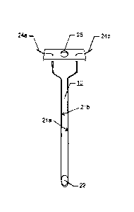
[014] Figure 1A is a drawing depicting a hypodermic needle with elongate
electrodes
integrated therein. The needle features a port for dispensing a liquid
formulation from a
lumen running there through, and a port for connecting to a fluid carrying
reservoir.
[015] Figure 2 depicts an alternate embodiment of the invention device wherein
the
anode and cathode electrodes are parallel to one another through a plane
formed in a spiral
around the needle.
[016] Figure 3A is another alternate embodiment wherein a delivery needle
comprises a
multiplicity of anode and cathode electrodes running straight and parallel
along the length
of the delivery needle. As also depicted, this figure includes an example of a
connector for
connecting the electrodes to a source of electrical energy. Figure 3B depicts
a view of the
cross section of one example of an invention electrode along line A-A. As
shown, in one
configuration, the electrodes can be layered by any number of techniques known
to those
of skill in the fabrication arts on the outer sections of a delivery tube and
lumen. In the
drawing is depicted an inner needle 53 with lumen 54 surrounded by an
insulating material
55 on which is layered the electrodes.
[017] Figure 4 is another example of an embodiment comprising electrodes
spiraled
around the delivery needle. The electrodes so spiraled can comprise a
multiplicity of anode
and cathode pairs, but typically comprise one or two pairs of electrodes, each
pair
comprising an anode and a cathode.
[018] Figures 5A-C depict one embodiment of the invention wherein the
invention
electrode is shown comprising further embodiments including a reservoir,
typically a
syringe styled reservoir, and a sharps cover which is capable of retracting as
the needle is
inserted into a patient tissue. The drawing also shows other features that can
be embodied
within the invention device such as a resilient membrane which can be pierced
such as by
a needle to fill the reservoir and mechanisms for allowing the sharps cover
and the syringe
plunger to be held in place either in an extended or retracted position.
Moreover, the
6a
Date Recue/Date Received 2021-05-07
WO 2007/095140 PCT/US2007/003615
retractable sharps cover also act as a needle guide and can be fitted with
stops to act as a
depth guide. Although not shown, the single needle electrode can be fitted to
a syringe
and attached to an automatic needle delivery/simultaneous fluid delivery
electroporation
device such as that depicted in US Patent Application 10/612,304 and
PCT/GB2003/002887. In such embodiment, the device would only have one needle
and
one syringe.
10191 Figure 6 shows a depiction of the invention device in use wherein during
insertion
or after the electrode/delivery needle is inserted into the tissue, the fluid
material
administered, the electrodes are energized so as to impart an electric field
outward from
the needle track and into the tissue. The electric field dissipates outward
into the tissue
from the site of the inserted needle.
[0201 Figure 7 shows a top view of a hypothetical tissue and a depiction of
typical
electric field that the invention device would generate in the tissue
surrounding the needle
track and having lateral dimensions (a) and (b).
1021] Figures 8A-C are drawings showing prior art arrays with typically
relatively
uniform lines of force and corresponding electric fields between array needles
as opposed
to that of the invention wherein a non-uniform lines of force and respective
electric field
surrounds the array and dissipates rapidly therefrom. For example, Figure 8A
shows three
opposing electrodes in a linear array wherein the lines of force between the
electrodes are
relatively uniform. In Figures 8B and C is depicted circular arrays wherein
the treatment
zone is central to the electrodes and under relatively uniform lines of force
and respective
electric fields (individually pulsed in opposing pairs, Figure 8B, or pulsed
in pairs of
opposing electrodes in different orientations, Figure 8C,).
[0221 Figures 9A-D show yet a further embodiment of the invention device which
comprises a guide for resting the needle and reservoir for penetration of
tissue to be treated
at an acute angle for use in methods that include delivery of treatment
substances near the
tissue surface. This angle is typically between 3 and 25 degrees from the
plane formed by
the general area of the tissue surface.
[023] Figure 10 shows partial view of delivery needles comprising electrodes
exposed
near the tip of the delivery needle. Figure 10A depicts a needle supporting
straight
electrodes while Figure 10B depicts a needle supporting spiral electrodes. The
leads for
each of the positive and negative anodes are depicted running up an internal
section of the
needle. Also, this depiction is intended to represent that the upper portion
of the elongate
7
CA 3063263 2019-11-29
WO 2007/095140 PCT/US2007/003615
=
needles can comprise insulation either around the electrode leads and/or
coating the upper
needle shaft.
10241 Figures 11A and B show results of electroporation in a tissue wherein
cells
primarily near the needle track have been affected by poration. In Figure 11A
is a series
of photos showing adjacent slices of tissue while Figure 11B shows a close-up
of a central
slice directly along the needle track.
10251 Figure 12 shows the results of a single injection into rabbit high
muscle of a
nucleic acid containing an expression vector encoding a fluorescent marker
protein (GFP)
using an electroporation device according to the invention.
[026] Figures 13A, B, C, D, and E show magnified photographs of a prototype
hypodermic needle wherein gold elongate electrodes have been etched onto a
standard
injection needle using MEMS technology, i.e., micro layering, and etching and
relayering
of materials onto the base injection needle shaft such that the electrodes
comprise 1/4 of
the needle shaft circumference each. Figure 13 A shows one view of the needle
showing
one long electrode running the length of the needle. In Figure 13 B, a detail
photo is
shown from an angle allowing visualization of the terminal sections of both
gold
electrodes. Figure 13 C is another perspective showing detail of the terminal
sections of
the electrodes etched onto the needle shaft. Figures 13D and E show another
embodiment
wherein the MEMs crafted electrodes are 1/16 the circumference of the needle
shaft.
10271 Figures 14 A, B, and C are drawings showing additional embodiments of
single-
.
needle design where in the shaft comprises electrically inert material such as
for example,
plastic extruded with electrode leads built into the extruded hypodermic shaft
Figure 14A
depicts straight electrodes running parallel to the needle shaft. Figure 14B
depicts
electrodes in a spiral about the shaft. Figure 14C depicts the cross section
AA¨AA of
Figure 14A showing one embodiment wherein the electrode of the shaft can be
connected
to electrode leads positioned on the needle hub.
10281 Figure 15 is a graph showing the level of rabbit anti-human IgG
antibodies
produced following electroporation pulse using the single needle invention (
mi) versus no
electroporation ( = )
10291 Figure 16 is a graph showing the level of rabbit anti-SEAP antibodies
produced
following electroporation pulse using the single needle invention ( 0) versus
no
electroporation ( = )
[0301 Figures 17 A and B are photographs showing results of green florescent
protein =
(GFP) expression following injection of plasmid DNA encoding GFP followed by
no
8
CA 3063263 2019-11-29
WO 2007/095140 PCT/US2007/003615
electroporation. In combination of natural and fluorescent light, Figure 17A
shows
adjacent slices of tissue in the vicinity of the injection/needle track site.
The photos show
no expression without electroporation.
[031] Figures 18A and B are photographs showing combination of natural light
and
green florescence, or fluorescence alone respectively, wherein injection of
plasmid DNA
encoding GFP was followed by electroporation carried out using a single needle
electrode
comprising a 23 gauge needle and anode and cathode electrodes having a width
of 1/16 the
circumference the needle shaft. In this experiment, the electrodes were pulsed
at a
constant current of 50 mA.
[032] Figures 19A and B are photographs showing combination of natural light
and
green florescence or fluorescence only, wherein injection of plasmid DNA
encoding GFP
was followed by electroporation carried out using a single needle electrode
comprising a
23 gauge needle and anode and cathode electrodes having a width of 1/16 the
circumference the needle shaft. In this experiment, the electrodes were pulsed
at a
constant current of 100 mA.
[033] Figures 20A and B are photographs showing combination of natural light
and
green florescence or fluorescence only, wherein injection of plasmid DNA
encoding GFP
was followed by electroporation carried out using a single needle electrode
comprising a
23 gauge needle and anode and cathode electrodes having a width of 1/4 the
circumference the needle shaft. In this experiment, the electrodes were pulsed
at a
constant current of 50 mA.
[034] Figures 21A and B are photographs showing combination of natural light
and
green florescence or fluorescence only, wherein injection of plasmid DNA
encoding GFP
was followed by electroporation was carried out using a single needle
electrode
comprising a 23 gauge needle and anode and cathode electrodes having a width
of 1/4 the
circumference the needle shaft. In this experiment, the electrodes were pulsed
at a
constant current of 100 mA.
[0351 Figures 22A and B are photographs showing combination of natural light
and
green florescence or fluorescence only, wherein injection of plasmid DNA
encoding GFP
was followed by electroporation was carried out using a single needle
electrode
comprising a 23 gauge needle and anode and cathode electrodes having a width
of 1/4 the
circumference the needle shaft. In this experiment, the electrodes were pulsed
at a
constant current of 150 mA.
=
9
CA 3063263 2019-11-29
WO 2007/095140 PCMS2007/003615
[0361 Figures 23A and B are photographs showing combination of natural light
and
green florescence or fluorescence only, respectively, wherein injection of
plasmid DNA
encoding GFP was followed by electroporation was carried out using a single
needle
electrode comprising electrodes lmm spacing without fluid delivery embodiment.
In this
experiment, the electrodes were pulsed at a constant current of 75 mA.
[037) Figures 24A and B are photographs showing combination of natural light
and
green florescence or fluorescence only, respectively, wherein injection of
plasmid DNA
encoding GFP was followed by electroporation was carried out using a single
needle
electrode comprising electrodes 1 mm spacing without fluid delivery
embodiment. In this
experiment, the electrodes were pulsed at a constant current of 150 mA.
[038] Figures 25A and B are photographs showing combination of natural light
and
green florescence or fluorescence only, respectively, wherein injection of
plasmid DNA
encoding GFP was followed by electroporation was carried out using a single
needle
electrode comprising electrodes lmm spacing without fluid delivery embodiment.
In this
experiment, the electrodes were pulsed at a constant current of 250 mA.
DETAILED DESCRIPTION OF THE PREFERRED EMBODIMENTS
[0391 In a first embodiment, the invention comprises a device for
electroporation of
tissue in vivo comprising a hollow shaft made of a material capable of
insertion into a
biologic tissue or organ in situ and of delivering therethrough a fluid medium
(i.e., a
delivery needle shaft), said shaft further comprising at least two electrodes
exposed at least
in part on an outer surface of said shaft, wherein said electrodes are spaced
from one
another and situated parallel with respect to one another along said needle
shaft.
Embodiments for electrodes can employ a variety of electrode structural
designs. For
example, anode and cathode electrodes can be placed in association with a
delivery needle
that run parallel to one-another and to the length of the delivery needle such
as disclosed
in Figures 1 and 3, or that are parallel to each other but are spiraled around
the needle
shaft as depicted in Figures 2 and 4. The invention device also includes
electric conduits
connecting each of said electrodes to an electrical energy source wherein said
electrodes
when said needle is inserted into a patient tissue are capable of being
energized
individually, generating an electric field to cells in a treatment zone
surrounding said
needle sufficient to cause cells along and near a track made by insertion of
said needle into
said tissue to become reversibly porated so as to allow treatment molecules to
enter said
cells.
CA 3063263 2019-11-29
WO 2007/095140 PCT/US2007/003615
[040] Manufacture of such electrode containing fluid delivery needles can be
carried out
by any number of well know methods including micromachining such as commonly
understood as MEMs technology. For example, a standard hypodermic needle
(which can
be any gauge such as 20gauge, 21 gauge, 22 gauge, 23 gauge, 24 gauge, 25 gauge
26
. gauge, 27 gauge, 28 gauge and 29 gauge) can be coated with an electrically
inert material
followed by deposition of electrically conductive material such as gold,
followed in turn
by etching away conductive material in the orientation desired on the surface
of the
needle. Specifically, generally the process comprises cleaning the hypodermic
needle
shaft in preparation for deposition of the inert substance, for example, a
polymer having
properties of evenly adhering to surfaces, such as parylene. Following
stripping of the
metal shaft, parylene is deposited, such as by vacuum deposition, on to the
needle. This is
in turn patterned using a laser to deposit electrode conductable material,
such as gold,
followed in turn by selective removal of the gold to form electrodes in a
predetermined
pattern on the needle shaft. In the current invention, the use of MEMs
technology provides
for an ability to manipulate the three dimensional needle and coatings and
etchings on a
miniature scale. The capability to manufacture a single needle electrode is
proven by the
photographs of Figures 13A to E. Manufacture can also be carried out by
extrusion
technology. As depicted in Figures 14A-C, in this aspect the electrodes 202
and 203 (Fig.
14A) are extruded as fine wire filaments with an electrically inert substance
such as
polyvinylchlorine .or the like in a linear fashion. The tip of the needle 204
can be
machined or cut to a penetrating tip and at the other end fitted to a hub 200
comprising
electrode leads 201a and 201b and a fitting 205 for attachment to a source of
fluid
medium. Figure 14B depicts an example of a structural embodiment comprising an
extruded needle with spiral electrodes and electrode leads 210 and 211.
[041] In a second embodiment, the invention comprises a method for delivering
molecules to cells in vivo comprising providing to a patient's tissue
containing said cells
an injection needle further comprising at least two elongate electrodes (i.e.,
a cathode and
an anode) positioned along the needle shaft and at least a reservoir
containing said
molecules wherein said reservoir and molecules are in fluid communication with
a lumen
running through said needle shaft, injecting the molecules into said tissue,
and energizing
the electrodes with electrical energy to provide an electric pulse sufficient
to cause cells in
the vicinity of the injection site and needle track to become reversibly
porated, thereby
electroporating said cells for their uptake of said molecules.
11
CA 3063263 2019-11-29
85784831
[042] In a third embodiment, the device provides for electroporation of cells
in a
narrowly defined location, particularly cells along or near the track make by
the injection
needle. Generally, the cells considered within the treatment site are those
cells lying in a
radius around the needle track of about 5mm, more typically about 3 mm, and
even more
particularly about 2mm, and most particularly about lmm. In a related
embodiment, the
generation of electric filed sufficient for electroporation of cells within
said treatment site
is a field that weakens outward from the central injection needle such that
the treatment
site is defined by the inability of the pulse energy to extend into the
tissues beyond a
certain distance from the electrodes.
[042a] In a further embodiment, the invention provides for any number of
structural
arrangements providing for at least two opposite electrode leads (i.e., at
least one anode
and at least one cathode) situated in association with a single elongate
electrically inert
shaft, which shaft itself can comprise electrodes and an electrically inert
material, such as
a medically acceptable plastic or polycarbonate, filling the space between the
electrodes a
0.05 mm to a 1.5 mm between, or can comprise just elongate opposing spaced
electrodes.
In either embodiment, the electrodes of the tissue penetrating single needle
electrode or
electrode containing shaft have spaced dimensions of between 0.05 mm and 1.5
mm. In a
related embodiment, the electrodes themselves can have a length exposed along
the
elongate shaft anywhere from the whole needle length to just a section of the
needle, such
as near the shaft penetration tip. Further, the electrodes can have cross
sectional
dimensions of between 0.005 and 0.80 mm. In yet another structural arrangement
embodiment, the single needle electrode can comprise a hypodermic needle
comprising at
least two elongate electrodes spaced along at least a portion of the length of
the
hypodermic needle exterior. For example, the hypodermic needle can include at
least two
electrodes (i.e., an anode and a cathode) running along a portion of the
length of the
needle. (See Figure 1A) In working embodiments, each electrode is connected to
a source
of electric energy for generating an electric field between opposite poles,
i.e., one
electrode is an anode and the other a cathode electrode. In other examples,
multiple
electrodes can be formed on the exterior of a hypodermic injection needle such
as
disclosed in Figure 3 comprising multiple straight and parallel electrodes, or
as depicted
in Figures 2 and 4 comprising multiple electrodes spiraled around the
injection needle. In
still further embodiments, the single-needle electrodes can be manufactured
using any
number of well understood methods including etching and layering per Micro
12
Date Recue/Date Received 2021-05-07
85784831
electromechanical systems (MEMS) technologies. In such manufacturing methods,
micromachining processes are used to add or strip away layers of substances
important to
the proper annealing, insulation, and conduct of electric pulses and
circuitry. Figures 13A,
B, C, D and E are photographs of the embodiment wherein the electrodes are
etched on to
the delivery needle shaft. Specifically, gold electrode layering has been
coated above a
layer of and inert substance (parylene) which itself had been layered over the
hypodermic
needle shaft. Additional methods for manufacturing the elongate electrodes
include
extrusion technologies wherein the electrode leads are formed into and or
along the shaft
of an electrically inert composition having insulating qualities, such a
plastic, a polyester
derivative, or polyvinylchloride (PVC), or insulative carbon fiber. As shown
in Figure
14A and B, an elongate hollow needle can be extruded with electrode component,
such as
for example, wire either along opposite sides of the hollow shaft or in a
spiral fashion as
shown in Figure 14B. Further still, the needle shaft can also comprise
sections with no
exposed electrodes. For example, one end of the needle shaft connects to a hub
forming a
connector for connecting to a source of fluid, such as for example, a syringe.
Insulation
near or along such section of the shaft may provide for additional lessening
of electric
stimulus sensation noticeable by the patient. In yet a further embodiment with
respect to
any such electrode configuration described herein, each of the electrodes are
individually
energizable so that any combination of the electrodes may be energized in
pairs (i.e., a
cathode and anode) simultaneously together, or in any given sequence, and
further using
any type of pulse including without limitation monopolar, bipolar, exponential
decaying,
or pulse train combinations of any of the former.
[043] In a further related embodiment, the invention calls for the novel use
of a single
elongate probe (which comprises the injection needle and electrodes) for
performing
in situ electroporation of a highly localized set of cells in the tissue.
[044] In another embodiment, the invention device may be used with any of a
variety of
electric pulsing conditions. For example, the electrodes can be charged with
at least one
pulse of constant current in the range of between 1-400 mAmps, typically
between 5-200
mAmps, and more preferably between 20 and 100 mAmps. In another example, the
electrodes can be charged with a voltage pulse in the range of 1 to 100 volts.
Further, the
electric pulse can be either a monopolar or a bipolar pulse wherein said pulse
can be a
single, a double or a multiple pulse sequence having various characteristics
such as a set
voltage drop, variable shaped pulse trains, or pulses employing constant
current.
12a
Date Recue/Date Received 2021-05-07
85784831
[045] In other embodiments, the device and method provide for delivering or
transfecting pharmaceutical drugs, proteins, nucleic acids including DNA and
RNA, and
synthetic modifications thereof as are well known to those of skill in the
art, into patient
tissues, particular to cells residing in the subcutaneous, intradermal, and
subdermal spaces
as well as skeletal and striated muscle compartments of a mammalian body, and
organs
including heart, lung, pancreas, spleen, liver, and organs of the alimentary
tract. Once
transfected with the selected material, cells will be directly affected by the
activity of the
drug, or protein or nucleic acid. Where nucleic acids are transfected,
typically such nucleic
acids are employed for the protein encoded thereby which can be expressed in
the cells of
the treatment site. Further, the substances can comprise cytokines,
chemokines, and
immune relevant bioactive molecules including such active molecules as immune
modulating molecules selected from the group consisting of IL 1, IL-2, IL-3,
IL-4, IL-5,
IL-6, IL-7, IL-8, IL-9, IL-10, IL-11,
IL-12, GM-CSF, M-CSF, G-CSF, LIF, LT, TGF-f3, IFN, TNF-a, BCGF, CD2, or ICAM.
12b
Date Recue/Date Received 2021-05-07
85784831
[046] In another embodiment, the material to be delivered to the cells can be
delivered in
a liquid form in a volume of between 0.01m1 to 1ml. In one embodiment, nucleic
acid
encoding a polypeptide can be dissolved in 0.9% sodium chloride (NaC1). The
exact
solvent, however, is not critical to the invention. For example, it is well
known in the art
that other solvents such as sucrose are capable of increasing nucleic acid
uptake in skeletal
muscle. In a related embodiment, the volume to be delivered can be adjusted in
relation to
the length of the needle (since the length of the needle shaft will determine
both the
volume of the substance being transported therethrough) and, the needle track
made so as
to determine the volume of the space available for said substance to fill upon
it being
expressed through the needle and into the needle track and surrounding tissue.
For
example, a 2mm long needle can be used for delivering substances to skin layer
tissues
and provide for injection of a volume in the range of 0.01m1 to 0.05m1, while
a 5nun long
needle can be used to deliver volumes in the range of 0.1m1 to 0.15m1, and a
1.5 to 2 cm
long needle can be used for delivering volumes in the range of 0.3 ml to
0.5m1.
1047] Other substances may also be co-fransfected with the molecule of
interest for a
variety of beneficial reasons. For example, the molecule P199 (lee, et al.
PNAS., 4524-8,
10, 89 (1992)), which is known to seal electropermeabilized membranes, may
beneficially
affect transfection efficiencies by increasing the survival rate of
transfected muscle fibers.
[048] With reference to Figure 6, the electrode carrying hypodermic needle is
inserted
into a patient tissue to a desired depth of penetration. The plunger of the
attached syringe
is activated to inject the volume of liquid containing the selected material
for injection,
and the electrodes are immediately thereafter, or alternatively simultaneously
with the
injection of the material, energized with at least one pulse of electric
energy sufficient to
cause at least some of the cells in the treatment zone to become reversibly
porated.
Although the syringe plunger is typically activated using animate means, such
as by use of
the hand, the syringe can also be affixed to a holding device such as
disclosed in Figure 9,
or even an automatic dispensing apparatus, such as a device disclosed in US
patent
application 10/612,304 filed July 3,2003.
[049] In other embodiments, the invention can be applied to electroporation of
cells at
various depths from the surface of a body tissue. For example, besides
electroporation of
cells residing within muscle tissue compartments in which delivery of
substances are
initiated by injection of materials into the tissue in an orientation
approximating 90
degrees from the surface of the tissue, in one embodiment the invention device
can be
13
Date Recue/Date Received 2021-05-07
WO 2007/095140 PCT/US2007/003615
used to electroporate cells in the subcutaneous, intraderrnal, or subdermal
spaces of skin.
It can also be used to electroporate substances into lymph nodes, or tissue
layers in other
organs such as cardiac and blood vessel tissue. With respect to
electroporating cells in
any of these locals, use of the device for electroporating cells in such
tissue layers can
include use of either short needles having a length sufficient for penetrating
outer portions
of the tissue layers (i.e., skin, subdermal, etc.) for injection and
electroporation at
approximately a 90 degree angle to the tissue surface, or where a delivery
needle is
relatively long, such as between 3 and 4 cm, insertion of the single needle
can be made at
an acute angle to the surface tissue using a holding device as depicted in
Figure 9A. This
will allow for electroporation of a larger portion of tissue within the
desired layer.
Further, the acute angle of insertion can be between 3 to 25 degrees of angle
from the
tissue surface. Such tissue surface can be described as forming generally a
flat surface
area fanning a plane encompassing the site for insertion of the single
needle/electrode. As
depicted in Figure 9A to D, the syringe can be connected to an attachment
means which is
designed to hold the syringe at a set angle on a planar guide tray 100 with
the needle
placed a set distance X into the tissue as determined based on the
predetermined desired
depth of insertion of the needle into the tissue. The guide tray with exposed
needle is
brought into contact with the tissue surface such that the needle inserts the
tissue at the
prescribed acute angle. After the needle is so inserted and the therapeutic
substance
expelled from the syringe, the electrodes can be energized to bring about
delivery of the
injected material into the subcutaneous, intradermal, or subdermal cells. Use
of the device
at an oblique angle as discussed above can also apply to electroporating
various layers of
organ tissue.
Examples:
[050] The following examples are given to illustrate various embodiments which
have
been made of the present invention. It is to be understood that the following
examples are
not comprehensive or exhaustive of the many types of embodiments which can be
prepared in accordance with the present invention.
Example I.
[051] Turning now to various aspects of the invention, the device can comprise
molecule
delivery reservoir 20 and electrode needle 10 components as shown for example
in
(Figure 5). Additional embodiments include sharps cover 11, resilient membrane
12
sealing a portion of the structure comprising the reservoir 20 for uses in
filling the
reservoir (such as by piercing of a syringe needle), and mechanisms such as
dimples 13
14
CA 3063263 2019-11-29
W020071095140 PCIMS2007/003615
and recesses 14 and 14* in the reservoir 20 housing structure for keeping the
sharps cover
11 in a semi fixed position of either open/retracted (Figure 5C), or
closed/covered (Figures
5A and B). Further embodiments include mechanisms for keeping the plunger 9 in
a semi
fixed open/retracted or a closed/expelled position, such as, for example,
dimples 15 and
recesses 16 and 16*. It should be clear to one of skill in the art that
regardless of the
method employed to provide for semi fixed positioning of the sharps cover 11
and plunger
9, such positioning can easily be changed with either animate energy, such as
force by
hand, or mechanically, such as by an electronically driven actuator. The
distal end of the
sharps cover 11 can include removably attached thereto a sterility cover 60.
The electrode
needle 10 further can comprise a lumen running therethrough ending in tissue
piercing tip
22, and orifice 25 for connecting to the reservoir 20 (See Figure 1). The
injection needle
can be of a gage between 18 and 29 standard hypodermic needle gauge sizes. In
a
preferred embodiment, the delivery needle comprises at least one pair of
electrodes, such
as electrodes 21a and 21b of Figure 1. The electrodes comprise at least one
anode and one
cathode electrodes which are in electrical communication with electrode leads
24a and
24b. Depending upon the design chosen for any particular invention product,
the leads
can terminate in a lead terminal 23 (see Figures 3 and 4, for example), or
connect by any
other means with lead wires running from the electrode to a source of
electrical energy,
such as a pulse generator. The needle component 10 can further include a
connector 26
(Figures 3 and 4) for attaching to a hypodermic syringe reservoir, or to a
syringe reservoir
affixed with a locking mechanism to detachably fasten the needle component 10
to a
hypodermic syringe port.
1052] In further embodiments, the reservoir 20 can be manufactured with a
predetermined substance for treating a particular condition. Alternatively,
the reservoir
can be filled with a substance of interest by either drawing such substance
into the
reservoir through the electrode needle 10 by extracting the plunger 9, or
preferably, the
reservoir can first be cleared of the plunger by retracting the plunger to the
open position
followed by delivering to the reservoir the substance by injecting it into the
reservoir via
the resilient seal 12, similarly to the procedure commonly performed in the
extracting of
drugs from sterile vials into syringes and introducing them into another
reservoir.
10531 The delivery needle 10 with its way of electrodes (such as electrodes
21a and b,
31a and b, 51a and b and 52a and b, or 41 and 42, Figures 1-4, respectively)
can be
inserted into the tissue, usually at an approximate 90 degrees to the tissue
surface, or
alternatively at an acute angle with respect to the tissue surface, and the
substance injected
CA 3063263 2019-11-29
. .
WO 2007/095140 PCT/US2007/003615
into the needle track and local tissues. The electrodes can be energized using
a pulse
generator either following the injection of said substance, or can be
energized
simultaneously with said injection of substance. As depicted in Figure 6, when
energized
with an electric pulse, the electrodes support the generation of an electric
field 20 that
provides for sufficient energy to cause reversible poration of the cells
within said field.
The electric filed generated is non-uniform in that it exponentially decreases
by the
distance from the needle track 80 (Figure 7). Thus, the electric field
sufficient to provide
such poration has, depending upon the energy employed, symmetrical lateral
dimensions
(a) x (b) (shown in Figure 7) forming a set diameter of an electroporating
electric field
which, with respect to the needle track length, forms a defined three
dimensional volume.
Generally, the poration sufficient electric field has a radius from the
electrode needle 10 of
between 0 and 5 mm, typically between 0 and 4 mm, and preferably between 0 and
3 mm
and most preferably between 0 and 2 mm.
10541 As is easily understood by those having skill in the electroporation
arts, the field
generated by the current invention's single needle electrode, unlike prior
electroporation
apparatuses, is a non-uniform electric field wherein the field intensity is
greater near the
needle and diminishes as measured outward from the electrodes In contrast to
the current
electrode arrangement, Figure 8 depicts prior electrode arrangements wherein a
uniform
electric filed is employed across a large volume treatment site. The instant
invention is
measurably distinct from former concepts that suggested a need to utilize a
"uniform"
field. Here, the invention employs a non-uniform field which provides for
reversible
poration of cells to a greater amount near the position of the delivery
needle, i.e., the
needle tract. This in turn allows a clear benefit to determine the precise
location of those
cells receiving a known dose of therapeutic materials. This invention through
its
embodiments therefore provides for "fitting" the electric field to the
injection site so as to
distribute material to cells more uniformly and confined to a local tissue
area as opposed
to the variable distribution allowed for with electroporation systems that use
a
conventional uniform electric field and an outer array of electrodes.
[055] With respect to the electrodes generally, they can comprise any metal
but
preferably are a metal that does not impart a toxicity due to metal ions to
the cells of the
electroporated tissue. Such materials include gold, tungsten, titanium
nitride, platinum,
platinum iridium, and iridium oxide. The electrode material can be formed on
the delivery
tube (i.e., injection needle) such that there is a layer of insulation between
the electrodes
and the delivery tube as suggested in Figure 313. Alternatively, the needle
can comprise a
16
CA 3063263 2019-11-29
W02007/095140 PCT/US2007/003615
material that is nonconductive itself eliminating a specific need to insulate
the electrodes
from the injection tube. In this aspect, the delivery tube can be constructed
from any
suitable material for insertion into tissue in situ that is non-conductive,
including, such as a
ceramic, or hardened biocompatible plastic, including polyvinylchlorine or the
like.
[056] In a further embodiment, the delivery needle:/electrode component can be
designed
such that the electrodes 90 or 101 (Figure 10) are exposed for electroporation
only near the
tip of the needle as depicted in Figures 9A, and 10A and B. The unexposed
portions 91
and 102 of the electrodes can be insulated and run along the delivery needle
exterior or
internal to the needle. Specifically, where it is desired to position the
defined treatment
volume (defined by the dimensions of the electroporation electric field
imparted to the
tissue by the electrode array) in a particular tissue, with the intent of
avoiding
electroporation of other tissues, electrodes, such as disclosed in Figure 10,
can be used, for
example, to electroporate deep muscle tissue and avoid other tissues lying
closer to the
tissue surface, such as fat cell layers, or alternatively to electroporate
tissues near the
surface, such as for example, subdermal tissues, as suggested in Figure 9A.
Such
embodiments provide for additional control over placement and size of the
treatment
volume.
Example II
[057] In this example, results are depicted for delivering molecules by
reversible
poration to cells situated along and near the track formed by the insertion of
the invention
single hypodermic needle electrode into a tissue.
10581 As depicted in Figures 11A and B, rabbit quadriceps muscle was injected
with
DNA encoding beta-galactosidase in a bolus comprising 0.2 ml and concentration
of 1
mg/ml. The electrodes were pulsed using 2 pulses of 250 mAmps, 20 millisec
duration.
Following electroporation, the beta-galactosidase gene was expressed in cells
affected by
the electroporation. At day 4 after electroporation, the rabbits were
sacrificed and the
muscles were prepared in 3 mm thick slices through the site on insertion of
the single
needle/electrode. Following chemical fixation, the beta galactosidase
expressing cells in
the muscle slices where visualized by an enzymatic reaction. The arrows in
Figure 11A
depict the direction of the insertion of the delivery tube into the rabbit
muscle. As shown,
staining occurs predominantly along the track formed by insertion into the
tissue of the
needle delivery electrode.
17
CA 3063263 2019-11-29
WO 2007/095140 PCT/1JS2007/003615
Example III
[059] This example describes experiments that employ an electroporation device
according to embodiments of the invention to deliver DNA encoding green
fluorescent
protein (GFP) into rabbit quadriceps muscle, the results are shown in Figure
12.
[060] Here, several New Zealand white male rabbits, each weighing 4-5 kg
(Perry
Scientific, San Diego, Califignia), were each injected with an expression
vector
(gWizGFP, lot 12311, purchased from Aldevron, LLC, Fargo, ND; see also Gene
Therapy
Systems, Inc., San Diego, CA) encoding a bright GFP (Cheng, et al. (1996),
Nature
biotechnology, vol. 14:606-9) the expression of which was under the control of
a modified
human cytomegalovirus immediate early promoter/enhancer.
[061] Prior to injection, each rabbit was first sedated with acepromazine
(lmg/kg) and
then anesthetized by intramuscular injection of a mixture of ketamine (35
mg/kg) and
xylazine (5 mg/kg) in the presence of glycopyrrolate (0.01 mg/kg), which had
been
previously administered subcutaneously to prevent uneven heart beating as a
result of the
ketamine/xylazine treatment. The rabbit was then shaved at the site where the
injection
was to be made, i.e., into the quadricepts muscle. A hole Was poked in the
skin covering
the muscle by first inserting an 18 gauge needle, and then slightly widened
using a scalpel.
A single needle electroporation device, made from an 18 gauge needle with two
parallel
electrodes applied opposite one another to the outer surface of the needle (as
depicted in
Figure 1), was then slowly inserted into the muscle tissue, periodically
pausing to inject
DNA every few millimeters to a final insertion depth of approximately 25 mm. A
total of
500 ul of DNA-containing solution containing 100 ug gWizGFP was injected into
each
injection site. Shortly after completing the injection and while the
needle/electrode device
was still inserted to its final insertion depth, electroporation was
commenced.
Specifically, five 250 mA pulses, each of twenty millisecond (ms) duration,
were applied
to the electroporation needle device at 10Hz intervals (i.e., 100 ms) using an
Elgen 1000
(Inovio AS, Oslo, Norway) current-clamped pulse.
[062] Four days post-treatment the animals were humanely euthanized. Skin
covering the
region of the leg where the vector was delivered was carefully removed, after
which each
animal was placed at -20 C for about 1 hour. Treated muscle was then removed
using a
scalpel and then placed at -20 C for another 1 to 2 hrs. The frozen muscle
tissue was then
sectioned into slices approximately 3 mm thick using a rotating meat slicer.
Muscle slices
where arranged in plastic trays and examined for GFP expression using a Leica
MZ 12
dissection microscope fitted with a UV light and GFP filter combination.
Figure 12 is a
18
CA 3063263 2019-11-29
WO 2007/095140 PCT/US2007/003615
=
representative photo of the results obtained by this analysis, and clearly
shows that an
electroporation device according to the invention can be used to successfully
deliver an
agent, for example an expression vector encoding a desired protein that is
then expressed
in active form, into cells.
Example IV
[063] In this example, data for which is shown in Figures 15 and 16, using the
invention
electrode configuration, plasmids encoding SEAP (pSEAP#3348, Aldevron) and IgG
(pLNOH 2hg3 #11765, Aldevron) were electroporated into cells of test animal
tissues
(i.e., intramuscular injection into the tibialis anterior of the animal) and
the expression
monitored to prove success of expression in rabbit muscle as well as measuring
immune
responses against both a "week' and a 'strong" antigen (SEAP and IgG,
respectively). In
these experiments the SEAP and IgG plasm id were administered at a final
concentration of
1 ug/ul.
[064] Animals used were New Zealand White male rabbits 3.5 to 4.5 kg.
Electroporation
was carried out using an Elgen 1000 (Inovio AS, Oslo, Norway Serial number
009) which
further comprised a current-clamped pulse generator (prototype) and a single
needle
prototype wherein the electrodes ran parallel to the injection track and
approximately
between 1 mm apart. The electrodes were pulsed for 20 millisec pulse length
with 5
pulses each at 150 mA with a 250 millisec interval between pulses (i.e., a
frequency of
about 4 Hz). The electrodes extended into the tissue to about 1.0 cm depth.
[065] The experiments each comprised a two-step delivery process, i.e.,
injection of the
plasmid solution (200 ul) using a 29 gauge insuline syringe with injection
during insertion
of the needle to distribute DNA at different depths, followed by removal of
the injector
needle and insertion of the single needle electrode.
[066] As shown in Table I below, each of the IgG and SEA? experiments had two
groups of test animals, i.e., one set of animals receiving electroporation and
the other not
(control)
[067] Table 1
Group Current Treatement
150-250 100 ul. x 2 SEAP lmg/ml, 100 ul x 2 left tibialis, IgG 1 mg/ml
1 mA 100 ul x 2 right tibialis
No EP 100 ul x 2 SEAP Img/ml, 100 ul x 2 left tibialis, IgG 1 mg/ml
100 ul x 2 right tibialis
19
CA 3063263 2019-11-29
WO 2007/095140 PCT/US2007/003615
[068] Samples were taken Day 0, 14 and day 21. The rabbits were then
terminated on
day 21 with subcutaneous injection of 0.5 ml hypnorm (Hypnorm 0.1 ml/kg)
followed by
i.v. injection of 1 ml/kg 0110% Pentorbarbital in the ear vein.
10691 As is clear from the results of Figures 15 and 16, the levels of
antibody titer
elicited from the single needle delivery are far in excess of the negative
control.
Specifically, the two test antigens (IgG and SEAP) elicited titers relative to
one another as
expected with IgG being a much stronger antigen that SEAP (see titer scale).
Both
antigens elicited antibody production in the electroporated samples and
virturally no
antibody production in the non-electropomted samples.
Experiment V
10701 In this experiment, prototype MEMs manufactured single needle electrodes
were
tested in rabbit tissue using a variety of pulsing energies and green
florescent protein
expression. As indicated in Table II, three different electrode embodiments
were tested,
(1) a single needle electrode in which the anode and cathode electrodes were
applied to a
23 gauge needle at 1/16 the circumference of the needle each and applied to
the full length
of the needle by MEMs technology (Figures 13D-E), (2) a single needle
electrode wherein
the electrodes are 1/4th the circumference of the needle shaft each (Figures
13A-C), and
(3) a single needle arrangement wherein the electrodes are 1 mm apart without
a fluid
medium delivery tube. As shown in Table II, the various combinations of
pulsing were
performed.
10711 The protocol used for each animal in this experiment comprised injecting
the GFP
plasmid at the noted concentrations, electroporating the tissue using an
embodiment of the
single needle electrode, followed by sacrificing of the animals and performing
tissue
preparation by slicing the treated muscle in adjacent slices and observing
florescence.
Generally, due to the difficulty of slicing the tissue so as to retrieve
slices parallel to the
injection track, GFP florescence in the figure photos often show up as circles
or elipses.
These florescence patterns prove that the single needle concept is functional
and provides
for electropration of tissue a very low voltages and relative electric current
in defined
locations surrounding the needle track and within the tissue.
CA 3063263 2019-11-29
WO 2007/095140 PCT/US2007/003615
[072] Table 11
Electrode Tissue site Constant Voltage Number pGFP DNA
design current (average V) of pulses ,
concentration/volume
Electrodes 1/4 Quadriceps 0.0 0.0 0.0 0.2 mg/m1
shaft
circumference
Electrodes Quadriceps 50 mA 8 2 0.2 mg/ml
1/16 shaft
circumference Quadriceps 100 mA 18 2 0.2 mg/m1
Electrodes 1/4 Quadriceps 50 mA 11 2 0.2 mg/ml
shaft Quadriceps 100 mA 15 2 0.2 mg/ml
circumference Quadriceps 150 mA 20 2 0.2 mg/ml
Quadriceps 250 mA 33 . 2 0.2 mg/ml
Electrodes Tibialis 75 mA 13 2 1.0 mg/ml
lmm spacing
without fluid Tibialis 150 mA 18 2 1.0 mg/ml
delivery Tibialis 250 mA 28 2 1.0 mg/m1
embodiment Quadriceps 150-200 20 2 1.0 mg/ml
Quadriceps 250-500 40 2 1.0 mg/m1
Quadriceps 600-1000 50 2 1.0 mg/ml
mA
[073] Figures 17A and B show both natural light and florescent light,
respectively,
photographs of GFP expression following injection of plasmid DNA encoding GFP
with
no electroporation. As indicated, there is virtually no green florescent
protein expression.
Thus, it is clear that without electroporation there is not sufficient uptake
and expression
of the desired gene.
[074) With respect to electroporation in situ using the 1/16 width electrode
model, the
ability to express electroporated GFP is shown in Figures 18A and B and 19A
and B.
Figures 18A and B show GFP expression results upon electroporation with a
constant
current of 50 mA, while Figures 19A and B show electroporation at 100 mA.
[075] For GFP expression using the 1/4 circumference single needle electrode,
results are
provided in Figures 20A and B, 21A and B, and 22A and B, wherein
electoporation was
carried out using 50, 100, and 150 mA, respectively.
[076] OFF expression was also testing using an embodiment wherein the single
needle
electrode did not comprise a fluid delivery tube associated with the
electrodes. As shown
in Figures 23A and B, 24A and B, and 25A and B, this invention device
embodiment was
tested at 75, 150, and 250 mA each at constant current. Here, the amount of
GFP plasmid
was five times the concentration of the experiments shown in Figures 19-22.
Consequently, the treatment zone appears more readily.
21
CA 3063263 2019-11-29
85784831
10771 All of the compositions and methods disclosed and claimed herein can be
made
and executed without undue experimentation in light of the present disclosure.
While the
compositions and methods of this invention have been described in terms of
preferred
embodiments, it will be apparent to those of skill in the art that variations
may be applied
to the compositions and methods and in the steps or in the sequence of steps
of the method
described herein without departing from the spirit and scope of the invention.
More
specifically, the described embodiments are to be considered in all respects
only as
illustrative and not restrictive. All similar substitutes and modifications
apparent to those
skilled in the art are deemed to be within the spirit and scope of the
invention as defined
by the appended claims.
[078] All patents, patent applications, and publications mentioned in the
specification are
indicative of the levels of those of ordinary skill in the art to which the
invention pertains.
[079] The invention illustratively described herein suitably may be practiced
in the
absence of any element(s) not specifically disclosed herein. Thus, for
example, in each
instance herein any of the terms "comprising", "consisting essentially of',
and "consisting
of' may be replaced with either of the other two terms. The terms and
expressions which
have been employed are used as terms of description and not of limitation, and
there is no
intention that use of such terms and expressions imply excluding any
equivalents of the
features shown and described in whole or in part thereof, but it is recognized
that various
modifications are possible within the scope of the invention claimed. Thus, it
should be
understood that although the present invention has been specifically disclosed
by preferred
embodiments and optional features, modification and variation of the concepts
herein
disclosed may be resorted to by those skilled in the art, and that such
modifications and
variations are considered to be within the scope of this invention as defined
by the
appended claims.
22
Date Recue/Date Received 2021-05-07