Note: Descriptions are shown in the official language in which they were submitted.
CA 03063527 2019-11-13
WO 2018/215935
PCT/IB2018/053622
1
ANTIBODY-CYTOKINE ENGRAFTED PROTEINS AND METHODS OF USE FOR
IMMUNE RELATED DISORDERS
CROSS-REFERENCE TO RELATED APPLICATIONS
[001] This application claims the benefit of U.S. Provisional Application
No.
62/510,514 filed May 24, 2017, the content of which is hereby incorporated by
reference in
its entirety.
FIELD
[002] The present disclosure relates to antibody-cytokine engrafted
proteins that
bind to interleukin-2 (IL2) high affinity receptor, and methods of treating
autoimmune and
immune related disorders.
SEQUENCE LISTING
[003] The instant application contains a Sequence Listing which has been
submitted
electronically in ASCII format and is hereby incorporated by reference in its
entirety. Said
ASCII copy, created on March 30, 2018, is named PAT056491-WO-PCT_SL.txt and is
115,255 bytes in size.
BACKGROUND
[004] IL2 was first cloned in 1983 (Taniguchi et al., Nature 1983, 302:305-
310,
Devos et al., Nucleic Acid Res. 1983, 11(13):4307-4323, Maeda et al., Biochem.
Biophys.
Res. Comm. 1983, 115:1040-1047). The IL2 protein has a length of 153 amino
acids with a
signal peptide from amino acids 1-20 and folds into a structure of 4 anti-
parallel, amphipathic
alpha-helices (Smith K.A., Science 1988, 240:1169-1176).
[005] IL2 mediates its biological effect by signalling through a high
affinity or low
affinity receptor (Kreig et al., PNAS 2010, 107(26)11906-11911). The high
affinity receptor
is trimeric, consisting of IL2-Ra (CD25) IL2-R13 (CD122) and IL2-Ry (CD132).
The low
affinity receptor is dimeric, consisting only of the IL2-RI3(CD122) and IL2-
Ry(CD132)
chains. The low affinity receptor binds IL2, but with 10-100 times less
affinity than the
trimeric, high affinity receptor, indicating that IL2-Ra (CD25) is important
for the increase in
affinity, but is not a signalling component (Kreig et al., supra). The
expression of the IL2
receptors is also distinct. The high affinity IL2 receptor is expressed on
activated T cells and
CD4+/Foxp3+ T regulatory cells (Treg). In contrast, the low affinity IL2
receptor is found on
CD8+ T memory cells, T convention cells (Tcon) and natural killer cells (NK).
CA 03063527 2019-11-13
WO 2018/215935
PCT/IB2018/053622
2
[006] Recombinant IL2 (rhIL2) was initially approved for clinical use in
1992
(Coventry et al., Cancer Mgt Res. 2012 4:215-221). Proleukin (Aldesleukin) is
a modified
IL2 that is aglycosylated, lacks an N-terminal alanine and has a serine
substituted for cysteine
at amino acid 125 (C125S). Proleukin was initially indicated as a therapy for
malignant
melanoma and renal cell carcinoma, but has been used for other cancer types
such as
colorectal, breast, lung and mesothelioma (Coventry et al., supra). A study
spanning 259
renal cell carcinoma patients from 1986 to 2006, found that 23 patients has a
complete
response and 30 had a partial response (Mapper et al., Cancer 2008 113(2):293-
301). This
accounted for an overall objective response rate of 20%, with complete tumor
regression in
7% of the patients with renal cell cancer (Klapper et al., supra).
[007] However, IL2 treatment of cancer was not without adverse effects. The
259
patient study noted capillary/vascular leakage, vasodilation and oliguria.
There were also
Grade 3 and Grade 4 infections, both of catheters and general infection,
attributed to
neutrophil dysfunction (Klapper et al., supra). Proleukin literature notes
that Proleukin
has been associated with exacerbation of autoimmune diseases and immune
related disorders
such as Crohn's Disease, scleroderma, thyroiditis, inflammatory arthritis,
diabetes mellitus,
oculo-bulbar myasthenia gravis, crescentic IgA glomerulonephritis,
cholecystitis, cerebral
vasculitis, Stevens-Johnson syndrome and bullous pemphigoid.
[008] The discovery that Treg cells constitutively expressed the high
affinity IL2
receptor and were dependent on IL2 for survival and function lead to work
focusing on IL2 as
a way to simulate Treg cells (D'Cruz et al., Nat. Immuno. 2005, 6:1152-1159).
Low dose
IL2 was shown to be a safe and effective treatment for patients with Type I
diabetes
(Hartemann et al., Lancet Diabetes Endocrinol. 2013, 1:295-305). Hepatitis C
Virus induced
vasculitis (Saadoun et al., N. Eng. J. Med. 2011, 365:2067-2077) and chronic
Graft versus
Host Disease (Koreth et al., N. Eng. J. Med. 2011 365:2055-2066). However, IL2
has a very
short half life in the body, requiring multiple administrations. This
illustrates the need for
IL2 therapeutics with selectivity for stimulating Tregs via the high affinity
receptor with
reduced activity on the IL2 low receptor and improved pharmacokinetics.
DESCRIPTION
[009] The present disclosure provides for antibody cytokine engrafted
proteins
having an IL2 molecule engrafted into the CDR sequences of an antibody having
preferred
therapeutic profiles over molecules known and used in the clinic. In
particular, the provided
antibody cytokine engrafted proteins increase or maintain Tregs activity
without increasing
CA 03063527 2019-11-13
WO 2018/215935
PCT/IB2018/053622
3
the activity of CD8+ T cells, Tcons or NK cells. Additionally, provided
compositions convey
improved half-life, stability and produceability over recombinant human IL2
formulations
such as Proleukin . The preferred properties of these compositions result in
preferable
therapeutic compositions over those previously used in the clinic or described
in the
literature. For example, some embodiments disclosed herein provide antibody
cytokine
engrafted proteins that preferably bind to and promote signaling through the
IL2 high affinity
receptor versus the IL2 low affinity receptor. Some embodiments disclosed
herein provide
antibody cytokine engrafted proteins that preferably activate Tregs versus
CD8+ T cells,
Tcons or NK cells.
[0010] Embodiments of the present disclosure provide antibody-cytokine
engrafted
proteins comprising:
(a) a heavy chain variable region (VH), comprising Complementarity Determining
Regions
(CDR) HCDR1, HCDR2, HCDR3; and
(b) a light chain variable region (VL), comprising LCDR1, LCDR2, LCDR3; and
(c) an Interleukin 2 (IL2) molecule engrafted into a CDR of the VH or the VL.
[0011] The antibody cytokine engrafted protein, comprising an IL2
molecule
engrafted into a heavy chain CDR.
[0012] The antibody cytokine engrafted protein, wherein the IL2 molecule
is
engrafted into a region selected from complementarity determining region 1
(HCDR1),
complementarity determining region 2 (HCDR2) or complementarity determining
region 3
(HCDR3).
[0013] The antibody cytokine engrafted protein, comprising an IL2
molecule
engrafted into a HCDR1.
[0014] The antibody cytokine engrafted protein, comprising an IL2
molecule
engrafted into a light chain CDR.
[0015] The antibody cytokine engrafted protein, wherein the IL2 molecule
is
engrafted into a region selected from complementarity determining region 1
(LCDR1),
complementarity determining region 2 (LCDR2), and complementarity determining
region 3
(LCDR3).
[0016] The antibody cytokine engrafted protein, comprising an IL2
molecule
containing a mutation that reduces the affinity of the IL2 molecule to the low
affinity IL2
receptor.
[0017] The antibody cytokine engrafted protein, where the antibody
cytokine
engrafted protein stimulates Treg cell proliferation greater than native IL2
or Proleukin .
CA 03063527 2019-11-13
WO 2018/215935
PCT/IB2018/053622
4
[0018] The antibody cytokine engrafted protein, where the antibody
cytokine
engrafted protein stimulates CD8 T effector proliferation less than native IL2
or Proleukin .
[0019] The antibody cytokine engrafted protein, where the antibody
cytokine
engrafted protein has a longer half life than native IL2 or Proleukin .
[0020] The antibody cytokine engrafted protein, wherein the IL2 molecule
consists of
SEQ ID NO:4.
[0021] The antibody cytokine engrafted protein, wherein the IL2 molecule
consists of
SEQ ID NO:6.
[0022] The antibody cytokine engrafted protein, comprising an IgG class
antibody
heavy chain.
[0023] The antibody cytokine engrafted protein, wherein the IgG class
antibody
heavy chain is selected from IgGl, IgG2, or IgG4.
[0024] The antibody cytokine engrafted protein, wherein the binding
specificity of the
antibody CDRs is reduced by the engrafted IL2 molecule.
[0025] The antibody cytokine engrafted protein, wherein the binding
specificity of the
antibody CDRs is retained in the presence of the engrafted IL2 molecule.
[0026] The antibody cytokine engrafted protein, wherein the binding
specificity of the
antibody CDRs is distinct from the binding specificity of the IL2 molecule.
[0027] The antibody cytokine engrafted protein, wherein the binding
specificity of the
antibody variable domain is to a non-human antigen.
[0028] The antibody cytokine engrafted protein, wherein the non-human
antigen is a
virus.
[0029] The antibody cytokine engrafted protein, wherein the virus is
respiratory
syncytial virus (RSV).
[0030] The antibody cytokine engrafted protein, wherein the RSV is
selected from
RSV subgroup A and RSV subgroup B.
[0031] The antibody cytokine engrafted protein, wherein the antibody
scaffold
portion of the antibody cytokine engrafted protein is humanized or human.
[0032] Embodiments of the present disclosure provide antibody cytokine
engrafted
proteins comprising:
(i) a heavy chain variable region that comprises (a) a HCDR1 of SEQ ID NO: 17,
(b) a
HCDR2 of SEQ ID NO:18, (c) a HCDR3 of SEQ ID NO:19 and a light chain variable
region
that comprises: (d) a LCDR1 of SEQ ID NO:30, (e) a LCDR2 of SEQ ID NO:31, and
(f) a
LCDR3 of SEQ ID NO:32;
CA 03063527 2019-11-13
WO 2018/215935
PCT/IB2018/053622
(ii) a heavy chain variable region that comprises (a) a HCDR1 of SEQ ID NO:43,
(b) a
HCDR2 of SEQ ID NO:44, (c) a HCDR3 of SEQ ID NO:45; and a light chain variable
region
that comprises: (d) a LCDR1 of SEQ ID NO:56, (e) a LCDR2 of SEQ ID NO:57, and
(f) a
LCDR3 of SEQ ID NO:58;
(iii) a heavy chain variable region that comprises (a) a HCDR1 of SEQ ID
NO:69, (b) a
HCDR2 of SEQ ID NO:70, (c) a HCDR3 of SEQ ID NO:71; and a light chain variable
region
that comprises: (d) a LCDR1 of SEQ ID NO:82, (e) a LCDR2 of SEQ ID NO:83, and
(f) a
LCDR3 of SEQ ID NO:84; or
(iv) a heavy chain variable region that comprises (a) a HCDR1 of SEQ ID NO:95,
(b) a
HCDR2 of SEQ ID NO:96, (c) a HCDR3 of SEQ ID NO:97; and a light chain variable
region
that comprises: (d) a LCDR1 of SEQ ID NO:108, (e) a LCDR2 of SEQ ID NO:109,
and (f) a
LCDR3 of SEQ ID NO:110.
[0033] Embodiments of the present disclosure provide antibody cytokine
engrafted
proteins comprising:
(i) a heavy chain variable region (VH) that comprises SEQ ID NO:20, and a
light chain
variable region (VL) that comprises SEQ ID NO: 33;
(ii) a heavy chain variable region (VH) that comprises SEQ ID NO: 46, and a
light chain
variable region (VL) that comprises SEQ ID NO: 59;
(iii) a heavy chain variable region (VH) that comprises SEQ ID NO:72, and a
light chain
variable region (VL) that comprises SEQ ID NO:85; or
(iv) a heavy chain variable region (VH) that comprises SEQ ID NO:98, and a
light chain
variable region (VL) that comprises SEQ ID NO:111.
[0034] The antibody cytokine engrafted protein, wherein the antibody
comprises a
modified Fc region corresponding with reduced effector function.
[0035] The antibody cytokine engrafted protein, wherein the modified Fc
region
comprises a mutation selected from one or more of D265A, P329A, P329G, N297A,
L234A,
and L235A.
[0036] The antibody cytokine engrafted protein, wherein the modified Fc
region
comprises a combination of mutations selected from one or more of D265A/P329A,
D265A/N297A, L234/L235A, P329A/L234A/L235A, and P329G/L234A/L235A.
[0037] Embodiments of the present disclosure provide antibody cytokine
engrafted
proteins comprising a HCDR1 of SEQ ID NO: 17, a HCDR2 of SEQ ID NO:18, a HCDR3
of
SEQ ID NO:19, a LCDR1 of SEQ ID NO:30, a LCDR2 of SEQ ID NO:31, a LCDR3 of SEQ
ID NO:32, a modified Fc region containing the mutation D265A/P329A, wherein
the
CA 03063527 2019-11-13
WO 2018/215935
PCT/IB2018/053622
6
antibody cytokine engrafted protein has greater activation of Treg cells when
compared to
Proleukin .
[0038] Embodiments of the present disclosure provide antibody cytokine
engrafted
proteins comprising a HCDR1 of SEQ ID NO:43, a HCDR2 of SEQ ID NO:44, a HCDR3
of
SEQ ID NO:45, a LCDR1 of SEQ ID NO:56, a LCDR2 of SEQ ID NO:57, a LCDR3 of SEQ
ID NO:58, a modified Fc region containing the mutation D265A/P329A, wherein
the
antibody cytokine engrafted protein has greater activation of Treg cells when
compared to
Proleukin .
[0039] Embodiments of the present disclosure provide antibody cytokine
engrafted
proteins comprising a HCDR1 of SEQ ID NO:69, a HCDR2 of SEQ ID NO:70, a HCDR3
of
SEQ ID NO:71, a LCDR1 of SEQ ID NO:82, a LCDR2 of SEQ ID NO:83, a LCDR3 of SEQ
ID NO: 84, a modified Fc region containing the mutation D265A/P329A, wherein
the
antibody cytokine engrafted protein has greater activation of Treg cells when
compared to
Proleukin .
[0040] Embodiments of the present disclosure provide antibody cytokine
engrafted
proteins comprising a HCDR1 of SEQ ID NO:95, a HCDR2 of SEQ ID NO:96, a HCDR3
of
SEQ ID NO:97, a LCDR1 of SEQ ID NO:108, a LCDR2 of SEQ ID NO:109, a LCDR3 of
SEQ ID NO:110, a modified Fc region containing the mutation D265A/P329A,
wherein the
antibody cytokine engrafted protein has greater activation of Treg cells when
compared to
Proleukin .
[0041] Embodiments of the present disclosure provide isolated nucleic
acids encoding
an antibody cytokine engrafted protein comprising:
(i) a heavy chain of SEQ ID NO:23 and a light chain of SEQ ID NO:36;
(ii) a heavy chain of SEQ ID NO:49 and a light chain of SEQ ID NO:62;
(iii) a heavy chain of SEQ ID NO:75 and a light chain of SEQ ID NO:88; or
(iv) a heavy chain of SEQ ID NO:101 and a light chain of SEQ ID NO:114.
[0042] Embodiments of the present disclosure provide recombinant host
cells suitable
for the production of an antibody cytokine engrafted protein, comprising the
nucleic acids
disclosed herein encoding the heavy and light chain polypeptides of the
protein, and
optionally, a secretion signal.
[0043] The recombinant host cell which is a mammalian cell line.
[0044] Embodiments of the present disclosure provide pharmaceutical
compositions
comprising the antibody cytokine engrafted protein and one or more
pharmaceutically
acceptable carrier.
CA 03063527 2019-11-13
WO 2018/215935
PCT/IB2018/053622
7
[0045] Embodiments of the present disclosure provide methods of treating
an immune
related disorder in an individual in need thereof, comprising administering to
the individual a
therapeutically effective amount of the antibody cytokine engrafted proteins
or
pharmaceutical compositions disclosed herein.
[0046] The method wherein said immune related disorder is selected from
the group
consisting of: Type 1 diabetes, System Lupus Erythematosus, Vitiligo, chronic
graft versus
host disease (cGvHD), prophylactic acute graft versus host disease (pGvHD),
HIV-induced
vasculitis, Alopecia areata, Systemic sclerosis morphoea, and primary anti-
phospholipid
syndrome.
[0047] The method wherein the antibody cytokine engrafted protein or
pharmaceutical composition is administered in combination with another
therapeutic agent.
[0048] The method wherein the therapeutic agent is another antibody
cytokine
engrafted protein.
[0049] Embodiments of the present disclosure provide methods of expanding
Treg
cells in a patient in need thereof, comprising administering an antibody
cytokine engrafted
protein or pharmaceutical composition to the patient.
[0050] The method wherein the Treg cells are expanded and CD8 T effector
cells are
not expanded.
[0051] The method wherein the Treg cells are expanded and NK cells are
not
expanded.
[0052] The method wherein the antibody cytokine engrafted protein or
pharmaceutical composition is administered in combination with another
therapeutic agent.
[0053] The method wherein the therapeutic agent is another antibody
cytokine
engrafted protein.
[0054] Embodiments of the present disclosure provide antibody cytokine
engrafted
proteins for use as a therapeutic agent.
[0055] The use wherein the antibody cytokine engrafted protein comprises:
(i) a
heavy chain variable region that comprises (a) a HCDR1 of SEQ ID NO: 17, (b) a
HCDR2 of
SEQ ID NO:18, (c) a HCDR3 of SEQ ID NO:19 and a light chain variable region
that
comprises: (d) a LCDR1 of SEQ ID NO:30, (e) a LCDR2 of SEQ ID NO:31, and (f) a
LCDR3 of SEQ ID NO:32; and (ii) a heavy chain variable region that comprises
(a) a
HCDR1 of SEQ ID NO:43, (b) a HCDR2 of SEQ ID NO:44, (c) a HCDR3 of SEQ ID
NO:45; and a light chain variable region that comprises: (d) a LCDR1 of SEQ ID
NO:56, (e)
CA 03063527 2019-11-13
WO 2018/215935
PCT/IB2018/053622
8
a LCDR2 of SEQ ID NO:57, and (f) a LCDR3 of SEQ ID NO:58, in the treatment of
immune
related disorders.
[0056] The use wherein the immune related disorder is selected from the
group
consisting of Type 1 diabetes, System Lupus Erythematosus, Vitiligo, chronic
graft versus
host disease (cGvHD), prophylactic acute graft versus host disease (pGvHD),
HIV-induced
vasculitis, Alopecia areata, Systemic sclerosis morphoea, and primary anti-
phospholipid
syndrome.
[0057] The use wherein the antibody cytokine engrafted protein is
administered in
combination with another therapeutic agent.
[0058] Embodiments of the present disclosure provide uses of an antibody
cytokine
engrafted protein disclosed herein for the manufacture of a medicament for the
treatment of
immune related disorder in an individual in need thereof.
[0059] The use wherein the immune related disorder is selected from the
group
consisting of: Type 1 diabetes, System Lupus Erythematosus, Vitiligo, chronic
graft versus
host disease (cGvHD), prophylactic acute graft versus host disease (pGvHD),
HIV-induced
vasculitis, Alopecia areata, Systemic sclerosis morphoea, and primary anti-
phospholipid
syndrome.
[0060] In certain embodiments, the antibody cytokine engrafted protein
comprises an
IgG class antibody Fc region. In particular embodiments, the immunoglobulin is
selected
from IgGl, IgG2, or IgG4 subclass Fc region. The antibody, antibody fragment,
or antigen
binding molecule optionally contains at least one modification that modulates
(i.e., increases
or decreases) binding of the antibody or antibody fragment to an Fc receptor.
The
immunoglobulin heavy chain may optionally comprise a modification conferring
modified
effector function. In particular embodiments the immunoglobulin heavy chain
may comprise
a mutation conferring reduced effector function selected from any of D265A,
P329A, P329G,
N297A, D265A/P329A, D265A/N297A, L234/L235A, P329A/L234A/L235A, and
P329G/L234A/L235A.
[0061] The antibody cytokine engrafted protein also comprises variations
in the IL2
portion of the molecule. The variations can be single amino acid changes,
single amino acid
deletions, multiple amino acid changes and multiple amino acid deletions.
These changes in
the IL2 cytokine portion of the molecule can decrease the affinity of the
cytokine engrafted
protein for the low-affinity IL2 receptor.
[0062] Furthermore, the disclosure provides polynucleotides encoding at
least a heavy
chain and/or a light chain protein of an antibody cytokine engrafted protein
as described
CA 03063527 2019-11-13
WO 2018/215935
PCT/IB2018/053622
9
herein. In another related aspect, the disclosure further provides host cells
suitable for the
production of an antibody cytokine engrafted protein as described herein. In
particular
embodiments, host cells comprise nucleic acids encoding a light chain and/or
heavy chain
polypeptide of the antibody cytokine engrafted protein. In still another
aspect, methods for
producing antibody cytokine engrafted proteins are provided, comprising
culturing provided
host cells as described herein under conditions suitable for expression,
formation, and
secretion of the antibody cytokine engrafted protein and recovering the
antibody cytokine
engrafted protein from the culture. In a further aspect, the disclosure
further provides kits
comprising an antibody cytokine engrafted protein, as described herein.
[0063] In another related aspect, the disclosure further provides
compositions
comprising an antibody cytokine engrafted protein, as described herein, and a
pharmaceutically acceptable carrier. In some embodiments, the disclosure
provides
pharmaceutical compositions comprising an antibody cytokine engrafted protein
for
administering to an individual.
[0064] In another aspect, the disclosure provides methods of treating an
immune
related disorder in an individual in need thereof, comprising administering to
the individual a
therapeutically effective amount of an antibody cytokine engrafted protein, as
described
herein. In a further aspect, the disclosure provides an antibody cytokine
engrafted protein for
use in treatment or prophylaxis of an immune related disorder in an
individual.
[0065] In some embodiments, the patient has an immune related disorder,
for
example, Type 1 diabetes, System Lupus Erythematosus, Vitiligo, chronic graft
versus host
disease (cGvHD), prophylactic acute graft versus host disease (pGvHD), HIV-
induced
vasculitis, Alopecia areata, Systemic sclerosis morphoea and primary anti-
phospholipid
syndrome. For some indications, the antibody cytokine engrafted protein is co-
administered
with a steroid (e.g., methylprednisolone, hydrocortisone, prednisone,
prednisolone,
budesonide, dexamethasone).
DEFINITIONS
[0066] An "antibody" refers to a molecule of the immunoglobulin family
comprising
a tetrameric structural unit. Each tetramer is composed of two identical pairs
of polypeptide
chains, each pair having one "light" chain (about 25 kD) and one "heavy" chain
(about 50-70
kD), connected through a disulfide bond. Recognized immunoglobulin genes
include the lc,
a, y, 6, e, and constant region genes, as well as the myriad immunoglobulin
variable
region genes. Light chains are classified as either lc or Heavy chains are
classified as y,
a, 6, or e, which in turn define the immunoglobulin classes, IgG, IgM, IgA,
IgD, and IgE,
CA 03063527 2019-11-13
WO 2018/215935
PCT/IB2018/053622
respectively. Antibodies can be of any isotype/class (e.g., IgG, IgM, IgA,
IgD, and IgE), or
any subclass (e.g., IgGl, IgG2, IgG3, IgG4, IgAl, IgA2).
[0067] Both the light and heavy chains are divided into regions of
structural and
functional homology. The terms "constant" and "variable" are used structurally
and
functionally. The N-terminus of each chain defines a variable (V) region or
domain of about
100 to 110 or more amino acids primarily responsible for antigen recognition.
The terms
variable light chain (VL) and variable heavy chain (VH) refer to these regions
of light and
heavy chains respectively. The pairing of a VH and VL together forms a single
antigen-
binding site. In addition to V regions, both heavy chains and light chains
contain a constant
(C) region or domain. A secreted form of a immunoglobulin C region is made up
of three C
domains, CH1, CH2, CH3, optionally CH4 (CO, and a hinge region. A membrane-
bound
form of an immunoglobulin C region also has membrane and intracellular
domains. Each
light chain has a VL at the N-terminus followed by a constant domain (C) at
its other end.
The constant domains of the light chain (CL) and the heavy chain (CH1, CH2 or
CH3) confer
important biological properties such as secretion, transplacental mobility, Fe
receptor
binding, complement binding, and the like. By convention, the numbering of the
constant
region domains increases as they become more distal from the antigen binding
site or amino-
terminus of the antibody. The N-terminus is a variable region and at the C-
terminus is a
constant region; the CH3 and CL domains actually comprise the carboxy-terminal
domains of
the heavy and light chain, respectively. The VL is aligned with the VH and the
CL is aligned
with the first constant domain of the heavy chain. As used herein, an
"antibody"
encompasses conventional antibody structures and variations of antibodies.
Thus, within the
scope of this concept are antibody cytokine engrafted proteins, full length
antibodies,
chimeric antibodies, humanized antibodies, human antibodies, and antibody
fragments
thereof.
[0068] Antibodies exist as intact immunoglobulin chains or as a number of
well-
characterized antibody fragments produced by digestion with various
peptidases. The term
"antibody fragment," as used herein, refers to one or more portions of an
antibody that retains
six CDRs. Thus, for example, pepsin digests an antibody below the disulfide
linkages in the
hinge region to produce F(ab)'2, a dimer of Fab' which itself is a light chain
joined to VH-CH1
by a disulfide bond. The F(ab)'2 may be reduced under mild conditions to break
the disulfide
linkage in the hinge region, thereby converting the F(ab)'2 dimer into an Fab'
monomer. The
Fab' monomer is essentially a Fab with a portion of the hinge region (Paul et
al.,
Fundamental Immunology 3d ed. (1993)). While various antibody fragments are
defined in
CA 03063527 2019-11-13
WO 2018/215935
PCT/IB2018/053622
11
terms of the digestion of an intact antibody, one of skill will appreciate
that such fragments
may be synthesized de novo either chemically or by using recombinant DNA
methodology.
As used herein, an "antibody fragment" refers to one or more portions of an
antibody, either
produced by the modification of whole antibodies, or those synthesized de novo
using
recombinant DNA methodologies, that retain binding specificity and functional
activity.
Examples of antibody fragments include Fv fragments, single chain antibodies
(ScFv), Fab,
Fab', Fd (Vh and CH1 domains), dAb (Vh and an isolated CDR); and multimeric
versions of
these fragments (e.g., F(ab')2,) with the same binding specificity. Antibody
cytokine
engrafted proteins can also comprise antibody fragments necessary to achieve
the desired
binding specificity and activity.
[0069] A "Fab" domain as used in the context comprises a heavy chain
variable
domain, a constant region CH1 domain, a light chain variable domain, and a
light chain
constant region CL domain. The interaction of the domains is stabilized by a
disulfide bond
between the CH1 and CL domains. In some embodiments, the heavy chain domains
of the
Fab are in the order, from N-terminus to C-terminus, VH-CH and the light chain
domains of a
Fab are in the order, from N-terminus to C-terminus, VL-CL. In some
embodiments, the
heavy chain domains of the Fab are in the order, from N-terminus to C-
terminus, CH-VH and
the light chain domains of the Fab are in the order CL-VL. Although the Fab
fragment was
historically identified by papain digestion of an intact immunoglobulin, a
"Fab" is typically
produced recombinantly by any method. Each Fab fragment is monovalent with
respect to
antigen binding, i.e., it has a single antigen-binding site.
[0070] "Complementarity-determining domains" or "complementary-
determining
regions" ("CDRs") interchangeably refer to the hypervariable regions of VL and
VH. CDRs
are the target protein-binding site of antibody chains that harbor specificity
for such target
protein. There are three CDRs (CDR1-3, numbered sequentially from the N-
terminus) in
each human VL or VH, constituting about 15-20% of the variable domains. CDRs
are
structurally complementary to the epitope of the target protein and are thus
directly
responsible for the binding specificity. The remaining stretches of the VL or
VH, the so-called
framework regions (FR), exhibit less variation in amino acid sequence (Kuby,
Immunology,
4th ed., Chapter 4. W.H. Freeman & Co., New York, 2000).
[0071] Positions of CDRs and framework regions can be determined using
various
well known definitions in the art, e.g., Kabat, Chothia, and AbM (see, e.g.,
Kabat et al. 1991
Sequences of Proteins of Immunological Interest, Fifth Edition, U.S.
Department of Health
and Human Services, NIH Publication No. 91-3242, Johnson et al., Nucleic Acids
Res.,
CA 03063527 2019-11-13
WO 2018/215935
PCT/IB2018/053622
12
29:205-206 (2001); Chothia and Lesk, J. Mol. Biol., 196:901-917 (1987);
Chothia et al.,
Nature, 342:877-883 (1989); Chothia et al., J. Mol. Biol., 227:799-817 (1992);
Al-Lazikani et
al., J.Mol.Biol., 273:927-748 (1997)). Definitions of antigen combining sites
are also
described in the following: Ruiz et al., Nucleic Acids Res., 28:219-221
(2000); and Lefranc,
M.P., Nucleic Acids Res., 29:207-209 (2001); (ImMunoGenTies (IMGT) numbering)
Lefranc, M.-P., The Immunologist, 7,132-136 (1999); Lefranc, M.-P. et al.,
Dev. Comp.
Immunol., 27,55-77 (2003); MacCallum et al., J. Mol. Biol., 262:732-745
(1996); and
Martin et al., Proc. Natl. Acad. Sci. USA, 86:9268-9272 (1989); Martin et al.,
Methods
Enzymol., 203:121-153 (1991); and Rees et al., In Sternberg M.J.E. (ed.),
Protein Structure
Prediction, Oxford University Press, Oxford, 141-172 (1996).
[0072] Under Kabat, CDR amino acid residues in the VH are numbered 31-35
(HCDR1), 50-65 (HCDR2), and 95-102 (HCDR3); and the CDR amino acid residues in
the
VL are numbered 24-34 (LCDR1), 50-56 (LCDR2), and 89-97 (LCDR3). Under
Chothia,
CDR amino acids in the VH are numbered 26-32 (HCDR1), 52-56 (HCDR2), and 95-
102
(HCDR3); and the amino acid residues in VL are numbered 26-32 (LCDR1), 50-52
(LCDR2), and 91-96 (LCDR3). By combining the CDR definitions of both Kabat and
Chothia, the CDRs consist of amino acid residues 26-35 (HCDR1), 50-65 (HCDR2),
and 95-
102 (HCDR3) in human VH and amino acid residues 24-34 (LCDR1), 50-56 (LCDR2),
and
89-97 (LCDR3) in human VL.
[0073] An "antibody variable light chain" or an "antibody variable heavy
chain" as
used herein refers to a polypeptide comprising the VL or VH, respectively. The
endogenous
VL is encoded by the gene segments V (variable) and J (junctional), and the
endogenous VH
by V, D (diversity), and J. Each of VL or VH includes the CDRs as well as the
framework
regions (FR). The term "variable region" or "V-region" interchangeably refer
to a heavy or
light chain comprising FR1-CDR1-FR2-CDR2-FR3-CDR3-FR4. A V-region can be
naturally occurring, recombinant or synthetic. In this application, antibody
light chains
and/or antibody heavy chains can be collectively referred to as "antibody
chains." As
provided and further described herein, an "antibody variable light chain" or
an "antibody
variable heavy chain" and/or a "variable region" and/or an "antibody chain"
optionally
comprises a cytokine polypeptide sequence incorporated into a CDR.
[0074] The C-terminal portion of an immunoglobulin heavy chain herein,
comprising,
e.g., CH2 and CH3 domains, is the "Fe" domain. An "Fc region" as used herein
refers to the
constant region of an antibody excluding the first constant region (CH1)
immunoglobulin
domain. Fc refers to the last two constant region immunoglobulin domains of
IgA, IgD, and
CA 03063527 2019-11-13
WO 2018/215935
PCT/IB2018/053622
13
IgG, and the last three constant region immunoglobulin domains of IgE and IgM,
and the
flexible hinge N-terminal to these domains. For IgA and IgM Fc may include the
J chain.
For IgG, Fc comprises immunoglobulin domains Cy2 and Cy3 and the hinge between
Cyl
and Cy. It is understood in the art that boundaries of the Fc region may vary,
however, the
human IgG heavy chain Fc region is usually defined to comprise residues C226
or P230 to its
carboxyl-terminus, using the numbering is according to the EU index as in
Kabat et al. (1991,
NIH Publication 91-3242, National Technical Information Service, Springfield,
Va.). "Fc
region" may refer to this region in isolation or this region in the context of
an antibody or
antibody fragment. "Fc region" includes naturally occurring allelic variants
of the Fc region,
e.g., in the CH2 and CH3 region, including, e.g., modifications that modulate
effector
function. Fc regions also include variants that don't result in alterations to
biological
function. For example, one or more amino acids are deleted from the N-terminus
or C-
terminus of the Fc region of an immunoglobulin without substantial loss of
biological
function. For example, in certain embodiments a C-terminal lysine is modified
replaced or
removed. In particular embodiments one or more C-terminal residues in the Fc
region is
altered or removed. In certain embodiments one or more C-terminal residues in
the Fc (e.g.,
a terminal lysine) is deleted. In certain other embodiments one or more C-
terminal residues
in the Fc is substituted with an alternate amino acid (e.g., a terminal lysine
is replaced). Such
variants are selected according to general rules known in the art so as to
have minimal effect
on activity (see, e.g., Bowie, et al., Science 247:306-1310, 1990). The Fc
domain is the
portion of the immunoglobulin (Ig) recognized by cell receptors, such as the
FcR, and to
which the complement-activating protein, Cl q, binds. The lower hinge region,
which is
encoded in the 5' portion of the CH2 exon, provides flexibility within the
antibody for
binding to FcR receptors.
[0075] A "chimeric antibody" is an antibody molecule in which (a) the
constant
region, or a portion thereof, is altered, replaced or exchanged so that the
antigen binding site
(variable region) is linked to a constant region of a different or altered
class, effector function
and/or species, or an entirely different molecule which confers new properties
to the chimeric
antibody, e.g., an enzyme, toxin, hormone, growth factor, and drug; or (b) the
variable region,
or a portion thereof, is altered, replaced or exchanged with a variable region
having a
different or altered antigen specificity.
[0076] A "humanized" antibody is an antibody that retains the reactivity
(e.g., binding
specificity, activity) of a non-human antibody while being less immunogenic in
humans.
This can be achieved, for instance, by retaining non-human CDR regions and
replacing
CA 03063527 2019-11-13
WO 2018/215935
PCT/IB2018/053622
14
remaining parts of an antibody with human counterparts. See, e.g., Morrison et
al., Proc.
Natl. Acad. Sci. USA, 81:6851-6855 (1984); Morrison and 0i, Adv. Immunol.,
44:65-92
(1988); Verhoeyen et al., Science, 239:1534-1536 (1988); Padlan, Molec.
Immun., 28:489-
498 (1991); Padlan, Molec. Immun., 31(3):169-217 (1994).
[0077] A "human antibody" includes antibodies having variable regions in
which
both the framework and CDR regions are derived from sequences of human origin.
Furthermore, if an antibody contains a constant region, the constant region
also is derived
from such human sequences, e.g., human germline sequences, or mutated versions
of human
germline sequences or antibody containing consensus framework sequences
derived from
human framework sequences analysis, for example, as described in Knappik et
al., J. Mol.
Biol. 296:57-86, 2000). Human antibodies may include amino acid residues not
encoded by
human sequences (e.g., mutations introduced by random or site-specific
mutagenesis in vitro
or by somatic mutation in vivo, or a conservative substitution to promote
stability or
manufacturing).
[0078] The term "corresponding human germline sequence" refers to a
nucleic acid
sequence encoding a human variable region amino acid sequence or subsequence
that shares
the highest determined amino acid sequence identity with a reference variable
region amino
acid sequence or subsequence in comparison to all other all other known
variable region
amino acid sequences encoded by human germline immunoglobulin variable region
sequences. A corresponding human germline sequence can also refer to the human
variable
region amino acid sequence or subsequence with the highest amino acid sequence
identity
with a reference variable region amino acid sequence or subsequence in
comparison to all
other evaluated variable region amino acid sequences. A corresponding human
germline
sequence can be framework regions only, complementary determining regions
only,
framework and complementary determining regions, a variable segment (as
defined above),
or other combinations of sequences or subsequences that comprise a variable
region.
Sequence identity can be determined using the methods described herein, for
example,
aligning two sequences using BLAST, ALIGN, or another alignment algorithm
known in the
art. The corresponding human germline nucleic acid or amino acid sequence can
have at
least about 90%, 91%, 92%, 93%, 94%, 95%, 96%, 97%, 98%, 99%, or 100% sequence
identity with the reference variable region nucleic acid or amino acid
sequence.
[0079] The term "valency" as used herein refers to the number of
potential target
binding sites in a polypeptide. Each target binding site specifically binds
one target molecule
or a specific site on a target molecule. When a polypeptide comprises more
than one target
CA 03063527 2019-11-13
WO 2018/215935
PCT/IB2018/053622
binding site, each target binding site may specifically bind the same or
different molecules
(e.g., may bind to different molecules, e.g., different antigens, or different
epitopes on the
same molecule). A conventional antibody, for example, has two binding sites
and is bivalent;
"trivalent" and "tetravalent" refer to the presence of three binding sites and
four binding sites,
respectively, in an antibody molecule. The antibody cytokine engrafted
proteins can be
monovalent (i.e., bind one target molecule), bivalent, or multivalent (i.e.,
bind more than one
target molecule).
[0080] The phrase "specifically binds" or "binding specificity" when used
in the
context of describing the interaction between a target (e.g., a protein) and
an antibody
cytokine engrafted protein, refers to a binding reaction that is determinative
of the presence
of the target in a heterogeneous population of proteins and other biologics,
e.g., in a
biological sample, e.g., a blood, serum, plasma or tissue sample. Thus, under
certain
designated conditions, an antibody cytokine engrafted protein with a
particular binding
specificity binds to a particular target at least two times the background and
does not
substantially bind in a significant amount to other targets present in the
sample. In one
embodiment, under designated conditions, an antibody cytokine engrafted
protein with a
particular binding specificity binds to a particular antigen at least ten (10)
times the
background and does not substantially bind in a significant amount to other
targets present in
the sample. Specific binding to an antibody cytokine engrafted protein under
such conditions
can require an antibody cytokine engrafted protein to have been selected for
its specificity for
a particular target protein. As used herein, specific binding includes
antibody cytokine
engrafted proteins that selectively bind to human IL2 high affinity receptor
and do not
include antibody cytokine engrafted proteins that cross-react with, e.g.,
other cytokine
receptor superfamily members. In some embodiments, antibody cytokine engrafted
proteins
are selected that selectively bind to human IL2 high affinity receptor and
cross-react with
non-human primate IL2R (e.g., cynomolgus IL2R). In some embodiments, antibody
engrafted proteins are selected that selectively bind to human IL2 high
affinity receptor and
react with an additional target antigen. A variety of formats may be used to
select antibody
cytokine engrafted proteins that are specifically reactive with a particular
target antigen
protein. For example, solid-phase ELISA immunoassays are routinely used to
select
antibodies specifically immunoreactive with a protein (see, e.g., Harlow &
Lane, Using
Antibodies, A Laboratory Manual (1998), for a description of immunoassay
formats and
conditions that can be used to determine specific immunoreactivity). Typically
a specific or
CA 03063527 2019-11-13
WO 2018/215935
PCT/IB2018/053622
16
selective binding reaction will produce a signal at least twice over the
background signal and
more typically at least than 10 to 100 times over the background.
[0081] The term "equilibrium dissociation constant (KD, M)" refers to the
dissociation rate constant (ka, time-1) divided by the association rate
constant (ka, time-1, M-1).
Equilibrium dissociation constants can be measured using any known method in
the art. The
antibody cytokine engrafted proteins generally will have an equilibrium
dissociation constant
of less than about 10-7 or 10-8 M, for example, less than about 10-9 M or 10-1
M, in some
embodiments, less than about 10-11 M 10-12 M or 10-13 M.
[0082] As used herein, the term "epitope" or "binding region" refers to a
domain in
the antigen protein that is responsible for the specific binding between the
antibody CDRs
and the antigen protein.
[0083] As used herein, the term "receptor-cytokine binding region" refers
to a domain
in the engrafted cytokine portion of the antibody cytokine engrafted protein
that is
responsible for the specific binding between the engrafted cytokine and its
receptor (e.g. the
IL2 high affinity receptor). There is at least one such receptor-cytokine
binding region
present in each antibody cytokine engrafted protein, and each of the binding
regions can be
identical or different from the others.
[0084] The term "agonist" interchangeably refer to an antibody capable of
activating
a receptor to induce a full or partial receptor-mediated response. For
example, an agonist of
the IL2 high affinity receptor binds to the IL2 high affinity receptor and
induces IL2-
mediated intracellular signaling, cell activation and/or proliferation of Treg
cells. The
antibody cytokine engrafted protein agonist stimulates signaling through the
IL2 high affinity
receptor similarly in some respects to the native IL2 ligand. The binding of
IL2 to the IL2
high affinity receptor induces Jakl and Jak2 activation which results in STAT5
phosphorylation. In some embodiments, an antibody cytokine engrafted protein
agonist can
be identified by its ability to bind IL2 high affinity receptor and induce
STAT5
phosphorylation, and/or proliferation of Treg cells.
[0085] The term "IL2" or "IL-2" or "interleukin 2" or "interleukin-2",
interchangeably, refer to an alpha helical cytokine family member wherein the
native protein
functions in the regulation and maintenance of inflammatory processes. A
property of IL2 is
that the N and C-termini are close to each other in space, which makes the IL2
cytokine
protein suitable for antibody grafting. IL2 comprising residues 21-153 of full
length native
human is utilized in the context of the agonist antibody cytokine engrafted
proteins. The
human IL2 as disclosed herein has over its full length at least about 90%,
91%, 92%, 93%,
CA 03063527 2019-11-13
WO 2018/215935
PCT/IB2018/053622
17
94%, 95%, 96%, 97%, 98%, 99%, or 100% sequence identity with the amino acid
SEQ ID
NO:2, and retains preferential agonist activity of the antibody cytokine
engrafted proteins as
described herein and has been published as GenBank Accession No: NP_000577.
SEQ ID
NO:1 is the human IL2 cDNA sequence. The human IL2 nucleic acid encoding for
the IL2
protein as disclosed herein has over its full length at least about 90%, 91%,
92%, 93%, 94%,
95%, 96%, 97%, 98%, 99%, or 100% sequence identity with the nucleic acid
sequence of
SEQ ID NO:1, and was published under GenBank Accession No: NM_000586.
[0086] The term "antibody cytokine engrafted protein" or "antibody
cytokine graft"
or "engrafted" means that at least one cytokine is incorporated directly
within a CDR of the
antibody, interrupting the sequence of the CDR. The cytokine can be
incorporated within
HCDR1, HCDR2, HCDR3, LCDR1, LCDR2 or LCDR3. The cytokine can be incorporated
within HCDR1, HCDR2, HCDR3, LCDR1, LCDR2 or LCDR3 and incorporated toward the
N-terminal sequence of the CDR or toward the C-terminal sequence of the CDR.
The
cytokine incorporated within a CDR can reduce the specific binding of the
antibody portion
to the original target protein or the antibody cytokine engrafted protein can
retain its specific
binding to its target protein. Exemplary cytokines include, but are not
limited to; IL-la, IL-
113, IL-2, IL-3, IL-4, IL-5, IL-6, IL-7, IL-8, IL-12, IFN-a, IFN-I3, IFN-y, GM-
CSF, MIP-1 a,
MIP-113, TGF-I3, TNF-a, and TNF-I3. It is also possible to engraft a cytokine
into a specific
CDR of one "arm" of the antibody and to engraft another, different cytokine
into a CDR of
the other "arm" of the antibody. For example, engrafting IL2 into the HCDR1 of
one "arm"
of the antibody and engrafting IL-7 into the LCDR1 of the other "arm" of the
antibody
cytokine engrafted protein, can create a dual function antibody cytokine
engrafted protein.
[0087] The term "isolated," when applied to a nucleic acid or protein,
denotes that the
nucleic acid or protein is essentially free of other cellular components with
which it is
associated in the natural state. It is preferably in a homogeneous state. It
can be in either a
dry or aqueous solution. Purity and homogeneity are typically determined using
analytical
chemistry techniques such as polyacrylamide gel electrophoresis or high
performance liquid
chromatography. A protein that is the predominant species present in a
preparation is
substantially purified. In particular, an isolated gene is separated from open
reading frames
that flank the gene and encode a protein other than the gene of interest. The
term "purified"
denotes that a nucleic acid or protein gives rise to essentially one band in
an electrophoretic
gel. Particularly, it means that the nucleic acid or protein is at least 85%
pure, more
preferably at least 95% pure, and most preferably at least 99% pure.
CA 03063527 2019-11-13
WO 2018/215935
PCT/IB2018/053622
18
[0088] The term "nucleic acid" or "polynucleotide" refers to
deoxyribonucleic acids
(DNA) or ribonucleic acids (RNA) and polymers thereof in either single- or
double-stranded
form. Unless specifically limited, the term encompasses nucleic acids
containing known
analogues of natural nucleotides that have similar binding properties as the
reference nucleic
acid and are metabolized in a manner similar to naturally occurring
nucleotides. Unless
otherwise indicated, a particular nucleic acid sequence also implicitly
encompasses
conservatively modified variants thereof (e.g., degenerate codon
substitutions), alleles,
orthologs, SNPs, and complementary sequences as well as the sequence
explicitly indicated.
Specifically, degenerate codon substitutions may be achieved by generating
sequences in
which the third position of one or more selected (or all) codons is
substituted with mixed-
base and/or deoxyinosine residues (Batzer et al., Nucleic Acid Res. 19:5081
(1991); Ohtsuka
et al., J. Biol. Chem. 260:2605-2608 (1985); and Rossolini et al., Mol. Cell.
Probes 8:91-98
(1994)).
[0089] The terms "polypeptide," "peptide," and "protein" are used
interchangeably
herein to refer to a polymer of amino acid residues. The terms apply to amino
acid polymers
in which one or more amino acid residue is an artificial chemical mimetic of a
corresponding
naturally occurring amino acid, as well as to naturally occurring amino acid
polymers and
non-naturally occurring amino acid polymer.
[0090] The term "amino acid" refers to naturally occurring and synthetic
amino acids,
as well as amino acid analogs and amino acid mimetics that function in a
manner similar to
the naturally occurring amino acids. Naturally occurring amino acids are those
encoded by
the genetic code, as well as those amino acids that are later modified, e.g.,
hydroxyproline, y-
carboxyglutamate, and 0-phosphoserine. Amino acid analogs refer to compounds
that have
the same basic chemical structure as a naturally occurring amino acid, i.e.,
an a-carbon that is
bound to a hydrogen, a carboxyl group, an amino group, and an R group, e.g.,
homoserine,
norleucine, methionine sulfoxide, methionine methyl sulfonium. Such analogs
have modified
R groups (e.g., norleucine) or modified peptide backbones, but retain the same
basic chemical
structure as a naturally occurring amino acid. Amino acid mimetics refers to
chemical
compounds that have a structure that is different from the general chemical
structure of an
amino acid, but that functions in a manner similar to a naturally occurring
amino acid.
[0091] "Conservatively modified variants" applies to both amino acid and
nucleic
acid sequences. With respect to particular nucleic acid sequences,
conservatively modified
variants refers to those nucleic acids which encode identical or essentially
identical amino
acid sequences, or where the nucleic acid does not encode an amino acid
sequence, to
CA 03063527 2019-11-13
WO 2018/215935
PCT/IB2018/053622
19
essentially identical sequences. Because of the degeneracy of the genetic
code, a large
number of functionally identical nucleic acids encode any given protein. For
instance, the
codons GCA, GCC, GCG, and GCU all encode the amino acid alanine. Thus, at
every
position where an alanine is specified by a codon, the codon can be altered to
any of the
corresponding codons described without altering the encoded polypeptide. Such
nucleic acid
variations are "silent variations," which are one species of conservatively
modified
variations. Every nucleic acid sequence herein which encodes a polypeptide
also describes
every possible silent variation of the nucleic acid. One of skill will
recognize that each codon
in a nucleic acid (except AUG, which is ordinarily the only codon for
methionine, and TGG,
which is ordinarily the only codon for tryptophan) can be modified to yield a
functionally
identical molecule. Accordingly, each silent variation of a nucleic acid that
encodes a
polypeptide is implicit in each described sequence.
[0092] As to amino acid sequences, one of skill will recognize that
individual
substitutions, deletions or additions to a nucleic acid, peptide, polypeptide,
or protein
sequence which alters, adds or deletes a single amino acid or a small
percentage of amino
acids in the encoded sequence is a "conservatively modified variant" where the
alteration
results in the substitution of an amino acid with a chemically similar amino
acid.
Conservative substitution tables providing functionally similar amino acids
are well known in
the art. Such conservatively modified variants are in addition to and do not
exclude
polymorphic variants, interspecies homologs, and alleles. The following eight
groups each
contain amino acids that are conservative substitutions for one another: 1)
Alanine (A),
Glycine (G); 2) Aspartic acid (D), Glutamic acid (E); 3) Asparagine (N),
Glutamine (Q); 4)
Arginine (R), Lysine (K); 5) Isoleucine (I), Leucine (L), Methionine (M),
Valine (V); 6)
Phenylalanine (F), Tyrosine (Y), Tryptophan (W); 7) Serine (S), Threonine (T);
and 8)
Cysteine (C), Methionine (M) (see, e.g., Creighton, Proteins (1984)).
[0093] "Percentage of sequence identity" is determined by comparing two
optimally
aligned sequences over a comparison window, wherein the portion of the
polynucleotide
sequence in the comparison window may comprise additions or deletions (i.e.,
gaps) as
compared to the reference sequence (e.g., a polypeptide), which does not
comprise additions
or deletions, for optimal alignment of the two sequences. The percentage is
calculated by
determining the number of positions at which the identical nucleic acid base
or amino acid
residue occurs in both sequences to yield the number of matched positions,
dividing the
number of matched positions by the total number of positions in the window of
comparison
and multiplying the result by 100 to yield the percentage of sequence
identity.
CA 03063527 2019-11-13
WO 2018/215935
PCT/IB2018/053622
[0094] The terms "identical" or percent "identity," in the context of two
or more
nucleic acids or polypeptide sequences, refer to two or more sequences or
subsequences that
are the same sequences. Two sequences are "substantially identical" if two
sequences have a
specified percentage of amino acid residues or nucleotides that are the same
(i.e., at least
85%, 90%, 91%, 92%, 93%, 94%, 95%, 96%, 97%, 98%, 99% or 100% sequence
identity
over a specified region, or, when not specified, over the entire sequence of a
reference
sequence), when compared and aligned for maximum correspondence over a
comparison
window, or designated region as measured using one of the following sequence
comparison
algorithms or by manual alignment and visual inspection. The disclosure
provides
polypeptides or polynucleotides that are substantially identical to the
polypeptides or
polynucleotides, respectively, exemplified herein (e.g., the variable regions
exemplified in
any one of SEQ ID NO:20, SEQ ID NO:33, SEQ ID NO:46, SEQ ID NO:59, SEQ ID
NO:72,
SEQ ID NO:85, SEQ ID NO:98, or SEQ ID NO:111. The identity exists over a
region that is
at least about 15, 25 or 50 nucleotides in length, or more preferably over a
region that is 100
to 500 or 1000 or more nucleotides in length, or over the full length of the
reference
sequence. With respect to amino acid sequences, identity or substantial
identity can exist
over a region that is at least 5, 10, 15 or 20 amino acids in length,
optionally at least about 25,
30, 35, 40, 50, 75 or 100 amino acids in length, optionally at least about
150, 200 or 250
amino acids in length, or over the full length of the reference sequence. With
respect to
shorter amino acid sequences, e.g., amino acid sequences of 20 or fewer amino
acids,
substantial identity exists when one or two amino acid residues are
conservatively
substituted, according to the conservative substitutions defined herein.
[0095] For sequence comparison, typically one sequence acts as a
reference sequence,
to which test sequences are compared. When using a sequence comparison
algorithm, test
and reference sequences are entered into a computer, subsequence coordinates
are designated,
if necessary, and sequence algorithm program parameters are designated.
Default program
parameters can be used, or alternative parameters can be designated. The
sequence
comparison algorithm then calculates the percent sequence identities for the
test sequences
relative to the reference sequence, based on the program parameters.
[0096] A "comparison window," as used herein, includes reference to a
segment of
any one of the number of contiguous positions selected from the group
consisting of from 20
to 600, usually about 50 to about 200, more usually about 100 to about 150 in
which a
sequence may be compared to a reference sequence of the same number of
contiguous
positions after the two sequences are optimally aligned. Methods of alignment
of sequences
CA 03063527 2019-11-13
WO 2018/215935
PCT/IB2018/053622
21
for comparison are well known in the art. Optimal alignment of sequences for
comparison
can be conducted, e.g., by the local homology algorithm of Smith and Waterman
(1970) Adv.
Appl. Math. 2:482c, by the homology alignment algorithm of Needleman and
Wunsch (1970)
J. MoL Biol. 48:443, by the search for similarity method of Pearson and Lipman
(1988) Proc.
Nat'l. Acad. Sci. USA 85:2444, by computerized implementations of these
algorithms (GAP,
BESTFIT, FASTA, and TFASTA in the Wisconsin Genetics Software Package,
Genetics
Computer Group, 575 Science Dr., Madison, WI), or by manual alignment and
visual
inspection (see, e.g., Ausubel et al., Current Protocols in Molecular Biology
(1995
supplement)).
[0097] Two examples of algorithms that are suitable for determining
percent
sequence identity and sequence similarity are the BLAST and BLAST 2.0
algorithms, which
are described in Altschul et al. (1977) Nuc. Acids Res. 25:3389-3402, and
Altschul et al.
(1990) J. Mol. Biol. 215:403-410, respectively. Software for performing BLAST
analyses is
publicly available through the National Center for Biotechnology Information.
This
algorithm involves first identifying high scoring sequence pairs (HSPs) by
identifying short
words of length W in the query sequence, which either match or satisfy some
positive-valued
threshold score T when aligned with a word of the same length in a database
sequence. T is
referred to as the neighborhood word score threshold (Altschul et al., supra).
These initial
neighborhood word hits act as seeds for initiating searches to find longer
HSPs containing
them. The word hits are extended in both directions along each sequence for as
far as the
cumulative alignment score can be increased. Cumulative scores are calculated
using, for
nucleotide sequences, the parameters M (reward score for a pair of matching
residues; always
> 0) and N (penalty score for mismatching residues; always <0). For amino acid
sequences,
a scoring matrix is used to calculate the cumulative score. Extension of the
word hits in each
direction are halted when: the cumulative alignment score falls off by the
quantity X from its
maximum achieved value; the cumulative score goes to zero or below, due to the
accumulation of one or more negative-scoring residue alignments; or the end of
either
sequence is reached. The BLAST algorithm parameters W, T, and X determine the
sensitivity and speed of the alignment. The BLASTN program (for nucleotide
sequences)
uses as defaults a wordlength (W) of 11, an expectation (E) or 10, M=5, N=-4
and a
comparison of both strands. For amino acid sequences, the BLASTP program uses
as
defaults a wordlength of 3, and expectation (E) of 10, and the BLOSUM62
scoring matrix
(see Henikoff and Henikoff (1989) Proc. Natl. Acad. Sci. USA 89:10915)
alignments (B) of
50, expectation (E) of 10, M=5, N=-4, and a comparison of both strands.
CA 03063527 2019-11-13
WO 2018/215935
PCT/IB2018/053622
22
[0098] The BLAST algorithm also performs a statistical analysis of the
similarity
between two sequences (see, e.g., Karlin and Altschul (1993) Proc. Natl. Acad.
Sci. USA
90:5873-5787). One measure of similarity provided by the BLAST algorithm is
the smallest
sum probability (P(N)), which provides an indication of the probability by
which a match
between two nucleotide or amino acid sequences would occur by chance. For
example, a
nucleic acid is considered similar to a reference sequence if the smallest sum
probability in a
comparison of the test nucleic acid to the reference nucleic acid is less than
about 0.2, more
preferably less than about 0.01, and most preferably less than about 0.001.
[0099] An indication that two nucleic acid sequences or polypeptides are
substantially
identical is that the polypeptide encoded by the first nucleic acid is
immunologically cross
reactive with the antibodies raised against the polypeptide encoded by the
second nucleic
acid, as described below. Thus, a polypeptide is typically substantially
identical to a second
polypeptide, for example, where the two peptides differ only by conservative
substitutions.
Another indication that two nucleic acid sequences are substantially identical
is that the two
molecules or their complements hybridize to each other under stringent
conditions, as
described below. Yet another indication that two nucleic acid sequences are
substantially
identical is that the same primers can be used to amplify the sequence.
[00100] The term "link," when used in the context of describing how the
binding
regions are connected within an antibody cytokine engrafted protein,
encompasses all
possible means for physically joining the regions. The multitude of binding
regions are
frequently joined by chemical bonds such as a covalent bond (e.g., a peptide
bond or a
disulfide bond) or a non-covalent bond, which can be either a direct bond
(i.e., without a
linker between two binding regions) or indirect bond (i.e., with the aid of at
least one linker
molecule between two or more binding regions).
[00101] An "immune related disorder" or "immune disease" refers to a
dysfunction of
the immune system, in which the body's own immune system attacks healthy
tissue. The
dysfunction can be in components of the immune cells, and includes both
overactive and
underactive immune systems.
[00102] The terms "subject," "patient," and "individual" interchangeably
refer to a
mammal, for example, a human or a non-human primate mammal. The mammal can
also be
a laboratory mammal, e.g., mouse, rat, rabbit, hamster. In some embodiments,
the mammal
can be an agricultural mammal (e.g., equine, ovine, bovine, porcine, camelid)
or domestic
mammal (e.g., canine, feline).
CA 03063527 2019-11-13
WO 2018/215935
PCT/IB2018/053622
23
[00103] As used herein, the terms "treat," "treating," or "treatment" of
any disease or
disorder refer in one embodiment, to ameliorating the disease or disorder
(i.e., slowing or
arresting or reducing the development of the disease or at least one of the
clinical symptoms
thereof). In another embodiment, "treat," "treating," or "treatment" refers to
alleviating or
ameliorating at least one physical parameter including those which may not be
discernible by
the patient. In yet another embodiment, "treat," "treating," or "treatment"
refers to
modulating the disease or disorder, either physically, (e.g., stabilization of
a discernible
symptom), physiologically, (e.g., stabilization of a physical parameter), or
both. In yet
another embodiment, "treat," "treating," or "treatment" refers to preventing
or delaying the
onset or development or progression of a disease or disorder.
[00104] The term "therapeutically acceptable amount" or "therapeutically
effective
dose" interchangeably refer to an amount sufficient to effect the desired
result (i.e., a
reduction in inflammation, inhibition of pain, prevention of inflammation,
inhibition or
prevention of inflammatory response). In some embodiments, a therapeutically
acceptable
amount does not induce or cause undesirable side effects. A therapeutically
acceptable
amount can be determined by first administering a low dose, and then
incrementally
increasing that dose until the desired effect is achieved. A "prophylactically
effective
dosage," and a "therapeutically effective dosage," of an IL2 antibody cytokine
engrafted
protein can prevent the onset of, or result in a decrease in severity of,
respectively, disease
symptoms, including symptoms associated with immune related disorders.
[00105] The term "co-administer" refers to the simultaneous presence of
two (or more)
active agents in an individual. Active agents that are co-administered can be
concurrently or
sequentially delivered.
[00106] As used herein, the phrase "consisting essentially of' refers to
the genera or
species of active pharmaceutical agents included in a method or composition,
as well as any
inactive carrier or excipients for the intended purpose of the methods or
compositions. In
some embodiments, the phrase "consisting essentially of' expressly excludes
the inclusion of
one or more additional active agents other than an IL2 antibody cytokine
engrafted protein.
In some embodiments, the phrase "consisting essentially of' expressly excludes
the inclusion
of more additional active agents other than an IL2 antibody cytokine engrafted
protein and a
second co-administered agent.
[00107] The terms "a," "an," and "the" include plural referents, unless
the context
clearly indicates otherwise.
CA 03063527 2019-11-13
WO 2018/215935
PCT/IB2018/053622
24
BRIEF DESCRIPTION OF THE DRAWINGS
[00108] Figure 1 is a table of antibody cytokine engrafted constructs,
showing that
IgG.IL2D49A.H1 preferentially expands Tregs in comparison to recombinant IL2
(Proleukin ).
[00109] Figure 2 is a table comparing antibody cytokine engrafted proteins
with
recombinant IL2 (Proleukin ). Note that the IgG.IL2D49A.H1 molecule stimulates
the IL2
receptor on Treg cells, but not on T effector cells (Teff) or NK cells as
measured by STAT5
phosphorylation. This molecule also has a longer half-life than Proleukin and
causes
greater expansion of Treg cells in vivo.
[00110] Figure 3 is a table of the fold changes in a panel of different
immunomodulatory cell types when equimolar doses of antibody cytokine
engrafted proteins
are compared to Proleukin .
[00111] Figure 4 shows experimental data on differential activation of the
IL2 low
affinity or high affinity receptor by antibody cytokine engrafted protein as
compared to
Proleukin and as measured by STAT5 phosphorylation. Note that the
IgG.IL2D49A.H1
stimulates the high affinity IL2 receptors expressed on Treg cells but not on
CD4+ or CD8+
Tcon cells.
[00112] Figure 5 shows experimental data that Tregs expanded with antibody
cytokine
engrafted proteins (e.g. IgG.IL2D49A.H1) are better suppressors of T effector
cells (Teti)
(see upper panel). The lower panel shows experimental data that Treg cells
expanded by
antibody cytokine engrafted proteins are stable by Foxp3 protein expression
and by Foxp3
methylation.
[00113] Figure 6 shows experimental data that antibody cytokine engrafted
proteins
have little to no effect on NK cells which express the IL2 low affinity
receptor. In contrast,
Proleukin stimulates NK cells as measured by pSTAT5 activation.
[00114] Figure 7 shows experimental data on pharmacokinetic (PK),
pharmacodynamic (PD) and toxicity profile of an antibody cytokine engrafted
protein
compared to Proleukin in cynomolgous monkeys. For example, IgG.IL2D49A.H1 has
a
much reduced eosinophilia toxicity profile than Proleukin .
[00115] Figure 8 is a graph depicting the extended half-life of
IgG.IL2D49.H1.
[00116] Figure 9 shows experimental data on antibody cytokine engrafted
protein
molecules in a mouse GvHD model. This shows that treatment with antibody
cytokine
engrafted proteins in this model expands Tregs better than Proleukin , while
having little to
no effect on CD4+/CD8+ Teff cells or NK cells.
CA 03063527 2019-11-13
WO 2018/215935
PCT/IB2018/053622
[00117] Figure 10 shows experimental data on the loss of body weight
associated with
Proleukin treatment in a GvHD mouse model, while there is little body weight
loss
associated with administration of IgG.IL2D49.H1.
[00118] Figure 11 shows experimental data on comparing antibody cytokine
engrafted
proteins to Proleukin in a prediabetic (NOD) mouse model, and demonstrates
that
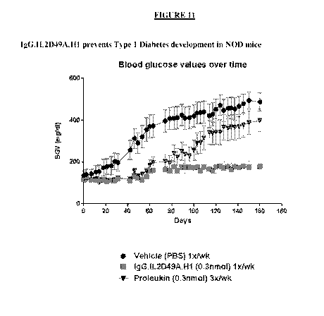
IgG.IL2D49A.H1 prevents Type 1 diabetes in this model.
[00119] Figure 12 shows experimental data on comparing the ratio of Treg
to CD8 T
effector cells in a pre-diabetic NOD mouse model.
[00120] Figure 13A shows experimental data on the pharmacokinetics of
IgG.IL2D49A.H1 in the NOD mouse model at a 1.3 mg/kg dose. Figure 13B shows
experimental data on the pharmacokinetics of IgG.IL2D49A.H1 in the NOD mouse
model at
a 0.43 mg/kg dose.
[00121] Figure 14 is a table of dose ranges used in the pre-diabetic NOD
mouse model,
and compares equimolar amounts of Proleukin .
[00122] Figure 15A shows a series of graphs depicting amount of pSTAT5
activation
on human PBMCs taken from a vitiligo patent and treated in vitro with
IgG.IL2D49.H1 and
compared with Proleukin . Figure 15B shows a series of graphs depicting amount
of
pSTAT5 activation on human PBMCs taken from a patent with type 1 diabetes and
treated in
vitro with IgG.IL2D49.H1 and Proleukin .
[00123] Figure 16 shows a graph of ELISA data showing that when IL2 is
engrafted
into CDRH1 of an anti-RSV antibody, RSV binding is maintained. However,
binding to
RSV is reduced when IL2 is engrafted into CDRL3. When IL2 is engrafted into a
different
antibody backbone (Xolair), there is no binding to RSV.
[00124] Figure 17 shows experimental data on Treg expansion in cynomolgus
monkey
after a single dose of IgG.IL2D49A.H1.
[00125] Figure 18 shows experimental data on effects of increasing
concentrations of a
crosslinker anti-human IgG antibody on the selective activities of GFTX3b_IL-2-
H1-D49A.
[00126] Figure 19 shows experimental data on selective signaling in
different
autoimmune patients PBMC with GFTX3b_IL-2-H1-D49A.
[00127] Figure 20 shows experimental data on GFTX3b_IL-2-H1-D49A having
higher
selectivity than Proleukin for signaling in Tregs over T effector cells in
human PBMC.
CA 03063527 2019-11-13
WO 2018/215935
PCT/IB2018/053622
26
Antibody Cytokine engrafted proteins targeting the IL2 High Affinity Receptor
[00128] Provided herein are protein constructs comprising IL2 engrafted to
into the
complementarity determining region (CDR) of an antibody. The antibody cytokine
engrafted
proteins show suitable properties to be used in human patients, for example,
they retain
immunostimulatory activity similar to that of native or recombinant human IL2.
However,
the negative effects are diminished, for example stimulation of NK cells.
Other activities and
characteristics are also demonstrated throughout the specification. Thus,
provided are
antibody cytokine engrafted proteins having an improved therapeutic profile
over previously
known IL2 and modified IL2 therapeutic agents, such as Proleukin , and methods
of use of
the provided antibody cytokine engrafted proteins in therapy.
[00129] Accordingly, the present disclosure provides antibody cytokine
engrafted
proteins that are agonists of the IL2 high affinity receptor, with selective
activity profiles.
Provided antibody cytokine engrafted proteins comprising an immunoglobulin
heavy chain
sequence and an immunoglobulin light chain sequence. Each immunoglobulin heavy
chain
sequence comprises a heavy chain variable region (VH) and a heavy chain
constant region
(CH), wherein the heavy chain constant region consists of CH1, CH2, and CH3
constant
regions. Each immunoglobulin light chain sequence comprises a light chain
variable region
(VL) and a light chain constant region (CL). In each antibody cytokine
engrafted protein an
IL2 molecule is incorporated into a complementarity determining region (CDR)
of the VH or
VL of the antibody.
[00130] In some embodiments, the antibody cytokine engrafted protein
comprises an
IL2 molecule incorporated into a heavy chain CDR. In certain embodiments the
IL2
molecule is incorporated into heavy chain complementarity determining region 1
(HCDR1).
In certain embodiments the IL2 molecule is incorporated into heavy chain
complementarity
determining region 2 (HCDR2). In certain embodiments the IL2 molecule is
incorporated
into heavy chain complementarity determining region 3 (HCDR3).
[00131] In some embodiments, the antibody cytokine engrafted protein
comprises an
IL2 molecule incorporated into a light chain CDR. In certain embodiments the
IL2 molecule
is incorporated into light chain complementarity determining region 1 (LCDR1).
In certain
embodiments the IL2 molecule is incorporated into light chain complementarity
determining
region 2 (LCDR2). In certain embodiments the IL2 molecule is incorporated into
light chain
complementarity determining region 3 (LCDR3).
CA 03063527 2019-11-13
WO 2018/215935
PCT/IB2018/053622
27
[00132] In some embodiments, the antibody cytokine engrafted comprises an
IL2
sequence incorporated into a CDR, whereby the IL2 sequence is inserted into
the CDR
sequence. The insertion can be at or near the N-terminal region of the CDR, in
the middle
region of the CDR or at or near the C-terminal region of the CDR. In other
embodiments, the
antibody cytokine engrafted comprises an IL2 molecule incorporated into a CDR,
whereby
the IL2 sequence replaces all or part of a CDR sequence. A replacement can be
the N-
terminal region of the CDR, in the middle region of the CDR or at or near the
C-terminal
region the CDR. A replacement can be as few as one or two amino acids of a CDR
sequence,
or the entire CDR sequence.
[00133] In some embodiments the IL2 molecule is engrafted directly into a
CDR
without a peptide linker, with no additional amino acids between the CDR
sequence and the
IL2 sequence.
[00134] In some embodiments antibody cytokine engrafted proteins comprise
immunoglobulin heavy chains of an IgG class antibody heavy chain. In certain
embodiments
an IgG heavy chain is any one of an IgGl, an IgG2 or an IgG4 subclass.
[00135] In some embodiments antibody cytokine engrafted proteins comprise
heavy
and light chain immunoglobulin sequences selected from a known, clinically
utilized
immunoglobulin sequence. In certain embodiments antibody cytokine engrafted
proteins
comprise heavy and light chain immunoglobulin sequences which are humanized
sequences.
In other certain embodiments antibody cytokine engrafted proteins comprise
heavy and light
chain immunoglobulin sequences which are human sequences.
[00136] In some embodiments antibody cytokine engrafted proteins comprise
heavy
and light chain immunoglobulin sequences selected from germline immunoglobulin
sequences.
[00137] In some embodiments antibody cytokine engrafted proteins comprise
heavy
and light chain immunoglobulin sequences having binding specificity of the
immunoglobulin
variable domains to a target distinct from the binding specificity of the IL2
molecule. In
some embodiments the binding specificity of the immunoglobulin variable domain
to its
target is retained by 10%, 20%, 30%, 40%, 50%, 60%, 70%, 80%, 90%, 95%, 98%,
99%, or
100%, in the presence of the engrafted cytokine. In certain embodiments the
retained binding
specificity is to a non-human target. In certain embodiments the retained
binding specificity it
to a virus, for example, RSV. In other embodiments the binding specificity is
to a human
target having therapeutic utility in conjunction with an IL2 therapy. In
certain embodiments,
targeting the binding specificity of the immunoglobulin conveys additional
therapeutic
CA 03063527 2019-11-13
WO 2018/215935
PCT/IB2018/053622
28
benefit to the IL2 component. In certain embodiments the binding specificity
of the
immunoglobulin to its target conveys synergistic activity with IL2.
[00138] In still other embodiments, the binding specificity of the
immunoglobulin to
its target is reduced 10%, 20%, 30%, 40%, 50%, 60%, 70%, 80%, 90%, 95%, 98%,
99%, or
100% by the engrafting of the IL2 molecule.
[00139] Provided antibody cytokine engrafted proteins comprise an IL2
molecule
incorporated into a complementarity determining region (CDR) of the VH or VL
of the
antibody cytokine engrafted protein. In some embodiments, the IL2 sequence has
at least
85%, 90%, 91%, 92%, 93%, 94%, 95%, 96%, 97%, 98%, 99% or 100% sequence
identity to
the amino acid sequence of SEQ ID NO:4. In some embodiments, the IL2 molecule
comprises the sequence of SEQ ID NO:4. In some embodiments, the IL2 molecule
consists
of the sequence of SEQ ID NO:4. Provided antibody cytokine engrafted proteins
comprise
an IL2 molecule incorporated into a complementarity determining region (CDR)
of the VH or
VL of the antibody cytokine engrafted protein. In some embodiments, the IL2
sequence has
at least 85%, 90%, 91%, 92%, 93%, 94%, 95%, 96%, 97%, 98%, 99% or 100%
sequence
identity to the amino acid sequence of SEQ ID NO:4. In some embodiments, the
IL2
molecule comprises the sequence of SEQ ID NO:4. In some embodiments, the IL2
molecule
consists of the sequence of SEQ ID NO:4.
[00140] Provided antibody cytokine engrafted proteins comprise an IL2
molecule
incorporated into a complementarity determining region (CDR) of the VH or VL
of the
antibody cytokine engrafted protein. In some embodiments, the IL2 sequence has
at least
85%, 90%, 91%, 92%, 93%, 94%, 95%, 96%, 97%, 98%, 99% or 100% sequence
identity to
the amino acid sequence of SEQ ID NO:6. In some embodiments, the IL2 molecule
comprises the sequence of SEQ ID NO:6. In some embodiments, the IL2 molecule
consists
of the sequence of SEQ ID NO:6. Provided antibody cytokine engrafted proteins
comprise
an IL2 molecule incorporated into a complementarity determining region (CDR)
of the VH or
VL of the antibody cytokine engrafted protein. In some embodiments, the IL2
sequence has
at least 85%, 90%, 91%, 92%, 93%, 94%, 95%, 96%, 97%, 98%, 99% or 100%
sequence
identity to the amino acid sequence of SEQ ID NO:6. In some embodiments, the
IL2
molecule comprises the sequence of SEQ ID NO:6. In some embodiments, the IL2
molecule
consists of the sequence of SEQ ID NO:6.
[00141] In some embodiments, the antibody cytokine engrafted confers anti-
inflammatory properties superior to human IL2, recombinant human IL2 or
Proleukin . In
some embodiments, the antibody cytokine engrafted proteins disclosed herein
confer
CA 03063527 2019-11-13
WO 2018/215935
PCT/IB2018/053622
29
increased activity on Treg cells while providing reduced proportional pro-
inflammatory
activity as compared to human IL2, recombinant human IL2 or Proleukin . In
some
embodiments, the antibody cytokine engrafted proteins disclosed herein provide
preferential
stimulation of the high affinity IL2 receptor . In some embodiments, the
antibody cytokine
engrafted proteins disclosed herein provide preferential activation of Treg
cells over Teff
cells, Tcon cells, and/or NK cells. In some embodiments, the antibody cytokine
engrafted
proteins disclosed herein provide preferential expansion of Treg cells over
Teff cells, Tcon
cells, and/or NK cells. In some embodiments, the antibody cytokine engrafted
proteins
disclosed herein provide increased expansion of Treg cells without expansion
of CD8 T
effector cells or NK cells. In some embodiments, the antibody cytokine
engrafted proteins
disclosed herein provide a ratio of expansion of Treg cells : NK cells that
is, is about, is
greater than, 1, 2, 3, 4, 5, 6, 7, 8, 9, 10. In some embodiments, the antibody
cytokine
engrafted proteins disclosed herein provide a ratio of expansion of Treg cells
: CD8 T
effector cells that is, is about, is greater than, 1, 2, 3, 4, 5, 6, 7, 8, 9,
10. In some
embodiments, the antibody cytokine engrafted proteins disclosed herein provide
a ratio of
expansion of Treg cells : CD4 Tcon cells that is, is about, is greater than,
1, 2, 3, 4, 5, 6, 7, 8,
9, 10.
[00142] In some embodiments, the antibody cytokine engrafted proteins
disclosed
herein provide IL-2R signalling potency that is reduced in CD4 Tcon cells in
comparison to
human IL2, recombinant human IL2 or Proleukin . In some embodiments, the
antibody
cytokine engrafted proteins disclosed herein provide IL-2R signalling potency
that is reduced
in CD8 Teff cells in comparison to human IL2, recombinant human IL2 or
Proleukin . In
some embodiments, the antibody cytokine engrafted proteins disclosed herein
provide IL-2R
signalling potency that is reduced in NK cells in comparison to human IL2,
recombinant
human IL2 or Proleukin . In some embodiments, the antibody cytokine engrafted
proteins
disclosed herein provide specific activation of Treg cells over CD4 T effector
cells that is
about 1,000 fold, about 2,000 fold, about 3,000 fold, about 4,000 fold, about
5,000 fold,
about 6,000 fold, about 7,000 fold, about 8,000 fold, about 9,000 fold, about
10,000 fold, or
more, higher than human IL2, recombinant human IL2 or Proleukin . In some
embodiments, the antibody cytokine engrafted proteins disclosed herein provide
specific
activation of Treg cells over CD8 T effector cells that is about 100 fold,
about 200 fold, about
300 fold, about 400 fold, about 500 fold, about 600 fold, about 700 fold,
about 800 fold,
about 900 fold, about 1,000 fold, or more, higher than human IL2, recombinant
human IL2 or
Proleukin . In some embodiments, the antibody cytokine engrafted proteins
disclosed
CA 03063527 2019-11-13
WO 2018/215935
PCT/IB2018/053622
herein provide specific activation of Treg cells over CD8 T effector/memory
cells that is
about 100 fold, about 200 fold, about 300 fold, about 400 fold, about 500
fold, about 600
fold, about 700 fold, about 800 fold, about 900 fold, about 1,000 fold, or
more, higher than
human IL2, recombinant human IL2 or Proleukin @.
[00143] In some embodiments, the antibody cytokine engrafted proteins
disclosed
herein provide reduced toxicity, such as expansion of eosinophilia, in
comparison to human
IL2, recombinant human IL2 or Proleukin . In some embodiments, the antibody
cytokine
engrafted proteins disclosed herein provide increased half life, such as more
than 4 hours,
more than 6 hours, more than 8 hours, more than 12 hours, more than 24 hours,
more than 48
hours, in comparison to human IL2, recombinant human IL2 or Proleukin . In
some
embodiments, the antibody cytokine engrafted proteins disclosed herein provide
reduced
body weight loss, such as in graft versus host disease (GvHD) patients, in
comparison to
human IL2, recombinant human IL2 or Proleukin . In some embodiments, the
antibody
cytokine engrafted proteins disclosed herein provide better protection against
type 1 diabetes
development in comparison to human IL2, recombinant human IL2 or Proleukin .
[00144] In some embodiments, the antibody cytokine engrafted proteins
comprise a
modified immunoglobulin IgG having a modified Fc conferring modified effector
function.
In certain embodiments the modified Fc region comprises a mutation selected
from one or
more of D265A, P329A, P329G, N297A, L234A, and L235A. In particular
embodiments the
immunoglobulin heavy chain may comprise a mutation or combination of mutations
conferring reduced effector function selected from any of D265A, P329A, P329G,
N297A,
D265A/P329A, D265A/N297A, L234/L235A, P329A/L234A/L235A, and
P329G/L234A/L235A.
[00145] In some embodiments, the antibody cytokine engrafted proteins
comprise (i) a
heavy chain variable region having at least 85%, 89%, 90%, 91%, 92%, 93%, 94%,
95%,
96%, 97%, 98%, 99%, or 100% amino acid sequence identity to a heavy chain
variable
region of SEQ ID NO:20 and (ii) a light chain variable region having at least
85%, 89%,
90%, 91%, 92%, 93%, 94%, 95%, 96%, 97%, 98%, 99%, or 100% amino acid sequence
identity to a light chain variable region of SEQ ID NO:33. The immunoglobulin
chain is an
IgG class selected from IgGl, IgG2, or IgG4. In certain embodiments the
immunoglobulin
optionally comprises a mutation or combination of mutations conferring reduced
effector
function selected from any of D265A, P329A, P329G, N297A, D265A/P329A,
D265A/N297A, L234/L235A, P329A/L234A/L235A, and P329G/L234A/L235A.
CA 03063527 2019-11-13
WO 2018/215935
PCT/IB2018/053622
31
[00146] In some embodiments, the antibody cytokine engrafted proteins
comprise (i) a
heavy chain variable region having at least 85%, 89%, 90%, 91%, 92%, 93%, 94%,
95%,
96%, 97%, 98%, 99%, or 100% amino acid sequence identity to a heavy chain
variable
region of SEQ ID NO:46 and (ii) a light chain variable region having at least
85%, 89%,
90%, 91%, 92%, 93%, 94%, 95%, 96%, 97%, 98%, 99%, or 100% amino acid sequence
identity to a light chain variable region of SEQ ID NO:58. The immunoglobulin
chain is an
IgG class selected from IgGl, IgG2, or IgG4. In certain embodiments the
immunoglobulin
optionally comprises a mutation or combination of mutations conferring reduced
effector
function selected from any of D265A, P329A, P329G, N297A, D265A/P329A,
D265A/N297A, L234/L235A, P329A/L234A/L235A, and P329G/L234A/L235A.
[00147] In some embodiments, the antibody cytokine engrafted proteins
comprise (i) a
heavy chain variable region having at least 85%, 89%, 90%, 91%, 92%, 93%, 94%,
95%,
96%, 97%, 98%, 99%, or 100% amino acid sequence identity to a heavy chain
variable
region of SEQ ID NO:71 and (ii) a light chain variable region having at least
85%, 89%,
90%, 91%, 92%, 93%, 94%, 95%, 96%, 97%, 98%, 99%, or 100% amino acid sequence
identity to a light chain variable region of SEQ ID NO: 84. The immunoglobulin
chain is an
IgG class selected from IgGl, IgG2, or IgG4. In certain embodiments the
immunoglobulin
optionally comprises a mutation or combination of mutations conferring reduced
effector
function selected from any of D265A, P329A, P329G, N297A, D265A/P329A,
D265A/N297A, L234/L235A, P329A/L234A/L235A, and P329G/L234A/L235A.
[00148] In some embodiments, the antibody cytokine engrafted proteins
comprise (i) a
heavy chain variable region having at least 85%, 89%, 90%, 91%, 92%, 93%, 94%,
95%,
96%, 97%, 98%, 99%, or 100% amino acid sequence identity to a heavy chain
variable
region of SEQ ID NO:97 and (ii) a light chain variable region having at least
85%, 89%,
90%, 91%, 92%, 93%, 94%, 95%, 96%, 97%, 98%, 99%, or 100% amino acid sequence
identity to a light chain variable region of SEQ ID NO:110. The immunoglobulin
chain is an
IgG class selected from IgGl, IgG2, or IgG4. In certain embodiments the
immunoglobulin
optionally comprises a mutation or combination of mutations conferring reduced
effector
function selected from any of D265A, P329A, P329G, N297A, D265A/P329A,
D265A/N297A, L234/L235A, P329A/L234A/L235A, and P329G/L234A/L235A.
Engineered and Modified Antibody Cytokine Engrafted Proteins
[00149] In certain aspects, antibody cytokine engrafted proteins are
generated by
engrafting an IL2 sequence into a CDR region of an immunoglobulin scaffold.
Both heavy
and light chain immunoglobulin chains are produced to generate final antibody
engrafted
CA 03063527 2019-11-13
WO 2018/215935
PCT/IB2018/053622
32
proteins. Antibody cytokine engrafted proteins confer preferred therapeutic
activity on Treg
cells, however, the antibody cytokine engrafted proteins have reduced pro-
inflammatory
activity as compared with native or recombinant human IL2 (rhIL2 or
Proleukin0).
[00150] To engineer antibody cytokine engrafted proteins, IL2 sequences
containing
specific muteins (SEQ ID NO:4 or SEQ ID NO:6), are inserted into a CDR loop of
an
immunoglobulin chain scaffold protein. Engrafted constructs can be prepared
using any of a
variety of known immunoglobulin sequences which have been utilized in clinical
settings,
known immunoglobulin sequences which are in current discovery and/or clinical
development, human germline antibody sequences, as well as sequences of novel
antibody
immunoglobulin chains. Constructs are produced using standard molecular
biology
methodology utilizing recombinant DNA encoding relevant sequences. Sequences
of IL2 in
an exemplary scaffold, referred to as GFTX3b, are depicted in TABLE 2.
Insertion points
were selected to be the mid-point of the loop based on available structural or
homology
model data, however, insertion points can be adjusted toward the N or C-
terminal end of the
CDR loop.
[00151] Thus the present disclosure provides for antibody cytokine
engrafted proteins
that specifically bind to the high affinity IL2 receptor comprising an IL2
protein
recombinantly inserted into a heterologous antibody protein or polypeptide to
generate
antibody cytokine engrafted proteins. In particular, the disclosure provides
antibody cytokine
engrafted proteins comprising an antibody or antigen-binding fragment of an
antibody
described herein or any other relevant scaffold antibody polypeptide (e.g., a
full antibody
immunoglobulin protein, a Fab fragment, Fc fragment, Fv fragment, F(ab)2
fragment, a VH
domain, a VH CDR, a VL domain, a VL CDR, etc.) and a heterologous cytokine
protein,
polypeptide, or peptide, e.g., IL2. Methods for fusing or conjugating
proteins, polypeptides,
or peptides to an antibody or an antibody fragment are known in the art. See,
e.g., U.S.
Patent Nos. 5,336,603, 5,622,929, 5,359,046, 5,349,053, 5,447,851, and
5,112,946; European
Patent Nos. EP 307,434 and EP 367,166; International Publication Nos. WO
96/04388 and
WO 91/06570; Ashkenazi et al., 1991, Proc. Natl. Acad. Sci. USA 88: 10535-
10539; Zheng
et al., 1995, J. Immunol. 154:5590-5600; and Vil et al., 1992, Proc. Natl.
Acad. Sci. USA
89:11337- 11341. Additionally, antibody cytokine engrafted proteins can be
generated
through the techniques of gene-shuffling, motif-shuffling, exon-shuffling,
and/or codon-
shuffling (collectively referred to as "DNA shuffling"). DNA shuffling may be
employed to
prepare antibody cytokine engrafted proteins and/or to alter the activities of
antibodies or
fragments thereof (e.g., antibodies or fragments thereof with higher
affinities and lower
CA 03063527 2019-11-13
WO 2018/215935
PCT/IB2018/053622
33
dissociation rates). See, generally, U.S. Patent Nos. 5,605,793, 5,811,238,
5,830,721,
5,834,252, and 5,837,458; Patten et al., 1997, Curr. Opinion Biotechnol. 8:724-
33;
Harayama, 1998, Trends Biotechnol. 16(2):76-82; Hansson, et al., 1999, J. Mol.
Biol.
287:265-76; and Lorenzo and Blasco, 1998, Biotechniques 24(2):308- 313 (each
of these
patents and publications are hereby incorporated by reference in its
entirety). Antibodies or
fragments thereof, or the encoded antibodies or fragments thereof, can be
altered by being
subjected to random mutagenesis by error-prone PCR, random nucleotide
insertion or other
methods prior to recombination. A polynucleotide encoding an antibody or
fragment thereof
that specifically binds to an antigen protein of interest may be recombined
with one or more
components, motifs, sections, parts, domains, fragments, etc. of one or more
heterologous
cytokine molecules, e.g., IL2, for preparation of antibody cytokine engrafted
proteins as
provided herein.
[00152] An antibody Fab contains six CDR loops, 3 in the light chain
(CDRL1,
CDRL2, CDRL3) and 3 in the heavy chain (CDRH1, CDRH2, CDRH3) which can serve
as
potential insertion sites for a cytokine protein. Structural and functional
considerations are
taken into account in order to determine which CDR loop(s) to insert the
cytokine. As a CDR
loop size and conformation vary greatly across different antibodies, the
optimal CDR for
insertion may be determined empirically for each particular antibody/protein
combination.
Additionally, since a cytokine protein will be inserted into a CDR loop, this
can put
additional constraints on the structure of the cytokine protein. For example,
the amino and
carboxy terminal of the cytokine protein should allow for possibility to be
constrained
relatively close in space (-25 A). Additionally, an antibody cytokine
engrafted protein
should not rely on oligomerization for biological activity. An analysis of
available protein
database structures indicates that many cytokine proteins, including IL2, fall
into this
category.
[00153] CDRs of immunoglobulin chains are determined by well known
numbering
systems known in the art, including those described herein. For example, CDRs
have been
identified and defined by (1) using the numbering system described in Kabat et
al. (1991),
"Sequences of Proteins of Immunological Interest," 5th Ed. Public Health
Service, National
Institutes of Health, Bethesda, MD ("Kabat" numbering scheme), NIH publication
No. 91-
3242; and (2) Chothia, see Al-Lazikani et al., (1997) "Standard conformations
for the
canonical structures of immunoglobulins," J.Mol.Biol. 273:927-948. For
identified CDR
amino acid sequences less than 20 amino acids in length, one or two
conservative amino acid
CA 03063527 2019-11-13
WO 2018/215935
PCT/IB2018/053622
34
residue substitutions can be tolerated while still retaining the desired
specific binding and/or
agonist activity.
[00154] An antibody cytokine engrafted protein further can be prepared
using an
antibody having one or more of the CDRs and/or VH and/or VL sequences shown
herein
(e.g., TABLE 2) as starting material to engineer a modified antibody cytokine
engrafted
protein, which may have altered properties from the starting antibody
engrafted protein.
Alternatively any known antibody sequences may be utilized as a scaffold to
engineer
modified antibody cytokine engrafted protein. For example, any known,
clinically utilized
antibody may be utilized as a starting materials scaffold for preparation of
antibody engrafted
protein. Known antibodies and corresponding immunoglobulin sequences include,
e.g.,
palivizumab, alirocumab, mepolizumab, necitumumab, nivolumab, dinutuximab,
secukinumab, evolocumab, blinatumomab, pembrolizumab, ramucirumab,
vedolizumab,
siltuximab, obinutuzumab, trastuzumab, raxibacumab, pertuzumab, belimumab,
ipilimumab,
denosumab, tocilizumab, ofatumumab, canakinumab, golimumab, ustekinumab,
certolizumab, catumaxomab, eculizumab, ranibizumab, panitumumab, natalizumab,
bevacizumab, cetuximab, efalizumab, omalizumab, tositumomab, ibritumomab
tiuxetan,
adalimumab, alemtuzumab, gemtuzumab, infliximab, basiliximab, daclizumab,
rituximab,
abciximab, muromonab, or modifications thereof. Known antibodies and
immunoglobulin
sequences also include germline antibody sequences. Framework sequences can be
obtained
from public DNA databases or published references that include germline
antibody gene
sequences. For example, germline DNA sequences for human heavy and light chain
variable
region genes can be found in the "VBase" human germline sequence database as
well as in
Kabat, E. A., et al., 1991 Sequences of Proteins of Immunological Interest,
Fifth Edition,
U.S. Department of Health and Human Services, NIH Publication No. 91-3242;
Tomlinson, I.
M., et al., 1992 J. fol. Biol. 227:776-798; and Cox, J. P. L. et al., 1994
Eur. J Immunol.
24:827-836. In still other examples, antibody and corresponding immunoglobulin
sequences
from other known entities which may be in early discovery and/or drug
development can be
similarly adapted as starting material to engineer a modified antibody
cytokine engrafted
protein.
[00155] A wide variety of antibody/immunoglobulin frameworks or scaffolds
can be
employed so long as the resulting polypeptide includes at least one binding
region which
accommodates incorporation of a cytokine (e.g., IL2). Such frameworks or
scaffolds include
the 5 main idiotypes of human immunoglobulins, or fragments thereof, and
include
immunoglobulins of other animal species, preferably having humanized and/or
human
CA 03063527 2019-11-13
WO 2018/215935
PCT/IB2018/053622
aspects. Novel antibodies, frameworks, scaffolds and fragments continue to be
discovered
and developed by those skilled in the art.
[00156] Antibodies can be generated using methods that are known in the
art. For
preparation of monoclonal antibodies, any technique known in the art can be
used (see, e.g.,
Kohler & Milstein, Nature 256:495-497 (1975); Kozbor et al., Immunology Today
4:72
(1983); Cole et al., Monoclonal Antibodies and Cancer Therapy, pp. 77-96. Alan
R. Liss, Inc.
1985). Techniques for the production of single chain antibodies (U.S. Patent
No. 4,946,778)
can be adapted to produce antibodies for use in antibody cytokine engrafted
proteins. Also,
transgenic mice, or other organisms such as other mammals, may be used to
express and
identify primatized or humanized or human antibodies. Alternatively, phage
display
technology can be used to identify antibodies and heteromeric Fab fragments
that specifically
bind to selected antigens for use in antibody cytokine engrafted proteins
(see, e.g.,
McCafferty et al., supra; Marks et al., Biotechnology, 10:779-783, (1992)).
[00157] Methods for primatizing or humanizing non-human antibodies are
well known
in the art. Generally, a primatized or humanized antibody has one or more
amino acid
residues introduced into it from a source which is non-primate or non-human.
Such non-
primate or non-human amino acid residues are often referred to as import
residues, which are
typically taken from an import variable domain. Humanization can be
essentially performed
following the method of Winter and co-workers (see, e.g., Jones et al., Nature
321:522-525
(1986); Riechmann et al., Nature 332:323-327 (1988); Verhoeyen et al., Science
239:1534-
1536 (1988) and Presta, Curr. Op. Struct. Biol. 2:593-596 (1992)), by
substituting rodent
CDRs or CDR sequences for the corresponding sequences of a human antibody.
Accordingly, such humanized antibodies are chimeric antibodies (U.S. Patent
No. 4,816,567),
wherein substantially less than an intact human variable domain has been
substituted by the
corresponding sequence from a non-human species. In practice, primatized or
humanized
antibodies are typically primate or human antibodies in which some
complementary
determining region ("CDR") residues and possibly some framework ("FR")
residues are
substituted by residues from analogous sites in an originating species (e.g.,
rodent antibodies)
to confer binding specificity.
[00158] Alternatively or additionally, an in vivo method for replacing a
nonhuman
antibody variable region with a human variable region in an antibody while
maintaining the
same or providing better binding characteristics relative to that of the
nonhuman antibody
may be utilized to convert non-human antibodies into engineered human
antibodies. See,
e.g., U.S. Patent Publication No. 20050008625, U.S. Patent Publication No.
2005/0255552.
CA 03063527 2019-11-13
WO 2018/215935
PCT/IB2018/053622
36
Alternatively, human V segment libraries can be generated by sequential
cassette replacement
in which only part of the reference antibody V segment is initially replaced
by a library of
human sequences; and identified human "cassettes" supporting binding in the
context of
residual reference antibody amino acid sequences are then recombined in a
second library
screen to generate completely human V segments (see, U.S. Patent Publication
No.
2006/0134098).
[00159] Various antibodies or antigen-binding fragments for use in
preparation of
antibody cytokine engrafted proteins can be produced by enzymatic or chemical
modification
of the intact antibodies, or synthesized de novo using recombinant DNA
methodologies (e.g.,
single chain Fv), or identified using phage display libraries (see, e.g.,
McCafferty et al.,
Nature 348:552-554, 1990). For example, minibodies can be generated using
methods
described in the art, e.g., Vaughan and Sollazzo, Comb Chem High Throughput
Screen.
4:417-30 2001. Bispecific antibodies can be produced by a variety of methods
including
engrafted of hybridomas or linking of Fab' fragments. See, e.g., Songsivilai &
Lachmann,
Clin. Exp. Immunol. 79:315-321 (1990); Kostelny et al., J. Immunol. 148, 1547-
1553 (1992).
Single chain antibodies can be identified using phage display libraries or
ribosome display
libraries, gene shuffled libraries. Such libraries can be constructed from
synthetic, semi-
synthetic or native and immunocompetent sources. Selected immunoglobulin
sequences may
thus be utilized in preparation of antibody cytokine engrafted protein
constructs as provided
herein.
[00160] Antibodies, antigen-binding molecules or antibody cytokine
engrafted
molecules of the present disclosure further include bispecific antibodies. A
bispecific or
bifunctional antibody is an artificial hybrid antibody having two different
heavy/light chain
pairs and two different binding sites. Other antigen-binding fragments or
antibody portions
include bivalent scFv (diabody), bispecific scFv antibodies where the antibody
molecule
recognizes two different epitopes, single binding domains (dAbs), and
minibodies. Selected
immunoglobulin sequences can thus be utilized in preparation of antibody
cytokine engrafted
protein constructs as provided herein.
[00161] Antigen-binding fragments of antibodies e.g., a Fab fragment,
scFv, can be
used as building blocks to construct antibody cytokine engrafted proteins, and
may optionally
include multivalent formats. In some embodiments, such multivalent molecules
comprise a
constant region of an antibody (e.g., Fc).
[00162] Antibody cytokine engrafted proteins can be engineered by
modifying one or
more residues within one or both variable regions (i.e., VH and/or VL) of an
antibody, for
CA 03063527 2019-11-13
WO 2018/215935
PCT/IB2018/053622
37
example within one or more CDR regions, and such adapted VH and/or VL region
sequences
utilized for engrafting a cytokine or for preparation of cytokine engrafting.
Antibodies
interact with target antigens predominantly through amino acid residues that
are located in
the six heavy and light chain complementarity determining regions (CDRs). For
this reason,
the amino acid sequences within CDRs are more diverse between individual
antibodies than
sequences outside of CDRs. CDR sequences are responsible for most antibody-
antigen
interactions, it is possible to express recombinant antibodies that mimic the
properties of a
specific antibody by constructing expression vectors that include CDR
sequences from a
specific antibody grafted onto framework sequences from a different antibody
with different
properties (see, e.g., Riechmann, L. et al., 1998 Nature 332:323-327; Jones,
P. et al., 1986
Nature 321:522-525; Queen, C. et al., 1989 Proc. Natl. Acad., U.S.A. 86:10029-
10033; U.S.
Patent No. 5,225,539 to Winter, and U.S. Patent Nos. 5,530,101; 5,585,089;
5,693,762 and
6,180,370 to Queen et al.). In certain instances it is beneficial to mutate
residues within the
framework regions to maintain or enhance the antigen binding ability of the
antibody (see
e.g., U.S. Patent Nos. 5,530,101; 5,585,089; 5,693,762 and 6,180,370 to Queen
et al).
[00163] In some aspects mutation of amino acid residues within the VH
and/or VL
CDR1, CDR2, and/or CDR3 regions to thereby improve one or more binding
properties (e.g.,
affinity) of the antibody of interest, known as "affinity maturation," can be
beneficial, e.g., to
optimize antigen binding of an antibody in conjunction with the context of the
cytokine
engrafted protein. Site-directed mutagenesis or PCR-mediated mutagenesis can
be performed
to introduce the mutation(s) and the effect on antibody binding, or other
functional property
of interest, can be evaluated in in vitro or in vivo assays as described
herein and provided in
the Examples and/or alternative or additional assays known in the art.
Conservative
modifications can be introduced. The mutations may be amino acid
substitutions, additions or
deletions. Moreover, typically no more than one, two, three, four or five
residues within a
CDR region are altered.
[00164] Engineered antibodies or antibody fragments include those in which
modifications have been made to framework residues within VH and/or VL, e.g.
to improve
the properties of the antibody. In some embodiments such framework
modifications are made
to decrease immunogenicity of the antibody. For example, one approach is to
change one or
more framework residues to the corresponding germline sequence. More
specifically, an
antibody that has undergone somatic mutation may contain framework residues
that differ
from germline sequence from which the antibody is derived. Such residues can
be identified
by comparing the antibody framework sequences to the germline sequences from
which the
CA 03063527 2019-11-13
WO 2018/215935
PCT/IB2018/053622
38
antibody is derived. To return the framework region sequences to their
germline
configuration, the somatic mutations can be "backmutated" to the germline
sequence by, for
example, site-directed mutagenesis. Additional framework modification involves
mutating
one or more residues within the framework region, or even within one or more
CDR regions,
to remove T cell epitopes to thereby reduce the potential immunogenicity of
the antibody.
This approach is also referred to as "deimmunization" and is described in
further detail in
U.S. Patent Publication No. 20030153043 by Can et al.
[00165] Constant regions of the antibodies or antibody fragments utilized
for
preparation of the antibody cytokine engrafted protein can be any type or
subtype, as
appropriate, and can be selected to be from the species of the subject to be
treated by the
present methods (e.g., human, non-human primate or other mammal, for example,
agricultural mammal (e.g., equine, ovine, bovine, porcine, camelid), domestic
mammal (e.g.,
canine, feline) or rodent (e.g., rat, mouse, hamster, rabbit). In some
embodiments antibodies
utilized in antibody cytokine engrafted proteins are engineered to generate
humanized or
Humaneered@ antibodies. In some embodiments antibodies utilized in antibody
cytokine
engrafted proteins are human antibodies. In some embodiments, antibody
constant region
isotype is IgG, for example, IgGl, IgG2, IgG3, IgG4. In certain embodiments
the constant
region isotype is IgGi. In some embodiments, antibody cytokine engrafted
proteins comprise
an IgG. In some embodiments, antibody cytokine engrafted proteins comprise an
IgG1 Fc.
In some embodiments, antibody cytokine engrafted proteins comprise an IgG2 Fc.
[00166] In addition or alternative to modifications made within framework
or CDR
regions, antibodies or antibody fragments utilized in preparation of antibody
cytokine
engrafted proteins may be engineered to include modifications within an Fc
region, typically
to alter one or more functional properties of the antibody, such as, e.g.,
serum half-life,
complement fixation, Fc receptor binding, and/or antigen-dependent cellular
cytotoxicity.
Furthermore, an antibody, antibody fragment, or antibody cytokine engrafted
protein can be
chemically modified (e.g., one or more chemical moieties can be attached to
the antibody) or
be modified to alter its glycosylation, again to alter one or more functional
properties of the
antibody cytokine engrafted protein.
[00167] In one embodiment, a hinge region of CH1 is modified such that the
number
of cysteine residues in the hinge region is altered, e.g., increased or
decreased. For example,
by the approach is described further in U.S. Patent No. 5,677,425 by Bodmer et
al. wherein
the number of cysteine residues in the hinge region of CH1 is altered to, for
example,
facilitate assembly of the light and heavy chains or to increase or decrease
the stability of the
CA 03063527 2019-11-13
WO 2018/215935
PCT/IB2018/053622
39
antibody cytokine engrafted protein. In another embodiment, an Fc hinge region
of an
antibody is mutated to alter the biological half-life of the antibody cytokine
engrafted protein.
More specifically, one or more amino acid mutations are introduced into the
CH2-CH3
domain interface region of the Fc-hinge fragment such that the antibody
cytokine engrafted
protein has impaired Staphylococcyl protein A (SpA) binding relative to native
Fc-hinge
domain SpA binding. This approach is described in further detail in U.S.
Patent No.
6,165,745 by Ward et al.
[00168] The present disclosure provides for antibody cytokine engrafted
proteins that
specifically bind to the IL2 high affinity receptor which have an extended
half-life in vivo. In
another embodiment, an antibody cytokine engrafted protein is modified to
increase its
biological half-life. Various approaches are possible. Antibody cytokine
engrafted proteins
having an increased half-life in vivo can also be generated introducing one or
more amino
acid modifications (i.e., substitutions, insertions or deletions) into an IgG
constant domain, or
FcRn binding fragment thereof (preferably a Fc or hinge Fc domain fragment).
For example,
one or more of the following mutations can be introduced: T252L, T2545, T256F,
as
described in U.S. Patent No. 6,277,375 to Ward. See, e.g., International
Publication No. WO
98/23289; International Publication No. WO 97/34631; and U.S. Patent No.
6,277,375.
Alternatively, to increase the biological half-life, the antibody cytokine
engrafted protein is
altered within the CH1 or CL region to contain a salvage receptor binding
epitope taken from
two loops of a CH2 domain of an Fc region of an IgG, as described in U.S.
Patent Nos.
5,869,046 and 6,121,022 by Presta et al. In yet other embodiments, the Fc
region is altered
by replacing at least one amino acid residue with a different amino acid
residue to alter the
effector functions of the antibody cytokine engrafted protein. For example,
one or more
amino acids can be replaced with a different amino acid residue such that the
antibody
cytokine engrafted protein has an altered affinity for an effector ligand but
retains antigen-
binding ability of the parent antibody. The effector ligand to which affinity
is altered can be,
for example, an Fc receptor (FcR) or the Cl component of complement. This
approach is
described in further detail in U.S. Patent Nos. 5,624,821 and 5,648,260, both
by Winter et al.
[00169] In another embodiment, one or more amino acids selected from amino
acid
residues can be replaced with a different amino acid residue such that the
antibody cytokine
engrafted protein has altered Clq binding and/or reduced or abolished
complement dependent
cytotoxicity (CDC). This approach is described in further detail in U.S.
Patent Nos. 6,194,551
by Idusogie et al.
CA 03063527 2019-11-13
WO 2018/215935
PCT/IB2018/053622
[00170] Antibody cytokine engrafted proteins containing such mutations
mediate
reduced or no antibody-dependent cellular cytotoxicity (ADCC) or complement-
dependent
cytotoxicity (CDC). In some embodiments, amino acid residues L234 and L235 of
the IgG1
constant region are substituted to Ala234 and Ala235. In some embodiments,
amino acid
residue N267 of the IgG1 constant region is substituted to Ala267.
[00171] In another embodiment, one or more amino acid residues are altered
to thereby
alter the ability of the antibody cytokine engrafted protein to fix
complement. This approach
is described further in PCT Publication WO 94/29351 by Bodmer et al.
[00172] In yet another embodiment, an Fc region is modified to increase
the ability of
the antibody to mediate antibody dependent cellular cytotoxicity (ADCC) and/or
to increase
the affinity of the antibody cytokine engrafted protein for an Fey receptor by
modifying one
or more amino acids. This approach is described further in PCT Publication WO
00/42072
by Presta. Moreover, binding sites on human IgG1 for FeyR1, FeyRII, FeyRIII
and FcRn
have been mapped and variants with improved binding have been described (see
Shields,
R.L. et al., 2001 J. Biol. Chen. 276:6591-6604).
[00173] In still another embodiment, glycosylation of an antibody cytokine
engrafted
protein is modified. For example, an aglycoslated antibody cytokine engrafted
protein can be
made (i.e., the antibody cytokine engrafted protein lacks glycosylation).
Glycosylation can be
altered to, for example, increase the affinity of the antibody for "antigen."
Such carbohydrate
modifications can be accomplished by, for example, altering one or more sites
of
glycosylation within the antibody sequence. For example, one or more amino
acid
substitutions can be made that result in elimination of one or more variable
region framework
glycosylation sites to thereby eliminate glycosylation at that site. Such
aglycosylation may
increase the affinity of the antibody for antigen. Such an approach is
described in further
detail in U.S. Patent Nos. 5,714,350 and 6,350,861 by Co et al.
[00174] Additionally or alternatively, an antibody cytokine engrafted
protein can be
made that has an altered type of glycosylation, such as a hypofucosylated
antibody cytokine
engrafted protein having reduced amounts of fucosyl residues or an antibody
having
increased bisecting GleNac structures. Such altered glycosylation patterns
have been
demonstrated to increase the antibody dependent cellular cytotoxicity (ADCC)
ability of
antibodies. Such carbohydrate modifications can be accomplished by, for
example,
expressing the antibody cytokine engrafted protein in a host cell with altered
glycosylation
machinery. Cells with altered glycosylation machinery have been described in
the art and can
be used as host cells in which to express recombinant antibody cytokine
engrafted proteins to
CA 03063527 2019-11-13
WO 2018/215935
PCT/IB2018/053622
41
thereby produce an antibody cytokine engrafted protein with altered
glycosylation. For
example, EP 1,176,195 by Hang et al. describes a cell line with a functionally
disrupted
FUT8 gene, which encodes a fucosyl transferase, such that antibody cytokine
engrafted
proteins expressed in such a cell line exhibit hypofucosylation. PCT
Publication WO
03/035835 by Presta describes a variant CHO cell line, Lec13 cells, with
reduced ability to
attach fucose to Asn(297)-linked carbohydrates, also resulting in
hypofucosylation of
antibody cytokine engrafted proteins expressed in that host cell (see also
Shields, R.L. et al.,
2002 J. Biol. Chem. 277:26733-26740). PCT Publication WO 99/54342 by Umana et
al.
describes cell lines engineered to express glycoprotein-modifying glycosyl
transferases (e.g.,
beta(1,4)-N acetylglucosaminyltransferase III (GnTIII)) such that antibody
cytokine
engrafted proteins expressed in the engineered cell lines exhibit increased
bisecting GlcNac
structures which results in increased ADCC activity of the antibodies (see
also Umana et al.,
1999 Nat. Biotech. 17:176-180).
[00175] In some embodiments, one or more domains, or regions, of an
antibody
cytokine engrafted protein are connected via a linker, for example, a peptide
linker, such as
those that are well known in the art (see e.g., Holliger, P., et al. (1993)
Proc. Natl. Acad. Sci.
USA 90:6444-6448; Poljak, RJ., et al. (1994) Structure 2:1121-1123). A peptide
linker may
vary in length, e.g., a linker can be 1-100 amino acids in length, typically a
linker is from five
to 50 amino acids in length, e.g., 5, 6, 7, 8, 9, 10, 11, 12, 13, 14, 15, 16,
17, 18, 19, 20, 21, 22,
23, 24, 25, 26, 27, 28, 29, 30, 31, 32, 33, 34, 35, 36, 37, 38, 39, 40, 41,
42, 43, 44, 45, 46, 47,
48, 49, or 50 amino acids in length.
[00176] In some embodiments IL2 is engrafted into the CDR sequence
optionally with
one or more peptide linker sequences. In certain embodiments one or more
peptide linkers is
independently selected from a (Glyn-Ser)m sequence (SEQ ID NO: 115), a (Glyn-
Ala)m
sequence (SEQ ID NO: 116), or any combination of a (Glyn-Ser)m/(Glyn-Ala)m
sequence
(SEQ ID NOS 115 and 116, respectively), wherein each n is independently an
integer from 1
to 5 and each m is independently an integer from 0 to 10. Examples of linkers
include, but
are not limited to, glycine-based linkers or gly/ser linkers G/S such as
(GmS)n wherein n is a
positive integer equal to 1, 2, 3, 4, 5, 6, 7, 8, 9, or 10 and m is an integer
equal to 0, 1, 2, 3, 4,
5, 6, 7, 8, 9, or 10 (SEQ ID NO: 117). In certain embodiments one or more
linkers include
G45 repeats (SEQ ID NO: 118), e.g., the Gly-Ser linker (G45). wherein n is a
positive integer
equal to or greater than 1 (SEQ ID NO: 118). For example, n=1, n=2, n=3. n=4,
n=5 and
n=6, n=7, n=8, n=9 and n=10. In some embodiments, Ser can be replaced with Ala
e.g.,
linkers G/A such as (GmA)n wherein n is a positive integer equal to 1, 2, 3,
4, 5, 6, 7, 8, 9, or
CA 03063527 2019-11-13
WO 2018/215935
PCT/IB2018/053622
42
and m is an integer equal to 0, 1, 2, 3, 4, 5, 6, 7, 8, 9, or 10 (SEQ ID NO:
119). In certain
embodiments one or more linkers include G4A repeats (SEQ ID NO: 120), (G4A)11
wherein n
is a positive integer equal to or greater than 1 (SEQ ID NO: 120). For
example, n=1, n=2,
n=3. n=4, n=5 and n=6, n=7, n=8, n=9 and n=10. In some embodiments, the linker
includes
multiple repeats of linkers. In other embodiments, a linker includes
combinations and
multiples of G45 (SEQ ID NO: 118) and G4A (SEQ ID NO: 120).
[00177] Other examples of linkers include those based on flexible linker
sequences
that occur naturally in antibodies to minimize immunogenicity arising from
linkers and
junctions. For example, there is a natural flexible linkage between the
variable domain and a
CH1 constant domain in antibody molecular structure. This natural linkage
comprises
approximately 10-12 amino acid residues, contributed by 4-6 residues from C-
terminus of V
domain and 4-6 residues from the N-terminus of the CH1 domain. Antibody
cytokine
engrafted proteins can, e.g., employ linkers incorporating terminal 5-6 amino
acid residues,
or 11-12 amino acid residues, of CH1 as a linker. The N-terminal residues of
the CH1
domain, particularly the first 5-6 amino acid residues, adopt a loop
conformation without
strong secondary structure, and, therefore, can act as a flexible linker. The
N-terminal
residues of the CH1 domain are a natural extension of the variable domains, as
they are part
of the Ig sequences, and, therefore, minimize to a large extent any
immunogenicity
potentially arising from the linkers and junctions. In some embodiments a
linker sequence
includes a modified peptide sequence based on a hinge sequence.
[00178] Moreover, the antibody cytokine engrafted proteins can be fused to
marker
sequences, such as a peptide to facilitate purification of antibody cytokine
engrafted proteins.
In preferred embodiments, a marker amino acid sequence is a hexa-histidine
peptide (SEQ ID
NO: 121), such as the tag provided in a pQE vector (QIAGEN, Inc., Chatsworth,
CA), among
others, many of which are commercially available. As described in Gentz et
al., 1989, Proc.
Natl. Acad. Sci. USA 86:821-824, for instance, hexa-histidine (SEQ ID NO: 121)
provides
for convenient purification of the engrafted protein. Other peptide tags
useful for purification
include, but are not limited to, the hemagglutinin ("HA") tag, which
corresponds to an
epitope derived from the influenza hemagglutinin protein (Wilson et al., 1984,
Cell 37:767),
and the "flag" tag.
[00179] Antibodies may also be attached to solid supports, which are
particularly
useful for immunoassays or purification of the target antigen. Such solid
supports include, but
are not limited to, glass, cellulose, polyacrylamide, nylon, polystyrene,
polyvinyl chloride or
polypropylene.
CA 03063527 2019-11-13
WO 2018/215935
PCT/IB2018/053622
43
Assays for Antibody Cytokine Engrafted Protein activity
[00180] Assays for identifying antibody cytokine engrafted proteins are
known in the
art and described herein. Agonist antibody cytokine engrafted proteins bind to
the IL2 high
affinity receptor and promote, induce, stimulate intracellular signaling
resulting in Treg
effects as well as immunostimulatory effects.
[00181] Binding of the antibody cytokine engrafted proteins to the IL2
high affinity
receptor can be determined using any method known in the art. For example,
binding to the
IL2 high affinity receptor can be determined using known techniques, including
without
limitation ELISA, Western blots, surface plasmon resonance (e.g., BIAcore),
and flow
cytometry.
[00182] Intracellular signaling through the IL2 high affinity receptor can
be measured
using any method known in the art. For example, activation through IL2
promotes STAT5
activation and signaling. Methods for measuring STAT5 activation are standard
in the art
(e.g., phosphorylation status of STAT5 protein, reporter gene assays,
downstream signaling
assays, etc.). Activation through the IL2 high affinity receptor has increased
Treg effects.
Additionally, the reduced binding of the IL2 low affinity receptor reduces
natural killer (NK)
cell and CD 8 T effector cell proliferation. Methods for measuring
proliferation of cells are
standard in the art (e.g., 3H-thymidine incorporation assays, CFSE labeling).
Methods for
measuring cytokine production are well known in the art (e.g., ELISA assays,
ELISpot
assays). In performing in vitro assays, test cells or culture supernatant from
test cells
contacted with an agonist antibody cytokine engrafted proteins can be compared
to control
cells or culture supernatants from control cells that have not been contacted
with an agonist
antibody cytokine engrafted proteins and/or those that have been contacted
with recombinant
human IL2 (e.g. Proleukin0).
[00183] The activity of the antibody cytokine engrafted proteins can also
be measured
ex vivo and/or in vivo. In some aspects, methods for measuring STAT5
activation across
various cell types ex vivo from animals treated with antibody cytokine
engrafted proteins as
compared to untreated control animals and/or animals similarly treated with
Proleukin can
be used to show differential activity of the agonist antibody engrafted
proteins across cell
types. Preferred agonist antibody cytokine engrafted proteins have the ability
to activate and
expand Treg cells. For example, in vivo activation and expansion of Treg cells
can be
measured using any method known in the art, e.g., by flow cytometry. Preferred
agonist
CA 03063527 2019-11-13
WO 2018/215935
PCT/IB2018/053622
44
antibody cytokine engrafted proteins can be therapeutically useful in
preventing, reducing,
inhibiting or eliminating immune related disorders, for example: Type 1
diabetes, System
Lupus Erythematosus, Vitiligo, chronic graft versus host disease (cGvHD),
prophylactic
acute graft versus host disease (pGvHD), HIV-induced vasculitis, Alopecia
areata, Systemic
sclerosis morphoea and primary anti-phospholipid syndrome. The efficacy of the
agonist
antibody cytokine engrafted proteins in Type 1 diabetes and SLE can be
determined by
administering a therapeutically effective amount of the antibody cytokine
engrafted protein to
a subject and comparing the subject before and after administration of the
antibody cytokine
engrafted protein. Efficacy of the agonist antibody cytokine engrafted
proteins in therapy for
Type 1 diabetes and SLE also can be determined by administering a
therapeutically effective
amount of an antibody cytokine engrafted protein to a test subject and
comparing the test
subject to a control subject who has not been administered the antibody and/or
comparison to
a subject similarly treated with Proleukin .
Polynucleotides Encoding Agonist Antibody Cytokine Engrafted Proteins
[00184] In another aspect, isolated nucleic acids encoding heavy and light
chain
proteins of the antibody cytokine engrafted proteins are provided. Antibody
cytokine
engrafted proteins can be produced by any means known in the art, including
but not limited
to, recombinant expression, chemical synthesis, and enzymatic digestion of
antibody
tetramers. Recombinant expression can be from any appropriate host cells known
in the art,
for example, mammalian host cells, bacterial host cells, yeast host cells,
insect host cells, etc.
[00185] Provided herein are polynucleotides that encode the variable
regions
exemplified in any one of SEQ ID NO:21, SEQ ID NO: 34, SEQ ID NO:47, SEQ ID
NO: 60,
SEQ ID NO:73, SEQ ID NO:86, SEQ ID NO:99, and SEQ ID NO:112.
[00186] The disclosure thus provides polynucleotides encoding the light
and/or heavy
chain polypeptides of the antibody cytokine engrafted proteins described
herein, e.g.,
polynucleotides encoding light or heavy chain variable regions or segments
comprising the
complementary determining regions as described herein. In some embodiments,
the
polynucleotide encoding the heavy chain variable regions comprises a sequence
having at
least 85%, 89%, 90%, 91%, 92%, 93%, 94%, 95%, 96%, 97%, 98%, 99%, or 100%
nucleic
acid sequence identity with a polynucleotide selected from the group
consisting of SEQ ID
NO: 21, SEQ ID NO: 47, SEQ ID NO:73, and SEQ ID NO:99. In some embodiments,
the
polynucleotide encoding the light chain variable regions comprises a sequence
having at least
CA 03063527 2019-11-13
WO 2018/215935
PCT/IB2018/053622
85%, 89%, 90%, 91%, 92%, 93%, 94%, 95%, 96%, 97%, 98%, 99%, or 100% nucleic
acid
sequence identity with a polynucleotide selected from the group consisting of
SEQ ID NO:
34, SEQ ID NO: 60, SEQ ID NO:86, and SEQ ID NO: 112.
[00187] In some embodiments, the polynucleotide encoding the heavy chain
has at
least 85%, 89%, 90%, 91%, 92%, 93%, 94%, 95%, 96%, 97%, 98%, 99%, or 100%
nucleic
acid sequence identity with a polynucleotide of SEQ ID NO:23. In some
embodiments, the
polynucleotide encoding the light chain has at least 85%, 89%, 90%, 91%, 92%,
93%, 94%,
95%, 96%, 97%, 98%, 99%, or 100% nucleic acid sequence identity with a
polynucleotide of
SEQ ID NO:36.
[00188] In some embodiments, the polynucleotide encoding the heavy chain
has at
least 85%, 89%, 90%, 91%, 92%, 93%, 94%, 95%, 96%, 97%, 98%, 99%, or 100%
nucleic
acid sequence identity with a polynucleotide of SEQ ID NO:49. In some
embodiments, the
polynucleotide encoding the light chain has at least 85%, 89%, 90%, 91%, 92%,
93%, 94%,
95%, 96%, 97%, 98%, 99%, or 100% nucleic acid sequence identity with a
polynucleotide of
SEQ ID NO:62.
[00189] In some embodiments, the polynucleotide encoding the heavy chain
has at
least 85%, 89%, 90%, 91%, 92%, 93%, 94%, 95%, 96%, 97%, 98%, 99%, or 100%
nucleic
acid sequence identity with a polynucleotide of SEQ ID NO:75. In some
embodiments, the
polynucleotide encoding the light chain has at least 85%, 89%, 90%, 91%, 92%,
93%, 94%,
95%, 96%, 97%, 98%, 99%, or 100% nucleic acid sequence identity with a
polynucleotide of
SEQ ID NO:88.
[00190] In some embodiments, the polynucleotide encoding the heavy chain
has at
least 85%, 89%, 90%, 91%, 92%, 93%, 94%, 95%, 96%, 97%, 98%, 99%, or 100%
nucleic
acid sequence identity with a polynucleotide of SEQ ID NO:101. In some
embodiments, the
polynucleotide encoding the light chain has at least 85%, 89%, 90%, 91%, 92%,
93%, 94%,
95%, 96%, 97%, 98%, 99%, or 100% nucleic acid sequence identity with a
polynucleotide of
SEQ ID NO:114.
[00191] Polynucleotides can encode only the variable region sequence of an
antibody
cytokine engrafted protein. They can also encode both a variable region and a
constant
region of the antibody cytokine engrafted protein. Some of the polynucleotide
sequences
encode a polypeptide that comprises variable regions of both the heavy chain
and the light
chain of one of the antibody cytokine engrafted proteins. Some other
polynucleotides encode
two polypeptide segments that respectively are substantially identical to the
variable regions
of the heavy chain and the light chain of one of the antibody protein
engrafteds.
CA 03063527 2019-11-13
WO 2018/215935
PCT/IB2018/053622
46
[00192] In certain embodiments polynucleotides or nucleic acids comprise
DNA. In
other embodiments polynucleotides or nucleic acids comprise RNA, which may be
single
stranded or double stranded.
[00193] In some embodiments a recombinant host cell comprising the nucleic
acids
encoding one or more immunoglobulin protein chain of an antibody cytokine
engrafted
protein, and optionally, secretion signals is provided. In certain embodiments
a recombinant
host cell comprises a vector encoding one immunoglobulin protein chain and
secretion
signals. In other certain embodiments a recombinant host cell comprises one or
more vectors
encoding two immunoglobulin protein chains of the antibody cytokine engrafted
protein and
secretion signals. In some embodiments a recombinant host cell comprises a
single vector
encoding two immunoglobulin protein chains of the antibody cytokine engrafted
protein and
secretion signals. In some embodiments a recombinant host cell comprises two
vectors, one
encoding a heavy chain immunoglobulin protein chain, and another encoding a
light chain
immunoglobulin protein chain of the antibody cytokine engrafted protein, with
each
including secretion signals. A recombinant host cell may be a prokaryotic or
eukaryotic cell.
In some embodiments the host cell is a eukaryotic cell line. In some
embodiments the host
cell is a mammalian cell line. In some embodiments the host cell is a CHO
cell. In some
embodiments, the host cell line is a CHO cell line for antibody production.
[00194] Additionally provided are methods for producing the antibody
cytokine
engrafted proteins. In some embodiments the method comprises the steps of (i)
culturing a
host cell comprising one or more vectors encoding immunoglobulin protein
chains of an
antibody cytokine engrafted protein under conditions suitable for expression,
formation, and
secretion of the antibody cytokine engrafted protein and (ii) recovering the
antibody cytokine
engrafted protein.
[00195] The polynucleotide sequences can be produced by de novo solid-
phase DNA
synthesis or by PCR mutagenesis of an existing sequence (e.g., sequences as
described
herein) encoding a polypeptide chain of an antibody cytokine engrafted
protein. Direct
chemical synthesis of nucleic acids can be accomplished by methods known in
the art, such
as the phosphotriester method of Narang et al., Meth. Enzymol. 68:90, 1979;
the
phosphodiester method of Brown et al., Meth. Enzymol. 68:109, 1979; the
diethylphosphoramidite method of Beaucage et al., Tetra. Lett., 22:1859, 1981;
and the solid
support method of U.S. Patent No. 4,458,066. Introducing mutations to a
polynucleotide
sequence by PCR can be performed as described in, e.g., PCR Technology:
Principles and
Applications for DNA Amplification, H.A. Erlich (Ed.), Freeman Press, NY, NY,
1992; PCR
CA 03063527 2019-11-13
WO 2018/215935
PCT/IB2018/053622
47
Protocols: A Guide to Methods and Applications, Innis et al. (Ed.), Academic
Press, San
Diego, CA, 1990; Mattila et al., Nucleic Acids Res. 19:967, 1991; and Eckert
et al., PCR
Methods and Applications 1:17, 1991.
[00196] Also provided are expression vectors and host cells for producing
the antibody
cytokine engrafted proteins described above. Various expression vectors can be
employed to
express polynucleotides encoding the immunoglobulin polypeptide chains, or
fragments, of
the antibody cytokine engrafted proteins. Both viral-based and nonviral
expression vectors
can be used to produce the immunoglobulin proteins in a mammalian host cell.
Nonviral
vectors and systems include plasmids, episomal vectors, typically with an
expression cassette
for expressing a protein or RNA, and human artificial chromosomes (see, e.g.,
Harrington et
al., Nat Genet 15:345, 1997). For example, nonviral vectors useful for
expression of the
antibody cytokine engrafted protein polynucleotides and polypeptides in
mammalian (e.g.,
human) cells include pThioHis A, B & C, pcDNA3.1/His, pEBVHis A, B & C
(Invitrogen,
San Diego, CA), MPSV vectors, and numerous other vectors known in the art for
expressing
other proteins. Useful viral vectors include vectors based on retroviruses,
adenoviruses,
adeno-associated viruses, herpes viruses, vectors based on 5V40, papilloma
virus, HBP
Epstein Barr virus, vaccinia virus vectors and Semliki Forest virus (SFV).
See, Brent et al.,
supra; Smith, Annu. Rev. Microbiol. 49:807, 1995; and Rosenfeld et al., Cell
68:143, 1992.
[00197] The choice of expression vector depends on the intended host cells
in which
the vector is to be expressed. Typically, the expression vectors contain a
promoter and other
regulatory sequences (e.g., enhancers) that are operably linked to the
polynucleotides
encoding an immunoglobulin protein of the antibody cytokine engrafted protein.
In some
embodiments, an inducible promoter is employed to prevent expression of
inserted sequences
except under inducing conditions. Inducible promoters include, e.g.,
arabinose, lacZ,
metallothionein promoter or a heat shock promoter. Cultures of transformed
organisms can
be expanded under noninducing conditions without biasing the population for
coding
sequences whose expression products are better tolerated by the host cells. In
addition to
promoters, other regulatory elements may also be required or desired for
efficient expression
of an immunoglobulin chain or fragment of the antibody cytokine engrafted
proteins. These
elements typically include an ATG initiation codon and adjacent ribosome
binding site or
other sequences. In addition, the efficiency of expression may be enhanced by
the inclusion
of enhancers appropriate to the cell system in use (see, e.g., Scharf et al.,
Results Probl. Cell
Differ. 20:125, 1994; and Bittner et al., Meth. Enzymol., 153:516, 1987). For
example, the
CA 03063527 2019-11-13
WO 2018/215935
PCT/IB2018/053622
48
SV40 enhancer or CMV enhancer can be used to increase expression in mammalian
host
cells.
[00198] Expression vectors can also provide a secretion signal sequence
position to
form an antibody cytokine engrafted protein that exported out of the cell and
into the culture
medium. In certain aspects, the inserted immunoglobulin sequences of the
antibody cytokine
engrafted proteins are linked to a signal sequences before inclusion in the
vector. Vectors to
be used to receive sequences encoding immunoglobulin light and heavy chain
variable
domains sometimes also encode constant regions or parts thereof. Such vectors
allow
expression of the variable regions as engrafted proteins with the constant
regions thereby
leading to production of intact antibody cytokine engrafted proteins or
fragments thereof.
Typically, such constant regions are human.
[00199] Host cells for harboring and expressing the antibody cytokine
engrafted
protein chains can be either prokaryotic or eukaryotic. E. coli is one
prokaryotic host useful
for cloning and expressing the polynucleotides of the present disclosure.
Other microbial
hosts suitable for use include bacilli, such as Bacillus subtilis, and other
enterobacteriaceae,
such as Salmonella, Serratia, and various Pseudomonas species. In these
prokaryotic hosts,
one can also make expression vectors, which typically contain expression
control sequences
compatible with the host cell (e.g., an origin of replication). In addition,
any number of a
variety of well-known promoters will be present, such as the lactose promoter
system, a
tryptophan (trp) promoter system, a beta-lactamase promoter system, or a
promoter system
from phage lambda. The promoters typically control expression, optionally with
an operator
sequence, and have ribosome binding site sequences and the like, for
initiating and
completing transcription and translation. Other microbes, such as yeast, can
also be
employed to express antibody cytokine engrafted protein polypeptides. Insect
cells in
combination with baculovirus vectors can also be used.
[00200] In some preferred embodiments, mammalian host cells are used to
express and
produce the antibody cytokine engrafted protein polypeptides. For example,
they can be
either a mammalian cell line harboring an exogenous expression vector. These
include any
normal mortal or normal or abnormal immortal animal or human cell. For
example, a
number of suitable host cell lines capable of secreting intact immunoglobulins
have been
developed, including the CHO cell lines, various Cos cell lines, HeLa cells,
myeloma cell
lines, transformed B-cells and hybridomas. The use of mammalian tissue cell
culture to
express polypeptides is discussed generally in, e.g., Winnacker, From Genes to
Clones, VCH
Publishers, N.Y., N.Y., 1987. Expression vectors for mammalian host cells can
include
CA 03063527 2019-11-13
WO 2018/215935
PCT/IB2018/053622
49
expression control sequences, such as an origin of replication, a promoter,
and an enhancer
(see, e.g., Queen et al., Immunol. Rev. 89:49-68, 1986), and necessary
processing
information sites, such as ribosome binding sites, RNA splice sites,
polyadenylation sites, and
transcriptional terminator sequences. These expression vectors usually contain
promoters
derived from mammalian genes or from mammalian viruses. Suitable promoters may
be
constitutive, cell type-specific, stage-specific, and/or modulatable or
regulatable. Useful
promoters include, but are not limited to, the metallothionein promoter, the
constitutive
adenovirus major late promoter, the dexamethasone-inducible MMTV promoter, the
5V40
promoter, the MRP polIII promoter, the constitutive MPSV promoter, the
tetracycline-
inducible CMV promoter (such as the human immediate-early CMV promoter), the
constitutive CMV promoter, and promoter-enhancer combinations known in the
art.
[00201] Methods for introducing expression vectors containing the
polynucleotide
sequences of interest vary depending on the type of cellular host. For
example, calcium
chloride transfection is commonly utilized for prokaryotic cells, whereas
calcium phosphate
treatment or electroporation may be used for other cellular hosts (see
generally Sambrook et
al., supra). Other methods include, e.g., electroporation, calcium phosphate
treatment,
liposome-mediated transformation, injection and microinjection, ballistic
methods,
virosomes, immunoliposomes, polycation:nucleic acid conjugates, naked DNA,
artificial
virions, engrafted to the herpes virus structural protein VP22 (Elliot and
O'Hare, Cell 88:223,
1997), agent-enhanced uptake of DNA, and ex vivo transduction. For long-term,
high-yield
production of recombinant proteins, stable expression will often be desired.
For example,
cell lines which stably express antibody cytokine engrafted protein
immunoglobulin chains
can be prepared using expression vectors which contain viral origins of
replication or
endogenous expression elements and a selectable marker gene. Following
introduction of the
vector, cells can be allowed to grow for 1-2 days in an enriched media before
they are
switched to selective media. The purpose of the selectable marker is to confer
resistance to
selection, and its presence allows growth of cells which successfully express
the introduced
sequences in selective media. Resistant, stably transfected cells can be
proliferated using
tissue culture techniques appropriate to the cell type.
Compositions Comprising Antibody Cytokine Engrafted Proteins
[00202] Provided are pharmaceutical compositions comprising an antibody
cytokine
engrafted protein formulated together with a pharmaceutically acceptable
carrier. Optionally,
CA 03063527 2019-11-13
WO 2018/215935
PCT/IB2018/053622
pharmaceutical compositions additionally contain other therapeutic agents that
are suitable
for treating or preventing a given disorder. Pharmaceutically acceptable
carriers enhance or
stabilize the composition, or facilitate preparation of the composition.
Pharmaceutically
acceptable carriers include solvents, dispersion media, coatings,
antibacterial and antifungal
agents, isotonic and absorption delaying agents, and the like that are
physiologically
compatible.
[00203] A pharmaceutical composition can be administered by a variety of
methods
known in the art. Route and/or mode of administration vary depending upon the
desired
results. It is preferred that administration be by parenteral administration
(e.g., selected from
any of intravenous, intramuscular, intraperitoneal, intrathecal,
intraarterial, or subcutaneous),
or administered proximal to the site of the target. A pharmaceutically
acceptable carrier is
suitable for administration by any one or more of intravenous, intramuscular,
intraperitoneal,
intrathecal, intraarterial, subcutaneous, intranasal, inhalational, spinal or
epidermal
administration (e.g., by injection or inengrafted). Depending on the route of
administration,
active compound, e.g., antibody cytokine engrafted protein, can be coated in a
material to
protect the compound from the action of acids and other natural conditions
that may
inactivate the compound. In some embodiments the pharmaceutical composition is
formulated for intravenous administration. In some embodiments the
pharmaceutical
composition is formulation for subcutaneous administration.
[00204] An antibody cytokine engrafted protein, alone or in combination
with other
suitable components, can be made into aerosol formulations (i.e., they can be
"nebulized") to
be administered via inhalation. Aerosol formulations can be placed into
pressurized
acceptable propellants, such as dichlorodifluoromethane, propane, nitrogen,
and the like.
[00205] In some embodiments, a pharmaceutical composition is sterile and
fluid.
Proper fluidity can be maintained, for example, by use of coating such as
lecithin, by
maintenance of required particle size in the case of dispersion and by use of
surfactants. In
many cases, it is preferable to include isotonic agents, for example, sugars,
polyalcohols such
as mannitol or sorbitol, and sodium chloride in the composition. Long-term
absorption of the
injectable compositions can be brought about by including in the composition
an agent which
delays absorption, for example, aluminum monostearate or gelatin. In certain
embodiments
compositions can be prepared for storage in a lyophilized form using
appropriate excipients
(e.g., sucrose).
[00206] Pharmaceutical compositions can be prepared in accordance with
methods
well known and routinely practiced in the art. Pharmaceutically acceptable
carriers are
CA 03063527 2019-11-13
WO 2018/215935
PCT/IB2018/053622
51
determined in part by the particular composition being administered, as well
as by the
particular method used to administer the composition. Accordingly, there is a
wide variety of
suitable formulations of pharmaceutical compositions. Applicable methods for
formulating
an antibody cytokine engrafted protein and determining appropriate dosing and
scheduling
can be found, for example, in Remington: The Science and Practice of Pharmacy,
21st Ed.,
University of the Sciences in Philadelphia, Eds., Lippincott Williams &
Wilkins (2005); and
in Martindale: The Complete Drug Reference, Sweetman, 2005, London:
Pharmaceutical
Press., and in Martindale, Martindale: The Extra Pharmacopoeia, 31st Edition.,
1996, Amer
Pharmaceutical Assn, and Sustained and Controlled Release Drug Delivery
Systems, J.R.
Robinson, ed., Marcel Dekker, Inc., New York, 1978, each of which are hereby
incorporated
herein by reference. Pharmaceutical compositions are preferably manufactured
under GMP
conditions. Typically, a therapeutically effective dose or efficacious dose of
an antibody
cytokine engrafted protein is employed in the pharmaceutical compositions. An
antibody
cytokine engrafted protein is formulated into pharmaceutically acceptable
dosage form by
conventional methods known to those of skill in the art. Dosage regimens are
adjusted to
provide the desired response (e.g., a therapeutic response). In determining a
therapeutically
or prophylactically effective dose, a low dose can be administered and then
incrementally
increased until a desired response is achieved with minimal or no undesired
side effects. For
example, a single bolus may be administered, several divided doses may be
administered
over time or the dose may be proportionally reduced or increased as indicated
by the
exigencies of the therapeutic situation. It is especially advantageous to
formulate parenteral
compositions in dosage unit form for ease of administration and uniformity of
dosage.
Dosage unit form as used herein refers to physically discrete units suited as
unitary dosages
for the subjects to be treated; each unit contains a predetermined quantity of
active compound
calculated to produce the desired therapeutic effect in association with the
required
pharmaceutical carrier.
[00207] Actual dosage levels of active ingredients in the pharmaceutical
compositions
of the present disclosure can be varied so as to obtain an amount of the
active ingredient
which is effective to achieve the desired therapeutic response for a
particular patient,
composition, and mode of administration, without being toxic to the patient.
The selected
dosage level depends upon a variety of pharmacokinetic factors including the
activity of the
particular compositions employed, or the ester, salt or amide thereof, the
route of
administration, the time of administration, the rate of excretion of the
particular compound
being employed, the duration of the treatment, other drugs, compounds and/or
materials used
CA 03063527 2019-11-13
WO 2018/215935
PCT/IB2018/053622
52
in combination with the particular compositions employed, the age, sex,
weight, condition,
general health and prior medical history of the patient being treated, and
like factors.
Co-Formulation with Second Agent
[00208] In some embodiments, the pharmacological compositions comprise a
mixture
of an antibody cytokine engrafted protein and one or more additional
pharmacological
agent(s). Exemplary second agents for inclusion in mixtures with the present
antibody
cytokine engrafted protein include without limitation anti-inflammatory
agents,
immunomodulatory agents, aminosalicylates, and antibiotics. Appropriate
selection may
depend on preferred formulation, dosage and/or delivery method.
[00209] In some embodiments an antibody cytokine engrafted protein is co-
formulated
(i.e., provided as a mixture or prepared in a mixture) with an anti-
inflammatory agent. In
particular embodiments corticosteroid anti-inflammatory agents may be used in
conjunction
with the antibody cytokine engrafted protein. Corticosteroids for use may be
selected from
any of methylprednisolone, hydrocortisone, prednisone, budenisonide,
mesalamine, and
dexamethasone. Appropriate selection will depend on formulation and delivery
preferences.
[00210] In some embodiments, an antibody cytokine engrafted protein is co-
formulated with an immunomodulatory agent. In particular embodiments
immunomodulator
selected from any of 6-mercaptopurine, azathioprine, cyclosporine A,
tacrolimus, and
methotrexate. In particular embodiments an immunomodulator is selected from an
anti-TNF
agent (e.g., infliximab, adalimumab, certolizumab, golimumab), natalizumab,
and
vedolizumab.
[00211] In some embodiments an antibody cytokine engrafted protein is co-
formulated
with an aminosalicylate agent. In particular embodiments an aminosalicylate is
selected from
sulfasalazine, mesalamine, balsalazide, olsalazine or other derivatives of 5-
aminosalicylic
acid.
[00212] In some embodiments an antibody cytokine engrafted protein is co-
formulated
with an antibacterial agent. Exemplary antibacterial agents include without
limitation
sulfonamides (e.g., sulfanilamide, sulfadiazine, sulfamethoxazole,
sulfisoxazole,
sulfacetamide), trimethoprim, quinolones (e.g., nalidixic acid, cinoxacin,
norfloxacin,
ciprofloxacin, ofloxacin, sparfloxacin, fleroxacin, perloxacin, levofloxacin,
garenoxacin and
gemifloxacin), methenamine, nitrofurantoin, penicillins (e.g., penicillin G,
penicillin V,
methicilin oxacillin, cloxacillin, dicloxacillin, nafcilin, ampicillin,
amoxicillin, carbenicillin,
ticarcillin, mezlocillin, and piperacillin), cephalosporins (e.g., cefazolin,
cephalexin,
cefadroxil, cefoxitin, cefaclor, cefprozil, cefuroxime, cefuroxime acetil,
loracarbef, cefotetan,
CA 03063527 2019-11-13
WO 2018/215935
PCT/IB2018/053622
53
ceforanide, cefotaxime, cefpodoxime proxetil, cefibuten, cefdinir, cefditoren
pivorxil,
ceftizoxime, ceftriaxone, cefoperazone, ceftazidime, and cefepine),
carbapenems (e.g.,
imipenem, aztreonam), and aminoglycosides (e.g., neomycin, kanamycin,
streptomycin,
gentamicin, toramycin, netilmicin, and amikacin).
Articles of Manufacture/Kits
[00213] In some aspects an antibody cytokine engrafted protein is provided
in an
article of manufacture (i.e., a kit). A provided antibody cytokine engrafted
protein is
generally in a vial or a container. Thus, an article of manufacture comprises
a container and a
label or package insert, on or associated with the container. Suitable
containers include, for
example, a bottle, vial, syringe, solution bag, etc. As appropriate, the
antibody cytokine
engrafted protein can be in liquid or dried (e.g., lyophilized) form. The
container holds a
composition which, by itself or combined with another composition, is
effective for preparing
a composition for treating, preventing and/or ameliorating an immune related
disorder. The
label or package insert indicates the composition is used for treating,
preventing and/or
ameliorating an immune related disorder. Articles of manufacture (kits)
comprising an
antibody cytokine engrafted protein, as described herein, optionally contain
one or more
additional agent. In some embodiments, an article of manufacture (kit)
contains antibody
cytokine engrafted protein and a pharmaceutically acceptable diluent. In some
embodiments
an antibody cytokine engrafted protein is provided in an article of
manufacture (kit) with one
or more additional active agent in the same formulation (e.g., as mixtures).
In some
embodiments an antibody cytokine engrafted protein is provided in an article
of manufacture
(kit) with a second or third agent in separate formulations (e.g., in separate
containers). In
certain embodiments an article of manufacture (kit) contains aliquots of the
antibody cytokine
engrafted protein wherein the aliquot provides for one or more doses. In some
embodiments
aliquots for multiple administrations are provided, wherein doses are uniform
or varied. In
particular embodiments varied dosing regimens are escalating or decreasing, as
appropriate.
In some embodiments dosages of an antibody cytokine engrafted protein and a
second agent
are independently uniform or independently varying. In certain embodiments, an
article of
manufacture (kit) comprises an additional agent selected from any of an anti-
inflammatory
agent, immunomodulatory agent, aminosalicylate, and antibiotic. Selection of
one or more
additional agent will depend on the dosage, delivery, and disease condition to
be treated.
CA 03063527 2019-11-13
WO 2018/215935
PCT/IB2018/053622
54
Methods of Treatment and Use of Compositions for Treatment of Immune related
disorders
Conditions Subject to Treatment or Prevention
[00214] Antibody cytokine engrafted proteins find use in treatment,
amelioration or
prophylaxis of immune related disorders. In one aspect, the disclosure
provides methods of
treatment of immune related disorders in an individual in need thereof,
comprising
administering to the individual a therapeutically effective amount of an
antibody cytokine
engrafted protein, as described herein. The disclosure also provides in one
aspect an antibody
cytokine engrafted protein for use as a therapeutic agent. In some embodiment
an antibody
cytokine engrafted protein is provided for use as a therapeutic agent in the
treatment or
prophylaxis of an immune related disorder in an individual. In some
embodiments use of an
antibody cytokine engrafted protein is provided for manufacture of a
medicament for
treatment of immune related disorder in an individual in need thereof. In a
further aspect, the
disclosure provides a composition comprising such an antibody cytokine
engrafted protein
for use in treating or ameliorating immune related disorder in an individual
in need thereof.
[00215] Conditions subject to treatment include immune related disorders.
For
therapeutic purposes, an individual may have an immune related disorder. For
preventative
or prophylactic purposes, an individual can be in remission from an active
state of immune
related disorder or may anticipate future onset. In some embodiments, the
patient has an
immune related disorder, is suspected of having an immune related disorder, or
is in
remission from an immune related disorder. Immune related disorders subject to
treatment
with the an antibody cytokine engrafted protein derive benefit from activation
of IL2 receptor
signaling on Treg cells. Immune related disorders subject to treatment include
without
limitation: Type 1 diabetes, System Lupus Erythematosus, Vitiligo, chronic
graft versus host
disease (cGvHD), prophylactic acute graft versus host disease (pGvHD), HIV-
induced
vasculitis, Alopecia areata, Systemic sclerosis morphoea and primary anti-
phospholipid
syndrome.
Administration of Antibody Cytokine Engrafted Proteins
[00216] A physician or veterinarian can start doses of an antibody
cytokine engrafted
protein employed in the pharmaceutical composition at levels lower than that
required to
achieve the desired therapeutic effect and gradually increase the dosage until
the desired
effect is achieved. In general, effective doses of the compositions vary
depending upon many
CA 03063527 2019-11-13
WO 2018/215935
PCT/IB2018/053622
different factors, including the specific disease or condition to be treated,
means of
administration, target site, physiological state of the patient, whether a
patient is human or an
animal, other medications administered, and whether treatment is prophylactic
or therapeutic.
Treatment dosages typically require titration to optimize safety and efficacy.
For
administration with an antibody cytokine engrafted protein, dosage ranges from
about 0.0001
to 100 mg/kg, and more usually 0.01 to 5 mg/kg, of the host body weight. For
example
dosages can be 1 mg/kg body weight or 10 mg/kg body weight or within the range
of 1-10
mg/kg. Dosing can be daily, weekly, bi-weekly, monthly, or more or less often,
as needed or
desired. An exemplary treatment regime entails administration once weekly,
once per every
two weeks or once a month or once every 3 to 6 months.
[00217] The antibody cytokine engrafted protein can be administered in
single or
divided doses. An antibody cytokine engrafted protein is usually administered
on multiple
occasions. Intervals between single dosages can be weekly, bi-weekly, monthly
or yearly, as
needed or desired. Intervals can also be irregular as indicated by measuring
blood levels of
antibody cytokine engrafted protein in the patient. In some methods, dosage is
adjusted to
achieve a plasma antibody cytokine engrafted protein concentration of 1-1000
tig/m1 and in
some methods 25-300 tig/ml. Alternatively, antibody cytokine engrafted protein
can be
administered as a sustained release formulation, in which case less frequent
administration is
required. Dosage and frequency vary depending on the half-life of the antibody
cytokine
engrafted protein in the patient. In general, antibody engrafted proteins show
longer half-life
than that of native IL2 or recombinant cytokines such as Proleukin . Dosage
and frequency
of administration can vary depending on whether treatment is prophylactic or
therapeutic. In
general for prophylactic applications, a relatively low dosage is administered
at relatively
infrequent intervals over a long period of time. In general for therapeutic
applications, a
relatively high dosage in relatively short intervals is sometimes required
until progression of
the disease is reduced or terminated, and preferably until the patient shows
partial or
complete amelioration of symptoms of disease. Thereafter, a patient may be
administered a
prophylactic regime.
Co-Administration with a Second Agent
[00218] The term "combination therapy" refers to the administration of two
or more
therapeutic agents to treat a therapeutic condition or disorder described in
the present
disclosure. Such administration encompasses co-administration of these
therapeutic agents in
a substantially simultaneous manner, such as in a single capsule having a
fixed ratio of active
ingredients. Alternatively, such administration encompasses co-administration
in multiple, or
CA 03063527 2019-11-13
WO 2018/215935
PCT/IB2018/053622
56
in separate containers (e.g., capsules, powders, and liquids) for each active
ingredient.
Powders and/or liquids may be reconstituted or diluted to a desired dose prior
to
administration. In addition, such administration also encompasses use of each
type of
therapeutic agent in a sequential manner, either at approximately the same
time or at different
times. In either case, the treatment regimen will provide beneficial effects
of the drug
combination in treating the conditions or disorders described herein.
[00219] In some embodiments, an antibody cytokine engrafted protein is co-
administered with one or more additional pharmacological agent(s). In some
embodiments,
an antibody cytokine engrafted protein and an additional one or more agent(s)
are
administered as a mixture. In some embodiments, an antibody cytokine engrafted
protein an
additional one or more agent(s) are administered as separate formulations. In
certain
embodiments where separate formulations are utilized, administration is
concurrent. In
certain embodiments where separate formulations are utilized, administration
is sequential.
In certain embodiments where separate formulations are utilized,
administration is via the
same route. In certain embodiments where separate formulations are utilized,
administration
is via different routes. Exemplary additional agents for co-administration
with an antibody
cytokine engrafted protein include without limitation anti-inflammatory
agents,
immunomodulatory agents, aminosalicylates, and antibiotics. Appropriate
selection may
depend on preferred formulation, dosage and/or delivery method. The antibody
cytokine
engrafted proteins also find use in combination therapies with additional
established
procedures for treating immune related disorder conditions, e.g., surgery.
[00220] In some embodiments an antibody cytokine engrafted protein is co-
administered with an anti-inflammatory agent. In particular embodiments
corticosteroid anti-
inflammatory agents may be used in conjunction with the antibody cytokine
engrafted
protein. Corticosteroids for use may be selected from any of
methylprednisolone,
hydrocortisone, prednisone, budenisonide, mesalamine, and dexamethasone.
Appropriate
selection will depend on formulation and delivery preferences.
[00221] In some embodiments, an antibody cytokine engrafted protein is co-
administered with an immunomodulatory agent. In particular embodiments
immunomodulator selected from any of 6-mercaptopurine, azathioprine,
cyclosporine A,
tacrolimus, and methotrexate. In another embodiment an immunomodulator is
selected from
an anti-TNF agent (e.g., infliximab, adalimumab, certolizumab, golimumab),
natalizumab,
and vedolizumab.
CA 03063527 2019-11-13
WO 2018/215935
PCT/IB2018/053622
57
[00222] In some embodiments an antibody cytokine engrafted protein is co-
administered with an aminosalicylate agent. In particular embodiments an
aminosalicylate is
selected from sulfasalazine, mesalamine, balsalazide, olsalazine or other
derivatives of 5-
aminosalicylic acid.
[00223] In some embodiments an antibody cytokine engrafted protein is co-
administered with an antibacterial agent. Exemplary antibacterial agents
include without
limitation sulfonamides (e.g., sulfanilamide, sulfadiazine, sulfamethoxazole,
sulfisoxazole,
sulfacetamide), trimethoprim, quinolones (e.g., nalidixic acid, cinoxacin,
norfloxacin,
ciprofloxacin, ofloxacin, sparfloxacin, fleroxacin, perloxacin, levofloxacin,
garenoxacin and
gemifloxacin), methenamine, nitrofurantoin, penicillins (e.g., penicillin G,
penicillin V,
methicilin oxacillin, cloxacillin, dicloxacillin, nafcilin, ampicillin,
amoxicillin, carbenicillin,
ticarcillin, mezlocillin, and piperacillin), cephalosporins (e.g., cefazolin,
cephalexin,
cefadroxil, cefoxitin, cefaclor, cefprozil, cefuroxime, cefuroxime acetil,
loracarbef, cefotetan,
ceforanide, cefotaxime, cefpodoxime proxetil, cefibuten, cefdinir, cefditoren
pivorxil,
ceftizoxime, ceftriaxone, cefoperazone, ceftazidime, and cefepine),
carbapenems (e.g.,
imipenem, aztreonam), and aminoglycosides (e.g., neomycin, kanamycin,
streptomycin,
gentamicin, toramycin, netilmicin, and amikacin).
[00224] In some embodiments an antibody cytokine engrafted protein is co-
administered with the standard of care. For example, in the treatment of Type
1 Diabetes, the
standard of care is the administration of insulin or insulin therapy. The
antibody cytokine
engrafted proteins as disclosed herein would still work well on promoting
immune tolerance
as insulin primarily restores normal sugar levels.
EXAMPLES
Example 1: Creation of IL2 antibody cytokine engrafted proteins
[00225] Antibody cytokine engrafted proteins were generated by engineering
an IL2
sequence into CDR regions of various immunoglobulin scaffolds, then both heavy
and light
chain immunoglobulin chains were used to generate final antibody cytokine
proteins.
Antibody cytokine engrafted proteins confer the preferred therapeutic
properties of IL2;
however, antibody cytokine engrafted proteins have reduced undesired effects,
such as
increased NK cell activity, as compared with rhIL2.
[00226] To create antibody cytokine engrafted proteins, IL2 sequences
containing
muteins (SEQ ID NO:4 or 6) were inserted into CDR loops of an immunoglobulin
chain
scaffold. Antibody cytokine engrafted proteins were prepared using a variety
of known
CA 03063527 2019-11-13
WO 2018/215935
PCT/IB2018/053622
58
immunoglobulin sequences which have been utilized in clinical settings as well
as germline
antibody sequences. Sequences of IL2 in an exemplary scaffold, referred to as
GFTX3b, are
depicted in TABLE 2. Insertion points were selected to be the mid-point of the
loop based on
available structural or homology model data. Antibody cytokine engrafted
proteins were
produced using standard molecular biology methodology utilizing recombinant
DNA
encoding the relevant sequences.
[00227] The selection of which CDR is chosen for cytokine engraftment is
chosen on
the parameters of: the required biology, biophysical properties and a
favorable development
profile. Modeling software was only partially useful in predicting which CDR
and which
location within the CDR will provide the desired parameters, so therefore all
six possible
antibody cytokine grafts are made and then evaluated in biological assays. If
the required
biological activity is achieved, then the biophysical properties such as
structural resolution as
to how the antibody cytokine engrafted molecule interacts with the respective
cytokine
receptor are resolved.
[00228] For the antibody IL2 engrafted molecules, the structure of the
antibody
candidate considered for cytokine engrafting was initially solved. From this
structure, it was
noted that the paratope, the portion of the antibody that interacts with the
epitope, was at the
extreme N-terminus of the antibody "arm" and that a cytokine engrafted into
this location
would present the cytokine to its respective receptor. Because of the grafting
technology,
each antibody IL2 engrafted protein is constrained by a CDR loop of different
length,
sequence and structural environments. As such, IL2 was engrafted into all six
CDRs,
corresponding to LCDR-1, LCDR-2, LCDR-3 and HCDR-1, HCDR-2 and HCDR-3. Figure
1
shows the different exemplary versions of the IL2 antibody cytokine grafts
that were made
which include various point mutations and shifting the IL2 insertion point to
either the N or C
portion of the CDR loop (nH1, cH 1, nH2, cH2). From the Table in Figure 1, it
is apparent
that the antibody cytokine engrafted proteins all differ in their activities,
including that IL2
engrafted into the light chain of CDR2 (IgG.IL2.L2) did not express. It was
also observed
that IL2 antibody cytokine grafts with altered Fc function (e.g. Fc silent)
had a better profile.
[00229] HCDR-1 was chosen because it had the best combination of
properties
(biophysical and biological) and the IL2 point mutations that were included
enhanced the
desired biological properties. For the selection of the insertion point, the
structural center of
the CDR loop was chosen as this would provide the most space on either side
(of linear size
3.8A x the number of residues) and without being bound by any one theory, this
provided a
stable molecule by allowing the IL2 to more readily fold independently. As the
structure of
CA 03063527 2019-11-13
WO 2018/215935
PCT/IB2018/053622
59
the grafting scaffold GFTX3b was already known, so the structural center of
each CDR was
also known. This coincided with the center of the CDR loop sequence as defined
using the
Chothia numbering format. As mentioned previously, the insertion point of the
IL-2 grafts
were shifted away from the center and toward either the N or C terminal
portion of the CDR
loop. However, shifting IL2 within the CDR loop did not make a significant
difference in
biological activity.
[00230] In summary, the
insertion point in each CDR was chosen on a structural basis,
with the hypothesis that grafting into the CDR would provide some level of
steric hindrance
to individual subunits of the IL2 receptor. The final selection of which CDR
graft was best
for a particular cytokine was based on desired biology and biophysical
properties. The nature
of the cytokine receptor, the cytokine/receptor interactions and the mechanism
of signaling
also played a role and this was done by comparing each individual antibody
cytokine
molecule for their respective properties.
TABLE 1
SEQ ID NO:1 wild type IL2
AGTTCCCTATCACTCTCTTTAATCACTACTCACAGTAACC
DNA TCAACTCCTGCCACAATGTACAGGATGCAACTCCTGTCTT
GCATTGCACTAAGTCTTGCACTTGTCACAAACAGTGCACC
TACTTCAAGTTCTACAAAGAAAACACAGCTACAACTGGAG
CATTTACTGCTGGATTTACAGATGATTTTGAATGGAATTA
ATAATTACAAGAATCCCAAACTCACCAGGATGCTCACATT
TAAGTTTTACATGCCCAAGAAGGCCACAGAACTGAAACAT
CTTCAGTGTCTAGAAGAAGAACTCAAACCTCTGGAGGAAG
TGCTAAATTTAGCTCAAAGCAAAAACTTTCACTTAAGACC
CAGGGACTTAATCAGCAATATCAACGTAATAGTTCTGGAA
CTAAAGGGATCTGAAACAACATTCATGTGTGAATATGCTG
ATGAGACAGCAACCATTGTAGAATTTCTGAACAGATGGAT
TACCTTTTGTCAAAGCATCATCTCAACACTGACTTGATAA
TTAAGTGCTTCCCACTTAAAACATATCAGGCCTTCTATTT
ATTTAAATATTTAAATTTTATATTTATTGTTGAATGTATG
GTTTGCTACCTATTGTAACTATTATTCTTAATCTTAAAAC
TATAAATATGGATCTTTTATGATTCTTTTTGTAAGCCCTA
GGGGCTCTAAAATGGTTTCACTTATTTATCCCAAAATATT
TATTATTATGTTGAATGTTAAATATAGTATCTATGTAGAT
TGGTTAGTAAAACTATTTAATAAATTTGATAAATATAAAA
AAAAAAAAAAAAAAAAAAAAAA
SEQ ID NO:2 Wild type IL2
MYRMQLLSCIALSLALVTNSAPTSSSTKKTQLQLEHLLLD
LQMILNGINNYKNPKLTRMLTEKEYMPKKATELKHLQCLE
protein
EELKPLEEVLNLAQSKNFHLRPRDLISNINVIVLELKGSE
TTFMCEYADETATIVEFLNRWITFCQSIISTLT
SEQ ID NO:3 IL2 mutein DNA
GCCCCTACCTCCTCCAGCACCAAGAAAACCCAGCTGCAGC
TCGAACATCTGCTGCTGGCCCTGCAGATGATCCTGAACGG
CATCAACAACTACAAGAACCCCAAGCTGACCCGGATGCTG
ACCTTCAAGTTCTACATGCCCAAGAAGGCCACCGAGCTGA
AACATCTGCAGTGCCTGGAAGAGGAACTGAAGCCCCTGGA
AGAAGTGCTGAACCTGGCCCAGTCCAAGAACTTCCACCTG
AGGCCTCGGGACCTGATCTCCAACATCAACGTGATCGTGC
CA 03063527 2019-11-13
WO 2018/215935 PCT/IB2018/053622
TGGAACTGAAGGGCTCCGAGACAACCTTCATGTGCGAGTA
CGCCGACGAGACAGCCACCATCGTGGAATTTCTGAACCGG
TGGATCACCTTCTGCCAGTCCATCATCTCCACCCTGACC
SEQ ID NO:4 IL2 mutein APTSSSTKKTQLQLEHLLLALQMILNGINNYKNPKLTRML
TFKFYMPKKATELKHLQCLEEELKPLEEVLNLAQSKNFHL
protein, the
RPRDLISNINVIVLELKGSETTFMCEYADETATIVEFLNR
mutein amino WITFCQSIISTLT
acid is bolded
and underlined
SEQ ID NO:5 IL2 mutein DNA GCCCCTACCTCCTCCAGCACCAAGAAAACCCAGCTGCAGC
TCGAACATCTGCTGCTGGCCCTGCAGATGATCCTGAACGG
CATCAACAACTACAAGAACCCCAAGCTGACCCGGATGCTG
ACCTTCAAGTTCTACATGCCCAAGAAGGCCACCGAGCTGA
AACATCTGCAGTGCCTGGAAGAGGAACTGAAGCCCCTGGA
AGAAGTGCTGAACCTGGCCCAGTCCAAGAACTTCCACCTG
AGGCCTCGGGACCTGATCTCCAACATCAACGTGATCGTGC
TGGAACTGAAGGGCTCCGAGACAACCTTCATGTGCGAGTA
CGCCGACGAGACAGCCACCATCGTGGAATTTCTGAACCGG
TGGATCACCTTCTCCCAGTCCATCATCTCCACCCTGACC
SEQ ID NO:6 IL2 mutein APTSSSTKKTQLQLEHLLLALQMILNGINNYKNPKLTRML
TFKFYMPKKATELKHLQCLEEELKPLEEVLNLAQSKNFHL
protein, the
RPRDLISNINVIVLELKGSETTFMCEYADETATIVEFLNR
mutein amino WITFSQSIISTLT
acids are bolded
and underlined
SEQ ID NO:7 IL2 DNA without GCCCCTACCTCCTCCAGCACCAAGAAAACCCAGCTGCAGC
TCGAACATCTGCTGCTGGACCTGCAGATGATCCTGAACGG
signal sequence
CATCAACAACTACAAGAACCCCAAGCTGACCCGGATGCTG
ACCTTCAAGTTCTACATGCCCAAGAAGGCCACCGAGCTGA
AACATCTGCAGTGCCTGGAAGAGGAACTGAAGCCCCTGGA
AGAAGTGCTGAACCTGGCCCAGTCCAAGAACTTCCACCTG
AGGCCTCGGGACCTGATCTCCAACATCAACGTGATCGTGC
TGGAACTGAAGGGCTCCGAGACAACCTTCATGTGCGAGTA
CGCCGACGAGACAGCCACCATCGTGGAATTTCTGAACCGG
TGGATCACCTTCTGCCAGTCCATCATCTCCACCCTGACC
SEQ ID NO:8 IL2 protein APTSSSTKKTQLQLEHLLLDLQMILNGINNYKNPKLTRML
TFKFYMPKKATELKHLQCLEEELKPLEEVLNLAQSKNFHL
without signal
RPRDLISNINVIVLELKGSETTFMCEYADETATIVEFLNR
sequence WITFCQSIISTLT
SEQ ID NO:9 IL2 mutein DNA GCCCCTACCTCCTCCAGCACCAAGAAAACCCAGCTGCAGC
TCGAACATCTGCTGCTGGACCTGCAGATGATCCTGAACGG
CATCAACAACTACAAGAACCCCAAGCTGACCCGGATGCTG
ACCTTCAAGTTCTACATGCCCAAGAAGGCCACCGAGCTGA
AACATCTGCAGTGCCTGGAAGAGGAACTGAAGCCCCTGGA
AGAAGTGCTGAACCTGGCCCAGTCCAAGAACTTCCACCTG
AGGCCTCGGGACCTGATCTCCAACATCAACGTGATCGTGC
TGGAACTGAAGGGCTCCGAGACAACCTTCATGTGCGAGTA
CGCCGACGAGACAGCCACCATCGTGGAATTTCTGAACCGG
TGGATCACCTTCTCCCAGTCCATCATCTCCACCCTGACC
SEQ ID NO:10 IL2 mutein APTSSSTKKTQLQLEHLLLDLQMILNGINNYKNPKLTRML
TFKFYMPKKATELKHLQCLEEELKPLEEVLNLAQSKNFHL
protein, the
RPRDLISNINVIVLELKGSETTFMCEYADETATIVEFLNR
WITFSQSIISTLT
CA 03063527 2019-11-13
WO 2018/215935 PCT/IB2018/053622
61
mutated amino
acid is bolded
and underlined
TABLE 2
IgG.IL2D49A.H1
SEQ ID NO:11 HCDR1 GFSLAPTSSSTKKTQLQLEHLLLALQMILNGINNYKNPKLTRML
(Combined) TFKFYMPKKATELKHLQCLEEELKPLEEVLNLAQSKNFHLRPRD
LISNINVIVLELKGSETTFMCEYADETATIVEFLNRWITFCQSI
ISTLTSTSGMSVG
SEQ ID NO:12 HCDR2 DIWWDDKKDYNPSLKS
(Combined)
SEQ ID NO: 13 HCDR3 SMITNWYFDV
(Combined)
SEQ ID NO:14 HCDR1 APTSSSTKKTQLQLEHLLLALQMILNGINNYKNPKLTRMLTFKF
(Kabat) YMPKKATELKHLQCLEEELKPLEEVLNLAQSKNFHLRPRDLISN
INVIVLELKGSETTFMCEYADETATIVEFLNRWITFCQSIISTL
TSTSGMSVG
SEQ ID NO:15 HCDR2 DIWWDDKKDYNPSLKS
(Kabat)
SEQ ID NO:16 HCDR3 SMITNWYFDV
(Kabat)
SEQ ID NO:17 HCDR1 GFSLAPTSSSTKKTQLQLEHLLLALQMILNGINNYKNPKLTRML
(Chothia) TFKFYMPKKATELKHLQCLEEELKPLEEVLNLAQSKNFHLRPRD
LISNINVIVLELKGSETTFMCEYADETATIVEFLNRWITFCQSI
ISTLTSTSGM
SEQ ID NO:18 HCDR2 WWDDK
(Chothia)
SEQ ID NO:19 HCDR3 SMITNWYFDV
(Chothia)
SEQ ID NO:20 VH QVTLRESGPALVKPTQTLTLTCTFSGFSLAPTSSSTKKTQLQLE
HLLLALQMILNGINNYKNPKLTRMLTFKFYMPKKATELKHLQCL
EEELKPLEEVLNLAQSKNFHLRPRDLISNINVIVLELKGSETTF
MCEYADETATIVEFLNRWITFCQSIISTLTSTSGMSVGWIRQPP
GKALEWLADIWWDDKKDYNPSLKSRLTISKDTSKNQVVLKVTNM
DPADTATYYCARSMITNWYFDVWGAGTTVTVSS
SEQ ID NO:21 DNA CAAGTCACACTGCGTGAAAGCGGCCCTGCCCTGGTCAAGCCCAC
VH CCAGACCCTGACCCTGACCTGCACCTTCTCCGGCTTCAGCCTGG
CCCCTACCTCCTCCAGCACCAAGAAAACCCAGCTGCAGCTCGAA
CATCTGCTGCTGGCCCTGCAGATGATCCTGAACGGCATCAACAA
CTACAAGAACCCCAAGCTGACCCGGATGCTGACCTTCAAGTTCT
ACATGCCCAAGAAGGCCACCGAGCTGAAACATCTGCAGTGCCTG
GAAGAGGAACTGAAGCCCCTGGAAGAAGTGCTGAACCTGGCCCA
GTCCAAGAACTTCCACCTGAGGCCTCGGGACCTGATCTCCAACA
TCAACGTGATCGTGCTGGAACTGAAGGGCTCCGAGACAACCTTC
ATGTGCGAGTACGCCGACGAGACAGCCACCATCGTGGAATTTCT
GAACCGGTGGATCACCTTCTGCCAGTCCATCATCTCCACCCTGA
CCTCCACCTCCGGCATGTCCGTGGGCTGGATCCGGCAGCCTCCT
CA 03063527 2019-11-13
W02018/215935 PCT/IB2018/053622
62
GGCAAGGCCCTGGAGTGGCTGGCCGACATTTGGTGGGACGACAA
GAAGGACTACAACCCCAGCCTGAAGTCCCGGCTGACCATCTCCA
AGGACACCTCCAAGAACCAAGTGGTGCTGAAAGTGACCAACATG
GACCCCGCCGACACCGCCACCTACTACTGCGCCCGGTCCATGAT
CACCAACTGGTACTTCGACGTGTGGGGCGCTGGCACCACCGTGA
CCGTGTCCTCT
SEQ ID NO:22 Heavy QVTLRESGPALVKPTQTLTLTCTFSGFSLAPTSSSTKKTQLQLE
Chain HLLLALQMILNGINNYKNPKLTRMLITKEYMPKKATELKHLQCL
EEELKPLEEVLNLAQSKNFHLRPRDLISNINVIVLELKGSETTF
MCEYADETATIVEFLNRWITFCQSIISTLTSTSGMSVGWIRQPP
GKALEWLADIWWDDKKDYNPSLKSRLTISKDTSKNQVVLKVTNM
DPADTATYYCARSMITNWYFDVWGAGTTVTVSSASTKGPSVFPL
APSSKSTSGGTAALGCLVKDYFPEPVTVSWNSGALTSGVHTFPA
VLQSSGLYSLSSVVTVPSSSLGTQTYICNVNHKPSNTKVDKRVE
PKSCDKTHTCPPCPAPELLGGPSVFLFPPKPKDTLMISRTPEVT
CVVVAVSHEDPEVKFNWYVDGVEVHNAKTKPREEQYNSTYRVVS
VLTVLHQDWLNGKEYKCKVSNKALAAPIEKTISKAKGQPREPQV
YTLPPSREEMTKNQVSLTCLVKGFYPSDIAVEWESNGQPENNYK
TTPPVLDSDGSFFLYSKLTVDKSRWQQGNVFSCSVMHEALHNHY
TQKSLSLSPGK
SEQ ID NO:23 DNA CAAGTCACACTGCGTGAAAGCGGCCCTGCCCTGGTCAAGCCCAC
Heavy CCAGACCCTGACCCTGACCTGCACCTTCTCCGGCTTCAGCCTGG
Chain CCCCTACCTCCTCCAGCACCAAGAAAACCCAGCTGCAGCTCGAA
CATCTGCTGCTGGCCCTGCAGATGATCCTGAACGGCATCAACAA
CTACAAGAACCCCAAGCTGACCCGGATGCTGACCTTCAAGTTCT
ACATGCCCAAGAAGGCCACCGAGCTGAAACATCTGCAGTGCCTG
GAAGAGGAACTGAAGCCCCTGGAAGAAGTGCTGAACCTGGCCCA
GTCCAAGAACTTCCACCTGAGGCCTCGGGACCTGATCTCCAACA
TCAACGTGATCGTGCTGGAACTGAAGGGCTCCGAGACAACCTTC
ATGTGCGAGTACGCCGACGAGACAGCCACCATCGTGGAATTTCT
GAACCGGTGGATCACCTTCTGCCAGTCCATCATCTCCACCCTGA
CCTCCACCTCCGGCATGTCCGTGGGCTGGATCCGGCAGCCTCCT
GGCAAGGCCCTGGAGTGGCTGGCCGACATTTGGTGGGACGACAA
GAAGGACTACAACCCCAGCCTGAAGTCCCGGCTGACCATCTCCA
AGGACACCTCCAAGAACCAAGTGGTGCTGAAAGTGACCAACATG
GACCCCGCCGACACCGCCACCTACTACTGCGCCCGGTCCATGAT
CACCAACTGGTACTTCGACGTGTGGGGCGCTGGCACCACCGTGA
CCGTGTCCTCTGCTAGCACCAAGGGCCCCTCCGTGTTCCCTCTG
GCCCCTTCCAGCAAGTCTACCTCCGGCGGCACAGCTGCTCTGGG
CTGCCTGGTCAAGGACTACTTCCCTGAGCCTGTGACAGTGTCCT
GGAACTCTGGCGCCCTGACCTCTGGCGTGCACACCTTCCCTGCC
GTGCTGCAGTCCTCCGGCCTGTACTCCCTGTCCTCCGTGGTCAC
AGTGCCTTCAAGCAGCCTGGGCACCCAGACCTATATCTGCAACG
TGAACCACAAGCCTTCCAACACCAAGGTGGACAAGCGGGTGGAG
CCTAAGTCCTGCGACAAGACCCACACCTGTCCTCCCTGCCCTGC
TCCTGAACTGCTGGGCGGCCCTTCTGTGTTCCTGTTCCCTCCAA
AGCCCAAGGACACCCTGATGATCTCCCGGACCCCTGAAGTGACC
TGCGTGGTGGTGGCCGTGTCCCACGAGGATCCTGAAGTGAAGTT
CAATTGGTACGTGGACGGCGTGGAGGTGCACAACGCCAAGACCA
AGCCTCGGGAGGAACAGTACAACTCCACCTACCGGGTGGTGTCC
GTGCTGACCGTGCTGCACCAGGACTGGCTGAACGGCAAAGAGTA
CAAGTGCAAAGTCTCCAACAAGGCCCTGGCCGCCCCTATCGAAA
CA 03063527 2019-11-13
W02018/215935 PCT/IB2018/053622
63
AGACAATCTCCAAGGCCAAGGGCCAGCCTAGGGAACCCCAGGTG
TACACCCTGCCACCCAGCCGGGAGGAAATGACCAAGAACCAGGT
GTCCCTGACCTGTCTGGTCAAGGGCTTCTACCCTTCCGATATCG
CCGTGGAGTGGGAGTCTAACGGCCAGCCTGAGAACAACTACAAG
ACCACCCCTCCTGTGCTGGACTCCGACGGCTCCTTCTTCCTGTA
CTCCAAACTGACCGTGGACAAGTCCCGGTGGCAGCAGGGCAACG
TGTTCTCCTGCTCCGTGATGCACGAGGCCCTGCACAACCACTAC
ACCCAGAAGTCCCTGTCCCTGTCTCCCGGCAAG
SEQ ID NO:24 LCDR1 KAQLSVGYMH
(Combined)
SEQ ID NO:25 LCDR2 DTSKLAS
(Combined)
SEQ ID NO:26 LCDR3 FQGSGYPFT
(Combined)
SEQ ID NO:27 LCDR1 KAQLSVGYMH
(Kabat)
SEQ ID NO:28 LCDR2 DTSKLAS
(Kabat)
SEQ ID NO:29 LCDR3 FQGSGYPFT
(Kabat)
SEQ ID NO:30 LCDR1 QLSVGY
(Chothia)
SEQ ID NO:31 LCDR2 DTS
(Chothia)
SEQ ID NO:32 LCDR3 GSGYPF
(Chothia)
SEQ ID NO: 33 VL DIQMTQSPSTLSASVGDRVTITCKAQLSVGYMHWYQQKPGKAPK
LLIYDTSKLASGVPSRFSGSGSGTEFTLTISSLQPDDFATYYCF
QGSGYPFTFGGGTKLEIK
SEQ ID NO:34 DNA GACATCCAGATGACCCAGAGCCCCTCCACCCTGTCCGCCTCCGT
VL GGGCGACAGAGTGACCATCACTTGCAAGGCCCAGCTGTCCGTGG
GCTACATGCACTGGTATCAGCAGAAGCCCGGCAAGGCCCCTAAG
CTGCTGATCTACGACACCTCCAAGCTGGCCTCCGGCGTGCCCTC
CAGATTCTCCGGCTCTGGCTCCGGCACCGAGTTCACCCTGACCA
TCTCCAGCCTGCAGCCCGACGACTTCGCCACCTACTACTGTTTT
CAAGGCTCCGGCTACCCCTTCACCTTCGGCGGAGGCACCAAGCT
GGAAATCAAG
SEQ ID NO: 35 Light DIQMTQSPSTLSASVGDRVTITCKAQLSVGYMHWYQQKPGKAPK
Chain LLIYDTSKLASGVPSRFSGSGSGTEFTLTISSLQPDDFATYYCF
QGSGYPFTFGGGTKLEIKRTVAAPSVFIFPPSDEQLKSGTASVV
CLLNNFYPREAKVQWKVDNALQSGNSQESVTEQDSKDSTYSLSS
TLTLSKADYEKHKVYACEVTHQGLSSPVTKSFNRGEC
SEQ ID NO:36 DNA GACATCCAGATGACCCAGAGCCCCTCCACCCTGTCCGCCTCCGT
Light GGGCGACAGAGTGACCATCACTTGCAAGGCCCAGCTGTCCGTGG
Chain GCTACATGCACTGGTATCAGCAGAAGCCCGGCAAGGCCCCTAAG
CTGCTGATCTACGACACCTCCAAGCTGGCCTCCGGCGTGCCCTC
CAGATTCTCCGGCTCTGGCTCCGGCACCGAGTTCACCCTGACCA
TCTCCAGCCTGCAGCCCGACGACTTCGCCACCTACTACTGTTTT
CAAGGCTCCGGCTACCCCTTCACCTTCGGCGGAGGCACCAAGCT
GGAAATCAAGCGTACGGTGGCCGCTCCCAGCGTGTTCATCTTCC
CCCCCAGCGACGAGCAGCTGAAGAGCGGCACCGCCAGCGTGGTG
CA 03063527 2019-11-13
WO 2018/215935 PCT/IB2018/053622
64
TGCCTGCTGAACAACTTCTACCCCCGGGAGGCCAAGGTGCAGTG
GAAGGTGGACAACGCCCTGCAGAGCGGCAACAGCCAGGAGAGCG
TCACCGAGCAGGACAGCAAGGACTCCACCTACAGCCTGAGCAGC
ACCCTGACCCTGAGCAAGGCCGACTACGAGAAGCATAAGGTGTA
CGCCTGCGAGGTGACCCACCAGGGCCTGTCCAGCCCCGTGACCA
AGAGCTTCAACAGGGGCGAGTGC
IgG.IL2D49A-
C154S.H1
SEQ ID NO:37 HCDR1 GFSLAPTSSSTKKTQLQLEHLLLALQMILNGINNYKNPKLTRML
(Combined) TFKFYMPKKATELKHLQCLEEELKPLEEVLNLAQSKNFHLRPRD
LISNINVIVLELKGSETTFMCEYADETATIVEFLNRWITFSQSI
ISTLTSTSGMSVG
SEQ ID NO:38 HCDR2 DIWWDDKKDYNPSLKS
(Combined)
SEQ ID NO:39 HCDR3 SMITNWYFDV
(Combined)
SEQ ID NO:40 HCDR1 APTSSSTKKTQLQLEHLLLALQMILNGINNYKNPKLTRMLTFKF
(Kabat) YMPKKATELKHLQCLEEELKPLEEVLNLAQSKNFHLRPRDLISN
INVIVLELKGSETTFMCEYADETATIVEFLNRWITFSQSIISTL
TSTSGMSVG
_
SEQ ID NO:41 HCDR2 DIWWDDKKDYNPSLKS
(Kabat)
SEQ ID NO:42 HCDR3 SMITNWYFDV
(Kabat)
SEQ ID NO:43 HCDR1 GFSLAPTSSSTKKTQLQLEHLLLALQMILNGINNYKNPKLTRML
(Chothia) TFKFYMPKKATELKHLQCLEEELKPLEEVLNLAQSKNFHLRPRD
LISNINVIVLELKGSETTFMCEYADETATIVEFLNRWITFSQSI
ISTLTSTSGM
SEQ ID NO:44 HCDR2 WWDDK
(Chothia)
SEQ ID NO:45 HCDR3 SMITNWYFDV
(Chothia)
SEQ ID NO:46 VH QVTLRESGPALVKPTQTLTLTCTFSGFSLAPTSSSTKKTQLQLE
HLLLALQMILNGINNYKNPKLTRMLTFKFYMPKKATELKHLQCL
EEELKPLEEVLNLAQSKNFHLRPRDLISNINVIVLELKGSETTF
MCEYADETATIVEFLNRWITFSQSIISTLTSTSGMSVGWIRQPP
GKALEWLADIWWDDKKDYNPSLKSRLTISKDTSKNQVVLKVTNM
DPADTATYYCARSMITNWYFDVWGAGTTVTVSS
SEQ ID NO:47 DNA CAAGTCACACTGCGTGAAAGCGGCCCTGCCCTGGTCAAGCCCAC
VH CCAGACCCTGACCCTGACCTGCACCTTCTCCGGCTTCAGCCTGG
CCCCTACCTCCTCCAGCACCAAGAAAACCCAGCTGCAGCTCGAA
CATCTGCTGCTGGCCCTGCAGATGATCCTGAACGGCATCAACAA
CTACAAGAACCCCAAGCTGACCCGGATGCTGACCTTCAAGTTCT
ACATGCCCAAGAAGGCCACCGAGCTGAAACATCTGCAGTGCCTG
GAAGAGGAACTGAAGCCCCTGGAAGAAGTGCTGAACCTGGCCCA
GTCCAAGAACTTCCACCTGAGGCCTCGGGACCTGATCTCCAACA
TCAACGTGATCGTGCTGGAACTGAAGGGCTCCGAGACAACCTTC
ATGTGCGAGTACGCCGACGAGACAGCCACCATCGTGGAATTTCT
GAACCGGTGGATCACCTTCTCCCAGTCCATCATCTCCACCCTGA
CCTCCACCTCCGGCATGTCCGTGGGCTGGATCCGGCAGCCTCCT
GGCAAGGCCCTGGAGTGGCTGGCCGACATTTGGTGGGACGACAA
CA 03063527 2019-11-13
W02018/215935 PCT/IB2018/053622
GAAGGACTACAACCCCAGCCTGAAGTCCCGGCTGACCATCTCCA
AGGACACCTCCAAGAACCAAGTGGTGCTGAAAGTGACCAACATG
GACCCCGCCGACACCGCCACCTACTACTGCGCCCGGTCCATGAT
CACCAACTGGTACTTCGACGTGTGGGGCGCTGGCACCACCGTGA
CCGTGTCCTCT
SEQ ID NO:48 Heavy QVTLRESGPALVKPTQTLTLTCTFSGFSLAPTSSSTKKTQLQLE
Chain HLLLALQMILNGINNYKNPKLTRMLITKEYMPKKATELKHLQCL
EEELKPLEEVLNLAQSKNFHLRPRDLISNINVIVLELKGSETTF
MCEYADETATIVEFLNRWITFSQSIISTLTSTSGMSVGWIRQPP
GKALEWLADIWWDDKKDYNPSLKSRLTISKDTSKNQVVLKVTNM
DPADTATYYCARSMITNWYFDVWGAGTTVTVSSASTKGPSVFPL
APSSKSTSGGTAALGCLVKDYFPEPVTVSWNSGALTSGVHTFPA
VLQSSGLYSLSSVVTVPSSSLGTQTYICNVNHKPSNTKVDKRVE
PKSCDKTHTCPPCPAPELLGGPSVFLFPPKPKDTLMISRTPEVT
CVVVAVSHEDPEVKFNWYVDGVEVHNAKTKPREEQYNSTYRVVS
VLTVLHQDWLNGKEYKCKVSNKALAAPIEKTISKAKGQPREPQV
YTLPPSREEMTKNQVSLTCLVKGFYPSDIAVEWESNGQPENNYK
TTPPVLDSDGSFFLYSKLTVDKSRWQQGNVFSCSVMHEALHNHY
TQKSLSLSPGK
SEQ ID NO:49 DNA CAAGTCACACTGCGTGAAAGCGGCCCTGCCCTGGTCAAGCCCAC
Heavy CCAGACCCTGACCCTGACCTGCACCTTCTCCGGCTTCAGCCTGG
Chain CCCCTACCTCCTCCAGCACCAAGAAAACCCAGCTGCAGCTCGAA
CATCTGCTGCTGGCCCTGCAGATGATCCTGAACGGCATCAACAA
CTACAAGAACCCCAAGCTGACCCGGATGCTGACCTTCAAGTTCT
ACATGCCCAAGAAGGCCACCGAGCTGAAACATCTGCAGTGCCTG
GAAGAGGAACTGAAGCCCCTGGAAGAAGTGCTGAACCTGGCCCA
GTCCAAGAACTTCCACCTGAGGCCTCGGGACCTGATCTCCAACA
TCAACGTGATCGTGCTGGAACTGAAGGGCTCCGAGACAACCTTC
ATGTGCGAGTACGCCGACGAGACAGCCACCATCGTGGAATTTCT
GAACCGGTGGATCACCTTCTCCCAGTCCATCATCTCCACCCTGA
CCTCCACCTCCGGCATGTCCGTGGGCTGGATCCGGCAGCCTCCT
GGCAAGGCCCTGGAGTGGCTGGCCGACATTTGGTGGGACGACAA
GAAGGACTACAACCCCAGCCTGAAGTCCCGGCTGACCATCTCCA
AGGACACCTCCAAGAACCAAGTGGTGCTGAAAGTGACCAACATG
GACCCCGCCGACACCGCCACCTACTACTGCGCCCGGTCCATGAT
CACCAACTGGTACTTCGACGTGTGGGGCGCTGGCACCACCGTGA
CCGTGTCCTCTGCTAGCACCAAGGGCCCCTCCGTGTTCCCTCTG
GCCCCTTCCAGCAAGTCTACCTCCGGCGGCACAGCTGCTCTGGG
CTGCCTGGTCAAGGACTACTTCCCTGAGCCTGTGACAGTGTCCT
GGAACTCTGGCGCCCTGACCTCTGGCGTGCACACCTTCCCTGCC
GTGCTGCAGTCCTCCGGCCTGTACTCCCTGTCCTCCGTGGTCAC
AGTGCCTTCAAGCAGCCTGGGCACCCAGACCTATATCTGCAACG
TGAACCACAAGCCTTCCAACACCAAGGTGGACAAGCGGGTGGAG
CCTAAGTCCTGCGACAAGACCCACACCTGTCCTCCCTGCCCTGC
TCCTGAACTGCTGGGCGGCCCTTCTGTGTTCCTGTTCCCTCCAA
AGCCCAAGGACACCCTGATGATCTCCCGGACCCCTGAAGTGACC
TGCGTGGTGGTGGCCGTGTCCCACGAGGATCCTGAAGTGAAGTT
CAATTGGTACGTGGACGGCGTGGAGGTGCACAACGCCAAGACCA
AGCCTCGGGAGGAACAGTACAACTCCACCTACCGGGTGGTGTCC
GTGCTGACCGTGCTGCACCAGGACTGGCTGAACGGCAAAGAGTA
CAAGTGCAAAGTCTCCAACAAGGCCCTGGCCGCCCCTATCGAAA
AGACAATCTCCAAGGCCAAGGGCCAGCCTAGGGAACCCCAGGTG
CA 03063527 2019-11-13
WO 2018/215935 PCT/IB2018/053622
66
TACACCCTGCCACCCAGCCGGGAGGAAATGACCAAGAACCAGGT
GTCCCTGACCTGTCTGGTCAAGGGCTTCTACCCTTCCGATATCG
CCGTGGAGTGGGAGTCTAACGGCCAGCCTGAGAACAACTACAAG
ACCACCCCTCCTGTGCTGGACTCCGACGGCTCCTTCTTCCTGTA
CTCCAAACTGACCGTGGACAAGTCCCGGTGGCAGCAGGGCAACG
TGTTCTCCTGCTCCGTGATGCACGAGGCCCTGCACAACCACTAC
ACCCAGAAGTCCCTGTCCCTGTCTCCCGGCAAG
SEQ ID NO:50 LCDR1 KAQLSVGYMH
(Combined)
SEQ ID NO:51 LCDR2 DTSKLAS
(Combined)
SEQ ID NO: LCDR3 FQGSGYPFT
52 (Combined)
SEQ ID NO:53 LCDR1 KAQLSVGYMH
(Kabat)
SEQ ID NO:54 LCDR2 DTSKLAS
(Kabat)
SEQ ID NO:55 LCDR3 FQGSGYPFT
(Kabat)
SEQ ID NO:56 LCDR1 QLSVGY
(Chothia)
SEQ ID NO:57 LCDR2 DTS
(Chothia)
SEQ ID NO:58 LCDR3 GSGYPF
(Chothia)
SEQ ID NO: 59 VL DIQMTQSPSTLSASVGDRVTITCKAQLSVGYMHWYQQKPGKAPK
LLIYDTSKLASGVPSRFSGSGSGTEFTLTISSLQPDDFATYYCF
QGSGYPFTFGGGTKLEIK
SEQ ID NO:60 DNA GACATCCAGATGACCCAGAGCCCCTCCACCCTGTCCGCCTCCGT
VL GGGCGACAGAGTGACCATCACTTGCAAGGCCCAGCTGTCCGTGG
GCTACATGCACTGGTATCAGCAGAAGCCCGGCAAGGCCCCTAAG
CTGCTGATCTACGACACCTCCAAGCTGGCCTCCGGCGTGCCCTC
CAGATTCTCCGGCTCTGGCTCCGGCACCGAGTTCACCCTGACCA
TCTCCAGCCTGCAGCCCGACGACTTCGCCACCTACTACTGTTTT
CAAGGCTCCGGCTACCCCTTCACCTTCGGCGGAGGCACCAAGCT
GGAAATCAAG
SEQ ID NO: 61 Light DIQMTQSPSTLSASVGDRVTITCKAQLSVGYMHWYQQKPGKAPK
Chain LLIYDTSKLASGVPSRFSGSGSGTEFTLTISSLQPDDFATYYCF
QGSGYPFTFGGGTKLEIKRTVAAPSVFIFPPSDEQLKSGTASVV
CLLNNFYPREAKVQWKVDNALQSGNSQESVTEQDSKDSTYSLSS
TLTLSKADYEKHKVYACEVTHQGLSSPVTKSFNRGEC
SEQ ID NO:62 DNA GACATCCAGATGACCCAGAGCCCCTCCACCCTGTCCGCCTCCGT
Light GGGCGACAGAGTGACCATCACTTGCAAGGCCCAGCTGTCCGTGG
Chain GCTACATGCACTGGTATCAGCAGAAGCCCGGCAAGGCCCCTAAG
CTGCTGATCTACGACACCTCCAAGCTGGCCTCCGGCGTGCCCTC
CAGATTCTCCGGCTCTGGCTCCGGCACCGAGTTCACCCTGACCA
TCTCCAGCCTGCAGCCCGACGACTTCGCCACCTACTACTGTTTT
CAAGGCTCCGGCTACCCCTTCACCTTCGGCGGAGGCACCAAGCT
GGAAATCAAGCGTACGGTGGCCGCTCCCAGCGTGTTCATCTTCC
CCCCCAGCGACGAGCAGCTGAAGAGCGGCACCGCCAGCGTGGTG
TGCCTGCTGAACAACTTCTACCCCCGGGAGGCCAAGGTGCAGTG
CA 03063527 2019-11-13
W02018/215935 PCT/IB2018/053622
67
GAAGGTGGACAACGCCCTGCAGAGCGGCAACAGCCAGGAGAGCG
TCACCGAGCAGGACAGCAAGGACTCCACCTACAGCCTGAGCAGC
ACCCTGACCCTGAGCAAGGCCGACTACGAGAAGCATAAGGTGTA
CGCCTGCGAGGTGACCCACCAGGGCCTGTCCAGCCCCGTGACCA
AGAGCTTCAACAGGGGCGAGTGC
IgG.IL2.L3
SEQ ID NO:63 HCDR1 GFSLSTSGMSVG
(Combined)
SEQ ID NO:64 HCDR2 DIWWDDKKDYNPSLKS
(Combined)
SEQ ID NO:65 HCDR3 SMITNWYFDV
(Combined)
SEQ ID NO:66 HCDR1 TSGMSVG
(Kabat)
SEQ ID NO:67 HCDR2 DIWWDDKKDYNPSLKS
(Kabat)
SEQ ID NO:68 HCDR3 SMITNWYFDV
(Kabat)
SEQ ID NO:69 HCDR1 GFSLSTSGM
(Chothia)
SEQ ID NO:70 HCDR2 WWDDK
(Chothia)
SEQ ID NO:71 HCDR3 SMITNWYFDV
(Chothia)
SEQ ID NO:72 VH QVTLRESGPALVKPTQTLTLTCTFSGFSLSTSGMSVGWIRQPPG
KALEWLADIWWDDKKDYNPSLKSRLTISKDTSKNQVVLKVTNMD
PADTATYYCARSMITNWYFDVWGAGTTVTVSS
SEQ ID NO:73 DNA CAAGTCACCCTGCGTGAAAGCGGCCCTGCCCTGGTCAAGCCCAC
VH CCAGACCCTGACCCTGACCTGCACCTTCTCCGGCTTCTCCCTGT
CCACCTCCGGCATGTCCGTGGGCTGGATCCGGCAGCCTCCTGGC
AAGGCCCTGGAGTGGCTGGCCGACATTTGGTGGGACGACAAGAA
GGACTACAACCCCAGCCTGAAGTCCCGGCTGACCATCTCCAAGG
ACACCTCCAAGAACCAAGTGGTGCTGAAAGTGACCAACATGGAC
CCCGCCGACACCGCCACCTACTACTGCGCCCGGTCCATGATCAC
CAACTGGTACTTCGACGTGTGGGGCGCTGGCACCACCGTGACCG
TGTCCTCT
SEQ ID NO:74 Heavy QVTLRESGPALVKPTQTLTLTCTFSGFSLSTSGMSVGWIRQPPG
Chain KALEWLADIWWDDKKDYNPSLKSRLTISKDTSKNQVVLKVTNMD
PADTATYYCARSMITNWYFDVWGAGTTVTVSSASTKGPSVFPLA
PSSKSTSGGTAALGCLVKDYFPEPVTVSWNSGALTSGVHTFPAV
LQSSGLYSLSSVVTVPSSSLGTQTYICNVNHKPSNTKVDKRVEP
KSCDKTHTCPPCPAPELLGGPSVFLFPPKPKDTLMISRTPEVTC
VVVAVSHEDPEVKFNWYVDGVEVHNAKTKPREEQYNSTYRVVSV
LTVLHQDWLNGKEYKCKVSNKALAAPIEKTISKAKGQPREPQVY
TLPPSREEMTKNQVSLTCLVKGFYPSDIAVEWESNGQPENNYKT
TPPVLDSDGSFFLYSKLTVDKSRWQQGNVFSCSVMHEALHNHYT
QKSLSLSPGK
SEQ ID NO:75 DNA CAAGTCACCCTGCGTGAAAGCGGCCCTGCCCTGGTCAAGCCCAC
Heavy CCAGACCCTGACCCTGACCTGCACCTTCTCCGGCTTCTCCCTGT
Chain CCACCTCCGGCATGTCCGTGGGCTGGATCCGGCAGCCTCCTGGC
AAGGCCCTGGAGTGGCTGGCCGACATTTGGTGGGACGACAAGAA
CA 03063527 2019-11-13
W02018/215935 PCT/IB2018/053622
68
GGACTACAACCCCAGCCTGAAGTCCCGGCTGACCATCTCCAAGG
ACACCTCCAAGAACCAAGTGGTGCTGAAAGTGACCAACATGGAC
CCCGCCGACACCGCCACCTACTACTGCGCCCGGTCCATGATCAC
CAACTGGTACTTCGACGTGTGGGGCGCTGGCACCACCGTGACCG
TGTCCTCTGCTAGCACCAAGGGCCCCTCCGTGTTCCCTCTGGCC
CCTTCCAGCAAGTCTACCTCCGGCGGCACAGCTGCTCTGGGCTG
CCTGGTCAAGGACTACTTCCCTGAGCCTGTGACAGTGTCCTGGA
ACTCTGGCGCCCTGACCTCTGGCGTGCACACCTTCCCTGCCGTG
CTGCAGTCCTCCGGCCTGTACTCCCTGTCCTCCGTGGTCACAGT
GCCTTCAAGCAGCCTGGGCACCCAGACCTATATCTGCAACGTGA
ACCACAAGCCTTCCAACACCAAGGTGGACAAGCGGGTGGAGCCT
AAGTCCTGCGACAAGACCCACACCTGTCCTCCCTGCCCTGCTCC
TGAACTGCTGGGCGGCCCTTCTGTGTTCCTGTTCCCTCCAAAGC
CCAAGGACACCCTGATGATCTCCCGGACCCCTGAAGTGACCTGC
GTGGTGGTGGCCGTGTCCCACGAGGATCCTGAAGTGAAGTTCAA
TTGGTACGTGGACGGCGTGGAGGTGCACAACGCCAAGACCAAGC
CTCGGGAGGAACAGTACAACTCCACCTACCGGGTGGTGTCCGTG
CTGACCGTGCTGCACCAGGACTGGCTGAACGGCAAAGAGTACAA
GTGCAAAGTCTCCAACAAGGCCCTGGCCGCCCCTATCGAAAAGA
CAATCTCCAAGGCCAAGGGCCAGCCTAGGGAACCCCAGGTGTAC
ACCCTGCCACCCAGCCGGGAGGAAATGACCAAGAACCAGGTGTC
CCTGACCTGTCTGGTCAAGGGCTTCTACCCTTCCGATATCGCCG
TGGAGTGGGAGTCTAACGGCCAGCCTGAGAACAACTACAAGACC
ACCCCTCCTGTGCTGGACTCCGACGGCTCCTTCTTCCTGTACTC
CAAACTGACCGTGGACAAGTCCCGGTGGCAGCAGGGCAACGTGT
TCTCCTGCTCCGTGATGCACGAGGCCCTGCACAACCACTACACC
CAGAAGTCCCTGTCCCTGTCTCCCGGCAAG
SEQ ID NO:76 LCDR1 KAQLSVGYMH
(Combined)
SEQ ID NO:77 LCDR2 DTSKLAS
(Combined)
SEQ ID NO:78 LCDR3 FQGSGAPTSSSTKKTQLQLEHLLLDLQMILNGINNYKNPKLTRM
(Combined) LTFKFYMPKKATELKHLQCLEEELKPLEEVLNLAQSKNFHLRPR
DLISNINVIVLELKGSETTFMCEYADETATIVEFLNRWITFCQS
IISTLTYPFT
SEQ ID NO:79 LCDR1 KAQLSVGYMH
(Kabat)
SEQ ID NO:80 LCDR2 DTSKLAS
(Kabat)
SEQ ID NO:81 LCDR3 FQGSGAPTSSSTKKTQLQLEHLLLDLQMILNGINNYKNPKLTRM
(Kabat) LTFKFYMPKKATELKHLQCLEEELKPLEEVLNLAQSKNFHLRPR
DLISNINVIVLELKGSETTFMCEYADETATIVEFLNRWITFCQS
IISTLTYPFT
SEQ ID NO:82 LCDR1 QLSVGY
(Chothia)
SEQ ID NO:83 LCDR2 DTS
(Chothia)
SEQ ID NO:84 LCDR3 GSGAPTSSSTKKTQLQLEHLLLDLQMILNGINNYKNPKLTRMLT
(Chothia) FKFYMPKKATELKHLQCLEEELKPLEEVLNLAQSKNFHLRPRDL
ISNINVIVLELKGSETTFMCEYADETATIVEFLNRWITFCQSII
STLTYPF
____
CA 03063527 2019-11-13
WO 2018/215935 PCT/IB2018/053622
69
SEQ ID NO: 85 VL DIQMTQSPSTLSASVGDRVT I TCKAQL SVGYMHWYQQKPGKAPK
LLIYDTSKLASGVPSRFSGSGSGTEFTLTISSLQPDDFATYYCF
QGSGAPTSSSTKKTQLQLEHLLLDLQMILNGINNYKNPKLTRML
TFKFYMPKKATELKHLQCLEEELKPLEEVLNLAQSKNFHLRPRD
LISNINVIVLELKGSETTFMCEYADETATIVEFLNRWITFCQSI
ISTLTYPFTFGGGTKLEIK
SEQ ID NO:86 DNA GACATCCAGATGACCCAGAGCCCCTCCACCCTGTCCGCCTCCGT
VL GGGCGACAGAGTGACCATCACTTGCAAGGCCCAGCTGTCCGTGG
GCTACATGCACTGGTATCAGCAGAAGCCCGGCAAGGCCCCTAAG
CTGCTGATCTACGACACCTCCAAGCTGGCCTCCGGCGTGCCCTC
CAGATTCTCCGGCTCTGGCTCCGGCACCGAGTTCACCCTGACCA
TCTCCAGCCTGCAGCCCGACGACTTCGCCACCTACTACTGTTTT
CAAGGCTCTGGCGCCCCTACCTCCTCCAGCACCAAGAAAACCCA
GCTGCAGCTCGAACATCTGCTGCTGGACCTGCAGATGATCCTGA
ACGGCATCAACAACTACAAGAACCCCAAGCTGACCCGGATGCTG
ACCTTCAAGTTCTACATGCCCAAGAAGGCCACCGAGCTGAAACA
TCTGCAGTGCCTGGAAGAGGAACTGAAGCCCCTGGAAGAAGTGC
TGAACCTGGCCCAGTCCAAGAACTTCCACCTGAGGCCTCGGGAC
CTGATCTCCAACATCAACGTGATCGTGCTGGAACTGAAGGGCTC
CGAGACAACCTTCATGTGCGAGTACGCCGACGAGACAGCCACCA
TCGTGGAATTTCTGAACCGGTGGATCACCTTCTGCCAGTCCATC
ATCTCCACCCTGACCTACCCCTTCACCTTCGGCGGAGGCACCAA
GCTGGAAATCAAG
SEQ ID NO: 87 Light DIQMTQSPSTLSASVGDRVTITCKAQLSVGYMHWYQQKPGKAPK
Chain LLIYDTSKLASGVPSRFSGSGSGTEFTLTISSLQPDDFATYYCF
QGSGAPTSSSTKKTQLQLEHLLLDLQMILNGINNYKNPKLTRML
TFKFYMPKKATELKHLQCLEEELKPLEEVLNLAQSKNFHLRPRD
LISNINVIVLELKGSETTFMCEYADETATIVEFLNRWITFCQSI
ISTLTYPFTFGGGTKLEIKRTVAAPSVFIFPPSDEQLKSGTASV
VCLLNNFYPREAKVQWKVDNALQSGNSQESVTEQDSKDSTYSLS
STLTLSKADYEKHKVYACEVTHQGLSSPVTKSFNRGEC
SEQ ID NO:88 DNA GACATCCAGATGACCCAGAGCCCCTCCACCCTGTCCGCCTCCGT
Light GGGCGACAGAGTGACCATCACTTGCAAGGCCCAGCTGTCCGTGG
Chain GCTACATGCACTGGTATCAGCAGAAGCCCGGCAAGGCCCCTAAG
CTGCTGATCTACGACACCTCCAAGCTGGCCTCCGGCGTGCCCTC
CAGATTCTCCGGCTCTGGCTCCGGCACCGAGTTCACCCTGACCA
TCTCCAGCCTGCAGCCCGACGACTTCGCCACCTACTACTGTTTT
CAAGGCTCTGGCGCCCCTACCTCCTCCAGCACCAAGAAAACCCA
GCTGCAGCTCGAACATCTGCTGCTGGACCTGCAGATGATCCTGA
ACGGCATCAACAACTACAAGAACCCCAAGCTGACCCGGATGCTG
ACCTTCAAGTTCTACATGCCCAAGAAGGCCACCGAGCTGAAACA
TCTGCAGTGCCTGGAAGAGGAACTGAAGCCCCTGGAAGAAGTGC
TGAACCTGGCCCAGTCCAAGAACTTCCACCTGAGGCCTCGGGAC
CTGATCTCCAACATCAACGTGATCGTGCTGGAACTGAAGGGCTC
CGAGACAACCTTCATGTGCGAGTACGCCGACGAGACAGCCACCA
TCGTGGAATTTCTGAACCGGTGGATCACCTTCTGCCAGTCCATC
ATCTCCACCCTGACCTACCCCTTCACCTTCGGCGGAGGCACCAA
GCTGGAAATCAAGCGTACGGTGGCCGCTCCCAGCGTGTTCATCT
TCCCCCCCAGCGACGAGCAGCTGAAGAGCGGCACCGCCAGCGTG
GTGTGCCTGCTGAACAACTTCTACCCCCGGGAGGCCAAGGTGCA
GTGGAAGGTGGACAACGCCCTGCAGAGCGGCAACAGCCAGGAGA
GCGTCACCGAGCAGGACAGCAAGGACTCCACCTACAGCCTGAGC
CA 03063527 2019-11-13
WO 2018/215935 PCT/IB2018/053622
AGCACCCTGACCCTGAGCAAGGCCGACTACGAGAAGCATAAGGT
GTACGCCTGCGAGGTGACCCACCAGGGCCTGTCCAGCCCCGTGA
CCAAGAGCTTCAACAGGGGCGAGTGC
IgG.IL2C217S.L3
SEQ ID NO:89 HCDR1 GFSLSTSGMSVG
(Combined)
SEQ ID NO:90 HCDR2 DIWWDDKKDYNPSLKS
(Combined)
SEQ ID NO:91 HCDR3 SMITNWYFDV
(Combined)
SEQ ID NO:92 HCDR1 TSGMSVG
(Kabat)
SEQ ID NO:93 HCDR2 DIWWDDKKDYNPSLKS
(Kabat)
SEQ ID NO:94 HCDR3 SMITNWYFDV
(Kabat)
SEQ ID NO:95 HCDR1 GFSLSTSGM
(Chothia)
SEQ ID NO:96 HCDR2 WWDDK
(Chothia)
SEQ ID NO:97 HCDR3 SMITNWYFDV
(Chothia)
SEQ ID NO:98 VH QVTLRESGPALVKPTQTLTLTCTFSGFSLSTSGMSVGWIRQPPG
KALEWLADIWWDDKKDYNPSLKSRLTISKDTSKNQVVLKVTNMD
PADTATYYCARSMITNWYFDVWGAGTTVTVSS
SEQ ID NO:99 DNA CAAGTCACCCTGCGTGAAAGCGGCCCTGCCCTGGTCAAGCCCAC
VH CCAGACCCTGACCCTGACCTGCACCTTCTCCGGCTTCTCCCTGT
CCACCTCCGGCATGTCCGTGGGCTGGATCCGGCAGCCTCCTGGC
AAGGCCCTGGAGTGGCTGGCCGACATTTGGTGGGACGACAAGAA
GGACTACAACCCCAGCCTGAAGTCCCGGCTGACCATCTCCAAGG
ACACCTCCAAGAACCAAGTGGTGCTGAAAGTGACCAACATGGAC
CCCGCCGACACCGCCACCTACTACTGCGCCCGGTCCATGATCAC
CAACTGGTACTTCGACGTGTGGGGCGCTGGCACCACCGTGACCG
TGTCCTCT
SEQ ID NO:100 Heavy QVTLRESGPALVKPTQTLTLTCTFSGFSLSTSGMSVGWIRQPPG
Chain KALEWLADIWWDDKKDYNPSLKSRLTISKDTSKNQVVLKVTNMD
PADTATYYCARSMITNWYFDVWGAGTTVTVSSASTKGPSVFPLA
PSSKSTSGGTAALGCLVKDYFPEPVTVSWNSGALTSGVHTFPAV
LQSSGLYSLSSVVTVPSSSLGTQTYICNVNHKPSNTKVDKRVEP
KSCDKTHTCPPCPAPELLGGPSVFLFPPKPKDTLMISRTPEVTC
VVVAVSHEDPEVKFNWYVDGVEVHNAKTKPREEQYNSTYRVVSV
LTVLHQDWLNGKEYKCKVSNKALAAPIEKTISKAKGQPREPQVY
TLPPSREEMTKNQVSLTCLVKGFYPSDIAVEWESNGQPENNYKT
TPPVLDSDGSFFLYSKLTVDKSRWQQGNVFSCSVMHEALHNHYT
QKSLSLSPGK
SEQ ID NO:101 DNA CAAGTCACCCTGCGTGAAAGCGGCCCTGCCCTGGTCAAGCCCAC
Heavy CCAGACCCTGACCCTGACCTGCACCTTCTCCGGCTTCTCCCTGT
Chain CCACCTCCGGCATGTCCGTGGGCTGGATCCGGCAGCCTCCTGGC
AAGGCCCTGGAGTGGCTGGCCGACATTTGGTGGGACGACAAGAA
GGACTACAACCCCAGCCTGAAGTCCCGGCTGACCATCTCCAAGG
ACACCTCCAAGAACCAAGTGGTGCTGAAAGTGACCAACATGGAC
CA 03063527 2019-11-13
WO 2018/215935 PCT/IB2018/053622
71
CCCGCCGACACCGCCACCTACTACTGCGCCCGGTCCATGATCAC
CAACTGGTACTTCGACGTGTGGGGCGCTGGCACCACCGTGACCG
TGTCCTCTGCTAGCACCAAGGGCCCCTCCGTGTTCCCTCTGGCC
CCTTCCAGCAAGTCTACCTCCGGCGGCACAGCTGCTCTGGGCTG
CCTGGTCAAGGACTACTTCCCTGAGCCTGTGACAGTGTCCTGGA
ACTCTGGCGCCCTGACCTCTGGCGTGCACACCTTCCCTGCCGTG
CTGCAGTCCTCCGGCCTGTACTCCCTGTCCTCCGTGGTCACAGT
GCCTTCAAGCAGCCTGGGCACCCAGACCTATATCTGCAACGTGA
ACCACAAGCCTTCCAACACCAAGGTGGACAAGCGGGTGGAGCCT
AAGTCCTGCGACAAGACCCACACCTGTCCTCCCTGCCCTGCTCC
TGAACTGCTGGGCGGCCCTTCTGTGTTCCTGTTCCCTCCAAAGC
CCAAGGACACCCTGATGATCTCCCGGACCCCTGAAGTGACCTGC
GTGGTGGTGGCCGTGTCCCACGAGGATCCTGAAGTGAAGTTCAA
TTGGTACGTGGACGGCGTGGAGGTGCACAACGCCAAGACCAAGC
CTCGGGAGGAACAGTACAACTCCACCTACCGGGTGGTGTCCGTG
CTGACCGTGCTGCACCAGGACTGGCTGAACGGCAAAGAGTACAA
GTGCAAAGTCTCCAACAAGGCCCTGGCCGCCCCTATCGAAAAGA
CAATCTCCAAGGCCAAGGGCCAGCCTAGGGAACCCCAGGTGTAC
ACCCTGCCACCCAGCCGGGAGGAAATGACCAAGAACCAGGTGTC
CCTGACCTGTCTGGTCAAGGGCTTCTACCCTTCCGATATCGCCG
TGGAGTGGGAGTCTAACGGCCAGCCTGAGAACAACTACAAGACC
ACCCCTCCTGTGCTGGACTCCGACGGCTCCTTCTTCCTGTACTC
CAAACTGACCGTGGACAAGTCCCGGTGGCAGCAGGGCAACGTGT
TCTCCTGCTCCGTGATGCACGAGGCCCTGCACAACCACTACACC
CAGAAGTCCCTGTCCCTGTCTCCCGGCAAG
SEQ ID NO:102 LCDR1 KAQLSVGYMH
(Combined)
SEQ ID NO:103 LCDR2 DTSKLAS
(Combined)
SEQ ID NO:104 LCDR3 FQGSGAPTSSSTKKTQLQLEHLLLDLQMILNGINNYKNPKLTRM
(Combined) LTFKFYMPKKATELKHLQCLEEELKPLEEVLNLAQSKNFHLRPR
DLISNINVIVLELKGSETTFMCEYADETATIVEFLNRWITFSQS
IISTLTYPFT
SEQ ID NO:105 LCDR1 KAQLSVGYMH
(Kabat)
SEQ ID NO:106 LCDR2 DTSKLAS
(Kabat)
SEQ ID NO:107 LCDR3 FQGSGAPTSSSTKKTQLQLEHLLLDLQMILNGINNYKNPKLTRM
(Kabat) LTFKFYMPKKATELKHLQCLEEELKPLEEVLNLAQSKNFHLRPR
DLISNINVIVLELKGSETTFMCEYADETATIVEFLNRWITFSQS
IISTLTYPFT
SEQ ID NO:108 LCDR1 QLSVGY
(Chothia)
SEQ ID NO:109 LCDR2 DTS
(Chothia)
SEQ ID NO:110 LCDR3 GSGAPTSSSTKKTQLQLEHLLLDLQMILNGINNYKNPKLTRMLT
(Chothia) FKFYMPKKATELKHLQCLEEELKPLEEVLNLAQSKNFHLRPRDL
ISNINVIVLELKGSETTFMCEYADETATIVEFLNRWITFSQSII
STLTYPF
SEQ ID NO: 111 VL DIQMTQSPSTLSASVGDRVTITCKAQLSVGYMHWYQQKPGKAPK
LLIYDTSKLASGVPSRFSGSGSGTEFTLTISSLQPDDFATYYCF
CA 03063527 2019-11-13
W02018/215935 PCT/IB2018/053622
72
QGSGAPTSSSTKKTQLQLEHLLLDLQMILNGINNYKNPKLTRML
TFKFYMPKKATELKHLQCLEEELKPLEEVLNLAQSKNFHLRPRD
LISNINVIVLELKGSETTFMCEYADETATIVEFLNRWITFSQSI
ISTLTYPFTFGGGTKLEIK
SEQ ID NO:112 DNA GACATCCAGATGACCCAGAGCCCCTCCACCCTGTCCGCCTCCGT
VL GGGCGACAGAGTGACCATCACTTGCAAGGCCCAGCTGTCCGTGG
GCTACATGCACTGGTATCAGCAGAAGCCCGGCAAGGCCCCTAAG
CTGCTGATCTACGACACCTCCAAGCTGGCCTCCGGCGTGCCCTC
CAGATTCTCCGGCTCTGGCTCCGGCACCGAGTTCACCCTGACCA
TCTCCAGCCTGCAGCCCGACGACTTCGCCACCTACTACTGTTTT
CAAGGCTCTGGCGCCCCTACCTCCTCCAGCACCAAGAAAACCCA
GCTGCAGCTCGAACATCTGCTGCTGGACCTGCAGATGATCCTGA
ACGGCATCAACAACTACAAGAACCCCAAGCTGACCCGGATGCTG
ACCTTCAAGTTCTACATGCCCAAGAAGGCCACCGAGCTGAAACA
TCTGCAGTGCCTGGAAGAGGAACTGAAGCCCCTGGAAGAAGTGC
TGAACCTGGCCCAGTCCAAGAACTTCCACCTGAGGCCTCGGGAC
CTGATCTCCAACATCAACGTGATCGTGCTGGAACTGAAGGGCTC
CGAGACAACCTTCATGTGCGAGTACGCCGACGAGACAGCCACCA
TCGTGGAATTTCTGAACCGGTGGATCACCTTCTCCCAGTCCATC
ATCTCCACCCTGACCTACCCCTTCACCTTCGGCGGAGGCACCAA
GCTGGAAATCAAG
SEQ ID NO: 113 Light DIQMTQSPSTLSASVGDRVTITCKAQLSVGYMHWYQQKPGKAPK
Chain LLIYDTSKLASGVPSRFSGSGSGTEFTLTISSLQPDDFATYYCF
QGSGAPTSSSTKKTQLQLEHLLLDLQMILNGINNYKNPKLTRML
TFKFYMPKKATELKHLQCLEEELKPLEEVLNLAQSKNFHLRPRD
LISNINVIVLELKGSETTFMCEYADETATIVEFLNRWITFSQSI
ISTLTYPFTFGGGTKLEIKRTVAAPSVFIFPPSDEQLKSGTASV
VCLLNNFYPREAKVQWKVDNALQSGNSQESVTEQDSKDSTYSLS
STLTLSKADYEKHKVYACEVTHQGLSSPVTKSFNRGEC
SEQ ID NO:114 DNA GACATCCAGATGACCCAGAGCCCCTCCACCCTGTCCGCCTCCGT
Light GGGCGACAGAGTGACCATCACTTGCAAGGCCCAGCTGTCCGTGG
Chain GCTACATGCACTGGTATCAGCAGAAGCCCGGCAAGGCCCCTAAG
CTGCTGATCTACGACACCTCCAAGCTGGCCTCCGGCGTGCCCTC
CAGATTCTCCGGCTCTGGCTCCGGCACCGAGTTCACCCTGACCA
TCTCCAGCCTGCAGCCCGACGACTTCGCCACCTACTACTGTTTT
CAAGGCTCTGGCGCCCCTACCTCCTCCAGCACCAAGAAAACCCA
GCTGCAGCTCGAACATCTGCTGCTGGACCTGCAGATGATCCTGA
ACGGCATCAACAACTACAAGAACCCCAAGCTGACCCGGATGCTG
ACCTTCAAGTTCTACATGCCCAAGAAGGCCACCGAGCTGAAACA
TCTGCAGTGCCTGGAAGAGGAACTGAAGCCCCTGGAAGAAGTGC
TGAACCTGGCCCAGTCCAAGAACTTCCACCTGAGGCCTCGGGAC
CTGATCTCCAACATCAACGTGATCGTGCTGGAACTGAAGGGCTC
CGAGACAACCTTCATGTGCGAGTACGCCGACGAGACAGCCACCA
TCGTGGAATTTCTGAACCGGTGGATCACCTTCTCCCAGTCCATC
ATCTCCACCCTGACCTACCCCTTCACCTTCGGCGGAGGCACCAA
GCTGGAAATCAAGCGTACGGTGGCCGCTCCCAGCGTGTTCATCT
TCCCCCCCAGCGACGAGCAGCTGAAGAGCGGCACCGCCAGCGTG
GTGTGCCTGCTGAACAACTTCTACCCCCGGGAGGCCAAGGTGCA
GTGGAAGGTGGACAACGCCCTGCAGAGCGGCAACAGCCAGGAGA
GCGTCACCGAGCAGGACAGCAAGGACTCCACCTACAGCCTGAGC
AGCACCCTGACCCTGAGCAAGGCCGACTACGAGAAGCATAAGGT
CA 03063527 2019-11-13
WO 2018/215935 PCT/IB2018/053622
73
GTACGCCTGCGAGGTGACCCACCAGGGCCTGTCCAGCCCCGTGA
CCAAGAGCTTCAACAGGGGCGAGTGC
Example 2: Antibody cytokine engrafted proteins show greater activity on Treg
cells and
increased half life
[00231] IgG.IL2D49A.H1 and IgG.IL2.L3 were selected as they acheived the
desired
biological effects over Proleukin (Figure 1 summarizes relative changes).
These effects
include; selectivity for the IL-2R on Tregs vs. Tcon and NK cells, greater
half-life expansion
of Tregs vs. Tcon and NK cells in mice.
[00232] In assessing for high affinity IL-2 receptor stimulation, both
Proleukin and
IgG.IL2D49A.H1 graft showed comparable signal potency on Treg cells, but
IgG.IL2D49A.H1 showed decreased to no activity on both CD8 Teffector cells and
NK cells,
unlike Proleukin . IL2 engrafted into CDRL3 (IgG.IL2.L3) showed less signal
potency on
Tregs than Proleukin , but no activity on NK cells. Human Peripheral blood
mononuclear
cells (hPBMC) were purchased from HemaCare Corp. and tested in vitro with
either
Proleukin , IgG.IL2D49A.H1 or IgG.IL2.L3 to assess selective activity on the
IL-2 high
affinity receptor. Cells were rested in serum free test media, and added to
each well. Either
antibody cytokine engrafted protein or native human IL-2 were added to the
wells, and
incubated for 20 min at 37 C. After 20 min, cells were fixed, stained with
surface markers,
permeabilized and stained with STAT5 antibody (BD Biosciences) following
manufacturer's
instructions.
[00233] Pharmacokinetics of IgG.IL2D49A.H1 or IgG.IL2.L3 in plasma showed
an
extended half-life over Proleukin after only 1 dose. Cellular expansion was
assessed in the
spleen of pre-diabetic NOD mice 8 days after one treatment with either
Proleukin or the
grafts. IgG.IL2D49A.H1 achieved superior Treg expansion over Teffector cells
and NK cells
and was better tolerated than Proleukin in pre-diabetic mice. The summary of
the STAT5
stimulation, the PK/PD of IgG.IL2D49A.H1 and IgG.IL2.L3 is shown in Figure 2.
This
shows that antibody cytokine engrafted proteins can not only have greater half-
life than
Proleukin , but stimulation of the targeted Treg cells, without unwanted
stimulation of
Teffector and NK cells.
Example 3: Antibody cytokine engrafed protein shows greater activity on Treg
cells
[00234] The non-obese diabetic (NOD) mouse develops type 1 diabetes
spontaneously
and is often used as an animal model for human type 1 diabetes. Pre-diabetic
NOD mice
CA 03063527 2019-11-13
WO 2018/215935
PCT/IB2018/053622
74
were administered equimolar Proleukin@ (3x weekly) and different antibody
cytokine
engrafted proteins (1x/week). Eight days after first treatment, spleens were
processed to
obtain a single cell suspension and washed in RPMI (10% FBS). Red blood cells
were lysed
with Red Blood Cell Lysis Buffer (Sigma #R7757) and cells counted for cell
number and
viability. FACS staining was performed under standard protocols using FACS
buffer (1xPBS
+ 0.5% BSA + 0.05% sodium azide). Cells were stained with surface antibodies:
Rat anti-
mouse CD3-BV605 (BD Pharmingen #563004), Rat anti-mouse CD4-Pacific Blue (BD
Pharmingen #558107), Rat antimouse CD8-PerCp (BD Pharmingen #553036), CD44
FITC
(Pharmingen #553133) Rat anti-mouse CD25-APC (Ebioscience #17-0251), and
subsequently fixed/permeabilized and stained for FoxP3 according to the Anti-
Mouse/Rat
FoxP3 Staining Set PE (Ebioscience #72-5775). Cells were analyzed on the BD
LSR
Fortessa@ or BD FACS LSR II , and data analyzed with FlowJo@ software. Figure
3 shows
the fold values and ratios calculated from each spleen as an absolute number,
comparing
IgG.IL2D49A.H1 and IgG.IL2D113A.H1 with Proleukin . The increased expansion of
Treg
cells without expansion of CD8 T effector cells or NK cells with
IgG.IL2D49A.H1 is shown
in the top row. This is in contrast to low dose and higher dose Proleukin@,
which leads to
expansion of all cell types.
Example 4: IL-2R signaling potency is reduced in CD4 Tcon and CD8 Teff but not
in
Tregs in vitro
[00235] Both Proleukin@ and IgG.IL2D49A.H1 were tested in vitro for signal
potency
on the IL-2R, on both human and cynomologus monkey PBMC. Both IgG.IL2D49A.H1
and
Proleukin@ at equimolar IL2 concentrations showed similar signal potency on
the Treg cells
which express high affinity IL-2R, but only IgG.IL2D49A.H1 showed reduced
potency on
conventional CD4 and CD8 T effector cells which express the low affinity IL-2
receptor.
These results were observed in both human and cynomolgus PBMC. For the assay,
PBMC
cells were rested in serum-free test media, and added to each well. Either
IgG.IL2D49A.H1
or Proleukin@ were added to the wells, and incubated for 20 minutes at 37 C.
After 20
minutes, cells were fixed, stained with surface markers, permeabilized and
stained with
STAT5 antibody (BD Biosciences) following manufacturer's instructions. Cells
were
analyzed on the BD LSR Fortessa@ and data analyzed with FlowJo@ software.
[00236] The result as shown in Figure 4, was especially apparent. Both in
human and
cynomologus PBMC, pSTAT5 activation by IgG.IL2D49A.H1 was found on Tregs, with
very little on CD8 T effectors.
CA 03063527 2019-11-13
WO 2018/215935
PCT/IB2018/053622
Example 5: IgG.IL2D49A.H1 expands functional and stable Tregs in vitro
[00237] Improved selectivity for Tregs is accompanied by a functional
effect. Tregs
expanded with IgG.IL2D49A.H1 are equivalent or better suppressors of
Teffectors than
Proleukin@ expanded Tregs. For this assay, human PBMC were purified from whole
blood
by centrifugation over Ficoll-Hypaque gradients (GE HealthCare cat#17-1440-
03). PBMCs
were RBC Lysed (Amimed cat # 3-13F00-H). CD4+ Tcells were enriched using
EasySep
CD4+ T-cell enrichment kit (StemCell Technologies cat#19052). Enriched CD4+
were
stained with V500 anti-CD4 (clone RPAT4), PerCP-Cy5.5 anti-CD127 (and APC anti-
CD25
and sorted to isolate CD4+CD127-CD25+ natural regulatory T cells (nTregs) and
CD4+CD127+CD25- T responder (Tresp). Sorted Tregs were plated (1x10⁵/100
1/well) in
replicates in 96-well round-bottom microplates filled with medium and
stimulated with
microbeads at 3:1 bead-to-cell ratios in the presence of 1nM or 0.3nM
Proleukin@ or
IgG.IL2D49A.H1 at equimolar concentrations. After 24 hour incubation at 37 C,
wells were
refilled with 100 1 medium containing the same IL2 concentration. On day 3,
cultures were
suspended, split in half and refilled with 100 1 medium containing the same
IL2
concentration. On day 6, cultures were processed as on day 3. On day 8, cells
were harvested,
pooled in tubes and the beads removed by placing tubes on a multistand magnet
for 1-2
minutes. Supernatants containing cells were collected and centrifuged at 200g
for 5 minutes
at room temperature. Cells were then counted, and plated again at about
5x10⁵/m1 in 48-well
flat-bottom microplates filled with medium containing 1/5 of the original IL2
concentration.
After 2 days rest, cells were harvested, counted and analyzed or used in
suppression assay.
Expanded Tregs and freshly thawed CD4+CD127+CD25- T responder (Tresp) cells
were
labeled as described in manufacturer's instructions with 0.8 M CTViolet (Life
Technologies
cat# C34557) and ltiM CFSE (Life Technologies cat# C34554), respectively. To
assess the
suppressive properties of expanded Tregs, 3x10⁴ CFSE-labeled Tresp were plated
in
triplicates alone or with CT Violet-labeled Tregs (different Tresp:Treg ratio)
and stimulated
with Dynabeads at 1:8 bead-to-cell ratio (final volume 200[d/well). After 4-5
days, cells were
collected and the proliferation of responder cells evaluated by flow
cytometry.
[00238] The methylation status was evaluated in fresh and expanded Tregs
compared
with Tresp cells. Genomic DNA (gDNA) was isolated from >5.0 x 10⁵ cells using
Allprep@
DNA/RNA Mini from Qiagen (cat #80204). Then, 200ng of gDNA was processed using
Imprint DNA modification kit from Sigma (cat #MOD50) to convert unmethylated
cytosines to uracil (while the methylated cytosines remain unchanged).
Quantitative
methylation was then evaluated on 8ng of bisulfite converted gDNA using
sequence-specific
CA 03063527 2019-11-13
WO 2018/215935
PCT/IB2018/053622
76
probe-based real-time PCR utilizing EpiTect MethyLight@ PCR+ROX (Qiagen cat
#59496),
Epitect control DNA (Qiagen cat #59695), Standard methylated (Life
Technologies, cat
#12AAZ7FP) and unmethylated (Life Technologies, cat #12AAZ7FP) plasmids, Treg-
specific demethylated region (TSDR) methylated and unmethylated forward and
reverse
primers, and probes (MicroSynth). The percentage of methylation was calculated
as
described in the EpiTect MethyLight@ PCR Handbook.
[00239] Figure 5 shows graphically the stable demethylation of the Foxp3
locus with
Proleukin@ and IgG.IL2D49A.H1 expanded Tregs. Human Tregs expanded with
IgG.IL2D49A.H1 in vitro are stable by Foxp3 expression and demethylation,
which leads to
stable Treg cells.
Example 6: Potency on IL-2R signaling reduced in human NKs in vitro with
IgG.IL2D49.H1
[00240] IgG.IL2D49A.H1 showed reduced potency of signaling in NK cells
compared
to Proleukin@ at equimolar concentrations. PBMC cells were rested in serum-
free test
media, and added to each well. Either IgG.IL2D49A.H1 or Proleukin@ were added
to the
wells, and incubated for 20 minutes at 37 C. After 20 minutes, cells were
fixed with 1.6%
formaldehyde, washed and stained with surface markers. After 30 minutes at
room
temperature, samples were washed and re-suspended cell pellets were
permeabilized with -
20 C methanol, washed and stained with STAT5 and DNA intercalators. Cells were
run on
Cytof and data analyzed with FlowJo software. The results are shown in Figure
6, wherein
IgG.IL2D49A.H1 had little to no effect on NK cells. In contrast, Proleukin@
treatement
increased pSTAT5 activity on NK cells, as an undesired side effect of the
Proleukin@
treatment.
Example 7: Evaluation of the pharmacokinetic (PK), pharmacodynamics (PD), and
toxicological effects of IgG.IL2D49A.H1 when administered subcutaneously to
female
cynomolgus monkeys.
[00241] IgG.IL2D49A.H1 in cynomolgus monkeys showed extended
pharmacokinetics, superior Treg expansion over Teffector cells and less
toxicity than low-
dose Proleukin . This nonclinical laboratory study was conducted in accordance
with the
Novartis Animal Care and Use Committee-approved generic protocol no. TX 4039,
with this
protocol and with facility Standard Operating Procedures (SOPs).
CA 03063527 2019-11-13
WO 2018/215935
PCT/IB2018/053622
77
[00242] Animals were dosed subcutaneously with either IgG.IL2D49A.H1 or
Proleukin on the first day of the study. Blood was collected from all animals
at each dose
level on study. Day 1 at pre-dose, 1 hour, 6 hours and 12 hours post-dose, and
then at days 2,
3, 4, 5, 6,7, 8, 10, and 12. All blood samples for pharmacokinetics and
pharmacodynamics
were centrifuged, and plasma samples obtained. Resulting plasma samples were
transferred
into a single polypropylene tube and frozen at approximately -70 C or below.
All samples
were analyzed, and concentrations of IgG.IL2D49A.H1 and Proleukin in plasma
measured
using immuno assays. Pharmacokinetic parameters such as half-life were
calculated, and cells
immunophenotyped by FACS for pharmacodynamics. The IL-2/IL-2 Gyros assay
protocol is
as follows. Each sample was run in duplicate, with each of the duplicated
analyses requiring
5iut of sample that had been diluted 1:20. Capture antibody is goat anti-human
IL-2
biotinylated antibody (R&D Systems BAF202) and detected with Alexa 647 anti-
human IL-
2, Clone MQ1-17H12 (Biolegend 500315) LOQ: 0.08 ng/ml, all immunoassay were
conducted using a Gyrolab Bioaffy200 with Gyros CD-200s .
[00243] Figure 7 shows the contrasts between IgG.IL2D49A.H1 and Proleukin
.
IgG.IL2D49A.H1 has a half-life of 12 hours, whereas Proleukin has a half-life
of 3 hours.
With the extended half-life of IgG.IL2D49A.H1 comes increased Treg activity
and much
reduced eosinophilia toxicity.
Example 8: IgG.IL2D49A.H1 shows an extended half-life over Proleukin
[00244] IgG.IL2D49A.H1 showed a half-life of approximately 12 hours
compared to
the Proleukin half-life of 4 hours after a single administration. Naive CD-1
animals were
dosed intravenously or subcutaneously and blood collected from all animals at
pre-dose, 1
hour, 3, 7, 24, 31, 48, 55 and 72 hours post-dose. Blood samples were
centrifuged, and
plasma samples obtained. Resulting plasma samples were transferred into a
single
polypropylene tube and frozen at -80 C. All samples were analysed, and
concentrations of
IgG.IL2D49A.H1 in plasma was measured using immunoassays. The IL-2/IL-2 Gyros
assay
protocol is as follows. Each sample was run in duplicate, with each of the
duplicated analyses
requiring 5 L of sample that had been diluted 1:20. Capture antibody is goat
anti-human IL-
2 biotinylated antibody (R&D Systems BAF202) and detected with Alexa 647 anti-
human
IL-2, Clone MQ1-17H12 (Biolegend 500315) LOQ: 0.08 ng/ml, all immunoassay were
conducted using a Gyrolab Bioaffy200 with Gyros CD-200s . This assay expands
upon
the half-life determination of Example 7. The results of this assay is shown
in Figure 8,
CA 03063527 2019-11-13
WO 2018/215935
PCT/IB2018/053622
78
where the half-life of IgG.IL2D49A.H1 is determined to be 12-14 hours, in
contrast with
Proleukin which has a half-life of 4 hours.
Example 9: Human Tregs expand but not Teffectors or NK cells in mice with xeno-
GvHD
[00245] IgG.IL2D49A.H1 selectively expands Tregs over Teffectors or NK
cells in the
xeno-GvHD model, while Proleukin does not. NOD-scid IL2R gamma null mice
(NSG)
were injected with hPBMCs from healthy donors via intraperitoneal injection
(HemaCare
Corp). 24 hours after injection, the animals were dosed with either
IgG.IL2D49A.H1 ix/week
or Proleukin 5x/week every week for the duration of the study. Body weight
was monitored
twice a week for the duration of the study. Four mice per group were harvested
28 days after
the first dose, and spleens were processed to obtain single cell suspensions
and washed in
RPMI (10% 1-BS). Red blood cells were lysed with Red Blood Cell Lysis Buffer
and cells
counted for cell number and viability. FACS staining was performed under
standard
protocols using FACS buffer (1xPBS + 0.5% BSA + 0.05% sodium azide). Cells
were
stained with surface antibodies and subsequently fixed/permeabilized and
stained for FoxP3
according to the Anti-Mouse/Rat FoxP3 Staining Set PE (Ebioscience #72-5775).
Cells were
analyzed on the BD LSR Fortessa and data analyzed with FlowJo software. Fold
values
and ratios are based on the relative number calculated from each spleen
absolute number.
Figure 9 shows that IgG.IL2D49A.H1 expands Treg cells much better than
Proleukin in
this mouse model and also reduces the undesired expansion of Tcons and NK
cells.
[00246] When the xeno-GvHD mice were treated with the IgG.IL2D49.H1, and
injected with human PBMCs (the foreign cells), they maintained a normal body
weight over
the course of the treatment. In contrast, mice treated with Proleukin had
severe body
weight loss. Body weight was monitored twice a week for the duration of the
study, and
percent body weight was calculated taking into consideration the initial
weight of the animals
at the time of enrollment. This improvement is associated with the effect
IgG.IL2D49A.H1
has on Treg enhancement in this model, and the data is shown graphically in
Figure 10. This
data indicates that IgG.IL2D49A.H1 and other antibody cytokine engrafted
proteins have a
greater therapeutic index and margin for safety.
Example 10: IgG.IL2D49A.H1 prevents Type I Diabetes development in a NOD mice
model of diabetes
[00247] Pre-diabetic NOD females were administered equimolar Proleukin
(3x
weekly) and IgG.IL2D49A.H1 (1x/weekly) by intraperitoneal injection. For the
duration of
CA 03063527 2019-11-13
WO 2018/215935
PCT/IB2018/053622
79
the study (4 months after first dose), the mice were monitored twice a week
for blood glucose
and body weight. Figure 11 shows that IgG.IL2D49A.H1 treated mice maintain a
low blood
glucose value. As such, mice treated with IgG.IL2D49A.H1 did not progress to
overt Type 1
diabetes (T1D). In contrast, Proleukin treated mice began with low blood
glucose values,
but this increased over time and resulted in type 1 diabetes symptoms.
Example 11: IgG.IL2D49A.H1 versus low dose Proleukin in pre-diabetic NOD mice
[00248] IgG.IL2D49A.H1 showed superior Treg expansion, better tolerability
and no
adverse events with one dose, compared to 3 doses of Proleukin in the NOD
mouse model.
Pre-diabetic NOD females were administered low dose equimolar Proleukin (3x
weekly)
and IgG.IL2D49A.H1 (1x/weekly) by intraperitoneal injection. Four mice per
group were
taken down 4 days after the first dose, and spleens were processed to obtain
single cell
suspensions and washed in RPMI (10% FBS). Red blood cells were lysed with Red
Blood
Cell Lysis Buffer and cells counted for cell number and viability. FACS
staining was
performed under standard protocols using FACS buffer (1xPBS + 0.5% BSA + 0.05%
sodium azide). Cells were stained with surface antibodies: Rat anti-mouse CD3-
BV605 (BD
Pharmingen #563004), Rat anti-mouse CD4-Pacific Blue (BD Pharmingen #558107),
Rat
antimouse CD8-PerCp (BD Pharmingen #553036), CD44 FITC (Pharmingen #553133)
Rat
anti-mouse CD25-APC (Ebioscience #17-0251), and subsequently
fixed/permeabilized and
stained for FoxP3 according to the Anti-Mouse/Rat FoxP3 Staining Set PE
(Ebioscience #72-
5775). Cells were analyzed on the BD LSR Fortessa or BD FACS LSR II , and
data
analyzed with FlowJo software. Fold values and ratios are based on the
relative number
calculated from each spleen absolute number. Administration of a single dose
of
IgG.IL2D49A.H1 showed greater expansion of Tregs than repeated administration
of
Proleukin in the NOD mouse model as shown in Figure 12.
Example 12: Pharmacokinetics of an efficacious dose of IgG.IL2D49A.H1 in the
NOD
mouse model
[00249] Pharmacokinetics of IgG.IL2D49A.H1 at 1.3 mg/kg and 0.43mg/kg was
assayed in plasma up to 48 hours after 1 dose. Pre-diabetic 10 week old NOD
mice were
dosed intraperitoneally with IgG.IL2D49A.H1 at two different concentrations
and blood
collected from all animals at 1 hour, 3, 7, 24 and 48 hours post-dose. Blood
samples were
centrifuged, and plasma samples obtained. Resulting plasma samples were
transferred into a
single polypropylene tube and frozen at -80 C. Each sample was analyzed to
detect
CA 03063527 2019-11-13
WO 2018/215935
PCT/IB2018/053622
IgG.IL2D49A.H1 plasma concentrations using three different methods adapted to
the Gyros
platform: 1) IL2-based capture and detect, 2) IL2-based capture and hFc-based
detect, and 3)
hFc-based capture and detect.
Each sample was run in duplicate, with each of the duplicated analyses
requiring 5 L of
sample that had been diluted 1:20. The Gyros IL-2/IL-2 assay uses a capture
goat anti-human
IL-2 biotinylated antibody (R&D Systems BAF202) and detects with Alexa 647
anti-human
IL-2, Clone MQ1-17H12 (Biolegend 500315). For IL-2/Fe detection, a capture
goat anti-
human IL-2 biotinylated antibody (R&D Systems BAF202) is used, and for
detection, an
Alexa 647 goat anti-human IgG, Fc specific (Jackson ImmunoResearch 109-605-
098)
antibody. For the human Fc/Fc assay, a capture Biotinylated goat anti-human
IgG, Fc
specific (Jackson ImmunoResearch #109-065-098) was used. The detection step
used an
Alexa 647 goat anti-human IgG, Fey specific (Jackson ImmunoResearch #109-605-
098). All
immunoassays were conducted using a Gyrolab Bioaffy200 with Gyros CD-200s.
The
limit of quantification (LOQ) in this mouse model is 48 hours as shown in
Figure 13A. This
is compared with Proleukin and an IL2-Fe fusion protein in Figure 13B. This
graph shows
that the LOQ is higher for antibody cyokine engrafted proteins such as
IgG.IL2D49.H1.
Example 13: Dose range finding in pre-diabetic NOD mice
[00250] IgG.IL2D49A.H1 showed superior Treg expansion over both CD4 Tcon
and
CD8 Teffectors when compared to Proleukin at the same equimolar
concentrations.
Adverse events such as mortality were found in the highest Proleukin groups,
and no
mortality was seen in mice treated with any dose of IgG.IL2D49.H1.
[00251] Pre-diabetic NOD females were administered low dose equimolar IL-2
(3x
weekly) and IgG.IL2D49A.H1 (1x/weekly) by intraperitoneal injection. Three
mice per
group were euthanized 8 days days after the first dose and spleens harvested.
Spleens were
processed to obtain single cell suspensions and washed in RPMI (10% FBS).
Blood was
collected, red blood cells were lysed with Red Blood Cell Lysis Buffer and
cells counted for
cell number and viability. FACS staining was performed under standard
protocols using
FACS buffer (1xPBS + 0.5% BSA + 0.05% sodium azide). Cells were stained with
surface
antibodies and subsequently fixed/permeabilized and stained for FoxP3
according to the
Anti-Mouse/Rat FoxP3 Staining Set PE (Ebioscience #72-5775). Cells were
analyzed on the
BD LSR Fortessa and data analyzed with FlowJo software. Ratios are based on
the
relative cell number calculated from each spleen. This data is provided in
Figure 14. The
table provides for a dose range format for antibody cytokine engrafted
proteins. It also
CA 03063527 2019-11-13
WO 2018/215935
PCT/IB2018/053622
81
demonstrates that IgG.IL2D49A.H1 had a greater therapeutic index than
Proleukin@ as
dosing was well tolerated over a larger range. In contrast, the administration
of Proleukin@
at higher doses produced morbidity and mortality in the mice. The highest dose
of
Proleukin@ produced sufficient morbidity and mortality that the treatment at
this dose had to
be discontinued.
Example 14: STAT5 signaling on human PBMC
[00252] IgG.IL2D49A.H1 was selective for Treg activation over Tcon and NK
in
healthy donor human PBMC as well as in PBMC from autoimmune donors. Potency of
STAT5 signaling was reduced in Tcon but not Tregs after treatment in vitro
with
IgG.IL2D49.H1. Human PBMC from healthy and autoimmune patients (Hemacare Corp)
cells were rested in serum-free test media, and added to each well.
IgG.IL2D49A.H1 was
added to the wells, and incubated for 20 min at 37 C. After 20 minutes, cells
were fixed,
stained with surface markers, permeabilized and stained with STAT5 antibody
(BD
Biosciences) following manufacturer's instructions. Cells were analyzed on the
BD LSR
Fortessa@ and data analyzed with FlowJo@ software. The data in Figure 15A
indicates that
IgG.IL2D49A.H1 treatment of PBMCs taken from human patients with vitiligo that
there was
very little activation of NK, CD4 T con, or CD8 T effector cells, while
maintaining Treg
activity. This result was also observed in PBMCs taken from patients with SLE
and
Hashimoto's disease (data not shown). Figure 15B shows that PBMCs taken from
human
patients with Type 1 Diabetics (T1D) and treated with IgG.IL2D49A.H1 and
Proleukin@ had
much reduced pSTAT5 activity on NK cells, CD8 T effector cells or CD4 Tcon
cells. As
IgG.IL2D49A.H1 treatment was effective in normal PBMCs and well tolerated in
PBMCs
taken from T1D patients, this indicates that antibody cytokine proteins would
be useful in the
treatment of T1D even if the patient is receiving insulin therapy. This
indicates that
IgG.IL2D49A.H1 would be well tolerated in patients with these immune related
disorders,
and is effective in dealing with these immune related disorders.
Example 15: Binding of antibody cytokine engrafted proteins
[00253] IL2 sequences containing muteins (SEQ ID NO:4 or 6) were inserted
into
CDR loops of an immunoglobulin chain scaffold. Antibody cytokine engrafted
proteins were
prepared using a variety of known immunoglobulin sequences which have been
utilized in
clinical settings as well as germline antibody sequences. One of the
antibodies used has RSV
as its antigen. To determine if engrafting IL2 into the CDRs of this antibody
reduced or
CA 03063527 2019-11-13
WO 2018/215935
PCT/IB2018/053622
82
abrogated binding to RSV, an ELISA assay was run on RSV proteins either in PBS
or a
carbonate buffer. As shown in Figure 16, this appears to be influenced by
which CDR was
chosen for IL2 engrafting. For example, IgG.IL2D49A.H1 has RSV binding similar
to the
ungrafted (un-modified) original antibody. In contrast, engrafting IL2 into
the light chain of
CDR3 (CDR-L3) or into CDR-H3 reduces binding. As expected, IL2 engrafted into
an
irrelevant antibody (Xolair) produces no binding. This demonstrates that
antibody cytokine
engrafted proteins can retain binding to the original target of the antibody
scaffold, or this
binding can be reduced.
Example 16: Treg expansion in non-human primates
[00254] IgG.IL2D49A.H1 was administered to cynomolgus monkeys in two
single
rising subcutaneous doses given with 4-week dosing free interval alternating
between 2 dose
groups (3M/group). This was followed by a 2-week multiple dose phase in two
groups
(3M/group) receiving 6 subcutaneous doses (every other day for two weeks) of
buffer or
5mg/kg IgG.IL2D49A.H1. Changes in lymphocyte populations assessed by flow
cytometry
(immunophenotyping) from the "single dose phase" (two doses given 29 days
apart) are
shown in Figure 17. At the 125 and 375 pig/kg doses, 3-4 fold and up to 5.5
fold increases in
absolute numbers of Treg were observed without any apparent effect on Tcon or
NK cells.
Maximum Treg expansion was seen on day 4 and Treg numbers return to near
baseline by
day 10. IgG.IL2D49A.H1 was safe and well tolerated and there were no
mortalities, clinical
signs or changes in body weight, food consumption, cytokine levels or clinical
pathology.
Furthermore no cardiovascular effects (ECG or blood pressure) were observed in
the study
after single dose up to 2.4 mg/kg or multiple dosing every other day for two
weeks at 5
mg/kg. There was no indication of vascular leak or other CV related findings.
Example 17: Risk of anti-drug antibody effects
[00255] To determine the risk of anti-drug antibody effects on GFTX3b_IL-2-
H1-
D49A's selective profile, signaling assays were conducted in the presence of
an Fc
crosslinking antibody in dose response.
[00256] Human PBMCs were resuspended in RPMI complete medium at 3x10⁶
cells/ml, plated at 3x10⁵ cells/ well (100u1) and rested for 2 hrs. On a
separate 96-well plate,
top concentrations of Proleukin, GFTX3b_IL-2-H1-D49A and GFTX3b_IL-2-H1-D49A +
molar excess of goat F(ab')2 anti-human IgG, goat F(ab')2 anti-human IgG alone
(Southern
Biotech Cat#2042-01, Lot#J1715-PI77) and titration curves were prepared at 2X
final
CA 03063527 2019-11-13
WO 2018/215935
PCT/IB2018/053622
83
concentration in medium. For all conditions, the top concentration of IL2
equivalent was
200nM. The top concentration was prepared and a 10-pt dilution curve was
prepared below
the top concentration (with an intercalating 1:2 dilution) for Proleukin and
GFTX3b_IL-2-
Hl-D49A. Additionally, titration curves were prepared with GFTX3b_IL-2-H1-D49A
top
concentration at 200nM (equivalent IL-2) and Anti-human IgG at 0.5x, ix, 2.5x,
5x and 10x
GFTX3b_IL-2-H1-D49A based on molarity. A 10-pt, 1:10 dilution curve was
prepared
below the top concentration for each condition. Anti-human IgG + GFTX3b_IL-2-
H1-D49A
titrations were prepared and incubated for 20 minutes at room temperature
prior to addition
on PBMCs.
[00257] Prepared conditions (or media alone) were added to PBMCs for a
final volume
of 100u1/ well. PBMCs were stimulated for 25 min at 37C, 5% CO2. After
incubation, cells
were quickly fixed, processed to be barcoded, stained for surface,
permeabilized and stained
for pSTAT5 and Foxp3. Cells were read on the Cytof and data analyzed with
FlowJo
software. Increasing concentrations of a crosslinker anti-human IgG antibody
does not
interfere with the selective activities of GFTX3b_IL-2-H1-D49A (Figure 18).
Example 18: Selective signaling preserved in different autoimmune patients
[00258] Human PBMC from autoimmune patients (Hemacare Corp) cells were
rested
in serum -free test media, and added to each well. On a separate plate, top
concentrations of
either Proleukin or GFTX3b_IL-2-H1-D49A were made and titration curves were
prepared at
4x final concentration in medium. The top concentration (200nM of IL-2
equivalent) was
prepared and a 4-pt dilution curve (or 10 pt for AD) was prepared below the
top
concentration of either GFTX3b_IL-2-H1-D49A or native human IL-2 (Proleukin).
The
prepared conditions were added to the wells, and incubated for 25 min at 37C.
After 25 min,
cells were fixed, stained with surface markers, permeabilized and stained with
intracellular
antibodies (pSTAT5, Foxp3) for acquisition on Cytof (all antibodies from
Fluidigm)
following manufacturer's instructions. Data was analyzed using FlowJo
software.
[00259] GFTX3b_IL-2-H1-D49A was selective for Legs over CD4 Te.. and CD8
Tar in
several tested autoimmune indications including type 1 diabetes, SLE, vitiligo
and atopic
dermatitis (Figure 19). Potency of IL-2R signaling was reduced in Te.n and Tar
but not Legs in
vitro.
CA 03063527 2019-11-13
WO 2018/215935
PCT/IB2018/053622
84
Example 19: Selectivity for signaling in Tregs over T effector cells in human
PBMC
[00260] PBMC cells were rested in RPMI media, and added to each well.
Either IL-2
graft D49A or native human IL-2 were added to the wells, and incubated for 25
min at 37C.
After 25 min, cells were fixed with 1.6% formaldehyde, washed and stained with
surface
markers. After 30 min at room temperature, samples were washed and re-
suspended cell
pellets were permeabilized with -20C methanol, washed and stained with STAT5
and DNA
intercalators. Cells were run on Cytof and data analyzed with FlowJo software.
[00261] GFTX3b_IL-2D49A showed higher specificity over Proleukin for
signaling on
Tregs over T effectors at equimolar concentrations of IL-2 (Figure 20).
[00262] It is understood that the examples and embodiments described
herein are for
illustrative purposes and that various modifications or changes in light
thereof will be
suggested to persons skilled in the art and are to be included within the
spirit and purview of
this application and scope of the appended claims. All publications, sequence
accession
numbers, patents, and patent applications cited herein are hereby incorporated
by reference in
their entirety for all purposes.